vitamin d microencapsulation and fortification_ trends and
TRANSCRIPT
Contents lists available at ScienceDirect
Journal of Steroid Biochemistry and Molecular Biology
journal homepage: www.elsevier.com/locate/jsbmb
Review
Vitamin D microencapsulation and fortification: Trends and technologies
Vaibhav Kumar Mauryaa, Khalid Bashirb, Manjeet Aggarwala,*a Department of Basic and Applied Sciences, National Institute of Food Technology, Entrepreneurship and Management, Kundli, Sonepat, 131028, Haryana, IndiabDepartment of Food Technology, JamiaHamdard University, New Delhi, 110062, India
A R T I C L E I N F O
Keywords:Vitamin DFortificationEncapsulationBioavailabilityMicro-/nano-encapsulationFunctional food
A B S T R A C T
Today, as per the latest medical reports available, majority of the population throughout globe is facing vitaminD (Vit D) deficiency. Even in sub-tropical countries like India and many others Vit D deficiency is highly pre-valent despite the exuberant available sunshine (a major source of Vit D) throughtout the year. The reason couldbe attributed to an array of factors including socioeconomical, cultural and religious. Further, other than thesunlight, there are very limited sources of Vit D to fulfil the recommended dietary allowance of Vit D (RDA:400–800 IU per day). A large proportion of Vit D is lost during food processing and storage due to environmentalstress conditions such as temperature, pH, salt, oxygen and light. Vita D, an important micronutrient, is es-sentially required for the prevention of disorders such as neurodegenerative diseases, cardiovascular diseases,cancer etc. in addition to its traditional role in bone metabolism. Therefore, in order to meet the daily re-quirements of Vit D for human body, WHO has recognized fortification as the most efficient and safest method toaddress malnutrition. But there are innumerable chellenges involved during food fortification using Vit D asfortificants such as homogeneity into the food matrix, physico-chemical/photochemical degradation, loss duringprocessing and storage, interactions with other components of food matrix resulting into change in taste, textureand appearance thus affecting acceptability, palatability and marketability. Fortification of Vit D into foodproducts especially the ones which have an aqueous portion, is not simple for food technologist. Recent advancesin nanotechnology offer various microencapsulation techniques such as liposome, solid-lipid particles, nanos-tructured lipid carriers, emulsion, spray drying etc. which have been used to design efficient nanomaterials withdesired functionality and have great potential for fortification of fortificants like Vit D. The present review is anundate on Vit D, in light of its fortification level, RDA, factors affecting its bioavailability and various micro-encapsulation techniques adopted to develop Vit D-nanomaterials and their fate in food fortification.
1. Introduction
The role of vitamin D (Vit D) in bone health (calcium and phos-phorus metabolism) is well reported in literature [2,10,45]. This is in-stantiated by the fact that between 1991 and 2019, there have beenapproximately 80,000 published articles, listed in PubMed, whichcontain the term “Vit D” in their title and there has been continuousscientific activity to overcome the elusiveness of Vit D. Accruing evi-dences clearly show the role of Vit D in different physiological functionsof the human body apart from bone health and calcium-phosphorusmetabolism [45]. Hence, its insufficient intake may result into completeor partial inhibition of those functions which may lead to osteoporosis,rickets, calcium-phosphorus imbalance, parathyroid imbalance,
diabetes etc. The recent research has further elaborated the role of Vit Din prevention of cancer, cardiovascular diseases, diabetes, cellulargrowth, cellular differentiation, embryonic development, fertility, im-munological disorder, liver disorder, neurological, renal and respiratorydisorders [1–5]. Millions of preschool-aged children are found to be VitD deficient [10]. As per the mortality reports of WHO, Vit D deficiencyis one of the major contributors to total deaths (0.8 million deaths) perannum [6–9]. In infants and young children, a concentration of 25-OH-D in serum below about 11 ng/L, 20–30 ng/L, ≥ 30 ng/L, and 300 ng/Lis an indication of deficiency, insufficiency, sufficiency and toxicity ofVit D respectively [9–12]. Vit D exists majority in two forms: (i) Vit D2
(ergocalciferol), synthesized only by plants and not by human body and(ii) Vit D3 (cholecalciferol) synthesized by the human body, especially
https://doi.org/10.1016/j.jsbmb.2019.105489Received 3 March 2019; Received in revised form 31 July 2019; Accepted 30 September 2019
Abbreviations: WHO, World Health Organization; FAO, Food and Agriculture Organization; IOM, Institute of Medicine; EC, European commission; UK, UnitedKingdom; NNR, nordic nutrition recommendations; CMCS, carboxymethyl chitosan; SPI, soy protein isolate; WPI, whey protein isolate; WPC, whey protein con-centrate; HACS, high amylose corn starch; MCT, medium chain triglycerides; DMPC, 1,2-dimyristoyl-sn-glycero-3-phosphocholine; PC, phosphatidylcholine
⁎ Corresponding author.E-mail addresses: [email protected], [email protected] (M. Aggarwal).
Journal of Steroid Biochemistry and Molecular Biology 196 (2020) 105489
Available online 02 October 20190960-0760/ © 2019 Published by Elsevier Ltd.
T
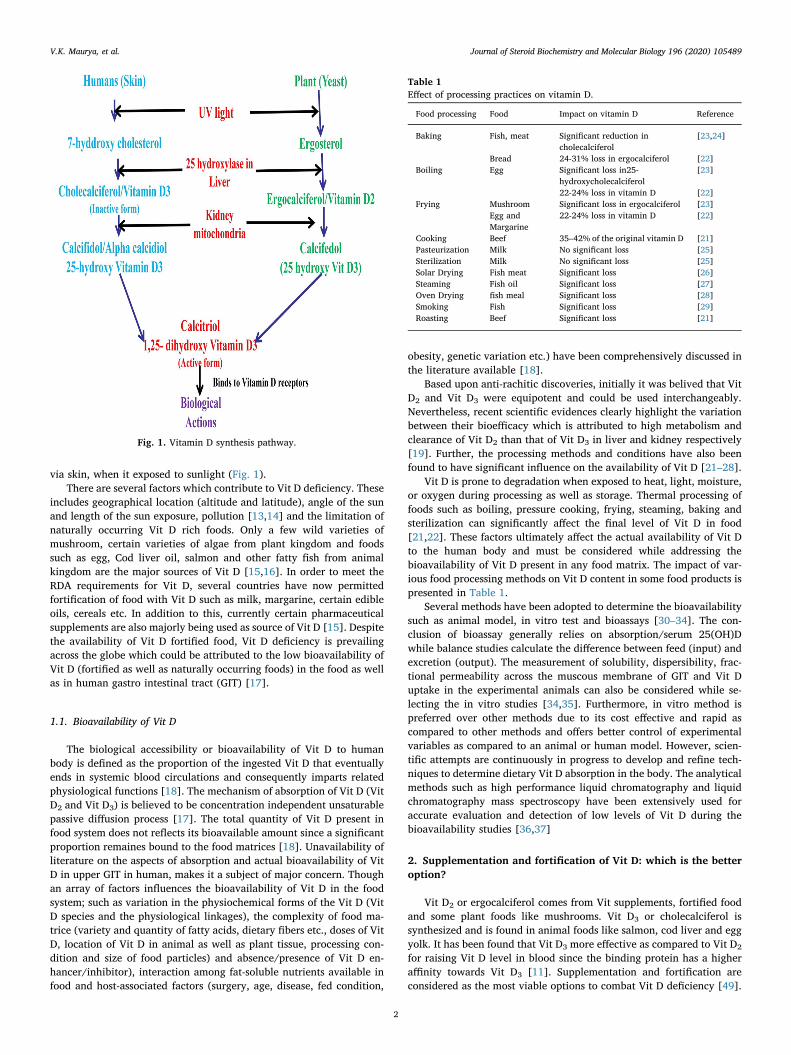
via skin, when it exposed to sunlight (Fig. 1).There are several factors which contribute to Vit D deficiency. These
includes geographical location (altitude and latitude), angle of the sunand length of the sun exposure, pollution [13,14] and the limitation ofnaturally occurring Vit D rich foods. Only a few wild varieties ofmushroom, certain varieties of algae from plant kingdom and foodssuch as egg, Cod liver oil, salmon and other fatty fish from animalkingdom are the major sources of Vit D [15,16]. In order to meet theRDA requirements for Vit D, several countries have now permittedfortification of food with Vit D such as milk, margarine, certain edibleoils, cereals etc. In addition to this, currently certain pharmaceuticalsupplements are also majorly being used as source of Vit D [15]. Despitethe availability of Vit D fortified food, Vit D deficiency is prevailingacross the globe which could be attributed to the low bioavailability ofVit D (fortified as well as naturally occurring foods) in the food as wellas in human gastro intestinal tract (GIT) [17].
1.1. Bioavailability of Vit D
The biological accessibility or bioavailability of Vit D to humanbody is defined as the proportion of the ingested Vit D that eventuallyends in systemic blood circulations and consequently imparts relatedphysiological functions [18]. The mechanism of absorption of Vit D (VitD2 and Vit D3) is believed to be concentration independent unsaturablepassive diffusion process [17]. The total quantity of Vit D present infood system does not reflects its bioavailable amount since a significantproportion remaines bound to the food matrices [18]. Unavailability ofliterature on the aspects of absorption and actual bioavailability of VitD in upper GIT in human, makes it a subject of major concern. Thoughan array of factors influences the bioavailability of Vit D in the foodsystem; such as variation in the physiochemical forms of the Vit D (VitD species and the physiological linkages), the complexity of food ma-trice (variety and quantity of fatty acids, dietary fibers etc., doses of VitD, location of Vit D in animal as well as plant tissue, processing con-dition and size of food particles) and absence/presence of Vit D en-hancer/inhibitor), interaction among fat-soluble nutrients available infood and host-associated factors (surgery, age, disease, fed condition,
obesity, genetic variation etc.) have been comprehensively discussed inthe literature available [18].
Based upon anti-rachitic discoveries, initially it was belived that VitD2 and Vit D3 were equipotent and could be used interchangeably.Nevertheless, recent scientific evidences clearly highlight the variationbetween their bioefficacy which is attributed to high metabolism andclearance of Vit D2 than that of Vit D3 in liver and kidney respectively[19]. Further, the processing methods and conditions have also beenfound to have significant influence on the availability of Vit D [21–28].
Vit D is prone to degradation when exposed to heat, light, moisture,or oxygen during processing as well as storage. Thermal processing offoods such as boiling, pressure cooking, frying, steaming, baking andsterilization can significantly affect the final level of Vit D in food[21,22]. These factors ultimately affect the actual availability of Vit Dto the human body and must be considered while addressing thebioavailability of Vit D present in any food matrix. The impact of var-ious food processing methods on Vit D content in some food products ispresented in Table 1.
Several methods have been adopted to determine the bioavailabilitysuch as animal model, in vitro test and bioassays [30–34]. The con-clusion of bioassay generally relies on absorption/serum 25(OH)Dwhile balance studies calculate the difference between feed (input) andexcretion (output). The measurement of solubility, dispersibility, frac-tional permeability across the muscous membrane of GIT and Vit Duptake in the experimental animals can also be considered while se-lecting the in vitro studies [34,35]. Furthermore, in vitro method ispreferred over other methods due to its cost effective and rapid ascompared to other methods and offers better control of experimentalvariables as compared to an animal or human model. However, scien-tific attempts are continuously in progress to develop and refine tech-niques to determine dietary Vit D absorption in the body. The analyticalmethods such as high performance liquid chromatography and liquidchromatography mass spectroscopy have been extensively used foraccurate evaluation and detection of low levels of Vit D during thebioavailability studies [36,37]
2. Supplementation and fortification of Vit D: which is the betteroption?
Vit D2 or ergocalciferol comes from Vit supplements, fortified foodand some plant foods like mushrooms. Vit D3 or cholecalciferol issynthesized and is found in animal foods like salmon, cod liver and eggyolk. It has been found that Vit D3 more effective as compared to Vit D2
for raising Vit D level in blood since the binding protein has a higheraffinity towards Vit D3 [11]. Supplementation and fortification areconsidered as the most viable options to combat Vit D deficiency [49].
Fig. 1. Vitamin D synthesis pathway.
Table 1Effect of processing practices on vitamin D.
Food processing Food Impact on vitamin D Reference
Baking Fish, meat Significant reduction incholecalciferol
[23,24]
Bread 24-31% loss in ergocalciferol [22]Boiling Egg Significant loss in25-
hydroxycholecalciferol[23]
22-24% loss in vitamin D [22]Frying Mushroom Significant loss in ergocalciferol [23]
Egg andMargarine
22-24% loss in vitamin D [22]
Cooking Beef 35–42% of the original vitamin D [21]Pasteurization Milk No significant loss [25]Sterilization Milk No significant loss [25]Solar Drying Fish meat Significant loss [26]Steaming Fish oil Significant loss [27]Oven Drying fish meal Significant loss [28]Smoking Fish Significant loss [29]Roasting Beef Significant loss [21]
V.K. Maurya, et al. Journal of Steroid Biochemistry and Molecular Biology 196 (2020) 105489
2
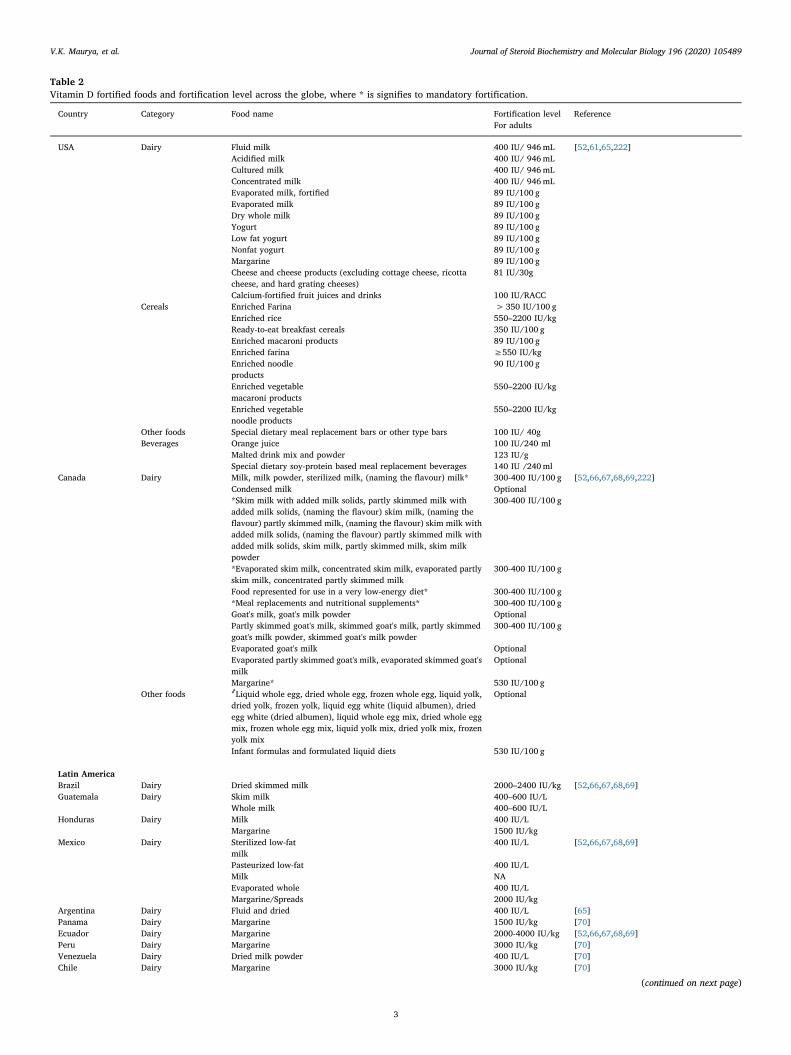
Table 2Vitamin D fortified foods and fortification level across the globe, where * is signifies to mandatory fortification.
Country Category Food name Fortification level ReferenceFor adults
USA Dairy Fluid milk 400 IU/ 946mL [52,61,65,222]Acidified milk 400 IU/ 946mLCultured milk 400 IU/ 946mLConcentrated milk 400 IU/ 946mLEvaporated milk, fortified 89 IU/100 gEvaporated milk 89 IU/100 gDry whole milk 89 IU/100 gYogurt 89 IU/100 gLow fat yogurt 89 IU/100 gNonfat yogurt 89 IU/100 gMargarine 89 IU/100 gCheese and cheese products (excluding cottage cheese, ricottacheese, and hard grating cheeses)
81 IU/30g
Calcium-fortified fruit juices and drinks 100 IU/RACCCereals Enriched Farina > 350 IU/100 g
Enriched rice 550–2200 IU/kgReady-to-eat breakfast cereals 350 IU/100 gEnriched macaroni products 89 IU/100 gEnriched farina ≥550 IU/kgEnriched noodle 90 IU/100 gproductsEnriched vegetable 550–2200 IU/kgmacaroni productsEnriched vegetable 550–2200 IU/kgnoodle products
Other foods Special dietary meal replacement bars or other type bars 100 IU/ 40gBeverages Orange juice 100 IU/240 ml
Malted drink mix and powder 123 IU/gSpecial dietary soy-protein based meal replacement beverages 140 IU /240ml
Canada Dairy Milk, milk powder, sterilized milk, (naming the flavour) milk* 300-400 IU/100 g [52,66,67,68,69,222]Condensed milk Optional*Skim milk with added milk solids, partly skimmed milk withadded milk solids, (naming the flavour) skim milk, (naming theflavour) partly skimmed milk, (naming the flavour) skim milk withadded milk solids, (naming the flavour) partly skimmed milk withadded milk solids, skim milk, partly skimmed milk, skim milkpowder
300-400 IU/100 g
*Evaporated skim milk, concentrated skim milk, evaporated partlyskim milk, concentrated partly skimmed milk
300-400 IU/100 g
Food represented for use in a very low-energy diet* 300-400 IU/100 g*Meal replacements and nutritional supplements* 300-400 IU/100 gGoat's milk, goat's milk powder OptionalPartly skimmed goat's milk, skimmed goat's milk, partly skimmedgoat's milk powder, skimmed goat's milk powder
300-400 IU/100 g
Evaporated goat's milk OptionalEvaporated partly skimmed goat's milk, evaporated skimmed goat'smilk
Optional
Margarine* 530 IU/100 gOther foods ҂Liquid whole egg, dried whole egg, frozen whole egg, liquid yolk,
dried yolk, frozen yolk, liquid egg white (liquid albumen), driedegg white (dried albumen), liquid whole egg mix, dried whole eggmix, frozen whole egg mix, liquid yolk mix, dried yolk mix, frozenyolk mix
Optional
Infant formulas and formulated liquid diets 530 IU/100 g
Latin AmericaBrazil Dairy Dried skimmed milk 2000–2400 IU/kg [52,66,67,68,69]Guatemala Dairy Skim milk 400–600 IU/L
Whole milk 400–600 IU/LHonduras Dairy Milk 400 IU/L
Margarine 1500 IU/kgMexico Dairy Sterilized low-fat 400 IU/L [52,66,67,68,69]
milkPasteurized low-fat 400 IU/LMilk NAEvaporated whole 400 IU/LMargarine/Spreads 2000 IU/kg
Argentina Dairy Fluid and dried 400 IU/L [65]Panama Dairy Margarine 1500 IU/kg [70]Ecuador Dairy Margarine 2000-4000 IU/kg [52,66,67,68,69]Peru Dairy Margarine 3000 IU/kg [70]Venezuela Dairy Dried milk powder 400 IU/L [70]Chile Dairy Margarine 3000 IU/kg [70]
(continued on next page)
V.K. Maurya, et al. Journal of Steroid Biochemistry and Molecular Biology 196 (2020) 105489
3
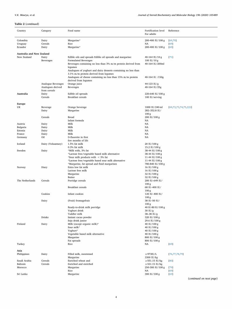
Table 2 (continued)
Country Category Food name Fortification level ReferenceFor adults
Colombia Dairy Margarine* 200-400 IU/100 g [69,70]Uruguay Cereals Rice NA [69]Ecuador Dairy Margarine* 200-400 IU/100 g [69]
Australia and New ZealandNew Zealand Dairy Edible oils and spreads Edible oil spreads and margarine: 40-164 IU/10 g [71]
Beverages Formulated Beverages 100 IU/10 gBeverages containing no less than 3% m/m protein derived fromlegumes
40-164 IU/200ml
Analogues of yoghurt and dairy desserts containing no less than3.1% m/m protein derived from legumesAnalogues of cheese containing no less than 15% m/m proteinderived from legumes
40-164 IU /150g
Analogue Beverages Orange juice 44-123 IU/gAnalogues derivedfrom cereals
Beverages 40-164 IU/25g
Australia Dairy Edible oil spreads 220-640 IU/100 gCereals Breakfast cereals 100 IU/serving
EuropeUK Beverage Orange beverage 1000 IU/240ml [69,72,73,74,75,222]
Dairy Margarine 282–352.8 IU/100 g
Cereals Bread 200 IU/100 gInfant formula NA
Austria Dairy Milk NABulgaria Dairy Milk NAEstonia Dairy Milk NAFrance Dairy Milk NAGermany Oil D-fluorette in first NA
few months of lifeIceland Dairy (Voluantary) 1.5% fat milk 20 IU/100 g
0.3% fat milk 15.2 IU/100 gSweden Dairy *Milk with, 3% fat 38-44 IU/100 g
*Lactose free/vegetable based milk alternative 38-44 IU/100 g*Sour milk products with <3% fat 11-44 IU/100 g*Lactose free/vegetable based sour milk alternative 11-44 IU/100 g*Margarine, fat spread and fluid margarines 780-840 IU/100 g
Norway Diary Extra low fat milk 16 IU/100 gLactose free milk 16 IU/100 gMargarine 32 IU/100 gButter 32 IU/100 g
The Netherlands Cereals Porridge cereals 200 IU–649 IU/100 g
Breakfast cereals 68 IU–400 IU/100 g
Cookies Infant cookies 120 IU–400 IU/100 g
Dairy (Fruit) fromagefrais 38 IU–50 IU/100 g
Ready-to-drink milk porridge 40 IU-80 IU/100 gYoghurt drink 30 IU/gToddler milk 38–38 IU/g
Drinks Instant cacao powder 320 IU/100 gSoja drink junior 29.6 IU/100 g
Finland Dairy Milk (except organic milk)* 40 IU/100 gSour milk* 40 IU/100 gYoghurt* 40 IU/100 gVegetable based milk alternative 40 IU/100 gMargarine 800 IU/100 gFat spreads 800 IU/100 g
Turkey Rice NA [69]
AsiaPhilippines Dairy Filled milk, sweetened ≥973IU/L [76,77,78,79]
Margarine 3300 IU/kgSaudi Arabia Cereals Enriched wheat and ≥551.15 IU/kg [80]Bahrain Cereals Enriched and enriched ≥551.15 IU/kgMorocco Margarine 250-300 IU/100 g [79]
Rice NA [69]Sri Lanka Margarine 300 IU/100 g [69]
(continued on next page)
V.K. Maurya, et al. Journal of Steroid Biochemistry and Molecular Biology 196 (2020) 105489
4
Supplementation involves the use of high dose of Vit D formulations.Generally, Vit D3 is administered in the form of cholecalciferol, alfa-calcidiol, and calcitriol as solo ingredient or in combination with cal-cium and other minerals or vitamins. Vit D supplements containingalfacalcidiol and calcitriol are generally available in the form of tabletsand capsules while the formulations containing cholecalciferol ingranules in sachets [38,39]. Cholecalciferol is the most favored form forprophylaxis and treatment of Vit D deficient states in not only India[38] but also worldwide [39]. Currently, Vit D supplement intake isvoluntary and its intake is the highest among infants, elderly adults andlowest among adolescents, children and young adults who are at highrisk of its deficiency. Further, the distribution of Vit D intake amongpopulation is greatly skewed to a small number of high dose supple-ments which poses a high risk of excessive intake [38,39]. The pro-curement and purchase of Vit D normally requires quite an expensivepre-packaging, an efficient distribution system and a high level ofconsumer compliance (particularly if supplements are to be consumedon a long-term basis) [40]. The shortage of supplies and poor com-pliance are constantly reported in usually adopted supplementationprogram, which result into main hurdles in its success. Hence, in viewof public health, food processors need to work on changing the shape ofVit D intake consumption pattern with the sustainable food basedstrategies; consequently filling the gap between current and re-commended intakes without putting the general population at risk ofhabitual either excessive or difficient intake. As on today, several in-novative methods have been reported for improving Vit D level in foodsby fortification and biofortification.
Biofortification relies on enhancing the levels of specific or limitingmicronutrients in edible tissues of plant/animal by combining cropmanagement, breeding, and genetic approaches [16]. Studies haveshown that Vit D2 level in fungi can be significantly enhanced by ex-posing them to UVB light [41,42]. Further, the stability of Vit D in theseirraditated mushroom can be further improved via cold storage [43].For example, the dried mushrooms are able to retain much of their Vit Dcontent even after 2–6 years of cold storage [20]. A significant increasein Vit D content in animal products (pigs, fish and hens) has also beenreported [44,47]. Vit D3 rich meat and liver can be produced by feedingpigs with Vit D3 rich feed [16,44]. Likewise, Vit D content in fish canalso be enhanced by feeding them Vit D3-rich feed [45] and hens whichwere fed on Vit D3 rich diet have shown to produce eggs with highcontent of Vit D [46,47].
Fortification of food products has been acknowledged by the World
Bank (1993) as the most cost effective way for combating the nutrientdeficiency problems among the available health interventions.Fortification refers as the addition of micronutrients to target foods forthe purpose of its enrichemnt with respect to a given micronutrient.This strategy has resulted in relatively rapid improvements in the mi-cronutrient status of a population at a very reasonable cost, particularlyif the existing technology and local distribution networks are exploited[48,49]. Unfortunately, implementation of fortification programs,especially in the developing world, has been lackadaisical [50]. Forthis, there may be several reasons including (1) lack of knowledge re-levant to micronutrient deficiency status; (2) lack of understanding ofthe significance of micronutrient deficiencies and its concern to thehealthcare system; (3) inadequate knowledge about food consumptionpatterns; and (4) the consumer acceptance, competitive and costsconcern of the food industry.
2.1. Present status of Vit D fortification
Several Vit D fortification programs have been implemented acrossthe globe. The various foods fortified with Vit D so far include mostlymilk, milk products, and edible oil. The food items normally selected forfortification solely depend on the consumption pattern of foods of thecountry’s population. Many of the foods are being fortified with Vit D inconjunction with Vit A. Various reports on successful fortification of VitD and regulatory compliance adopted for North Americans have beenpublished [51–53]. Presently Vit D fortification has become mandatoryin milk (expect goat milk and condensed milk) and margarine in Ca-nada where it is regulated by the Canadian Food and Drug Regulations[54–60] while in USA, Vit D fortification is voluntary in fluid milk andif fortified, needs to be displayed on the label [61,62]. It is also evidentthat the majority of the milk-derived products such as butter, cream,cottage cheese, sour crease, ice cream, hard and soft cheese, and yogurtare not routinely fortified with Vit D [52,61,63]. In addition to theseproducts, infant formulations are being fortified globally (40–100 IU/100 g) [64]. The food products that are being fortified with differentlevel of Vit D across the globe are listed in Table 2.
Today the fortification practices adopted by different countries inthe world depend upon the country’s regulation. Initially, all margarinemanufactured for domestic use in the UK and Ireland was subject tomandatory fortification but now it become voluntary [91]. Similarly,other foods like dried and evaporated milk, breakfast cereals, macaroni,noodles, beverages, edible oils, and wheat flour may also be voluntarily
Table 2 (continued)
Country Category Food name Fortification level ReferenceFor adults
India Oil Vanaspati 44 IU– 64 IU/100 g
[48]
Edible oil 44 IU– 64 IU/100 g
Dairy Milk 200 -300 IU/LIndonesia Margarine* 2500-3500 IU/kg [70,77,78,79,81,82,83,84,85,86,87,88,89]Thialand Sweetened condensed milk*Malaysia Dairy Condensed milk 111 IU/100 g
Malted milk Powder 667 IU/100 gLiquid foods 100 IU/100 gDried milk 333 IU/100 g
Cereals Bread 83 IU/100 gBreakfast cereals 333 IU/100 gWheat Flour 167 IU/100 gExtract of meat 2000 IU/100 gOther solid food 167 IU/100 g
Singapore Food not specified 400 IU/servingBrunei Darussalam Food not specified 50 IU/ serving
AfricaZimbabwe Cooking oil NA [70,90]Nigeria Margarine NA [69]
V.K. Maurya, et al. Journal of Steroid Biochemistry and Molecular Biology 196 (2020) 105489
5
fortified with Vit D along with other micronutrients (Table 2). How-ever, information pertaining the continuation and compliance of thesefortification regulations is very scanty [92,93]. The stability, dis-pensability, and solubility of Vit D during production and storage offoods are the key concerns for food processors.
2.2. Stability of Vit D in fortified food
In general, the success of Vit D fortification mainly depends on thestability of the fat matrix in the food as Vit D is fat soluble. Fortificationof Vit D has been a challenge to the food industry due to its instabilityand heterogeneous distribution in food. Loss of Vit D was observed invarious food systems fortified with Vit D such as milk [93], cheese[97,100,101], yogurt [102–104], and other milk based products[105,161,224]. The loss is mainly due to oxidation and isomerizationduring processing and storage [105,106]. Similarly, Vit D found to besusceptible to oxidation with poor retention property in extruded foodproducts also during storage [107]. Food processing methods such asbaking, cooking, frying and water boiling (fish, mushroom, and egg)cause significant degradation of Vit D [21,22,25,29]. In addition to thestability, uniform distribution or the homogeneity of Vit D in the for-tieid food matrix is again one of the major concern for the food in-dustry. The stability studies in fortified foods other than milk are verylimited and reports on uniform distribution are even rarer. Thus, studiesaddressing stability, homogenization, and bioavailability of Vit D in thefortified foods need to be conducted to gain a better understanding indesigning the fortified foods.
2.3. Methods for Vit D fortification
For sustainable fortification, various techniques have been adoptedsuch as direct addition, emulsification, and microencapsulation. In caseof Vit D, direct addition is the most widely adopted method for for-tification of milk and milk products [51,52,54]. In general, these pro-ducts are being spiked with Vit D where Vit D is dissolved in food gradeorganic solvent (ethanol) and butter oil, and then homogenized into thefood matrix to ensure the uniform distribution [94–96]. The depositionof Vit D inside the packaging materials especially the polypacks ortetrapacks and its degradation in aqueous food matrix leading to the VitD instability in food matrix. In emulsification method an oil phase,having Vit D, is dispersed as fine droplets in water and these fine dro-plets are then mixed with target food material such as cheese, milk andbread [97–99]. Homogenization of Vit D in the food matrix and limitedavailability of food grade emulsifiers are major challenges while de-veloping stable emulsion.
The major challenges being faced by food technologists duringfortification of Vit D are its compatibility or suitability with food ma-trix, dispersibility, homogeneity and stability in the food matrix andultimately its bioavailability to the body in required doses for com-bating the deficiency. All theses chalanges are the driving forces leadingto the development of various innovative techniques for fortifying Vit Din different food matrixs. Recent literature suggests that nano-technology offers great stability and ensures homogeneity by en-capsulation of bioactive core ingredient into a matrix with a size lowerthan 1000 nm. Microencapsulation is basically insulation of bioactivecore material by secondary wall materials which protect the core fromits external environment [108–112]. In addition to giving protection tothe bioactive compound, it also helps in controlled release of en-capsulant with high physiochemical stability. Microencapsulation alsopromises that the nanomaterials so formed would ensure high bioa-vailability, water dispersibility and better homogeneity of the for-tificant in the target food irrespective of complexity of food matrix[111]. The rising demand for functional foods has been the majordriving force for designing and production of novel nanomaterials thatare suitable for fortifying the food. Literature reports several nanoma-terials, which could be efficient carrier systems for Vit D for the purpose
of food fortification [113]. The fortification using nanomaterials offersvarious advantages over direct addition and emulsification methodsuch as high stability, better homogeneity and improved physiochem-ical as well as organoleptic characteristics [111].
3. Use of microencapsulation techniques
The success of microencapsulation of Vit D in pharmaceuticals en-couraged its application in food with the following objectives (i) beatssolubility barrier between Vit D and the food matrix (ii) shields Vit Dagainst physiochemical stress such as moisture, oxidation, pH, tem-perature, mechanical etc. (iii) guarantees better bioavailability with thecontrolled and targeted release of encapsulated Vit D (iv) does notmanipulate appearance, taste, quality of food matrix, thus sustainingcustomer acceptability.
3.1. Status of Vit D microencapsulation
The high dispensability of lipophilic drug in aqueous media ofpharmaceutical formulation made research community to assume thatsolubility of these lipophilic drugs can also be improved in the foodmatrix by microencapsulation. This assumption was evaluated by sev-eral dedicated studies such as 100-time high solubility was achievedwhen tretinoin was encapsulated with β-cyclodextrin [115] while it was10000-times for anandamide [116]. However, these cyclic moleculeshave the ability to host Vit D molecule, but its drug loading capacitywas very poor [116]. To address this problem, nanomaterials have beenintroduced that can offer high drug stability and encapsulation effi-ciency (EE). The potential of nanomaterials to become an efficientcarrier is continuously ested in pharmaceutical and the food industries.Literature reports about a range of nanomaterials such as emulsion[118,119], liposome [100,120–132], niosome [133–137], solid lipidnanoparticles [138] and nanostructured lipid carriers [139]. Thoughseveral excellent reviews are available focusing the wall material, mi-croencapsulation techniques, and nanomaterials for bioactive com-pounds [111,114,140,141] but there is a lack of dedicated reports ad-dressing microencapsulation techniques which are exclusively used todevelop Vit D nanomaterials for food application (Table 3).
3.1.1. Vit D microencapsulation using spray drying techniqueSpray drying is renowned as one of the oldest technique used for
bioactive compounds encapsulation. Vit D is needed to be homogenizedin a dispersion containing wall materials (polymers). Then, the homo-genized dispersion needs to be fed to the spray dryer and atomized byhot air that leads to the development of nanomaterials in consequenceof water evaporation. The encapsulation process is subjected to a rangeof factors like homogeneity of dispersion system, quantity, quality andtype of emulsifier used, feed rate, viscosity of dispersion system, pres-sure of hot air, the flow rate of hot air and inlet and outlet temperature.In spite of better control on the shape and size of nanomaterials con-tinuous and reproducible nature, low cost, easy scale-up, spray drying isnot quite popular for bioactive compounds exclusively for heat sensitivecompounds [141,173–175]. Further, several researchers have compre-hensively described the key factors need to be taken under considera-tion during spray drying while designing nanomaterials for food ap-plication [140,141,176–181]. Furthermore, spray drying offers greatflexibility for choice of wall materials, one or more than one but the useof spray drying in Vit D microencapsulation is even rarer as it mandatesVit D to be in water dispersed form. Despite several advantages, the fullpotential of spray drying is still fully unexplored for Vit D encapsulationwhich could be accredited to resultant porous nanomaterials that areprone to degradation of encapsulated Vit D hence lacking the aim ofencapsulation [170–172]. Vit D was encapsulated using differentcombinations of maltodextrin, gum arabic, modified starch and wheyprotein concentrate to study the effect of temperature on the physico-chemical characteristics of spray-dried whey nanoparticles
V.K. Maurya, et al. Journal of Steroid Biochemistry and Molecular Biology 196 (2020) 105489
6
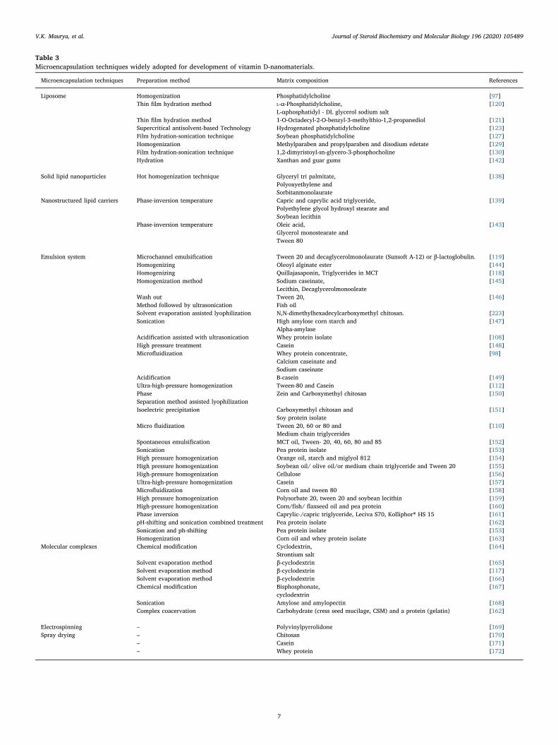
Table 3Microencapsulation techniques widely adopted for development of vitamin D-nanomaterials.
Microencapsulation techniques Preparation method Matrix composition References
Liposome Homogenization Phosphatidylcholine [97]Thin film hydration method L-α-Phosphatidylcholine,
L-αphosphatidyl - DL glycerol sodium salt[120]
Thin film hydration method 1-O-Octadecyl-2-O-benzyl-3-methylthio-1,2-propanediol [121]Supercritical antisolvent-based Technology Hydrogenated phosphatidylcholine [123]Film hydration-sonication technique Soybean phosphatidylcholine [127]Homogenization Methylparaben and propylparaben and disodium edetate [129]Film hydration-sonication technique 1,2-dimyristoyl-sn-glycero-3-phosphocholine [130]Hydration Xanthan and guar gums [142]
Solid lipid nanoparticles Hot homogenization technique Glyceryl tri palmitate,Polyoxyethylene andSorbitanmonolaurate
[138]
Nanostructured lipid carriers Phase-inversion temperature Capric and caprylic acid triglyceride,Polyethylene glycol hydroxyl stearate andSoybean lecithin
[139]
Phase-inversion temperature Oleic acid,Glycerol monostearate andTween 80
[143]
Emulsion system Microchannel emulsification Tween 20 and decaglycerolmonolaurate (Sunsoft A-12) or β-lactoglobulin. [119]Homogenizing Oleoyl alginate ester [144]Homogenizing Quillajasaponin, Triglycerides in MCT [118]Homogenization method Sodium caseinate,
Lecithin, Decaglycerolmonooleate[145]
Wash outMethod followed by ultrasonication
Tween 20,Fish oil
[146]
Solvent evaporation assisted lyophilization N,N-dimethylhexadecylcarboxymethyl chitosan. [223]Sonication High amylose corn starch and
Alpha-amylase[147]
Acidification assisted with ultrasonication Whey protein isolate [108]High pressure treatment Casein [148]Microfluidization Whey protein concentrate,
Calcium caseinate andSodium caseinate
[98]
Acidification B-casein [149]Ultra-high-pressure homogenization Tween-80 and Casein [112]PhaseSeparation method assisted lyophilization
Zein and Carboxymethyl chitosan [150]
Isoelectric precipitation Carboxymethyl chitosan andSoy protein isolate
[151]
Micro fluidization Tween 20, 60 or 80 andMedium chain triglycerides
[110]
Spontaneous emulsification MCT oil, Tween- 20, 40, 60, 80 and 85 [152]Sonication Pea protein isolate [153]High pressure homogenization Orange oil, starch and miglyol 812 [154]High pressure homogenization Soybean oil/ olive oil/or medium chain triglyceride and Tween 20 [155]High-pressure homogenization Cellulose [156]Ultra-high-pressure homogenization Casein [157]Microfluidization Corn oil and tween 80 [158]High pressure homogenization Polysorbate 20, tween 20 and soybean lecithin [159]High-pressure homogenization Corn/fish/ flaxseed oil and pea protein [160]Phase inversion Caprylic-/capric triglyceride, Leciva S70, Kolliphor® HS 15 [161]pH-shifting and sonication combined treatment Pea protein isolate [162]Sonication and ph-shifting Pea protein isolate [153]Homogenization Corn oil and whey protein isolate [163]
Molecular complexes Chemical modification Cyclodextrin,Strontium salt
[164]
Solvent evaporation method β-cyclodextrin [165]Solvent evaporation method β-cyclodextrin [117]Solvent evaporation method β-cyclodextrin [166]Chemical modification Bisphosphonate,
cyclodextrin[167]
Sonication Amylose and amylopectin [168]Complex coacervation Carbohydrate (cress seed mucilage, CSM) and a protein (gelatin) [162]
Electrospinning – Polyvinylpyrrolidone [169]Spray drying – Chitosan [170]
– Casein [171]– Whey protein [172]
V.K. Maurya, et al. Journal of Steroid Biochemistry and Molecular Biology 196 (2020) 105489
7
encapsulating Vit D [172]. Higher stability and greater bioavailabilityof Vit D2 were achieved when it was encapsulated in casein micellesusing spray drying [171]. Similarly sustained release of Vit D2 in si-mulated GIT conditions was demonstrated by ethylcellulose coatedspray dried nanomaterials containing chitosan [170]. The stability issuecan be resolved by proper selection of wall materials and associationwith other microencapsulation techniques.
3.1.2. Vit D microencapsulation using emulsification techniqueThis system involves at least two immiscible phases (lipid and
water) where one phase needs to be dispersed as small spherical dro-plets within another phase. On the basis of the spatial arrangement oftwo phases, the emulsion system is generally classified into two classesi.e. oil in water (O/W) or water in oil (W/O). Then, these two im-miscible phases need to be stabilized by surfactants and emulsifiers[182]. Several complex emulsion system like oil-in-water-in-oil (O/W/O), water-in-oil-in-water (W/O/W), water-in-oil-in-oil (W/O/O) orwater-in-oil-in-oil-in-water (W/O/O/W), are reported in literature[183–185]. Several researchers have explored emulsion techniques todevelop Vit D-nanomaterials using food grade materials such as wheyprotein isolate (WPI) [108], casein [149], Medium chaing triglycerides(MCT) and Tween 20, 40, 80, 85 [152], MCT and Tween 20, 60, 80[110], carboxymethyl chitosan and SPI [151], zein and carboxymethylchitosan [150], Tween 20 and casein [151,213], WPI, calcium caseinateand sodium caseinate [98], casein [148], HACS and α-amylase [147],Tween 20 and fish oil [146], sodium caseinate and lecithin [145],quilajapaponin [118] and oleoyl alginate ester [144] and PPI [153]. VitD emulsion fabricated with sodium caseinate, calcium caseinate, nonfatdry milk, and whey protein have found to be stable during cheddarcheese preparation [98]. The selection of emulsion method for Vit Dencapsulation depends on various factors such as absence/presence ofantioxidants, quantity and type of carrier oils and surfactant. It wasobserved that the stability of encapsulated Vit D is highly correlated tothe stability of emulsion system. Further, it is also evident that thepresence of an antioxidant in the emulsion system also enhances thestability of Vit D.
3.1.3. Vit D microencapsulation using liposomeLiterature reveled about various preparation methods for liposome
which are comprehensively discussed by researchers in their excellentreviews [131,136,186–192]. In general, liposomes are referred to thespherical liquid structures in which an aqueous core bounded by asingle (unilamellar liposomes) or multiple lipid bilayers (multilamellarliposomes). The ability to host both hydrophilic and hydrophobicbioactive ingredients individually or simultaneously makes liposomethe most adopted encapsulation technique for Vit D. In addition toflexibility in composition and size, liposome also promises high bio-compatibility with animal tissue as it mimics with the natural plasmamembrane [188]. Several researchers have fabricated liposome forencapsulation of Vit D using 1,2-dimyristoyl-sn-glycero-3-phos-phocholine (DMPC) [130], methylparaben and propylparaben and thedi-sodium edentate [129], L-α-phosphatidylcholine and L-α phospha-tidyl-DL glycerol [120], 1-O-Octadecyl-2-O-benzyl-3-methylthio-1,2-propanediol [121], phosphatidylcholine [100], hydrogenated phos-phatidylcholine [123] and soybean phosphatidylcholine [127].Though, Vit D shows high chemical stability when it is integratedwithin liposome but its application in food fortification is still not fullyexplored. The limited use of liposome in Vit D fortification could beattributed to its dependency on soya derived lecithin which carriesintense smell. This issue can be easily resolved by replacing soya de-rived lecithin with milk-based lecithin or hydrogenated lecithin.
3.1.4. Vit D microencapsulation using solid lipid nanoparticlesIt is referred as the most suitable encapsulation technique for vita-
mins encapsulation as it has the hybrid structure of liposome andemulsion system hence tenders a range of advantages like high drug
loading capacity, higher encapsulation efficiency, and better chemicalstability against physiochemical stress. The literature describes thepreparation methods for solid lipid nanoparticles (SLN)[136,137,193–201]. The ability of SLN to encapsulate and protect Vit Dis still untapped and the only single report has been generated till datein which Vit D-SLN was prepared using molten tripalmitin [201].
3.1.5. Vit D microencapsulation using nanostructured lipid carriers (NLC)NLC generally encompasses unstructured solid lipid matrix com-
prised of a mixture of liquid and solid lipid blend and an aqueous phaseconsisting of a surfactant or a mixture of surfactants. Typically, liquidand solid lipids are blended in a ratio that could vary from 70:30 to99.90:0.10 while the surfactant content is kept between 1.5–5% (W/V)[202]. The unstructured/partially solid matrix creates interesting na-nostructures, which enhance the stability of the entrapped bioactivecompound, facilitate high loading capacity and offers controlled/targetrelease. Literature dictates various methods for NLC preparations[202–205]. Despite being the most promising technique for drug de-livery, NLC is among the least explored method for Vit D encapsulation.Till date, only three dedicated studies were reported addressing Vit Dencapsulation in NLC [139,143,224]. In the first report, Vit D loadedNLC was formulated by phase-inversion temperature method displayedhigh physical and chemical stability for NLC as well as Vit D and wasfound to be a suitable vehicle for milk fortification [139]. While thesecond report was conducted to evaluate the drug release kinetic of VitD loaded NLC and were fabricated with oleic acid, glycerol monosterateand Tween 80 using hot high-pressure homogenization [143]. NLCparticles displayed biphasic kinetic release (burst effect) resulting inalmost 50% of the Vit D released during the first 2 h and 80% releasedafter 4 h of digestion, followed by a sustained release until 90.9% of theVit D during 8 h [143].
3.1.6. Vit D microencapsulation using molecular complexIn general, the molecular complex is formed with the use of cyclo-
dextrin which can host bioactive agents within its void. Cyclodextrin isusually applied for encapsulation of Vit D in pharmaceutical formula-tions to assess its chemical stability against various physiochemicalstresses [117,119,144,153,167].
3.1.7. Vit D microencapsulation using electrospinningIt is a fiber producing technique which exertes electric force to draw
charged fiber of polymer solutions or polymer melts up to diameters ofnanoscale. This continuous process is performed by extruding disper-sion of polymer through the needle on rotating drums to impact chargeon fibers. The literature describes electrospinning as the most suitabletechniques for thermo-sensitive bioactive agents, but its use for Vit Dencapsulation is very scant. Till date single report is documented inwhich Vit D-nanofiber fabricated using poly (vinyl pyrrolidone) [169].
3.2. The fate of Vit D-nanoscale materials in GIT
The small intestine is recognized as the site of absorption of Vit Dafter its oral ingestion [206,207]. Fig. 2 illustrates the main routes ofVit D absorption in small intestine. Nanomaterials encapsulating Vit Dhave demonstrated its improved absorption [114,122,139,161] and themechanism how nanomaterials improve its oral bioavailability has al-ready been reviewed in the previous article [114]. Generally, mixedmicelles are generated as a result of digestion of lipid as well as na-nomaterials and facilitate Vit D passage passing through the aqueousmucous layer to make it bioavailable to brush bordered enterocytes.The absorbed Vit D is then encased into chylomicrons within the en-terocytes depending on their hydrophobicity [208,209]. The chylomi-crons and lipid particles are endogenously produced within the en-terocytes using lipid components (monoacyglycerols, free fatty acids,and sterol) of mixed micelles [210]. Then the chylomicrons in-corporating Vit D are transported to the lymphatic circulation system
V.K. Maurya, et al. Journal of Steroid Biochemistry and Molecular Biology 196 (2020) 105489
8
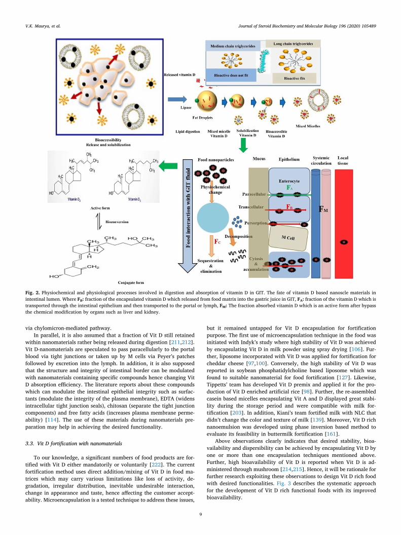
via chylomicron-mediated pathway.In parallel, it is also assumed that a fraction of Vit D still retained
within nanomaterials rather being released during digestion [211,212].Vit D-nanomaterials are speculated to pass paracellularly to the portalblood via tight junctions or taken up by M cells via Peyer’s patchesfollowed by excretion into the lymph. In addition, it is also supposedthat the structure and integrity of intestinal border can be modulatedwith nanomaterials containing specific compounds hence changing VitD absorption efficiency. The literature reports about these compoundswhich can modulate the intestinal epithelial integrity such as surfac-tants (modulate the integrity of the plasma membrane), EDTA (widensintracellular tight junction seals), chitosan (separate the tight junctioncomponents) and free fatty acids (increases plasma membrane perme-ability) [114]. The use of these materials during nanomaterials pre-paration may help in achieving the desired functionality.
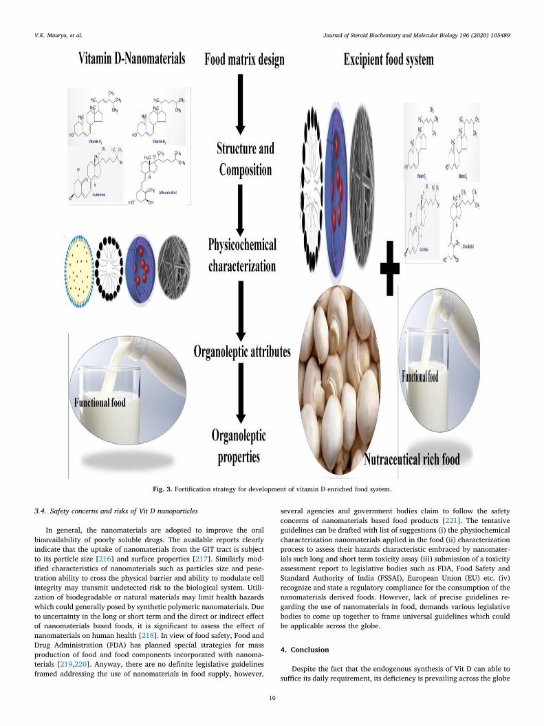
3.3. Vit D fortification with nanomaterials
To our knowledge, a significant numbers of food products are for-tified with Vit D either mandatorily or voluntarily [222]. The currentfortification method uses direct addition/mixing of Vit D in food ma-trices which may carry various limitations like loss of activity, de-gradation, irregular distribution, inevitable undesirable interaction,change in appearance and taste, hence affecting the customer accept-ability. Microencapsulation is a tested technique to address these issues,
but it remained untapped for Vit D encapsulation for fortificationpurpose. The first use of microencapsulation technique in the food wasinitiated with Indyk’s study where high stability of Vit D was achievedby encapsulating Vit D in milk powder using spray drying [106]. Fur-ther, liposome incorporated with Vit D was applied for fortification forcheddar cheese [97,100]. Conversely, the high stability of Vit D wasreported in soybean phosphatidylcholine based liposome which wasfound to suitable nanomaterial for food fortification [127]. Likewise,Tippetts’ team has developed Vit D premix and applied it for the pro-duction of Vit D enriched artificial rice [98]. Further, the re-assembledcasein based micelles encapsulating Vit A and D displayed great stabi-lity during the storage period and were compatible with milk for-tification [203]. In addition, Kiani’s team fortified milk with NLC thatdidn’t change the color and texture of milk [139]. Moreover, Vit D richnanoemulsion was developed using phase inversion based method toevaluate its feasibility in buttermilk fortification [161].
Above observations clearly indicates that desired stability, bioa-vailability and dispersibility can be achieved by encapsulating Vit D byone or more than one encapsulation techniques mentioned above.Further, high bioavailability of Vit D is reported when Vit D is ad-ministered through mushroom [214,215]. Hence, it will be rationale forfurther research exploiting these observations to design Vit D rich foodwith desired functionalities. Fig. 3 describes the systematic approachfor the development of Vit D rich functional foods with its improvedbioavailability.
Fig. 2. Physiochemical and physiological processes involved in digestion and absorption of vitamin D in GIT. The fate of vitamin D based nanoscle materials inintestinal lumen. Where FB: fraction of the encapsulated vitamin D which released from food matrix into the gastric juice in GIT, FA: fraction of the vitamin D which istransported through the intestinal epithelium and then transported to the portal or lymph, FM: The fraction absorbed vitamin D which is an active form after bypassthe chemical modification by organs such as liver and kidney.
V.K. Maurya, et al. Journal of Steroid Biochemistry and Molecular Biology 196 (2020) 105489
9
3.4. Safety concerns and risks of Vit D nanoparticles
In general, the nanomaterials are adopted to improve the oralbioavailability of poorly soluble drugs. The available reports clearlyindicate that the uptake of nanomaterials from the GIT tract is subjectto its particle size [216] and surface properties [217]. Similarly mod-ified characteristics of nanomaterials such as particles size and pene-tration ability to cross the physical barrier and ability to modulate cellintegrity may transmit undetected risk to the biological system. Utili-zation of biodegradable or natural materials may limit health hazardswhich could generally posed by synthetic polymeric nanomaterials. Dueto uncertainty in the long or short term and the direct or indirect effectof nanomaterials based foods, it is significant to assess the effect ofnanomaterials on human health [218]. In view of food safety, Food andDrug Administration (FDA) has planned special strategies for massproduction of food and food components incorporated with nanoma-terials [219,220]. Anyway, there are no definite legislative guidelinesframed addressing the use of nanomaterials in food supply, however,
several agencies and government bodies claim to follow the safetyconcerns of nanomaterials based food products [221]. The tentativeguidelines can be drafted with list of suggestions (i) the physiochemicalcharacterization nanomaterials applied in the food (ii) characterizationprocess to assess their hazards characteristic embraced by nanomater-ials such long and short term toxicity assay (iii) submission of a toxicityassessment report to legislative bodies such as FDA, Food Safety andStandard Authority of India (FSSAI), European Union (EU) etc. (iv)recognize and state a regulatory compliance for the consumption of thenanomaterials derived foods. However, lack of precise guidelines re-garding the use of nanomaterials in food, demands various legislativebodies to come up together to frame universal guidelines which couldbe applicable across the globe.
4. Conclusion
Despite the fact that the endogenous synthesis of Vit D can able tosuffice its daily requirement, its deficiency is prevailing across the globe
Fig. 3. Fortification strategy for development of vitamin D enriched food system.
V.K. Maurya, et al. Journal of Steroid Biochemistry and Molecular Biology 196 (2020) 105489
10
which could be attributed to various factors such awareness, socio-economic, cultural and religious constraints and lack of diversity in VitD rich foods. These factors equally contribute to the determination of itsRDA and fortification level, which are subject to the country’s regula-tions. Fortification is considered as the most effective among theavailable health interventions, but it brings inevitable interactions withfood components resulting in the loss during food processing and sto-rage. Vit D bioavailability in food can be improved either through itsdirect fortification or by the use of Vit D-nanomaterials in processedfoods. Microencapsulation seems to be an indispensable tool to designVit D-nanomaterials with desired functionality such as high stabilityagainst photochemical and mechanical stress, better homogeneity withthe food system, improved oral bioavailability, avoidance the over-dosing and improved organoleptic properties. Rationale knowledgeabout Vit D in the view of its chemistry, source, factors influencing itsdeficiency as well as bioavailability, RDA and fortification level, andmicroencapsulation techniques may aid better understanding in thedesigning of novel nanomaterials with desired properties for food for-tification.
References
[1] M. Abu el Maaty, F. Almouhanna, S. Wölfl, Expression of TXNIP in cancer cells andregulation by 1, 25 (OH) 2D3: is it really the vitamin D3 upregulated protein? Int.J. Mol. Sci. 19 (2018) 796.
[2] M. Atteritano, L. Mirarchi, E. Venanzi-Rullo, D. Santoro, C. Iaria, A. Catalano,A. Lasco, V. Arcoraci, A. Lo Gullo, A. Bitto, Vitamin D status and the relationshipwith bone fragility fractures in HIV-infected patients: a case control study, Int. J.Mol. Sci. 19 (2018) 119, https://doi.org/10.3390/ijms19010119.
[3] C. Legarth, D. Grimm, M. Wehland, J. Bauer, M. Krüger, The impact of vitamin Din the treatment of essential hypertension, Int. J. Mol. Sci. 19 (2018) 455, https://doi.org/10.3390/ijms19020455.
[4] A.T. Slominski, A.A. Brożyna, C. Skobowiat, M.A. Zmijewski, T.-K. Kim,Z. Janjetovic, A.S. Oak, W. Jozwicki, A.M. Jetten, R.S. Mason, On the role ofclassical and novel forms of vitamin D in melanoma progression and management,J. Steroid Biochem. Mol. Biol. 177 (2018) 159–170, https://doi.org/10.1016/j.jsbmb.2017.06.013.
[5] J. Wierzbicka, A. Binek, T. Ahrends, J.D. Nowacka, A. Szydłowska, Ł. Turczyk,T. Wąsiewicz, P. Wierzbicki, R. Sądej, R. Tuckey, Differential antitumor effects ofvitamin D analogues on colorectal carcinoma in culture, Int. J. Oncol. 47 (2015)1084–1096, https://doi.org/10.3892/ijo.2015.3088.
[6] W.H. Organization, Global Prevalence of Vitamin A Deficiency in Populations atRisk 1995-2005, WHO global database on vitamin A deficiency, 2009, https://doi.org/10.1016/j.gheart.2014.03.2321.
[7] W. Grant, An estimate of the global reduction in mortality rates through doublingvitamin D levels, Eur. J. Clin. Nutr. 65 (2011) 1016, https://doi.org/10.1038/ejcn.2011.68.
[8] W.B. Grant, G.K. Schwalfenberg, S.J. Genuis, S.J. Whiting, An estimate of theeconomic burden and premature deaths due to vitamin D deficiency in Canada,Mol. Nutr. Food Res. 54 (2010) 1172–1181, https://doi.org/10.1002/mnfr.200900420.
[9] R. Zhang, D.P. Naughton, Vitamin D in health and disease: current perspectives,Nutr. J. 9 (2010) 65, https://doi.org/10.1186/1475-2891-9-65.
[10] M.F. Holick, T.C. Chen, Vitamin D deficiency: a worldwide problem with healthconsequences, Am. J. Clin. Nutr. 87 (2008) 1080S–1086S, https://doi.org/10.1093/ajcn/87.4.1080s.
[11] R.P. Heaney, R.R. Recker, J. Grote, R.L. Horst, L.A. Armas, Vitamin D3 is morepotent than vitamin D2 in humans, J. Clin. Endocrinol. Metab. 96 (2011)E447–E452, https://doi.org/10.1210/jc.2010-2230.
[12] A. Mithal, D.A. Wahl, J.-P. Bonjour, P. Burckhardt, B. Dawson-Hughes,J.A. Eisman, G.E.-H. Fuleihan, R.G. Josse, P. Lips, J. Morales-Torres, Global vi-tamin D status and determinants of hypovitaminosis D, Osteoporosis Int. 20 (2009)1807–1820, https://doi.org/10.1007/s00198-009-0954-6.
[13] C. Harinarayan, S.R. Joshi, Vitamin D status in India–its implications and remedialmeasures, JAPI 57 (2009) 40–48, https://doi.org/10.1007/978-1-60327-303-9_28.
[14] M.G. Kimlin, Geographic location and vitamin D synthesis, Mol. Asp. Med. 29(2008) 453–461, https://doi.org/10.1016/j.mam.2008.08.005.
[15] L. O’Mahony, M. Stepien, M.J. Gibney, A.P. Nugent, L. Brennan, The potential roleof vitamin D enhanced foods in improving vitamin D status, Nutrients 3 (2011)1023–1041, https://doi.org/10.3390/nu3121023.
[16] S.K. Duffy, A.K. Kelly, R. Gaurav, J. O’Doherty, Biofortification of meat with vi-tamin D, CAB Rev. 13 (2018) 1–11, https://doi.org/10.1079/pavsnnr201813045.
[17] P. Borel, D. Caillaud, N. Cano, Vitamin D bioavailability: state of the art, Crit. Rev.Food Sci. Nutr. 55 (2015) 1193–1205, https://doi.org/10.1080/10408398.2012.688897.
[18] V.K. Maurya, M. Aggarwal, Factors influencing the absorption of vitamin D in GIT:an overview, J. Food Sci. Technol. 54 (2017) 3753–3765, https://doi.org/10.1007/s13197-017-2840-0.
[19] L.A. Armas, B.W. Hollis, R.P. Heaney, Vitamin D2 is much less effective than vi-tamin D3 in humans, J. Clin. Endocrinol. Metab. 89 (2004) 5387–5391, https://doi.org/10.1210/jc.2004-0360.
[20] J.I. Rangel-Castro, A. Staffas, E. Danell, The ergocalciferol content of dried pig-mented and albino Cantharellus cibarius fruit bodies, Mycol. Res. 106 (2002)70–73, https://doi.org/10.1017/s0953756201005299.
[21] M. Bennink, K. Ono, B.12 Vitamin, E and D content of raw and cooked beef, J.Food Sci. 47 (1982) 1786–1792, https://doi.org/10.1111/j.1365-2621.1982.tb12883.x.
[22] J. Jakobsen, P. Knuthsen, Stability of vitamin D in foodstuffs during cooking, FoodChem. 148 (2014) 170–175, https://doi.org/10.1016/j.foodchem.2013.10.043.
[23] P. Mattila, R. Ronkainen, K. Lehikoinen, V. Piironen, Effect of household cookingon the vitamin D content in fish, eggs, and wild mushrooms, J. Food Composit.Anal. 12 (1999) 153–160, https://doi.org/10.1006/jfca.1999.0828.
[24] E. Mawer, U. Gomes, Estimation of vitamin D and its metabolites in meat, vitaminD, a pluripotent steroid hormone: structural studies, Mol. Endocrinol. Clin. Appl.(1994) 775–776.
[25] O. Fennema, Chemical Changes in Food During Processing—An Overview,Chemical Changes in Food During Processing, Springer, 1985, pp. 1–16, https://doi.org/10.1007/978-1-4613-2265-8_1.
[26] H. Suzuki, S. Hayakawa, S. Wada, E. Okazaki, M. Yamazawa, Effect of solar dryingon vitamin D3 and provitamin D3 contents in fish meat, J. Agric. Food. Chem. 36(1988) 803–806, https://doi.org/10.1021/jf00082a033.
[27] K. Scott, J. Latshaw, Effects of commercial processing on the fat-soluble vitamincontent of menhaden fish oil, J. Am. Oil Chem. Soc. 68 (1991) 234–236, https://doi.org/10.1007/bf02657615.
[28] K.C. Scott, J.D. Latshaw, The vitamin D3 and precholecalciferol content of men-haden fish meal as affected by drying conditions, Anim. Feed Sci. Technol. 47(1994) 99–105, https://doi.org/10.1016/0377-8401(94)90163-5.
[29] A.A. Bhuiyan, W. Ratnayake, R. Ackman, Nutritional composition of raw andsmoked Atlantic mackerel (scomber scombrus): oil-and water-soluble vitamins, J.Food Composition Analysis 6 (1993) 172–184, https://doi.org/10.1006/jfca.1993.1019.
[30] F. Farraye, H. Nimitphong, A. Stucchi, K. Dendrinos, A. Boulanger,A. Vijjeswarapu, A. Tanennbaum, R. Biancuzzo, T. Chen, M. Holick, Use of a novelvitamin D bioavailability test demonstrates that vitamin D absorption is decreasedin patients with quiescent crohn’s disease, Inflamm. Bowel Dis. 17 (2011)2116–2121, https://doi.org/10.1002/ibd.21595.
[31] K. Hutchinson, M. Healy, V. Crowley, M. Louw, Y. Rochev, Verification of abbott25-OH-vitamin D assay on the architect system, Practical Laboratory Medicine 7(2017) 27–35, https://doi.org/10.1016/j.plabm.2017.01.001.
[32] Y. Cohen, M. Levi, U. Lesmes, M. Margier, E. Reboul, Y.D. Livney, Re-assembledcasein micelles improve in vitro bioavailability of vitamin D in a caco-2 cell model,Food & Funct. 8 (2017) 2133–2141, https://doi.org/10.1039/c7fo00323d.
[33] A.S. Kadappan, C. Guo, C.E. Gumus, A. Bessey, R.J. Wood, D.J. McClements,Z. Liu, The efficacy of nanoemulsion‐based delivery to improve vitamin D ab-sorption: comparison of In Vitro and In Vivo Studies, Mol. Nutr. Food Res. 62(2018) 1700836, , https://doi.org/10.1002/mnfr.201700836.
[34] H. Leyva-Jimenez, Y. Jameel, M. Al-Ajeeli, A. Alsadwi, R. Abdaljaleel, C. Bailey,Relative bioavailability determination of highly concentrated cholecalciferol (vi-tamin D3) sources employing a broiler chick bioassay, J. Appl. Poultry Res. 27(2018) 363–370, https://doi.org/10.3382/japr/pfy007.
[35] H. Leyva-Jimenez, M. Khan, K. Gardner, R.A. Abdaljaleel, Y. AL-Jumaa,A.M. Alsadwi, C.A. Bailey, Developing a novel oral vitamin D3 intake bioassay tore-evaluate the vitamin D3 requirement for modern broiler chickens, Poult. Sci. 98(9) (2019) 3770–3776, https://doi.org/10.3382/ps/pez074.
[36] J. Adamec, A. Jannasch, J. Huang, E. Hohman, J.C. Fleet, M. Peacock,M.G. Ferruzzi, B. Martin, C.M. Weaver, Development and optimization of anLC‐MS/MS‐based method for simultaneous quantification of vitamin D2, vitaminD3, 25‐hydroxyvitamin D2 and 25‐hydroxyvitamin D3, J. Sep. Sci. 34 (2011)11–20, https://doi.org/10.1002/jssc.201000410.
[37] F.G. Strathmann, T.J. Laha, A.N. Hoofnagle, Quantification of 1α, 25-dihydroxyvitamin D by immunoextraction and liquid chromatography–tandem mass spec-trometry, Clin. Chem. 57 (9) (2011) 1279–1285.
[38] Y. Lhamo, P.K. Chugh, C. Tripathi, Vitamin D supplements in the Indian Market,Indian J. Pharmaceutical Sciences 78 (2016) 41, https://doi.org/10.4103/0250-474x.180248.
[39] S. Garg, D. Sabri, J. Kanji, P. Rakkar, Y. Lee, N. Naidoo, D. Svirskis, Evaluation ofvitamin D medicines and dietary supplements and the physicochemical analysis ofselected formulations, The J. Nutrition, Health & aging 17 (2013) 158–161,https://doi.org/10.1007/s12603-012-0090-4.
[40] J. Dwyer, P. Coates, M. Smith, Dietary supplements: regulatory challenges andresearch resources, Nutrients 10 (2018) 41, https://doi.org/10.3390/nu10010041.
[41] J. Ko, B. Lee, J. Lee, H.J. Park, Effect of UV-B exposure on the concentration ofvitamin D2 in sliced shiitake mushroom (lentinus edodes) and white buttonmushroom (agaricus bisporus), J. Agric. Food. Chem. 56 (2008) 3671–3674,https://doi.org/10.1021/jf073398s.
[42] S.R. Koyyalamudi, S.-C. Jeong, C.-H. Song, K.Y. Cho, G. Pang, Vitamin D2 for-mation and bioavailability from agaricus bisporus button mushrooms treated withultraviolet irradiation, J. Agric. Food. Chem. 57 (2009) 3351–3355, https://doi.org/10.1021/jf803908q.
[43] J.S. Roberts, A. Teichert, T.H. McHugh, Vitamin D2 formation from post-harvestUV-B treatment of mushrooms (agaricus bisporus) and retention during storage, J.Agric. Food. Chem. 56 (2008) 4541–4544, https://doi.org/10.1021/jf0732511.
[44] S.K. Duffy, A.K. Kelly, G. Rajauria, J. Jakobsen, L.C. Clarke, F.J. Monahan,
V.K. Maurya, et al. Journal of Steroid Biochemistry and Molecular Biology 196 (2020) 105489
11
K.G. Dowling, G. Hull, K. Galvin, K.D. Cashman, The use of synthetic and naturalvitamin D sources in pig diets to improve meat quality and vitamin D content,Meat Science 143 (2018) 60–68, https://doi.org/10.1016/j.meatsci.2018.04.014.
[45] I. Graff, S. Høie, G. Totland, Ø. Lie, Three different levels of dietary vitamin D3 fedto first‐feeding fry of Atlantic salmon (salmo salar L.): Effect on growth, mortality,calcium content and bone formation, Aquaculture Nutrition 8 (2002) 103–111,https://doi.org/10.1046/j.1365-2095.2002.00197.x.
[46] P. Mattila, J. Valaja, L. Rossow, E. Venäläinen, T. Tupasela, Effect of vitamin D2-and D3-enriched diets on egg vitamin D content, production, and bird conditionduring an entire production period, Poult. Sci. 83 (2004) 433–440, https://doi.org/10.1093/ps/83.3.433.
[47] P. Mattila, Enrichment of hen eggs with vitamin D for human health, Handbook ofEggs in Human Function, Wageningen Academic Publishers, 2015, pp.C178–C183, https://doi.org/10.3920/978-90-8686-804-9_10.
[48] FSSAI, Large Scale Food Fortification in India, (2016) https://archive.fssai.gov.in/dam/jcr:c746d723/Large_scale_Food_Fortification.pdf.
[49] O. Dary, R. Hurrell, Guidelines on Food Fortification With Micronutrients,Switzerland World Health Organization, Food and Agricultural Organization ofthe United Nations, Geneva, 2006https://www.who.int/nutrition/publications/guide_food_fortification_micronutrients.pdf.
[50] A. Gupta, Fortification of foods with vitamin D in India, Nutrients 6 (2014)3601–3623, https://doi.org/10.3390/nu6093601.
[51] M.S. Calvo, S.J. Whiting, Survey of current vitamin D food fortification practices inthe United States and Canada, J. Steroid Biochem. Mol. Biol. 136 (2013) 211–213,https://doi.org/10.1016/j.jsbmb.2012.09.034.
[52] M.S. Calvo, S.J. Whiting, C.N. Barton, Vitamin D fortification in the United Statesand Canada: current status and data needs, The American J. Clinical Nutrition 80(2004) 1710S–1716S, https://doi.org/10.1093/ajcn/80.6.1710s.
[53] H. Vatanparast, M.S. Calvo, T.J. Green, S.J. Whiting, Despite mandatory for-tification of staple foods, vitamin D intakes of Canadian children and adults areinadequate, J. Steroid Biochem. Mol. Biol. 121 (2010) 301–303, https://doi.org/10.1016/j.jsbmb.2010.03.079.
[54] K.D. Cashman, M. Kiely, Tackling inadequate vitamin D intakes within the po-pulation: fortification of dairy products with vitamin D may not be enough,Endocrine 51 (2016) 38–46, https://doi.org/10.1007/s12020-015-0711-x.
[55] Health Canada, Food & Drug Act B.09.016, (2018) https://www.canada.ca/en/health-canada/services/food-nutrition/legislation-guidelines/acts-regulations.html.
[56] Health Canada, Food & Drug ActB.08.003, (2018) https://laws-lois.justice.gc.ca/eng/regulations/c.r.c.,_c._870/.
[57] Health Canada, Consolidation of the Food and Drugs Act and the Food and DrugRegulations, (2001) https://laws-lois.justice.gc.ca/eng/acts/F-27/index.html.
[58] Health Canada, Interim Marketing Authorization, (2018) http://www.inspection.gc.ca/food/requirements-and-guidance/labelling/industry/fortification/eng/1468504433692/1468504697186.
[59] Health Canada, Food & Drug Act B.01.404, (2018) https://laws-lois.justice.gc.ca/eng/regulations/c.r.c.,_c._870/.
[60] C.F.I. Agency, Foods to Which Vitamins, Mineral Nutrients and Amino Acids Mayor Must Be Added, (2017) http://www.inspection.gc.ca/food/requirements-and-guidance/labelling/industry/nutrient-content/reference-nformation/eng/1389908857542/1389908896254?chap=1.
[61] U.S.F.D.A. FDA, Food Additive Status List, (2018) https://www.fda.gov/food/food-additives-petitions/food-additive-status-list.
[62] S. Pilz, W. März, K.D. Cashman, M.E. Kiely, S.J. Whiting, M.F. Holick, W.B. Grant,P. Pludowski, M. Hiligsmann, C. Trummer, Rationale and plan for vitamin D foodfortification: A review and guidance paper, Frontiers Endocrinology 9 (2018) 373,https://doi.org/10.3389/fendo.2018.00373.
[63] E.A. Yetley, Assessing the vitamin D status of the US population, The American J.Clinical Nutrition 88 (2008) 558S–564S, https://doi.org/10.1093/ajcn/88.2.558s.
[64] C.F.R. Code of Federal Regulations 21, Nutrient Requirements for Infant Formulas.Infant Formula Act of 1980. Public Law No. 96-359, 94 Stat. 1190 [Codified at 21U.S.C. 350(a), 301, 321 (Aa), 331, 374 (a)], (1980) https://www.govinfo.gov/content/pkg/STATUTE-94/pdf/STATUTE-94-Pg1190.pdf.
[65] Fortification library, Summary of Mandatory and Voluntary Staple FoodFortification in Developing Countries, (2018).
[66] Canadian Food Inspection Agency, Dairy Vitamin Addition, (2018) http://www.inspection.gc.ca/food/requirements-and-guidance/labelling/industry/fortification/eng/1468504433692/1468504697186.
[67] Health Canada, Canda, Food and Drug Regulations (C.R.C., C. 870), (2019)https://laws-lois.justice.gc.ca/eng/regulations/c.r.c.,_c._870/.
[68] J. Laws, Food and Drug Regulations (C.R.C., C. 870), (2019) https://laws-lois.justice.gc.ca/eng/regulations/c.r.c.,_c._870/.
[69] Food and Agriculture Organization, Legislation Pertaining to Food Fortification,(1992) http://www.fao.org/3/w2840e/w2840e0e.htm.
[70] A. Nilson, J. Piza, Food fortification: a tool for fighting hidden hunger, Food Nutr.Bull. 19 (1998) 49–60, https://doi.org/10.1177/156482659801900109.
[71] Australia New Zealand Food Standards Code, Australia New Zealand FoodStandards Code - Standard 1.3.2 - Vitamins and Minerals, (2017) https://www.legislation.gov.au/Details/F2017C00313.
[72] National Nutrition Council, Report of Vitamin D Working Group (ValtionRavitsemusneuvottelukunta D-Vitamiinityöryhmän Raportti In Finnish), (2010)https://www.ruokavirasto.fi/globalassets/teemat/terveytta-edistava-ruokavalio/ravitsemus–ja-ruokasuositukset/erityisohjeet-ja-rajoitukset/d-vitamiiniraportti2010.pdf.
[73] Ministry of Agriculture and Forestry of Finland, Maa-Ja MetsätalousministeriönAsetus Rasvattoman Homogenoidun Maidon D-Vitaminoinnista, (2019) https://
www.finlex.fi/fi/laki/alkup/2016/20160754.[74] Livsmedelsverkets, Livsmedelsverkets Författningssamling, (2018) https://www.
livsmedelsverket.se/globalassets/om-oss/lagstiftning/berikn—kosttillsk—livsm-spec-gr-fsmp/livsfs-2018-5_web.pdf?AspxAutoDetectCookieSupport=1.
[75] Nasjonalt råd for ernaering, Vitamin D I Norge: Behov for Tiltak for å Sikre GodVitamin D-Status? (Vitamin D In Norway, (2018) https://www.matportalen.no/kosthold_og_helse/tema/kostrad/nasjonalt_raad_for_ernaering_foreslaar_nye_tiltak_for_aa_oke_inntaket_av_vitamin_d.
[76] Draft Report of the Philippines Food Fortification Project Strategic Plan for 1999-2004, (1999) https://extranet.who.int/nutrition/gina/sites/default/files/PHL%201999%20Medium-Turm%20Philippine%20Plan%20of%20Action%20for%20Nutrition%201994-2004.pdf.
[77] Food and Drug Administration Philippines, Administrative Order No. 4-A S. 1995,Guidelines on Micronutrient, (1995) https://ww2.fda.gov.ph/index.php/issuances-2/food-laws-and-regulations-pertaining-to-all-regulated-food-products-and-supplements/food-administrative-order/15892-aono4-as1995.
[78] Food and Drug Administration Philippines, Republic Act No. 8976, PhilippineFood Fortification Act of 2000, (2000) http://ap.fftc.agnet.org/ap_db.php?id=363.
[79] M. C.P. Isabelle, S.Y. Wijaya, Report on Regulatory Status of MicronutrientFortification in Southeast Asia, (2014) file:///C:/Users/Viraj/Downloads/224__ilsi_sea_region_report.pdf.
[80] Standardization and Metrology Organization for G.C.C. (GSMO), Guidelines forVitamins and Minerals Food Supplements, (2018) https://www.gso.org.sa/store/gso/standards/GSO:704988/GSO%20CAC-GL%2055:2015.
[81] J. Gayer, G. Smith, Micronutrient fortification of food in Southeast Asia: re-commendations from an expert workshop, Nutrients 7 (2015) 646–658, https://doi.org/10.3390/nu7010646.
[82] Agri-Food & Veterinary Authority of Singapore, A Guide to Food Labelling andAdvertisements, (2010) http://www.classe-export.info/assistance/ANIA_ALLIANCE7/LAITIER/SINGAPOUR/ETIQUETAGE/2-8-1_Guide-Food-Labelling-Advertisements.pdf.
[83] Agri-Food & Veterinary Authority of Singapore, Sale of Food Act (Chapter 283)Food (Amendment) Regulations 2011, (2011) https://sso.agc.gov.sg/SL-Supp/S195-2011/Published/20110415?DocDate=20110415.
[84] Attorney General’s Chamber Brunei Darussalam, Public Health (Food) Act(Chapter 182), (2001) http://www.agc.gov.bn/AGC%20Images/LAWS/ACT_PDF/Cap.182.pdf.
[85] Food and Drug Administration Thailand, Rice., Notification of the Ministry ofPublic Health No.150 / 2536(W) Vitaminized, (1993) http://www.fao.org/faolex/results/details/en/c/LEX-FAOC160484.
[86] Food and Drug Administration Thailand, Notification of the Ministry of PublicHealth No.182 / 2541(1998) Nutrition Labelling, (1998) http://food.fda.moph.go.th/law/data/announ_moph/V.English/No.182-41%20Nutrition%20Labelling.pdf.
[87] Food and Drug Administration Thailand, Notification of the Ministry of PublicHealth No.207 / 2543(2001) Margarine, (2000) http://www.fao.org/faolex/results/details/en/c/LEX-FAOC160572.
[88] F.D.A. Thailand, Notification of the Ministry of Public Health No. 350 / 2556(2002) Cow’S Milk, (2002) http://extwprlegs1.fao.org/docs/pdf/tha159849.pdf.
[89] F.D.A. USA, A Food Labeling Guide, (2013) https://www.fda.gov/media/81606/download.
[90] WHO, Zimbabwe Launches National Food Fortification Strategy, (2015) https://www.afro.who.int/news/zimbabwe-launches-national-food-fortification-strategy.
[91] UK Gov, Red Tape Challenge, (2015) https://webarchive.nationalarchives.gov.uk/20150507103822/http://www.redtapechallenge.cabinetoffice.gov.uk/about/.
[92] P.R. Pehrsson, K.Y. Patterson, M.A. Khan, Selected vitamins, minerals and fattyacids in infant formulas in the United States, J. Food Composition Analysis 36(2014) 66–71, https://doi.org/10.1016/j.jfca.2014.06.004.
[93] M.F. Holick, Q. Shao, W.W. Liu, T.C. Chen, The vitamin D content of fortified milkand infant formula, Pediatr. Nephrol. 7 (1993), https://doi.org/10.1007/bf00864378 142-142.
[94] J. Johnson, V. Mistry, M. Vukovich, T. Hogie-Lorenzen, B. Hollis, B. Specker,Bioavailability of vitamin D from fortified process cheese and effects on vitamin Dstatus in the elderly, J. Dairy Sci. 88 (2005) 2295–2301, https://doi.org/10.3168/jds.s0022-0302(05)72907-6.
[95] R. Kaushik, B. Sachdeva, S. Arora, B.K. Wadhwa, Development of an analyticalprotocol for the estimation of vitamin D2 in fortified toned milk, Food Chem. 151(2014) 225–230, https://doi.org/10.1016/j.foodchem.2013.11.085.
[96] D. Wagner, G. Sidhom, S.J. Whiting, D. Rousseau, R. Vieth, The bioavailability ofvitamin D from fortified cheeses and supplements is equivalent in adults, J. Nutr.138 (2008) 1365–1371, https://doi.org/10.1093/jn/138.7.1365.
[97] B. Ganesan, C. Brothersen, D.J. McMahon, Fortification of Cheddar cheese withvitamin D does not alter cheese flavor perception, J. Dairy Sci. 94 (2011)3708–3714, https://doi.org/10.3168/jds.2010-4020.
[98] M. Tippetts, S. Martini, C. Brothersen, D. McMahon, Fortification of cheese withvitamin D3 using dairy protein emulsions as delivery systems, J. Dairy Sci. 95(2012) 4768–4774, https://doi.org/10.3168/jds.2011-5134.
[99] A.-M. Natri, P. Salo, T. Vikstedt, A. Palssa, M. Huttunen, M.U. Kärkkäinen,H. Salovaara, V. Piironen, J. Jakobsen, C.J. Lamberg-Allardt, Bread fortified withcholecalciferol increases the serum 25-hydroxyvitamin D concentration in womenas effectively as a cholecalciferol supplement, J. Nutr. 136 (2006) 123–127,https://doi.org/10.1093/jn/136.1.123.
[100] C. Banville, J. Vuillemard, C. Lacroix, Comparison of different methods for for-tifying Cheddar cheese with vitamin D, International Dairy. J. 10 (2000) 375–382,https://doi.org/10.1016/s0958-6946(00)00054-6.
[101] D. Wagner, D.r. Rousseau, G. Sidhom, M. Pouliot, P. Audet, R. Vieth, Vitamin D3
V.K. Maurya, et al. Journal of Steroid Biochemistry and Molecular Biology 196 (2020) 105489
12
fortification, quantification, and long-term stability in Cheddar and low-fatcheeses, J. Agric. Food. Chem. 56 (2008) 7964–7969, https://doi.org/10.1021/jf801316q.
[102] S.A. Kazmi, R. Vieth, D. Rousseau, Vitamin D3 fortification and quantification inprocessed dairy products, International Dairy. J. 17 (2007) 753–759, https://doi.org/10.1016/j.idairyj.2006.09.009.
[103] V.K. Maurya, M. Aggarwal, Impact of Aqueous/Ethanolic goji Berry (Lyciumbarbarum) fruit extract supplementation on vitamin D stability in yoghurt, Int. J.Curr. Microbiol. App. Sci. 6 (2017) 2016–2029, https://doi.org/10.20546/ijcmas.2017.608.240.
[104] L.A. Alfaro Sanabria, Development of a Frozen Yogurt Fortified With a Nano-Emulsion Containing Purple Rice Bran Oil, (2012), https://doi.org/10.1016/j.lwt.2015.01.055.
[105] A.L. Hanson, L.E. Metzger, Evaluation of increased vitamin D fortification in high-temperature, short-time–processed 2% milk, UHT-processed 2% fat chocolatemilk, and low-fat strawberry yogurt, J. Dairy Sci. 93 (2010) 801–807, https://doi.org/10.3168/jds.2009-2694.
[106] H. Indyk, V. Littlejohn, D. Woollard, Stability of vitamin D3 during spray-drying ofmilk, Food Chem. 57 (1996) 283–286, https://doi.org/10.1016/0308-8146(95)00225-1.
[107] M.N. Riaz, M. Asif, R. Ali, Stability of vitamins during extrusion, Crit. Rev. FoodSci. Nutr. 49 (2009) 361–368, https://doi.org/10.1080/10408390802067290.
[108] A. Abbasi, Z. Emam-Djomeh, M.A.E. Mousavi, D. Davoodi, Stability of vitamin D3encapsulated in nanoparticles of whey protein isolate, Food Chem. 143 (2014)379–383, https://doi.org/10.1016/j.foodchem.2013.08.018.
[109] N.J.A. Domingues, Carrier Systems for Vitamin D, (2013) http://citeseerx.ist.psu.edu/viewdoc/download?doi=10.1.1.904.2127&rep=rep1&type=pdf.
[110] K. Ziani, Y. Fang, D.J. McClements, Encapsulation of functional lipophilic com-ponents in surfactant-based colloidal delivery systems: vitamin E, vitamin D, andlemon oil, Food Chem. 134 (2012) 1106–1112, https://doi.org/10.1016/j.foodchem.2012.03.027.
[111] R. Gupta, C. Behera, G. Paudwal, N. Rawat, A. Baldi, P.N. Gupta, Recent advancesin formulation strategies for efficient delivery of vitamin D, AAPS PharmSciTech20 (2019) 11, https://doi.org/10.1208/s12249-018-1231-9.
[112] M. Haham, S. Ish-Shalom, M. Nodelman, I. Duek, E. Segal, M. Kustanovich,Y.D. Livney, Stability and bioavailability of vitamin D nanoencapsulated in caseinmicelles, Food & Funct. 3 (2012) 737–744, https://doi.org/10.1039/c2fo10249h.
[113] N. Garti, D.J. McClements, Encapsulation Technologies and Delivery Systems forFood Ingredients and Nutraceuticals, Elsevier, 2012, https://doi.org/10.1533/9780857095909.
[114] V.K. Maurya, M. Aggarwal, Enhancing bio-availability of vitamin D by nano-en-gineered based delivery systems-an overview, Int. J. Curr. Microbiol. App. Sci. 6(2017) 340–353, https://doi.org/10.20546/ijcmas.2017.607.040.
[115] Z.H. Qi, W.J. Shieh, Aqueous media for effective delivery of tretinoin, J. InclusionPhenom. Macrocyclic Chem. 44 (2002) 133–136 https://link.springer.com/article/10.1023/A:1023078126084.
[116] P. Jarho, A. Urtti, K. Järvinen, D.W. Pate, T. Järvinen, Hydroxypropyl-β-cyclo-dextrin increases aqueous solubility and stability of anandamide, Life Sci. 58(1996) 181–185, https://doi.org/10.1016/0024-3205(96)00024-0.
[117] J. Szejtli, E. Bolla-Pusztai, P. Szabo, T. Ferenczy, Enhancement of stability andbiological effect on cholecalciferol by beta-cyclodextrin complexation, DiePharmazie 35 (1980) 779–787.
[118] B. Ozturk, S. Argin, M. Ozilgen, D.J. McClements, Nanoemulsion delivery systemsfor oil-soluble vitamins: influence of carrier oil type on lipid digestion and vitaminD3 bioaccessibility, Food Chem. 187 (2015) 499–506, https://doi.org/10.1016/j.foodchem.2015.04.065.
[119] N. Khalid, I. Kobayashi, Z. Wang, M.A. Neves, K. Uemura, M. Nakajima,H. Nabetani, Formulation characteristics of triacylglycerol oil-in-water emulsionsloaded with ergocalciferol using microchannel emulsification, RSC Adv. 5 (2015)97151–97162, https://doi.org/10.1039/c5ra18354e.
[120] S. Bochicchio, A.A. Barba, G. Grassi, G. Lamberti, Vitamin delivery: carriers basedon nanoliposomes produced via ultrasonic irradiation, LWT-Food ScienceTechnology 69 (2016) 9–16, https://doi.org/10.1016/j.lwt.2016.01.025.
[121] N. Dattagupta, A.R. Das, C.N. Sridhar, J.R. Patel, Liposomes containing cationiclipids and vitamin D, Google Patents, 1998. https://doi.org/10.1517/13543776.8.9.1125.
[122] B. Farhang, Encapsulation of Bioactive Compounds in Liposomes Prepared WithMilk Fat Globule Membrane-Derived Phospholipids, (2013), https://doi.org/10.1007/s13594-012-0072-7.
[123] X. Fei, J. Heyang, Z. Yaping, G. Xinqiu, Supercritical antisolvent-based technologyfor preparation of vitamin D3 proliposome and its characteristics, Chin. J. Chem.Eng. 19 (2011) 1039–1046, https://doi.org/10.1016/s1004-9541(11)60089-x.
[124] M. Frankenberger, B. Hofmann, B. Emmerich, C. Nerl, R.A. Schwendener,H. LÖMS ZIEGLER‐HEITBROCK, 1 Liposomal, 25 (OH) 2 vitamin D3 compoundsblock proliferation and induce differentiation in myelomonocytic leukaemia cells,Br. J. Haematol. 98 (1997) 186–194, https://doi.org/10.1046/j.1365-2141.1997.1682984.x.
[125] N.Ø. Knudsen, S. Rønholt, R.D. Salte, L. Jorgensen, T. Thormann, L.H. Basse,J. Hansen, S. Frokjaer, C. Foged, Calcipotriol delivery into the skin with PEGylatedliposomes, Eur. J. Pharm. Biopharm. 81 (2012) 532–539, https://doi.org/10.1016/j.ejpb.2012.04.005.
[126] M.M. Mady, M.W. Shafaa, E.R. Abbase, A.H. Fahium, Interaction of doxorubicinand dipalmitoylphosphatidylcholine liposomes, Cell Biochem. Biophys. 62 (2012)481–486, https://doi.org/10.1007/s12013-011-9334-x.
[127] M. Mohammadi, B. Ghanbarzadeh, H. Hamishehkar, Formulation of nanolipo-somal vitamin D3 for potential application in beverage fortification, Advanced
Pharmaceutical Bulletin 4 (2014) 569, https://doi.org/10.15171/apb.2017.008.[128] M. Mohammadi, B. Ghanbaarzaadeh, R. Rezaei Mokarram, M. Yar Hosseini,
H. Hamishehkar, Study of stability, zeta-potential, and steady rheological prop-erties of nanoliposomes containing vitamin D3, Med. J Tabriz Univ. Med. Sci. 36(2014) 102–111, https://doi.org/10.1080/02652048.2018.1554011.
[129] R. Naeff, S. Delmenico, R. Spycher, M. Corbo, F. Flother, Liposome-based topicalvitamin D formulation, Google Patents, 1998. https://doi.org/10.1016/0169-409x(95)00080-q.
[130] K. Prüfer, K. Merz, A. Barth, U. Wollina, B. Sternberg, Interaction of liposomalincorporated vitamin D3-analogues and human keratinocytes, J. Drug Target. 2(1994) 419–429, https://doi.org/10.3109/10611869408996818.
[131] M. Reza Mozafari, C. Johnson, S. Hatziantoniou, C. Demetzos, Nanoliposomes andtheir applications in food nanotechnology, J. Liposome Res. 18 (2008) 309–327,https://doi.org/10.1080/08982100802465941.
[132] A.K. Thompson, A. Couchoud, H. Singh, Comparison of hydrophobic and hydro-philic encapsulation using liposomes prepared from milk fat globule-derivedphospholipids and soya phospholipids, Dairy. Science & Technology 89 (2009)99–113, https://doi.org/10.1051/dst/2008036.
[133] S. Moghassemi, A. Hadjizadeh, Nano-niosomes as nanoscale drug delivery sys-tems: an illustrated review, J. Controlled Release 185 (2014) 22–36, https://doi.org/10.1016/j.jconrel.2014.04.015.
[134] R. Arora, Advances in niosome as a drug carrier: a review, Asian J. Pharm.aceutics(AJP): Free. Full Text. Articles Asian J Pharm. 1 (2016), https://doi.org/10.4103/0973-8398.150030.
[135] V.R. Yasam, S.L. Jakki, J. Natarajan, G. Kuppusamy, A review on novel vesiculardrug delivery: proniosomes, Drug Deliv. 21 (2014) 243–249, https://doi.org/10.3109/10717544.2013.841783.
[136] F. Haghiralsadata, G. Amoabedinyb, S. Naderinezhadc, M.N. Helderd,E.A. Kharanaghie, B. Zandieh-Doulabid, Overview of preparation methods ofpolymeric and lipid-based (noisome, solid lipid, liposome) nanoparticles: A com-prehensive review, Name International J. Polymeric Mater. PolymericBiomaterials (2017), https://doi.org/10.1080/00914037.2017.1332623.
[137] R.R. Wakaskar, General overview of lipid–polymer hybrid nanoparticles, den-drimers, micelles, liposomes, spongosomes and cubosomes, J. Drug Target. 26 (4)(2018) 311–318.
[138] M.R. Patel, M.F. San Martin‐Gonzalez, Characterization of ergocalciferol loadedsolid lipid nanoparticles, J. Food Sci. 77 (2012) N8–N13, https://doi.org/10.1111/j.1750-3841.2011.02517.x.
[139] A. Kiani, M. Fathi, S.M. Ghasemi, Production of novel vitamin D3 loaded lipidnanocapsules for milk fortification, International J. Food Properties 20 (2017)2466–2476, https://doi.org/10.1080/10942912.2016.1240690.
[140] I. Katouzian, S.M. Jafari, Nano-encapsulation as a promising approach for targeteddelivery and controlled release of vitamins, Trends Food Sci. Technol. 53 (2016)34–48, https://doi.org/10.1016/j.tifs.2016.05.002.
[141] K.G.H. Desai, H. Jin Park, Recent developments in microencapsulation of foodingredients, Drying Technol. 23 (2005) 1361–1394, https://doi.org/10.1081/drt-200063478.
[142] M.A. Chaves, P.L. Oseliero Filho, C.G. Jange, R. Sinigaglia-Coimbra,C.L.P. Oliveira, S.C. Pinho, Structural characterization of multilamellar liposomescoencapsulating curcumin and vitamin D3, Colloids Surf. A: Physicochem. Eng.Aspects 549 (2018) 112–121, https://doi.org/10.1016/j.colsurfa.2018.04.018.
[143] S.J. Park, C.V. Garcia, G.H. Shin, J.T. Kim, Development of nanostructured lipidcarriers for the encapsulation and controlled release of vitamin D3, Food Chem.225 (2017) 213–219, https://doi.org/10.1016/j.foodchem.2017.01.015.
[144] F. Sun, C. Ju, J. Chen, S. Liu, N. Liu, K. Wang, C. Liu, Nanoparticles based onhydrophobic alginate derivative as nutraceutical delivery vehicle: vitamin D3loading, Artificial Cells, Blood Substitutes, BioteChnology 40 (2012) 113–119,https://doi.org/10.3109/10731199.2011.597759.
[145] G. Shu, N. Khalid, Y. Zhao, M.A. Neves, I. Kobayashi, M. Nakajima, Formulationand stability assessment of ergocalciferol loaded oil-in-water nanoemulsions: in-sights of emulsifiers effect on stabilization mechanism, Food Res. Int. 90 (2016)320–327, https://doi.org/10.1016/j.foodres.2016.10.021.
[146] N. Walia, N. Dasgupta, S. Ranjan, L. Chen, C. Ramalingam, Fish oil based vitaminD nanoencapsulation by ultrasonication and bioaccessibility analysis in simulatedgastro-intestinal tract, Ultrason. Sonochem. 39 (2017) 623–635, https://doi.org/10.1016/j.ultsonch.2017.05.021.
[147] E. Hasanvand, M. Fathi, A. Bassiri, M. Javanmard, R. Abbaszadeh, Novel starchbased nanocarrier for vitamin D fortification of milk: production and character-ization, Food Bioprod Process. 96 (2015) 264–277, https://doi.org/10.1016/j.fbp.2015.09.007.
[148] O. Menéndez-Aguirre, A. Kessler, W. Stuetz, T. Grune, J. Weiss, J. Hinrichs,Increased loading of vitamin D2 in reassembled casein micelles with temperature-modulated high pressure treatment, Food Res. Int. 64 (2014) 74–80, https://doi.org/10.1016/j.foodres.2014.06.010.
[149] D. Danino, Y.D. Livney, O. Ramon, I. Portnoy, U. Cogan, Beta-casein assemblies forenrichment of food and beverages and methods of preparation thereof, GooglePatents, 2014. https://doi.org/10.1021/jf060119c.
[150] Y. Luo, Z. Teng, Q. Wang, Development of zein nanoparticles coated with car-boxymethyl chitosan for encapsulation and controlled release of vitamin D3, J.Agric. Food. Chem. 60 (2012) 836–843, https://doi.org/10.1021/jf204194z.
[151] Z. Teng, Y. Luo, Q. Wang, Carboxymethyl chitosan–soy protein complex nano-particles for the encapsulation and controlled release of vitamin D3, Food Chem.141 (2013) 524–532, https://doi.org/10.1016/j.foodchem.2013.03.043.
[152] M. Guttoff, A.H. Saberi, D.J. McClements, Formation of vitamin D nanoemulsion-based delivery systems by spontaneous emulsification: factors affecting particlesize and stability, Food Chem. 171 (2015) 117–122, https://doi.org/10.1016/j.
V.K. Maurya, et al. Journal of Steroid Biochemistry and Molecular Biology 196 (2020) 105489
13
foodchem.2014.08.087.[153] A. Almajwal, M. Abulmeaty, H. Feng, N. Alruwaili, A. Dominguez-Uscanga,
J. Andrade, S. Razak, M. ElSadek, Stabilization of vitamin D in pea protein isolatenanoemulsions increases its bioefficacy in rats, Nutrients 11 (2019) 75, https://doi.org/10.20944/preprints201811.0305.v1.
[154] M. Matos, A. Laca, F. Rea, O. Iglesias, M. Rayner, G. Gutiérrez, O/W emulsionsstabilized by OSA-modified starch granules versus non-ionic surfactant: stability,rheological behaviour and resveratrol encapsulation, J. Food Eng. 222 (2018)207–217, https://doi.org/10.1016/j.jfoodeng.2017.11.009.
[155] N. Khalid, I. Kobayashi, M.A. Neves, K. Uemura, M. Nakajima, H. Nabetani,Encapsulation of cholecalciferol and ergocalciferol in oil‐in‐water emulsions bydifferent homogenization techniques, Eur. J. Lipid Sci. Technol. 119 (2017)1600247, , https://doi.org/10.1002/ejlt.201600247.
[156] T. Winuprasith, P. Khomein, W. Mitbumrung, M. Suphantharika, A. Nitithamyong,D.J. McClements, Encapsulation of vitamin D3 in pickering emulsions stabilizedby nanofibrillated mangosteen cellulose: impact on in vitro digestion and bioac-cessibility, Food Hydrocoll. 83 (2018) 153–164, https://doi.org/10.1016/j.foodhyd.2018.04.047.
[157] A. Loewen, B. Chan, E.C. Li-Chan, Optimization of vitamins A and D3 loading inre-assembled casein micelles and effect of loading on stability of vitamin D3during storage, Food Chem. 240 (2018) 472–481, https://doi.org/10.1016/j.foodchem.2017.07.126.
[158] L. Salvia-Trujillo, B. Fumiaki, Y. Park, D. McClements, The influence of lipiddroplet size on the oral bioavailability of vitamin D 2 encapsulated in emulsions:an in vitro and in vivo study, Food & Funct. 8 (2017) 767–777, https://doi.org/10.1039/c6fo01565d.
[159] I. Golfomitsou, E. Mitsou, A. Xenakis, V. Papadimitriou, Development of foodgrade O/W nanoemulsions as carriers of vitamin D for the fortification of emulsionbased food matrices: a structural and activity study, J. Mol. Liq. 268 (2018)734–742, https://doi.org/10.1016/j.molliq.2018.07.109.
[160] A.L. Schoener, R. Zhang, S. Lv, J. Weiss, D.J. McClements, Fabrication of plant-based vitamin D 3-fortified nanoemulsions: influence of carrier oil type on vitaminbioaccessibility, Food & Funct. (2019), https://doi.org/10.1039/c9fo00116f.
[161] V.K. Maurya, M. Aggarwal, A phase inversion based nanoemulsion fabricationprocess to encapsulate vitamin D3 for food applications, J. Steroid Biochem. Mol.Biol. 190 (2019) 88–98.
[162] S. Jiang, G. Yildiz, J. Ding, J. Andrade, T.M. Rababahb, A. Almajwalc,M.M. Abulmeatyc, H. Feng, Pea protein nanoemulsion and nanocomplex as car-riers for protection of cholecalciferol (vitamin D3), Food Bioprocess. Technology12 (6) (2019) 1031–1040.
[163] Y. Tan, J. Liu, H. Zhou, J.M. Mundo, D.J. McClements, Impact of an indigestible oilphase (mineral oil) on the bioaccessibility of vitamin D3 encapsulated in wheyprotein-stabilized nanoemulsions, Food Res. Int. 120 (2019) 264–274, https://doi.org/10.1016/j.foodres.2019.02.031.
[164] G. Briault, X. Quenault, C. Poirier, J.-M. Pean, Pharmaceutical composition com-prising a strontium salt, vitamin D and A cyclodextrin, Google Patents, 2011.https://patents.google.com/patent/US20110130370A1/en.
[165] D. Soares, M. Noseda, J. Felcman, M.A. Khan, G. Bouet, A.R. Mercê,Supramolecular assemblies of Al 3+ complexes with vitamin D 3 (cholecalciferol)and phenothiazine. Encapsulation and complexation studies in β-cyclodextrin, J.Inclusion Phenom. Macrocyclic Chem. 75 (2013) 137–145, https://doi.org/10.1007/s10847-012-0154-4.
[166] J. Szejtli, A. Gerloczy, A. Fonagy, Improvement of the absorption of 3H-chole-calciferol by formation of its cyclodextrin complex, Die Pharmazie 38 (1983)100–101, https://doi.org/10.1007/978-94-009-7855-3_11.
[167] J.S. Woo, H.G. Yi, J.N. Jin, Complex formulation for preventing or treating os-teoporosis which comprises solid dispersion of vitamin D or its derivative andbisphosphonate, Google Patents, 2010. https://doi.org/10.1021/acs.joc.6b01508.
[168] E. Hasanvand, M. Fathi, A. Bassiri, Production and characterization of vitamin D 3loaded starch nanoparticles: effect of amylose to amylopectin ratio and sonicationparameters, J. Food Science Technology 55 (2018) 1314–1324, https://doi.org/10.1007/s13197-018-3042-0.
[169] X. Li, L. Lin, Y. Zhu, W. Liu, T. Yu, M. Ge, Preparation of ultrafine fast‐dissolvingcholecalciferol‐loaded poly (vinyl pyrrolidone) fiber mats via electrospinning,Polym. Compos. 34 (2013) 282–287, https://doi.org/10.1002/pc.22402.
[170] X.-Y. Shi, T.-W. Tan, Preparation of chitosan/ethylcellulose complex microcapsuleand its application in controlled release of vitamin D2, Biomaterials 23 (2002)4469–4473, https://doi.org/10.1016/s0142-9612(02)00165-5.
[171] H. Moeller, D. Martin, K. Schrader, W. Hoffmann, P.C. Lorenzen, Spray-or freeze-drying of casein micelles loaded with vitamin D2: studies on storage stability andin vitro digestibility, LWT 97 (2018) 87–93, https://doi.org/10.1016/j.lwt.2018.04.003.
[172] S. Mahdi Jafari, S. Masoudi, A. Bahrami, A taguchi approach production of spray-dried whey powder enriched with nanoencapsulated vitamin D3, Drying Technol.(2019) 1–13, https://doi.org/10.1080/07373937.2018.1552598.
[173] N. Schafroth, C. Arpagaus, U.Y. Jadhav, S. Makne, D. Douroumis, Nano and mi-croparticle engineering of water insoluble drugs using a novel spray-drying pro-cess, Colloids Surf. B 90 (2012) 8–15, https://doi.org/10.1016/j.colsurfb.2011.09.038.
[174] Y. Xie, A. WANg, Q. Lu, M. Hui, The effects of rheological properties of wallmaterials on morphology and particle size distribution of microcapsule, Czech J.Food. Sci. 28 (2010) 433–439, https://doi.org/10.17221/49/2009-cjfs.
[175] Y.L. Xie, H.M. Zhou, H.F. Qian, Effect of addition of peach gum on physico-chemical properties of gelatin‐based microcapsule, J. Food Biochem. 30 (2006)302–312, https://doi.org/10.1111/j.1745-4514.2006.00061.x.
[176] R. Murugesan, V. Orsat, Spray drying for the production of nutraceutical
ingredients—a review, Food Bioprocess. Technology 5 (2012) 3–14, https://doi.org/10.1007/s11947-011-0638-z.
[177] N. Wilson, N. Shah, Microencapsulation of vitamins, ASEAN Food J. 14 (2007) 1,https://doi.org/10.1016/b978-0-12-404568-2.00038-8.
[178] A. Goncalves, B.N. Estevinho, F. Rocha, Microencapsulation of vitamin A: A re-view, Trends Food Sci. Technol. 51 (2016) 76–87, https://doi.org/10.1016/j.tifs.2016.03.001.
[179] P. Ezhilarasi, P. Karthik, N. Chhanwal, C. Anandharamakrishnan,Nanoencapsulation techniques for food bioactive components: a review, FoodBioprocess. Technology 6 (2013) 628–647, https://doi.org/10.1007/s11947-012-0944-0.
[180] Y.-L. Xie, H.-M. Zhou, X.-H. Liang, B.-S. He, X.-X. Han, Study on the morphology,particle size and thermal properties of vitamin A microencapsulated by starchoctenylsucciniate, Agricultural Sciences China 9 (2010) 1058–1064, https://doi.org/10.1016/s1671-2927(09)60190-5.
[181] Y.L. Xie, H.M. Zhou, Z.R. Zhang, Effect of relative humidity on retention andstability of vitamin A microencapsulated by spray drying, J. Food Biochem. 31(2007) 68–80, https://doi.org/10.1111/j.1745-4514.2007.00099.x.
[182] S.M. Loveday, H. Singh, Recent advances in technologies for vitamin A protectionin foods, Trends Food Sci. Technol. 19 (2008) 657–668, https://doi.org/10.1016/j.tifs.2008.08.002.
[183] Q. Gao, C. Wang, H. Liu, Y. Chen, Z. Tong, Dual nanocomposite multihollowpolymer microspheres prepared by suspension polymerization based on a multiplepickering emulsion, Polym. Chem. 1 (2010) 75–77, https://doi.org/10.1039/b9py00255c.
[184] W. Zheng, A water-in-oil-in-oil-in-water (W/O/O/W) method for producing drug-releasing, double-walled microspheres, Int. J. Pharm. 374 (2009) 90–95, https://doi.org/10.1016/j.ijpharm.2009.03.015.
[185] M.-H. Lee, S.-G. Oh, S.-K. Moon, S.-Y. Bae, Preparation of silica particles en-capsulating retinol using O/W/O multiple emulsions, J. Colloid Interface Sci. 240(2001) 83–89, https://doi.org/10.1006/jcis.2001.7699.
[186] M. Daeihamed, S. Dadashzadeh, A. Haeri, M. Faghih Akhlaghi, Potential of lipo-somes for enhancement of oral drug absorption, Curr. Drug Deliv. 14 (2017)289–303, https://doi.org/10.2174/1567201813666160115125756.
[187] Z. Huang, X. Li, T. Zhang, Y. Song, Z. She, J. Li, Y. Deng, Progress involving newtechniques for liposome preparation, Asian J. Pharmaceutical Sciences 9 (2014)176–182, https://doi.org/10.1016/j.ajps.2014.06.001.
[188] G. Bozzuto, A. Molinari, Liposomes as nanomedical devices, Int. J. Nanomed. 10(2015) 975, https://doi.org/10.2147/ijn.s68861.
[189] V. Nekkanti, N. Venkatesan, G.V. Betageri, Proliposomes for oral delivery: pro-gress and challenges, Curr. Pharm. Biotechnol. 16 (2015) 303–312, https://doi.org/10.2174/1389201016666150118134256.
[190] W. Liu, A. Ye, H. Singh, Progress in Applications of Liposomes in Food Systems,Microencapsulation and Microspheres for Food Applications, Elsevier, 2015, pp.151–170, https://doi.org/10.1016/b978-0-12-800350-3.00025-x.
[191] J.-S. Kim, Liposomal drug delivery system, J. Pharmaceutical Investigation 46(2016) 387–392, https://doi.org/10.1007/s40005-016-0260-1.
[192] L. Sagalowicz, M.E. Leser, Delivery systems for liquid food products, Curr. Opin.Colloid Interface Sci. 15 (2010) 61–72, https://doi.org/10.1016/j.cocis.2009.12.003.
[193] N. Naseri, H. Valizadeh, P. Zakeri-Milani, Solid lipid nanoparticles and nanos-tructured lipid carriers: structure, preparation and application, AdvancedPharmaceutical Bulletin 5 (2015) 305, https://doi.org/10.15171/apb.2015.043.
[194] N. Aditya, S. Ko, Solid lipid nanoparticles (SLNs): delivery vehicles for foodbioactives, RSC Adv. 5 (2015) 30902–30911, https://doi.org/10.1039/c4ra17127f.
[195] V.K. Sharma, Solid lipid nanoparticles system: an overview, International J. Res.Pharmaceutical Sciences 2 (2016) 450–461 https://www.pharmascope.org/index.php/ijrps/article/download/904/847.
[196] M. Geszke-Moritz, M. Moritz, Solid lipid nanoparticles as attractive drug vehicles:composition, properties and therapeutic strategies, Mater. Sci. Eng. C 68 (2016)982–994, https://doi.org/10.1016/j.msec.2016.05.119.
[197] N. Yadav, S. Khatak, U.S. Sara, Solid lipid nanoparticles-a review, Int. J. Appl.Pharm. 5 (2013) 8–18 https://www.tsijournals.com/articles/solid-lipid-nanoparticles-a-review.pdf.
[198] S. Weber, A. Zimmer, J. Pardeike, Solid lipid nanoparticles (SLN) and nanos-tructured lipid carriers (NLC) for pulmonary application: a review of the state ofthe art, Eur. J. Pharm. Biopharm. 86 (2014) 7–22, https://doi.org/10.1016/j.ejpb.2013.08.013.
[199] S. Gao, D.J. McClements, Formation and stability of solid lipid nanoparticlesfabricated using phase inversion temperature method, Colloids Surf. A:Physicochem. Eng. Aspects 499 (2016) 79–87, https://doi.org/10.1016/j.colsurfa.2016.03.065.
[200] T.C.B.-O.J. Gobbi de Lima, S.C. de Pinho, Characterization and evaluation ofsensory acceptability of ice creams incorporated with beta-carotene encapsulatedin solid lipid microparticles, Food Science Technology 36 (2016) 664–671,https://doi.org/10.1590/1678-457x.13416.
[201] A.M. Nik, S. Langmaid, A.J. Wright, Nonionic surfactant and interfacial structureimpact crystallinity and stability of β-carotene loaded lipid nanodispersions, J.Agric. Food. Chem. 60 (2012) 4126–4135, https://doi.org/10.1021/jf204810m.
[202] R.H. Müller, U. Alexiev, P. Sinambela, C.M. Keck, Nanostructured Lipid Carriers(NLC): the Second Generation of Solid Lipid Nanoparticles, PercutaneousPenetration Enhancers Chemical Methods in Penetration Enhancement, Springer,2016, pp. 161–185, https://doi.org/10.1007/978-3-662-47862-2_11.
[203] F. Tamjidi, M. Shahedi, J. Varshosaz, A. Nasirpour, Nanostructured lipid carriers(NLC): A potential delivery system for bioactive food molecules, Innovative Food
V.K. Maurya, et al. Journal of Steroid Biochemistry and Molecular Biology 196 (2020) 105489
14
Science & Emerg. Technologies 19 (2013) 29–43, https://doi.org/10.1016/j.ifset.2013.03.002.
[204] A. Beloqui, M.Á. Solinís, A. Rodríguez-Gascón, A.J. Almeida, V. Préat,Nanostructured lipid carriers: promising drug delivery systems for future clinics,nanomedicine: nanotechnology, Biology Medicine 12 (2016) 143–161, https://doi.org/10.1016/j.nano.2015.09.004.
[205] P. Jaiswal, B. Gidwani, A. Vyas, Nanostructured lipid carriers and their currentapplication in targeted drug delivery, Artificial Cells, Nanomed., Biotechnology 44(2016) 27–40, https://doi.org/10.3109/21691401.2014.909822.
[206] A. Goncalves, B. Gleize, R. Bott, M. Nowicki, M.J. Amiot, D. Lairon, P. Borel,E. Reboul, Phytosterols can impair vitamin D intestinal absorption in vitro and inmice, Mol. Nutr. Food Res. 55 (2011) S303–S311, https://doi.org/10.1002/mnfr.201100055.
[207] A. Goncalves, S. Roi, M. Nowicki, A. Dhaussy, A. Huertas, M.-J. Amiot, E. Reboul,Fat-soluble vitamin intestinal absorption: absorption sites in the intestine andinteractions for absorption, Food Chem. 172 (2015) 155–160, https://doi.org/10.1016/j.foodchem.2014.09.021.
[208] C.W. Pouton, C.J. Porter, Formulation of lipid-based delivery systems for oraladministration: materials, methods and strategies, Adv. Drug Deliv. Rev. 60(2008) 625–637, https://doi.org/10.1016/j.addr.2007.10.010.
[209] J.A. Yáñez, S.W. Wang, I.W. Knemeyer, M.A. Wirth, K.B. Alton, Intestinal lym-phatic transport for drug delivery, Adv. Drug Deliv. Rev. 63 (2011) 923–942,https://doi.org/10.1016/j.addr.2011.05.019.
[210] M. Yao, J. Chen, J. Zheng, M. Song, D.J. McClements, H. Xiao, Enhanced lym-phatic transport of bioactive lipids: cell culture study of polymethoxyflavone in-corporation into chylomicrons, Food & Funct. 4 (2013) 1662–1667, https://doi.org/10.1039/c3fo60335k.
[211] H. Harde, M. Das, S. Jain, Solid lipid nanoparticles: an oral bioavailability en-hancer vehicle, Expert Opin. Drug Deliv. 8 (2011) 1407–1424, https://doi.org/10.1517/17425247.2011.604311.
[212] J. Norton, Y.G. Espinosa, R. Watson, F. Spyropoulos, I. Norton, Functional foodmicrostructures for macronutrient release and delivery, Food & Funct. 6 (2015)663–678, https://doi.org/10.1039/c4fo00965g.
[213] A.J. Loewen, Optimizing the Loading of Vitamin A and Vitamin D into Re-Assembled Casein Micelles and Investigating the Effect of Micellar Complexationon Vitamin D Stability, University of British Columbia, 2014, https://doi.org/10.1016/j.foodchem.2017.07.126.
[214] T.A. Outila, P.H. Mattila, V.I. Piironen, C.J. Lamberg-Allardt, Bioavailability ofvitamin D from wild edible mushrooms (cantharellus tubaeformis) as measuredwith a human bioassay, The American J. Clinical Nutrition 69 (1999) 95–98,https://doi.org/10.1093/ajcn/69.1.95.
[215] V.J. Jasinghe, C.O. Perera, P.J. Barlow, Bioavailability of vitamin D 2 from irra-diated mushrooms: an in vivo study, Br. J. Nutr. 93 (2005) 951–955, https://doi.org/10.1079/bjn20051416.
[216] J.F. Hillyer, R.M. Albrecht, Gastrointestinal persorption and tissue distribution ofdifferently sized colloidal gold nanoparticles, J. Pharm. Sci. 90 (2001) 1927–1936,https://doi.org/10.1002/jps.1143.
[217] P. Jani, G. Halbert, J. Langridge, A. Florence, The uptake and translocation of latexnanospheres and microspheres after oral administration to rats, J. Pharm.Pharmacol. 41 (1989) 809–812, https://doi.org/10.1111/j.2042-7158.1989.tb06377.x.
[218] A.P. Dowling, Development of nanotechnologies, Mater. Today 7 (2004) 30–35.[219] C.-F. Chau, S.-H. Wu, G.-C. Yen, The development of regulations for food nano-
technology, Trends Food Sci. Technol. 18 (2007) 269–280, https://doi.org/10.1016/j.tifs.2007.01.007.
[220] B. Laurent, The politics of governance: nanotechnology and the transformations ofscience policy, Nanotechnol. Agriculture Food Science (2017), https://doi.org/10.1002/9783527697724.ch2.
[221] V. Amenta, K. Aschberger, M. Arena, H. Bouwmeester, F.B. Moniz, P. Brandhoff,S. Gottardo, H.J. Marvin, A. Mech, L.Q. Pesudo, Regulatory aspects of nano-technology in the agri/feed/food sector in EU and non-EU countries, Regul.Toxicol. Pharmacol. 73 (1) (2015) 463–476.
[222] S. Itkonen, M. Erkkola, C. Lamberg-Allardt, Vitamin D fortification of fluid milkproducts and their contribution to vitamin D intake and vitamin D status in ob-servational studies—A review, Nutrients 10 (2018) 1054, https://doi.org/10.3390/nu10081054.
[223] W. Li, H. Peng, F. Ning, L. Yao, M. Luo, Q. Zhao, X. Zhu, H. Xiong, Amphiphilicchitosan derivative-based core–shell micelles: synthesis, characterisation andproperties for sustained release of vitamin D3, Food Chem. 152 (2014) 307–315,https://doi.org/10.1016/j.foodchem.2013.11.147.
[224] V.K. Maurya, M. Aggarwal, Fabrication of nano-structured lipid carrier for en-capsulation of vitamin D3 for fortification of ‘Lassi’; A milk based beverage, J.Steroid Biochem. Mol. Biol. (2019) 105429, https://doi.org/10.1016/j.jsbmb.2019.105429.
V.K. Maurya, et al. Journal of Steroid Biochemistry and Molecular Biology 196 (2020) 105489
15