immunological and technical considerations in application of alginate-based microencapsulation...
TRANSCRIPT
BIOENGINEERING AND BIOTECHNOLOGYREVIEW ARTICLE
published: 06 August 2014doi: 10.3389/fbioe.2014.00026
Immunological and technical considerations in applicationof alginate-based microencapsulation systemsGenaro Alberto Paredes Juárez 1*, Milica Spasojevic 1,2, Marijke M. Faas1 and Paul de Vos1
1 Section of Immunoendocrinology, Department of Pathology and Medical Biology, University Medical Center Groningen, University of Groningen, Groningen,Netherlands
2 Department of Polymer Chemistry, Zernike Institute for Advanced Materials, University of Groningen, Groningen, Netherlands
Edited by:Jian Yang, The Pennsylvania StateUniversity, USA
Reviewed by:Yi Hong, University of Texas atArlington, USASangamesh G. Kumbar, University ofConnecticut, USA
*Correspondence:Genaro Alberto Paredes Juárez,Section of Immunoendocrinology,Department of Pathology and MedicalBiology, University Medical CenterGroningen, University of Groningen,Hanzeplein 1, EA11, Groningen 9700RB, Netherlandse-mail: [email protected]
Islets encapsulated in immunoprotective microcapsules are being proposed as an alter-native for insulin therapy for treatment of type 1 diabetes. Many materials for producingmicrocapsules have been proposed but only alginate does currently qualify as ready forclinical application. However, many different alginate-based capsule systems do exist. Apitfall in the field is that these systems are applied without a targeted strategy with vary-ing degrees of success as a consequence. In the current review, the different propertiesof alginate-based systems are reviewed in view of future application in humans. The useof allogeneic and xenogeneic islet sources are discussed with acknowledging the differ-ent degrees of immune protection the encapsulation system should supply. Also issuessuch as oxygen supply and the role of danger associated molecular patterns (DAMPS) inimmune activation are being reviewed. A common property of the encapsulation systemsis that alginates for medical application should have an extreme high degree of purity andlack pathogen-associated molecular patterns (PAMPs) to avoid activation of the recipient’simmune system. Up to now, non-inflammatory alginates are only produced on a lab-scaleand are not yet commercially available. This is a major pitfall on the route to human appli-cation. Also the lack of predictive pre-clinical models is a burden. The principle differencesbetween relevant innate and adaptive immune responses in humans and other species arereviewed. Especially, the extreme differences between the immune system of non-humanprimates and humans are cumbersome as non-human primates may not be predictive ofthe immune responses in humans, as opposed to the popular belief of regulatory agen-cies. Current insight is that although the technology is versatile major research efforts arerequired for identifying the mechanical, immunological, and physico-chemical requirementsthat alginate-based capsules should meet for successful human application.
Keywords: alginate, purification, microencapsulation, PAMPs, DAMPs, innate immune activation, non-humanmodels
INTRODUCTIONPatients suffering from diabetes would benefit from an endocrineinsulin source that regulates glucose metabolism on a minute-to-minute level. A minute-to-minute regulation avoids frequentepisodes of hyperglycemia and hypoglycemia as with exogenousinsulin therapy, and therefore, improves the quality of life (Vaith-ilingam and Tuch, 2011). Therapeutically, there are two optionsfor transplanting an endocrine insulin source, i.e., transplanta-tion of the whole pancreas or transplantation of only pancre-atic islets. Pancreas transplantation is currently being performedin almost all major surgical centers but requires major surgery,life-long immunosuppression, and is associated with morbidity(Vaithilingam and Tuch, 2011). For these reasons, pancreas trans-plantation is only applied in diabetics that suffer from end-stagerenal failure and receive a combined kidney and pancreas as alife-saving intervention.
Transplantation of pancreatic islets is not associated with majorsurgery as it involves a small amount of tissue and is, therefore,considered to be a better option for diabetic patients (Mittal et al.,
2014). Another advantage of transplantation of pancreatic isletsis their size. Islets are small organs of 50–350 µm that can bemanipulated in order to prevent rejection. Currently, however,islet-transplantation requires chronic application of immunosup-pressive therapy that restricts the use of this technique (Ryanet al., 2005; Figliuzzi et al., 2014). Up to now, only patients withunstable metabolic control, repeated severe episodes of hypo-glycemia and hypoglycemic unawareness, or those with rapidlyprogressive diabetes-associated complications are eligible for islet-transplantation in most centers (Vantyghem et al., 2014). Thiswill change with new technical approaches that minimize orcompletely prevent rejection of islets.
Application of immunosuppressive medication can be avoidedwhen islets are enveloped in immunoprotective membranes. Thesemembranes protect pancreatic islets from the effector side of thehost immune system and thus prevent rejection. This encap-sulation in immunoprotective membranes can be done in twogeometries, i.e., macro- and microcapsules. In macrocapsules, thecells are packed in relatively large diffusion chambers. The walls of
www.frontiersin.org August 2014 | Volume 2 | Article 26 | 1
Paredes Juárez et al. Considerations in alginate-microencapsulation systems
these chambers are semipermeable. Macrocapsules can be appliedas intra- or extravascular devices. In intravascular approaches, thecells are seeded outside of artificial capillaries and connected to theblood stream. The advantage of intravascular devices is close con-tact of cells with the blood stream implying the fast exchange ofglucose and insulin. A major disadvantage, however, is that throm-bosis may occur. This makes the use of life-long anti-coagulationtherapy a requirement (Uludag et al., 2000; de Vos et al., 2006a;Krishnamurthy and Gimi, 2011; Nafea et al., 2011; Vaithilingamand Tuch, 2011). For diabetic patients, this risk of thrombosismakes intravascular devices an unacceptable alternative for insulintherapy. For this reason, most groups currently focus on extravas-cular devices, in which pancreatic islets are enveloped withinsemipermeable diffusion chambers and implanted under the skinor in the peritoneal cavity without direct vascular access. The tech-nology is associated with minor surgery and allows replacementof the graft when the islets have to be substituted.
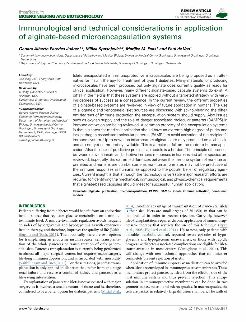
Microcapsules are very different in concept from macrocap-sules. Within this technology, islets are individually packed in theirown, individual capsule. An advantage over macrocapsules is thatthe microcapsules have an optimal surface to volume ratio, whichimplies a faster exchange of glucose, insulin, and nutrients (Uludaget al., 2000; Krishnamurthy and Gimi, 2011; Nafea et al., 2011;Vaithilingam and Tuch, 2011). For this reason, microencapsulationis preferred over macrocapsules as nutrient supply is more read-ily available in microcapsules than in macrocapsules. Especially,oxygen diffusion is an issue. Islets require relative high amountsof oxygen for both function and survival. Oxygen availability tothe islets depends more on the O2 partial pressure (pO2) in thetransplantation site rather than on O2 concentrations such as withother nutrient (Johnson et al., 2011; Colton, 2014) (Figure 1).
Microcapsules, however, can be obtained in many differentforms, sizes, composition, and with different permeability (de Voset al., 2014). Some use cation-crosslinked concepts while othersuse multiple layer systems (Lebedeva et al., 2004; de Vos et al.,2009; Dufrane and Gianello, 2012). The capsules are being usedfor allogeneic sources as well as for xenogeneic sources withouttaking into account that for xenogeneic sources other require-ments have to be met than those for allogeneic sources. For
FIGURE 1 |The three different categories of encapsulation devices usedfor immunoisolation of islets. Comparison between size and diffusionrate of nutrients and oxygen through the semipermeable membranes.
protection of allogeneic tissue, the prevention of cell–cell con-tact between donor-islets and immune-cells is considered to besufficient to prevent rejection (Duvivier-Kali et al., 2001). Simplesystems such as cation-alginate capsules (Mazzitelli et al., 2008)can, therefore, be effective in protection for allogeneic responses.However, when the donor-recipient histocompatibility becomesmore discordant such as with xenografts other processes comeinto play (Anderson and Kirk, 2013). When highly immunoreac-tive epitopes on xenogenic islets diffuse out of capsules, such asgalactosyl residues, they may react with naturally occurring (anti-Gal) and non-Gal IgM antibodies. The previous may activate theclassical complement pathway and may lead to neutrophil infil-tration (Dufrane and Gianello, 2012) and release of deleteriouscytokines that can pass the capsule membrane. IgM is not ableto pass most of the capsule’s membrane but the IgM-mediatedhumoral reaction against xenogeneic epitopes can also inducethe typical delayed-type hypersensitivity response associated withxenografts (Dufrane and Gianello, 2012), which leads to signifi-cant production of even smaller cytokines that can freely pass themembrane. These immunological processes in association withthe type of microcapsule system will be discussed in the presentreview in view of future application for protection of grafts forsmall cytokines produced in several immunological responses thatoccur after implantation of microencapsulated pancreatic islets.
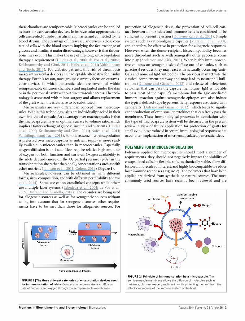
POLYMERS FOR MICROENCAPSULATIONPolymers applied for microcapsules should meet a number ofrequirements, they should not negatively impact the viability ofencapsulated cells, be flexible, soft, mechanically stable, allow dif-fusion of molecules of interest, and highly biocompatible to reducehost immune responses (Figure 2). The polymers that have beenapplied are derived from synthetic or natural sources. The mostcommonly used sources have recently been reviewed and are
FIGURE 2 | Principle of immunoisolation by a microcapsule. Thesemipermeable membrane allows the diffusion of molecules such asnutrients, glucose, oxygen, and insulin while protecting the graft from theeffector molecules of the immune system of the host.
Frontiers in Bioengineering and Biotechnology | Biomaterials August 2014 | Volume 2 | Article 26 | 2
Paredes Juárez et al. Considerations in alginate-microencapsulation systems
poly(ethylene glycol), polyvinyl alcohol, polyurethane, polyether-sulfone, polypropylene, sodium polystyrene sulfate, polyacrylate,agarose, chitosan, cellulose, collagen, xanthan, and alginate (de Voset al., 2014). The current consensus is that only alginate has beenstudied in sufficient detail to qualify as safe for human application(de Vos et al., 2014). Other polymers might become available inthe future but have not been sufficiently studied in terms of com-patibility with functional survival with the enveloped tissue andthe host. Also diffusion characteristics of essential immunologi-cal molecules and nutrients have been poorly studied with otherpolymers. Therefore, when describing the immunological issuesin the next sections and the different approaches of encapsulation,we will mainly concentrate on alginate as the core biomaterial. Wewould like to emphasize, however, that these are all generic issuesthat also other systems have to take into account.
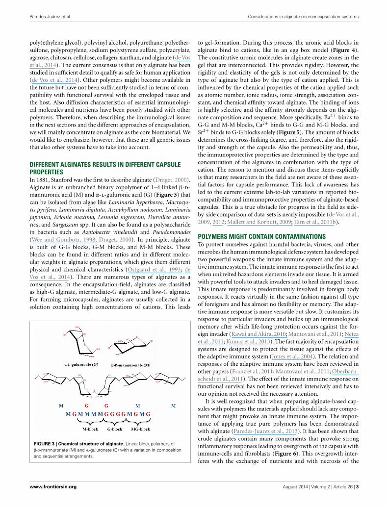
DIFFERENT ALGINATES RESULTS IN DIFFERENT CAPSULEPROPERTIESIn 1881, Stanford was the first to describe alginate (Draget, 2000).Alginate is an unbranched binary copolymer of 1–4 linked β-d-mannuronic acid (M) and α-l-guluronic acid (G) (Figure 3) thatcan be isolated from algae like Laminaria hyperborea, Macrocys-tis pyrifera, Laminaria digitata, Ascophyllum nodosum, Laminariajaponica, Eclonia maxima, Lessonia nigrescens, Durvillea antarc-tica, and Sargassum spp. It can also be found as a polysaccharidein bacteria such as Azotobacter vinelandii and Pseudomonades(Wee and Gombotz, 1998; Draget, 2000). In principle, alginateis built of G-G blocks, G-M blocks, and M-M blocks. Theseblocks can be found in different ratios and in different molec-ular weights in alginate preparations, which gives them differentphysical and chemical characteristics (Ostgaard et al., 1993; deVos et al., 2014). There are numerous types of alginates as aconsequence. In the encapsulation-field, alginates are classifiedas high-G alginate, intermediate-G alginate, and low-G alginate.For forming microcapsules, alginates are usually collected in asolution containing high concentrations of cations. This leads
FIGURE 3 | Chemical structure of alginate. Linear block polymers ofβ-D-mannuronate (M) and -L-guluronate (G) with a variation in compositionand sequential arrangements.
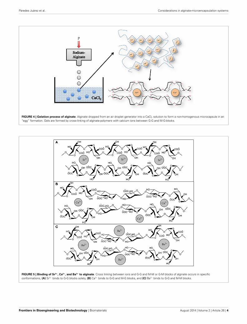
to gel-formation. During this process, the uronic acid blocks inalginate bind to cations, like in an egg box model (Figure 4).The constitutive uronic molecules in alginate create zones in thegel that are interconnected. This provides rigidity. However, therigidity and elasticity of the gels is not only determined by thetype of alginate but also by the type of cation applied. This isinfluenced by the chemical properties of the cation applied suchas atomic number, ionic radius, ionic strength, association con-stant, and chemical affinity toward alginate. The binding of ionsis highly selective and the affinity strongly depends on the algi-nate composition and sequence. More specifically, Ba2+ binds toG-G and M-M blocks, Ca2+ binds to G-G and M-G blocks, andSr2+ binds to G-G blocks solely (Figure 5). The amount of blocksdetermines the cross-linking degree, and therefore, also the rigid-ity and strength of the capsule. Also the permeability and, thus,the immunoprotective properties are determined by the type andconcentration of the alginates in combination with the type ofcation. The reason to mention and discuss these items explicitlyis that many researchers in the field are not aware of these essen-tial factors for capsule performance. This lack of awareness hasled to the current extreme lab-to-lab variations in reported bio-compatibility and immunoprotective properties of alginate-basedcapsules. This is a true obstacle for progress in the field as side-by-side comparison of data-sets is nearly impossible (de Vos et al.,2009, 2012; Mallett and Korbutt, 2009; Tam et al., 2011b).
POLYMERS MIGHT CONTAIN CONTAMINATIONSTo protect ourselves against harmful bacteria, viruses, and othermicrobes the human immunological defense system has developedtwo powerful weapons: the innate immune system and the adap-tive immune system. The innate immune response is the first to actwhen uninvited hazardous elements invade our tissue. It is armedwith powerful tools to attack invaders and to heal damaged tissue.This innate response is predominantly involved in foreign bodyresponses. It reacts virtually in the same fashion against all typeof foreigners and has almost no flexibility or memory. The adap-tive immune response is more versatile but slow. It customizes itsresponse to particular invaders and builds up an immunologicalmemory after which life-long protection occurs against the for-eign invader (Kawai and Akira, 2010; Mantovani et al., 2011; Neteaet al., 2011; Kumar et al., 2013). The fast majority of encapsulationsystems are designed to protect the tissue against the effects ofthe adaptive immune system (Jones et al., 2004). The relation andresponses of the adaptive immune system have been reviewed inother papers (Franz et al., 2011; Mantovani et al., 2011; Oberbarn-scheidt et al., 2011). The effect of the innate immune response onfunctional survival has not been reviewed intensively and has toour opinion not received the necessary attention.
It is well recognized that when preparing alginate-based cap-sules with polymers the materials applied should lack any compo-nent that might provoke an innate immune system. The impor-tance of applying true pure polymers has been demonstratedwith alginate (Paredes-Juarez et al., 2013). It has been shown thatcrude alginates contain many components that provoke stronginflammatory responses leading to overgrowth of the capsule withimmune-cells and fibroblasts (Figure 6). This overgrowth inter-feres with the exchange of nutrients and with necrosis of the
www.frontiersin.org August 2014 | Volume 2 | Article 26 | 3
Paredes Juárez et al. Considerations in alginate-microencapsulation systems
FIGURE 4 | Gelation process of alginate. Alginate dropped from an air droplet generator into a CaCl2 solution to form a non-homogenous microcapsule in an“egg” formation. Gels are formed by cross-linking of alginate-polymers with calcium ions between G-G and M-G-blocks.
FIGURE 5 | Binding of Sr2+, Ca2+, and Ba2+ to alginate. Cross linking between ions and G-G and M-M or G-M blocks of alginate occurs in specificconformations, (A) Sr2+ binds to G-G blocks solely, (B) Ca2+ binds to G-G and M-G blocks, and (C) Ba2+ binds to G-G and M-M blocks.
Frontiers in Bioengineering and Biotechnology | Biomaterials August 2014 | Volume 2 | Article 26 | 4
Paredes Juárez et al. Considerations in alginate-microencapsulation systems
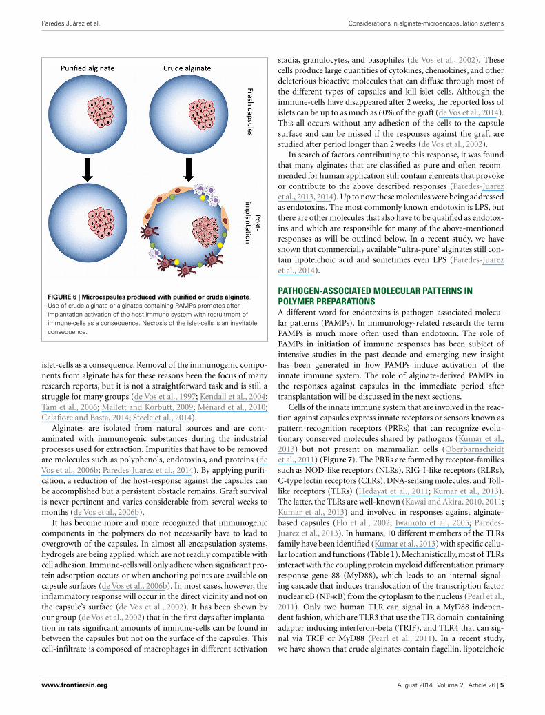
FIGURE 6 | Microcapsules produced with purified or crude alginate.Use of crude alginate or alginates containing PAMPs promotes afterimplantation activation of the host immune system with recruitment ofimmune-cells as a consequence. Necrosis of the islet-cells is an inevitableconsequence.
islet-cells as a consequence. Removal of the immunogenic compo-nents from alginate has for these reasons been the focus of manyresearch reports, but it is not a straightforward task and is still astruggle for many groups (de Vos et al., 1997; Kendall et al., 2004;Tam et al., 2006; Mallett and Korbutt, 2009; Ménard et al., 2010;Calafiore and Basta, 2014; Steele et al., 2014).
Alginates are isolated from natural sources and are cont-aminated with immunogenic substances during the industrialprocesses used for extraction. Impurities that have to be removedare molecules such as polyphenols, endotoxins, and proteins (deVos et al., 2006b; Paredes-Juarez et al., 2014). By applying purifi-cation, a reduction of the host-response against the capsules canbe accomplished but a persistent obstacle remains. Graft survivalis never pertinent and varies considerable from several weeks tomonths (de Vos et al., 2006b).
It has become more and more recognized that immunogeniccomponents in the polymers do not necessarily have to lead toovergrowth of the capsules. In almost all encapsulation systems,hydrogels are being applied, which are not readily compatible withcell adhesion. Immune-cells will only adhere when significant pro-tein adsorption occurs or when anchoring points are available oncapsule surfaces (de Vos et al., 2006b). In most cases, however, theinflammatory response will occur in the direct vicinity and not onthe capsule’s surface (de Vos et al., 2002). It has been shown byour group (de Vos et al., 2002) that in the first days after implanta-tion in rats significant amounts of immune-cells can be found inbetween the capsules but not on the surface of the capsules. Thiscell-infiltrate is composed of macrophages in different activation
stadia, granulocytes, and basophiles (de Vos et al., 2002). Thesecells produce large quantities of cytokines, chemokines, and otherdeleterious bioactive molecules that can diffuse through most ofthe different types of capsules and kill islet-cells. Although theimmune-cells have disappeared after 2 weeks, the reported loss ofislets can be up to as much as 60% of the graft (de Vos et al., 2014).This all occurs without any adhesion of the cells to the capsulesurface and can be missed if the responses against the graft arestudied after period longer than 2 weeks (de Vos et al., 2002).
In search of factors contributing to this response, it was foundthat many alginates that are classified as pure and often recom-mended for human application still contain elements that provokeor contribute to the above described responses (Paredes-Juarezet al., 2013, 2014). Up to now these molecules were being addressedas endotoxins. The most commonly known endotoxin is LPS, butthere are other molecules that also have to be qualified as endotox-ins and which are responsible for many of the above-mentionedresponses as will be outlined below. In a recent study, we haveshown that commercially available “ultra-pure” alginates still con-tain lipoteichoic acid and sometimes even LPS (Paredes-Juarezet al., 2014).
PATHOGEN-ASSOCIATED MOLECULAR PATTERNS INPOLYMER PREPARATIONSA different word for endotoxins is pathogen-associated molecu-lar patterns (PAMPs). In immunology-related research the termPAMPs is much more often used than endotoxin. The role ofPAMPs in initiation of immune responses has been subject ofintensive studies in the past decade and emerging new insighthas been generated in how PAMPs induce activation of theinnate immune system. The role of alginate-derived PAMPs inthe responses against capsules in the immediate period aftertransplantation will be discussed in the next sections.
Cells of the innate immune system that are involved in the reac-tion against capsules express innate receptors or sensors known aspattern-recognition receptors (PRRs) that can recognize evolu-tionary conserved molecules shared by pathogens (Kumar et al.,2013) but not present on mammalian cells (Oberbarnscheidtet al., 2011) (Figure 7). The PRRs are formed by receptor-familiessuch as NOD-like receptors (NLRs), RIG-I-like receptors (RLRs),C-type lectin receptors (CLRs), DNA-sensing molecules, and Toll-like receptors (TLRs) (Hedayat et al., 2011; Kumar et al., 2013).The latter, the TLRs are well-known (Kawai and Akira, 2010, 2011;Kumar et al., 2013) and involved in responses against alginate-based capsules (Flo et al., 2002; Iwamoto et al., 2005; Paredes-Juarez et al., 2013). In humans, 10 different members of the TLRsfamily have been identified (Kumar et al., 2013) with specific cellu-lar location and functions (Table 1). Mechanistically, most of TLRsinteract with the coupling protein myeloid differentiation primaryresponse gene 88 (MyD88), which leads to an internal signal-ing cascade that induces translocation of the transcription factornuclear κB (NF-κB) from the cytoplasm to the nucleus (Pearl et al.,2011). Only two human TLR can signal in a MyD88 indepen-dent fashion, which are TLR3 that use the TIR domain-containingadapter inducing interferon-beta (TRIF), and TLR4 that can sig-nal via TRIF or MyD88 (Pearl et al., 2011). In a recent study,we have shown that crude alginates contain flagellin, lipoteichoic
www.frontiersin.org August 2014 | Volume 2 | Article 26 | 5
Paredes Juárez et al. Considerations in alginate-microencapsulation systems
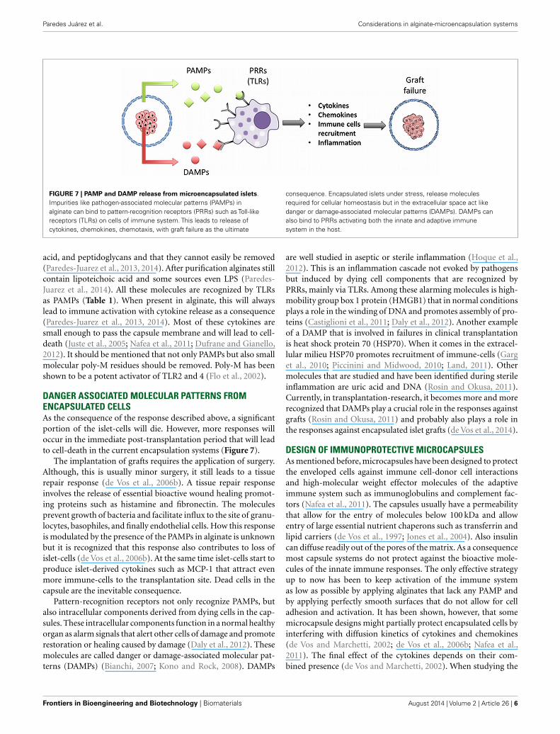
FIGURE 7 | PAMP and DAMP release from microencapsulated islets.Impurities like pathogen-associated molecular patterns (PAMPs) inalginate can bind to pattern-recognition receptors (PRRs) such as Toll-likereceptors (TLRs) on cells of immune system. This leads to release ofcytokines, chemokines, chemotaxis, with graft failure as the ultimate
consequence. Encapsulated islets under stress, release moleculesrequired for cellular homeostasis but in the extracellular space act likedanger or damage-associated molecular patterns (DAMPs). DAMPs canalso bind to PRRs activating both the innate and adaptive immunesystem in the host.
acid, and peptidoglycans and that they cannot easily be removed(Paredes-Juarez et al., 2013, 2014). After purification alginates stillcontain lipoteichoic acid and some sources even LPS (Paredes-Juarez et al., 2014). All these molecules are recognized by TLRsas PAMPs (Table 1). When present in alginate, this will alwayslead to immune activation with cytokine release as a consequence(Paredes-Juarez et al., 2013, 2014). Most of these cytokines aresmall enough to pass the capsule membrane and will lead to cell-death (Juste et al., 2005; Nafea et al., 2011; Dufrane and Gianello,2012). It should be mentioned that not only PAMPs but also smallmolecular poly-M residues should be removed. Poly-M has beenshown to be a potent activator of TLR2 and 4 (Flo et al., 2002).
DANGER ASSOCIATED MOLECULAR PATTERNS FROMENCAPSULATED CELLSAs the consequence of the response described above, a significantportion of the islet-cells will die. However, more responses willoccur in the immediate post-transplantation period that will leadto cell-death in the current encapsulation systems (Figure 7).
The implantation of grafts requires the application of surgery.Although, this is usually minor surgery, it still leads to a tissuerepair response (de Vos et al., 2006b). A tissue repair responseinvolves the release of essential bioactive wound healing promot-ing proteins such as histamine and fibronectin. The moleculesprevent growth of bacteria and facilitate influx to the site of granu-locytes, basophiles, and finally endothelial cells. How this responseis modulated by the presence of the PAMPs in alginate is unknownbut it is recognized that this response also contributes to loss ofislet-cells (de Vos et al., 2006b). At the same time islet-cells start toproduce islet-derived cytokines such as MCP-1 that attract evenmore immune-cells to the transplantation site. Dead cells in thecapsule are the inevitable consequence.
Pattern-recognition receptors not only recognize PAMPs, butalso intracellular components derived from dying cells in the cap-sules. These intracellular components function in a normal healthyorgan as alarm signals that alert other cells of damage and promoterestoration or healing caused by damage (Daly et al., 2012). Thesemolecules are called danger or damage-associated molecular pat-terns (DAMPs) (Bianchi, 2007; Kono and Rock, 2008). DAMPs
are well studied in aseptic or sterile inflammation (Hoque et al.,2012). This is an inflammation cascade not evoked by pathogensbut induced by dying cell components that are recognized byPRRs, mainly via TLRs. Among these alarming molecules is high-mobility group box 1 protein (HMGB1) that in normal conditionsplays a role in the winding of DNA and promotes assembly of pro-teins (Castiglioni et al., 2011; Daly et al., 2012). Another exampleof a DAMP that is involved in failures in clinical transplantationis heat shock protein 70 (HSP70). When it comes in the extracel-lular milieu HSP70 promotes recruitment of immune-cells (Garget al., 2010; Piccinini and Midwood, 2010; Land, 2011). Othermolecules that are studied and have been identified during sterileinflammation are uric acid and DNA (Rosin and Okusa, 2011).Currently, in transplantation-research, it becomes more and morerecognized that DAMPs play a crucial role in the responses againstgrafts (Rosin and Okusa, 2011) and probably also plays a role inthe responses against encapsulated islet grafts (de Vos et al., 2014).
DESIGN OF IMMUNOPROTECTIVE MICROCAPSULESAs mentioned before, microcapsules have been designed to protectthe enveloped cells against immune cell-donor cell interactionsand high-molecular weight effector molecules of the adaptiveimmune system such as immunoglobulins and complement fac-tors (Nafea et al., 2011). The capsules usually have a permeabilitythat allow for the entry of molecules below 100 kDa and allowentry of large essential nutrient chaperons such as transferrin andlipid carriers (de Vos et al., 1997; Jones et al., 2004). Also insulincan diffuse readily out of the pores of the matrix. As a consequencemost capsule systems do not protect against the bioactive mole-cules of the innate immune responses. The only effective strategyup to now has been to keep activation of the immune systemas low as possible by applying alginates that lack any PAMP andby applying perfectly smooth surfaces that do not allow for celladhesion and activation. It has been shown, however, that somemicrocapsule designs might partially protect encapsulated cells byinterfering with diffusion kinetics of cytokines and chemokines(de Vos and Marchetti, 2002; de Vos et al., 2006b; Nafea et al.,2011). The final effect of the cytokines depends on their com-bined presence (de Vos and Marchetti, 2002). When studying the
Frontiers in Bioengineering and Biotechnology | Biomaterials August 2014 | Volume 2 | Article 26 | 6
Paredes Juárez et al. Considerations in alginate-microencapsulation systems
Table 1 | Human homodimeric toll-like receptors, PAMPs, and DAMPs
that can be recognized by the differentTLRs (Fritz and Girardin, 2005;
Piccinini and Midwood, 2010; Basith et al., 2011; Rosin and Okusa,
2011; Liu and Ji, 2014).
TLR Ligating pathogen-
associated molecular
patterns (PAMPs)
Ligating damage-
associated molecular
patterns (DAMPs)
TLR1 Triacy lipopeptides β(-defensin-3
Soluble factors
TLR2 Zymosan HMGB1
Lipoteichoic acid HSPs
Lipoproteins Monosodium uric acid
Lipopeptides Hyaluronan
Peptidoglycan Biglycan
Bacterial porines Versican
Viric hemagglutinin
Glycolipids
Glycoinositol phospholipids
TLR3 dsRNA mRNA
TLR4 Lipopolysaccharides HMGB1
Taxol HSPs
Viral proteins Monosodium uric acid
Defensins
Lactoferrin
Hyaluronan
Biglycan
Fibrinogen
Heparan sulfate
Fibronectin extra domain A
Envelope proteins
TLR5 Bacterial flagellin Unknown
TLR6 Diacyl lipopeptides Unknown
Zymosan
Lipoteichoic acid
Phenol-soluble modulin
TLR7 ssRNA Cathelicidins
Imidazoquinoline
Loxoribine
TLR8 ssRNA ssRNA
Antiphospholipid antibodies
TLR9 Unmethylated CpG DNA HMGB1
DNA
Cathelicidins
IgG-chromatin complexes
TLR11 Uropathogenic bacteria Unknown
Profiling-like molecule
effects of the cytotoxic cocktail IL1-beta, TNF-alpha, and IFN-gamma, it has been shown that when the capsule network is dense,diffusion might be hampered, and partial protection might occur(de Vos et al., 2003). Diffusion of solutes through alginate does
not only depend on the molecular weight of the molecule butalso on the three-dimensional structure, the factor of gyration,and their charge density (Wee and Gombotz, 1998; de Vos et al.,2006b). TNF-alpha is a 50 kDa trimeric molecule that does not dif-fuse through the majority of microcapsule systems. Some capsuletypes containing an in-homogeneous core have even been shownto interfere with the diffusion of even smaller cytokines such asIL-1beta (17.5 kDa) (Kulseng et al., 1997). Applying or adaptingthe current microencapsulation systems might reduce cytotoxicityby innate immune effector molecules.
HOMOGENEOUS AND IN-HOMOGENEOUS CORE CAPSULESMicrocapsules can be prepared with a homogeneous or an in-homogeneous core. Homogeneous beads have a similar if notidentical concentration of alginate throughout the bead while non-homogeneous beads typically have a higher alginate concentrationin the periphery and a lower concentration at the center of the bead(Figure 8) (Qi et al., 2008). In-homogeneous alginate gels canbe produced by manipulating the diffusion of the alginate mole-cules in the bead. This can be done by substituting the ions in thegelling solution for iso-osmolites such as mannitol. The gellingsolution should have a low divalent cation concentration. Whatsubsequently will happen is that the alginate molecules will diffusetoward the surface of the bead in order to compete for the scarcedivalent cations. The final consequence is a higher concentrationof alginate-polymers on the surface and a lower concentration inthe core. By lowering the cation concentration or increasing thepolymer concentration a gradual increase of the alginate concen-tration at the rim of the capsule can be achieved (Mørch et al.,2006; Qi et al., 2008). This approach can be applied to manip-ulate the permeability properties of the capsules (Uludag et al.,2000) and might be an effective tool to modulate the diffusion ofcytokines into the network.
This elegant method has been applied (Qi et al., 2008; Safleyet al., 2008) but has in addition to advantages also disadvantages.During formation of in-homogeneous gels, osmotic pressure ispushing the envelope tissue toward the rim of the capsules. Thisholds two risks. The first is that during this process significantshear forces are being put on the tissue. This may lead to fractureof the islets and loss of function. The second risk is the increasedchance on protrusion of cells. The distribution of cells throughouta capsule is determined by chance. When a capsule with a radiusof 500 µm is divided in zones of 100 µm, it can be calculated thatthe chance that a cell will end up in one of the outer rims is high(Figure 9). This is also confirmed by experimental studies (de Voset al., 1994; Del Guerra et al., 2001). This implies that the cells willbe pulled during the process of gel-formation toward the edge andcan even protrude from the capsules. When using this method, itis advisable to apply assays to detect and/or remove capsules withprotruding cells (de Vos et al., 1996).
Although a very elegant method, it still remains to be deter-mined whether this system composed of alginate only can providesufficient protection against the complex and inevitable activa-tion of the innate and adaptive immune system. Especially, wehave doubts whether the permeability and diffusion character-istics can be manipulated as such that protection is achievedagainst the harsh xenogeneic responses. To the best of our
www.frontiersin.org August 2014 | Volume 2 | Article 26 | 7
Paredes Juárez et al. Considerations in alginate-microencapsulation systems
knowledge, there are no efficacy studies available of this systemin non-immunosuppressed recipients.
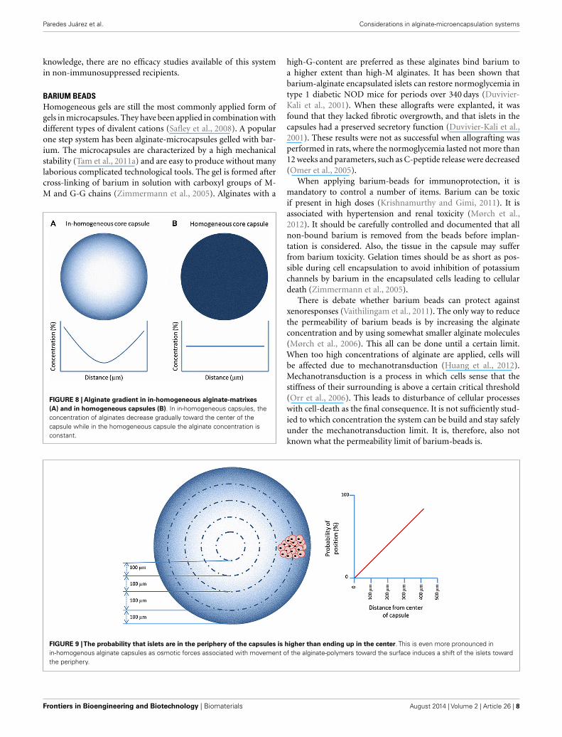
BARIUM BEADSHomogeneous gels are still the most commonly applied form ofgels in microcapsules. They have been applied in combination withdifferent types of divalent cations (Safley et al., 2008). A popularone step system has been alginate-microcapsules gelled with bar-ium. The microcapsules are characterized by a high mechanicalstability (Tam et al., 2011a) and are easy to produce without manylaborious complicated technological tools. The gel is formed aftercross-linking of barium in solution with carboxyl groups of M-M and G-G chains (Zimmermann et al., 2005). Alginates with a
FIGURE 8 | Alginate gradient in in-homogeneous alginate-matrixes(A) and in homogeneous capsules (B). In in-homogeneous capsules, theconcentration of alginates decrease gradually toward the center of thecapsule while in the homogeneous capsule the alginate concentration isconstant.
high-G-content are preferred as these alginates bind barium toa higher extent than high-M alginates. It has been shown thatbarium-alginate encapsulated islets can restore normoglycemia intype 1 diabetic NOD mice for periods over 340 days (Duvivier-Kali et al., 2001). When these allografts were explanted, it wasfound that they lacked fibrotic overgrowth, and that islets in thecapsules had a preserved secretory function (Duvivier-Kali et al.,2001). These results were not as successful when allografting wasperformed in rats, where the normoglycemia lasted not more than12 weeks and parameters, such as C-peptide release were decreased(Omer et al., 2005).
When applying barium-beads for immunoprotection, it ismandatory to control a number of items. Barium can be toxicif present in high doses (Krishnamurthy and Gimi, 2011). It isassociated with hypertension and renal toxicity (Mørch et al.,2012). It should be carefully controlled and documented that allnon-bound barium is removed from the beads before implan-tation is considered. Also, the tissue in the capsule may sufferfrom barium toxicity. Gelation times should be as short as pos-sible during cell encapsulation to avoid inhibition of potassiumchannels by barium in the encapsulated cells leading to cellulardeath (Zimmermann et al., 2005).
There is debate whether barium beads can protect againstxenoresponses (Vaithilingam et al., 2011). The only way to reducethe permeability of barium beads is by increasing the alginateconcentration and by using somewhat smaller alginate molecules(Mørch et al., 2006). This all can be done until a certain limit.When too high concentrations of alginate are applied, cells willbe affected due to mechanotransduction (Huang et al., 2012).Mechanotransduction is a process in which cells sense that thestiffness of their surrounding is above a certain critical threshold(Orr et al., 2006). This leads to disturbance of cellular processeswith cell-death as the final consequence. It is not sufficiently stud-ied to which concentration the system can be build and stay safelyunder the mechanotransduction limit. It is, therefore, also notknown what the permeability limit of barium-beads is.
FIGURE 9 |The probability that islets are in the periphery of the capsules is higher than ending up in the center. This is even more pronounced inin-homogenous alginate capsules as osmotic forces associated with movement of the alginate-polymers toward the surface induces a shift of the islets towardthe periphery.
Frontiers in Bioengineering and Biotechnology | Biomaterials August 2014 | Volume 2 | Article 26 | 8
Paredes Juárez et al. Considerations in alginate-microencapsulation systems
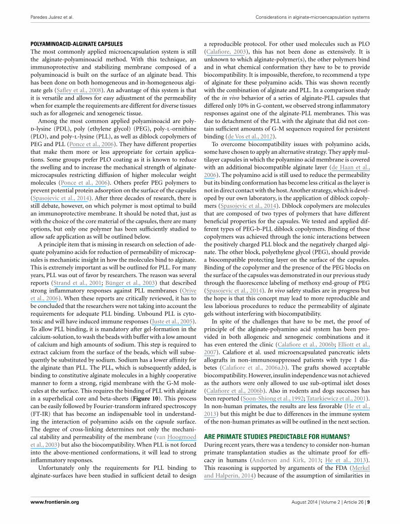
POLYAMINOACID-ALGINATE CAPSULESThe most commonly applied microencapsulation system is stillthe alginate-polyaminoacid method. With this technique, animmunoprotective and stabilizing membrane composed of apolyaminoacid is built on the surface of an alginate bead. Thishas been done on both homogeneous and in-homogeneous algi-nate gels (Safley et al., 2008). An advantage of this system is thatit is versatile and allows for easy adjustment of the permeabilitywhen for example the requirements are different for diverse tissuessuch as for allogeneic and xenogeneic tissue.
Among the most common applied polyaminoacid are poly-d-lysine (PDL), poly (ethylene glycol) (PEG), poly-l-ornithine(PLO), and poly-l-lysine (PLL), as well as diblock copolymers ofPEG and PLL (Ponce et al., 2006). They have different propertiesthat make them more or less appropriate for certain applica-tions. Some groups prefer PLO coating as it is known to reducethe swelling and to increase the mechanical strength of alginate-microcapsules restricting diffusion of higher molecular weightmolecules (Ponce et al., 2006). Others prefer PEG polymers toprevent potential protein adsorption on the surface of the capsules(Spasojevic et al., 2014). After three decades of research, there isstill debate, however, on which polymer is most optimal to buildan immunoprotective membrane. It should be noted that, just aswith the choice of the core material of the capsules, there are manyoptions, but only one polymer has been sufficiently studied toallow safe application as will be outlined below.
A principle item that is missing in research on selection of ade-quate polyamino acids for reduction of permeability of microcap-sules is mechanistic insight in how the molecules bind to alginate.This is extremely important as will be outlined for PLL. For manyyears, PLL was out of favor by researchers. The reason was severalreports (Strand et al., 2001; Bünger et al., 2003) that describedstrong inflammatory responses against PLL membranes (Oriveet al., 2006). When these reports are critically reviewed, it has tobe concluded that the researchers were not taking into account therequirements for adequate PLL binding. Unbound PLL is cyto-toxic and will have induced immune responses (Juste et al., 2005).To allow PLL binding, it is mandatory after gel-formation in thecalcium-solution, to wash the beads with buffer with a low amountof calcium and high amounts of sodium. This step is required toextract calcium from the surface of the beads, which will subse-quently be substituted by sodium. Sodium has a lower affinity forthe alginate than PLL. The PLL, which is subsequently added, isbinding to constitutive alginate molecules in a highly cooperativemanner to form a strong, rigid membrane with the G-M mole-cules at the surface. This requires the binding of PLL with alginatein a superhelical core and beta-sheets (Figure 10). This processcan be easily followed by Fourier-transform infrared spectroscopy(FT-IR) that has become an indispensable tool in understand-ing the interaction of polyamino acids on the capsule surface.The degree of cross-linking determines not only the mechani-cal stability and permeability of the membrane (van Hoogmoedet al., 2003) but also the biocompatibility. When PLL is not forcedinto the above-mentioned conformations, it will lead to stronginflammatory responses.
Unfortunately only the requirements for PLL binding toalginate-surfaces have been studied in sufficient detail to design
a reproducible protocol. For other used molecules such as PLO(Calafiore, 2003), this has not been done as extensively. It isunknown to which alginate-polymer(s), the other polymers bindand in what chemical conformation they have to be to providebiocompatibility. It is impossible, therefore, to recommend a typeof alginate for these polyamino acids. This was shown recentlywith the combination of alginate and PLL. In a comparison studyof the in vivo behavior of a series of alginate-PLL capsules thatdiffered only 10% in G-content, we observed strong inflammatoryresponses against one of the alginate-PLL membranes. This wasdue to detachment of the PLL with the alginate that did not con-tain sufficient amounts of G-M sequences required for persistentbinding (de Vos et al., 2012).
To overcome biocompatibility issues with polyamino acids,some have chosen to apply an alternative strategy. They apply mul-tilayer capsules in which the polyamino acid membrane is coveredwith an additional biocompatible alginate layer (de Haan et al.,2006). The polyamino acid is still used to reduce the permeabilitybut its binding conformation has become less critical as the layer isnot in direct contact with the host. Another strategy,which is devel-oped by our own laboratory, is the application of diblock copoly-mers (Spasojevic et al., 2014). Diblock copolymers are moleculesthat are composed of two types of polymers that have differentbeneficial properties for the capsules. We tested and applied dif-ferent types of PEG-b-PLL diblock copolymers. Binding of thesecopolymers was achieved through the ionic interactions betweenthe positively charged PLL block and the negatively charged algi-nate. The other block, polyethylene glycol (PEG), should providea biocompatible protecting layer on the surface of the capsules.Binding of the copolymer and the presence of the PEG blocks onthe surface of the capsules was demonstrated in our previous studythrough the fluorescence labeling of methoxy end-group of PEG(Spasojevic et al., 2014). In vivo safety studies are in progress butthe hope is that this concept may lead to more reproducible andless laborious procedures to reduce the permeability of alginategels without interfering with biocompatibility.
In spite of the challenges that have to be met, the proof ofprinciple of the alginate-polyamino acid system has been pro-vided in both allogeneic and xenogeneic combinations and ithas even entered the clinic (Calafiore et al., 2006b; Elliott et al.,2007). Calafiore et al. used microencapsulated pancreatic isletsallografts in non-immunosuppressed patients with type 1 dia-betes (Calafiore et al., 2006a,b). The grafts showed acceptablebiocompatibility. However, insulin independence was not achievedas the authors were only allowed to use sub-optimal islet doses(Calafiore et al., 2006b). Also in rodents and dogs successes hasbeen reported (Soon-Shiong et al., 1992; Tatarkiewicz et al., 2001).In non-human primates, the results are less favorable (He et al.,2013) but this might be due to differences in the immune systemof the non-human primates as will be outlined in the next section.
ARE PRIMATE STUDIES PREDICTABLE FOR HUMANS?During recent years, there was a tendency to consider non-humanprimate transplantation studies as the ultimate proof for effi-cacy in humans (Anderson and Kirk, 2013; He et al., 2013).This reasoning is supported by arguments of the FDA (Merkeland Halperin, 2014) because of the assumption of similarities in
www.frontiersin.org August 2014 | Volume 2 | Article 26 | 9
Paredes Juárez et al. Considerations in alginate-microencapsulation systems
FIGURE 10 | Structure of alginate-poly-L-lysine capsules. Thesystem is composed of two layers and not of three layers as wronglystated in many papers. The core of the capsule is composed of a matrixof alginate and calcium in which the islets are entrapped. The secondlayer is obtained after incubation of the calcium bead in sodium richsolutions to form a sodium-alginate complex at the surface. Sodium can
be substituted by poly-L-lysine (PLL) to form a complex alginate-PLL-alginate (APA). The PLL-alginate layer can take three different structuresby intramolecular hydrogen binding, (i) random coil formation betweenalginate and PLL, (ii) α-helicoidal structure between amide groups ofPLL, and (iii) antiparallel β-sheet structure between amide groupsof PLL.
immune responses between humans and non-human primates(Collins and Jones, 1978; Mezey et al., 1983; Vaithilingam andTuch, 2011). Despite absence of scientific proof and meetingmany ethical issues, the believers considered responses in non-human primates to be analogous to that in humans (Andersonand Kirk, 2013). To our opinion, this is a major misassumption.As will be outlined below, there are pertinent immunological dif-ferences between non-human primates and humans making thenon-human primate, an inadequate model for human responsesagainst encapsulated grafts. Immune responses may occur in non-human primates but will not occur in humans and specific innateresponses not present in non-human primates may happen inhumans.
Immunological differences are considered to be the reasonfor early mortality rates and life span rate differences betweenhumans and non-human primates such as chimpanzees (Finch,2010). Humans differ from chimpanzees by low mortality rates inboth the juvenile and adult ages, and by the later onset of mor-tality rate acceleration, usually by chronic degenerative diseases(Finch, 2010). For wild chimpanzees, an early mortality rate of20% per year in infancy is normal with a decrease to about 3.5%per year in pre-adult ages. Infections play a major role in the high
chimpanzee mortality rates. The life expectancy at birth of a chim-panzee is about 13 years, whereas animals that reach adulthoodhave about 15 years of further life (Finch, 2010). This is very dif-ferent in humans. Even in areas with limited access to medicine,life expectation after birth is much longer and the high early agemortality declines to a minimum at the approach of adulthood(Finch, 2010). There is consensus that this species difference inmortality rate and life span is due to differences in their immunesystem that were developed more than a million years ago (Finch,2010; Anderson and Kirk, 2013).
Finch (2010) has done basic research in differences betweenhumans and non-human primates innate and adaptive immuneresponses. Finch was interested in the variations in immune-genesthat might explain the early differences in mortality rates betweennon-human primates and humans. To this end, Finch performeda stepwise comparison between chimpanzee and human-specificimmunological genes and did some important discoveries. Themost remarkable difference was in the composition of apolipopro-tein E (ApoE) genes. This important gene is involved in metabo-lism of lipids and in regulating innate immune responses. Thereare three uniquely human ApoE alleles, of which ApoE e4 andApoE e3 are the most prevalent (Schaffer et al., 2014). The ApoE e4
Frontiers in Bioengineering and Biotechnology | Biomaterials August 2014 | Volume 2 | Article 26 | 10
Paredes Juárez et al. Considerations in alginate-microencapsulation systems
differs in several critical amino acids from the chimpanzee equiv-alent. It is the most relevant gene for the current discussion as itis responsible for boosting the human innate immune response,which is diminished in non-human primates relative to humans(Finch, 2010). ApoE e4 induces the acute innate response andinitiates the production of important innate immune cytokinessuch as IL-6 and TNF-α (Vitek et al., 2009). Evolutionary, thisgave humans the benefit to better fight harmful intruders thatthey ingested in food and encountered in their surroundings. Thisdifference in innate immunity is considered to be a major factorin the difference in mortality rates in the juvenile and adult ageclasses between humans and chimpanzees (Barreiro et al., 2010).Also, this is pertinent when considering non-human primates asa relevant immunological model for humans since mimicking thehuman innate immune responses is a key in studies predicting theefficacy of encapsulated grafts in humans.
Besides differences in activity of the innate immune system,there are more immunological differences between humans andnon-human primates that are relevant for the discussion on theadequacy of non-human primates as model for humans in ourtype of research. Humans lack for example specific genes or havespecific polymorphisms making them incapable to perform somespecific immunological responses. Major differences have beenfound in the major histocompatibility complex (MHC) (Finch,2010). The MHC system is important for both innate and adap-tive immune responses. An important species difference is the lossof polymorphisms in class IA and B genes in humans. This impliesa class-specific loss of variation and therefore a selective elimina-tion of responses against specific families of epitopes (de Grootet al., 2008). Also a pertinent difference in PRRs has been reported(Brinkworth et al., 2012). Human CD4+ T cells have low expres-sion of Siglecs relative to chimpanzees (Nguyen et al., 2006). TheSiglec lectin family of proteins (Ig superfamily) is involved in T-cellreceptor (TCR) interactions and is implicated in the much milderchimpanzee symptoms of HIV-1 and hepatitis B or C (Bibollet-Ruche et al., 2008) infections due to more specific responses. Thisillustrates that non-human primates may respond differently andsometimes more rigorously against antigens than humans.
PRE-CLINICAL MODELSThe foregoing obviously calls for suggestions for better animalmodels. The answer is simple. There is not a single step model thatpredicts efficacy of encapsulated grafts in humans. Technologieshave emerged that has brought this research field into a next stepwere animal studies and ex vivo studies are combined to predictefficacy in humans. A first step is the need for animal models andex vivo systems mimicking human innate responses and avoidingharsh innate immune responses is a key in the success of encapsu-lated islets in humans. Although abandoned by some (Andersonand Kirk, 2013; He et al., 2013) experimental animals such as miceand rats have a more developed innate immune response thannon-human primates and are therefore more appropriate. Trans-genic mice are available that resemble human innate responsesas close as it can get. There are TRapoE4 mice available thathave near identical human IL-6 and TNFα responses (Vitek et al.,2009). Also, technology platforms are under development contain-ing human PRRs giving insight in how these sensors of the human
immune system will react on encapsulated cell systems (Paredes-Juarez et al., 2013, 2014). Another advance and to our opinionmandatory research item is that some groups combine the fore-going studies with mimicking protein adsorption processes thatoccur in humans (de Haan et al., 2011; Rokstad et al., 2011).Protein adsorption to biomaterial surfaces is assumed to be themajor cause of species differences (Vaithilingam and Tuch, 2011)in foreign body responses. It was shown by our group that theadsorption and changes at the capsule surface are highly depen-dent on the polymer applied and can be avoided (Spasojevic et al.,2014). This combined approach with a proof of principle study inany larger mammal with a well-developed innate immune systemwill be our preferred approach to come to a predictive outcome inhumans.
FUTURE CONSIDERATIONAs outlined in the preceding sections, there are many choices whenconsidering alginate-based microcapsules for immunoprotectionof pancreatic islets. Most of the systems mentioned above haveshown some efficacy in allogeneic settings but in xenogeneic set-tings not many successful studies are available (Murua et al., 2009).This might not always be the consequence of immunologicalissues. Incompatibility in metabolism may also be a contributingfactor. It has been shown, for example, that rat islets maintain theirmetabolism when transplanted in mice (Bobzien et al., 1983). Thismay lead to rapid exhaustion when the recipient has a higher meta-bolic demand than the donor. Another or combined explanationis that the prerequisites of the membrane are far more strict withxenografts than with allografts. Xenografts may provoke strongerresponses by leaking immunoreactive epitopes, such as galacto-syl residues, that may react with pre-existing, naturally occur-ring (anti-Gal) and non-Gal IgM antibodies with strong innateresponses as a consequence. Currently, it is unknown whether themembranes can provide sufficient protection against the cytokinesand deleterious bioactive molecules that are produced duringxenoresponses (de Vos et al., 2014). It has been shown, however(de Vos and Marchetti, 2002), that human cytokines are not veryeffective in killing xenogeneic tissue due to structural differencesin cytokine and receptor composition between humans and ani-mals (Brinkworth et al., 2012). It is imperative for the future ofencapsulation to determine, which of the above-mentioned factorscontribute to the success or failure of immunoprotected xenograftsand which sources have the capacity to maintain long-term graftfunction in humans.
Irrespective of the islet-source or system, it is mandatory to pro-duce high-quality alginates that lack pro-inflammatory moleculessuch as PAMPs. PAMPs are introduced in alginates at differentstages of production and can even be included during purificationdue to the application of chemicals or instruments that containPAMPs (Paredes-Juarez et al., 2014). As outlined above all com-mercial alginates, even those that are marketed as “ultra-pure”and recommended for clinical application, do contain PAMPs anddo initiate inflammatory responses (Paredes-Juarez et al., 2014).This is to our opinion, a major obstacle for clinical applicationof encapsulated islets. The ease of removal of PAMPs is in ourhands alginate-type dependent. Removal of PAMPs from high-Galginates is more difficult than from intermediate-G sources. This
www.frontiersin.org August 2014 | Volume 2 | Article 26 | 11
Paredes Juárez et al. Considerations in alginate-microencapsulation systems
is probably due to the fact that high-G alginate solutions havehigher viscosities, which makes the washing steps during purifica-tion often less efficacious. Chemical extraction methods are mostsuccessful as well as dialysis against endotoxin free water. Com-binations of these technologies as well as specific precipitationmethods are probably required to remove PAMPs from alginatesources that are harsh to purify.
Stress signals released by microencapsulated islets themselveshave not gained much attention in the success and failure of encap-sulated islet grafts. As cell-death induced by necrosis is a majorplayer in the failure of islet-grafts, it is plausible that DAMPs docontribute to the magnitude of the innate responses and failureof the grafts. A conceivable approach to overcome these types ofissues, is the use of smaller islets (i.e., smaller than 150 µm) thatcan manage the relative low oxygen tensions without significantloss of cells by necrosis (Colton, 2014). Other strategies might beto design membranes that retain DAMPS or inclusion of phar-macological drugs such as necrostatin-1 that inhibits necrosis ofβ-cells (Tamura et al., 2011).
The above and most of the papers addressing species-specificefficacy of encapsulation systems discuss predominantly immuno-logical issues. An overlooked but pertinent issue when comparingefficacy of encapsulation systems is the different requirements formechanical stability, the capsules have to meet. It is unknown,for example, how rigid and elastic capsules should be in largermammals. In rats, we know that capsules that can withstand aforce of 8 g can survive up to 2 years in rats, i.e., the lifespan ofrats. In pigs, however, the shear forces are much higher and morecapsules were having imperfections on the membrane and con-tained immune-cells. This illustrates that, going from one speciesto another, adaptations have to be made in the mechanic-physicproperties of the capsules and that caution should be taken to inter-pret the host-reactions as species-specific responses against thecapsules. In our experience going from one organism to anotherrequires significant adaptations of the system before any scien-tifically sound comparison in immune responses can be made(Scharp and Marchetti, 2014). Unfortunately, these types of adap-tations are rarely done but are, to our opinion, mandatory now thatencapsulation devices are coming closer to clinical application.
AUTHOR CONTRIBUTIONSGenaro Alberto Paredes Juárez, Milica Spasojevic, Marijke M. Faas,and Paul de Vos contributed with conception and design of workand writing of the paper.
ACKNOWLEDGMENTSTo Consejo Nacional de Ciencia y Tecnología (scholarship 310053)and the Secretary of Public Education of the Government ofMexico for the grant to Genaro Alberto Paredes Juárez.
REFERENCESAnderson, D. J., and Kirk, A. D. (2013). Primate models in organ transplantation.
Cold Spring Harb. Perspect. Med. 3, a015503. doi:10.1101/cshperspect.a015503Barreiro, L. B., Marioni, J. C., Blekhman, R., Stephens, M., and Gilad, Y. (2010).
Functional comparison of innate immune signaling pathways in primates. PLoSGenet. 6:e1001249. doi:10.1371/journal.pgen.1001249
Basith, S., Manavalan, B., Lee, G., Kim, S. G., and Choi, S. (2011). Toll-like receptormodulators: a patent review (2006-2010). Expert Opin. Ther. Pat. 21, 927–944.doi:10.1517/13543776.2011.569494
Bianchi, M. E. (2007). DAMPs, PAMPs and alarmins: all we need to know aboutdanger. J. Leukoc. Biol. 81, 1–5. doi:10.1189/jlb.0306164
Bibollet-Ruche, F., McKinney, B. A., Duverger, A., Wagner, F. H., Ansari, A. A.,and Kutsch, O. (2008). The quality of chimpanzee T-cell activation and simianimmunodeficiency virus/human immunodeficiency virus susceptibility achievedvia antibody-mediated T-cell receptor/CD3 stimulation is a function of the anti-CD3 antibody isotype. J. Virol. 82, 10271–10278. doi:10.1128/JVI.01319-08
Bobzien, B., Yasunami, Y., Majercik, M., Lacy, P. E., and Davie, J. M. (1983). Intrat-esticular transplants of islet xenografts (rat to mouse). Diabetes 32, 213–216.doi:10.2337/diab.32.3.213
Brinkworth, J. F., Pechenkina, E. A., Silver, J., and Goyert, S. M. (2012). Innateimmune responses to TLR2 and TLR4 agonists differ between baboons, chim-panzees and humans. J. Med. Primatol. 41, 388–393. doi:10.1111/jmp.12002
Bünger, C. M., Gerlach, C., Freier, T., Schmitz, K. P., Pilz, M., Werner, C.,et al. (2003). Biocompatibility and surface structure of chemically modifiedimmunoisolating alginate-PLL capsules. J. Biomed. Mater. Res. A 67, 1219–1227.doi:10.1002/jbm.a.10094
Calafiore, R. (2003). Alginate microcapsules for pancreatic islet cell graft immuno-protection: struggle and progress towards the final cure for type 1 diabetes mel-litus. Expert Opin. Biol. Ther. 3, 201–205. doi:10.1517/14712598.3.2.201
Calafiore, R., and Basta, G. (2014). Clinical application of microencapsulated islets:actual prospectives on progress and challenges. Adv. Drug Deliv. Rev. 6, 84–92.doi:10.1016/j.addr.2013.09.020
Calafiore, R., Basta, G., Luca, G., Lemmi, A., Racanicchi, L., Mancuso, F., et al.(2006a). Standard technical procedures for microencapsulation of human isletsfor graft into nonimmunosuppressed patients with type 1 diabetes mellitus.Transplant. Proc. 38, 1156–1157. doi:10.1016/j.transproceed.2006.03.014
Calafiore, R., Basta, G., Luca, G., Lemmi, A., Montanucci, M. P., Calabrese, G., et al.(2006b). Microencapsulated pancreatic islet allografts into nonimmunosup-pressed patients with type 1 diabetes: first two cases. Diabetes Care 29, 137–138.doi:10.2337/diacare.29.01.06.dc05-1270
Castiglioni, A., Canti, V., Rovere-Querini, P., and Manfredi, A. A. (2011). High-mobility group box 1 (HMGB1) as a master regulator of innate immunity. CellTissue Res. 343, 189–199. doi:10.1007/s00441-010-1033-1
Collins, J. F., and Jones, M. A. (1978). Connective tissue proteins of the baboon lung:concentration, content and synthesis of collagen in the normal lung. Connect.Tissue Res. 5, 211–215. doi:10.3109/03008207809152275
Colton, C. K. (2014). Oxygen supply to encapsulated therapeutic cells. Adv. DrugDeliv. Rev. 67-68C, 93–110. doi:10.1016/j.addr.2014.02.007
Daly, K. A., Liu, S., Agrawal, V., Brown, B. N., Johnson, S. A., Medberry, C. J.,et al. (2012). Damage associated molecular patterns within xenogeneic bio-logic scaffolds and their effects on host remodeling. Biomaterials 33, 91–101.doi:10.1016/j.biomaterials.2011.09.040
de Groot, N. G., Heijmans, C. M. C., de Groot, N., Otting, N., de Vos-Rouweller, A.J. M., Remarque, E. J., et al. (2008). Pinpointing a selective sweep to the chim-panzee MHC class I region by comparative genomics. Mol. Ecol. 17, 2074–2088.doi:10.1111/j.1365-294X.2008.03716.x
de Haan, B. J., Faas, M. M., Hamel, A. F., and de Vos, P. (2006). “Experimentalapproaches for transplantation of islets in the absence of immunosuppression,”in Trends in Diabetes Research, ed. A. M. Ford (New York: Nova Science Publisher,Inc.), 131–162.
de Haan, B. J., Rossi, A., Faas, M. M., Smelt, M. J., Sonvico, F., Colombo, P., et al.(2011). Structural surface changes and inflammatory responses against alginate-based microcapsules after exposure to human peritoneal fluid. J. Biomed. Mater.Res. A 98, 394–403. doi:10.1002/jbm.a.33123
de Vos, P., Anderson, A., Tam, S. K., Faas, M. M., and Halle, J. P. (2006a).Advances and barriers in mammalian cell encapsulation for treatment of dia-betes. Immunol. Endocr. Metab. Agents. Med. Chem. 6, 139–153. doi:10.2174/187152206776359948
de Vos, P., Faas, M. M., Strand, B., and Calafiore, R. (2006b). Alginate-based micro-capsules for immunoisolation of pancreatic islets. Biomaterials 27, 5603–5617.doi:10.1016/j.biomaterials.2006.07.010
de Vos, P., Bucko, M., Gemeiner, P., Navrátil, M., Svitel, J., Faas, M., et al. (2009).Multiscale requirements for bioencapsulation in medicine and biotechnology.Biomaterials 30, 2559–2570. doi:10.1016/j.biomaterials.2009.01.014
de Vos, P., de Haan, B. J., Pater, J., and van Schilfgaarde, R. (1996). Associa-tion between capsule diameter, adequacy of encapsulation, and survival ofmicroencapsulated rat islets allografts. Transplantation 62, 893–899. doi:10.1097/00007890-199610150-00004
Frontiers in Bioengineering and Biotechnology | Biomaterials August 2014 | Volume 2 | Article 26 | 12
Paredes Juárez et al. Considerations in alginate-microencapsulation systems
de Vos, P., De Haan, B. J., Wolters, G. H., Strubbe, J. H., and Van Schilfgaarde, R.(1997). Improved biocompatibility but limited graft survival after purificationof alginate for microencapsulation of pancreatic islets. Diabetologia 40, 262–270.doi:10.1007/s001250050673
de Vos, P., Lazarjani, H. A., Poncelet, D., and Faas, M. M. (2014). Polymers in cellencapsulation from an enveloped cell perspective. Adv. Drug Deliv. Rev. 67-68,15–34. doi:10.1016/j.addr.2013.11.005
de Vos, P., and Marchetti, P. (2002). Encapsulation of pancreatic islets for trans-plantation in diabetes: the untouchable islets. Trends Mol. Med. 8, 363–366.doi:10.1016/S1471-4914(02)02381-X
de Vos, P., Smedema, I., van Goor, H., Moes, H., van Zanten, J., Netters, S.,et al. (2003). Association between macrophage activation and function ofmicro-encapsulated rat islets. Diabetologia 46, 666–673. doi:10.1007/s00125-003-1087-7
de Vos, P., Spasojevic, M., de Haan, B. J., and Faas, M. M. (2012). The associ-ation between in vivo physicochemical changes and inflammatory responsesagainst alginate based microcapsules. Biomaterials 33, 5552–5559. doi:10.1016/j.biomaterials.2012.04.039
de Vos, P., van Hoogmoed, C. G., de Haan, B. J., and Busscher, H. J. (2002). Tissueresponses against immunoisolating alginate-PLL capsules in the immediate post-transplant period. J. Biomed. Mater. Res. 62, 430–437. doi:10.1002/jbm.10345
de Vos, P., Wolters, G. H., and van Schilfgaarde, R. (1994). Possible relation-ship between fibrotic overgrowth of alginate-polylysine-alginate microencap-sulated pancreatic islets and the microcapsule integrity. Transplant. Proc. 26,782–783.
Del Guerra, S., Bracci, C., Nilsson, K., Belcourt, A., Kessler, L., Lupi, R., et al.(2001). Entrapment of dispersed pancreatic islet cells in cultispher-s macrop-orous gelatin microcarriers : preparation, in vitro characterization, and microen-capsulation. Biotechnol Bioeng. 75, 741–744. doi:10.1002/bit.10053
Draget, K. I. (2000). “Alginates,” in Handbook of Hydrocolloids, eds G. O. Phillips,and P. A. Williams (Cambridge, UK: Woodhead Publishing Limited), 379–395.
Dufrane, D., and Gianello, P. (2012). Macro- or microencapsulation of pig islets tocure type 1 diabetes. World J. Gastroenterol. 18, 6885–6893. doi:10.3748/wjg.v18.i47.6885
Duvivier-Kali, V. F., Omer, A., Parent, R. J., O’Neil, J. J., and Weir, G. C. (2001).Complete protection of islets against allorejection and autoimmunity by a sim-ple barium-alginate membrane. Diabetes 50, 1698–1705. doi:10.2337/diabetes.50.8.1698
Elliott, R. B., Escobar, L., Tan, P. L. J., Muzina, M., Zwain, S., and Buchanan, C.(2007). Live encapsulated porcine islets from a type 1 diabetic patient 9.5 yrafter xenotransplantation. Xenotransplantation 14, 157–161. doi:10.1111/j.1399-3089.2007.00384.x
Figliuzzi, M., Bonandrini, B., Silvani, S., and Remuzzi, A. (2014). Mesenchymal stemcells help pancreatic islet transplantation to control type 1 diabetes. World J. StemCells 6, 163–172. doi:10.4252/wjsc.v6.i2.163
Finch, C. E. (2010). Evolution in health and medicine Sackler colloquium: evolutionof the human lifespan and diseases of aging: roles of infection, inflammation,and nutrition. Proc. Natl. Acad. Sci. U.S.A. 107, 1718–1724. doi:10.1073/pnas.0909606106
Flo, T. H., Ryan, L., Latz, E., Takeuchi, O., Monks, B. G., Lien, E., et al. (2002). Involve-ment of toll-like receptor (TLR) 2 and TLR4 in cell activation by mannuronicacid polymers. J. Biol. Chem. 277, 35489–35495. doi:10.1074/jbc.M201366200
Franz, S., Rammelt, S., Scharnweber, D., and Simon, J. C. (2011). Immune responsesto implants - a review of the implications for the design of immunomodula-tory biomaterials. Biomaterials 32, 6692–6709. doi:10.1016/j.biomaterials.2011.05.078
Fritz, J. H., and Girardin, S. E. (2005). How toll-like receptors and nod-like recep-tors contribute to innate immunity in mammals. J. Endotoxin Res. 11, 390–394.doi:10.1179/096805105X76850
Garg, A. D., Nowis, D., Golab, J., Vandenabeele, P., Krysko, D. V., and Agostinis, P.(2010). Immunogenic cell death, DAMPs and anticancer therapeutics: an emerg-ing amalgamation. Biochim. Biophys. Acta 1805, 53–71. doi:10.1016/j.bbcan.2009.08.003
He, S., Wang, D., and Wei, L. (2013). Practical and critical instruction for nonhu-man primate diabetic models. Transplant. Proc. 45, 1856–1865. doi:10.1016/j.transproceed.2012.11.027
Hedayat, M., Netea, M. G., and Rezaei, N. (2011). Targeting of toll-like receptors:a decade of progress in combating infectious diseases. Lancet Infect. Dis. 11,702–712. doi:10.1016/S1473-3099(11)70099-8
Hoque, R., Malik, A. F., Gorelick, F., and Mehal, W. Z. (2012). Sterile inflamma-tory response in acute pancreatitis. Pancreas 41, 353–357. doi:10.1097/MPA.0b013e3182321500
Huang, X., Zhang, X., Wang, X., Wang, C., and Tang, B. (2012). Microenvironmentof alginate-based microcapsules for cell culture and tissue engineering. J. Biosci.Bioeng. 114, 1–8. doi:10.1016/j.jbiosc.2012.02.024
Iwamoto, M., Kurachi, M., Nakashima, T., Kim, D., Yamaguchi, K., Oda, T., et al.(2005). Structure-activity relationship of alginate oligosaccharides in the induc-tion of cytokine production from RAW264.7 cells. FEBS Lett. 579, 4423–4429.doi:10.1016/j.febslet.2005.07.007
Johnson, A. S., O’Sullivan, E., D’Aoust, L. N., Omer, A., Bonner-Weir, S., Fisher, R. J.,et al. (2011). Quantitative assessment of islets of Langerhans encapsulated in algi-nate. Tissue Eng. Part C. Methods 17, 435–449. doi:10.1089/ten.TEC.2009.0510
Jones, K. S., Sefton, M. V., and Gorczynski, R. M. (2004). In vivo recognition by thehost adaptive immune system of microencapsulated xenogeneic cells. Transplan-tation 78, 1454–1462. doi:10.1097/01.TP.0000142094.63083.FB
Juste, S., Lessard, M., Henley, N., Ménard, M., and Hallé, J.-P. (2005). Effect ofpoly-L-lysine coating on macrophage activation by alginate-based microcap-sules: assessment using a new in vitro method. J. Biomed. Mater. Res. A 72,389–398. doi:10.1002/jbm.a.30254
Kawai, T., and Akira, S. (2010). The role of pattern-recognition receptors ininnate immunity: update on toll-like receptors. Nat. Immunol. 11, 373–384.doi:10.1038/ni.1863
Kawai, T., and Akira, S. (2011). Toll-like receptors and their crosstalk with otherinnate receptors in infection and immunity. Immunity 34, 637–650. doi:10.1016/j.immuni.2011.05.006
Kendall, W. F., Darrabie, M. D., El-Shewy, H. M., and Opara, E. C. (2004). Effect ofalginate composition and purity on alginate microspheres. J. Microencapsul. 21,821–828. doi:10.1080/02652040400015452
Kono, H., and Rock, K. L. (2008). How dying cells alert the immune system todanger. Nat. Rev. Immunol. 8, 279–289. doi:10.1038/nri2215
Krishnamurthy, N. V., and Gimi, B. (2011). Encapsulated cell grafts to treatcellular deficiencies and dysfunction. Crit. Rev. Biomed. Eng. 39, 473–491.doi:10.1615/CritRevBiomedEng.v39.i6.10
Kulseng, B., Thu, B., Espevik, T., and Skjåk-Bræk, G. (1997). Alginate polylysinemicrocapsules as immune barrier: permeability of cytokines and immunoglobu-lins over the capsule membrane. Cell Transplant. 6, 387–394. doi:10.1016/S0963-6897(97)00002-X
Kumar, S., Ingle, H., Prasad, D. V. R., and Kumar, H. (2013). Recognition of bac-terial infection by innate immune sensors. Crit. Rev. Microbiol. 39, 229–246.doi:10.3109/1040841X.2012.706249
Land, W. G. (2011). Role of heat shock protein 70 in innate alloimmunity. Front.Immunol. 2:89. doi:10.3389/fimmu.2011.00089
Lebedeva, O. V., Kim, B.-S., and Vinogradova, O. I. (2004). Mechanical propertiesof polyelectrolyte-filled multilayer microcapsules studied by atomic force andconfocal microscopy. Langmuir 20, 10685–10690. doi:10.1021/la048665s
Liu, T., and Ji, R.-R. (2014). “Toll-like receptors and itch,” in Itch: Mechanisms andTreatment, eds E. Carstens and T. Akiyama (Boca Raton, FL: CRC Press), 257–270.
Mallett, A. G., and Korbutt, G. S. (2009). Alginate modification improves long-termsurvival and function of transplanted encapsulated islets. Tissue Eng. Part A 15,1301–1309. doi:10.1089/ten.tea.2008.0118
Mantovani, A., Cassatella, M. A., Costantini, C., and Jaillon, S. (2011). Neutrophilsin the activation and regulation of innate and adaptive immunity. Nat. Rev.Immunol. 11, 519–531. doi:10.1038/nri3024
Mazzitelli, S., Tosi, A., Balestra, C., Nastruzzi, C., Luca, G., Mancuso, F., et al.(2008). Production and characterization of alginate microcapsules produced bya vibrational encapsulation device. J. Biomater. Appl. 23, 123–145. doi:10.1177/0885328207084958
Ménard, M., Dusseault, J., Langlois, G., Baille, W. E., Tam, S. K., Yahia, L., et al.(2010). Role of protein contaminants in the immunogenicity of alginates. J. Bio-med. Mater. Res. Part B Appl. Biomater. 93, 333–340. doi:10.1002/jbm.b.31570
Merkel, T. J., and Halperin, S. A. (2014). Nonhuman primate and human challengemodels of pertussis. J. Infect. Dis. 209, S20–S23. doi:10.1093/infdis/jit493
Mezey, E., Potter, J. J., French, S. W., Tamura, T., and Halsted, C. H. (1983). Effect ofchronic ethanol feeding on hepatic collagen in the monkey. Hepatology 3, 41–44.doi:10.1002/hep.1840030106
Mittal, S., Johnson, P., and Friend, P. (2014). Pancreas transplantation: solid organand islet. Cold Spring Harb. Perspect. Med. 4:a015610. doi:10.1101/cshperspect.a015610
www.frontiersin.org August 2014 | Volume 2 | Article 26 | 13
Paredes Juárez et al. Considerations in alginate-microencapsulation systems
Mørch, Y. A., Donati, I., Strand, B. L., and Skjåk-Braek, G. (2006). Effect ofCa2+, Ba2+, and Sr2+ on alginate microbeads. Biomacromolecules 7, 1471–1480.doi:10.1021/bm060010d
Mørch, Y. A., Qi, M., Gundersen, P. O. M., Formo, K., Lacik, I., Skjåk-Braek, G., et al.(2012). Binding and leakage of barium in alginate microbeads. J. Biomed. Mater.Res. A 100, 2939–2947. doi:10.1002/jbm.a.34237
Murua, A., Orive, G., Hernández, R. M., and Pedraz, J. L. (2009). Xenogeneictransplantation of erythropoietin-secreting cells immobilized in microcap-sules using transient immunosuppression. J. Control. Release 137, 174–178.doi:10.1016/j.jconrel.2009.04.009
Nafea, E. H., Marson, A., Poole-Warren, L. A., and Martens, P. J. (2011). Immunoiso-lating semi-permeable membranes for cell encapsulation: focus on hydrogels.J. Control. Release 154, 110–122. doi:10.1016/j.jconrel.2011.04.022
Netea, M. G., Quintin, J., and van der Meer, J. W. M. (2011). Trained immunity:a memory for innate host defense. Cell Host Microbe 9, 355–361. doi:10.1016/j.chom.2011.04.006
Nguyen, D. H., Hurtado-Ziola, N., Gagneux, P., and Varki, A. (2006). Loss of siglecexpression on T lymphocytes during human evolution. Proc. Natl. Acad. Sci.U.S.A. 103, 7765–7770. doi:10.1073/pnas.0510484103
Oberbarnscheidt, M. H., Zecher, D., and Lakkis, F. G. (2011). The innate immunesystem in transplantation. Semin. Immunol. 23, 264–272. doi:10.1016/j.smim.2011.06.006
Omer, A., Duvivier-Kali,V., Fernandes, J., Tchipashvili,V., Colton, C. K., and Weir, G.C. (2005). Long-term normoglycemia in rats receiving transplants with encapsu-lated islets. Transplantation 79, 52–58. doi:10.1097/01.TP.0000149340.37865.46
Orive, G., Tam, S. K., Pedraz, J. L., and Hallé, J.-P. (2006). Biocompatibil-ity of alginate-poly-L-lysine microcapsules for cell therapy. Biomaterials 27,3691–3700. doi:10.1016/j.biomaterials.2006.02.048
Orr, A. W., Helmke, B. P., Blackman, B. R., and Schwartz, M. A. (2006). Mechanismsof mechanotransduction. Dev. Cell 10, 11–20. doi:10.1016/j.devcel.2005.12.006
Ostgaard, K., Knutsen, S. H., Dyrset, N., and Aasen, I. M. (1993). Produc-tion and characterization of guluronate lyase from Klebsiella pneumoniae forapplications in seaweed biotechnology. Enzyme Microb. Technol. 15, 756–763.doi:10.1016/0141-0229(93)90006-N
Paredes-Juarez, G., de Haan, B., Faas, M., and de Vos, P. (2014). A technology plat-form to test the efficacy of purification of alginate. Materials 7, 2087–2103.doi:10.3390/ma7032087
Paredes-Juarez, G. A., de Haan, B. J., Faas, M. M., and de Vos, P. (2013). The role ofpathogen-associated molecular patterns in inflammatory responses against algi-nate based microcapsules. J. Control. Release 172, 983–992. doi:10.1016/j.jconrel.2013.09.009
Pearl, J. I., Ma, T., Irani, A. R., Huang, Z., Robinson, W. H., Smith, R. L., et al. (2011).Role of the toll-like receptor pathway in the recognition of orthopedic implantwear-debris particles. Biomaterials 32, 5535–5542. doi:10.1016/j.biomaterials.2011.04.046
Piccinini, A. M., and Midwood, K. S. (2010). DAMPening inflammation by modu-lating TLR signalling. Mediators Inflamm. 2010, 1–21. doi:10.1155/2010/672395
Ponce, S., Orive, G., Hernández, R., Gascón, A. R., Pedraz, J. L., de Haan, B.J., et al. (2006). Chemistry and the biological response against immunoiso-lating alginate-polycation capsules of different composition. Biomaterials 27,4831–4839. doi:10.1016/j.biomaterials.2006.05.014
Qi, M., Strand, B. L., Mørch, Y., Lacík, I., Wang, Y., Salehi, P., et al. (2008). Encapsu-lation of human islets in novel inhomogeneous alginate-ca2+/ba2+ microbeads:in vitro and in vivo function. Artif. Cells. Blood Substit. Immobil. Biotechnol. 36,403–420. doi:10.1080/10731190802369755
Rokstad, A. M., Brekke, O.-L., Steinkjer, B., Ryan, L., Kolláriková, G., Strand, B.L., et al. (2011). Alginate microbeads are complement compatible, in contrastto polycation containing microcapsules, as revealed in a human whole bloodmodel. Acta Biomater. 7, 2566–2578. doi:10.1016/j.actbio.2011.03.011
Rosin, D. L., and Okusa, M. D. (2011). Dangers within: DAMP responses todamage and cell death in kidney disease. J. Am. Soc. Nephrol. 22, 416–425.doi:10.1681/ASN.2010040430
Ryan, E. A., Paty, B. W., Senior, P. A., Bigam, D., Alfadhli, E., Kneteman, N. M.,et al. (2005). Five-year follow-up after clinical islet transplantation. Diabetes 54,2060–2069. doi:10.2337/diabetes.54.7.2060
Safley, S. A., Cui, H., Cauffiel, S., Tucker-Burden, C., and Weber, C. J. (2008). Biocom-patibility and immune acceptance of adult porcine islets transplanted intraperi-toneally in diabetic NOD mice in calcium alginate poly-L-lysine microcapsules
versus barium alginate microcapsules without poly-L-lysine. J. Diabetes Sci. Tech-nol. 2, 760–767. doi:10.1177/193229680800200503
Schaffer, S., Lam, V. Y. M., Ernst, I. M. A., Huebbe, P., Rimbach, G., and Halliwell, B.(2014). Variability in APOE genotype status in human-derived cell lines: a causefor concern in cell culture studies? Genes Nutr. 9, 364. doi:10.1007/s12263-013-0364-4
Scharp, D. W., and Marchetti, P. (2014). Encapsulated islets for diabetes therapy:history, current progress, and critical issues requiring solution. Adv. Drug Deliv.Rev. 6, 35–73. doi:10.1016/j.addr.2013.07.018
Soon-Shiong, P., Feldman, E., Nelson, R., Komtebedde, J., Smidsrod, O., Skjak-Braek, G., et al. (1992). Successful reversal of spontaneous diabetes in dogs byintraperitoneal microencapsulated islets. Transplantation 54, 769–774. doi:10.1097/00007890-199211000-00001
Spasojevic, M., Bhujbal, S., Paredes, G., de Haan, B. J., Schouten, A. J., and de Vos, P.(2014). Considerations in binding diblock copolymers on hydrophilic alginatebeads for providing an immunoprotective membrane. J. Biomed. Mater. Res.A 102, 1887–1896. doi:10.1002/jbm.a.34863 Stanford ECC. British patent 142.1881.
Steele, J. A. M., Hallé, J.-P., Poncelet, D., and Neufeld, R. J. (2014). Therapeutic cellencapsulation techniques and applications in diabetes. Adv. Drug Deliv. Rev. 6,74–83. doi:10.1016/j.addr.2013.09.015
Strand, B. L., Ryan, T. L., In’t Veld, P., Kulseng, B., Rokstad, A. M., Skjak-Brek, G.,et al. (2001). Poly-L-lysine induces fibrosis on alginate microcapsules via theinduction of cytokines. Cell Transplant. 10, 263–275.
Tam, S. K., Bilodeau, S., Dusseault, J., Langlois, G., Hallé, J.-P., and Yahia, L.H. (2011a). Biocompatibility and physicochemical characteristics of alginate-polycation microcapsules. Acta Biomater. 7, 1683–1692. doi:10.1016/j.actbio.2010.12.006
Tam, S. K., Dusseault, J., Bilodeau, S., Langlois, G., Hallé, J.-P., and Yahia, L. (2011b).Factors influencing alginate gel biocompatibility. J. Biomed. Mater. Res. A 98,40–52. doi:10.1002/jbm.a.33047
Tam, S. K., Dusseault, J., Polizu, S., Ménard, M., Hallé, J.-P., and Yahia, L. (2006).Impact of residual contamination on the biofunctional properties of purifiedalginates used for cell encapsulation. Biomaterials 27, 1296–1305. doi:10.1016/j.biomaterials.2005.08.027
Tamura, Y., Chiba, Y., Tanioka, T., Shimizu, N., Shinozaki, S., Yamada, M., et al.(2011). NO donor induces Nec-1-inhibitable, but RIP1-independent, necroticcell death in pancreatic β-cells. FEBS Lett. 585, 3058–3064. doi:10.1016/j.febslet.2011.08.028
Tatarkiewicz, K., Garcia, M., Omer, A., Van Schilfgaarde, R., Weir, G. C., andDe Vos, P. (2001). C-peptide responses after meal challenge in mice trans-planted with microencapsulated rat islets. Diabetologia 44, 646–653. doi:10.1007/s001250051672
Uludag, H., De Vos, P., and Tresco, P. A. (2000). Technology of mammalian cellencapsulation. Adv. Drug Deliv. Rev. 42, 29–64. doi:10.1016/S0169-409X(00)00053-3
Vaithilingam, V., Kollarikova, G., Qi, M., Lacik, I., Oberholzer, J., Guillemin, G.J., et al. (2011). Effect of prolonged gelling time on the intrinsic properties ofbarium alginate microcapsules and its biocompatibility. J. Microencapsul. 28,499–507. doi:10.3109/02652048.2011.586067
Vaithilingam, V., and Tuch, B. E. (2011). Islet transplantation and encapsulation: anupdate on recent developments. Rev. Diabet. Stud. 8, 51–67. doi:10.1900/RDS.2011.8.51
van Hoogmoed, C. G., Busscher, H. J., and de Vos, P. (2003). Fourier transforminfrared spectroscopy studies of alginate-PLL capsules with varying composi-tions. J. Biomed. Mater. Res. A 67, 172–178. doi:10.1002/jbm.a.10086
Vantyghem, M.-C., Defrance, F., Quintin, D., Leroy, C., Raverdi, V., Prévost, G.,et al. (2014). Treating diabetes with islet transplantation: lessons from the pastdecade in Lille. Diabetes Metab. 40, 108–119. doi:10.1016/j.diabet.2013.10.003
Vitek, M. P., Brown, C. M., and Colton, C. A. (2009). APOE genotype-specificdifferences in the innate immune response. Neurobiol. Aging 30, 1350–1360.doi:10.1016/j.neurobiolaging.2007.11.014
Wee, S., and Gombotz, W. (1998). Protein release from alginate matrices. Adv. DrugDeliv. Rev. 31, 267–285. doi:10.1016/S0169-409X(97)00124-5
Zimmermann, H., Zimmermann, D., Reuss, R., Feilen, P. J., Manz, B., Katsen,A., et al.(2005). Towards a medically approved technology for alginate-based microcap-sules allowing long-term immunoisolated transplantation. J. Mater. Sci. Mater.Med. 16, 491–501. doi:10.1007/s10856-005-0523-2
Frontiers in Bioengineering and Biotechnology | Biomaterials August 2014 | Volume 2 | Article 26 | 14
Paredes Juárez et al. Considerations in alginate-microencapsulation systems
Conflict of Interest Statement: The authors declare that the research was conductedin the absence of any commercial or financial relationships that could be construedas a potential conflict of interest.
Received: 01 July 2014; accepted: 17 July 2014; published online: 06 August 2014.Citation: Paredes Juárez GA, Spasojevic M, Faas MM and de Vos P (2014) Immunolog-ical and technical considerations in application of alginate-based microencapsulationsystems. Front. Bioeng. Biotechnol. 2:26. doi: 10.3389/fbioe.2014.00026
This article was submitted to Biomaterials, a section of the journal Frontiers inBioengineering and Biotechnology.Copyright © 2014 Paredes Juárez, Spasojevic, Faas and de Vos. This is an open-accessarticle distributed under the terms of the Creative Commons Attribution License (CCBY). The use, distribution or reproduction in other forums is permitted, provided theoriginal author(s) or licensor are credited and that the original publication in thisjournal is cited, in accordance with accepted academic practice. No use, distribution orreproduction is permitted which does not comply with these terms.
www.frontiersin.org August 2014 | Volume 2 | Article 26 | 15