engineering alginate as bioink for bioprinting
TRANSCRIPT
Acta Biomaterialia 10 (2014) 4323–4331
Contents lists available at ScienceDirect
Acta Biomaterialia
journal homepage: www.elsevier .com/locate /actabiomat
Engineering alginate as bioink for bioprinting
http://dx.doi.org/10.1016/j.actbio.2014.06.0341742-7061/� 2014 Acta Materialia Inc. Published by Elsevier Ltd. All rights reserved.
⇑ Corresponding author at: Bioengineering Department, Clemson University,Clemson, SC 29634, USA. Fax: +1 (843) 876 2416.
E-mail address: [email protected] (Y. Mei).1 These authors contributed equally to this work.
Jia Jia a,1, Dylan J. Richards a,1, Samuel Pollard a, Yu Tan a, Joshua Rodriguez a, Richard P. Visconti b,Thomas C. Trusk b, Michael J. Yost b, Hai Yao a,b, Roger R. Markwald b, Ying Mei a,b,⇑a Bioengineering Department, Clemson University, Clemson, SC 29634, USAb Department of Regenerative Medicine and Cell Biology, Medical University of South Carolina, Charleston, SC 29425, USA
a r t i c l e i n f o
Article history:Received 20 January 2014Received in revised form 6 June 2014Accepted 20 June 2014Available online 1 July 2014
Keywords:Oxidized alginateAdipose-derived stem cellsBioinkHydrogel scaffoldBioprinting
a b s t r a c t
Recent advances in three-dimensional (3-D) printing offer an excellent opportunity to address criticalchallenges faced by current tissue engineering approaches. Alginate hydrogels have been used exten-sively as bioinks for 3-D bioprinting. However, most previous research has focused on native alginateswith limited degradation. The application of oxidized alginates with controlled degradation in bioprintinghas not been explored. Here, a collection of 30 different alginate hydrogels with varied oxidation percent-ages and concentrations was prepared to develop a bioink platform that can be applied to a multitude oftissue engineering applications. The authors systematically investigated the effects of two key materialproperties (i.e. viscosity and density) of alginate solutions on their printabilities to identify a suitablerange of material properties of alginates to be applied to bioprinting. Further, four alginate solutions withvaried biodegradability were printed with human adipose-derived stem cells (hADSCs) into lattice-structured,cell-laden hydrogels with high accuracy. Notably, these alginate-based bioinks were shown to be capableof modulating proliferation and spreading of hADSCs without affecting the structure integrity of thelattice structures (except the highly degradable one) after 8 days in culture. This research lays a founda-tion for the development of alginate-based bioink for tissue-specific tissue engineering applications.
� 2014 Acta Materialia Inc. Published by Elsevier Ltd. All rights reserved.
1. Introduction develop a tunable bioink platform for bioprinting that can be mod-
Three-dimensional (3-D) bioprinting provides a rapid and robustapproach to fabricating functional tissues in vitro [1–6]. To facilitatetissue formation, alginates have been extensively used as bioink toprovide a matrix scaffold to direct a specific 3-D cell growth, becauseit can robustly form cell-compatible hydrogels in physiological con-ditions. In addition, they can be modified for a variety of tissue engi-neering applications, including bone, vascular and adipose tissueengineering [7–18]. However, native alginate is a bioinert material(i.e. lacks cell-adhesive moieties) with limited biodegradation[4,9,12]. Mooney and co-workers [19–21] have shown that chemicalmodification of alginate through oxidation allows for controlleddegradation. Because of this desirable characteristic for tissue engi-neering applications, oxidized alginate holds great potential as inkfor bioprinting. However, little previous research has explored theapplications of oxidized alginates in bioprinting.
In this study, a library of 30 different alginate solutions with var-ied oxidation percentages and concentrations was prepared to
ified for a wide range of tissue engineering applications. To this end,two key physical properties were analyzed (i.e. viscosity and den-sity) for the alginate solutions in the library, and the effects of thosephysical properties of the alginates on their printability were sys-tematically investigated, using a piston-driven, liquid-dispensingsystem and human adipose-derived stem cells (hADSCs). hADSCswere selected in this study because of their high proliferation rates,persistent multipotency and a well-characterized morphology intwo-dimensional culture [24]. This allowed for the identificationof a suitable range of material properties of alginates for bioinkdevelopment. Further, the alginate-based bioinks were shown tobe capable of modulating important stem cell behavior, such as pro-liferation and spreading, without affecting their printability andstructural integrity after 8 days in cell culture (Fig. 1). The researchreported here will accelerate the development of alginate-basedbioink for tissue-specific tissue engineering applications.
2. Materials and methods
2.1. Materials
Sodium alginate was purchased from FMC BioPolymer (Phila-delphia, PA). Ethylene glycol was purchased from Mallinckrodt

Fig. 1. Schematic representation of biodegradable oxidized alginate as bioink for bioprinting. A bioink consisting of RGD-modified oxidized alginate hADSCs was printed in adefined lattice structure on a gelatin substrate to crosslink the hydrogel. The constructs were then evaluated over an 8-day period for cellular behavior (i.e. cell proliferationand spreading).
Fig. 2. Density-based analysis of printability of different alginate solutions. (a) Density (mean ± SD) measurements of each sample with successful cell suspension results(green). Red denotes alginate compositions that did not completely dissolve into solution after 2 days. The other materials (white) were unable to maintain a homogeneouscell distribution. (b) Calcein-stained hADSCs in the 5% ox.–10% conc. (left) and 5% ox.–2% conc. (right) material with and without successful cell suspension, respectively (scalebar = 500 lm).
4324 J. Jia et al. / Acta Biomaterialia 10 (2014) 4323–4331
Baker, Inc. (Phillipsburg, NJ). All other chemicals used for this studywere purchased from Sigma–Aldrich (St. Louis, MO) unless other-wise stated.
2.2. Alginate synthesis and oxidation
Sodium alginate was prepared using the method established byBouhadir et al. [20]. Briefly, 1 g sodium alginate was dissolved in100 ml of distilled water. Sodium periodate was used as the oxidiz-ing reagent and was added at room temperature in varying quan-tities, based on the desired percentage oxidation (at oxidationpercentages of 1%, 3%, 5%, 10%, w/w). The reaction was terminatedby the addition of ethylene glycol after 24 h. Sodium chloride (3 g)was then dissolved in the solution. An excess amount of ethyl alco-hol was added to the solution (2:1 ratio), precipitating the oxidizedalginates. The solution was centrifuged to collect the precipitates,and the ethanol wash was repeated. The oxidized alginate pelletswere then lyophilized and stored at �20 �C.
2.3. RGD–alginate conjugation
To promote cell attachment and spreading, RGD peptides wereconjugated into oxidized alginates using a method similar to thatof Mooney’s group, reported by Rowley [9]: using aqueous carbo-diimide chemistry with G4RGDSP-OH (International Peptides,
Louisville, KY). The final modification percentage of RGD to algi-nate was 1% (w/w).
2.4. Identification of viscosity and density
Aqueous alginate solutions (at oxidation percentages of 0%, 1%,3%, 5%, 10% w/w) of varying concentration (2%, 5%, 8%, 10%, 15%,20% w/w) were made using a weight-to-total-weight ratio. To testthe viscosity, �8 ml of varying alginate solutions were made andtested three times by Cannon–Fenske Opaque Calibrated viscome-ters (Cannon� Instrument Company, Inc., USA) at 40 �C. Viscosityvalues were calculated according to Poiseuille’s law associatedwith the calibrated viscometers and then converted to kinematicviscosities by dividing by density. The densities of the alginatesolutions were calculated by measuring the mass of 1 ml of algi-nate aqueous solution three times and then averaging.
2.5. Preparation of Ca2+-containing gelatin substrate for 3-D-printingalginate hydrogels
To avoid reduced viability with high Ca2+ concentration solu-tions, a calcium substrate was prepared for alginate, as done previ-ously [25]. Briefly, a 100 mM CaCl2 gelatin solution was preparedby combining calcium chloride dehydrate, sodium chloride(0.9 wt.%), and porcine gelatin (2 wt.%) in distilled water and boiled

J. Jia et al. / Acta Biomaterialia 10 (2014) 4323–4331 4325
for 2 min. Aliquots of 5 ml of gelatin were put into standard petridishes to gel in a refrigerator overnight. Titanium dioxide(0.3 wt.%) was added to the same solution and stirred for 10 minto increase the opacity of the resulting surface. Three millilitersof the gelatin/TiO2 mixture was spread evenly across the surfaceof the previously prepared gelatin plates and put in the refrigeratorovernight to be used within 3 days.
2.6. Printing process of alginate hydrogels
Aqueous alginate samples were prepared according to a weightto total weight ratio using either distilled water (for density andviscosity tests) or cell medium (for dot analysis and cell studies)using the same preparation protocol. The bioink (with cells for cellstudy) were then loaded into a printer-compatible syringe. Allprinting was performed on the Palmetto Printer, a custom-made,piston-driven deposition system, on a temperature-controlledplate at 4 �C. Droplet volume was maintained at 230 nl with a dis-pensing speed of 10 ll s�1.
For dot analysis, a 5 � 5 dot array was printed for each sampleand left to gel for 40 min before macroscopic imaging with theOlympus SZX16 stereomicroscope.
For lattice structure fabrication, single-layer, cell-laden, latticestructures were printed with 7 columns and 7 rows, using apoint-to-point strategy to print every other dot with optimalexpected dimensions of 12.6 mm � 12.6 mm (X, Y), which is aresult of a printing design with dimensions of 12 � 12 mm (fromthe center of dot placement) (Fig. 3a and Supplementary Video1). In liquid-dispensing printing, the point-to-point method allowsfor accurate control of design-specific structures. The bioink usedfor the structures were alginate solutions of 0% ox.–8% conc., 5%ox.–10% conc., 5% ox.–15% conc., 5% ox.–20% conc., 10% ox.–15%conc. Following printing, 8 ml of media was added to the gelatinplate and put in the incubator for �30 min, melting the Ca2+-con-taining gelatin to maximize alginate crosslinking. Lattice structureswere then transferred to individual wells in a 6-well plate forextended culture. Macroscopic pictures were taken with the Olym-pus SZX16 stereomicroscope, and media were changed every4 days for 8 days.
2.7. Cell culture and cell behavior studies
hADSCs (Lonza, Basel, Switzerland) were used to investigate theeffects of the alginate gel’s oxidation and concentration on cell via-bility, attachment and proliferation. The cells were cultured in lowglucose Dulbecco’s modified Eagle’s medium with 10% fetal bovine
Fig. 3. Viscosity-based analysis on printability of different alginate solutions. (a) Viscosilevels that passed the density requirement shown with a favorable area for higher-resolnot completely dissolved into solution after 2 days. The other materials were either too vfunctional unit of liquid-dispensing strategies and a representation of resolution, a printresolution (low) (scale bar = 1 mm). (c) A plot of areas of dots vs. viscosity shows a direcrepresent general flow of data.
serum and 1% penicillin–streptomycin, 1% glutamine and 1% anti-mycin (Gibco Life Technologies, Grand Island, NY). At >80% conflu-ency, cells were detached using trypLE Express (Gibco LifeTechnologies) and passaged. All experiments were conductedusing passage 5 (P5) hADSCs. Aqueous alginate solutions of varyingoxidation and concentration were prepared using the cell culturemedium and mixed with detached cells at a concentration of1.3 million cells ml�1. The samples were loaded into printer syrin-ges, transported to the printer, and then printed as described in theprevious section.hADSC suspension tests were performed by mix-ing aqueous alginate samples (made with media) of varying con-centrations with live cells (1 ml of 1.3 million cells per ml)labeled with Calcein, AM (Gibco Life Technologies). The sampleswere loaded into NMR tubes and were left stationary for 3 h. Then,each solution was partitioned from top to bottom into 5 parts, andthe number of cells in each part was counted microscopically andanalyzed. The macroscopic images were taken with the OlympusSZX16 stereomicroscope.
The cell-seeded hydrogels were assessed for cell viability post-printing, using the live/dead cell viability assay kit from InvitrogenLife Technologies (Grand Island, NY) following the protocol of thekit. The cell viability percentage was calculated as the number oflive cells (green-stained) over the total number of cells (greenand red). Viability assays were performed for the printed struc-tures again after 8 days of culture, using portions of each structure.High-viscosity samples of each oxidation (0% ox.–10% conc., 1%ox.–10% conc., 3% ox.–15% conc., 10% ox.–20% conc.) were latertested using the same post-printing method. All the fluorescentpictures were taken on a Leica TCS SP5 AOBS confocal microscopesystem.
The lattice structures of five samples (0% ox.–8% conc., 5%ox.–10% conc., 5% ox.–15% conc., 5% ox.–20% conc., 10% ox.–15%conc.) were printed with hADSCs, following the protocol above.Cell proliferation and morphology for these samples were assessedat day 0, day 4 and day 8 using portions of each structure with afluorescent DAPI and phalloidin staining kit from Invitrogen LifeTechnologies (Grand Island, NY), following the kit protocol. For cellproliferation analysis, the following cell counting method was usedon each of the given days. At least three different pictures(600 mm � 600 lm, z-layer thickness�100 lm) were taken of therespective hydrogel samples, and the cell number in three randomareas (200 mm � 200 lm, >10 cells/area) of each picture weremanually counted based on DAPI and phalloidin staining. The pro-liferation index was calculated as the cell number of each day ofeach sample divided by the original cell number on day 0 for the0% ox.–8% conc. alginate sample. For cell morphology, fluorescent
ty values of various alginate solutions with a range of concentrations and oxidationution bioprinting with hADSCs (green). Red denotes alginate compositions that hadiscous to prepare for printing or failed the density test (white). (b) Using dots as theed dot array (5 � 5) shows examples of low printing resolution (top), high printingt relationship between printability and viscosity of alginate samples. Guiding lines

4326 J. Jia et al. / Acta Biomaterialia 10 (2014) 4323–4331
images of each sample (at least four random pictures per sample,600 mm � 600 lm per picture) were taken by the Leica TCS SP5AOBS Confocal Microscope System using Z-stack parameters of30 optical slices over a 300 lm depth. Cell sizes were measuredthrough ImageJ threshold-based edge detection tools as a totalcell area divided by the number of cells normalized over the val-ues at day 0 for the 0% ox.–8% conc. alginate sample (Fig. 4c).The cell–matrix interactions between hADSCs and alginates werestudied using mouse primary antibody for avb3 integrin (Abcam,Cambridge, MA) and rat (anti-mouse) secondary antibody, AlexaFluor 546 (Invitrogen, Carlsbad, CA), both at a 1:200 ratio.
2.8. Rebuilt 3-D image for printed lattice structure
The cells in the printed lattice structure were stained with fluo-rescently labeled phalloidin and imaged on a Leica TCS SP5 AOBSconfocal microscope system. These images were rebuilt into a3-D rendering, using Amira software.
2.9. Statistical analysis
The results were expressed as the mean ± standard deviation(SD) and analyzed using JMP 11 and Excel statistical software. Lat-tice dimensions were compared between day 0, day 4 and day 8 bya Student’s t-test. Dot analysis, viscosities and densities were ana-lyzed separately, using unpaired Student’s t-tests. A confidenceinterval of 95% was used.
3. Results and discussion
Alginate has been widely used as a cell encapsulation materialand in tissue engineering scaffolds [7,26]. However, native alginateis a bioinert material with limited biodegradation. To improve this,Mooney’s group introduced a controllable degradation processusing oxidized alginates reported by Bouhadir et al. [20], which
Fig. 4. Cell viability assay of density and viscosity criterion-filtered samples. (a) Sampledead assay after printing: (i) high cell viability sample (e.g. 5% ox.–15% conc.); (ii) low celat day 8. Except for the 5% ox.–20% conc. sample (0% viability), the remaining four sam
showed promising capability as a scaffold for tissue engineeringapplications [19,20,27]. These biodegradable alginates thus havegreat potential to develop a bioink platform for 3-D bioprinting.
In 3-D printing, there are multiple, unique dispensing systemsto achieve high resolution, and each system has specific require-ments for the optimal ink [28]. Among the various liquid-dispens-ing systems, piston-driven deposition has recently receivedsignificant attention, because it offers a significantly high fabrica-tion speed and is capable of fabricating anatomically shaped, clin-ically relevant-sized constructs [29,30]. They require a bioink withsuitable density and viscosity as well as the capability to retainprinting fidelity and high cell viability post-printing [31,32]. Thisstudy used a custom-made, piston-driven deposition system as atest bed to examine the printability of biodegradable alginates.The printability here is defined as having high printing resolutionand fidelity, a homogeneous cell distribution and high cell viabilitypost-printing [23]. In addition, a lattice structure was used as astandard to evaluate printing resolution and fidelity in this study,because it has been shown to promote cell viability and cell func-tion (i.e. proliferation), offering the possibility of long-term culturein vitro without the assistance of a bioreactor [33].
Initial efforts focused on the oxidized alginate previously usedfor tissue engineering applications, such as 5% ox.–2% conc. (i.e.alginate with 5% oxidation and 2% (w/w) concentration) [20]. How-ever, these alginate solutions did not provide sufficient printingresolution for a lattice structure, owing to low viscosity (Fig. 3b,top). Also, alternating the oxidization percentage between 0%(non-degradable), 1%, 3%, 5% and 10% proved to be inadequate toincrease the resolution for a 2% concentration solution. Meanwhile,other reported attempts to increase resolution while maintaininghigh cell viability in the literature used higher concentrations[33]. Therefore, a range of concentrations (2%, 5%, 8%, 10%, 15%and 20%) mixed with different oxidation levels was designed tocreate a library composed of 30 different alginates (Fig. 2). Usingthe three criteria for printability evaluation (i.e. homogeneous cell
s of high viability (>90%) after printing (green). (b) The fluorescent pictures of live–l viability sample (e.g. 5% ox.–20% conc.) (scale bar = 100 lm). (c) Cell viability assayples showed high viabilities (>95%) after 8 days in culture (scale bar = 100 lm).

Fig. 5. Summary table of the preferable range of alginate samples with highprintability (green) based on the three established printability criteria (i.e.homogeneous cell suspension, high printing resolution and high cell viability).
J. Jia et al. / Acta Biomaterialia 10 (2014) 4323–4331 4327
distribution, high printing resolution and high cell viability post-printing) [22,23,33,34], the present authors systematically exam-ined the material properties and printability of these alginateswith hADSCs as a model cell line using a piston-driven, liquid-dispensing system.
3.1. Density and cell suspension tests of different alginate solutions
Homogeneity of cell distribution after printing is critical for theideal bioink. To achieve a homogeneous cell suspension through-out the whole printing process, the density of the biomaterialshould be close to or above that of the examined cell type [35].By altering oxidation and concentration, a matrix of alginate den-sities was made (Fig. 2a). With increasing concentration anddecreasing degree of oxidation, the densities of alginates showedan increasing trend. Given a printing operation time of 3 h, therange of materials that keep cells homogeneously distributed forprinting with a piston-driven deposition system was identified(Fig. 2a, green), which was verified by fluorescently labeled cellsuspension tests (Fig. 2b). The results of this experiment and den-sity measurements of all 30 materials confirm that a density�1.05 g cm�3 can maintain a homogeneous cell suspension forhADSCs. This is in agreement with the study by Lin et al. [35]thatoptimized a polyethylene glycol-based hydrogel with 37.5% Percollto attain the density needed to suspend hADSCs. Density, thus, candefine a limit for bioprinting alginate solutions. The individual celldensity and biomaterial density must be taken into account whenapplying a density-based selection to other systems. It is importantto note that a wider range of densities �1.05 g cm�3 can maintain ahADSC suspension within a 3-h printing process time without sig-nificant cell movement, owing to the relatively high viscosity,which slows the process of cell movement.
3.2. Viscosities of 30 different alginate solutions
Given that viscosity plays an important role in a liquid-dispens-ing printer [28], its effects on printability (i.e. printing resolution)were examined to determine a viscosity-based range for oxidizedalginate ink. It was found that altering oxidation and concentrationresulted in a practical method of controlling the viscosity ofdegradable alginates. A similar pattern to the density measure-ments was observed: as the concentration increased or the degreeof oxidation decreased, the viscosity showed an increasing trend(Fig. 3a). In addition, 5 � 5 arrays of dots were printed using differ-ent biodegradable alginates to examine the effect of viscosity ondot fidelity and size, which determines printing resolution. Asshown in Fig. 3b and c, there is an optimal range of viscosity(i.e. �200 mm2 s�1 to �3000 mm2 s�1) for the improved printingresolution, independent of concentration and oxidation degree ofalginate solution. This indicates the critical role of viscosity inprinting resolution for a piston-driven, liquid-dispensing system.At the low-viscosity end (i.e. <200 mm2 s�1), low concentrationalginates produced larger dots compared with alginates withinthe optimal range of viscosities (Fig. 3c). However, an upperthreshold (i.e. >3000 mm2 s�1) also exists owing to the increasedliquid handling difficulty after a certain viscosity value (Fig. 3c).It is known that high viscosity can alter the liquid-dispensing char-acteristics of the solution. For example, Schuurman and co-workers[36]reported that high viscosity materials can form filamentsrather than droplets when depositing. Notably, alginate solutionswith viscosities between �200 mm2 s�1 and �400 mm2 s�1 typi-cally had densities lower than the threshold necessary for main-taining homogeneous cell suspension (i.e. <1.05 g ml�1). Theviscosity range suitable for high printability was therefore refinedto �400 mm2 s�1 to �3000 mm2 s�1. The alginate solutions withinthe optimal range of viscosity are highlighted in green in Fig. 3a.
The data presented in Fig. 3a will enhance the available viscositydata on alginates used in other bioink development research withliquid-dispensing systems [37–39].
3.3. hADSC viability in the printed alginate
Cell viability, as one of the standards of biocompatibility for bio-ink, was analyzed after printing [22]. To examine the effect of print-ing on cells, viabilities were assayed immediately following theprinting process (Fig. 4a and b). A compromised cell viability(<90%) was found in the high-viscosity alginates (>3000 mm2 s�1),while high cell viability (>90%) was found in the alginate solu-tions with the optimal/medium viscosity (�400 mm2 s�1 to�3000 mm2 s�1). The decreased cell viability in the high viscosityalginate samples was attributed to the limited nutrient transport(i.e. diffusivity). Viability assays were performed again after 8 daysin cell culture, as shown in Fig. 4c. The alginates with optimal/medium viscosity (i.e. �400 mm2 s�1 to �3000 mm2 s�1) againexhibited high viability (>90%), whereas the alginates with highviscosity showed negligible viability (0%). The results indicate thatalginates with high viscosity, although printable, may create anenvironment with poor nutrient transport, resulting in compro-mised cell viability, in accordance with previous studies [40,41].Given the importance of maintaining high cell viability post-printing, it is a key parameter to include when defining the rangeof engineered alginates for bioink.
3.4. Summary of the oxidized alginates based on three printabilitycriteria
The capacity to maintain homogeneous cell suspension (influ-enced by the densities of alginates samples) and achieve highprinting resolution and viability (both influenced by the viscositiesof samples) are the three criteria for the bioink selection. Based onthese criteria, a new table was assembled to highlight the printablealginate solutions (Fig. 5). It is important to note that these high-lighted alginates can serve as base materials for developing bioinkswith desired physical/chemical/biological properties for tissue-specific tissue engineering applications [19]. Further, the density,viscosity and viability analysis allowed for the identification of asuitable range of material properties of alginate solutions to beapplied to bioprinting. This can provide a guideline for developingthe next generation of alginate-based bioink for 3-D bioprinting-facilitated tissue engineering. Notably, Mooney and co-workers[42]demonstrated the independent control of elastic moduli anddegradation rates of alginate hydrogels by creating binary alginatehydrogels.

4328 J. Jia et al. / Acta Biomaterialia 10 (2014) 4323–4331
3.5. Printing lattice-structured, cell-laden alginate hydrogels and cellculture
Lattice structures have been shown to support a higher cellviability and proliferation rate because they offer a conduciveenvironment for nutrient supply and waste excretion [33]. In thisstudy, the present authors designed a point-to-point protocol toproduce cell-laden hydrogels with a lattice structure of 7 columnsand 7 rows. They have optimal expected dimensions of12.6 mm � 12.6 mm (X, Y) based on printing design dimensionsof 12 mm � 12 mm (from the center of dot placement) and a Zdimension limited to one layer (Fig. 6a). To examine the effectsof the biodegradability of oxidized alginate, samples were selectedwith low degradable (0% ox.), medium degradable (5% ox.) andhigh degradable (10% ox.) characteristics. The density, viscosityand viability analyses allow for the selection of the appropriateconcentrations for each alginate sample (0% ox.–8% conc., 5% ox.–10% conc., 5% ox.–15% conc., 10% ox.–15% conc.) to be printedwith suspended hADSCs. Although these four materials have sig-nificant differences in their physical properties (i.e. density andviscosity) (Figs. 2 and 3), they can all be successfully printed intolattice-structured, cell-laden hydrogels with homogeneous cell dis-tribution (Figs. 6 and 7a). Notably, the dimensions of all the printedlattice structures were within 10% of the optimal expected dimen-sions (i.e. 12.6 mm � 12.6 mm) (Fig. 6c), which can be defined asaccurately printed structures [43].
All lattice structures were cultured for 8 days. For the samplesthat maintained measurable structures, the changes in lengthand width were measured at day 0, day 4 and day 8, and the extentof dimensional changes was determined (Fig. 6c). The 0% ox.–8%conc., 5% ox.–10% conc., and 5% ox.–15% conc. samples remainedintact through 8 days in culture, and the rapid degradation of10% ox.–15% conc. alginate resulted in almost complete fractureof the structure by day 8 [20]. Further, no significant change indimension (Y dimension, p = 0.10; X dimension, p = 0.27) wasobserved in the 0% ox.–8% conc. sample from day 0 to day 8
Fig. 6. Lattice structures printed with bioprinting-compatible materials and their dimenlattice structures (5% ox.–10% conc. sample) at day 0, day 4, day 8 show that printed str8 days in culture (scale bar = 2 mm). (c) Normalized comparison between the initial d(0% ox.–8% conc., 5% ox.–10% conc., 5% ox.–15% conc.) after 8 days in culture. All values
(Fig. 6c), whereas a significant dimensional decrease (Y dimension,p < 0.05; X dimension, p < 0.05) was observed in the 5% ox.–10%conc. and the 5% ox.–15% conc. samples from day 0 to day 8. Thedecrease in dimensions of the oxidized alginate samples wasattributed to their biodegradation and cell activities. This consis-tent decrease should be considered, especially for dimension-specific designs when using dynamic or prolonged culture methods.
3.6. hADSC spreading and proliferation assays in printed latticestructures
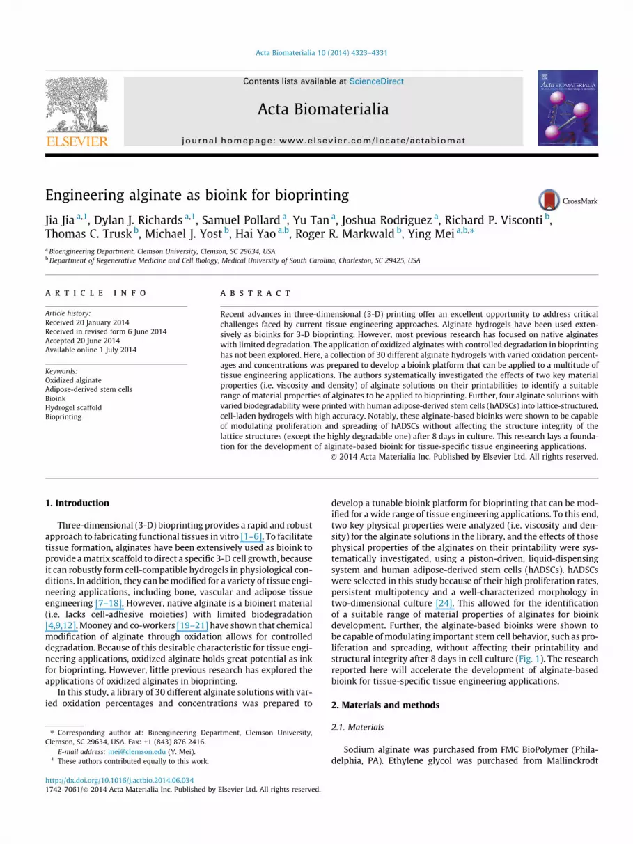
In addition to maintaining cell viability, a bioink platformshould allow for the modulation of cell behaviors. In this study,proliferation and spreading of hADSCs were examined because oftheir important roles in tissue engineering applications. In particu-lar, cell spreading can be used to modulate self-renewal and differ-entiation of hADSCs [44–46]. Cell-adhesive RGD peptide, a widelyknown cell attachment promoter molecule [9,47–49], was conju-gated to alginate bioink in this study to promote cell proliferationand spreading (Supplementary Fig. 1). DAPI staining for nuclei andphalloidin staining for actin were used to characterize the cellbehavior in each structure (Fig. 7a). As shown in Fig. 7a, b and c,the oxidized alginate bioinks (i.e. 5% ox.–10% conc., 5% ox.–15%conc. and 10% ox.–15% conc. samples) can effectively promote cellproliferation and spreading compared with the non-oxidized algi-nate bioink (i.e. 0% ox.–8% conc. alginate). Quantitatively, the cellproliferation percentages after 8 days in culture were 173% forthe 0% ox.–8% conc. alginate, 232% for the 5% ox.–10% conc. algi-nate, 248% for the 5% ox.–15% conc. alginate (Fig. 7b), and 190%for the 10% ox.–15% conc. alginate at day 4. For cell spreading,the average cell size did not change significantly after 8 days in cul-ture for the 0% ox.–8% conc. alginate, while cell size increased 25%for the 5% ox.–10% conc. alginate, 161% for the 5% ox.–15% conc.alginate at day 8, and 69% for the 10% ox.–15% conc. alginate atday 4 (Fig. 7c). The low cell proliferation and cell spreading inthe 0% ox.–8% conc. alginate was assumed to be attributed to its
sional change in 8 days. (a) Initial design of lattice structure. (b) Pictures of printeductures highly matched the initial design with apparent dimensional changes afteresign (12.6 mm � 12.6 mm) and the X and Y dimensions of the lattice structuresare mean ± SD. Asterisk denotes significant difference between day 0 and day 8.
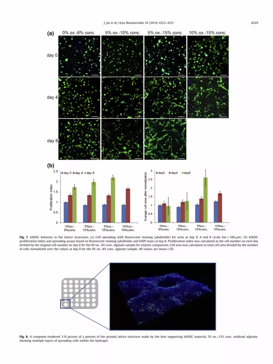
Fig. 7. hADSC behavior in the lattice structures. (a) Cell spreading with fluorescent staining (phalloidin) for actin at day 0, 4 and 8 (scale bar = 100 lm). (b) hADSCproliferation index and spreading assays based on fluorescent staining (phalloidin and DAPI stain) at day 8. Proliferation index was calculated as the cell number on each daydivided by the original cell number on day 0 for the 0% ox.–8% conc. alginate sample for relative comparison. Cell area was calculated as total cell area divided by the numberof cells normalized over the values at day 0 for the 0% ox.–8% conc. alginate sample. All values are mean ± SD.
Fig. 8. A computer-rendered 3-D picture of a portion of the printed lattice structure made by the best supporting hADSC material, 5% ox.–15% conc. oxidized alginate,showing multiple layers of spreading cells within the hydrogel.
J. Jia et al. / Acta Biomaterialia 10 (2014) 4323–4331 4329

4330 J. Jia et al. / Acta Biomaterialia 10 (2014) 4323–4331
persistent low porosity, given its limited degradability. In contrast,the significant increase in cell proliferation and spreading in the 5%ox.–15% conc. alginate was assumed to be attributed to theincreasing porosity associated with its degradation. The vast differ-ence in cell spreading among alginate bioinks with varied degree ofoxidation and concentration (i.e. 0% ox.–8% conc., 5% ox.–10% conc.and 5% ox.–15% conc. samples) demonstrates their great potentialin printing scaffolds tailored for specific tissue engineering applica-tions. For example, the 0% ox.–8% conc. alginate induced a roundcell morphology that can be applied to chondrogenesis, while the5% ox.–15% conc. alginate was associated with an increasedspreading phenotype that could facilitate osteogenesis [50–55].In addition, the cell–matrix interactions between hADSCs and algi-nates were investigated using avb3 integrin immunofluorescentstaining, as shown in Supplementary Fig. 2. The RGD conjugationwas found to promote integrin expression at all three dates (i.e.day 0, day 4 and day 8). Interestingly, the avb3 integrin expressionwas found on day 8 for alginate samples without RGD conjugation,which was attributed to the extracellular matrix secreted by hAD-SCs, consistent with previous research [56].
Fig. 8 is a 3-D picture showing a portion of a lattice structurefrom the 5% ox.–15% conc. oxidized alginate with hADSCs after8 days in culture. The distribution of the observed spreading cellsconfirmed that the homogeneous cell distribution in the alginate-based bioink pre-printing was maintained in the lattice structureafter 8 days in cell culture. Summarizing the results shown inFigs. 6 and 7, the alginate-based bioinks are capable of modulatinghADSC proliferation and spreading without affecting structuralintegrity after 8 days in culture, apart from the highly degradablealginate sample, 10% ox.–15% conc. This creates a solid foundationfor the development of alginate-based bioink for 3-D bioprinting-tailored tissue engineering scaffolds.
4. Conclusion
The primary characteristics influencing the printability of oxi-dized alginate as ink for bioprinting was defined by its ability tohold a homogeneous cell suspension, to have high printing resolu-tion and to support high cell viability. According to these factors,an ideal printable range for using oxidized alginate as a bioink plat-form was established. Further, these alginate-based bioinks wereshown to be capable of modulating hADSC functions withoutaffecting their printability and structure integrity after cell culture.The introduction of oxidized alginate to bioprinting has led to thecreation of a tunable bioink platform for a range of tissue fabrica-tions. Based on the present authors’ findings, the observed func-tional relationship between the material properties (i.e. viscosityand density) of alginate and its printability allows for an enhancedprogression in alginate bioink development for liquid-dispensingprinting. When using other cell types, metabolic demands mayrequire an altered density and viscosity of oxidized alginate toallow for optimal diffusion of nutrients. Apart from diffusion limi-tations, the Ca2+ concentration used in the gelation process mayalso affect Ca2+-sensitive cell types, such as chondrocytes [57]. ThisCa2+ concentration can be adjusted to achieve higher viability,though a prolonged gelation time may affect the printing fidelityand total printing time [25]. Future studies in bioprinting technol-ogy and a widened understanding of self-assembly mechanismsbetween cells may reshape the demands for ink printability.
Acknowledgements
This work is supported by the National Science Foundation -United States (NSF-EPS-0903795), the startup funds from ClemsonUniversity, and the National Institutes of Health (8P20 GM103444).
This study used the services of the Morphology, Imaging andInstrumentation Core, which is supported by NIH-NIGMS P30GM103342 to the South Carolina COBRE for DevelopmentallyBased Cardiovascular Diseases. The authors would like to thankAgnes Nagy Mehesz for support in cell culture. Joshua Rodriguezwas supported by the NSF-REU program.
Appendix A. Figures with essential color discrimination
Certain figures in this article, particularly Figs. 1–8, are difficultto interpret in black and white. The full color images can be foundin the online version, at http://dx.doi.org/10.1016/j.actbio.2014.06.034.
Appendix B. Supplementary data
Supplementary data associated with this article can befound, in the online version, at http://dx.doi.org/10.1016/j.actbio.2014.06.034.
References
[1] Visconti RP, Kasyanov V, Gentile C, Zhang J, Markwald RR, Mironov V. Towardsorgan printing: engineering an intra-organ branched vascular tree. Expert OpinBiol Ther 2010;10:409–20.
[2] Fedorovich NE, Alblas J, Hennink WE, Oner FC, Dhert WJA. Organ printing: thefuture of bone regeneration? Trends Biotechnol 2011;29:601–6.
[3] Mironov V, Kasyanov V, Markwald RR. Organ printing: from bioprinter toorgan biofabrication line. Curr Opin Biotechnol 2011;22:667–73.
[4] Norotte C, Marga FS, Niklason LE, Forgacs G. Scaffold-free vascular tissueengineering using bioprinting. Biomaterials 2009;30:5910–7.
[5] Seliktar D, Dikovsky D, Napadensky E. Bioprinting and tissue engineering:recent advances and future perspectives. Isr J Chem 2013;53:1–10.
[6] Kim SS, Utsunomiya H, Koski Ja, Wu BM, Cima MJ, Sohn J, et al. Survival andfunction of hepatocytes on a novel three-dimensional synthetic biodegradablepolymer scaffold with an intrinsic network of channels. Ann Surg1998;228:8–13.
[7] Lin H-R, Yeh Y-J. Porous alginate/hydroxyapatite composite scaffolds for bonetissue engineering: preparation, characterization, and in vitro studies. JBiomed Mater Res Part B Appl Biomater 2004;71:52–65.
[8] Yao R, Zhang R, Luan J, Lin F. Alginate and alginate/gelatin microspheres forhuman adipose-derived stem cell encapsulation and differentiation.Biofabrication 2012;4:025007.
[9] Rowley JA, Madlambayan G, Mooney DJ. Alginate hydrogels as syntheticextracellular matrix materials. Biomaterials 1999;20:45–53.
[10] Nakamura M, Iwanaga S, Henmi C, Arai K, Nishiyama Y. Biomatrices andbiomaterials for future developments of bioprinting and biofabrication.Biofabrication 2010;2:014110.
[11] Xu C, Chai W, Huang Y, Markwald RR. Scaffold-free inkjet printing of three-dimensional zigzag cellular tubes. Biotechnol Bioeng 2012;109:3152–60.
[12] Billiet T, Vandenhaute M, Schelfhout J, Van Vlierberghe S, Dubruel P. A reviewof trends and limitations in hydrogel-rapid prototyping for tissue engineering.Biomaterials 2012;33:6020–41.
[13] Lewis KJR, Anseth KS. Hydrogel scaffolds to study cell biology in fourdimensions. MRS Bull 2013;38:260–8.
[14] Badylak SF. The extracellular matrix as a scaffold for tissue reconstruction. CellDev Biol 2002;13:377–83.
[15] Kim B-S, Mooney DJ. Development of biocompatible synthetic extracellularmatrices for tissue engineering. Trends Biotechnol 1998;16:224–30.
[16] Orive G, Carcaboso AM, Hernández AM, Gascón AR, Pedraz JL. Biocompatibilityevaluation of different alginates and alginate-based microcapsules.Biomacromolecules 2005;6:927–31.
[17] Al-shamkhani A, Duncan R. Radioiodination of alginate via covalently-boundtyrosinamide allows monitoring of its fate in vivo. J Bioact Compat Polym1995;10:4–13.
[18] Lee M, Wu BM. Recent Advances in 3D printing of tissue engineering scaffolds.Comput Tissue Eng 2012:257–67 [cited 2013 May 22].
[19] Wang L, Shansky J, Borselli C, Mooney D, Vandenburgh H. Design andfabrication of a biodegradable, covalently crosslinked shape-memory alginatescaffold for cell and growth factor delivery. Tissue Eng Part A 2012;18:2000–7.
[20] Bouhadir KH, Lee KY, Alsberg E, Damm KL, Anderson KW, Mooney DJ.Degradation of partially oxidized alginate and its potential application fortissue engineering. Biotechnol Prog 2001;17:945–50.
[21] Lee KY, Mooney DJ. Alginate: properties and biomedical applications. ProgPolym Sci 2012;37:106–26.
[22] Murphy SV, Skardal A, Atala A. Evaluation of hydrogels for bio-printingapplications. J Biomed Mater Res A 2013;101:272–84.

J. Jia et al. / Acta Biomaterialia 10 (2014) 4323–4331 4331
[23] Chung JHY, Naficy S, Yue Z, Kapsa R, Quigley A, Moulton SE, et al. Bio-inkproperties and printability for extrusion printing living cells. Biomater Sci2013;1:763.
[24] Gimble JM, Katz AJ, Bunnell BA. Adipose-derived stem cells for regenerativemedicine. Circ Res 2007;100:1249–60.
[25] Pataky K, Braschler T, Negro A, Renaud P, Lutolf MP, Brugger J. Microdropprinting of hydrogel bioinks into 3D tissue-like geometries. Adv Mater2011;24:391–6.
[26] Alsberg E, Anderson KW, Albeiruti A, Rowley JA, Mooney DJ. Engineeringgrowing tissues. Proc Natl Acad Sci USA 2002;99:12025–30.
[27] Boontheekul T, Kong H-J, Mooney DJ. Controlling alginate gel degradationutilizing partial oxidation and bimodal molecular weight distribution.Biomaterials 2005;26:2455–65.
[28] Malda J, Visser J, Melchels FP, Jüngst T, Hennink WE, Dhert WJA, et al. 25thanniversary article: engineering hydrogels for biofabrication. Adv Mater2013:1–18.
[29] Derby B. Printing and prototyping of tissues and scaffolds. Science (80-)2012;338:921–6.
[30] Ferris CJ, Gilmore KG, Wallace GG. In het Panhuis M. biofabrication: anoverview of the approaches used for printing of living cells. Appl MicrobiolBiotechnol 2013;97:4243–58.
[31] Blaeser A, Duarte Campos DF, Weber M, Neuss S, Theek B, Fischer H, et al.Biofabrication under fluorocarbon: a novel freeform fabrication technique togenerate high aspect ratio tissue-engineered constructs. BioRes Open Access2013;2:374–84.
[32] Maher PS, Keatch RP, Donnelly K, Mackay RE, Paxton JZ. Construction of 3Dbiological matrices using rapid prototyping technology. Rapid Prototyp J2009;15:204–10.
[33] Gaetani R, Doevendans PA, Metz CHG, Alblas J, Messina E, Giacomello A, et al.Cardiac tissue engineering using tissue printing technology and human cardiacprogenitor cells. Biomaterials 2012;33:1782–90.
[34] Guillotin B, Souquet A, Catros S, Duocastella M, Pippenger B, Bellance S, et al.Laser assisted bioprinting of engineered tissue with high cell density andmicroscale organization. Biomaterials 2010;31:7250–6.
[35] Lin H, Zhang D, Alexander PG, Yang G, Tan J, Cheng AW-M, et al. Application ofvisible light-based projection stereolithography for live cell-scaffoldfabrication with designed architecture. Biomaterials 2013;34:331–9.
[36] Schuurman O, Levett PA, Pot MW, van Weeren PR, Dhert WJA, Hutmacher DW,et al. Gelatin-methacrylamide hydrogels as potential biomaterials forfabrication of tissue-engineered cartilage constructs. Macromol Biosci2013;13:551–61.
[37] Fedorovich NE, De Wijn JR, Verbout AJ, Alblas J, Dhert WJA. Three-dimensionalfiber deposition of cell-laden, viable, patterned constructs for bone tissueprinting. Tissue Eng Part A 2008;14:127–33.
[38] Fedorovich NE, Schuurman W, Wijnberg HM, Prins H-J, van Weeren PR, MaldaJ, et al. Biofabrication of osteochondral tissue equivalents by printingtopologically defined, cell-laden hydrogel scaffolds. Tissue Eng Part CMethods 2012;18:33–44.
[39] Tirella A, Orsini A, Vozzi G, Ahluwalia A. A phase diagram for microfabricationof geometrically controlled hydrogel scaffolds. Biofabrication 2009;1:045002.
[40] Bryant SJ, Anseth KS. Hydrogel properties influence ECM production bychondrocytes photoencapsulated in poly(ethylene glycol) hydrogels. J BiomedMater Res 2002;59:63–72.
[41] Nicodemus GD, Bryant SJ. Review: cell encapsulation in biodegradablehydrogels for tissue engineering applications. Tissue Eng Part B2008;14:149–65.
[42] Kong HJ, Kaigler D, Kim K, Mooney DJ. Controlling rigidity and degradation ofalginate hydrogels via molecular weight distribution. Biomacromolecules2004;5:1720–7.
[43] Kang KH, Hockaday LA, Butcher JT. Quantitative optimization of solid freeformdeposition of aqueous hydrogels. Biofabrication 2013;5:035001.
[44] Tholpady SS, Aojanepong C, Llull R, Jeong J-H, Mason AC, Futrell JW, et al. Thecellular plasticity of human adipocytes. Ann Plast Surg 2005;54:651–6.
[45] Spiegelman BM, Ginty CA. Fibronectin modulation of cell shape and lipogenicgene expression in 3T3-adipocytes. Cell 1983;35:657–66.
[46] McBeath R, Pirone DM, Nelson CM, Bhadriraju K, Chen CS. Cell shape,cytoskeletal tension, and RhoA regulate stem cell lineage commitment. DevCell 2004;6:483–95.
[47] Ruoslahti E. RGD and other recognition sequences for integrins. Annu Rev CellDev Biol 1996;12:697–715.
[48] D’Souza SE, Ginsberg MHPE. Arginyl-glycyl-aspartic acid (RGD): a celladhesion motif. Trends Biochem Sci 1991;16:246–50.
[49] Pfaff M, Tangemanno K, Mullero B, Gurrathnll M, Miillerll G, Kesslern H, et al.Selective recognition of cyclic RGD peptides of NRlR defined. J Biol Chem1994;269:20233–8.
[50] Zuk PA, Zhu M, Ashjian P, De Ugarte DA, Huang JI, Mizuno H, et al. Humanadipose tissue is a source of multipotent stem cells. Mol Biol Cell2002;13:4279–95.
[51] Zuk PA, Zhu M, Mizuno H, Huang J, Futrell JW, Katz AJ, et al. Multilineage cellsfrom human adipose tissue: implications for cell-based therapies. Tissue Eng2001;7:211–28.
[52] Zhu Y, Liu T, Song K, Fan X, Ma X, Cui Z. Adipose-derived stem cell: a betterstem cell than BMSC. Cell Biochem Funct 2008;26:664–75.
[53] Matsuoka F, Takeuchi I, Agata H, Kagami H, Shiono H, Kiyota Y, et al.Morphology-based prediction of osteogenic differentiation potential of humanmesenchymal stem cells. PLoS One 2013;8:e55082.
[54] Häuselmann HJ, Fernandes RJ, Mok SS, Schmid TM, Block JA, Aydelotte MB,et al. Phenotypic stability of bovine articular chondrocytes after long-termculture in alginate beads. J Cell Sci 1994;107:17–27.
[55] Stains JP, Civitelli R. Cell–cell interactions in regulating osteogenesis andosteoblast function. Birth Defects Res C Embryo Today 2005;75:72–80.
[56] Benton JA, Fairbanks BD, Anseth KS. Characterization of valvular interstitialcell function in three dimensional matrix metalloproteinase degradable PEGhydrogels. Biomaterials 2009;30:6593–603.
[57] Yellowley CE, Hancox JC, Donahue HJ. Effects of cell swelling on intracellularcalcium and membrane currents in bovine articular chondrocytes. J CellBiochem 2002;86:290–301.