first transplantation of isolated murine follicles in alginate
TRANSCRIPT
609Regen. Med. (2014) 9(5), 609–619 ISSN 1746-0751
part of
Research Article
10.2217/RME.14.33 © 2014 Future Medicine Ltd
Regen. Med.
10.2217/RME.14.33
Review
Vanacker, Dolmans, Luyckx, Donnez & AmorimFirst transplantation of isolated murine fol-licles in alginate
9
5
2014
Aim: Our aim is to develop an artificial ovary allowing survival and growth of isolated follicles and ovarian cells, to restore fertility in women diagnosed with pathologies at high risk of ovarian involvement. Materials & methods: For this, alginate beads containing isolated preantral follicles and ovarian cells were autografted to immunocompetent mice. Results: One week after grafting, the beads were invaded by proliferating murine cells (12.1%) and capillaries. The recovery rate of follicles per graft ranged from 0% to 35.5%. Of the analyzed follicles, 77% were Ki67-positive and 81%, TUNEL-negative. Three antral follicles were also identified, evidencing their ability to grow in the matrix. Conclusion: Our results suggest that an artificial ovary is now conceivable, opening new perspectives to restore fertility in women.
Keywords: alginate • artificial ovary • cancer • degradation • isolated preantral follicles • ovarian cells
In recent years, we have seen an increase in available fertility preservation options and they are now more commonly proposed. While embryo and oocyte cryopreservation can yield good results in terms of fertility restoration, cryopreservation and transplan-tation of ovarian tissue is the only method for patients of prepubertal age or those who require immediate cancer treatment. Indeed, this approach can restore ovarian function, with 22 live births reported to date [1]. Even if frozen–thawed ovarian tissue transplanta-tion is generally considered a safe procedure, reintroducing cancer cells that may be pres-ent in the cryopreserved tissue remains a risk, especially in the case of leukemia [2–4]. For patients suffering from this type of cancer, a safer option would be isolation and graft-ing of preantral follicles, since these entities are surrounded by a basement membrane that separates them from blood vessels, white blood cells and nerves [5].
To ensure survival, development and safe handling of isolated follicles, it is essential to encapsulate them in an artificial ovary. Fol-licles need to maintain their 3D structure
in order to avoid breakdown of the meta-bolic link between granulosa cells (GCs) and oocytes, which could lead to uncoordi-nated growth and differentiation of somatic and germ cells [6]. Another advantage of a 3D artificial ovary would be the ability to effectively mimic physiological conditions, since many cellular processes in organogen-esis occur exclusively in 3D [7]. The artificial ovary would also need to be biodegradable, since during folliculogenesis there is exponen-tial growth of follicles from the primordial (0.03 mm) to the antral stage (18–24 mm) and massive recruitment of cells and vessels to support follicle development [8].
Because hematological malignancies rep-resent one of the most common malignant diseases for which ovarian tissue needs to be cryobanked, an increasing population of young female patients would benefit from artificial ovary grafting [9]. Unfortunately, this procedure is not yet available. The first steps towards its development were taken in 1993 by Carrol and Gosden [10], who grafted a plasma clot containing a reorga-nized frozen–thawed suspension of digested
First transplantation of isolated murine follicles in alginate
Julie Vanacker1, Marie-Madeleine Dolmans*,1,2,, Valérie Luyckx1, Jacques Donnez3 & Christiani A Amorim1
1Pôle de Recherche en Gynécologie,
Institut de Recherche Expérimentale
et Clinique, Université Catholique de
Louvain, 1200 Brussels, Belgium 2Cliniques Universitaires Saint-Luc,
Gynecology Department, 1200 Brussels,
Belgium 3SRI (Society for Research into Infertility),
Brussels, Belgium
*Author for correspondence:
Author Pro
of

610 Regen. Med. (2014) 9(5) future science group
Research Article Vanacker, Dolmans, Luyckx, Donnez & Amorim
murine ovaries to the ovarian bursa of mice and obtained live births. In the pilot study of Dolmans et al. [11], in which a plasma clot was used to encap-sulate isolated preantral human follicles, we reported follicular development up to the antral stage after long-term grafting to severe combined immunodeficient (SCID) mice [11]. However, low recovery rates after grafting and the undefined composition of plasma led us to look for a more standardized and adapted matrix [8]. We therefore conducted a preliminary study [12], which showed that an alginate–matrigel matrix was able to degrade after 1 week of autografting in mice, allowing murine ovarian cells (OCs), a mix between stromal and endothelial cells, to survive and prolifer-ate. Moreover, vascularization and a low inflammatory response were also observed [12].
In the present study, our aim was to develop a trans-plantable artificial ovary suitable for clinical applica-tions. To this end, after testing different types of algi-nate, NovaMatrix 1% sterelized alginate (SLM) was chosen and used to autograft encapsulated isolated murine follicles and OCs to a peritoneal pocket that was created on the inner side of the peritoneum. After one week of grafting, we evaluated OC proliferation and survival, follicle survival, development and health status, and alginate matrix degradation, vascularization and inflammatory reaction.
Materials & methods Ovariectomy procedureThe Committee on Animal Research of the Université Catholique de Louvain (Brussels, Belgium) approved the guidelines for animal welfare. For this study, we used six female NMRI mice aged 36–40 weeks, which were housed and ovariectomized as described [12].
Isolation of murine follicles & OCsA tissue chopper (McIlwain Tissue Chopper, Mickle Laboratory, Guildford, UK) adjusted to 0.5 mm was used to mince the ovaries. The cutting procedure was swift (<5 min) and uniform-sized pieces were obtained. The minced ovarian tissue was then placed in 50 ml conical tubes containing 10 ml Dulbecco’s phosphate-buffered saline (PBS) without calcium and magnesium (Lonza, Verviers, Belgium) supplemented with 10% fetal bovine serum (FBS, Sigma-Aldrich, Bornem, Belgium) and resuspended several times with a Pasteur pipette in order to further disrupt the tissue to collect as many follicles as possible. The cell suspension was centrifuged at 40G for 10 min at 4°C and the supernatant was discarded, except for 5 ml placed in two sterile plastic Petri dishes. The follicles were picked up by two operators under a stereomicro-scope (Leica, Van Hopplynus Instruments, Brussels,
Belgium) over the course of 1 h maximum using a polycarbonate micropipette (Flexipet™ micro-manipulation pipettes, 130 μm inner diameter; Cook Ob/Gyn, IN, USA), and placed in droplets of PBS without calcium and magnesium supplemented with 10% FBS at 4°C, where they were left until the pro-cess of follicular retrieval was completed. The two Petri dishes were then pooled together in a 50 ml conical tube and rinsed with 2 ml of PBS. The sus-pension was filtered through sterilized 80 μm and 11 μm nylon net filters (Millipore, Brussels, Belgium) and centrifuged at 231G for 5 min at 4°C. OCs were counted using Trypan Blue (Sigma-Aldrich) and a Bürker chamber (VWR, Leuven, Belgium) and resuspended in PBS to obtain a final concentration of 50,000 OCs/3 μl of medium. The remaining cells were fixed in 4% formaldehyde for 1 h, and cytospin slides were prepared using the Thermo Electron Shan-don Cytospin 2 Cytocentrifuge (Thermo Scientific, Erembodegem-Aalst, Belgium).
Calcium alginate embeddingNovaMatrix 3% SLM alginate (NovaMatrix, Sand-vika, Norway) was diluted to 1% with 3-(N-mor-pholino)propanesulfonic acid buffer (MOPS, Sigma-Aldrich). A 3 μl droplet containing isolated OCs and another of 2 μl containing 37–45 isolated follicles were transferred to 20 μl droplets of alginate solu-tion. A solution of 0.1 M CaCl
2 (Sigma-Aldrich) was
slowly poured around the alginate droplets to form the beads, which were incubated for 7 min and then moved to a small Petri dish containing MOPS buffer for 5 min.
Autotransplantation procedureTo graft the artificial ovary, the animals were reanes-thetized using the protocol previously described for ovariectomy. As the alginate bead was larger than the ovarian bursa, we decided to create a pocket in the peritoneum to accommodate the bead. To this end, a circle was sutured on the anterior wall of the peritoneum at the level of the bladder using nonab-sorbable 6/0 Prolene (Ethicon, Johnson & Johnson Medical NV/SA, Diegem, Belgium). The bead was then placed in the center of this circle and both ends of the thread were pulled together and tied to close the pocket. To induce angiogenesis before placing the alginate bead, the pocket was scratched with a scal-pel blade. In case of bead fragmentation, pieces of the alginate bead were grafted, and those remaining were recovered in order to establish their follicular content. One bead was grafted per mouse, except for mouse 5, in which two beads could be grafted due to the high number of recovered isolated follicles.
Author Pro
of

www.futuremedicine.com 611future science group
First transplantation of isolated murine follicles in alginate Research Article
The abdominal wall and skin were then closed with non-absorbable 6/0 and 4/0 Prolene (Ethicon), respectively. After surgery, injection of atipamezole (1 mg/kg; Antisedan, Pfizer) was used to reverse anes-thesia. After 1 week, the animals were asphyxiated with CO
2, and the grafts were recovered under visual
control, embedded in agarose 2% (UltraPureTM Aga-rose, Invitrogen, Merelbeke, Belgium) and fixed in formalin.
Histological analysisAfter fixation in formalin, the grafts were embedded in paraffin for histological analysis. They were then cut into 5 μm serial sections and every fourth section was stained with hematoxylin–eosin for histological analy-sis in order to first identify the alginate matrix. To iden-tify and evaluate isolated follicles and OCs, histological analysis was performed on recovered alginate beads.
ImmunohistochemistryCell characterizationTo characterize isolated OCs to be grafted, vimentin and CD34 immunohistochemical stainings were per-formed. Vimentin is a cytoskeleton protein mainly expressed in the cytoplasm of cells of mesenchymal ori-gin, such as stromal and endothelial cells, and strongly expressed in ovarian stromal connective tissue [13]. On the other hand, CD34 protein is expressed in early hematopoietic and vascular tissue.
Vimentin and CD34 immunostainings were carried out according to a previously described protocol [12].
To quantify vessels (group of CD34-positive cells), one slide per graft, located in the middle of the algi-nate bead, was scanned by a Mirax Scan apparatus (Zeiss, Germany) and visualized using Mirax Viewer software. Around each alginate bead edge, a zone of 1 mm was defined. All vessels present in this area were counted and delineated, and the total vessel surface area was calculated.
Cell proliferationOC and GC proliferation were analyzed by immuno-histochemistry against Ki67, a nuclear protein present during G1, S, G2 and M replication phases, as previ-ously described [12]. The only difference was that the primary antibody used overnight at 4°C was arat anti-mouse Ki67 antibody (1:50 dilution; M7249, Dako, Glostrup, Denmark).
For OC proliferation, one slide per graft, located in the middle of the alginate bead, was scanned by a Mirax Scan apparatus and visualized using Mirax Viewer software. Positive and negative cells present inside the alginate matrix were counted in order to calculate the proportion of proliferating cells. For follicular growth,
follicles with at least one Ki67-positive GC were considered to be growing [14–17].
ApoptosisApoptosis was evaluated by terminal deoxynucleo-tidyl transferase-mediated dUTP nick-end labeling (TUNEL) to detect DNA fragmentation, according to a previously described protocol [12]. Tonsil tissue was used as a positive control.
TUNEL-positive surface areas were morphometri-cally analyzed to quantify apoptosis. To this end, sec-tions were examined at 10 or 20× magnification, and all high-power fields (HPFs) were digitized, either for TUNEL staining or DAPI counterstaining, using a Leica DFC320 camera and IM50 program (Leica, Van Hopplynus Instruments, Brussels, Belgium). After grafting, TUNEL-positive and -negative cells pres-ent inside and on the edge of the alginate matrix were counted. For follicle apoptosis, TUNEL-positive and negative GCs were counted in order to calculate the proportion of positive GCs. Follicles were classified as undamaged when both the oocyte and all GCs were alive, moderately damaged when up to 50% of GCs were alive and highly damaged when both the oocyte and more than 50% of GCs were dead [18–20].
Analysis of the inflammatory reaction after graftingImmunohistochemistry for CD45 was used to evalu-ate inflammation around grafted material, as CD45 is a marker of inflammatory cells, including leukocytes, macrophages and T lymphocytes [21]. For this pur-pose, the protocol described by Vanacker et al. [12] was followed.
One slide per graft, located in the middle of the alginate bead, was scanned by a Mirax Scan apparatus and visualized using Mirax Viewer software. A zone of 1 mm around each alginate bead edge was defined. All CD45-positive cells present in this area were counted in order to assess the CD45-positive cell population in each mouse.
Health status of the folliclesInhibin alpha immunostaining was used to character-ize GC health status. Inhibin alpha is secreted by GCs of murine and human follicles in order to inhibit FSH secretion by the pituitary gland. Inhibin alpha stain-ing was assessed to clearly identify follicular structures without visible oocytes on hematoxylin–eosin slides. Histosafe was used to deparaffinize the sections, which were rehydrated in 2-propanol. Endogenous peroxi-dase activity was blocked and a demasking step was performed for 75 min at 98°C with citrate buffer and Triton 20%. Incubation for 30 min with goat serum
Author Pro
of

612 Regen. Med. (2014) 9(5) future science group
Research Article Vanacker, Dolmans, Luyckx, Donnez & Amorim
and BSA was performed to block nonspecific binding sites, before incubation with mouse anti-human inhibin alpha antibody (1:200 dilution; MCA951S, Serotec) overnight at 4°C. The slides were incubated for 30 min at room temperature with streptavidin-HRP (1:2 dilu-tion; K4001, Dako, Glostrup, Denmark). The chro-mogen used was diaminobenzidine (SK4100, Vector, Laboratories, Peterborough, UK). The slides were then counterstained with hematoxylin and mounted with aqueous mounting medium (Aquatex, Merck, Darm-stadt, Germany). Negative (dilution solution without any anti-inhibin alpha antibody) and positive (mouse ovary) controls were carried out.
Groups of GCs with no visible oocyte on hematoxylin–eosin slides stained for inhibin alpha were considered as follicles.
ResultsFollicle population characterization before graftingIn order to avoid follicular viability impairment, iso-lated follicles could not be staged before grafting. A supplementary isolation procedure was therefore performed to assess which follicle stages were grafted in our model. Non-grafted control isolated murine preantral follicles were retrieved with a polycarbonate micropipette (130 μm inner diameter) and were found at primordial to secondary stages, ranging from 17 to 101 μm in diameter.
OC population characterization before graftingFollowing the isolation procedure, non-grafted OCs were fixed and cytospin slides prepared. After anti-vimentin staining, 5029 cells were counted and 90.5% of them were vimentin-positive. Anti-CD34 immuno-staining was subsequently performed on these fixed iso-lated OCs and 10% of the 12,762 counted cells were CD34-positive. We can therefore assume that a large number of isolated cells encapsulated in the alginate matrix were stromal cells of mesenchymal origin.
Graft recovery rate & macroscopic aspectAfter 1 week of autografting of isolated murine follicles and OCs, all seven peritoneal bursae could be clearly identified thanks to sutures in the six mice after dissect-ing the skin from the peritoneum with blunt scissors. All seven matrices were easily recovered from the intra-peritoneal cavity. No adhesions with intraperitoneal organs or major inflammatory reactions around the grafting site were noted. The peritoneal scar was satis-factorily healed in all cases. Alginate beads occupied a mean of 314 sections after grafting (292 in graft 1, 404 in graft 2, 280 in graft 3, 228 in graft 4, 312 in graft 5 [right], 352 in graft 5 [left] and 336 in graft 6).
Tab
le 1
. An
alys
is o
f fo
llicl
e re
cove
ry, v
esse
l fo
rmat
ion
an
d C
D45
-po
siti
ve c
ells
aro
un
d a
nd
infi
ltra
tin
g t
he
alg
inat
e m
atri
x af
ter
on
e w
eek
of
gra
ftin
g.
Mo
use
Bef
ore
gra
ftin
gA
fter
gra
ftin
g
Nu
mb
er
of
gra
fted
O
Cs
Nu
mb
er
of
gra
fted
fo
llicl
es
Folli
cles
Ves
sel f
orm
atio
nC
D45
-po
siti
ve c
ells
Nu
mb
er
of
folli
cles
fo
un
d
Nu
mb
er o
f in
hib
in a
lph
a-
po
siti
ve
folli
cula
r st
ruct
ure
s
Tota
l n
um
ber
o
f ve
ssel
s an
alyz
ed
Tota
l su
rfac
e ar
ea o
f ve
ssel
s (μ
m²)
Tota
l su
rfac
e ar
ea
anal
yzed
(μ
m²)
Ves
sel
surf
ace
area
/to
tal
surf
ace
area
(%
)
Nu
mb
er
of
vess
els/
μm²
Tota
l n
um
ber
o
f C
D45
+
cells
Tota
l su
rfac
e ar
ea
anal
yzed
(μ
m²)
Nu
mb
er
of
CD
45+
ce
lls/μ
m²
150
,00
04
44
555
957
,951
6,3
28,8
100.
928
.83
× 1
0-5
619
6,6
38
,145
9.32
× 1
0-5
250
,00
04
04
227
955
,305
5,63
3,35
70.
98
4.9
5 ×
10
-536
45,
578
,243
6.53
× 1
0-5
350
,00
04
01
353
714
1,36
89,
224
,862
1.53
5.82
× 1
0-5
443
9,70
1,32
44
.57
× 1
0-5
450
,00
037
71
622
79,7
00
8,6
92,2
270.
927.
16 ×
10
-527
58
,99
8,6
663.
06
× 1
0-5
5 R
igh
t50
,00
045
35
439
79,8
86
9,24
0,4
870.
86
4.7
5 ×
10
-54
4110
,727
,992
4.1
1 ×
10
-5
5 Le
ft50
,00
045
79
482
210,
981
9,18
1,59
32.
305.
25 ×
10
-525
75,
387
,791
4.7
7 ×
10
-5
650
,00
043
54
547
87,2
3710
,99
6,3
760.
794
.97
× 1
0-5
439,
40
0,83
34
.57
x 10
-6
OC: Ovarian cell.
Author Pro
of
www.futuremedicine.com 613
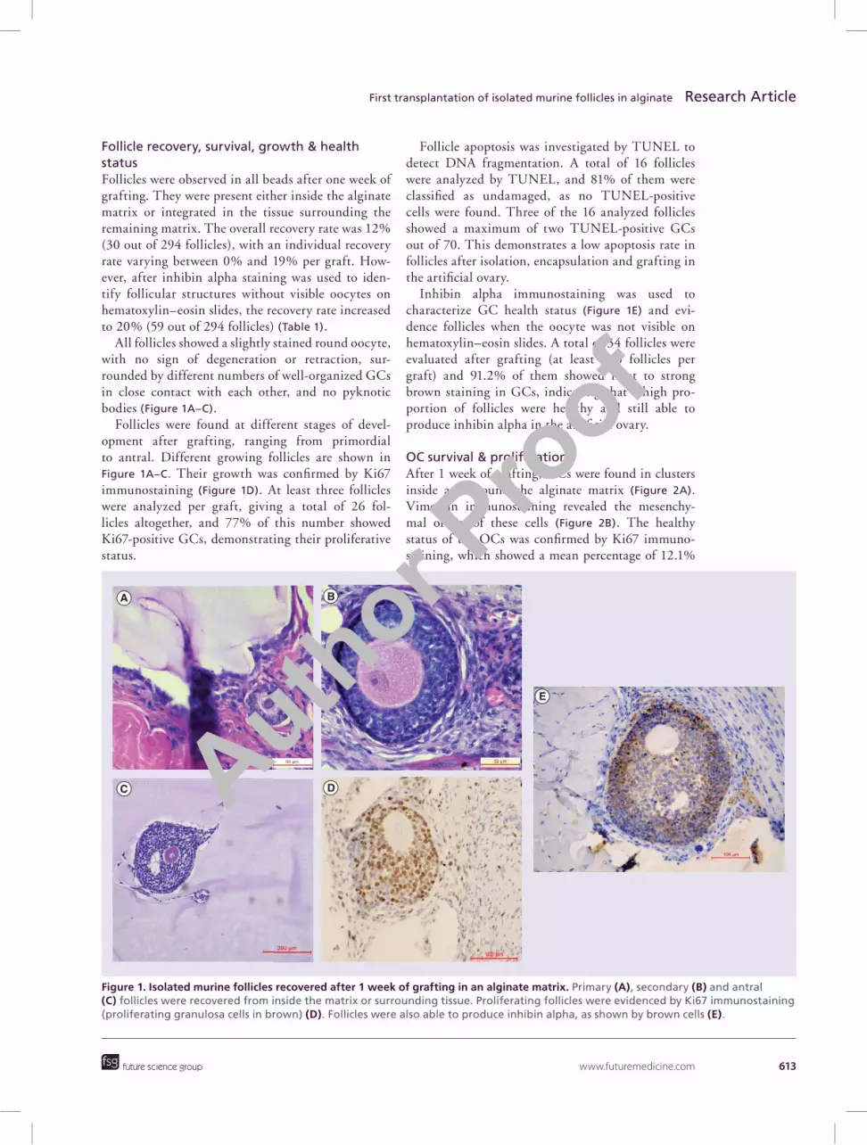
Figure 1. Isolated murine follicles recovered after 1 week of grafting in an alginate matrix. Primary (A), secondary (B) and antral (C) follicles were recovered from inside the matrix or surrounding tissue. Proliferating follicles were evidenced by Ki67 immunostaining (proliferating granulosa cells in brown) (D). Follicles were also able to produce inhibin alpha, as shown by brown cells (E).
A B
C D
E
future science group
First transplantation of isolated murine follicles in alginate Research Article
Follicle recovery, survival, growth & health statusFollicles were observed in all beads after one week of grafting. They were present either inside the alginate matrix or integrated in the tissue surrounding the remaining matrix. The overall recovery rate was 12% (30 out of 294 follicles), with an individual recovery rate varying between 0% and 19% per graft. How-ever, after inhibin alpha staining was used to iden-tify follicular structures without visible oocytes on hematoxylin–eosin slides, the recovery rate increased to 20% (59 out of 294 follicles) (Table 1).
All follicles showed a slightly stained round oocyte, with no sign of degeneration or retraction, sur-rounded by different numbers of well-organized GCs in close contact with each other, and no pyknotic bodies (Figure 1A–C).
Follicles were found at different stages of devel-opment after grafting, ranging from primordial to antral. Different growing follicles are shown in Figure 1A–C. Their growth was confirmed by Ki67 immunostaining (Figure 1D). At least three follicles were analyzed per graft, giving a total of 26 fol-licles altogether, and 77% of this number showed Ki67-positive GCs, demonstrating their proliferative status.
Follicle apoptosis was investigated by TUNEL to detect DNA fragmentation. A total of 16 follicles were analyzed by TUNEL, and 81% of them were classified as undamaged, as no TUNEL-positive cells were found. Three of the 16 analyzed follicles showed a maximum of two TUNEL-positive GCs out of 70. This demonstrates a low apoptosis rate in follicles after isolation, encapsulation and grafting in the artificial ovary.
Inhibin alpha immunostaining was used to characterize GC health status (Figure 1E) and evi-dence follicles when the oocyte was not visible on hematoxylin–eosin slides. A total of 34 follicles were evaluated after grafting (at least two follicles per graft) and 91.2% of them showed faint to strong brown staining in GCs, indicating that a high pro-portion of follicles were healthy and still able to produce inhibin alpha in the artificial ovary.
OC survival & proliferationAfter 1 week of grafting, OCs were found in clusters inside and around the alginate matrix (Figure 2A). Vimentin immunostaining revealed the mesenchy-mal origin of these cells (Figure 2B). The healthy status of the OCs was confirmed by Ki67 immuno-staining, which showed a mean percentage of 12.1%
Author Pro
of

614 Regen. Med. (2014) 9(5) future science group
Research Article Vanacker, Dolmans, Luyckx, Donnez & Amorim
Figure 2. Grafted murine ovarian cells were alive and proliferating after 1 week of grafting. Ovarian cancers are shown grouped in clusters within the alginate matrix (A). The mesenchymal origin of these cells was evidenced by anti-vimentin immunostaining (B). Ovarian cells were able to proliferate inside the matrix, as shown by Ki67-positive brown cells (C). DNA strand breaks were analyzed by TUNEL in isolated ovarian cells encapsulated in the alginate matrix: DAPI counterstaining (cells in blue) and TUNEL staining (cells in red) images are merged (D).
A B100 µm
C D
proliferating OCs (Figure 2C). DNA fragmentation was assessed by TUNEL, which evidenced 2.3% of dead cells (Figure 2D).
Scaffold degradation, revascularization & inflammatory responseAlthough five beads broke up into fragments during the grafting procedure, the pieces did not entirely disappear after 1 week of grafting. Thus, while deg-radation was found to have started after such a short period, it was not complete. In all grafts, beads and bead fragments were populated by proliferating OCs grouped in clusters and isolated growing follicles, as shown in Figures 1 & 2.
Blood capillaries and endothelial cells were observed around and inside the alginate matrix. Indeed, CD34-positive cells were found to surround and infiltrate the matrix (Figure 3A). Moreover, as indicated by the pres-ence of red blood cells, functional blood vessels were also seen inside the alginate matrix after 1 week of grafting (Figure 3B, arrows). Table 1 shows the number
of grafted follicles and OCs, and the number of vessels per surface unit, which varied from mouse to mouse but remained within the same range.
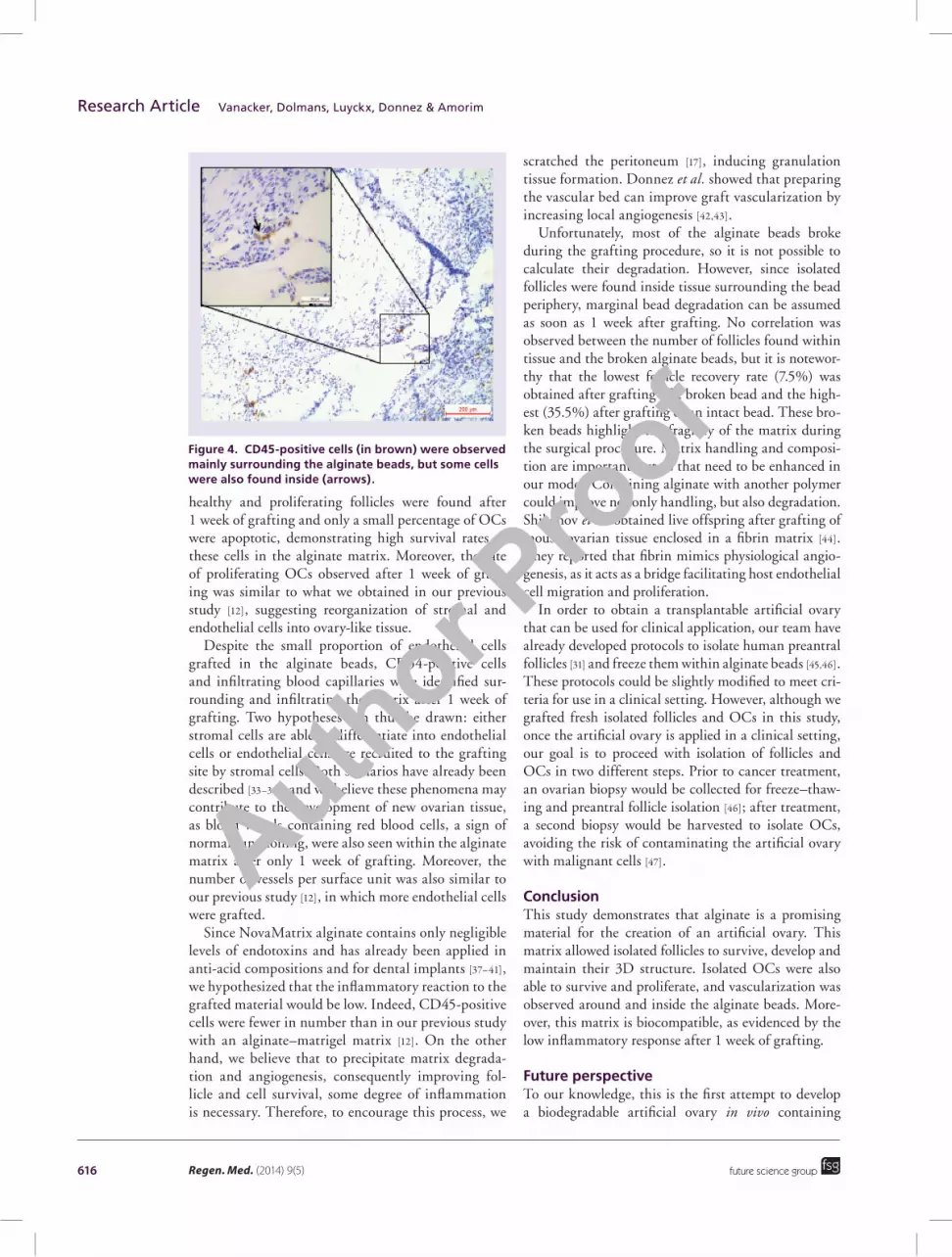
The inflammatory reaction, evidenced by CD45 immunostaining, was limited to the matrix periphery and no fibrous capsule was observed around the grafts. Some occasional CD45-positive cells were also found infiltrating the matrix (Figure 4). The small and glo-boid shape of these cells (stained in brown) suggests a leukocyte-type reaction. As shown in Table 1, a small number of CD45-positive cells per surface unit were identified in the grafts. Moreover, no mice died as a result of the procedure. They were all healthy after one week of grafting, proving that no major reaction was initiated by the alginate matrix.
DiscussionThe present study shows promising results, indicat-ing that it may well be feasible to develop an artifi-cial ovary to graft isolated follicles and OCs. Indeed, previous pilot studies [10,11] have demonstrated survival
Author Pro
of

www.futuremedicine.com 615future science group
First transplantation of isolated murine follicles in alginate Research Article
and development of mouse and human follicles after grafting in a plasma clot. Here, we identified a GMP-grade alginate matrix (NovaMatrix 1% SLM) able to overcome the drawbacks encountered with plasma clots, such as low recovery rates or undefined plasma composition. Moreover, this matrix meets all the cri-teria to support isolated follicle and OC grafting, as it is degradable, biocompatible and easy to handle, while allowing follicle growth and development, stromal tis-sue restructuring and angiogenesis. This study is also the first to analyze grafting of isolated follicles and OCs together.
The follicle recovery rate after one week of graft-ing in an alginate matrix was similar to results previ-ously obtained after transplantation of human ovarian follicles encapsulated in plasma clots to the ovarian bursa of SCID mice [11]. However, while all alginate beads and 7.5–35.5% of grafted follicles were recov-ered in our study, Dolmans et al. [11] recovered approxi-mately 20% of grafted follicles, but less than 60% of grafted clots. Our higher graft recovery rate may be partially explained by the grafting procedure, as the plasma clot could be lost due to the hole made in the ovarian bursa. Regarding the 80% of follicles that dis-appeared after grafting, we believe that this was due to ischemic injury that occurs in the first days after trans-plantation [22]. Indeed, follicle loss after autografting of ovarian tissue has been reported to be between 60 and 80% [23,24].
Follicles were found either inside the alginate matrix or in tissue around the bead periphery. We believe this was due to the peripheral localization of some follicles within the matrix at the time of grafting. After 1 week, degradation and colonization of peripheral parts of the beads by grafted murine OCs and peritoneal tissue can be assumed, integrating follicles within tissue.
Interestingly, three antral follicles were found after 1 week of grafting. This may have been due to pick-up of secondary follicles, which is very common in suspensions of isolated murine preantral follicles. It is, thus, very likely that these secondary follicles grew to the antral stage during the week of grafting, as murine ovarian folliculogenesis is known to be completed within 3 weeks [25].
A high follicle survival rate was obtained within the artificial ovary after one week of grafting, as evidenced by histology, TUNEL analysis and Ki67 immunos-taining. Indeed, most of the recovered follicles were morphologically normal and developing, as demon-strated by proliferating GCs. After TUNEL analysis, only three follicles showed less than 3% of atretic GCs. According to some authors, GC death is not only related to follicle atresia, but can also occur naturally, to a certain extent, during normal follicle develop-
ment [26–30]. Moreover, follicles were able to produce inhibin alpha after isolation, encapsulation and 1 week of grafting, proving that they were healthy.
In a previous study [31], we set up a follicle isola-tion protocol adapted for human ovarian tissue using an enzyme called Liberase Dispase High (DH). Since murine ovarian tissue is softer than human tissue, we did not use this enzymatic digestion method in the present study, as there was a risk of destroying the murine follicles. We performed a filtration step through 80 and 11 μm nylon net filters, as previously developed for human stromal cell isolation [32]. We believe this procedure is the reason why we grafted a smaller number of endothelial cells than in our pre-vious study [12]. Indeed, vessels may not be disrupted by a mechanical isolation procedure alone. However,
Figure 3. Blood vessels surrounding and infiltrating the alginate matrix after 1 week of grafting. Blood vessels and endothelial cells, stained in brown owing to anti-CD34 immunostaining (arrows), were located at the bead periphery or inside the matrix (A). Some vessels were already functional after 1 week of grafting, as indicated by the presence of red blood cells (B, arrows).
A
B
Author Pro
of
616 Regen. Med. (2014) 9(5) future science group
Research Article Vanacker, Dolmans, Luyckx, Donnez & Amorim
healthy and proliferating follicles were found after 1 week of grafting and only a small percentage of OCs were apoptotic, demonstrating high survival rates of these cells in the alginate matrix. Moreover, the rate of proliferating OCs observed after 1 week of graft-ing was similar to what we obtained in our previous study [12], suggesting reorganization of stromal and endothelial cells into ovary-like tissue.
Despite the small proportion of endothelial cells grafted in the alginate beads, CD34-positive cells and infiltrating blood capillaries were identified sur-rounding and infiltrating the matrix after 1 week of grafting. Two hypotheses can thus be drawn: either stromal cells are able to differentiate into endothelial cells or endothelial cells are recruited to the grafting site by stromal cells. Both scenarios have already been described [33–36], and we believe these phenomena may contribute to the development of new ovarian tissue, as blood vessels containing red blood cells, a sign of normal functioning, were also seen within the alginate matrix after only 1 week of grafting. Moreover, the number of vessels per surface unit was also similar to our previous study [12], in which more endothelial cells were grafted.
Since NovaMatrix alginate contains only negligible levels of endotoxins and has already been applied in anti-acid compositions and for dental implants [37–41], we hypothesized that the inflammatory reaction to the grafted material would be low. Indeed, CD45-positive cells were fewer in number than in our previous study with an alginate–matrigel matrix [12]. On the other hand, we believe that to precipitate matrix degrada-tion and angiogenesis, consequently improving fol-licle and cell survival, some degree of inflammation is necessary. Therefore, to encourage this process, we
scratched the peritoneum [17], inducing granulation tissue formation. Donnez et al. showed that preparing the vascular bed can improve graft vascularization by increasing local angiogenesis [42,43].
Unfortunately, most of the alginate beads broke during the grafting procedure, so it is not possible to calculate their degradation. However, since isolated follicles were found inside tissue surrounding the bead periphery, marginal bead degradation can be assumed as soon as 1 week after grafting. No correlation was observed between the number of follicles found within tissue and the broken alginate beads, but it is notewor-thy that the lowest follicle recovery rate (7.5%) was obtained after grafting of a broken bead and the high-est (35.5%) after grafting of an intact bead. These bro-ken beads highlight the fragility of the matrix during the surgical procedure. Matrix handling and composi-tion are important factors that need to be enhanced in our model. Combining alginate with another polymer could improve not only handling, but also degradation. Shikanov et al. obtained live offspring after grafting of mouse ovarian tissue enclosed in a fibrin matrix [44]. They reported that fibrin mimics physiological angio-genesis, as it acts as a bridge facilitating host endothelial cell migration and proliferation.
In order to obtain a transplantable artificial ovary that can be used for clinical application, our team have already developed protocols to isolate human preantral follicles [31] and freeze them within alginate beads [45,46]. These protocols could be slightly modified to meet cri-teria for use in a clinical setting. However, although we grafted fresh isolated follicles and OCs in this study, once the artificial ovary is applied in a clinical setting, our goal is to proceed with isolation of follicles and OCs in two different steps. Prior to cancer treatment, an ovarian biopsy would be collected for freeze–thaw-ing and preantral follicle isolation [46]; after treatment, a second biopsy would be harvested to isolate OCs, avoiding the risk of contaminating the artificial ovary with malignant cells [47].
ConclusionThis study demonstrates that alginate is a promising material for the creation of an artificial ovary. This matrix allowed isolated follicles to survive, develop and maintain their 3D structure. Isolated OCs were also able to survive and proliferate, and vascularization was observed around and inside the alginate beads. More-over, this matrix is biocompatible, as evidenced by the low inflammatory response after 1 week of grafting.
Future perspectiveTo our knowledge, this is the first attempt to develop a biodegradable artificial ovary in vivo containing
Figure 4. CD45-positive cells (in brown) were observed mainly surrounding the alginate beads, but some cells were also found inside (arrows).
Author Pro
of
www.futuremedicine.com 617future science group
First transplantation of isolated murine follicles in alginate Research Article
isolated murine follicles and OCs suitable for clinical applications. We plan to extend our transplantation period in order to assess the final stages of follicle devel-opment and study degradation kinetics of the matrix. It will also be necessary to test xenografting of isolated human follicles and OCs in mice.
AcknowledgementsThe authors thank Mira Hryniuk for reviewing the manuscript,
Dolores Gonzalez and Olivier Van Kerk for their technical
assistance.
Financial & competing interests disclosureThe present study was supported by grants from the Fonds Na-
tional de la Recherche Scientifique de Belgique (grant Télévie
N– 7.4507.10, grant 3.4.590.08 awarded to M-M Dolmans),
the Fonds Spéciaux de Recherche, the Fondation St Luc and
the Foundation Against Cancer, and donations from Mr Pietro
Ferrero, Baron Frère and Viscount Philippe de Spoelberch.
No writing assistance was utilized in the production of this
manuscript.
Ethical conduct of researchApproval for this study was obtained from the Ethics Com-
mittee of the Faculty of Medicine of the Catholic University
of Louvain (ref. 2014/UCL/MD/007). The authors state that
they have obtained appropriate institutional review board
approval or have followed the principles outlined in the Dec-
laration of Helsinki for all human or animal experimental
investigations.
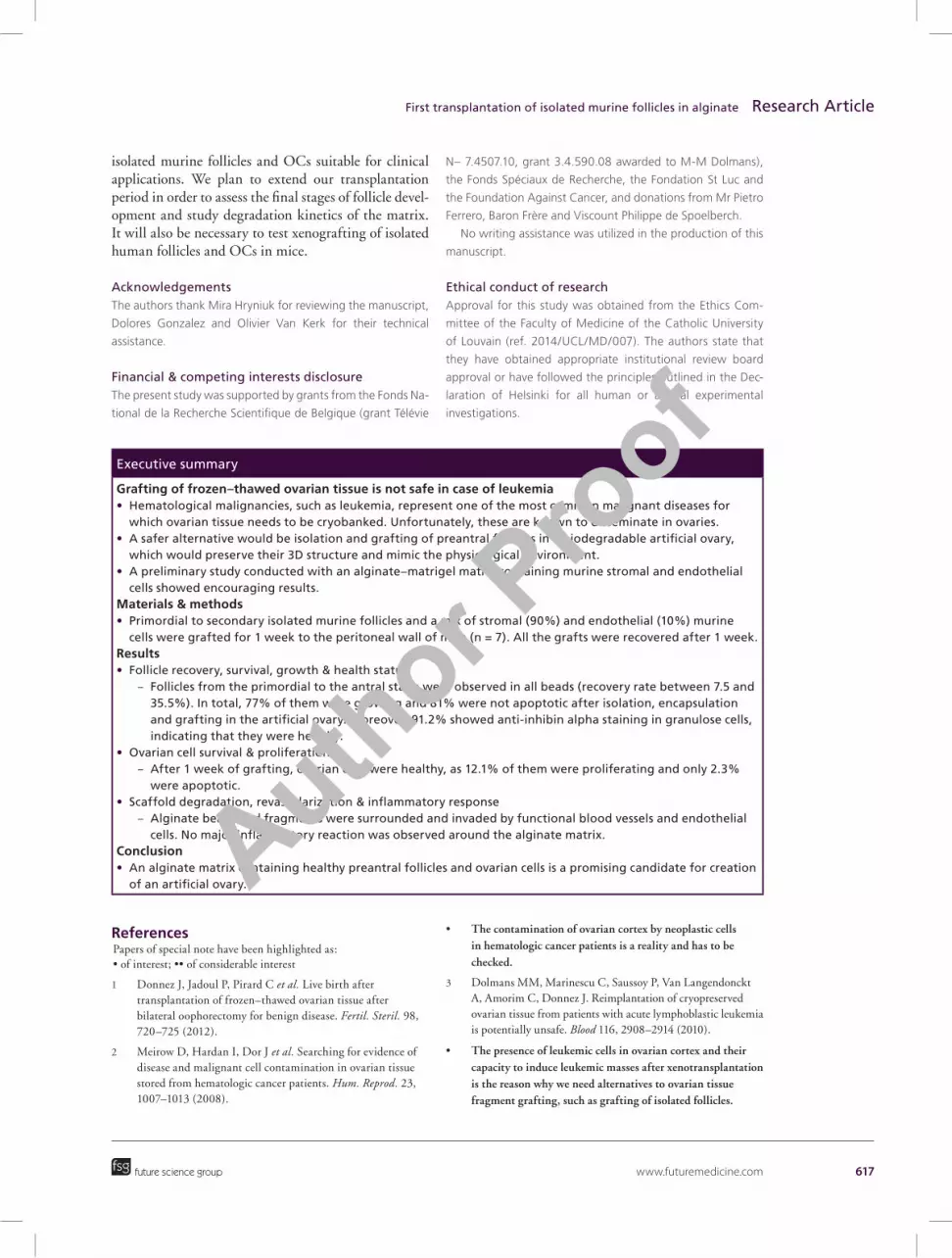
Executive summary
Grafting of frozen–thawed ovarian tissue is not safe in case of leukemia• Hematological malignancies, such as leukemia, represent one of the most common malignant diseases for
which ovarian tissue needs to be cryobanked. Unfortunately, these are known to disseminate in ovaries.• A safer alternative would be isolation and grafting of preantral follicles in a biodegradable artificial ovary,
which would preserve their 3D structure and mimic the physiological environment.• A preliminary study conducted with an alginate–matrigel matrix containing murine stromal and endothelial
cells showed encouraging results.Materials & methods• Primordial to secondary isolated murine follicles and a mix of stromal (90%) and endothelial (10%) murine
cells were grafted for 1 week to the peritoneal wall of mice (n = 7). All the grafts were recovered after 1 week.Results• Follicle recovery, survival, growth & health status
– Follicles from the primordial to the antral stage were observed in all beads (recovery rate between 7.5 and 35.5%). In total, 77% of them were growing and 81% were not apoptotic after isolation, encapsulation and grafting in the artificial ovary. Moreover, 91.2% showed anti-inhibin alpha staining in granulose cells, indicating that they were healthy.
• Ovarian cell survival & proliferation – After 1 week of grafting, ovarian cells were healthy, as 12.1% of them were proliferating and only 2.3%
were apoptotic.• Scaffold degradation, revascularization & inflammatory response
– Alginate beads and fragments were surrounded and invaded by functional blood vessels and endothelial cells. No major inflammatory reaction was observed around the alginate matrix.
Conclusion• An alginate matrix containing healthy preantral follicles and ovarian cells is a promising candidate for creation
of an artificial ovary.
ReferencesPapers of special note have been highlighted as: • of interest; •• of considerable interest
1 Donnez J, Jadoul P, Pirard C et al. Live birth after transplantation of frozen–thawed ovarian tissue after bilateral oophorectomy for benign disease. Fertil. Steril. 98, 720–725 (2012).
2 Meirow D, Hardan I, Dor J et al. Searching for evidence of disease and malignant cell contamination in ovarian tissue stored from hematologic cancer patients. Hum. Reprod. 23, 1007–1013 (2008).
• The contamination of ovarian cortex by neoplastic cells in hematologic cancer patients is a reality and has to be checked.
3 Dolmans MM, Marinescu C, Saussoy P, Van Langendonckt A, Amorim C, Donnez J. Reimplantation of cryopreserved ovarian tissue from patients with acute lymphoblastic leukemia is potentially unsafe. Blood 116, 2908–2914 (2010).
• The presence of leukemic cells in ovarian cortex and their capacity to induce leukemic masses after xenotransplantation is the reason why we need alternatives to ovarian tissue fragment grafting, such as grafting of isolated follicles.
Author Pro
of

618 Regen. Med. (2014) 9(5) future science group
Research Article Vanacker, Dolmans, Luyckx, Donnez & Amorim
4 Rosendahl M, Andersen MT, Ralfkiær E, Kjeldsen L, Andersen MK, Andersen CY. Evidence of residual disease in cryopreserved ovarian cortex from female patients with leukemia. Fertil. Steril. 94, 2186–2190 (2010).
5 Rodgers RJ, Irving-Rodgers HF, Russell DL. Extracellular matrix of the developing ovarian follicle. Reproduction 126, 415–424 (2003).
6 Picton HM, Gosden RG. In vitro growth of human primordial follicles from frozen-banked ovarian tissue. Mol. Cell. Endocrinol. 166, 27–35 (2000).
7 Xu M, West E, Shea LD, Woodruff TK. Identification of a stage-specific permissive in vitro culture environment for follicle growth and oocyte development. Biol. Reprod. 75, 916–923 (2006).
8 Amorim CA. Artificial ovary. In: Principles and Practice of Fertility Preservation. Donnez J, Kim SS (Eds). Cambridge University Press, Cambridge, UK, 448–458 (2011).
9 Donnez J, Dolmans MM. Preservation of fertility in females with haematological malignancy. Br. J. Haematol. 154, 175–184 (2011).
10 Carroll J, Gosden RG. Transplantation of frozen–thawed mouse primordial follicles. Hum. Reprod. 8, 1163–1167 (1993).
11 Dolmans MM, Yuan WY, Camboni A et al. Development of antral follicles after xenografting of isolated small human preantral follicles. Reprod. Biomed. Online 16, 705–711 (2008).
• The first attempt by our group to graft human isolated ovarian follicles for a long time period in a plasma clot.
12 Vanacker J, Luyckx V, Dolmans MM et al. Transplantation of an alginate–matrigel matrix containing isolated ovarian cells: first step in developing a biodegradable scaffold to transplant isolated preantral follicles and ovarian cells. Biomaterials 33, 6079–6085 (2012).
•• The first step toward a bioartificial ovary composed of alginate and containing isolated cells.
13 Czernobilsky B, Moll R, Levy R, Franke WW. Co-expression of cytokeratin and vimentin filaments in mesothelial, granulosa and rete ovarii cells of the human ovary. Eur. J. Cell. Biol. 37, 175–190 (1985).
14 Grazul-Bilska AT, Caton JS, Arndt W et al. Cellular proliferation and vascularization in ovine fetal ovaries: effects of undernutrition and selenium in maternal diet. Reproduction 137, 699–707 (2009).
15 Amorim CA, Jacobs S, Devireddy RV et al. Successful vitrification and autografting of baboon (Papio anubis) ovarian tissue. Hum. Reprod. 28, 2146–2156 (2013).
16 Luyckx V, Scalercio S, Jadoul P et al. Evaluation of cryopreserved ovarian tissue from prepubertal patients after long-term xenografting and exogenous stimulation. Fertil. Steril. 100, 1350–1357 (2013).
17 Luyckx V, Dolmans MM, Vanacker J et al. A new step toward the artificial ovary: survival and proliferation of isolated murine follicles after autologous transplantation in a fibrin scaffold. Fertil. Steril. 101(4), 1149–1156 (2014).
•• Similar encouraging results to this study have been obtained with a fibrin scaffold.
18 Martinez-Madrid B, Dolmans MM, Van Langendonckt A, Defrère S, Van Eyck AS, Donnez J. Ficoll density gradient method for recovery of isolated human ovarian primordial follicles. Fertil. Steril. 82, 1648–1653 (2004).
19 Aerts JM, Martinez-Madrid B, Flothmann K, De Clercq JB, Van Aelst S, Bols PE. Quantification and viability assessment of isolated bovine primordial and primary ovarian follicles retrieved through a standardized biopsy pick-up procedure. Reprod. Domest. Anim. 43, 360–366 (2008).
20 Xing W, Zhou C, Bian J et al. Solid-surface vitrification is an appropriate and convenient method for cryorpeservation of isolated rat follicles. Reprod. Biol. Endocrinol. 8, 42 (2010).
21 Charbonneau H, Tonks NK, Walsh KA, Fischer EH. The leukocyte common antigen (CD45): a putative receptor-linked protein tyrosine phosphatase. Proc. Natl Acad. Sci. USA 85, 7182–7186 (1988).
22 Van Eyck AS, Bouzin C, Feron O et al. Both host and graft vessels contribute to revascularization of xenografted human ovarian tissue in a murine model. Fertil. Steril. 93, 1676–1685 (2009).
23 Candy CJ, Wood MJ, Whittingham DG. Effect of cryoprotectants on the survival of follicles in frozen mouse ovaries. J. Reprod. Fertil. 110, 11–19 (1997).
24 Aubard Y, Piver P, Congnié Y, Fermeaux V, Poulin N, Driancourt MA. Orthotopic and heterotopic autografts of frozen–thawed ovarian cortex in sheep. Hum. Reprod. 14, 2149–2154 (1999).
25 Amleh A, Dean J. Mouse genetics provides insight into folliculogenesis, fertilization and early embryonic development. Hum. Reprod. Update 8, 395–403 (2002).
26 Ireland JJ, Roche JF. Growth and differentiation of large antral follicles after spontaneous luteolysis in heifers: changes in concentration of hormones in follicular fluid and specific binding of gonadotropins to follicles. J. Anim. Sci. 57, 157–167 (1983).
27 Jolly PD, Tisdall DJ, Heath DA, Lun S, McNatty KP. Apoptosis in bovine granulosa cells in relation to steroid synthesis, cyclic adenosine 3’,5’-monophosphate response to follicle-stimulating hormone and luteinizing hormone, and follicular atresia. Biol. Reprod. 51, 934–944 (1994).
28 Van Wezel IL, Dharmarajan AM, Lavranos TC, Rodgers RJ. Evidence for alternative pathways of granulosa cell death in healthy and slightly atretic bovine antral follicles. Endocrinology 140, 2602–2612 (1999).
29 Rodgers RJ, Irving-Rodgers HF. Formation of the ovarian dollicular antrum and follicular fluid. Biol. Reprod. 82, 1021–1029 (2010).
30 Rodgers RJ, Irving-Rodgers HF. Morphological classification of bovine ovarian follicles. Reproduction 139, 309–318 (2010).
31 Vanacker J, Camboni A, Dath C et al. Enzymatic isolation of human primordial and primary ovarian follicles with Liberase DH: protocol for application in a clinical setting. Fertil. Steril. 96, 379–383 (2011).
• This follicular isolation protocol allows the use of isolated follicles in clinics.
Author Pro
of
www.futuremedicine.com 619
32 Dath C, Dethy A, Van Langendonckt A et al. Endothelial cells are essential for ovarian stromal tissue restructuring after xenotransplantation of isolated ovarian stromal cells. Hum. Reprod. 26, 1431–1439 (2011).
•• Explains the importance of co-grafting endothelial and stromal ovarian cells with isolated follicles.
33 Vittorio O, Jacchetti E, Pacini S, Cecchini M. Endothelial differentiation of mesenchymal stromal cells: when traditional biology meets mechanotransduction. Integr. Biol. 5, 291–299 (2013).
34 Vila OF, Bagó JR, Navarro M et al. Calcium phosphate glass improves angiogenesis capacity of poly(lactic acid) scaffolds and stimulates differentiation of adipose tissue-derived mesenchymal stromal cells to the endothelial lineage. J. Biomed. Mater. Res. A 101, 932–941 (2013).
35 Yu J, Wang A, Tang Z et al. The effect of stromal cell-derived factor-1α/heparin coating of biodegradable vascular grafts on the recruitment of both endothelial and smooth muscle progenitor cells for accelerated regeneration. Biomaterials 33, 8062–8074 (2012).
36 Ryu JK, Tumurbaatar M, Jin HR et al. Intracavernous delivery of freshly isolated stromal vascular fraction rescues erectile function by enhancing endothelial regeneration in the streptozotocin-induced diabetic mouse. J. Sex. Med. 9, 3051–3065 (2012).
37 Hagstam H. Alginates and heartburn – evaluation of a medicine with a mechanical mode of action. In: Gums and Stabilizers in the Food Industry. Phillips GO (Ed.). Elsevier, Amsterdam, The Netherlands, 363–370 (1986).
38 Thomas S. Alginate dressings in surgery and wound management – part 1. J. Wound Care 9, 56–60 (2000).
39 Thomas S. Alginate dressings in surgery and wound management: part 2. J. Wound Care 9, 115–119 (2000).
40 Thomas S. Alginate dressings in surgery and wound management: part 3. J. Wound Care 9, 163–166 (2000).
41 Mandel KG, Daggy BP, Brodie DA, Jacoby HI. Review article: alginate-raft formulations in the treatment of heartburn and acid reflux. Aliment. Pharmacol. Ther. 14, 669–690 (2000) (2000).
42 Donnez J, Dommans MM, Pellicer A et al. Restoration of ovarian activity and pregnancy after transplantation of cryopreserved ovarian tissue: a review of 60 cases of reimplantation. Fertil. Steril. 99, 1503–1513 (2013).
43 Donnez J, Dolmans MM, Demylle D et al. Livebirth after orthotopic transplantation of cryopreserved ovarian tissue. Lancet 364, 1405–1410 (2004).
44 Shikanov A, Zhang Z, Xu M et al. Fibrin encapsulation and vascular endothelial growth factor delivery promotes ovarian graft survival in mice. Tissue Eng. Part A 17, 3095–3104 (2011).
45 Camboni A, Van Langendonckt A, Donnez J, Vanacker J, Dolmans MM, Amorim CA. Alginate beads as a tool to handle, cryopreserve and culture isolated human primordial/primary follicles. Cryobiology 67, 64–69 (2013).
• Shows other applications of alginate beads containing isolated follicles.
46 Vanacker J, Luyckx V, Amorim C et al. Should we isolate human preantral follicles before or after cryopreservation of ovarian tissue? Fertil. Steril. 99, 1363–1368 (2013).
47 Dolmans MM, Martinez-Madrid B, Gadisseux E et al. Short-term transplantation of isolated human ovarian follicles and cortical tissue into nude mice. Reproduction 134, 253–262 (2007).
future science group
First transplantation of isolated murine follicles in alginate Research Article
Author Pro
of