pva bio-nanocomposites: a new take-off using cellulose nanocrystals and plga nanoparticles
TRANSCRIPT
Pa
NSa
b
c
a
ARRAA
KBPCS
1
pes
paitt2oactf
ns
0h
Carbohydrate Polymers 99 (2014) 47– 58
Contents lists available at ScienceDirect
Carbohydrate Polymers
jo ur nal homep age: www.elsev ier .com/ locate /carbpol
VA bio-nanocomposites: A new take-off using cellulose nanocrystalsnd PLGA nanoparticles
. Rescignanoa, E. Fortunatib,∗, S. Montesanoc, C. Emiliani c, J.M. Kennya,b,. Martinoc, I. Armentanob
Institute of Polymer Science and Technology, ICTP – CSIC, Madrid, SpainMaterials Engineering Center, UdR INSTM, Strada di Pentima 4, 05100 Terni, ItalyDepartment of Experimental Medicine and Biochemical Sciences, University of Perugia, Perugia, Italy
r t i c l e i n f o
rticle history:eceived 2 July 2013eceived in revised form 19 August 2013ccepted 22 August 2013vailable online 28 August 2013
eywords:
a b s t r a c t
The formation of a new generation of hybrid bio-nanocomposites is reported: these are intended atmodulating the mechanical, thermal and biocompatibility properties of the poly(vinyl alcohol) (PVA) bythe combination of cellulose nanocrystals (CNC) and poly (d,l-lactide-co-glycolide) (PLGA) nanoparti-cles (NPs) loaded with bovine serum albumin fluorescein isothiocynate conjugate (FITC-BSA). CNC weresynthesized from microcrystalline cellulose by hydrolysis, while PLGA nanoparticles were produced bya double emulsion with subsequent solvent evaporation. Firstly, binary bio-nanocomposites with differ-
io-nanocompositeoly(vinyl alcohol)ellulose nanocrystalstem cells
ent CNC amounts were developed in order to select the right content of CNC. Next, ternary PVA/CNC/NPsbio-nanocomposites were developed. The addition of CNC increased the elongation properties withoutcompromising the other mechanical responses. Thermal analysis underlined the nucleation effect of thesynergic presence of cellulose and nanoparticles. Remarkably, bio-nanocomposite films are suitable tovehiculate biopolymeric nanoparticles to adult bone marrow mesenchymal stem cells successfully, thus
or dru
representing a new tool f. Introduction
There is a growing interest in drug delivery systems that canrovide site-specific and continuous therapeutic drug levels forxtended periods of time. These systems can been visioned as adhe-ive patches or implantable devices (Rescignano et al., 2013).
Poly(vinyl alcohol) (PVA) is the largest synthetic water-solubleolymer produced in the world (Ding et al., 2002). It is a hydrophilicnd biocompatible synthetic polymer and it has been widely usedn different areas of the biotechnological and biomedical fields dueo its excellent chemical and physical properties, easy processingechnique and low cytotoxicity (Chiellini, Corti, D’Antone, & Solaro,003). PVA is a highly versatile polymer offering a wide spectrumf property profiles opening the way of using it in a broad field ofpplications, among which is their use as a matrix for biodegradableomposites. The nanocomposite approach has emerged in the lastwo decades as an efficient strategy to upgrade the structural andunctional properties of synthetic polymers.
Generally, polymer nanocomposites are the result of the combi-ation of polymers and inorganic/organic fillers at the nanometercale (Armentano et al., 2013; Armentano, Dottori, Fortunati,
∗ Corresponding author. Tel.: +39 0744 492921; fax: +39 0744 492950.E-mail address: [email protected] (E. Fortunati).
144-8617/$ – see front matter © 2013 Elsevier Ltd. All rights reserved.ttp://dx.doi.org/10.1016/j.carbpol.2013.08.061
g delivery strategies.© 2013 Elsevier Ltd. All rights reserved.
Mattioli, & Kenny, 2010). A large variety of nanocomposites havebeen prepared using PVA as a matrix and nano-reinforcement suchas layered silicate, silica, cadmium sulfide nanoparticles and car-bon nanotubes (Guo, Ma, Hu, & Jiang, 2007; Paiva et al., 2004;Strawhecker & Manias, 2000; Wang, Ding, & Cheng, 2007). Nor-mally, nanofillers used to prepare nanocomposites are inorganicand their processability, biocompatibility and biodegradability aremuch more limited than it is the case with those of organic nature(Armentano et al., 2010, 2013). Moreover, the use of cellulosenanocrystals (CNC) is increasing as the load-bearing constituentin developing new and inexpensive biodegradable materials dueto their high aspect ratio. CNC have many additional advantagesincluding a positive ecological effect, low density, low-energy con-sumption in manufacturing, ease for recycling by combustion, highsound attenuation and comparatively easy processability due totheir non abrasive nature, which allows high filling levels, in turnresulting in significant cost savings. Moreover, CNC can be extractedfrom a wide variety of natural sources available throughout theworld. The announcement of using cellulose nanocrystals fromtunicin cellulose (structures from animal sources) as a reinforc-ing phase in a matrix, was firstly reported by Favier, Chanzy, and
Cavaille (1995); the use of CNC from various sources such as ramie(Habibi & Dufresne, 2008), potato and starch (Dufresne, Cavaillé,& Helbert, 1997; Dufresne, Dupeyre, & Vignon, 2000), cotton andwood for the preparation of high performance composites has been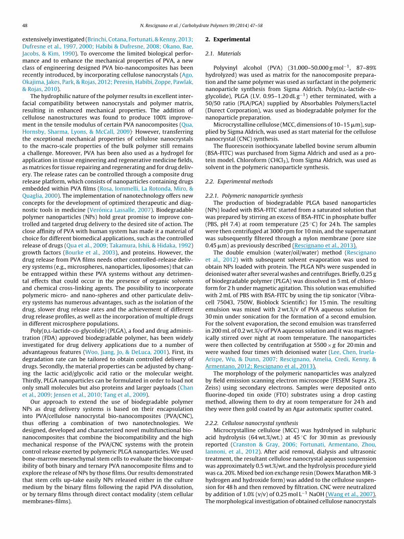
4 hydra
eDJmcrO&
frcmHttaaaereQcnptccrgdebtapeddi
tiaddiToe
Nitdnmcbietmom
8 N. Rescignano et al. / Carbo
xtensively investigated (Brinchi, Cotana, Fortunati, & Kenny, 2013;ufresne et al., 1997, 2000; Habibi & Dufresne, 2008; Okano, Bae,
acobs, & Kim, 1990). To overcome the limited biological perfor-ance and to enhance the mechanical properties of PVA, a new
lass of engineering designed PVA bio-nanocomposites has beenecently introduced, by incorporating cellulose nanocrystals (Ago,kajima, Jakes, Park, & Rojas, 2012; Peresin, Habibi, Zoppe, Pawlak,
Rojas, 2010).The hydrophilic nature of the polymer results in excellent inter-
acial compatibility between nanocrystals and polymer matrix,esulting in enhanced mechanical properties. The addition ofellulose nanostructures was found to produce 100% improve-ent in the tensile modulus of certain PVA nanocomposites (Qua,ornsby, Sharma, Lyons, & McCall, 2009). However, transferring
he exceptional mechanical properties of cellulose nanocrystalso the macro-scale properties of the bulk polymer still remains
challenge. Moreover, PVA has been also used as a hydrogel forpplication in tissue engineering and regenerative medicine fields,s matrices for tissue repairing and regenerating and for drug deliv-ry. The release rates can be controlled through a composite drugelease platform, which consists of nanoparticles containing drugsmbedded within PVA films (Rosa, Iommelli, La Rotonda, Miro, &uaglia, 2000). The implementation of nanotechnology offers newoncepts for the development of optimized therapeutic and diag-ostic tools in medicine (Verónica Lassalle, 2007). Biodegradableolymer nanoparticles (NPs) hold great promise to improve con-rolled and targeted drug delivery to the desired site of action. Thelose affinity of PVA with human system has made it a material ofhoice for different biomedical applications, such as the controlledelease of drugs (Qua et al., 2009; Takamura, Ishii, & Hidaka, 1992)rowth factors (Bourke et al., 2003), and proteins. However, therug release from PVA films needs other controlled-release deliv-ry systems (e.g., microspheres, nanoparticles, liposomes) that cane entrapped within these PVA systems without any detrimen-al effects that could occur in the presence of organic solventsnd chemical cross-linking agents. The possibility to incorporateolymeric micro- and nano-spheres and other particulate deliv-ry systems has numerous advantages, such as the isolation of therug, slower drug release rates and the achievement of differentrug release profiles, as well as the incorporation of multiple drugs
n different microsphere populations.Poly(d,l-lactide-co-glycolide) (PLGA), a food and drug adminis-
ration (FDA) approved biodegradable polymer, has been widelynvestigated for drug delivery applications due to a number ofdvantageous features (Woo, Jiang, Jo, & DeLuca, 2001). First, itsegradation rate can be tailored to obtain controlled delivery ofrugs. Secondly, the material properties can be adjusted by chang-
ng the lactic acid/glycolic acid ratio or the molecular weight.hirdly, PLGA nanoparticles can be formulated in order to load notnly small molecules but also proteins and larger payloads (Chant al., 2009; Jensen et al., 2010; Tang et al., 2009).
Our approach to extend the use of biodegradable polymerPs as drug delivery systems is based on their encapsulation
nto PVA/cellulose nanocrystal bio-nanocomposites (PVA/CNC),hus offering a combination of two nanotechnologies. Weesigned, developed and characterized novel multifunctional bio-anocomposites that combine the biocompatibility and the highechanical response of the PVA/CNC systems with the protein
ontrol release exerted by polymeric PLGA nanoparticles. We usedone-marrow mesenchymal stem cells to evaluate the biocompat-
bility of both binary and ternary PVA nanocomposite films and toxplore the release of NPs by those films. Our results demonstrated
hat stem cells up-take easily NPs released either in the cultureedium by the binary films following the rapid PVA dissolution,r by ternary films through direct contact modality (stem cellularembranes-films).
te Polymers 99 (2014) 47– 58
2. Experimental
2.1. Materials
Polyvinyl alcohol (PVA) (31.000–50.000 g mol−1, 87–89%hydrolyzed) was used as matrix for the nanocomposite prepara-tion and the same polymer was used as surfactant in the polymericnanoparticle synthesis from Sigma Aldrich. Poly(d,l-lactide-co-glycolide), PLGA (I.V. 0.95–1.20 dL g−1) ether terminated, with a50/50 ratio (PLA/PGA) supplied by Absorbables Polymers/Lactel(Durect Corporation), was used as biodegradable polymer for thenanoparticle preparation.
Microcrystalline cellulose (MCC, dimensions of 10–15 �m), sup-plied by Sigma Aldrich, was used as start material for the cellulosenanocrystal (CNC) synthesis.
The fluorescein isothiocyanate labelled bovine serum albumin(BSA-FITC) was purchased from Sigma Aldrich and used as a pro-tein model. Chloroform (CHCl3), from Sigma Aldrich, was used assolvent in the polymeric nanoparticle synthesis.
2.2. Experimental methods
2.2.1. Polymeric nanoparticle synthesisThe production of biodegradable PLGA based nanoparticles
(NPs) loaded with BSA-FITC started from a saturated solution thatwas prepared by stirring an excess of BSA-FITC in phosphate buffer(PBS, pH 7.4) at room temperature (25 ◦C) for 24 h. The sampleswere then centrifuged at 3000 rpm for 10 min, and the supernatantwas subsequently filtered through a nylon membrane (pore size0.45 �m) as previously described (Rescignano et al., 2013).
The double emulsion (water/oil/water) method (Rescignanoet al., 2012) with subsequent solvent evaporation was used toobtain NPs loaded with protein. The PLGA NPs were suspended indeionised water after several washes and centrifuges. Briefly, 0.25 gof biodegradable polymer (PLGA) was dissolved in 5 mL of chloro-form for 2 h under magnetic agitation. This solution was emulsifiedwith 2 mL of PBS with BSA-FITC by using the tip sonicator (Vibra-cell 75043, 750W, Bioblock Scientific) for 15 min. The resultingemulsion was mixed with 2 wt.%/v of PVA aqueous solution for30 min under sonication for the formation of a second emulsion.For the solvent evaporation, the second emulsion was transferredin 200 mL of 0.2 wt.%/v of PVA aqueous solution and it was magnet-ically stirred over night at room temperature. The nanoparticleswere then collected by centrifugation at 5500 × g for 20 min andwere washed four times with deionised water (Lee, Chen, Iruela-Arispe, Wu, & Dunn, 2007; Rescignano, Amelia, Credi, Kenny, &Armentano, 2012; Rescignano et al., 2013).
The morphology of the polymeric nanoparticles was analyzedby field emission scanning electron microscope (FESEM Supra 25,Zeiss) using secondary electrons. Samples were deposited ontofluorine-doped tin oxide (FTO) substrates using a drop castingmethod, allowing them to dry at room temperature for 24 h andthey were then gold coated by an Agar automatic sputter coated.
2.2.2. Cellulose nanocrystal synthesisMicrocrystalline cellulose (MCC) was hydrolysed in sulphuric
acid hydrolysis (64 wt.%/wt.) at 45 ◦C for 30 min as previouslyreported (Cranston & Gray, 2006; Fortunati, Armentano, Zhou,Iannoni, et al., 2012). After acid removal, dialysis and ultrasonictreatment, the resultant cellulose nanocrystal aqueous suspensionwas approximately 0.5 wt.%/wt. and the hydrolysis procedure yieldwas ca. 20%. Mixed bed ion exchange resin (Dowex Marathon MR-3
hydrogen and hydroxide form) was added to the cellulose suspen-sion for 48 h and then removed by filtration. CNC were neutralizedby addition of 1.0% (v/v) of 0.25 mol L−1 NaOH (Wang et al., 2007).The morphological investigation of obtained cellulose nanocrystals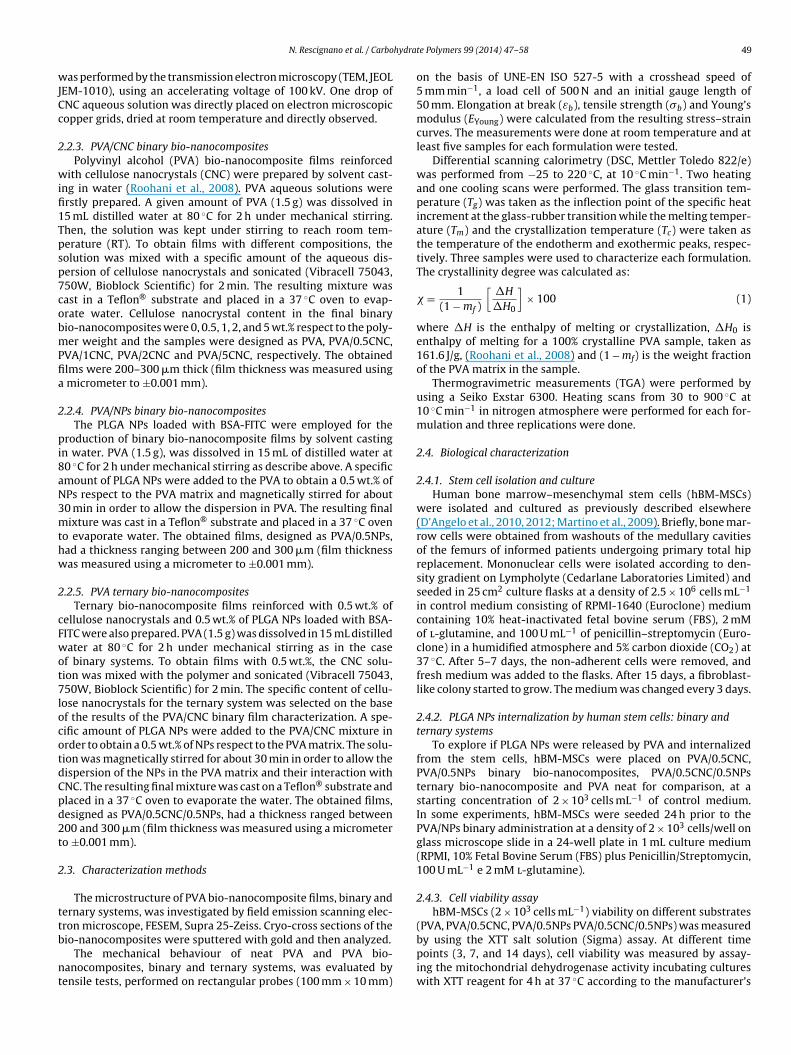
hydra
wJCc
2
wifi1Tpsp7cobmPfia
2
pi8aN3mthw
2
cFwot7locotdCpd2t
2
ttb
nt
N. Rescignano et al. / Carbo
as performed by the transmission electron microscopy (TEM, JEOLEM-1010), using an accelerating voltage of 100 kV. One drop ofNC aqueous solution was directly placed on electron microscopicopper grids, dried at room temperature and directly observed.
.2.3. PVA/CNC binary bio-nanocompositesPolyvinyl alcohol (PVA) bio-nanocomposite films reinforced
ith cellulose nanocrystals (CNC) were prepared by solvent cast-ng in water (Roohani et al., 2008). PVA aqueous solutions wererstly prepared. A given amount of PVA (1.5 g) was dissolved in5 mL distilled water at 80 ◦C for 2 h under mechanical stirring.hen, the solution was kept under stirring to reach room tem-erature (RT). To obtain films with different compositions, theolution was mixed with a specific amount of the aqueous dis-ersion of cellulose nanocrystals and sonicated (Vibracell 75043,50W, Bioblock Scientific) for 2 min. The resulting mixture wasast in a Teflon® substrate and placed in a 37 ◦C oven to evap-rate water. Cellulose nanocrystal content in the final binaryio-nanocomposites were 0, 0.5, 1, 2, and 5 wt.% respect to the poly-er weight and the samples were designed as PVA, PVA/0.5CNC,
VA/1CNC, PVA/2CNC and PVA/5CNC, respectively. The obtainedlms were 200–300 �m thick (film thickness was measured using
micrometer to ±0.001 mm).
.2.4. PVA/NPs binary bio-nanocompositesThe PLGA NPs loaded with BSA-FITC were employed for the
roduction of binary bio-nanocomposite films by solvent castingn water. PVA (1.5 g), was dissolved in 15 mL of distilled water at0 ◦C for 2 h under mechanical stirring as describe above. A specificmount of PLGA NPs were added to the PVA to obtain a 0.5 wt.% ofPs respect to the PVA matrix and magnetically stirred for about0 min in order to allow the dispersion in PVA. The resulting finalixture was cast in a Teflon® substrate and placed in a 37 ◦C oven
o evaporate water. The obtained films, designed as PVA/0.5NPs,ad a thickness ranging between 200 and 300 �m (film thicknessas measured using a micrometer to ±0.001 mm).
.2.5. PVA ternary bio-nanocompositesTernary bio-nanocomposite films reinforced with 0.5 wt.% of
ellulose nanocrystals and 0.5 wt.% of PLGA NPs loaded with BSA-ITC were also prepared. PVA (1.5 g) was dissolved in 15 mL distilledater at 80 ◦C for 2 h under mechanical stirring as in the case
f binary systems. To obtain films with 0.5 wt.%, the CNC solu-ion was mixed with the polymer and sonicated (Vibracell 75043,50W, Bioblock Scientific) for 2 min. The specific content of cellu-
ose nanocrystals for the ternary system was selected on the basef the results of the PVA/CNC binary film characterization. A spe-ific amount of PLGA NPs were added to the PVA/CNC mixture inrder to obtain a 0.5 wt.% of NPs respect to the PVA matrix. The solu-ion was magnetically stirred for about 30 min in order to allow theispersion of the NPs in the PVA matrix and their interaction withNC. The resulting final mixture was cast on a Teflon® substrate andlaced in a 37 ◦C oven to evaporate the water. The obtained films,esigned as PVA/0.5CNC/0.5NPs, had a thickness ranged between00 and 300 �m (film thickness was measured using a micrometero ±0.001 mm).
.3. Characterization methods
The microstructure of PVA bio-nanocomposite films, binary andernary systems, was investigated by field emission scanning elec-ron microscope, FESEM, Supra 25-Zeiss. Cryo-cross sections of the
io-nanocomposites were sputtered with gold and then analyzed.The mechanical behaviour of neat PVA and PVA bio-anocomposites, binary and ternary systems, was evaluated byensile tests, performed on rectangular probes (100 mm × 10 mm)
te Polymers 99 (2014) 47– 58 49
on the basis of UNE-EN ISO 527-5 with a crosshead speed of5 mm min−1, a load cell of 500 N and an initial gauge length of50 mm. Elongation at break (εb), tensile strength (�b) and Young’smodulus (EYoung) were calculated from the resulting stress–straincurves. The measurements were done at room temperature and atleast five samples for each formulation were tested.
Differential scanning calorimetry (DSC, Mettler Toledo 822/e)was performed from −25 to 220 ◦C, at 10 ◦C min−1. Two heatingand one cooling scans were performed. The glass transition tem-perature (Tg) was taken as the inflection point of the specific heatincrement at the glass-rubber transition while the melting temper-ature (Tm) and the crystallization temperature (Tc) were taken asthe temperature of the endotherm and exothermic peaks, respec-tively. Three samples were used to characterize each formulation.The crystallinity degree was calculated as:
� = 1(1 − mf )
[�H
�H0
]× 100 (1)
where �H is the enthalpy of melting or crystallization, �H0 isenthalpy of melting for a 100% crystalline PVA sample, taken as161.6 J/g, (Roohani et al., 2008) and (1 − mf) is the weight fractionof the PVA matrix in the sample.
Thermogravimetric measurements (TGA) were performed byusing a Seiko Exstar 6300. Heating scans from 30 to 900 ◦C at10 ◦C min−1 in nitrogen atmosphere were performed for each for-mulation and three replications were done.
2.4. Biological characterization
2.4.1. Stem cell isolation and cultureHuman bone marrow–mesenchymal stem cells (hBM-MSCs)
were isolated and cultured as previously described elsewhere(D’Angelo et al., 2010, 2012; Martino et al., 2009). Briefly, bone mar-row cells were obtained from washouts of the medullary cavitiesof the femurs of informed patients undergoing primary total hipreplacement. Mononuclear cells were isolated according to den-sity gradient on Lympholyte (Cedarlane Laboratories Limited) andseeded in 25 cm2 culture flasks at a density of 2.5 × 10⁶ cells mL−1
in control medium consisting of RPMI-1640 (Euroclone) mediumcontaining 10% heat-inactivated fetal bovine serum (FBS), 2 mMof l-glutamine, and 100 U mL−1 of penicillin–streptomycin (Euro-clone) in a humidified atmosphere and 5% carbon dioxide (CO2) at37 ◦C. After 5–7 days, the non-adherent cells were removed, andfresh medium was added to the flasks. After 15 days, a fibroblast-like colony started to grow. The medium was changed every 3 days.
2.4.2. PLGA NPs internalization by human stem cells: binary andternary systems
To explore if PLGA NPs were released by PVA and internalizedfrom the stem cells, hBM-MSCs were placed on PVA/0.5CNC,PVA/0.5NPs binary bio-nanocomposites, PVA/0.5CNC/0.5NPsternary bio-nanocomposite and PVA neat for comparison, at astarting concentration of 2 × 10³ cells mL−1 of control medium.In some experiments, hBM-MSCs were seeded 24 h prior to thePVA/NPs binary administration at a density of 2 × 10³ cells/well onglass microscope slide in a 24-well plate in 1 mL culture medium(RPMI, 10% Fetal Bovine Serum (FBS) plus Penicillin/Streptomycin,100 U mL−1 e 2 mM l-glutamine).
2.4.3. Cell viability assayhBM-MSCs (2 × 10³ cells mL−1) viability on different substrates
(PVA, PVA/0.5CNC, PVA/0.5NPs PVA/0.5CNC/0.5NPs) was measured
by using the XTT salt solution (Sigma) assay. At different timepoints (3, 7, and 14 days), cell viability was measured by assay-ing the mitochondrial dehydrogenase activity incubating cultureswith XTT reagent for 4 h at 37 ◦C according to the manufacturer’s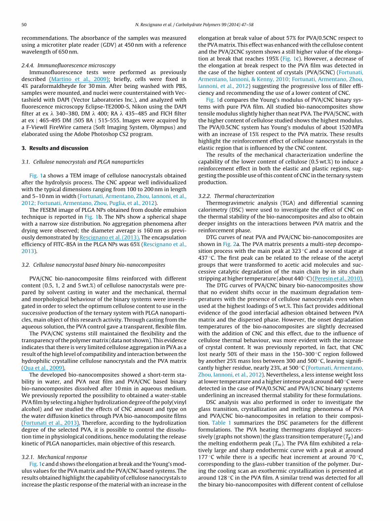
5 hydra
ruw
2
d4stflfiaae
3
3
awa2
twdoe2
3
cpagsca
tirh(
bbWPat(dtk
3
uri
0 N. Rescignano et al. / Carbo
ecommendations. The absorbance of the samples was measuredsing a microtiter plate reader (GDV) at 450 nm with a referenceavelength of 650 nm.
.4.4. Immunofluorescence microscopyImmunofluorescence tests were performed as previously
escribed (Martino et al., 2009); briefly, cells were fixed in% paraformaldheyde for 30 min. After being washed with PBS,amples were mounted, and nuclei were counterstained with Vec-ashield with DAPI (Vector Laboratories Inc.), and analyzed withuorescence microscopy Eclipse-TE2000-S, Nikon using the DAPIlter at ex � 340–380, DM � 400; RA � 435–485 and FICH filtert ex | 465-495 DM |505 BA | 515-555. Images were acquired by
F-ViewII FireWire camera (Soft Imaging System, Olympus) andlaborated using the Adobe Photoshop CS2 program.
. Results and discussion
.1. Cellulose nanocrystals and PLGA nanoparticles
Fig. 1a shows a TEM image of cellulose nanocrystals obtainedfter the hydrolysis process. The CNC appear well individualizedith the typical dimensions ranging from 100 to 200 nm in length
nd 5–10 nm in width (Fortunati, Armentano, Zhou, Iannoni, et al.,012; Fortunati, Armentano, Zhou, Puglia, et al., 2012).
The FESEM image of PLGA NPs obtained from double emulsionechnique is reported in Fig. 1b. The NPs show a spherical shapeith a narrow size distribution. No aggregation phenomena afterrying were observed; the diameter average is 160 nm as previ-usly demonstrated by Rescignano et al. (2013). The encapsulationfficiency of FITC-BSA in the PLGA NPs was 65% (Rescignano et al.,013).
.2. Cellulose nanocrystal based binary bio-nanocomposites
PVA/CNC bio-nanocomposite films reinforced with differentontent (0.5, 1, 2 and 5 wt.%) of cellulose nanocrystals were pre-ared by solvent casting in water and the mechanical, thermalnd morphological behaviour of the binary systems were investi-ated in order to select the optimum cellulose content to use in theuccessive production of the ternary system with PLGA nanoparti-les, main object of this research activity. Through casting from thequeous solution, the PVA control gave a transparent, flexible film.
The PVA/CNC systems still maintained the flexibility and theransparency of the polymer matrix (data not shown). This evidencendicates that there is very limited cellulose aggregation in PVA as aesult of the high level of compatibility and interaction between theydrophilic crystalline cellulose nanocrystals and the PVA matrixQua et al., 2009).
The developed bio-nanocomposites showed a short-term sta-ility in water, and PVA neat film and PVA/CNC based binaryio-nanocomposites dissolved after 10 min in aqueous medium.e previously reported the possibility to obtained a water-stable
VA film by selecting a higher hydrolization degree of the poly(vinyllcohol) and we studied the effects of CNC amount and type onhe water diffusion kinetics through PVA bio-nanocomposite filmsFortunati et al., 2013). Therefore, according to the hydrolizationegree of the selected PVA, it is possible to control the dissolu-ion time in physiological conditions, hence modulating the releaseinetic of PLGA nanoparticles, main objective of this research.
.2.1. Mechanical response
Fig. 1c and d shows the elongation at break and the Young’s mod-lus values for the PVA matrix and the PVA/CNC based systems. Theesults obtained highlight the capability of cellulose nanocrystals toncrease the plastic response of the material with an increase in the
te Polymers 99 (2014) 47– 58
elongation at break value of about 57% for PVA/0.5CNC respect tothe PVA matrix. This effect was enhanced with the cellulose contentand the PVA/2CNC system shows a still higher value of the elonga-tion at break that reaches 195% (Fig. 1c). However, a decrease ofthe elongation at break respect to the PVA film was detected inthe case of the higher content of crystals (PVA/5CNC) (Fortunati,Armentano, Iannoni, & Kenny, 2010; Fortunati, Armentano, Zhou,Iannoni, et al., 2012) suggesting the progressive loss of filler effi-ciency and recommending the use of a lower content of CNC.
Fig. 1d compares the Young’s modulus of PVA/CNC binary sys-tems with pure PVA film. All studied bio-nanocomposites showtensile modulus slightly higher than neat PVA. The PVA/5CNC, withthe higher content of cellulose studied shows the highest modulus.The PVA/0.5CNC system has Young’s modulus of about 1520 MPawith an increase of 15% respect to the PVA matrix. These resultshighlight the reinforcement effect of cellulose nanocrystals in theelastic region that is influenced by the CNC content.
The results of the mechanical characterization underline thecapability of the lower content of cellulose (0.5 wt.%) to induce areinforcement effect in both the elastic and plastic regions, sug-gesting the possible use of this content of CNC in the ternary systemproduction.
3.2.2. Thermal characterizationThermogravimetric analysis (TGA) and differential scanning
calorimetry (DSC) were used to investigate the effect of CNC onthe thermal stability of the bio-nanocomposites and also to obtaindeeper insights on the interactions between PVA matrix and thereinforcement phase.
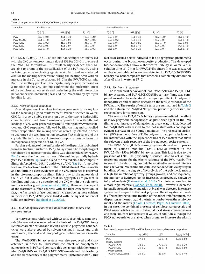
DTG curves of neat PVA and PVA/CNC bio-nanocomposites areshown in Fig. 2a. The PVA matrix presents a multi-step decompo-sition process with the main peak at 323 ◦C and a second stage at437 ◦C. The first peak can be related to the release of the acetylgroups that were transformed to acetic acid molecules and suc-cessive catalytic degradation of the main chain by in situ chainstripping at higher temperature (about 440 ◦C) (Peresin et al., 2010).
The DTG curves of PVA/CNC binary bio-nanocomposites showthat no evident shifts occur in the maximum degradation tem-peratures with the presence of cellulose nanocrystals even whenused at the highest loadings of 5 wt.%. This fact provides additionalevidence of the good interfacial adhesion obtained between PVAmatrix and the dispersed phase. However, the onset degradationtemperatures of the bio-nanocomposites are slightly decreasedwith the addition of CNC and this effect, due to the influence ofcellulose thermal behaviour, was more evident with the increaseof crystal content. It was previously reported, in fact, that CNClost nearly 50% of their mass in the 150–300 ◦C region followedby another 25% mass loss between 300 and 500 ◦C, leaving signifi-cantly higher residue, nearly 23%, at 500 ◦C (Fortunati, Armentano,Zhou, Iannoni, et al., 2012). Nevertheless, a less intense weight lossat lower temperature and a higher intense peak around 440 ◦C weredetected in the case of PVA/0.5CNC and PVA/1CNC binary systemsunderlining an increased thermal stability for these formulations.
DSC analysis was also performed in order to investigate theglass transition, crystallization and melting phenomena of PVAand PVA/CNC bio-nanocomposites in relation to their composi-tion. Table 1 summarizes the DSC parameters for the differentformulations. The PVA heating thermograms displayed succes-sively (graphs not shown) the glass transition temperature (Tg) andthe melting endotherm peak (Tm). The PVA film exhibited a rela-tively large and sharp endothermic curve with a peak at around177 ◦C while there is a specific heat increment at around 70 ◦C,
corresponding to the glass-rubber transition of the polymer. Dur-ing the cooling scan an exothermic crystallization is presented ataround 128 ◦C in the PVA film. A similar trend was detected for allthe binary bio-nanocomposites with different content of cellulose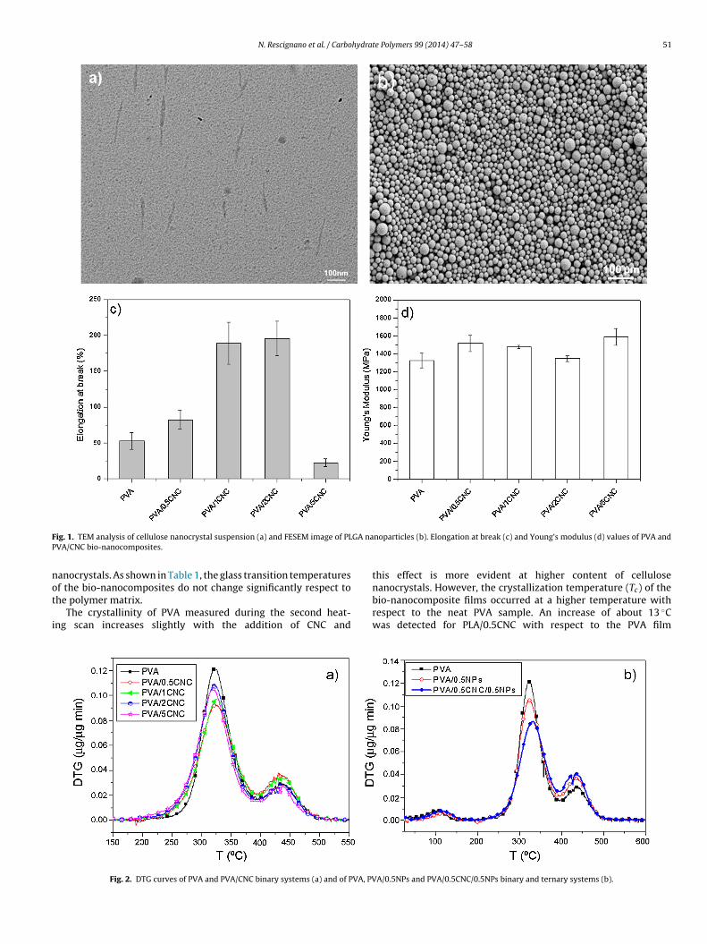
N. Rescignano et al. / Carbohydrate Polymers 99 (2014) 47– 58 51
F GA naP
not
i
ig. 1. TEM analysis of cellulose nanocrystal suspension (a) and FESEM image of PLVA/CNC bio-nanocomposites.
anocrystals. As shown in Table 1, the glass transition temperaturesf the bio-nanocomposites do not change significantly respect to
he polymer matrix.The crystallinity of PVA measured during the second heat-ng scan increases slightly with the addition of CNC and
Fig. 2. DTG curves of PVA and PVA/CNC binary systems (a) and of PVA, PV
noparticles (b). Elongation at break (c) and Young’s modulus (d) values of PVA and
this effect is more evident at higher content of cellulosenanocrystals. However, the crystallization temperature (T ) of the
cbio-nanocomposite films occurred at a higher temperature withrespect to the neat PVA sample. An increase of about 13 ◦Cwas detected for PLA/0.5CNC with respect to the PVA film
A/0.5NPs and PVA/0.5CNC/0.5NPs binary and ternary systems (b).
52 N. Rescignano et al. / Carbohydrate Polymers 99 (2014) 47– 58
Table 1Thermal properties of PVA and PVA/CNC binary nanocomposites.
Samples Cooling scan Second heating scan
Tg [◦C] �Hc [J/g] Tc [◦C] Tg [◦C] �Hc [J/g] Tc [◦C] Xc [%]
PVA 66.3 ± 0.9 25.1 ± 3.0 127.6 ± 2.0 68.9 ± 0.1 18.3 ± 3.0 176.7 ± 3.0 11.3 ± 1.0PVA/0.5CNC 66.1 ± 1.0 17.4 ± 0.1 140.4 ± 0.6 69.2 ± 0.2 19.8 ± 3.0 179.7 ± 0.1 12.3 ± 1.0
(wtaaaiBaobb
3
tCcltwtch
ftFyfifaittmofic
3t
tstmg
anfia
In our case, the combined presence of cellulose nanocrystals andPLGA nanoparticles causes substantial local stress concentrationsand then failure at reduced strain values. In addition, although thePLGA nanoparticles are able, when alone, to increase the plastic
Table 2Mechanical properties of PVA and PVA binary and ternary bio-nanocomposites.
Samples �b [MPa] εb [%] Eyoung [MPa]
PVA 37 ± 5 53 ± 12 1320 ± 80Binary systems
PVA/1CNC 66.2 ± 0.2 21.0 ± 0.8 145.0 ± 1.0
PVA/2CNC 64.6 ± 0.5 23.1 ± 0.4 156.9 ± 0.1
PVA/5CNC 63.6 ± 1.0 27.4 ± 2.0 159.9 ± 0.2
Tc = 127.6 ± 2.0 ◦C). The Tc of the bio-nanocomposites increasedith the CNC content reaching a value of 159.9 ± 0.2 ◦C in the case of
he PVA/5CNC formulation. This result clearly evidences that CNCre able to promote the crystallization of the PVA matrix, actings heterogeneous nucleating agents. A similar trend was detectedlso for the melting temperature during the heating scan with anncrease in the Tm value of about 16 ◦C in the PVA/5CNC sample.oth the melting point and the crystallinity degree increased as
function of the CNC content confirming the nucleation effectf the cellulose nanocrystals and underlining the well interactionetween the reinforcement phase and the PVA chains by hydrogenonding forces.
.2.3. Morphological behaviourGood dispersion of cellulose in the polymer matrix is a key fac-
or for achieving a good reinforcement. When dispersed in water,NC form a very stable suspension due to the strong hydrophilicharacteristics of cellulose. Bio-nanocomposite films with differentoadings of CNC were prepared by careful mixing of PVA water solu-ion and CNC suspension followed by film casting and controlledater evaporation. The mixing time was carefully selected in order
o guarantee the well interaction between PVA molecules and therystals. The transparency of bio-nanocomposite films confirms theomogeneous dispersion of CNC in PVA.
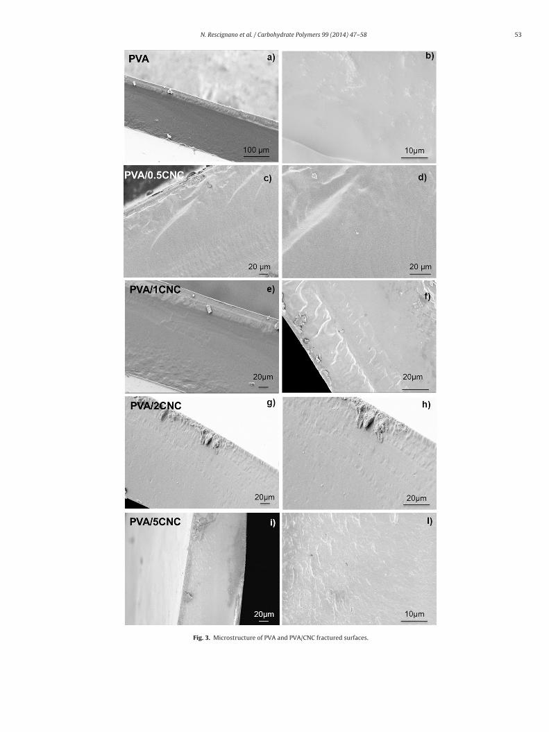
Further evidence of the uniformity of the dispersion is obtainedrom the fractured surface of PVA/CNC systems. The morphology ofhe binary bio-nanocomposite films was characterized by FESEM.ig. 3 shows the fractured surface of the unfilled partially hydrol-sed PVA matrix (Fig. 3a and b) and the related bio-nanocompositelms reinforced with 0.5, 1, 2 and 5 wt.% of CNC (Fig. 3c–h), just after
racture. The fractured surface of the unfilled films of PVA is smoothnd uniform. No clear evidences of the CNC presence is observedn the bio-nanocomposite films. This is due to the nanoscale ofhe filler, but it also indicates that no aggregates are present inhe films and that the dispersion of the CNC within the polymeric
atrix is rather good (Roohani et al., 2008). However, the aspectf the fractured surface changes with the filler concentration. Inact, the fractured surface roughness increases being more evidentn the case of PVA/5CNC system loaded with the highest content ofellulose analyzed (Roohani et al., 2008).
.3. PLGA nanoparticle based bio-nanocomposites: binary andernary systems
Ternary systems reinforced with 0.5 wt.% of cellulose nanocrys-als (the content was selected on the basis of the PVA/CNC binaryystem characterization) and 0.5 wt.% of PLGA polymeric nanopar-icles were also prepared by solvent casting in water and their
echanical, thermal and morphological behaviour was investi-ated.
The PVA/0.5NPs binary system was also produced and char-
cterized in order to understand the effect of biopolymericanoparticles in PVA and compare this behaviour with the ternarylm. PVA/0.5NPs and PVA/0.5CNC/0.5NPs maintained the flexibilitynd the transparency of the polymer matrix (data not shown). This69.8 ± 0.4 20.7 ± 0.2 180.7 ± 1.0 13.0 ± 0.568.3 ± 0.1 23.2 ± 1.0 187.4 ± 0.7 14.7 ± 1.068.3 ± 0.1 30.7 ± 2.0 192.7 ± 0.1 20.1 ± 1.0
fact as described below indicated that no aggregation phenomenaoccur during the bio-nanocomposite production. The developedbio-nanocomposites show a short-term stability in water; a dis-solution time of 10 min for PVA/0.5NPs binary film was measuredwhile a more stable behaviour was detected for PVA/0.5CNC/0.5NPsternary bio-nanocomposite that reached a completely dissolutionafter 45 min in water at 37 ◦C.
3.3.1. Mechanical responseThe mechanical behaviour of PVA, PVA/0.5NPs and PVA/0.5CNC
binary systems, and PVA/0.5CNC/0.5NPs ternary films, was com-pared in order to understand the synergic effect of polymericnanoparticles and cellulose crystals on the tensile response of thePVA matrix. The results of tensile tests are summarized in Table 2and the data on the PVA/0.5CNC system, previously discussed, arereported here for comparison.
The results for PVA/0.5NPs binary system underlined the effectof PLGA polymeric nanoparticles as plasticizer agent in the PVAfilm. A great increase of elongation at break was, in fact, detectedfor PVA/0.5NPs with respect to the PVA matrix with a consequentevident decrease in the Young’s modulus. The presence of surfac-tant (PVA) on the surface of PLGA polymeric nanoparticles favoursthe interactions with the adjacent chains of PVA matrix that allowthe relevant plastic response of the bio-nanocomposite systems.
The PVA/0.5CNC/0.5NPs ternary system showed an improve-ment of Young’s modulus (1240 ± 40 MPa) respect to thePVA/0.5NPs (150 ± 20 MPa) binary system. This fact is due to thepresence of CNC, like previously discussed, able to act as rein-forcement agents for the elastic response of the PVA matrix. Theincrease in the elastic region could be ascribed to increased interac-tions between PVA chains and cellulose nanocrystals via hydrogenbonding. When the degree of hydrolysis of the polymeric matrixis high, the number of hydroxyl groups growths and consequently,the number of hydrogen bonds increases, as previously shown byinfrared analysis (Fortunati et al., 2013). Such interactions lead toa more rigid material (Roohani et al., 2008). However, a decreasein tensile strength and elongation at break was detected in ternarysystem with respect to the neat polymer. The elongation at breakis affected by the volume fraction of the added reinforcement, thedispersion in the matrix, and the interaction between the reinforce-ment and the matrix (Colom, Carrasco, Pages, & Canavate, 2003).
PVA/0.5NPs 34 ± 3 270 ± 30 150 ± 20PVA/0.5CNC 25 ± 2 83 ± 13 1520 ± 90
Ternary systemPVA/0.5CNC/0.5NPs 28 ± 2 22 ± 6 1240 ± 40
N. Rescignano et al. / Carbohydrate Polymers 99 (2014) 47– 58 53
Fig. 3. Microstructure of PVA and PVA/CNC fractured surfaces.
54 N. Rescignano et al. / Carbohydrate Polymers 99 (2014) 47– 58
Table 3Thermal properties of PVA and PVA binary and ternary nanocomposites.
Samples Cooling scan Second heating scan
Tg [◦C] �Hc [J/g] Tc [◦C] Tg [◦C] �Hc [J/g] Tc [◦C] Xc [%]
PVA 66.3 ± 0.9 25.1 ± 3.0 127.6 ± 2.0 68.9 ± 0.1 18.3 ± 3.0 176.7 ± 3.0 11.3 ± 1.0Binary systems
PVA/0.5NPs 62 ± 4 27.4 ± 1.5 149.2 ± 3.0 63.8 ± 2.6 21.0 ± 1.7 187.4 ± 1.1 13.0 ± 1.06
2
rP(
titntlasAt(cur
3
cC
PVA/0.5CNC 66.1 ± 1.0 17.4 ± 0.1 140.4 ± 0.Ternary system
PVA/0.5CNC/0.5NPs 64.9 ± 0.9 30.9 ± 3.0 157.5 ± 1.
esponse of the system, probably the interfacial adhesion with theVA was not enhanced by the presence of cellulose nanocrystalsPei, Zhou, & Berglund, 2010).
The results obtained demonstrate the possibility to modulatehe mechanical response of these multifunctional systems takingnto account the final application. Our idea is to create a system ableo release specific drugs thanks to the presence of PLGA polymericanoparticles, to interact with a biological system (stem cells), ando control its mechanical properties with the presence of nanocellu-ose crystals. The effect, in fact, of chemico-physical, topographicalnd mechanical properties of a biomaterial on the induction oftem cell differentiation (D’Angelo et al., 2010; Martino, D’Angelo,rmentano, Kenny, & Orlacchio, 2012) as well as on the preserva-
ion of their stem cell status is well known in the scientific literatureD’Angelo et al., 2011; McMurray et al., 2011). Stem cells ability toonvert mechanical cues on biochemical signals and in turn mod-late their fate is a relatively recent acquisition of the scientificesearch (Engler, Sen, Sweeney, & Discher, 2006).
.3.2. Thermal characterizationThermogravimetric analysis (TGA) and differential scanning
alorimetry (DSC) were used to investigate the effect ofNC and PLGA nanoparticles on the thermal stability of the
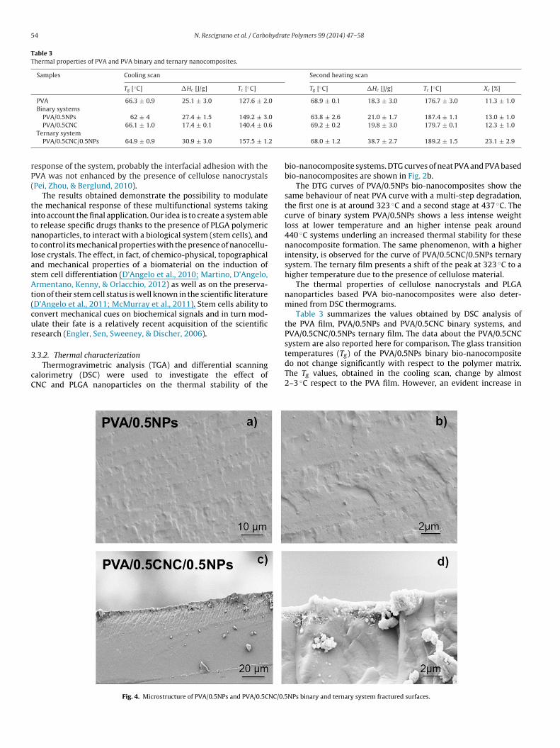
Fig. 4. Microstructure of PVA/0.5NPs and PVA/0.5CNC/0.5
69.2 ± 0.2 19.8 ± 3.0 179.7 ± 0.1 12.3 ± 1.0
68.0 ± 1.2 38.7 ± 2.7 189.2 ± 1.5 23.1 ± 2.9
bio-nanocomposite systems. DTG curves of neat PVA and PVA basedbio-nanocomposites are shown in Fig. 2b.
The DTG curves of PVA/0.5NPs bio-nanocomposites show thesame behaviour of neat PVA curve with a multi-step degradation,the first one is at around 323 ◦C and a second stage at 437 ◦C. Thecurve of binary system PVA/0.5NPs shows a less intense weightloss at lower temperature and an higher intense peak around440 ◦C systems underling an increased thermal stability for thesenanocomposite formation. The same phenomenon, with a higherintensity, is observed for the curve of PVA/0.5CNC/0.5NPs ternarysystem. The ternary film presents a shift of the peak at 323 ◦C to ahigher temperature due to the presence of cellulose material.
The thermal properties of cellulose nanocrystals and PLGAnanoparticles based PVA bio-nanocomposites were also deter-mined from DSC thermograms.
Table 3 summarizes the values obtained by DSC analysis ofthe PVA film, PVA/0.5NPs and PVA/0.5CNC binary systems, andPVA/0.5CNC/0.5NPs ternary film. The data about the PVA/0.5CNCsystem are also reported here for comparison. The glass transition
temperatures (Tg) of the PVA/0.5NPs binary bio-nanocompositedo not change significantly with respect to the polymer matrix.The Tg values, obtained in the cooling scan, change by almost2–3 ◦C respect to the PVA film. However, an evident increase inNPs binary and ternary system fractured surfaces.
N. Rescignano et al. / Carbohydrate Polymers 99 (2014) 47– 58 55
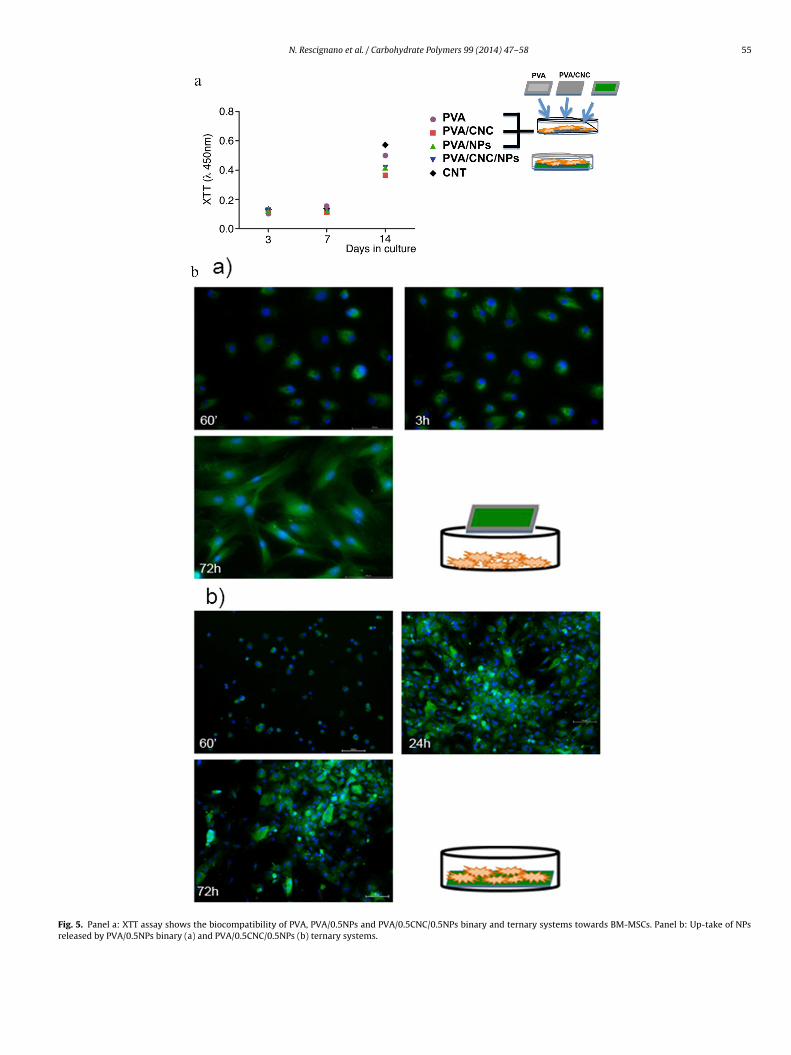
Fig. 5. Panel a: XTT assay shows the biocompatibility of PVA, PVA/0.5NPs and PVA/0.5CNC/0.5NPs binary and ternary systems towards BM-MSCs. Panel b: Up-take of NPsreleased by PVA/0.5NPs binary (a) and PVA/0.5CNC/0.5NPs (b) ternary systems.

5 hydra
tfdaTtoorsaneeaiMPsschf
3
wasb
fitf2tew(Seipc(tTieodAa
3
bncbcNeeP
6 N. Rescignano et al. / Carbo
he crystallization temperature (Tc) of about 22 ◦C was detectedor PVA/0.5NPs respect to the PVA matrix. This behaviour was alsoetected in the case of PVA/0.5CNC binary film, highlighting thebility of cellulose to act as nucleating agent in the polymer matrix.his nucleation effect was enhanced in the case of the ternary sys-em due to the combined effects of CNC and PLGA nanoparticlesn the thermal properties of PVA. The Tc of PLA/0.5CNC/0.5NPsccurred, in fact, at 157.5 ± 1.2 ◦C with an increase of about 30 ◦Cespect to PVA. Moreover, the PLA/0.5CNC/0.5NPs ternary systemhows an increase of the crystallization degree that raised up frombout 11% for the PVA matrix to 23% for the PVA/0.5CNC/0.5NPs bio-anocomposite during the second heating scan. This result clearlyvidences the crystallization effect induced by the synergic pres-nce of cellulose and PLGA polymeric nanoparticles that explainslso the increased elastic response and the low plastic responsen the mechanical behaviour of the analyzed bio-nanocomposites.
oreover, an increase in the Tm value of about 10 ◦C for theLA/0.5NPs binary systems and of 13 ◦C for the PLA/0.5CNC/0.5NPsample was detected during the heating scan. This is ascribed totrong interactions between PVA and nanofillers and between adja-ent PVA chains by hydrogen bonding forces and, as a consequence,ighly crystalline polymers and bigger crystalline domains could be
ormed (Roohani et al., 2008).
.3.3. Morphological behaviourThe morphology of PVA/0.5NPs and PVA/0.5CNC/0.5NPs films
as investigated in order to study the synergic effect of NPsnd CNC incorporation on the bio-nanocomposite structure. Fig. 4hows the fractured surface of PVA/0.5NPs and PVA/0.5CNC/0.5NPsio-nanocomposites just after cold-fracture.
On the fractured surface of the PVA/0.5NPs (Fig. 4a and b) binarylm it is possible to observe the presence of PLGA nanoparticleshat are well dispersed in the PVA matrix. The presence of the sur-actant (PVA) on the PLGA nanoparticle surface (Rescignano et al.,012) supports the interaction of biopolymeric nanosystem withhe matrix. The PVA shell around the NPs, also influences the differ-nt physical properties of PLGA nanoparticles and their interactionith the surrounding environment including cells membrane
Boury et al., 1995; Shakesheff, Evora, Soriano, & Langer, 1997).ahoo, Panyam, Prabha, and Labhasetwar (2002), investigated theffects of various factors that influenced the amount of PVA form-ng the shell around the polymeric NPs. They concluded that it isossible to control the residual PVA by altering the surfactant con-entration or type of organic solvent used during NPs productionSahoo et al., 2002). The control of this parameter is an impor-ant factor that influences the cellular uptake of biopolymeric NPs.he fracture surfaces of PVA ternary bio-nanocomposites were alsonvestigated by FESEM (Fig. 4c and d) in order to evaluate the influ-nce of cellulose nanocrystal and PLGA nanoparticles introductionn the microstructure. An increase in the surface roughness wasetected for this system due to the presence of cellulose (Fortunati,rmentano, Zhou, Iannoni, et al., 2012), while some CNC flakes andgglomerates of nanoparticles appear embedded in the PVA matrix.
.4. Biological evaluation
First, we tested the stability of PVA, PVA/0.5CNC, PVA/0.5NPsinary bio-nanocomposites and PVA/0.5CNC/0.5NPs ternary bio-anocomposite within the culture medium under canonical cellulture conditions (T = 37 ◦C and 5% CO2). After 10 min of incu-ation, PVA, of the ternary systems, is dissolved and as aonsequence CNC and NPs (content of 0.5 wt.% of CNC and
Ps) were delivered within the culture medium. The combinedffect of hydrophobic PLGA and CNC influence the differ-nt diffusion properties, and in turn the stability in water ofVA/0.5CNC/0.5NPs ternary film that remains stable over 45 min.te Polymers 99 (2014) 47– 58
As a consequence, in our experimental conditions the ternary,PVA bio-nanocomposite is dissolved gradually and NPs deliv-ered progressively. Next, we evaluated the biocompatibility ofPVA, PVA/0.5CNC, PVA/0.5NPs binary bio-nanocomposites, andPVA/0.5CNC/0.5NPs ternary bio-nanocomposite on stem cells. Wedesigned two different experimental plans for the binary andternary bio-nanocomposites. In the first set of experiments, due tothe rapid PVA dissolution time, the binary bio-nanocomposite filmwas added to the culture medium of stem cells seeded on a glasscover slip. Here PLGA NPs and CNC were first delivered within theculture medium and then to the cells (Fig. 5 Panel a, scheme).
In the second set of experiments, due to the longest timeof PVA/0.5CNC/0.5NPs ternary film dissolution, stem cells wereseeded on the PVA ternary film. Here, NPs and CNC are delivered tothe cells by a direct contact of the cellular plasma membrane andthe film (Fig. 5 Panel a, scheme).
Fig. 5 Panel A, shows the absence of signs of toxicity in allculture conditions indicating that the PVA polymer dissolution inculture medium does not affect the cellular viability. This emergesby the measure of the mitochondrial dehydrogenase activity thatwas comparable in hBM-MSCs cultured in the presence of PVA/CNC,PVA/NPs binary bio-nanocomposites and on PVA/CNC/NPs ternaryfilms and control culture stem cells in canonical polystyrene cul-ture tissues. The internalization time of PLGA NPs released bythe PVA binary bio-nanocomposites and the PVA ternary bio-nanocomposites was then evaluated. In Fig. 5 Panel b(a) is reportedthe internalization by stem cells of the PLGA NPs released by thePVA binary bio-nanocomposites. NPs rapidly delivered by the PVAdissolution are fast internalized by hBM-MSCs after 15 min of incu-bation. Representative images indicated that up-taken NPs areextra-nuclear and have a wide distribution within the cytoplasm(Fig. 5 Panel b(a)). Fluorescent stem cell number increases with thetime of incubation. Thus, above 60 min of incubation 80% of the totalstem cell population were NPs-BSA-FITC positive, whereas theywere totally positive after 3 and 72 h of incubation with the PVAbinary film (Fig. 5 Panel b(a)). These results were recapitulated byPVA/0.5CNC/0.5NPs ternary bio-nanocomposites. Fig. 5 Panel b(b)shows representative time points of the NPs-BSA-FITC up-taken byhBM-MSCs seeded on PVA ternary bio-nanocomposites. After 1 h ofseeding on the substrate almost all the stem cell population inter-nalized the PLGA NPs indicating that they are easily released bythe PVA and fast up-taken by the stem cells. Representative imagesrevealed a fluorescence increase at 24 and 72 h of seeding of hBM-MSCs on ternary film and showed an extra-nuclear distribution ofNPs with a spreading within the cytoplasm (Fig. 5 Panel b(b), rep-resentative images). Altogether these results indicated that bothbinary and ternary bio-nanocomposite films are suitable tools forvehiculating polymeric nanoparticles loaded with specific drugs tostem cells. Furthermore the comparable results in terms of NPsuptake (time and concentration) are independent of the delivermodality suggest that the two different PVA bio-nanocompositescould represent a good device as NPs vehicles.
4. Conclusions
In this study, we have shown the feasibility to incorporateBSA-FITC loaded PLGA NPs into PVA/CNC bio-nanocomposite films.These systems show potential for a variety of localized and fastcontrolled drug delivery applications. Binary and ternary bio-nanocomposites of PVA were successfully produced by the solventcasting method in water, showing a good transparency. Cellulose
nanocrystals induced an increase of the Young’s modulus and of theelongation at break, underlining the capability of the lower con-tent of cellulose (0.5 wt.%) to induce a reinforcement effect in boththe elastic and plastic regions. In the ternary bio-nanocomposites,
hydra
tscpn
vedh
nsi
A
f(s“tcLPe
R
A
A
A
B
B
B
C
C
C
C
D
D
D
D
N. Rescignano et al. / Carbo
he thermal properties evidenced the crystallization effects of theynergic presence of cellulose and PLGA polymeric nanoparti-les that explain also the increased elastic response and the lowlastic response in the mechanical behaviour of the analyzed bio-anocomposites.
Both binary and ternary bio-nanocomposite films are suitableehicle of biopolymeric nanoparticles to adult bone marrow mes-nchymal stem cells. By selection PVA with different hydrolizationegrees we could modulate the dissolution time in culture medium,ence modulating the release kinetics of PLGA NPs.
Together these characteristics make our “nano with nano” bio-anocomposites based on PVA, CNC and polymeric nanoparticles,uitable to be loaded with bio-macromolecules with pharmacolog-cal properties appropriate for therapeutic applications.
cknowledgements
The authors gratefully acknowledge the financial supportrom the Italian Consortium of Materials Science and TechnologyINSTM). The Author Elena Fortunati is the recipient of the fellow-hip “L’Orèal Italia per le Donne e la Scienza 2012” for the projectProgettazione, sviluppo e caratterizzazione di biomateriali nanos-rutturati capaci di modulare la risposta e il differenziamento delleellule staminali”. The authors gratefully acknowledge Prof. Juanópez Martínez (Instituto de Tecnología de Materiales, Universitatolitècnica de València, Spain) and Dr. Marina P. Arrieta for TEMxaminations.
eferences
go, M., Okajima, K., Jakes, J. E., Park, S., & Rojas, O. J. (2012). Lignin-based elec-trospun nanofibers reinforced with cellulose nanocrystals. Biomacromolecules,13(3), 918–926.
rmentano, I., Bitinis, N., Fortunati, E., Mattioli, S., Rescignano, N., Verdejo, R., et al.(2013). Multifunctional nanostructured PLA materials for packaging and tissueengineering. Progress in Polymer Science, (98.).
rmentano, I., Dottori, M., Fortunati, E., Mattioli, S., & Kenny, J. M. (2010). Biodegrad-able polymer matrix nanocomposites for tissue engineering: A review. PolymerDegradation and Stability, 95(11), 2126–2146.
ourke, S. L., Al-Khalili, M., Briggs, T., Michniak, B. B., Kohn, J., & Poole-Warren, L. A.(2003). A photo-crosslinked poly (vinyl alcohol) hydrogel growth factor releasevehicle for wound healing applications. AAPS PharmSci, 5(4), 101–111.
oury, F., Ivanova, T., Panaiotov, I., Proust, J. E., Bois, A., & Richou, J. (1995). Dynamicproperties of poly(d,l-lactide) and polyvinyl alcohol monolayers at the air/waterand dichloromethane/water interfaces. Journal of Colloid and Interface Science,169(2), 380–392.
rinchi, L., Cotana, F., Fortunati, E., & Kenny, J. M. (2013). Production of nanocrys-talline cellulose from lignocellulosic biomass: Technology and applications.Carbohydrate Polymers, 94(1), 154–169.
han, J. M., Zhang, L., Yuet, K. P., Liao, G., Rhee, J.-W., Langer, R., et al. (2009).PLGA–lecithin–PEG core–shell nanoparticles for controlled drug delivery. Bio-materials, 30(8), 1627–1634.
hiellini, E., Corti, A., D’Antone, S., & Solaro, R. (2003). Biodegradation of poly (vinylalcohol) based materials. Progress in Polymer Science, 28(6), 963–1014.
olom, X., Carrasco, F., Pages, P., & Canavate, J. (2003). Effects of different treatmentson the interface of HDPE/lignocellulosic fiber composites. Composites Science andTechnology, 63(2), 161–169.
ranston, E. D., & Gray, D. G. (2006). Morphological and optical characterization ofpolyelectrolyte multilayers incorporating nanocrystalline cellulose. Biomacro-molecules, 7(9), 2522–2530.
’Angelo, F., Armentano, I., Cacciotti, I., Tiribuzi, R., Quattrocelli, M., Del Gau-dio, C., et al. (2012). Tuning multi/pluri-potent stem cell fate by electrospunpoly (l-lactic acid)-calcium-deficient hydroxyapatite nanocomposite mats.Biomacromolecules, 13(5), 1350–1360.
’Angelo, F., Armentano, I., Mattioli, S., Crispoltoni, L., Tiribuzi, R., Cerulli, G., et al.(2010). Micropatterned hydrogenated amorphous carbon guides mesenchymalstem cells towards neuronal differentiation. European Cells and Materials, 20,231–244.
’Angelo, F., Tiribuzi, R., Armentano, I., Kenny, J. M., Martino, S., & Orlacchio, A.(2011). Mechanotransduction: Tuning stem cells fate. Journal of Functional Bio-
materials, 2(4), 67–87.ing, B., Kim, H.-Y., Lee, S.-C., Shao, C.-L., Lee, D.-R., Park, S.-J., et al. (2002). Prepa-ration and characterization of a nanoscale poly(vinyl alcohol) fiber aggregateproduced by an electrospinning method. Journal of Polymer Science Part B: Poly-mer Physics, 40(13), 1261–1268.
te Polymers 99 (2014) 47– 58 57
Dufresne, A., Cavaillé, J.-Y., & Helbert, W. (1997). Thermoplastic nanocompositesfilled with wheat straw cellulose whiskers. Part II: Effect of processing andmodeling. Polymer Composites, 18(2), 198–210.
Dufresne, A., Dupeyre, D., & Vignon, M. R. (2000). Cellulose microfibrils from potatotuber cells: Processing and characterization of starch–cellulose microfibril com-posites. Journal of Applied Polymer Science, 76(14), 2080–2092.
Engler, A. J., Sen, S., Sweeney, H. L., & Discher, D. E. (2006). Matrix elasticity directsstem cell lineage specification. Cell, 126(4), 677–689.
Favier, V., Chanzy, H., & Cavaille, J. Y. (1995). Polymer nanocomposites reinforced bycellulose whiskers. Macromolecules, 28(18), 6365–6367.
Fortunati, E., Armentano, I., Iannoni, A., & Kenny, J. M. (2010). Development andthermal behaviour of ternary PLA matrix composites. Polymer Degradation andStability, 95(11), 2200–2206.
Fortunati, E., Armentano, I., Zhou, Q., Iannoni, A., Saino, E., Visai, L., et al. (2012). Mul-tifunctional bionanocomposite films of poly(lactic acid), cellulose nanocrystalsand silver nanoparticles. Carbohydrate Polymers, 87(2), 1596–1605.
Fortunati, E., Armentano, I., Zhou, Q., Puglia, D., Terenzi, A., Berglund, L. A., et al.(2012). Microstructure and nonisothermal cold crystallization of PLA com-posites based on silver nanoparticles and nanocrystalline cellulose. PolymerDegradation and Stability, 97(10), 2027–2036.
Fortunati, E., Puglia, D., Luzi, F., Santulli, C., Kenny, J. M., & Torre, L. (2013). Binary PVAbio-nanocomposites containing cellulose nanocrystals extracted from differentnatural sources: Part I. Carbohydrate Polymers, 97, 825–836.
Guo, R., Ma, X., Hu, C., & Jiang, Z. (2007). Novel PVA–silica nanocomposite membranefor pervaporative dehydration of ethylene glycol aqueous solution. Polymer,48(10), 2939–2945.
Habibi, Y., & Dufresne, A. (2008). Highly filled bionanocomposites from functional-ized polysaccharide nanocrystals. Biomacromolecules, 9(7), 1974–1980.
Jensen, D. M. K., Cun, D., Maltesen, M. J., Frokjaer, S., Nielsen, H. M., & Foged, C. (2010).Spray drying of siRNA-containing PLGA nanoparticles intended for inhalation.Journal of Controlled Release, 142(1), 138–145.
Lee, M., Chen, T. T., Iruela-Arispe, M. L., Wu, B. M., & Dunn, J. C. Y. (2007). Modula-tion of protein delivery from modular polymer scaffolds. Biomaterials, 28(10),1862–1870.
Martino, S., D’Angelo, F., Armentano, I., Kenny, J. M., & Orlacchio, A. (2012). Stemcell–biomaterial interactions for regenerative medicine. Biotechnology Advances,30(1), 338–351.
Martino, S., D’Angelo, F., Armentano, I., Tiribuzi, R., Pennacchi, M., Dottori, M., et al.(2009). Hydrogenated amorphous carbon nanopatterned film designs drivehuman bone marrow mesenchymal stem cell cytoskeleton architecture. TissueEngineering Part A, 15(10), 3139–3149.
McMurray, R. J., Gadegaard, N., Tsimbouri, P. M., Burgess, K. V., McNamara, L. E.,Tare, R., et al. (2011). Nanoscale surfaces for the long-term maintenance ofmesenchymal stem cell phenotype and multipotency. Nature Materials, 10(8),637–644.
Okano, T., Bae, Y. H., Jacobs, H., & Kim, S. W. (1990). Thermally on–off switchingpolymers for drug permeation and release. Journal of Controlled Release, 11(1–3),255–265.
Paiva, M. C., Zhou, B., Fernando, K. A. S., Lin, Y., Kennedy, J. M., & Sun, Y. P. (2004).Mechanical and morphological characterization of polymer–carbon nanocom-posites from functionalized carbon nanotubes. Carbon, 42(14), 2849–2854.
Pei, A., Zhou, Q., & Berglund, L. A. (2010). Functionalized cellulose nanocrystalsas biobased nucleation agents in poly (l-lactide)(PLLA) – Crystallization andmechanical property effects. Composites Science and Technology, 70(5), 815–821.
Peresin, M. S., Habibi, Y., Zoppe, J. O., Pawlak, J. J., & Rojas, O. J. (2010). Nanofibercomposites of polyvinyl alcohol and cellulose nanocrystals: Manufacture andcharacterization. Biomacromolecules, 11(3), 674–681.
Qua, E. H., Hornsby, P. R., Sharma, H. S. S., Lyons, G., & McCall, R. D. (2009). Preparationand characterization of poly(vinyl alcohol) nanocomposites made from cellulosenanofibers. Journal of Applied Polymer Science, 113(4), 2238–2247.
Rescignano, N., Amelia, M., Credi, A., Kenny, J. M., & Armentano, I. (2012). Morpho-logical and thermal behavior of porous biopolymeric nanoparticles. EuropeanPolymer Journal, 48(7), 1152–1159.
Rescignano, N., Tarpani, L., Tiribuzi, R., Montesano, S., Martino, S., Latterini, L.,et al. (2013). Protein encapsulation in biodegradable polymeric nanoparticles:morphology, fluorescence behaviour and stem cell uptake. Macromolecular Bio-science.
Roohani, M., Habibi, Y., Belgacem, N. M., Ebrahim, G., Karimi, A. N., & Dufresne, A.(2008). Cellulose whiskers reinforced polyvinyl alcohol copolymers nanocom-posites. European Polymer Journal, 44(8), 2489–2498.
Rosa, G. D., Iommelli, R., La Rotonda, M. I., Miro, A., & Quaglia, F. (2000). Influenceof the co-encapsulation of different non-ionic surfactants on the properties ofPLGA insulin-loaded microspheres. Journal of Controlled Release, 69(2), 283–295.
Sahoo, S. K., Panyam, J., Prabha, S., & Labhasetwar, V. (2002). Residual polyvinyl alco-hol associated with poly (d,l-lactide-co-glycolide) nanoparticles affects theirphysical properties and cellular uptake. Journal of Controlled Release, 82(1),105–114.
Shakesheff, K. M., Evora, C., Soriano, I., & Langer, R. (1997). The adsorption ofpoly(vinyl alcohol) to biodegradable microparticles studied by X-ray photo-electron spectroscopy (XPS). Journal of Colloid and Interface Science, 185(2),538–547.
Strawhecker, K. E., & Manias, E. (2000). Structure and properties of poly(vinylalcohol)/Na+ montmorillonite nanocomposites. Chemistry of Materials, 12(10),2943–2949.
Takamura, A., Ishii, F., & Hidaka, H. (1992). Drug release from poly(vinyl alcohol) gelprepared by freeze–thaw procedure. Journal of Controlled Release, 20(1), 21–27.
5 hydra
T
V
of spherical cellulose nanocrystals with sulfate groups. Polymer, 48(12),
8 N. Rescignano et al. / Carbo
ang, B. C., Dawson, M., Lai, S. K., Wang, Y.-Y., Suk, J. S., Yang, M., et al.(2009). Biodegradable polymer nanoparticles that rapidly penetrate the human
mucus barrier. Proceedings of the National Academy of Science, 106(46), 19268–19273.erónica Lassalle, M. L. F. (2007). PLA nano- and microparticles for drug delivery:An overview of the methods of preparation. Macromolecular Bioscience, 7(6),767–783.
te Polymers 99 (2014) 47– 58
Wang, N., Ding, E., & Cheng, R. (2007). Thermal degradation behaviors
3486–3493.Woo, B., Jiang, G., Jo, Y., & DeLuca, P. (2001). Preparation and characterization of a
composite plga and poly(acryloyl hydroxyethyl starch) microsphere system forprotein delivery. Pharmaceutical Research, 18(11), 1600–1606.