degradation of poly(lactic-co-glycolic acid) microspheres
TRANSCRIPT
Biomaterials 16 (1995)1123-1130 0 1995 Elsevier Science Limited
Printed in Great Britain. All rights reserved 014%9612/95/$10.00
For the past decade, poly(L-lactic acid) (PLA) and its
Degradation of poly(lactic-co-glycolic acid) microspheres: effect of copolymer composition
Tae Gwan Park School of Pharmacy, Temple University, 3307 N. Broad Street, Philadelphia, PA 19140, USA
The in vitro degradation behaviour of a wide range of pOly(D,L-lactic-CO-glyCOliC acid) (PLGA) has been
examined in terms of degree of degradation and morphological change during an incubation period of
up to 53 d. Gel permeation chromatography and differential scanning calorimetry were employed to
characterize their degradation profiles. It was found that amorphous PLGA exhibited a transient
multiple crystallization behaviour of D- or L-lactic acid oligomers during degradation. This indicated
that the hydrolytic scission of ester bonds tends to primarily target the linkage between glycolic acid
and D- or L-lactic acid or glycolic acid. In addition, two distinctive glass transition temperatures
appeared when these crystallization phenomena occurred, suggesting the transient presence of fast
and slowly eroding polymer domains within microspheres during the degradation. This study supports
the heterogeneous bulk degradation for PLGA microspheres which has been proposed recently for a
large specimen.
Keywords: Poly(oL-lactic-co-glycolic acid), degradation mechanism, microspheres, biodegradable polyester, differential scanning calorimetry
Received 21 June 1994; accepted 18 January 1995
copolymers with ~-lactic acid or glycolic acid have been extensively utilized for controlled drug delivery systemsl. These biodegradable and biocompatible polymers are safe in the body and are hydrolysed into metabolic by-products. Recently, these polyester materials have been widely used to deliver peptide or protein drugs which have unusual physico-chemical properties compared to low-molecular-weight drug.?. A number of studies have been carried out to formulate the protein drugs in the form of polyester microspheres. In the previous studies of protein microencapsulation witbin poly(D,L-lactic-co-glycolic acid) (PLGA) microspheres, it was found that protein release kinetics from the microspheres were hard to predict: often they exhibited an initial burst release, followed by very slow release kinetics3j6.
To successfully deliver protein drugs for a desired period, it is essential to understand protein stability problems as well as the degradation profiles of the microspheres. The degradation rate depends on molecular weight, copolymer composition and crystal- linity of the polymer, all of which control water accessi- bility to the ester linkage. However, there are few quantitative studies about the degradation behaviour of the PLGA microspheres with respect to the polymer
Correspondence to Dr ?‘.G. Park, Department of Biotechnol- ogy, Chung Ang University, Ansung, Kyunggido 456-756, South Korea.
properties7-‘. In general, a bulk erosion mechanism has been considered as the main degradation pathway for PLA and PLGA: random chain scission on the linkage of ester bonds in the polymer backbone proceeds homogeneously throughout the device. Recently, it was reported that massive devices of poly(D,L-lactic acid) (PDLA), PLA and PLGA degrade via a heterogeneous mechanism1°-12, i.e. the degrada- tion proceeds more rapidly in the centre than at the surface. This was attributed to the autocatalytic action of the carboxylic acid end groups of degrading products which were trapped in the matrix. Various experimental techniques such as gel permeation chromatography (GPC), differential scanning calorime- try @SC), and scanning electron microscopy (SEM) were used during the degradation period to unequivo- cally demonstrate the heterogeneous degradation.
In a previous study13, it has been demonstrated that PDLA microspheres exhibited quite different degrada- tion behaviours with respect to polymer molecular weight. This was due to the change in polymer morphology when in a hydrated state. The lower molecular weight PDLA microspheres that became rubbery at the incubation temperature of 37°C degraded much faster than the higher molecular weight microspheres, which were still in a glassy state at the same incubation condition. Ln addition, two ‘glass transition temperatures were observed during the degradation, which suggests the presence of two differ- ent degrading polymer domains in the same
1123 Biomaterials 1995, Vol. 16 No. 15
1124 Degradation of PLGA microspheres: T.G. Park
microsphere. Furthermore, it was observed that multiple melting peaks of crystallinity appeared at a later stage of degradation. These results support in part the heterogeneous degradation mechanism.
In this study, a variety of PLGA microspheres having different copolymer compositions were prepared without any active ingredients, and their degradation behaviours were characterized with respect to morpho- logical change and extent of degradation. Organic and aqueous phase GPC, differential scanning calorimetry (DSC) and SEM techniques were used to examine the degradation PLGA microspheres incubated at 37” C up to 53 d. A particular emphasis is placed on the elucida- tion of their degradation mechanism.
MATERIALS AND METHODS
Materials
A wide range of PLGA and poly(L-lactic acid-co- glycolic acid) were purchased from different manufac- turers as listed in Table 1. Weight and number average molecular weights of the prepared microspheres were determined by a GPC method, as reported previously13. For convenience, abbreviations for different polymers are used. PDLLGA X : Y indicates poly(D,L-lactic acid- co-glycolic acid) with X and Y molar ratios of D-, L-
lactic acid and glycolic acid, respectively. PLLGA means poly(L-lactic acid-co-glycolic acid). The term PLGA indicates the general copolymers composed of lactic and glycolic acids regardless of their composi- tion. Other chemicals including methylene chloride and chloroform were reagent grade.
Preparation of microspheres
Microspheres were prepared by an in-water solvent evaporation method. One gram of polymer dissolved
Table 1 Aliphatic copolyesters used for the preparation of microspheres
No. Sample Manufacturer &’ &,t (x10-3) (x10-3) p:,
1 Poly(n,L-lactic- Polysciences 9.1 12.1 49.2 co-glycolic acid), 9O:lO
2 Poly(o,L-lactic- Polysciences 26.2 27.6 44.1 co-glycolic acid), 60:20
3 Poly(o,L-lactic- Polysciences 16.6 31.0 46.1 co-glycolic acid), 70:30
4 Poly(o,L-lactic- Medisorb 16.7 29.6 46.1 co-glycolic acid), 50:50
5 Poly(n,L-lactic Medisorb 16.0 41 .o 47.1 acid)
6 Poly(r_-lactic- Wako 4.9 na 36.3 co-glycolic acid), 5010, 50:50
7 Poly(L-lactic- Wako 2.0 na 26.6 co-glycolic acid), 5005, 50:50
‘Weight average molecular weight of the microspheres. ‘Weight average molecular weight of as-received raw polymers. tGlass transition temperature of the microspheres.
in 5 ml of methylene chloride was first emulsified in 20 ml of 1% poly(viny1 alcohol) (PVA) solution saturated with methylene chloride by sonicating for 30s using a Braun Sonic 2000 (power output 20 W with a needle probe 40 T). The sonication was used to generate fine microdroplets of O/W emulsion. The above solution was added into 400 ml of 0.1% PVA solution in a l-1 beaker under rapid stirring conditions. The stirring was continued for 3 h at room temperature to evaporate the solvent. The hardened microspheres were centrifuged and washed three times with deionized water, then vacuum dried for at least 2 d to remove as much residual solvent as possible. The dried microspheres were kept at -20” C under desicca- tion until use. The size distribution of microspheres was relatively broad, with less than 10 pm average diameter.
Degradation studies
Ten milligrams of PLGA microspheres were placed in 1 ml of phosphate-buffered saline, pH 7.4, using an Eppendorf centrifuge tube, and incubated at 37” C. At various time intervals, the tube was centrifuged to separate the supernatant from the microsphere pellet. The buffer was not changed until the sampling time. The supernatant containing the water-soluble degrada- tion products was used to determine the molecular weight and the concentration of L-lactic acid. The microsphere pellet was dried under vacuum and then used for DSC, SEM and GPC studies.
Gel permeation chromatography
The molecular weights of the PLGA raw materials, microspheres during the degradation, and their degrada- tion products in the aqueous phase were determined by GPC using a Hewlett-Packard 1850 pump with a Shodex RI-71 refractive index detector. For the determination of molecular weights of the raw polymer and microspheres, the following conditions were adopted: a Shodex K803 column was used; chloroform was the mobile phase at a flow rate of 1 ml mm-‘. The microspheres were dissolved in chloroform, filtered and then injected with a 20 ~1 sample size. Weight and number average molecular weights were calculated from the G.PC curve using a series of polystyrene standards. For the determination of molecular weights of the water-soluble degradation products, the following conditions were used: the column was Shodex OHPak Q802; water was the mobile phase at a flow rate of 1 ml mm-‘. The column that can separate macromolecules up to molecular weight 5000 was thermally equilibrated at 50°C. The sample was filtered and then injected with a 20 ~1 sample size. Average molecular weights were calculated from a calibration curve constructed using a series of poly(ethy- lene oxide) standards (molecular weights 5000, 3350,
1000 and 200; polydispersity 1.05-1.10).
Differential scanning calorimetry
Measurements of glass transition temperature (T,) were performed with a Perkin-Elmer 7-Series differential scanning calorimeter. All the samples were placed in an aluminium pan which were scanned from -35 to
Biomaterials 1995, Vol. 16 No. 15
Degradation of PLGA microspheres: T.G. Park 1125
200°C with a heatin,g rate of 20°C min-‘. All the DSC thermograms were obtained from the first heating cycle. Nitrogen was used as a sweeping gas.
L-Lactic acid assay
L-Lactic acid conceniration in the aqueous medium was measured by an enzymatic method using an L-lactic acid assay kit obtained from Sigma Co.
Scanning electron microscopy
Surface morphologies of microspheres during the incubation period were observed by an Amary 120 scanning electron microscope. The samples were coated with gold particles.
RESULTS AND DIlSCUSSION
The degradation of PLGA involves chain scissions of ester bond linkages in the polymer backbone by hydrolytic attack of water molecules. Since many varieties of PLGA are amorphous, it is thought that the extent of water hydr,ation in the matrix is greater than that of the semi-crystalline PLA. Thus, the hydrolysis- labile ester linkages in PLGA are more accessible to water than those in the homopolymer PLA, leading to
I I I I J 30.00 60.00 90.00 120.00 150.00 180.00 210.00
Temperature (C)
II I I I I I I I I II I I I I I I I I -30.00 0.00 30.00 60.00 90.00 120.00 150.00 180.00 210.00 -30.00 0.00 30.00 60.00 90.00 120.00 150.00 180.00 210.00
Temperature (C) d Temperature (C)
faster degradation in the former case. It is known that bulk degradation (a homogeneous chain cleavage reaction throughout the matrix) is responsible for the degradation of various kinds of PLGA, PLA and PDLA matrices7j8. The bulk degradation mechanism has been concluded based on the in vitro observation that although the molecular weight of a sample decreased immediately upon contact with water, the weight loss did not start until a critical molecular weight of the sample was reached’. No immediate release of degrada- tion products into the aqueous medium was attributed to the homogeneous bulk degradation in PLGA.
However, recent studies on the degradation of various PDLA and PLGA sam les show a heterogeneous degradation mechanism’ 8*2, The degradation products generated in the interior of the device autocatalytically accelerate the degradation process. This is due to an increased amount of carboxylic acid end groups, which are responsible for the faster degradation in the centre of the device than at the surface. These studies were carried out using large plate-shaped or cylindrical devices. Thus, it would be interesting to observe the heterogeneous degradation, if any, in small particulate systems like microspheres, which have been extensively used for injectable drug delivery systems.
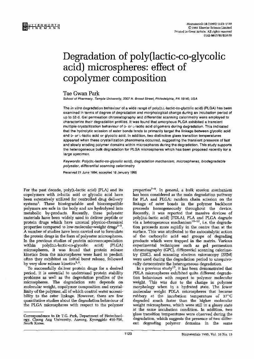
Figure 2 shows DSC thermograms of various PDLLGA microspheres as a function of degradation time. It can
I I I I I I I I I -30.00 0.00 30.00 60.00 90.00 120.00 150.00 180.00 210.00
b Temperature (C)
8
2
Figure 1 Differential scanning calorimetry thermograms of various compositions of poly(o,t.-lactic-co-glycolic acid) (PDLLGA) microspheres as a function of incubation time (in days). PDLLGA 9O:lO a, 80:20 b, 70:30 c and 50:50 d. The arrow indicates evident glass transition, temperatures.
Biomaterials 1995, Vol. 16 No. 15
1126 Degradation of PLGA microspheres: T.G. Park
be seen that amorphous PDLLGA microspheres having different copolymer compositions of D- and L-lactic acid and glycolic acid in their backbone degrade with a very complicated morphological change, such as decrease in Ts, appearance of double glass>transitions and evolution of crystalline melting peaks. All the PDLLGA microspheres exhibit a sharp single Ts (except for PDLLGA 80:20) before degradation, but demonstrate apparent split Ts values that mostly appear below 60°C during the degradation period. However, the characteristics of the glass transitions depend on the copolymer composition. For instance, PDLLGA 9O:lO shows two distinct 7’s values at days 2, 8,14 and 33; PDLLGA 80:20 at days 2 and 8; PDLLGA 70:30 at days 2, 8 and 14; PDLLGA 50:50 at days 2 and 8. In most cases, the first Ts has a typical glass transi- tion behaviour that accompanies a baseline inflection to the endothermic side, while the second Tg is often very sharp and looks like an endothermic crystalline melting peak (typical example: PDLLGA 70:30 at day 8). This sharp transition, however, cannot be assigned as the melting endotherm of crystallized degradation products, since its temperature of below 60°C is too low as a melting peak. A similarly shaped Tg was observed in many PLGA microspheres under these experimental conditions. Typical examples can be found in PDLLGA 9O:lO and 70:30 microspheres at day 0. The sharpness is related to the polymer chain relaxation kinetics in response to the thermal scanning rate. The glass transition is a rather kinetic phenom- enon which involves the polymer viscoelasticity which, in turn, is affected by experimental conditions such as heating rate and previous thermal history of the sample. The heating rate used in this study, 20” C min-’ was chosen because there were no notice- able discrepancies in DSC thermograms upon changing the heating rate. The double glass transitions along with a sharp endotherm-like Ts peak were more clearly observed during the first heating run in DSC scanning than the second run, suggesting that further heat treatment to the microspheres changed the relaxa- tion properties of the samples. In a previous report13, the same behaviour was also seen for low-molecular- weight PDLA microspheres. In some cases of semi- crystalline polymers, double glass transitions can often be found14. This is due to the presence of the same amorphous polymer chains located in different regions: a region of purely amorphous polymer chains which is located far horn the crystallites and a region of immobilized polymer chains which exists in the vicinity of crystallites. In the amorphous PLGA polymers studied here, however, the two observed glass transitions which appear without any accompany- ing crystalline melting peaks are clearly due to the differential degradation profiles in different regions of the microsphere. Therefore, the observed double Ts values can be attributed to the presence of two different polymer domains during the degradation: a fast and a slow degrading region in the microsphere. As the incubation continues, it can be seen that the first T, tends to shift to the low temperature region, while the second Tg tends to stay at its original transition temperature, then disappears. The transient second glass transition may be caused by the generation and
disappearance of a slow degrading polymer domain during the degradation.
In addition, multiple crystalline melting endothermic peaks appear above the glass transition temperature for all the initially amorphous PDLLGA samples during the degradation. The magnitude of endothermic enthalpies at various melting temperatures depends on the composition of polymers. For example, the fast degrad- ing PDLLGA 50:50 sample exhibits very sharp peaks around 120-130” C at day 14 and broad peaks at day 33, whereas PDLLGA 70:30 shows multiple small peaks at day 14. These peaks were generated by the crystalliza- tion of degradation products trapped in the microsphere. Since three different monomers, two stereoisomers (D- and L-lactic acid) and glycolic acid, constitute the polymer structure, these crystallization behaviours can be explained by the formation of crystallizable oligomers that have enriched D- or L-lactic acids in their sequence of polymer backbone. For semi-crystalline PLLA, two crystalline conformations have been identified: 1013 and 311 helices15. Thus, low-molecular-weight degrada- tion products presumably having a helical conformation in aqueous solution could be re-assembled and crystal- lized inside of the microsphere. It has been reported that PDLLGA does not possess a random distribution of D- (or L-) lactic acid and glycolic acid in its polymer backbone, but has a segregated structure because of the higher reactivity of the glycolic acid than that of L- (or D-) attic acid during the ring opening polymerization16. Consequently, the ester bonds linked with the glycolic acid unit (glycolic-glycolic acid, G-G; or glycolic- lactic acid, GL) may be preferentially cleaved, as compared to those of the lactic-lactic acid (L-L) linked ester, due to its inherent high reactivity with water and/or its greater hydrophilicityg. Therefore, not only stereoregular and crystallizable D- or L-lactic acid oligomers, but also even a stereo-complex of D- and L- helical structures17 are expected to be produced with the preferential degradation of glycolic acid-enriched segments. Even in the case of PDLA, crystallization behaviours were observed for the low-molecular- weight sample, suggesting the non-random distribution of D- and L-lactic acid in the sequence13. Vert and co- workers’1*‘8 also observed similar crystallization behaviours for amorphous PLGA films during degrada- tion.
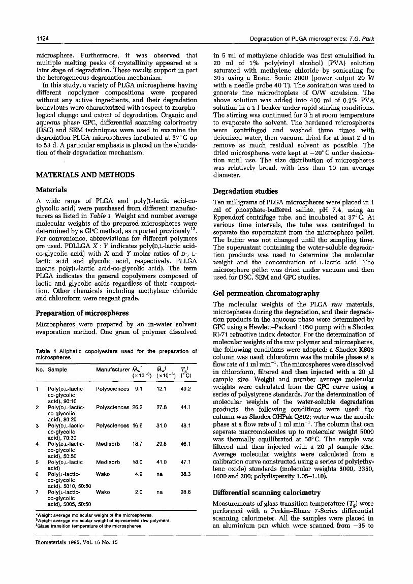
PLLGA 50:50 microspheres were prepared to determine whether they demonstrated a more pronounced oligomer crystallization effect. It is expected that the absence of D-lactic acid in the polymer constituents allows more crystallizable degradation products, mainly composed of L-lactic acid. Indeed, more clear multiple crystalline melting endotherms which span a wide range of temperatures between about 100 and 170°C can be observed, with two evident glass transitions as shown in Figure 2. Since the preferential hydrolysis of the G-G and GL ester linkages would produce the highly crystallizable L-lactic acid oligomers, the crystallization of degrada- tion products can be seen shortly after incubation, at day 2. These two microspheres have relatively low weight average molecular weights of 4900 and 2000, resulting in fast degradation. In particular, PLLGA 50:50 with molecular weight 2000 shows a series of
Biomaterials 1995, Vol. 16 No. 15
Degradation of PLGA microspheres: T.G. Park 1127
/ 14 a
I’r I I I I I I I I -30.00 0.00 30.00 60.00 90.00 120.00 l>O.OO 180.00 210.00
a
b -
Temperature (C)
I I I I I I I I I 0.00 0.00 30.00 60.00 90.00 120.00 150.00 180.00 210.00
Temperature (C)
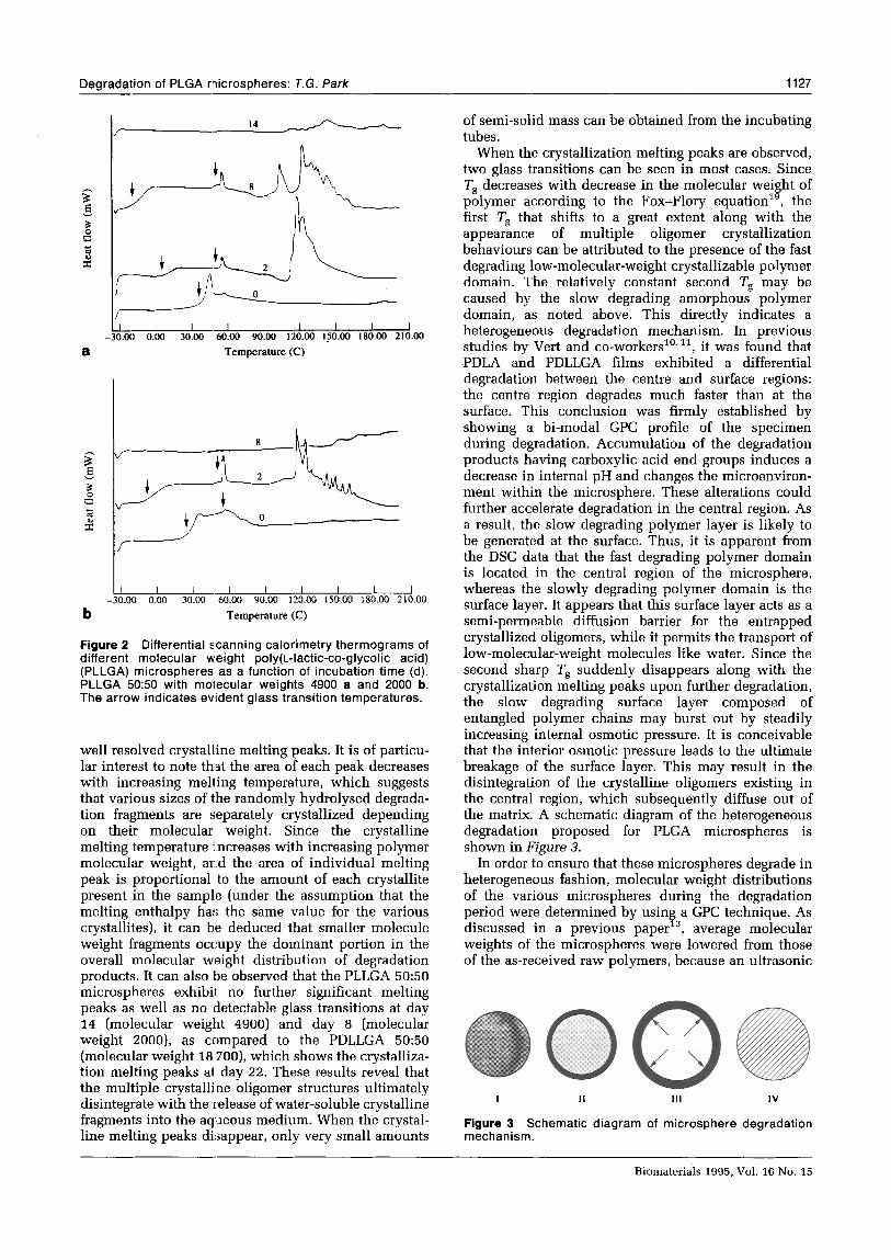
Figure 2 Differential scanning calorimetry thermograms of different molecular weight poly(t_-lactic-co-glycolic acid) (PLLGA) microspheres as a function of incubation time (d). PLLGA 50:50 with molecular weights 4900 a and 2000 b. The arrow indicates evident glass transition temperatures.
well resolved crystalline melting peaks. It is of particu- lar interest to note th.at the area of each peak decreases with increasing melting temperature, which suggests that various sizes of the randomly hydrolysed degrada- tion fragments are separately crystallized depending on their molecular weight. Since the crystalline melting temperature mcreases with increasing polymer molecular weight, and the area of individual melting peak is proportional to the amount of each crystallite present in the sample (under the assumption that the melting enthalpy has the same value for the various crystallites), it can ble deduced that smaller molecule weight fragments occupy the dominant portion in the overall molecular weight distribution of degradation products. It can also be observed that the PLLGA 50:50 microspheres exhibit no further significant melting peaks as well as no detectable glass transitions at day 14 (molecular weight 4900) and day 8 (molecular weight ZOOO), as compared to the PDLLGA 50:50 (molecular weight 18 7OO), which shows the crystalliza- tion melting peaks at day 22. These results reveal that the multiple crystalline oligomer structures ultimately disintegrate with the release of water-soluble crystalline fragments into the aqueous medium. When the crystal- line melting peaks disappear, only very small amounts
of semi-solid mass can be obtained from the incubating tubes.
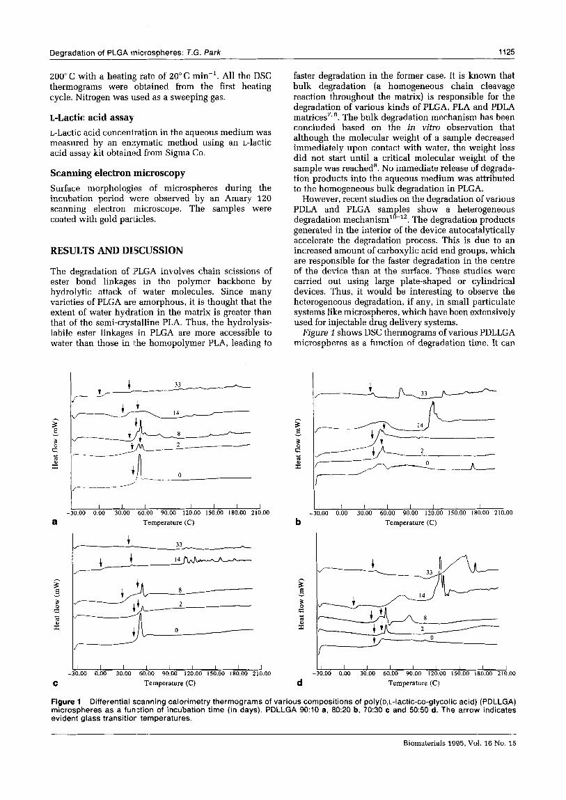
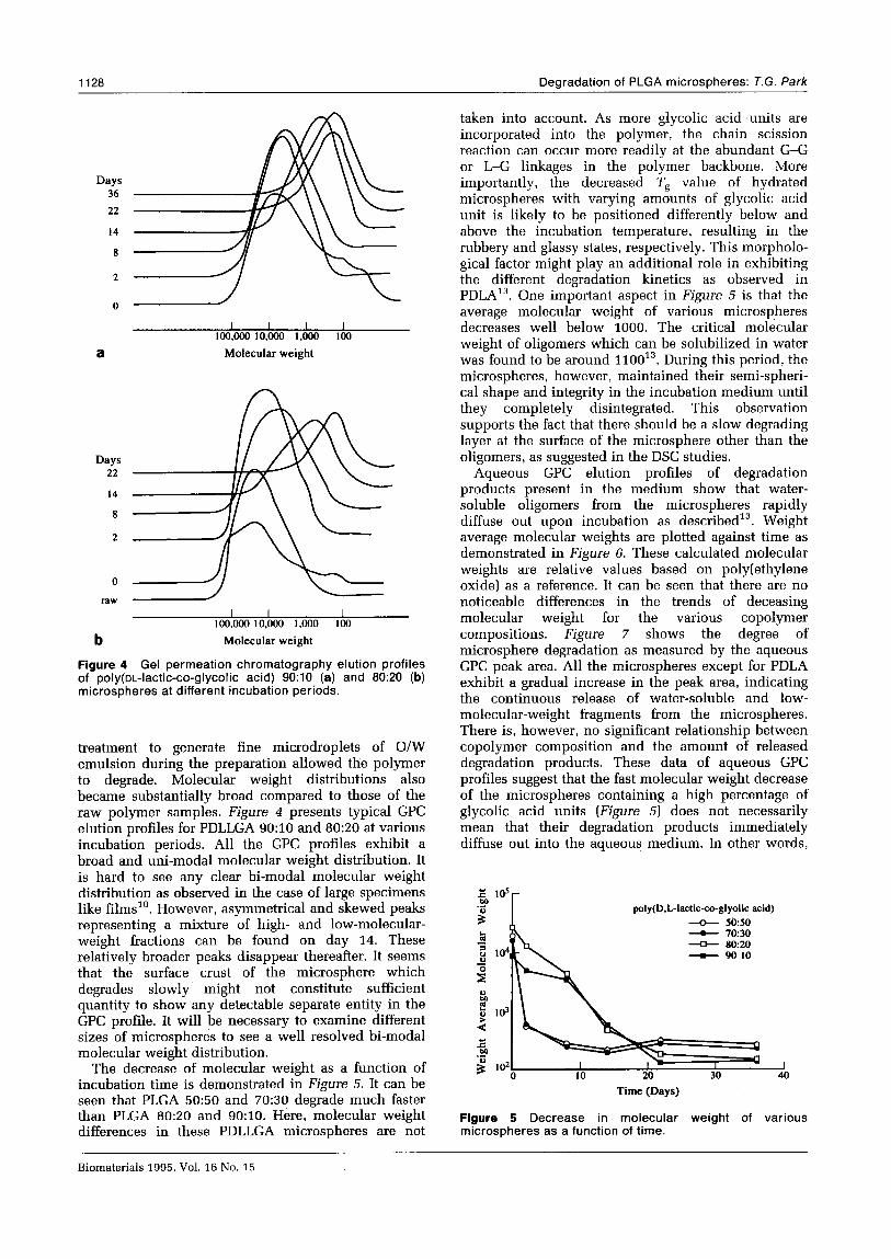
When the crystallization melting peaks are observed, two glass transitions can be seen in most cases. Since Ts decreases with decrease in the molecular weight of polymer according to the Fox-Flory equationlg, the first Ts that shifts to a great extent along with the appearance of multiple oligomer crystallization behaviours can be attributed to the presence of the fast degrading low-molecular-weight crystallizable polymer domain. The relatively constant second Tg may be caused by the slow degrading amorphous polymer domain, as noted above. This directly indicates a heterogeneous degradation mechanism. In previous studies by Vert and co-workers”,“, it was found that PDLA and PDLLGA films exhibited a differential degradation between the centre and surface regions: the centre region degrades much faster than at the surface. This conclusion was firmly established by showing a bi-modal GPC profile of the specimen during degradation. Accumulation of the degradation products having carboxylic acid end groups induces a decrease in internal pH and changes the microenviron- ment within the microsphere. These alterations could further accelerate degradation in the central region. As a result, the slow degrading polymer layer is likely to be generated at the surface. Thus, it is apparent from the DSC data that the fast degrading polymer domain is located in the central region of the microsphere, whereas the slowly degrading polymer domain is the surface layer. It appears that this surface layer acts as a semi-permeable diffusion barrier for the entrapped crystallized oligomers, while it permits the transport of low-molecular-weight molecules like water. Since the second sharp Ts suddenly disappears along with the crystallization melting peaks upon further degradation, the slow degrading surface layer composed of entangled polymer chains may burst out by steadily increasing internal osmotic pressure. It is conceivable that the interior osmotic pressure leads to the ultimate breakage of the surface layer. This may result in the disintegration of the crystalline oligomers existing in the central region, which subsequently diffuse out of the matrix. A schematic diagram of the heterogeneous degradation proposed for PLGA microspheres is shown in Figure 3.
In order to ensure that these microspheres degrade in heterogeneous fashion, molecular weight distributions of the various microspheres during the degradation period were determined by using a GPC technique. As discussed in a previous paper13, average molecular weights of the microspheres were lowered from those of the as-received raw polymers, because an ultrasonic
0 I II III IV
Figure 3 Schematic diagram of microsphere degradation mechanism.
Biomaterials 1995, Vol. 16 No. 15
1128 Dearadation of PLGA microsoheres: T.G. Park
Days 36
22
14
8
I I I I 100,000 10,ooo 1,ooo 100
a Molecular weight
Days 22
14
8
2
0
raw -
I I I I
1oo.ooo 10,ooo 1,000 loo
b Molecular weight
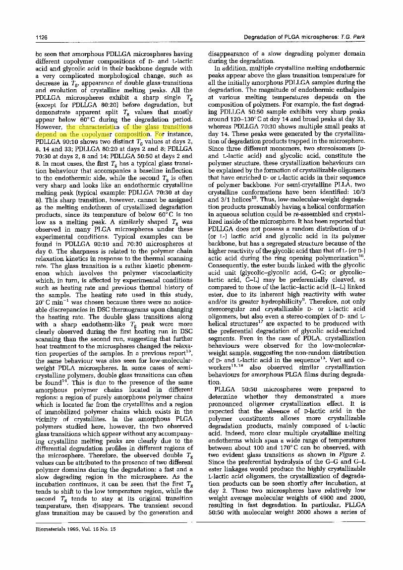
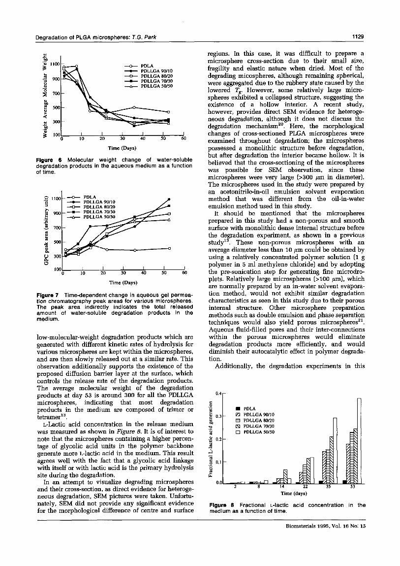
Figure 4 Gel permeation chromatography elution profiles of poly(oL-lactic-co-glycolic acid) 9O:lO (a) and 80:20 (b) microspheres at different incubation periods.
treatment to generate fine microdroplets of O/W emulsion during the preparation allowed the polymer to degrade. Molecular weight distributions also became substantially broad compared to those of the raw polymer samples. Figure 4 presents typical GPC elution profiles for PDLLGA 9O:lO and 8O:ZO at various incubation periods. All the GPC profiles exhibit a broad and uni-modal molecular weight distribution. It is hard to see any clear bi-modal molecular weight distribution as observed in the case of large specimens like films”. However, asymmetrical and skewed peaks representing a mixture of high- and low-molecular- weight fractions can be found on day 14. These relatively broader peaks disappear thereafter. It seems that the surface crust of the microsphere which degrades slowly might not constitute sufficient quantity to show any detectable separate entity in the GPC profile. It will be necessary to examine different sizes of microspheres to see a well resolved bi-modal molecular weight distribution.
The decrease of molecular weight as a function of incubation time is demonstrated in Figure 5. It can be seen that PLGA 5050 and 7050 degrade much faster than PLGA 8020 and 9O:lO. Here, molecular weight differences in these PDLLGA microspheres are not
taken into account. As more glycolic acid ,units are incorporated into the polymer, the chain scission reaction can occur more readily at the abundant G-G or L-G linkages in the polymer backbone. More importantly, the decreased Ts value of hydrated microspheres with varying amounts of glycolic acid unit is likely to be positioned differently below and above the incubation temperature, resulting in the rubbery and glassy states, respectively. This morpholo- gical factor might play an additional role in exhibiting the different degradation kinetics as observed in PDLA13. One important aspect in Figure 5 is that the average molecular weight of various microspheres decreases well below 1000. The critical molecular weight of oligomers which can be solubilized in water was found to be around 110013. During this period, the microspheres, however, maintained their semi-spheri- cal shape and integrity in the incubation medium until they completely disintegrated. This observation supports the fact that there should be a slow degrading layer at the surface of the microsphere other than the oligomers, as suggested in the DSC studies.
Aqueous GPC elution profiles of degradation products present in the medium show that water- soluble oligomers from the microspheres rapidly diffuse out upon incubation as described13. Weight average molecular weights are plotted against time as demonstrated in Figure 6. These calculated molecular weights are relative values based on poly(ethylene oxide) as a reference. It can be seen that there are no noticeable differences in the trends of deceasing molecular weight for the various copolymer compositions. Figure 7 shows the degree of microsphere degradation as measured by the aqueous GPC peak area. All the microspheres except for PDLA exhibit a gradual increase in the peak area, indicating the continuous release of water-soluble and low- molecular-weight fragments from the microspheres. There is, however, no significant relationship between copolymer composition and the amount of released degradation products. These data of aqueous GPC profiles suggest that the fast molecular weight decrease of the microspheres containing a high percentage of glycolic acid units (Figure 5) does not necessarily mean that their degradation products immediately diffuse out into the aqueous medium. In other words,
poly(D.L-lactic-co-glyolic acid) + 50:50 - lo:30 + 80~20 - 90 IO
Time (Days)
Figure 5 Decrease in molecular weight of various microspheres as a function of time.
Biomaterials 1995, Vol. 16 No. 15
Degradation of PLGA miicrospheres: T.G. Park 1129
e PDLA - PDLLGA90110 + PDLLGASOIZO - PDLLCiA70130 -b- PDLLGA50150
I I I I 30 40 50 60
Time (Days)
Figure 6 Molecular weight change of water-soluble degradation products in the aqueous medium as a function of time.
- PDLA --t PDLLGA90/10 + PDLLGA80120
- PDLLGA50/50
lw*a do 60 :o cio Time (Days)
Figure 7 Time-dependent change in aqueous gel permea- tion chromatography peak areas for various microspheres. The peak area indirectly indicates the total released amount of water-soluble degradation products in the medium.
low-molecular-weight degradation products which are generated with different kinetic rates of hydrolysis for various microspheres are kept within the microspheres, and are then slowly released out at a similar rate. This observation additionally supports the existence of the proposed diffusion barrier layer at the surface, which controls the release rate of the degradation products. The average molecu’lar weight of the degradation products at day 53 is around 300 for all the PDLLGA microspheres, indicating that most degradation products in the medium are composed of trimer or tetramer13.
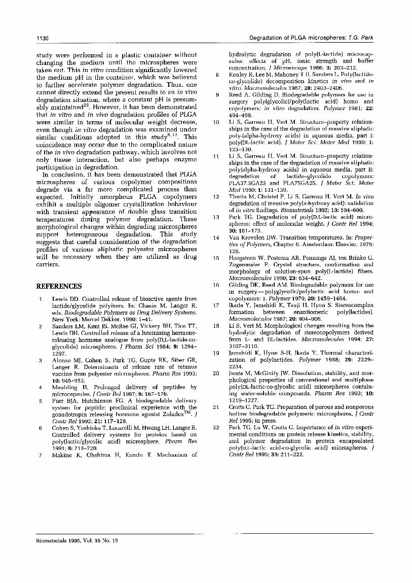
L-Lactic acid concentration in the release medium was measured as shown in Figure 8. It is of interest to note that the microspheres containing a higher percen- tage of glycolic acid units in the polymer backbone generate more L-lactic acid in the medium. This result agrees well with the fact that a glycolic acid linkage with itself or with lactic acid is the primary hydrolysis site during the degradation.
In an attempt to visualize degrading microspheres and their cross-section, as direct evidence for heteroge- neous degradation, SEM pictures were taken. Unfortu- nately, SEM did not provide any significant evidence for the morphological difference of centre and surface
regions. In this case, it was difficult to prepare a microsphere cross-section due to their small size, fragility and elastic nature when dried. Most of the degrading micospheres, although remaining spherical, were aggregated due to the rubbery state caused by the lowered Ts. However, some relatively large micro- spheres exhibited a collapsed structure, suggesting the existence of a hollow interior. A recent study, however, provides direct SEM evidence for heteroge- neous degradation, although it does not discuss the degradation mechanismzO. Here, the morphological changes of cross-sectioned PLGA microspheres were examined throughout degradation; the microspheres possessed a monolithic structure before degradation, but after degradation the interior became hollow. It is believed that the cross-sectioning of the microspheres was possible for SEM observation, since these microspheres were very large (>300 pm in diameter). The microspheres used in the study were prepared by an acetonitrile-in-oil emulsion solvent evaporation method that was different from tbe oil-in-water emulsion method used in this study.
It should be mentioned that the microspheres prepared in this study had a non-porous and smooth surface with monolithic dense internal structure before the d:padation experiment, as shown in a previous study . These non-porous microspheres with an average diameter less than 10 pm could be obtained by using a relatively concentrated polymer solution (1 g polymer in 5 ml methylene chloride) and by adopting the pre-sonication step for generating fine microdro- plets. Relatively large microspheres (~100 pm), which are normally prepared by an in-water solvent evapora- tion method, would not exhibit similar degradation characteristics as seen in this study due to their porous internal structure. Other microsphere preparation methods such as double emulsion and phase separation techniques would also yield porous microsphere?. Aqueous fluid-filled pores and their inter-connections within the porous microspheres would eliminate degradation products more efficiently, and would diminish their autocatalytic effect in polymer degrada- tion.
Additionally, the degradation experiments in this
0.4
8 ._ 2 I PDLA 2 0.3
: FJ PDLLGA90/10
D q PDLLGA SO/20 = ll PDLLGA70/30 ._ P 0 PDLLGA50/50
.o 0.2 2 7 t
Time(days)
Figure 6 Fractional L-lactic acid concentration in the medium as a function of time.
Biomaterials 1995,Vol. 16 No: 15
1130 Degradation of PLGA microspheres: T.G. Park
study were performed in a plastic container without changing the medium until the microspheres were taken out. This in vitro condition significantly lowered the medium pH in the container, which was believed to further accelerate polymer degradation. Thus, one cannot directly extend the present results to an in vivo degradation situation, where a constant pH is presum- ably maintained”. However, it has been demonstrated that in vitro and in viva degradation profiles of PLGA were similar in terms of molecular weight decrease, even though in vitro degradation was examined under similar conditions adopted in this studysS*‘. This coincidence may occur due to the complicated nature of the in viva degradation pathway, which involves not only tissue interaction, but also perhaps enzyme participation in degradation.
8
9
10
11
In conclusion, it has been demonstrated that PLGA microspheres of various copolymer compositions degrade via a far more complicated process than expected. Initially amorphous PLGA copolymers exhibit a multiple oligomer crystallization behaviour with transient appearance of double glass transition temperatures during polymer degradation. These morphological changes within degrading microspheres support heterogeneous degradation. This study suggests that careful consideration of the degradation profiles of various aliphatic polyester microspheres will be necessary when they are utilized as drug carriers.
12
13
14
15
REFERENCES 16
Lewis DD. Controlled release of bioactive agents from lactide/glycolide polyiners. In: Chasin M, Langer R, eds. Biodegradable Polymers as Drug Delivery Systems. New York: Marcel Dekker, 1990: 1-41. Sanders LM, Kent JS, McRae GI, Vickery BH, Tice TT, Lewis DH. Controlled release of a luteinizing hormone- releasing hormone analogue from poly(D,L-lactide-co- glycolide) microspheres. I Pharm Sci 1984; 9: 1294- 1297. Alonso MJ, Cohen S, Park TG, Gupta RK, Siber GR, Langer R. Determinants of release rate of tetanus vaccine from polyester microspheres. Pharm Res 1993; 10:945-953. Maulding H. Prolonged delivery of peptides by microcapsules. 1 Contr Rel 1987; 6: 167-176. Furr BJA, Hutchinson FG. A biodegradable delivery system for peptide: preclinical experience with the gonadotropin releasing hormone agonist ZoladexTM. I Contr Rell992; 21: 117-128. Cohen S, Yoshioka T, Lucarelli M, Hwang LH, Langer R. Controlled delivery systems for proteins based on poly(lactic/glycolic acid) microsphere. Pharm Res 1991; 8:713-720. Makino K, Ohshima H, Kondo T. Mechanism of
17
18
19
20
21
22
hydrolytic degradation of poly(L-lactide) microcap- sules: effects of pH, ionic strength and buffer concentration. J Microencaps 1986; 3: 203-212. Kenley R, Lee M, Mahoney T II, Sanders L. Poly(lactide- co-glycolide) decomposition kinetics in vivo and in vitro. Macromolecules 1987; 20: 2403-2406. Reed A, Gilding D. Biodegradable polymers for use in surgery poly(glycolic)/poly(lactic acid) homo and copolymers: in vitro degradation. Polymer 1981; 22: 494-498. Li S, Garreau H, Vert M. Structure-property relation- ships in the case of the degradation of massive aliphatic poly-(alpha-hydroxy acids) in aqueous media, part I: poly(DL-lactic acid). J Mater Sci: Mater Med 1990; 1: 123-130. Li S, Garreau H, Vert M. Structure-property relation- ships in the case of the degradation of massive aliphatic poly(alpha-hydroxy acids) in aqueous media, part II: degradation of lactide-glycolide copolymers: PLA37.5GA25 and PLA75GA25. J Mater Sci: Mater Med1990; 1: 131-139. Therin M, Christel P, Li S, Garreau H, Vert M. In vivo degradation of massive poly(a-hydroxy acid): validation of in vitro findings. Biomaterials 1992; 13: 594-600. Park TG. Degradation of poly(D,L-lactic acid) micro- spheres: effect of molecular weight. J Contr Rel 1994; 30:161-173. Van Krevelen DW. Transition temperatures. In: Proper- ties of Polymers, Chapter 6. Amsterdam: Elsevier, 1976: 126. Hoogsteen W, Postema AR, Pennings AJ, ten Brinke G, Zugenmaier P. Crystal structure, conformation and morphology of solution-spun poly(L-lactide) fibers. Macromolecules 1990; 23: 634-642. Gilding DK, Reed AM. Biodegradable polymers for use in surgery- polyglycolic/polylactic acid homo- and copolymers: 1. Polymer 1979; 20: 1459-1464. Ikada Y, Jamshidi K, Tsuji H, Hyon S. Stereocomplex formation between enantiomeric poly(lactides). Macromolecules 1987; 20: 904-906. Li S, Vert M. Morphological changes resulting from the hydrolytic degradation of stereocopolymers derived from L- and DL-lactides. Macromolecules 1994; 27: 3107-3110. Jamshidi K, Hyon S-H, Ikada Y. Thermal characteri- zation of polylactides. Polymer 1988; 29: 2229- 2234. Iwata M, McGinity JW. Dissolution, stability, and mor- phological properties of conventional and multiphase poly(DL-lactic-co-glycolic acid) microspheres contain- ing water-soluble compounds. Pharm Res 1993; 10: 1219-1227. Crotts G, Park TG. Preparation of porous and nonporous hollow biodegradable polymeric microspheres, J Contr Rell995; in press. Park TG, Lu W, Crotts G. Importance of in vitro experi- mental conditions on protein release kinetics, stability, and polymer degradation in protein encapsulated poly(D,L-lactic acid-co-glycolic acid) microspheres. J Contr Rell995; 33: 211-222.
Biomaterials 1995. Vol. 16 No. 15