dexketoprofen trometamol-loaded poly-lactic-co-glycolic acid
TRANSCRIPT
Ozturk et al
Trop J Pharm Res, January 2019; 18(1): 1
Tropical Journal of Pharmaceutical Research January 2019; 18 (1): 1-11 ISSN: 1596-5996 (print); 1596-9827 (electronic)
© Pharmacotherapy Group, Faculty of Pharmacy, University of Benin, Benin City, 300001 Nigeria.
Available online at http://www.tjpr.org http://dx.doi.org/10.4314/tjpr.v18i1.1
Original Research Article
Dexketoprofen trometamol-loaded poly-lactic-co-glycolic acid (PLGA) nanoparticles: Preparation, in vitro characterization and cyctotoxity
A. Alper Öztürk1,2*, Lucia Martin Banderas2, Maria D. Cayero Otero2, Evrim Yenilmez1, Behiye Şenel3, Yasemin Yazan1 1Department of Pharmaceutical Technology, Faculty of Pharmacy, Anadolu University, Eskişehir, Turkey, 2Department of Pharmacy and Pharmaceutical Technology, Faculty of Pharmacy, University of Seville, Seville, Spain, 3Department of Pharmaceutical Biotechnology, Faculty of Pharmacy, Anadolu University, Eskişehir, Turkey *For correspondence: Email: [email protected]; Tel: +90 22 335 05 80 / 3731 Sent for review: 18 July 2018 Revised accepted: 16 November 2018
Abstract
Purpose: To design, formulate and characterize sustained-release formulations of dexketoprofen trometamol (DT) nanoparticles (NPs) Methods: Dexketoprofen trometamol (DT)-loaded poly(lactic-co-glycolic acid) (PLGA) NPs were produced by double emulsion-solvent evaporation method. The NPs were variously characterized for drug loading and release, particle profile, as well as by thermal analysis, x-ray difraction (XRD), Fourier transform infrared spectroscopy (FTIR) and nuclear magnetic resonance analysis (1H-NMR). Furthermore, the NPs were evaluated for cytotoxicity against NIH-3T3 cells by 3-(4,5-dimethylthiazol-2-Yl)-2,5-diphenyltetrazolium bromide (MTT) assay. Results: The DT-loaded NPs demonstrated nanostructural characteristics and extended drug release. Particle size was in the range of 243 and 295 nm which remained unchanged in drug stability testing in simulated gastrointestinal media. Encapsulation efficiency ranged from 49 – 64 % for all the formulations. Higuchi and Korsmeyer-Peppas were the best-fit release kinetic models for the NPs containing 5 and 10 % DT, respectively. The NPs with 10 % DT presented no significant cytotoxicty at the doses and periods studied. Conclusion: Stable and non-toxic DT NPs with potential for sustained and controlled release of the drug have been successfully developed. Keywords: Dexketoprofen trometamol, Poly-lactic-co-glycolic acid (PLGA), Nanoparticles, Release kinetics, Stability
This is an Open Access article that uses a funding model which does not charge readers or their institutions for access and distributed under the terms of the Creative Commons Attribution License (http://creativecommons.org/licenses/by/4.0) and the Budapest Open Access Initiative (http://www.budapestopenaccessinitiative.org/read), which permit unrestricted use, distribution, and reproduction in any medium, provided the original work is properly credited.
Tropical Journal of Pharmaceutical Research is indexed by Science Citation Index (SciSearch), Scopus, International Pharmaceutical Abstract, Chemical Abstracts, Embase, Index Copernicus, EBSCO, African Index Medicus, JournalSeek, Journal Citation Reports/Science Edition, Directory of Open Access Journals (DOAJ), African Journal Online, Bioline International, Open-J-Gate and Pharmacy Abstracts
INTRODUCTION The ability to manage and treat pain continues to be one of the most common clinical objectives of practitioners. Pain has a very important effect on
the biological, psychological, sociological and economic dispositions of a patient [1]. Non-steroidal anti-inflammatory drugs (NSAIDs), skeletal muscle relaxants and opioid analgesics are among the most prescribed drugs for pain
-----------------------------------------------------------------------------------------------------------------------------------------------------© 2019 The authors. This work is licensed under the Creative Commons Attribution 4.0 International License
Ozturk et al
Trop J Pharm Res, January 2019; 18(1): 2
[2]. The NSAIDs block a group of physiological proteins called cyclooxygenase (COX) which are invoved in the production of prostaglandins and thromboxanes responsible for pain and inflammation [3]. Ketoprofen, a member of the arylpropionate group of NSAIDs has well-defined analgesic and anti-inflammatory effects. Racemic ketoprofen is used as an analgesic and anti-inflammatory agent, and it is one of the most potent inhibitors of prostaglandin synthesis in vitro [4]. Dexketoprofen trometamol (DT) was developed as a water-soluble trometamine. İt is a rapidly-acting analgesic agent in the treatment of painful musculoskeletal disorders such as back pain and osteoarthritis [5]. Polymeric nanoparticles (PNPs) are being extensively investigated as drug delivery systems worldwide for pharmaceutical applications. They provide modified release of drugs for extended periods of time [6]. The most commonly used biodegradable polymers in the production of polymeric NPs for modified release are polyglycolic acid, polylactic acid and their copolymers, e.g., PLGA (polylactic-co-glycolic acid) [7]. Modified drug release can be obtained by polymeric NPs owing to different properties of PLGA used [8]. Polyglycolic acid is a hydrophilic agent and dissolves readily in intracellular conditions. Polylactic acid is more hydrophobic than polyglycolic acid due to an additional methyl group in the side chain; and therefore biodegradation takes place much longer than polyglycolic acid [9]. Double emulsion techniques are commonly used for the encapsulation of hydrophilic molecules with low encapsulation efficiency because of rapid drug partitioning into the external aqueous phase in classical emulsions. In double emulsification technique, displacement of a lipophilic solvent with a water-miscible semi-polar solvent results in accumulation of polymer on the surface. Rapid diffusion of the non-solvent reduces the interfacial tension between the two phases leading to an increase in surface area and thus formation of small organic solvent droplets. It is a very simple and economic method [10]. Surveying extant patents and publications, some modified release formulations and locally available commercial preparations of DT were found [11-13]. The present study was carried out to design, formulate and characterize sustained-release formulations of DT NPs.
EXPERİMENTAL Chemicals Dexketoprofen trometamol was a kind gift from Abdi İbrahim (İstanbul/Turkey). Poly(lactic-co-glycolic acid) (PLGA) (Resomer® RG 504 H) was obtained from Evonik Industries (Germany). Polyvinil alcohol (PVA) was purchased from Sigma-Aldrich (St. Louis, MO, USA), while ethyl acetate and sodium acetate trihydrate were obtained from Panreac Química (Barcelona, Spain). Methanol was from Merck (Gradient grade for liquid chromatography, Darmstadt/ Germany). Deionized and filtered water was used in all experiments (Milli-Q Academic, Millipore, Molsheim, France). All other chemicals used were of analytical grade. Preparation of PLGA nanoparticles The PLGA NPs were prepared by ‘Double Emulsification Solvent Evaporation Technique’ [14]. Weighed amount of PLGA was dissolved in ethyl acetate to obtain a concentration of 40 mg/mL. Then, 100 µL aqueous solution of PVA (0.5 % w/v) was added drop-wise to 2 mL PLGA solution using ultrasonic bath (1 minute, JP Selctra) (W1/O). Two mL of this emulsion was then added drop-wise to 10 mL of PVA aqueous solution (0.5 %, w/v) using high-speed homogenizer (Polytron PT 2500 E) at 24.000 rpm for 1 min (W1/O/W2). The ethyl acetate was evaporated at room temperature in 4 h to obtain an aqueous dispersion which was finally centrifuged to obtain the NPs (11.000 rpm, 30 min, and 4 °C; Eppendorf 504R, Eppendorf AG, Hamburg, Germany). For the preparation of DT-loaded PLGA NPs, the procedure was started by adding 4 - 8 mg DT [5 % DT (Alp-5) and 10 % DT (Alp-10)] to 100 µL aqueous solution of PVA (0.5 % w/v). The aqueous solution of PVA containing 100 µL DT was added drop-wise to 2 mL PLGA solution in an ultrasonic bath (1 min, JP Selctra) (W1/O). Two mL of this emulsion was then added drop-wise to 10 mL of PVA aqueous solution (0.5 %, w/v) under high-speed homogenizer (Polytron PT 2500 E) at 24.000 rpm for 1 min (W1/O/W2). The ethyl acetate was evaporated at room temperature in 4 h to obtain an aqueous dispersion which was finally centrifuged to obtain the NPs (11,000 rpm, 30 min, 4 °C; Eppendorf 504R, Eppendorf AG, Hamburg, Germany). Determination of particle size, PDI, zeta potential and morphology Particle size (PS) and PDI were measured on freshly prepared samples using Malvern analyzer (Zetamaster 3000, Malvern Instruments Ltd.,
Ozturk et al
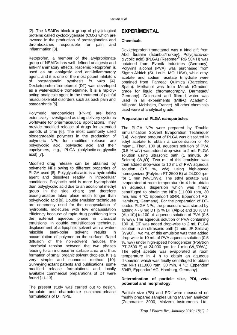
Trop J Pharm Res, January 2019; 18(1): 3
UK). Samples of all PLGA NPs were diluted with distilled water prior to analysis. All analyses were repeated three times at room temperature. Surface electrical charge (zeta potential) of PLGA NPs was characterized by laser Doppler electrophoresis (Zetamaster 3000, Malvern Instruments Ltd, UK). Measurements were repeated three times at room temperature. Morphological and structural characteristics of the PLGA NPs prepared were investigated using SEM (Hitachi TM 3030 Plus, Japan). Lyophilized samples were coated with thin layer of gold using a coater (Karaltay Scientific Instruments, China) under 50 mA for 30 seconds before observation under a scanning electron micoscope (SEM). Assessment of cryoprotectant effect on PLGA NPs Some experiments were carried out on Alp-5 and Alp-10 formulations to determine storage and lyophilization conditions. Following PS measurement of fresh formulations, they were centrifuged and the supernatants were discarded. The resulting particles were added to 1 mL of 5 % (w/v) trehalose solution and the PS measurement was performed again. The dispersion was then divided into 10 equal portions in 10 eppendorf tubes numbered 1 - 10. While no trehalose solution was added to tubes 1 and 2, 100 µL, 200 µL, 300 µL and 400 µL trehalose solution (5 % w/v) were added to tubes 3 and 4; 5 and 6; 7 and 8; 9 and 10, respectively (i.e. in duplicates). All formulations were then frozen at -20 °C prior to PS analysis after melting tubes 1, 3, 5, 7 and 9 at room temperature. Following lyophilization (Telstar® CRYODOS, Liofilizador de Laboratorio, Spain) of tubes 2, 4, 6, 8 and 10, the dry particles were removed from the lyophilizer and dispersed in 1 mL of water, followed by PS analysis. Evaluation of stability of PLGA NPs The NPs prepared using hydrolytic degradable polymers are known to degrade over time. Effects of pH and temperature have a very significant effect on long-term stability [15]. Before testing the stability of PLGA NPs, solutions simulating gastrointestinal fluids were prepared. The solutions were pepsin-free HCl (pH 1.2, Solution 1), pepsin-containing HCl solution (pH 1.2, Solution 2, 0.32 % pepsin, w/v); intestinal fluid phosphate buffer solution (Solution 3, pH 6.8); phosphate buffer solution (Solution 4, pH 7.4); physiological serum (0.9 % NaCl) solution (Solution 5) and Milli-Q water. All five solutions and Milli-Q water were placed in a shaking water bath at a stirring speed of 40
rpm at a temperature of 37 ºC to simulate the gastric medium. One set of formulations (Alp-5 and Alp-10) was prepared and dispersed in trehalose solution at 5 % (w/v) concentration. Then, 1 mL of this dispersion was added to solutions incubated at 37±1°C. Samples were collected after preincubation periods of 1, 2, 3, 4, 5, 6, 7 and 24 h and centrifuged at 4.000 rpm for 5 min to precipitate NPs. Finally, the average particle size of the NPs was determined with laser scattering. HPLC conditions The amount of DT loaded into PLGA NPs and dissolution study of each formulation was performed using HPLC (Shimadzu Corporation, Kyoto, Japan) with reversed-phase Symetry® and C18 (5.0 µm, 250 mm x 4.6 mm, Symmetry US) column. In the HPLC system, the mobile phase was methanol:acetate buffer (65:35 v/v) mixture, the flow rate was 0.9 mL.min-1, while detection was performed at 242 nm at 40°C. Injection volume and run time were 25 µL and 12 min, respectively. Mobile phase was prepared daily, de-gassed by sonication and filtered through 0.45 µm membrane filter before the experiment. The method was validated for precision, accuracy, specificity and linearity [16]. Evaluation of entrapment efficiency The loading of DT into PLGA NPs was determined by reverse phase-HPLC (RP-HPLC) method as described earlier. The DT content of PLGA NPs was assessed directly by extracting DT from NPs. Lyophilized NPs (about 5 mg) were weighed, 1 mL ethyl acetate was added and the mixture was vortexed to dissolve the particles in the organic phase. All solutions were filtered through 0.22 µm polyamid filter prior to analysis with HPLC system. The DT content was expressed as encapsulation efficiency (EE) as in Eq 1. EE (%) = {(W1-W2)/W1}100 ......... (1) where W1 is the actual amount of DT loaded into PLGA NPs and W2 is the theoretical amount of DT loaded into PLGA NPs. In vitro dissolution studies The ability of the PLGA formulations to release DT in the stomach (pH 1 or 1.2), small intestine (pH 6.8) and colon (pH 7.4) was assessed by release studies in simulated gastric and intestinal media [17]. In vitro release profiles from PLGA NPs were investigated over 10 days using a dialysis membrane. PLGA NPs equivalent to 10

Ozturk et al
Trop J Pharm Res, January 2019; 18(1): 4
mg DT was placed in a cellulose acetate dialysis bag with a molecular weight of 12-14 kDa (Sigma). After sealing the two ends, in vitro release study was started in 75 mL 0.1 N hydrochloric acid (pH 1.0) at 37 °C with stirring at 100 rpm using a magnetic stirrer. The study was carried out for 2 h, and then continued at pH 6.8 (obtained by addition of 25 mL 0.2 M tribasic sodium phosphate solution) for the next 3 h, and thereafter at pH 7.4 (obtained by addition of 5 mL 0.2 M tribasic sodium phosphate solution) for the remaining time intervals. Stirring was maintained at 100 rpm and temperature at 37 ± 0.5 °C in all media. Samples were collected at certain intervals and replaced with the same volume of fresh dissolution medium. The samples were then analyzed using HPLC method. Determination of in vitro release kinetics Data obtained in the in vitro drug release studies were further investigated for release kinetics using DDSolver software program [18]. Thermal analysis Thermal analysis of the NPs prepared was carried out using DSC (Schimadzu DSC-60, Japan) in a pressure-assisted aluminum sample vessel at a nitrogen gas flow rate of 50 mL·min-1 and a temperature increase rate of 10°C·min-1 at 50-250°C with aluminum reference. XRD analysis The NPs were subjected to XRD analysis performed with a Rikagu generator (XRD Rikagu Rint 2000, Japan) at a speed of 40 kV, current intensity of 30 mA, angle of 2Ɵ and speed of 1°·min-1 in the range of 5°-45°. FT-IR analysis The FT-IR spectra of the NPs prepared were determined by FT-IR (Schimadzu IR Prestige-21, Japan) in the wavelength range of 4000-1 and 500 cm-1. 1H-NMR analysis The PLGA NPs were subjected to NMR analysis (1H-NMR). They were collected by centrifugation, lyophilized and dissolved in deutero chloroform (CDCl3) prior to analysis using NMR (Bruker 500 MHz UltraShield NMR, Germany).
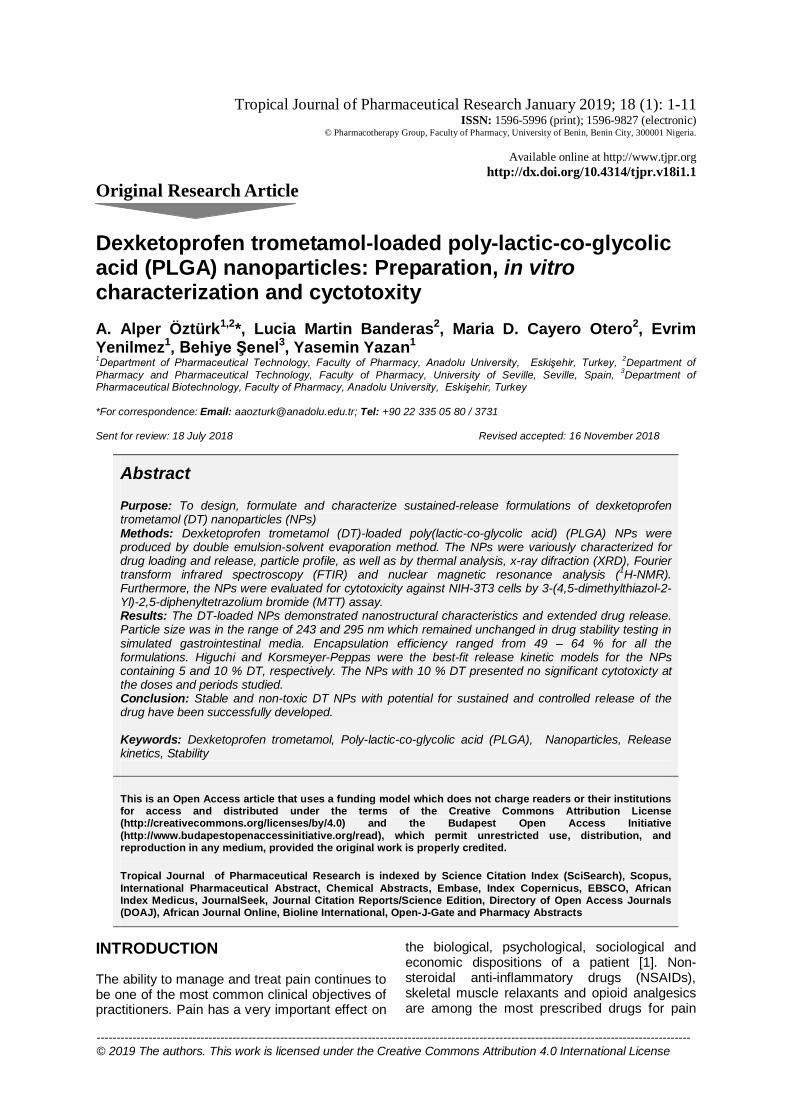
Cell culture Mouse embryonic fibroblast cells (NIH/3T3) were selected for culture studies. The cells were grown in Dulbecco's Modified Eagle's Medium (DMEM) supplemented with 10 % fetal bovine serum (Sigma Aldrich, Germany), 2 mM L-glutamine and penicillin-streptomycin (Sigma Aldrich, Germany) at 37 °C in a humidified atmosphere containing 5 % CO2. Cytotoxicity studies Quantitative cell cytotoxicity was determined by colorimetric MTT (3-(4, 5-dimethyl thiazol-2yl)-2, 5-diphenyl tetrazolium bromide) assay. Briefly, cells removed from the flask were seeded in 96-well plates and left to incubate for 24 h. At the end of the incubation period, fresh growth media containing different concentrations of NPs were added to the wells to give final concentrations in the range 5 -1000 µg/mL. The plates were re-incubated for another 24 - 48 h, and MTT dye was added at end of each incubation time. The resultant formazan crystals were dissolved with DMSO, and the absorbance of solution was read at 572 nm in a microplate reader (BioTek Cytation5, Germany) [19]. The results obtained were expressed as percentage inhibition relative to control cells where cell survival was taken as 100 %. The experiments were performed in triplicate. Statistical analysis Each experiment was carried out three independent times and the data are presented as mean ± standard deviation (SD). Microsoft Excel and DDSolver were employed for statistical analysis. RESULTS Particle size, polydispersity index and zeta potential The size and size distribution of PLGA NPs measured by laser scattering method are presented in Table 1. All data obtained from six independent experiments were expressed as the mean diameter and standard deviation (SD) values. Figure 1 shows SEM images of the prepared PLGA NPs. The images revealed regular and symmetrical morphology. Effect of cryoprotectant on PLGA NPs Figure 2 shows the effect of cryoprotectant addition on Alp-5 and Alp-10 NP sizes and size distributions.
Ozturk et al
Trop J Pharm Res, January 2019; 18(1): 5
Table 1: Particle size, PDI, zeta potential and entrapment efficiency of DT nanoparticles (mean ± SD) Code PS (nm) PDI ZP (mV) EE % ± SD Alp-blank 295.7 ± 2.9 0.101 ± 0.015 -29.59 ± 0.23 - Alp-5 243.8 ± 5.3 0.062 ± 0.024 -27.26 ± 0.92 64.194 ± 0.484 Alp-10 251.9 ± 2.8 0.075 ± 0.020 -26.48 ± 0.63 49.239 ± 1.129 PS: particle size, PDI: polydispersity index, ZP: zeta potential, EE: entrapment efficiency, SD: standard deviation
Figure 1: Scanning electron micrographs of PLGA NPs. a: Alp-blank, b: Alp-5, c: Alp-10
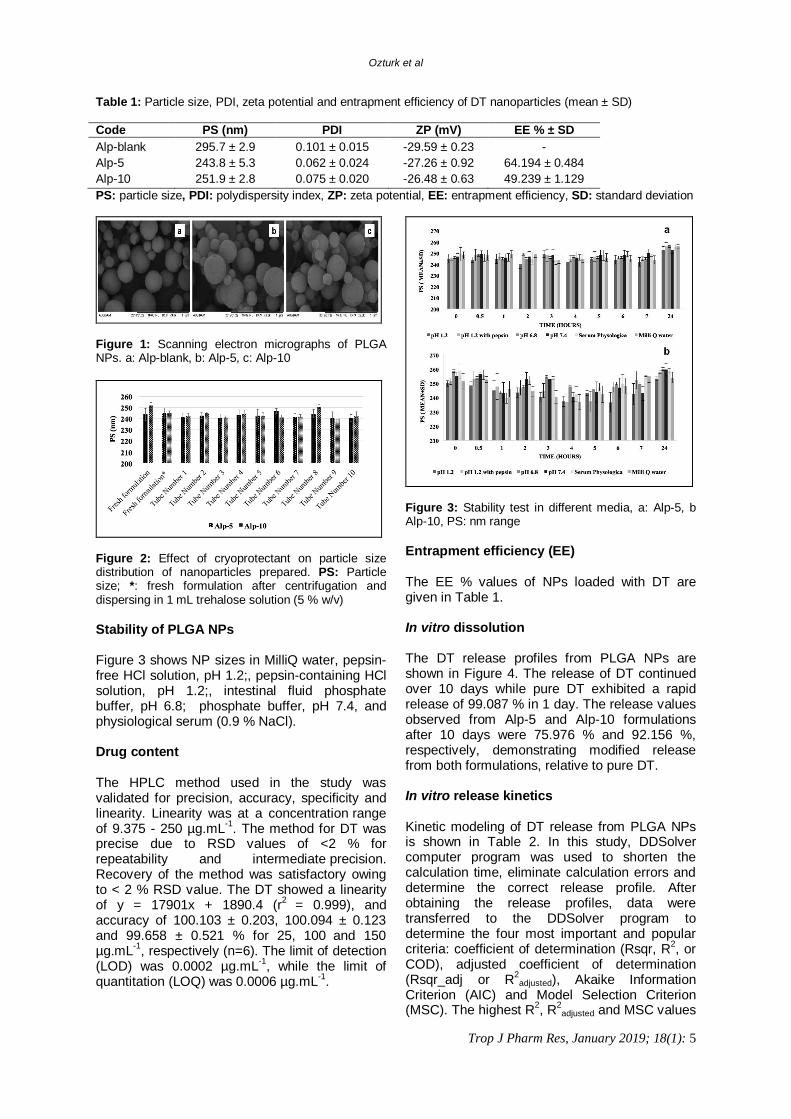
Figure 2: Effect of cryoprotectant on particle size distribution of nanoparticles prepared. PS: Particle size; *: fresh formulation after centrifugation and dispersing in 1 mL trehalose solution (5 % w/v) Stability of PLGA NPs Figure 3 shows NP sizes in MilliQ water, pepsin-free HCl solution, pH 1.2;, pepsin-containing HCl solution, pH 1.2;, intestinal fluid phosphate buffer, pH 6.8; phosphate buffer, pH 7.4, and physiological serum (0.9 % NaCl). Drug content The HPLC method used in the study was validated for precision, accuracy, specificity and linearity. Linearity was at a concentration range of 9.375 - 250 µg.mL-1. The method for DT was precise due to RSD values of <2 % for repeatability and intermediate precision. Recovery of the method was satisfactory owing to < 2 % RSD value. The DT showed a linearity of y = 17901x + 1890.4 (r2 = 0.999), and accuracy of 100.103 ± 0.203, 100.094 ± 0.123 and 99.658 ± 0.521 % for 25, 100 and 150 µg.mL-1, respectively (n=6). The limit of detection (LOD) was 0.0002 µg.mL-1, while the limit of quantitation (LOQ) was 0.0006 µg.mL-1.
Figure 3: Stability test in different media, a: Alp-5, b Alp-10, PS: nm range Entrapment efficiency (EE) The EE % values of NPs loaded with DT are given in Table 1. In vitro dissolution The DT release profiles from PLGA NPs are shown in Figure 4. The release of DT continued over 10 days while pure DT exhibited a rapid release of 99.087 % in 1 day. The release values observed from Alp-5 and Alp-10 formulations after 10 days were 75.976 % and 92.156 %, respectively, demonstrating modified release from both formulations, relative to pure DT. In vitro release kinetics Kinetic modeling of DT release from PLGA NPs is shown in Table 2. In this study, DDSolver computer program was used to shorten the calculation time, eliminate calculation errors and determine the correct release profile. After obtaining the release profiles, data were transferred to the DDSolver program to determine the four most important and popular criteria: coefficient of determination (Rsqr, R2, or COD), adjusted coefficient of determination (Rsqr_adj or R2
adjusted), Akaike Information Criterion (AIC) and Model Selection Criterion (MSC). The highest R2, R2
adjusted and MSC values
Ozturk et al
Trop J Pharm Res, January 2019; 18(1): 6
and the lowest AIC values were used for evaluating zero-order kinetics, first-order kinetics, Higuchi, and Korsmeyer-Peppas models [18]. The R2, R2
adjusted, MSC and AIC obtained are shown in Table 3.
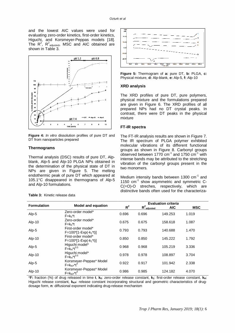
Figure 4: In vitro dissolution profiles of pure DT and DT from nanoparticles prepared Thermograms Thermal analysis (DSC) results of pure DT, Alp-blank, Alp-5 and Alp-10 PLGA NPs obtained in the determination of the physical state of DT in NPs are given in Figure 5. The melting endothermic peak of pure DT which appeared at 105.1°C disappeared in thermograms of Alp-5 and Alp-10 formulations.
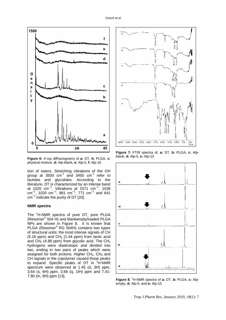
Figure 5: Thermogram of a: pure DT, b: PLGA, c: Physical mixture, d: Alp-blank, e: Alp-5, f: Alp-10 XRD analysis The XRD profiles of pure DT, pure polymers, physical mixture and the formulations prepared are given in Figure 6. The XRD profiles of all prepared NPs had no DT crystal peaks. In contrast, there were DT peaks in the physical mixture FT-IR spectra The FT-IR analysis results are shown in Figure 7. The IR spectrum of PLGA polymer exhibited molecular vibrations of its different functional groups as shown in Figure 8. Carbonyl groups observed between 1770 cm-1 and 1750 cm-1 with intense bands may be attributed to the stretching vibration of the carbonyl groups present in the two monomers. Medium intensity bands between 1300 cm-1 and 1150 cm-1 show asymmetric and symmetric C-C(=O)-O streches, respectively, which are distinctive bands often used for the characteriza-
Table 3: Kinetic release data Formulation Model and equation Evaluation criteria
R2 R2adjusted AIC MSC
Alp-5 Zero-order model* F=k0*t
0.696 0.696 149.253 1.019
Alp-10 Zero-order model* F=k0*t
0.675 0.675 158.618 1.087
Alp-5 First-order model* F=100*[1-Exp(-k1*t)]
0.793 0.793 140.688 1.470
Alp-10 First-order model* F=100*[1-Exp(-k1*t)] 0.850 0.850 145.222 1.792
Alp-5 Higuchi model* F=kH*t0.5 0.968 0.968 105.219 3.336
Alp-10 Higuchi model* F=kH*t0.5 0.978 0.978 108.897 3.704
Alp-5 Korsmeyer-Peppas* Model F=kKP*tn 0.922 0.917 101.942 2.338
Alp-10 Korsmeyer-Peppas* Model F=kKP*tn 0.986 0.985 124.182 4.070
*F: fraction (%) of drug released in time t, k0: zero-order release constant, k1: first-order release constant, kH: Higuchi release constant, kKP: release constant incorporating structural and geometric characteristics of drug-dosage form, n: diffusional exponent indicating drug-release mechanism
Ozturk et al
Trop J Pharm Res, January 2019; 18(1): 7
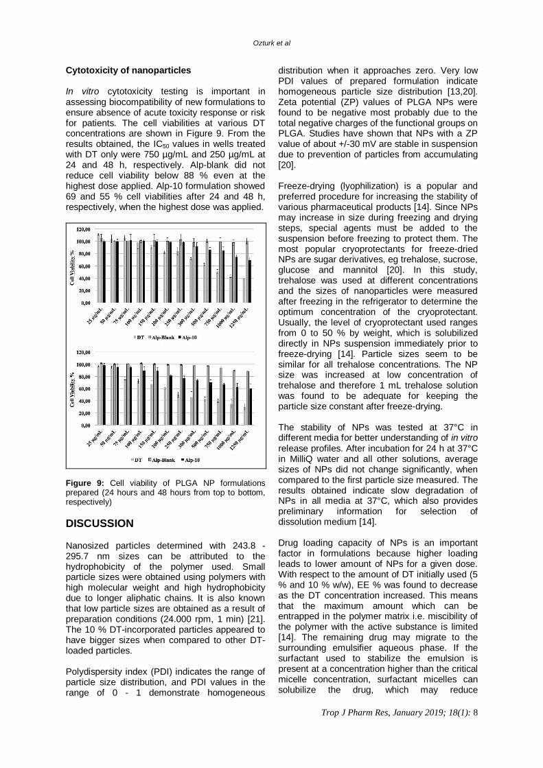
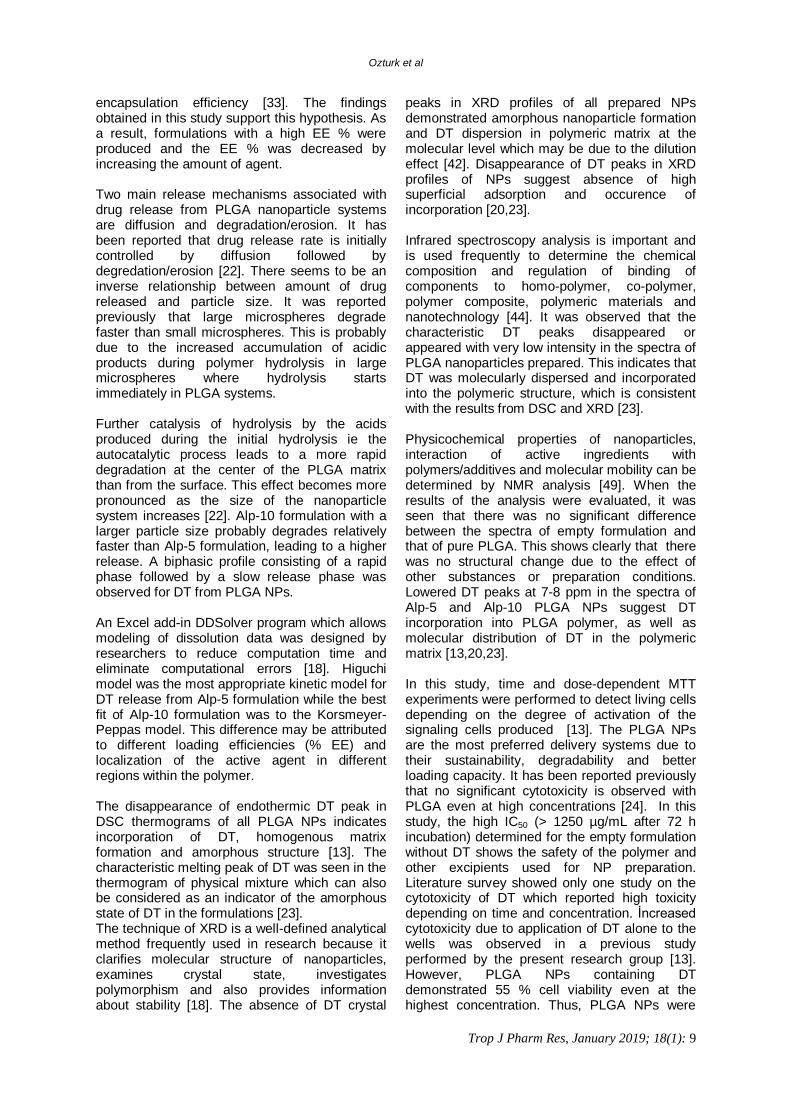
Figure 6: X-ray diffractograms of a: DT, b: PLGA, c: physical mixture, d: Alp-blank, e: Alp-5, f: Alp-10 tion of esters. Stretching vibrations of the OH group at 3500 cm-1 and 3450 cm-1 refer to lactides and glycolides. According to the literature, DT is characterized by an intense band at 1020 cm−1. Vibrations at 1571 cm−1, 1536 cm−1, 1020 cm−1, 881 cm−1, 771 cm−1 and 641 cm−1 indicate the purity of DT [20]. NMR spectra The 1H-NMR spectra of pure DT, pure PLGA (Resomer® 504 H) and blankempty/loaded PLGA NPs are shown in Figure 8. It is known that PLGA (Resomer® RG 504H) contains two types of structural units: the most intense signals of CH (5.16 ppm) and CH3 (1.44 ppm) from lactic acid and CH2 (4.88 ppm) from glycolic acid. The CH2 hydrogens were diastrotopic and divided into two, ending in two pairs of peaks which were assigned for both protons. Higher CH2, CH3 and CH signals in the copolymer caused those peaks to expand. Specific peaks of DT in 1H-NMR spectrum were observed at 1.45 (d, 3H) ppm, 3.64 (s, 6H) ppm, 3.66 (q, 1IH) ppm and 7.41-7.80 (m, 9H) ppm [13].
Figure 7: FTIR spectra of, a: DT, b: PLGA, c: Alp-blank, d: Alp-5, e: Alp-10
Figure 8. 1H-NMR spectra of a: DT, b: PLGA, c: Alp- empty, d: Alp-5, and e: Alp-10
Ozturk et al
Trop J Pharm Res, January 2019; 18(1): 8
Cytotoxicity of nanoparticles In vitro cytotoxicity testing is important in assessing biocompatibility of new formulations to ensure absence of acute toxicity response or risk for patients. The cell viabilities at various DT concentrations are shown in Figure 9. From the results obtained, the IC50 values in wells treated with DT only were 750 µg/mL and 250 µg/mL at 24 and 48 h, respectively. Alp-blank did not reduce cell viability below 88 % even at the highest dose applied. Alp-10 formulation showed 69 and 55 % cell viabilities after 24 and 48 h, respectively, when the highest dose was applied.
Figure 9: Cell viability of PLGA NP formulations prepared (24 hours and 48 hours from top to bottom, respectively) DISCUSSION Nanosized particles determined with 243.8 - 295.7 nm sizes can be attributed to the hydrophobicity of the polymer used. Small particle sizes were obtained using polymers with high molecular weight and high hydrophobicity due to longer aliphatic chains. It is also known that low particle sizes are obtained as a result of preparation conditions (24.000 rpm, 1 min) [21]. The 10 % DT-incorporated particles appeared to have bigger sizes when compared to other DT-loaded particles. Polydispersity index (PDI) indicates the range of particle size distribution, and PDI values in the range of 0 - 1 demonstrate homogeneous
distribution when it approaches zero. Very low PDI values of prepared formulation indicate homogeneous particle size distribution [13,20]. Zeta potential (ZP) values of PLGA NPs were found to be negative most probably due to the total negative charges of the functional groups on PLGA. Studies have shown that NPs with a ZP value of about +/-30 mV are stable in suspension due to prevention of particles from accumulating [20]. Freeze-drying (lyophilization) is a popular and preferred procedure for increasing the stability of various pharmaceutical products [14]. Since NPs may increase in size during freezing and drying steps, special agents must be added to the suspension before freezing to protect them. The most popular cryoprotectants for freeze-dried NPs are sugar derivatives, eg trehalose, sucrose, glucose and mannitol [20]. In this study, trehalose was used at different concentrations and the sizes of nanoparticles were measured after freezing in the refrigerator to determine the optimum concentration of the cryoprotectant. Usually, the level of cryoprotectant used ranges from 0 to 50 % by weight, which is solubilized directly in NPs suspension immediately prior to freeze-drying [14]. Particle sizes seem to be similar for all trehalose concentrations. The NP size was increased at low concentration of trehalose and therefore 1 mL trehalose solution was found to be adequate for keeping the particle size constant after freeze-drying. The stability of NPs was tested at 37°C in different media for better understanding of in vitro release profiles. After incubation for 24 h at 37°C in MilliQ water and all other solutions, average sizes of NPs did not change significantly, when compared to the first particle size measured. The results obtained indicate slow degradation of NPs in all media at 37°C, which also provides preliminary information for selection of dissolution medium [14]. Drug loading capacity of NPs is an important factor in formulations because higher loading leads to lower amount of NPs for a given dose. With respect to the amount of DT initially used (5 % and 10 % w/w), EE % was found to decrease as the DT concentration increased. This means that the maximum amount which can be entrapped in the polymer matrix i.e. miscibility of the polymer with the active substance is limited [14]. The remaining drug may migrate to the surrounding emulsifier aqueous phase. If the surfactant used to stabilize the emulsion is present at a concentration higher than the critical micelle concentration, surfactant micelles can solubilize the drug, which may reduce
Ozturk et al
Trop J Pharm Res, January 2019; 18(1): 9
encapsulation efficiency [33]. The findings obtained in this study support this hypothesis. As a result, formulations with a high EE % were produced and the EE % was decreased by increasing the amount of agent. Two main release mechanisms associated with drug release from PLGA nanoparticle systems are diffusion and degradation/erosion. It has been reported that drug release rate is initially controlled by diffusion followed by degredation/erosion [22]. There seems to be an inverse relationship between amount of drug released and particle size. It was reported previously that large microspheres degrade faster than small microspheres. This is probably due to the increased accumulation of acidic products during polymer hydrolysis in large microspheres where hydrolysis starts immediately in PLGA systems. Further catalysis of hydrolysis by the acids produced during the initial hydrolysis ie the autocatalytic process leads to a more rapid degradation at the center of the PLGA matrix than from the surface. This effect becomes more pronounced as the size of the nanoparticle system increases [22]. Alp-10 formulation with a larger particle size probably degrades relatively faster than Alp-5 formulation, leading to a higher release. A biphasic profile consisting of a rapid phase followed by a slow release phase was observed for DT from PLGA NPs. An Excel add-in DDSolver program which allows modeling of dissolution data was designed by researchers to reduce computation time and eliminate computational errors [18]. Higuchi model was the most appropriate kinetic model for DT release from Alp-5 formulation while the best fit of Alp-10 formulation was to the Korsmeyer-Peppas model. This difference may be attributed to different loading efficiencies (% EE) and localization of the active agent in different regions within the polymer. The disappearance of endothermic DT peak in DSC thermograms of all PLGA NPs indicates incorporation of DT, homogenous matrix formation and amorphous structure [13]. The characteristic melting peak of DT was seen in the thermogram of physical mixture which can also be considered as an indicator of the amorphous state of DT in the formulations [23]. The technique of XRD is a well-defined analytical method frequently used in research because it clarifies molecular structure of nanoparticles, examines crystal state, investigates polymorphism and also provides information about stability [18]. The absence of DT crystal
peaks in XRD profiles of all prepared NPs demonstrated amorphous nanoparticle formation and DT dispersion in polymeric matrix at the molecular level which may be due to the dilution effect [42]. Disappearance of DT peaks in XRD profiles of NPs suggest absence of high superficial adsorption and occurence of incorporation [20,23]. Infrared spectroscopy analysis is important and is used frequently to determine the chemical composition and regulation of binding of components to homo-polymer, co-polymer, polymer composite, polymeric materials and nanotechnology [44]. It was observed that the characteristic DT peaks disappeared or appeared with very low intensity in the spectra of PLGA nanoparticles prepared. This indicates that DT was molecularly dispersed and incorporated into the polymeric structure, which is consistent with the results from DSC and XRD [23]. Physicochemical properties of nanoparticles, interaction of active ingredients with polymers/additives and molecular mobility can be determined by NMR analysis [49]. When the results of the analysis were evaluated, it was seen that there was no significant difference between the spectra of empty formulation and that of pure PLGA. This shows clearly that there was no structural change due to the effect of other substances or preparation conditions. Lowered DT peaks at 7-8 ppm in the spectra of Alp-5 and Alp-10 PLGA NPs suggest DT incorporation into PLGA polymer, as well as molecular distribution of DT in the polymeric matrix [13,20,23]. In this study, time and dose-dependent MTT experiments were performed to detect living cells depending on the degree of activation of the signaling cells produced [13]. The PLGA NPs are the most preferred delivery systems due to their sustainability, degradability and better loading capacity. It has been reported previously that no significant cytotoxicity is observed with PLGA even at high concentrations [24]. In this study, the high IC50 (> 1250 µg/mL after 72 h incubation) determined for the empty formulation without DT shows the safety of the polymer and other excipients used for NP preparation. Literature survey showed only one study on the cytotoxicity of DT which reported high toxicity depending on time and concentration. İncreased cytotoxicity due to application of DT alone to the wells was observed in a previous study performed by the present research group [13]. However, PLGA NPs containing DT demonstrated 55 % cell viability even at the highest concentration. Thus, PLGA NPs were
Ozturk et al
Trop J Pharm Res, January 2019; 18(1): 10
able to prevent cytotoxicity at high DT concentrations. CONCLUSION This study shows that DT loaded PLGA NPs can be prepared successfully by double emulsion solvent evaporation method. The particle size of the NPs obtained were in the nanometer range and remained constant in the short-term GI stability testing. The nanoparticle formulation did not show significant cytotoxicity at dose- and time-dependent considerations. Thus, it is considered appropriate for drug delivery purposes. The DT-loaded PLGA NPs are stable and represent a promising system for sustained and controlled delivery of DT. DECLARATIONS Acknowledgement This study was financed by Anadolu University Scientific Research Project Foundation (no. 1708S471). The authors would like to thank Abdi İbrahim (İstanbul, Turkey) for kindly providing DT. Faculty of Science is acknowledged for XRD, DOPNALAB Faculty of Pharmacy for FTIR and 1H-NMR and AUBIBAM for SEM analyses. We also thank University of Seville, Faculty of Pharmacy, Department of Pharmacy and Pharmaceutical Technology for making available their facilities for this work. Conflict of Interest No conflict of interest associated with this work. Contribution of Authors The authors declare that this work was done by the authors named in this article and all liabilities pertaining to claims relating to the content of this article will be borne by them. REFERENCES 1. Breivik H, Collett B, Ventafridda V. Survey of Chronic
Pain in Europe: Prevalence, Impact on Daily Life, and Treatment. Eur J Pain 2006; 10: 287-333.
2. Romano CL, Romano D, Lacerenza M. Antineuropathic and Antinociceptive Drugs Combination in Patients with Chronic Low Back Pain: A Systematic Review. Pain Res Treat 2012: 154781.
3. Jin J. Nonsteroidal Anti-inflammatory Drugs. JAMA 2015; 314 (10): 1084.
4. Mainardi F, Maggioni F, Pezzola D, Zava D, Zanchiny G. Dexketoprofen Trometamol in the Acute Treatment of
Migraine Attack: A Phase II, Randomized, Double-Blind, Crossover, Placebo-Controlled, Dose Optimization Study. J Pain 2014; 15(4): 388-394.
5. Öztürk AA, Yenilmez E, Yazan Y. Development and Validation of High Performance Liquid Chromatography (HPLC) Modified Method for Dexketoprofen Trometamol. Eur Int J Sci Tech 2017; 6(5): 33-41.
6. Kapse SV, Gaikwad RV, Samad A, Devarajan PV. Self Nanoprecipitating Preconcentrate of Tamoxifen Citrate for Enhanced Bioavailability. Int J Pharm 2012; 429: 104-112.
7. Kumari A, Yadav SK, Yadav SC. Biodegradable Polymeric Nanoparticles Based Drug Delivery Systems. Colloids Surf B Biointerfaces 2010; 75: 1-18.
8. Hines DJ, Kaplan DL. Poly (Lactic-Co-Glycolic Acid) Controlled Release Systems: Experimental and Modeling Insights. Crit Rev Ther Drug Carrier Syst 2013; 30(3): 257-276.
9. Derman S, Kızılbey K, Akdeste ZM. Polymeric Nanoparticles. J Eng Nat Sci 2013; 31: 107-120.
10. Iqbal M, Zafar N, Fessi H, Elaissari A. Double Emulsion Solvent Evaporation Techniques Used for Drug Encapsulation. Int J Pharm 2015; 496: 173-190.
11. Yong H, Yonglin W, Lin Z, Yu LY, Yongjun Li, Lingyun S, Feng H, Zhirong Z, Hui C. Preparation Method of Dexketoprofen Trometamol Double-Layer Sustained-Release Tablets. 2012; Patent Number: CN 102813638 A.
12. Yong H, Yonglin W, Lingyun S, Lin Z, Feng H, Yanyu L, Yongjun L, Aimin W, Shanggao L, Xiaozhong F, Wen Z, Zhirong Z, Hui C. Dexketoprofen Trometamol Quick-Release/Sustained-Release Double-Layer Tablet and Preparation Method Thereof. 2014; Patent Number: CN 103655504 A.
13. Öztürk AA, Yenilmez E, Arslan R, Şenel B, Yazan Y. Dexketoprofen Trometamol-Loaded Kollidon® SR and Eudragit® RS 100 Polymeric Nanoparticles: Formulation and In Vitro-In Vivo Evaluation. Lat Am J Pharm 2017; 36(11): 2153-2165.
14. Martín-Banderas L, Alvarez-Fuentes J, Duran-Lobato M, Prados J, Melguizo C, Fernandez-Arvelo M, Holgado MaA. Cannabinoid Derivate-Loaded PLGA Nanocarriers for Oral Administration: Formulation, Characterization, and Cytotoxicity Studies. Int J Nanomed 2012; 7: 5793-5806.
15. Abdelwahed W, Degobert G, Stainmesse S, Fessi H. Freeze-drying of Nanoparticles: Formulation, Process and Storage Considerations. Adv Drug Deliv Rev 2006; 58(15):1688-1713
16. Bhusari VK, Dhaneshwar SR. Development of a Validated Stability-Indicating HPLC Assay Method for Dexketoprofen Trometamol. Int J Pharm Pharm Sci 2012; 4 (1): 321-326.
17. Obitte NC, Chukwu A, Onyishi IV. The Use of a pH-Dependent and Non pH-Dependent Natural Hydrophobic Biopolymer (Landolphia owariensis latex) as Capsule Coating Agents in In Vitro Controlled
Ozturk et al
Trop J Pharm Res, January 2019; 18(1): 11
Release of Metronidazole for Possible Colon Targeted Delivery. Int J Appl Res Nat Prod 2010; 3(1): 1-17.
18. Zhang Y, Huo M, Zhou J, Zou A, Li W, Yao C, Xie S. DDSolver: an Add-In Program for Modeling and Comparison of Drug Dissolution Profiles. The AAPS J 2010; 12: 263-271.
19. Gencer S, Cebeci A, Irmak-Yazicioglu MB. Silencing of the MMP-3 Gene by siRNA Transfection in Gastric Cancer AGS Cells. J Gastrointestin Liver Dis 2010; 20: 19-26
20. Öztürk AA, Martin Banderas L, Cayero Otero MD, Yenilmez E, Yazan Y. New Approach to Hypertension Treatment: Carvediol-Loaded PLGA Nanoparticles, Preparation, In Vitro Characterization and Gastrointestinal Stability. Lat Am J Pharm. 2018; 37(9): 1730-1741
21. Palacio J, Orozco VH, Lopez BL. Effect of the Molecular Weight on the Physicochemical Properties of Poly(Lactic
Acid) Nanoparticles and on the Amount of Ovalbumin Adsorption. J Braz Chem Soc 2011; 22(12): 2304-2311.
22. Fredenberg S, Wahlgren M, Reslow M, Axelsson A. The Mechanisms of Drug Release in Poly(Lactic-Co-Glycolic Acid)-Based Drug Delivery Systems- A Review. Int J Pharm 2011; 15(1-2): 34-52.
23. Öztürk AA, Güven UM, Yenı̇lmez E, Şenel B. Effects of Different Derivatives of Eudragit Polymer on Entrapment Efficiency, In Vitro Dissolution, Release Kinetics and Cell Viability Results on Extended Release Flurbiprofen Loaded Nanomedicines. Lat Am J Pharm 2018; 37(10): 1981-1992.
24. Jain DS, Athawale RB, Bajaj AN. Unraveling the Cytotoxic Potential of Temozolomide Loaded into PLGA Nanoparticles. DARU J Pharm Sci 2014; 22(1): 18. doi:10.1186/2008-2231-22-18.