towards accelerated bone regeneration by altering poly(d,l-lactic- co-glycolic) acid porogen content...
TRANSCRIPT

Towards accelerated bone regeneration by altering poly(D,L-lactic-
co-glycolic) acid porogen content in calcium phosphate cement
C.I.A. van Houdta, R.S. Preethanath
a,c, B.A.J.A. van Oirschot
a, P.H.W. Zwarts
a, D.J.O. Ulrich
b, S.
Anilc, J.A. Jansen
a, J.J.J.P. van den Beucken
a,*
a Dept. Biomaterials, Radboudumc, Nijmegen, the Netherlands
b Dept. Plastic and Reconstructive Surgery, Radboudumc, Nijmegen, The Netherlands
c Dept. Periodontics and Community Dentistry, King Saud University, Riyadh, Saudi Arabia
*Corresponding author: Dept. Biomaterials (309), Radboudumc, PO Box 9101, 6500 HB Nijmegen, the
Netherlands
This article has been accepted for publication and undergone full peer review but has not beenthrough the copyediting, typesetting, pagination and proofreading process which may lead todifferences between this version and the Version of Record. Please cite this article as an‘Accepted Article’, doi: 10.1002/jbm.a.35584
This article is protected by copyright. All rights reserved.

Accelerated bone regeneration by altering porogen content
2
Abstract
This work aimed to compare in vitro degradation of dense PLGA microspheres and milled
PLGA particles as porogens within CPC, considering that the manufacturing of milled PLGA is
more cost-effective compared to PLGA microspheres. Additionally, we aimed to examine the
effect of porogen amount within CPC/PLGA on degradation and bone formation. Our in vitro
results showed no differences between both forms of PLGA particles (as porogens in CPC;
spherical for microspheres, irregular for milled) regarding morphology, porosity and
degradation. Using milled PLGA as porogens within CPC/PLGA, we evaluated the effect of
porogen amount on degradation and bone forming capacity in vivo. Titanium landmarks
surrounded by CPC/PLGA with 30 and 50 wt% PLGA, were implanted in forty femoral bone
defects of twenty male Wistar rats. Histomorphometrical results showed a significant
temporal decrease in the amount of CPC, for both formulas, and confirmed that 50 wt%
PLGA degrades faster than 30 wt%, and allows for a 1.5-fold higher amount of newly formed
bone. Taken together, this study demonstrated that (i) milled PLGA particles perform equal
to PLGA microspheres, and (ii) tuning of the PLGA content in CPC/PLGA is a feasible
approach to leverage material degradation and bone formation.
Keywords: bone substitute, calcium phosphate cement, poly(D,L-lactic-co-glycolic) acid,
material degradation, bone regeneration
Page 2 of 37
John Wiley & Sons, Inc.
Journal of Biomedical Materials Research: Part A
This article is protected by copyright. All rights reserved.
Accelerated bone regeneration by altering porogen content
3
Introduction
Synthetic bone substitutes are widely accepted materials in the field of bone reconstruction
to promote bone regeneration and replace autologous bone grafting. Commercially available
synthetic bone substitutes are available in various forms and shapes, adapted to the specific
needs of different bone defects.1, 2
Synthetic bone substitutes can be subdivided into three
material classes, i.e. polymers, ceramics and composites.1 Ceramics, especially those based
on calcium phosphates (CaPs), are favourable materials due to their biocompatible, bioactive
and osteoconductive properties resulting from a chemical composition that is similar to the
mineral phase of bone.3-7
In view of optimized handling properties, CaP ceramics have
become available in the form of injectable CaP cement (CPC).8 First proposed in the early
80s, CPCs were designed to overcome the handling difficulties of the traditional ceramic
blocks and granules.9, 10
Depending on the end-product after setting, CPCs can be classified into two main groups, i.e.
brushite and apatitic CPCs. Brushite CPCs have the ability to be resorbed under physiological
conditions, whereas apatitic cements degrade relatively poorly, limiting new bone
formation.11
Resorption of CPC occurs either via active resorption (a cell-mediated process of
osteoclasts and macrophages) or via passive resorption (based on the cement solubility).12-15
Passive resorption is mainly influenced by porosity and cement composition, i.e. apatite
being less soluble than brushite. Brushite CPCs, however, have low mechanical properties
and handling can be difficult due to a fast setting time (~30 s).11
The favourable mechanical
and handling properties of apatitic cements lead research to focus on improving their
degradation properties.
Ideally, the rate of material degradation is equal to the rate of bone formation gradually
providing room for bone to grow.16
Balancing degradation rate to new bone formation rate
Page 3 of 37
John Wiley & Sons, Inc.
Journal of Biomedical Materials Research: Part A
This article is protected by copyright. All rights reserved.

Accelerated bone regeneration by altering porogen content
4
is an important material property to control. Accelerated degradation of CPC has been
achieved via addition of porogens.17, 18
Generally, macropores (pores > 100 µm) are used to
increase the surface of the CPC leading to a faster degradation. The term “macropores” is
used to distinguish between the pores from the scaffold’s own micro- or nanoporosity.18
Macropores are mainly created by leaching or degradation of incorporated particles, gas
foaming, and droplet emulsion.9, 17, 19-24
Poly(D,L-lactic-co-glycolic) acid (PLGA) is a rapidly degradable synthetic polymer and hence
interesting to use as porogen in CPC. PLGA has a long clinical history and already several
PLGA incorporated medical devices have been approved by the FDA for clinical use.25
In vivo
degradation of PLGA occurs via hydrolysis and is a relatively fast process. During
degradation, lactic acid and glycolic acid monomers are released, which are metabolized into
carbon dioxide and water and excreted by the body.25
Parameters influencing the
degradation of PLGA are molecular weight, end-group functionalisation (acid terminated or
end capped) and ratio of lactic and glycolic.7, 26-31
Previously, it was shown that PLGA
microspheres enhance the degradation of apatitic CPC and improve bone regeneration.26, 27,
32, 33 When incorporated into CPC, the degradation is influenced by the particle size,
morphology (hollow or dense spheres), and relative amount of PLGA.7, 26-31
PLGA porogens can be fabricated following several methods, such as water-in-oil-in-water
emulsion or oil-in-water emulsion.25
These chemical processes are associated with an
acceptable yield (i.e. 60-80%), but such loss of product during synthesis and the time
consuming nature are impeding large-scale utilization. Alternatively, PLGA milling can be
applied to generate PLGA porogens.32, 33
In this way, irregularly-shaped PLGA particles are
obtained relatively fast and in a cost-effective manner. In summary, with these novel milled
Page 4 of 37
John Wiley & Sons, Inc.
Journal of Biomedical Materials Research: Part A
This article is protected by copyright. All rights reserved.

Accelerated bone regeneration by altering porogen content
5
PLGA particles as porogens in CPC, a relatively low cost additive can be used to control CPC
degradation and simultaneously stimulate bone regeneration.
The aim of this study was to comparatively evaluate the in vitro degradation effects of dense
PLGA microspheres with milled PLGA particles as porogens in apatitic CPC. We hypothesized
that milled PLGA particles would degrade CPC/PLGA similarly when compared to dense PLGA
microspheres. Additionally, we aimed to examine the effect of PLGA porogen amount in
CPC/PLGA via an in vivo rat femoral bone defect model to determine degradation and bone
formation. For this, we hypothesized that a higher PLGA porogen amount would increase the
porosity, leading to faster degradation and consequently more bone formation in vivo.
Page 5 of 37
John Wiley & Sons, Inc.
Journal of Biomedical Materials Research: Part A
This article is protected by copyright. All rights reserved.
Accelerated bone regeneration by altering porogen content
6
Materials and methods
Materials
The CPC used for both in vitro and in vivo experiments consisted of 100% α-TCP (kindly
provided by CAM Bioceramics B.V., Leiden, the Netherlands) sieved at <150 µm.
Carboxymethyl cellulose (CMC; 0.75 wt%) was added to the CPC to improve injectability.
PLGA (Purasorb®, PDLG 5002A, Mw = 17kDa, acid-terminated) was obtained from Corbion
B.V. (Gorinchem, the Netherlands). Milled PLGA (PDLG 5002A, acid-terminated) was kindly
provided by CAM Bioceramics B.V. Custom-made cylindrical thumbtack-shaped titanium
implants (grade 2) were obtained from Machinefabriek G. Jansen B.V. (Valkenswaard, the
Netherlands).
Preparation of dense PLGA microspheres
Dense PLGA microspheres, used for in vitro analysis, were fabricated by an emulsion
technique as described previously.30
Briefly, PLGA was dissolved in 12 ml of dichloromethane
(DCM, Merck, Germany). The solution was decanted over three beakers containing 150 ml of
0.3% polyvinyl alcohol (PVA, Acros Organics, the Netherlands). This second emulsion was
stirred (800 rpm) for 5 min and then 200 ml of 2% isopropanol (Labscan Ltd., the
Netherlands) was added. The suspension was stirred for 1 h, after which the PLGA
microspheres were allowed to precipitate. Thereafter, the supernatant was decanted and
the sediment was washed twice with ultrapure water. The microspheres were lyophilized
and stored at −20 °C.
Preparation of CPC/PLGA scaffolds (for in vitro analysis)
Page 6 of 37
John Wiley & Sons, Inc.
Journal of Biomedical Materials Research: Part A
This article is protected by copyright. All rights reserved.

Accelerated bone regeneration by altering porogen content
7
The α-TCP, CMC, and PLGA porogen were combined to obtain formulations with either 30 or
50 wt% PLGA (Table 1). Per 1 g of powder, a solution of 500 µl 4% NaH2PO4 was added. For
the preparation of the scaffolds, a Teflon® mould was used with holes of 4.5 mm in diameter
and 9.2 mm in depth. During injection into the Teflon® mould, attention was paid to
completely fill the space and any air being trapped under the cement was removed. The
cement was left to air dry at room temperature for a minimum of 24 h.
Preparation of titanium-CPC/PLGA scaffolds (for in vivo analysis)
Titanium implants (thumbtack-shape; length 6 mm, base diameter 3 mm, core diameter 1
mm), were cleaned using a tabletop ultrasonic cleaner via consecutive rinsing with acetone
and alcohol, and then air dried. Then, the titanium implants were placed into a Teflon®
mould, with holes of 3 mm in diameter and 6 mm in length.
CPC/PLGA was mixed with 500 µl of 4% NaH2PO4 solution per 1 g powder and injected with
an 18 G needle into the space around the titanium implants. During injection, attention was
paid to completely fill the space and any air being trapped under the cement was removed.
Then, the cement was left to air dry at room temperature for a minimum of 24 h, after which
the implants were gently removed from the mould. Dimensions of the Ti-CPC/PLGA implants
are depicted in Figure 1. Finally, sterilization was performed by gamma irradiation (25-50
kGy; SynergyHealth Ede B.V., Ede, the Netherlands).
In vitro material characterization
PLGA porogens
The size of the PLGA microspheres was measured by optical microscopy (DM RBE Leica,
Germany) with image analysis software (Qwin Leica, Germany).
Page 7 of 37
John Wiley & Sons, Inc.
Journal of Biomedical Materials Research: Part A
This article is protected by copyright. All rights reserved.

Accelerated bone regeneration by altering porogen content
8
CPC/PLGA scaffolds
After removal from the mould, scaffolds were each placed in 1.5 ml of phosphate buffered
saline (PBS) and left on a shaking apparatus at 37 oC.
Scaffold morphology
A scanning electron microscope (SEM; JEOL JSM 6340, 10 kV) was used for morphological
analysis. For each material group, samples were scanned after 4 weeks and 8 weeks of
incubation.
Porosity calculations and measurements
The macroporosity of the different CPC/PLGA experimental formulas (n = 3) was determined
as previously described by Habraken et al. (2006).34
The equations used to calculate the total
porosity and macroporosity are as shown in Figure 2.
Degradation and pH changes
Degradation of the scaffolds was determined by analyzing Ca2+
release into the PBS,
measured at the end of each week, for 8 consecutive weeks. The absolute amounts of Ca2+
-
ions in the PBS media were analyzed by the orthocresolphtalein complexone (OCPC) method
(Sigma-Aldrich Chemie B.V., Zwijndrecht, the Netherlands) as described previously by Lopez-
Heredia et al. (2012).30
Additionally, the pH of the PBS media was monitored, using pH-
indicator strips, at the same weekly time points.
In vivo analysis
Animals
Twenty healthy mature male Wistar rats were used for this experiment at 3 months of age
with an average weight of 250 g at the start of the experiment. An acclimatization period of
7 d was used and housing was provided per pair in standard macrolon type 3 cages with
Page 8 of 37
John Wiley & Sons, Inc.
Journal of Biomedical Materials Research: Part A
This article is protected by copyright. All rights reserved.

Accelerated bone regeneration by altering porogen content
9
sawdust as bedding material. Standard rodent chow and bottled tap water was provided ad
libitum. The housing room was maintained under standard laboratory conditions (light-dark
cycle: 12:12 h, temperature: 20-22 °C, relative humidity: 45-55%). All experiments were
conducted in accordance with institutional, national and international guidelines for animal
care and the Dutch law concerning animal welfare. The studies were reviewed and approved
by the Experimental Animal Committee of the Radboud University (RUDEC 2012-243).
Surgical procedure
A rat femoral bone defect model was used, as described previously by Bongio et al. (2015).35
Pre-operatively, pain medication was provided by an injection of Carprofen (5 mg/kg;
Rimadyl®, Pfizer Animal Health, New York, USA) given 15 min before surgery. Anaesthesia
was induced and maintained by Isoflurane inhalation (Rhodia Organique Fine Ltd.,
Avonmouth, Bristol, UK) combined with oxygen. After confirmation of effective anaesthesia,
the surgical area was shaved and disinfection was performed by application of a povidone
iodine solution. The rats were placed on a heating mat to prevent hypothermia and
immobilized in supine position. With the knee maximally flexed, a longitudinal incision
through skin and muscle was made on the medial surface. After exposure of the medial side
of the distal femoral condyle, the knee capsule was incised. Then, the knee was extended to
luxate the patella laterally. When a clear view of the knee joint was established, a femoral
bone defect (diameter 3 mm, depth 6 mm) was created in the same direction as the axis of
the femur, using a dental drill (Elcomed 9927 SPS; W&H Dentalwerk Burmoos GmbH,
Burmoos, Austria). A series of increasing bur diameters were used with the final drill of 3 mm
in diameter, at a speed of maximum 5000 rpm and constant cooling by dripping saline. The
drilled cavity was then washed with a spray of saline and dried using sterile gauze, after
Page 9 of 37
John Wiley & Sons, Inc.
Journal of Biomedical Materials Research: Part A
This article is protected by copyright. All rights reserved.
Accelerated bone regeneration by altering porogen content
10
which the pre-set titanium-CPC/PLGA scaffolds were placed into the defects. Each rat
received both implants (Ti-50/50- MP and Ti-70/30-MP), alternating between each rat the
two weigh percentages for the left and right knee. After introducing the implants into the
defects, the muscular tissue layer was closed with absorbable sutures (Vicryl® 4.0) after
which the skin was closed with small staples (Agraven®; InstruVet B.V., Cuijk, the
Netherlands).To diminish postoperative discomfort, Buprenorfine (0.02 mg/kg; Temgesic®
Reckitt Benckiser Pharmaceuticals Ltd., Slough, UK) was given every 12 h till 2 d after
surgery. All animals had free access to pellet food and water. In the initial postoperative
period, the intake of water and food was monitored daily as well as the weight of the
animals. In addition, the animals were observed for signs of pain, infection and proper
activity. Weekly weighing of each animal was continued until the end of the experiment.
Implant retrieval
After implantation periods of 4 and 8 weeks, the animals were euthanized by CO2-
suffocation. The retrieved specimens were harvested and fixed in 10% neutral buffered
formalin solution for 24 h, after which the specimens were dehydrated in a graded series of
ethanol.
Histological processing
All specimens were embedded in poly(methylmethacrylate) (pMMA). After polymerization,
three sections per specimen of ~10 μm were prepared perpendicular to the axis of the femur
using a diamond blade saw (Leica Microsystems SP 1600, Nussloch, Germany). Methylene
blue and basic fuchsine staining was performed for each section.
Page 10 of 37
John Wiley & Sons, Inc.
Journal of Biomedical Materials Research: Part A
This article is protected by copyright. All rights reserved.
Accelerated bone regeneration by altering porogen content
11
Descriptive histology and histomorphometry
Descriptive histology was performed by light microscopy examination (Leica Microsystems
AG, Wetzlar, Germany) comparing all histological sections. Staining allowed for
discrimination between soft tissue, bone tissue and CPC. Attention was paid to bone and
CPC morphology, and amounts of bone and CPC.
Histomorphometry for both implantation periods of 4 and 8 weeks, pMMA histological
sections (n = 3 per femoral bone defect) were assessed quantitatively using computer-based
image analysis techniques (Leica® Qwin Pro-image analysis system, Wetzlar, Germany). From
the digitalized images of the sections at a 5x magnification, a circular region of interest (ROI)
of 3 mm in diameter was selected to assign the defect area with the titanium pin as the
centre. Quantitative measurements within this ROI for area of newly formed bone and area
of CPC material, were performed using the Leica® Qwin computer-based image analysis
techniques. Vital bone could be distinguished by the pink colour and distinctive cell
morphology including a nucleus, allowing to be easily discerned from other tissues as well as
from CPC. The maturity of bone was differentiated by morphology, as mature trabecular
bone is formed in lamellae, whereas newly formed bone has a more woven structure.
Relative bone forming capacity is defined as relative increment in bone area per relative
decline in CPC area. Bone-to-implant contact (BIC) area was defined as the length around the
circumference of the titanium pin that is in contact with bone. The standardized diameter of
the defect and titanium pin allowed calculating areas of bone, CPC and BIC into percentages.
Statistical analysis
Data for new bone formation and CPC remnants were presented as means with standard
deviations (SD). The mean area percentages were statistically compared between the groups
Page 11 of 37
John Wiley & Sons, Inc.
Journal of Biomedical Materials Research: Part A
This article is protected by copyright. All rights reserved.

Accelerated bone regeneration by altering porogen content
12
using one-way ANOVA (GraphPad, Prism 5, GraphPad Software Inc., La Jolla, California USA)
with a post-hoc Tukey-Kramer Multiple Comparisons test. Differences were considered
significant at p < 0.05.
Page 12 of 37
John Wiley & Sons, Inc.
Journal of Biomedical Materials Research: Part A
This article is protected by copyright. All rights reserved.

Accelerated bone regeneration by altering porogen content
13
Results
Material characterization
PLGA porogen
Representative images of both PLGA porogens are depicted in Figure 3A-B, showing
spherically-shaped PLGA microspheres and irregularly-shaped milled particles. The mean
particle size was 39.95 ± 9.21 µm for the microspheres and 48.65 ± 22.07 µm for the milled
particles.
CPC/PLGA scaffolds
Scaffold morphology
SEM images revealed that at the start of the in vivo experiment, the PLGA microspheres and
milled PLGA particles were embedded within the CPC. Already after 4 weeks of incubation in
PBS, the PLGA porogens in all four groups were degraded and the CPC showed signs of
degradation (Figure 4A). At higher magnifications, precipitate formation in the form of
brushite crystals was detected (Figure 4B).
Porosity calculations
Porosity calculations showed that differences in porogen amount led to significant
differences in porosity, comparing 70/30-MS to 50/50-MS and 70/30-MP to 50/50-MP (both
p < 0.001; Table 2). Comparing the two types of porogens at the same wt% (i.e. 70/30-MS to
70/30-MP and 50/50-MS to 50/50-MP) the differences were not significant (p > 0.05; Table
2).
Degradation and pH changes
At all time points, the cumulative Ca2+
amount was significantly higher for the 70/30
CPC/PLGA formula compared to the 50/50 CPC/PLGA (Figure 4C). CPC/PLGA formulations
Page 13 of 37
John Wiley & Sons, Inc.
Journal of Biomedical Materials Research: Part A
This article is protected by copyright. All rights reserved.

Accelerated bone regeneration by altering porogen content
14
with the same amount of PLGA (microspheres or milled particles) showed similar Ca2+
release profiles (p > 0.05) with an accelerated release from 3 to 4 weeks. In contrast,
significantly higher (p < 0.05) Ca2+
release profiles were observed for both 50/50-MS and
50/50-MP compared to both 70/30-MS and 70/30-MP, at all time points.
For the pH-values of the incubation media (data not shown), the initial pH-value of 7.4
dropped to ~4 within the first week for all CPC/PLGA formulas. Subsequently, a further
gradual decrease of the pH was observed to ~3.3 after 8 weeks of incubation.
In vivo analysis
Clinical evaluation following surgery
All twenty rats recovered well from surgery and remained in good health during the course
of the experiment. None of the animals showed signs of illness or compromised wound
healing. Examination of the wound and implantation areas after euthanasia at either 4 or 8
weeks showed no inflammatory or other adverse tissue reaction. All installed implants were
retrieved and used for further analysis.
Descriptive histology
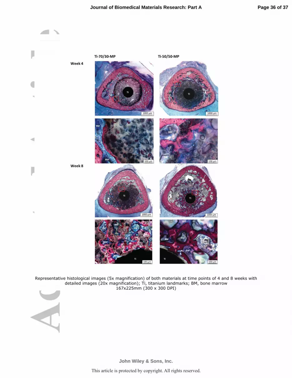
Representative histological overview images are provided in Figure 5, with additional
detailed images at 20x magnification. Newly formed bone could be macroscopically detected
at the periphery of the CPC after both 4 and 8 weeks of implantation for both Ti-70/30-MP
and Ti-50/50-MP. In all groups, this bone was in direct contact with the CPC, without
intervening soft tissue layers. Additionally, no signs of inflammatory responses were
observed for any of the groups. In several cases of each group, cracks were observed within
the CPC with bone tissue growing into these cracks.
Page 14 of 37
John Wiley & Sons, Inc.
Journal of Biomedical Materials Research: Part A
This article is protected by copyright. All rights reserved.

Accelerated bone regeneration by altering porogen content
15
At 4 weeks, no apparent differences in CPC degradation were observed between Ti-70/30-
MP and Ti-50/50-MP. At 8 weeks, however, a clear difference was observed, with Ti-50/50-
MP showing faster CPC degradation than Ti-70/30-MP. Within the newly formed bone, some
remnant CPC was detectable at a higher magnification for both Ti-70/30-MP and Ti-50/50-
MP. For specimens showing almost fully degraded CPC, the new bone present within the
defect area was trabecular-shaped with bone marrow between bone trabeculae.
Histomorphometry
CPC area (%)
The CPC area within the ROI showed a temporal decrease for both Ti-50/50-MP and Ti-
70/30-MP (Table 3 & Figure 6A). The area of CPC measured at 0 weeks were 81.0 ± 0.6% for
Ti-70/30-MP and 74.8 ± 0.7% for Ti-50/50-MP (p < 0.001). After 4 weeks of implantation, the
CPC area was 60.9 ± 13.5% for Ti-70/30-MP and 53.5 ± 17.3% for Ti-50/50-MP (p > 0.05).
After 8 weeks of implantation were 35.2 ± 21.2% for Ti-70/30-MP and 9.1 ± 9.7% for Ti-
50/50-MP (p < 0.001).
Comparing the different time points revealed a significant temporal decrease after the first 4
weeks in CPC area for Ti-50/50-MP (p < 0.05). After 8 weeks the CPC area was decreased
further, significantly for both Ti-70/30-MP (p < 0.01) and Ti-50/50-MP (p < 0.001). The
decrease was also significant from 4 to 8 weeks for both Ti-70/30-MP (p < 0.01) and Ti-
50/50-MP (p < 0.001).
Bone area (%)
Newly formed bone within the ROI increased over time for both Ti-70/30-MP and Ti-50/50-
MP (Table 3 & Figure 6B). After 4 weeks, the bone area within the ROI was 5.8 ± 5.0% for Ti-
70/30-MP and 8.3 ± 2.8% for Ti-50/50-MP (p > 0.05). After 8 weeks, the bone area revealed a
Page 15 of 37
John Wiley & Sons, Inc.
Journal of Biomedical Materials Research: Part A
This article is protected by copyright. All rights reserved.
Accelerated bone regeneration by altering porogen content
16
significant temporal increase (p < 0.001) for both experimental groups and a significant inter-
group difference (p < 0.05) with values of 22.9 ± 5.2% and 35.0 ± 15.0% for Ti-70/30-MP and
Ti-50/50-MP, respectively.
Bone forming capacity
For extended visualization of temporal decreases of CPC area and increases in bone area
(Figure 6C), calculations were made on the relative bone forming capacity (Table 4).
In the first 4 weeks, 1% CPC degradation resulted in 0.29% new bone for the Ti-70/30-MP
compared to 0.39% for the Ti-50/50-MP. From week 4 to 8, 1% CPC degradation allowed for
0.67% new bone for Ti-70/30-MP compared to 0.60% for Ti-50/50-MP. Overall, more bone
was formed within the ROI of Ti-50/50-MP over the time period of 0 to 8 weeks with a total
of 0.53% new bone per 1% CPC degradation for Ti-50/50-MP, compared to 0.50% for Ti-
70/30-MP.
Bone-to-implant contact (BIC%)
Bone-to-implant contact was calculated for both time periods for each experimental group
(Table 3 & Figure 6D). At 4 weeks, similar marginal BIC values (p > 0.05) of 2.4 ± 4.7% and 0.5
± 0.7% were found for Ti-70/30-MP and Ti-50/50-MP, respectively. At 8 weeks, BIC values for
both experimental groups showed a temporal increase of 11.7 ±14.7% for Ti-70/30-MP and
11.9 ±21.2% for Ti-50/50-MP. Between the groups the difference at both time points was
not significant. The increase of BIC% between 4 and 8 weeks was significant for Ti-70/30-MP
(p = 0.0371), but not for Ti-50/50-MP (p = 0.0536).
Page 16 of 37
John Wiley & Sons, Inc.
Journal of Biomedical Materials Research: Part A
This article is protected by copyright. All rights reserved.

Accelerated bone regeneration by altering porogen content
17
Discussion
The aim of this study was to comparatively evaluate the in vitro degradation effects of dense
PLGA microspheres and milled PLGA particles as porogens within CPC. We hypothesized that
milled PLGA particles would evoke similar CPC/PLGA degradation compared to dense PLGA
microspheres. Additionally, we aimed to examine the effect of PLGA porogen amount in
CPC/PLGA via an in vivo rat femoral bone defect model to determine CPC degradation and
bone formation. We hypothesized that a higher PLGA porogen amount would increase the
porosity leading to faster degradation and consequently more bone formation in vivo. The
main finding of the in vitro study was that equal amounts of either PLGA microspheres or
milled PLGA particles resulted in similar CPC/PLGA degradation characteristics, while
increased amount of PLGA porogens within CPC accelerated CPC/PLGA degradation. In vivo
results further showed that CPC/PLGA with milled PLGA particles showed favourable bone
responses without adverse events, for which a higher amount of milled PLGA particles (i.e.
50 versus 30 wt%) incorporated within the CPC accelerated both CPC degradation and new
bone formation within the defect area.
Previous studies have shown that properties of PLGA porogens are critical for the
characteristics of CPC/PLGA degradation.26, 27, 31
For example, dense PLGA microspheres
accelerate CPC degradation compared to hollow PLGA microspheres, with inferior additional
effects related to PLGA molecular weight and chemical end-group modification.26, 27
In view
of future clinical application of injectable CPC/PLGA for bone regenerative purposes and the
cost-effectiveness thereof, our in vitro study proved similar efficacy, in terms of porosity and
degradation, in both percentages for PLGA porogens generated in the form of microspheres
and milled particles.
Page 17 of 37
John Wiley & Sons, Inc.
Journal of Biomedical Materials Research: Part A
This article is protected by copyright. All rights reserved.

Accelerated bone regeneration by altering porogen content
18
Our in vivo results of CPC/PGLA milled particles, in two weight percentages (wt%), showed a
correlation between the degradation rate and new bone formation. Increasing the
degradation rate of CPC with milled PLGA particles resulted in enhanced bone formation in a
non load-bearing site in rats. The correlation between degradation and new bone formation
was also found in the study of Felix Lanao et al. (2011).26
Using a distal femoral condyle
model in, they examined the influence of varying the end-group functionalisation (acid
terminated versus end capped) as well as morphology (hollow versus dense) of PLGA
microspheres. The rate of new bone formation after 12 weeks ranged from 5 to 55% and the
increase in the amount of bone within the defect area could be related to a faster
dissolution rate of the CPC. The cement area was significantly reduced for the CPC
containing dense microspheres, which resulted in more bone formation compared to CPC
with hollow microspheres.
Ruhé et al. (2005)31
compared different wt% of CPC/PLGA in a non-critical sized calvarial rat
model using PLGA microspheres. In contrast to our results, Ruhé et al. (2005)31
concluded
that the 70/30 wt% shows the most favourable biological response, whereas the 50/50 wt%
of CPC/PLGA had less osteogenic performance compared to lower levels of PLGA. The
different defect site can explain the discrepancy compared to our results, as the calvarial
bone is different in terms of vascularisation and fluid exchange compared to the femoral
bone.
For osteoconductive materials, the ideal rate of degradation is balanced with the rate of new
bone formation.16, 36
By performing calculations of the amount of cement being replaced by
bone phase over time, we proved this balance to be tuneable. Although faster degradation
leads to more bone formation this was not consistent over time, as seen in the period of 4 to
8 weeks where the cement of Ti-50/50-MP degraded faster than bone could regenerate,
Page 18 of 37
John Wiley & Sons, Inc.
Journal of Biomedical Materials Research: Part A
This article is protected by copyright. All rights reserved.

Accelerated bone regeneration by altering porogen content
19
indicating the difficulty of creating a degradable material with the optimal degradation rate
at different time points.
For future clinical application PLGA milled particles appear to be promising as CPC porogen,
as the manufacturing yield is much higher resulting in a more cost-effective process
compared to PLGA microspheres. Furthermore, the different morphology and wider range of
particle size with similar mean size do not significantly influence the outcome of CPC
degradation and bone formation. Moreover, by varying the PLGA porogen wt% degradation
rate of CPC can be tuned to desired demands such as for different defect sites and patient
conditions.
Page 19 of 37
John Wiley & Sons, Inc.
Journal of Biomedical Materials Research: Part A
This article is protected by copyright. All rights reserved.

Accelerated bone regeneration by altering porogen content
20
Conclusions
The results of our in vitro study of equal amounts of either PLGA microspheres or milled
particles showed similar scaffold morphology, CPC porosity and degradation. By increasing
the amount of PLGA porogen within CPC, in vitro degradation of CPC/PLGA was accelerated.
The in vivo results of CPC/PLGA with milled PLGA showed favourable bone responses.
Furthermore, a higher amount of milled PLGA particles (i.e. 50 versus 30 wt%) accelerated
both CPC degradation and new bone formation. For future clinical application, milled PLGA
particles appear to be promising as CPC porogen, since the manufacturing is more cost-
effective while maintaining a favourable bone response. Moreover, by varying the PLGA
porogen amount, the degradation rate of CPC is tuneable to meet desired demands of
different defect sites and patient conditions.
Page 20 of 37
John Wiley & Sons, Inc.
Journal of Biomedical Materials Research: Part A
This article is protected by copyright. All rights reserved.

Accelerated bone regeneration by altering porogen content
21
Acknowledgements
The authors would like to acknowledge Mrs. Natasja van Dijk for her assistance with
histological sectioning and dr. Ralf-Peter Herber and Mr. Wilfred Versteeg (both from CAM
Bioceramics B.V.) for kindly providing the α-TCP, milled PLGA and assistance with the
fabrication of the CPC/PLGA and Ti-CPC/PLGA scaffolds.
Page 21 of 37
John Wiley & Sons, Inc.
Journal of Biomedical Materials Research: Part A
This article is protected by copyright. All rights reserved.

Accelerated bone regeneration by altering porogen content
22
References
1. Bongio M, van den Beucken JJJP, Leeuwenburgh SCG, Jansen JA. Development of bone
substitute materials: from 'biocompatible' to 'instructive'. J Mater Chem.
2010;20(40):8747-59.
2. Van der Stok J, Van Lieshout EM, El-Massoudi Y, Van Kralingen GH, Patka P. Bone
substitutes in the Netherlands - a systematic literature review. Acta biomaterialia.
2011;7(2):739-50.
3. Kolk A, Handschel J, Drescher W, Rothamel D, Kloss F, Blessmann M, et al. Current trends
and future perspectives of bone substitute materials - from space holders to innovative
biomaterials. Journal of cranio-maxillo-facial surgery : official publication of the European
Association for Cranio-Maxillo-Facial Surgery. 2012;40(8):706-18.
4. Low KL, Tan SH, Zein SH, Roether JA, Mourino V, Boccaccini AR. Calcium phosphate-
based composites as injectable bone substitute materials. Journal of biomedical
materials research Part B, Applied biomaterials. 2010;94(1):273-86.
5. Verron E, Bouler JM, Guicheux J. Controlling the biological function of calcium phosphate
bone substitutes with drugs. Acta biomaterialia. 2012;8(10):3541-51.
6. Zhang J, Liu W, Schnitzler V, Tancret F, Bouler JM. Calcium phosphate cements for bone
substitution: chemistry, handling and mechanical properties. Acta biomaterialia.
2014;10(3):1035-49.
7. LeGeros RZ. Properties of osteoconductive biomaterials: calcium phosphates. Clinical
orthopaedics and related research. 2002(395):81-98.
8. Bohner M. Design of ceramic-based cements and putties for bone graft substitution.
European cells & materials. 2010;20:1-12.
Page 22 of 37
John Wiley & Sons, Inc.
Journal of Biomedical Materials Research: Part A
This article is protected by copyright. All rights reserved.

Accelerated bone regeneration by altering porogen content
23
9. Bohner M, Gbureck U, Barralet JE. Technological issues for the development of more
efficient calcium phosphate bone cements: a critical assessment. Biomaterials.
2005;26(33):6423-9.
10. Brown WE, Chow LC. A New Calcium-Phosphate Setting Cement. J Dent Res.
1983;62:672-.
11. Tamimi F, Sheikh Z, Barralet J. Dicalcium phosphate cements: brushite and monetite.
Acta biomaterialia. 2012;8(2):474-87.
12. Grossardt C, Ewald A, Grover LM, Barralet JE, Gbureck U. Passive and active in vitro
resorption of calcium and magnesium phosphate cements by osteoclastic cells. Tissue
engineering Part A. 2010;16(12):3687-95.
13. Bohner M, Galea L, Doebelin N. Calcium phosphate bone graft substitutes: Failures and
hopes. Journal of the European Ceramic Society. 2012;32(11):2663-71.
14. LeGeros RZ. Biodegradation and bioresorption of calcium phosphate ceramics. Clin
Mater. 1993;14(1):65-88.
15. Koerten HK, van der Meulen J. Degradation of calcium phosphate ceramics. J Biomed
Mater Res. 1999;44(1):78-86.
16. Hannink G, Arts JJ. Bioresorbability, porosity and mechanical strength of bone
substitutes: what is optimal for bone regeneration? Injury. 2011;42 Suppl 2:S22-5.
17. Habraken WJ, Wolke JG, Jansen JA. Ceramic composites as matrices and scaffolds for
drug delivery in tissue engineering. Advanced drug delivery reviews. 2007;59(4-5):234-
48.
18. Karageorgiou V, Kaplan D. Porosity of 3D biomaterial scaffolds and osteogenesis.
Biomaterials. 2005;26(27):5474-91.
Page 23 of 37
John Wiley & Sons, Inc.
Journal of Biomedical Materials Research: Part A
This article is protected by copyright. All rights reserved.

Accelerated bone regeneration by altering porogen content
24
19. del Real RP, Ooms E, Wolke JG, Vallet-Regi M, Jansen JA. In vivo bone response to porous
calcium phosphate cement. Journal of Biomedical Materials Research Part A.
2003;65(1):30-6.
20. Ginebra MP, Espanol M, Montufar EB, Perez RA, Mestres G. New processing approaches
in calcium phosphate cements and their applications in regenerative medicine. Acta
biomaterialia. 2010;6(8):2863-73.
21. del Real RP, Wolke JG, Vallet-Regi M, Jansen JA. A new method to produce macropores in
calcium phosphate cements. Biomaterials. 2002;23(17):3673-80.
22. Panzavolta S, Fini M, Nicoletti A, Bracci B, Rubini K, Giardino R, et al. Porous composite
scaffolds based on gelatin and partially hydrolyzed alpha-tricalcium phosphate. Acta
biomaterialia. 2009;5(2):636-43.
23. Perez RA, Del Valle S, Altankov G, Ginebra MP. Porous hydroxyapatite and
gelatin/hydroxyapatite microspheres obtained by calcium phosphate cement emulsion.
Journal of biomedical materials research Part B, Applied biomaterials. 2011;97(1):156-66.
24. Qi X, Ye J, Wang Y. Alginate/poly (lactic-co-glycolic acid)/calcium phosphate cement
scaffold with oriented pore structure for bone tissue engineering. Journal of Biomedical
Materials Research Part A. 2009;89(4):980-7.
25. Felix Lanao RP, Jonker AM, Wolke JG, Jansen JA, van Hest JC, Leeuwenburgh SC.
Physicochemical properties and applications of poly(lactic-co-glycolic acid) for use in
bone regeneration. Tissue engineering Part B, Reviews. 2013;19(4):380-90.
26. Felix Lanao RP, Leeuwenburgh SC, Wolke JG, Jansen JA. Bone response to fast-degrading,
injectable calcium phosphate cements containing PLGA microparticles. Biomaterials.
2011;32(34):8839-47.
Page 24 of 37
John Wiley & Sons, Inc.
Journal of Biomedical Materials Research: Part A
This article is protected by copyright. All rights reserved.
Accelerated bone regeneration by altering porogen content
25
27. Felix Lanao RP, Leeuwenburgh SC, Wolke JG, Jansen JA. In vitro degradation rate of
apatitic calcium phosphate cement with incorporated PLGA microspheres. Acta
biomaterialia. 2011;7(9):3459-68.
28. Klijn RJ, van den Beucken JJ, Felix Lanao RP, Veldhuis G, Leeuwenburgh SC, Wolke JG, et
al. Three different strategies to obtain porous calcium phosphate cements: comparison
of performance in a rat skull bone augmentation model. Tissue engineering Part A.
2012;18(11-12):1171-82.
29. Link DP, van den Dolder J, Jurgens WJ, Wolke JG, Jansen JA. Mechanical evaluation of
implanted calcium phosphate cement incorporated with PLGA microparticles.
Biomaterials. 2006;27(28):4941-7.
30. Lopez-Heredia MA, Sariibrahimoglu K, Yang W, Bohner M, Yamashita D, Kunstar A, et al.
Influence of the pore generator on the evolution of the mechanical properties and the
porosity and interconnectivity of a calcium phosphate cement. Acta biomaterialia.
2012;8(1):404-14.
31. Ruhe PQ, Hedberg EL, Padron NT, Spauwen PH, Jansen JA, Mikos AG. Biocompatibility
and degradation of poly(DL-lactic-co-glycolic acid)/calcium phosphate cement
composites. Journal of biomedical materials research Part A. 2005;74(4):533-44.
32. Shabir A, Alhusban F, Perrie Y, Mohammed AR. Effects of ball-milling on PLGA polymer
and its implication on lansoprazole-loaded nanoparticles. Journal of basic and clinical
pharmacy. 2011;2(2):71-82.
33. Sawkins JN. It's a trap: bone abnormalities and autoimmune disorders resulting from
TRAP deficiency. Clinical genetics. 2011;80(1):26-8.
Page 25 of 37
John Wiley & Sons, Inc.
Journal of Biomedical Materials Research: Part A
This article is protected by copyright. All rights reserved.

Accelerated bone regeneration by altering porogen content
26
34. Habraken WJ, Wolke JG, Mikos AG, Jansen JA. Injectable PLGA microsphere/calcium
phosphate cements: physical properties and degradation characteristics. Journal of
biomaterials science Polymer edition. 2006;17(9):1057-74.
35. Bongio M, van den Beucken JJ, Leeuwenburgh SC, Jansen JA. Preclinical evaluation of
injectable bone substitute materials. Journal of tissue engineering and regenerative
medicine. 2015;9(3):191-209.
36. Chow LC. Next generation calcium phosphate-based biomaterials. Dent Mater J.
2009;28(1):1-10.
Page 26 of 37
John Wiley & Sons, Inc.
Journal of Biomedical Materials Research: Part A
This article is protected by copyright. All rights reserved.

Accelerated bone regeneration by altering porogen content
27
Captions figures
Figure 1: Schematic representation of titanium landmarks with CPC/PLGA (left) with
dimensions of a diameter of maximum 3mm and minimum 1mm, length maximum 6mm;
Photograph of titanium landmarks with CPC/PLGA (right); CPC, calcium phosphate cement;
PLGA, poly(D,L-lactic-co-glycolic) acid
Figure 2: Equations 1 and 2 used to calculate macro- and microporosity (%) of the scaffolds
Figure 3A-B: Representative SEM images of PLGA; A) microspheres and B) milled particles;
SEM, scanning electron microscope; PLGA, poly(D,L-lactic-co-glycolic) acid
Figure 4 A-C: A) SEM images of CPC/PLGA with pore formation by degraded PLGA; B) higher
magnification showing detail of brushite crystal formation; C) In vitro cumulative Ca2+
release
in mg/scaffold for each CPC/PGLA formulation, differences between 70/30-MS and 70/30-
MP formulations were significantly different at all time points compared to 50/50-MS and
50/50-MP; CPC, calcium phosphate cement; PLGA, poly(D,L-lactic-co-glycolic) acid; MS,
microspheres; MP, milled particles
Figure 5: Representative histological images (5x magnification) of both materials at time
points of 4 and 8 weeks with detailed images (20x magnification); Ti, titanium landmarks;
BM, bone marrow
Figure 6 A-D: A) CPC area in the ROI (mean +SD); B) Bone area in the ROI (mean ±SD); C)
Graph of CPC degradation rate versus bone formation rate; D) Bone to implant contact (BIC)
(mean ±SD); Intergroup differences *) p < 0.05, **) p < 0.01, ***) p < 0.001; Temporal
differences ^) p < 0.05 compared to 4 week equivalent, #) p < 0.05 compared to 8-week
equivalent; ##) p < 0.01 compared to 8-week equivalent, ###) p < 0.001 compared to 8-week
equivalent
Page 27 of 37
John Wiley & Sons, Inc.
Journal of Biomedical Materials Research: Part A
This article is protected by copyright. All rights reserved.
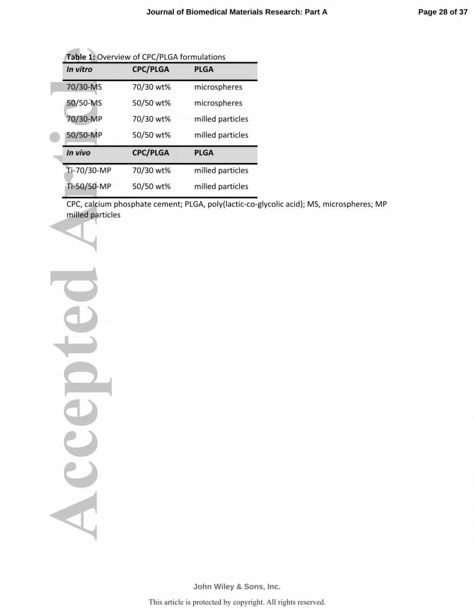
Table 1: Overview of CPC/PLGA formulations
In vitro CPC/PLGA PLGA
70/30-MS 70/30 wt% microspheres
50/50-MS 50/50 wt% microspheres
70/30-MP 70/30 wt% milled particles
50/50-MP 50/50 wt% milled particles
In vivo CPC/PLGA PLGA
Ti-70/30-MP 70/30 wt% milled particles
Ti-50/50-MP 50/50 wt% milled particles
CPC, calcium phosphate cement; PLGA, poly(lactic-co-glycolic acid); MS, microspheres; MP
milled particles
Page 28 of 37
John Wiley & Sons, Inc.
Journal of Biomedical Materials Research: Part A
This article is protected by copyright. All rights reserved.

Table 2: Porosity calculations of the scaffolds*
Mean mass
(g)
Density
(g/cm3)
Total
porosity (%)
Macroporosity
(%)
Microporosity
(%)
CPC only 0.213 ± 0.003 0.0305 ± 0.0004 52.9 ± 0.7 n.a. 52.9 ± 0.7
70/30-MS 0.095 ± 0.004
0.0136 ± 0.0005 78.9 ± 0.8 55.3 ± 1.8 23.7 ± 0.9
50/50-MS 0.068 ± 0.005 0.0097 ± 0.0007 85.0 ± 1.1 68.1 ± 2.3 16.9 ± 1.2
70/30-MP 0.099 ± 0.003 0.0141 ± 0.0005 78.2 ± 0.7 53.7 ± 1.6 24.5 ± 0.8
50/50-MP 0.068 ± 0.006 0.0097 ± 0.0008 85.0 ± 1.2 68.1 ± 2.6 16.8 ± 1.4
*Porosity calculations were performed based on CPC with burned out PLGA; The differences
were significant (p<0.001) comparing both 70/30-MS and 70/30-MP to 50/50-MS and 50/50-
MP; CPC, calcium phosphate cement; MS, microspheres; MP milled particles
Page 29 of 37
John Wiley & Sons, Inc.
Journal of Biomedical Materials Research: Part A
This article is protected by copyright. All rights reserved.

Table 3: Data (means +SD) for bone area, CPC area and BIC percentages at all time points
Ti-70/30-MP Ti-50/50-MP
CPC area Mean(%) ± SD n Mean(%) ± SD n
week 0 81.0 ± 0.6 3 74.8 ± 0.7 3
week 4 60.9 ± 13.5 10 53.4 ± 17.3 10
week 8 35.2 ± 21.6 10 9.1 ± 9.7 10
Bone area Mean(%) ± SD n Mean(%) ± SD n
week 0 0.0 ± 0.0 0 0.0 ± 0.0 0
week 4 5.8 ± 4.9 10 8.3 ± 2.8 10
week 8 22.9 ± 5.2 10 35.0 ± 15.0 10
BIC Mean(%) ± SD n Mean(%) ± SD n
week 0 0.0 ± 0.0 0 0.0 ± 0.0 0
week 4 2.4 ± 4.7 10 0.5 ± 0.7 10
week 8 11.7 ± 14.6 10 11.9 ± 21.2 10
BIC, bone-to-implant contact; Ti, titanium MP, milled particles; SD, standard deviation
Page 30 of 37
John Wiley & Sons, Inc.
Journal of Biomedical Materials Research: Part A
This article is protected by copyright. All rights reserved.

Table 4: Relative bone formation per 1% degraded material
Ti-70/30-MP Ti-50/50-MP
0-4 weeks 0.29 % 0.39 %
4-8 weeks 0.67 % 0.60 %
0-8 weeks 0.50 % 0.53 %
Page 31 of 37
John Wiley & Sons, Inc.
Journal of Biomedical Materials Research: Part A
This article is protected by copyright. All rights reserved.

Schematic representation of titanium landmarks with CPC/PLGA (left) with dimensions of a diameter of maximum 3mm and minimum 1mm, length maximum 6mm; Photograph of titanium landmarks with
CPC/PLGA (right); CPC, calcium phosphate cement; PLGA, poly(D,L-lactic-co-glycolic) acid
126x88mm (300 x 300 DPI)
Page 32 of 37
John Wiley & Sons, Inc.
Journal of Biomedical Materials Research: Part A
This article is protected by copyright. All rights reserved.

Equations 1 and 2 used to calculate macro- and microporosity (%) of the scaffolds
164x73mm (300 x 300 DPI)
Page 33 of 37
John Wiley & Sons, Inc.
Journal of Biomedical Materials Research: Part A
This article is protected by copyright. All rights reserved.

Representative SEM images of PLGA; A) microspheres and B) milled particles; SEM, scanning electron microscope; PLGA, poly(D,L-lactic-co-glycolic) acid
187x85mm (300 x 300 DPI)
Page 34 of 37
John Wiley & Sons, Inc.
Journal of Biomedical Materials Research: Part A
This article is protected by copyright. All rights reserved.

A) SEM images of CPC/PLGA with pore formation by degraded PLGA; B) higher magnification showing detail of brushite crystal formation; C) In vitro cumulative Ca2+ release in mg/scaffold for each CPC/PGLA
formulation, differences between 70/30-MS and 70/30-MP formulations were significantly different at all time points compared to 50/50-MS and 50/50-MP; CPC, calcium phosphate cement; PLGA, poly(D,L-lactic-
co-glycolic) acid; MS, microspheres; MP, milled particles 202x209mm (300 x 300 DPI)
Page 35 of 37
John Wiley & Sons, Inc.
Journal of Biomedical Materials Research: Part A
This article is protected by copyright. All rights reserved.
Representative histological images (5x magnification) of both materials at time points of 4 and 8 weeks with detailed images (20x magnification); Ti, titanium landmarks; BM, bone marrow
167x225mm (300 x 300 DPI)
Page 36 of 37
John Wiley & Sons, Inc.
Journal of Biomedical Materials Research: Part A
This article is protected by copyright. All rights reserved.

A) CPC area in the ROI (mean +SD); B) Bone area in the ROI (mean ±SD); C) Graph of CPC degradation rate versus bone formation rate; D) Bone to implant contact (BIC) (mean ±SD); Intergroup differences *) p < 0.05, **) p < 0.01, ***) p < 0.001; Temporal differences ^) p < 0.05 compared to 4 week equivalent, #)
p < 0.05 compared to 8-week equivalent; ##) p < 0.01 compared to 8-week equivalent, ###) p < 0.001 compared to 8-week equivalent 254x190mm (96 x 96 DPI)
Page 37 of 37
John Wiley & Sons, Inc.
Journal of Biomedical Materials Research: Part A
This article is protected by copyright. All rights reserved.