the release profiles and bioactivity of parathyroid hormone from poly(lactic-co-glycolic acid)...
TRANSCRIPT
Biomaterials 25 (2004) 345–352
ARTICLE IN PRESS
*Correspondin
Sciences, Univer
Avenue, Ann A
fax: +1-734-647
E-mail addres
0142-9612/$ - see
doi:10.1016/S014
The release profiles and bioactivity of parathyroid hormone frompoly(lactic-co-glycolic acid) microspheres
Guobao Weia, Glenda J. Pettwaya, Laurie K. McCauleyb,c, Peter X. Maa,d,e,*aDepartment of Biomedical Engineering, University of Michigan, Ann Arbor, MI 48109-2099, USA
bDepartment of Periodontics/Prevention/Geriatrics, University of Michigan, Ann Arbor, MI 48109-1078, USAcDepartment of Pathology, Medical School, University of Michigan, Ann Arbor, MI 48109-1078, USA
dDepartment of Biological and Materials Sciences, University of Michigan, Ann Arbor, MI 48109-1078, USAeMacromolecular Science and Engineering Center, University of Michigan, Ann Arbor, MI 48109-1078, USA
Received 12 May 2003; accepted 19 June 2003
Abstract
Poly(lactic-co-glycolic acid) (PLGA) microspheres containing bovine serum albumin (BSA) or human parathyroid hormone
(PTH)(1–34) were prepared using a double emulsion method with high encapsulation efficiency and controlled particle sizes. The
microspheres were characterized with regard to their surface morphology, size, protein loading, degradation and release kinetics,
and in vitro and in vivo assessments of biological activity of released PTH. PLGA5050 microspheres degraded rapidly after a 3-week
lag time and were degraded completely within 4 months. In vitro BSA release kinetics from PLGA5050 microspheres were
characterized by a burst effect followed by a slow release phase within 1–7 weeks and a second burst release at 8 weeks, which was
consistent with the degradation study. The PTH incorporated PLGA5050 microspheres released detectable PTH in the initial 24 h,
and the released PTH was biologically active as evidenced by the stimulated release of cAMP from ROS 17/2.8 osteosarcoma cells as
well as increased serum calcium levels when injected subcutaneously into mice. Both in vitro and in vivo assays demonstrated that
the bioactivity of PTH was maintained largely during the fabrication of PLGA microspheres and upon release. These studies
illustrate the feasibility of achieving local delivery of PTH to induce a biologically active response in bone by a microsphere
encapsulation technique.
r 2003 Elsevier Ltd. All rights reserved.
Keywords: Microspheres; PLGA; Degradation; Controlled release; PTH; Bioactivity
1. Introduction
Proteins and peptides have received extensive interestfor their therapeutic applications in diverse clinicalsettings [1]. Many recent tissue engineering studies havealso focused on the use of growth factors to stimulatecellular activity in vivo, and regulate tissue regeneration[2–4]. However, protein and peptide drugs in generalhave short plasma half-lives, are unstable in thegastrointestinal tract and also have low bioavailabilitiesdue to their relatively large molecular weight and high
g author. Department of Biological and Materials
sity of Michigan, Room 2211, 1011 North University
rbor, MI 48109-1078, USA. Tel.: +1-734-764-2209;
-2110.
s: [email protected] (P.X. Ma).
front matter r 2003 Elsevier Ltd. All rights reserved.
2-9612(03)00528-3
aqueous solubilities. These properties prevent themfrom being effectively used clinically [5]. In order toachieve high administration efficacy of proteins andpeptides, polymeric particulate carriers (micro- andnanospheres) have been developed as an effective wayto control the release profile of the contained substanceand to protect unstable biologically active moleculesfrom degradation. Both natural and synthetic biode-gradable polymers have been investigated for controlleddrug release [6–9]. Among these polymers, poly(lacticacid) (PLA) and poly(lactic-co-glycolic acid) (PLGA)were found to be remarkable for their application indrug delivery due to their excellent biocompatibility andbiodegradability through natural pathways [10–13].Injectable and in situ formed microspheres have alsobeen developed for the treatment of human diseases andanimal health [14,15].

ARTICLE IN PRESSG. Wei et al. / Biomaterials 25 (2004) 345–352346
PTH is a peptide hormone that has both anabolicand catabolic effects on bone, depending on the dosageregimes and the delivery patterns [16]. It is well acceptedthat high levels of circulating PTH lead to a catabolicresponse whereas either high doses intermittently or lowdoses continuously may lead to an anabolic response[17,18]. PTH(1–34) exhibits full biological activity of thefull-length PTH(1–84) and has been shown to beeffective by intermittent subcutaneous (sc) injection forthe treatment of osteoporosis in Phase III clinical trials[19]. Alternative PTH delivery methods have beeninvestigated recently with various successes. A deliverydeveloped by Leone-Bay et al. showed that PTH(1–34)administrated orally remained biologically active. How-ever, the bioavailability of PTH is low, only 5% and2.1%, relative to sc administration in rats and monkeys,respectively [20]. The pulmonary route has also beenevaluated in rats by means of intratracheal instillationor inhalation of dry powders and the bioavailabilitieswere 40% and 34%, respectively [21,22]. Other methodsfor intermittent PTH delivery include programmedadministration by osmotic pump [23] and pulsatiletransdermal administration [24]. Both of themshowed anabolic actions of PTH on bone, equivalentto sc administration. While PTH has been usedextensively via systemic administration for treatingosteoporosis, there is relatively little work focusing onlocal delivery of PTH. Notably, these few studies haveinvestigated PTH administered locally via a direct genedelivery which was found to be beneficial in thetreatment of bony defects [25,26]. Gene therapystrategies, although promising, have limitations andapproaches to deliver PTH peptide merit investigationof initial feasibility.The double emulsion technique is one of the most
popular methods used to encapsulate hydrophilic drugs,particularly protein and peptide drugs, into micro-spheres. In this study, microspheres with different sizeswere prepared by double emulsion techniques. Bothdegradation and BSA release kinetics were examinedin vitro. A recombinant peptide hormone, PTH(1–34)was also encapsulated in PLGA5050 micropheres toexamine in vitro and in vivo bioactivity after initialrelease.
Table 1
Effects of PVA concentration and homogenization strength on particle size
Polymer type PVA concentration
(%)
Stirring condition
PLGA5050 1 Mechanical stirring a
PLGA5050 1 Sonication (15W)
PLGA5050 5 Mechanical stirring a
PLGA7525 1 Mechanical stirring a
PLGA8515 1 Mechanical stirring a
PLLA 1 Mechanical stirring a
2. Materials and methods
2.1. Materials
PLGA polymers (Medisorbs, PLGA5050, LA:GA=50:50, Zinh ¼ 0:75; PLGA7525, LA:GA=75:25, Zinh ¼0:76; PLGA8515, LA:GA=85:15, Zinh ¼ 0:78) werepurchased from Alkermes Inc. (Wilmington, OH).Poly(lactic acid) with inherent viscosity of 1.6 dl/g waspurchased from Boehringer Ingelheim (Ingelheim, Ger-many). Lyophilized parathyroid hormone PTH(1–34)was obtained from Bachem Bioscience Inc. (Torrance,CA). Other chemicals used were poly(vinyl alcohol)(PVA) (88mol% hydrolyzed, MW=25,000) obtainedfrom Polysciences Inc. (Warrington, PA); bovineserum albumin (BSA, Fraction V) from Sigma;dichloromethane from Aldrich Chemical Company(Milwaukee, WI).
2.2. Microsphere preparation and characterization
Microspheres of various sizes were fabricated by atypical double emulsion technique [27] using optimizedformulation parameters as shown in Table 1. Briefly,100 ml BSA (3% w/v) solution or PTH buffersolution (PTH in 4mm HCl/0.1% BSA solution)was emulsified in a 10% polymer solution indichloromethane (DCM), using a probe sonicator atan output power of 15W (Virsonic 100, Cardiner, NY)for 20 s over an ice bath to form primary water-in-oil(w/o) emulsion. The w/o emulsion was graduallyadded into 250ml aqueous PVA solution under stirringor sonication to form a water-in-oil-in-water (w/o/w)double emulsion. The solution was stirred at roomtemperature for 3 h to evaporate dichloromethaneand then centrifuged to collect solid microspheres.The resultant microspheres were washed with distilledwater three times and freeze dried. The overallmorphology of the microspheres was examined usingscanning electron microscopy (SEM) (Hitachi S3200,Tokyo, Japan) after gold coating of the microspheresamples on a stub.
and BSA loading efficiency
Average
size (mm)BSA loading
(w/w%)
Encapsulation
efficiency (%)
t 700 rpm 20–50 2.4870.26 82.672.6o1 2.2370.16 74.271.6
t 700 rpm 2–5 2.5970.64 86.476.4t 700 rpm 20–50 2.2970.12 76.370.12t 700 rpm 20–50 2.3370.21 77.570.21t 700 rpm 20–50 2.2670.32 75.470.32
ARTICLE IN PRESSG. Wei et al. / Biomaterials 25 (2004) 345–352 347
2.3. Degradation study of PLGA5050 microspheres
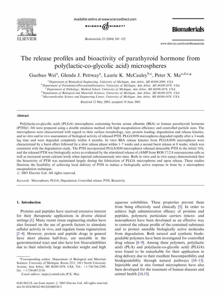
Preweighed PLGA5050 microspheres (about 10mg)were placed in individual test tubes containing 1.0ml ofphosphate buffered saline (PBS, 0.1m, pH 7.4). Thetubes were kept in an incubator that was maintained at37�C. At predetermined degradation intervals, themicrospheres were collected by centrifugation, washedwith distilled water to remove residual buffer salts, anddried to constant weight in a vacuum desiccator. Massloss of the microspheres was determined gravimetrically.The surface morphology of degraded PLGA5050 micro-spheres was analyzed using SEM at 15 kV.
2.4. Protein loading and encapsulation efficiency
Ten-milligram BSA-loaded microspheres were hydro-lyzed in a mixture of 0.9ml of 1m NaOH and 0.1ml PBSwith vigorous shaking at room temperature for 2 h. BSAstandard solutions (0.1ml) were also hydrolyzed byadding 0.9ml 1m NaOH using the same procedures.After hydrolysis, 1ml 0.9m HCl was added to neutralizethe sample solutions. Protein concentrations weredetermined by MicroBCA method (Pierce, Rockford,IL). Protein loading and encapsulation efficiency weredetermined by Eqs. (1) and (2), respectively:
Protein loading ðw=w%Þ
¼Amount of protein in microspheres
Amount of microspheres; ð1Þ
Encapsulation efficiency
¼Actual protein loading
Theoretical protein loading
¼Retained protein amount
Initially loaded protein amount: ð2Þ
2.5. In vitro BSA release
In vitro BSA release profiles from PLGA micro-spheres were determined as follows. Ten milligramsmicrospheres were suspended in 2ml PBS (0.1m,pH=7.4) with 0.02% sodium azide as preservative.The microsphere suspensions were incubated at 37�Cwithout shaking. At designated times, 1ml releasemedium was collected by centrifugation and replacedwith equal amount of fresh PBS. The amount of releasedBSA was measured using a MicroBCA protein assay(Pierce, Rockford, IL).
2.6. PTH(1–34) immunoassay
The concentration of PTH(1–34) in the releasemedium was measured using a PTH(1–34) ELISA kitwith PTH antibody coated wells using the manufac-
turer’s recommendations (Immutopics, San Clemente,CA). Triplicate wells were used for each time point(first 24 h). Absorbance measurements read at450 nm recorded by a microtiter plate reader wereused to calculate the PTH concentrations by thelog-logit method using the GraphPad Prisms program(GraphPad Software, San Diego, CA) with a standardcurve.
2.7. In vitro PTH bioactivity assay
The in vitro bioactivity of PTH released fromPLGA5050 microspheres was determined by adenylatecyclase stimulation assay and cAMP binding proteinassay. The adenylate cyclase stimulation assay wasperformed as previously described [28]. Briefly, ratosteosarcoma cells (ROS 17/2.8) were plated in triplicateinto 24-well plates and cultured to confluence. Tostimulate PTH receptor-mediated adenylate cyclase,the ROS cells were treated with known concentrationsof PTH(1–34) or with microsphere eluent for designatedtimes point in calcium- and magnesium-free hanks’balanced salt solution (Invitrogen, Carlsbad, CA)containing 0.1% bovine serum albumin (BSA) and1mm isobutylmethylxanthine (IBMX). The treatedcells were incubated at 37�C for 10min. The reactionwas stopped by gently aspirating the media andthe cAMP in cells was extracted by adding 250 ml/wellcold 5% perchloric acid and incubating at �20�Cuntil completely frozen. After thawing, the cAMPextracts were transferred to borosilicate glass tubes,and the pH was adjusted to 7.5 with 64 ml 4n KOH/well. The extract was centrifuged to remove theprecipitate.The cAMP binding protein assay was performed as
previously described to determine cAMP levels usingknown cAMP standards [28]. The cAMP extracts ofsamples were diluted 1:16 with cAMP binding buffer inorder to be quantified within the standard curve. Beforeuse, the [3H]-cAMP (ICN, Irvine, CA) was diluted to10,000–15,000 cpm/25 ml with cAMP binding buffer.The [3H]-cAMP was incubated with standards orunknowns and cAMP binding protein for 90min onice. Dextran-coated charcoal was added to the reactantsafter the binding protein reaction to remove theunbound [3H]-cAMP. The samples were incubated onice for 20min and then centrifuged to pellet the dextran-coated charcoal. The supernatant of each tube wasdecanted carefully to a scintillation tube, 5ml ofscintillation fluid was added, and the radioactivity ofthe supernatants was determined using a liquidscintillation counter (Wallacs 1410; Wallac, Gaithers-burg, MD) and cAMP levels were calculated by the log-logit method using the GraphPad Prisms program(GraphPad Software, San Diego, CA) with a standardcurve.
ARTICLE IN PRESSG. Wei et al. / Biomaterials 25 (2004) 345–352348
2.8. In vivo PTH bioactivity assay
Five mice per group received sc injections of PTH orvehicle microspheres in 100 ml PBS. The PTH micro-spheres had a calculated theoretical PTH concentrationof 1 mg PTH/1mg microsphere. After 3 h, the mice wereanesthesized with an intraperitoneal injection of keta-mine (90mg/kg) and xylazine (5mg/kg) and terminalserum samples were collected. Serum calcium levels weredetermined by colorimetric assay with the cresolphtha-lein complexone method (Sigma) as previously described[29].
0
20
40
60
80
100
0 2 4 6 8 10 12 14 16
Time (Weeks)
Wei
gh
t re
mai
nin
g (
%)
Fig. 2. Time dependence of mass loss profiles of PLGA5050 micro-
spheres degrading in PBS at 37�C ðn ¼ 3Þ:
3. Results
3.1. PLGA microspheres
PLGA5050 microspheres with three different particlesizes were obtained by adjusting the fabrication para-meters of double emulsion processes (Fig. 1). Both thePVA concentration and emulsification strength em-ployed in the second emulsion formation (mechanicalstirring or sonication) greatly affected the size of themicrospheres (Table 1). Under mechanical stirring, ahigher PVA concentration (5%) produced microsphereswith size of 2–5 mm while a lower PVA concentration(1%) resulted in microspheres having a size of 20–50 mm.When probe sonication was applied, nanospheres(o1 mm) were obtained even when a low concentrationof PVA (1%) was used. As examined using SEM, all
(a)
(b)
(c)
(d)
Fig. 1. SEM micrographs of PLGA5050 microspheres prepared under differe
(c, d) 5% PVA solution, mechanical stirring at 700 rpm; and (e, f) 1% PVA
microspheres had spherical morphologies and smoothsurface structures. The composition of the copolymer(ratio of LA to GA) had little effect on the size of themicrospheres.
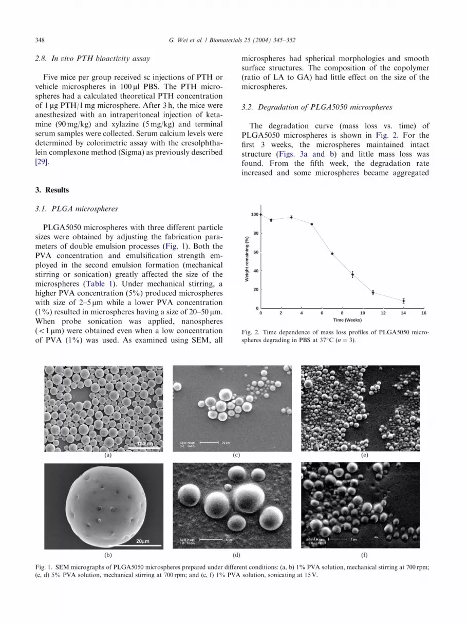
3.2. Degradation of PLGA5050 microspheres
The degradation curve (mass loss vs. time) ofPLGA5050 microspheres is shown in Fig. 2. For thefirst 3 weeks, the microspheres maintained intactstructure (Figs. 3a and b) and little mass loss wasfound. From the fifth week, the degradation rateincreased and some microspheres became aggregated
(e)
(f)
nt conditions: (a, b) 1% PVA solution, mechanical stirring at 700 rpm;
solution, sonicating at 15V.

ARTICLE IN PRESS
(a)
(b)
(c)
(d)
(e)
(f)
Fig. 3. SEM micrographs of PLGA50/50 microspheres at different degradation stages: (a) 24 h, (b) 3 weeks, (c) 5-weeks, (d) 7 weeks, and (e, f) 11
weeks.
0
10
20
30
40
50
0 5 10 15 20 25
Time (Days)
Cu
mm
ula
tive
rel
ease
(%
)
PLLAPLGA85/15PLGA75/25PLGA50/50
0
10
20
30
40
50
60
0 20 40 60 80 100 120 140
Time (days)
Co
mm
ula
tive
rel
ease
(%
)
(a)
(b)
Fig. 4. Release of BSA from PLGA microspheres ðn ¼ 3Þ: (a) 4-weekBSA release from PLGA microspheres with varying LA/GA ratio; and
(b) 20-week BSA release from PLGA5050 microspheres.
G. Wei et al. / Biomaterials 25 (2004) 345–352 349
(Figs. 3c–f). The microspheres deformed from originalspherical shape into irregular shape with a rough surfacetexture. After 11–14 weeks of degradation, the micro-spheres had almost entirely degraded and the remainingmass was about 18% and 7%, respectively. ThePLGA5050 microspheres were totally collapsed anddisintegrated into irregular particles, and no intactspheres were observed.
3.3. In vitro BSA release
The release kinetics of PLGA microspheres wasinvestigated using BSA as a model protein. Fig. 4aillustrates the cumulative release of BSA in a 4-weekperiod from PLGA microspheres with different LA/GAcomposition. About 30% BSA was release fromPLGA5050 microspheres, while only 10% was releasedfrom PLLA microspheres. According to the degradationstudy, the release of BSA in the first 4 weeks was mainlydue to diffusion rather than degradation. Since PLLA ismuch more hydrophobic and also more densely packed(crystalline) than PLGA5050, it is more difficult forwater to diffuse into and BSA to be released from PLLAmicrospheres. In a long release period of 20 weeks, therewas another burst release for PLGA5050 between 10and 12 weeks. The release of BSA was due to thedegradation of PLGA5050 microspheres. From thedegradation study, a great loss of mass began at 7weeks while a sudden BSA release began at 12 weeks(Fig. 4b). The difference can be explained by the factthat PBS was not changed during the entire degradationstudy, whereas it was changed frequently during the

ARTICLE IN PRESS
cAM
P (
pm
ol/w
ell)
0
100
200
300
400
6himmediaterelease
12h 24h
vehicle microspheresPTH microspheres
Release time
Fig. 6. Biologic activity of microsphere eluent. PTH receptor-
mediated adenylate cyclase was stimulated by treating ROS cells with
known concentrations of PTH(1–34) or with microsphere eluent (from
vehicle controls or PTH microspheres) for each time point. The cAMP
in the cells was extracted and a cAMP binding protein assay was
performed to determine cAMP levels. All values are the mean for
triplicate samples. The highest biologic activity of PTH was observed
at the 6-h incubation time point and a significant decrease occurred at
12-h. Note the level of cAMP in all of the vehicle microspheres was less
G. Wei et al. / Biomaterials 25 (2004) 345–352350
release study. In the degradation study, accumulatedacidity accelerated the degradation of the PLGA5050microspheres.
3.4. In vitro PTH release and bioactivity
Fig. 5 shows the absolute amount of PTH(1–34)released from PLGA 5050 microspheres in the initial24 h. The corresponding bioactivity of released PTH ineluent is shown in Fig. 6. At 0 h, the microspheres weredissolved in dichloromethane and then extracted withdistilled water. PTH levels in these samples were high(not shown in Fig. 5. since the amount was more than100 pg/ml), but the bioactivity indicated by cAMP levelswas lower than that of 6 h release group. This maylargely be due to the harshness of the solvent, whichlikely diminished the bioactivity of PTH during theextraction. The highest levels of biologically active PTHin the initial 24 h were found at 6 h and levels decreasedthereafter. However, there were still significant levels ofPTH bioactivity in the 24 h group. The bioactivity ofPTH at 6 h was equivalent to 10 nm PTH (a standardcurve of PTH was used as a positive control). These dataindicate that PTH retained its bioactivity after beingincorporated into the microspheres.
PT
H (
pg
/µµl)
0
5
10
15
20
25
30
6hcontrol 12h 24hTime
Fig. 5. PTH concentration in microsphere eluent. PTH microspheres
were incubated at 37�C in PBS for 6, 12, and 24 h. The concentration
of PTH released in the microsphere eluent at each time point was
measured using a PTH(1–34) ELISA kit with PTH antibody coated
wells. All values are the mean for triplicate samples. The concentration
of PTH in the eluent of the control microspheres (BSA only) was less
than 5 pg/ml. The concentration of PTH was the highest at the 6-h
incubation time point and gradually decreased thereafter.
than 5 pmol/well.
3.5. In vivo PTH bioactivity
To further evaluate PTH microsphere bioactivityin vivo, PTH-incorporated PLGA5050 microsphereswere injected into mice and serum calcium was analyzedvia colorimetric assay. Fig. 7 shows that mice thatreceived PTH microspheres had significantly elevatedserum calcium levels, indicating effective delivery ofbiologically active PTH from the microspheres in vivo.
4. Discussion
Drugs formulated in polymeric devices are released bydiffusion through the polymer barrier, by degradationof the polymer materials, or a combination of bothdiffusion and degradation mechanisms [30]. Therefore,the drug release kinetics from biodegradable carriers isclosely related to degradation behavior of the materials.From this study, we found that the mass loss ofdegrading PLGA5050 microspheres had a lag time of3 weeks. Following the lag time, there was a rapid massloss. In addition, the PLGA5050 microspheres demon-strated an agglomeration tendency and lump formationbefore they disintegrated into small pieces or particles.The initial smooth surface of intact microspheresdeveloped wrinkles and defects after 5 weeks ofdegradation. It was these wrinkles and defects thatcaused the microspheres to aggregate together and form

ARTICLE IN PRESS
0
2
4
6
8
10
12
14
Control PTH
*
*p<0.05 versus control (n=5 mice/group)
Ser
um
Cal
ciu
m (
ng
/dL
)
Fig. 7. Serum calcium levels in response to microsphere injection. Five
mice per group were injected microspheres containing 10mg PTH or
vehicle microspheres in 100ml PBS. The mice were anesthetized after3 h and terminal serum samples were collected. The animals injected
with PTH microspheres had significantly higher (�po0:05) serumcalcium levels than the animals injected with vehicle microspheres.
G. Wei et al. / Biomaterials 25 (2004) 345–352 351
lumps [31]. The aggregation tendency and the surfacechanges are an indication of the plasticization of thepolymer, which may result from the reduction of glasstransition temperature (Tg) below 37
�C (the incubationtemperature). The release of BSA from PLGA5050microspheres exhibited corresponding patterns to thedegradation profile. BSA was released from thesesystems in three phases: initial burst followed by a lagphase and final degradation-controlled release. Theinitial burst release of BSA was largely due to BSAbeing on the surface layer. The slow release after initialburst was the result of diffusion from the microspheres,which might be further slowed down due to theformation of intermolecular linkages and non-specificadsorption of the protein to the polymer. A second burstrelease at 8 weeks resulted from the rapid degradationand disintegration of the microspheres.The size of microspheres was controlled by varying
the fabrication parameters, mainly the PVA concentra-tion and the emulsification strength. The preparednanospheres had a protein encapsulation efficiency of74% and diameter of several hundred nanometers.Nanospheres have been extensively used in drug deliveryfor cancer therapy as well as controlled delivery ofvaccines [32–34].The encapsulation efficiency for PTH was evidently
lower than that of BSA. This might be due to thedifferences in molecular weights and structures betweenPTH and BSA. BSA has a globular structure with a highmolecular weight of about 66 kDa while PTH(1–34)fragment is a linear small peptide with a low molecular
weight of 4 kDa. Therefore, it was expected that morePTH than BSA would be lost during the formation ofthe final double emulsion.Besides achieving desired release profiles, the main
challenge of protein and peptides delivery is to maintainthe bioactivity of the bioactive molecules during andafter delivery. Intact PTH(1–84) and PTH(1–34) haveplasma half lives of less than 3 and 11min, respectively[21]. The rapid metabolic degradation of biologicallyactive PTH may make multiple administrations of PTHnecessary to maintain its effectiveness. This was also oneof the main factors that has contributed to lowbioavailability of PTH by oral administration [20]. Inthis study, PTH was resuspended in BSA/HCl solutionand then encapsulated in PLGA microspheres. Thedouble emulsion technique improved the encapsulationefficiency and minimized the loss of bioactivity bothin vitro and in vivo. The polymer protected biologicallyactive PTH from sensitive factors such as temperatureand oxidation in vitro and enzymatic degradationin vivo. The solvent used has a critical effect on thebioactivity of PTH. Complete exposure of PTHmolecules to some organic solvents, such as dichlor-omethane led to substantial loss of bioactivity. The factthat PTH remains active after encapsulation within,and release from microspheres clarifies an importantinitial step in tissue engineering strategies aimed at theuse of PTH as an anabolic agent to improve boneregeneration.
5. Conclusion
PLGA microspheres with desired sizes were preparedby double emulsion technique in this study. Both a largeprotein (BSA) and a small peptide (PTH) were success-fully incorporated into the micropheres. The long-termBSA release data demonstrated the potential of PLGAmicrospheres for controlled delivery of large proteins.PTH maintained its bioactivity both in vitro and in vivoafter initial release, indicating that a local delivery ofPTH to evoke a skeletal response can be accomplishedby a microsphere encapsulation technique.
Acknowledgements
The authors wish to acknowledge support from NIH(DE 14755, PXM; DK 53904, LKM), Center forCraniofacial Regeneration and Center for BiomedicalEngineering Research (University of Michigan: LKM &PXM), Nano Materials Initiative (University ofMichigan: PXM), the DuPont Young ProfessorAward (PXM). The authors also would like tothank Ms. Amy J. Koh-Paige for technical assistancein PTH assay.

ARTICLE IN PRESSG. Wei et al. / Biomaterials 25 (2004) 345–352352
References
[1] Langer R. New methods of drug delivery. Science 1990;249:
1527–33.
[2] Babensee JE, McIntire LV, Mikos AG. Growth factor delivery for
tissue engineering. Pharm Res 2000;17(5):497–504.
[3] Reddi AH. Role of morphogenetic proteins in skeletal tissue
engineering and regeneration. Nat Biotechnol 1998;16(3):247–52.
[4] Lee H, Cusick RA, Browne F, Kim TH, Ma PX, Utsunomiya H,
Langer R, Vacanti JP. Local delivery of basic fibroblast growth
factor increases both angiogenesis and engraftment of hepatocytes
in tissue-engineered polymer devices. Transplantation
2002;73:1589–93.
[5] Lee VH. Enzymatic barriers to peptide and protein absorption.
CRC Crit Rev Ther Drug Carr 1988;5:69–87.
[6] Narayani R, Rao KP. Gelatin microsphere cocktails of different
sizes for the controlled release of anticancer drugs. Int J Pharm
1996;143:255–8.
[7] Avgoustakis K, Beletsi A, Panagi Z, Klepetsanis P, Karydas AG,
Ithakissios DS. PLGA-mPEG nanoparticles of cisplatin: in vitro
nanoparticle degradation, in vitro drug release and in vivo drug
residence in blood properties. J Controlled Rel 2002;79:123–35.
[8] Le Ray AM, Chiffoleau S, Iooss P, Grimandi G, Gouyette A,
Daculsi G, Merle C. Vancomycin encapsulation in biodegradable
poly(e-caprolactone) microparticles for bone implantation. Influ-
ence of the formulation process on size, drug loading, in vitro
release and cytocompatibility. Biomaterials 2003;24:443–9.
[9] Carino GP, Jacob JS, Mathiowitz E. Nanosphere based oral
insulin delivery. J Controlled Rel 2000;65:261–9.
[10] Wu XS. Synthesis, properties of biodegradable lactic/glycolic acid
polymers. In: Wise, et al., editors. Encyclopedic handbook of
biomaterials and bioengineering. New York: Marcel Dekker;
1995. p. 1015–54.
[11] Singh M, Shirley B, Bajwa K, Samara E, Hora M, O’hagan D.
Controlled release of recombinant insulin-like growth factor from
a novel formulation of polylactide-co-glycolide Microparticles.
J Control Rel 2001;70:21–8.
[12] Oldham JB, Lu L, Zhu X, Poter BD, Hefferan TE, Larson DR,
Currier BL, Mikos AG, Yaszemski MJ. Biological activity of
rhBMP-2 released from PLGA microspheres. J Biomech Eng
2000;122:289–92.
[13] Lu L, Stamatas GN, Mikos AG. Controlled release of transform-
ing growth factor beta1 from biodegradable polymer micropar-
ticles. J Biomed Mater Sci 2000;50:440–51.
[14] Tracy MA. Development and scale-up of a microsphere protein
delivery system. Biotechnol Progr 1998;14:108–15.
[15] Jain RA, Rhodes CT, Railkar AM, Malick AW, Shah NH.
Controlled delivery of drugs from a novel injectable in situ formed
biodegradable PLGA microsphere system. J Microencapsul
2000;17(3):343–62.
[16] Morley P, Whitfield JF, Willick GE. Parathyroid hormone: an
anabolic treatment for osteoporosis. Curr Pharm Des
2001;7(8):671–87.
[17] Stanislaus D, Yang X, Liang JD, Wolfe J, Cain RL, Onyia JE,
Falla N, Marder P, Bidwell JP, Queener SW, Hock JM. In vivo
regulation of apoptosis in metaphyseal trabecular bone of young
rats by synthetic human parathyroid hormone (1–34) fragment.
Bone 2000;27:209–18.
[18] Andreassen TT, Ejersted C, Oxlund H. Intermittent parathyroid
hormone (1–34) treatment increases callus formation and
mechanical strength of healing rat fractures. J Bone Miner Res
1999;14:960–8.
[19] Neer RM, Arnaud CD, Zanchetta JR, Prince R, Gaich GA,
Reginster J-Y, Hodsman AB, Eriksen EF, Ish-Shalom S, Genant
HK, Wang O, Mitlak BH. Effect of parathyroid hormone (1–34)
on fractures and bone mineral density in postmenopausal women
with osteoporosis. N Engl J Med 2001;344:1434–41.
[20] Leone-Bay A, Sato M, Paton D, Hunt AH, Sarubbi D, Carozza
M, Chou J, McDonough J, Baughman RA. Oral delivery
of biologically active parathyroid hormone. Pharm Res
2001;18(7):964–70.
[21] Codrons V, Vanderbist R, Verbeeck RK, Arras M, Lison D,
Preat V, Vanbever R. Systemic delivery of parathyroid hormone
(1–34) using inhalation dry powders in rats. J Pharm Sci
2003;92(5):938–50.
[22] Patton JS, Trinchero P, Platz RM. Bioavailability of pulmonary
delivered peptides and proteins: a-interferon, calcitonins and
parathyroid hormones. J Control Rel 1994;28:79–85.
[23] Dobnig H, Turner RT. The effects of programmed administration
of human parathyroid hormone fragment (1–34) on bone
histomorphometry and serum chemistry in rats. Endocrinology
1997;138:4607–12.
[24] Suzuki Y, Nagase Y, Iga K, Kawase M, Oka M, Yanai S,
Matsumoto Y, Nakagawa S, Fukuda T, Adachi H, Higo N,
Ogawa Y. Prevention of bone loss in ovariectomized rats by
pulsatile transdermal iontophoretic administration of human
PYH (1–34). J Pharm Sci 2002;91:350–61.
[25] Fang JM, Zhu YY, Smily E, Bonadio J, Rouleau JP, Goldstein
SA, McCauley LK, Davidson BL, Roessler BJ. Stimulation of
new bone formation by direct transfer of osteogenic plasmid
genes. Proc Nat Acad Sci USA 1996;93:5753–8.
[26] Bonadio J, Smiley E, Patil P, Goldstein S. Localized, direct
plasmid gene delivery in vivo: prolonged therapy results in
reproducible tissue regeneration. Nat Med 1999;5:753–9.
[27] Cohen S, Yoshioka T, Lucarelli M, Hwang LH, Langer R.
Controlled delivery systems for proteins based on poly(lactic/
glycolic acid) microspheres. Pharm Res 1991;8:713–20.
[28] Chen HL, McCauley LK, D’Silva NJ. CAMP binding protein
assay for widespread use in cell signaling studies. Biotechniques
2002;33:1–5.
[29] Demiralp B, Chen HL, Koh AJ, Keller ET, McCauley LK.
Anabolic actions of parathyroid hormone during bone growth are
dependent on c-fos. Endocrinology 2002;10:4038–47.
[30] Wu XS. Preparation, characterization, and drug delivery applica-
tions of microspheres based on biodegradable lactic/glycolic acid
polymers. In: Wise, et al., editors. Encyclopedic handbook of
biomaterials and bioengineering. New York: Marcel Dekker,
1995; p. 1151–200.
[31] Viswanathan NB, Patil SS, Pandit JK, Lele AK, Kulkarni MG,
Mashelkar RA. Morphological changes in degrading PLGA and
P(DL)LA microspheres: implications for the design of controlled
release systems. J Microencapsul 2001;18:783–800.
[32] Desai M, Hilfinger J, Amidon G, Levy RJ, Lavhasetwar V.
Immune response with biodegradable nanospheres and alum:
study in rabbits using staphylococcal entertoxin B-toxoid.
J Microencapsul 1999;17:215–25.
[33] Lamprecht A, Ubrich N, Yamamoto H, Schafer U, Takeuchi H,
Maincent P, Kawashima Y, Lehr CM. Biodegradable
nanoparticles for targeted drug delivery in treatment of
inflammatory bowel disease. J Pharmacol Exp Ther 2001;
299:775–81.
[34] Hans ML, Lowman AM. Biodegradable nanoparticles for drug
delivery and targeting. Curr Opin Solid State Mater Sci
2002;6:319–27.