tracking of intentionally inoculated lactic acid bacteria
TRANSCRIPT

Microorganisms 2020, 8, 5; doi:10.3390/microorganisms8010005 www.mdpi.com/journal/microorganisms
Article
Tracking of Intentionally Inoculated Lactic Acid Bacteria
Strains in Yogurt and Probiotic Powder
Anshul Sharma 1,2,3, Jasmine Kaur 1, Sulhee Lee 1,4 and Young-Seo Park 1,*
1 Department of Food Science and Biotechnology, Gachon University, Gyeonggi-do 13120, Korea;
[email protected] (A.S.); [email protected] (J.K.); [email protected] (S.L.) 2 Department of Food and Nutrition, Gachon University, Gyeonggi-do 13120, Korea 3 Faculty of Applied Sciences and Biotechnology, Shoolini University of Biotechnology and Management Sciences,
Bajhol, Solan, Himachal Pradesh 173229, India 4 Research Group of Healthcare, Korea Food Research Institute, Wanju 55365, Korea
* Correspondence: [email protected]; Tel.: +82-31-750-5378
Received: 26 November 2019; Accepted: 16 December 2019; Published: 18 December 2019
Abstract: The present work aimed at tracking intentionally inoculated lactic acid bacteria (LAB) strains in
yogurt and probiotic powder. Leuconostoc (Leu.) mesenteroides (11251), Lactobacillus (L.) brevis (B151), and
Lactobacillus plantarum (LB41K) strains were tracked in yogurt, and L. plantarum (LB41P) was tracked in a
commercial probiotic powder. The yogurt was intentionally inoculated with the selected bacterial strains.
Two types of yogurt with known and unknown bacterial pools were utilized. The standard 16S rRNA gene
sequencing was used to evaluate the initial screening. The molecular typing tools, random amplified
polymorphic DNA (RAPD), repetitive element palindromic PCR (rep-PCR), and comparative gene
sequence analysis of selected housekeeping loci were used to track the inoculated dubious strains. Out of
30 random selections for each inoculation, the developed method identified seven (11251), nine (B151),
and five (LB41K) colonies in the yogurt. The validation was performed by identifying 7 colonies (LB41P)
out of 30 in the probiotic powder. The DNA banding profiles and the gene sequence alignments led to the
identification of the correct inoculated strains. Overall, the study summarizes the use of molecular tools to
identify the deliberately inoculated LAB strains. In conclusion, the proposed polyphasic approach
effectively tracked the intentionally inoculated strains: Leu. mesenteroides, L. brevis, and L. plantarum (LB41K)
in yogurt and L. plantarum (LB41P) in probiotic powder. The study demonstrates how to track industrially
relevant misused LAB strains in marketable food products.
Keywords: lactic acid bacteria; Leuconostoc mesenteroides; Lactobacillus brevis; Lactobacillus plantarum;
tracking; RAPD-PCR; rep-PCR; housekeeping genes
1. Introduction
For over thousands of years, the intentional addition of bacteria to commercial products as starters or
as food additives has been a continuous practice across the globe. These days, consumers are attracted more
to food products with high quality, safety, and viable bacterial additives for societal health benefits [1]. On
the other hand, food producers are showing interest in novel and industrially productive bacterial strains
[2]. Among the industrial workhorses, lactic acid bacteria (LAB) are well thought of as unique and harmless
bacteria, and most of the strains have earned the ‘‘generally recognized as safe’’ (GRAS) status [3].
Furthermore, the growing wealth of scientific literature has addressed the several health-promoting
activities of LAB, including their immunomodulatory, antidiabetic, antiobesity, antioxidant, and anticancer
activities [4,5]. In this era of a competitive world, the unscrupulous use of beneficial bacteria cannot be
repudiated, which raises concerns about the safety of industrially relevant microorganisms. Hence, there is

Microorganisms 2020, 8, 5 2 of 15
a necessity to develop measures to expose the dubious strains, which could be used for the manufacturing
of food products.
The accurate species-specific documentation of LAB is significant from technical and safety
perspectives [6]. A recent review article has meticulously described the use of various methodologies for
the tracking of microorganisms [7]. Analytical approaches such as phenotypic and genotypic analyses have
been exploited to identify and characterize LAB from food products [8,9]. Phenotypic approaches include
morphological and physiological analysis, biochemical depiction, and protein profiling. However, these
methods have low discriminatory power, lack sensitivity, and lack reproducibility [9]. The genotypic
characterization of LAB involves a plethora of tools; they offer various advantages, including accuracy,
reproducibility, and are timesaving [10,11]. In the present study, we utilized sequencing-based approaches
such as 16S rRNA and housekeeping gene sequence analyses, and polymerase chain reaction (PCR)-based
methods such as the random amplification of polymorphic DNA (RAPD) and repetitive genomic element
(rep)-PCR to assess the fingerprint profile of the intentionally inoculated LAB strains.
RAPD is a molecular tool based on the random amplification of DNA fragments using short arbitrary
sequence primers that bind at different locations within the genome, thereby generating a fingerprint of the
bacteria [12]. The advantages of this tool include fastness, requiring less amount of DNA, flexible primer
choice, and cost-effectiveness [13]. In contrast, the rep-PCR technique works by amplifying non-coding
repetitive elements of bacterial DNA using specific primers for subspecies classification and the strain level
discrimination [14,15]. The technique offers many advantages, such as fastness, cost-effectiveness, and high
discriminative power [16]. Furthermore, the evaluation of the constitutive (housekeeping) loci as
classification markers holds great potential in the expansion of globally available molecular tools for the
documentation of LAB [17].
The industrialized food units are mostly interested in LAB species of the following genera: Leuconostoc,
Lactobacillus, Oenococcus, Lactococcus, Pediococcus, Streptococcus, and Enterococcus [18]. Among them, three
industrially important species, namely Leuconostoc (Leu.) mesenteroides, Lactobacillus (L.) brevis, and
Lactobacillus plantarum, have been selected to validate the tracking technology. Leuconostoc species are
mostly found on vegetables and fruits, and play an important role in various food and industrial
fermentations; they are responsible for the organoleptic properties [12,19]. L. brevis has been reported in
plants, fermented foods, and beverages, and its strains are characterized as probiotics [20,21]. Recently, L.
brevis strain was also reported for its oral probiotic properties [22]. Contrariwise, L. brevis strains have also
been reported for the spoilage of beer [23]. L. plantarum is a versatile and heterogeneous bacterium found
in fermented foods and feed products, as well as in the human gastrointestinal tract (GIT) [24]. It is used in
the processing of sauerkraut and cheese, and the fermentation of wine and green olives [25]. Some L.
plantarum strains also synthesize vitamins and induce the production of host immunomodulatory particles
[26,27]. In this study, considering their relevance, we evaluated the presence of the three deliberately
inoculated industrially important strains, namely, Leu. mesenteroides, L. plantarum, and L. brevis in
commercial yogurt by using various molecular tools. Further validation was assessed by tracing L.
plantarum strains from a commercial probiotic powder.
2. Materials and Methods
2.1. Bacterial Strains
The three LAB strains used in this study were Leu. mesenteroides 11251, L. plantarum LB41K, and L. brevis
B151. The strains were procured from the Korean culture collection of probiotics (KCCP) and maintained
on De Man Rogosa Sharpe (MRS; Difco, Sparks, MD, USA) medium at 37 °C under aerobic conditions for
24 h. For the quantification of selected strains, the viable colonies were counted from 50 µL (serially diluted
to 10−6) cultures grown on MRS agar plates and expressed as colony-forming units (CFU) per mL.
Microorganisms 2020, 8, 5 3 of 15
2.2. Yogurt and Probiotic Powder
Two types of yogurts were purchased from a supermarket in Korea for a better insight of the presented
technology to identify the suspected LAB strains. A yogurt with an unknown bacterial pool (Denmark
drinking yogurt, Dongwon, Korea) was used for the deliberate addition of Leu. mesenteroides, whereas
yogurts with known bacterial strains (Streptococcus thermophilus, Lactobacillus casei, Bifidobacterium longum,
and Lactobacillus acidophilus; Mechnikop, Korea Yakult Co., Ltd., Seoul, Korea) were used for the inoculation
of L. brevis and L. plantarum. The yogurts were inoculated (with a 10th of the total volume of the sample)
using selected strains, rigorously vortexed, and serially diluted in phosphate-buffered saline (PBS; 10−6
dilution). The dilutions were spread plated on MRS agar plates and incubated for 24 h at 37 °C. The next
day, cell viability was measured. For each experiment, 30 colonies were arbitrarily selected and labelled as
Y1–30 for Leu. mesenteroides, YB1–30 for L. brevis, and YP1–30 for L. plantarum. For the final validation, a
commercial probiotic powder (Aram Co., Inc., Gwangju, Korea) containing four bacterial strains (L.
plantarum, B. longum, S. thermophilus, and L. acidophilus) was used to track the L. plantarum (LB41P) strain.
The industry also provided the pure culture of the target strain (LB41P) as a reference. Figure 1 describes
the overall working of the technique.
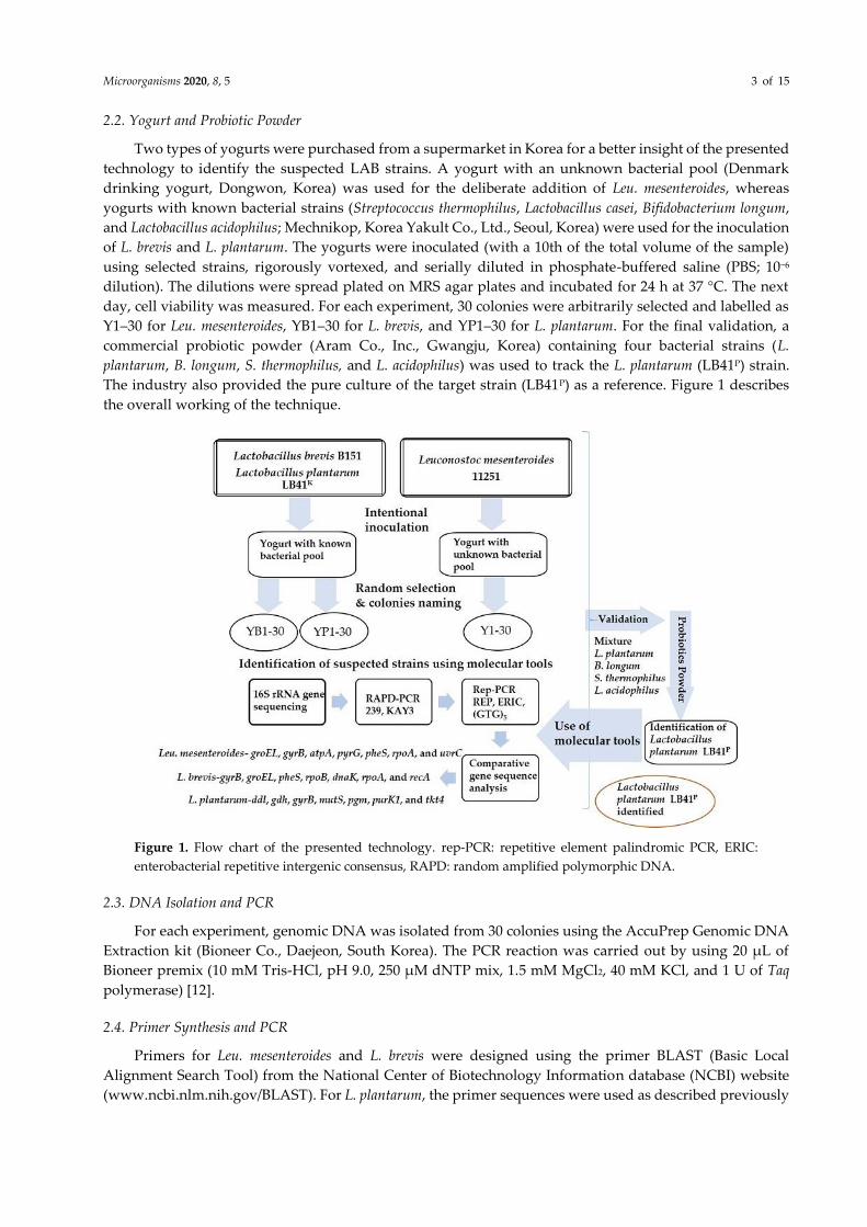
Figure 1. Flow chart of the presented technology. rep-PCR: repetitive element palindromic PCR, ERIC:
enterobacterial repetitive intergenic consensus, RAPD: random amplified polymorphic DNA.
2.3. DNA Isolation and PCR
For each experiment, genomic DNA was isolated from 30 colonies using the AccuPrep Genomic DNA
Extraction kit (Bioneer Co., Daejeon, South Korea). The PCR reaction was carried out by using 20 μL of
Bioneer premix (10 mM Tris-HCl, pH 9.0, 250 µM dNTP mix, 1.5 mM MgCl2, 40 mM KCl, and 1 U of Taq
polymerase) [12].
2.4. Primer Synthesis and PCR
Primers for Leu. mesenteroides and L. brevis were designed using the primer BLAST (Basic Local
Alignment Search Tool) from the National Center of Biotechnology Information database (NCBI) website
(www.ncbi.nlm.nih.gov/BLAST). For L. plantarum, the primer sequences were used as described previously

Microorganisms 2020, 8, 5 4 of 15
[25]. The primer synthesis was carried out by the Macrogen sequencing service (Seoul, South Korea). The
amplification was performed in a BIORAD thermal cycler (Hercules, CA, USA). Gel imaging was carried
out using the Bio-Rad Gel Doc XR+ gel documentation system. For fingerprint analysis, two DNA size
markers, 100 bp (Bioneer, Daejeon, South Korea) and 1 kb (Takara, Japan), were used. The gel-purified
(Wizard SV Gel and PCR Clean-Up system kits; Promega, USA) PCR products were sequenced by Bioneer
Co. (Daejeon, South Korea).
2.5. 16 S rRNA Gene Sequencing and RAPD Analysis
The detailed procedure was performed as reported by reference [12].
2.6. Rep-PCR Analysis
To obtain the genomic fingerprints, the second typing method rep-PCR was performed using three
primers: (GTG)5, enterobacterial repetitive intergenic consensus (ERIC), and repetitive extragenic
palindromic (REP) for the analysis of 30 colonies for each experiment. The PCR procedure followed has
been described previously [28]. Each reaction included 2 µL of DNA template, 1 µL (10 pmol/µL) of the
reverse primer, 1 µL (10 pmol/µL) of the forward primer, and 16 µL of autoclaved distilled water to bring
the total volume to 20 µL. The PCR products (5 µL) were then examined using 1.5% (w/v) agarose (Seakem,
Lonza, Alpharetta, GA, USA) gel electrophoresis at 70 V for 5 h. The PCR amplification conditions for all
the three rep-primers used in the present study are described in Table S1.
2.7. Comparative Sequence Analysis of the Housekeeping Genes
The third tracing method was based on the sequencing and analysis of the housekeeping genes. The
genes, their products, and other relevant information for 11251, B151, and LB41 (LB41K and LB41P) have
been described in Tables S2–S4. Each PCR amplification experiment was carried out by using 1 µL of
purified genomic DNA, 1 µL of the reverse primer, and 1 µL of the forward primer, and followed by the
addition of autoclaved distilled water (17 µL) to make a final volume of 20 µL. The experiment was
performed for the reference strains (11251, B151, and LB41) and the suspected colonies. The PCR
amplification conditions are described in Table S1. The PCR products (5 µL) were electrophoresed using 1%
(w/v) agarose gel at 70 V.
2.8. Tracing of the Intentionally Inoculated Strains
For each experiment, the 16S rRNA gene was amplified, sequenced, and BLAST-analyzed to retrieve
the species-level information of the suspected colonies. After the species-level identification, the suspected
colonies′ fingerprints were matched with the fingerprints of the reference strains (11251, B151, and LB41)
using RAPD and Rep-PCR tools. Finally, the tracking was performed using comparative gene sequence
analysis of the housekeeping genes through sequence trimming and alignments with the BioEdit [29]
Sequence Alignment Editor (v. 7.2.5) and ClustalX v. 1.83 tools [30]. The colony without a single nucleotide
polymorphisms (SNPs) was considered to be the inoculated reference strain.
The gene sequences from this study have been deposited at the GenBank, and their accession numbers
are as follows: for 16S rRNA of Leu. mesenteroides, L. brevis, and L. plantarum LB41K: MF540920 to MF541009
and MF541070 to MF541098 for L. plantarum LB41P. Housekeeping gene sequences of Leu. mesenteroides:
MG003189 to MG003195 (atpA), MG003211 to MG003217 (pyrG), MG003299 to MG003305 (gyrB), MG003321
to MG003327 (groEL), MG003255 to MG003261 (pheS), MG003277 to MG003283 (rpoA), MG003233 to
MG003239 (uvrC); L. brevis: MF988109 to MF988117(dnaK), MF988120 to MF988128 (groEL), MF988131 to
MF988139 (gyrB), MF988142 to MF988150 (pheS), MF988153 to MF988161 (recA), MF988164 to MF988172
(rpoA), MF988175 to MF988183 (rpoB); L. plantarum LB41K: MF988186 to MF988227; L. plantarum LB41P:
MF988228 to MF988283.

Microorganisms 2020, 8, 5 5 of 15
3. Results
3.1. Analysis of 16S rRNA Sequences
For the Denmark drinking yogurt with unknown bacteria, out of the 30 colonies selected from the Leu.
mesenteroides-inoculated culture, 22 colonies were identified as S. thermophilus, 7 (Y3, Y7, Y10, Y14–16, and
Y27) colonies were identified as Leu. mesenteroides, and 1 colony (Y6) was identified as L. plantarum (Table
1). On the other hand, with Yakult yogurt (known bacteria), 21 out of 30 colonies from the L. brevis-
inoculated yogurt were identified as S. thermophilus, whereas the remaining 9 colonies (YB2, YB4, YB9, YB15,
YB18, YB21, YB23, YB25, and YB30) were identified as L. brevis (Table 1). From the L. plantarum LB41K-
inoculated yogurt, 5 (YP5, YP6, YP9, YP15, and YP29) out of 30 colonies were identified as L. plantarum,
whereas the remaining 25 colonies were identified as S. thermophilus (Table 1).
Table 1. Results of the 16S rRNA gene sequencing for Leu. mesenteroides (11251), L. brevis (B151), and L.
plantarum (LB41K) strains inoculated in yogurt.
Serial No. Colony ID BLAST * Result Colony ID BLAST Result Colony ID BLAST Result
1. Y1 S. thermophilus YB1 S. thermophilus YP1 S. thermophilus
2. Y2 S. thermophilus YB2 L. brevis YP2 S. thermophilus
3. Y3 Leu. mesenteroides YB3 S. thermophilus YP3 S. thermophilus
4. Y4 S. thermophilus YB4 L. brevis YP4 S. thermophilus
5. Y5 S. thermophilus YB5 S. thermophilus YP5 L. plantarum
6. Y6 L. plantarum YB6 S. thermophilus YP6 L. plantarum
7. Y7 Leu. mesenteroides YB7 S. thermophilus YP7 S. thermophilus
8. Y8 S. thermophilus YB8 S. thermophilus YP8 S. thermophilus
9. Y9 S. thermophilus YB9 L. brevis YP9 L. plantarum
10. Y10 Leu. mesenteroides YB10 S. thermophilus YP10 S. thermophilus
11. Y11 S. thermophilus YB11 S. thermophilus YP11 S. thermophilus
12. Y12 S. thermophilus YB12 S. thermophilus YP12 S. thermophilus
13. Y13 S. thermophilus YB13 S. thermophilus YP13 S. thermophilus
14. Y14 Leu. mesenteroides YB14 S. thermophilus YP14 S. thermophilus
15. Y15 Leu. mesenteroides YB15 L. brevis YP15 L. plantarum
16. Y16 Leu. mesenteroides YB16 S. thermophilus YP16 S. thermophilus
17. Y17 S. thermophilus YB17 S. thermophilus YP17 S. thermophilus
18. Y18 S. thermophilus YB18 L. brevis YP18 S. thermophilus
19. Y19 S. thermophilus YB19 S. thermophilus YP19 S. thermophilus
20. Y20 S. thermophilus YB20 S. thermophilus YP20 S. thermophilus
21. Y21 S. thermophilus YB21 L. brevis YP21 S. thermophilus
22. Y22 S. thermophilus YB22 S. thermophilus YP22 S. thermophilus
23. Y23 S. thermophilus YB23 L. brevis YP23 S. thermophilus
24. Y24 S. thermophilus YB24 S. thermophilus YP24 S. thermophilus
25. Y25 S. thermophilus YB25 L. brevis YP25 S. thermophilus
26. Y26 S. thermophilus YB26 S. thermophilus YP26 S. thermophilus
27. Y27 Leu. mesenteroides YB27 S. thermophilus YP27 S. thermophilus
28. Y28 S. thermophilus YB28 S. thermophilus YP28 S. thermophilus
29. Y29 S. thermophilus YB29 S. thermophilus YP29 L. plantarum
30. Y30 S. thermophilus YB30 L. brevis YP30 S. thermophilus
* BLAST: Basic Local Alignment Search Tool; Leuconostoc (Leu.) mesenteroides; Lactobacillus (L.) brevis;
Streptococcus (S.) thermophilus; Lactobacillus (L.) plantarum.
3.2. Analysis of RAPD-PCR Fingerprints
For each experiment, to trace the reference bacteria, the fingerprint profiles acquired with 239 and
KAY3 RAPD primers were compared to fingerprints of the suspected colonies. In contrast to the 16S rRNA
gene sequencing outcome, the primer 239 reactions displayed maximum colonies (24) that were likely to be
similar to the reference strain, with a prominent band at 1100 bp (Figure 2a). However, the KAY3 primer

Microorganisms 2020, 8, 5 6 of 15
matched the profile of seven suspected colonies, consisting of a major band at 1600 bp along with two light
bands (Figure 2b). As expected, the identified colonies (using 16S rRNA gene sequencing) showed a Leu.
mesenteroides 11251-specific banding pattern with both primers (Figure 2a,b).
However, few other colonies have also shown a similar banding profile to the control strain with the
239 primer. On the other hand, the tracing of L. brevis showed that nine colonies with a matching fingerprint
profile to B151 strain had bright bands at 1200 and 660 bp with primers 239 (Figure 2c) and KAY3 (Figure
2d), respectively. Thus, these findings support the 16S rRNA BLAST results, suggesting a definite tracing
of L. brevis. For the L. plantarum strain Lb41K, the RAPD assay revealed five colonies with a similar
fingerprint and a bright band at 3000 bp using primer 239 (Figure 2e) and two major bands at 2000 and 2500
bp with the primer KAY3 (Figure 2f), which supports the results of the 16S rRNA sequencing analysis.
(a) (b)
(c) (d)
(e) (f)
Figure 2. Tracing of the suspected colonies using RAPD-PCR analysis. Yogurt with strain 11251 (Leu.
mesenteroides) using 239 primer (a); lane 1—marker 100 bp, lane 2–16 (colonies Y1–Y15), lane 17—11251 strain;
lower half of the gel: lane 18—marker 100 bp, lane 19–33 (colonies Y16–Y30), lane 34—11251 strain; KAY3
primer (b); lane 1—marker 100 bp, lane 2—11251 strain, lane 3–17 (colonies Y1–15); lower half of the gel: lane
18—marker 100 bp, lane 19–33 (colonies Y16–30), lane 34—11251 strain. From yogurt with strain B151 (L.
brevis) using 239 primer (c); KAY3 primer (d); lane 1—marker 100 bp, lane 2–16 (colonies YB1–15), lane 17—
B151 strain, lane 18—marker 100 bp, lane 19—marker 1 kb; lower half of the gel: lane 20—marker 100 bp,
Microorganisms 2020, 8, 5 7 of 15
lane 21–35 (colonies YB16–30), lane 36—B151 strain, lane 37—marker 100 bp, lane 38—marker 1 kb. From
yogurt with strain LB41K (L. plantarum) using 239 primer (e); KAY3 primer (f); lane 1—marker 1 kb, lane 2—
marker 100 bp, lane 3–17 (colonies YP1–15), lane 18—LB41K strain, lane 19—marker 100 bp, lane 20—marker
1 kb; lower half of the gel: lane 21—marker 1 kb, lane 22—marker 100 bp, lane 23–37 (colonies YP16–30), lane
38—LB41K strain, lane 39—marker 100 bp, lane 40—marker 1 kb. Positive controls—11251, B151, and LB41K.
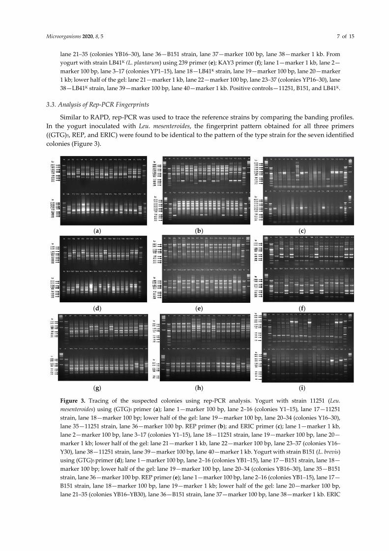
3.3. Analysis of Rep-PCR Fingerprints
Similar to RAPD, rep-PCR was used to trace the reference strains by comparing the banding profiles.
In the yogurt inoculated with Leu. mesenteroides, the fingerprint pattern obtained for all three primers
((GTG)5, REP, and ERIC) were found to be identical to the pattern of the type strain for the seven identified
colonies (Figure 3).
(a) (b) (c)
(d) (e) (f)
(g) (h) (i)
Figure 3. Tracing of the suspected colonies using rep-PCR analysis. Yogurt with strain 11251 (Leu.
mesenteroides) using (GTG)5 primer (a); lane 1—marker 100 bp, lane 2–16 (colonies Y1–15), lane 17—11251
strain, lane 18—marker 100 bp; lower half of the gel: lane 19—marker 100 bp, lane 20–34 (colonies Y16–30),
lane 35—11251 strain, lane 36—marker 100 bp. REP primer (b); and ERIC primer (c); lane 1—marker 1 kb,
lane 2—marker 100 bp, lane 3–17 (colonies Y1–15), lane 18—11251 strain, lane 19—marker 100 bp, lane 20—
marker 1 kb; lower half of the gel: lane 21—marker 1 kb, lane 22—marker 100 bp, lane 23–37 (colonies Y16–
Y30), lane 38—11251 strain, lane 39—marker 100 bp, lane 40—marker 1 kb. Yogurt with strain B151 (L. brevis)
using (GTG)5 primer (d); lane 1—marker 100 bp, lane 2–16 (colonies YB1–15), lane 17—B151 strain, lane 18—
marker 100 bp; lower half of the gel: lane 19—marker 100 bp, lane 20–34 (colonies YB16–30), lane 35—B151
strain, lane 36—marker 100 bp. REP primer (e); lane 1—marker 100 bp, lane 2–16 (colonies YB1–15), lane 17—
B151 strain, lane 18—marker 100 bp, lane 19—marker 1 kb; lower half of the gel: lane 20—marker 100 bp,
lane 21–35 (colonies YB16–YB30), lane 36—B151 strain, lane 37—marker 100 bp, lane 38—marker 1 kb. ERIC

Microorganisms 2020, 8, 5 8 of 15
primer (f); lane 1—marker 1 kb, lane 2—marker 100 bp, lane 3–17 (colonies YB1–15), lane 18—B151 strain,
lane 19—marker 100 bp, lane 20—marker 1 kb; lower half of the gel: lane 21—marker 1 kb, lane 22—marker
100 bp, lane 23–37(colonies YB16–30), lane 38—B151 strain, lane 39—marker 100 bp, lane 40—marker 1 kb.
Yogurt with strain LB41K (L. plantarum) using (GTG)5 primer (g); REP primer (h); ERIC primer (i); lane 1—
marker 1 kb, lane 2—marker 100 bp, lane 3–17 (colonies YP1–15), lane 18—strain LB41K, lane 19—marker 100
bp, lane 20—marker 1 kb; lower half of the gel: lane 21—marker 1 kb, lane 22—marker 100 bp, lane 23–37
(colonies YP16–30), lane 38—LB41K strain, lane 39—marker 100 bp, lane 40—marker 1 kb. Positive controls—
11251, B151, and LB41K.
Among the three primers, the fingerprint profile with bands at 430, 700, 900, 1150, and 1200 bp
positions was observed with (GTG)5 primer (Figure 3a). rep-PCR represented bands at 350, 1050, 1500, 1610,
2000, and 2500 bp in the reference as well as the suspected colonies (Figure 3b). With the ERIC primer, two
main bands of 900 and 1050 bp were observed between the reference strain 11251 and seven suspected
colonies (Figure 3c). These results confirmed that the seven colonies could be the inoculated strain 11251.
On the other hand, parallel to RAPD analysis, nine colonies showed a matching banding profile to B151
with all rep-PCR primers (Figure 3).
With the (GTG)5 primer, three prominent bands at 1000, 1100, and 1500 bp were observed both in the
reference and suspected bacterial colonies (Figure 3d). With REP-PCR, two bands at 1600 and 2000 bp
positions were observed in B151 and nine colonies (Figure 3e). Likewise, the ERIC primer fingerprint
pattern also showed a similar profile with bands at 350, 500, 1200, 2000, and 2200 bp (Figure 3f). The
outcomes of the rep-PCR supported the RAPD analysis, thereby confirming that five colonies were similar
to L. plantarum LB41K (Figure 2). With the (GTG)5 primer, the bands can be seen at 350, 700, 800, and 1000–
1200 bp positions both in the suspected colonies and LB41K (Figure 3g). The REP primer displayed bands at
1000, 1200, 1500, 1700, 2100, and 4000 bp in LB41K and the suspected colonies (Figure 3h). The ERIC primer
produced bands at 380, 1700, and 2800 bp, which were similar in five colonies and LB41K along with a few
lighter bands (Figure 3i).
3.4. Comparative Housekeeping Gene Analysis
Finally, tracing of the inoculated strains was performed by comparing the partial sequence analysis of
the seven housekeeping genes (Tables S2–S4). The colonies confirmed by the previously described
molecular tools were used for the analysis. Therefore, the partial sequences of the housekeeping genes for
Leu. mesenteroides (seven colonies), L. brevis (nine colonies), and of L. plantarum (five colonies) were
compared and analyzed for the presence of SNPs with the reference strains. Alignment of the seven
sequences with the respective partial gene sequences from the reference strain 11251 showed no SNPs in
the seven identified colonies among groEL, gyrB, atpA, pyrG, pheS, rpoA, and uvrC housekeeping genes
(Figure S1A–G). Similar results were obtained for the nine colonies, and all the nucleotide bases matched
the consensus sequences of the B151 strain (Figure S2A–G); no SNPs were observed in any of the
housekeeping (gyrB, groEL, pheS, rpoB, dnaK, rpoA, and recA) genes. Thus, we concluded that the suspected
colonies, Y3, Y7, Y10, Y14–16, and Y27, were the initially inoculated Leu. mesenteroides strain 11251, and the
nine suspected colonies, YB2, YB4, YB9, YB15, YB18, YB21, YB23, YB25, and YB30, were the L. brevis B151
reference strain. Likewise, the comparative gene sequence analysis of ddl, gdh, gyrB, mutS, pgm, purK1, and
tkt4 showed that each of the partial sequences matched with the sequence of LB41K without any SNP in the
housekeeping gene sequences (Figure S3A–G). This confirmed that the detected colonies, YP5, YP6, YP9,
YP15, and YP29, were the LB41 K reference type that was initially inoculated in the yogurt.
3.5. Validation Using Tracing in a Probiotic Powder
For validation, tracing of L. plantarum (Lb41P) was evaluated in a probiotic powder consisting of a
mixture of four bacterial species. As with the other inoculations, 30 colonies were randomly picked and
labelled as PP1–30. The 16S rRNA gene sequencing results (Table 2) showed that seven (PP4, PP7, PP13,
PP14, PP16, PP22, and PP25) of the 29 colonies were L. plantarum, whereas the rest of the colonies were
Microorganisms 2020, 8, 5 9 of 15
identified as S. thermophilus and L. acidophilus. We could not obtain the 16S rRNA gene sequence for the PP8
colony.
Table 2. Results of the 16S rRNA gene sequencing for L. plantarum (LB41p) identified from probiotic powder.
Serial No. Colony ID BLAST Result Serial No. Colony ID BLAST Result
1. PP1 S. thermophilus 16. PP16 L. plantarum
2. PP2 S. thermophilus 17. PP17 S. thermophiles
3. PP3 S. thermophilus 18. PP18 L. acidophilus
4. PP4 L. plantarum 19. PP19 S. thermophilus
5. PP5 S. thermophilus 20. PP20 S. thermophilus
6. PP6 S. thermophilus 21. PP21 S. thermophilus
7. PP7 L. plantarum 22. PP22 L. plantarum
8. PP8 * ------------- 23. PP23 L. acidophilus
9. PP9 L. acidophilus 24. PP24 S. thermophilus
10. PP10 L. acidophilus 25. PP25 L. plantarum
11. PP11 L. acidophilus 26. PP26 L. acidophilus
12. PP12 L. acidophilus 27. PP27 L. acidophilus
13. PP13 L. plantarum 28. PP28 S. thermophilus
14. PP14 L. plantarum 29. PP29 L. acidophilus
15. PP15 S. thermophilus 30. PP30 S. thermophilus
* Sequence data was not available; BLAST: Basic Local Alignment Search Tool; Lactobacillus (L.) plantarum;
Streptococcus (S.) thermophilus; Lactobacillus (L.) acidophilus.
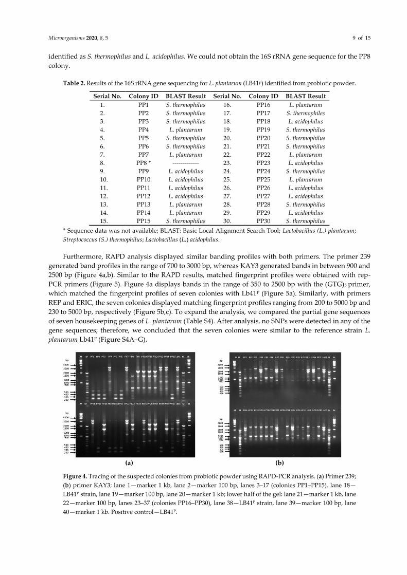
Furthermore, RAPD analysis displayed similar banding profiles with both primers. The primer 239
generated band profiles in the range of 700 to 3000 bp, whereas KAY3 generated bands in between 900 and
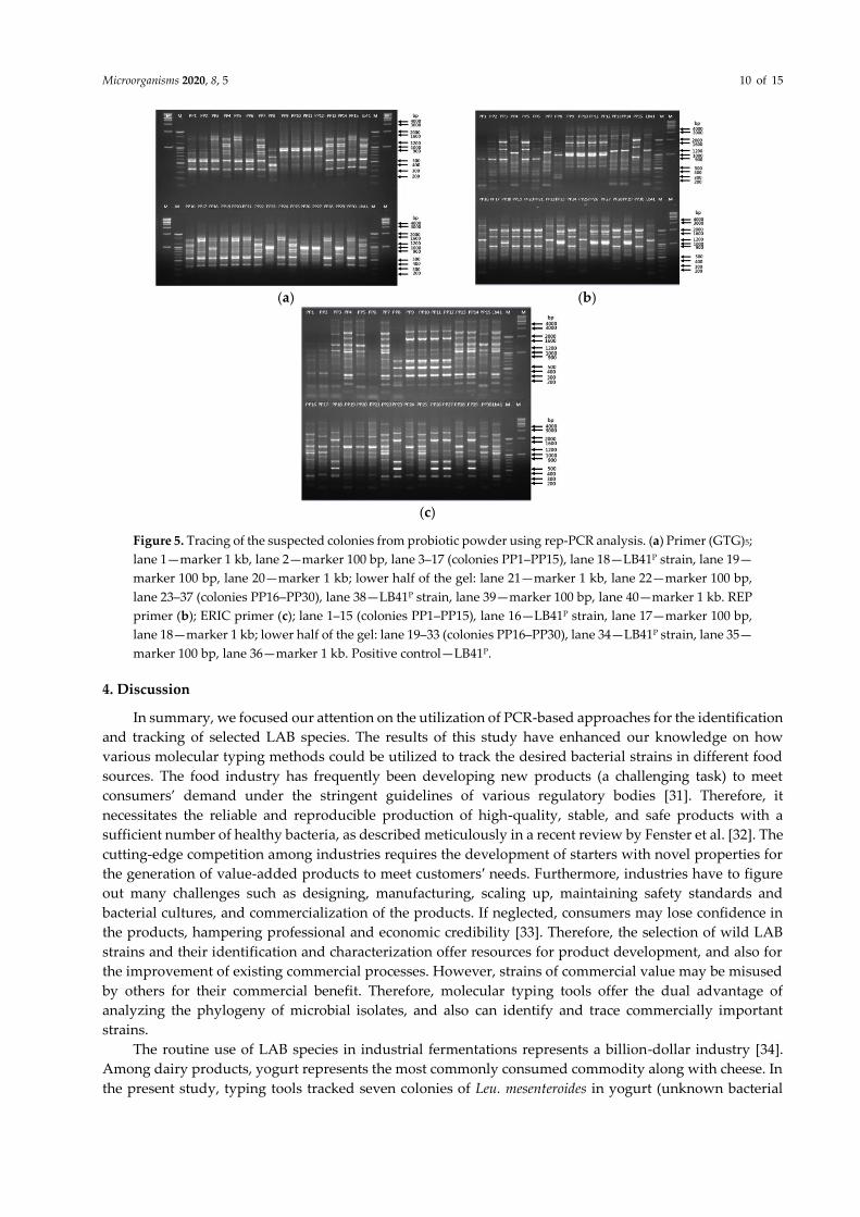
2500 bp (Figure 4a,b). Similar to the RAPD results, matched fingerprint profiles were obtained with rep-
PCR primers (Figure 5). Figure 4a displays bands in the range of 350 to 2500 bp with the (GTG)5 primer,
which matched the fingerprint profiles of seven colonies with Lb41P (Figure 5a). Similarly, with primers
REP and ERIC, the seven colonies displayed matching fingerprint profiles ranging from 200 to 5000 bp and
230 to 5000 bp, respectively (Figure 5b,c). To expand the analysis, we compared the partial gene sequences
of seven housekeeping genes of L. plantarum (Table S4). After analysis, no SNPs were detected in any of the
gene sequences; therefore, we concluded that the seven colonies were similar to the reference strain L.
plantarum Lb41P (Figure S4A–G).
(a) (b)
Figure 4. Tracing of the suspected colonies from probiotic powder using RAPD-PCR analysis. (a) Primer 239;
(b) primer KAY3; lane 1—marker 1 kb, lane 2—marker 100 bp, lanes 3–17 (colonies PP1–PP15), lane 18—
LB41P strain, lane 19—marker 100 bp, lane 20—marker 1 kb; lower half of the gel: lane 21—marker 1 kb, lane
22—marker 100 bp, lanes 23–37 (colonies PP16–PP30), lane 38—LB41P strain, lane 39—marker 100 bp, lane
40—marker 1 kb. Positive control—LB41P.
Microorganisms 2020, 8, 5 10 of 15
(a) (b)
(c)
Figure 5. Tracing of the suspected colonies from probiotic powder using rep-PCR analysis. (a) Primer (GTG)5;
lane 1—marker 1 kb, lane 2—marker 100 bp, lane 3–17 (colonies PP1–PP15), lane 18—LB41P strain, lane 19—
marker 100 bp, lane 20—marker 1 kb; lower half of the gel: lane 21—marker 1 kb, lane 22—marker 100 bp,
lane 23–37 (colonies PP16–PP30), lane 38—LB41P strain, lane 39—marker 100 bp, lane 40—marker 1 kb. REP
primer (b); ERIC primer (c); lane 1–15 (colonies PP1–PP15), lane 16—LB41P strain, lane 17—marker 100 bp,
lane 18—marker 1 kb; lower half of the gel: lane 19–33 (colonies PP16–PP30), lane 34—LB41P strain, lane 35—
marker 100 bp, lane 36—marker 1 kb. Positive control—LB41P.
4. Discussion
In summary, we focused our attention on the utilization of PCR-based approaches for the identification
and tracking of selected LAB species. The results of this study have enhanced our knowledge on how
various molecular typing methods could be utilized to track the desired bacterial strains in different food
sources. The food industry has frequently been developing new products (a challenging task) to meet
consumers’ demand under the stringent guidelines of various regulatory bodies [31]. Therefore, it
necessitates the reliable and reproducible production of high-quality, stable, and safe products with a
sufficient number of healthy bacteria, as described meticulously in a recent review by Fenster et al. [32]. The
cutting-edge competition among industries requires the development of starters with novel properties for
the generation of value-added products to meet customers′ needs. Furthermore, industries have to figure
out many challenges such as designing, manufacturing, scaling up, maintaining safety standards and
bacterial cultures, and commercialization of the products. If neglected, consumers may lose confidence in
the products, hampering professional and economic credibility [33]. Therefore, the selection of wild LAB
strains and their identification and characterization offer resources for product development, and also for
the improvement of existing commercial processes. However, strains of commercial value may be misused
by others for their commercial benefit. Therefore, molecular typing tools offer the dual advantage of
analyzing the phylogeny of microbial isolates, and also can identify and trace commercially important
strains.
The routine use of LAB species in industrial fermentations represents a billion-dollar industry [34].
Among dairy products, yogurt represents the most commonly consumed commodity along with cheese. In
the present study, typing tools tracked seven colonies of Leu. mesenteroides in yogurt (unknown bacterial

Microorganisms 2020, 8, 5 11 of 15
pool), which were identical to the reference strain, 11251. For the tracking of L. brevis B151 and L. plantarum
LB41K in yogurt (known pool—S. thermophilus, B. longum, L. casei, and L. acidophilus), nine and five colonies
were identified, respectively. It is to be noted that Leu. mesenteroides is not generally used to produce
commercial yogurt; however, some strains of Leu. mesenteroides have been used in the production of cheese
[35]. Hence, as per the 16S rRNA gene sequencing results, it can be presumed that the colonies were the
inoculated strain, Leu. mesenteroides 11251. However, a study by Chun et al. explained that 16S rRNA gene
sequencing is not a suitable tool for inferring the phylogeny of Leu. mesenteroides strains [36]. Usually, also,
16S rRNA gene sequencing does not permit explicit separation of all bacterial strains at the species or
subspecies level, which necessitates the use of other molecular tools [17,37]. In the RAPD analysis, most of
the colonies displayed identical banding profiles to the 11251 strain with primer 239, which could be due to
the similar binding sites for the primer in the genomes isolated from the 30 colonies. In Mechnikop yogurt,
three (B. longum, L. casei, and L. acidophilus) of the four species were not detected, which reflects that these
species require supplemented growth media and different culture conditions to grow. Bifidobacterium spp.
generally prefer to grow MRS/MRS-nalidixic acid, paromomycin, neomycin sulphate, and lithium chloride
(NPNL) at 37 °C for 72 h under anaerobic conditions. L. acidophilus shows selective growth on MRS agar
supplemented with maltose (MRSM) or 5-bromo-4-chloro-3-indolyl-β-D-glucopyranoside (X-Glu) followed
by anaerobic incubation. On the other hand, L. casei can grow on selective medium containing ribose (1%
w/v) LC agar, anaerobic incubation at 27 °C. In addition, many other media have also been reported for the
enumeration of these aforementioned bacterial species [38]. The remaining colonies, other than those
suspected, were identified as S. thermophilus. Furthermore, the bacterial pool in the yogurt (Korea Yakult)
inoculated with L. brevis and L. plantarum LB41K strains were already known (marked on the label), and it
was certain that the yogurt was not made by utilizing any of the L. brevis and L. plantarum strains. Thus, the
identified colonies should be the inoculated strains, B151 and L. plantarum LB41K.
PCR-based fingerprinting tools such as RAPD [12] and rep-PCR [39] had parallel discriminatory
powers and appeared apposite for the distinction of bacteria. However, the pitfalls include reproducibility
and comparability between different research laboratories [40]. Therefore, the necessity of identification and
higher discrimination of LAB has led to the use of housekeeping gene sequence analysis [41,42]. Research
by Shevtsov et al. showed that the identification of the Lactobacillus genus using housekeeping gene
sequences is superior and more sensitive than the 16S rRNA gene sequencing [43]. Previously, our group
characterized different strains of Leu. mesenteroides and L. brevis from Korea using the multilocus sequence
typing (MLST) molecular tool [44]. The same set of gene sequences were utilized for the comparative gene
sequence analysis to identify the deliberately inoculated Leu. mesenteroides 11251 and L. brevis B151 in yogurt
[45,46]. For the L. plantarum strains, the housekeeping genes were used from an earlier described MLST
study [25].
In industrial fermentations, L. plantarum is the most commonly used bacterial species as a microbial
starter or probiotic bacteria [47]. Therefore, to validate the approach, the tracing of a L. plantarum strain from
a commercial probiotic powder was investigated by identifying the LB41P strain. All four molecular tools
effectively identified seven colonies as the reference strain. These results confirm the utility of the developed
technology for the tracking of desired bacterial strains.
Capillary sequencing (also known as Sanger sequencing) is a fast and cost-effective technology that is
suitable for a low number of targets such as cloned DNA fragments or PCR products. It suffers challenges
such as low sensitivity, high cost per sample for large number of targets, and challenging to scale. On the
other hand, using next-generation technologies (NGS), one can reach a better conclusion about the
identification of the suspected strain. The important criteria of this technology include read length, quality
of the sequence, and cost. Ideally, the sequence data should be with long read length and low error rates.
However, there is no report of such a technology. This technology is favored where novel or unique variants
of the bacteria are required. Moreover, NGS is a labor-intensive, costly affair that requires technical expertise
with heavy and costly equipment. Moreover, approximately 4000 USD is required to sequence the complete
genome of probiotic bacteria [33]. Moreover, not all probiotic industrial strains have been sequenced.
Therefore, NGS use is not feasible for every industry (especially small-scale industry) or laboratory. In
Microorganisms 2020, 8, 5 12 of 15
contrast, the tools used in this study are comparatively easy, cost-effective, and can be used in the
identification of dubious strains.
Flow cytometry (FC) is a quick and automated method for enumeration, detection, and microbial
profiling. However, the identity of microorganisms depends upon the use of different probes and
fluorescent dyes. Also, the cost of this sophisticated and expensive instrument and technical operator is
much higher compared to the tools used in the present technology. On the other hand, analysis time for FC
is faster, around 15–20 min compared to 2–5 days for culturing of bacteria [48]. Recently, FC has been
utilized for the viability assessment and quantification of microorganisms in multi-strain probiotic products
[49]. Mass spectrometry is a fast and less labor-intensive technique for bacterial identification. The
approximate cost was reported to be 0.5 to 1.00 USD per sample [50]. However, pretreatment of chemicals,
temperature, and media composition may affect the quality of spectra. In addition, the technique is suitable
for pure cultures only, and provides information at the genus level, not at the species or subspecies level
[51]. Nonetheless, these methods are still research-based and costs vary depending upon the number of
samples for analysis.
In our research, all the molecular methods were proven to be practical tools in the tracing of
intentionally inoculated strains. In addition, the yogurt utilized was not made from any of the LAB strains
selected for the present study. Nonetheless, as discussed earlier, all the molecular tools have some
disadvantages; therefore, the identification of the desired strain cannot depend on a particular method and
should be followed by a combined approach. Notably, the selection of the typing method depends on the
objective of the research, the availability of skilled personnel, and, most importantly, the resources in the
laboratory. It is a fact that the comparison of housekeeping gene sequences is a measured approach.
However, new alternatives such as whole-genome sequencing (WGS) and whole-genome MLST (wgMLST)
are becoming popular (subject to affordability) for the description and identification of a bacterial species.
Further investigations should be focused on the utilization of these technologies.
5. Strengths and Limitations of the Study
The strength of the present study includes the use of practically viable molecular tools such as RAPD,
rep-PCR, and comparative gene sequence analysis for the identification of three LAB species. Moreover, the
technology was validated by identifying target bacterial species from a commercial product. In addition,
the presented cost-effective technology can be easily performed in a basic molecular biology lab. It can be
used for the identification of bacteria in most food products. The limitations of the study include the time-
consuming culture dependency and the selection of food products without the presence of target reference
strains.
6. Conclusions
Molecular tools for identifying microorganisms have been emerging in recent decades. Nonetheless,
many limitations such as high running costs, skilled workforce, and expensive equipment need to be
overcome for many of these newly developed technologies. Herein, our approach describes a technology
with low cost and simple instrumentation for the identification of LAB. Our analysis of the 30 random
selections and corresponding reference strains showed the practical feasibility of the approach for
identifying suspected LAB strains. These PCR-based molecular tools showed efficacy for the identification
of suspected Leu. mesenteroides, L. brevis, and L. plantarum (LB41P) in commercial yogurt, and substantiated
viability by identifying L. plantarum (LB41K) in a commercial probiotic powder. Overall, the union of these
low-cost molecular tools would help users for the identification of suspected LAB or other probiotics strains.
Supplementary Materials: The following are available online at www.mdpi.com/xxx/s1: Figure S1: Comparative gene
sequence analysis—groEL (A), gyrB (B), atpA (C), pyrG (D), pheS (E), rpoA (F), and uvrC (G) of seven colonies isolated
from yogurt inoculated with reference strain 11251 (Leu. mesenteroides). Figure S2: Comparative gene sequence
analysis—gyrB (A), groEL (B), pheS (C), rpoB (D), dnaK (E), rpoA (F), and recA (G) of nine colonies isolated from yoghurt
inoculated with reference strain B151 (L. brevis). Figure S3: Comparative gene sequence analysis—ddl (A), gdh (B), gyrB
(C), mutS (D), pgm (E), purK1 (F), and tkt4 (G) of five colonies isolated from yogurt inoculated with the reference strain

Microorganisms 2020, 8, 5 13 of 15
LB41K (L. plantarum). Figure S4: Comparative gene sequence analysis—ddl (A), gdh (B), gyrB (C), mutS (D), pgm (E),
purK1 (F), and tkt4 (G) of seven colonies isolated from probiotic powder with the reference strain LB41P (L. plantarum).
Table S1: PCR conditions used for rep-PCR and for the amplification of housekeeping genes of the target colonies. Table
S2: Information of housekeeping loci and primers used for Leu. mesenteroides 11251 strain. Table S3: Information of
housekeeping loci and primers used for L. brevis B151 strain. Table S4: Information of housekeeping loci and primers
used for L. plantarum LB41 strains.
Author Contributions: Conceptualization and supervision Y.-S.P; methodology, J.K. and A.S.; validation, A.S. and J.K.;
writing—original draft preparation, A.S.; investigation, Y.-S.P., S.L., and A.S.; writing—review and editing, A.S. and
Y.-S.P.; supervision, Y.-S.P.; project administration, S.L. and J.K.; funding acquisition, Y.-S.P.
Funding: This research was funded by the Korea Institute of Planning and Evaluation for Technology in Food,
Agriculture, Forestry, and Fisheries (IPET) through the High Value-Added Food Technology Development Program,
funded by the Ministry of Agriculture, Food, and Rural Affairs (grant number 314073-03-2-HD040) and Gachon
University research fund of 2018 (GCU-2018-0677).
Conflicts of Interest: The authors declare no conflict of interest.
References
1. Wassenaar, T.M.; Klein, G. Safety aspects and implications of regulation of probiotic bacteria in food and food
supplements. J. Food Protect. 2008, 71, 1734–1741.
2. Marteau, P.R.; Vrese, M.D.; Cellier, C.J.; Schrezenmeir, J. Protection from gastrointestinal diseases with the use of
probiotics. Am. J. Clin. Nutr. 2001, 73, 430s–436s.
3. Douglas, G.L.; Klaenhammer, T.R. Genomic evolution of domesticated microorganisms. Ann. Rev. Food Sci.
Technol. 2010, 1, 397–414.
4. Kang, C.-H.; Han, S.H.; Kim, J.-S.; Kim, Y.; Jeong, Y.; Park, H.M.; Paek, N.-S. Inhibition of nitric oxide production,
oxidative stress prevention, and probiotic activity of lactic acid bacteria isolated from the human vagina and
fermented food. Microorganisms 2019, 7, 109.
5. Kamiya, T.; Watanabe, Y.; Makino, S.; Kano, H.; Tsuji, N. Improvement of intestinal immune cell function by lactic
acid bacteria for dairy products. Microorganisms 2016, 5, 1.
6. Baradaran, A.; Foo, H.L.; Sieo, C.C.; Raha, A. Isolation, identification and characterization of lactic acid bacteria
from Polygonum minus. Rom. Biotechnol. Lett. 2012, 17, 7245–7252.
7. Fu, L.-L.; Li, J.-R. Microbial source tracking: A tool for identifying sources of microbial contamination in the food
chain. Crit. Rev. Food Sci. Nutr. 2014, 54, 699–707.
8. Temmerman, R.; Huys, G.; Swings, J. Identification of lactic acid bacteria: Culture-dependent and culture-
independent methods. Trends Food Sci. Technol. 2004, 15, 348–359.
9. Dan, T.; Liu, W.; Sun, Z.; Lv, Q.; Xu, H.; Song, Y.; Zhang, H. A novel multi-locus sequence typing (MLST) protocol
for Leuconostoc lactis isolates from traditional dairy products in China and Mongolia. BMC Microbiol. 2014, 14, 150.
10. Ben Amor, K.; Vaughan, E.E.; de Vos, W.M. Advanced molecular tools for the identification of lactic acid bacteria.
J. Nutr. 2007, 137, 741S–747S.
11. Davis, C. Enumeration of probiotic strains: Review of culture-dependent and alternative techniques to quantify
viable bacteria. J. Microbiol. Methods 2014, 103, 9–17.
12. Kaur, J.; Lee, S.; Park, Y.-S.; Sharma, A. RAPD analysis of Leuconostoc mesenteroides strains associated with
vegetables and food products from Korea. LWT-Food Sci. Technol. 2017, 77, 383–388.
13. Abdollahniya, D.; Hosseini, S.M.; Baghbaderani, B.K.; Mordadi, A.; Reza, M. Identification of Lactobacillus species
isolated from traditional dairy products using RAPD-PCR. Avicenna J. Clin. Microbiol. Infect. 2018, 5, 7–13.
14. Gevers, D.; Huys, G.; Swings, J. Applicability of rep-PCR fingerprinting for identification of Lactobacillus species.
FEMS Microbiol. Lett. 2001, 205, 31–36.
15. Healy, M.; Huong, J.; Bittner, T.; Lising, M.; Frye, S.; Raza, S.; Schrock, R.; Manry, J.; Renwick, A.; Nieto, R.
Microbial DNA typing by automated repetitive-sequence-based PCR. J. Clin. Microbiol. 2005, 43, 199–207.
16. Bautista-Gallego, J.; Arroyo-López, F.; Rantsiou, K.; Jiménez-Díaz, R.; Garrido-Fernández, A.; Cocolin, L.
Screening of lactic acid bacteria isolated from fermented table olives with probiotic potential. Food Res. Int. 2013,
50, 135–142.

Microorganisms 2020, 8, 5 14 of 15
17. Vankerckhoven, V.; Huys, G.; Vancanneyt, M.; Vael, C.; Klare, I.; Romond, M.-B.; Entenza, J.M.; Moreillon, P.;
Wind, R.D.; Knol, J. Biosafety assessment of probiotics used for human consumption: Recommendations from the
EU-PROSAFE project. Trends Food Sci. Technol. 2008, 19, 102–114.
18. Laroute, V.; Tormo, H.; Couderc, C.; Mercier-Bonin, M.; Le Bourgeois, P.; Cocaign-Bousquet, M.; Daveran-Mingot,
M.-L. From genome to phenotype: An integrative approach to evaluate the biodiversity of Lactococcus lactis.
Microorganisms 2017, 5, 27.
19. Ruiz, P.; Celada, L.; Seseña, S.; Palop, M.L. Leuconostoc mesenteroides in the brewing process: A controversial role.
Food Control 2018, 90, 415–421.
20. Salvetti, E.; Torriani, S.; Felis, G.E. The genus Lactobacillus: A taxonomic update. Probiotics Antimicrob. Proteins 2012,
4, 217–226.
21. Fusco, V.; Quero, G.M.; Chieffi, D.; Franz, C.M. Identification of Lactobacillus brevis using a species-specific AFLP-
derived marker. Int. J. Food Microbiol. 2016, 232, 90–94.
22. Fang, F.; Xu, J.; Li, Q.; Xia, X.; Du, G. Characterization of a Lactobacillus brevis strain with potential oral probiotic
properties. BMC Microbiol. 2018, 18, 221.
23. Bergsveinson, J.; Pittet, V.; Ewen, E.; Baecker, N.; Ziola, B. Genome sequence of rapid beer-spoiling isolate
Lactobacillus brevis BSO 464. Genome Announc. 2015, 3, e01411–e01415.
24. Siezen, R.J.; Tzeneva, V.A.; Castioni, A.; Wels, M.; Phan, H.T.; Rademaker, J.L.; Starrenburg, M.J.; Kleerebezem,
M.; Molenaar, D.; van Hylckama Vlieg, J.E. Phenotypic and genomic diversity of Lactobacillus plantarum strains
isolated from various environmental niches. Environ. Microbiol. 2010, 12, 758–773.
25. De las Rivas, B.; Marcobal, Á.; Muñoz, R. Development of a multilocus sequence typing method for analysis of
Lactobacillus plantarum strains. Microbiology 2006, 152, 85–93.
26. Altay, F.; Karbancıoglu-Güler, F.; Daskaya-Dikmen, C.; Heperkan, D. A review on traditional Turkish fermented
non-alcoholic beverages: Microbiota, fermentation process and quality characteristics. Int. J. Food Microbiol. 2013,
167, 44–56.
27. Guidone, A.; Zotta, T.; Ross, R.P.; Stanton, C.; Rea, M.C.; Parente, E.; Ricciardi, A. Functional properties of
Lactobacillus plantarum strains: A multivariate screening study. LWT-Food Sci. Technol. 2014, 56, 69–76.
28. Versalovic, J.; Schneider, M.; De Bruijn, F.; Lupski, J.R. Genomic fingerprinting of bacteria using repetitive
sequence-based polymerase chain reaction. Methods Mol. Cell. Biol. 1994, 5, 25–40.
29. Hall, T.A. BioEdit: A user-friendly biological sequence alignment editor and analysis program for Windows
95/98/NT. Nucleic Acids Symp. Ser. 1999, 41, 95–98.
30. Thompson, J.D.; Gibson, T.J.; Plewniak, F.; Jeanmougin, F.; Higgins, D.G. The CLUSTAL_X windows interface:
Flexible strategies for multiple sequence alignment aided by quality analysis tools. Nucleic Acids Res. 1997, 25,
4876–4882.
31. Franz, C.; Cho, G.-S.; Holzapfel, W.H.; Gálvez, A. Safety of Lactic Acid Bacteria. Wiley Online Library: Hoboken, NJ,
USA, 2010; pp 341–360.
32. Fenster, K.; Freeburg, B.; Hollard, C.; Wong, C.; Rønhave Laursen, R.; Ouwehand, A.C. The production and
delivery of probiotics: A review of a practical approach. Microorganisms 2019, 7, 83.
33. Jackson, S.A.; Schoeni, J.L.; Vegge, C.; Pane, M.; Stahl, B.; Bradley, M.; Goldman, V.S.; Burguière, P.; Atwater, J.B.;
Sanders, M.E. Improving end-user trust in the quality of commercial probiotic products. Front. Microbiol. 2019, 10,
739.
34. De Vos, W.M. Systems solutions by lactic acid bacteria: From paradigms to practice. In Proceedings of the
Microbial Cell Factories, Egmond aan Zee, The Netherlands, 28 August-1 September 2011; p. S2.
35. Cibik, R.; Lepage, E.; Tailliez, P. Molecular diversity of Leuconostoc mesenteroides and Leuconostoc citreum isolated
from traditional French cheeses as revealed by RAPD GTG printing, 16S rDNA sequencing and 16S rDNA
fragment amplification. Syst. Appl. Microbiol. 2000, 23, 267–278.
36. Chun, B.H.; Kim, K.H.; Jeon, H.H.; Lee, S.H.; Jeon, C.O. Pan-genomic and transcriptomic analyses of Leuconostoc
mesenteroides provide insights into its genomic and metabolic features and roles in kimchi fermentation. Sci. Rep.
2017, 7, 11504.
37. Fox, G.E.; Wisotzkey, J.D.; Jurtshuk P., Jr. How close is close: 16S rRNA sequence identity may not be sufficient to
guarantee species identity. Int. J. Syst. Evol. Microbiol. 1992, 42, 166–170.

Microorganisms 2020, 8, 5 15 of 15
38. Ashraf, R.; Shah, N.P. Selective and differential enumerations of Lactobacillus delbrueckii subsp. bulgaricus,
Streptococcus thermophilus, Lactobacillus acidophilus, Lactobacillus casei and Bifidobacterium spp. in yoghurt—
A review. Int. J. Food Microbiol. 2011, 149, 194–208.
39. Kaur, J.; Lee, S.; Sharma, A.; Park, Y.-S. DNA profiling of Leuconostoc mesenteroides strains isolated from fermented
foods and farm produce in Korea by repetitive-element PCR. Food Sci. Biotechnol. 2017, 26, 1667–1673.
40. Williams, J.G.; Hanafey, M.K.; Rafalski, J.A.; Tingey, S.V. Genetic analysis using random amplified polymorphic
DNA markers. In Recombinant DNA Methodology II; Elsevier: Amsterdam, The Netherlands, 1995; pp. 849–884.
41. Gevers, D.; Cohan, F.M.; Lawrence, J.G.; Spratt, B.G.; Coenye, T.; Feil, E.J.; Stackebrandt, E.; Van de Peer, Y.;
Vandamme, P.; Thompson, F.L. Re-evaluating prokaryotic species. Nat. Rev. Microbiol. 2005, 3, 733.
42. Konstantinidis, K.T.; Tiedje, J.M. Towards a genome-based taxonomy for prokaryotes. J. Bacteriol. 2005, 187, 6258–
6264.
43. Shevtsov, A.; Kushugulova, A.; Tynybaeva, I.; Kozhakhmetov, S.; Abzhalelov, A.; Momynaliev, K.; Stoyanova, L.
Identification of phenotypically and genotypically related Lactobacillus strains based on nucleotide sequence
analysis of the groEL, rpoB, rplB, and 16S rRNA genes. Microbiology 2011, 80, 672.
44. Sharma, A.; Kaur, J.; Lee, S.; Park, Y.-S. Analysis of Leuconostoc citreum strains using multilocus sequence typing.
Food Sci. Biotechnol. 2018, 27, 1755–1760.
45. Sharma, A.; Kaur, J.; Lee, S.; Park, Y.-S. Genetic diversity analysis of Leuconostoc mesenteroides from Korean
vegetables and food products by multilocus sequence typing. Appl. Microbiol. Biotechnol. 2018, 102, 4853–4861.
46. Sharma, A.; Kaur, J.; Lee, S.; Park, Y.-S. Molecular discrimination of Lactobacillus brevis strains isolated from food
products in South Korea using multilocus sequence typing. LWT-Food Sci. Technol. 2017, 86, 337–343.
47. Behera, S.S.; Ray, R.C.; Zdolec, N. Lactobacillus plantarum with functional properties: An approach to increase safety
and shelf-life of fermented foods. Biol. Med. Res. Int. 2018, 2018, doi:10.1155/2018/9361614.
48. Van Nevel, S.; Koetzsch, S.; Proctor, C.R.; Besmer, M.D.; Prest, E.I.; Vrouwenvelder, J.S.; Knezev, A.; Boon, N.;
Hammes, F. Flow cytometric bacterial cell counts challenge conventional heterotrophic plate counts for routine
microbiological drinking water monitoring. Water Res. 2017, 113, 191–206.
49. Chiron, C.; Tompkins, T.A.; Burguière, P. Flow cytometry: A versatile technology for specific quantification and
viability assessment of micro-organisms in multistrain probiotic products. J. Appl. Microbiol. 2018, 124, 572–584.
50. Rodríguez-Sánchez, B.; Marín, M.; Sánchez-Carrillo, C.; Cercenado, E.; Ruiz, A.; Rodríguez-Créixems, M.; Bouza,
E. Improvement of matrix-assisted laser desorption/ionization time-of-flight mass spectrometry identification of
difficult-to-identify bacteria and its impact in the workflow of a clinical microbiology laboratory. Diagn. Microbiol.
Infect. Dis. 2014, 79, 1–6.
51. Sloan, A.; Wang, G.; Cheng, K. Traditional approaches versus mass spectrometry in bacterial identification and
typing. Clin. Chim. Acta 2017, 473, 180–185.
© 2019 by the authors. Licensee MDPI, Basel, Switzerland. This article is an open access
article distributed under the terms and conditions of the Creative Commons Attribution
(CC BY) license (http://creativecommons.org/licenses/by/4.0/).