effects of common sterilization methods on the structure and properties of poly(d,l...
TRANSCRIPT
Effects of Common Sterilization Methods on the Structure and
Properties of Poly(D,L Lactic-Co-Glycolic Acid) Scaffolds
HOLLY SHEARER, M.Eng., MARIANNE J. ELLIS, Ph.D., SEMALI P. PERERA, Ph.D.,and JULIAN B. CHAUDHURI, Ph.D.
ABSTRACT
While methods for the production of scaffolds with the appropriate mechanical properties and archi-tecture for tissue engineering are attracting much attention, the effects of subsequent sterilization pro-cesses on the scaffold properties have often been overlooked. This study sought to determine the effects ofsterilization with ethanol, peracetic acid, ultraviolet irradiation, and antibiotic solution on the structure of50:50 (mol:mol) 65:35, and 85:15 poly(D,L-lactic-co-glycolic acid [PLGA]) flat-sheet and hollow-fiberscaffolds. All methods resulted in scaffold sterilization, but scanning electron microscopy revealed de-formations to the scaffold surface for all treatments. The extent of surface damage increased with treat-ment duration. This was further investigated by measurement of pore sizes, water flux, breaking strain,and Young’s modulus. External pore size and water flux was found to be increased by all treatments in thefollowing order: ethanol (largest), antibiotics, ultraviolet light, and peracetic acid. Pore sizes were 0.25 to0.17lm and water flux ranged from 0.01 kg�m–2 � s–1 to 3.34 kg�m–2 � s–1. For all samples, the Young’smodulus was 1.0 to 31.1MPa and breaking strain was 1.2 to 2.4MPa. The results of this study suggest thatantibiotic treatment shows the most potential to sterilize PLGA hollow fibers for tissue engineering.
INTRODUCTION
T ISSUE ENGINEERING IS WIDELY ANTICIPATED TO REPLACE
traditional graft procedures for treatment of tissue and
organ defects. Successful tissue generation in vitro requires
highly specialized scaffolds. Mass transfer, topography,1
surface chemistry,1,2 mechanical properties,3 and degrada-
tion rates3 are just some of the factors that influence the
ability of cells to colonize a scaffold, produce extracellular
matrix molecules, and form an organized tissue construct.
Implantation in vivo requires the scaffold to be biocom-
patible, integrate with the surrounding natural tissue, and,
in many situations over a favorable time scale, to be com-
pletely eliminated from the host via biodegradation.
Materials and production methods for suitable polymer
scaffolds attract much research attention; however, the ef-
fects of sterilizing these scaffolds before cell seeding are
often overlooked. Low polymer melting points, complex
architectures, and hydrolytic degradation mechanisms result
in scaffolds that may be easily damaged by harsh steriliza-
tion protocols. Typical medical or cell culture sterilization
methods may prove unsuitable for polymer scaffolds. For
example, standard techniques such as autoclaving, ethylene
oxide treatment, or gamma irradiation have been shown to
be unsuitable for polymer scaffolds because of deformation
from elevated temperatures, lengthy degassing, and deteri-
oration due to decreased molecular weights.4
This study investigates 4 methods for sterilizing flat-sheet
and hollow-fiber poly(D,L-lactic-co-glycolic) (PLGA) scaf-
folds with the objective of achieving scaffold sterilizat-
ion with minimal or no structural damage. The treatments
comprise 70% ethanol treatment; ultraviolet irradiation;
peracetic acid treatment; and antibiotic treatment with pe-
nicillin G, streptomycin sulfate, and amphotericin B. Chan-
ges to the topography of the scaffold, which may affect the
ability of cells to adhere to the scaffold, were determined by
Centre for Regenerative Medicine, Department of Chemical Engineering, University of Bath, Bath, United Kingdom.
TISSUE ENGINEERINGVolume 12, Number 10, 2006# Mary Ann Liebert, Inc.
2717
scanning electron microscopy. Gas permeation and water
flux were measured to calculate the effects of sterilization
method on the pore diameter and to quantify mass transfer
parameters, respectively. The mechanical properties of the
fiber were determined by measuring the breaking stress and
strain.
MATERIALS AND METHODS
Preparation of polymer flat sheets
and hollow fibers
Polymer solutions were prepared from 50:50 (mol:mol),
65:35, and 85:15 PLGA (Alkermes, Inc., Cincinnati, Ohio)
with 1-methyl-2-pyrrolidinone (NMP;AcrosOrganics,Geel,
Belgium) in a 20% (w/w) solution.5 Flat-sheet scaffolds
were formed by solvent exchange with distilled water; 100-
mm thickness sheets were cast onto glass supports and im-
mersed in distilled water at 10–158C. After a fewminutes the
scaffolds were lifted off the glass support but left in water for
24 h to ensure solvent removal. The scaffolds were then
removed from the water tank and dried in ambient condi-
tions (8–208C) for at least 24 h. The 50:50 PLGA hollow
fibers were wet-spun through a double-orifice spinneret;
polymer was extruded under low pressure (approximately 2
barg) through the outer annular orifice (outer diameter,
1.0mm; inner diameter, 0.7mm), polymer flow rate was
controlled with a ball valve. Distilled water was filtered
(40 mm; Swagelok, Bristol, UK) and pumped concurrently
through the inner orifice (inner diameter, 0.4mm) at a rate of
3.5–7.0mLmin–1. Fibers passed through 2 water tanks, and
a motorized roller guided them at a rate of 7–9mmin–1 into a
final water tank, where they were left for 24 h before drying
in ambient conditions (8–208C).
Sterilization treatment
Flat sheets were cut into circular 13-mm (outer diameter)
membrane supports (Minucells and Minutissue, Vertriebs
GmbH, Bad Abbach, Germany), excess scaffold was re-
moved with a scalpel. Hollow fibers were cut into 10-mm
lengths. One flat-sheet or 2 hollow-fiber sections were ster-
ilized in individual cells in a polystyrene 24-well plate
(Nunclon, Nalge Nunc International, Roskilde, Denmark).
Both flat-sheet and hollow-fiber samples had a mean mass of
4� 0.6mg (mean� SD; n¼ 12) before treatment.
Ethanol sterilization. A 1 mL 70% (v/v) ethanol solution
(diluted from 96% [v/v] ethanol [Fisher Scientific UK Ltd,
Loughborough, UK], with distilled water) was added to
each sample. Samples were treated for 15min, 30min, 1 h,
2 h, 5 h, and 24 h and were subsequently rinsed 5 times with
1mL phosphate-buffered saline (PBS; Sigma-Aldrich,
Dorset, UK).6 For water flux experiments, fibers were trea-
ted after potting into the bioreactor by submerging the
reactor in ethanol solution for 30min, followed by rinsing
thoroughly with deionized water.
Ultraviolet treatment. Irradiation was carried out with a
12-W ultraviolet lamp (Syngene, Cambridge, UK) at 254-
nm wavelength at a distance of 15 cm. Samples were irra-
diated for a total time of 30min, 1 h, 2 h, and 5 h,7 and were
turned over halfway through the treatment to irradiate the
top and bottom surfaces. Flat-sheet samples were also pre-
pared without turning.
Antibiotic treatment. Polymer samples were placed in a
solution of 1% (v/v) antibiotic antimycotic solution (10 000
unitsmL–1 penicillin G, 10mgmL–1 streptomycin sulfate,
and 25 mgmL–1 amphotericin B [Sigma-Aldrich, Dorset,
UK) in PBS and incubated at 48C for 6 h, 15 h, 24 h, and
31 h. Samples were rinsed 3 times with PBS before use.
Peracetic acid treatment. Peracetic acid treatment was
performed for the same time intervals as for ethanol treat-
ment with a solution of 0.1% (v/v) peracetic acid (peracetic
acid solution 39% in acetic acid, Sigma-Aldrich, Gilling-
ham, UK), 4% (v/v) ethanol 95.9% (v/v) distilled water.
Samples were rinsed 3 times in 1mL PBS for 1 h each.8
Preparation of controls. Control flat-sheet samples were
prepared as for antibiotic treatment; PBS or distilled water
replaced the antibiotic solution. Samples were not rinsed
after treatment. Untreated control scaffolds were also tested.
Analysis
Sterility testing. 50:50 PLGA samples were incubated for
48 h at 378C and 5% carbon dioxide in 1mL media (Dul-
becco’s modified Eagle’s medium [GIBCO, Paisley, UK,
supplemented with 10% fetal bovine serum, 1% nones-
sential amino acids [Sigma-Aldrich, Dorset, UK, and 1%
sodium pyruvate [GIBCO]) and checked periodically
for signs of infection, indicated by yellowing of the media
(Dulbecco’s modified Eagle’s medium contains the indi-
cator phenol red) and increased media opacity. Unsterilized
scaffolds were used as a positive control, and media with
no scaffold was used as a negative control.
Scanning electron microscopy. Samples were prepared
with a scalpel to reveal all the surfaces; liquid nitrogen was
used as required to freeze the polymer and prevent struc-
tural damage. Samples were mounted on a sample tray
using double-sided sticky carbon discs and sputtered with
gold (5150B sputter coater, BOC Edwards, West Sussex,
UK) before being viewed with the scanning electron micro-
scope (JSM6310, JEOL, Herts, UK). The surface porosity
and pore size were approximated from scanning electron
microscopy images using ImageJ software.9 Pore size and
porosity could be estimated only for samples showing a
high contrast between the pores and the polymer surface.
2718 SHEARER ET AL.
Measurements could not be made for highly wrinkled sur-
faces.
Gas permeation. Hollow fibers 120mm long were ster-
ilized in batches of 12 in 70-mL glass boiling tubes. The
membrane characterization technique of gas permeation was
used to determine pore diameter. Samples were glued using
epoxy resin (Araldite Rapid, Huntsman, Everberg, Belgium)
into 10-cm-long, 6-mm-diameter stainless-steel tubes. At one
end the lumen of the fiber was sealed, and at the other end
space around the fiber (the ablumen) was sealed. Nitrogen
permeation through the fiber walls was measured for inlet
pressures between 1 barg andmembrane failure (typically 5–6
barg). Pore sizes were calculated on the basis of the as-
sumption of Poiseuille flow through porous media; permea-
tion flux was plotted against transmembrane pressure,
allowing the intercept (K0) and gradient (P0) to be used with
the gas constant (R), the temperature (T), the molecular
weight of the gas (M), and the gas viscosity (m) to calculate
pore radius (r).10
r ¼ 16
3
P0
K0
� �8RT
pM
� �0:5
l (1)
Water flux. Samples were prepared as for gas permea-
tion and fitted into a stainless-steel custom bioreactor; a
100-mm-long, 12-mm-diameter tube formed the body of
the reactor with two 6-mm side ports. Ten fibers were
placed in the bioreactor; the shell side was sealed at both
ends using epoxy resin. Water circulated through the tube
side by means of a peristaltic pump (503U, Watson-Marlow
Bredel Pumps Ltd, Cornwall, UK); permeate was collected
on the shell side. Pressure gauges (0–6 barg; Bailey &
Mackey Ltd., Birmingham, UK) measured inlet and outlet
pressures, and a nonrotating stem valve on the outlet con-
trolled the transmembrane pressure.
Mechanical testing. Hollow fibers were tested to breaking
tension (model 1122 electromechanical test system, Instron,
Norwood, MA) with a full scale load of 500 g and a cross-
head speed of 20mmmin–1. Samples were tested dry or were
kept in distilled water after sterilization and tested within
30min. Ultraviolet and control wet samples were soaked in
distilled water for 2 h before testing. Breaking stress (s) andYoung’s modulus (E) were calculated as follows:
r ¼ mg
A(2)
e ¼ l
L(3)
E ¼ re; (4)
where m is load at failure, g is acceleration due to gravity, A
is fiber cross-sectional area, e is strain, l is extension, and L
is original length.
Statistics
A 2-tailed Student’s t-test was performed to assess sta-
tistical differences between treated samples and the control
and between wet and dry samples for the mechanical pro-
perties. Statistical difference was defined as p< .05 for a
95% confidence interval.
RESULTS
Scaffold structure
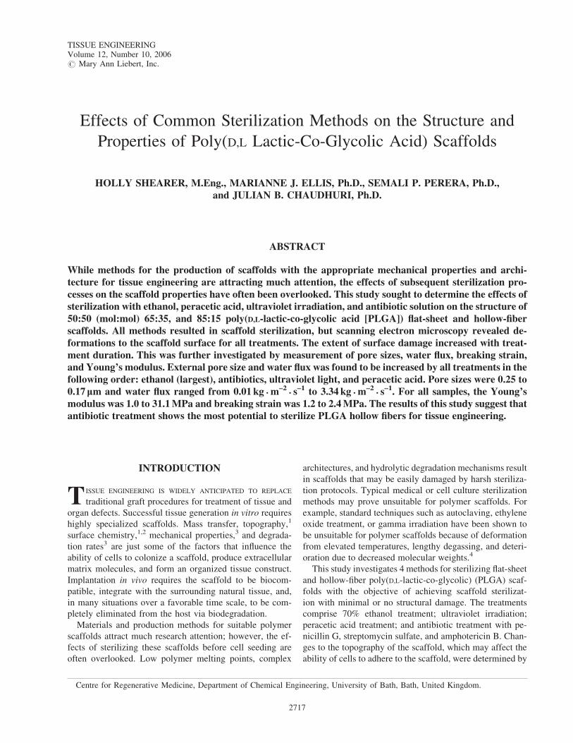
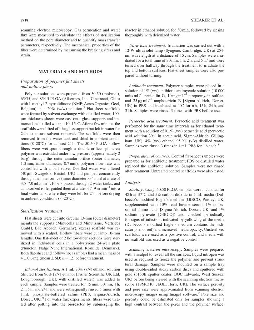
Flat-sheet and hollow-fiber scaffolds were success-
fully fabricated from 20% (w/w) PLGA in 1-methyl-2-
pyrrolidinone solution. Scanning electron microscopy
analysis revealed that flat sheets had an asymmetric cross-
section, with a dense skin (1–2 mm thick) present on the top,
below which a region of parallel pores ran perpendicular to
the surface; the bottom of the sheet exhibited a random
pore network with large macropores (Fig. 1). Hollow-fiber
cross-sections showed skin layers approximately 8–10 mmon the interior and exterior of the fiber (Fig. 1). Parallel
finger-like pores extended halfway across the fiber wall
from both the interior and exterior of the fiber.
Effect of sterilization on scaffold structure
Sterilization effectiveness was determined qualitatively
by the absence of signs of infection after a 48-h culture
period; infection was indicated by a change of indicator
color and opacity of culture medium. Only the 50:50 poly-
mer composition was tested for sterilization effectiveness
because no difference in the ability of the 4 sterilization
treatments to sterilize different PLGA ratios was antici-
pated. Two hollow-fiber and 2 flat-sheet samples were
tested for each combination of treatment and duration.
Unsterilized controls all showed infection, and all control
media (i.e., no scaffold) remained free of infection for the
time period. All the treated scaffold samples indicated
successful sterilization in treatment durations equal to or
less than those quoted in literature (Table 1).6–8 These re-
sults indicate the suitability of the sterilization technique to
the particular scaffolds. While the full treatment duration
suggested in the literature was not always found to be re-
quired, it is assumed that the low stringency of the sterility
assay could not justify a reduction in treatment duration.
Treatment duration for sterilization was therefore assumed
to be 30min for ethanol, 2 h for ultraviolet light and per-
acetic acid, and 24 h for antibiotic antimycotic treatment.
The structure of the scaffolds changed over the complete
treatment range; in all cases increasing the treatment dura-
tion led to an increased effect on the structure.
Table 2 gives details of the scanning electron microscopy
images of all the scaffolds both before and after steriliza-
tion treatment, along with estimates of porosity and pore
size from ImageJ.9 The scanning electronic microscopy
EFFECTS OF STERILIZATION ON POLY(D,L LACTIC-CO-GLYCOLIC ACID) SCAFFOLDS 2719
images of the 50:50 PLGA flat sheets and hollow fibers are
shown in Figs. 2 and 4, respectively. The scanning electron
microscopy images of the untreated control samples all
showed homogeneity across the top surfaces; pore sizes for
all 3 polymer compositions were similar, with the 65:35
sample showing a slightly lower porosity. The bottom
surface of the flat sheets appeared more irregular, with
larger pores confined to patches of localized increased
porosity. Pores appeared larger on the hollow fibers than on
the flat surfaces, and the exterior of the fiber appeared
rougher than the interior, with wrinkles and lines of pores
following the length of the fiber.
Samples sterilized in ethanol showed reduced porosity
on the top surface of the flat sheet and increased surface
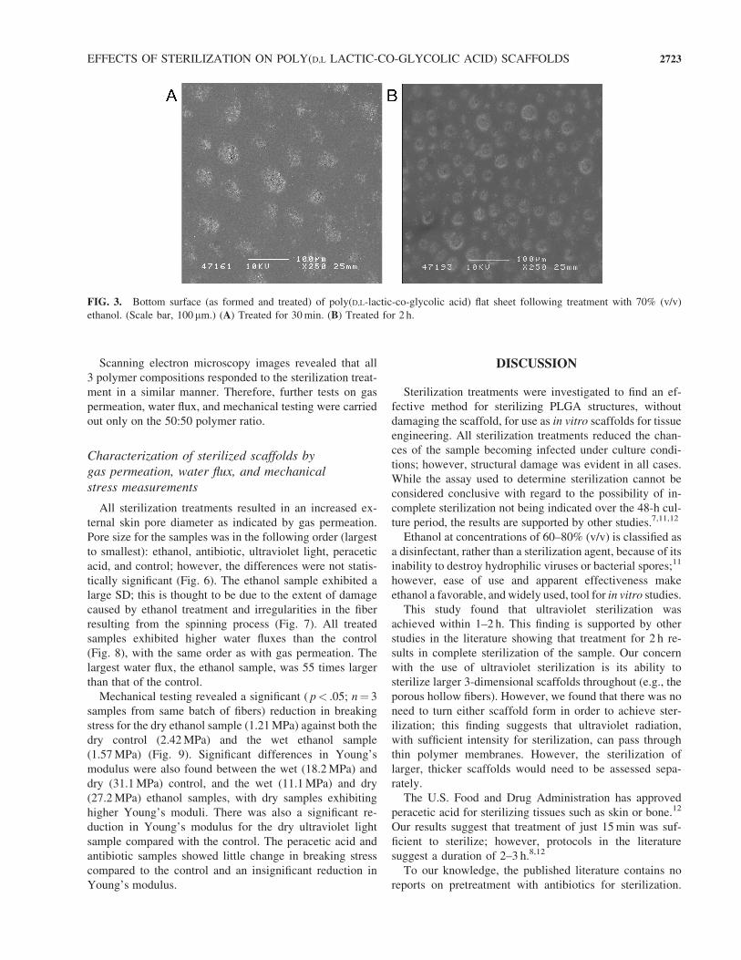
wrinkling (Fig. 2D). The porous patches on the bottom of
the flat sheets appeared more prominent, with larger pores
following longer treatment durations (Figs. 2C and 3).
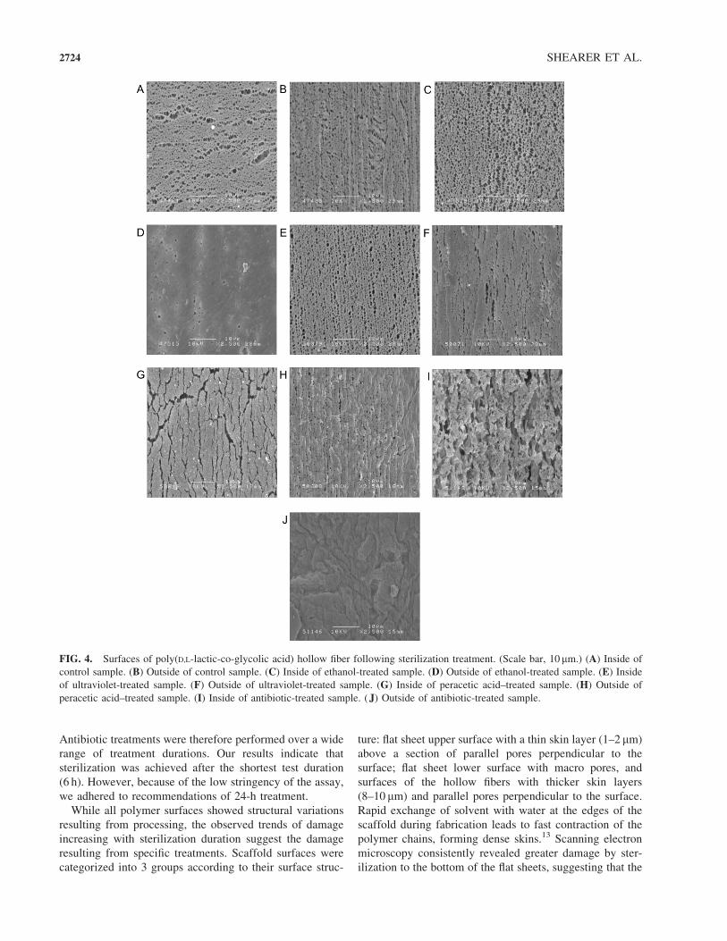
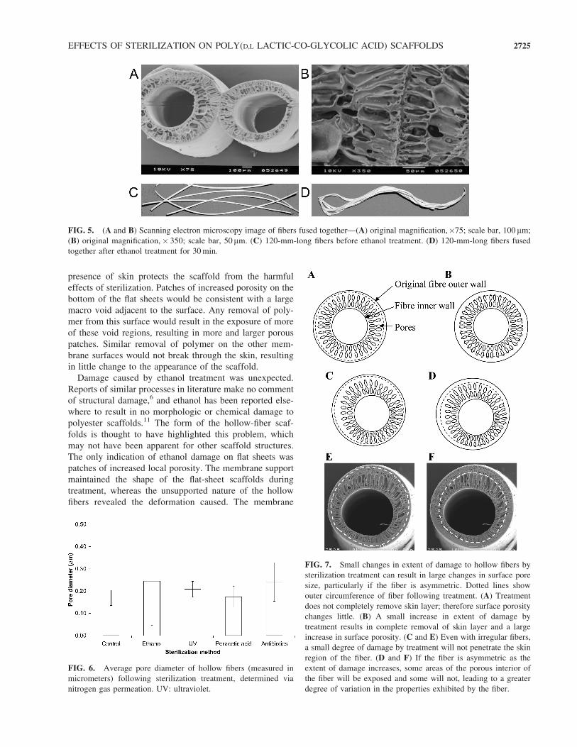
Hollow fibers exhibited larger pores on the interior of the
fiber (Fig. 4C), while the exterior of the fiber (Fig. 4D)
showed very few pores after treatment. After less than
10min in ethanol, the longer hollow fibers had deformed
and fused together (Fig. 5). The membranes treated with
ethanol for longer periods were more fragile under scan-
ning electron microscopy; magnifications above�2500 re-
esulted in damage to the sample within seconds.
Ultraviolet-treated scaffolds showed little change in
porosity (Fig. 2E and F). The exterior of the fibers showed
smoothing of the surface and smaller pores (Fig. 4F). The
interiors of hollow fibers were not directly exposed to ultra-
violet light and consequently showed very little change in
appearance (Fig. 4E).
Peracetic acid had a large impact on the surface of
the scaffolds as compared to the controls (Fig. 2G and H
and Fig. 4G and H). Surfaces appeared much more
FIG. 1. Cross-section views of poly(D,L-lactic-co-glycolic acid) flat-sheet and hollow-fiber scaffolds illustrating the presence of skin
on the top of the flat sheet and both interior and exterior of the hollow fiber. (A) Flat sheet with 1–2-mm skin on the top surface. (Scale
bar, 10 mm.) (B) Hollow fiber with 8–10-mm skin on both surfaces. (Scale bar, 100 mm.) Color images available online at www.
liebertpub.com/ten.
TABLE 1. COMPARISON OF TREATMENT METHOD AND DURATION REQUIRED FOR STERILIZATION OF FLAT-SHEET
AND HOLLOW-FIBER POLY(D,L-LACTIC-CO-GLYCOLIC ACID) SCAFFOLDS*
Time
Treatment 0 0.25 h 0.5 h 1 h 2 h 5 h 6 h 15 h 24 h 31 h
Negative control (no sample) H — — — — — — — — —
Positive control (not sterilized) � — — — — — — — — —
Ethanol � � H H H — — — H —
Ultraviolet light (turned) � — � H H H — — — —
Ultraviolet light (not turned) � — � H H H — — — —
Peracetic acid � H H H H — — — H —
Antibiotics � — — — — — H H H H
*Two hollow-fiber and 2 flat-sheet samples were tested for each combination of treatment and duration. Unsterilized controls showed signs of infection
in the time period; all control media (no scaffold) showed no signs of infection for the time period.H indicates no sign of infection after 48 h of culture in
Dulbecco’s modified Eagle’s medium with 10% fetal calf serum, 1% nonessential amino acids, 1% sodium pyruvate at 378C 5% carbon dioxide,
�indicates infection may have occurred in some or all samples (media changed form pink to yellow and became opaque). Dashes indicate that treatment/
duration combination was not tested. Shading indicates that times were equal to or longer than duration required for sterilization quoted in literature.5–9
2720 SHEARER ET AL.
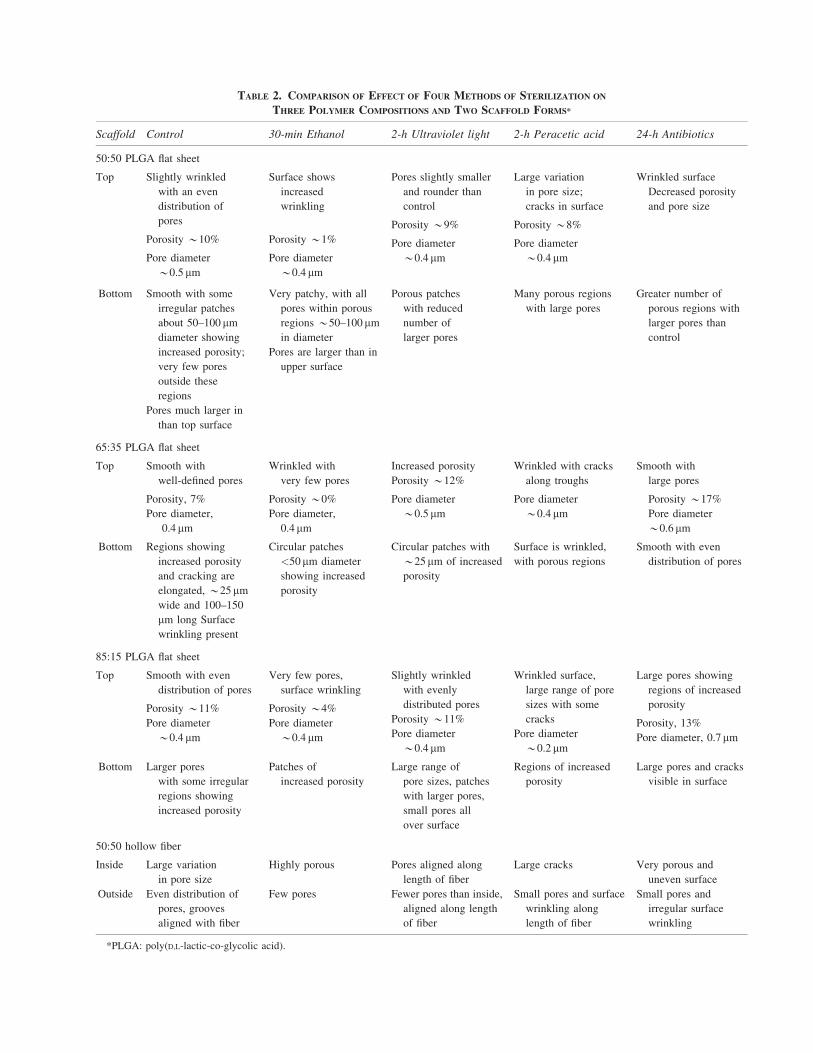
TABLE 2. COMPARISON OF EFFECT OF FOUR METHODS OF STERILIZATION ON
THREE POLYMER COMPOSITIONS AND TWO SCAFFOLD FORMS*
Scaffold Control 30-min Ethanol 2-h Ultraviolet light 2-h Peracetic acid 24-h Antibiotics
50:50 PLGA flat sheet
Top Slightly wrinkled
with an even
distribution of
pores
Porosity *10%
Pore diameter
*0.5 mm
Surface shows
increased
wrinkling
Porosity *1%
Pore diameter
*0.4 mm
Pores slightly smaller
and rounder than
control
Porosity *9%
Pore diameter
*0.4 mm
Large variation
in pore size;
cracks in surface
Porosity *8%
Pore diameter
*0.4 mm
Wrinkled surface
Decreased porosity
and pore size
Bottom Smooth with some
irregular patches
about 50–100 mmdiameter showing
increased porosity;
very few pores
outside these
regions
Pores much larger in
than top surface
Very patchy, with all
pores within porous
regions *50–100 mmin diameter
Pores are larger than in
upper surface
Porous patches
with reduced
number of
larger pores
Many porous regions
with large pores
Greater number of
porous regions with
larger pores than
control
65:35 PLGA flat sheet
Top Smooth with
well-defined pores
Porosity, 7%
Pore diameter,
0.4 mm
Wrinkled with
very few pores
Porosity *0%
Pore diameter,
0.4mm
Increased porosity
Porosity *12%
Pore diameter
*0.5 mm
Wrinkled with cracks
along troughs
Pore diameter
*0.4 mm
Smooth with
large pores
Porosity *17%
Pore diameter
*0.6 mm
Bottom Regions showing
increased porosity
and cracking are
elongated, *25mmwide and 100–150
mm long Surface
wrinkling present
Circular patches
<50mm diameter
showing increased
porosity
Circular patches with
*25mm of increased
porosity
Surface is wrinkled,
with porous regions
Smooth with even
distribution of pores
85:15 PLGA flat sheet
Top Smooth with even
distribution of pores
Porosity *11%
Pore diameter
*0.4 mm
Very few pores,
surface wrinkling
Porosity *4%
Pore diameter
*0.4 mm
Slightly wrinkled
with evenly
distributed pores
Porosity *11%
Pore diameter
*0.4 mm
Wrinkled surface,
large range of pore
sizes with some
cracks
Pore diameter
*0.2 mm
Large pores showing
regions of increased
porosity
Porosity, 13%
Pore diameter, 0.7mm
Bottom Larger pores
with some irregular
regions showing
increased porosity
Patches of
increased porosity
Large range of
pore sizes, patches
with larger pores,
small pores all
over surface
Regions of increased
porosity
Large pores and cracks
visible in surface
50:50 hollow fiber
Inside Large variation
in pore size
Highly porous Pores aligned along
length of fiber
Large cracks Very porous and
uneven surface
Outside Even distribution of
pores, grooves
aligned with fiber
Few pores Fewer pores than inside,
aligned along length
of fiber
Small pores and surface
wrinkling along
length of fiber
Small pores and
irregular surface
wrinkling
*PLGA: poly(D,L-lactic-co-glycolic acid).
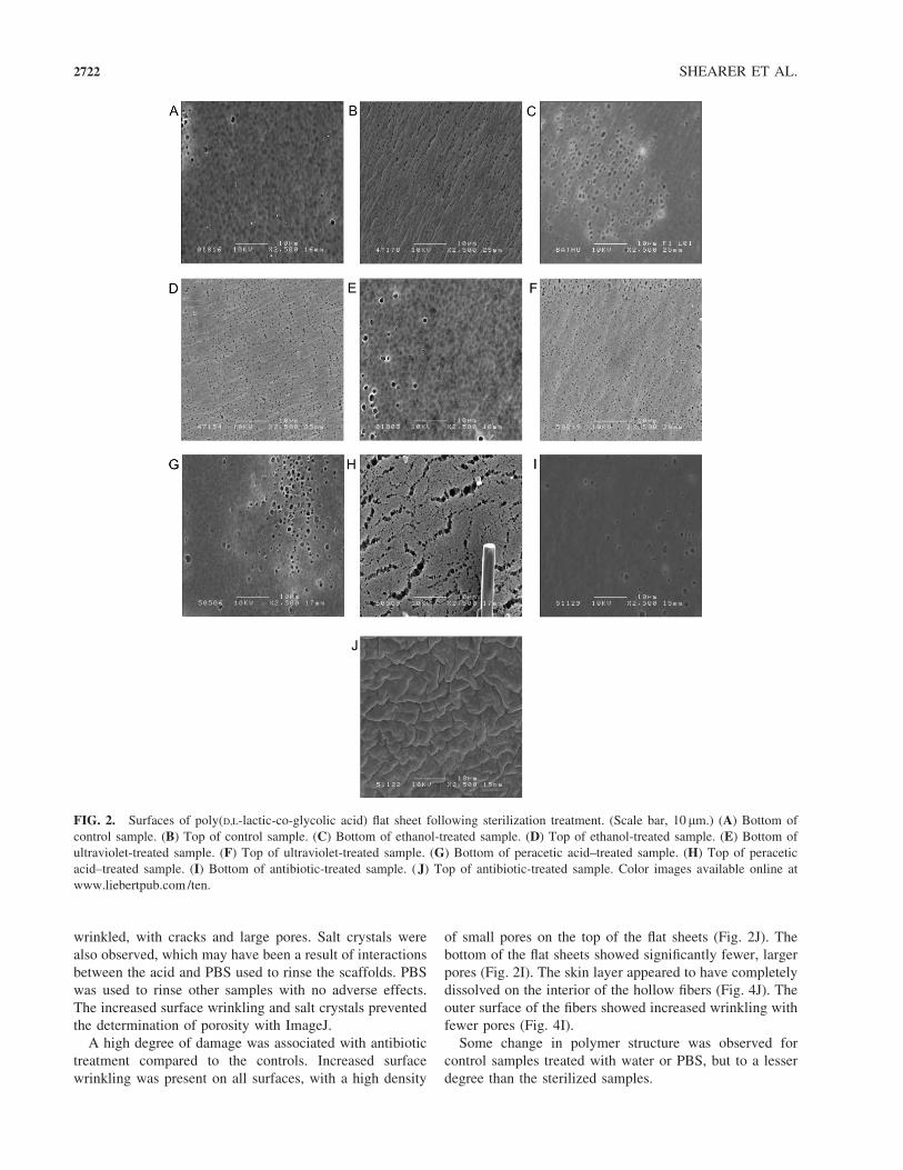
wrinkled, with cracks and large pores. Salt crystals were
also observed, which may have been a result of interactions
between the acid and PBS used to rinse the scaffolds. PBS
was used to rinse other samples with no adverse effects.
The increased surface wrinkling and salt crystals prevented
the determination of porosity with ImageJ.
A high degree of damage was associated with antibiotic
treatment compared to the controls. Increased surface
wrinkling was present on all surfaces, with a high density
of small pores on the top of the flat sheets (Fig. 2J). The
bottom of the flat sheets showed significantly fewer, larger
pores (Fig. 2I). The skin layer appeared to have completely
dissolved on the interior of the hollow fibers (Fig. 4J). The
outer surface of the fibers showed increased wrinkling with
fewer pores (Fig. 4I).
Some change in polymer structure was observed for
control samples treated with water or PBS, but to a lesser
degree than the sterilized samples.
FIG. 2. Surfaces of poly(D,L-lactic-co-glycolic acid) flat sheet following sterilization treatment. (Scale bar, 10 mm.) (A) Bottom of
control sample. (B) Top of control sample. (C) Bottom of ethanol-treated sample. (D) Top of ethanol-treated sample. (E) Bottom of
ultraviolet-treated sample. (F) Top of ultraviolet-treated sample. (G) Bottom of peracetic acid–treated sample. (H) Top of peracetic
acid–treated sample. (I) Bottom of antibiotic-treated sample. (J) Top of antibiotic-treated sample. Color images available online at
www.liebertpub.com/ten.
2722 SHEARER ET AL.
Scanning electron microscopy images revealed that all
3 polymer compositions responded to the sterilization treat-
ment in a similar manner. Therefore, further tests on gas
permeation, water flux, and mechanical testing were carried
out only on the 50:50 polymer ratio.
Characterization of sterilized scaffolds by
gas permeation, water flux, and mechanical
stress measurements
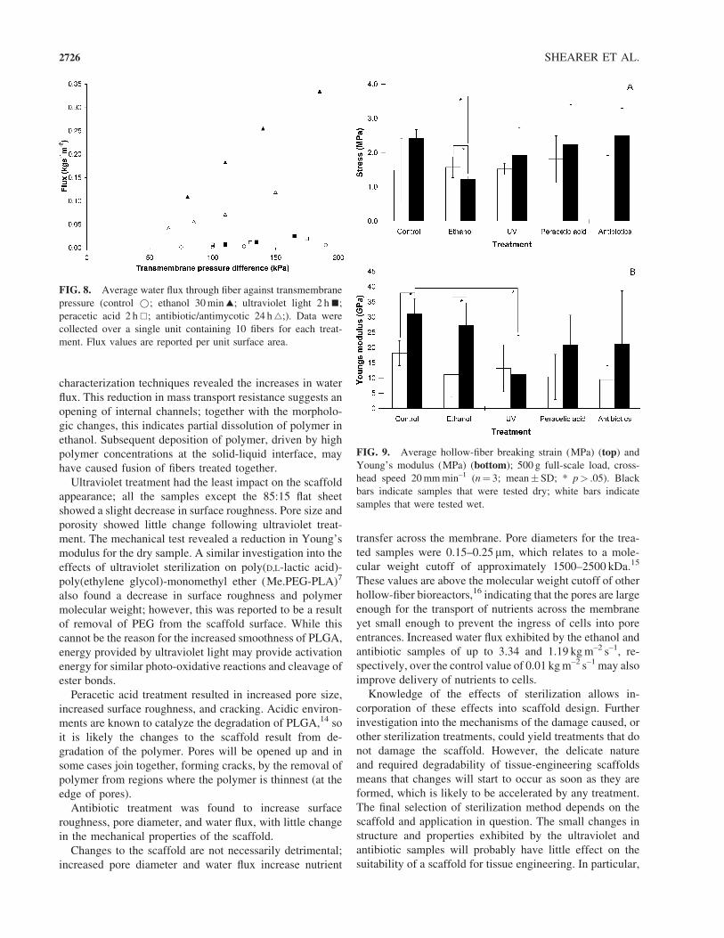
All sterilization treatments resulted in an increased ex-
ternal skin pore diameter as indicated by gas permeation.
Pore size for the samples was in the following order (largest
to smallest): ethanol, antibiotic, ultraviolet light, peracetic
acid, and control; however, the differences were not statis-
tically significant (Fig. 6). The ethanol sample exhibited a
large SD; this is thought to be due to the extent of damage
caused by ethanol treatment and irregularities in the fiber
resulting from the spinning process (Fig. 7). All treated
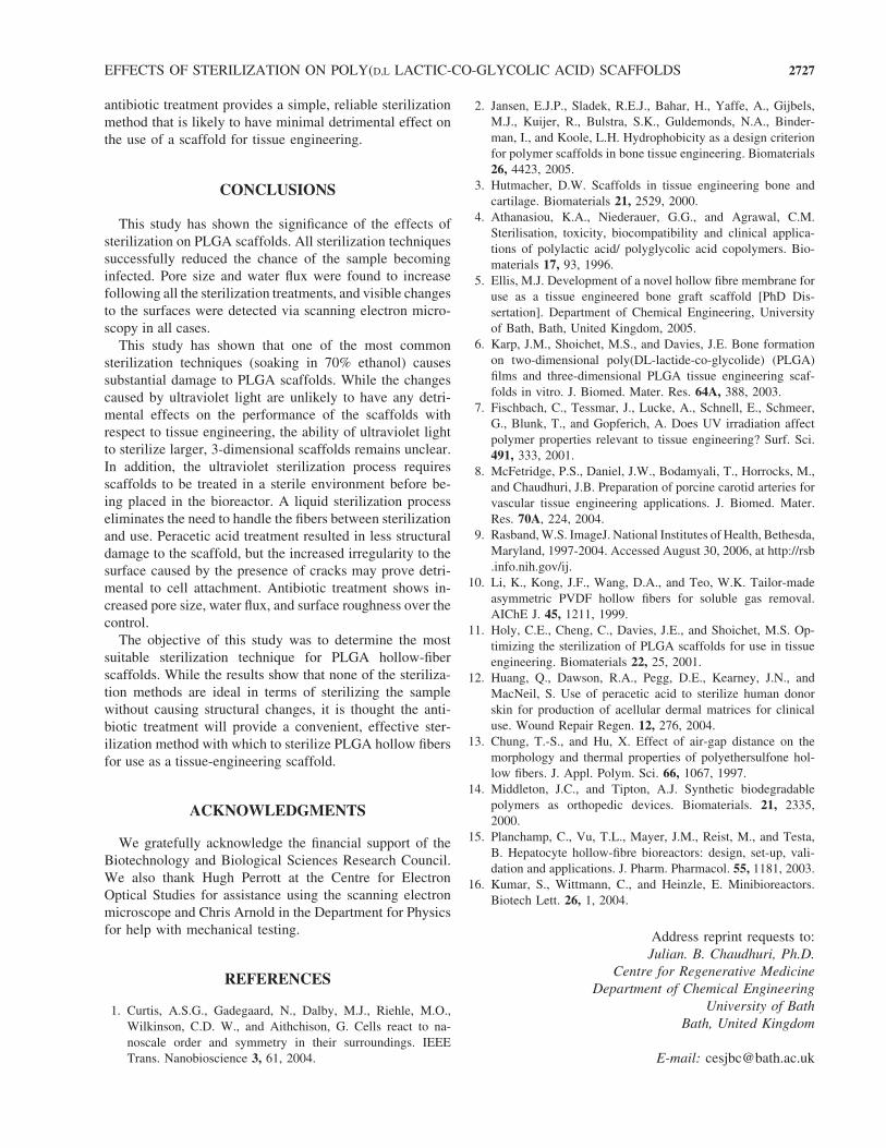
samples exhibited higher water fluxes than the control
(Fig. 8), with the same order as with gas permeation. The
largest water flux, the ethanol sample, was 55 times larger
than that of the control.
Mechanical testing revealed a significant ( p< .05; n¼ 3
samples from same batch of fibers) reduction in breaking
stress for the dry ethanol sample (1.21MPa) against both the
dry control (2.42MPa) and the wet ethanol sample
(1.57MPa) (Fig. 9). Significant differences in Young’s
modulus were also found between the wet (18.2MPa) and
dry (31.1MPa) control, and the wet (11.1MPa) and dry
(27.2MPa) ethanol samples, with dry samples exhibiting
higher Young’s moduli. There was also a significant re-
duction in Young’s modulus for the dry ultraviolet light
sample compared with the control. The peracetic acid and
antibiotic samples showed little change in breaking stress
compared to the control and an insignificant reduction in
Young’s modulus.
DISCUSSION
Sterilization treatments were investigated to find an ef-
fective method for sterilizing PLGA structures, without
damaging the scaffold, for use as in vitro scaffolds for tissue
engineering. All sterilization treatments reduced the chan-
ces of the sample becoming infected under culture condi-
tions; however, structural damage was evident in all cases.
While the assay used to determine sterilization cannot be
considered conclusive with regard to the possibility of in-
complete sterilization not being indicated over the 48-h cul-
ture period, the results are supported by other studies.7,11,12
Ethanol at concentrations of 60–80% (v/v) is classified as
a disinfectant, rather than a sterilization agent, because of its
inability to destroy hydrophilic viruses or bacterial spores;11
however, ease of use and apparent effectiveness make
ethanol a favorable, and widely used, tool for in vitro studies.
This study found that ultraviolet sterilization was
achieved within 1–2 h. This finding is supported by other
studies in the literature showing that treatment for 2 h re-
sults in complete sterilization of the sample. Our concern
with the use of ultraviolet sterilization is its ability to
sterilize larger 3-dimensional scaffolds throughout (e.g., the
porous hollow fibers). However, we found that there was no
need to turn either scaffold form in order to achieve ster-
ilization; this finding suggests that ultraviolet radiation,
with sufficient intensity for sterilization, can pass through
thin polymer membranes. However, the sterilization of
larger, thicker scaffolds would need to be assessed sepa-
rately.
The U.S. Food and Drug Administration has approved
peracetic acid for sterilizing tissues such as skin or bone.12
Our results suggest that treatment of just 15min was suf-
ficient to sterilize; however, protocols in the literature
suggest a duration of 2–3 h.8,12
To our knowledge, the published literature contains no
reports on pretreatment with antibiotics for sterilization.
FIG. 3. Bottom surface (as formed and treated) of poly(D,L-lactic-co-glycolic acid) flat sheet following treatment with 70% (v/v)
ethanol. (Scale bar, 100mm.) (A) Treated for 30min. (B) Treated for 2 h.
EFFECTS OF STERILIZATION ON POLY(D,L LACTIC-CO-GLYCOLIC ACID) SCAFFOLDS 2723
Antibiotic treatments were therefore performed over a wide
range of treatment durations. Our results indicate that
sterilization was achieved after the shortest test duration
(6 h). However, because of the low stringency of the assay,
we adhered to recommendations of 24-h treatment.
While all polymer surfaces showed structural variations
resulting from processing, the observed trends of damage
increasing with sterilization duration suggest the damage
resulting from specific treatments. Scaffold surfaces were
categorized into 3 groups according to their surface struc-
ture: flat sheet upper surface with a thin skin layer (1–2 mm)
above a section of parallel pores perpendicular to the
surface; flat sheet lower surface with macro pores, and
surfaces of the hollow fibers with thicker skin layers
(8–10 mm) and parallel pores perpendicular to the surface.
Rapid exchange of solvent with water at the edges of the
scaffold during fabrication leads to fast contraction of the
polymer chains, forming dense skins.13 Scanning electron
microscopy consistently revealed greater damage by ster-
ilization to the bottom of the flat sheets, suggesting that the
FIG. 4. Surfaces of poly(D,L-lactic-co-glycolic acid) hollow fiber following sterilization treatment. (Scale bar, 10mm.) (A) Inside of
control sample. (B) Outside of control sample. (C) Inside of ethanol-treated sample. (D) Outside of ethanol-treated sample. (E) Inside
of ultraviolet-treated sample. (F) Outside of ultraviolet-treated sample. (G) Inside of peracetic acid–treated sample. (H) Outside of
peracetic acid–treated sample. (I) Inside of antibiotic-treated sample. (J) Outside of antibiotic-treated sample.
2724 SHEARER ET AL.
presence of skin protects the scaffold from the harmful
effects of sterilization. Patches of increased porosity on the
bottom of the flat sheets would be consistent with a large
macro void adjacent to the surface. Any removal of poly-
mer from this surface would result in the exposure of more
of these void regions, resulting in more and larger porous
patches. Similar removal of polymer on the other mem-
brane surfaces would not break through the skin, resulting
in little change to the appearance of the scaffold.
Damage caused by ethanol treatment was unexpected.
Reports of similar processes in literature make no comment
of structural damage,6 and ethanol has been reported else-
where to result in no morphologic or chemical damage to
polyester scaffolds.11 The form of the hollow-fiber scaf-
folds is thought to have highlighted this problem, which
may not have been apparent for other scaffold structures.
The only indication of ethanol damage on flat sheets was
patches of increased local porosity. The membrane support
maintained the shape of the flat-sheet scaffolds during
treatment, whereas the unsupported nature of the hollow
fibers revealed the deformation caused. The membrane
FIG. 5. (A and B) Scanning electron microscopy image of fibers fused together—(A) original magnification,�75; scale bar, 100 mm;
(B) original magnification,� 350; scale bar, 50mm. (C) 120-mm-long fibers before ethanol treatment. (D) 120-mm-long fibers fused
together after ethanol treatment for 30min.
FIG. 6. Average pore diameter of hollow fibers (measured in
micrometers) following sterilization treatment, determined via
nitrogen gas permeation. UV: ultraviolet.
FIG. 7. Small changes in extent of damage to hollow fibers by
sterilization treatment can result in large changes in surface pore
size, particularly if the fiber is asymmetric. Dotted lines show
outer circumference of fiber following treatment. (A) Treatment
does not completely remove skin layer; therefore surface porosity
changes little. (B) A small increase in extent of damage by
treatment results in complete removal of skin layer and a large
increase in surface porosity. (C and E) Even with irregular fibers,
a small degree of damage by treatment will not penetrate the skin
region of the fiber. (D and F) If the fiber is asymmetric as the
extent of damage increases, some areas of the porous interior of
the fiber will be exposed and some will not, leading to a greater
degree of variation in the properties exhibited by the fiber.
EFFECTS OF STERILIZATION ON POLY(D,L LACTIC-CO-GLYCOLIC ACID) SCAFFOLDS 2725
characterization techniques revealed the increases in water
flux. This reduction in mass transport resistance suggests an
opening of internal channels; together with the morpholo-
gic changes, this indicates partial dissolution of polymer in
ethanol. Subsequent deposition of polymer, driven by high
polymer concentrations at the solid-liquid interface, may
have caused fusion of fibers treated together.
Ultraviolet treatment had the least impact on the scaffold
appearance; all the samples except the 85:15 flat sheet
showed a slight decrease in surface roughness. Pore size and
porosity showed little change following ultraviolet treat-
ment. The mechanical test revealed a reduction in Young’s
modulus for the dry sample. A similar investigation into the
effects of ultraviolet sterilization on poly(D,L-lactic acid)-
poly(ethylene glycol)-monomethyl ether (Me.PEG-PLA)7
also found a decrease in surface roughness and polymer
molecular weight; however, this was reported to be a result
of removal of PEG from the scaffold surface. While this
cannot be the reason for the increased smoothness of PLGA,
energy provided by ultraviolet light may provide activation
energy for similar photo-oxidative reactions and cleavage of
ester bonds.
Peracetic acid treatment resulted in increased pore size,
increased surface roughness, and cracking. Acidic environ-
ments are known to catalyze the degradation of PLGA,14 so
it is likely the changes to the scaffold result from de-
gradation of the polymer. Pores will be opened up and in
some cases join together, forming cracks, by the removal of
polymer from regions where the polymer is thinnest (at the
edge of pores).
Antibiotic treatment was found to increase surface
roughness, pore diameter, and water flux, with little change
in the mechanical properties of the scaffold.
Changes to the scaffold are not necessarily detrimental;
increased pore diameter and water flux increase nutrient
transfer across the membrane. Pore diameters for the trea-
ted samples were 0.15–0.25 mm, which relates to a mole-
cular weight cutoff of approximately 1500–2500 kDa.15
These values are above the molecular weight cutoff of other
hollow-fiber bioreactors,16 indicating that the pores are large
enough for the transport of nutrients across the membrane
yet small enough to prevent the ingress of cells into pore
entrances. Increased water flux exhibited by the ethanol and
antibiotic samples of up to 3.34 and 1.19 kgm–2 s–1, re-
spectively, over the control value of 0.01 kgm–2 s–1 may also
improve delivery of nutrients to cells.
Knowledge of the effects of sterilization allows in-
corporation of these effects into scaffold design. Further
investigation into the mechanisms of the damage caused, or
other sterilization treatments, could yield treatments that do
not damage the scaffold. However, the delicate nature
and required degradability of tissue-engineering scaffolds
means that changes will start to occur as soon as they are
formed, which is likely to be accelerated by any treatment.
The final selection of sterilization method depends on the
scaffold and application in question. The small changes in
structure and properties exhibited by the ultraviolet and
antibiotic samples will probably have little effect on the
suitability of a scaffold for tissue engineering. In particular,
FIG. 8. Average water flux through fiber against transmembrane
pressure (control *; ethanol 30min~; ultraviolet light 2 h&;
peracetic acid 2 h&; antibiotic/antimycotic 24 h~;). Data were
collected over a single unit containing 10 fibers for each treat-
ment. Flux values are reported per unit surface area.
FIG. 9. Average hollow-fiber breaking strain (MPa) (top) and
Young’s modulus (MPa) (bottom); 500 g full-scale load, cross-
head speed 20mmmin–1 (n¼ 3; mean� SD; * p> .05). Black
bars indicate samples that were tested dry; white bars indicate
samples that were tested wet.
2726 SHEARER ET AL.
antibiotic treatment provides a simple, reliable sterilization
method that is likely to have minimal detrimental effect on
the use of a scaffold for tissue engineering.
CONCLUSIONS
This study has shown the significance of the effects of
sterilization on PLGA scaffolds. All sterilization techniques
successfully reduced the chance of the sample becoming
infected. Pore size and water flux were found to increase
following all the sterilization treatments, and visible changes
to the surfaces were detected via scanning electron micro-
scopy in all cases.
This study has shown that one of the most common
sterilization techniques (soaking in 70% ethanol) causes
substantial damage to PLGA scaffolds. While the changes
caused by ultraviolet light are unlikely to have any detri-
mental effects on the performance of the scaffolds with
respect to tissue engineering, the ability of ultraviolet light
to sterilize larger, 3-dimensional scaffolds remains unclear.
In addition, the ultraviolet sterilization process requires
scaffolds to be treated in a sterile environment before be-
ing placed in the bioreactor. A liquid sterilization process
eliminates the need to handle the fibers between sterilization
and use. Peracetic acid treatment resulted in less structural
damage to the scaffold, but the increased irregularity to the
surface caused by the presence of cracks may prove detri-
mental to cell attachment. Antibiotic treatment shows in-
creased pore size, water flux, and surface roughness over the
control.
The objective of this study was to determine the most
suitable sterilization technique for PLGA hollow-fiber
scaffolds. While the results show that none of the steriliza-
tion methods are ideal in terms of sterilizing the sample
without causing structural changes, it is thought the anti-
biotic treatment will provide a convenient, effective ster-
ilization method with which to sterilize PLGA hollow fibers
for use as a tissue-engineering scaffold.
ACKNOWLEDGMENTS
We gratefully acknowledge the financial support of the
Biotechnology and Biological Sciences Research Council.
We also thank Hugh Perrott at the Centre for Electron
Optical Studies for assistance using the scanning electron
microscope and Chris Arnold in the Department for Physics
for help with mechanical testing.
REFERENCES
1. Curtis, A.S.G., Gadegaard, N., Dalby, M.J., Riehle, M.O.,
Wilkinson, C.D. W., and Aithchison, G. Cells react to na-
noscale order and symmetry in their surroundings. IEEE
Trans. Nanobioscience 3, 61, 2004.
2. Jansen, E.J.P., Sladek, R.E.J., Bahar, H., Yaffe, A., Gijbels,
M.J., Kuijer, R., Bulstra, S.K., Guldemonds, N.A., Binder-
man, I., and Koole, L.H. Hydrophobicity as a design criterion
for polymer scaffolds in bone tissue engineering. Biomaterials
26, 4423, 2005.
3. Hutmacher, D.W. Scaffolds in tissue engineering bone and
cartilage. Biomaterials 21, 2529, 2000.
4. Athanasiou, K.A., Niederauer, G.G., and Agrawal, C.M.
Sterilisation, toxicity, biocompatibility and clinical applica-
tions of polylactic acid/ polyglycolic acid copolymers. Bio-
materials 17, 93, 1996.
5. Ellis, M.J. Development of a novel hollow fibre membrane for
use as a tissue engineered bone graft scaffold [PhD Dis-
sertation]. Department of Chemical Engineering, University
of Bath, Bath, United Kingdom, 2005.
6. Karp, J.M., Shoichet, M.S., and Davies, J.E. Bone formation
on two-dimensional poly(DL-lactide-co-glycolide) (PLGA)
films and three-dimensional PLGA tissue engineering scaf-
folds in vitro. J. Biomed. Mater. Res. 64A, 388, 2003.
7. Fischbach, C., Tessmar, J., Lucke, A., Schnell, E., Schmeer,
G., Blunk, T., and Gopferich, A. Does UV irradiation affect
polymer properties relevant to tissue engineering? Surf. Sci.
491, 333, 2001.
8. McFetridge, P.S., Daniel, J.W., Bodamyali, T., Horrocks, M.,
and Chaudhuri, J.B. Preparation of porcine carotid arteries for
vascular tissue engineering applications. J. Biomed. Mater.
Res. 70A, 224, 2004.
9. Rasband,W.S. ImageJ. National Institutes of Health, Bethesda,
Maryland, 1997-2004. Accessed August 30, 2006, at http://rsb
.info.nih.gov/ij.
10. Li, K., Kong, J.F., Wang, D.A., and Teo, W.K. Tailor-made
asymmetric PVDF hollow fibers for soluble gas removal.
AIChE J. 45, 1211, 1999.
11. Holy, C.E., Cheng, C., Davies, J.E., and Shoichet, M.S. Op-
timizing the sterilization of PLGA scaffolds for use in tissue
engineering. Biomaterials 22, 25, 2001.
12. Huang, Q., Dawson, R.A., Pegg, D.E., Kearney, J.N., and
MacNeil, S. Use of peracetic acid to sterilize human donor
skin for production of acellular dermal matrices for clinical
use. Wound Repair Regen. 12, 276, 2004.
13. Chung, T.-S., and Hu, X. Effect of air-gap distance on the
morphology and thermal properties of polyethersulfone hol-
low fibers. J. Appl. Polym. Sci. 66, 1067, 1997.
14. Middleton, J.C., and Tipton, A.J. Synthetic biodegradable
polymers as orthopedic devices. Biomaterials. 21, 2335,
2000.
15. Planchamp, C., Vu, T.L., Mayer, J.M., Reist, M., and Testa,
B. Hepatocyte hollow-fibre bioreactors: design, set-up, vali-
dation and applications. J. Pharm. Pharmacol. 55, 1181, 2003.
16. Kumar, S., Wittmann, C., and Heinzle, E. Minibioreactors.
Biotech Lett. 26, 1, 2004.
Address reprint requests to:
Julian. B. Chaudhuri, Ph.D.
Centre for Regenerative Medicine
Department of Chemical Engineering
University of Bath
Bath, United Kingdom
E-mail: [email protected]
EFFECTS OF STERILIZATION ON POLY(D,L LACTIC-CO-GLYCOLIC ACID) SCAFFOLDS 2727
