visuo-haptic neuronal convergence demonstrated with an inversely effective pattern of bold...
TRANSCRIPT
UncorrectedProof
Visuo-haptic Neuronal Convergence Demonstrated withan Inversely Effective Pattern of BOLD Activation
Sunah Kim1,2, Ryan A. Stevenson1,3, and Thomas W. James1
Abstract
■ We investigated the neural substrates involved in visuo-haptic neuronal convergence using an additive-factors designin combination with fMRI. Stimuli were explored under threesensory modality conditions: viewing the object through a mir-ror without touching (V), touching the object with eyes closed(H), or simultaneously viewing and touching the object (VH).This modality factor was crossed with a task difficulty factor,which had two levels. On the basis of an idea similar to the prin-ciple of inverse effectiveness, we predicted that increasing dif-ficulty would increase the relative level of multisensory gain in
brain regions where visual and haptic sensory inputs converged.An ROI analysis focused on the lateral occipital tactile–visualarea found evidence of inverse effectiveness in the left lateraloccipital tactile–visual area, but not in the right. A whole-brainanalysis also found evidence for the same pattern in the ante-rior aspect of the intraparietal sulcus, the premotor cortex, andthe posterior insula, all in the left hemisphere. In conclusion,this study is the first to demonstrate visuo-haptic neuronalconvergence based on an inversely effective pattern of brainactivation. ■
INTRODUCTION
There has been growing interest in the study of multi-sensory integration for the last few decades, leading re-searchers to investigate the neural substrates involvedin visual and haptic object recognition (for reviews, seeJames, Kim, & Fisher, 2007; Amedi, von Kriegstein, vanAtteveldt, Beauchamp, & Naumer, 2005). Results of thesestudies suggest that vision and touch share similar neuralsubstrates for processing the macrogeometric propertiesof objects (i.e., form/shape) in occipito-temporal (Stilla& Sathian, 2008; Peltier et al., 2007; Pietrini et al., 2004;Zhang, Weisser, Stilla, Prather, & Sathian, 2004; Stoeszet al., 2003; James et al., 2002; Amedi, Malach, Hendler,Peled, & Zohary, 2001) and intraparietal cortices (Stilla &Sathian, 2008; Zhang et al., 2004; Grefkes, Weiss, Zilles, &Fink, 2002; Culham & Kanwisher, 2001) as well as in cere-bellum (Naumer et al., 2010; Stevenson, Kim, & James,2009). These studies also suggest that information fromvision and haptics converges at these cortical sites forthe purpose of visuo-haptic multisensory perceptionand/or action ( James & Kim, 2010; Dijkerman & de Haan,2007; James et al., 2007; Reed, Klatzky, & Halgren, 2005).In general, most of these previous fMRI studies have as-sessed the overlap of visual and haptic representations.Overlap of inputs from two or more sensory systems is in-dicative of ‘areal’ convergence. In the case of vision andhaptics, areal convergence would imply the comingling inone brain region (or voxel cluster) of visual neurons and
haptic neurons. Areal convergence, however, does notimply the presence of multisensory neurons in the re-gion. The presence of multisensory neurons in a regionis termed ‘neuronal’ convergence; multisensory neuronsreceive converging inputs from two or more sensory sys-tems. Finding overlap of fMRI activation in a brain withvisual and haptic stimuli is evidence that that region maybe a site of areal convergence, that is, an area that hasboth visual and haptic neurons. However, overlap aloneis not enough to imply neuronal convergence ( James &Stevenson, in press; James, Stevenson, & Kim, in press;Stevenson et al., 2009). Thus, despite the number of stud-ies investigating haptic and visuo-haptic object process-ing, there is very little evidence concerning whether theoccipito-temporal and intraparietal cortices show evi-dence of ‘neuronal’ convergence (Kim & James, 2010; Tal& Amedi, 2009).
The principle of inverse effectiveness has been em-ployed to investigate multisensory integration for almostthree decades, because Meredith and Stein (1983) de-scribed the inverse relationship between unisensory effec-tiveness and multisensory enhancement in cat superiorcolliculus cells. The principle of inverse effectiveness statesthat multisensory gain increases as the responsiveness tothe constituent unisensory stimuli decreases. Inverse ef-fectiveness is typically (but not necessarily) evaluated bydegrading the quality of the stimuli. Degraded stimuli gen-erally produce less activation than less degraded stimuli,providing a gradient of activation along which one canassess inverse effectiveness. The principle of inverse effec-tiveness has been widely used to investigate multisensory
1Indiana University, 2University of California, Berkeley, 3VanderbiltUniversity
© Massachusetts Institute of Technology Journal of Cognitive Neuroscience X:Y, pp. 1–13
UncorrectedProof
integration in non-human animals (Perrault, Vaughan, Stein,& Wallace, 2005; Meredith & Stein, 1983, 1986b). Morerecently, researchers have also started to study inverse ef-fective patterns of activation using human neuroimaging( James et al., in press; Kim & James, 2010; Stevenson &James, 2009; Werner & Noppeney, 2009; Kayser, Petkov,Augath, & Logothetis, 2005), but inverse effectiveness hasyet to be shown in relation to visuo-haptic multisensoryobject processing.
In a previous study (Kim & James, 2010) using anadditive-factors design (Sternberg, 1969), we attemptedto relate the principle of inverse effectiveness to the studyof visuo-haptic object recognition. As with typical studiesof inverse effectiveness, stimuli of different quality werepresented and inverse effectiveness was assessed acrossthe levels of quality. Unexpectedly, the study showedevidence for ‘enhanced effectiveness’ in three distinctobject-selective brain regions: the left lateral occipitaltactile–visual area (LOtv), the left intraparietal sulcus (IPS),and the anterior aspect of the left fusiform gyrus. Enhancedeffectiveness is an increase in multisensory enhancementas the effectiveness of the constituent unisensory stimuliincreases, which is the opposite of inverse effectiveness.Although this effect is not the same as inverse effective-ness, finding an effect in that direction also implies neuronalconvergence of multisensory inputs (Kim & James, 2010;Stevenson et al., 2009).
In that study, novel objects made up of simple shape fea-tures were presented visually, haptically, or visuo-hapticallyat different levels of stimulus quality while participants per-formed a shape categorization task. Visual stimuli were pic-tures of the objects and were degraded by adding noiseand reducing contrast. Tangible haptic stimuli were ex-plored by subjects with their hands and were degraded byhaving subjects wear gloves. In the visuo-haptic condition,the procedures for degrading the constituent unisensorystimuli led to incongruencies in spatial location and tem-poral synchrony between visual and haptic inputs. Spatialand temporal incongruencies are known to influence firingrates in multisensory neurons (Meredith, Nemitz, & Stein,1987; Meredith & Stein, 1986a; King & Palmer, 1985) aswell as BOLD signals in audiovisual multisensory neuronalpopulations (Stevenson, VanDerKlok, Pisoni, & James,2011; Stevenson, Altieri, Kim, Pisoni, & James, 2010; Miller& DʼEsposito, 2005). Although the exact mechanism re-mains unclear, it is quite possible that a combination ofthese incongruencies in the previous study may have al-tered the integration of visual and haptic signals such thatit gave rise to enhanced effectiveness instead of inverseeffectiveness in the population measurement.
On the basis of this explanation of the previous findings,the first goal of this study was to develop a procedure forinvestigating visuo-haptic object recognition that would re-duce incongruencies in stimulus presentation parametersand produce optimal multisensory integration of visualand haptic shape information. We chose two candidatefactors for optimization, temporal and spatial congruency.
Instead of viewing pictures of objects while touching tan-gible objects, participants in the current study were ableto view the tangible objects and view their hand touchingthem through a mirror. This change to the procedurelessened the spatial incongruency caused by viewing andtouching the object in different location. A recent fMRIstudy employed a similar procedure of visual and tactilestimulation where participants looked directly at their handbeing touched by objects (Gentile, Petkova, & Ehrsson,2011). In this study, however, participants did not activelyexplore the stimuli but rather passively felt them beingstroked on their index finger and were not asked to makeany perceptual or cognitive judgments on the shape of thestimuli, whereas the participants in the current study ac-tively touched the stimuli while carrying out perceptualjudgment tasks. Participants in the current study were alsotrained and specifically asked to open their eyes only whenthey began touching the object and to close their eyes onlywhen they finished touching the object. This change tothe procedure lessened the temporal incongruency causedby the difference in time required to move the hand com-pared with opening/closing the eyes.To implement this new protocol, it was necessary to
alter the task from the previous study. In the previousstudy, effectiveness was manipulated by degrading thestimuli, which is highly typical in studies of inverse effec-tiveness (Kim & James, 2010; Stevenson & James, 2009;Werner & Noppeney, 2009; Kayser et al., 2005; Perraultet al., 2005). To allow subjects to view the object and theirhand touching the object simultaneously, we manipulatedeffectiveness by changing the level of similarity among theobjects and thus the difficulty of object recognition. Thereis evidence to suggest that increasing the level of similarityproduces changes in effectiveness in object-selective brainregions in the desired direction for assessing inverse ef-fectiveness ( Joseph & Farley, 2004). Thus, in the presentexperiment, we varied the level of behavioral performanceand BOLD activation effectiveness by changing the simi-larity between objects, rather than by degrading them.
METHODS
Participants
Fourteen volunteers (seven women and seven men,age = 20–34 years) participated in the study with mone-tary compensation. All participants were strongly right-handed (mean = 98.98, SD = 3.82) according to a revisedEdinburgh Handedness Inventory (Oldfield, 1971). Threeproblematic items among the 10 original items of theEdinburgh Handedness Inventory were excluded to im-prove its measurement properties (Dragovic, 2004). Allparticipants had normal or corrected-to-normal visualacuity, normal sensation of touch, and no history of neuro-logical disorders. One participant was excluded becauseof excessive head motion (see below for criteria; finalN= 13). The study was approved by the Indiana University
2 Journal of Cognitive Neuroscience Volume X, Number Y
UncorrectedProof
Institutional Review Board. Written informed consent wasobtained from all participants before the experiments.
Stimuli and Procedures
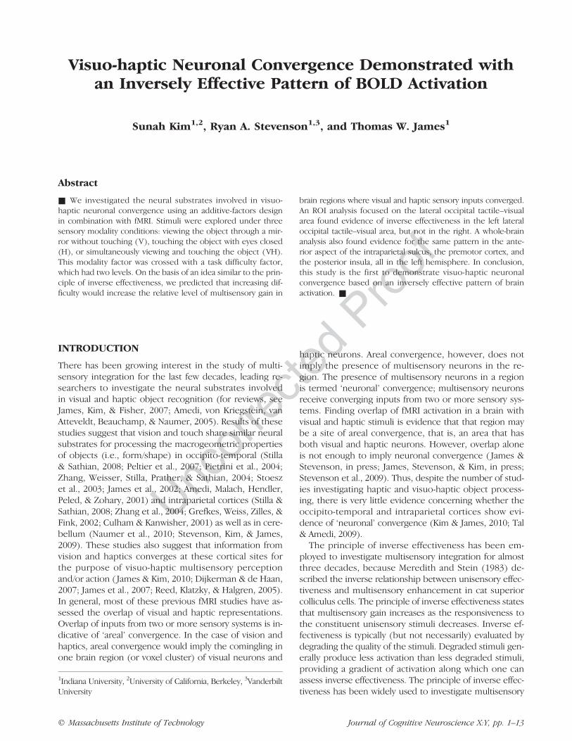
Different sets of stimuli were used in localizer runs andexperimental runs. Fifteen 3-D objects and 15 textureswere used in the visual object localizer run. Objects andtextures were tangible stimuli with a size of approximately2 × 2 × 2 cm for objects and 2 × 2 cm for textures madeof white acrylonitrile butadiene styrene (ABS) plastic(Figure 1). They were presented on a custom-made tableplaced over the participantʼs abdomen and viewed througha mirror. The same stimuli were used in the haptic objectlocalizer run. Participants explored the stimuli with theirright hand with their eyes closed in the haptic runs. Theuse of the right hand was chosen based on a study show-ing that BOLD activation during haptic object explorationin higher cortical areas such as LOtv is bilateral, regardlessof the hand of use (Amedi, Raz, Azulay, Malach, & Zohary,2010).Stimuli used in the experimental runs were 3-D tangible
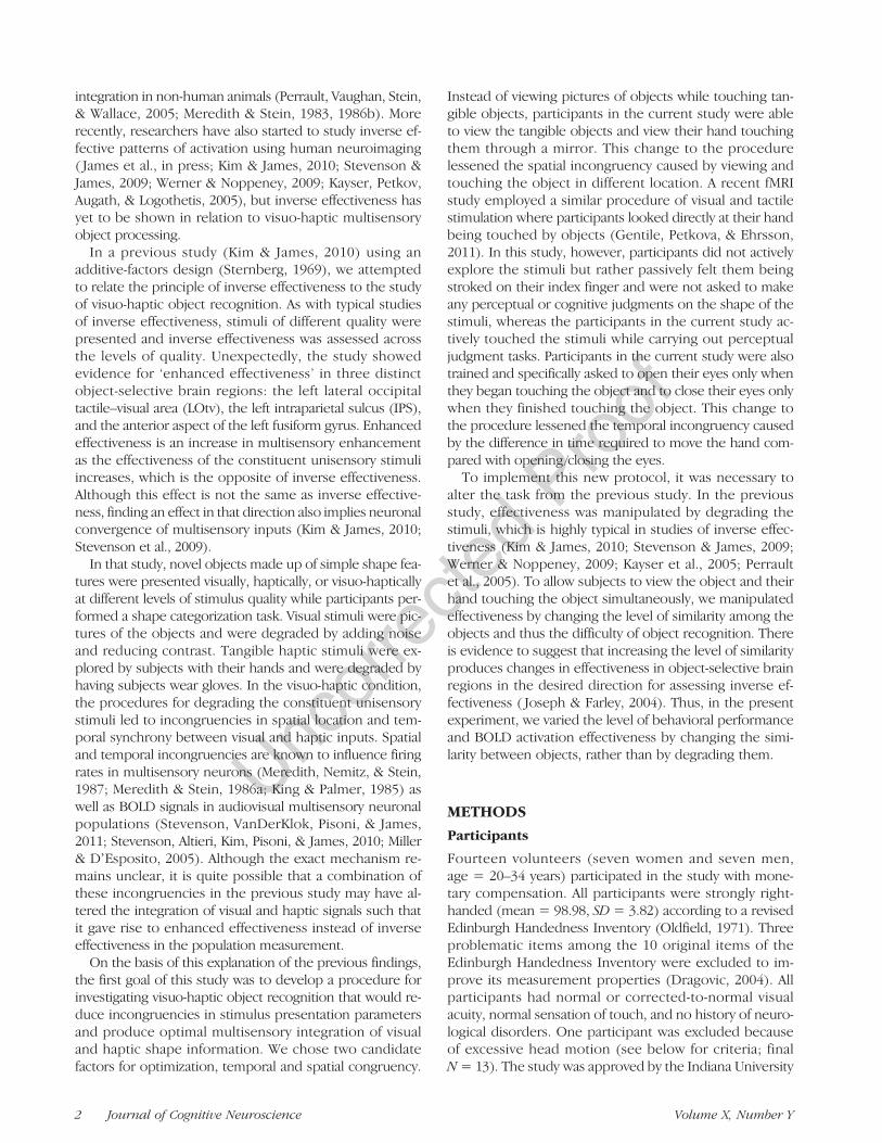
objects with a size of 2 × 2 × 2 cm, made of white ABSplastic. The top of each object varied in its curvature suchthat the least curved object was a square shape, and themost curved object was a circular shape (Figure 2). Stimuliwere explored under three experimental conditions: view-ing the object through a mirror without touching (V),touching the object with eyes closed (H), or viewing theobject through a mirror while touching the object (VH).The possibility of head movements evoked by touchingmovements was limited by having participants use onlytheir right index finger to touch the objects and use onlysmall movements of the finger and wrist (i.e., no elbowor shoulder movement). Participants performed a two-alternative forced-choice (2AFC) task in which they judgedwhether each presented object was the more circular(half-cylinder-like) or the more square (cube-like) one.Difficulty was manipulated in the experimental runs by
varying the distinctness of the curvature of the objects.Two difficulty levels were used. On low-difficulty trials, the2AFC decision was unambiguous, that is, objects wereclearly more circular or more square. On high-difficulty
trials, the 2AFC decision was more ambiguous, becausethe curvature of the objects was more similar or closertogether along the perceptual dimension of curvature (Fig-ure 2). In summary, sensory modality and task difficultywere two independent variables in a 3 × 2 factorial design.It should be noted that the stimulus quality was equivalentfor all conditions, which is a departure from the typical de-sign of a study investigating inverse effectiveness. Instead,it was expected that the pattern of inverse effectivenesscould be assessed over the predicted changes in effective-ness in BOLD activation produced by the manipulation ofobject similarity.
All 3-D stimuli were designed in Rhinoceros 3.0 (RobertMcNeel & Associates, Seattle, WA) were made into tangibleobjects using 3-D printing on a STRATASYS Prodigy Plus(Stratasys, Inc., Eden Prairie, MN) rapid prototyping ma-chine and were rendered to 2-D images for Figure 2 usingFlamingo 1.0 (Robert McNeel & Associates, Seattle, WA).
fMRI Imaging Procedures
Before fMRI imaging sessions, participants were trained inan fMRI simulator until they were fully familiarized withthe task. Each fMRI imaging session began with two visualobject localizer runs and two haptic object localizer runs.The order of these localizer runs was randomized acrossparticipants. The localizer runs were conducted using ablocked design. Each localizer run contained 10 stimula-tion blocks, including of five blocks of an object conditionand five blocks of a texture condition. The stimulation
Figure 1. Object and texture stimuli used in localizer runs. Shownare two examples of 3-D tangible objects (A) and two examples of2-D tangible textures (B).
Figure 2. Object stimuli used in experimental runs. The top of eachobject varies in curvature. (A) In the low-difficulty condition, objectsare distinctly more circular or more square. (B) In the high-difficultycondition, the curvature of the objects is more similar.
Kim, Stevenson, and James 3
UncorrectedProof
blocks were interleaved with 16-sec rest periods. Eachstimulation block had four stimulus presentations (eitherobjects or textures, depending on the block type) witheach stimulus presented for 3 sec followed by a 1-sec ISI.Participants performed a one-back matching task on eachstimulus by pressing the left index finger button (same asthe previous stimulus) or middle finger button (differentfrom the previous stimulus). The order of object blocksand texture blocks was randomized across runs and partici-pants. Runs also had 16-sec rest periods at the beginningand at the end. Across the four localizer runs, there were40 stimulus blocks divided equally among four stimulusconditions (VObject, VTexture, HObject, and HTexture), resultingin 10 blocks per stimulus condition. During the localizerruns, objects or textures were placed on a custom-made“table” on the participantʼs abdomen by the experimenter.During visual runs, participants viewed the objects andtextures through a mirror mounted on the head coil.During the haptic runs, participants were instructed tokeep their eyes closed and touch the objects or textureson the table with all of the digits of their right hand. Audi-tory cues were presented during haptic and visual localizerruns to indicate stimulus onset and offset so that partici-pants knew when to start and stop exploring the stimuli.Ambient lighting for the visual conditions was providedby the MRI bore light, which was located at the rear ofthe bore, behind the subjectʼs head. All other lights inthe MRI room and control room were turned off. Theexperimenter could identify the stimuli in the dark withglow-in-the-dark marks on the back of each stimulus thatwere not visible to subjects. The experimenter receivedthe same auditory cues as the subject to control stimuluspresentation time.
In the experimental runs, stimuli were presented ina rapid event-related design, and each trial was pseudo-randomly chosen from a cell in a 3× 2 experimental designthat crossed sensory modality (V, H, and VH) and task dif-ficulty level (low and high). Each stimulus was presentedfor 2 sec, followed by a variable ISI. The duration andnumber of ISIs were pseudorandomly chosen from among4, 6, and 8 sec. Each run contained 28 trials of stimuluspresentation, with 16-sec rest periods at the beginningand at the end. The total number of trials per conditionwas 42 across nine runs. Participants performed a 2AFCtask based on the curvature of the object stimulus and re-sponded whether the stimulus was circular or square. Taskdifficulty was manipulated by changing the degree of dis-tinctness of the stimulus, that is, how circular or squareit was. For the three stimulus modality conditions, par-ticipants either viewed the objects without touching (V),touched the objects while their eyes were closed (H), orviewed and touched the objects simultaneously (VH). Inthe H and VH conditions, participants were instructed toexplore the stimulus by moving their right index fingerpad across its surface. In the V condition, participants wereasked to view the stimulus while mimicking their rightindex finger sweeping motion from the H and VH condi-
tion, but not actually touching the stimulus. It should bespecifically noted that in the V and VH conditions, partici-pants were able to see their finger. By having participantsmimic the finger sweeping motion during the V condition,motor activation elicited by finger movement was con-trolled across all three modality conditions. Furthermore,the visual input produced by finger movement was alsocontrolled across the V and VH conditions. Subjects in pilottesting verified that seeing oneʼs finger touch the objectsproduced an extremely strong sense of spatial congruencebetween the visual and tactile perceptions.During experimental runs, participants were specifi-
cally asked to begin and terminate visual and haptic stim-ulus exploration simultaneously in the VH condition tobetter control the temporal synchrony of the input be-tween sensory modalities. Before the imaging sessions,participants practiced until they were able to consistentlyachieve simultaneous onset and offset. An auditory beepwas presented 2 sec before the stimulus onset to alertparticipants, and the task instruction (V, H, or VH) wasgiven with the stimulus onset, followed by another audi-tory beep for the offset of the stimulus after 2 sec of ex-ploration. The task instructions were given by a femalespeaker saying either “look” for V, “touch” for H, or“together” for VH condition. Behavioral responses weremade with the left hand, with a left index finger buttonpress for circular and a left middle finger button press forsquare. For the trials in which participants failed to followinstructions (e.g., accidently opened their eyes in theH condition), they were asked to withhold a response.Such trials, whether due to not following instructions ornot being able to respond, were coded as ‘no response’and were removed from further analyses.Pilot testing showed that the experimenter required
approximately two additional seconds per trial to presentthe stimuli in a specific predetermined order that com-bined the factor of difficulty with the factor of curvature(i.e., more circular versus more square) compared withwhen the stimuli presented in a specific predeterminedorder that used only the factor of difficulty. Pilot testingalso showed that subjects were more accurate and fasterto respond with the less similar pair of objects than themore similar pair. Thus, to maximize the number of trialsper condition per subject in the allotted time, the objectswere presented in a predetermined order based on dif-ficulty condition, but the decision of whether to presentthe more circular or more square object on a given trialwas made on-line by the experimenter. The use of thispresentation order precluded the calculation of accuracyfor the trials presented in the scanner but still allowed forthe measurement of RTs.Over the course of the scanning sessions, participants
were instructed to limit their movements and trained tominimize their arm and shoulder movements. Beforetheir first scanning session, participants were trained withfeedback in an MRI simulator on how to produce the ap-propriate movements. During imaging, each participantʼs
4 Journal of Cognitive Neuroscience Volume X, Number Y
UncorrectedProof
head was restrained tightly with foam padding in thehead coil within the limit to which the foam paddingdid not cause discomfort. Each participantʼs elbow wassupported by a foam pad to limit arm fatigue and reducemovement of the elbow and shoulder joints, which couldalso have caused incidental head movements.
Imaging Parameters and Analysis
Imaging was carried out using a Siemens Magnetom TIMTrio 3T whole-body scanner with an eight-channelphased-array head coil. Auditory cues and instructionswere presented through headphones connected to aMacintosh computer operated by Mac OS 10 (Apple Com-puter, Inc., Cupertino, CA). The whole-brain functionalvolumes were acquired with a field of view of 220 ×220 mm, an in-plane resolution of 64 × 64 pixels, and33 axial slices with 3.4-mm thickness and 0-mm slice gap,resulting in a voxel size of 3.4 × 3.4 × 3.4 mm. Readoutinteractions between slices were managed by collectingslices in an interleaved ascending order. Functional imageswere collected using a relatively standard gradient-echoEPI pulse sequence (echo time = 25 msec, repetitiontime = 2000 msec, flip angle = 70°). The number ofEPI volumes per session was 176 and 116 in the localizerand experimental runs, respectively. High-resolutionT1-weighted anatomical volumes with 160 sagittal slices(voxel size = 1 × 1 × 1 mm) were acquired using Turbo-flash 3-D (TI = 1100 msec, echo time = 3.93 msec, repeti-tion time = 14.375 msec, flip angle = 12°).Imaging data were analyzed using BrainVoyager QX
(Brain Innovation, Maastricht, Netherlands) run on a PCoperated by Windows XP Professional (Microsoft Corpora-tion, Redmond, WA). Anatomical imaging data were trans-formed into a standard space corresponding to Talairachʼscoplanner stereotaxic atlas of the human brain (Talairach& Tournoux, 1988) using an eight-parameter affine trans-form. Functional imaging data were aligned to the firstvolume of the last run (the run acquired closest in timeto the anatomical data acquisition), registered to the trans-formed anatomical data, and preprocessed. The prepro-cessing procedure included 3-D motion correction, slicescan time correction, 3-D spatial Gaussian smoothing(FWHM = 6 mm), and linear trend removal. Trials forwhich the participant did not respond were excluded fromthe analyses. The number of ‘no response’ trials per condi-tion was less than 2 (mean = 1.95, SD = 3.85) of 42 trialsper participant. Functional runs in which transient headmovements exceeded 1 mm and/or gradual drift of thehead exceeded 2 mm were excluded from the analyses.Only one individual was excluded based on these criteria.For the localizer runs, a random effects general linear
model (GLM) was conducted on the data of the wholegroup and a fixed-effects GLM on the data of each indi-vidual. Group and individual SPMs were created from theintersection (i.e., conjunction) of three GLM contrasts:(VObject > VTexture), (HObject > HTexture), and (HObject >
VTexture). In previous studies, the intersection of only thefirst two contrasts was used to isolate visuo-haptic object-selective brain regions (Kim & James, 2010; Amedi et al.,2001). Here, using that approach uncovered several clus-ters in the visual cortex that produced more activation withvisual textures than to haptic objects. Thus, the third con-trast (HObject > VTexture) was included to ensure that thelocalized clusters produced more activation with eachobject condition than with either texture condition. Inother words, the third contrast was included to ensure thatthe cluster was clearly object selective. A fourth contrast(VTexture > HObject) could have been added to the conjunc-tion, but it was deemed unnecessary, because no clusterswere found with the three-contrast conjunction in whichvisual object stimuli produced less activation than haptictexture stimuli.
Experimental runs were analyzed using individual-basedROI analyses, with the ROIs selected from the indepen-dent localizer runs. Functional time courses were extractedfrom each participantʼs unique ROIs. The individual ROIanalysis ensured that the functional time courses for eachsubject were taken from a region with similar functionalspecialization. Although the primary interest was the pat-tern of activation during the experimental runs, for des-criptive purposes, the percent BOLD signal change wasalso calculated for the localizer time courses as the averagepercent signal change across a time window that began6 sec after the onset of the stimulus block and endedat the end of the block, and for the experimental timecourses as the average percept signal change across atime window between 4 and 10 sec after the onset of thestimulus trial.
RESULTS
Behavioral Results
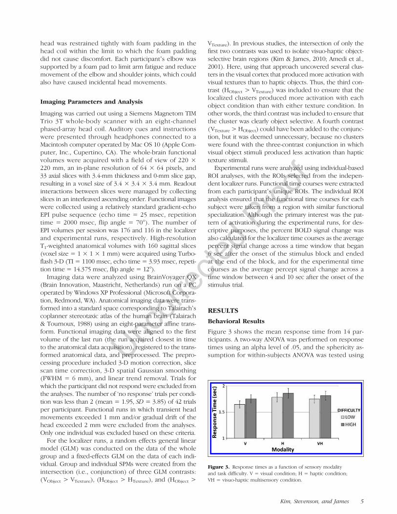
Figure 3 shows the mean response time from 14 par-ticipants. A two-way ANOVA was performed on responsetimes using an alpha level of .05, and the sphericity as-sumption for within-subjects ANOVA was tested using
Figure 3. Response times as a function of sensory modalityand task difficulty. V = visual condition; H = haptic condition;VH = visuo-haptic multisensory condition.
Kim, Stevenson, and James 5
UncorrectedProof
Mauchlyʼs test. Under the assumption of sphericity, theANOVA showed significant effects of sensory modality(F(2, 26) = 6.41, p = .005) and task difficulty (F(1, 3) =11.37, p = .005). Post hoc t tests showed significant differ-ences in response time between low- and high-difficultylevels in V (t(13) = 3.41, p = .002), H (t(13) = 2.91, p =.006), and VH (t(13) = 2.14, p = .026) conditions. The re-sults demonstrate that manipulating the similarity of thestimuli influenced difficulty in the predicted direction.Although the effect of difficulty appeared to be weaker forthe multisensory VH condition compared with unisensoryV and H conditions, this observation was not born outstatistically, as the interaction between modality and diffi-culty was not significant (F(2, 26) = 1.46, p = .252).
The VH response time was longer than the V responsetime, which would not be predicted based on multisensoryfacilitation. Differences in response time between modalityconditions were not considered meaningful because of dif-ferences in the instructions for the V, H, and VH conditions.For instance, in the VH condition, participants were spe-cifically instructed and trained to open their eyes when
they made contact with the stimulus. On the other hand,in the V condition, no contact with the stimulus was madeand participants opened their eyes at cue onset. Thus, thelonger response time with the VH condition comparedwith the V condition is attributable to the extra time takenfor the finger to travel from the start position and makefirst contact with the stimulus in the VH condition.
ROI Analysis
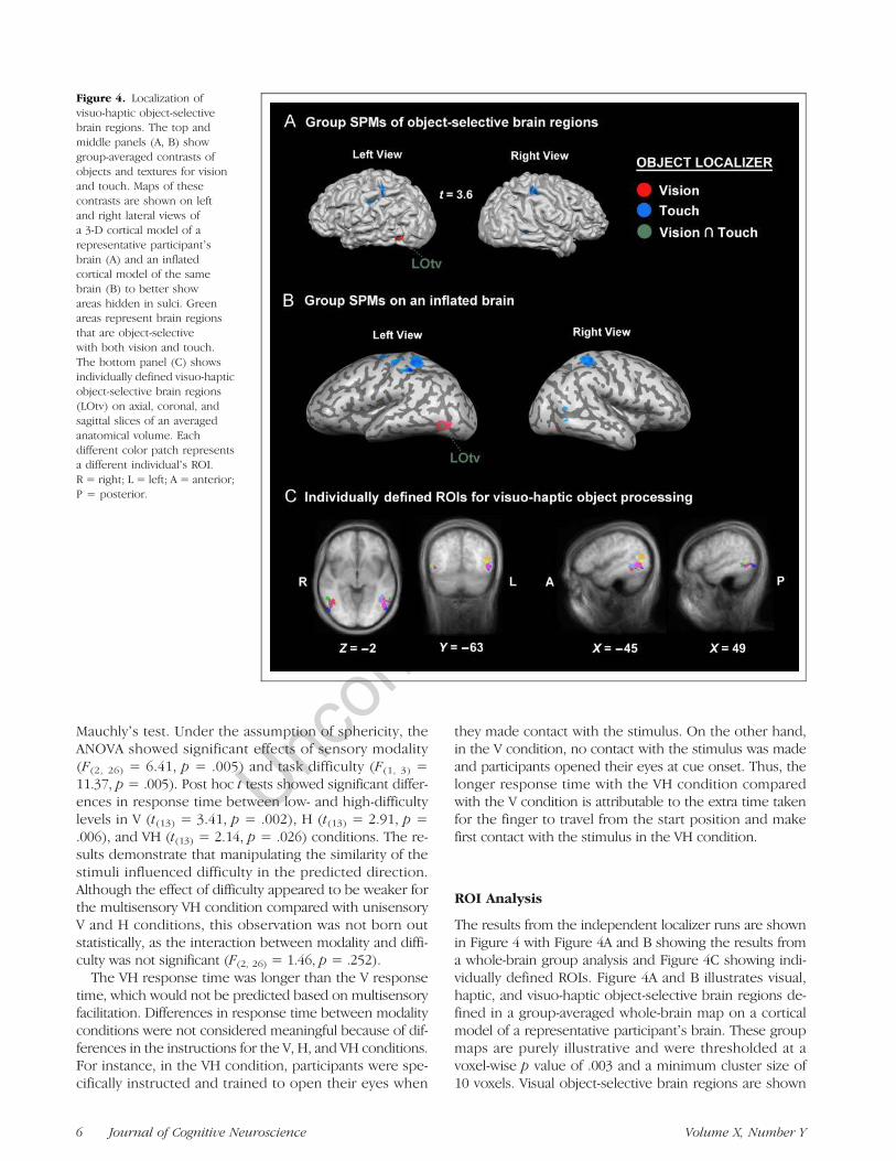
The results from the independent localizer runs are shownin Figure 4 with Figure 4A and B showing the results froma whole-brain group analysis and Figure 4C showing indi-vidually defined ROIs. Figure 4A and B illustrates visual,haptic, and visuo-haptic object-selective brain regions de-fined in a group-averaged whole-brain map on a corticalmodel of a representative participantʼs brain. These groupmaps are purely illustrative and were thresholded at avoxel-wise p value of .003 and a minimum cluster size of10 voxels. Visual object-selective brain regions are shown
Figure 4. Localization ofvisuo-haptic object-selectivebrain regions. The top andmiddle panels (A, B) showgroup-averaged contrasts ofobjects and textures for visionand touch. Maps of thesecontrasts are shown on leftand right lateral views ofa 3-D cortical model of arepresentative participantʼsbrain (A) and an inflatedcortical model of the samebrain (B) to better showareas hidden in sulci. Greenareas represent brain regionsthat are object-selectivewith both vision and touch.The bottom panel (C) showsindividually defined visuo-hapticobject-selective brain regions(LOtv) on axial, coronal, andsagittal slices of an averagedanatomical volume. Eachdifferent color patch representsa different individualʼs ROI.R = right; L = left; A = anterior;P = posterior.
6 Journal of Cognitive Neuroscience Volume X, Number Y
UncorrectedProof
in red and haptic object-selective brain regions are shown inblue. Green represents the intersection of the twoobject-selective maps, that is, brain regions that respondedto both visual and haptic objects more than visual and haptictextures.In addition to the group-averagedmap, maps of the same
contrast were generated for each participant. Because clus-ter size varied considerably across individuals at a fixedstatistical threshold, we adopted a procedure for activeROI selection that used a different threshold for each par-ticipant. The threshold was chosen for each participantbased on two criteria. First, aminimum acceptable thresholdt value (t = 1.0) was adopted to ensure that the data fromboth hemispheres of as many participants as possible wereincluded in the ROI analysis. If no clusters greater than
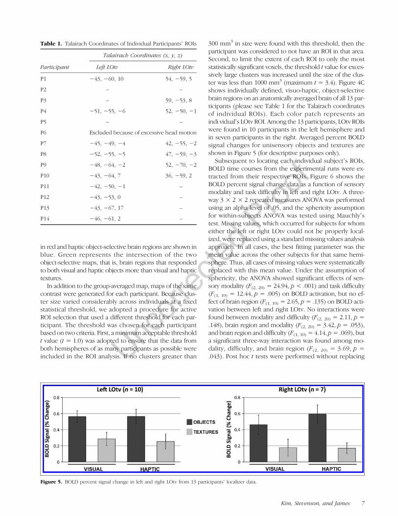
300 mm3 in size were found with this threshold, then theparticipant was considered to not have an ROI in that area.Second, to limit the extent of each ROI to only the moststatistically significant voxels, the threshold t value for exces-sively large clusters was increased until the size of the clus-ter was less than 1000 mm3 (maximum t = 3.4). Figure 4Cshows individually defined, visuo-haptic, object-selectivebrain regions on an anatomically averaged brain of all 13 par-ticipants (please see Table 1 for the Talairach coordinatesof individual ROIs). Each color patch represents anindi vidualʼs LOtv ROI. Among the 13 participants, LOtv ROIswere found in 10 participants in the left hemisphere andin seven participants in the right. Averaged percent BOLDsignal changes for unisensory objects and textures areshown in Figure 5 (for descriptive purposes only).
Subsequent to locating each individual subjectʼs ROIs,BOLD time courses from the experimental runs were ex-tracted from their respective ROIs. Figure 6 shows theBOLD percent signal change data as a function of sensorymodality and task difficulty in left and right LOtv. A three-way 3 × 2 × 2 repeated measures ANOVA was performedusing an alpha level of .05, and the sphericity assumptionfor within-subjects ANOVA was tested using Mauchlyʼstest. Missing values, which occurred for subjects for whomeither the left or right LOtv could not be properly local-ized, were replaced using a standardmissing values analysisapproach. In all cases, the best fitting parameter was themean value across the other subjects for that same hemi-sphere. Thus, all cases of missing values were systematicallyreplaced with this mean value. Under the assumption ofsphericity, the ANOVA showed significant effects of sen-sory modality (F(2, 20) = 24.94, p < .001) and task difficulty(F(1, 10) = 12.44, p = .005) on BOLD activation, but no ef-fect of brain region (F(1, 10) = 2.65, p= .135) on BOLD acti-vation between left and right LOtv. No interactions werefound between modality and difficulty (F(2, 20) = 2.11, p =.148), brain region and modality (F(2, 20) = 3.42, p = .053),and brain region and difficulty (F(1, 10) = 4.14, p= .069), buta significant three-way interaction was found among mo-dality, difficulty, and brain region (F(2, 20) = 3.69, p =.043). Post hoc t tests were performed without replacing
Table 1. Talairach Coordinates of Individual Participantsʼ ROIs
Participant
Talairach Coordinates (x, y, z)
Left LOtv Right LOtv
P1 −43, −60, 10 54, −59, 5
P2 – –
P3 – 59, −53, 8
P4 −51, −55, −6 52, −50, −1
P5 – –
P6 Excluded because of excessive head motion
P7 −45, −49, −4 42, −55, −2
P8 −52, −55, −5 47, −59, −3
P9 −48, −64, −2 52, −70, −2
P10 −43, −64, 7 36, −59, 2
P11 −42, −50, −1 –
P12 −43, −53, 0 –
P13 −43, −67, 17 –
P14 −46, −61, 2 –
Figure 5. BOLD percent signal change in left and right LOtv from 13 participantsʼ localizer data.
Kim, Stevenson, and James 7
UncorrectedProof
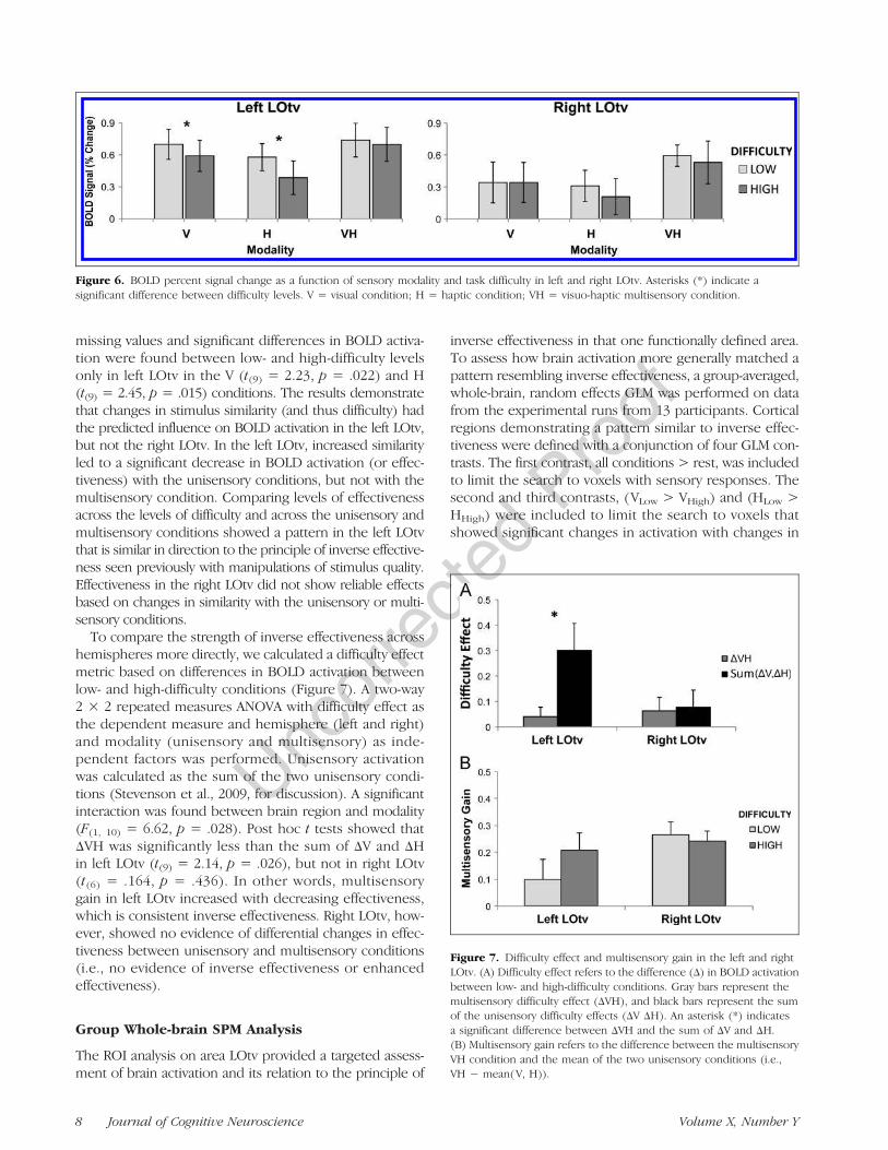
missing values and significant differences in BOLD activa-tion were found between low- and high-difficulty levelsonly in left LOtv in the V (t(9) = 2.23, p = .022) and H(t(9) = 2.45, p = .015) conditions. The results demonstratethat changes in stimulus similarity (and thus difficulty) hadthe predicted influence on BOLD activation in the left LOtv,but not the right LOtv. In the left LOtv, increased similarityled to a significant decrease in BOLD activation (or effec-tiveness) with the unisensory conditions, but not with themultisensory condition. Comparing levels of effectivenessacross the levels of difficulty and across the unisensory andmultisensory conditions showed a pattern in the left LOtvthat is similar in direction to the principle of inverse effective-ness seen previously with manipulations of stimulus quality.Effectiveness in the right LOtv did not show reliable effectsbased on changes in similarity with the unisensory or multi-sensory conditions.
To compare the strength of inverse effectiveness acrosshemispheres more directly, we calculated a difficulty effectmetric based on differences in BOLD activation betweenlow- and high-difficulty conditions (Figure 7). A two-way2 × 2 repeated measures ANOVA with difficulty effect asthe dependent measure and hemisphere (left and right)and modality (unisensory and multisensory) as inde-pendent factors was performed. Unisensory activationwas calculated as the sum of the two unisensory condi-tions (Stevenson et al., 2009, for discussion). A significantinteraction was found between brain region and modality(F(1, 10) = 6.62, p = .028). Post hoc t tests showed thatΔVH was significantly less than the sum of ΔV and ΔHin left LOtv (t(9) = 2.14, p = .026), but not in right LOtv(t(6) = .164, p = .436). In other words, multisensorygain in left LOtv increased with decreasing effectiveness,which is consistent inverse effectiveness. Right LOtv, how-ever, showed no evidence of differential changes in effec-tiveness between unisensory and multisensory conditions(i.e., no evidence of inverse effectiveness or enhancedeffectiveness).
Group Whole-brain SPM Analysis
The ROI analysis on area LOtv provided a targeted assess-ment of brain activation and its relation to the principle of
inverse effectiveness in that one functionally defined area.To assess how brain activation more generally matched apattern resembling inverse effectiveness, a group-averaged,whole-brain, random effects GLM was performed on datafrom the experimental runs from 13 participants. Corticalregions demonstrating a pattern similar to inverse effec-tiveness were defined with a conjunction of four GLM con-trasts. The first contrast, all conditions > rest, was includedto limit the search to voxels with sensory responses. Thesecond and third contrasts, (VLow > VHigh) and (HLow >HHigh) were included to limit the search to voxels thatshowed significant changes in activation with changes in
Figure 6. BOLD percent signal change as a function of sensory modality and task difficulty in left and right LOtv. Asterisks (*) indicate asignificant difference between difficulty levels. V = visual condition; H = haptic condition; VH = visuo-haptic multisensory condition.
Figure 7. Difficulty effect and multisensory gain in the left and rightLOtv. (A) Difficulty effect refers to the difference (Δ) in BOLD activationbetween low- and high-difficulty conditions. Gray bars represent themultisensory difficulty effect (ΔVH), and black bars represent the sumof the unisensory difficulty effects (ΔV ΔH). An asterisk (*) indicatesa significant difference between ΔVH and the sum of ΔV and ΔH.(B) Multisensory gain refers to the difference between the multisensoryVH condition and the mean of the two unisensory conditions (i.e.,VH − mean(V, H)).
8 Journal of Cognitive Neuroscience Volume X, Number Y
UncorrectedProof
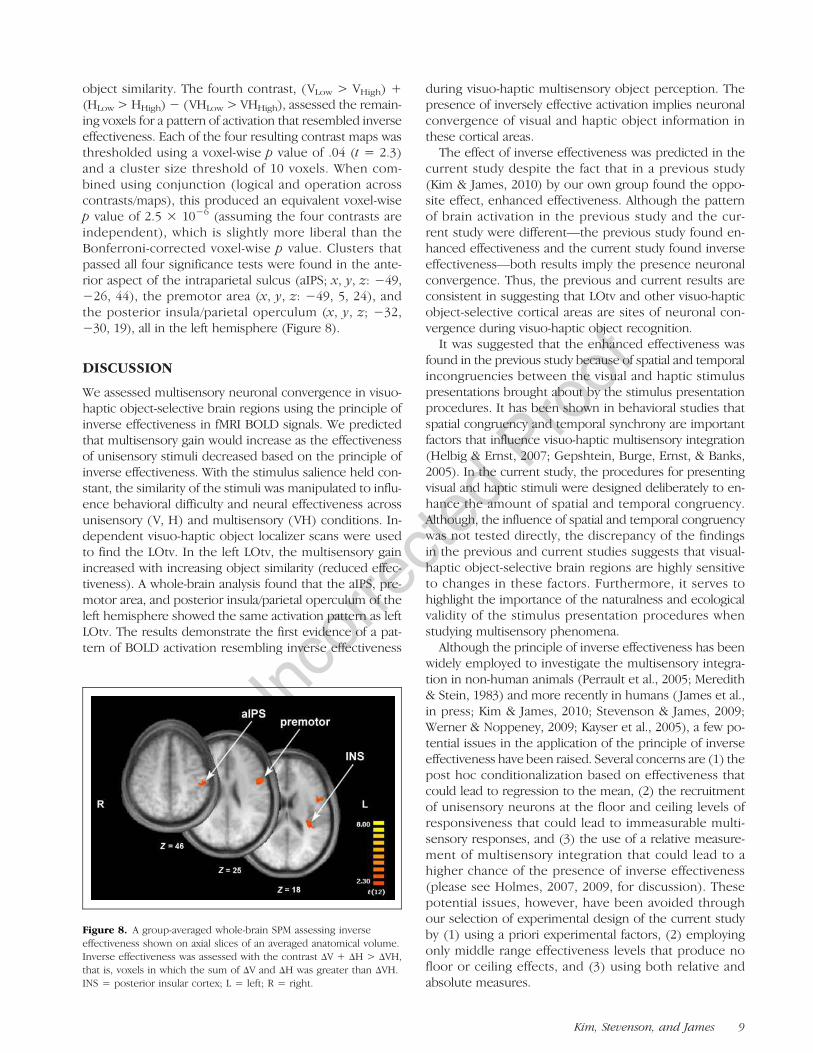
object similarity. The fourth contrast, (VLow > VHigh) +(HLow > HHigh)− (VHLow > VHHigh), assessed the remain-ing voxels for a pattern of activation that resembled inverseeffectiveness. Each of the four resulting contrast maps wasthresholded using a voxel-wise p value of .04 (t = 2.3)and a cluster size threshold of 10 voxels. When com-bined using conjunction (logical and operation acrosscontrasts/maps), this produced an equivalent voxel-wisep value of 2.5 × 10⁻⁶ (assuming the four contrasts areindependent), which is slightly more liberal than theBonferroni-corrected voxel-wise p value. Clusters thatpassed all four significance tests were found in the ante-rior aspect of the intraparietal sulcus (aIPS; x, y, z: −49,−26, 44), the premotor area (x, y, z: −49, 5, 24), andthe posterior insula/parietal operculum (x, y, z; −32,−30, 19), all in the left hemisphere (Figure 8).
DISCUSSION
We assessed multisensory neuronal convergence in visuo-haptic object-selective brain regions using the principle ofinverse effectiveness in fMRI BOLD signals. We predictedthat multisensory gain would increase as the effectivenessof unisensory stimuli decreased based on the principle ofinverse effectiveness. With the stimulus salience held con-stant, the similarity of the stimuli was manipulated to influ-ence behavioral difficulty and neural effectiveness acrossunisensory (V, H) and multisensory (VH) conditions. In-dependent visuo-haptic object localizer scans were usedto find the LOtv. In the left LOtv, the multisensory gainincreased with increasing object similarity (reduced effec-tiveness). A whole-brain analysis found that the aIPS, pre-motor area, and posterior insula/parietal operculum of theleft hemisphere showed the same activation pattern as leftLOtv. The results demonstrate the first evidence of a pat-tern of BOLD activation resembling inverse effectiveness
during visuo-haptic multisensory object perception. Thepresence of inversely effective activation implies neuronalconvergence of visual and haptic object information inthese cortical areas.
The effect of inverse effectiveness was predicted in thecurrent study despite the fact that in a previous study(Kim & James, 2010) by our own group found the oppo-site effect, enhanced effectiveness. Although the patternof brain activation in the previous study and the cur-rent study were different—the previous study found en-hanced effectiveness and the current study found inverseeffectiveness—both results imply the presence neuronalconvergence. Thus, the previous and current results areconsistent in suggesting that LOtv and other visuo-hapticobject-selective cortical areas are sites of neuronal con-vergence during visuo-haptic object recognition.
It was suggested that the enhanced effectiveness wasfound in the previous study because of spatial and temporalincongruencies between the visual and haptic stimuluspresentations brought about by the stimulus presentationprocedures. It has been shown in behavioral studies thatspatial congruency and temporal synchrony are importantfactors that influence visuo-haptic multisensory integration(Helbig & Ernst, 2007; Gepshtein, Burge, Ernst, & Banks,2005). In the current study, the procedures for presentingvisual and haptic stimuli were designed deliberately to en-hance the amount of spatial and temporal congruency.Although, the influence of spatial and temporal congruencywas not tested directly, the discrepancy of the findingsin the previous and current studies suggests that visual-haptic object-selective brain regions are highly sensitiveto changes in these factors. Furthermore, it serves tohighlight the importance of the naturalness and ecologicalvalidity of the stimulus presentation procedures whenstudying multisensory phenomena.
Although the principle of inverse effectiveness has beenwidely employed to investigate the multisensory integra-tion in non-human animals (Perrault et al., 2005; Meredith& Stein, 1983) and more recently in humans ( James et al.,in press; Kim & James, 2010; Stevenson & James, 2009;Werner & Noppeney, 2009; Kayser et al., 2005), a few po-tential issues in the application of the principle of inverseeffectiveness have been raised. Several concerns are (1) thepost hoc conditionalization based on effectiveness thatcould lead to regression to the mean, (2) the recruitmentof unisensory neurons at the floor and ceiling levels ofresponsiveness that could lead to immeasurable multi-sensory responses, and (3) the use of a relative measure-ment of multisensory integration that could lead to ahigher chance of the presence of inverse effectiveness(please see Holmes, 2007, 2009, for discussion). Thesepotential issues, however, have been avoided throughour selection of experimental design of the current studyby (1) using a priori experimental factors, (2) employingonly middle range effectiveness levels that produce nofloor or ceiling effects, and (3) using both relative andabsolute measures.
Figure 8. A group-averaged whole-brain SPM assessing inverseeffectiveness shown on axial slices of an averaged anatomical volume.Inverse effectiveness was assessed with the contrast ΔV + ΔH > ΔVH,that is, voxels in which the sum of ΔV and ΔH was greater than ΔVH.INS = posterior insular cortex; L = left; R = right.
Kim, Stevenson, and James 9
UncorrectedProof
Both the whole-brain analysis and the ROI analysis ofthe current study revealed significant inverse effectivenessin the left hemisphere only. This effect was most strikingin the ROI analysis, where it was shown that the difficultyhad little effect on activation in the right hemisphereROI. Although bimodal visuo-haptic activation tends tobe found bilaterally in most individuals, there is growingevidence that when bimodal activation is not found bilat-erally, it is usually found in the left hemisphere (Kim &James, 2010; James, Servos, Kilgour, Huh, & Lederman,2006; Kilgour, Kitada, Servos, James, & Lederman, 2005;Grefkes et al., 2002; Banati, Goerres, Tjoa, Aggleton, &Grasby, 2000). Similarly, a recent fMRI adaptation studyshowed bilateral visuo-haptic repetition suppression ef-fects in LOtv and aIPS, however, that the suppressioneffects were stronger in the left hemisphere than the right(Tal & Amedi, 2009).
Not all of the evidence supports the hypothesis of aleft hemisphere bias for visuo-haptic convergence, for in-stance, some researchers have found bimodal activationin right insula (Hadjikhani & Roland, 1998) and in rightlateral occipital complex (Stilla & Sathian, 2008; Prather,Votaw, & Sathian, 2004). The same studies have, however,shown bimodal activation in left IPS (Stilla & Sathian, 2008;Prather et al., 2004; Grefkes et al., 2002), which is consis-tent with our results. In addition to the left IPS, our find-ing of left insula in visuo-haptic multisensory integrationis supported by an earlier PET study that showed leftlateralized insula activation for visuo-tactile multisensoryintegration (Banati et al., 2000). It should be noted, how-ever, that all of these studies examined multisensory arealconvergence, not necessarily multisensory neuronal con-vergence. The current study found multisensory arealconvergence in both hemispheres similar to the previousstudies but found multisensory neuronal convergence, asdemonstrated by the presence of inverse effectiveness,only in the left hemisphere. This result is similar to a re-cent fMRI adaptation study that also aimed to examinevisuo-haptic neuronal convergence (Tal & Amedi, 2009)and which found stronger neuronal convergence in theleft hemisphere than right hemisphere.
Considering that most fMRI studies of haptic explora-tion have right-handed participants palpate with theirright hand, left lateralization of multisensory neuronal con-vergence could simply be considered a consequence of thecontralateral representation of right-handed exploration.However, several previous studies have demonstrated thatthe hand of use during haptic exploration does not in-fluence activation in higher-level cortical areas suchas the lateral occipital complex. Amedi and colleagues(2010) compared left- and right-handed palpation duringtactile exploration of objects and showed that the ac-tivation in LOtv was bilateral, irrespective of the handof use. Furthermore, left-lateralized activation has beenfound in other studies where participants explored ob-jects with either their left hand ( James et al., 2006; Kilgouret al., 2005) or with both hands (Kim & James, 2010).
In summary, previous studies combined with the cur-rent study suggested that visuo-haptic object-selectiveactivation in the LOtv and possibly other brain regionsis generally bilateral but may be biased to be strongeror more reliable in the left hemisphere than in the right.All of the previous studies, however, used right-handedparticipants; therefore, although it seems clear that thehand-of-use does not contribute to the bias, it is possi-ble handedness may. Further study is needed to test thispossibility.In the current study, analysis of the object-selective lo-
calizer data found significant voxel clusters in the locationof LOtv, but not in the location of IPS. Our previous studyfound significant voxel clusters in both regions using asimilar statistical contrast (Kim & James, 2010). The IPShas been suggested to be a site of multisensory conver-gence for visuo-haptic object recognition (Stilla & Sathian,2008; James et al., 2007; Zhang et al., 2004; Grefkes et al.,2002; Culham & Kanwisher, 2001) and to be involved inprocessing visual shape information particularly for visuallyguided reaching and grasping ( James, Culham, Humphrey,Milner, & Goodale, 2003). There are several possible rea-sons why significant bimodal object-selective activationwas not found in IPS in the current study. First, in thecurrent study, subjects viewed tangible objects directly,whereas in previous studies, subjects viewed pictures ofobjects. Second, the size (visual angle of 2.57° × 2.57°)of tangible stimuli used in the current study was smallerthan the size of the pictures used in our previous study(visual angle of 12° × 12°). Third (and the most likely),participantsʼ hand movements were more restricted inthe current study compared with the previous study. Partic-ipants were trained to make an almost automatic fingermovement to the stimulus. By contrast, in most previousstudies, participants were required to perform a ballisticreaching or grasping movement to the object with oneor both hands to begin exploration. The lack of a needto action planning may have limited the recruitment ofIPS in the current study, relative to previous studies. Al-though IPS was not found in the localizer data, a group-averaged SPM from the experimental data revealed theinvolvement of aIPS in visuo-haptic neural convergence ofshape information.Besides the involvement of LOtv and IPS, the cerebel-
lum has been also found to be involved in multisensoryvisual and haptic object recognition in some human neuro-imaging studies (Gentile et al., 2011; Naumer et al., 2010;Stevenson et al., 2009). There is also growing evidenceover the last few decades indicating that the cerebellumplays a role in perception and cognition, not merelyin motor control (Strick, Dum, & Fiez, 2009; Gao et al.,1996). Neither the ROI analysis, nor the whole-brainanalysis, however, showed evidence of cerebellar in-volvement in the current study, implying a potential dis-crepancy between the underlying multisensory networksrecruited in the previous studies and the current study.Further study is certainly needed to investigate the precise
10 Journal of Cognitive Neuroscience Volume X, Number Y
UncorrectedProof
function of cerebellum in visuo-haptic multisensory objectrecognition and multisensory integration. One speculativeexplanation for the discrepancy between our study andprevious studies is the involvement of a deliberate handoperation during exploration of stimuli. Participants inthe previous studies (Gentile et al., 2011; Naumer et al.,2010; Stevenson et al., 2009) used their whole hand topalpate the objects or viewed their whole hand while partof it was touched by an object. In contrast, participants inthe current study palpated the object with one fingerand were constrained to using the same, repetitive, ratherautomatic sweeping movement on all trials throughoutthe experiment. If the cerebellum-related activity duringvisuo-haptic multisensory processing in other studies isrelated to coordination of sensory input of the body (inthis case the hand) and sensory inputs of other objects,then the cerebellum may not have been recruited dif-ferentially in the current study, because the finger move-ment aspects of the study were so closely controlled acrossconditions.Some studies have shown that eyes-opened and eyes-
closed states without external stimulation have a differentimpact on brain activation patterns in sighted (Marx et al.,2003, 2004) and blind subjects (Hufner et al., 2009), sug-gesting that the choice of state as rest condition may leadto different interpretations of results. According to thesestudies, the eyes-closed state enhances brain activationin various sensory areas including visual, somatosensory,auditory, and vestibular systems, whereas the eyes-openedstate enhances attentional and ocular motor system ac-tivities. Because participants in the current study hadeyes opened or closed, depending on the condition (eyesclosed in H condition; eyes opened in V and VH condi-tions), the changes of state may have been a confoundingfactor. The choice of rest condition, however, stayed con-sistent throughout the whole session in the current study,and all experimental conditions were compared with thesame type of rest condition, eyes-closed. In addition tothe homogeneous rest state, having object-selective brainregions selected by subtracting the texture condition fromthe object condition in the localizer runs should have can-celed out the effect of rest condition in the end. Althoughit is possible that H condition with eyes closed may haveinduced increased BOLD activation in visual and somato-sensory cortical areas during that condition comparedwith V or VH condition with eyes open, the effect isnot seen in percent change of BOLD signal in Figure 6.H conditions did not produce increased BOLD activationin either low- or high-difficulty conditions compared withV and VH conditions. Hence, the state of the eyes did notseem to have a considerable impact on the interpretationof our results.In conclusion, the neural substrates involved in visuo-
haptic neuronal convergence were investigated using anadditive-factors design. An ROI analysis on the object-selective brain regions that responded more to both vi-sual and haptic objects than to textures found evidence
of inverse effectiveness in the left LOtv. A whole-brainanalysis also found evidence of inverse effectiveness inaIPS, premotor, and posterior insular cortices of the lefthemisphere. This study is the first evidence of inverseeffectiveness in the human brain with visuo-haptic objectrecognition.
Acknowledgments
This research was supported in part by the Indiana METACytInitiative of Indiana University and funded in part by a major grantfrom the Lilly Endowment, Inc. and by a grant to T. W. James fromIndiana Universityʼs Faculty Research Support Program adminis-tered by the Office of the Vice Provost for Research. We also grate-fully acknowledge Daniel Eylath for stimulus presentation; TheaAtwood and Rebecca Ward for their technical support; KarinHarman James and the Indiana University Neuroimaging Groupfor their insights on this study; and June Yong Lee and LaurelStevenson for their support.
Reprint requests should be sent to Sunah Kim, 360 Minor Hall,University of California, Berkeley, Berkeley, CA 94720, or viae-mail: [email protected].
REFERENCES
Amedi, A., Malach, R., Hendler, T., Peled, S., & Zohary, E.(2001). Visuo-haptic object-related activation in theventral visual pathway. Nature Neuroscience, 4, 324–330.
Amedi, A., Raz, N., Azulay, H., Malach, R., & Zohary, E.(2010). Cortical activity during tactile exploration ofobjects in blind and sighted humans. RestorativeNeurology and Neuroscience, 28, 143–156.
Amedi, A., von Kriegstein, K., van Atteveldt, N. M., Beauchamp,M. S., & Naumer, M. J. (2005). Functional imaging ofhuman crossmodal identification and object recognition.Experimental Brain Research, 166, 559–571.
Banati, R. B., Goerres, G. W., Tjoa, C., Aggleton, J. P., &Grasby, P. (2000). The functional anatomy of visual-tactileintegration in man: A study using positron emissiontomography. Neuropsychologia, 38, 115–124.
Culham, J. C., & Kanwisher, N. G. (2001). Neuroimagingof cognitive functions in human parietal cortex. CurrentOpinion in Neurobiology, 11, 157–163.
Dijkerman, H. C., & de Haan, E. H. (2007). Somatosensoryprocesses subserving perception and action. Behavioraland Brain Sciences, 30, 189–201; discussion 201-139.
Dragovic, M. (2004). Towards an improved measure of theEdinburgh Handedness Inventory: A one-factor congenericmeasurement model using confirmatory factor analysis.Laterality, 9, 411–419.
Gao, J. H., Parsons, L. M., Bower, J. M., Xiong, J., Li, J., & Fox,P. T. (1996). Cerebellum implicated in sensory acquisitionand discrimination rather than motor control. Science,272, 545–547.
Gentile, G., Petkova, V. I., & Ehrsson, H. H. (2011). Integrationof visual and tactile signals from the hand in the humanbrain: An fMRI study. Journal of Neurophysiology, 105,910–922.
Gepshtein, S., Burge, J., Ernst, M. O., & Banks, M. S. (2005).The combination of vision and touch depends on spatialproximity. Journal of Vision, 5, 1013–1023.
Grefkes, C., Weiss, P. H., Zilles, K., & Fink, G. R. (2002).Crossmodal processing of object features in humananterior intraparietal cortex: An fMRI study implies
Kim, Stevenson, and James 11
UncorrectedProof
equivalencies between humans and monkeys. Neuron,35, 173–184.
Hadjikhani, N., & Roland, P. E. (1998). Cross-modaltransfer of information between the tactile and thevisual representations in the human brain: A positronemission tomographic study. Journal of Neuroscience,18, 1072–1084.
Helbig, H. B., & Ernst, M. O. (2007). Knowledge about acommon source can promote visual-haptic integration.Perception, 36, 1523–1533.
Holmes, N. P. (2007). The law of inverse effectiveness inneurons and behaviour: Multisensory integration versusnormal variability. Neuropsychologia, 45, 3340–3345.
Holmes, N. P. (2009). The principle of inverse effectiveness inmultisensory integration: Some statistical considerations.Brain Topography, 21, 168–176.
Hufner, K., Stephan, T., Flanagin, V. L., Deutschlander, A.,Stein, A., Kalla, R., et al. (2009). Differential effects of eyesopen or closed in darkness on brain activation patterns inblind subjects. Neuroscience Letters, 466, 30–34.
James, T. W., Culham, J., Humphrey, G. K., Milner, A. D.,& Goodale, M. A. (2003). Ventral occipital lesions impairobject recognition but not object-directed grasping:An fMRI study. Brain, 126, 2463–2475.
James, T. W., Humphrey, G. K., Gati, J. S., Servos, P., Menon, R. S.,& Goodale, M. A. (2002). Haptic study of three-dimensionalobjects activates extrastriate visual areas. Neuropsychologia,40, 1706–1714.
James, T. W., & Kim, S. (2010). Dorsal and ventral corticalpathways for visuo-haptic integration revealed usingfMRI. In M. J. Naumer & J. Kaiser (Eds.), Multisensoryobject perception in the primate brain. New York:Springer.
James, T. W., Kim, S., & Fisher, J. S. (2007). The neural basis ofhaptic object processing. Canadian Journal of ExperimentalPsychology, 61, 219–229.
James, T. W., Servos, P., Kilgour, A. R., Huh, E., & Lederman, S.(2006). The influence of familiarity on brain activationduring haptic exploration of 3-D facemasks. NeuroscienceLetters, 397, 269–273.
James, T. W., & Stevenson, R. A. (in press). The use of fMRI toassess multisensory integration. In M. Wallace & M. Murray(Eds.), Frontiers in the neural bases of multisensoryprocesses. London: Taylor and Francis Group.
James, T. W., Stevenson, R. A., & Kim, S. (in press). Inverseeffectiveness in multisensory processing. In B. Stein (Ed.),The new handbook of multisensory processes. Cambridge,MA: MIT Press.
Joseph, J. E., & Farley, A. B. (2004). Cortical regionsassociated with different aspects of object recognitionperformance. Cognitive, Affective & BehavioralNeuroscience, 4, 364–378.
Kayser, C., Petkov, C. I., Augath, M., & Logothetis, N. K.(2005). Integration of touch and sound in auditory cortex.Neuron, 48, 373–384.
Kilgour, A. R., Kitada, R., Servos, P., James, T. W., & Lederman,S. J. (2005). Haptic face identification activates ventraloccipital and temporal areas: An fMRI study. Brainand Cognition, 59, 246–257.
Kim, S., & James, T. W. (2010). Enhanced effectiveness invisuo-haptic object-selective brain regions with increasingstimulus salience. Human Brain Mapping, 31, 678–693.
King, A. J., & Palmer, A. R. (1985). Integration of visual andauditory information in bimodal neurones in the guinea-pigsuperior colliculus. Experimental Brain Research, 60,492–500.
Marx, E., Deutschlander, A., Stephan, T., Dieterich, M.,Wiesmann, M., & Brandt, T. (2004). Eyes open and eyes
closed as rest conditions: Impact on brain activationpatterns. Neuroimage, 21, 1818–1824.
Marx, E., Stephan, T., Nolte, A., Deutschlander, A., Seelos,K. C., Dieterich, M., et al. (2003). Eye closure in darknessanimates sensory systems. Neuroimage, 19, 924–934.
Meredith, M. A., Nemitz, J. W., & Stein, B. E. (1987).Determinants of multisensory integration in superiorcolliculus neurons. I. Temporal factors. Journal ofNeuroscience, 7, 3215–3229.
Meredith, M. A., & Stein, B. E. (1983). Interactions amongconverging sensory inputs in the superior colliculus.Science, 221, 389–391.
Meredith, M. A., & Stein, B. E. (1986a). Spatial factors determinethe activity of multisensory neurons in cat superior colliculus.Brain Research, 365, 350–354.
Meredith, M. A., & Stein, B. E. (1986b). Visual, auditory,and somatosensory convergence on cells in superiorcolliculus results in multisensory integration. Journalof Neurophysiology, 56, 640–662.
Miller, L. M., & DʼEsposito, M. (2005). Perceptual fusionand stimulus coincidence in the cross-modal integrationof speech. The Journal of Neuroscience: The OfficialJournal of the Society for Neuroscience, 25, 5884–5893.
Naumer, M. J., Ratz, L., Yalachkov, Y., Polony, A., Doehrmann, O.,van de Ven, V., et al. (2010). Visuohaptic convergence in acorticocerebellar network. European Journal of Neuroscience,31, 1730–1736.
Oldfield, R. C. (1971). The assessment and analysis ofhandedness: The Edinburgh inventory. Neuropsychologia,9, 97–113.
Peltier, S., Stilla, R., Mariola, E., LaConte, S., Hu, X., &Sathian, K. (2007). Activity and effective connectivityof parietal and occipital cortical regions during hapticshape perception. Neuropsychologia, 45, 476–483.
Perrault, T. J., Jr., Vaughan, J. W., Stein, B. E., & Wallace,M. T. (2005). Superior colliculus neurons use distinctoperational modes in the integration of multisensorystimuli. Journal of Neurophysiology, 93, 2575–2586.
Pietrini, P., Furey, M. L., Ricciardi, E., Gobbini, M. I., Wu,W. H., Cohen, L., et al. (2004). Beyond sensory images:Object-based representation in the human ventral pathway.Proceedings of the National Academy of Sciences, U.S.A.,101, 5658–5663.
Prather, S. C., Votaw, J. R., & Sathian, K. (2004). Task-specificrecruitment of dorsal and ventral visual areas during tactileperception. Neuropsychologia, 42, 1079–1087.
Reed, C. L., Klatzky, R. L., & Halgren, E. (2005). What vs.where in touch: An fMRI study. Neuroimage, 25, 718–726.
Sternberg, S. (1969). Memory-scanning: Mental processesrevealed by reaction-time experiments. American Science,57, 421–457.
Stevenson, R. A., Altieri, N. A., Kim, S., Pisoni, D. B., &James, T. W. (2010). Neural processing of asynchronousaudiovisual speech perception. Neuroimage, 49,3308–3318.
Stevenson, R. A., & James, T. W. (2009). Audiovisual integrationin human superior temporal sulcus: Inverse effectiveness andthe neural processing of speech and object recognition.Neuroimage, 44, 1210–1223.
Stevenson, R. A., Kim, S., & James, T. W. (2009). Anadditive-factors design to disambiguate neuronal and arealconvergence: Measuring multisensory interactions betweenaudio, visual, and haptic sensory streams using fMRI.Experimental Brain Research, 198, 183–194.
Stevenson, R. A., VanDerKlok, R. M., Pisoni, D. B., & James, T. W.(2011). Discrete neural substrates underlie complementaryaudiovisual speech integration processes. Neuroimage, 55,1339–1345.
12 Journal of Cognitive Neuroscience Volume X, Number Y

UncorrectedProof
Stilla, R., & Sathian, K. (2008). Selective visuo-hapticprocessing of shape and texture. Human Brain Mapping,29, 1123–1138.
Stoesz, M. R., Zhang, M., Weisser, V. D., Prather, S. C., Mao, H.,& Sathian, K. (2003). Neural networks active during tactileform perception: Common and differential activity duringmacrospatial and microspatial tasks. International Journalof Psychophysiology, 50, 41–49.
Strick, P. L., Dum, R. P., & Fiez, J. A. (2009). Cerebellum andnonmotor function. Annual Review of Neuroscience, 32,413–434.
Tal, N., & Amedi, A. (2009). Multisensory visual-tactile objectrelated network in humans: Insights gained using a novelcrossmodal adaptation approach. Experimental BrainResearch, 198, 165–182.
Werner, S., & Noppeney, U. (2009). Superadditive responses insuperior temporal sulcus predict audiovisual benefits inobject categorization. Cerebral Cortex.
Zhang, M., Weisser, V. D., Stilla, R., Prather, S. C., & Sathian, K.(2004). Multisensory cortical processing of object shapeand its relation to mental imagery. Cognitive, Affective &Behavioral Neuroscience, 4, 251–259.
Kim, Stevenson, and James 13