prism adaptation in the rehabilitation of patients with visuo-spatial cognitive disorders
TRANSCRIPT
WCO/18546; Total nos of Pages: 9;
WCO 18546
Prism adaptation in the rehabili
tation of patients withvisuo-spatial cognitive disordersLaure Pisellaa,b, Gilles Rodea,b,c,d, Alessandro Farnea,b,Caroline Tiliketea,b and Yves Rossettia,b,c,d
Purpose of review
The traditional focus of neurorehabilitaion has been on the
patients’ attention on their deficit, such that they should
become aware of their problems and gain intentional control
of compensatory strategies (descending approach). We
review prism adaptation as one of the approaches that
emphasizes ascending rather than descending strategies to
the rehabilitation of visuo-spatial disorders. The clinical
outcome of prism adaptation highlights the need for a
theoretical reconsideration of some previous stances to
neurological rehabilitation.
Recent findings
Recent years have given rise to a growing body of
experimental studies showing that the descending strategy
is not always optimal, especially when higher-level cognition
is affected by the patients’ condition. Ascending
approaches have, for example, used visuo-manual
adaptation for the rehabilitation of visuo-spatial deficits.
A simple task of pointing to visual targets while wearing
prismatic goggles can produce remarkable improvements
of various aspects of unilateral neglect.
Summary
The neural mechanisms underpinning visuo-manual
plasticity can be viewed as a powerful rehabilitation tool that
produces straightforward effects not only on visual and
motor parameters, but on visuo-spatial, attentional and
higher cognitive neurological functions. The use of prism
adaptation therapy in neglect and other visuo-spatial
disorders has just started to reveal its potential, both at a
practical and theoretical level.
Keywords
neglect, prism adaptation, rehabilitation, visuo-spatial
Curr Opin Neurol 19:000–000. � 2006 Lippincott Williams & Wilkins.
aINSERM, U 534, Espace et Action, Bron, France, bUniversite Claude BernardLyon I, Lyon, France, cHopital Henry Gabrielle, Hospices Civils de Lyon, Route deVourles, St Genis Laval, France and d‘Mouvement et Handicap’ Plateforme IFNL-HCL, Institut Federatif des Neurosciences de Lyon, Hospices Civils de Lyon, Lyon,France
Correspondence to Y. Rossetti, INSERM U534, Espace et Action, Institut Nationalde la Sante et de la Recherche Medicale, Universite Claude Bernard and HospicesCivils de Lyon, 16 avenue Lepine, Case 13, 69676 Bron, FranceTel: +33 472 91 34 00; fax: +33 472 91 34 01; e-mail: [email protected]
Sponsorship: This work was supported by the INSERM AVENIR grant R05265CS,Hospices Civils de Lyon, Universite Claude Bernard and Programme Hospitalier deRecherche Clinique.
Current Opinion in Neurology 2006, 19:000–000
Abbreviation
PPC p
osterior parietal cortex� 2006 Lippincott Williams & Wilkins1350-7540
IntroductionA large proportion of right-hemisphere stroke patients
show unilateral neglect. This is a multifaceted neuro-
logical deficit potentially affecting perception, attention,
representation and/or motor control within their left-
sided space [1–3,4�], as well as the right-sided hemispace
[5�,6,7], inducing many functionally debilitating effects
on everyday life, and is responsible for poor functional
recovery and ability to benefit from treatment [8–10].
The various manifestations of unilateral neglect share
one major feature – patients remain unaware of the
deficits they exhibit or at least fail to fully consciously
attend to these deficits. This lack of awareness is
dramatically expressed in anosognosia and hemiasoma-
tognosia [1]. It is therefore astonishing that the first
methods proposed for neglect rehabilitation were mainly
based on leftward voluntary orienting of attention. This
paradox was already underlined by Diller and Weinberg
([11], p. 67): ‘The first step in the treatment of hemi-
inattention is to make the patient aware of the problem.
This is particularly difficult in hemi-inattention since this
failure in awareness appears to be at the heart of the
patient’s difficulty’. It may indeed appear paradoxical to
base a rehabilitation procedure on awareness and inten-
tion in patients with a deficit in consciousness. Accord-
ingly, these techniques have produced significant results,
but are clearly exposed to several limitations, i.e. the
voluntary monitoring of attention is restricted to a specific
context and does not apply as soon as more automatic
control is required.
To act on higher-level cognition in such a way as to
bypass the impaired conscious awareness and intention
one should, at least in principle, find another entry route
to space representation systems. Rubens [12] pioneered
such an ascending alternative, both theoretically and
experimentally, by providing preliminary support to
the prediction made by Jeannerod and Biguer [13]: a
unilateral lesion would produce an illusory displacement
1
WCO/18546; Total nos of Pages: 9;
2 Trauma and rehabilitation
Figure 1 Prism adaptation in neglect
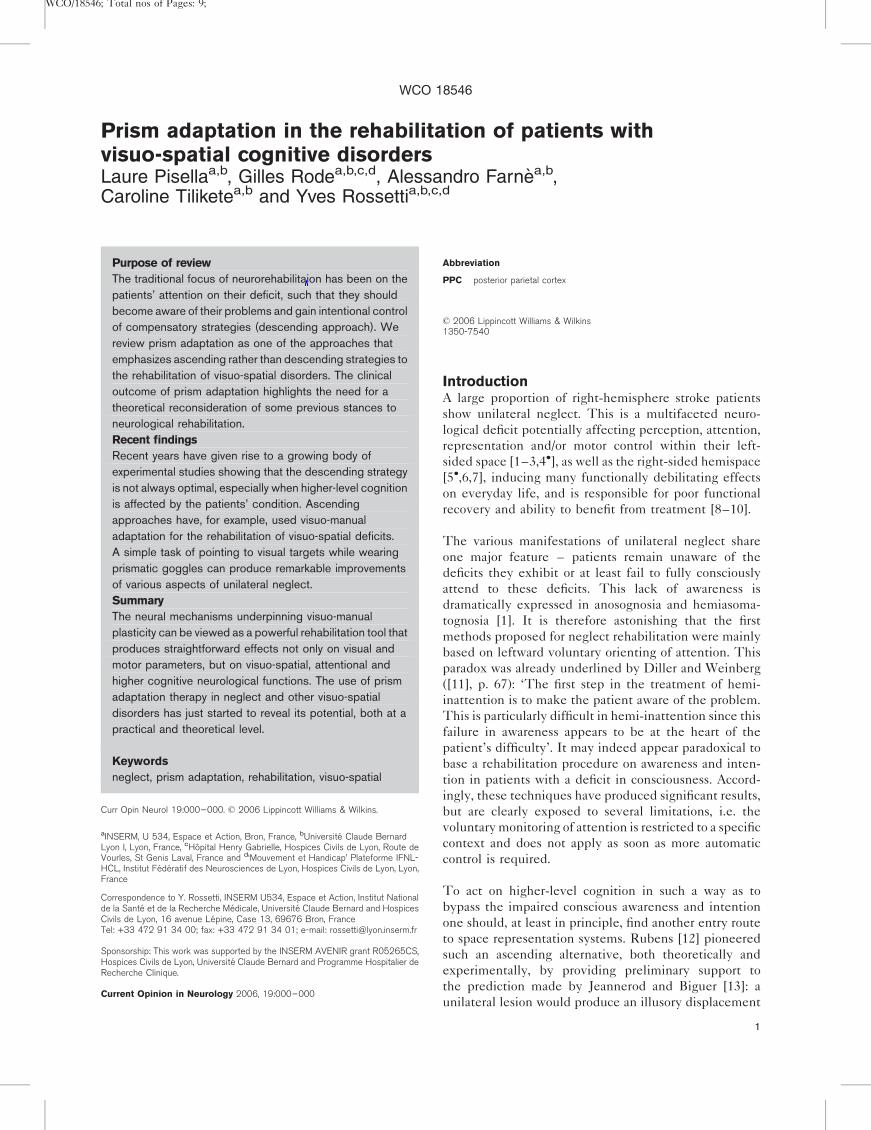
(a) Neglect patients adapt more than normals. In the pre-adaptive test, neglect patients exhibit a manual straight-ahead demonstration shifted to theright with respect to normals. In the post-test, they reveal larger after-effects (redrawn from [15]). (b) Two examples of pre, immediate post and late(þ2 h) performance of copy drawing.
of the egocentric reference, somewhat as if the subject
felt being constantly rotated toward the lesion side.
Rubens reported that vestibular stimulation, obtained
by pouring cold water into the neglect patients’ left
ear, instantly produced a dramatic, although transient
improvement of neglect.
Cognitive effects of prism adaptationBoth approaches to neglect rehabilitation have, however,
important limitations either in terms of absence of gener-
alization (descending approach) or in terms of very lim-
ited duration (ascending approach) of the beneficial
effects. It was therefore a challenge to develop a strategy
to combine the advantages of the two approaches and to
propose a technique that, bypassing the awareness level,
could also promote long-lasting effects. Adaptation to
wedge prisms is a simple way of producing low-level,
automatic modifications of visuo-motor correspondences,
demonstrated by the presence of measurable after-effects
after the prism-exposure phase (Fig. 1a). In contrast to
more complex visual reorganization requiring extended
exposure [14], adaptation to wedge prisms has long been
known to quickly develop over the course of a 5-min
simple pointing session. Despite over 100 years of stu-
dies, only visuo-motor after-effects had been described
until the discovery that prism adaptation (prism adap-
tation) can improve higher cognitive deficits such as
unilateral neglect [15] (Fig. 1b).
The principles of prismatic adaptationWhen someone first looks through wedge prisms that
optically displace the visual field, e.g. 108 in the rightward
direction, he/she may have little feeling that anything is
out of the ordinary, until he/she experiences extraordi-
nary difficulty in perceptual-motor tasks (i.e. direct
effects of prism exposure). For example, pointing toward
a visual target produces an error to the right of the target
position. First, a relatively abrupt reduction of the lateral
WCO/18546; Total nos of Pages: 9;
Prism adaptation Pisella et al. 3
Figure 2 Prism adaptation procedure
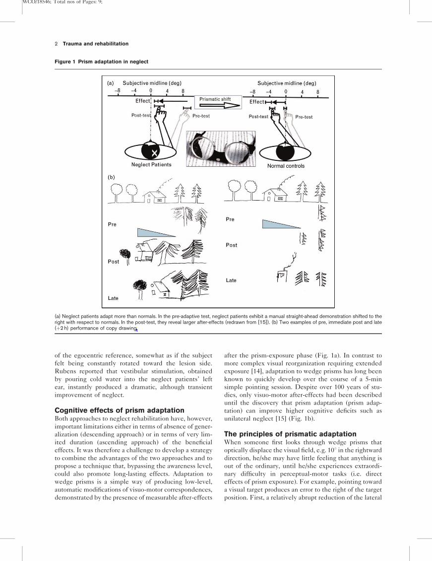
A proper prism adaptation session should include three periods: pre-tests, prism exposure and post-tests. During the early phase of the exposure, errorreduction (mainly accounted for by the strategic component) does not imply that prism adaptation is already effective. The post- and pre-tests, optimallyperformed with nonexposed targets, are used to compute the amount of after-effect, i.e. proper adaptation.
error can be observed due to a strategic component of
adaptation. Then, a more gradual reduction of the term-
inal error is observed, returning to pre-exposure levels as
the person makes repeated attempts at target pointing.
Whereas the strategic component is at work only over a
short period of time [16], the true adaptation to the
prismatic displacement (or realignment) develops more
gradually and is more purely expressed during the sub-
sequent slow phase of error reduction. When the prisms
are removed the person experiences unforeseen errors in
the opposite direction, to the left of the target! This
negative after-effect of prism exposure demonstrates
persistence of the adaptation acquired during exposure.
In most of our studies sham goggles were (and should be)
used to control for the spurious effects due to directional
visuo-motor activity. They were made of two pairs of 58prisms producing opposite shifts, i.e. a total shift of 08(same weight and same opacity as the 108 prisms). The
real and sham prisms were fitted into glacier goggles
(Cebe) in order to prevent any access to unshifted vision
(Optique Peter, Lyon, France; www.optiquepeter.com).
Vision of the starting hand position is usually occluded to
ensure the optimal development of the adaptation [17]. A
pointing task without visual feedback (open loop) is
performed before and after the adaptation procedure to
evaluate the development of a visuo-manual adaptation
to the visual shift.
Thus, the basic prism adaptation procedure simply
involves (1) pre-exposure baseline measurement of
pointing performance, (2) active exposure to prismatic
displacement to produce adaptation and (3) post-
exposure after-effect measurement of adaptation persist-
ence (Fig. 2). Is this all there is to prism adaptation? Prism
adaptation may misleadingly appear simple when com-
pared to the profound effects it can exert on spatial
cognition.
GeneralizationLogically, the effects of prism adaptation should be
restricted to, or at least be best for, visuo-motor tasks,
because they share more common features with the
visuo-manual adaptation procedure. In the original study,
we observed the best improvement for the Schenkenberg
bisection test (6/6 patients markedly improved), whereas
the weakest improvement was obtained for text reading
(2/6 patients markedly improved) [15]. Many of the
therapeutic effects described since actually involved a
visual or a manual component, which may be directly
affected by the visuo-manual adaptation procedure [18].
It was therefore of prime interest to investigate the
possibility that prism adaptation could also improve
symptoms of unilateral neglect that may not be directly
affected by the adaptation (Fig. 3) and many other
neglect symptoms were therefore investigated (reviewed
in [19–21]).
The level of space representation assessed by mental
imagery tasks, for example, clearly differs from the sen-
sory-motor level that is directly involved in the prism
adaptation procedure. Rode et al. [22,23] explored the
effect of prism adaptation on visual imagery and found
clear-cut improvements in neglect patients who initially
could not evoke city names on the western half of an
WCO/18546; Total nos of Pages: 9;
4 Trauma and rehabilitation
Figure 3 A variety of therapeutic effects
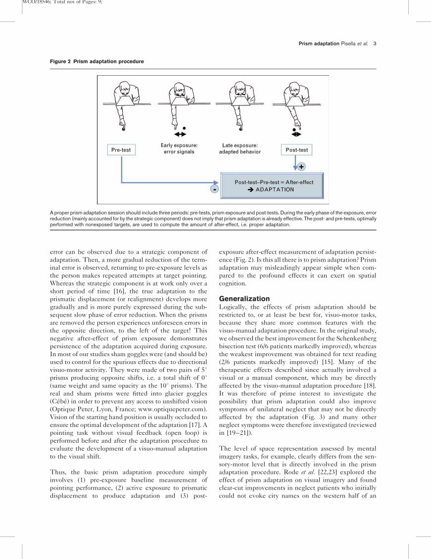
(a) Reading performance before and after prism adaptation. The delineated area depicts the area that was correctly read by the patient. (b) Mentalimagery task performed before and after prism adaptation. The patient was asked to imagine the map of France and then to name as many cities hecould see on this internal map. The mental path followed the lines from 1 to the total number of cities. The dot and whiskers indicate the average locationand horizontal extent of this internal scanning (redrawn from [22]) (c) Postural control before and after prism adaptation. The sagittal position of thecentre of mass was recorded for 50 s (redrawn from [32]) (d) Writing under dictation before and after prism adaptation. The patient performance isshown together with his daisy drawing from memory (from [71�]). (e) Wheelchair driving before and after prism adaptation. Clockwise and, in particular,counter-clockwise directions of driving were improved by prism adaptation (from [31�]).
internally generated map of France. This result strongly
suggests that the after-effects of visuo-manual adaptation
are not restricted to visual and motor parameters. In the
same vein, Farne et al. [24] compared the effects of prism
adaptation in six neglect patients on both (1) visuo-motor
tasks, such as line and bell cancellation, and two subtests
taken from the Behavioral Inattention Test (letter can-
cellation and line bisection) battery, and (2) visuo-verbal
tasks (the visual scanning test, also taken from the
Behavioral Inattention Test, requiring a verbal descrip-
tion of the objects depicted on a colored picture, an
object-naming task with 30 Snodgrass pictures of familiar
objects intermingled with geometric shapes as distractors,
word and nonword reading). They observed that both
types of tasks followed a parallel improvement, which
lasted for at least 24 h.
The fact that different tasks based upon other sensory
modalities can be improved (haptic circle centering
[25–27]) and that several nonmanual tasks (postural
control, wheelchair driving, imagery, verbal reports) were
also improved, demonstrates that the effects of prism
adaptation on visuo-spatial defective abilities go well
beyond the visuo-manual parameters usually affected
in normal subjects. Recently, Berberovic et al. [28] have
shown that even a nonspatial and nonmanual aspect of
neglect could be improved, i.e. temporal order judgment.
Furthermore, we recently described beneficial effects of
prism adaptation on a new feature of unilateral neglect
reported by Zorzi et al. [29]. They introduced a mental
number bisection test, whereby patients have to verbally
indicate the middle between two numbers (e.g. between
WCO/18546; Total nos of Pages: 9;
Prism adaptation Pisella et al. 5
11 and 19) and found a bias towards larger numbers in
neglect patients, as if their ‘mental number line’ was
distorted (similar to what classically occurs in line bisec-
tion). Even for this abstract task, we found that prism
adaptation strongly improved the bisecting bias [30].
Overall, these results make clear that adaptation to wedge
prisms affects a core component of unilateral neglect’s
complex spatial deficits.
Although generalization beyond purely visuo-motor tasks
is crucial for any putative rehabilitation technique, the
plasticity triggered by prism adaptation also produces
effects on nontrained tasks in the motor domain, such
as wheelchair driving [31�] and postural control [32]. Such
effects are presumably mediated via spatial cognition
levels. In addition, the intentional component of neglect
deficits can also be improved by prism adaptation [18,33].
In this case, patients where asked to reach and grasp a
centrally located ball, and then to throw it into a left- or
right-sided basket. The kinematics of the centrally
directed reach-to-grasp movements showed that neglect
patients are overall slower when the secondary move-
ment is directed to the left [33]. After a short prism
adaptation session, this asymmetry was modified for
several movement parameters (reaction time, movement
time, peak velocity, time to peak velocity). The pattern
of result observed immediately after prism adaptation
even showed the reverse pattern – reach movements
were slower when the ball had to be thrown to the right.
Therefore, the intentional control of action can be modi-
fied by prism adaptation.
Still at a motor level, there are several qualitative obser-
vations that prism adaptation can improve the motor
behavior of patients in everyday life [25]. One of the
crucial questions raised by the observation of a strong and
sustained improvement of unilateral neglect by a single
short adaptation session is whether this plastic effect is
restricted to the acute phase of the deficit. In our original
study patients were tested between 3 weeks and
14 months poststroke [15]. We have now collected data
on a group of patients who were exposed to the adap-
tation procedure between 5 and 28 years poststroke, and
amazingly found comparable amounts of improvement
[20,21].
DurationRetention over time is another crucial feature of any
rehabilitation method. The effects of a single prism
adaptation session (for repeated sessions, see below) last
much longer (at least 2 h) than for any other sensory
stimulations (about 15 min) reported to date. A group
of patients showed a sustained improvement 24 h after
the training session [24], but individual cases may exhibit
even longer-lasting amelioration of neglect, now demon-
strated to last up to about 1 week (e.g. [25,34]). An
interesting feature of prism adaptation is that its cognitive
beneficial effects, although often present immediately
after adaptation, seem to develop over short periods of
time thereafter, thus lagging somewhat behind the lower-
level after-effects. Indeed, delayed cognitive effects (i.e.
2–4 h after prism adaptation) tend to be stronger then
immediate ones (measured just after prism adaptation)
even in chronic patients [15,35�]. Although prism adap-
tation has long been thought to give rise to short-lasting
after-effects in healthy subjects, recent investigations
showed that the unaware mode of prism exposure can
give rise to astonishingly stronger after-effects [36�,37].
As suggested below, another clinical interest of prism
adaptation may be found in the repetition of adaptation
sessions.
Anatomo-functional hypothesisThe literature is somewhat controversial about the neural
structures involved in prism adaptation. On the one hand,
neuropsychological evidence historically suggested that
only cerebellar patients are impaired in prism adaptation
(e.g. [38], reviewed in [39]). On the other hand, recent
imaging data [40] have suggested that the human
posterior parietal cortex (PPC) contralateral to the adapted
arm is the only area activated during prism exposure.
More recently, we established that the superior parietal
lobule, which when damaged leads to impairment of
visuo-manual guidance (optic ataxia), is not crucially
involved in visuo-manual prism adaptation [41]. The
clinical effect of prism adaptation on visual neglect also
precludes that the inferior parietal lobule, at least in the
right hemisphere, could be crucial for adaptation to right-
ward prisms. Rather, neglect patients exhibit ‘hyper-
adaptation’, characterized by more robust and durable
after-effects [15,24,25,32,34,42]. A bilateral optic ataxia
patient also demonstrated a larger inter-manual transfer,
highlighting the cerebellum as the most likely neural
substrate of true adaptation, although the PPC may also
contribute to the strategic component. We therefore
proposed a simple model [41] in which the cerebellum
and the PPC are, respectively, specialized for the adap-
tive and strategic components of prism adaptation
(Fig. 4): if the lesion of the PPC reduces the strategic
component, adaptation would consequently be mostly
achieved by realignment processes (true adaptive com-
ponent); hence, it would more likely to be stronger,
longer-lasting and having more potential to generalize.
Accordingly, neglect patients do not notice the alteration
resulting from the optical deviation and normal subjects
exhibit larger adaptive after-effects when the prismatic
deviation is not noticeable [36�,43].
In a patient with a cerebellar lesion (including
the superior part of the dentate nucleus and mostly
the anterior lobe of the left cerebellar hemisphere) we
WCO/18546; Total nos of Pages: 9;
6 Trauma and rehabilitation
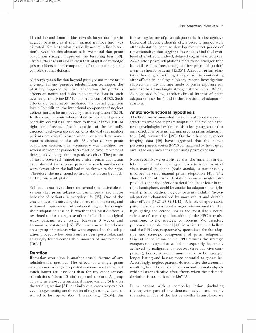
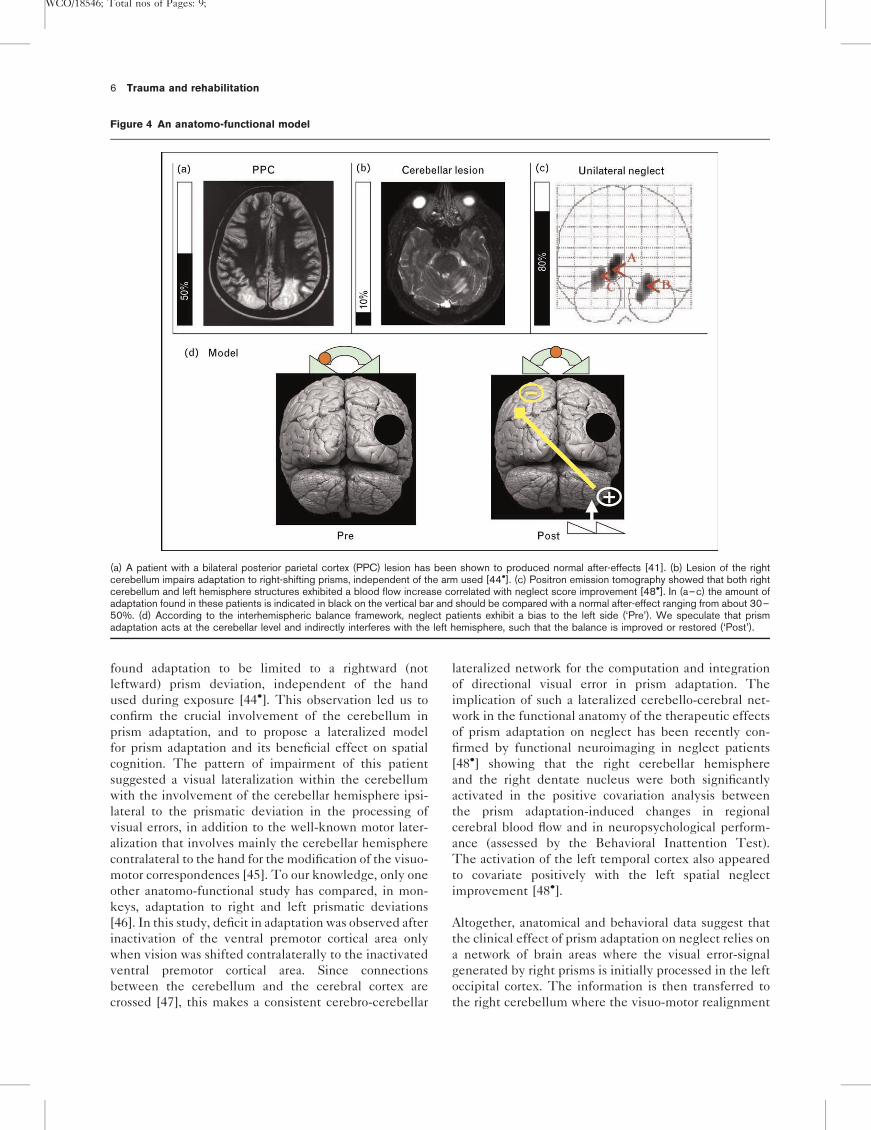
Figure 4 An anatomo-functional model
(a) A patient with a bilateral posterior parietal cortex (PPC) lesion has been shown to produced normal after-effects [41]. (b) Lesion of the rightcerebellum impairs adaptation to right-shifting prisms, independent of the arm used [44�]. (c) Positron emission tomography showed that both rightcerebellum and left hemisphere structures exhibited a blood flow increase correlated with neglect score improvement [48�]. In (a–c) the amount ofadaptation found in these patients is indicated in black on the vertical bar and should be compared with a normal after-effect ranging from about 30–50%. (d) According to the interhemispheric balance framework, neglect patients exhibit a bias to the left side (‘Pre’). We speculate that prismadaptation acts at the cerebellar level and indirectly interferes with the left hemisphere, such that the balance is improved or restored (‘Post’).
found adaptation to be limited to a rightward (not
leftward) prism deviation, independent of the hand
used during exposure [44�]. This observation led us to
confirm the crucial involvement of the cerebellum in
prism adaptation, and to propose a lateralized model
for prism adaptation and its beneficial effect on spatial
cognition. The pattern of impairment of this patient
suggested a visual lateralization within the cerebellum
with the involvement of the cerebellar hemisphere ipsi-
lateral to the prismatic deviation in the processing of
visual errors, in addition to the well-known motor later-
alization that involves mainly the cerebellar hemisphere
contralateral to the hand for the modification of the visuo-
motor correspondences [45]. To our knowledge, only one
other anatomo-functional study has compared, in mon-
keys, adaptation to right and left prismatic deviations
[46]. In this study, deficit in adaptation was observed after
inactivation of the ventral premotor cortical area only
when vision was shifted contralaterally to the inactivated
ventral premotor cortical area. Since connections
between the cerebellum and the cerebral cortex are
crossed [47], this makes a consistent cerebro-cerebellar
lateralized network for the computation and integration
of directional visual error in prism adaptation. The
implication of such a lateralized cerebello-cerebral net-
work in the functional anatomy of the therapeutic effects
of prism adaptation on neglect has been recently con-
firmed by functional neuroimaging in neglect patients
[48�] showing that the right cerebellar hemisphere
and the right dentate nucleus were both significantly
activated in the positive covariation analysis between
the prism adaptation-induced changes in regional
cerebral blood flow and in neuropsychological perform-
ance (assessed by the Behavioral Inattention Test).
The activation of the left temporal cortex also appeared
to covariate positively with the left spatial neglect
improvement [48�].
Altogether, anatomical and behavioral data suggest that
the clinical effect of prism adaptation on neglect relies on
a network of brain areas where the visual error-signal
generated by right prisms is initially processed in the left
occipital cortex. The information is then transferred to
the right cerebellum where the visuo-motor realignment
WCO/18546; Total nos of Pages: 9;
Prism adaptation Pisella et al. 7
(i.e. ‘true adaptation’) takes place in congruence with the
rightward deviation of prisms. The clinical effect could
be mediated through the modulation of cerebral areas in
the left hemisphere via a bottom-up signal generated by
the cerebellum [20,44�,49]. Notably, the temporal cortex,
the frontal cortex and the PPC have been shown to be
targets of output from the cerebellum via a neuronal loop
also implicating the dentate nucleus and subcortical
structures, such as the thalamus and the globus pallidus
[50–52]. The clinical effect might therefore be mediated
by the recruitment of pathways in the left hemisphere
that are functionally homologous to those involved in
spatial cognition in the damaged hemisphere.
Reciprocally, the cognitive effects of prism adaptation
found in healthy subjects [53–55], demonstrated by an
asymmetrical pattern of performance on several spatial
tasks, are strictly dependent upon the direction of the
prismatic shift [49]. On the basis of the latter studies and
considering that the right parietal cortex seems to be
specifically involved in line bisection judgment tasks
[56–57], we hypothesized that the function of the right
parietal lobe would be inhibited by inputs from the left
cerebellar cortex, coherent with the use of the leftward
prismatic deviation, and create ‘neglect-like’ symptoms
[58�].
In principle, the proposed model is compatible with the
involvement of the prism adaptation-induced realign-
ment of the oculomotor system that, by reducing the
rightward scanning bias, may facilitate exploration of the
left neglected side of space [59,60,61�]. Several dis-
sociations have, however, been documented between
oculomotor change and the amelioration of visuo-spatial
behavioral performances [62,63]. Another proposal is that
adaptation acts through plastic modification of the integ-
ration of proprioceptive and visual information, which
would be particularly beneficial in neglect patients whose
symptoms result in part from an impaired visual-motor
mapping of space (see [64]). One could speculate that
prism adaptation permits an enlargement of this visual-
motor mapping of space not only on the left side, but also
on the right side, as suggested by the improvement of
constructional apraxia [21] and spatial dysgraphia follow-
ing prism adaptation. Recent findings mainly point to the
need for appropriately applying prism exposure con-
ditions and quantification [18], for evaluating the role
played by the type of prism adaptation (strategic vs.
realignment; [59,60]), as well as the sufficient amount
of adaptation (as measured in terms of after-effect)
required to produce consistent neglect improvement
[65]. There is still the need to build up a coherent
framework integrating one century of prism adaptation
investigations with the recent body of patient literature
[66]. An interesting issue also remains to identify domain-
specific aspects of neglect, such as chimeric face percep-
tion, which seem even intractable by prism adaptation
[35�,63,67].
ConclusionAdaptation to prismatic displacement is particularly sui-
ted for clinical application [38] because its incremental
nature permits examination over relatively short time
periods, in contrast to prismatic distortions like left–right
or up–down reversal of the visual field that require
extended exposure for adaptation to occur [68]. The
efficacy of single-session prism adaptation has proven
to generalize the improvement to several neglect symp-
toms. Several nonmotor as well as motor aspects of the
neglect syndrome, such as motor neglect and/or extinc-
tion, might actually benefit from prism adaptation as well.
It can be considered the most-promising rehabilitation
method for unilateral neglect to date [4�,69], especially in
light of the fact that spontaneous recovery from neglect is
very limited [70]. We wish to put forward that prism
adaptation might improve spatial-cognition deficits in
neglect as well as in other pathologies. Constructional
deficits, dysgraphia [71�], as well as spatial attention
distortions contributing not only to neglect [22,72,42],
but also to other pathological manifestations affecting
spatial and bodily representations [73] (e.g. Complex
Regional Pain Syndrome [74,75], are on the list of candi-
dates for this bottom-up rehabilitation track. In spite of
these possibilities, it is now clear that not all neglect
patients can benefit from prism adaptation and not to the
same extent. Unfortunately, to our knowledge, only
one randomized controlled trial has been performed to
date [21]. In addition, several important questions still
await definitive answers: why is adaptation quantitatively
more important and lasts longer in patients vs. healthy
controls, why it affects higher-order cognitive domains,
why it is direction-specific and what are the predictors of
prism adaptation clinical efficacy? Beyond prism adap-
tation in and of itself, it is also promising to consider that
other original bottom-up approaches may prove to be
more effective on cognitive disorders than the traditional
top-down stance.
The practical clinical potential of repetitive-session
prism adaptation procedures has just begun to be
explored. Frassinetti et al. [76] reported that a group of
patients who benefited from two prism adaptation ses-
sions daily over 2 weeks (a total of 10 sessions) exhibited
an improvement that lasted over 5 weeks after the end of
the treatment. A daily prism adaptation session has been
reported to improve neglect up to 3 months after treat-
ment [60]. Long-standing chronic neglect (11 years) has
also been demonstrated to improve with repetitive prism
adaptation sessions [35�]. Using the neck vibration tech-
nique and visual-scanning training, Schindler et al. [77]
also explored the effects of repetitive sessions, and found
a sustained improvement following an intensive daily
WCO/18546; Total nos of Pages: 9;
8 Trauma and rehabilitation
programme. Obviously, such studies should be system-
atically undertaken to determine the optimal training
frequency and duration, as well as the optimal combi-
nation of techniques that can be used routinely for
rehabilitation [78].
AcknowledgementsThe authors wish to thank Dominique Boisson, Sophie Jacquin-Courtois, Jacques Luaute, Denis Pelisson, Claude Prablanc and AlainVighetto for fruitful discussions, and Stephanie Maxfield for hercomments on a previous version of this manuscript.
References and recommended readingPapers of particular interest, published within the annual period of review, havebeen highlighted as:� of special interest�� of outstanding interest
Additional references related to this topic can also be found in the CurrentWorld Literature section in this issue (pp. 000–000).
1 Bisiach E. Unilateral neglect and related disorders. In: Denes F, Pizzamiglio L,editors. Handbook of clinical and experimental neuropsychology. Hove:Psychology Press; 1999. pp. 479–496.
2 Halligan PW, Marshall JC, Wade DT. Visuospatial neglect: underlying factorsand test sensitivity. Lancet 1989; ii:908–911.
3 Kerkhoff G. Spatial hemineglect in humans. Prog Neurobiol 2001; 63:1–27.
4
�Milner AD, McIntosh RD. The neurological basis of visual neglect. Curr OpinNeurol 2005; 18:748–753.
A review embracing the various aspects of the neglect syndrome and extinctionphenomena, particularly focusing on the recently reappraised neuroanatomical issue.
5
�Snow JC, Mattingley JB. Goal-driven selective attention in patients with righthemisphere lesions: how intact is the ipsilesional field? Brain 2006;129:168–181.
An elegant demonstration that the ipsilesional hemifield of neglect patients isneither normal nor hyperfunctional, but dysfunctional.
6 Natale E, Posteraro L, Prior M, Marzi CA. What kind of visual spatial attentionis impaired in neglect? Neuropsychologia 2005; 43:1072–1085.
7 Rusconi ML, Maravita A, Bottini G, Vallar G. Is the intact side really intact?Perseverative responses in patients with unilateral neglect: a productivemanifestation. Neuropsychologia 2002; 40594–40604.
8 Denes G, Semenza C, Stoppa E, Lis A. Unilateral spatial neglect and recoveryfrom hemiplegia: a follow-up study. Brain 1982; 105:543–552.
9 Fullerton KJ, McSherry D, Stout RW. Albert’s test: a neglected test ofperceptual neglect. Lancet 1986; 327:430–432.
10 Kerkhoff G, Rossetti Y. Plasticity in hemispatial neglect: recovery and reha-bilitation [editorial]. Restor Neurol Neurosci 2006; 24: in press.
11 Diller L, Weinberg J. Hemi-inattention in rehabilitation: the evolution of arational remediation program. Adv Neurol 1977; 18:63–82.
12 Rubens AB. Caloric stimulation and unilateral visual neglect. Neurology 1985;35:1019–1024.
13 Jeannerod M, Biguer B. The directional coding of reaching movements Avisuo-motor conception of spatial neglect. In: Jeannerod M, editor. Neuro-physiological and neuropsychological aspects of spatial neglect. Amsterdam:North-Holland; 1987. pp. 87–113.
14 Stratton GM. Some preliminary experiments on vision without inversion of theretinal image. Psychol Rev 1896; 3:611–617.
15 Rossetti Y, Rode G, Pisella L, et al. Prism adaptation to a rightward opticaldeviation rehabilitates left hemispatial neglect. Nature 1998; 395:166–169.
16 Rossetti Y, Koga K, Mano T. Prismatic displacement of vision inducestransient changes in the timing of eye-hand coordination. Percept Psychophys1983; 54:355–364.
17 Redding GM, Wallace B. Adaptive spatial alignment. Mahwah: LawrenceErlbaum; 1997.
18 Redding G, Rossetti Y, Wallace B. Application of prism adaptation: a tutorialin theory and method. Neurosci Biobehav Rev 2005; 29:431–444.
19 Rossetti Y, Rode G. Reducing spatial neglect by visual and other sensorymanipulations: noncognitive (physiological) routes to the rehabilitation of acognitive disorder. In: Karnath HO, Milner AD, Vallar G, editors. The cognitiveand neural bases of spatial neglect. Oxford: Oxford University Press; 2002.pp. 375–396.
20 Rode G, Pisella L, Rossetti Y, et al. Bottom-up transfer of visuo-motorplasticity. Prism adaptation. Prog Brain Res 2003; 273–287.
21 Rode G, Klos T, Courtois-Jacquin S, Rossetti Y. Neglect and prism adapta-tion. A new therapeutic tool for spatial cognition disorders. Restor NeurolNeurosci 2006; 24: in press.
22 Rode G, Rossetti Y, Li L, Boisson D. The effect of prism adaptation on neglectfor visual imagery. Behav Neurol 1998; 11:251–258.
23 Rode G, Rossetti Y, Boisson D. Prism adaptation improves representationalneglect. Neuropsychologia 2001; 39:1250–1254.
24 Farne A, Rossetti Y, Toniolo S, Ladavas E. Ameliorating neglect with prismadaptation: Visuo-manual vs visuo-verbal measures. Neuropsychologia 2002;40:1069–1080.
25 McIntosh RM, Rossetti Y, Milner AD. Prism adaptation improves chronic visualand haptic neglect. Cortex 2002; 38:309–320.
26 Maravita A, McNeil J, Malhotra P, et al. Prism adaptation can improve con-tralesional tactile perception in neglect. Neurology 2003; 60:1829–1831.
27 Jacquin-Courtois S, Rossetti Y, Rode G, et al. Effect of prism adaptation onauditory extinction: an attentional effect? Presented at: Third World Congresson Neurological Rehabilitation. Venice, 2002; http://www.lyon.inserm.fr/symposium534/posters/11courtois-jacquin.html.
28 Berberovic N, Pisella L, Morris AP, Mattingley JB. Prismatic adaptationreduces biased temporal order judgements in spatial neglect. Neuroreport2004; 15:1199–1204.
29 Zorzi M, Priftis K, Umilta C. Neglect disrupts the mental number line. Nature2002; 417:138–139.
30 Rossetti Y, Jacquin-Courtois S, Rode G, et al. Does action make the linkbetween number and space representation? Visuo-manual adaptation im-proves number bisection in unilateral neglect. Psychol Sci 2004; 15:426–430.
31
�Jacquin-Courtois S, Rode G, Boisson D, Rossetti Y. Wheel-chair drivingimprovement following visuo-manual prism adaptation. Cortex 2006; in press.
This single case study shows significant effects of prism adaptation on a crucialevery-day life activity: wheel-chair driving.
32 Tilikete C, Rode G, Rossetti Y, et al. Prism adaptation to rightward opticaldeviation improves postural imbalance in left-hemiparetic patients. Curr Biol2001; 11:524–528.
33 Rossetti Y, Goldenberg G, Rode G. Current issues in sensori-motorrehabilitation. In: Freund HJ, Jeannerod M, Hallett M, Leguarda, editors.Higher-order motor disorders. Oxford University Press: Oxford; 2005.pp. 475–498.
34 Pisella L, Rode G, Farne A, et al. Dissociated long lasting improvements ofstraight-ahead pointing and line bisection tasks in two hemineglect patients.Neuropsychologia 2002; 40:327–334.
35
�Humphreys GW, Watelet A, Riddoch MA. Long-term effects of prism adapta-tion in chronic visual neglect: a single case study. Cognitive Neuropsychol2006; 23:463–478.
Therapeutic after-effects are found up to 1 year post-adaptation in a patientsuffering from neglect for 11 years. Interestingly, the iterative benefit of prismadaptation decreased on immediate performance, but it increased across sessionsand within sessions when performance was tested up to 90 min after prismadaptation.
36
�Michel C, Pisella L, Prablanc C, et al. Enhancing visuo-motor adaptation byreducing error signals: Single-step (aware) versus multiple-step (unaware)exposure to wedge prisms. J Cognitive Neurosci 2006; in press.
This paper provides an insight about the exceptional generalization of prismadaptation in neglect patients by showing in normals that an unaware mode ofprism exposure gives rise to deeper adaptation after-effects. An unaware mode ofprism exposure, assumed to mimic the condition of neglect patients, producesmore after-effects than the usual single-step exposure.
37 Hatada Y, Miall C, Rossetti Y. Two waves of a long-lasting after-effect ofprism adaptation measured over 7 days. Exp Brain Res 2006; 169:417–426.
38 Weiner MJ, Hallett M, Funkenstein HH. Adaptation to lateral displacement ofvision in patients with lesions of the central nervous system. Neurology 1983;33:766–772.
39 Jeannerod M, Rossetti Y. Visuomotor coordination as a dissociable visualfunction: experimental and clinical evidence. Baillere’s Clin Neurol 1993;2:439–460.
40 Clower DM, Hoffman JM, Votaw JR, et al. Role of posterior parietal cortex inthe recalibration of visually guided reaching. Nature 1996; 383:618–621.
41 Pisella L, Michel C, Grea H, et al. Preserved prism adaptation in bilateral opticataxia: strategic versus adaptive reaction to prisms. Exp Brain Res 2004;156:399–408.
WCO/18546; Total nos of Pages: 9;
Prism adaptation Pisella et al. 9
42 Rode G, Jacquin-Courtois S, Revol P, et al. Bottom-up effects of sensoryconflict and adaptation on mental imagery: sensorimotor grounds for highlevel cognition? In: Mast FW, Jancke L, editors. Spatial processing innavigation, imagery, and perception. Springer Science: New York; 2006.in press.
43 Jakobson LS, Goodale MA. Trajectories of reaches to prismatically-displacedtargets: evidence for ‘automatic’ visuomotor recalibration. Exp Brain Res1989; 78:575–587.
44
�Pisella L, Rossetti Y, Michel C, et al. Ipsidirectional impairment of prismadaptation after unilateral lesion of anterior cerebellum. Neurology 2005;65:150–152.
This study provides clear evidence for the functional role played by the cerebellumin prism adaptation and provides support to the cerebello-cerebral model of theeffect of prism adaptation on visuo-spatial cognition.
45 Martin TA, Keating JG, Goodkin HP, et al. Throwing while looking throughprisms. I. Focal olivocerebellar lesions impair adaptation. Brain 1996;119:1183–1198.
46 Kurata K, Hoshi E. Reacquisition deficits in prism adaptation after muscimolmicroinjection into the ventral premotor cortex of monkeys. J Neurophysiol1999; 81:1927–1938.
47 Schmahmann JD, Pandya DN. The cerebrocerebellar system. Int Rev Neu-robiol 1997; 41:31–60.
48
�Luaute J, Michel C, Rode G, et al. Functional anatomy of the therapeuticeffects of prism adaptation on left neglect. Neurology 2006; 66:1859–1867.
This first brain imaging study of prism adaptation in neglect patients showed thatthe amelioration of neglect is correlated with an increase in blood flow in both theright cerebellum and the left temporal cortex, presumably related to the exploratorynature of the tasks employed to compute the correlation.
49 Michel C, Pisella L, Halligan PW, et al. Simulating unilateral neglect in normalsusing prism adaptation: implications for theory. Neuropsychologia 2003;41:25–39.
50 Middleton FA, Strick PL. Basal ganglia and cerebellar loops: motor andcognitive circuits. Brain Res Brain Res Rev 2000; 31:236–250.
51 Dum RP, Strick PL. An unfolded map of the cerebellar dentate nucleus and itsprojections to the cerebral cortex. J Neurophysiol 2003; 89:634–639.
52 Clower DM, West RA, Lynch JC, Strick PL. The inferior parietal lobule is thetarget of output from the superior colliculus, hippocampus, and cerebellum.J Neurosci 2001; 21:6283–6291.
53 Berberovic N, Mattingley JB. Effects of prismatic adaptation on judgements ofspatial extent in peripersonal and extrapersonal space. Neuropsychologia2003; 41:493–503.
54 Girardi M, McIntosh RD, Michel C, et al. Sensorimotor effects on centralspace representation: prism adaptation influences haptic and visual repre-sentations in normal subjects. Neuropsychologia 2004; 42:1477–1487.
55 Goebel S, Calabria M, Farne A, Rossetti Y. Parietal rTMS distorts the mentalnumber line: simulating ‘spatial’ neglect in healthy subjects. Neuropsycho-logia 2006; 44:860–868.
56 Fierro B, Brighina F, Oliveri M, et al. Contralateral neglect induced by rightposterior parietal rTMS in healthy subjects. Neuroreport 2000; 10:1519–1521.
57 Fink GR, Marshall JC, Shah NJ, et al. Line bisection judgments implicate rightparietal cortex and cerebellum as assessed by fMRI. Neurology 2000;54:1324–1331.
58
�Michel C. Simulating unilateral neglect in normals: myth or reality? RestorNeurol Neurosci 2006; 24: in press.
This review provides an integrated overview of the variety of cognitive after-effectsof prism adaptation that have been described in healthy subjects.
59 Angeli V, Benassi MG, Ladavas E. Recovery of oculo-motor bias in neglectpatients after prism adaptation. Neuropsychologia 2004; 42:1223–1234.
60 Serino A, Angeli V, Frassinetti F, Ladavas E. Mechanisms underlying neglectrecovery after prism adaptation. Neuropsychologia 2006; 44:1068–1078.
61
�Malhotra P, Coulthard E, Husain M. Hemispatial neglect, balance and eye-movement control. Curr Opin Neurol 2006; 19:14–20.
An updated review on neglect and pusher syndrome emphasizing the recentadvances in treatment for neglect, including the use of prism adaptation and pilotdata on noradrenergic stimulation.
62 Dijkerman HC, McIntosh RD, et al. Ocular scanning and perceptual sizedistortion in hemispatial neglect: effects of prism adaptation and sequentialstimulus presentation. Exp Brain Res 2003; 153:220–230.
63 Ferber S, Danckert J, Joanisse M, et al. Eye movements tell only half the story.Neurology 2003; 60:1826–1829.
64 Pisella L, Mattingley JB. The contribution of spatial remapping impairments tounilateral visual neglect. Neurosci Biobehav Rev 2004; 28:181–200.
65 Rousseaux M, Bernati T, et al. Ineffectiveness of prism adaptation on spatialneglect signs. Stroke 2006; 37:542–543.
66 Redding GM, Wallace B. Prism adaptation and unilateral neglect: review andanalysis. Neuropsychologia 2006; 44:1–20.
67 Sarri M, Kalra L, Greenwood R, Driver J. Prism adaptation changes perceptualawareness for chimeric visual objects but not for chimeric faces in spatialneglect after right-hemisphere stroke. Neurocase 2006; 12:127–135.
68 Sekiyama K, Miyauchi S, Imaruoka T, et al. Body image as a visuomotortransformation device revealed in adaptation to reversed vision. Nature 2000;407:374–377.
69 Luaute J, Halligan P. Prism adaptation first among equals in alleviating leftneglect. A review. Restor Neurol Neurosci 2006; 24: in press.
70 Farne A, Buxbaum LJ, Ferraro M, et al. Patterns of spontaneous recovery ofneglect and associated disorders in acute right brain-damaged patients.J Neurol Neurosurg Psychiatry 2004; 75:1401–1410.
71
�Rode G, Pisella L, Marsal L, et al. Prism adaptation improves spatial dysgra-phia following right brain damage. Neuropsychologia 2006; 44:2487–2493.
A study documenting that a single prism adaptation session may producebeneficial effects that go beyond neglect and affect constructional aspects ofwriting under dictation.
72 Morris AP, Kritikos A, Berberovic N, et al. Prism adaptation and spatialattention: a study of visual search in normals and patients with unilateralneglect. Cortex 2004; 40:703–721.
73 Rossetti Y, Rode G, Farne A, Rossetti A. Implicit body representation inaction: a neuropsychological approach. In: De Preester H, Knockaert V,editors. Body image and body schema. Amsterdam: Benjamins; 2005. pp.111–125.
74 Sumitani M, Rossetti Y, Shibata M, et al. Prism adaptation to optical deviationalleviates pathological pain. Neurology 2006; in press.
75 Sumitani M, Shibata M, Yagisawa M, et al. Prism adaptation to opticaldeviation alleviates complex regional pain syndrome: longitudinal single casestudy [abstract]. Neurorehabil Neural Repair 2006; 20:141–142
76 Frassinetti F, Angeli V, Meneghello F, et al. Long-lasting amelioration ofvisuospatial neglect by prism adaptation. Brain 2002; 125:608–623.
77 Schindler I, Kerkhoff G, Karnath HO, et al. Neck muscle vibration induceslasting recovery in spatial neglect. J Neurol Neurosurg Psychiatry 2002;73:412–419.
78 Bowen A, Lincoln NB. The need for randomized treatment studies in neglectresearch. Restor Neurol Neurosci 2006; 24: in press.

WCO Manuscript No. 18546
Current Opinion in Neurology Typeset by Thomson Digital
for Lippincott Williams & Wilkins
Dear Author, During the preparation of your manuscript for typesetting, some queries have arisen. These are listed below. Please check your typeset proof carefully and mark any corrections in the margin as neatly as possible or compile them as a separate list. This form should then be returned with your marked proof/list of corrections to the Production Editor.
QUERIES: to be answered by AUTHOR/EDITOR
AUTHOR: The following queries have arisen during the editing of your manuscript. Please answer the queries by marking the requisite corrections at the appropriate positions in the text.
QUERY NO.
QUERY DETAILS
No query.