visuo-proprioceptive interactions in degenerative cervical spine diseases requiring surgery
TRANSCRIPT
Neuroscience 255 (2013) 226–232
VISUO-PROPRIOCEPTIVE INTERACTIONS IN DEGENERATIVECERVICAL SPINE DISEASES REQUIRING SURGERY
S. FREPPEL, a,b A. BISDORFF, a,c
S. COLNAT-COULBOIS, a,d H. CEYTE, a,e C. CIAN, e
G. GAUCHARD, a J. AUQUE d AND P. PERRIN a,f*
aUniversite de Lorraine, EA 3450 DevAH – Development,
Adaptation and Disadvantage, Faculty of Medicine and UFR
STAPS, Nancy, France
bDepartment of Neurosurgery, University Hospital, Groupe
Hospitalier Sud Reunion, Saint Pierre, Reunion Island, France
cDepartment of Neurology, Centre Hospitalier Emile Mayrisch,
Esch-sur-Alzette, Luxembourg
dDepartment of Neurosurgery, University Hospital of Nancy,
Nancy, France
eThe Institut de Recherches Biomedicales des Armees, La
Tronche, France
fDepartment of Oto-Rhino-Laryngology, University Hospital,
Vandoeuvre-les-Nancy, France
Abstract—Cervical proprioception plays a key role in pos-
tural control, but its specific contribution is controversial.
Postural impairment was shown in whiplash injuries without
demonstrating the sole involvement of the cervical spine.
The consequences of degenerative cervical spine diseases
are underreported in posture-related scientific literature in
spite of their high prevalence. No report has focused on
the two different mechanisms underlying cervicobrachial
pain: herniated discs and spondylosis. This study aimed
to evaluate postural control of two groups of patients with
degenerative cervical spine diseases with or without optoki-
netic stimulation before and after surgical treatment. Seven-
teen patients with radiculopathy were recruited and divided
into two groups according to the spondylotic or discal ori-
gin of the nerve compression. All patients and a control pop-
ulation of 31 healthy individuals underwent a static
posturographic test with 12 recordings; the first four record-
ings with the head in 0� position: eyes closed, eyes open
without optokinetic stimulation, with clockwise and counter
clockwise optokinetic stimulations. These four sensorial sit-
uations were repeated with the head rotated 30� to the left
and to the right. Patients repeated these 12 recordings
6 weeks postoperatively. None of the patients reported ver-
tigo or balance disorders before or after surgery. Prior to
surgery, in the eyes closed condition, the herniated disc
group was more stable than the spondylosis group. After
surgery, the contribution of visual input to postural control
in a dynamic visual environment was reduced in both
0306-4522/13 $36.00 � 2013 IBRO. Published by Elsevier Ltd. All rights reservehttp://dx.doi.org/10.1016/j.neuroscience.2013.09.060
*Correspondence to: P. Perrin, EA 3450 DevAH – Developpement,Adaptation et Handicap, Universite de Lorraine, UFR STAPS deNancy, 30 rue du Jardin Botanique, 54600 Villers-les-Nancy, France.Tel: +33-3-83-15-49-68.
E-mail address: [email protected] (P. Perrin).Abbreviations: CCRM, counterclockwise rotary motion; CRM,clockwise rotary motion; EC, eyes closed situation; EO, eyes opensituation; RQ, Romberg quotient; VKQ, visual-kinetic quotient.
226
cervical spine diseases whereas in a stable visual environ-
ment visual contribution was reduced only in the spondylosis
group. The relative importance of visual and proprioceptive
inputs to postural control varies according to the type of
pathology and surgery tends to reduce visual contribution
mostly in the spondylosis group. � 2013 IBRO. Published by
Elsevier Ltd. All rights reserved.
Key words: cervical proprioception, degenerative cervical
spine diseases, surgical treatment, static postural control,
postural sensorimotor strategies.
INTRODUCTION
Postural control is achieved through a multisensory
control mechanism involving visual, vestibular and
somatosensory information (Massion, 1992; Gangloff
and Perrin, 2002). These inputs are all interconnected
allowing compensation of dysfunctions but making it
very challenging to study the cues of one particular
system without the interference of another.
The sensory contribution to postural control was
studied by various experimental paradigms. Several
authors (Wolsley et al., 1996; Thurrell and Bronstein,
2002) showed that an optokinetic stimulation induced a
visually evoked postural sway. Others have shown the
contribution of cervical proprioception to postural control
by the vibratory method. Indeed a vibration applied to
the neck, through the stimulation of receptors in cervical
muscles, can induce a postural response (Roll and Roll,
1988; Bove et al., 2001, 2002; Dumas et al., 2013) by
modifying the perception of the head or whole body
orientation (Biguer et al., 1988; Taylor and McCloskey,
1991; Ceyte et al., 2006).
Analysis of postural control of patients with
posttraumatic cervical pain is a good example of the
challenging difficulty, in a pathological condition, to
study solely one type of sensory information. There is
clear evidence of postural control impairment in patients
with whiplash injuries (Treleaven et al., 2003) but the
sole responsibility of cervical proprioception has never
been demonstrated as vestibular or vertebral artery
lesions can also occur during deceleration (Brandt and
Bronstein, 2001).
Degenerative cervical spine diseases have raised far
less interest when it comes to proprioception and
postural control despite being initially suspected as a
probable cause of the controversial ‘‘cervical vertigo’’
(Ryan and Cope, 1955). Considering the high
prevalence of these pathologies, their current
d.

S. Freppel et al. / Neuroscience 255 (2013) 226–232 227
representation in posture-related scientific literature
seems inadequate. Posture and cervical proprioception
have been studied in cases of non-traumatic neck pain
regardless of a potential underlying degenerative spine
disease (Revel et al., 1994; Field et al., 2008; Pinsault
et al., 2008). A few studies of the degenerative spine
focus on vertigo and decreased blood flow velocity in
the vertebral artery (Cevik et al., 2010; Machaly et al.,
2011).
There are, so far, only three reports on impaired
postural control in patients with cervicobrachial pain and
objective, radiological signs of degenerative cervical
spine disease (Vitte et al., 1992; Karlberg et al., 1995;
Persson et al., 1996). The postural effects of
degenerative spine disease and surgical treatment were
studied but solely in perturbative conditions (vibratory
stimulus to the calf and paraspinal neck muscles or
bipolar binaural galvanic stimulation). These stimulations
are not frequently encountered outside of a research
laboratory. The effects of degenerative cervical spine
disease and its surgical treatment on unperturbed
postural control remain unknown.
Moreover, there are two different types of
degenerative spine diseases. A disc herniation usually
has an acute onset whereas spondylosis is a chronic
disease in which age-related degenerative changes of
the discs and osteophytic bars may narrow the cervical
canal or the foramina. The consequences of these two
specific diseases on postural control have never been
analyzed.
The aim of this study was to evaluate visuo-
proprioceptive interactions in the postural control of
patients with two degenerative spine diseases by
generating an optokinetic stimulation in different head
on trunk positions, before and after surgery. Our
objectives were to investigate, in cervical spine
diseases, the different sensorial strategies in posture
and assess their evolution after surgical treatment.
EXPERIMENTAL PROCEDURES
Patient population
Seventeen patients (10 females and seven males) with
degenerative cervical spine disease, age range from 35
to 75 years (median age 50 years, interquartile range
10.5 years) were recruited. All experiments were
conducted in accordance with the Declaration of
Helsinki and all patients gave written informed consent
to participate in the study. Exclusion criteria were:
history of neurological, spinal or inner ear disease and
signs of myelopathy on clinical examination or imaging.
All patients consulted initially for radiculopathy resisting
medical treatment and required surgery. None of them
complained of dizziness or vertigo.
Radicular compression was caused by spondylosis for
nine patients (four females and five males, median age
51 years) and by a herniated disc for eight patients (six
females and two males, median age 47.5 years). The
patient population was therefore divided into a
spondylosis group and a herniated disc group. In the
spondylosis group, four patients required surgery on one
level, four patients on two levels and one patient on four
levels. In the herniated disc group, seven patients
required surgery on one level and one patient on two
levels.
Control population
Thirty-one individuals, age range from 26 to 60 years
(median age 45 years, interquartile range 15.5 years)
were recruited among the medical and paramedical staff
and included in a control group. This control group will
be used as a reference to evaluate the influence of
cervical degenerative diseases on the patient population
in a preoperative session. All experiments were
conducted in accordance with the Declaration of
Helsinki and all subjects gave written informed consent
to participate in the study. Individuals with a history of
neurological, spinal or inner ear disease were excluded
from the control group.
Platform
Data were collected on a vertical force platform
(Medicapteur, Nice, France) mounted on three strain-
gauge force transducers, providing a measurement of
the body sway in terms of displacement of the center of
foot pressure (CoP) in a two-dimensional horizontal
plane with a sampling rate of 40 Hz. Data were
analyzed with the Winposture 1.62 software.
Roll optokinetic stimulation
The visual display consisted of a large cupola (1 m
diameter) positioned 25 cm from the subject at eye
level, such that it covered a large area of the subject’s
visual field (visual angle 127�). This cupola was covered
in randomly distributed color circles of different
diameters and could be rotated around the visual axis at
an angular velocity of 50�/s either clockwise or
anti-clockwise (Fig. 1).
Surgery
All patients required surgery. In our institution, patients
with spondylosis benefit from a Cloward procedure
(Cloward, 1958) whereas patients with herniated disc
will benefit from an arthroplasty. In both cases, the
intervertebral disc is removed through a right anterior
approach whether the radiculopathy is located on the
right or on the left side. The skin and the platysma are
incised. The right sternocleidomastoid muscle is
retracted laterally and the carotid and esophagus are
separated to expose the cervical spine. Both longus collimuscles are partially cauterized, cut and dissected from
the anterior aspect of the vertebral bodies. The disc and
both anterior and posterior common vertebral ligaments
are removed. Both left and right nerve roots of the
pathological level are decompressed.
In the case of a Cloward procedure, a polyether ether
ketone (PEEK) cage filled with spongious bone chips
harvested on the iliac crest is placed in the
intervertebral space in order to obtain fusion of the
adjacent vertebras.

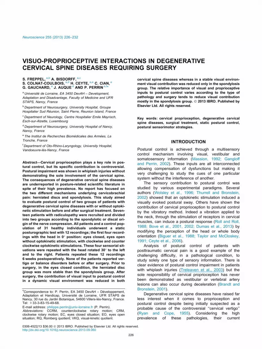
Fig. 1. 28-year-old female from the control population facing the
device in the 0� position (A) and statokinesigram collected on the
platform (Medicapteur, Nice and Winposture 1.62 software) of
the same individual in the 0� position, eyes open facing clockwise
rotary motion (B). Sway path length of the center of foot pressure
(broken line) and area (ellipsoid) including 90% of the instantaneous
positions of the center of foot pressure.
Fig. 2. 12 recordings are made during each session. The first four
recordings are made with the head facing forward (0�), one in eyes
closed situation, one in eyes open situation without rotary motion of
the device, one in eyes open situation facing a clockwise rotary
motion of the device and one in eyes open situations facing a
counterclockwise rotary motion of the device. These four sensorial
situations are repeated with the head rotated 30� left and 30� right stillfacing the cupola.
228 S. Freppel et al. / Neuroscience 255 (2013) 226–232
In the case of an arthroplasty, a cervical disc
prosthesis (Scient’X Discocerv�) is placed in the
intervertebral space in order to preserve motion and
therefore protect the adjacent discs.
Static posturographic tests
The participants stood bare-foot on the posturography
platform, heels together with a 30� angle between the
medial sides of the feet and eye level facing the center
of the cupola. They were requested to stand upright
remaining as stable as possible, breathing normally,
with their arms on their sides during 20 s (Fig. 2). This
task was performed in three different head–trunk
alignments: with the head aligned to the trunk (0�position), with the head actively rotated 30� to the left
and 30� to the right, facing the cupola (no eye in orbit
deviation). In each of these sessions four recordings
were made: one in eyes closed situation (EC), one in
eyes open situation (EO), one with eyes open facing a
clockwise rotary motion of the device (CRM) and finally,
one with eyes open facing a counterclockwise rotary
motion (CCRM). The participant was given a 2-min
break between each session. The rotation of the head
was chosen as a means to modulate proprioceptive
input from the neck, and the 30� angle was chosen as
an angle most if not all potential participants were likely
to achieve.
The patient group performed this protocol the day
before surgery and repeated it 6 weeks after surgery.
Postoperatively, patients were asked not to wear a
cervical collar and preoperative medications were left
unchanged. The control group, used as a reference to
evaluate the influence of cervical degenerative disease
on the patient population preoperatively, performed this
protocol only once.
Data and statistical analyses
During these recordings, we analyzed the area of the CoP
(the ellipsoid including 90% of the instantaneous positions
of CoP). To assess the visual contribution to posture in a
stable visual environment we calculated the Romberg
quotient (RQ). The RQ was obtained by calculating the
ratio of CoP area in EC to CoP area in EO. To assess
the visual contribution to posture in a moving visual
environment we calculated the visual-kinetic quotient
(VKQ). This quotient was obtained by calculating the
ratio of the average CoP area in CRM and in CCRM to
the CoP area in EO.
Considering the sample size, non-parametric tests
were used. To compare pre and postoperative data of
the same population we used a Wilcoxon test. In order to
compare different populations, we used a Mann–Whitney
test (SAS Statview 5 software for Windows). We
considered p< 0.05 as a significant difference and a

Table 1. Romberg quotient (RQ) is the ratio of center of foot pressure (CoP) area eyes closed to CoP area eyes open with no rotation. Visual-kinetic
quotient (VKQ) is the ratio of the average CoP area eyes open with cupola rotation to CoP area eyes open without rotation. Median and interquartile (IQ)
data are reported in the 0�, 30� left and 30� right positions for the herniated disc population (n = 8), the spondylosis population (n = 9) and the control
population (n = 31). Data of the herniated disc population and the spondylosis population are compared pre and postoperatively (p). Data of the control
population are compared to the preoperative data of the herniated disc population (p⁄) and the spondylosis population (p⁄⁄)
Herniated disc population (n= 8) Spondylosis population (n= 9) Control population (n= 31)
Median IQ Median IQ p Median IQ p⁄ p⁄⁄
Preoperative RQ RQ 0� 2.7 2.4 6.9 5.3 0.002 4.5 5.3 0.052 0.095
RQ 30� left 2.1 3 3.3 5.7 0.248 3.5 3.5 0.118 0.118
RQ 30� right 3.2 3.3 2.5 3.5 0.847 3.6 4.7 0.465 0.465
VKQ VKQ 0� 5.6 13.8 7.6 31.5 0.248 5.6 8.4 0.972 0.141
VKQ 30� left 2.6 2.3 5.6 6.3 0.034 6.4 7.2 0.044 0.859
VKQ 30� right 3.2 1.8 3.5 16.7 0.29 4.3 5.9 0.531 0.686
Postoperative RQ RQ 0� 2.1 3.6 4.7 4.2 0.027
RQ 30� left 3 2.4 6.9 4.1 0.068
RQ 30� right 4.2 2.6 2.8 2.4 0.387
VKQ VKQ 0� 2.1 2 6.7 6.2 0.068
VKQ 30� left 2.8 6.7 6.8 19.6 0.211
VKQ 30� right 3.2 8.1 6.2 10.6 0.102
In bold, significant differences (p< 0.05).
Fig. 3. Box-and-whisker plot of the Romberg quotient (ratio of center
of foot pressure area eyes closed to center of foot pressure area eyes
open without rotation) in the 0� position. The spondylosis group
(n= 9) is compared to the herniated disc group (n= 8) both pre
(light gray) and postoperatively (dark gray). Pre and postoperative
values of each patient group’s Romberg quotient are also compared.
S. Freppel et al. / Neuroscience 255 (2013) 226–232 229
p value between 0.05 and 0.1 as a trend toward
significance.
RESULTS
The RQ and VKQ of the patient and control populations
are reported in Table 1.
Spondylosis and herniated disc groups beforesurgery
There was no difference in age between spondylosis and
herniated disc groups (p= 0.311).
Preoperatively, in the 0� position, the herniated disc
group had a significantly lower RQ when compared to
the spondylosis group (p= 0.002) (Fig. 3) but no
difference was found in the 30� left (p= 0.248) and 30�right (p= 0.847) positions.
The difference in RQ results in the 0� position between
the two patient groups was due to a lower CoP area in EC
in the herniated disc group compared to the spondylosis
group (p= 0.002) (Table 2).
Concerning the VKQ, no difference was found,
preoperatively, between the two groups in the 0� (Fig. 4)
or 30� right positions but the herniated disc group had a
significantly lower VKQ in the 30� left position
(p= 0.034).
Patients and control
There were no significant differences in age between the
herniated disc group and the control population
(p= 0.3658) but the spondylosis group tended to be
older than the control group (p= 0.092).
Preoperatively, in the 0� position, the herniated disc
group tended to have a lower RQ than the control group
(p= 0.052) whereas the spondylosis group tended to
have a higher RQ than the control group (p= 0.095).
No difference was found for the 30� left and right
positions.
Concerning the VKQ, no preoperative differences
were found in the 0� or the 30� right positions.
Nonetheless, the herniated disc group had a
significantly lower VKQ than the control group in the 30�left position (p= 0.044).
Spondylosis and herniated disc groups after surgery
Postoperatively, in the 0� position, the herniated disc
group still had a significantly lower RQ compared to the
spondylosis group (p= 0.027) (Fig. 3). The herniated
disc group tends to have a lower RQ than the
spondylosis group in the 30� left position (p= 0.068)
but no difference was found in the 30� right position
(p= 0.386). The CoP area in EC was still lower for the
herniated disc group than the spondylosis group
(p= 0.003) (Table 2).

Table 2. Area of the center of foot pressure (CoP) in eyes closed and eyes open with no rotation situations. Median and interquartile (IQ) data are
reported in the 0� position. Data of the spondylosis group are compared to the herniated disc group pre (p) and postoperatively (p⁄)
Herniated disc (n= 8) Spondylosis (n= 9) p p⁄
Preoperative Postoperative Preoperative Postoperative
Median
(mm2)
IQ
(mm2)
Median
(mm2)
IQ
(mm2)
Median
(mm2)
IQ
(mm2)
Median
(mm2)
IQ
(mm2)
Area
(mm2)
0� eyes closed 78.9 89.2 98.7 86 325.5 326.7 203.6 115.6 0.002 0.003
0� eyes open no
rotation
28.7 24.3 35.3 31.2 46.4 32.4 53.1 26.2 0.102 0.401
In bold, significant differences (p< 0.05).
Fig. 4. Box-and-whisker plot of the visual-kinetic quotient (ratio of
average center of foot pressure area facing rotation to center of foot
pressure area eyes open without rotation) in the 0� position. The
spondylosis group (n= 9) is compared to the herniated disc group
(n= 8) both pre (light gray) and postoperatively (dark gray). Pre and
postoperative values of each patient group’s visual-kinetic quotient
are also compared.
230 S. Freppel et al. / Neuroscience 255 (2013) 226–232
Postoperatively, in the 0� position, the herniated disc
group tended to have a lower VKQ than the spondylosis
group (p= 0.068) (Fig. 4). There was no difference
between the two groups in the 30� left and 30� right
positions.
Surgical effects
For the herniated disc group, surgery did not have any
significant effect on the RQ in the 0� (p= 0.124)
(Fig. 3), 30� left (p= 0.484) and 30� right (p= 0.484)
positions. Nonetheless, in the 0� position, VKQ tended
to be lower postoperatively only in the 0� position
(p= 0.069) (Fig. 4).
The spondylosis group tended, only in the 0� position,to have lower RQ (p= 0.066) (Fig. 3) and VKQ
(p= 0.051) (Fig. 4) after surgery. No postoperative
changes were noted for RQ and VKQ in the 30� left and
right positions in the spondylosis group.
DISCUSSION
Two different diseases
We report some significant differences between the two
patient groups. Preoperatively, in the 0� position, the
herniated disc group has a lower RQ than the
spondylosis group. A lower area of the CoP excursions
in the EC is the main cause of a lower RQ in the
herniated disc group. Moreover, when compared to the
control population, the herniated disc group tends to
have a lower RQ whereas the spondylosis group tends
to have a higher RQ. Nonetheless, in the 0� position,
there are no preoperative differences between the
herniated disc, spondylosis and control groups
concerning the VKQ.
The results of the present study point to a different
sensorial strategy in posture in these two diseases
causing cervicobrachial pain despite the absence of a
postural complaint. Before this study, only three reports
focused on posture and cervical root compression (Vitte
et al., 1992; Karlberg et al., 1995; Persson et al., 1996).
None of them highlighted the two different mechanisms
underlying cervicobrachial pain. Spondylosis is
characterized by the chronic degeneration of the
cervical disc, its dehydration and the formation of
osteophytes. Radiculopathy is the consequence of
radicular compression in narrow neural foramina. Most
of the time, multiple levels are involved and cervical
range of motion is reduced (McCormack and Weinstein,
1996). Herniated disc is characterized by the extrusion
of fibrocartilage outside of the disc and usually occurs
acutely.
Albeit identical neurological symptoms, these two
diseases affect cervical mobility very differently as
spondylosis extending across the intervertebral space
reduces cervical range of motion. It is therefore possible
that the decreased cervical mobility of the spondylosis
group reduces the relative importance of cervical
proprioception in postural control.
The acute onset of herniated discs and the chronic
nature of spondylosis must also be taken into account.
The spondylosis group of patients may have had more
time to adapt to their pathology, increasing the relative
importance of vision in postural control. Moreover, acute
pain in the herniated disc group could induce a
sensitization of cervical proprioception (Lee et al., 2005)
which could explain the lower RQ than in the
spondylosis and the control groups. The preoperative
differences in postural control between the herniated
disc, spondylosis and control groups was observed in a
stable visual environment but not in a dynamic one.
Rotation of the cupola stimulating peripheral vision,
proprioceptive differences between the three groups
could be compensated by dynamic visual cues.

S. Freppel et al. / Neuroscience 255 (2013) 226–232 231
Peripheral vision in pathological groups contributes to a
stable standing posture similar to the control group
(Berencsi et al., 2005).
Cervical surgery effect
For the spondylosis group, surgical treatment tends to
reduce RQ in the 0� position, meaning that these
patients rely proportionally less on central visual cues to
control their posture. The VKQ in the 0� position, tends,
postoperatively, to be reduced in both herniated disc
and spondylosis groups suggesting a lesser role played
by peripheral vision in postural control postoperatively.
This is, to our knowledge the first report of a direct
impact of surgery on posture in unperturbed quiet
stance. Previous studies reported improved postural
control after surgical treatment of cervical root
compression but only during vibratory or galvanic
stimuli, never in neutral conditions (Karlberg et al., 1995;
Persson et al., 1996). One possible mechanism could
be that the surgical decompression of the cervical nerve
root allows a better transmission of proprioceptive
signals to the central nervous system, thus enhancing
cervical proprioception and modifying postural control
(Persson et al., 1996). Moreover, although no clinical or
radiological signs of myelopathy were found in patients
participating to this study, a subclinical dynamic
compression of the spinal cord altering transmission of
proprioceptive cues is possible (Vitte et al., 1992).
Surgery, in addition to cervical nerve root
decompression would avoid micro trauma to the cervical
spinal cord during flexion and extension maneuvers.
Moreover, it has been reported that surgical fusion of
one or two levels in degenerative spine diseases can
increase active range of motion (Bell et al., 2011). In the
spondylosis group, eight patients were operated on one
or two levels and only one on four levels. It is possible
that once again, a greater mobility of the neck
postoperatively increases the relative importance of
cervical proprioception in postural control.
For the herniated disc group, an arthroplasty,
preserving motion, was performed. Therefore, in a
stable visual environment, the preoperative greater
mobility of the herniated disc group being preserved
after surgery, the sensorial strategy in posture seems to
be the same.
Head rotation effect
Preoperatively, VKQ is significantly lower in the herniated
disc group than in the spondylosis and control groups in
the 30� left position. No difference is found for this
parameter in the 0� position. This result may be
explained by an improved cervical proprioception due to
greater mobility and a greater sensitization by pain
during head rotation in this acute pathology (Lee et al.,
2005). In addition, postoperatively, RQ tends to be
lower in the herniated disc group than in the spondylosis
group in the 30� left position. Nonetheless, all these
results in the 30� left position are not found in the 30�right position. Recordings in this position were always
performed last, leading to a possible learning effect.
Limitations and perspectives
So that all subjects undergo the same experimental
conditions, the 12 recordings were knowingly performed
in the same order for control and patient populations pre
and postoperatively. This could lead to an order effect
bias therefore we did not compare data obtained over
the same series of tests. Comparisons were only made
between different groups or between pre and
postoperative states thus avoiding bias. Nonetheless, as
we saw, a learning effect might explain that some
results in the 30� left position are not found in the 30�right position. Randomizing the sequence of head
positions will not cancel the learning effect, only diluting
it in the different recordings. One solution could be to
randomize patients in each group so that they undergo
the recordings in only one of the three positions but this
would require a larger sample of patients.
Furthermore, proprioceptive deficits have
inconsistently been reported in patients with chronic
neck pain (Revel et al., 1991; Rix and Bagust, 2001;
Kristjansson et al., 2003). The clinical significance in
terms of balance symptoms and performance is another
issue. In our study population nobody was symptomatic
from a balance point of view. Any possible
proprioceptive disturbance was apparently compensated.
CONCLUSION
Degenerative cervical spine diseases, depending on their
nature, lead to different sensorial strategies in posture.
Surgery seems to reduce visual contribution in a stable
and dynamic visual environment mostly in patients with
spondylosis. This might be due to a proprioceptive
neglect in the presence of a mechanical disease of the
cervical spine, at least partially reversible after surgery.
Acknowledgment—The authors wish to thank Mr. Pierre-Alain
Barraud, designer of the optokinetic stimulation device, the Insti-
tut de Recherches Biomedicales des Armees, La Tronche –
Grenoble.
REFERENCES
Bell KM, Bechara BP, Hartman RA, Shively C, Frazier EC, Lee JY,
Kang JD, Donaldson WF (2011) Influence of number of operated
levels and postoperative time on active range of motion following
anterior cervical decompression and fusion procedures. Spine
(Phila Pa 1976) 36:263–268.
Berencsi A, Ishihara M, Imanaka K (2005) The functional role of
central and peripheral vision in the control of posture. Hum Mov
Sci 24:689–709.
Biguer B, Donaldson IM, Hein A, Jeannerod M (1988) Neck muscle
vibration modifies the representation of visual motion and
direction in man. Brain 111(Pt 6):1405–1424.
Bove M, Courtine G, Schieppati M (2002) Neck muscle vibration
and spatial orientation during stepping in place in humans.
J Neurophysiol 88:2232–2241.
Bove M, Diverio M, Pozzo T, Schieppati M (2001) Neck muscle
vibration disrupts steering of locomotion. J Appl Physiol
91:581–588.
Brandt T, Bronstein AM (2001) Cervical vertigo. J Neurol Neurosurg
Psychiatry 71:8–12.

232 S. Freppel et al. / Neuroscience 255 (2013) 226–232
Cevik R, Bilici A, Nas K, Demircan Z, Tekin RC (2010) Non-invasive
evaluation of vertebral artery blood flow in cervical spondylosis
with and without vertigo and association with degenerative
changes. Clin Rheumatol 29:541–546.
Ceyte H, Cian C, Nougier V, Olivier I, Roux A (2006) Effects of neck
muscles vibration on the perception of the head and trunk midline
position. Exp Brain Res 170:136–140.
Cloward RB (1958) The anterior approach for removal of ruptured
cervical disks. J Neurosurg 15:602–617.
Dumas G, Lion A, Gauchard GC, Herpin G, Magnusson M, Perrin PP
(2013) Clinical interest of postural and vestibulo-ocular reflex
changes induced by cervical muscles and skull vibration in
compensated unilateral vestibular lesion patients. J Vestib Res
23:41–49.
Field S, Treleaven J, Jull G (2008) Standing balance: a comparison
between idiopathic and whiplash-induced neck pain. Man Ther
13:183–191.
Gangloff P, Perrin PP (2002) Unilateral trigeminal anaesthesia
modifies postural control in human subjects. Neurosci Lett
330:179–182.
Karlberg M, Persson L, Magnusson M (1995) Impaired postural
control in patients with cervico-brachial pain. Acta Otolaryngol
Suppl 520(Pt 2):440–442.
Kristjansson E, Dall’Alba P, Jull G (2003) A study of five
cervicocephalic relocation tests in three different subject groups.
Clin Rehabil 17:768–774.
Lee H, Nicholson LL, Adams RD, Bae SS (2005) Proprioception and
rotation range sensitization associated with subclinical neck pain.
Spine (Phila Pa 1976) 30:E60–67.
Machaly SA, Senna MK, Sadek AG (2011) Vertigo is associated with
advanced degenerative changes in patients with cervical
spondylosis. Clin Rheumatol 30:1527–1534.
Massion J (1992) Movement, posture and equilibrium: interaction and
coordination. Prog Neurobiol 38:35–56.
McCormack BM, Weinstein PR (1996) Cervical spondylosis. An
update. West J Med 165:43–51.
Persson L, Karlberg M, Magnusson M (1996) Effects of different
treatments on postural performance in patients with cervical root
compression. A randomized prospective study assessing the
importance of the neck in postural control. J Vestib Res
6:439–453.
Pinsault N, Vuillerme N, Pavan P (2008) Cervicocephalic relocation
test to the neutral head position: assessment in bilateral
labyrinthine-defective and chronic, nontraumatic neck pain
patients. Arch Phys Med Rehabil 89:2375–2378.
Revel M, Andre-Deshays C, Minguet M (1991) Cervicocephalic
kinesthetic sensibility in patients with cervical pain. Arch Phys
Med Rehabil 72:288–291.
Revel M, Minguet M, Gregoy P, Vaillant J, Manuel JL (1994) Changes
in cervicocephalic kinesthesia after a proprioceptive rehabilitation
program in patients with neck pain: a randomized controlled
study. Arch Phys Med Rehabil 75:895–899.
Rix GD, Bagust J (2001) Cervicocephalic kinesthetic sensibility in
patients with chronic, nontraumatic cervical spine pain. Arch Phys
Med Rehabil 82:911–919.
Roll JP, Roll R (1988) From eye to foot: a proprioceptive chain
involved in postural control. In: Amblard B, Berthoz A, Clarac F,
editors. Posture and gait: development, adaptation and
modulation. Amsterdam: Elsevier BV (Biomedical Division). p.
155–164.
Ryan GM, Cope S (1955) Cervical vertigo. Lancet 269:1355–1358.
Taylor JL, McCloskey DI (1991) Illusions of head and visual target
displacement induced by vibration of neck muscles. Brain 114(Pt
2):755–759.
Thurrell AE, Bronstein AM (2002) Vection increases the magnitude
and accuracy of visually evoked postural responses. Exp Brain
Res 147:558–560.
Treleaven J, Jull G, Sterling M (2003) Dizziness and unsteadiness
following whiplash injury: characteristic features and relationship
with cervical joint position error. J Rehabil Med 35:36–43.
Vitte E, Lazennec J, Pharaboz C, Semont A, Freyss G (1992)
Induced perturbations of Equitest in cervical spine pathologies. In:
Woollacott M, Horak FB, editors. Posture and gait: control
mechanisms, Vol. 2. Eugene, Oregon: University of Oregon
Books. p. 176–179.
Wolsley CJ, Sakellari V, Bronstein AM (1996) Reorientation of
visually evoked postural responses by different eye-in-orbit and
head-on-trunk angular positions. Exp Brain Res 111:283–288.
(Accepted 26 September 2013)(Available online 10 October 2013)