saprophytic intracellular rhizobia in alfalfa nodules
TRANSCRIPT
1204 / Molecular Plant-Microbe Interactions
MPMI Vol. 13, No. 11, 2000, pp. 1204–1213. Publication no. M-2000-0914-03R. © 2000 The American Phytopathological Society
Saprophytic Intracellular Rhizobia in Alfalfa NodulesAntonius C. J. Timmers, Eric Soupène, Marie-Christine Auriac, Françoise de Billy, Jacques Vasse,Pierre Boistard, and Georges Truchet
Laboratoire de Biologie Moléculaire des Relations Plantes-Microorganismes, CNRS-INRA, BP 27, 31326Castanet-Tolosan Cedex, France.
Accepted 20 July 2000.
In indeterminate alfalfa nodules, the establishment of thesenescent zone IV, in which both symbionts undergo si-multaneous degeneration, has been considered, until now,as the end point of the symbiotic interaction. However, wenow describe an additional zone, zone V, proximal to thesenescent zone IV and present in alfalfa nodules more than6 weeks old. In zone V, a new round of bacterial releaseoccurs from remaining infection threads, leading to thereinvasion of plant cells that have completely senesced.These intracellular rhizobia are rod shaped and do notdisplay the ultrastructural differentiation features of bac-teroids observed in the more distal zones of the nodule.Interestingly, we have found that oxygen is available inzone V at a concentration compatible with both bacterialdevelopment and nitrogen fixation gene expression innewly released rhizobia. However, this expression is notcorrelated with acetylene reduction. Moreover, the patternof nifH expression in this zone, as well as new data relatingto expression in zone II, strongly suggest that nifH tran-scription in the nodule is under the control of a negativeregulator in addition to oxygen. Our results support theconclusion that zone V is an ecological niche where intra-cellular rhizobia take advantage of the interaction for theirexclusive benefit and live as parallel saprophytic partners.The demonstration of such an advantage for rhizobia innodules was the missing evidence that Rhizobium–legumeinteractions are indeed symbiotic and, in particular, sug-gests that benefits to the two partners are associated withdifferent developmental stages within the nodule.
Additional keywords: nodulation, nodule development, rhizo-bium, symbiosis.
The symbiotic interaction between prokaryotic rhizobia andleguminous plants results in the formation of novel plant or-gans known as nodules. Within nodules, the microsymbiontuses photosynthesis-derived energy to convert atmosphericnitrogen into ammonia, a form of reduced nitrogen that isassimilated by the plant.
Root nodulation starts with a molecular dialogue betweenthe two partners (Dénarié et al. 1996; Long 1996) and takes
place through a series of developmental stages (Brewin 1991;Hirsch 1992; Kijne 1992; Schultze and Kondorosi 1998;Timmers et al. 1998, 1999; Vasse et al. 1990). In temperatelegumes such as clover, pea, or alfalfa, cell division is ob-served in the root pericycle and in the root cortex where thenodule primordium forms (Libbenga and Harkes 1973; Tim-mers et al. 1999). Simultaneously, root hairs curl and are in-fected by means of infection threads that convey the rhizobiato the innermost initial primordial cell layer (Timmers et al.1999; van Brussel et al. 1992). After initial infection, cellsin a medial position within the primordium undergo a com-plete dedifferentiation process, resulting in the formation ofthe nodule meristem that ensures nodule growth (Timmerset al. 1999).
The persistent activity of the meristem results in the devel-opment of elongate indeterminate nodules which, until now,have been subdivided into five central zones that differentiatesuccessively from the tip (distal) to the base (proximal) of thenodule (Vasse et al. 1990): the apical meristematic zone I,which ensures continued nodule growth over many weeks; theprefixing (infection) zone II, in which bacteria are releasedfrom growing infection threads; the narrow interzone II-III,representing major developmental changes and characterizedby the accumulation of starch in plastids; zone III, which canbe subdivided into a distal nitrogen-fixing region and a proxi-mal nonfixing region; and, finally, the senescent zone IV,characterized by the degeneration of both symbionts.
Symbionts undergo simultaneous differentiation as the nod-ule grows and the term “bacteroid” refers to the symbioticintracellular form of the microsymbiont (Bergersen 1974). Inalfalfa, five ultrastructural types of bacteroid differentiation(types 1 to 5) have been described, each restricted to a well-defined histological region of the nodule (Vasse et al. 1990).Ultrastructurally, most of the bacteroid types appear differentfrom the free-living, rod-shaped rhizobia, with the exceptionof type 1, the newly released and actively dividing bacteroidsin distal zone II (Paau et al. 1978, 1980; Vasse et al. 1990).Simultaneously to bacteroid differentiation, dramatic changesoccur in invaded plant cells, which enlarge, become highlypolyploid (Truchet 1980), and are the site of a comprehensivereorganization of the cytoplasm thay can be correlated withmicrotubular cytoskeleton rearrangements (Timmers et al.1998). Following nitrogen fixation, rapid degeneration of bothsymbionts initiates at the proximal end of zone III, resulting inthe formation of the senescent zone IV, where plant cells onlycontain plant organelle- and bacteroid-derived membranes(Kijne 1975; Paau et al. 1978; Truchet and Coulomb 1973;Vance et al. 1980; Vasse et al. 1990). In alfalfa, degeneration
Corresponding author: G. Truchet; Telephone: +33 5 61 28 50 51;Fax: +33 5 61 28 50 61; E-mail: [email protected]
Current address of E. Soupène: Department of Plant and Microbial Biol-ogy, 111, Koshland Hall, University of California, Berkeley 94720, U.S.A.
A. C. J. Timmers and E. Soupène contributed equally to the paper.
Vol. 13, No. 11, 2000 / 1205
occurs significantly earlier in inefficient nodules either elic-ited by Fix– Sinorhizobium meliloti mutant strains (Vasse et al.1990) or resulting from deficiencies in the host plant (Vanceand Johnson 1983).
Symbiont degeneration is generally considered to be the endpoint of the symbiotic interaction. However, we now describea new zone, zone V, that differentiates at the most proximalend of the nodule after the symbionts have senesced in zone
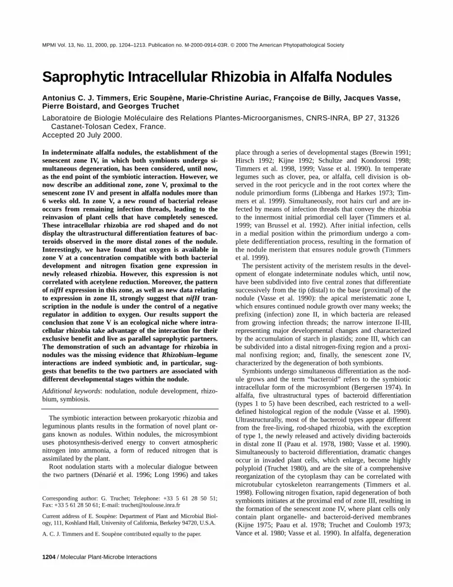
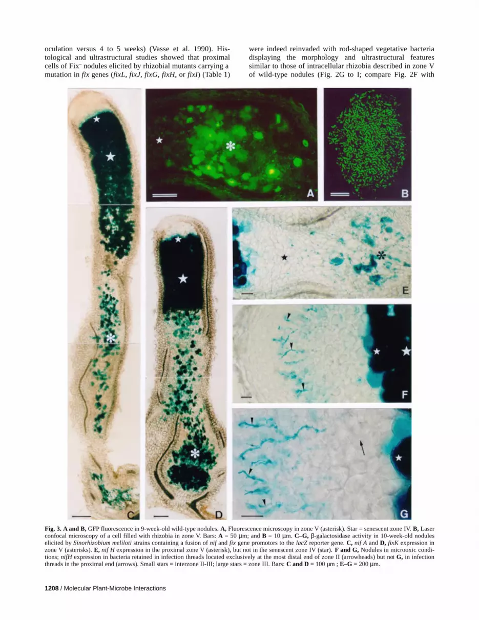
Fig. 1. Differentiation of zone V in alfalfa nodules. Bright field microscopy of longitudinal sections of nodules elicited by a bacterial strain carrying ahemA::lacZ gene. β-galactosidase staining. A–C, Different stages of differentiation of zone V at the proximal region (asterisk) of nodules collected A, 5;B, 7; and C, 9 weeks after inoculation. White stars = zone III. Bars = 200 µm. D, Infection network (arrowheads) in the proximal postsenescent region ofa 6-week-old nodule. E, Intracellular rhizobia in cells of zone V (asterisk) of a 9-week-old nodule. Arrowheads = infection threads. Bars = 50 µm.

1206 / Molecular Plant-Microbe Interactions
IV. Zone V results from a new round of plant cell invasion byrhizobia that are not surrounded by a membrane envelope, donot undergo bacteroid differentiation, and possess ultrastruc-tural features of free-living bacteria. Both nif and fix genes arestrongly expressed in bacteria in zone V, although no acety-lene reduction can be detected. The significance of this newproximal zone V in terms of the mutualistic interaction will bediscussed.
RESULTS
Identification of a new histological zone, zone V, in alfalfa nodules.
Histological studies were performed on longitudinal sec-tions of nodules harvested at weekly intervals from week 3through week 12 following inoculation of alfalfa with thestrain S. meliloti GMI6526 (Table 1). This strain carries aconstitutively expressed hemA/lacZ fusion that allows thevisualization of bacteria throughout nodule development. Ex-amination of a minimum of 10 nodules per time point showedthe typical organization of indeterminate nodules with centraltissues organized into successive zones: the apical meristem-atic zone I; the bacteroid-infected zones II, II-III, and III; andthe proximal senescent zone IV, comprising cells that appearempty under light microscopic observation (Fig. 1A to C)(Vasse et al. 1990). In addition, from 5 weeks after inoculationonward, β-galactosidase staining revealed the presence of themicrosymbiont in cells located proximally to senescent zoneIV, thus defining a new central zone, zone V (Fig. 1C). De-tailed observations showed that, in zone V, β-galactosidaseactivity could be correlated with the presence of bacteria, ei-ther retained in infection threads (Fig. 1D and E) or intracel-lular within senescent plant cells (Fig. 1C and E). Zone V wasonly rarely observed in nodules at 5 weeks postinoculation,but became clearly distinct in older nodules 7 to 12 weekspostinoculation.
Rhizobia are released passivelyfrom infection threadsin zone V.
We investigated the origin of rhizobia in zone V by two ap-proaches. First, we examined whether cell reinvasion in zoneV could be due to the penetration of rhizosphere-located rhi-zobia from the proximal sides of the nodule adjacent to thesenescent region. To test this, we inoculated alfalfa with the
wild-type strain S. meliloti 2011 and then reinoculated thesame plants, 3 weeks later, with the isogenic strain GMI6526containing a constitutively expressed hemA/lacZ fusion. Nod-ules were collected 5 weeks after the second inoculation andstained for β-galactosidase activity. The fact that enzymaticactivity was never observed in any of the nodule tissues,whereas rhizobia were frequently observed in the proximalzone V, indicated that the presence of rhizobia in zone V couldnot be due to nodule invasion by rhizospheric rhizobia (datanot shown). Second, we used electron microscopy to studycell reinvasion, assuming that this could be the result of a newround of bacterial release from the numerous infection threadsobserved in zone V (Fig. 1). Despite many observations, therewas no indication that rhizobial release in zone V occurred byan endocytotic process similar to that observed in distal zoneII of the nodule (data not shown) (Kijne 1992). However, werepeatedly observed that infection digitations protruding intocells in zone V underwent dramatic changes. These resulted,first, in a lack of plant-derived membrane (i.e., plasmalemma)at the thread’s periphery and, next, in a progressive disorgani-zation of the cell wall. The wall becomes thinner by the pro-gressive loss of its fibrillar constituents (compare Fig. 2A with2B and C), which can be clearly seen distributed within therecipient plant cell (Fig. 2D). At the end of this process, thebacteria passively leave the thread’s mucilage to invade thecell volume (Fig. 2D). These observations, and the fact that noultrastructural intermediates appear to exist between the highlydifferentiated bacteroids (Fig. 2A) (Vasse et al. 1990) and therod-shaped rhizobia in zone V (Fig. 2E and F), make it un-likely that there is a direct link between the two types of intra-cellular bacteria and led us to the conclusion that late rhizobialrelease in zone V occurs passively from infection threads.
Electron microscopy was also used to define the ultrastruc-tural features of both symbionts in zone V. We first confirmedthat rhizobia were able to reinvade highly senescent plant cells(Fig. 2E) containing ghost membranes, probably of both plantand bacteroid origin (Fig. 2B and C). Interestingly, intracellu-lar bacteria in zone V never show any of the ultrastructuraldifferentiation features of bacteroids in the distal zones of thenodule (Vasse et al. 1990) and are not surrounded by a peri-bacteroid membrane (Fig. 2D). Ultrastructurally, rhizobia inzone V generally possessed a rod-shaped morphology (Fig.2D to F), although extended focus image confocal micros-copy revealed that they could elongate slightly (Fig. 3B),
Table 1. Sinorhizobium meliloti strains used in this study
Strain Relevant characteristics References
RCR2011 SU47, wild type, Nod+ Fix+ Rosenberg et al. 1981GMI5600 lac, pSym20::Tn5,Nod+ Fix+ David et al. 1988GMI6526 (pXLGD4), Nod+ Fix+ Ardourel et al. 1994GMI6473 = GMI5600(pCHK57) lac, pnifA-lacZ, Nod+ Fix+ Soupène et al. 1995GMI6334 = GM5600(pRKP9) lac , pnifH-lacZ, Nod+ Fix+ This studyGMI6324 = GMI5600(pJJ5) lac , pfixK-lacZ, Nod+ Fix+ Soupène et al. 1995GMI11251 = GMI5600(pML330) lac, plac-fixJC, pnifH-lacZ, Nod+ Fix+ This studyGMI5801 (pGMI149nodA::lacZ), Nod+ Fix+ Maillet et al. 1990GMI6028 (pRm57nodC::lacZ), Nod+ Fix+ Mulligan and Long 1985GMI395 fixL::Tn5#2.66, Nod+ Fix– David et al. 1988GMI347 fixJ::Tn5#2.3, Nod+ Fix– Batut et al. 1985GMI5443 fixG::Tn5#2.247, Nod+ Fix– Kahn et al. 1989GMI5441 fixH::Tn5#2.222, Nod+ Fix– Kahn et al. 1989GMI351 fixI::Tn5#2.1, Nod+ Fix– Kahn et al. 1989Rm1021(pHC60) GFP-S65T, Nod+ Fix+ Cheng and Walker 1998

Vol. 13, No. 11, 2000 / 1207
thus resembling free-living rhizobia. However, they ap-peared different from their siblings retained in the infectionthreads, because they possessed a more homogenous cyto-plasm with a clear nucleoid central zone and lacked poly-β-hydroxybutyrate granules (compare Fig. 2B and C with 2F).These differences are probably due to more favorable nutri-ent conditions for rhizobia in zone V compared with bacteriaretained in infection threads.
Cell reinvasion is widely observed in Fix− nodules.Cell reinvasion occurs after nodule cells have senesced
naturally; therefore, we further investigated the correlationbetween senescence and cell reinvasion by using different S.meliloti mutant strains carrying Tn5 insertions in fix genes(Table 1). On alfalfa, Fix– S. meliloti mutants induce non–nitrogen-fixing nodules, which senesce much earlier andmore rapidly than wild-type nodules (2 to 3 weeks after in-
Fig. 2. A–F, Ultrastructural studies of zone V in nitrogen-fixing nodules using transmission electron microscopy. A–C, Ultrastructural changes of infec-tion thread digitations. Progressive disorganization of the cell wall (arrows) of infection thread digitations from A, a cell in zone III filled with nitrogen-fixing bacteroids (asterisks) to B and C, cells in zone V containing ghost membranes (stars). Note that no plasmalemma-derived membrane surroundsthe infection thread wall in zone V. Bars = 2 µm. D, Rhizobia late release (star) taking place in zone V where infection thread cell wall is disorganized(arrow). Note the lack of plant-derived membrane around released rhizobia (asterisks). Arrowheads point to fibrillar material in the lumen of the cell. Eand F, Nodule cells filled with rod-shaped rhizobia. Bars: D and F = 1 µm; and E = 2 µm. G–I, Rod-shaped intracellular rhizobia in Fix– nodules. G andH, Light micrographs showing the presence of rhizobia in senescing cells of nodules elicited by Sinorhizobium meliloti strains mutated in G, fixL and H,fixJ. I, Electron micrograph of rhizobia in a nodule elicited by a fixL mutant. Bars: G and H = 25 µm; I = 1 µm.
1208 / Molecular Plant-Microbe Interactions
oculation versus 4 to 5 weeks) (Vasse et al. 1990). His-tological and ultrastructural studies showed that proximalcells of Fix– nodules elicited by rhizobial mutants carrying amutation in fix genes (fixL, fixJ, fixG, fixH, or fixI) (Table 1)
were indeed reinvaded with rod-shaped vegetative bacteriadisplaying the morphology and ultrastructural featuressimilar to those of intracellular rhizobia described in zone Vof wild-type nodules (Fig. 2G to I; compare Fig. 2F with
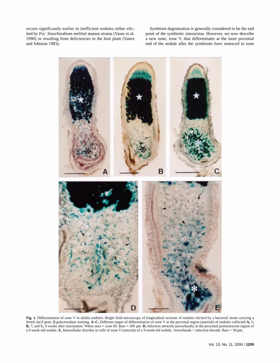
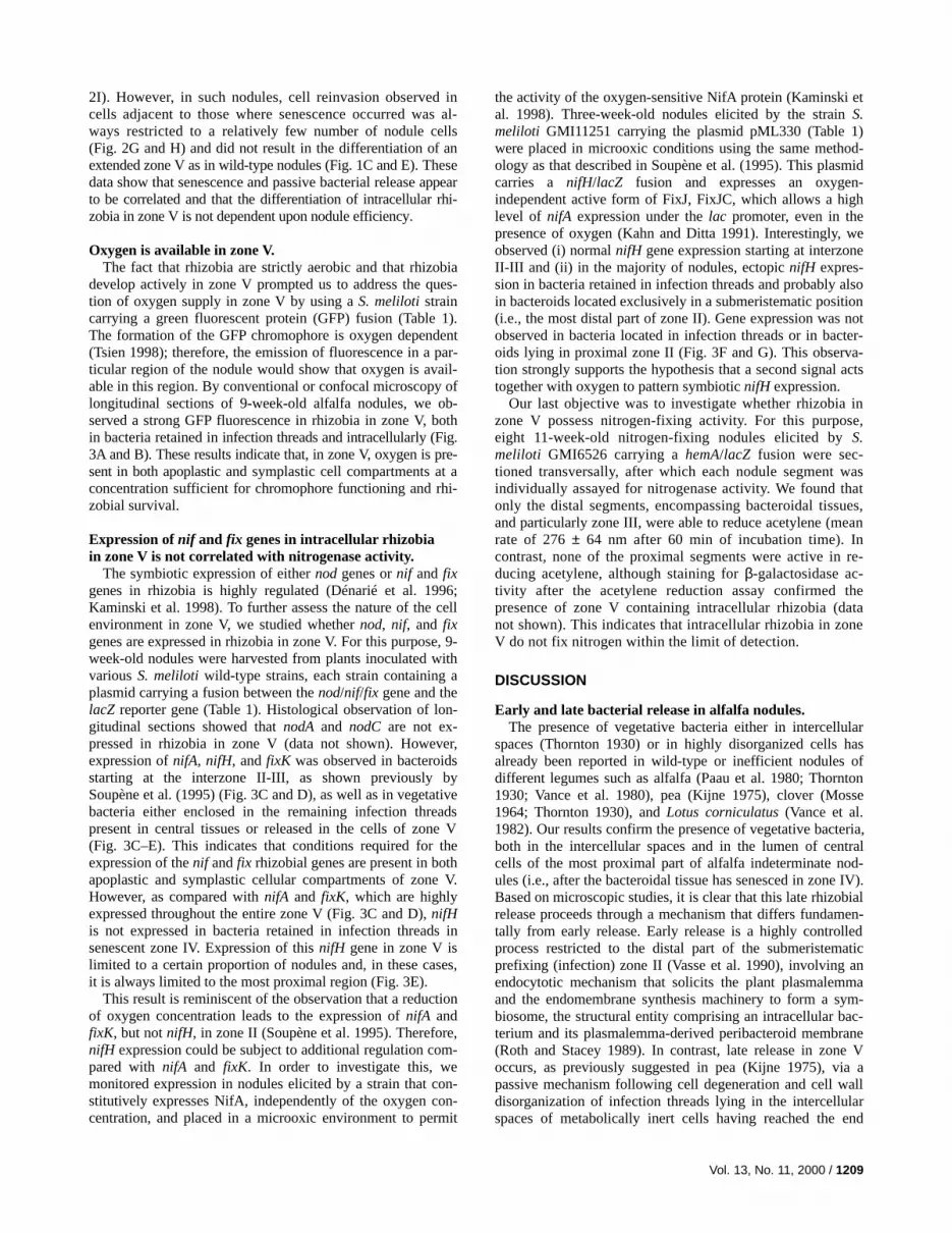
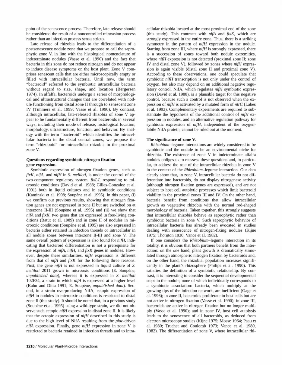
Fig. 3. A and B, GFP fluorescence in 9-week-old wild-type nodules. A, Fluorescence microscopy in zone V (asterisk). Star = senescent zone IV. B, Laserconfocal microscopy of a cell filled with rhizobia in zone V. Bars: A = 50 µm; and B = 10 µm. C–G, β-galactosidase activity in 10-week-old noduleselicited by Sinorhizobium meliloti strains containing a fusion of nif and fix gene promotors to the lacZ reporter gene. C, nif A and D, fixK expression inzone V (asterisks). E, nif H expression in the proximal zone V (asterisk), but not in the senescent zone IV (star). F and G, Nodules in microoxic condi-tions; nifH expression in bacteria retained in infection threads located exclusively at the most distal end of zone II (arrowheads) but not G, in infectionthreads in the proximal end (arrows). Small stars = interzone II-III; large stars = zone III. Bars: C and D = 100 µm ; E–G = 200 µm.
Vol. 13, No. 11, 2000 / 1209
2I). However, in such nodules, cell reinvasion observed incells adjacent to those where senescence occurred was al-ways restricted to a relatively few number of nodule cells(Fig. 2G and H) and did not result in the differentiation of anextended zone V as in wild-type nodules (Fig. 1C and E). Thesedata show that senescence and passive bacterial release appearto be correlated and that the differentiation of intracellular rhi-zobia in zone V is not dependent upon nodule efficiency.
Oxygen is available in zone V.The fact that rhizobia are strictly aerobic and that rhizobia
develop actively in zone V prompted us to address the ques-tion of oxygen supply in zone V by using a S. meliloti straincarrying a green fluorescent protein (GFP) fusion (Table 1).The formation of the GFP chromophore is oxygen dependent(Tsien 1998); therefore, the emission of fluorescence in a par-ticular region of the nodule would show that oxygen is avail-able in this region. By conventional or confocal microscopy oflongitudinal sections of 9-week-old alfalfa nodules, we ob-served a strong GFP fluorescence in rhizobia in zone V, bothin bacteria retained in infection threads and intracellularly (Fig.3A and B). These results indicate that, in zone V, oxygen is pre-sent in both apoplastic and symplastic cell compartments at aconcentration sufficient for chromophore functioning and rhi-zobial survival.
Expression of nif and fix genes in intracellular rhizobiain zone V is not correlated with nitrogenase activity.
The symbiotic expression of either nod genes or nif and fixgenes in rhizobia is highly regulated (Dénarié et al. 1996;Kaminski et al. 1998). To further assess the nature of the cellenvironment in zone V, we studied whether nod, nif, and fixgenes are expressed in rhizobia in zone V. For this purpose, 9-week-old nodules were harvested from plants inoculated withvarious S. meliloti wild-type strains, each strain containing aplasmid carrying a fusion between the nod/nif/fix gene and thelacZ reporter gene (Table 1). Histological observation of lon-gitudinal sections showed that nodA and nodC are not ex-pressed in rhizobia in zone V (data not shown). However,expression of nifA, nifH, and fixK was observed in bacteroidsstarting at the interzone II-III, as shown previously bySoupène et al. (1995) (Fig. 3C and D), as well as in vegetativebacteria either enclosed in the remaining infection threadspresent in central tissues or released in the cells of zone V(Fig. 3C–E). This indicates that conditions required for theexpression of the nif and fix rhizobial genes are present in bothapoplastic and symplastic cellular compartments of zone V.However, as compared with nifA and fixK, which are highlyexpressed throughout the entire zone V (Fig. 3C and D), nifHis not expressed in bacteria retained in infection threads insenescent zone IV. Expression of this nifH gene in zone V islimited to a certain proportion of nodules and, in these cases,it is always limited to the most proximal region (Fig. 3E).
This result is reminiscent of the observation that a reductionof oxygen concentration leads to the expression of nifA andfixK, but not nifH, in zone II (Soupène et al. 1995). Therefore,nifH expression could be subject to additional regulation com-pared with nifA and fixK. In order to investigate this, wemonitored expression in nodules elicited by a strain that con-stitutively expresses NifA, independently of the oxygen con-centration, and placed in a microoxic environment to permit
the activity of the oxygen-sensitive NifA protein (Kaminski etal. 1998). Three-week-old nodules elicited by the strain S.meliloti GMI11251 carrying the plasmid pML330 (Table 1)were placed in microoxic conditions using the same method-ology as that described in Soupène et al. (1995). This plasmidcarries a nifH/lacZ fusion and expresses an oxygen-independent active form of FixJ, FixJC, which allows a highlevel of nifA expression under the lac promoter, even in thepresence of oxygen (Kahn and Ditta 1991). Interestingly, weobserved (i) normal nifH gene expression starting at interzoneII-III and (ii) in the majority of nodules, ectopic nifH expres-sion in bacteria retained in infection threads and probably alsoin bacteroids located exclusively in a submeristematic position(i.e., the most distal part of zone II). Gene expression was notobserved in bacteria located in infection threads or in bacter-oids lying in proximal zone II (Fig. 3F and G). This observa-tion strongly supports the hypothesis that a second signal actstogether with oxygen to pattern symbiotic nifH expression.
Our last objective was to investigate whether rhizobia inzone V possess nitrogen-fixing activity. For this purpose,eight 11-week-old nitrogen-fixing nodules elicited by S.meliloti GMI6526 carrying a hemA/lacZ fusion were sec-tioned transversally, after which each nodule segment wasindividually assayed for nitrogenase activity. We found thatonly the distal segments, encompassing bacteroidal tissues,and particularly zone III, were able to reduce acetylene (meanrate of 276 ± 64 nm after 60 min of incubation time). Incontrast, none of the proximal segments were active in re-ducing acetylene, although staining for β-galactosidase ac-tivity after the acetylene reduction assay confirmed thepresence of zone V containing intracellular rhizobia (datanot shown). This indicates that intracellular rhizobia in zoneV do not fix nitrogen within the limit of detection.
DISCUSSION
Early and late bacterial release in alfalfa nodules.The presence of vegetative bacteria either in intercellular
spaces (Thornton 1930) or in highly disorganized cells hasalready been reported in wild-type or inefficient nodules ofdifferent legumes such as alfalfa (Paau et al. 1980; Thornton1930; Vance et al. 1980), pea (Kijne 1975), clover (Mosse1964; Thornton 1930), and Lotus corniculatus (Vance et al.1982). Our results confirm the presence of vegetative bacteria,both in the intercellular spaces and in the lumen of centralcells of the most proximal part of alfalfa indeterminate nod-ules (i.e., after the bacteroidal tissue has senesced in zone IV).Based on microscopic studies, it is clear that this late rhizobialrelease proceeds through a mechanism that differs fundamen-tally from early release. Early release is a highly controlledprocess restricted to the distal part of the submeristematicprefixing (infection) zone II (Vasse et al. 1990), involving anendocytotic mechanism that solicits the plant plasmalemmaand the endomembrane synthesis machinery to form a sym-biosome, the structural entity comprising an intracellular bac-terium and its plasmalemma-derived peribacteroid membrane(Roth and Stacey 1989). In contrast, late release in zone Voccurs, as previously suggested in pea (Kijne 1975), via apassive mechanism following cell degeneration and cell walldisorganization of infection threads lying in the intercellularspaces of metabolically inert cells having reached the end
1210 / Molecular Plant-Microbe Interactions
point of the senescence process. Therefore, late release shouldbe considered the result of a noncontrolled reinvasion processrather than an infection process sensu stricto.
Late release of rhizobia leads to the differentiation of apostsenescence nodule zone that we propose to call the sapro-phytic zone V, in line with the histological nomenclature ofindeterminate nodules (Vasse et al. 1990) and the fact thatbacteria in this zone do not reduce nitrogen and do not appearto induce disease symptoms on the host plant. Zone V com-prises senescent cells that are either microscopically empty orfilled with intracellular bacteria. Until now, the term“bacteroid” referred to the symbiotic intracellular bacteriawithout regard to size, shape, and location (Bergersen1974). In alfalfa, bacteroids undergo a series of morphologi-cal and ultrastructural changes that are correlated with nod-ule functioning from distal zone II through to senescent zoneIV (Timmers et al. 1999; Vasse et al. 1990). By contrast,although intracellular, late-released rhizobia of zone V ap-pear to be fundamentally different from bacteroids in severalways, including their mode of release, histological location,morphology, ultrastructure, function, and behavior. By anal-ogy with the term “bacteroid” which identifies the intracel-lular bacteria in the distal central zones, we propose theterm “rhizoboid” for intracellular rhizobia in the proximalzone V.
Questions regarding symbiotic nitrogen fixationgene expression.
Symbiotic expression of nitrogen fixation genes, such asfixK, nifA, and nifH in S. meliloti, is under the control of thetwo-component regulatory system, fixLJ, responding to mi-crooxic conditions (David et al. 1988; Gilles-Gonzalez et al.1991) both in liquid cultures and in symbiotic conditions(Kaminski et al. 1998; Soupène et al. 1995). In this paper, (i)we confirm our previous results, showing that nitrogen fixa-tion genes are not expressed in zone II but are switched on atinterzone II-III (Soupène et al. 1995) and (ii) we show thatnifA and fixK, two genes that are expressed in free-living con-ditions (Batut et al. 1989) and in zone II of nodules in mi-crooxic conditions (Soupène et al. 1995) are also expressed inbacteria either retained in infection threads or intracellular inall nodule zones between interzone II-III and zone V. Thesame overall pattern of expression is also found for nifH, indi-cating that bacteroid differentiation is not a prerequisite forthe expression of nifA, nifH, and fixK genes in nodules. How-ever, despite these similarities, nifH expression is differentfrom that of nifA and fixK for the following three reasons.First, the gene nifH is not expressed in liquid culture of S.meliloti 2011 grown in microoxic conditions (E. Soupène,unpublished data), whereas it is expressed in S. meliloti102F34, a strain in which nifA is expressed at a higher level(Kahn and Ditta 1991; E. Soupène, unpublished data). Sec-ond, in a strain overproducing NifA, ectopic expression ofnifH in nodules in microoxic conditions is restricted to distalzone II (this study). It should be noted that, in a previous study(Soupène et al. 1995) using a wild-type strain, we did not ob-serve such ectopic nifH expression in distal zone II. It is likelythat the ectopic expression of nifH described in this study isdue to the high level of NifA resulting from the plac-drivennifA expression. Finally, gene nifH expression in zone V isrestricted to bacteria retained in infection threads and to intra-
cellular rhizobia located at the most proximal end of the zone(this study). This contrasts with nifA and fixK, which arestrongly expressed in the entire zone. Thus, there is a strikingsymmetry in the pattern of nifH expression in the nodule.Starting from zone III, where nifH is strongly expressed, thereis a succession of zones toward both nodule extremities,where nifH expression is not detected (proximal zone II; zoneIV and distal zone V), followed by zones where nifH expres-sion is again visible (distal zone II and proximal zone V).According to these observations, one could speculate thatsymbiotic nifH transcription is not only under the control ofoxygen, but also may depend on an additional negative regu-latory control. NifA, which regulates nifH symbiotic expres-sion (David et al. 1988), is a plausible target for this negativecontrol, because such a control is not observed when the ex-pression of nifH is activated by a mutated form of ntrC (Labeset al. 1993). Complementary experiments are required to sub-stantiate the hypothesis of the additional control of nifH ex-pression in nodules, and an alternative regulation pathway forsymbiotic expression of nifH, independent of the oxygen-labile NifA protein, cannot be ruled out at the moment.
The significance of zone V.Rhizobium–legume interactions are widely considered to be
symbiotic and the nodule to be an environmental niche forrhizobia. The existence of zone V in indeterminate alfalfanodules obliges us to reassess these questions and, in particu-lar, to address the role of the intracellular rhizobia in zone Vin the context of the Rhizobium–legume interaction. Our dataclearly show that, in zone V, intracellular bacteria do not dif-ferentiate into bacteroids, do not display nitrogenase activity(although nitrogen fixation genes are expressed), and are notsubject to host cell autolytic processes which limit bacteroidviability in the proximal zones III and IV. On the other hand,bacteria benefit from conditions that allow intracellulargrowth as vegetative rhizobia with the normal rod-shapedmorphology of bacteria. Taken together, this strongly suggeststhat intracellular rhizobia behave as saprophytic rather thansymbiotic bacteria in zone V. Such saprophytic behavior ofintracellular bacteria has already been evocated in studiesdealing with senescence of nitrogen-fixing nodules (Kijne1975; Thornton 1930; Vance et al. 1982).
If one considers the Rhizobium–legume interaction in itstotality, it is obvious that both partners benefit from the inter-action: on the one hand, plant growth is dramatically stimu-lated through atmospheric nitrogen fixation by bacteroids and,on the other hand, the rhizobial population increases signifi-cantly in the plant’s rhizosphere (Philips et al. 1990). Thissatisfies the definition of a symbiotic relationship. By con-trast, it is interesting to consider the sequential developmentalsteps in the nodule, none of which individually corresponds toa symbiotic association: bacteria, which multiply at thegrowing tips of the infection network, are inefficient (Gage etal. 1996); in zone II, bacteroids proliferate in host cells but arenot active in nitrogen fixation (Vasse et al. 1990); in zone III,bacteroids are active in nitrogen fixation but no longer multi-ply (Vasse et al. 1990); and in zone IV, host cell autolysisleads to the senescence of all bacteroids, as deduced fromelectron microscopy studies (Kijne 1975; Mosse 1964; Paau etal. 1980; Truchet and Coulomb 1973; Vance et al. 1980,1982). The differentiation of zone V, where intracellular rhi-

Vol. 13, No. 11, 2000 / 1211
zobia do not display nitrogenase activity (this study), alsodoes not satisfy, on its own, the condition of reciprocal andsimultaneous benefit. Nevertheless, we believe that zone Vrepresents an important feature of the symbiotic Rhizobium–legume interaction. Until now, as underlined by Brewin(1998), it remains to determine the selective advantage forRhizobium spp. of undergoing a differentiation process thatleads, first, to a block in bacterial proliferation and then to atotal breakdown of the bacteroid. In our opinion, zone V rep-resents an ecological niche where rhizobia take advantage ofthe interaction and escape from plant control. Finally, the factthat rod-shaped intracellular rhizobia differentiate in non-nitrogen-fixing nodules also shows that the microsymbiontcan benefit selfishly from the association. Thus, the nitrogen-fixing nodule is an organ where reciprocal benefits for bothpartners occur in different regions of the nodule.
During the last decade, an interesting theory has been pro-posed based on the altruistic behavior of nitrogen-fixing bac-teroids that sacrifice their own reproduction to indirectly bene-fit undifferentiated siblings present in nearby soil or retainedin infection threads inside nodules (Jimenez and Casadesus1989; Olivieri and Frank 1994). Moreover, it is widely ac-cepted that “symbiotic” rhizobia from decomposing nodulesreenter the soil as free bacteria (Thornton 1930), although itremains confusing which rhizobial forms have the capacity toreadapt to free-living conditions. It seems likely that bacteriathat do not undergo the extensive ultrastructural differentiationpathway toward bacteroids (i.e., those retained in infectionthreads) (Thornton 1930) or viable bacteroids in the most re-cently infected plant cells (Quispel 1988), probably corre-sponding to the dividing type 1 rod-shaped bacteroids in distalprefixing zone II (Vasse et al. 1990), both have the capacity toreintegrate the life cycle of Rhizobium spp. in the soil afterplant tissue disintegration. We suggest that one function of“altruistic” nitrogen-fixing bacteroids is to sustain nodulelifetime sufficiently to allow the formation of a zone V whosesize increases as a function of nodule age. As a result, intra-cellular bacteria in zone V, a viable form of “symbiotic” rhi-zobia, will make a more significant contribution to the in-crease in bacterial population in the vicinity of a nodulatedlegume after nodule breakdown.
MATERIALS AND METHODS
Bacterial strains.The bacterial strains used in this study (Table 1) were grown
on yeast extract/tryptone (YT) medium supplemented with 6mM calcium chloride and appropriate selective antibiotics.Plasmid pML330 present in strain GMI11251 is a cointegrantbetween pMB210 (pnifH-lacZ) (Better et al. 1985) and pDK330(plac-fixJC) (Kahn and Ditta 1991). Both vectors were line-arized with HindIII and ligated together. In the recombinantpML330, the promoters plac and pnifH are divergent. Plasmidswere introduced into S. meliloti by triparental conjugation(David et al. 1988).
Nodulation assays.Seeds of Medicago sativa cv. Europe (Florimont Desprez,
F59242 Cappelle en Pévèle, France) were surface-sterilized,germinated, and grown in test tubes on nitrogen-free agar slantsas described by Ardourel et al. (1994). Plants were flood inocu-
lated with exponentially growing bacteria resuspended in water(approximately 106 bacteria per ml, 100 µl per plant). A mi-crooxic environment was established as described in Soupène etal. (1995) with an incubation time in soft agar of 16 h.
Microscopic methods.Nodules were harvested at weekly intervals, between 3 and
12 weeks following inoculation, and fixed with a solution of2.5% glutaraldehyde in 0.2 M sodium cacodylate (pH 7.2). Thehistological organization of nodules was observed either on 80-µm longitudinal slices of fixed nonembedded material(Microcut H1200; Bio-Rad Laboratories, Watford, U.K.)(Truchet et al. 1989) or on semithin sections (1 to 2 µm) ofEpon-embedded nodules, stained by the basic fuchsin-methylene blue method (Huber et al. 1968). Observations wereperformed using either bright field or dark field microscopy withan Olympus Vanox light microscope (Olympus, Tokyo). Ultra-structural differentiation was studied on ultrathin sections ofnodules stained with uranyl acetate and lead citrate (Reynolds1963) and observed with a Hitachi EM 600 electron microscope(Hitachi, Tokyo). β-galactosidase activity was monitored bylight microscopy on 100-µm nodule slices stained according toBoivin et al. (1990) and cleared with sodium hypochlorite be-fore observation (Truchet et al. 1989). Fluorescence microscopy(Axiophot 2; Zeiss, Jena, Germany) and confocal laser scanningmicroscopy (LSM 410 Invert; Zeiss) were performed accordingto Timmers et al. (1998) on 100-µm longitudinal sections ofnodules either untreated and mounted in water or fixed with 4%formaldehyde in phosphate-buffered saline (pH 7.4, 1 h), treatedwith Evans blue (E-2129; Sigma-Aldrich, St. Louis, U.S.A.) toquench autofluorescence, and then mounted in mowiol 4-88(Calbiochem 475904; Calbiochem, La Jolla, CA, U.S.A.).
Nitrogenase activity.Nitrogenase activity was assayed by the acetylene reduction
technique (Turner and Gibson 1980). Eleven-week-old noduleselicited by S. meliloti GMI6526, a strain carrying a constitu-tively expressed lacZ fusion, were transversally cut to generatetwo nodule halves, the distal region, with the bacteroid-containing tissues, and the proximal, deflated part. Each halfwas assayed for nitrogenase activity immediately after dissec-tion as described in Vasse et al. (1990). A sample was scored asFix+ if acetylene-dependent ethylene production could be de-tected within the 30-min incubation time. After the acetylenereduction assay, the presence of living rhizobia in complemen-tary nodule halves was monitored by β-galactosidase activity(described above).
ACKNOWLEDGMENTS
We thank J. Batut, D. Barker, and P. Gamas for helpful comments andvaluable criticism of the manuscript; H. Bergès, C. Cosseau, A.-M.Garnerone, and F. Maillet for helpful technical assistance; J. Kijne andC. Vance for providing us with ancestral reprints; and D. Barker forEnglish reviewing. A. C. J. Timmers was supported by a TMR fellow-ship and this work was funded by a grant from the European CommunityTMR Programme (FMRW-CT96-0039).
LITERATURE CITED
Ardourel, M., Demont, N., Debellé, F., Maillet, F., De Billy, F.,Promé, J.-C., Dénarié, J., and Truchet, G. 1994. Rhizobium meliloti

1212 / Molecular Plant-Microbe Interactions
lipooligosaccharide nodulation factors: Different structural re-quirements for bacterial entry into target root hair cells and induc-tion of plant symbiotic developmental responses. Plant Cell6:1357-1374.
Batut, J., Daveran-Mingot, M.-L., David, M., Jacobs, J., Garnerone,A.-M., and Kahn, D. 1989. fixK, a gene homologous with fnr andcrp from Escherichia coli, regulates nitrogen fixation genes bothpositively and negatively in Rhizobium meliloti. EMBO J. 8:1279-1286.
Batut, J., Terzaghi, B., Ghérardi, M., Huguet, M., Terzaghi, E., Garnerone,A.-M., Boistard, P., and Huguet, T. 1985. Localization of a symbi-otic fix region on Rhizobium meliloti pSym megaplasmid more than200 kilobases from the nod-nif region. Mol. Gen. Genet. 199:232-239.
Bergersen, F. J. 1974. Formation and function of bacteroids. Pages 476-498 in: The Biology of Nitrogen Fixation. A. Quispel, ed. North-Holland Publishing Company, Amsterdam.
Better, M., Ditta, G., and Helinski, D. R. 1985. Deletion analysis ofRhizobium meliloti symbiotic promoters. EMBO J. 4:2419-2424.
Boivin, C., Camut, S., Malpica, C. A., Truchet, G., and Rosenberg, C.1990. Rhizobium meliloti genes encoding catabolism of trigonellineare induced under symbiotic conditions. Plant Cell 2:1157-1170.
Brewin, N. J. 1991. Development of the legume root nodule. Annu. Rev.Cell Biol. 7:191-226.
Brewin, N. J. 1998. Tissue and cell invasion by Rhizobium: The struc-ture and development of infection threads and symbiosomes. Pages417-429 in: Rhizobiaceae, Molecular Biology of Model Plant-Associated Bacteria. H. P. Spaink, A. Kondorosi, and P. J. J.Hooykaas, eds. Kluwer Academic Press, Dordrecht, The Nether-lands.
Cheng, H.-P., and Walker, G. C. 1998. Succinoglycan is required forinitiation and elongation of infection threads during nodulation of al-falfa by Rhizobium meliloti. J. Bacteriol. 180:5183-5191.
David, M., Daveran, M.-L., Batut, J., Dedieu, A., Domergue, O.,Ghai, J., Hertig, C., Boistard, P., and Kahn, D. 1988. Cascaderegulation of nif gene expression in Rhizobium meliloti. Cell54:671-683.
Dénarié, J., Debellé, F., and Promé, J.-C. 1996. Rhizobium lipo-chitooligosaccharide nodulation factors: Signaling molecules medi-ating recognition and morphogenesis. Annu. Rev. Biochem. 65:503-535.
Gage, D. J., Bobo, T., and Long, S. R. 1996. Use of green fluorescentprotein to visualize the early events of symbiosis between Rhizo-bium meliloti and alfalfa (Medicago sativa). J. Bacteriol. 178:7159-7166.
Gilles-Gonzalez, M., Ditta, G. S., and Helinski, D. R. 1991. A haemo-protein with kinase activity encoded by the oxygen sensor of Rhizo-bium meliloti. Nature 150:170-172.
Hirsch, A. M. 1992. Developmental biology of legume nodulation. NewPhytol. 122:211-237.
Huber, J. D., Parker, F., and Odland, G. F. 1968. A basic fuchsin andalkalinized methylene blue rapid stain for epoxy embedded tissue.Stain Technol. 43:83-87.
Jimenez, J., and Casadesus, J. 1989. An altruistic model of the Rhizo-bium-legume association. J. Hered. 80:335-337.
Kahn, D., David, M., Dommergue, O., Daveran, M.-H., Ghai, G., Hirsch,P., and Batut, J. 1989. Rhizobium meliloti fixGHI sequence predictsinvolvement of a specific cation pump in symbiotic nitrogen fixation.J. Bacteriol. 171:929-939.
Kahn, D., and Ditta, G. 1991. Modular structure of FixJ: Homology ofthe transcriptional activator domain with the –35 binding domain ofthe sigma factors. Mol. Microbiol. 5:987-997.
Kaminski, P. A., Batut, J., and Boistard, P. 1998. A survey of symbioticnitrogen fixation by rhizobia. Pages 431-460 in: Rhizobiaceae, Mo-lecular Biology of Model Plant-Associated Bacteria. H. P. Spaink, A.Kondorosi, and P. J. J. Hooykaas, eds. Kluwer Academic Press, Dor-drecht, The Netherlands.
Kijne, J. W. 1975. The fine structure of pea root nodules. 2. Senes-cence and disintegration of the bacteroid tissue. Physiol. Plant Pa-thol. 7:17-21.
Kijne, J. W. 1992. The rhizobium infection process. Pages 349-398 in:Biological Nitrogen Fixation. G. Stacey, R. H. Burris, and H. J.Evans, eds. Chapmann and Hall, New York.
Labes, M., Rastogi, V., Watson, R., and Finan, T. M. 1993. Symbiotic
nitrogen fixation by a nifA deletion mutant of Rhizobium meliloti: Therole of an unusual ntrC allele. J. Bacteriol. 175:2662-2673.
Libbenga, K. R., and Harkes, P. A. A. 1973. Initial proliferation of corti-cal cells in the formation of root nodules in Pisum sativum L. Planta114:17-28.
Long, S. R. 1996. Rhizobium symbiosis: Nod factors in perspective.Plant Cell 8:1885-1898.
Maillet, F., Debellé, F., and Dénarié, J. 1990. Role of the nod and syrMgenes in the activation of the regulatory gene nodD3, and of thecommon and host-specific nod genes of Rhizobium meliloti. Mol. Mi-crobiol. 4:1975-1984.
Mosse, B. 1964. Electron-microscope studies of nodule development insome clover species. J. Gen. Microbiol. 36:49-66.
Mulligan, J. T., and Long, S. R. 1985. Induction of Rhizobium melilotinodC expression by plant exudate requires nodD. Proc. Natl. Acad.Sci. USA 82:6609-6613.
Olivieri, I., and Frank, S. A. 1994. The evolution of nodulation in Rhi-zobium: Altruism in the rhizosphere. J. Hered. 85:46-47.
Paau, A. S., Bloch, C. B., and Brill, W. J. 1980. Developmental fate ofRhizobium meliloti bacteroids in alfalfa nodules. J. Bacteriol.143:1480-1490.
Paau, A. S., Cowles, J. R., and Raveed, D. 1978. Development ofbacteroids in alfalfa (Medicago sativa) nodules. Plant Physiol. 62:526-530.
Philips, D. A., Harwig, U. A., Maxwell, C. A., Joseph, C. M., Wery, J.,Hungria, M., and Tsai, S. M. 1990. Host legume control of nodulationby flavonoids. Pages 331-338 in: Nitrogen Fixation: Achievementsand Objectives. P. M. Gresshoff, L. E. Roth, G. Stacey, and W. E.Newton, eds. Chapmann and Hall, New York.
Quispel, A. 1988. Bacteria-plant interactions in symbiotic nitrogenfixation. Physiol. Plant. 74:783-790.
Reynolds, E. S. 1963. The use of lead citrate at high pH as an electronopaque stain in electron microscopy. J. Cell Biol. 17:208-213.
Rosenberg, C., Boistard, P., Dénarié, J., and Casse-Delbart, F. 1981.Genes controlling early and late functions in symbiosis are locatedon a megaplasmid in Rhizobium meliloti. Mol. Gen. Genet. 184:326-333.
Roth, L. E., and Stacey, G. 1989. Bacterium release into host cells ofnitrogen-fixing soybean nodules: The symbiosome membrane comesfrom three sources. Eur. J. Cell Biol. 49:13-23.
Schultze, M., and Kondorosi, A. 1998. Regulation of symbiotic rootnodule development. Annu. Rev. Genet. 32:33-57.
Soupène, E., Foussard, M., Boistard, P., Truchet, G., and Batut, J. 1995.Oxygen as a key developmental regulator of Rhizobium meliloti N2-fixation gene expression within the alfalfa root nodule. Proc. Natl.Acad. Sci. USA 92:3759-3763.
Thornton, H. G. 1930. The influence of the host plant in inducing para-sitism in lucerne and clover nodules. Proc. Roy. Soc. London Ser. B106:110-122.
Timmers, A. C. J., Auriac, M.-C., de Billy, F., and Truchet, G. 1998.Nod factor internalization and microtubular cytoskeleton changes oc-cur concomitantly during nodule differentiation in alfalfa. Develop-ment 125:339-349.
Timmers, A. C. J., Auriac, M.-C., and Truchet, G. 1999. Refined analy-sis of early symbiotic steps of the Rhizobium-Medicago interaction inrelationship with microtubular cytoskeleton rearrangements. Devel-opment 126:3617-3628.
Truchet, G. 1980. Sur l’état diploïde des cellules du méristème desnodules radiculaires des légumineuses. Ann. Sci. Nat. Bot. Biol.Vég. 19:3-38.
Truchet, G., Camut, S., de Billy, F., Odorico, R., and Vasse, J. 1989.The Rhizobium-legume symbiosis: Two methods to discriminatebetween nodules and other root-derived structures. Protoplasma149:82-88.
Truchet, G., and Coulomb, P. 1973. Mise en évidence et évolution dusystème phyto-lysosomal dans les cellules des différentes zones desnodules radiculaires de Pois (Pisum sativum L.). Notion d’hétérophagie.J. Ultrastr. Res. 43:36-57.
Tsien, R. Y. 1998. The green fluorescent protein. Annu. Rev. Biochem.67:509-544.
Turner, G. L., and Gibson, A. H. 1980. Measurement of nitrogenfixation by indirect means. Pages 111-138 in: Methods for Evalu-ating Biological Nitrogen Fixation. F. J. Bergersen, ed. Wiley,Chichester, U.K.

Vol. 13, No. 11, 2000 / 1213
Van Brussel, A. A. N., Bakhuizen, R., Van Spronsen, A. C., Spaink, H.P., Tak, T., Lugtenberg, B. J. J., and Kijne, J. 1992. Induction of prein-fection thread structures in the leguminous host plant by mitogeniclipooligosaccharides of Rhizobium. Science 257:70-72.
Vance, C. P., and Johnson, L. E. B. 1983. Plant determined ineffectivenodules in alfalfa (Medicago sativa): structural and biochemical com-parisons. Can. J. Bot. 61:93-106.
Vance, C. P., Johnson, L. E. B., Halvorsen, A. M., Heichel, G. H., andBarnes, D. K. 1980. Histological and ultrastructural observations of
Medicago sativa root nodule senescence after foliage removal. Can. J.Bot. 58:295-309.
Vance, C. P., Johnson, L. E. B., Stade, S., and Groat, R. G. 1982.Birdsfoot trefoil (Lotus corniculatus) root nodules: Morphogenesisand the effect of forage harvest on structure and function. Can. J. Bot.60:505-518.
Vasse, J., de Billy, F., Camut, S., and Truchet, G. 1990. Correlationbetween ultrastructural differentiation of bacteroids and nitrogenfixation in alfalfa nodules. J. Bacteriol. 172:4295-4306.