multiple small heat shock proteins in rhizobia
TRANSCRIPT
JOURNAL OF BACTERIOLOGY,0021-9193/99/$04.0010
Jan. 1999, p. 83–90 Vol. 181, No. 1
Copyright © 1999, American Society for Microbiology. All Rights Reserved.
Multiple Small Heat Shock Proteins in RhizobiaMARTIN MUNCHBACH,1 ANDREAS NOCKER,2 AND FRANZ NARBERHAUS2*
Protein Chemistry Laboratory1 and Mikrobiologisches Institut,2 EidgenossischeTechnische Hochschule, CH-8092 Zurich, Switzerland
Received 22 July 1998/Accepted 28 October 1998
Seven genes coding for small heat shock proteins (sHsps) in Bradyrhizobium japonicum have been identified.They are organized in five operons that are coordinately regulated by ROSE, a negatively cis-acting DNAelement. The deduced sHsps can be divided into two separate classes: class A, consisting of proteins that showsimilarity to Escherichia coli IbpA and IbpB, and class B, whose members display significant similarity to othersHsps from prokaryotes and eukaryotes. Two-dimensional gel electrophoresis and Edman sequencing revealedthe presence of at least 12 sHsps in B. japonicum, indicating a remarkable abundance of sHsps in thisorganism. Three additional members of class A and two potentially novel heat shock proteins were identifiedon the basis of their amino termini. The presence of multiple sHsps was also demonstrated for a variety ofRhizobium and Bradyrhizobium species by immunoblot analysis and two-dimensional gel electrophoresis. Anextensive database survey revealed that, in contrast to the rhizobia, other bacteria contain maximally twosHsps whereas many plants have been reported to possess a sHsp superfamily.
All organisms so far examined respond to a sudden increasein growth temperature by inducing the synthesis of a number ofheat shock proteins (Hsps). Some of these proteins are alsoimportant during normal growth. The regulation, structure,and function of several Hsps have been studied in great detail.The chaperone machineries GroES/GroEL and DnaK/DnaJ/GrpE, for example, are involved in diverse processes such asprotein folding and protein degradation, assembly of proteincomplexes, and transport of proteins across membranes. Theirfunction appears to be highly conserved between prokaryotesand eukaryotes (reviewed in references 7 and 40).
Comparatively little is known, however, about small Hsps(sHsps). In contrast to the highly conserved DnaK and GroELproteins, sHsps show much less sequence similarity. This pro-tein family is characterized by the following criteria: (i) a mo-lecular mass typically between 12 and 30 kDa; (ii) a conservedcentral domain, referred to as the a-crystallin domain; (iii)formation of large oligomeric complexes, ranging from 150 to800 kDa; (iv) ATP-independent chaperone activity (9, 27, 51).The latter concept, however, has been challenged by the ob-servation that ATP enhances the molecular chaperone activityof aB-crystallin (36). According to the present model, sHspsbind to denatured proteins accumulated under stress condi-tions and maintain them in a folding-competent state (15, 32).Recently, the crystal structure of a sHsp from Methanococcusjannaschii has been solved (29). Twenty-four monomers form ahollow spherical complex with a total of 14 “windows” thatmight allow polypeptides to enter the complex.
Not surprisingly, most of the work on sHsps has been con-ducted with eukaryotic members of this superfamily becausethey are related to the a-crystallin proteins of the vertebrateeye lens (26). a-Crystallins play a structural role in maintaininglens stability and transparency but notably they are also ex-pressed in nonlenticular tissues, e.g., in heart, muscle, andkidney (3). A remarkable abundance of sHsps was reported inheat-stressed plants. Up to 30 different sHsps comprising six
different classes are induced after a temperature upshift, de-pending on the plant species. Each gene family encodes pro-teins localized in a distinct cellular compartment (51).
Most bacteria appear to have only a small number of heatshock proteins. The completed genome sequences indicatethat Mycoplasma genitalium completely lacks any gene codingfor sHsps (19). M. jannaschii encodes one and Escherichia coliencodes two sHsps (8, 17). The first hint that Rhizobiaceae maybe an exception in that they possess a larger set of sHsps wasprovided by a two-dimensional gel analysis by Michiels et al.(34). The authors compared the induction of Hsps in a heat-tolerant and a heat-sensitive Rhizobium strain and observedeight heat-inducible protein spots in extracts from the temper-ature-sensitive strain. By contrast, the tropical, heat-tolerantstrain induced only two sHsps.
In the process of elucidating the complex regulatory networkthat controls the heat shock response of Bradyrhizobium ja-ponicum, the nitrogen-fixing root-nodule symbiont of soybean,we identified six genes encoding sHsps (37, 38). They areorganized in four operons that are located in an extendedheat-shock gene cluster. Each operon is preceded by a con-served DNA element of approximately 100 bp that is posi-tioned between the transcription start and the start codon ofthe first gene. This element was designated ROSE (for Re-pression Of heat Shock gene Expression), and several lines ofevidence suggest that it serves as a binding site for a putativerepressor protein under non-heat shock conditions (37).
Here we report on a bacterial sHsp superfamily comprisingat least 12 members. The seven B. japonicum sHsps identifiedso far can be grouped into two distinct classes. We monitoredthe induction of sHsps under various stress conditions andexamined their heat shock induction by two-dimensional gelelectrophoresis. Finally, we provide evidence that the presenceof a sHsp superfamily is not restricted to B. japonicum butmight be widespread in the Rhizobiaceae.
MATERIALS AND METHODS
Bacterial strains, plasmids, and growth conditions. B. japonicum 110spc4 wasgrown aerobically at 28°C in PSY medium (43) supplemented with 0.1% (wt/vol)arabinose and 100 mg of spectinomycin per ml. YEM medium (12) supplementedwith 10 mM KNO3 was used for anaerobic B. japonicum cultures. Bradyrhizobiumsp. (Parasponia) ANU289 and Bradyrhizobium sp. (Lupinus) ATCC 10319 werepropagated in PSY medium with 0.1% (wt/vol) arabinose. TY medium (5) was
* Corresponding author. Mailing address: Mikrobiologisches Insti-tut, Eidgenossische Technische Hochschule, Schmelzbergstrasse 7,CH-8092 Zurich, Switzerland. Phone: 41-1-632-2586. Fax: 41-1-632-1148. E-mail: [email protected].
83
on May 31, 2015 by guest
http://jb.asm.org/
Dow
nloaded from

used to grow Rhizobium leguminosarum bv. viciae 897, Rhizobium etli (formerlyR. leguminosarum bv. phaseoli) 8002, R. leguminosarum bv. trifolii ATCC 14480,Rhizobium sp. strain NGR234, and Sinorhizobium meliloti 2011. E. coli cells weregrown in Luria-Bertani medium (35) supplemented with ampicillin (200 mg/ml)if required.
DNA manipulations and sequence analysis. Recombinant DNA techniqueswere performed according to standard protocols (45). Chromosomal DNA wasisolated as described previously (23). Southern blot hybridizations using DIG(digoxigenin-11-dUTP)-labeled DNA probes were performed according to themanufacturer’s instructions (Boehringer GmbH, Mannheim, Germany). The111-bp ROSE1 probe was produced by PCR using plasmid pRJ5035, a pUC18derivative containing a 1.7-kb HindIII insert carrying the hspA gene region (38),and the oligonucleotides Sig36 (59-CGCCGCGACAAGCGGTCC-39) and Sig37(59-GTCCTCATAGCCAAATCCTCC-39). Plasmid DNA was sequenced by thechain termination method (46) with a Model 373 DNA sequencer (AppliedBiosystems, Foster City, Calif.). The DNA sequence was analyzed with thesoftware package of the Genetics Computer Group of the University of Wiscon-sin—Madison (UWGCG) (version 8.0) or the National Center for BiotechnologyInformation network server. Multiple sequence alignments were generated withthe PILEUP program provided by the UWGCG software.
Western blot (immunoblot) analysis. Crude cell extracts were prepared, sep-arated on sodium dodecyl sulfate–12% polyacrylamide gels, and transferred tonitrocellulose membranes as described previously (39). Bacteroid extracts wereprepared as described elsewhere (16). Anti-E. coli IbpA serum (2) was kindlyprovided by A. Easton (St. Louis, Mo.) and was used in 1,500-fold dilution.Primary rabbit antibodies were detected with the Chemiluminescence WesternBlotting Kit (Boehringer GmbH).
Two-dimensional gel electrophoresis and Edman sequencing. Two-dimen-sional gel electrophoresis, protein elution, concentration, electrotransfer, andN-terminal sequencing were performed as described elsewhere (42).
Transcript mapping. RNA isolation and primer extension analysis was per-formed as described elsewhere (4). The oligonucleotides AN1 (59-CTGAACATAGTCTGCCAGGTTGAACTGC-39) and AN18 (59-CCGTTTCAACGAGGTCGAAAAGGC-39) were used to determine the hspH transcription start site.
Nucleotide sequence accession numbers. The nucleotide sequences describedhere have been deposited in the EMBL, GenBank, and DDBJ databases underthe following accession numbers: U55047 (hspA, hspB, and hspC gene region),AJ003064 (hspD, hspE, and hspF gene region), and AJ010144 (hspH gene re-gion).
RESULTS AND DISCUSSIONTwo classes of sHsps in B. japonicum. Six genes coding for
small Hsps (hspA, -B, -C, -D, -E, and -F) have recently beenidentified in a heat shock gene cluster of B. japonicum (37, 38).They are organized in four operons together with some addi-
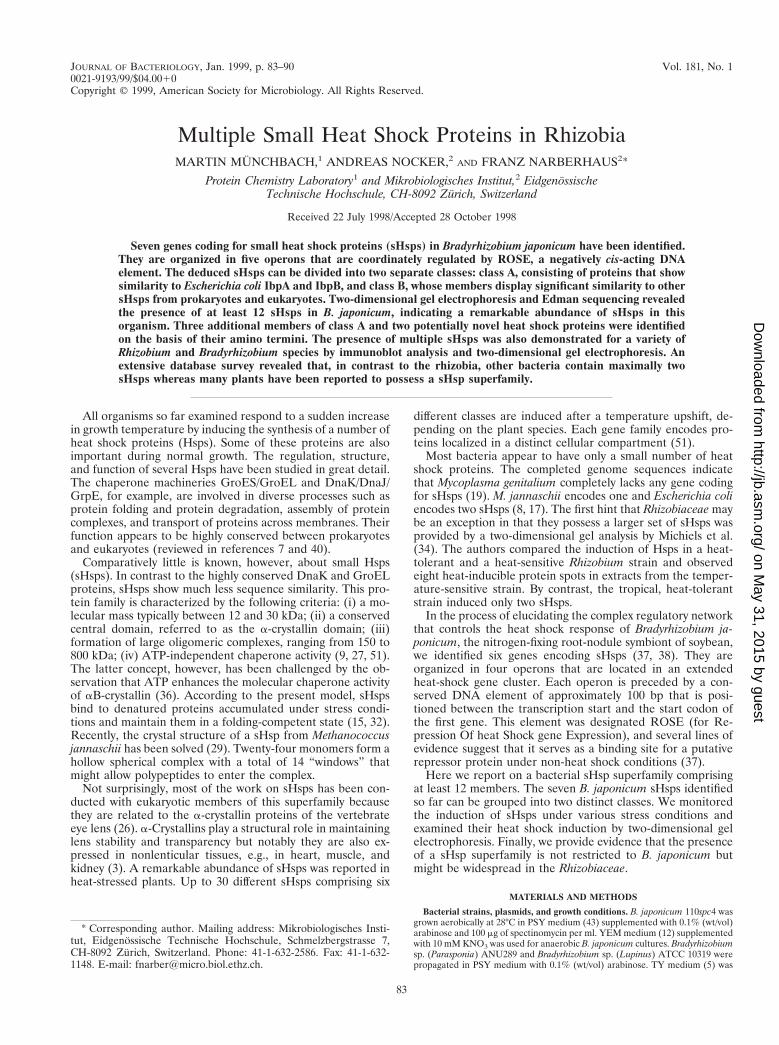
tional heat shock genes. Heat-inducible transcription of eachoperon is mediated by ROSE, a novel regulatory element thatconsists of approximately 100 bp and precedes the first gene ofeach operon (37). We identified a putative fifth ROSE-depen-dent operon by using a ROSE1 fragment as a probe in South-ern hybridization experiments (data not shown). Two hybrid-izing fragments, a 5.8-kb BamHI fragment and a 5.6-kb SalIfragment, were subsequently cloned and found to contain thehspH gene region (Fig. 1A). No additional heat shock geneswere present up- or downstream of hspH. An amino acid se-quence comparison of the deduced small Hsps revealed thatthey fall into two distinct classes, as indicated in Fig. 1A andshown more precisely in Fig. 2. Class A contains only bacterialproteins, namely the B. japonicum proteins HspA, -B, -D, -E,and -H, E. coli IbpA and IbpB, and Legionella pneumophilaGspA. It is evident from the alignment that proteins belongingto this class are highly similar to each other throughout theirentire length (between 34 and 73% positional amino acid se-quence identity). The similarity is not restricted to the a-crys-tallin domain but extends into the flanking amino- and car-boxy-terminal regions. Class B proteins are much moredivergent in length, sequence, and phylogenetic origin. Theyinclude prokaryotic as well as eukaryotic members from a widevariety of organisms. The similarity between class A proteinsand class B proteins is rather low (around 20% amino acidsequence identity). Although the degree of homology withinclass B is significant (between 30 and 60% identical aminoacids), only the B. japonicum proteins HspC and HspF reachthe latter, highest score. The identity among the other mem-bers is generally between 30 and 35%. Identical amino acidsare almost exclusively displayed in the a-crystallin domain, andthe flanking regions are highly variable in length and sequence(with the exception of HspC and HspF).
The B. japonicum hspH gene is preceded by a typical s70-type promoter and a ROSE element with high sequence sim-ilarity to all previously identified ROSE elements (37). Withthe exception of one nucleotide (a G instead of a C at the
FIG. 1. Physical map of five ROSE-dependent heat shock operons of B. japonicum and determination of the transcription start site upstream of ROSE5. (A)Schematic representation of the ROSE-dependent operons. The ROSE elements (1 through 5) are represented by black boxes. Class A and class B small heat shockgenes are indicated. No significant open reading frames were identified downstream of hspD and hspH. (B) Primer extension analysis to determine the transcriptionstart site upstream of hspH. The extension product of primer AN18 is shown. The same primer was used for the corresponding sequencing reaction (TCGA).
84 MUNCHBACH ET AL. J. BACTERIOL.
on May 31, 2015 by guest
http://jb.asm.org/
Dow
nloaded from
ROSE1-equivalent position 132), all previously described con-served ROSE nucleotides were conserved in the ROSE5 se-quence. In particular, the nucleotides in the promoter-distalhalf of ROSE are highly conserved (data not shown). Tran-scription of hspH was heat inducible, and the transcriptionstart site was located at the expected position just upstream ofROSE5, as determined by primer extension (Fig. 1B). Thus, allpresently known sHsp genes of B. japonicum are coordinatelyregulated by ROSE, a negatively cis-acting DNA element thatprecedes each class A gene and presumably serves as a repres-sor binding site under normal growth conditions (37). Thetwo-dimensional gel analysis revealed that the degree of induc-tion varied from protein to protein (see below), suggesting thatposttranscriptional or posttranslational mechanisms mightcontribute to their regulation. Heat-induced expression of classA genes in other organisms (the E. coli ibpAB operon and theL. pneumophila gspA gene) is dependent on a s32-type pro-moter (1, 2) whereas several class B genes are under negativecontrol. Transcription of the Streptomyces albus hsp18 gene issubject to repression by the OrfY protein at low temperatures(49). Transcriptional repression has also been proposed tocontrol the expression of Clostridium acetobutylicum hsp18,Leuconostoc oenos hsp18, and Synechococcus vulcanus hspA(28, 44, 47). These genes are transcribed from a typical s70-
FIG
.2.
Am
inoacid
sequencealignm
entof
sHsp
representativesfrom
classA
andclass
B.R
egionscorresponding
tothe
N-term
inaldomain,
a-crystallin
domain,and
C-term
inalextensionare
indicated(9,33).T
heconsensus
sequences(C
onAand
ConB
)are
definedby
capitalletters
when
anam
inoacid
ispresent
inall
eightaligned
sequencesand
bylow
ercaseletters
when
anam
inoacid
ispresent
inat
leastfive
ofthe
eightsequences.A
mino
acidsthatconform
with
theconsensus
areindicated
byasterisks.A
mino
acidsappearing
inboth
consensussequences
areconnected
bya
line.The
complete
sequenceofthe
deducedproteins
isshow
nand
designatedas
follows:H
spAto
HspH
,B.japonicum
HspA
toH
spH(references
37and
38and
thisw
ork);IbpAand
IbpB,E
.coliIbpAand
IbpB(2);G
spA,L
.pneumophila
GspA
(1);SP21,S.aurantiacaSP21
(24);YocM
,B.subtilis
YocM
(31);Cace,C
.acetobutylicumH
sp18(47);Salb,S.albus
Hsp18
(48);Mjan,M
.jannaschiiHsp16.5
(8);Gm
ax,Glycine
max
Hsp17.5
(11).
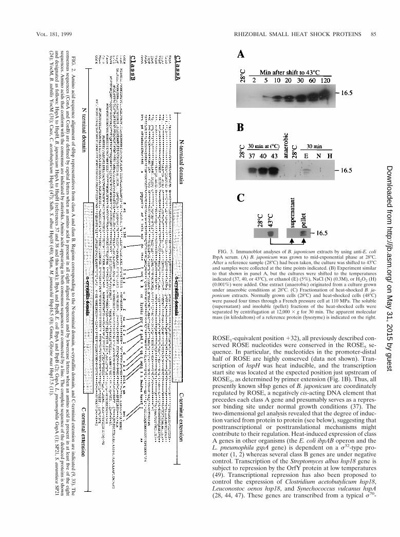
FIG. 3. Immunoblot analyses of B. japonicum extracts by using anti-E. coliIbpA serum. (A) B. japonicum was grown to mid-exponential phase at 28°C.After a reference sample (28°C) had been taken, the culture was shifted to 43°Cand samples were collected at the time points indicated. (B) Experiment similarto that shown in panel A, but the cultures were shifted to the temperaturesindicated (37, 40, or 43°C), or ethanol (E) (5%), NaCl (N) (0.3M), or H2O2 (H)(0.001%) were added. One extract (anaerobic) originated from a culture grownunder anaerobic conditions at 28°C. (C) Fractionation of heat-shocked B. ja-ponicum extracts. Normally grown cells (28°C) and heat-shocked cells (48°C)were passed four times through a French pressure cell at 110 MPa. The soluble(supernatant) and insoluble (pellet) fractions of the heat-shocked cells wereseparated by centrifugation at 12,000 3 g for 30 min. The apparent molecularmass (in kilodaltons) of a reference protein (lysozyme) is indicated on the right.
VOL. 181, 1999 RHIZOBIAL SMALL HEAT SHOCK PROTEINS 85
on May 31, 2015 by guest
http://jb.asm.org/
Dow
nloaded from
type housekeeping promoter, which implies that additionalmechanisms must prevent their expression during normalgrowth. However, the exact control mechanisms have not beenelucidated yet.
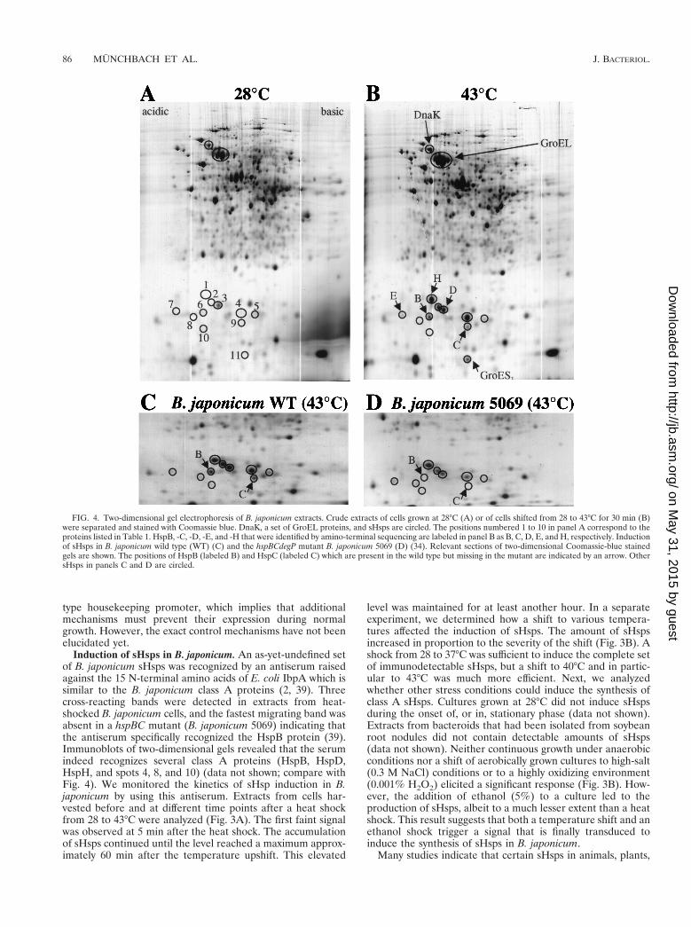
Induction of sHsps in B. japonicum. An as-yet-undefined setof B. japonicum sHsps was recognized by an antiserum raisedagainst the 15 N-terminal amino acids of E. coli IbpA which issimilar to the B. japonicum class A proteins (2, 39). Threecross-reacting bands were detected in extracts from heat-shocked B. japonicum cells, and the fastest migrating band wasabsent in a hspBC mutant (B. japonicum 5069) indicating thatthe antiserum specifically recognized the HspB protein (39).Immunoblots of two-dimensional gels revealed that the serumindeed recognizes several class A proteins (HspB, HspD,HspH, and spots 4, 8, and 10) (data not shown; compare withFig. 4). We monitored the kinetics of sHsp induction in B.japonicum by using this antiserum. Extracts from cells har-vested before and at different time points after a heat shockfrom 28 to 43°C were analyzed (Fig. 3A). The first faint signalwas observed at 5 min after the heat shock. The accumulationof sHsps continued until the level reached a maximum approx-imately 60 min after the temperature upshift. This elevated
level was maintained for at least another hour. In a separateexperiment, we determined how a shift to various tempera-tures affected the induction of sHsps. The amount of sHspsincreased in proportion to the severity of the shift (Fig. 3B). Ashock from 28 to 37°C was sufficient to induce the complete setof immunodetectable sHsps, but a shift to 40°C and in partic-ular to 43°C was much more efficient. Next, we analyzedwhether other stress conditions could induce the synthesis ofclass A sHsps. Cultures grown at 28°C did not induce sHspsduring the onset of, or in, stationary phase (data not shown).Extracts from bacteroids that had been isolated from soybeanroot nodules did not contain detectable amounts of sHsps(data not shown). Neither continuous growth under anaerobicconditions nor a shift of aerobically grown cultures to high-salt(0.3 M NaCl) conditions or to a highly oxidizing environment(0.001% H2O2) elicited a significant response (Fig. 3B). How-ever, the addition of ethanol (5%) to a culture led to theproduction of sHsps, albeit to a much lesser extent than a heatshock. This result suggests that both a temperature shift and anethanol shock trigger a signal that is finally transduced toinduce the synthesis of sHsps in B. japonicum.
Many studies indicate that certain sHsps in animals, plants,
FIG. 4. Two-dimensional gel electrophoresis of B. japonicum extracts. Crude extracts of cells grown at 28°C (A) or of cells shifted from 28 to 43°C for 30 min (B)were separated and stained with Coomassie blue. DnaK, a set of GroEL proteins, and sHsps are circled. The positions numbered 1 to 10 in panel A correspond to theproteins listed in Table 1. HspB, -C, -D, -E, and -H that were identified by amino-terminal sequencing are labeled in panel B as B, C, D, E, and H, respectively. Inductionof sHsps in B. japonicum wild type (WT) (C) and the hspBCdegP mutant B. japonicum 5069 (D) (34). Relevant sections of two-dimensional Coomassie-blue stainedgels are shown. The positions of HspB (labeled B) and HspC (labeled C) which are present in the wild type but missing in the mutant are indicated by an arrow. OthersHsps in panels C and D are circled.
86 MUNCHBACH ET AL. J. BACTERIOL.
on May 31, 2015 by guest
http://jb.asm.org/
Dow
nloaded from

and bacteria are regulated by a variety of environmental anddevelopmental cues. Developmental synthesis of sHsps in eu-karyotes is often tissue-specific in contrast to the coordinateheat shock induction of sHsps in almost all tissues. Constitu-tive, but low expression of Hsp27, a mammalian sHsp, wasobserved in different cell types. This protein plays a role inregulating the dynamics of actin filaments and probably con-fers stability to actin fibers (reviewed in reference 3). Expres-sion of plant sHsps during pollen development and seed andfruit maturation has been reported. Again, only a subset of thesHsps reacts to the developmental signals, and their expressionis temporally and spatially controlled (51). A number of bac-terial sHsps can also be induced by developmental signals,although heat shock often is the major elicitor. L. pneumophilaGspA is expressed during intracellular infection of macro-phages and mycobacterial Hsp16 might also be induced inresponse to stresses encountered during an infection process(1, 52). The Bacillus subtilis CotM protein is developmentallyinduced during sporulation and Stigmatella aurantiaca SP21 issynthesized during sporulation and fruiting body formation(24, 25). Induction of C. acetobutylicum Hsp18 was demon-strated during a metabolic shift from acid to solvent produc-tion (41, 47). By contrast, our investigation indicates that atleast the immunodetectable B. japonicum sHsps are classicalheat stress proteins.
Small Hsps aggregate after heat shock in vivo. Extracts ofheat-shocked B. japonicum cells were separated into a solubleand insoluble fraction. The immunodetectable sHps were al-most exclusively found in the pellet fraction (Fig. 3C), indicat-ing that they form insoluble aggregates after heat shock.Whether these aggregates consist only of sHsps (homo- orheterooligomers) or whether substrate proteins are bound tothe sHsps cannot be determined at present.
Identification of B. japonicum sHsps by two-dimensional gelanalysis. The presence of at least seven genes coding for sHspsin B. japonicum prompted us to investigate the induction ofsuch proteins by comparative two-dimensional gel electro-phoresis (Fig. 4). The positions of DnaK and GroEL are indi-cated for comparison (Fig. 4A and B). Note that B. japonicumcontains five groESL operons and that the GroEL spot repre-sents a composite of several GroEL proteins (16, 36a). At least11 small proteins were reproducibly upregulated after a heatshock and visible on Coomassie-stained two-dimensional gels.GroES1, HspB, -C, -D, -E, and -H were identified by N-termi-nal sequencing of the collected protein spots from several gels(Fig. 4B). A comparison of the sHsp pattern after heat shockin the wild type and the hspBCdegP mutant 5069 confirmed theidentity of the HspB and HspC spots because they were in factmissing in the mutant (Fig. 4C and D). HspA and HspF couldnot be identified. HspA may not be detectable due to a ca-thodic drift in the first dimension (calculated isoelectric pointof 8.42). The amount of HspF is probably too low to be de-tectable because HspE, the product of the first gene of thehspEForfG operon, is also barely visible. The amino termini ofproteins 4, 8, and 10 (MRTYDLTP, MRTYDFLP, andMRSYDFSPLWRSTXTG, respectively; compare with Fig. 2)indicated that B. japonicum contains at least three additionalclass A sHsps whose structural genes and regulatory elementshave yet to be identified. The amino-terminal sequence of twoproteins (ALYEHVFL and AGTVEQKL for spots 2 and 5 inFig. 4, respectively) did not show similarity to class A or classB proteins or any other proteins in the databases which sug-gests that there might be additional sHsp classes in B. japoni-cum. In summary, we predict that B. japonicum contains a totalof at least 12 sHsps.
FIG. 5. Induction of sHsps in Bradyrhizobium and Rhizobium strains. Crudeextracts of cells grown at 28°C or of cells shifted from 28 to 43°C for 30 min wereseparated by two-dimensional gel electrophoresis and stained with Coomassieblue. Only the section of the gel containing sHsps (in the range between approx-imately 10 and 20 kDa) is shown. Potential sHsps are circled. Spots marked byrectangles were not considered in Table 1. Based on their apparent molecularmasses they might represent GroES proteins (cf. Fig. 4) or other proteins that donot belong to the sHsp family.
VOL. 181, 1999 RHIZOBIAL SMALL HEAT SHOCK PROTEINS 87
on May 31, 2015 by guest
http://jb.asm.org/
Dow
nloaded from
A set of sHsps is present in other rhizobia. In order to testwhether a superfamily of sHsps is present in other rhizobialspecies, we screened a variety of Bradyrhizobium and Rhizo-bium strains by immunoblot analysis using the anti-E. coli IbpAserum. Heat induction of one or several bands was observed ineach case, indicating that all species tested possess class A-typesHsps (data not shown). To monitor the heat-induced proteinsmore accurately, we performed two-dimensional gel electro-phoresis of extracts from six rhizobial species. Between 3 and10 potential sHsps were observed in each strain (Fig. 5; Table1). In summary, we conclude that the existence of a sHspfamily is not restricted to B. japonicum but occurs in manyrhizobial species.
The presence of multiple sHsps in a bacterium is a ratheruncommon feature. A literature and database survey that in-cluded the 35 microbial genomes that are completed or cur-rently being sequenced, revealed that bacteria other than rhi-zobia encode either no or maximally two sHsps (Table 1). Forexample, no a-crystallin-like protein was found in the genomesof the pathogens M. genitalium, H. influenzae, Helicobacterpylori, and Borrelia burgdorferi. The available sequence of
Rhodobacter capsulatus, an a-proteobacterium and close rela-tive of rhizobia, also did not reveal any sHsp. One or two sHspsare encoded in the genome of a number of eubacteria andarchaebacteria and in yeast. Interestingly, if one of these or-ganisms contains two sHsps, they always belong to the sameclass.
The existence of a sHsp superfamily comprising definedclasses is well established in plants (Table 1). For example, thesequences of 10 soybean (Glycine max) sHsps are deposited inthe public databases. They clearly fall into class B but havebeen further subdivided in different subfamilies. Six groupswere classified: two classes (class I and II) localized to thecytosol, and one class each localized to the chloroplast, endo-plasmic reticulum, mitochondrium, and membrane compart-ment (51). The homology between individual members ofthese classes is restricted to only a few amino acids in thea-crystallin domain. An phylogenetic analysis suggested thatthe abundance of plant sHsps arose from an ancient geneduplication or amplification more than 150 million years agothat was followed by sequence divergence (51). A similar genemultiplication event with subsequent diversification might have
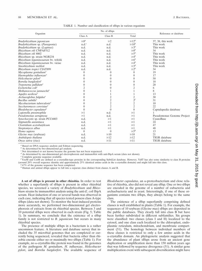
TABLE 1. Number and classification of sHsps in various organisms
OrganismNo. of sHsps
Reference or databaseClass A Class B Total
Bradyrhizobium japonicum $8a $2 $12b 37, 38, this workBradyrhizobium sp. (Parasponia) n.d.c n.d. $10b This workBradyrhizobium sp. (Lupinus) n.d. n.d. $3b This workRhizobium etli CNPAF512 n.d. n.d. $8b 34Rhizobium etli 8002 n.d. n.d. $5b This workRhizobium sp. strain NGR234 n.d. n.d. $4b This workRhizobium leguminosarum bv. trifolii n.d. n.d. $6b This workRhizobium leguminosarum bv. viciae n.d. n.d. $4b This workSinorhizobium meliloti n.d. n.d. $3d This workRhizobium tropici CIAT899 n.d. n.d. $2b 34Mycoplasma genitaliume 0 0 0 19Haemophilus influenzaee 0 0 0 17Helicobacter pylorie 0 0 0 50Borrelia burgdorferie 0 0 0 18Treponema pallidume 0 0 0 20Escherichia colie 2 0 2 6Methanococcus jannaschiie 0 1 1 8Aquifex aeolicuse 0 1 1 13Archaeoglobus fulgiduse 0 2 2 30Bacillus subtilise 0 2 2f 31Mycobacterium tuberculosise 0 2 2 10Saccharomyces cerevisiaee 0 2 2 22Rhodobacter capsulatusg 0 0 0 Capsulapedia databaseLegionella pneumophila $1 n.d. $1 1Pseudomonas aeruginosa $1 n.d. $1 Pseudomonas Genome ProjectSynechocystis sp. strain PCC6803 n.d. $1 $1 CyanoBaseStigmatella aurantiaca n.d. $1 $1 24Clostridium acetobutylicum n.d. $1 $1 47Streptomyces albus n.d. $1 $1 48Homo sapiens 0 0 $5h 13Glycine max (soybean) n.d. $10 $10 21Arabidopsis thaliana n.d. $12 $12 TIGR databaseOryza sativa (rice) n.d. $11 $11 TIGR database
a Based on DNA sequence analysis and Edman sequencing.b As determined by two-dimensional gel analysis.c Not determined or not known because the genome has not been sequenced.d As determined by one-dimensional gel electrophoresis and immunoblot with anti-IbpA serum (data not shown).e Complete genome sequence available.f YocM and CotM are defined as a-crystallin-type proteins in the corresponding SubtiList database. However, YdfT has also some similarity to class B proteins
(around 20% overall sequence identity and approximately 25% identical amino acids in the a-crystallin domain) and might fall into this class.g 67% of the genome sequence has been completed.h Human and animal sHsps appear to fall into a separate class distinct from classes A and B.
88 MUNCHBACH ET AL. J. BACTERIOL.
on May 31, 2015 by guest
http://jb.asm.org/
Dow
nloaded from

occurred in B. japonicum, giving rise to the unusual broadspectrum of bacterial sHsps. The localization of six B. japoni-cum genes (hspA to hspE) encoding sHsps in a heat shock genecluster probably supports this assumption. Five human sHspshave been described (14). The ongoing genome sequencingprojects will reveal whether sHsp superfamilies are common inmammals.
It is unclear why the rhizobia analyzed in this work containmultiple sHsps whereas most other organisms do not. Therelative abundance of rhizobial sHsps after a heat shock cer-tainly implies an important cellular function offering an advan-tage in their natural environment. Short periods of intensesunlight, for example, might cause protein damage. Whenchaperones become temporarily overloaded with potentialsubstrates, sHsps might play an important role as buffer forotherwise aggregation-prone enzymes. In agreement with arecent model (15, 32), one can imagine that this reservoir offolding-competent proteins will later be refolded by the cellu-lar chaperone machineries under conditions when their capac-ity becomes available again. A tropical Rhizobium strain that isadapted to high temperatures apparently does not require mul-tiple sHsps because it contains only two small heat-inducibleproteins (34). The reason for the heat tolerance of this strain isunknown. Bacteria which thrive as mammalian pathogens livein an environment with more or less constant temperaturesand may be able to cope without a sophisticated heat shockresponse. Their lifestyle is reflected by a comparatively smallnumber of sHsp genes in their genome.
ACKNOWLEDGMENTS
We are grateful to Hauke Hennecke and Peter James for generoussupport, continuous interest in our work, and helpful comments on themanuscript. Hans-Martin Fischer and Evelyne Bauer are acknowl-edged for providing B. japonicum extracts. Rhizobium strains wereobtained from Michael Gottfert. We thank Alan Easton for the gen-erous gift of antisera and Wolfgang Weiglhofer for performing theexperiment whose results are shown in Fig. 3A.
This study was supported by grants from the Swiss National Foun-dation for Scientific Research and the Swiss Federal Institute of Tech-nology, Zurich.
REFERENCES
1. Abu Kwaik, Y., and N. C. Engleberg. 1994. Cloning and molecular charac-terization of a Legionella pneumophila gene induced by intracellular infectionand by various in vitro stress conditions. Mol. Microbiol. 13:243–251.
2. Allen, S. P., J. O. Polazzi, J. K. Gierse, and A. M. Easton. 1992. Two novelheat shock genes encoding proteins produced in response to heterologousprotein expression in Escherichia coli. J. Bacteriol. 174:6938–6947.
3. Arrigo, A. P., and J. Landry. 1994. Expression and function of the low-molecular-weight heat shock proteins, p. 335–373. In R. I. Morimoto, A.Tissieres, and C. Georgopoulos (ed.), The biology of heat shock proteins andmolecular chaperones. Cold Spring Harbor Laboratory Press, Cold SpringHarbor, N.Y.
4. Babst, M., H. Hennecke, and H. M. Fischer. 1996. Two different mechanismsare involved in the heat-shock regulation of chaperonin gene expression inBradyrhizobium japonicum. Mol. Microbiol. 19:827–839.
5. Beringer, J. E. 1974. R factor transfer in Rhizobium leguminosarum. J. Gen.Microbiol. 84:188–198.
6. Blattner, F. R., G. Plunkett, C. A. Bloch, N. T. Perna, V. Burland, et al. 1997.The complete genome sequence of Escherichia coli K-12. Science 277:1453–1474.
7. Bukau, B., and A. L. Horwich. 1998. The Hsp70 and Hsp60 chaperonemachines. Cell 92:351–366.
8. Bult, C. J., O. White, G. J. Olsen, L. X. Zhou, R. D. Fleischmann, et al. 1996.Complete genome sequence of the methanogenic archaeon, Methanococcusjannaschii. Science 273:1058–1073.
9. Caspers, G. J., J. A. M. Leunissen, and W. W. de Jong. 1995. The expandingsmall heat-shock protein family, and structure predictions of the conserved‘a-crystallin domain’. J. Mol. Evol. 40:238–248.
10. Cole, S. T., R. Brosch, J. Parkhill, T. Garnier, C. Churcher, et al. 1998.Deciphering the biology of Mycobacterium tuberculosis from the completegenome sequence. Nature 393:537–544.
11. Czarnecka, E., W. B. Gurley, R. T. Nagao, L. A. Mosquera, and J. L. Key.
1985. DNA sequence and transcript mapping of a soybean gene encoding asmall heat shock protein. Proc. Natl. Acad. Sci. USA 82:3726–3730.
12. Daniel, R. M., and C. A. Appleby. 1972. Anaerobic-nitrate, symbiotic andaerobic growth of Rhizobium japonicum: effects on cytochrome P450, otherhaemoproteins, nitrate and nitrite reductases. Biochim. Biophys. Acta 275:347–354.
13. Deckert, G., P. V. Warren, T. Gaasterland, W. G. Young, A. L. Lenox, et al.1998. The complete genome of the hyperthermophilic bacterium Aquifexaeolicus. Nature 392:353–358.
14. De Jong, W. W., G. J. Caspers, and J. A. M. Leunissen. 1998. Genealogy ofthe a-crystallin–small heat-shock protein superfamily. Int. J. Biol. Macromol.22:151–162.
15. Ehrnsperger, M., S. Graber, M. Gaestel, and J. Buchner. 1997. Binding ofnon-native protein to Hsp25 during heat shock creates a reservoir of foldingintermediates for reactivation. EMBO J. 16:221–229.
16. Fischer, H. M., M. Babst, T. Kaspar, G. Acuna, F. Arigoni, and H. Hennecke.1993. One member of a groESL-like chaperonin multigene family in Brady-rhizobium japonicum is co-regulated with symbiotic nitrogen fixation genes.EMBO J. 12:2901–2912.
17. Fleischmann, R. D., M. D. Adams, O. White, R. A. Clayton, E. F. Kirkness,et al. 1995. Whole-genome random sequencing and assembly of Haemophilusinfluenzae Rd. Science 269:496–512.
18. Fraser, C. M., S. Casjens, W. M. Huang, G. G. Sutton, R. Clayton, et al.1997. Genomic sequence of a Lyme disease spirochaete, Borrelia burgdorferi.Nature 390:580–586.
19. Fraser, C. M., J. D. Gocayne, O. White, M. D. Adams, R. A. Clayton, et al.1995. The minimal gene complement of Mycoplasma genitalium. Science270:397–403.
20. Fraser, C. M., S. J. Norris, G. M. Weinstock, O. White, G. G. Sutton, et al.1998. Complete genome sequence of Treponema pallidum, the syphilis spi-rochete. Science 281:375–388.
21. Gaestel, M., E. Vierling, and J. Buchner. 1997. The small heat shock protein(sHSP) family: an overview, p. 269–272. In M. J. Gething (ed.), Guidebookto molecular chaperones and protein-folding catalysts. Oxford UniversityPress, Oxford, England.
22. Goffeau, A., et al. 1997. The yeast genome directory. Nature 387(Suppl.):1–107.
23. Hahn, M., and H. Hennecke. 1984. Localized mutagenesis in Rhizobiumjaponicum. Mol. Gen. Genet. 193:46–52.
24. Heidelbach, M., H. Skladny, and H. U. Schairer. 1993. Heat shock anddevelopment induce synthesis of a low-molecular-weight stress-responsiveprotein in the myxobacterium Stigmatella aurantiaca. J. Bacteriol. 175:7479–7482.
25. Henriques, A. O., B. W. Beall, and C. P. Moran. 1997. CotM of Bacillussubtilis, a member of the a-crystallin family of stress proteins, is inducedduring development and participates in spore outer coat formation. J. Bac-teriol. 179:1887–1897.
26. Ingolia, T. D., and E. A. Craig. 1982. Four small Drosophila heat shockproteins are related to each other and to mammalian a-crystallin. Proc. Natl.Acad. Sci. USA 79:2360–2364.
27. Jakob, U., M. Gaestel, K. Engel, and J. Buchner. 1993. Small heat shockproteins are molecular chaperones. J. Biol. Chem. 268:1517–1520.
28. Jobin, M.-P., F. Delmas, D. Garmyn, C. Divies, and J. Guzzo. 1997. Molec-ular characterization of the gene encoding an 18-kilodalton small heat shockprotein associated with the membrane of Leuconostoc oenos. Appl. Environ.Microbiol. 63:609–614.
29. Kim, K. K., R. Kim, and S. H. Kim. 1998. Crystal structure of a smallheat-shock protein. Nature 394:595–599.
30. Klenk, H. P., R. A. Clayton, J. F. Tomb, O. White, K. E. Nelson, et al. 1997.The complete genome sequence of the hyperthermophilic, sulphate-reducingarchaeon Archaeoglobus fulgidus. Nature 390:364–370.
31. Kunst, F., N. Ogasawara, I. Moszer, A. M. Albertini, G. Alloni, et al. 1997.The complete genome sequence of the Gram-positive bacterium Bacillussubtilis. Nature 390:249–256.
32. Lee, G. J., A. M. Roseman, H. R. Saibil, and E. Vierling. 1997. A small heatshock protein stably binds heat denatured model substrates and can maintaina substrate in a folding competent state. EMBO J. 16:659–671.
33. Leroux, M. R., R. Melki, B. Gordon, G. Batelier, and E. P. M. Candido. 1997.Structure-function studies on small heat shock protein oligomeric assemblyand interaction with unfolded polypeptides. J. Biol. Chem. 272:24646–24656.
34. Michiels, J., C. Verreth, and J. Vanderleyden. 1994. Effects of temperaturestress on bean-nodulating Rhizobium strains. Appl. Env. Microbiol. 60:1206–1212.
35. Miller, J. H. 1972. Experiments in molecular genetics. Cold Spring HarborLaboratory Press, Cold Spring Harbor, N.Y.
36. Muchowski, P. J., and J. I. Clark. 1998. ATP-enhanced molecular chaperonefunctions of the small heat shock protein human aB crystallin. Proc. Natl.Acad. Sci. USA 95:1004–1009.
36a.Munchbach, M., and H. M. Fischer. Unpublished results.37. Narberhaus, F., R. Kaser, A. Nocker, and H. Hennecke. 1998. A novel DNA
element that controls bacterial heat shock gene expression. Mol. Microbiol.28:315–323.
VOL. 181, 1999 RHIZOBIAL SMALL HEAT SHOCK PROTEINS 89
on May 31, 2015 by guest
http://jb.asm.org/
Dow
nloaded from

38. Narberhaus, F., W. Weiglhofer, H.-M. Fischer, and H. Hennecke. 1996. TheBradyrhizobium japonicum rpoH1 gene encoding a s32-like protein is part ofa unique heat shock gene cluster together with groESL1 and three small heatshock genes. J. Bacteriol. 178:5337–5346.
39. Narberhaus, F., W. Weiglhofer, H. M. Fischer, and H. Hennecke. 1998.Identification of the Bradyrhizobium japonicum degP gene as part of anoperon containing small heat-shock protein genes. Arch. Microbiol. 169:89–97.
40. Netzer, W. J., and F. U. Hartl. 1998. Protein folding in the cytosol: chap-eronin-dependent and -independent mechanisms. Trends Biochem. Sci. 23:68–73.
41. Pich, A., F. Narberhaus, and H. Bahl. 1990. Induction of heat shock proteinsduring initiation of solvent formation in Clostridium acetobutylicum. Appl.Microbiol. Biotechnol. 33:697–704.
42. Quadroni, M., W. Staudenmann, M. Kertesz, and P. James. 1996. Analysisof global responses by protein and peptide fingerprinting of proteins isolatedby two-dimensional gel electrophoresis: application to the sulfate-starvationresponse of Escherichia coli. Eur. J. Biochem. 239:773–781.
43. Regensburger, B., and H. Hennecke. 1983. RNA polymerase from Rhizobiumjaponicum. Arch. Microbiol. 135:103–109.
44. Roy, S. K., and H. Nakamoto. 1998. Cloning, characterization, and transcrip-tional analysis of a gene encoding an a-crystallin-related, small heat shock
protein from the thermophilic cyanobacterium Synechococcus vulcanus. J.Bacteriol. 180:3997–4001.
45. Sambrook, J., E. F. Fritsch, and T. Maniatis. 1989. Molecular cloning: alaboratory manual, 2nd ed. Cold Spring Harbor Laboratory Press, ColdSpring Harbor, N.Y.
46. Sanger, F., S. Nicklen, and A. R. Coulson. 1977. DNA sequencing withchain-terminating inhibitors. Proc. Natl. Acad. Sci. USA 74:5463–5467.
47. Sauer, U., and P. Durre. 1993. Sequence and molecular characterization ofa DNA region encoding a small heat shock protein of Clostridium acetobu-tylicum. J. Bacteriol. 175:3394–3400.
48. Servant, P., and P. Mazodier. 1995. Characterization of Streptomyces albus18-kilodalton heat shock-responsive protein. J. Bacteriol. 177:2998–3003.
49. Servant, P., and P. Mazodier. 1996. Heat induction of hsp18 gene expressionin Streptomyces albus G: transcriptional and posttranscriptional regulation. J.Bacteriol. 178:7031–7036.
50. Tomb, J. F., O. White, A. R. Kerlavage, R. A. Clayton, G. G. Sutton, et al.1997. The complete genome sequence of the gastric pathogen Helicobacterpylori. Nature 388:539–547.
51. Waters, E. R., G. J. Lee, and E. Vierling. 1996. Evolution, structure andfunction of the small heat shock proteins in plants. J. Exp. Bot. 47:325–338.
52. Yuan, Y., D. D. Crane, and C. E. Barry. 1996. Stationary phase-associatedprotein expression in Mycobacterium tuberculosis: function of the mycobac-terial a-crystallin homolog. J. Bacteriol. 178:4484–4492.
90 MUNCHBACH ET AL. J. BACTERIOL.
on May 31, 2015 by guest
http://jb.asm.org/
Dow
nloaded from