association of 70-kilodalton heat-shock cognate proteins with acclimation to cold
TRANSCRIPT
Plant Physiol. (1992) 99, 1362-13690032-0889/92/99/1 362/08/$01 .00/0
Received for publication December 31, 1991Accepted March 7, 1992
Association of 70-Kilodalton Heat-Shock Cognate Proteinswith Acclimation to Cold'
Lisa G. Neven, Dale W. Haskell, Charles L. Guy*, Nancy Denslow, Paul A. Klein, Linda G. Green, andAllison Silverman
Department of Environmental Horticulture, Institute of Food and Agricultural Sciences, University of Florida,Gainesville, Florida 32611 (L.G.N., D.W.H., C.L.G., A.S.); Department of Biochemistry and Molecular Biology,University of Florida, Gainesville, Florida 32610 (N.D.); and Department of Pathology and Laboratory Medicine,
University of Florida, Gainesville, Florida 32610 (P.A.K., L.G.G.)
ABSTRACT
Exposure of young spinach seedlings (Spinacia oleracea L. cvBloomsdale) to 5 C leads to an increase in the synthesis of several79-kilodalton proteins that are present in leaf tissue grown at 200 C.Protein sequence analyses and immunological cross-reactivity in-dicate that this group of proteins belongs to the 70-kilodalton heat-shock family. Steady-state transcript levels and protein synthesisare increased two- to threefold within 1 day, but immunoblotanalyses suggest that the steady-state concentration of this proteingroup in leaf tissue only gradually accumulates at low temperature.It is proposed that the increased synthesis of several members ofthe 70-kilodalton heat-shock family could result from an influenceof low temperature on protein folding and/or assembly processes.
Plants are poikilotherms and must be able to adjust tochanges in ambient temperature to ensure survival. In keep-ing with this need to adjust to a changing environment, polar,temperate, and alpine perennial plants follow a cyclicalrhythm of growth and dormancy. In these plants, the cessa-
tion of vegetative growth and onset of dormancy is a prereq-uisite for the induction of tolerance to the stresses imposedby freezing (35). Collectively, the set of biochemical andphysiological events that augments a plant's tolerance to lowtemperature stress is termed cold acclimation (35). In hardyplants, cold acclimation performs two major functions: (a)the adjustment of cellular metabolism and function to thekinetic and biophysical constraints imposed by reduced tem-perature, and (b) the induction of freezing tolerance (11).At low nonfreezing temperatures that promote cold accli-
mation, many plant species exhibit subtle changes in gene
expression (11, 15) that appear to be different from theresponses induced by other types of environmental stresses(30). For example, unlike the heat-shock response (3), plantsexposed to low temperature continue synthesis of the vastmajority of housekeeping proteins while simultaneously in-
' Florida Agricultural Experiment Station Journal Series, No. R-02485. Financial support for this work was provided by the U.S.Department of Agriculture CSRS 88-37264-4024, the National Sci-ence Foundation DCB-9017625, the Institute of Food and Agricul-tural Sciences, and the Interdisciplinary Center for BiotechnologyResearch, University of Florida.
1362
ducing or upregulating the synthesis of selected proteins (12).The purpose of this increased expression of certain proteinsby plants in response to low temperature is not understood.Do these proteins serve in the adjustment of metabolic func-tions to kinetic restraints imposed by low nonfreezing tem-peratures, or do they play a role in freezing tolerance mech-anisms? Here we describe the identification of a group ofproteins belonging to the 70-kD heat-shock family that maybe involved with a metabolic adjustment of plants to lownonfreezing temperature during cold acclimation.
MATERIALS AND METHODS
Plant Material
Spinach seedlings (Spinacia oleracea L. cv Bloomsdale) weregrown from seed in a controlled environment as previouslydescribed (12). Cold acclimation and deacclimation treat-ments were conducted with a 12-h photoperiod as previouslydescribed (12).
Protein Purification
Protein was extracted from etiolated hypocotyl-cotyledontissue grown at 50C for 4 weeks. Frozen tissue was groundin a dry-ice-cooled mortar. Forty grams of tissue was homog-enized in a Polytron blender with 100 mL of 80% (v/v)distilled phenol buffered with 120 mm Tris-HCl (pH 6.8), 50mm EDTA, 100 mm KCI, 2% (v/v) Triton X-100, 5% (v/v)glycerol, 2% (v/v) 2-mercaptoethanol, and an additional 100mL of the preceding buffer (based on ref. 13). The totalprotein extract was centrifuged at 15,000g for 5 min. Theaqueous phase was filtered through glass wool to removecellular debris. Protein was precipitated from the phenol with5 volumes of -200C acetone with 1% (v/v) 2-mercaptoeth-anol for 2 h at -200C and pelleted at 15,000g for 10 min.The protein pellet was vacuum dried, dissolved in 5 mL ofbuffer solution (1 mi Tris-HCl, pH 8.0, 8 M urea, 8 mm 3-[(3-cholamidopropyl)dimethylammonio]-1-propanesulfonic-acid, 5% [v/v] glycerol) and centrifuged at 1 1,OOOg for 5 minto remove undissolved material. The supematant was storedat -200C. Protein content was determined by the Bradfordmethod (5).
Protein extracts containing 35 to 180 mg of protein werefractionated by free-solution-isoelectric focusing in a Rotofor
LOW TEMPERATURE AND 70-KD HEAT-SHOCK COGNATES
apparatus (Bio-Rad) in a pH gradient containing 2% ampho-lytes (0.8% pH 5-7, 0.8% pH 4-6, and 0.4% pH 3-10). Theampholyte solution was prefocused for 1 h at 12 W constantpower with cooling at 100C to establish the pH gradient.Ampholytes were added to the protein sample to make a 2%ampholyte concentration. The protein-ampholyte solutionwas loaded into the middle of the pH gradient (approximatelypH 6.5). Proteins were focused for 5 h at 12 W constantpower. Coolant at 100C was circulated through the focusingcell to minimize heat buildup. After focusing, the fractionswere collected and pH values determined. SDS-PAGE anal-yses revealed that free-solution-isoelectric focused fractionsclosely approximated analytical two-dimensional separations(12) for spinach cold acclimation proteins, which facilitatedidentification of fractions containing CAP79.2
Fractions most enriched for CAP79 were selected for fur-ther purification. The amount of CAP79 present in selectedfractions was determined by densitometry following SDS-PAGE and Coomassie blue staining. Prior to CNBr cleavage,protein was precipitated from CAP79-enriched fractions with5 volumes of -200C acetone with 1% 2-mercaptoethanol for2 h followed by centrifugation at 11,000g. Coprecipitatedurea was removed by washing with 10 mL of methanol. Aftercentrifugation, the supematant was discarded and the proteinpellet dried under vacuum. CAP79 was further purified bySDS-PAGE fractionation. Following electroblotting to PVDFand staining with Coomassie blue, the discrete CAP79 bandwas excised and the protein cleaved with CNBr in 70% formicacid (23) overnight in an Eppendorf tube. The cleavagesolution was lyophilized to remove the formic acid, and thedried residue dissolved in sterile distilled water and lyophi-lized twice before final SDS-PAGE fractionation and electro-blotting to PVDF. Gas-phase sequencing was performed onamido black-stained bands excised from the PVDF mem-brane. Polyclonal antibodies against CAP79 were preparedusing protein purified as described for sequencing with onlyslight modification. After resolution of CAP79 by preparativeSDS-PAGE, the gel was stained with Coomassie blue. Thevisualized band (about 66 ,ug) was excised and the gel slicehomogenized in PBS and injected into Balb/c mice. The finalantiserum titer was 1:6400 as determined by westem blotanalyses.
Chemiluminescent Quantitation of Protein Blots
Protein extracts were fractionated by SDS-PAGE and elec-troblotted onto PVDF membrane. Blots were probed with ananti-CAP79 mouse polyclonal antisera overnight at 40C.Antigen-antibody complexes were visualized with an alkalinephosphatase-conjugated anti-mouse second antibody in thepresence of Lumi-Phos 530 (31). Light produced from thechemiluminescence reaction, catalyzed by alkaline phospha-tase, was recorded on x-ray film. Film exposures ranged from1 to 10 min. Lumigraphs were scanned using a MolecularDynamics Computing Densitometer model 300. Each blot for
2Abbreviations: CAP79, cold acclimation protein 79; CNBr, cyan-ogen bromide; PCR, polymerase chain reaction; PVDF, polyvinyldi-fluoride; HSP70, 70-kD heat-shock protein; BiP, binding immuno-globulin protein.
CAP79 quantitation was separated between the 60- and 70-kD region and the upper half was used for CAP79 immu-noblots. The lower half was probed with a rabbit antiserumreactive against the large subunit of Rubisco. All determina-tions of CAP79 are standardized intemally on the sameprotein blot against the signal for Rubisco.
Amplification and Cloning of Genomic DNA Sequences
Spinach genomic DNA was purified from spinach leavesusing the method of Dellaporta et al. (7) with modifications.Synthetic primers were constructed using HSP70 sequencesfrom maize and Arabidopsis (28, 37). Genomic DNA wasamplified using Taq polymerase according to the directionsof Perkin-Elmer/Cetus. The 5' oligo TC(ACTG) TC(ACTG)CG(CT) GCT CG(CT) TTC GAG GAG and the 3' oligo(AGC)GT CAT (GA)AC (GA)CC ACC (ATG)GC (AG)GTCTC were used to amplify a discrete 350-base pair region ofgenomic DNA (spin350) that should contain the region ofthe gene encoding for the sequenced peptide. The DNA wasdenatured for 3 min at 940C and amplified during 35 cyclesof denaturation for 1 min at 940C, annealing for 2 min at500C, and extension for 3 min at 720C. The PCR productswere then incubated at 720C for 7 min to complete extensionof all products. PCR products were excised from a 2% LMPagarose (BRL) gel, solubilized in 10 mm Tris, 1 mm EDTA,and 0.5% SDS at 650C for 20 min, purified by phenol/chloroform extraction and ethanol precipitation, and sub-jected to an additional round of 25 cycles of PCR amplifica-tion. After gel purification, PCR products were made bluntended with T4 DNA polymerase and phosphorylated withT4 polynucleotide kinase (24). After phenol extraction andethanol precipitation, the PCR products were cloned intoEcoRV-digested Bluescript plasmid (Stratagene). Templateswere purified and sequenced by the automated dideoxynu-cleotide method (DuPont).
RNA Purification and Northern AnalysisRNA was extracted from spinach leaves using the phenol
extraction LiCl precipitation method (1). Poly(A+) RNA waspurified using oligo(dT)-cellulose for northern blot analysesas described (24). Equal amounts of poly(A+) RNA from eachtime point of the cold-acclimation regimen were separatedon a 1.2% formaldehyde agarose gel (24). The gel was stainedwith ethidium bromide, photographed, and pressure blottedonto Hybond-N (Amersham) nylon membrane. The RNAwas fixed and hybridized to a random-primed [a-32P]dCTP-labeled spin350 probe (24). RNA blots were stripped andstandardized against the message for actin using an Arabi-dopsis cDNA. For RNA abundance determinations, laser den-sitometric analyses of blots using the spin350 probe werenormalized as the ratio of the signal with that obtained usingactin. Actin expression appears to be invariant at the temper-atures and durations used in this study.
RESULTS
Isolation and Identification of CAP79 Proteins
Early studies involving two-dimensional gel analyses of invivo labeled proteins in spinach seedlings undergoing cold
1363
Plant Physiol. Vol. 99, 1992
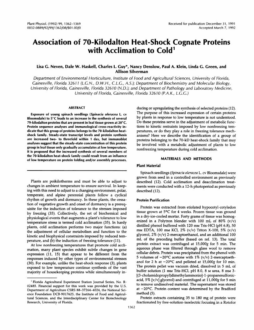
acclimation at 50C revealed about a threefold increase in thesynthesis of CAP79 with an isoelectric point of 5.3 (12).Using preparative free-solution-isoelectric focusing and SDSgel electrophoresis, we were able to prepare adequateamounts of pure CAP79 from cold-acclimated hypocotyltissue for use in protein sequencing and antibody production.Increased synthesis of CAP79 upon exposure to 50C wasconfirmed by SDS gel analysis of immunoprecipitated proteinfrom nondenatured cell lysates prepared from leaf tissuesthat were pulse-labeled with [35S]methionine (Fig. 1). Greaterradiolabel incorporation in CAP79 was observed during thefirst 2 d of exposure to 50C, but by 7 d the level of incorpo-ration returned to that seen in plants kept at 200C. Thedecline in synthesis after the initial increase at low tempera-ture contrasts with previous in vivo labeling data, whichshowed peak synthesis at 7 d (12). This discrepancy couldresult from differences in experimental design: (a) two-di-mensional PAGE analyses of total in vivo labeled products
20(i
li h
9'
66
A
.si
...... ,_~~~~~~~~~~~~~~~~~~if
.0Oqm.u
B
C
A A AA A
Figure 1. Immunoprecipitation of in vivo labeled CAP79. Leaf tissuewas radiolabeled (12) with [35S]methionine for 24 h, and cell lysateswere prepared in 50 mm Mops, pH 7.5, 10 mM MgCI2, 2.5 mm DTT,1 mM EDTA, 1 mM methionine, and 0.05% Triton X-100. CAP79was immunoprecipitated from 1 O6 TCA-insoluble counts with 10 ALanti-CAP79 antiserum. Immune complexes were dissolved and theproteins resolved by SDS-PAGE and visualized by fluorography. A,Lane 1, nonimmune serum; lane 2, nonacclimated; lane 3, cold-acclimated 1 d; lane 4, cold-acclimated 2 d; lane 5, cold-acclimated7 d; lane 6, deacclimated 1 d; lane 7, deacclimated 7 d. Arrowshows the CAP79 band. B and C, Two-dimensional gel analyses ofimmunoprecipitates from nonacclimated and 1-d cold-acclimatedtissue. The panels are oriented with the acidic end on the left. Thearrowheads in C denote the five isoforms.
versus immunoprecipitation of a specific labeled product and/or (b) variation in the culture and age of the plants. Theantibody used in the immunoprecipitation reactions was pre-pared against denatured CAP79. It is fortunate that thisantibody immunoprecipates native CAP79, but its efficiencyin this reaction and its ability to recognize all members of theCAP79 complex remains uncharacterized. Also, in the pre-vious report (12), plants were younger and grown in vitro,but in this study the plants were older and grown ex vitro ina soil medium. Differences in age and cultural conditionscould contribute to the disparity with the previous work (12).Two-dimensional gel analyses of CAP79 immunoprecipitatesof in vivo radiolabeled proteins detected the synthesisof three isoforms in leaves of plants grown at 200C (Fig.1B), and five isoforms from leaves exposed to 5oC for 1 d(Fig. 1C).Attempts at Edman gas-phase sequencing from the NH2
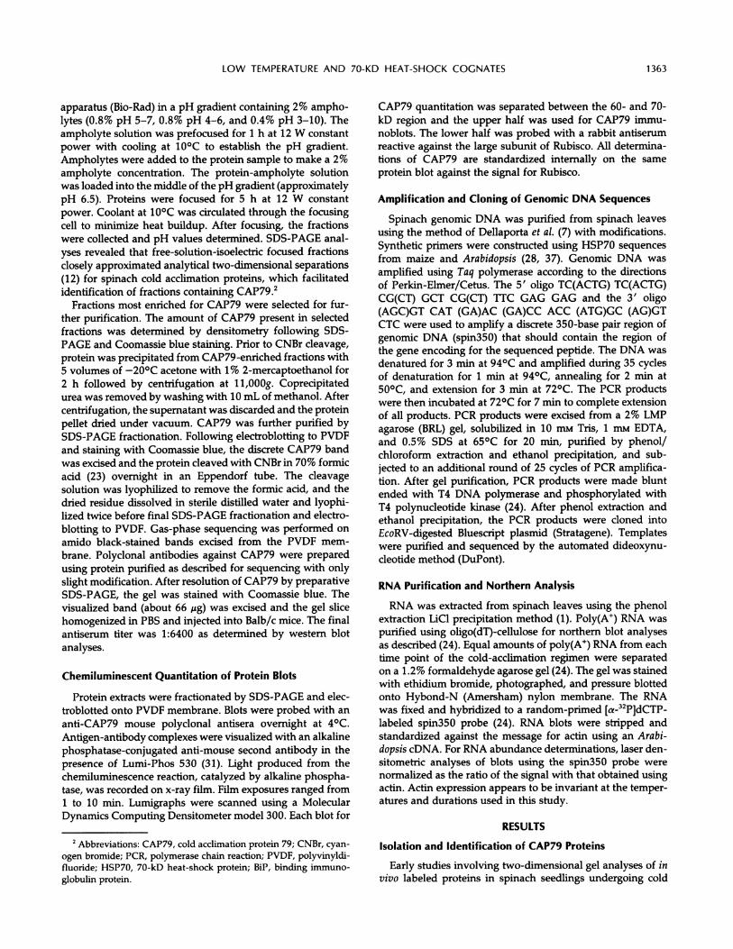
terminus of CAP79 purified from analytical two-dimensionalgel electroblots failed because of N-terminal blockage. Com-positional analyses of small amounts of the purified proteinfrom the two-dimensional gel electroblots revealed the pres-ence of 18 methionine residues (14). Partial cleavage ofpurified CAP79 by CNBr produced two large peptides thatwere resolved from smaller peptides by SDS-PAGE (Fig. 2A).Gas-phase sequencing demonstrated that these two peptideshad identical NH2 termini. Homology searches of the Na-tional Biomedical Research Foundation and GenBank data-bases revealed CAP79's relationship to HSP70s (Fig. 2B). Thegreatest similarity was found with a Petunia HSP70 where31 of 32 residues were identical to CAP79/30 (36). Thesequence homology of the two CAP79 CNBr fragments witha previously described maize HSP70 sequence begins atmethionine residue 330 and runs through residue 362 (28).This region is a highly conserved domain in HSP70s (26) anddefines the high degree of homology of CAP79 within thisheat-shock family. Because the purified CAP79 is composedof several isoforms, it is possible that both CNBr peptideswere products from the same polypeptide or from differentisoforms. The generation of 30- and 40-kD partial CNBrdigest peptides having the same NH2 terminus and identicalamino acid sequence can be explained by a 10-kD truncationof the carboxyl terminus at one of several methionine targetsites (28). Generation of a 40-kD CNBr peptide beginning atresidue 330 suggests a molecular mass greater than 70 kD inkeeping with our estimated size of 79 kD for CAP79. HSP70sgenerally vary from 633 to 666 residues in length (26). IfCAP79 were a classical heat-inducible HSP70, the longestexpected fragment beginning at residue 330 and extendingto the carboxyl terminus would be about 320 residues, orroughly 35 kD. To account for the extra 5 kD of the 40-kDfragment would require roughly an extra 45 residues, extend-ing the protein to about 700 residues in length. This wouldsuggest a protein strikingly similar in length to an Arabidopsisheat-shock cognate protein (HSC70) that was reported to be76 kD and approximately 700 residues in length (37).CAP79 is not a typical heat-inducible HSP70 (18, 20),
because it is present in nonheat-shocked and cold-acclimatedtissues, and its synthesis is not strongly responsive to heat-shock treatment (12). Western blot analysis, with the poly-clonal mouse antiserum raised against the purified CAP79
1364 NEVEN ET AL.
LOW TEMPERATURE AND 70-KD HEAT-SHOCK COGNATES
B
CAP79/40 M D K S T V H D V V L V G G S T X I P K V QO L LCAP79/30 M D K S T V H D V V L V G G S T R I P K V 0 0 L L - Q D F F N G ? ? L
PETUNIA HSP70MAIZE HSP70TOMATO HSC70DROSOPHILA HSP70YEAST SSB1MAIZE b-70YEAST BiPRAT BiPE. COLI DnaK
. . . s
. . . s
. . .0
e . . Q
e . K DK DS V . D
i .
iDi -.DiDi D
ee
E
Q m..M
* a
S .
K.
. .
K K V
M
R
T N . .,.. e . . HS . . . DD Y . D -
E S Y . DK e . . .
Ae -
1365
KE.KE.KE.KN.
KE P
KKa
KE P
KE P
Figure 2. Homology of CAP79 CNBr fragments with HSP70s. A, The partial CNBr cleavage peptides resolved by SDS-PAGE used for gas-phase amino acid sequencing. B, The homology of the CAP79 with HSP70s. The amino acid sequences are indicated by the one-letter code.Residues identical to CAP79 sequences are marked by dots, homologous substitutions by lowercase letters, and nonhomologous substitutionsby uppercase letters. The HSP70 sequences compared were maize b-70 (9), Petunia (36), maize (28), tomato (26), Drosophila (19), yeast SSB1(32), yeast BiP (27), rat BiP (25), and E. coli DnaK (2).
and a rat monoclonal antibody (7.10, produced against a
Drosophila HSP70 that cross-reacts with a wide range of heat-shock cognates of various organisms) (34), provides definitiveevidence to support the assignment of spinach CAP79 as a
member of the HSP70 family. Both antibodies identified a
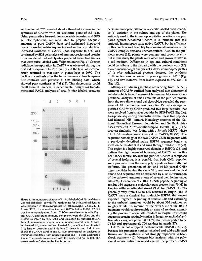
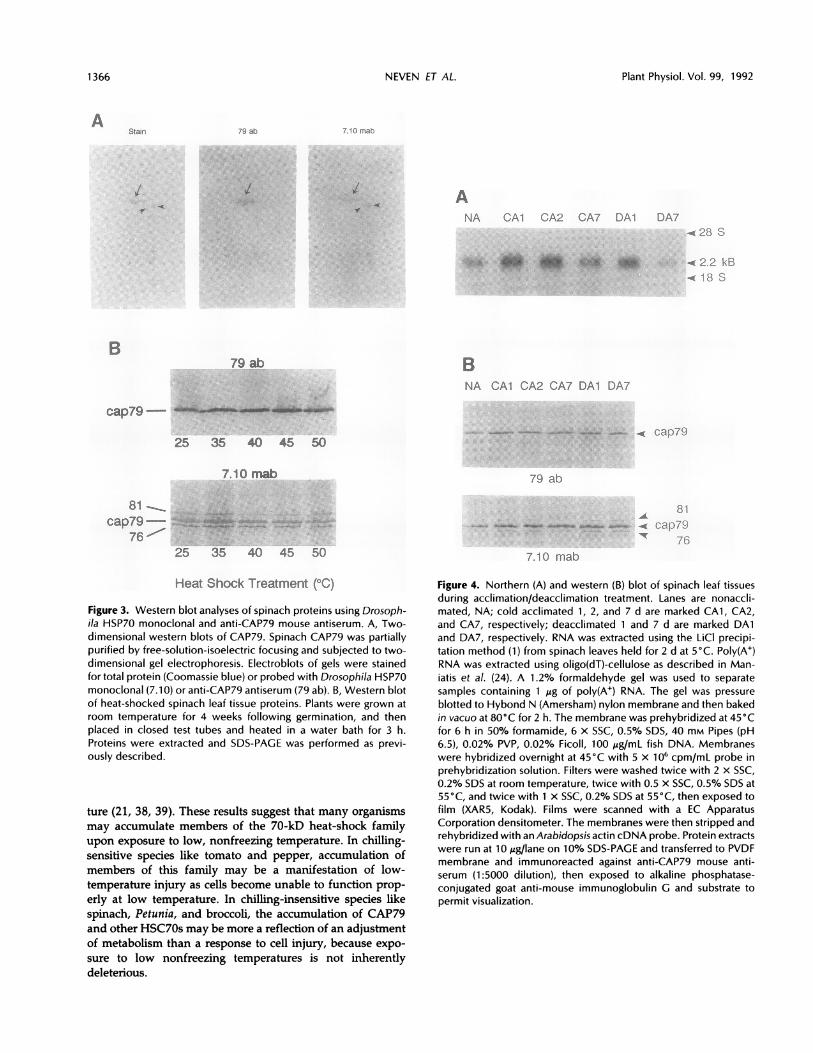
family of proteins of between 70 and 80 kD on western blotsof two-dimensional gels (Fig. 3A). Close inspection of theCAP79 reactivity on these blots suggests it represents a
closely migrating constellation of HSC70s. The Drosophilamonoclonal antibody also shows reactivity with two addi-tional proteins of 76 kD that the CAP79 antiserum does not.Experiments were conducted to determine whether theCAP79 complex was responsive to heat shock. When spinachleaf tissue was heat shocked for 3 h over a temperature rangeknown to elicit the heat-shock response (33), only moderatechanges were observed in the concentration of the CAP79band, as judged by immunoblotting (Fig. 3B). The 1.5-foldincrease at 350C and virtually no change at higher tempera-tures agrees with previous conclusions (12) that this complexis not strongly induced by heat shock. The moderate increaseat 350C in CAP79 abundance may result from cross-reactivityto unique heat shock-induced isoforms, or by the heat shock-induced synthesis of the constitutive CAP79 isoforms.Further two-dimensional analyses should resolve thisuncertainty.
Analysis of CAP79 during Cold Acclimation
Synthetic oligonucleotides were prepared using DNA se-
quence information from HSP70 sequences of maize andArabidopsis (28, 37) that would likely bracket the region ofthe gene(s) corresponding to the known protein sequence(Fig. 2B). These oligonucleotides were used to amplify spin-ach genomic DNA by PCR. Of the three amplified productsobtained from this procedure, a 350-base pair fragment(spin350) was determined through sequence analysis to be-long to an HSP70 gene. Although it is not identical to any
previously reported HSP70 gene, spin350 was most similarto b-70 (9) and showed homology to other BiPs (25, 27, 29).This fragment was found to have homology with the proteinsequence determined for CAP79, but it also was not identical.Northern blot analyses of poly(A+) RNA isolated from 2-d
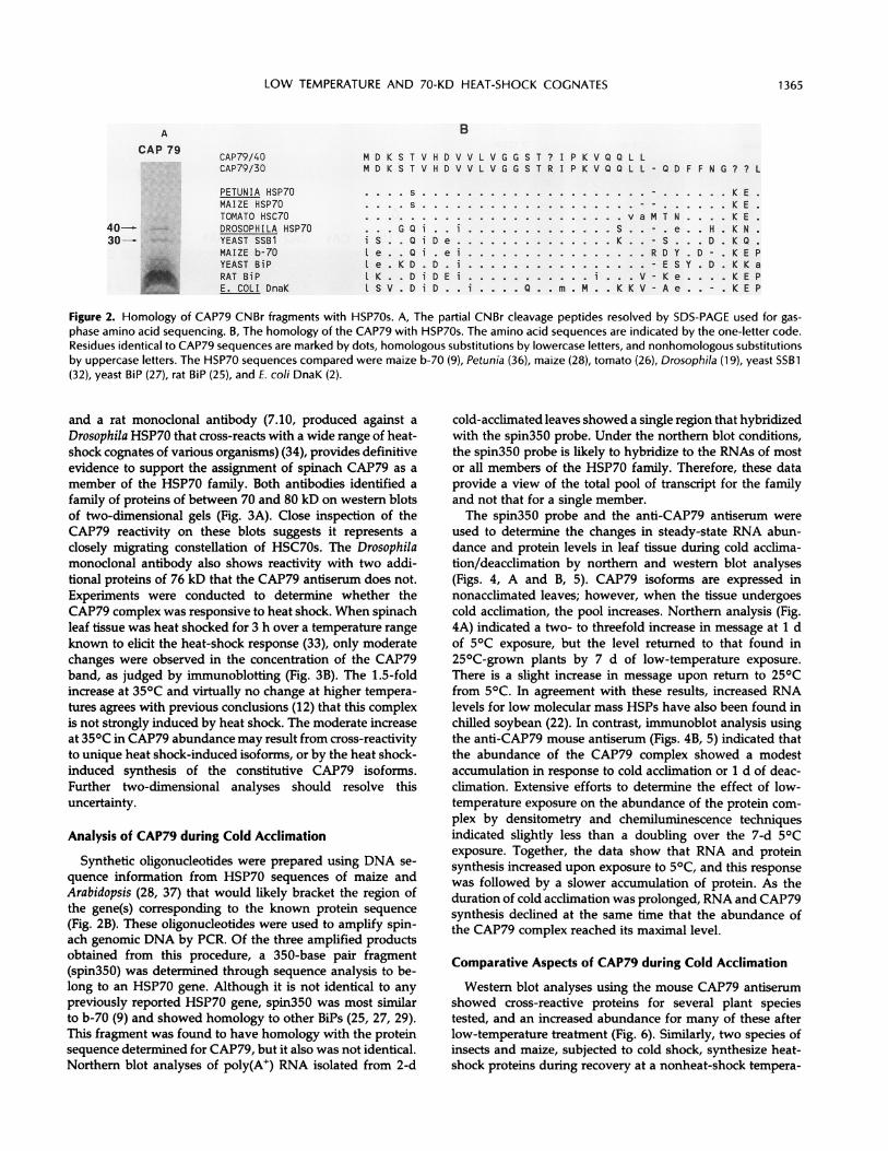
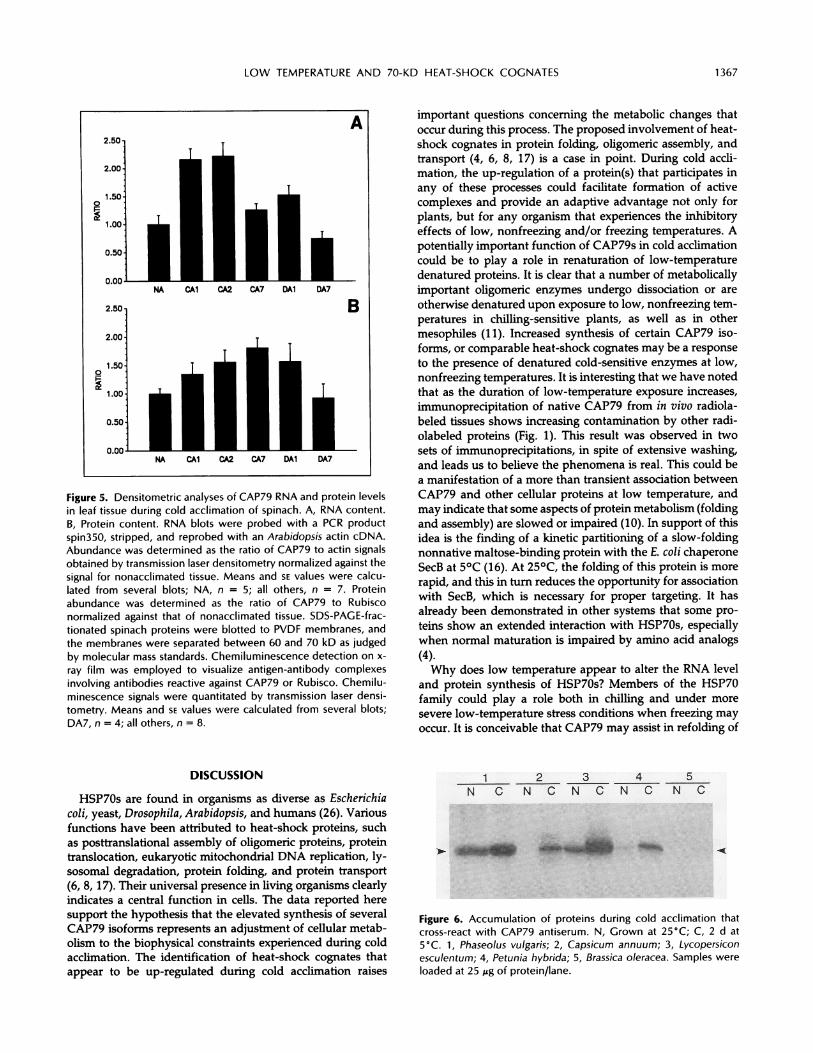
cold-acclimated leaves showed a single region that hybridizedwith the spin350 probe. Under the northern blot conditions,the spin350 probe is likely to hybridize to the RNAs of mostor all members of the HSP70 family. Therefore, these dataprovide a view of the total pool of transcript for the familyand not that for a single member.The spin350 probe and the anti-CAP79 antiserum were
used to determine the changes in steady-state RNA abun-dance and protein levels in leaf tissue during cold acclima-tion/deacclimation by northern and western blot analyses(Figs. 4, A and B, 5). CAP79 isoforms are expressed innonacclimated leaves; however, when the tissue undergoescold acclimation, the pool increases. Northern analysis (Fig.4A) indicated a two- to threefold increase in message at 1 dof 50C exposure, but the level returned to that found in25°C-grown plants by 7 d of low-temperature exposure.There is a slight increase in message upon return to 250Cfrom 50C. In agreement with these results, increased RNAlevels for low molecular mass HSPs have also been found inchilled soybean (22). In contrast, immunoblot analysis usingthe anti-CAP79 mouse antiserum (Figs. 4B, 5) indicated thatthe abundance of the CAP79 complex showed a modestaccumulation in response to cold acclimation or 1 d of deac-cimation. Extensive efforts to determine the effect of low-temperature exposure on the abundance of the protein com-
plex by densitometry and chemiluminescence techniquesindicated slightly less than a doubling over the 7-d 50Cexposure. Together, the data show that RNA and proteinsynthesis increased upon exposure to 50C, and this response
was followed by a slower accumulation of protein. As theduration of cold acclimation was prolonged, RNA and CAP79synthesis declined at the same time that the abundance ofthe CAP79 complex reached its maximal level.
Comparative Aspects of CAP79 during Cold Acclimation
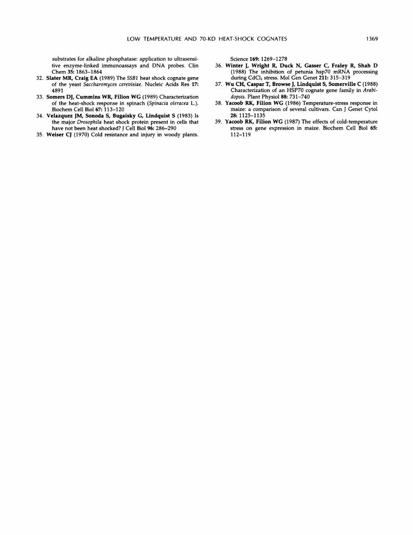
Western blot analyses using the mouse CAP79 antiserumshowed cross-reactive proteins for several plant speciestested, and an increased abundance for many of these afterlow-temperature treatment (Fig. 6). Similarly, two species ofinsects and maize, subjected to cold shock, synthesize heat-shock proteins during recovery at a nonheat-shock tempera-
ACAP 79
iLLII
i
iii
i
Plant Physiol. Vol. 99, 1992
ANA CAI CA2 GA7DAi ui
79 ab BNA CAl CA2 CA7 DAI [DA47
25 35 40 45 50
7.10 mab
81 -,
cap79-76
25 35 40 45
79 ab
1w
7110 mab50
Heat Shock Treatment ("C)
Figure 3. Western blot analyses of spinach proteins using Drosoph-i/a HSP70 monoclonal and anti-CAP79 mouse antiserum. A, Two-dimensional western blots of CAP79. Spinach CAP79 was partiallypurified by free-solution-isoelectric focusing and subjected to two-dimensional gel electrophoresis. Electroblots of gels were stainedfor total protein (Coomassie blue) or probed with Drosophila HSP70monoclonal (7.10) or anti-CAP79 antiserum (79 ab). B, Western blotof heat-shocked spinach leaf tissue proteins. Plants were grown atroom temperature for 4 weeks following germination, and thenplaced in closed test tubes and heated in a water bath for 3 h.Proteins were extracted and SDS-PAGE was performed as previ-ously described.
ture (21, 38, 39). These results suggest that many organismsmay accumulate members of the 70-kD heat-shock familyupon exposure to low, nonfreezing temperature. In chilling-sensitive species like tomato and pepper, accumulation ofmembers of this family may be a manifestation of low-temperature injury as cells become unable to function prop-
erly at low temperature. In chilling-insensitive species likespinach, Petunia, and broccoli, the accumulation of CAP79and other HSC70s may be more a reflection of an adjustmentof metabolism than a response to cell injury, because expo-
sure to low nonfreezing temperatures is not inherentlydeleterious.
Figure 4. Northern (A) and western (B) blot of spinach leaf tissuesduring acclimation/deacclimation treatment. Lanes are nonaccli-mated, NA; cold acclimated 1, 2, and 7 d are marked CA1, CA2,and CA7, respectively; deacclimated 1 and 7 d are marked DA1and DA7, respectively. RNA was extracted using the LiCI precipi-tation method (1) from spinach leaves held for 2 d at 5°C. Poly(A+)RNA was extracted using oligo(dT)-cellulose as described in Man-iatis et al. (24). A 1.2% formaldehyde gel was used to separatesamples containing 1 gg of poly(A+) RNA. The gel was pressure
blotted to Hybond N (Amersham) nylon membrane and then bakedin vacuo at 80°C for 2 h. The membrane was prehybridized at 45°Cfor 6 h in 50% formamide, 6 x SSC, 0.5% SDS, 40 mm Pipes (pH6.5), 0.02% PVP, 0.02% Ficoll, 100 uig/mL fish DNA. Membraneswere hybridized overnight at 45°C with 5 x 10⁶ cpm/mL probe inprehybridization solution. Filters were washed twice with 2 x SSC,0.2% SDS at room temperature, twice with 0.5 X SSC, 0.5% SDS at55°C, and twice with 1 x SSC, 0.2% SDS at 55°C, then exposed tofilm (XAR5, Kodak). Films were scanned with a EC ApparatusCorporation densitometer. The membranes were then stripped andrehybridized with an Arabidopsis actin cDNA probe. Protein extractswere run at 10 jg/lane on 10% SDS-PAGE and transferred to PVDFmembrane and immunoreacted against anti-CAP79 mouse anti-serum (1:5000 dilution), then exposed to alkaline phosphatase-conjugated goat anti-mouse immunoglobulin G and substrate topermit visualization.
A',italF
l I/f
B
cap79- alilsliu
1366 NEVEN ET AL.
LOW TEMPERATURE AND 70-KD HEAT-SHOCK COGNATES
A
00
0.00-
NA CAl CA2 CA7 DAl DA7
2.50
2.00-
1.500
U.UUNA CAI CA2 CA7 DAl DA7
Figure 5. Densitometric analyses of CAP79 RNA and protein levelsin leaf tissue during cold acclimation of spinach. A, RNA content.B, Protein content. RNA blots were probed with a PCR productspin350, stripped, and reprobed with an Arabidopsis actin cDNA.Abundance was determined as the ratio of CAP79 to actin signalsobtained by transmission laser densitometry normalized against thesignal for nonacclimated tissue. Means and SE values were calcu-lated from several blots; NA, n = 5; all others, n = 7. Proteinabundance was determined as the ratio of CAP79 to Rubisconormalized against that of nonacclimated tissue. SDS-PAGE-frac-tionated spinach proteins were blotted to PVDF membranes, andthe membranes were separated between 60 and 70 kD as judgedby molecular mass standards. Chemiluminescence detection on x-
ray film was employed to visualize antigen-antibody complexesinvolving antibodies reactive against CAP79 or Rubisco. Chemilu-minescence signals were quantitated by transmission laser densi-tometry. Means and SE values were calculated from several blots;DA7, n = 4; all others, n = 8.
DISCUSSION
HSP70s are found in organisms as diverse as Escherichiacoli, yeast, Drosophila, Arabidopsis, and humans (26). Variousfunctions have been attributed to heat-shock proteins, suchas posttranslational assembly of oligomeric proteins, proteintranslocation, eukaryotic mitochondrial DNA replication, ly-sosomal degradation, protein folding, and protein transport(6, 8, 17). Their universal presence in living organisms clearlyindicates a central function in cells. The data reported heresupport the hypothesis that the elevated synthesis of severalCAP79 isoforms represents an adjustment of cellular metab-olism to the biophysical constraints experienced during coldacclimation. The identification of heat-shock cognates thatappear to be up-regulated during cold acclimation raises
B
important questions concerning the metabolic changes thatoccur during this process. The proposed involvement of heat-shock cognates in protein folding, oligomeric assembly, andtransport (4, 6, 8, 17) is a case in point. During cold accli-mation, the up-regulation of a protein(s) that participates inany of these processes could facilitate formation of activecomplexes and provide an adaptive advantage not only forplants, but for any organism that experiences the inhibitoryeffects of low, nonfreezing and/or freezing temperatures. Apotentially important function of CAP79s in cold acclimationcould be to play a role in renaturation of low-temperaturedenatured proteins. It is clear that a number of metabolicallyimportant oligomeric enzymes undergo dissociation or areotherwise denatured upon exposure to low, nonfreezing tem-peratures in chilling-sensitive plants, as well as in othermesophiles (11). Increased synthesis of certain CAP79 iso-forms, or comparable heat-shock cognates may be a responseto the presence of denatured cold-sensitive enzymes at low,nonfreezing temperatures. It is interesting that we have notedthat as the duration of low-temperature exposure increases,immunoprecipitation of native CAP79 from in vivo radiola-beled tissues shows increasing contamination by other radi-olabeled proteins (Fig. 1). This result was observed in twosets of immunoprecipitations, in spite of extensive washing,and leads us to believe the phenomena is real. This could bea manifestation of a more than transient association betweenCAP79 and other cellular proteins at low temperature, andmay indicate that some aspects of protein metabolism (foldingand assembly) are slowed or impaired (10). In support of thisidea is the finding of a kinetic partitioning of a slow-foldingnonnative maltose-binding protein with the E. coli chaperoneSecB at 50C (16). At 250C, the folding of this protein is morerapid, and this in turn reduces the opportunity for associationwith SecB, which is necessary for proper targeting. It hasalready been demonstrated in other systems that some pro-teins show an extended interaction with HSP70s, especiallywhen normal maturation is impaired by amino acid analogs(4).Why does low temperature appear to alter the RNA level
and protein synthesis of HSP70s? Members of the HSP70family could play a role both in chilling and under moresevere low-temperature stress conditions when freezing mayoccur. It is conceivable that CAP79 may assist in refolding of
1 2 3 4 5N C N C N C N C N C
Figure 6. Accumulation of proteins during cold acclimation thatcross-react with CAP79 antiserum. N, Grown at 25°C; C, 2 d at5'C. 1, Phaseolus vulgaris; 2, Capsicum annuum; 3, Lycopersiconesculentum; 4, Petunia hybrida; 5, Brassica oleracea. Samples wereloaded at 25 ,ug of protein/lane.
1 367

Plant Physiol. Vol. 99, 1992
freeze/thaw denatured proteins following return to warmer
temperatures in the same manner as heat-shock proteinsseem to do following heat denaturation (10). In freezing-tolerant plants exposed to subzero temperatures, the extra-cellular solution freezes, causing cellular desiccation, in-creased solute concentration, ionic strength changes, and pHshifts, all of which can favor protein denaturation (11, 35).Consequently, freezing-tolerant organisms must have theability to safeguard against or minimize protein denaturationduring a freeze/thaw cycle. A mechanism that avoids aggre-
gation and favors folding and renaturation at low tempera-ture would be advantageous in restoring the proper confor-mation and function of proteins upon thawing and return tofavorable temperatures. Thus, the accumulation of HSP70sat low temperature may not only be indicative of a stresscondition but may also have adaptive value as well.A surprising finding in this work was that CAP79 protein
abundance showed smaller increases during low-temperatureexposure than expected considering the extent of the in-creases in mRNA level and protein synthesis observed in thisand previous studies (13). The reason for the disparity is notclear. One possibility is that current synthesis is small relativeto the total pool abundance. If this were the case, thenmoderate changes in synthesis might require long durationsto alter abundance of the total pool, as seems to be indicatedin Figure 5. Another explanation is that temperature affectsprotein turnover in such a fashion as to maintain a somewhatuniform abundance. Additional studies will be required toclarify this disparity.
ACKNOWLEDGMENTS
We thank B. Parten and L. Dinehart for assistance with this work,S. Lindquist for the kind gift of the Drosophila HSP70 monoclonalantibody, and R. Ferl for the Arabidopsis actin cDNA. We thank KenCline and Mike Kane for critical review of this manuscript. Thesequence of spin350 is available upon request.
LITERATURE CITED
1. Ausubel FM, Brent R, Kingston RE, Moore DD, Seidman JG,Smith JA, Struhl K (1990) Current Protocols in MolecularBiology. New York, John Wiley & Sons
2. Bardwell JC, Craig EA (1984) Major heat shock gene of Dro-sophila and Escherichia coli heat inducible dnaK gene are ho-mologous. Proc Natl Acad Sci USA 81: 848-852
3. Barnett T, Altschuler M, McDaniel CN, MascarenhasJP (1980)Heat shock induced proteins in plant cells. Dev Genet 1:331-340
4. Beckmann RP, Mizzen LA, Welch WJ (1990) Interaction of Hsp70 with newly synthesized proteins: implications for foldingand assembly. Science 248: 850-854
5. Bradford MM (1976) A rapid and sensitive method for thequantitation of microgram quantities of protein utilizing theprinciple of protein-dye binding. Anal Biochem 72: 248-254
6. Chirico WJ, Waters MG, Blobel G (1988. 70K heat shock relatedproteins stimulate protein translocation into microsomes. Na-ture 332: 805-810
7. Dellaporta SL, Wood J, Hicks JB (1983) A plant DNA minipre-paration: version II. Plant Mol Biol Rep 1: 19-21
8. Deshaies RJ, Koch BD, Werner-Washburne M, Craig EA,Schekman R (1988) A subfamily of stress proteins facilitatestranslocation of secretory and mitochondrial precursor poly-peptides. Nature 332: 800-805
9. Fontes EBP, Shank BB, Wrobel RL, Moose SP, O'Brian GR,Wurtzel ET, Boston RS (1991) Characterization of an immu-noglobulin binding protein homolog in the maize floury-2endosperm mutant. Plant Cell 3: 483-496
10. Gaitanaris GA, Papavassiliou AG, Rubock P, Silverstein SJ,Gottesman ME (1990) Renaturation of denatured X repressorrequires heat shock proteins. Cell 61: 1013-1020
11. Guy CL (1990) Cold acclimation and freezing stress tolerance:role of protein metabolism. Annu Rev Plant Physiol Plant MolBiol 41: 187-223
12. Guy CL, Haskell D (1987) Induction of freezing tolerance inspinach is associated with the synthesis of cold acclimationinduced proteins. Plant Physiol 84: 872-878
13. Guy CL, Haskell D (1988) Detection of polypeptides associatedwith the cold acclimation process. Electrophoresis 9: 787-796
14. Guy CL, Haskell D (1989) Preliminary characterization of highmolecular mass proteins associated with cold acclimation inspinach. Plant Physiol Biochem 27: 777-784
15. Guy CL, Niemi KJ, Brambl R (1985) Altered gene expressionduring cold acclimation in spinach. Proc Natl Acad Sci USA82: 3673-3677
16. Hardy SJS, Randall LL (1991) A kinetic partitioning model ofselective binding of nonnative proteins by the bacterial chap-erone SecB. Science 251: 439-443
17. Hemmingsen SM, Woolford C, van der Vies SM, Tilly K,Dennis DT, Georgopoulos CP, Hendrix RW, Ellis RJ (1988)Homologous plant and bacterial proteins chaperone oligomericprotein assembly. Nature 333: 330-334
18. Ingolia TD, Craig EA (1982) Drosophila gene related to themajor heat shock-induced gene is transcribed at normal tem-peratures and not induced by heat shock. Proc Natl Acad SciUSA 79: 525-529
19. Ingolia TD, Craig EA, McCarthy BJ (1980) Sequence of threecopies of the gene for the major Drosophila heat shock inducedprotein and their flanking regions. Cell 21: 669-679
20. Ingolia TD, Slater MR, Craig EA (1982) Saccharomyces cerevis-iae contains a complex multigene family related to the majorheat shock-inducible gene of Drosophila. Mol Cell Biol 2:1388-1398
21. Joplin KH, Yocum GD, Denlinger DL (1990) Cold shock elicitsexpression of heat shock proteins in the flesh fly, Sarcophagacrassipalpis. J Insect Physiol 36: 825-834
22. Kuznetsov VV, KimpelJA, Goekjian G, KeyJL (1987) Elementsof nonspecificity in responses of the plant genome to chillingand heat stress. Soviet Plant Physiol 34: 685-693
23. Mahboub S, Richard C, Delacourte A, Han KK (1986) Appli-cations of chemical cleavage procedures to the peptide map-ping of neurofilament triplet protein bands in sodium dodecylsulfate-polyacrylamide gel electrophoresis. Anal Biochem 154:171-182
24. Maniatis T, Fritsch EF, Sambrook J (1982) Molecular Cloning:A Laboratory Manual. Cold Spring Harbor Laboratory Press,Cold Spring Harbor, NY
25. Munro S,Pelham HRB (1986) An Hsp7O-like protein in the ER:identity with the 78 kd glucose-regulated protein and immu-noglobulin heavy chain binding protein. Cell 46: 291-300
26. Neumann D, Nover L, Parthier B, Rieger R, Scharf K-D,Wollgiehn R, zur Nieden U (1989) Heat shock and otherstress response systems of plants. Biol Zentralbl 8: 1-156
27. Normington K, Kozutsami K, Gething M-J, SambrookJ (1989)S. cerevisae encodes an essential protein homologous in se-quence and function to mammalian BiP. Cell 57: 1223-1236
28. Rochester DE, Winter JA, Shah DM (1986) The structure andexpression of maize genes encoding the major heat shockprotein, hsp7o. EMBO J 5: 451-458
29. Rose MD, Misra LM, Vogel JP (1989) KAR2, a karyotype, isthe yeast homolog of the mammalian BiP/GRP78 gene. Cell57: 1211-1221
30. Sachs MM, Ho T-HD (1986) Alteration of gene expressionduring environmental stress in plants. Annu Rev Plant Physiol37: 363-376
31. Schaap AP, Akhavan H, RomanoLJ (1989) Chemiluminescent
1368 NEVEN ET AL.
LOW TEMPERATURE AND 70-KD HEAT-SHOCK COGNATES
substrates for alkaline phosphatase: application to ultrasensi-tive enzyme-linked immunoassays and DNA probes. ClinChem 35: 1863-1864
32. Slater MR, Craig EA (1989) The SSB1 heat shock cognate gene
of the yeast Saccharomyces cerevisiae. Nucleic Acids Res 17:4891
33. Somers DJ, Cummins WR, Filion WG (1989) Characterizationof the heat-shock response in spinach (Spinacia oleracea L.).Biochem Cell Biol 67: 113-120
34. Velazquez JM, Sonoda S, Bugaisky G, Lindquist S (1983) Isthe major Drosophila heat shock protein present in cells thathave not been heat shocked? J Cell Biol 96: 286-290
35. Weiser CJ (1970) Cold resistance and injury in woody plants.
Science 169: 1269-127836. Winter J, Wright R, Duck N, Gasser C, Fraley R, Shah D
(1988) The inhibition of petunia hsp70 mRNA processingduring CdCl2 stress. Mol Gen Genet 211: 315-319
37. Wu CH, Caspar T, Browse J, Lindquist S, Somerville C (1988)Characterization of an HSP70 cognate gene family in Arabi-dopsis. Plant Physiol 88: 731-740
38. Yacoob RK, Filion WG (1986) Temperature-stress response inmaize: a comparison of several cultivars. Can J Genet Cytol28: 1125-1135
39. Yacoob RK, Filion WG (1987) The effects of cold-temperaturestress on gene expression in maize. Biochem Cell Biol 65:112-119
1369