two ccaat-box-binding transcription factors redundantly regulate early steps of the legume-rhizobia...
TRANSCRIPT

Two CCAAT-box-binding transcription factors redundantlyregulate early steps of the legume-rhizobia endosymbiosis
Tom Laloum1,2,†,‡, Ma€el Baudin1,2,†, Lisa Frances1,2, Agnes Lepage1,2, Benjamin Billault-Penneteau1,2, Marion R. Cerri1,2,§,
Federico Ariel3, Marie-Franc�oise Jardinaud1,2,4, Pascal Gamas1,2, Fernanda de Carvalho-Niebel1,2 and Andreas Niebel1,2,*1Laboratoire des Interactions Plantes-Microorganismes (LIPM), INRA, UMR441, F-31326 Castanet-Tolosan, France,2Laboratoire des Interactions Plantes-Microorganismes (LIPM), CNRS, UMR2594, F-31326 Castanet-Tolosan, France,3Centre National de la Recherche Scientifique, Institut des Sciences du V�eg�etal, F-91198 Gif-sur-Yvette Cedex, France,
and 4ENSAT, INPT-Universit�e de Toulouse, Avenue de l’Agrobiopole, Auzeville-Tolosane, 31326 Castanet-Tolosan, France
Received 10 February 2014; revised 30 April 2014; accepted 2 June 2014.
*For correspondence (e-mail [email protected]).†These authors contributed equally to this work.‡Present address: Faculty of Biology, Technion, Haifa, Israel.§Present address: Faculty of Biology, Genetics, University of Munich (LMU), D-82152 Martinsried, Germany.
SUMMARY
During endosymbiotic interactions between legume plants and nitrogen-fixing rhizobia, successful root
infection by bacteria and nodule organogenesis requires the perception and transduction of bacterial
lipo-chitooligosaccharidic signal called Nod factor (NF). NF perception in legume roots leads to the activa-
tion of an early signaling pathway and of a set of symbiotic genes which is controlled by specific early
transcription factors (TFs) including CYCLOPS/IPD3, NSP1, NSP2, ERN1 and NIN. In this study, we bring
convincing evidence that the Medicago truncatula CCAAT-box-binding NF-YA1 TF, previously associated
with later stages of rhizobial infection and nodule meristem formation is, together with its closest homo-
log NF-YA2, also an essential positive regulator of the NF-signaling pathway. Here we show that NF-YA1
and NF-YA2 are both expressed in epidermal cells responding to NFs and their knock-down by reverse
genetic approaches severely affects the NF-induced expression of symbiotic genes and rhizobial infection.
Further over-expression, transactivation and ChIP-PCR approaches indicate that NF-YA1 and NF-YA2 func-
tion, at least in part, via the direct activation of ERN1. We thus propose a model in which NF-YA1 and NF-
YA2 appear as early symbiotic regulators acting downstream of DMI3 and NIN and possibly within the
same regulatory complexes as NSP1/2 to directly activate the expression of ERN1.
Keywords: transcription factor, nuclear factor Y, legume–rhizobium symbiosis, Nod factor signaling, CCAAT
box-binding factor, Medicago truncatula.
INTRODUCTION
Legume plants are able to establish root symbioses with
soil microorganisms to improve nutrient acquisition. The
root endosymbiotic association between legumes and soil
bacteria known as rhizobia leads to the formation of root
nodules in which the differentiated bacteria convert atmo-
spheric nitrogen into a form that can be assimilated by the
host plant in exchange for photosynthates. Nodulation is
generally initiated within a specific ‘susceptible’ root zone
close to the root tip, where initial bacteria–host recognition
takes place. In temperate legumes such as Medicago trun-
catula, local remodeling of the cell wall and plasma mem-
brane of susceptible root hairs (RHs) leads to the formation
of tubular structures called infection threads (ITs) through
which bacteria reach the inner root cortical cell layers
(Oldroyd and Downie, 2008). Coincident with infection, cell
divisions are activated in root cortical cells that are
located beneath the growing ITs, leading to nodule organ-
ogenesis. These concomitant processes require a bacteria–
plant molecular dialogue in which secreted rhizobial
lipo-chitooligosaccharide molecules called Nod Factors
(NFs) play a key role (Oldroyd, 2013). Studies conducted
with different model legumes have shown that NFs acti-
vate a particular signaling pathway that ultimately results
in characteristic nuclear-associated calcium oscillations
(Sieberer et al., 2009; Capoen et al., 2011). Within the
nucleus, this sustained calcium spiking is decoded by a
calcium and calmodulin-dependent kinase called CCaMK
or DMI3 in M. truncatula. CCaMK then phosphorylates
© 2014 The AuthorsThe Plant Journal © 2014 John Wiley & Sons Ltd
1
The Plant Journal (2014) doi: 10.1111/tpj.12587

CYCLOPS/IPD3 (Yano et al., 2008) that has recently been
shown to function as a transcriptional regulator in Lotus
japonicus (L. japonicus) (Singh et al., 2014). CYCLOPS,
together with other TFs belonging to the GRAS (NSP1 and
NSP2) (Kalo et al., 2005; Smit et al., 2005), ERF (ERN1)
(Andriankaja et al., 2007; Middleton et al., 2007; Cerri et al.,
2012), and NIN (Marsh et al., 2007) families are reported to
act downstream of CCaMK/DMI3 to modulate early symbi-
otic gene expression. In the case of Medicago truncatula,
NSP1/NSP2 and ERN1 have been implicated in the epider-
mis-specific transcriptional activation of the well character-
ized ENOD11 marker gene, encoding a proline-rich cell-wall
associated protein (Journet et al., 2001; Boisson-Dernier
et al., 2005). While NSP1 binds to the promoter of ERN1 and
together with NSP2 positively regulates ERN1, ERN1 in turn
directly regulates the transcription of ENOD11 in RHs
responding to NFs (Hirsch et al., 2009; Cerri et al., 2012).
Several members of the Nuclear Factor Y (NF-Y) family
involved in the legume rhizobium interaction have also been
identified and studied (Combier et al., 2006, 2008; Zanetti
et al., 2010; Soyano et al., 2013; Laporte et al., 2014). NF-Y is
a CCAAT-box-binding heterotrimeric TF complex composed
of three distinct proteins called NF-YA, NF-YB and NF-YC
(Mantovani, 1999), and encoded in plants by small multigen-
ic families of approximately 10 members (Laloum et al.,
2013). In common bean a positive role during nodule organ-
ogenesis and rhizobial infection has been described for one
NF-YC member called PvNF-YC1 (Zanetti et al., 2010). In
L. japonicus, LjNF-YA1 and LjNF-YB1 have been shown to
play an important role during nodule organogenesis by pro-
moting and/or stimulating cortical cell divisions under the
direct control of the NIN TF (Soyano et al., 2013). Initially
discovered as a gene strongly upregulated during nodule
organogenesis (El-Yahyaoui et al., 2004; Moreau et al.,
2011), the M. truncatula NF-YA1 (formerly called HAP2-1)
has been shown to regulate nodule meristem function and
persistence (Combier et al., 2006, 2008) and more recently
also progression of rhizobial infection (Laporte et al., 2014).
In addition, NF-YA1 expression was shown to be upregulat-
ed also prior to rhizobial infection, as early as 6 h post-inoc-
ulation, suggesting a potential role also during early
signaling stages preceding rhizobial infection (Laporte et al.,
2014). However, a null mutant of NF-YA1 has a less severe
infection and nodule organogenesis phenotype as com-
pared with that described for plants mutated in other NF-sig-
naling genes (Combier et al., 2006; Laporte et al., 2014). We
now show that this can be explained by the presence of the
closely related NF-YA2 TF gene (Laloum et al., 2013), whose
activity is partly redundant with NF-YA1. Indeed, the specific
knock-down of both NF-YA1 and NF-YA2 strongly impairs
NF-dependent signaling as well as the initiation of infection
in M. truncatula RHs. We also demonstrate that NF-YA1,
together with NF-YA2, control ENOD11 expression by
directly and positively regulating the transcription of ERN1.
RESULTS
The closely related NF-YA1 and NF-YA2 genes show
overlapping expression patterns during early and late
stages of nodulation
As shown by a phylogenetic analysis of plant NF-YA
proteins the M. truncatula, NF-YA1 and NF-YA2 belong to
the same subgroup of root- and nodule-expressed genes
together with other plant NF-YAs (Laloum et al., 2013;
Soyano et al., 2013). The NF-YA1 and NF-YA2 proteins dis-
play more than 70% of overall protein sequence identity,
and share highly conserved DNA-binding and subunit-
assembly domains (100 and 90.5% of identity, respectively)
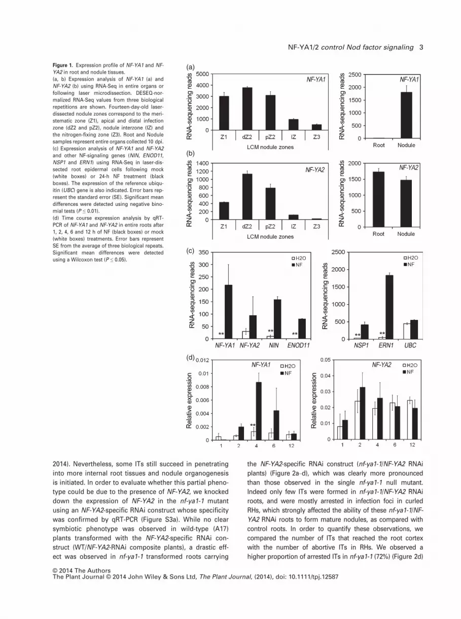
(Figure S1). Using RNA sequencing from laser micro-dis-
sected nodule tissues (Roux et al., 2014), we have shown
that NF-YA1 and NF-YA2 transcripts both exhibit a simi-
lar expression gradient from proximal to apical nodule
tissues, where the highest transcript levels are found
(Figure 1a,b). However, while transcripts of both TFs are
detected at high levels in nodules, only NF-YA2 is well
expressed in nitrogen-starved roots in the absence of
bacterial inoculation (Figure 1a,b).
We have recently shown that NF-YA1 expression in rhi-
zobium-inoculated roots depends upon a functional
NF-perception and signaling machinery (Laporte et al.,
2014). To evaluate whether NF-YA1 and NF-YA2 were also
expressed during the earliest stages of NF signaling in
roots, we thus analyzed their transcript levels in roots in
response to NF treatments. RNA-seq analysis of laser-dis-
sected root epidermal tissues of the pre-infection zone,
24 h after treatment with NFs, showed that NF-YA1 tran-
scripts were strongly upregulated, together with the known
epidermal NF markers NSP1, ERN1, ENOD11 and the
NF-responsive TF gene NIN, while the increase of NF-YA2
transcript levels was not statistically significant due to high
variability (Figure 1c). Experiments using promoter-GUS
lines confirmed expression of NF-YA1 and NF-YA2 in RHs
of NF-treated roots (Figure S2). While symbiotic expression
of NF-YA1 was specific to RHs, NF-YA2 expression was
also detected in cortical and vascular tissues. Time course
experiments further showed that the upregulation of
NF-YA1 by NFs could be detected by qRT-PCR in entire
roots as early as 2 h after treatment and was maximal after
4 h, while NF-YA2 expression remained more or less con-
stant (Figure 1d). Given the high level of identity for the
two encoded proteins and the partially overlapping expres-
sion patterns of NF-YA1 and NF-YA2 we hypothesized that
these two genes could play sequential and partially over-
lapping roles during nodulation.
NF-YA1 and NF-YA2 play partially overlapping roles in the
regulation of rhizobial infection and nodulation
In a previous study, we showed that IT progression is
severely altered in the nf-ya1-1 null mutant (Laporte et al.,
© 2014 The AuthorsThe Plant Journal © 2014 John Wiley & Sons Ltd, The Plant Journal, (2014), doi: 10.1111/tpj.12587
2 Tom Laloum et al.
2014). Nevertheless, some ITs still succeed in penetrating
into more internal root tissues and nodule organogenesis
is initiated. In order to evaluate whether this partial pheno-
type could be due to the presence of NF-YA2, we knocked
down the expression of NF-YA2 in the nf-ya1-1 mutant
using an NF-YA2-specific RNAi construct whose specificity
was confirmed by qRT-PCR (Figure S3a). While no clear
symbiotic phenotype was observed in wild-type (A17)
plants transformed with the NF-YA2-specific RNAi con-
struct (WT/NF-YA2-RNAi composite plants), a drastic eff-
ect was observed in nf-ya1-1 transformed roots carrying
the NF-YA2-specific RNAi construct (nf-ya1-1/NF-YA2 RNAi
plants) (Figure 2a–d), which was clearly more pronounced
than those observed in the single nf-ya1-1 null mutant.
Indeed only few ITs were formed in nf-ya1-1/NF-YA2 RNAi
roots, and were mostly arrested in infection foci in curled
RHs, which strongly affected the ability of these nf-ya1-1/NF-
YA2 RNAi roots to form mature nodules, as compared with
control roots. In order to quantify these observations, we
compared the number of ITs that reached the root cortex
with the number of abortive ITs in RHs. We observed a
higher proportion of arrested ITs in nf-ya1-1 (72%) (Figure 2d)
(a)
(b)
(c)
(d)
Figure 1. Expression profile of NF-YA1 and NF-
YA2 in root and nodule tissues.
(a, b) Expression analysis of NF-YA1 (a) and
NF-YA2 (b) using RNA-Seq in entire organs or
following laser microdissection. DESEQ-nor-
malized RNA-Seq values from three biological
repetitions are shown. Fourteen-day-old laser-
dissected nodule zones correspond to the meri-
stematic zone (Z1), apical and distal infection
zone (dZ2 and pZ2), nodule interzone (IZ) and
the nitrogen-fixing zone (Z3). Root and Nodule
samples represent entire organs collected 10 dpi.
(c) Expression analysis of NF-YA1 and NF-YA2
and other NF-signaling genes (NIN, ENOD11,
NSP1 and ERN1) using RNA-Seq in laser-dis-
sected root epidermal cells following mock
(white boxes) or 24-h NF treatment (black
boxes). The expression of the reference ubiqu-
itin (UBC) gene is also indicated. Error bars rep-
resent the standard error (SE). Significant mean
differences were detected using negative bino-
mial tests (P ≤ 0.01).
(d) Time course expression analysis by qRT-
PCR of NF-YA1 and NF-YA2 in entire roots after
1, 2, 4, 6 and 12 h of NF (black boxes) or mock
(white boxes) treatments. Error bars represent
SE from the average of three biological repeats.
Significant mean differences were detected
using a Wilcoxon test (P ≤ 0.05).
© 2014 The AuthorsThe Plant Journal © 2014 John Wiley & Sons Ltd, The Plant Journal, (2014), doi: 10.1111/tpj.12587
NF-YA1/2 control Nod factor signaling 3

compared with control roots (51%) and an even higher
proportion in the nf-ya1-1/NF-YA2-RNAi roots, in which
almost all infections were arrested in RHs (93.%) (Figure 2d).
As a complementary means to confirm the redundant roles
of NF-YA1 and NF-YA2 during nodulation, we designed an
RNAi construct, termed NF-YA RNAi, strongly reducing the
expression of both NF-YA1 and NF-YA2, while the expres-
sion of the other NF-YA genes was not affected with the
exception of NF-YA6, for which a slight but significant
reduction was observed (Figure S3b). As expected, the
nodulation of NF-YA RNAi composite plants was severely
impaired. Most ITs of RNAi composite plants were arrested
early in infection foci formed in curled RHs (Figure S4). Six-
teen days post-inoculation, 50% of NF-YA RNAi plants
failed to develop nodules, in contrast with control plants.
When NF-YA RNAi plants did develop nodules, only 2 � 1
nodules were detected per plant at 16 dpi, compared with
9 � 1.5 nodules in control plants (Figure S4c). Taken
together, these data indicate that NF-YA1 and NF-YA2 can
redundantly contribute to the regulation of rhizobial infec-
tion and nodulation in M. truncatula.
NF-YA1 and NF-YA2 both regulate ERN1 and ENOD11
expression in response to NFs
In order to evaluate to what extent NF-YA1 and NF-YA2
have redundant functions during pre-infection stages, we
measured expression levels of the known NF-signaling
genes NSP1, ERN1 and ENOD11 in root samples of control
and NF-YA RNAi plants after 4 h of NF treatment. While
the expression of NSP1 was still upregulated by NFs in
NF-YA RNAi roots, the upregulation of both ERN1 and
ENOD11 were significantly reduced (Figure 3). Indeed this
effect was striking as compared with that observed in the
single mutant nf-ya1-1, for which there was no difference
in ENOD11 and ERN1 upregulation by NFs (Figure S5a). As
for infection/nodulation, no effect on ENOD11/ERN1 upreg-
ulation was observed in NF-YA2 RNAi roots (Figure S5b,c).
These results suggest that NF-YA1 and NF-YA2 are both
involved in NF-induced expression of ERN1 and ENOD11
but not of NSP1 and that they may act upstream of ERN1
to regulate its expression during early steps of the NF
response.
(a) (b) (c) (d) Figure 2. Infection thread phenotypes of NF-
YA-silenced roots.
(a–c) Predominant IT phenotype in control
plants (WT GUS RNAi) (a), in the single null
mutant nf-ya1-1 (b) and in nf-ya1-1 expressing a
NF-YA2 RNAi construct (c). Observations were
made 6 days after inoculation (dai) using a
LacZ-expressing S. meliloti strain (blue). Bars =50 µm.
(d) Histograms representing the number of ITs
that reach the cortex (white) or end in RHs
(grey). Error bars correspond to the confidence
interval (a = 0.05) for the total amount of infec-
tion per cm of root. Groups were set following
a Kruskall–Wallis test (P ≤ 0.05).
Figure 3. NF-YA1 and NF-YA2 are both requi-
red for NF-induced expression of ENOD11,
ERN1 but not NSP1.
qRT-PCR analysis of ENOD11, ERN1 and NSP1
expression in H2O (white boxes) or NF-treated
(black boxes) transgenic roots expressing a
GUS RNAi (control) or NF-YA RNAi construct,
silencing both NF-YA1/2. Groups were set fol-
lowing a Kruskall–Wallis test (P ≤ 0.05).
© 2014 The AuthorsThe Plant Journal © 2014 John Wiley & Sons Ltd, The Plant Journal, (2014), doi: 10.1111/tpj.12587
4 Tom Laloum et al.

To investigate whether the expression patterns of
NF-YA1 and NF-YA2 are consistent with putative roles
upstream of ERN1 during NF signaling, their expression
profiles were analyzed in M. truncatula lines carrying
mutations in major NF-signaling genes positioned upstr-
eam of ERN1. Our results showed that the expression of
NF-YA1 was induced in WT (A17), nsp1 and ern1 roots 4 h
after NF treatment, while this induction was abolished in
the dmi3 and nin mutant backgrounds (Figure S6b). On the
other hand, the expression of NF-YA2 was not significantly
upregulated in the different genotypes (Figure S6a). This
indicates that NF-YA1 (but not NF-YA2) expression is
dependent on DMI3 and NIN. However, the fact that NF-
induction of NF-YA1 is not dependent on either NSP1 or
ERN1 implies a potential role of NF-YA1 either upstream of
these two regulators or in a parallel pathway. When we
checked the NF-induced expression of ENOD11 in the same
mutant backgrounds we confirmed that, as expected,
ENOD11 expression depends upon DMI3 and also upon
NSP1 and ERN1. In the nin mutant background the decr-
ease in ENOD11 expression is however not statistically
significant, possibly as the result of the constitutive expres-
sion of NF-YA2 in roots (Figure S6c). Taken together these
results suggest that NF-YA1 and NF-YA2 can both regulate
redundantly NF-induced expression of ERN1 and ENOD11
and that NF-YA1 but not NF-YA2 acts downstream of DMI3
and NIN and independently of NSP1.
The NF-YA1 and NF-YA2 TFs activate ENOD11 expression
via ERN1 in Medicago truncatula roots
Our previous analyses using NF-YA RNAi constructs sug-
gested that NF-YA1/2 are acting upstream of ERN1 and
ENOD11 in the NF-signaling pathway (Figure 3). ERN1 acts
as a positive regulator of ENOD11 and is able to mimic the
NF-elicited ENOD11 response in non-symbiotic conditions
when over-expressed in transgenic roots under the control
of the 35S constitutive promoter (Andriankaja et al., 2007;
Cerri et al., 2012). To evaluate whether NF-YA TFs indeed
act upstream ERN1 and ENOD11 during NF signaling, we
addressed the question of whether NF-YA1 over-expres-
sion was also able to activate the transcription of a 2.3-kb
pENOD11:GUS fusion in transgenic roots. Using the same
approach as for ERN1 (Cerri et al., 2012) we showed that a
high proportion of NF-YA1-over-expressing roots displayed
a clear activation of the pENOD11:GUS fusion in epider-
mal root tissues in the absence of symbiotic treatment
(Figure 4). Similar results were obtained by over-express-
ing NF-YA2 (Figure S7). These results suggest that over-
expression of NF-YA TFs is able to mimic NF-elicited
activation of the pENOD11:GUS fusion in root epidermal
cells as previously demonstrated for ERN1. To evaluate
whether the activation of the pENOD11:GUS fusion by
NF-YA1 over-expression is dependent on the presence of
ERN1 we decided to conduct the same experiments in the
ern1 mutant background. As shown in Figure 4a NF-YA1
over-expression was no longer able to activate ENOD11
expression in this mutant background, providing evidence
that NF-YA1 acts upstream of ERN1 to activate the expres-
sion of pENOD11:GUS fusion in transgenic roots. Similar
results were observed in the nsp1 mutant background
(Figure 4), in which NF-YA1 upregulation by NFs is how-
ever normal (Figure 3b). Taken together, these results
indicate that constitutive expression of NF-YA1 or NF-YA2
can bypass the early NF-signal transduction leading to the
(a)
(b)
Figure 4. NF-independent transactivation of
ENOD11 expression in M. truncatula roots.
Transgenic M. truncatula roots carrying a p35S:
NF-YA1 construct were generated following
A. rhizogenes transformation of stable M. trun-
catula lines (wild-type A17, dmi3, nsp1 or ern1)
carrying the pENOD11:GUS fusion. Transgenic
roots expressing NF-YA1 showed significant
activation of the pENOD11:GUS fusion in root
epidermal cells only in A17 and in dmi3 mutant
backgrounds.
(a) The relative intensity of GUS staining of
roots from independent composite plants (n)
were visually annotated and represented in (a)
as the percentage of composite plants showing
moderate (GUS+, light blue bars) to strong/very
strong (GUS++/+++, dark blue bars) stained
roots after 4 h of the GUS reaction as illustrated
in (b). Bar = 1 cm.
© 2014 The AuthorsThe Plant Journal © 2014 John Wiley & Sons Ltd, The Plant Journal, (2014), doi: 10.1111/tpj.12587
NF-YA1/2 control Nod factor signaling 5

activation of ENOD11 via an ERN1-and potentially NSP1-
dependent pathway.
NF-YA1 is a direct positive regulator of ERN1
Studies in both animals and plants have shown that the
NF-YA TF subunit is localized in the nucleus. Using NF-YA1
N-terminally-tagged by YFP, we have been able to show
that NF-YA1 also displays a strict nuclear localization con-
sistent with its predicted role as a CCAAT-box binding TF
(Figure S8a–c). To evaluate whether NF-YA1 can act as a
direct transcriptional activator of ERN1 and ENOD11 we
performed transactivation assays in Nicotiana benthami-
ana cells. ENOD11 transcription was previously shown to
be under the control of two distinct promoter regulatory
units conferring expression either in RHs responding to
NFs or at rhizobial infection sites (Boisson-Dernier et al.,
2005). Despite the fact that the NF-YA1 protein is expressed
in N. benthamiana leaves, none of the pENOD11-derived
transcriptional fusions was activated by NF-YA1, while the
4xNFbox-p35Smin:GUS and the INF-GUS fusion constructs
were in turn respectively activated by ERN1 and NSP1/
NSP2, in line with previously published data (Figure S9a,b)
(Cerri et al., 2012). This suggests that NF-YA1 is not able to
directly activate the transcription of ENOD11 target sequ-
ences. We then tested the ability of NF-YA1 to activate the
transcription of a 2.3-kb pERN1:GUS reporter construct
shown before to be sufficient for conferring symbiotic
expression in M. truncatula (Cerri et al., 2012). In compari-
son to control samples, an almost 10-fold increase of the
GUS activity level was observed when NF-YA1 was co-infil-
trated with this construct (Figures 5a and S8d,e). This
transactivation was also observed in the presence of
NF-YA2 (Figure S10) and was lost when using a mutated
version of NF-YA1, impaired in DNA recognition (Fig-
ures 5a and S8 g,h), as described in Mantovani et al.
(1994). These data therefore indicate that NF-YA1 and NF-
YA2 can positively regulate the transcription of ERN1.
However, ERN1 can also be transcriptionally regulated by
the NSP1/NSP2 heterodimer (Cerri et al., 2012). We thus
examined whether the co-expression of NSP1 and NSP2
together with NF-YA1 would influence the transcriptional
activation of ERN1. Interestingly, such co-expression led to
a significant increase in pERN1:GUS transcriptional activa-
tion levels when compared with NF-YA1*, a mutated NF-
YA1 version comprising a substitution of the conserved
RGE tripeptide by an AAA triplet in the DNA-binding
domain or YFP control samples (Figure 5d). This suggests
that NF-YA1 could act synergistically with the NSP1/NSP2
complex to activate ERN1 transcription.
A detailed inspection of the ERN1 promoter revealed six
putative CCAAT motifs in a region of 1.4 Kb upstream of
the Transcription start site (TSS), which are either overlap-
ping or in the neighborhood of several putative NSP1 bind-
ing sites (Hirsch et al., 2009) (Figures S11 and 5c). In order
(a)
(b)
(c)
(d)
Figure 5. ERN1 is a direct target of NF-YA1.
(a, b) Transactivation assays of pERN1-GUS. Transcription activation assays
were performed in N. benthamiana epidermal cells by agroinfiltration of a
pERN1:GUS transcriptional fusion in combination with a number of 3xHA-
tagged protein constructs. Fluorimetric GUS activity was measured in
N. benthamiana leaf discs after agroinfiltration of pERN1:GUS alone (Ø), or
in combination with NF-YA1 or NF-YA1*, a mutated NF-YA1 version com-
prising a substitution of the conserved RGE tripeptide by an AAA triplet in
the DNA-binding domain (Figure S8). Fluorimetric GUS activity driven by
the pERN1:GUS fusion was also measured in N. benthamiana leaf discs
co-expressing NSP1 and NSP2 together with a control YFP protein or an
active (NF-YA1) or inactive (NF-YA1*) form of NF-YA1 (b). Data are repre-
sented as fold induction in relation to control (Ø) samples. Error bars corre-
spond to the standard error (SE) of mean GUS activity values derived from
10 to 20 individual samples from three independent experiments. Groups a,
b and c were set following a Kruskal–Wallis test (P ≤ 0.05).
(c) Schematic representation of CCAAT-boxes found in 1.4 kb sequences of
ERN1 upstream of the Transcription Start Site (TTS). The CCAAT-boxes are
numbered as 1–6, and their respective distances from the TSS are indicated.
Arrows indicate the DNA region amplified by qPCR in the genomic input
DNA or in NF-YA1-co-precipitated samples. The preferentially bound box
no. 3 is highlighted by a grey box.
(d) ChIP-qPCR analysis of NF-YA1 binding to the ERN1 promoter. Trans-
genic roots expressing NF-YA1-GFP (black boxes) or GFP-NF-YA1 (grey
boxes) as well as the GFP alone (negative control, white boxes) under the
control of the 35S promoter were generated and used in ChIP experiments
using anti-GFP antibodies. qPCR was performed using primers surrounding
the six CCAAT-boxes present in the promoter of ERN1 (c). IP values were
normalized against those obtained using input genomic DNA. Values repre-
sent the average value calculated from two biological repeats and error bars
correspond to the standard error (SE) of the mean percentage of input.
© 2014 The AuthorsThe Plant Journal © 2014 John Wiley & Sons Ltd, The Plant Journal, (2014), doi: 10.1111/tpj.12587
6 Tom Laloum et al.

to investigate whether NF-YA1 can directly interact in vivo
with one of these putative CCAAT-binding motifs, we
performed ChIP followed by real-time quantitative PCR
(ChIP-qPCR) experiments. Transgenic M. truncatula roots
over-expressing either GFP alone (negative control) or N-
and C-terminal GFP-tagged NF-YA1 proteins were gener-
ated. In young nodulated roots (4 dpi), qPCR amplification
of the DNA regions surrounding the six CCAAT-boxes
detected a strong enrichment of co-precipitated box 3 DNA
fragments for both N- and C-terminal NF-YA1 GFP fusion
expressing roots, but not in the GFP control (Figure 5d).
This result strongly suggests that NF-YA1 specifically binds
to the third CCAAT-box located 423 bp upstream of the
transcription start site (TSS) of ERN1, and provides addi-
tional evidence that NF-YA1 and NF-YA2 are both direct
positive regulators of ERN1 expression.
DISCUSSION
Despite the symbiotic expression of NF-YA1 associated
with early NF-signaling pre-infection events (Laporte et al.,
2014), a null nf-ya1-1 mutant is less severely affected dur-
ing nodulation than previously described mutants of other
TFs regulating early symbiotic steps such as nin, nsp1,
nsp2 or ern1 (Kalo et al., 2005; Smit et al., 2005; Marsh
et al., 2007; Middleton et al., 2007). Here we show that this
is most likely due to the existence of the closely related
NF-YA2 that is redundant with NF-YA1 during NF signaling
and rhizobial infection.
Functional overlap between NF-YA1 and NF-YA2 was
demonstrated by the use of two complementary RNAi
strategies to knock down the expression of both TFs. These
strategies similarly led to a specific reduction in the
expression of both TFs and, as a consequence, to a more
severe symbiotic phenotype than observed with the single
nf-ya1-1 mutant (Laporte et al., 2014). Firstly, a much
higher proportion of arrested ITs were unable to reach the
root cortex, and secondly most infection events were
blocked at the microcolony stage before ITs are initiated.
This therefore suggests a potential role for NF-YA1/2 in the
initiation of rhizobial infection. Finally, nodule formation
was also much more severely impaired.
This enhanced symbiotic phenotype is associated with
the specific knock down of both NF-YA1 and NF-YA2
genes. The expression of all the remaining NF-YA-related
genes, with the exception of NF-YA6, was not affected by
this RNAi knock-down (Figure S3a). However NF-YA6 tran-
script levels were not reduced when using the NF-YA2
RNAi construct in conjunction with the nf-ya1-1 mutant
(Figure S3a,b), and in addition, NF-YA6 is significantly less
expressed in comparison to NF-YA1 and NF-YA2 during
nodulation (Roux et al., 2014). Even if both NF-YA1 and
NF-YA2 play redundant roles during initial signaling and
infection steps of nodulation, the pronounced nodulat-
ion phenotype of the single nf-ya1-1 mutant leads us to
propose that NF-YA1, which is highly and more specifically
expressed during rhizobial symbiosis, plays the primary
role during nodulation. Nevertheless, the basal expression
level of NF-YA2 in roots and RHs prior to infection is con-
sistent with a possible role for this gene prior to NF-YA1
induction. Unfortunately, the use of the NF-YA2 RNAi con-
struct has not revealed such a role yet, and future experi-
ments using nf-ya2 knock-out mutants are now required
to examine the possibility of such a sequential mode of
action.
It was previously suggested, using phylogenetic analy-
sis, that NF-YA1 and NF-YA2 result from a recent gene
duplication event (Laloum et al., 2013) and our results indi-
cate that they have mainly diverged at the level of their
expression profiles, as frequently described for functionally
redundant or partially redundant TFs (Singh and Hannenh-
alli, 2008). This situation is reminiscent of the ERN1 and
ERN2 genes that also share partially overlapping expres-
sion profiles and may also possess redundant function
during the rhizobial symbiosis (Cerri et al., 2012). On the
basis of genome structure studies, it has been suggested
that ERN2 could be the original copy and might play a
more prominent role during the more ancient arbuscular
mycorrhizal symbiosis (Young et al., 2011; Young and
Bharti, 2012). Along this line, the soybean orthologs of
NF-YA2 (GmNF-YA1a/b) have recently been shown to play
a positive role in the establishment of the AM-symbiosis
(Schaarschmidt et al., 2013), and the two M. truncatula
NF-YB and NF-YC genes are also specifically transcribed in
arbuscule-containing cells (Hogekamp et al., 2011). Future
studies will determine whether MtNF-YA1 and MtNF-YA2
play key roles during the AM symbiosis.
Because silencing of both TFs also led to a severe reduc-
tion in the transcriptional activation of ERN1 and ENOD11
in response to NFs, NF-YA1/2 are also likely to play an
active role in the early NF-signaling pathway that takes
place in the root epidermis during rhizobial pre-infection
symbiotic stages. We present converging evidence for the
positioning of these TFs in the NF-signaling cascade as
direct regulators of ERN1 expression. ERN1 encodes an
ERF transcription factor that is necessary for NF-induced
ENOD11 expression and rhizobial infection (Middleton
et al., 2007). In most cases, ITs that form in the ern1
mutant are blocked in infection foci, while limited numbers
of ITs abort within the RH shaft. This is consistent with the
phenotype of nf-ya1-1/NF-YA2 RNAi and in the NF-YA RNAi
roots also affected in IT formation and progression. We
thus propose that ERN1 is an important target of NF-YA1
and NF-YA2, accounting for a substantial part of the role of
these TFs during symbiotic pre-infection and infection. Fur-
ther transcriptomic studies in the context of the double
knock-down NF-YA-RNAi, associated with genome-wide
ChIP-seq approaches should now help to identify addi-
tional direct targets of NF-Y TFs during different symbiotic
© 2014 The AuthorsThe Plant Journal © 2014 John Wiley & Sons Ltd, The Plant Journal, (2014), doi: 10.1111/tpj.12587
NF-YA1/2 control Nod factor signaling 7

stages, thus leading to a more precise picture of the roles
of NF-YA1 and NF-YA2.
We show here that NF-elicited expression of NF-YA1 is
dependent on NIN and this is consistent with recent data
obtained in L. japonicus. Indeed, the NIN TF, that has been
described as an important positive regulator of initial steps
of epidermal infection (Schauser et al., 1999) and nodule
development (Marsh et al., 2007), was recently shown to
directly target LjNF-YA1, the Lotus ortholog of NF-YA1,
and LjNF-YB1 (Soyano et al., 2013). In addition to posi-
tively regulating NF-Y genes, NIN was also shown to act as
a direct positive regulator of NPL (Nodulation Pectate
Lyase) involved in both pre-infection and infection events
(Xie et al., 2012). Our data suggest that NIN, by regulating
the expression of NF-YA1 regulates the subsequent tran-
scription of ERN1 and ENOD11. Nevertheless, ENOD11
transcription is still activated in the nin mutant background
during early NF signaling [Figure S6c and (Marsh et al.,
2007)], probably due to the activity of NF-YA2 whose regu-
lation is not dependent on NIN (Figure S6b). NIN was, as
ERN3, also suggested to act as a repressor of NF-elicited
ENOD11 expression (Andriankaja et al., 2007; Marsh et al.,
2007). Indeed, the nin mutant displays a more widespread
and stronger pENOD11-GUS activity level compared with
WT roots but only at later symbiotic stages. However, this
‘inhibitory effect’ of NIN was not observed at early stages,
consistent with our results obtained at 4H after NF treat-
ment (Figure S6c), but only at rather late time points after
NF treatment (i.e., mainly 4 days and to a lesser extent 24H
post treatment). This is in line with the recent characteriza-
tion of the L. japonicus daphne nin mutant, in which a
repressor role of NIN was also suggested during later
stages of rhizobial infection (Yoro et al., 2014). Taken
together it is suggested that NIN may have a dual positive
and negative role during nodulation and this may depend
on the symbiotic stage considered, and thus the molecular
environment of this TF, or by a differential regulation by
NSP1 or IPD3, both implicated in the regulation of NIN
expression (Hirsch et al., 2009; Singh et al., 2014).
We show here that NF-YA TFs can bind to the promoter
of ERN1. Nevertheless, ERN1 expression is also dependent
on NSP1, which can, as NF-Y, bind directly to the promoter
of ERN1 (Hirsch et al., 2009; Cerri et al., 2012). NSP1 binds
to AATTT-binding sites which are found many times as
such or in their inverted form in ERN1 promoter, but the
precise binding site(s) has(ve) not yet been determined.
Interestingly, AATTT motifs are found in the close vicinity
of the third inverted CCAAT-box (ATTGG) of the ERN1 pro-
moter (Figure S11), shown here to be directly recognized by
NF-YA1. In addition, both NSP1/NSP2 and NF-YA1 are able
to activate the transcription of ERN1 in N. benthamiana
cells, raising the question of the functional links between
these TFs in relation to the regulation of ERN1 express-
ion [Figure 5b and Cerri et al. (2012)]. Our co-expression
experiments in N. benthamiana cells suggest that NSP1/
NSP2 and NF-YA1 may act synergistically to activate ERN1
expression. In Arabidopsis it has been suggested that an
NF-Y complex composed of AtNF-YA4, AtNF-YB3 and AtNF-
YC2 subunits would function as a stabilizing enhancer of
bZIP28 during the ER-stress response (Liu and Howell,
2010). Furthermore an NF-Y complex potentiates CON-
STANS-mediated activation of the FT gene (Tiwari et al.,
2010). The recent analysis of the crystal structure of the
human NF-Y complex bound to a CCAAT-box (Nardini
et al., 2013) suggests that the NF-Y complex could, through
the histone-fold motifs of its NF-YB and NF-YC subunits,
modify the chromatin structure locally to facilitate transcrip-
tional regulation either by the CCAAT-box binding subunit
NF-YA or by other TFs present in the same transcriptional
complex (Gnesutta et al., 2013).
Taken together, our results lead us to propose that NF-
YA1/2-containing complexes could act as major facilitators
for enhancing ERN1 transcriptional activation in association
with NSP1/2 complexes. However, to thoroughly understand
the role of NF-YA1/2 during nodulation it now appears
essential to identify and characterize their interacting protein
partners within different transcriptional complexes. This
includes not only the NF-YB and NF-YC subunits, but also
additional potential transcriptional co-regulators such as
NSP1/2.
Our data establish NF-YA1 and NF-YA2 as important reg-
ulators of NF-mediated signaling and rhizobial infection
initiation in M. truncatula. We propose a model, shown in
Figure 6, positioning these two regulators in the context of
Figure 6. A model of the transcriptional regulatory network controlling
NF-signal transduction in Medicago truncatula.
This model proposes the existence of at least two signaling pathways,
involving the sequential or concomitant action of NF-YA or NSP TFs to reg-
ulate the expression of ERN1 and ENOD11 in root epidermal cells respond-
ing to NFs. Full arrows represent clear genetic or molecular evidence of
direct or indirect links, while dotted arrows represent indirect evidence or
evidence coming from another model (Lotus japonicus). See Discussion for
detailed references.
© 2014 The AuthorsThe Plant Journal © 2014 John Wiley & Sons Ltd, The Plant Journal, (2014), doi: 10.1111/tpj.12587
8 Tom Laloum et al.

what is known about early symbiotic signaling in both
L. japonicus and M. truncatula. Since it is a phosphoryla-
tion substrate of DMI3, CYCLOPS/IPD3 appears as a pri-
mary response gene at the top of the NF-signaling cascade
in L. japonicus (Yano et al., 2008) and possibly also in
M. truncatula (Grimsrud et al., 2010; Rose et al., 2012). Fur-
thermore, it was recently shown in L. japonicus, that IPD3
directly controls the expression of NIN (Singh et al., 2014).
It remains to be proven that this is also the case in M. trun-
catula. NF-induced expression of NSP2 depends on DMI3
(Kalo et al., 2005) but beyond this genetic evidence, the
molecular mechanism, involving CYCLOPS/IPD3 or not,
remains however unknown. In addition to being a target of
CYCLOPS, NIN also seems to be a target of NSP1 in
M. truncatula (Hirsch et al., 2009), and of IPN2, a MYB TF
that interacts with NSP2 in L. japonicus (Kang et al., 2014).
However, it remains to be determined whether this is the
case during earlier NF-signaling steps. The expression of
NIN thus seems to be controlled by both CYCLOPS/IPD3
and NSP1/2 but their respective roles or interconnections
remain to be clarified.
The present manuscript shows that NF-YA1 and NF-YA2
can directly regulate the NF-induced expression of ERN1,
possibly within the same transcriptional complex as NSP1
and NSP2. While NF-YA1 expression is NF-regulated via
DMI3 and NIN, the expression of NF-YA2 seems more con-
stitutive in roots and its regulation is unknown. Our model
thus proposes the existence of at least two independent
pathways, one via IPD3/NIN/NF-YAs and the other via the
NSPs, both leading to NF-induced ERN1 and thus ENOD11
expression. This is in accordance with previous models
that propose the existence of alternative or parallel symbi-
otic signaling pathways that are independent or partially
independent of early symbiotic TFs like NSPs or NIN
(Murray, 2011).
Despite the difficulty of comparing results from different
plants and a variety of experimental systems, the picture
that is now emerging is that of a growing complexity of
the regulatory circuits due to functional redundancies, as
well as parallel, dynamic pathways that could vary depend-
ing on the symbiotic stage or the tissue considered.
EXPERIMENTAL PROCEDURES
Biological material and treatments
Several Jemalong lines were used in this study. Wild-type andL416 derived A17 lines harboring a 2.3 kb-promoter region ofENOD11 fused to the GUS reporter gene (L416) (Journet et al.,2001), and dmi3 (TRV25 allele) (Catoira et al., 2000), nsp1-1 andnsp1-2 (B85 and C54 alleles, respectively) (Catoira et al., 2000),ern1 (bit1-1 allele) (Middleton et al., 2007), nin-1 (12S allele)(Marsh et al., 2007), and nf-ya1-1 (Laporte et al., 2014) with orwithout the pENOD11:GUS fusion. Medicago seeds were germi-nated and grown on agar Fahraeus plates or in aeroponic ‘cais-sons’ as described in the Medicago truncatula Handbook. DNA
constructs were introduced in E. coli DH5a, A. rhizogenes ARqua1(Quandt et al., 1993), or A. tumefaciens GV3101 and GV3103strains. The 2011 S. meliloti strain (Ardourel et al., 1994) constitu-tively expressing the hemA–lacZ fusion was used for this study.All the strains were grown on LB or TY medium supplementedwith the appropriated antibiotic combinations.
NF treatments were performed by immersing roots for 1–12 hin either liquid Fahraeus (control) or a Fahraeus solution comple-mented with purified NFs (10�9 or 10�8
M).
DNA constructs
The pNF-YA1:GUS construct was obtained as described in(Laporte et al., 2014). A 2.6 kb-DNA region upstream of theNF-YA2 start codon was recombined in the pKGWFS7 vector(Karimi et al., 2002) carrying the uidA gene using the Gatewaytechnology (Invitrogen, http://www.lifetechnologies.com/). For theNF-YA RNAi and NF-YA2 RNAi constructs, a 340-bp DNA region inthe NF-YA1 coding sequence and a 200-bp region in the NF-YA2 30
UTR have been introduced in the pFRN (derived from pFGC5941;NCBI accession number AY310901) vector using the Gatewaytechnology (Invitrogen). For the transactivation studies in Medica-go L416 roots, the NF-YA1 ORF was introduced downstream ofthe p35S promoter in a pBL121-derived binary vector, after DNAdigestion with BamH1 and Sac1 restriction enzymes (Promega,Life Technologies, www.lifetechnologies.com/fr). For transienttransformation of N. benthamiana leaves, YFP fusions wereobtained by recombination of the corresponding ORFs into thePAM-PAT p35S:YFP-GW or p35S:3xHA-GW destination vectors (L.Deslandes, Laboratory of Plant Microbe Interactions). 3xHA con-structs are the same as in Cerri et al. (2012). For the ChIP experi-ments, the NF-YA1 coding sequence was amplified by PCR andrecombined into the pK7FWG2 and pK7WGF2 according to theGateway manufacturer’s protocol (https://tools.lifetechnologies.com/content/sfs/manuals/gatewayman.pdf).
Transient expression in Nicotiana benthamiana
Plasmid constructs were introduced in GV3101 and GV3103A. tumefaciens strains. For subcellular localization studies, YFP-NF-Y constructs were co-infiltrated with the viral silencing inhibi-tor protein P19 (Voinnet et al., 2003). A. tumefaciens strains weregrown in Luria-Bertoni medium under antibiotic selection at 28°Cand harvested as described in (Andriankaja et al., 2007). After 2 hof incubation at room temperature, equal volumes of A. tumefac-iens cultures (OD600 = 0.5 for subcellular localization studies;OD600 = 0.25 for transactivation studies) were co-infiltrated into3-week-old N. benthamiana leaves using a 1-mL syringe. Infil-trated plants were kept at 21°C in a growth chamber (16-h photo-period, 70 lE m�2 s�1 light intensity). Subcellular localization,b-glucuronidase activity and protein expression were analyzed onleaf discs collected 36 h after infiltration. Protein extracts werecharged on a poly-acrylamide gel, and gel blots were performedusing anti-HA-peroxidase rat monoclonal antibodies (Roche,https://lifescience.roche.com/shop/home).
RNA extraction and quantitative RT-PCR
Total RNA was extracted from Medicago roots using the Mache-rey-Nagel Total RNA Isolation Kit according to the manufacturer’sinstructions (http://www.mn-net.com/). The RNA samples werequantified and RNA integrity was checked by Agilent RNA Nano-Chip (Agilent, http://www.home.agilent.com/). First-strand cDNAsynthesis was performed using 500 ng to 1 lg of total RNA withan anchored oligo(dT) and SuperScript II (Invitrogen) or Rochereverse transcriptases, following the manufacturers’ protocols.
© 2014 The AuthorsThe Plant Journal © 2014 John Wiley & Sons Ltd, The Plant Journal, (2014), doi: 10.1111/tpj.12587
NF-YA1/2 control Nod factor signaling 9

Quantitative RT-PCR was performed on 384-well plates, with theLight Cycler 480 system (Roche) and using the SYBR Green I Mas-ter mix (Roche). The cycling conditions were as follows: 95°C for5 min followed by 45 cycles of denaturation at 95°C for 15 sec,annealing and extension at 60°C for 1 min. Transcript levels foreach of the target genes were normalized to the endogenousubiquitin transcript level. The data shown are mean valuesobtained from two to five independent biological experimentswith two to three technical repeats. Primer sequences for quantita-tive RT-PCR analyses are shown in Table S1.
Nodule and root microdissection, RNA sequencing and
statistical analyses
RNA-seq analyses of root and nodule samples were performed asfollows. Preparation of whole root and nodule samples, as wellas laser-dissected nodule regions has been described in Rouxet al. (2014). Laser-dissection of mock and NF-treated (10�8
M)epidermal root samples was conducted on 20 lm transversal sec-tions, using a NF-reactive 1 cm-long region 24 h after treatment.RNA extraction, rRNA depletion and RNA in vitro amplificationwere carried out as published (Roux et al., 2014). Orientedpaired-end sequencing (2 9 50 nt) was performed using an Illu-mina HiSeq 2000 by Fasteris SA (https://www.fasteris.com). Nor-malization of raw counts was carried out using DESEQ R packagev1.12.0 (Anders and Huber, 2010). Estimated dispersion was cal-culated using the ‘pooled’ method with the sharing mode set to‘fit-only’. For each gene, we set generalized linear models (GLMs)according to a full model testing which regresses the genesexpression on the treatment condition (Mock or NF). Tests for dif-ferential expression were performed as described in the DESEQ
package.
GUS and b-galactosidase assays
Histochemical and fluorometric assays wer performed as descri-bed in Cerri et al. (2012).
The magenta-Gal and X-Gal (5-bromo-6-chloro-3-indolyl-b-D-galactopyranoside; Biosynth B7200) substrates were used forhistochemical (purple and blue, respectively) staining of theb-galactosidase activity within S. meliloti-containing ITs.
Microscopic methods
After coloration, root tissues were fixed in a 0.1 M phosphate buffercomplemented with 2% glutaraldehyde, dehydrated in ethanol andincluded in agarose or Technovit 7100 (Heraeus Kulzer, http://heraeus-kulzer.fr/) resin. Roots were then sliced into 5- to 40-lm sec-tions using a Reichter Jung 2040 microtome. Root tissues wereobserved using a light microscope (Axioplan 2 Imaging; Carl Zeiss).Subcellular localization of NF-YA proteins in N. benthamiana leafdiscs were observed with an epifluorescence microscope (Axiophot2; Carl Zeiss, http://www.zeiss.fr).
Chromatin immuno-precipitation analysis
ChIP assays were performed 4 days after rhizobia inoculation ofMedicago transformed roots bearing the following constructs:35S:GFP (used as a negative control), 35S:NF-YA1-GFP or 35S:GFP-NF-YA1. Briefly, after plant material fixation in 1% (v/v) form-aldehyde, tissues were homogenized, nuclei isolated and lysed,according to Gendrel et al. (2005). Cross-linked chromatin wasthen sonicated using a water bath Bioruptor� Plus (Diagenode;30 sec on/30 sec off pulses, at high intensity for 60 cycles: twice10 cycles in TPX tubes, Diagenode, plus 40 cycles in ordinary1.5 ml tubes). ChIP was performed in an SX-8G IP-Star� Compact
Automated System (Diagenode, http://www.diagenode.com),using a Direct ChIP protocol incubating the samples with the anti-bodies (anti-GFP, ABCAM ab6556) for 12 h and Invitrogen ProteinA Dynabeads�, using standard parameters. ImmunoprecipitatedDNA was then recovered using the IPure kit in the SX-8G IP-Star�
(Diagenode) and analysed by quantitative real-time PCR. An ali-quot of untreated sonicated chromatin was processed in parallelfor use as the total input DNA control. Two biological replicateswere used for each construct and the result is given as the meanof both independent results.
ACKNOWLEDGEMENTS
The authors wish to thank David Barker for critical reading of themanuscript. This work was funded by the ANR-09-BLAN-0033-01HAPIHUB and ANR-08-GENO-106 SYMbiMICS for RNA-Seq analy-ses, and supported by the French Laboratory of Excellence project‘TULIP’ (ANR-10-LABX-41; ANR-11-IDEX-0002-02). T.L. and M.R.C.were funded by a grant from the French Ministry of Education andResearch, M.B. by an INRA CJS (Contrat Jeune Scientifique)contract and F.A. by a long term EMBO fellowship.
SUPPORTING INFORMATION
Additional Supporting Information may be found in the online ver-sion of this article.Figure S1. Protein alignment of NF-YA1 and NF-YA2 conserveddomains.Figure S2. Expression patterns of NF-YA1 and NF-YA2 in roots inresponse to NFs.Figure S3. Specific downregulation of NF-Y genes by NF-YA2 orNF-YA–RNAi constructs.Figure S4. NF-YA RNAi roots are strongly impaired in IT initiationand nodule formation.Figure S5. Expression analyses of NF-YAs and early symbioticmarker genes in NF-YA1 or NF-YA2-down-regulated roots.Figure S6. Expression of NF-YA1 and NF-YA2 and ENOD11 inmutants of the NF signaling pathway.Figure S7. NF-YA2 can, as ERN1 and NF-YA1 transactivateENOD11 expression in M. truncatula roots.Figure S8. A mutation in the DNA-binding domain of NF-YA1 cont-roling pERN::GUS activation.Figure S9. NF-YA1 does not activate the transcription of ENOD11regulatory sequences.Figure S10. NF-YA2 can transactivate ERN1 transcription in Nicoti-ana benthamiana cells.Figure S11. Three putative NSP1 binding sites surround the 3rdCCAAT box in the ERN1 promoter.
Table S1. List of primers used.
REFERENCES
Anders, S. and Huber, W. (2010) Differential expression analysis for
sequence count data. Genome Biol. 11, R106.
Andriankaja, A., Boisson-Demier, A., Frances, L., Sauviac, L., Jauneau, A.,
Barker, D. and de Carvalho-Niebel, F. (2007) AP2-ERF transcription fac-
tors mediate Nod factor-dependent mt ENOD11 activation in root hairs
via a novel cis-regulatory motif. Plant Cell, 19, 2866–2885.Ardourel, M., Demont, N., Debelle, F.D., Maillet, F., Debilly, F., Prome, J.C.,
Denarie, J. and Truchet, G. (1994) Rhizobium meliloti lipooligosaccharide
nodulation factors different structural requirements for bacterial entry
into target root hair-cells and induction of plant symbiotic developmental
responses. Plant Cell, 6, 1357–1374.Boisson-Dernier, A., Andriankaja, A., Chabaud, M., Niebel, A., Journet, E.P.,
Barker, D.G. and de Carvalho-Niebel, F. (2005) MtENOD11 gene activa-
tion during rhizobial infection and mycorrhizal arbuscule development
© 2014 The AuthorsThe Plant Journal © 2014 John Wiley & Sons Ltd, The Plant Journal, (2014), doi: 10.1111/tpj.12587
10 Tom Laloum et al.

requires a common AT-rich-containing regulatory sequence. Mol. Plant
Microbe Interact. 18, 1269–1276.Capoen, W., Sun, J., Wysham, D. et al. (2011) Nuclear membranes control
symbiotic calcium signaling of legumes. Proc. Natl Acad. Sci. USA, 108,
14348–14353.Catoira, R., Galera, C., de Billy, F., Penmetsa, R.V., Journet, E.P., Maillet, F.,
Rosenberg, C., Cook, D., Gough, C. and Denarie, J. (2000) Four genes of
Medicago truncatula controlling components of a Nod factor transduc-
tion pathway. Plant Cell, 12, 1647–1666.Cerri, M.R., Frances, L., Laloum, T., Auriac, M.C., Niebel, A., Oldroyd, G.E.D.,
Barker, D.G., Fournier, J. and de Carvalho-Niebel, F. (2012) Medicago
truncatula ERN transcription factors: regulatory interplay with NSP1/
NSP2 GRAS factors and expression dynamics throughout rhizobial infec-
tion. Plant Physiol. 160, 2155–2172.Combier, J.P., Frugier, F., de Billy, F. et al. (2006) MtHAP2-1 is a key tran-
scriptional regulator of symbiotic nodule development regulated by
microRNA169 in Medicago truncatula. Genes Dev. 20, 3084–3088.Combier, J.P., de Billy, F., Gamas, P., Niebel, A. and Rivas, S. (2008)
Trans-regulation of the expression of the transcription factor MtHAP2-1
by a uORF controls root nodule development. Genes Dev. 22, 1549–1559.El-Yahyaoui, F., Kuster, H., Ben Amor, B. et al. (2004) Expression profiling in
Medicago truncatula identifies more than 750 genes differentially
expressed during nodulation, including many potential regulators of the
symbiotic program(1 w). Plant Physiol. 136, 3159–3176.Gendrel, A.V., Lippman, Z., Martienssen, R. and Colot, V. (2005) Profiling
histone modification patterns in plants using genomic tiling microarrays.
Nat. Methods, 2, 213–218.Gnesutta, N., Nardini, M. and Mantovani, R. (2013) The H2A/H2B-like
histone-fold domain proteins at the crossroad between chromatin and
different DNA metabolisms. Transcription, 4, 114–119.Grimsrud, P.A., den Os, D., Wenger, C.D., Swaney, D.L., Schwartz, D.,
Sussman, M.R., An�e, J.M. and Coon, J.J. (2010) Large-scale phosphopro-
tein analysis in Medicago truncatula roots provides insight into in vivo
kinase activity in legumes. Plant Physiol. 152, 19–28.Hirsch, S., Kim, J., Munoz, A., Heckmann, A.B., Downie, J.A. and Oldroyd,
G.E.D. (2009) GRAS proteins form a DNA binding complex to induce
gene expression during nodulation signaling in Medicago truncatula.
Plant Cell, 21, 545–557.Hogekamp, C., Arndt, D., Pereira, P.A., Becker, J.D., Hohnjec, N. and K€uster,
H. (2011) Laser microdissection unravels cell-type-specific transcription
in arbuscular mycorrhizal roots, including CAAT-box transcription factor
gene expression correlating with fungal contact and spread. Plant Phys-
iol. 157, 2023–2043.Journet, E.P., El-Gachtouli, N., Vernoud, V., de Billy, F., Pichon, M., Dedieu,
A., Arnould, C., Morandi, D., Barker, D.G. and Gianinazzi-Pearson, V.
(2001) Medicago truncatula ENOD11: a novel RPRP-encoding early nodu-
lin gene expressed during mycorrhization in arbuscule-containing cells.
Mol. Plant Microbe Interact. 14, 737–748.Kalo, P., Gleason, C., Edwards, A. et al. (2005) Nodulation signaling in
legumes requires NSP2, a member of the GRAS family of transcriptional
regulators. Science, 308, 1786–1789.Kang, H., Chu, X., Wang, C. et al. (2014) A MYB coiled coil transcription fac-
tor interacts with NSP2 and is involved in nodulation in Lotus japonicus.
New Phytol. 201, 837–849.Karimi, M., Inze, D. and Depicker, A. (2002) GATEWAY((TM)) vectors for
Agrobacterium-mediated plant transformation. Trends Plant Sci. 7, 193–195.
Laloum, T., De Mita, S., Gamas, P., Baudin, M. and Niebel, A. (2013)
CCAAT-box binding transcription factors in plants: Y so many? Trends
Plant Sci. 18, 157–166.Laporte, P., Lepage, A., Fournier, J. et al. (2014) The CCAAT box-binding
transcription factor NF-YA1 controls rhizobial infection. J. Exp. Bot. 65,
481–494.Liu, J.-X. and Howell, S.H. (2010) bZIP28 and NF-Y transcription factors are
activated by ER stress and assemble into a transcriptional complex to
regulate stress response genes in Arabidopsis. Plant Cell, 22, 782–796.Mantovani, R. (1999) The molecular biology of the CCAAT-binding factor
NF-Y. Gene, 239, 15–27.Mantovani, R., Lio, X., Pessara, U., Vanhuisjduijnen, R., Benoist, C. and
Mathis, D. (1994) Dominant-negative analogs of NF-YA. J. Biol. Chem.
269, 20340–20346.
Marsh, J., Rakocevic, A., Mitra, R., Brocard, L., Sun, J., Eschstruth, A., Long,
S., Schultze, M., Ratet, P. and Oldroyd, G. (2007) Medicago truncatula
NIN is essential for rhizobial-independent nodule organogenesis induced
by autoactive calcium/calmodulin-dependent protein kinase. Plant Phys-
iol. 144, 324–335.Middleton, P., Jakab, J., Penmetsa, R. et al. (2007) An ERF transcription fac-
tor in Medicago truncatula that is essential for Nod factor signal trans-
duction. Plant Cell, 19, 1221–1234.Moreau, S., Verdenaud, M., Ott, T., Letort, S., de Billy, F., Niebel, A., Gouzy, J.,
de Carvalho-Niebel, F. and Gamas, P. (2011) Transcription reprogramming
during root nodule development in Medicago truncatula. PLoS One, 6,
e16463.
Murray, J.D. (2011) Invasion by invitation: rhizobial infection in legumes.
Mol. Plant Microbe Interact. 24, 631–639.Nardini, M., Gnesutta, N., Donati, G. et al. (2013) Sequence-specific tran-
scription factor NF-Y displays histone-like DNA binding and H2B-like
ubiquitination. Cell, 152, 132–143.Oldroyd, G.E.D. (2013) Speak, friend, and enter: signalling systems that pro-
mote beneficial symbiotic associations in plants. Nat. Rev. Microbiol. 11,
252–263.Oldroyd, G.E.D. and Downie, J.A. (2008) Coordinating nodule morphogenesis
with rhizobial infection in legumes. Annu. Rev. Plant Biol. 59, 519–546.Quandt, H.J., Puhler, A. and Broer, I. (1993) Transgenic root-nodules of Vicia
hirsuta a fast and efficient system for the study of gene-expression in
indeterminate-type nodules. Mol. Plant Microbe Interact. 6, 699–706.Rose, C.M., Venkateshwaran, M., Grimsrud, P.A., Westphall, M.S.,
Sussman, M.R., Coon, J.J. and Ane, J.-M. (2012) Medicago PhosphoPro-
tein Database: a repository for Medicago truncatula phosphoprotein
data. Front. Plant Sci. 3, 122.
Roux, B., Rodde, N., Jardinaud, M.F. et al. (2014) An integrated analysis of
plant and bacterial gene expression in symbiotic root nodules using
laser-capture microdissection coupled to RNA sequencing. Plant J. 77,
817–837.Schaarschmidt, S., Gresshoff, P.M. and Hause, B. (2013) Analyzing the soy-
bean transcriptome during autoregulation of mycorrhization identifies
the transcription factors GmNF-YA1a/b as positive regulators of arbuscu-
lar mycorrhization. Genome Biol. 14, R62.
Schauser, L., Roussis, A., Stiller, J. and Stougaard, J. (1999) A plant regula-
tor controlling development of symbiotic root nodules. Nature, 402, 191–195.
Sieberer, B.J., Chabaud, M., Timmers, A.C., Monin, A., Fournier, J. and Bar-
ker, D.G. (2009) A nuclear-targeted cameleon demonstrates intranuclear
Ca2+ spiking in Medicago truncatula root hairs in response to rhizobial
nodulation factors. Plant Physiol. 151, 1197–1206.Singh, L.N. and Hannenhalli, S. (2008) Functional diversification of paralo-
gous transcription factors via divergence in DNA binding site motif and
in expression. PLoS One, 3, e2345.
Singh, S., Katzer, K., Lambert, J., Cerri, M. and Parniske, M. (2014)
CYCLOPS, a DNA-binding transcriptional activator, orchestrates symbi-
otic root nodule development. Cell Host Microbe, 15, 139–152.Smit, P., Raedts, J., Portyanko, V., Debelle, F., Gough, C., Bisseling, T. and
Geurts, R. (2005) NSP1 of the GRAS protein family is essential for rhizo-
bial Nod factor-induced transcription. Science, 308, 1789–1791.Soyano, T., Kouchi, H., Hirota, A. and Hayashi, M. (2013) NODULE INCEP-
TION directly targets nf-y subunit genes to regulate essential processes of
root nodule development in Lotus japonicus. PLoS Genet. 9, e1003352.
Tiwari, S.B., Shen, Y., Chang, H.-C. et al. (2010) The flowering time regula-
tor CONSTANS is recruited to the FLOWERING LOCUS T promoter via a
unique cis-element. New Phytol. 187, 57–66.Voinnet, O., Rivas, S., Mestre, P. and Baulcombe, D. (2003) An enhanced
transient expression system in plants based on suppression of gene
silencing by the p19 protein of tomato bushy stunt virus. Plant J. 33,
949–956.Xie, F., Murray, J.D., Kim, J., Heckmann, A.B., Edwards, A., Oldroyd, G.E.
and Downie, J.A. (2012) Legume pectate lyase required for root infection
by rhizobia. Proc. Natl Acad. Sci. USA, 109, 633–638.Yano, K., Yoshida, S., M€uller, J. et al. (2008) CYCLOPS, a mediator of symbi-
otic intracellular accommodation. Proc. Natl Acad. Sci. USA, 105, 20540–20545.
Yoro, E., Suzaki, T., Toyokura, K., Miyazawa, H., Fukaki, H. and Kawaguchi,
M. (2014) A positive regulator of nodule organogenesis, NODULE
© 2014 The AuthorsThe Plant Journal © 2014 John Wiley & Sons Ltd, The Plant Journal, (2014), doi: 10.1111/tpj.12587
NF-YA1/2 control Nod factor signaling 11

INCEPTION, acts as a negative regulator of rhizobial infection in Lotus
japonicus. Plant Physiol. 165, 747–758.Young, N.D. and Bharti, A.K. (2012) Genome-enabled insights into legume
biology. Annu. Rev. Plant Biol. 63, 283–305.Young, N.D., Debelle, F., Oldroyd, G.E.D. et al. (2011) The Medicago gen-
ome provides insight into the evolution of rhizobial symbioses. Nature,
480, 520–524.
Zanetti, M.E., Blanco, F.A., Beker, M.P., Battaglia, M. and Aguilar, O.M.
(2010) A C subunit of the plant nuclear factor NF-Y required for rhizobial
infection and nodule development affects partner selection in the com-
mon bean–rhizobium etli symbiosis. Plant Cell, 22, 4142–4157.
© 2014 The AuthorsThe Plant Journal © 2014 John Wiley & Sons Ltd, The Plant Journal, (2014), doi: 10.1111/tpj.12587
12 Tom Laloum et al.