cellular models to study dopaminergic injury responses
TRANSCRIPT
AD
Award Number: DAMD17-01-1-0697
TITLE: International Conference on Parkinson's Disease
PRINCIPAL INVESTIGATOR: Rashid Shaikh,' Ph.D.
CONTRACTING ORGANIZATION: , New York Academy of Sciences New York, NY 10021
REPORT DATE: January 2003
TYPE OF REPORT: Final Proceedings
PREPARED FOR: U.S. Army Medical Research and Materiel Command Fort Detrick, Maryland 21702-5012
DISTRIBUTION STATEMENT: Approved for Public' Release,- Distribution Unlimited
The views, opinions and/or findings contained in this report are those of the author(s) and should not be construed as an official Department of the Army position, policy or decision unless so designated by other documentation.
20040112 107

REPORT DOCUMENTATION PAGE
Form Approved 0MB No. 074-0188
Public reporting burden for this collection of infomiaUon Is estimated to average 1 hour per response, induding the lime for reviewing Instructions, searching existing data sources, gathering and malnlalnina the data needed, and completing and reviewing this collection of Information. Send comments regarding this burden estimate or any other aspect of this colleclion of infonnatlon. Including sugaestions for reducing this burden to Washington Headquarters Services, Directorate for Infonnatlon Operations and Reports, 1215 Jefferson Davis Highway, Suite 1204, Artlngton, VA 22202-4302 and to the nmrt, al Management and Budget, Papenvork Reduction Project (0704-0188). Washington, DC 20503 ' <=""".<= ui
1. AGENCY USE ONLY (Leave blank)
2. REPORT DATE January 2003
4. TITLE AND SUBTITLE
3. REPORT TYPE AND DATES COVERED Final Proceedings(1 May 2001- 31 Dec 2002)
International Conference on Parkinson's Disease
6. AUTHOR(S)
Rashid Skaikh, Ph.D.
7. PERFORMING ORGANIZATION NAME(S) AND ADDRESS(ES)
New York Academy of Sciences New York, NY 10021
E-Mail: rshaikhOnyas. org
9. SPONSORING / MONITORING AGENCY NAME(S) AND ADDRESS(ES)
U.S. Army Medical Research and Materiel Command Fort Detrick, Maryland 21702-5012
11. SUPPLEMENTARY NOTES
5. FUNDING NUMBERS
DAMD17-01-1-0697
8. PERFORMING ORGANIZATION REPORT NUMBER
10. SPONSORING / MONITORING AGENCY REPORT NUMBER
12a. DISTRIBUTION / AVAILABILITY STATEMENT
Approved for Public Release; Distribution Unlimited
13. ABSTRACT (MAXIMUM 200 WORDS)
NO ABSTRACT PROVIDED
12b. DISTRIBUTION CODE
14. SUBJECT TERMS
17. SECURITY CLASSIFICATION OF REPORT
Unclassified NSN 7540-01-280-5500
18. SECURITY CLASSIFICATION OF THIS PAGE
Unclassified
19. SECURITY CLASSIFICATION OF ABSTRACT
Unclassified
15. NUMBER OF PAGES 376
16. PRICE CODE
20. LIMITATION OF ABSTRACT
Unlimited Standard Form 298 (Rev. 2-89) Prescribed by ANSI Std. Z39-18 298-102
Parkinsons Disease The Life Cycle of the Dopamine Neuron
Editors: Howard J. Federoff, Robert E. Burke, Stanley Fahn and Gary Fiskum
ANNALS OF THE NEW YORK ACADEMY OF SCIENCES VOLUME 991

ANNALS OF THE NEW YORK ACADEMY OF SCIENCES
Volume 991
EDITORIAL STAFF
Director, Publishing and New Media SARAH GREENE
Managing Editor JUSTD^CULLINAN ^ »r v ; >» j fc ■ The New York Academy of Sciences Associate Editor 2 East 63rd Street RICHARD STIEFEL New York, New York 10021
THE NEW YORK ACADEMY OF SCIENCES (Founded in 1817)
BOARD OF GOVERNORS, September 2002 - September 2003
TORSTEN N. WIESEL, Chairman of the Board JOHN F. NIBLACK, Vice Chairman
JOHN T. MORGAN, Treasurer ELLIS RUBINSTEIN, Chief Executive Officer [ex officio]
Honorary Life Governors WILLIAM T. GOLDEN JOSHUA LEDERBERG
Governors
ELEANOR BAUM KAREN E. BURKE PRAVEEN CHAUDHARI
R. BRIAN FERGUSON GERALD D. HSCHBACH RONALD L. GRAHAM
MARNIEIMHOFF JACQUELINE LEO BRUCE McEWEN
PAUL MARKS RONAY MENSCHEL SANDRA PANEM
PETER RINGROSE LEE G.VANCE DEBORAH WILEY
HELENE L. KAPLAN, Counsel [ex officio]
PARKINSON'S DISEASE THE LIFE CYCLE OF THE DOPAMINE NEURON

ANNALS OF THE NEW YORK ACADEMY OF SCIENCES
Volume 991
PARKINSON'S DISEASE THE LIFE CYCLE OF THE DOPAMINE NEURON
Edited by Howard J. Federoff, Robert E. Burke, Stanley Fahn, and Gary Fiskum
The New York Academy of Sciences New York, New York
2003
Copyright © 2003 by the New York Academy of Sciences. All rights reserved. Under the provisions of the United States Copyright Act of 1976, individual readers of the Annals are permitted to make fair use of the material in them for teaching or research. Permission is granted to quote from the Annals provided that the customary acknowledgment is made of the source. Material in the Annals may be republished only by permission of the Acad- emy. Address inquiries to the Permissions Department (editorial® nyas.org) at the New York Academy of Sciences.
Copying fees: For each copy of an article made beyond the free copying permitted under Section 107 or 108 of the 1976 Copyright Act, a fee should be paid through the Copyright Clearance Center, Inc., 222 Rosewood Drive, Danvers, MA 01923 (www, copvrieht. com).
© The paper used in this publication meets the minimum requirements of the American National Standard for Information Sciences—Permanence of Paper for Printed Library Materials, ANSI Z39.48-1984.
Library of Congress Cataloging-in-Publication Data
Parlcinson's disease: the life cycle of the dopamine neuron / edited by Howard J. Federoff... [et al.].
p. ; cm. -- (Annals of the New York Academy of Sciences; v. 991) "This volume is the result of a conference entitled Parkinson's disease: the life cycle of the dopamine neuron, sponsored by the New York Academy of Sciences held on September 18-20, 2002 in Princeton, New Jersey"—Contents p. Includes bibliographical references and index.
ISBN 1-57331-448-X (cloth : alk. paper) - ISBN 1-57331-449-8 (paper : alk. paper)
I. Parkinson's disease-Pathophysiology-Congresses. 2. Parkinson's disease-Molecular aspects-Congresses. 3. Dopaminergic neurons-Congresses. 4. Dopaminergic mechanisms-Congresses.
[DNLM: 1. Parkinson's Disease-Congresses. 2. Dopamine-biosynthesis-Congresses. WL 359 P25176 2003] I. Federoff, Howard. II. Series.
Q11.N5 vol. 991 [RC382] 500s-dc2I [616.8/33
2003009584
GYAT/PCP
Printed in the United States of America ISBN 1-57331-448-X (cloth) ISBN 1-57331-449-8 (paper)
ISSN 0077-8923
ANNALS OF THE NEW YORK ACADEMY OF SCIENCES
Volume 991 June 2003
PARKINSON'S DISEASE THE LIFE CYCLE OF THE
DOPAMINE NEURON
Editors HOWARD J. FEDEROFF, ROBERT E. BURKE, STANLEY FAHN,
AND GARY FISKUM
Conference Organizers HOWARD J. FEDEROFF, ROBERT E. BURKE, STANLEY FAHN,
GARY FISKUM, AND OLE ISACSON
This volume is the result of a conference entitled Parkinson's Disease: The Life Cycle of the Dopamine Neuron, which was sponsored by the New York Academy of Sciences and held September 18-20 in Princeton, New Jersey.
CONTENTS
Preface. By HOWARD J. FEDEROFF ix
Part I. Clinical Syndrome
Description of Parkinson's Disease as a Clinical Syndrome. By STANLEY FAHN 1
Physiology and Pathophysiology of Parkinson's Disease. By CLEMENT HAMANI AND ANDRES M. LOZANO 15
PET Studies on the Function of Dopamine in Health and Parkinson's Disease. By DAVID J. BROOKS 22
Part n. Development
Midbrain Dopaminergic Neurons: Determination of Their Developmental Fate by Transcription Factors. By HORST H. SiMON, LAVINIA BHATT, DANIEL GHERBASSI, PAOLA SGAD6, AND LAVINIA ALBERI 36
Transcriptional Control of Dopamine Neuron Development. By ksk WALLfiN AND THOMAS PERLMANN 48

Transcription Factors in the Development of Midbrain Dopamine Neurons. By J. PETER H. BURBACH, SIMONE SMITS, AND MARTEN P. SMDT 61
Postnatal Developmental Programmed Cell Death in Dopamine Neurons. By ROBERT E. BURKE 69
Part III. From Molecules to Organism: The Molecules
The Cast of Molecular Characters in Parkinson's Disease: Felons, Conspirators, and Suspects. By KAH LEONG LIM, VALINA L. DAWSON, AND TED M. DAWSON 80
Oxidati ve Modifications of a-Synuclein. By HARRY IscfflROPOULOS 93
Parkin and Endoplasmic Reticulum Stress. By RYOSUKE TAKAHASHI, YUZURUIMAI,NOBUTAKAHATTORI,ANDYOSHIKUNIMIZUNO 101
Parkinson's Disease and Related a-Synucleinopathies Are Brain Amyloidoses. By JOHN Q. TROJANOWSKI AND VIRGINIA M-Y. LEE 107
Part rV. From Molecules to Organism: The Cells
Mitochondrial Mechanisms of Neural Cell Death and Neuroprotective Interventions in Parkinson's Disease. By GARY FlSKUM, ANATOLY STARKOV, BRIAN M. POLSTER, AND CHRISTOS CHINOPOULOS Ill
Mitochondria, Oxidative Damage, and Inflammation in Parkinson's Disease. ByM. FLINT REAL 120
Apoptosis Inducing Factor and PARP-Mediated Injury in the MPTP Mouse Model of Parkinson's Disease. By HONGMIN WANG, MiKA SHIMOJI, SEONG-WOON YU, TED M. DAWSON, AND VALINA L. DAWSON 132
Cellular Models to Study Dopaminergic Injury Responses. By TIMOTHY J. COLLIER, KATHY STEECE-COLLIER, SUSAN MCGUIRE, AND CARYLE. SORTWELL 140
Convergent Pathobiologic Model of Parkinson's Disease. By KATHLEEN A. MAGUIRE-ZEISS AND HOWARD J. FEDEROFF 152
The Relationship between Lewy Body Disease, Parkinson's Disease, and Alzheimer^s Disease. B}) JOHN HARDY 167
Part V. From Molecules to Organism: The Organism
Transgenic Models of a-Synuclein Pathology: Past, Present, and Future. By MAKOTO HASHIMOTO, EDWARD ROCKENSTEIN, AND ELIEZER MASLIAH .. 171
The l-Methyl-4-Phenyl-l,2,3,6-Tetrahydropyridine Mouse Model: A Tool to Explore the Pathogenesis of Parkinson's Disease. By SERGE PRZEDBORSKI AND MIQUEL VILA 189
Pathophysiology of Parkinson's Disease: The MPTP Primate Model of the Human Disorder. By THOMAS WICHMANN AND MAHLON R. DELONG. .. 199
The Role of dial Reaction and Inflammation in Parkinson's Disease. By E. C. HiRSCH, T BREIDERT, E. ROUSSELET, S. HUNOT, A. HARTMANN, AND R P MICHEL 214
Part VI. Convergence of Cell Genesis and Cell Transfer
Induction of Adult Neurogenesis: Molecular Manipulation of Neural Precursors in Situ. By PAOLA ARLOTTA, SANJAY S. MAGAVI, AND JEFFREY D. MACKLIS 229
Molecular Mechanisms of Neuronal Cell Death. By KiM A. HEIDENREICH.... 237
Redox State as a Central Modulator of Precursor Cell Function. By MARK NOBLE, JOEL SMITH, JENNIFER POWER, AND MARGOT MAYER-PR6SCHEL 251
COX-2 and Neurodegeneration in Parkinson's Disease. By P. TtlSMANN, M. VILA, D.-K. CHOI, K. TIEU, D. C. WU, V. JACKSON-LEWIS, AND S. PRZEDBORSKI 272
Part Vn. Poster Papers
Striatopalhdal Changes in a Preclinical Rat Model of Parkinson's Disease. By A. E. GRISSELL, T. M. BUCHANAN, AND M. A. ARIANO 278
Neurotoxicity-Induced Changes in Striatal Dopamine Receptor Function. By ANNA-LIISA BROWNELL, IRIS Y. CHEN, XUKUI WANG, MEIXIANG YU, ANDBRUCEG.JENKINS 281
The Developmental Time Course of Glial Cell Line-Derived Neurotrophic Factor (GDNF) and GDNF Receptor a-1 mRNA Expression in the Striatum and Substantia Nigra. By JINWHAN CHO, NIKOLAI G. KHOLODILOV, AND ROBERT E. BURKE 284
Subcellular Compartmentalization of P-ERKs in the Lewy Body Disease Substantia Nigra. By CHARLEEN T. CHU AND JIAN-HUI ZHU 288
Altered Striatal Neuronal Morphology Is Associated with AstrogUosis in a Chronic Mouse Model of Parkinson's Disease. By A. G. DERVAN, S. TOTTERDELL, Y.-S. LAU, AND G. E. MEREDITH 291
Is the Initial Insult in Parkinson's Disease and Dementia with Lewy Bodies a Neuritic Dystrophy? By JOHN E. DUDA, BENOIT I. GlASSON, VIRGINIA M-Y. LEE, AND JOHN Q. TROJANOWSKI 295
Neuroplasticity in the MPTP-Lesioned Mouse and Nonhuman Primate. By MICHAEL W. JAKOWEC, BETH FISHER, KERRY NIXON, ELIZABETH HOGG, CHARLES MESHUL, SAMUEL BREMMER, TOM MCNEILL, AND GISELLEM.PETZINGER 298
An Experimental Infection by Filterable Forms of Nocardia asteroides and Late-Onset Movement Disorder in Mice. By S. KOHBATA AND C. KADOYA 302
A Characterization of Dopaminergic Neurodegeneration in Organotypic Cultures. By GERALDINE J. KRESS AND IAN J. REYNOLDS 304
Developmental Cell Death and Oxidative Stress: Lessons from Nigral Dopamine Neurons. By LAURENT GROC, TANGELLA JACKSON HUNTER, HAO JIANG, LAURENT BEZIN, AND ROBERT A. LEVINE 307
Identification of a Novel Gene Linked to Parkin via a Bidirectional Promoter. By PAUL J. LOCKHART, ANDREW B. "V^ST, CASEY A. O'FARRELL, AND MATTHEW J.FARRER 311
Gene Expression Analysis of tlie MPTP-Lesioned Substantia Nigra in Mice. By R. M. MILLER, C. CASACELI, L. CHEN, AND H. J. FEDEROFF 315
Neuroimaging and Proteomic Tracking of Neurodegeneration in MPTP- Treated Mice. By HOWARD E. GENDELMAN, CHRISTOPHER J. DESTACHE, MARINA L. ZELIVYANSKAYA, JAY A. NELSON, MICHAEL D. BOSKA, TOM M. BiSKUP, MICHAEL K. MCCARTHY, KIMBERLY A. CARLSON, CRAIG NEMECHEK, ERIC J. BENNER, ANDR. LEEMOSLEY 319
PCBs and Dopamine Function: Neurological Effects of Polychlorinated Biphenyls: Does Occupational Exposure Alter Dopamine-Mediated Function? By RICHARD F. SEEGAL 322
A Murine Model of ALS-PDC with Behavioral and Neuropathological Features of Parkinsonism. By J. D. SCHULZ, J. M. B. WiLSON, AND C. A. SHAW 326
Neuroprotective GangUoside Derivatives. By K. CONN, S. DOHERTY, P. ElSENHAUER, R. FINE, J. "VWELLS, AND M. D. ULLMAN 330
Redox- and Metalloregulated RNA Bioaptamer Targets of Proteins Associated with Parkinson's and Other Neurodegenerative Diseases: Factors of Relevance in the Life Cycle of Cells.fi>'JOSEF H.WISSLER 333
Dopaminergic Stimulatory Polypeptides from Immortalized Striatal Cells. By ALFRED HELLER, MARTIN GROSS, SUZANNE HESSEFORT, NANCY BUBULA, AND LISA WON 339
Glutathione and Ascorbate: Their Role in Protein Glutathione Mixed Disulfide Formation during Oxidative Stress and Potential Relevance to Parkinson's Disease. By GAIL D. ZEEVALK, LAURA P. BERNARD, AND JULIEEHRHART 342
Dopaminergic Neurons in Human Striatum and Neurogenesis in Adult Monkey Striatum. By MARTINE COSSETTE, ANDR^ANNE BEDARD, AND ANDR6 PARENT 346
Secreted Factors from Primary Midbrain Glia Regulate Nurrl Activity. By Y. Luo AND H. J. FEDEROFF 350
Development of Nurrl Stable Cell Lines for the Identification of Downstream Targets. By Yu Luo, LEIGH A. HENRICKSEN, KATHLEEN A. MAGUIRE-ZEISS, AND HOWARD J. FEDEROFF 354
* * * Index of Contributors 359
The conference on which these proceedings are based was made possible by an educational grant from the US. Army Medical Research and Materiel Command.
The New York Academy of Sciences believes it has a responsibility to provide an open forum for discussion of scientific questions. The positions taken by the participants in the reported conferences are their own and not necessarily those of the Academy. The Academy has no intent to influence legislation by providing such forums.
Preface
Parkinson's disease and its motor manifestations result predominantly from a loss of midbrain dopamine neurons. Although other neuronal populations are also affected in the disease, we conceptuaUzed this meeting and volume around the theme of the life cycle of the dopamine neuron. By bringing together neurologists, neuro- surgeons, developmental biologists, neuropathologists, fly geneticists, systems and other neuroscientists, and molecular biologists, we hoped for an extensive inter- disciplinary exchange of ideas to occur. By all accounts this did happen: the level of discussion throughout the meeting ranged over the entire spectrum of scientific ap- proaches and levels of inquiry.
This meeting would not have occurred without the thoughtful suggestions of Col. Karl Friedl, Ph.D., and Stephen Grate, Ph.D. As important, the support provided by Col. Friedl and the Department of Defense was essential to achieving the proper set- ting, balance of participants, and sponsorship by the prestigious New York Academy of Sciences. The inspiration for the meeting arose from the highly interactive and prescient Organizing Committee. The New York Academy of Sciences' Dr. Rashid Shaikh, Renee Wilkerson-Brown, and other staff members provided critical guid- ance and operations assistance that made the meeting successful. The completion of the proceedings would not have been possible without the stewardship of the New York Academy of Sciences' Richard Stiefel. On behalf of the Organizing Committee I am extremely thankful to all who contributed to making Parkinson's Disease: The Life Cycle of the Dopamine Neuron a most enjoyable and stimulating conference and, it is hoped, a useful book.
HOWARD J. FEDEROFF, M.D., PH.D.
Professor of Neurology University of Rochester School of Medicine
Ann. N.Y. Acad. Sci. 991: ix (2003). © 2003 New York Academy of Sciences.
Description of Parkinson's Disease as a Clinical Syndrome
STANLEY FAHN
Department of Neurology, Columbia University College of Physicians & Surgeons, New York, New York 10032, USA
ABSTRACT: Parkinsonism is a clinical syndrome comprising combinations of motor problems-^amely, bradyldnesia, resting tremor, rigidity, flexed pos- ture, "freezing," and loss of postural reflexes. Parkinson's disease (PD) is the major cause of parkinsonism. PD is a slowly progressive parkinsonian syn- drome that begins insidiously and usually affects one side of the body before spreading to involve the other side. Pathology shows loss of neuromelanin-con- taining monoamine neurons, particularly dopamine (DA) neurons in the sub- stantia nigra pars compacta. A pathologic hallmark is the presence of cytoplasmic eosinophilic inclusions (Lewy bodies) in monoamine neurons. The loss of DA content in the nigrostriatal neurons accounts for many of the motor symptoms, which can be ameliorated by DA replacement therapy—that is, levodopa. Most cases are sporadic, of unknown etiology; but rare cases of mo- nogenic mutations (10 genes at present count) show that there are multiple causes for the neuronal degeneration. The pathogenesis of PD remains un- known. Clinical fluctuations and dyskinesias are frequent complications of levodopa therapy; these, as well as some motor features of PD, improve by re- setting the abnormal brain physiology towards normal by surgical therapy. Nonmotor symptoms (depression, lack of motivation, passivity, and dementia) are common. As the disease progresses, even motor symptoms become intrac- table to therapy. No proven means of slowing progression have yet been found.
KEYWORDS: Parkinson's disease; parkinsonism; Lewy body; dopamine; levodopa
fflSTORICAL INTRODUCTION
Clinical Description
By amazing coincidence, James Parkinson published a monograph describing the entity subsequently bearing his name in the same year, 1817, that the New York Academy of Sciences was founded. ^ He described six individuals with the clinical features. One was followed in detail over a long period of time; the other five con- sisted of brief descriptions, including two whom he had met walking in the street and another whom he had observed at a distance. Such distant observations without a medical examination demonstrates how readily distinguishable the condition is
Address for correspondence: Dr. Stanley Fahn, Neurological Institute, 710 West 168th Street, New York, NY 10032. Voice: 212-305-5295; fax: 212-305-3530.
Ann. N.Y. Acad. Sci. 991: 1-14 (2003). © 2003 New York Academy of Sciences.
ANNALS NEW YORK ACADEMY OF SCIENCES
merely from the patients' appearance of flexed posture, resting tremor, and shuffling gait. Parkinson's opening description has the key essentials: "Involuntary tremulous motion, with lessened muscular power, in parts not in action and even when support- ed; with a propensity to bend the trunk forward, and to pass from a walking to a run- ning pace: the senses and intellects being uninjured." Despite the small number of patients examined, Parkinson provided a detailed description of the symptoms and also discussed the progressive worsening of the disorder, which he called the shaking palsy and by the Latin Xtrrn paralysis agitans.
In his monograph, Parkinson reviewed the different kinds of tremors previously reported and specifically cited the tremor in his "An Essay on the Shaking Palsy" as occurring when the body part is at rest and not during an active voluntary movement. Seventy years later Charcot emphasized that tremor need not be present in the disor- der and argued against the term paralysis agitans; he suggested, instead, that the name of the disorder be Parkinson's disease (see Goetz, 1987^ for English translation).
The iexxm paralysis sxiA palsy in paralysis agitans and shaking palsy are also in- appropriate. There is no true paralysis. Today, the "lessened muscular power" men- tioned by Parkinson is recognized to be a slowness of movement that is called akinesia, hypokinesia, or bradykinesia, all three terms often being used interchange- ably. These terms represent a paucity of movement in the absence of weakness or paralysis.
Recognition and development of the term akinesia came about slowly. Charcot, in his Tuesday Lessons of 1888, related slowness to rigidity and specifically exclud- ed weakness as a cause (see Ref. 2). Gowers in 1893^ described Parkinson's disease as consisting of tremor, weakness, rigidity, flexed posture, and short steps, with slowness due in part to rigidity. Oppenheim in 191 \^ mentioned that impairment and retardation of active movements might occur in the absence of rigidity. He did not relate it to weakness. Wechsler in the 1932 edition of his textbook^ commented on the special difficulty of initiation of movement as a feature of slowness. Wilson in his large neurology opus of 1940^ used the terms akinesia, akinesis, and hypokine- sia. Under these ternis, he related the masked fades, the unblinking eyes, the poverty of movement, and the patient sitting immobile. Schwab, England, and Peterson de- voted an entire paper in 1959' to the subject of akinesia, which by this time was firm- ly established as the "lessened muscular power" mentioned by Parkinson. Within the definition of akinesia, these authors mentioned fatigue, decrementing amplitude of movements, diificulty shifting to other contraction patterns, apathy, inability to com- plete actions, difficulty initiating an act, and the ability to reach normal movement briefly under sudden motivation. Furthermore, they described the difficulty for a pa- tient with Parkinson's disease to execute two motor events simultaneously, all under the rubric of akinesia.
Pathology of Parkinson's Disease
It was many years after Parkinson's original description before the basal ganglia were recognized by Meynert in 1871^ as being involved in disorders of abnormal movements. And it was not until 1895 that the substantia nigra was suggested to be affected in Parkinson's disease. Brissaud (1895)^ suggested this on tiie basis of a re- port by Blocq and Marinesco (1893)'^ of a tuberculoma in that site that was associ-

FAHN: CLINICAL DESCRIPTION OF PARKINSON'S DISEASE
ated with hemiparkinsonian tremor. These authors were careful to point out that the pyramidal tract and the brachium conjuctivum above and below the level of the le- sion contained no degenerating fibers. The importance of the substantia nigra was emphasized by Tretiakoff in 1919,^1 who studied the substantia nigra in nine cases of Parkinson's disease, one case of hemiparkinsonism, and three cases of posten- cephalitic parkinsonism, finding lesions in this nucleus in all cases. With the hemi- parkinsonian case Tretiakoff found a lesion in the nigra on the opposite side, concluding that the nucleus served the motor activity on the contralateral side of the body. The substantia nigra, so named because of its normal content of neuromelanin pigment, was noted to show depigmentation, loss of nerve cells, and gliosis. These findings remain the histopathologic features of the disease. In his study, Tretiakoff also confirmed the earUer observation of Lewy (1914),'^ who had discovered the presence of cytoplasmic inclusions in Parkinson's disease, now widely recognized as the major pathologic hallmark of the disorder and referred to as Lewy bodies.
Foix and Nicolesco made a detailed study of the pathology of Parkinson's disease in 1925'^ and found that the most constant and severe lesions are in the substantia nigra. Since then many workers, including Hassler (1938)''* and Greenfield and Bosanquet (1953),'^ have confirmed these findings and added other observations, in- cluding involvement of other brain stem nuclei such as the locus ceruleus.
Biochemistry of Parkinson's Disease
Prior to 1957, the parkinsonian syndrome in animals and humans induced by re- serpine was thought to be due to a depletion of brain serotonin. But in that year Carlsson and colleagues'^ discovered that L-dopa reversed the reseipine-induced parkinsonian state in rabbits; while tiie precursor of serotonin, L-5-hydroxytiyp- tophan, did not. L-dopa is tiie precursor to dopamine and norepinephrine, and at that time it was tiiought that dopamine did not have an independent function but served solely as a precursor of norepinephrine. In 1958, after he developed a method for its chemical assay, Carlsson determined that dopamine was present in brain.'^ By the following year the regional distiibution of dopamine was mapped out in brain in both animals'^ and humans.'^ In 1959, at the Intemational Catecholamine Symposium, Carlsson suggested that Parkinson's disease was related to brain dopamine.^" In 1960, Ehringer and Homykiewicz, using Carlsson's methodology, measured dopam- ine and norepinephrine in humans with basal ganglia disorders and discovered a neo- striatal dopamine deficiency in parkinsonism.^' Thus began the modem era of understanding parkinsonism and the role of dopamine in brain. Carlsson's contiibu- tions eventually led to his being awarded the Nobel Prize in Physiology and Medi- cine in 2000.
DISTINGUISHING BETWEEN PARKINSON'S DISEASE AND PARKINSONISM
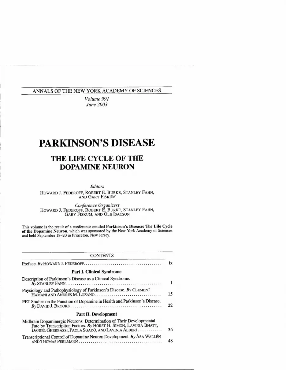
The syndrome of parkinsonism must be understood before understanding what is Parkinson's disease. Today, the Xtrva parkinsonism is defined by any combination of six specific motoric featores: tremor at rest, bradykinesia, rigidity, loss of postural reflexes, flexed posture (RG. 1), and the freezing phenomenon (where the feet are

ANNALS NEW YORK ACADEMY OF SCIENCES
FIGURE 1. Drawing of a patient with Parkinson disease demonstrating the flexed pos- ture typically seen in this disorder. (Modified from Gowers, 1893, p. 639.^)
transiently "glued to the ground")^^ (TABLE 1). Not all six of these cardinal features need be present, but at least two should be before the diagnosis of parkinsonism is made, with at least one of them being tremor at rest or bradykinesia. Parkinsonism is classified into four categories (TABLE 2). PD or primary parkinsonism will be the principal focus of this volume. It is the category that is most commonly encountered by the general clinician; it is also the category on which much research has been car- ried out and the one we know the most about. The great majority of cases of primary parkinsonism are sporadic, but in the last few years several gene mutations have been discovered to cause PD (TABLE 3). Whether primary parkinsonism is genetic or id- iopathic in etiology, the common denominator is that it is not caused by known in- sults to the brain (the main feature of secondary parkinsonism) and is not associated with other motoric neurologic features (the main feature of Parkinson-plus syn- dromes). The uncovering of genetic causes of primary parkinsonism has shed light on probable pathogenetic mechanisms that may be a factor in even the more common idiopathic cases of PD. It may even turn out that many of the idiopathic cases will be linked to gene mutations, this discovery is yet to be made. Although the term id- iopathic PD has been applied to primary parkinsonism, the fact that there are now known genetic causes encourages us to adopt instead the term primary parkin- sonism, for the former term implies that the etiology is unknown.
Three of the most helpful clues that one is likely to be dealing with PD rather than another category of parkinsonism are: (1) an asymmetrical onset of symptoms (PD often begins on one side of the body); (2) the presence of rest tremor (although rest tremor may be absent in patients with PD, it is almost always absent in Parkinson-

FAHN: CLINICAL DESCRIPTION OF PARKINSON'S DISEASE
TABLE 1. Six cardinal clinical features of parkinsonism
Tremor at rest Flexed posture of neck, trunk, and limbs
Rigidity Loss of postural reflexes
Bradykinesia/hypokinesia/akinesia Freezing phenomenon
TABLE 2. Classification of the parkinsonian states
Primary parkinsonism (Parkinson's disease) Sporadic Known genetic etiology (see Table 3)
Secondary parkinsonism (environmental etiology Drugs
Dopamine receptor blockers (most commonly antipsychotic medications) Dopamine storage depletors (reserpine, tetrabenazine)
Postencephalitic Toxins: Mn,CO, MPTP, cyanide Vascular Brain tumors Head trauma Normal-pressure hydrocephalus
Parkinsonism-plus syndromes Progressive supranuclear palsy Multiple system atrophy Cortical-basal ganglionic degeneration Parkinson-dementia-ALS complex of Guam Progressive pallidal atrophy Diffuse Lewy body disease (DLBD)
Heredodegenerative disorders Alzheimer's disease Wilson's disease Huntington's disease Frontotemporal dementia (tau mutation on chromosome 17q21) X-linked dystonia-parkinsonism (in Filipino men; known as lubag)
plus syndromes); and (3) substantial clinical response to adequate levodopa therapy (usually, Parkinson-plus syndromes do not respond to levodopa therapy). In this chapter, we will concentrate on PD and not the other categories of parkinsonism. One common misdiagnosis as PD is the presence of tremor due to the entity known as essential tremor, which can even be unilateral, although it more commonly is bi- lateral. Helpful in the diagnosis is that the tremor due to PD is a rest tremor (tremor appears with the affected body part is at rest), whereas the tremor due to essential tremor is not present at rest, but appears with holding the arms in front of the body and increases in amplitude with activity of the arm, such as with handwriting or per- forming the finger-to-nose maneuver.
ANNALS NEW YORK ACADEMY OF SCIENCES
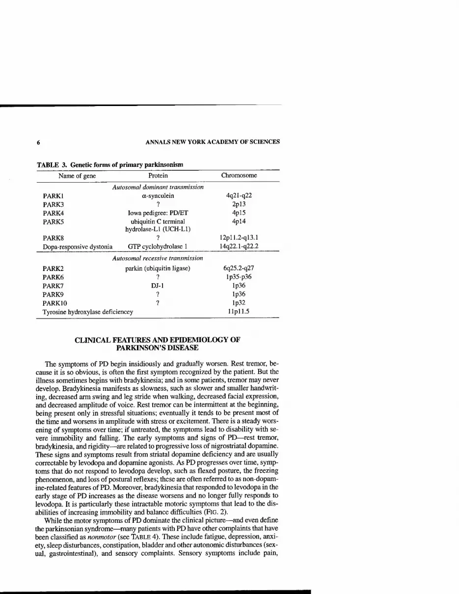
TABLE 3. Genetic forms of primary parkinsonism
Name of gene Protein Chromosome
Autosomal dominant transmission PARKl a-synculein 4q21-q22 PARK3 ? 2pl3 PARK4 Iowa pedigree: PD/ET 4pl5 PARKS ubiquitin C terminal
hydrolase-Ll (UCH-Ll) 4pl4
PARKS ? 12pll.2-ql3.1 Dopa-responsive dystonia GTP cyclohydrolase 1 14q22.1-q22.2
Autosomal recessive transmission
PARK2 parkin (ubiquitin ligase) 6q25.2-q27 PARK6 7 Ip35-p36 PARK? DJ-1 lp36 PARK9 ? lp36 PARKIO ? lp32 Tyrosine hydroxylase deficiencey llpll.5
CLINICAL FEATURES AND EPIDEMIOLOGY OF PARKINSON'S DISEASE
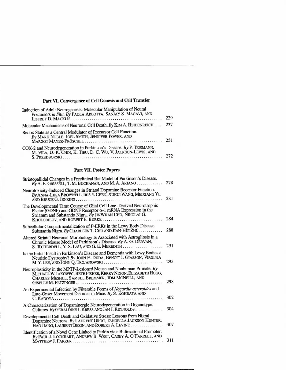
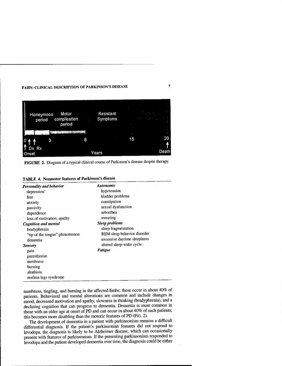
The symptoms of PD begin insidiously and gradually worsen. Rest tremor, be- cause it is so obvious, is often the first symptom recognized by the patient. But the illness sometimes begins with bradykinesia; and in some patients, tremor may never develop. Bradykinesia manifests as slowness, such as slower and smaller handwrit- ing, decreased arm swing and leg stride when walking, decreased facial expression, and decreased amplitude of voice. Rest tremor can be intermittent at the beginning, being present only in stressful situations; eventually it tends to be present most of the time and worsens in amplitude with stress or excitement. There is a steady wors- ening of symptoms over time; if untreated, the symptoms lead to disability with se- vere immobility and falling. The early symptoms and signs of PD—rest tremor, bradykinesia, and rigidity—^are related to progressive loss of nigrostriatal dopamine. These signs and symptoms result from striatal dopamine deficiency and are usually correctable by levodopa and dopamine agonists. As PD progresses over time, symp- toms that do not respond to levodopa develop, such as flexed posture, the freezing phenomenon, and loss of postural reflexes; these are often referred to as non-dopam- ine-related features of PD. Moreover, bradykinesia that responded to levodopa in the early stage of PD increases as the disease worsens and no longer fully responds to levodopa. It is particularly these intractable motoric symptoms that lead to the dis- abilities of increasing immobility and balance difficulties (RG. 2).
While the motor symptoms of PD dominate the chnical picture—and even define the parkinsonian syndrome—many patients with PD have other complaints that have been classified as nonmotor (see TABLE 4). These include fatigue, depression, anxi- ety, sleep disturbances, constipation, bladder and other autonomic disturbances (sex- ual, gastrointestinal), and sensory complaints. Sensory symptoms include pain.
FAHN: CLINICAL DESCRIPTION OF PARKINSON'S DISEASE
Honeymoon Motor Resistant period complication Symptoms
period
11 Dx Rx Onset Years
FIGURE 2. Diagram of a typical clinical course of Parkinson's disease despite therapy.
TABLE 4. Nonmotor features of Parkinson's disease
Personality and behavior depression' fear anxiety passivity dependence loss of motivation, apathy
Cognition and mental bradyphrenia "tip of the tongue" phenomenon dementia
Sensory pain paresthesiae numbness burning akathisia restless legs syndrome
Autonomic hypotension bladder problems constipation sexual dysfunction seborrhea sweating
Sleep problems sleep fragmentation REM sleep behavior disorder excessive daytime sleepiness altered sleep-wake cycle
Fatigue
numbness, tingling, and burning in the affected limbs; these occur in about 40% of patients. Behavioral and mental alterations are common and include changes in mood, decreased motivation and apathy, slowness in thinking (bradyphrenia), and a declining cognition that can progress to dementia. Dementia is most common in those with an older age at onset of PD and can occur in about 40% of such patients; this becomes more disabling than the motoric features of PD (RG. 2).
The development of dementia in a patient with parkinsonism remains a difficult differential diagnosis. If the patient's parkinsonian features did not respond to levodopa, the diagnosis is likely to be Alzheimer disease, which can occasionally present with features of parkinsonism. If the presenting parkinsonism responded to levodopa and the patient developed dementia over time, the diagnosis could be either
ANNALS NEW YORK ACADEMY OF SCIENCES
PD or diffuse Lewy body disease (DLBD) (also called dementia with Lewy bodies). If hallucinations occur with or without levodopa therapy, DLBD is the most Ukely diagnosis. DLBD is a condition where Lewy bodies are present in the cerebral cortex as well as in the brain stem nuclei. The heredodegenerative disease known asfron- totempoml dementia is an autosomal dominant disorder due to mutations in the tau gene on chromosome 17; the full syndrome presents with dementia, loss of inhibi- tion, parkinsonism, and sometimes muscle wasting.
Although PD can develop at any age, it begins most commonly in older adults, with a peak age at onset at around 60 years. The likelihood of developing PD increas- es with age, with a lifetime risk of about 2%?-^ A positive family history doubles the risk of developing PD to about 4%. Twin studies indicate that PD with an onset under the age of 50 years is more likely to have a genetic relationship than PD with a later age at onset.^ Males have higher prevalence and incidence rates than females. Pa- tients with PD can live 20 or more years, depending on the age at onset. The mortal- ity rate is about 1.6 times that of normal individuals of the same age.^ Death in PD is usually due to some concurrent unrelated illness or due to the effects of decreased mobility, aspiration, or increased falUng with subsequent physical injury. At the present time, approximately 850,000 individuals in the U.S. have PD, with the num- ber expected to grow as the population ages.
There are no practical diagnostic laboratory tests for PD, and the diagnosis rests on the clinical features and on excluding other causes of parkinsonism. The research tool of fluorodopa (FDOPA) positron emission tomography (PET) measures levodopa uptake into dopamine nerve terminals, and this shows a decline of about 8% per year of the striatal uptake. A similar result is seen using ligands for the dopamine transporter, either by PET or by single-photon emission computed tomog- raphy (SPECT); these ligands also label the dopamine nerve terminals. All these neuroimaging techniques reveal decreased dopaminergic nerve terminals in the stri- atum in both PD and the Parkinson-plus syndromes and do not distinguish between them. A substantial response to levodopa is most helpful in the differential diagnosis, indicating presynaptic dopamine deficiency with intact postsynaptic dopamine re- ceptors, features typical of PD.
Some adults may develop a more benign form of PD, in which the symptoms re- spond to very-low-dosage levodopa, and the disease does not worsen severely with time. This form is usually due to the autosomal dominant disorder known as dopa- responsive dystonia, which typically begins in childhood as a dystonia. But when it starts in adult life, it can present with parkinsonism. There is no neuronal degenera- tion. The pathogenesis is due to a biochemical deficiency involving dopamine syn- thesis. The gene defect is for an enzyme (GTP cyclohydrolase I) required to synthesize the cofactor for tyrosine hydroxylase activity, the crucial rate-limiting first step in the synthesis of dopamine and norepinephrine. Infantile parkinsonism is due to the autosomal recessive deficiency of tyrosine hydroxylase, another cause of a biochemical dopamine deficiency disorder.
PATHOLOGY, BIOCHEMISTRY AND PHYSIOLOGY OF PARKINSON'S DISEASE
PD and the Parkinson-plus syndromes have in common a degeneration of substan- tia nigra pars compacta dopaminergic neurons, with a resulting deficiency of striatal

FAHN: CLINICAL DESCRIPTION OF PARKINSON'S DISEASE
TABLE 5. Dopamine concentration in striatum is associated witli severity of bradykinesia
Severely of bradykinesia Caudate nucleus Putamen
Mild
Marked
0.58 (13)
0.44 (9)
0.44 (12)
0.05 (9)
Normal controls 2.65 (28) 3.44 (28)
NOTE: Date from Bemheimer et al?^ Results are means in |ig/g fresh tissue. Numbers in parentheses are the number of cases studied.
dopamine due to loss of the nigrostriatal neurons. Accompanying this neuronal loss is an increase in glial cells in the nigra and a loss of the neuromelanin normally contained in the dopaminergic neurons. In PD, intracytoplasmic eosinophihc inclusions, called Lewy bodies, are usually present in many of the surviving neurons. It is recognized to- day diat not all patients with PD have Lewy bodies; those with the homozygous muta- tion in the PARK2 gene—mainly young-onset PD patients—have nigral neuronal degeneration without Lewy bodies. Lewy bodies contain many proteins, including the fibrillar form of a-synuclein, discovered because PARKl 's mutations involve the gene for this protein. There are no Lewy bodies in the Parkinson-plus syndromes.
With the progressive loss of the nigrostriatal dopaminergic neurons, there is a cor- responding decrease of dopamine content in both the nigra and the striatum, which, as mentioned above, accounts particularly for the bradykinesia and rigidity in PD. There are compensatory changes, such as supersensitivity of dopamine receptors, so that symptoms of PD are first encountered only when there is about an 80% reduc- tion of dopamine concentration in the putamen (or a loss of 60% of nigral dopamin- ergic neurons).^^ With fiirther loss of dopamine concentration, parkinsonian bradykinesia becomes more severe (TABLE 5). The progressive loss of tihe dopamin- ergic nigrostriatal pathway can be detected in vivo using PET and SPECT scanning; these show a continuing reduction of FDOPA and dopamine transporter ligand bind- ing in the striatum.^^"^^
The consequence of nigrostriatal loss is an altered physiology downstream from the striatum. The striatum contains Dl and D2 receptors. The current thinking is that dopamine is excitatory at the Dl receptor and inhibitory at the D2 receptor. Deficien- cy of dopamine at these receptors results in alteration at the downstream nuclei: ex- cessive activity of the subthalamic nucleus and globus pallidus intema, and increased inhibition in the thalamus and cerebral cortex.32-34 xjiegg altered physio- logical patterns are restored towards normal with treatment by levodopa.
CAUSES AND PATHOGENESIS OF PD AND PARKINSON-PLUS SYNDROMES
Other than known genetic causes of PD (TABLE 3), the etiology of these disorders remains unknown. Three (PARKl, PARK2, and PARK5) of the four identified mu- tated genes causing PD—^involving the proteins a-synuclein, parkin, and ubiquitin C terminal hydrolase-Ll—^point to an impairment of protein degradation with a buildup of toxic proteins that cannot be degraded via the ubiquitin-proteasomal path-

10 ANNALS NEW YORK ACADEMY OF SCIENCES
ENVIRONMENTAL OR
ENDOGENOUS TOXINS
PATHOGENESIS
Oxldative Protein Mlto- Stress aggregation chondria
Inflam- Apoptosis mation Cascade
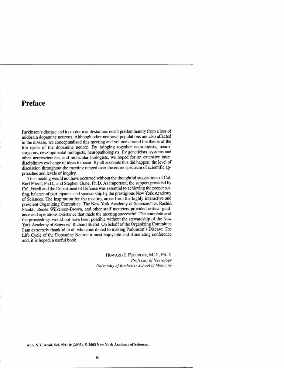
PARKINSON'S DISEASE
FIGURE 3. Diagram of the concept of the etiology and pathogenesis of Parkinson's disease.
way. The fourth, PARK7—involving a nuclear protein of unknown function—ap- pears also to play a role in protein degradation. These findings have led to the concept that perhaps most, if not all, cases of sporadic PD have an impairment of protein degradation. A current hypothesis is that oxidative stress with the formation of oxyradicals, such as dopamine quinone, can lead to reactions with oc-synuclein to form oligomers of a-synuclein (so-called protofibrils), which accumulate because they cannot be degraded by the ubiquitin-proteasomal pathway, leading finally to cell death.^^ Other pathogenetic mechanisms being considered are (1) other effects from oxidative stress, such as the reaction of oxyradicals with nitric oxide to form the highly reactive peroxynitrite radical; (2) impaired mitochondria leading to both reduced ATP production and accumulation of electrons that aggravate oxidative stress, with the final outcome being apoptosis and cell death; and (3) inflammatory changes in the nigra, producing cytokines that augment apoptosis (FiG. 3). These ac- tions lead to an apoptotic cascade that leads to cell death. These concepts on patho- genesis are leading researchers to test agents that affect these potential mechanisms in an attempt to reduce the rate of neurodegeneration in PD.
THERAPY OF PARKINSON'S DISEASE
Neuroprotective Therapy
So far no drug or surgical approach has been shown unequivocally to slow the rate of progression of PD, but if any drug should be proved to delay the progression of the disease process, it should be incorporated in treatment early in the course of the dis-
FAHN: CLINICAL DESCRIPTION OF PARKINSON'S DISEASE 11
ease. There are some controlled clinical trials that were sufficiently positive to have raised the possibility that the propargylamine agents selegiline and rasagiline and the mitochondrial enhancing agent coenzyme Qjq could have some neuroprotective qualities.^^^^ Larger clinical trials with neuroimaging of striatal dopamine nerve terminals would be necessary to provide adequate documentation of neuroprotection.
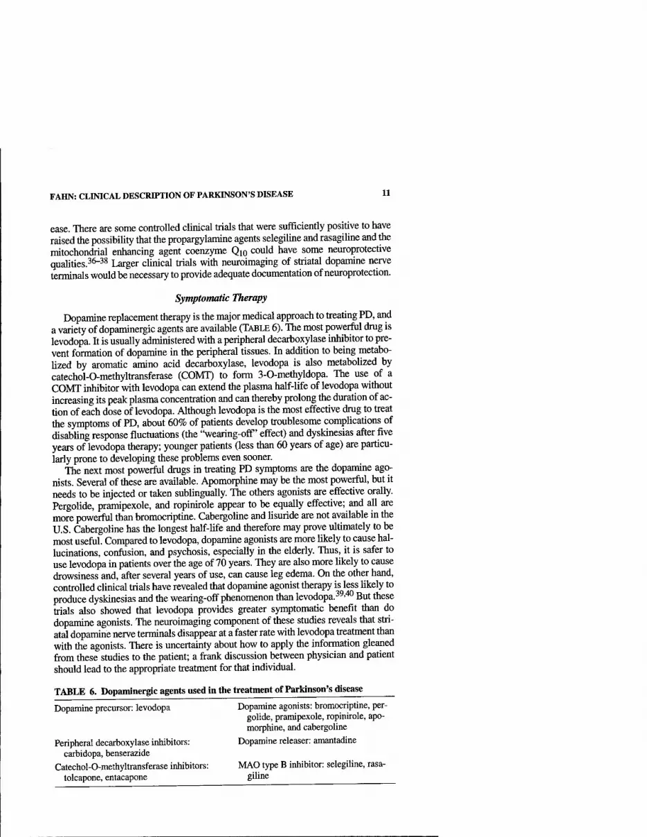
Symptomatic Therapy
Dopamine replacement therapy is the major medical approach to treating PD, and a variety of dopaminergic agents are available (TABLE 6). The most powerful drug is levodopa. It is usually administered with a peripheral decarboxylase inhibitor to pre- vent formation of dopamine in the peripheral tissues. In addition to being metabo- Uzed by aromatic amino acid decarboxylase, levodopa is also metabolized by catechol-0-methyltransferase (COMT) to form 3-0-methyldopa. The use of a COMT inhibitor with levodopa can extend the plasma half-life of levodopa without increasing its peak plasma concentration and can thereby prolong the duration of ac- tion of each dose of levodopa. Although levodopa is the most effective drug to treat the symptoms of PD, about 60% of patients develop troublesome complications of disabling response fluctuations (the "wearing-off' effect) and dyskinesias after five years of levodopa therapy; younger patients (less than 60 years of age) are particu- larly prone to developing these problems even sooner.
The next most powerful drugs in treating PD symptoms are the dopamine ago- nists. Several of these are available. Apomorphine may be the most powerful, but it needs to be injected or taken sublingually. The others agonists are effective orally. Pergolide, pramipexole, and ropinirole appear to be equally effective; and all are more powerful than bromocriptine. Cabergoline and Usuride are not available in the U.S. Cabergoline has the longest half-life and therefore may prove ultimately to be most useful. Compared to levodopa, dopamine agonists are more likely to cause hal- lucinations, confusion, and psychosis, especially in the elderly. Thus, it is safer to use levodopa in patients over the age of 70 years. They are also more likely to cause drowsiness and, after several years of use, can cause leg edema. On the other hand, controlled clinical trials have revealed that dopamine agonist therapy is less likely to produce dyskinesias and the wearing-off phenomenon than levodopa.^^■'^^ But these trials also showed that levodopa provides greater symptomatic benefit than do dopamine agonists. The neuroimaging component of these studies reveals that stri- atal dopamine nerve terminals disappear at a faster rate with levodopa treatment than with the agonists. There is uncertainty about how to apply the information gleaned fi-om these studies to the patient; a frank discussion between physician and patient should lead to the appropriate treatment for that individual.
TABLE 6. Dopaminergic agents used in tlie treatment of Parkinson's disease
Dopamine precursor: levodopa Dopamine agonists: bromocriptine, per- golide, pramipexole, ropinirole, apo- morphine, and cabergoline
Peripheral decarboxylase inhibitors: Dopamine releaser: amantadine carbidopa, benserazide
Catechol-0-methyltransferase inhibitors: MAO type B inhibitor: selegiline, rasa- tolcapone, entacapone giline

12 ANNALS NEW YORK ACADEMY OF SCIENCES
Amantadine has several actions: it has antimuscarinic effects, but more impor- tantly it can activate release of dopamine from nerve terminals, block dopamine up- take into the nerve terminals, and block glutamate receptors. Its dopaminergic actions make it a useful drug to relieve symptoms in about two-thirds of patients, but it can induce livedo reticularis, ankle edema, visual hallucinations, and confusion. Its antiglutamatergic action is useful in reducing the severity of levodopa-induced dyskinesias. The elderly do not tolerate amantadine well because of the adverse mental effects. Monoamine oxidase type B (MAO-B) inhibitors (e.g., selegiline) of- fer mildly effective symptomatic benefit and are without the hypertensive "cheese effect" seen with MAO-A inhibitors; therefore, they can be used in the presence of levodopa therapy. Although there has been considerable debate about the possible protective benefit of selegiline, recent studies evaluating its long-term use indicate that selegiline is associated with less freezing of gait and with a slower rate of clin- ical worsening compared to placebo-treated subjects. These benefits appear to be separate from its mild symptomatic effects because all subjects were receiving the symptomatic benefit from concurrent levodopa therapy.^^ Nondopaminergic agents are also useful to treat many PD symptoms, both motoric and nonmotoric; they are beyond the scope of this review.
Surgical Therapy
Surgery for PD is becoming increasingly available as new techniques of electrical stimulation have been developed and a better understanding of basal ganglia physi- ology has been attained. Stereotaxic deep brain stimulation (DBS) is fast becoming the treatment of choice because ablative lesioning involves greater risk of inducing neurological deficits. With stimulating electrodes, the stimulation can be adjusted, and the electrodes can be removed if necessary. However, DBS is more costly than creating a lesion in the target, and frequent adjustments of the stimulator are usually needed. The location of the stereotaxic target is the other major factor that needs to be individualized for each patient. The thalamus, particularly the ventral intermedi- ate nucleus, appears to be the most successful target for controlling tremor, but this target does not eliminate bradykinesia; so stereotaxic thalamotomy or thalamic DBS is not a preferred choice today. The globus pallidus intema is a more satisfactory tar- get for controlling choreic and dystonic dyskinesias due to levodopa therapy. But the subthalamic nucleus appears to be the best target for controlling bradykinesia. DBS of the subthalamic nucleus, by reducing bradykinesia, allows for a reduction of levodopa dosage, thus reducing the severity of dyskinesias as well. This surgical ap- proach seems the most promising. Surgical procedures for patients with PD are best performed at specialty centers by an experienced team consisting of a neurosurgeon, a neurophysiologist to monitor the target during the operative procedure, and a neu- rologist to program the stimulators. The patient needs close follow-up to adjust the stimulator settings to their optimum.
REFERENCES
1. PARKINSON, J. 1817. An Essay on the Shaking Palsy. Sherwood, Neely, and Jones. London. 2. GoETZ, C.G. 1987. Charcot, the Clinician: the Tuesday Lessons. Excerpts from Nine
Case Presentations on General Neurology Delivered at the Salpetriere Hospital in

FAHN: CLINICAL DESCRIPTION OF PARKINSON'S DISEASE 13
1887-88 by Jean-Martin Charcot. Translated with commentary: pp. 123-124. Raven Press. New York. ,,,„-, j j-^
3. GowERS, W.R. 1893. A Manual of Diseases of the Nervous System, Vol. II, 2nd edit.: p. 644. Blakiston. Philadelphia.
4 OPPENHEM H 1911. Textbook of Nervous Diseases for Physicians and Students, 5th edit., trans, by H. Bruce: pp. 1301-1302. Otto Schulze. Edinburgh.
5. "WtCHSLER, I.S. 1932. A Textbook of Clinical Neurology, 2nd edit.: p. 576. W.B. Saun- ders. Philadelphia.
6 WILSON S A.K. 1940. Neurology, vol. H: p. 793. Williams & Wilkms. Baltimore. 7. SCHWAB, R.S., A.C. ENGLAND & E. PETERSON. 1959. Akinesia in Parkinson's disease.
Neurology 9:65-72. , . . T ■ 8. MEYNERT, T. 1871. Ueber Beitrage zur differential Diagnose der paralytischen Irrsmns.
Wiener Med. Presse 11: 645-647. 9 BRISSAUD, E. 1895. Lecons sur les Maladies Nerveuses. Masson et Cie. Pans.
10. BLOCQ, R & G. MARINESCO. 1893. Sur un cas de tremblement Parkinsonien hemiple- gique, symptomatique d'une tumeur de peduncule cerebral. C. R. Soc. Biol. Paris 5: 105-111. . , , . ^ ,
11 TREnAKOFF, C. 1919. Contribution a I'etude de I'anatoniie pathologique du locus mger de Soemmering avec quelques dedutions relatives a la pathogenic des troubles du tonus musculaire et de la maladie de Parkinson. These de Paris.
12. LEWY, F.H. 1914. Zur pathologischen Anatomic der Paralysis agitans. Dtsch. Z. Ner- venheilk 1: 50-55. ^ . ^
13 FOK, C. & I. NicoLESCO. 1925. Anatomic Cerebrale; Les Noyeux Gns Centraux et la Region Mesencephalo-Sous-Opitique, Suive d'un Appendice sur I'Anatomie Pathologique de la Maladie de Parkinson. Masson et Cie. Paris.
14. HASSLER, R. 1938. Zur Pathologic der Paralysis Agitans und des postenzephalitischen Parkinsonismus. J. Psychol. Neurol. 48: 387^76.
15. GREENFIELD, J.G. & F.D. BOSANQIKT. 1953. The brain-stem lesions in parkinsonism. J. Neurol. Neurosurg. Psychiatry 16:213-226. , , , . ,
16. CARLSSON, A., M. LINDQVIST & T. MAGNUSSON. 1957. 3,4-Dihydroxyphenylalanine and 5-hydroxytryptophan as reserpine antagonists. Nature 180: 1200.
17. CARLSSON, A., M. LINDQVIST, T MAGNUSSON & B. WALDECK. 1958. On the presence of 3-hydroxytyramine in brain. Science 127: 471.
18. BERTLER, A. & E. ROSENGREN. 1959. Occurrence and distribution of catechol amines in brain. ActaPhysiol.Scand. 47:350-361. .
19. SANO, I., T. GAMO, Y. KAMMOTO, et al. 1959. Distribution of catechol compounds in human brain. Biochim. Biophys. Acta 32: 586-587.
20. CARLSSON, A. 1959. The occurrence, distribution and physiological role of catechola- mines in the nervous system. Pharmacol. Rev. 11: 490-493.
21 EHRINGER, H. & O. HoRNYKiEWicz. 1960. Verteilung von Noradrenalin und Dopamin (3-Hydroxytyramin) im Gehim des Menschen und ihr Verhalten bei Erkrankungen der extrapyramidalen Systems. Klin. Wochenschr. 38: 1236-1239.
22 FAHN S & S. PRZEDBORSKI. 2000. Parkinsonism. In Merritt's Neurology, 10th edit. L P.' Rowland, Ed.: 679-^93. Lippincott Williams & Wilkins. Philadelphia.
23. ELBAZ, A., J.H. BOWER, D.M. MARAGANORE, et al. 2002. Risk tables for parkinsonism and Parkinson's disease. J. Clin. Epidemiol. 55(1): 25-31.
24 TANNER, CM., R. OTTMAN, S.M. GOLDMAN, et al. 1999. Parkinson disease in twins— an etiologic study. JAMA 281: 341-346.
25. ELBAZ, A., J.H. BOWER, B.J. PETERSON, et al. 2003. Survival study of Parkinson disease in oimsted county, Minnesota. Arch. Neurol. 60(1): 91-96.
26. BERNHEIMER, H., W BIRKMAYER, O. HORNYKIEWICZ, et al. 1973. Brain dopamine and the syndromes of Parkinson and Huntington. J. Neurol. Sci. 20: 415^55.
27 SNOW, B.J., C.S. LEE, M. SCHULZER, et al. 1994. Longitudinal fluorodopa positron emission tomographic studies of the evolution of idiopathic Parkinsomsm. Ann. Neurol. 36:759-764. , . .
28 SEIBYL J P KL MAREK, D. QUINLAN, et al. 1995. Decreased smgle-photon emission computed tomographic [(123)]I beta-CIT striatal uptake correlates with symptom severity in Parkinson's disease. Ann. Neurol. 38: 589-598.
14 ANNALS NEW YORK ACADEMY OF SCIENCES
29. EiDELBERG, D., J.R. MOELLER, T. ISIKAWA, et al. 1995. Assessment of disease severity in parkinsonism with fluorine-18- fluorodeoxyglucose and PET. J. Nucl. Med. 36: 378-383.
30. MoRRiSH, P.K., G.V. SAWLE & D.J. BROOKS. 1996. An [F-18]dopa-PET and clinical study of the rate of progression in Parkinson's disease. Brain 119: 585-591.
31. BENAMER, H.T.S., J. PATTERSON, D.J. WYPER, et al. 2000. Correlation of Parkinson's disease severity and duration with 1-123- FP-CIT SPECT striatal uptake. Mov. Dis- ord. 15(4): 692-698.
32. PENNEY, J.B., JR & A.B. YOUNG. 1986. Striatal inhomogeneities and basal ganglia function. Mov. Disord. 1: 3-14.
33. MILLER, W.C. & M.R. DELONG. 1988. Parkinsonian symptomatology: an anatomical and physiological analysis. Ann. N.Y. Acad. Sci. 515: 287-302.
34. MITCHELL, I.J., C.E. CLARKE, S. BOYCE, et al. 1989. Neural mechanisms underiying parkinsonian symptoms based upon regional uptake of 2-deoxyglucose in monkeys exposed to l-methyl-4-phenyl-l,2,3,6-tetrahydropyridine. Neuroscience 32: 213- 226.
35. CONWAY, K.A., J.C. ROCHET, R.M. BIEGANSKI & P.T.J. LANSBURY. 2001. Kinetic stabi- lization of the alpha-synuclein protofibril by a dopamine-alpha-synuclein adduct. Science 294: 1267-1268.
36. SHOULSON, I., D. OAKES, S. FAHN, et al.; PARKINSON STUDY GROUP. 2002. Impact of sustained deprenyl (selegiline) in levodopa-treated Parkinson's disease: a random- ized placebo-controlled extension of the deprenyl and tocopherol antioxidative ther- apy of parkinsonism trial. Ann. Neurol. 51(5): 604-612.
37. PARKINSON STUDY GROUP. 2002. A controlled trial of rasagiline in early Parkinson dis- ease—the TEMPO study. Arch. Neurol. 59(12): 1937-1943.
38. SHULTS, C.W., D. OAKES, K. KIEBURTZ, et al. 2002. Effects of coenzyme Qig in early Parkinson disease. Evidence of slowing of the functional decline. Arch. Neurol. 59: 1541-1550.
39. RASCOL, O., D.J. BROOKS, A.D. KORCZYN, et al. 2000. A five-year study of the inci- dence of dyskinesia in patients with early Parkinson's disease who were treated with ropinirole or levodopa. N. Engl. J. Med. 342: 1484-1491.
40. PARKINSON STUDY GROUP. 2000. Pramipexole versus levodopa as the initial treatment for Parkinson's disease: a randomized controlled trial. JAMA 284: 1931-1938.

Physiology and Pathophysiology of Parkinson's Disease
CLEMENT HAMANI AND ANDRES M. LOZANO
Division ofNeurosurgery, Toronto Western Hospital, University of Toronto, Toronto, Ontario, Canada
ABSTRACT: The behavior of neurons in the basal ganglia is severely disrupted in Parkinson's disease (PD). In nonhuman parkinsonian primate models, the disturbance in neurons in basal ganglia output structures include increased fir- ing, bursting, an augmented synchrony, correlated activity, and a tendency to- wards loss of specificity m their receptive fields. This abnormal neuronal behavior, transmitted to the thalamus, cortex and brainstem, is thought to dis- rupt the functioning of the motor system and underlie the major motor mani- festations of PD—tremor, ri^dity, akinesia, gait, and postural disturbances. The mainstay of treatment has been to replace the missing dopamine with med- ication. With time and disease progression, however, dopamine replacement becomes less efficacious and new adverse effects, including the development of motor fluctuations and drug-induced involuntary movements or dyskinesias, emerge. When the patients reach this stage, surgical therapy becomes an op- tion. Most surgical interventions are performed at the level of the thalamus, globus palUdus, and subthalamic nucleus, aiming at the disruption of the pathological activity that accompanies the Parkinson's deficiency state. With this abnormal neuronal activity neutralized, normal movements can in many cases be restored.
KEYWORDS: Parkinson's disease; sui^ery; subthalamic nucleus; globus palli- dus; physiopathology; movement disorders
INTRODUCTION
It is estimated that approximately one million North Americans suffer from Par- kinson's disease. To date, little is known about the etiology of this disorder; but some important clues are coming from a variety of sources, including genetic studies and studies of environmental toxins. ^-^ Despite these advances, the etiology of Parkin- son's disease for most patients remains enigmatic.
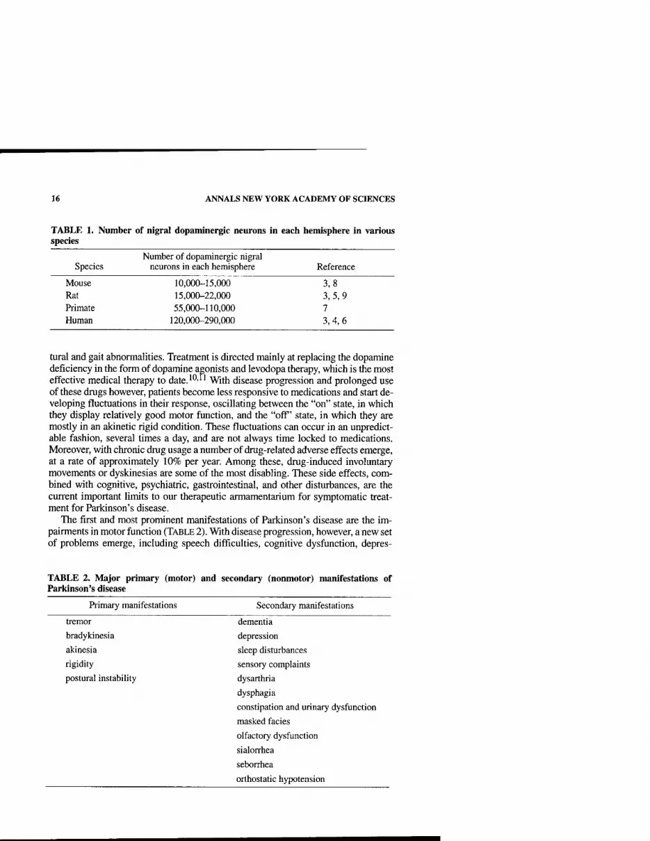
The number of dopaminergic neurons in the substantia nigra varies from species to species^^ (TABLE 1). Humans have approximately 220,000 dopaminergic neurons in the substantia nigra of each hemisphere.^ When more than 50% of these cells are lost, patients start to develop the signs and symptoms of the disease—tremor, rigid- ity, akinesia and bradykinesia (poverty and slowness of movement), as well as pos-
Address for correspondence: Andres Lozano, Toronto Western Hospital, West Wing 4-447, 399 Bathurst Street, Toronto, ON M5T 2S8, Canada. Voice: 416-603-6200; fax: 416-603-5298.
Ann. N.Y. Acad. Sci. 991:15-21 (2003). © 2003 New York Academy of Sciences.
15
16 ANNALS NEW YORK ACADEMY OF SCIENCES
TABLE 1. Number of nigral dopaminergic neurons in each hemisphere in various species
Number of dopaminergic nigral Species neurons in each hemisphere Reference
Mouse 10,000-15,000 Rat 15,000-22,000 Primate 55,000-110,000 Human 120,000-290,000
3,8 3,5,9 7 3,4,6
tural and gait abnormalities. Treatment is directed mainly at replacing the dopamine deficiency in the form of dopamine agonists and levodopa therapy, which is the most effective medical therapy to date.^*^-'^ With disease progression and prolonged use of these drugs however, patients become less responsive to medications and start de- veloping fluctuations in their response, oscillating between the "on" state, in which they display relatively good motor function, and the "off' state, in which they are mostly in an akinetic rigid condition. These fluctuations can occur in an unpredict- able fashion, several times a day, and are not always time locked to medications. Moreover, with chronic drug usage a number of drug-related adverse effects emerge, at a rate of approximately 10% per year. Among these, drug-induced involuntary movements or dyskinesias are some of the most disabling. These side effects, com- bined with cognitive, psychiatric, gastrointestinal, and other disturbances, are the current important limits to our therapeutic armamentarium for symptomatic treat- ment for Parkinson's disease.
The first and most prominent manifestations of Parkinson's disease are the im- pairments in motor function (TABLE 2). With disease progression, however, a new set of problems emerge, including speech difficulties, cognitive dysfunction, depres-
TABLE 2. Major primary (motor) and secondary (nonmotor) manifestations of Parl(inson's disease
Primary manifestations Secondary manifestations
tremor
bradykinesia
akinesia
rigidity
postural instability
dementia
depression
sleep disturbances
sensory complaints
dysarthria
dysphagia
constipation and urinary dysfunction
masked fades
olfactory dysfunction
sialorrhea
seborrhea
orthostatic hypotension

HAMANI & LOZANO: PHYSIOLOGY AND PATHOPHYSIOLOGY OF PD 17
sion, sleep disturbances, constipation, bladder and sexual dysfunctions, and a series of autonomic problems that are outlined in TABLE 2.2'12-14 Most of these problems are poorly or not responsive to dopaminergic replacement, suggesting that other neurotransmitter systems might be involved in their pathogenesis. In fact, there is in- creasing evidence for abnormalities in other neurotransmitter systems as well as for cell loss and degeneration that extends beyond the dopaminergic pathways. Indeed, the locus ceroeuleus catecholaminergic system, the raphe nuclei serotonergic sys- tem, and the cholinergic neurons from the nucleus basalis and a number of other ar- eas,' including the cortex, olfactory bulb, sympathetic ganglia, and the central sympathetic nervous system, are compromised in Parkinson's disease as ^gll 1,12,13,15 Degeneration in these diverse systems indicates that we are dealing with a disorder that extends beyond dopaminergic neurons, and both therapeutic maneuvers and pathogenic mechanisms have to take these associated conditions into account.
PATHOPHYSIOLOGY OF THE MAJOR PARKINSON'S DISEASE SYMPTOMS
How Does the Dopamine Deficiency State Lead to the Signs and Symptoms ofPD?
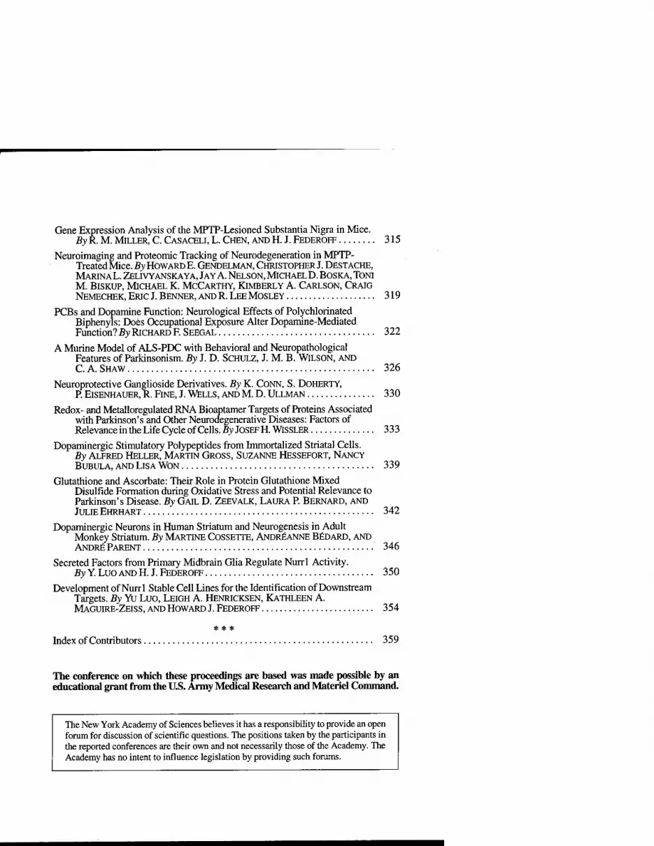
In animal models and in humans with Parkinson's disease, the neurophysiologic consequences of dopamine deficiency are striking. A model of basal ganglia func- tion in normal and dopamine deficiency states has been proposed (FlG.l).^^' This model proposes that dopamine deficiency produces dysfunction in the striatum, leading to: (1) decreased activity in the direct pathway, from GABAergic striatal neurons to the intemal segment of the globus pallidus (GPi) and substantia nigra pars reticulata (SNpr) and (2) increased drive through the indirect pathway, involving par- ticularly the external segment of the globus pallidus (GPe) and subthalamic nucleus (STN). As a consequence, there is disruption of the activity in basal gangUa output structures (GPi and SNpr), which in turn disrupts the activity in brain stem motor areas, including the pedunculopontine nucleus and the thalamocortical motor sys- tem. ^'^ This disruption is thought to be responsible for the difficulty in initiation of movements and the poverty of motion that are characteristic of Parkinson's dis- ease. 1^"^^ Although the model does reasonably well at explaining akinesia, it is does not adequately explain some of the other cardinal features of Parkinson's disease, such as tremor or rigidity.^^ Further, the model does not take into account that dopamine exerts its effects not only in the striatum but also throughout basal gangUa nuclei and at cortical levels.
The development of the nonhuman primate MPTP model of Parkinson's disease has made possible an examination of the behavior of neurons in normal and parkin- sonian states, leading to significant insights into the disrupted activity that accompa- nies this condition.^^"^''' Moreover, these new concepts have led to the reemergence of neurosurgery to treat medically refractory Parkinson's disease. The surgical ap- proaches are producing striking symptomatic benefits in parkinsonian patients ' and are providing a unique opportunity to examine cellular activity in the thalamus, globus pallidus, and subthalamic nucleus in Parkinson's disease.
18 ANNALS NEW YORK ACADEMY OF SCIENCES
A Cortex
1 f Glutamate
c
Motor Thalamus -^
Glutamate
GABA/Subst.P
Striatum Dl D2
GABA/Enkephalin
Brain Stem
S.Nigra compacta
GABA
S.Nigra reticulata
Subthalamic nucleus
Glutamate
Glutamate
B Striatum Dl D2
GABA/Enkephalin
Glutamate
Motor Thalamus
Brain Stem
FIGURE 1. Proposed functional model of the basal ganglia in normal subjects (A) and patients with Parkinson's disease (B). In B, the width of the arrows indicates the degree of overall functional change in activity as compared with the normal state. Dotted lines indicate the dysfunctional nigrostriatal dopamine system in Parkinson's disease. GPe, external seg- ment of the globus pallidus; GPi, internal segment of the globus pallidus; S. Nigra, substan- tia nigra; Subst.P, substantia P.

HAMANI & LOZANO: PHYSIOLOGY AND PATHOPHYSIOLOGY OF PD 19
NEUROSURGICAL PROCEDURES FOR PARKINSON'S DISEASE
The renaissance of surgery for movement disorders has come as a consequence of the unmet needs of a large number of patients who continue to be disabled despite the best available medical therapy, coupled with important technological advances in brain imaging and physiological recordings that have increased the accuracy and safety of surgery. By using these techniques, particularly microelectrode recordings, it has become possible to obtain direct measures of cellular activity of the basal gan- gha in humans with Parkinson's disease and other movement disorders and to exam- ine whether the pathological alterations seen in parkinsonian MPT? nonhuman primate models are also present in humans with Parkinson's disease.^'^^'^
There is one main surgical strategy in current clinical use for the treatment of Par- kinson's disease. It is the suppression of abnormal neural activity in basal ganglia circuits. This is achieved by either lesioning the involved structure or by the appU- cation of constant electrical stimulation, termed deep-brain stimulation or DBS, to block the pathological activity. These interventions occur at the level of the thala- mus, globus pallidus, and subthalamic nucleus.
The overall surgical experience has made it clear that there are certain symptoms, including tremor, rigidity, and akinesia, that respond dramatically to surgery; while others respond less so, and yet an additional group is unresponsive. Levodopa-in- duced involuntary movements or dyskinesias are among the most effectively treated conditions. With surgical treatment one can anticipate reductions of up to 80-90% in these symptoms. Tremor improvement reaches 80% with any of the three targets previously described. Rigidity and akinesia scores improve by approximately 60%, as assessed with the Unified Parkinson's Disease Rating Scale, after bilateral deep- brain stimulation procedures. Gait and postural abnormalities are less responsive, al- though significant benefits can be observed. The effectiveness of surgical procedures seems to be equivalent whether lesions are performed or chronic deep-brain stimu- lation electrodes are implanted in the basal ganglia. Nevertheless, bilateral lesions lead to an unexpectedly high incidence of side effects, mostly deterioration in speech and cognitive effects. Because patients with Parkinson's disease are disabled by bi- lateral symptoms, deep-brain stimulation is becoming the procedure of choice as, in contrast to lesions, DBS is adjustable and reversible.'^^-^^'^^
An alternative surgical therapy, aiming at replacement of the missing neurotrans- mitter by transplantation of dopaminergic neurons, is under investigation.'**^''^' To date however, these procedures provide only modest benefits and are associated with significant adverse effects, particularly due to the fact that dopamine release cannot be controlled. Therefore, transplants are not being used in clinical practice but are being studied in research protocols.
Shortcomings of Surgery
Despite these advances in therapy, there continues to be a number of signs and symptoms of Parkinson's disease that fail to respond to dopamine or surgery. The reason for this is likely related to coexisting pathological changes in other circuits and neurotransmitters that are also involved in the pathogenesis of the disease.
In conclusion, modem neurosurgery has allowed an examination of the neuronal activity that occurs in the brain of patients with Parkinson's disease. These studies

20 ANNALS NEW YORK ACADEMY OF SCIENCES
have revealed prominent disruption in the activity of basal ganglia neurons. Surgical approaches alter, block, or neutralize this abnormal activity. In this sense, it appears that in physiological terms it is better to receive no neural activity from the motor component of the basal ganglia than to receive a pathological one. In other words,"no news is better than bad news."
To date, all surgical treatments are symptomatic, which means that we are treat- ing only the symptoms of the disease without influencing its natural history. What is now needed are treatments that change the natural course of the disease, that slow it down or stop it. Future aspects of surgical treatment, including advancements in deep-brain stimulation, local delivery of neural active substances, the appUcation of neurotrophins and stem cells, gene therapy, and molecular neurosurgery, are all promising strategies and can be considered bright lights in the horizon.
ACKNOWLEDGMENTS
C.H. is currently receiving a CAPES postdoctoral fellowship.
REFERENCES
1. LANG, A.E. & A.M. LOZANO. 1998. Parkinson's disease. First of two parts. N. Engl. J. Med. 339: 1044-1053.
2. JELLINGER, K.A. 2001. The pathology of Parkinson's disease. Adv. Neurol. 86: 55-72. 3. GERMAN, D.C, D.S. SCHLUSSELBERG & D.J. WOODWARD. 1983. Three-dimensional
computer reconstruction of midbrain dopaminergic neuronal populations: from mouse to man. J. Neural. Transm. 57: 243-254.
4. GERMAN, D.C, et al. 1989. Midbrain dopaminergic cell loss in Parkinson's disease: computer visualization. Ann. Neurol. 26: 507-514.
5. GERMAN, D.C. & K.F. MANAYE. 1993. Midbrain dopaminergic neurons (nuclei AS, A9, and AlO): three-dimensional reconstruction in the rat. J. Comp. Neurol. 331: 297-309.
6. GRAYBIEL, A.M., E.C. HiRSCH & Y. AGID. 1990. The nigrostriatal system in Parkin- son's disease. Adv. Neurol. 53: 17-29.
7. EMBORG, M.E., et al. 1998. Age-related declines in nigral neuronal function correlate with motor impairments in rhesus monkeys. J. Comp. Neurol. 401: 253-265.
8. NELSON, E.L., et al. 1996. Midbrain dopaminergic neurons in the mouse: computer- assisted mapping. J. Comp. Neurol. 369: 361-371.
9. SMTTH, Y. & J.Z. KiEVAL. 2000. Anatomy of the dopamine system in the basal ganglia. Trends Neurosci. 23: S28-33.
10. JANKOVIC, J. 2000. Complications and limitations of drug therapy for Parkinson' dis- ease. Neurology 55: S2-6.
11. RASCOL, O., et al. 2002. Treatment interventions for Parkinson's disease: an evidence based assessment. Lancet 359: 1589-1598.
12. SINGER, C, W.J. WEINER & J.R. SANCHEZ-RAMOS. 1992. Autonomic dysfunction in men with Parkinson's disease. Eur Neurol. 32: 134—140.
13. WOLTERS, E.C. 2000. Psychiatric complications in Parkinson's disease. J. Neural Transm. Suppl. (60): 1291-302.
14. WOLTERS, E.C. 2001. Psychiatric complications in the treatment of Parkinson's dis- ease. Adv. Neurol. 86: 385-393.
15. CHURCHYARD, A. & A.J. LEES. 1997. The relationship between dementia and direct involvement of the hippocampus and amygdala in Parkinson's disease. Neurology 49: 1570-1576.
16. MINK, J.W. 1996. The basal ganglia: focused selection and inhibition of competing motor programs. Prog. Neurobiol. 50: 381^25.

HAMANI & LOZANO: PHYSIOLOGY AND PATHOPHYSIOLOGY OF PD 21
17. ALBIN, R.L., A.B. YOUNG & J.B. PEI^JEY. 1989. The functional anatomy of basal gan- glia disorders. Trends Neurosci. 12: 366-375.
18 PAHAPHi, P.A. & A.M. LozANO. 2000. The pedunculopontine nucleus and Parkinson's disease. Brain 123 (Pt. 9): 1767-1783.
19 BERARDELLI, A., et al. 2001. Pathophysiology of bradykinesia in Parkinson's disease. Brain 124: 2131-2146. ^„ .
20. BLOEM, B.R., J.P VAN VuGT & D.J. BECKLEY. 2001. Postural instability and falls in Parkinson's disease. Adv. Neurol. 87: 209-223.
21. CANTELLO, R., et al. 1996. Pathophysiology of Parkinson's disease rigidity. Role of corticospinal motor projections. Adv Neurol. 69: 129-133.
22. DELWAIDE, PJ., J.L. PEPIN & A. MAERTENS DE NOORDHOUT. 1990. [Parkinsonian rigid- ity: clinical and physiopathologic aspects]. Rev. Neurol. (Paris) 146: 548-554.
23. YANAGISAWA, N., R. HAYASHI & H. MITOMA. 2001. Pathophysiology of frozen gait in Parkinsonism. Adv. Neurol. 87: 199-207.
24. CARR, J. 2002. Tremor in Parkinson's disease. Parkinsonism Relat. Disord. 8: 223- 234.
25. BERGMAN, H., et al. 1998. Physiological aspects of information processing m the basal ganglia of normal and parkinsonian primates. Trends Neurosco. 21: 32-38.
26. BERGMAN, H. & G. DEUSCHL. 2002. Pathophysiology of Parkinson's disease: from cUn- ical neurology to basic neuroscience and back. Mov. Disord. 17 (Suppl. 3): 828^0.
27. DELONG, M.R. 1990. Primate models of movement disorders of basal ganglia origin. Trends Neurosci. 13: 281-285.
28 LANG, A.E. & A.M. LOZANO. 1998. Parkinson's disease. Second of two parts. N. Engl. J.Med. 339:1130-1143.
29 LANG, A.E., et al. 1997. Posteroventral medial pallidotomy in advanced Parkinson s disease. N. Engl. J. Med. 337: 1036-1042.
30. GURIDI, J., et al. 2000. Targeting the basal gangMa for deep brain stimulation in Parkin- son's disease. Neurology 55: S21-28.
31. GARONZIK, LM., et al. 2002. Intraoperative microelectrode and semi-micfoelectrode recording during the physiological localization of the thalamic nucleus ventral inter- mediate. Mov. Disord. 17 (Suppl. 3): S135-144.
32. HUTCHISON, W.D., et al. 1994. Differential neuronal activity in segments of globus pal- lidus in Parkinson's disease patients. Neuroreport 5: 1533-1537.
33. HUTCHISON, W.D., et al. 1998. Neurophysiological identification of the subthalamic nucleus in surgery for Parkinson's disease. Ann. Neurol. 44: 622-628.
34. LOZANO, A.M., et al. 1995. Effect of GPi pallidotomy on motor function in Parkinson's disease. Lancet 346: 1383-1387.
35. MAGNIN, M., A. MOREL & D. JEANMONOD. 2000. Single-unit analysis of the pallidum, thalamus and subthalamic nucleus in parkinsonian patients. Neuroscience 96: 549- 564.
36. LENZ, F.A., et al. 1988. Single unit analysis of the human ventral thalamic nuclear group: correlation of thalamic "tremor cells" with the 3-6 Hz component of parkin- sonian tremor. J. Neurosci. 8: 754-764.
37. LENZ, F.A., et al. 1988. Single-unit analysis of the human ventral thalamic nuclear group: somatosensory responses. J. Neurophysiol. 59: 299-316.
38. LANOTTE, M.M., et al. 2002. Deep brain stimulation of the subthalamic nucleus: ana- tomical, neurophysiological, and outcome correlations with the effects of stimula- tion. J. Neurol. Neurosurg. Psychaitry 72: 53-58.
39. OBESO, J.A., et al. 2000. Pathophysiologic basis of surgery for Parkinson's disease. Neurology 55: S7-12.
40. OLANOW, C.W., T. FREEMAN & J. KORDOWER. 2001. Transplantation of embryonic dopamine neurons for severe Parkinson's disease. N. Engl. J. Med. 345: 146; discus- sion 147.
41. ISACSON, O., et al. 2001. Improved surgical cell therapy in Parkinson's disease. Physi- ological basis and new transplantation methodology. Adv. Neurol. 86: 447-454.
PET Studies on the Function of Dopamine in Health and Parkinson's Disease
DAVID J. BROOKS
MRC Clinical Sciences Centre and Division ofNeuroscience, Faculty of Medicine, Imperial College, London, UK
ABSTRACT: Positron emission tomography (PET) can detect the presence of striatal, paUidal, midbrain, and cortical dopamine terminal dysfunction in vivo in Parkinson's disease (PD). In addition, dopamine release during motor tasks can be assessed as reflected by changes in receptor availability to PET ligands. Furthermore, the functional effects of focal dopamine replacement via implan- tation of fetal cells or glia-derived neurotrophic factor (GDNF) infusion into putamen can be monitored. In this review, the insight that PET has given us concerning the role of dopamine in motor control is presented, and the func- tional substrates underlying PD symptomatologies are discussed.
KEYWORDS: Parkinson; dopamine; serotonin; motor; PET; activation; GDNF
INTRODUCTION
Functional imaging allows four potential approaches to determining possible roles of dopamine in control of motor functions: First, loss of dopamine terminal function, as reflected by levels of striatal and extrastriatal '^F-dopa uptake or dopamine transporter (DAT) ligand binding, can be correlated with disabihty and cognitive performance. Second, the locomotor and behavioral responses to focal stri- atal replacement of dopaminergic tone with implants of fetal mesencephalic cells or local neurotropic factor infusions can be correlated with dopaminergic function. Third, alterations in the patterns of resting and activated blood flow and metabolism in PD and their normalization following dopaminergic replacement can be moni- tored. Fourth, dopamine release in striatal and cortical areas during task performance or following pharmacological challenges can be measured indirectly in vivo as re- flected by reductions in dopamine receptor availability to antagonists such as ''C- raclopride. Based on microdialysis studies, it has been estimated that a 1% change in striatal "C-raclopride binding corresponds to at least an 8% change in synaptic dopamine levels.^
Dopamine fibers arise from the substantia nigra compacta and the ventral teg- mentum (VTA) of the midbrain and project to dorsal (caudate, putamen) and ventral (caudate, putamen, nucleus accumbens, olfactory tubercle) striatum. There is a less-
Address for correspondence: David J. Brooks, M.D., D.Sc, F.R.C.P., RMed.Sci., Hartnett Professor of Neurology, Cyclotron Building, Hammersmith Hospital, Du Cane Rd., London W12 ONN, UK.
Ann. N.Y. Acad. Sci. 991: 22-35 (2003). © 2003 New York Academy of Sciences.
22

BROOKS: PET STUDIES ON THE FUNCTION OF DOPAMINE 23
er output to frontal cortex, amygdala, and the anterior cingulate area. Our under- standing about the precise role that dopamine plays in motor control and cognition is still limited. Awake animal studies have suggested that burst release of dopamine alerts animals to novel and rewarding stimuli, burst firing of dopamine neurons be- ing correlated with the presence of an incentive rather than with movement per se.^'^ Unexpected rewards are most effective in inducing phasic firing of dopamine neu- rons. As conditioning to a stimulus reliably predicts the presence of a reward, firing of dopamine neurons on reward presentation becomes attenuated, while firing on presentation of the conditioning stimulus increases.^ Once conditioning is complete and timing of the reward on stimulus presentation becomes entirely predictable, dopamine neurons cease to fire on presentation of the conditioning stimulus. Phasic firing is then observed only if an unexpected reward occurs, and depression of activ- ity is seen if a predicted reward fails to occur. These observations suggest that dopamine neurons are reacting to errors in reward prediction rather than presentation of the reward per se. This in turn suggests that they act to reinforce rewarding pat- terns of behavior and so play a primary role in conditional motor learning.
Dopamine, however, does not simply mediate rewarded behavior but almost cer- tainly facilitates conditional learning in general.^ When rats are aversively condi- tioned to auditory and visual stimuli with an electric foot shock, the first presentation of the foot shock with the conditioning stimulus results in an increase in frontal dopamine release, while ventral striatal dopamine levels remain unaltered.^ Success- ful aversive conditioning, then, results in progressive increases in ventral striatal dopamine release, while frontal dopamine release diminishes. These findings sup- port the concept that dopamine release in striatum reinforces learning but suggest the frontal dopamine release is concerned more with alerting animals to novel circum- stances and mediating selective attention.^
Trying to dissociate the functional roles of basal ganglia and frontal dopamine re- lease is probably inappropriate, as the basal ganglia and frontal areas are relays in a system of parallel disttibuted corticosubcortical loops. At least five separate loops link the basal gangUa via the ventral and dorsomedial thalamus to cortical premotor and prefrontal areas.^ These include supplementary motor area (S]V[A)-putamen; dorsolateral prefrontal cortex (DLPFC>-dorsal caudate; orbitofrontal cortex (OFC) -ventral striatal; and anterior cingulate cortex (ACA)-ventral stiiatal loops. These loops appear to be spatially segregated based on the findings of anterograde and ret- rograde tracer experiments in nonhuman primates.^ It has been proposed that tonic background dopamine release in the basal ganglia may act to "bind" or focus and fil- ter the individual functions of these segregated loops.^
The pathology of Parkinson's disease (PD) targets the dopamine cells in the sub- stantia nigra in association with the formation of neuronal Lewy inclusion bodies. Midbrain tegmental dopamine neurons projecting to anterior cingulate, orbitofrontal cortex, and amygdala are involved to a lesser extent. Serotonergic cells in the median raphe and noradrenergic cells in the locus ceruleus also degenerate, as do other pig- mented and brain stem nuclei including the locus coeruleus and nucleus basaUs.^*^ Loss of cells from the substantia nigra in PD results in profound dopamine depletion in the shiatum, lateral nigral projections to putamen being most affected.^^'^^While the pathology of PD targets subcortical nuclei, anterior cingulate and association neocortex can also show Lewy body degeneration; and currentiy it remains uncertain whether dementia of Lewy body type and PD represent ends of a spectrum. ^^

24 ANNALS NEW YORK ACADEMY OF SCIENCES
FIGURE 1. A statistical parametric map superimposed on an MRI template showing bilateral putamen, cingulate, and motor cortex loss of dopamine storage in early hemi-PD.
FOCAL STRIATAL REPLACEMENT OF DOPAMINE IN PD
Visual inspection of '^F-dopa positron emission tomography (PET) images of pa- tients with early Parkinson's disease gives the impression that loss of dopamine ter- minal function is confined to nigrostriatal projections. Using statistical parametric mapping (SPM) to compare mean dopamine storage capacity at a voxel level be- tween early PD cases and age-matched normal subjects, however, reveals early mo- tor cortex and anterior cingulate terminal dysfunction (see RG. I). This emphasises that PD does not simply provide a natural lesion model for basal gangUa dysfunc- tion. As the disease advances, progressive loss of striatal, cingulate, and frontal dopamine storage is seen. Significant inverse correlations between disability scores rated with the Unified Parkinson's Disease Rating Scale (UPDRS) and putamen, midbrain, and anterior cingulate '^F-dopa influx constants (Ki) are evident across the mild-to-advanced range of PD (FiG. 2. This raises the question, how great an in- fluence does frontal loss of dopamine have in actuality on locomotor and behavioral function in PD?
One approach to answer this question is simply to correlate scores on behavioral tasks with regional levels of brain '^F-dopa uptake in PD. When correct scores on the Tower of London task were compared with regional brain dopamine storage in PD, only individual right head of caudate nucleus Ki's showed a significant correla- tion with efficiency of spatial planning.^'* This finding implies that adequate levels of caudate dopamine are crucial for performance of spatial executive tasks. However, one cannot rule out a role of frontal dopamine even though no correlation was seen with either cingulate or prefrontal Ki levels, as these signals are relatively low (only 20% of caudate levels in normal subjects).
A more novel way of approaching the problem is to examine the behavioral ef- fects of focal striatal replacement of dopamine using implants of human fetal mes- encephalic cells or intraputamenal infusions of neurotropic factors such as glia- derived neurotrophic factor (GDNF). Several open series and a double-blind con- trolled trial of the efficacy of transplants in advanced PD have demonstrated signif- icant increases in striatal ^^F-dopa storage after grafting in PD, which correlate with improved UPDRS scores. '^ It has also been shown that grafts survive in man for over 10 years and release normal levels of dopamine after an amphetamine challenge. ^^
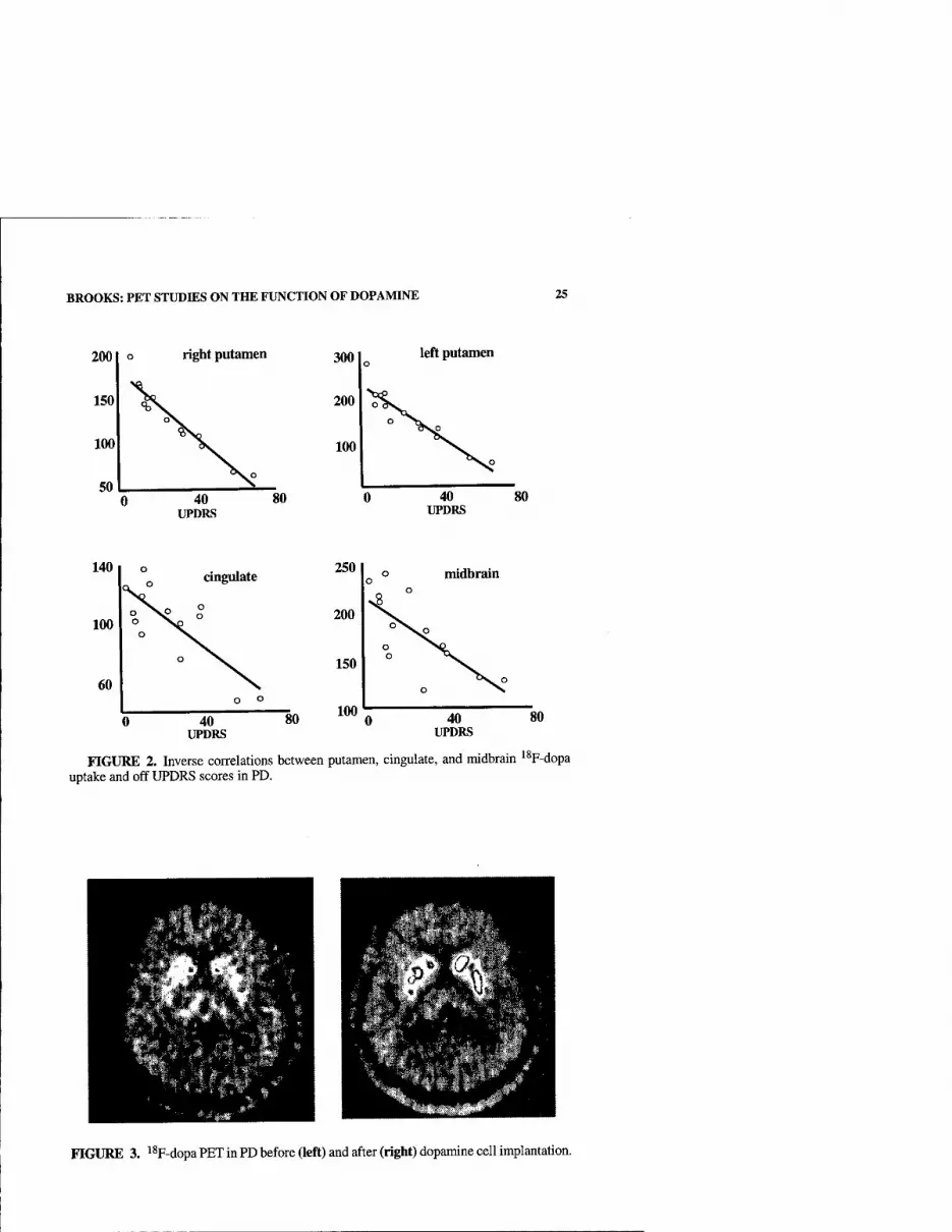
BROOKS: PET STUDIES ON THE FUNCTION OF DOPAMINE 25
200 o right putamen
150 X V 100 "V SO V
left putamen
40 UPDRS
80
cingulate
40 UPDRS
250
200
150
100
midbrain
40 UPDRS
80
FIGURE 2. Inverse correlations between putamen, cingulate, and midbrain '^F-dopa uptake and off UPDRS scores in PD.
FIGURE 3. i^F-dopa PET in PD before (left) and after (right) dopamine cell implantation.

26 ANNALS NEW YORK ACADEMY OF SCIENCES
0
Putamen '*F-dopa
UPDRS
SMA actn
DLPFC actn
10 —I 1 1
15 20 25 months
FIGURE 4. Recovery of PD cortical activation vs. dopamine function after grafting. Serial putamen '^F-dopa Ki values, off UPDRS scores, and percentage increases in supple- mentary motor area and dorsolateral prefrontal activation measured with H2'^0 PET in four transplanted PD patients. *P < 0.05.
Piccini and coworkers have studied the effects of implanting head of caudate and putamen bilaterally with human fetal mesencephalic cells'" (see RG. 3). At 6.5 months after transplantation mean striatal dopamine storage capacity, as measured by '^F-dopa PET, was significantly elevated in these patients (putamen 78%, caudate 27%). This was associated with a nonsignificant mean 12-point clinical improve- ment on the UPDRS (see RG. 4). At 18 months postsurgery there was further signif- icant cUnical improvement (24 points compared with baseUne) in the absence of further increases in striatal '^F-dopa uptake, suggesting that the graft had developed more effective connections.
PD patients, when withdrawn from medication, show a characteristic impairment of activation of striatomesial premotor-prefrontal projections during performance of motor tasks.'^ It has been demonstrated that replacement of dopaminergic tone by giving oral levodopa or subcutaneous apomorphine, a D1/D2 agonist, functionally restores cortical activation in PD'^"22 ^jong with reduction of akinesia. More recent- ly, the effects of focally implanting the striatum with fetal dopaminergic cells on movement-related premotor and prefrontal activation in PD have been studied.'^ Four PD patients who received bilateral human fetal mesencephalic transplants into caudate and putamen were studied with H2'^0 PET at baseline and over two years following surgery. At baseline, H2'^0 PET studies showed reduced premotor and absent prefrontal activation during performance of a paced motor task where the pa- tient moved a joystick in freely chosen directions. At six months there was no sig- nificant change in cortical activation, but by 18 months mesial premotor and dorsal prefrontal activation had significantly improved (FiG. 4). These observations suggest

BROOKS: PET STUDIES ON THE FUNCTION OF DOPAMINE 27
that dorsal striatal grafts of dopamine cells in PD are eff'ective in restoring levels of dorsal premotor and prefrontal activation and improving executive behavior. Addi- tionally, graft function goes beyond that of a simple dopamine delivery system, and integration of the grafted neurons into the host brain is necessary in order to produce substantial clinical recovery in PD. This strengthens the argument that frontal dopamine loss, though present in PD, may be less critical than striatal loss for medi- ating dorsal prefrontal executive behaviors.
GDNF is a potent neurotrophic factor known to prevent the degeneration of dopamine neurons in rodent and primate models of PD where nigral degeneration is toxically induced. Gill and colleagues have recently examined the safety and effica- cy of infusing GDNF directly into the posterior putamen of five patients with PD via in-dwelling catheters.^^ All patients tolerated constant GDNF delivery at a final lev- el of 14 [ig/day (6 mlVhour) into the posterior dorsal putamen via mechanical pumps for over one year, unilaterally in one and bilaterally in four patients, without serious side effects. Significant improvements were found in UPDRS subscores: 39% and 61% improvements in the off-medication motor III and activities of daily living II subscales, respectively, at 12 months. No change in cognitive status was detected on a battery of behavioral tests. Regions of interest placed in the immediate vicinity of the catheter tip showed 18-24% increases in putaminal ^^F-dopa Ki, which were confirmed with statistical parametric mapping. Additionally, SPM detected 16-26% increases in nigral dopamine storage, suggesting that retrograde transport of GDNF had occurred. These findings imply that local GDNF infusion appears to be safe and represents a potential restorative therapy for PD when applied directly to striatum.
Motor UPDRS
"F-dopaKi
30
10
0.008
0.007
0.006
0.005
30 40 50 60 70 80 0.004
Disease duration
FIGURE 5. Levels of internal pallidal ^^F-dopa Ki versus off motor UPDRS scores in PD. While pallidal ^^F-dopa uptake is raised, the UPDRS score is maintained low but rap- idly rises with fluctuations when pallidal ^^F-dopa uptake falls below normal.

28 ANNALS NEW YORK ACADEMY OF SCIENCES
Interestingly, dramatic improvements in basic locomotor functions were achieved with a neurotropic effect targeting primarily posterior dorsal putamen. Similar ob- servations have been reported when implants of fetal mesenecephalic tissue are lim- ited to posterior putamen alone.^^
THE ROLE OF PALLffiAL DOPAMINE
While striatal and frontal brain regions show loss of '^F-dopa uptake in early PD, this is by no means true of all areas. Recently, the internal pallidum, which receives dopamine projections from medial nigral cells at around 20% the level of the stria- tum, has been shown to increase its '%-dopa uptake by 25% in early PD^^ (RG. 5). In contrast, PD patients with fluctuating responses to levodopa show reduced pal- lidal '^F-dopa uptake. These findings suggest that increased internal pallidal dopam- ine storage may be a compensatory mechanism to loss of striatal dopamine tone, helping to maintain a fluent output from the basal ganglia and keep UPDRS disabil- ity scores at a low level. When both putamen and pallidal dopamine levels fall below normal, then basal ganglia output becomes unpredictable due to the double hit, lead- ing to a rapid rise in UPDRS ratings and fluctuating treatment responses.
DOPAMINE AND THE CARDINAL SYMPTOMS OF PD
Several series have shown a correlation between putamen ^^F-dopa uptake in PD and ratings of both bradykinesia and rigidity. Similar results have been obtained with PET and single-photon computed tomography (SPECT) markers of terminal dopam- ine transporter binding. A consistent finding of these studies has been the lack of a correlation between severity of rest tremor and levels of striatal dopaminergic integ- rity. In animal models, a pure lesion of nigrostriatal dopaminergic projections is as- sociated with rigidity and bradykinesia but not tremor. Lesions of cerebellar connections in the presence of harmaline lead to a postural 4-8-Hz tremor with es- sential characteristics; but in order to produce a 3-5-Hz tremor it is necessary to le- sion the midbrain, interrupting cerebellar-thalamic, rubrospinal, and nigrostriatal connections along with serotonergic projections from the median raphe.
Midbrain S-HTJA binding can be measured in vivo with ' 'C-WAY100635 PET and provides a functional measure of the integrity of both the serotonergic system and the tegmentum (FiG. 6a). Doder and coworkers^^ have studied 23 PD patients and 8 age- matched healthy volunteers with "C-WAY100635 PET and found a mean 25% reduc- tion in the midbrain raphe 5-HTIA binding potential in patients with PD compared to healthy volunteers (P < 0.01) (FiG. 6b). UPDRS composite (P < 0.01) and rest (P < 0.001) tremor scores both correlated significantly with 5-HTIA binding in the raphe, but not rigidity or bradykinesia. These findings confirm that serotonergic neurotrans- mission is decreased in PD and also show an association between 5-HTIA receptor availability in the raphe and severity of tremor in PD. This may indicate a role of the serotonergic system in the generation of rest tremor; it is known, for instance, that HTjA blockers such as propranolol can relieve tremor. However, it is equally possible that these findings simply reflect the presence of additional tegmental pathology re- quired on top of nigrostriatal disruption to cause rest tremor in PD patients.
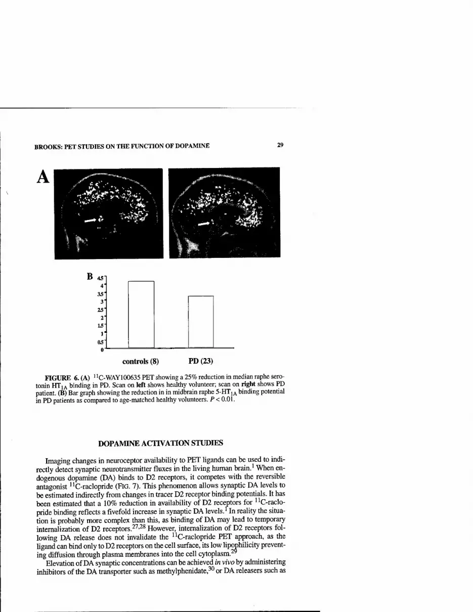
BROOKS: PET STUDIES ON THE FUNCTION OF DOPAMINE 29
B
controls (8) PD (23)
FIGURE 6. (A) ' ^C-WAY100635 PET showing a 25% reduction in median raphe sero- tonin HTiA binding in PD. Scan on left sliows healthy volunteer; scan on right shows PD patient. (B) Bar graph showing the reduction in in midbrain raphe 5-HTIA binding potential in PD patients as compared to age-matched healthy volunteers. P < 0.01.
DOPAMINE ACTIVATION STUDIES
Imaging changes in neuroceptor availability to PET ligands can be used to indi- rectly detect synaptic nenrotransmitter fluxes in the living human brain. ^ When en- dogenous dopamine (DA) binds to D2 receptors, it competes with the reversible antagonist I'C-raclopride (FiG. 7). This phenomenon allows synaptic DA levels to be estimated indirectly from changes in tracer D2 receptor binding potentials. It has been estimated that a 10% reduction in availability of D2 receptors for I'C-raclo- pride binding reflects a fivefold increase in synaptic DA levels.'^In reality the situa- tion is probably more complex than this, as binding of DA may lead to temporary intemalization of D2 receptors.^'^'^^ However, intemaUzation of D2 receptors fol- lowing DA release does not invalidate the iiC-raclopride PET approach, as the ligand can bind only to D2 receptors on the cell surface, its low lipophihcity prevent- ing diffusion through plasma membranes into the cell cytoplasm.^^
Elevation of DA synaptic concentrations can be achieved in vivo by administering inhibitors of the DA transporter such as methylphenidate,^^ or DA releasers such as
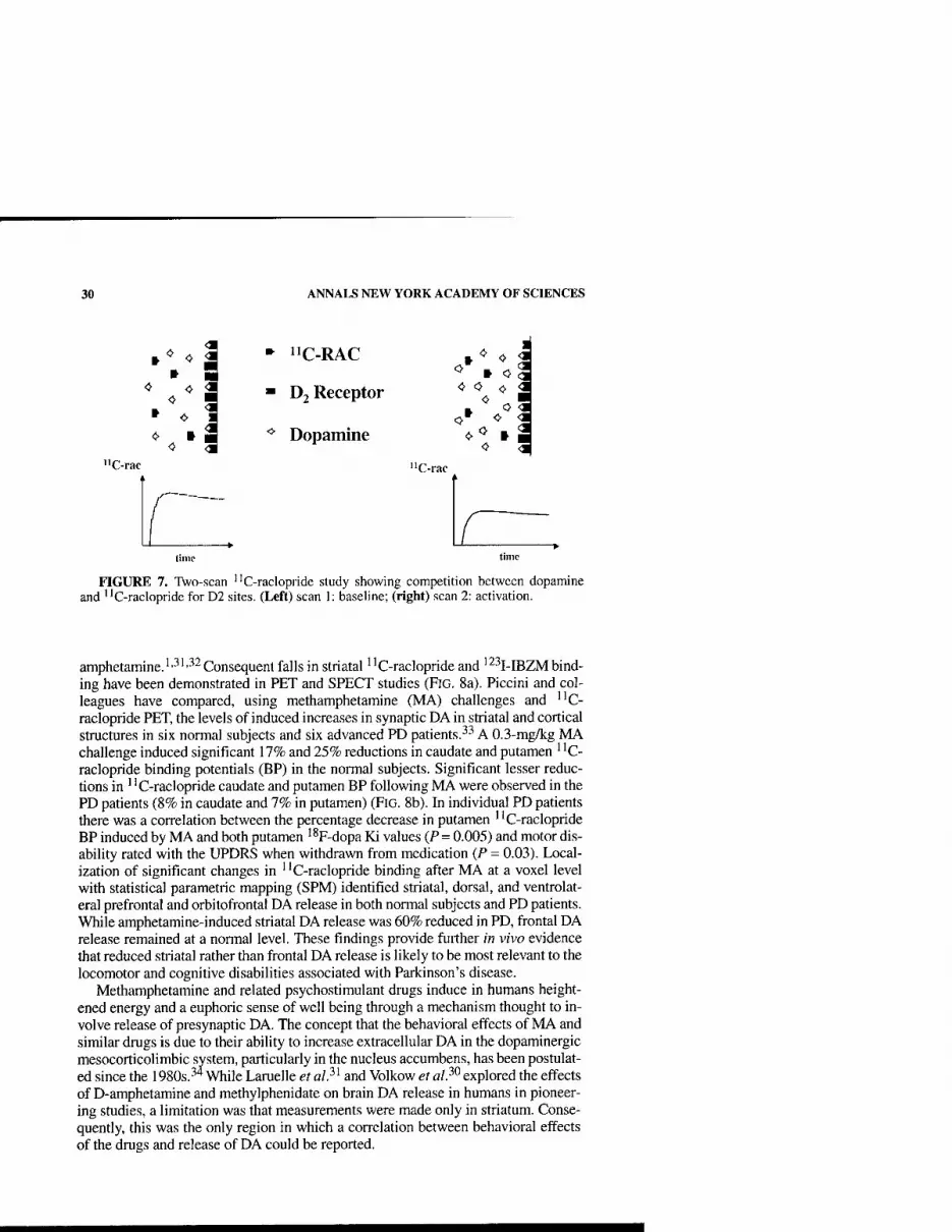
30 ANNALS NEW YORK ACADEMY OF SCIENCES
»* 0
<■ 0 <1 0
<>
•I
- "C-RAC
■ D2 Receptor
** Dopamine
"C-rac "C-rac
FIGURE 7. Two-scan "C-raclopride study showing competition between dopamine and "C-raclopride for D2 sites. (Left) scan 1: baseline; (right) scan 2: activation.
amphetamine.'•■''•^^ Consequent falls in stiiatal "C-raclopride and '^^I-IBZM bind- ing have been demonstrated in PET and SPECT studies (FiG. 8a). Piccini and col- leagues have compared, using methamphetamine (MA) challenges and ''C- raclopride PET, the levels of induced increases in synaptic DA in striatal and cortical structures in six normal subjects and six advanced PD patients.^^ A 0.3-mg/kg MA challenge induced significant 17% and 25% reductions in caudate and putamen ' 'C- raclopride binding potentials (BP) in the normal subjects. Significant lesser reduc- tions in " C-raclopride caudate and putamen BP following MA were observed in the PD patients (8% in caudate and 7% in putamen) (FiG. 8b). In individual PD patients there was a correlation between the percentage decrease in putamen " C-raclopride BP induced by MA and both putamen '*F-dopa Ki values (P= 0.005) and motor dis- ability rated with the UPDRS when withdrawn from medication (P = 0.03). Local- ization of significant changes in ' 'C-raclopride binding after MA at a voxel level with statistical parametric mapping (SPM) identified striatal, dorsal, and ventrolat- eral prefrontal and orbitofrontal DA release in both normal subjects and PD patients. While amphetamine-induced striatal DA release was 60% reduced in PD, frontal DA release remained at a normal level. These findings provide further in vivo evidence that reduced striatal rather than frontal DA release is likely to be most relevant to the locomotor and cognitive disabilities associated with Parkinson's disease.
Methamphetamine and related psychostimulant drugs induce in humans height- ened energy and a euphoric sense of well being through a mechanism thought to in- volve release of presynaptic DA. The concept that the behavioral effects of MA and similar drugs is due to their ability to increase extracellular DA in the dopaminergic mesocorticolimbic system, particularly in the nucleus accumbens, has been postulat- ed since the 1980s.^'* While Laruelle et al?^ and Volkow et al?^ explored the effects of D-amphetamine and methylphenidate on brain DA release in humans in pioneer- ing studies, a limitation was that measurements were made only in striatum. Conse- quently, this was the only region in which a correlation between behavioral effects of the drugs and release of DA could be reported.
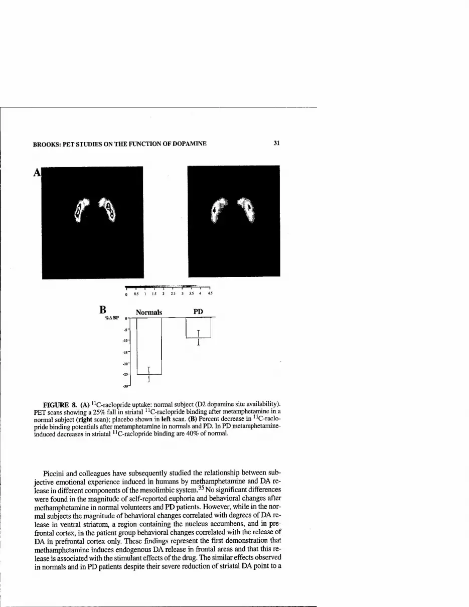
BROOKS: PET STUDIES ON THE FUNCTION OF DOPAMINE 31
't^T'™F~*T™""r""°^T''^"T"'i—I 0.5 1 1.5 2 2.5 3 3.5 4 4.5
B %ABP 0-
Normals PD
FIGURE 8. (A) ''C-raclopride uptake: normal subject (D2 dopamine site availability). PET scans showing a 25% fall in striatal "C-raclopride binding after metamphetamine in a normal subject (right scan); placebo shown in left scan. (B) Percent decrease in ^'C-raclo- pride binding potentials after metamphetamine in normals and PD. In PD metamphetamine- induced decreases in striatal ^'C-raclopride binding are 40% of normal.
Hccini and colleagues have subsequently studied the relationship between sub- jective emotional experience induced in humans by methamphetamine and DA re- lease in different components of the mesolimbic system. ^^ No significant differences were found in the magnitude of self-reported euphoria and behavioral changes after methamphetamine in normal volunteers and PD patients. However, while in the nor- mal subjects the magnitude of behavioral changes correlated with degrees of DA re- lease in ventral striatum, a region containing the nucleus accumbens, and in pre- frontal cortex, in the patient group behavioral changes correlated with the release of DA in prefrontal cortex only. These findings represent the first demonstration that methamphetamine induces endogenous DA release in frontal areas and that this re- lease is associated with the stimulant effects of the drug. The similar effects observed m normals and in PD patients despite their severe reduction of striatal DA point to a
32 ANNALS NEW YORK ACADEMY OF SCIENCES
direct role of prefrontal cortex and of prefrontal cortical DA in the reinforcing effects of psychostimulant drugs in humans.
DOPAMINE RELEASE DURING MOTOR TASKS
Koepp and colleagues^^ were the first to report a reduction of striatal ' 'C-raclo- pride binding during performance of behavioral task. These workers asked subjects to play a video game, and they were financially rewarded according to their level of success. Subjects had to navigate a tank through a battlefield with a computer mouse, shoot at and avoid the shells of enemy tanks, and collect flags. If all flags were col- lected, they progressed to a higher level. The control task was watching a blank com- puter display. There were mean 7.5% and 12.8% reductions in dorsal and ventral striatal ' 'C-raclopride binding during task performance, and individual reductions correlated significantly with the level of financial reward. These findings suggest that ventral striatal levels of dopamine at least doubled during participation in the video game and were increased to a greater extent than dorsal levels in this rewarded paradigm. However, the presence of dorsal striatal "C-raclopride binding reductions suggested that nonmotivational learning per se may also require dopamine release.
A difficulty with interpreting the findings of the above study is that appetitive stimuli, reinforcement of learning, motor coordination, and response selection were all features. Graybiel and colleagues^^ have shown that learning is impaired in mon- keys after adminstration of the nigral toxin MPTP. Lawrence and Brooks,^^ there- fore, investigated whether increased dopamine release could be detected during acquisition of a novel sequence of finger movements by trial and error, as previously decribed, compared with performance of preleamed sequential finger movements. Finger movements were paced by a tone at the same frequency in both tasks. The average number of novel eight-move sequences learned by each subject during the 50-minute "C-raclopride PET study was 14, and during task performance there was a significant additional 5% fall in dorsal putamen and 4% fall in caudate '^C-raclo- pride binding relative to performance of preleamed sequential finger movements. This finding demonstrates that dopamine release does occur in dorsal putamen dur- ing unrewarded motor tasks such as sequence learning and is line wdth the view that dopamine acts to reinforce motor learning as well as to alert to the presence of re- warding stimuli.
This raises the question as to whether simple finger movements induce release of dopamine. Goerendt and coworkers^^ used ''C-raclopride PET to investigate levels of striatal dopamine release by healthy volunteers and PD patients during perfor- mance of a preleamed sequence of finger movements. Five healthy volunteers and six patients with early unilateral PD were studied. Preleamed sequential finger movements in healthy volunteers significantiy decreased ' 'C-raclopride binding bi- laterally in the dorsal putamen by 8-11% and ipsilaterally by 12% in the caudate. PD patients showed no significant reduction of putamen ' 'C-raclopride binding dur- ing successful performance of this task, but a 5% reduction was detected in the cau- date contralateral to the affected limbs. These findings, therefore, suggest that even unrewarded preleamed sequential finger movements are associated with significant putamen and caudate dopamine release in normal volunteers. Endogenous striatal

BROOKS: PET STUDIES ON THE FUNCTION OF DOPAMINE 33
dopamine release can also be detected during this task in Parkinson's disease, but only at a low level and in the caudate, where dopamine levels are most preserved.
CONCLUSIONS
• Striatal dopamine release can be detected with PET during unrewarded actions such as performance of learned sequential finger movements. Active motor learning and the presence of an incentiuve lead to further increases.
• Loss of putamen dopamine in PD correlates with severity of akinesia and rigidity but not rest tremor, which correlates with loss of midbrain raphe sero- tonin HTjA binding.
• Reduced palUdal dopamine in PD is associated with loss of the honeymoon phase to levodopa medication and onset of motor fluctuations.
• In PD frontal dopamine release following an amphetamine challenge appears to be preserved, and levels correlate well with induced euphoria.
• Striatal implants of fetal mesencephalic tissue restore cortical activation, while direct GDNF infusion into posterior putamen increases dopamine stor- age and improves locomotor function.
REFERENCES
1. BREIER, A., T.P. SU, R. SAUNDERS, et al. 1997. Schizophrenia is associated with ele- vated amphetamine-induced synaptic dopamine concentrations: evidence from novel positron emission tomography method. Proc. Natl. Acad. Sci. USA 94: 2569-2574.
2. SCHULTZ, W. 1998. Predictive reward signal of dopamine neurons. J. Neurophysiol. 80: 1-27.
3. ScHULTZ, W. 2000. Multiple reward signals in the brain. Nat. Rev. Neurosci. 1: 199- 207.
4. MIRENOWICZ, J. & W. SCHULTZ. 1996. Preferential activation of midbrain dopamine neurons by appetitive rather than aversive stimuli. Nature 379: 449-451.
5. TAYLOR, A.E., J.A. SAINT-CYR & A.E. LANG. 1986. Frontal lobe dysfunction in Parkin- son's disease. Brain 109: 845-883.
6. WILKINSON, L.S., T. HUMBY, A.S. KILLCROSS, et al. 1998. Dissociations in dopamine release in medial prefrontal cortex and ventral striatum during the acquisition and extinc- tion of classical aversive conditioning in the rat. Eur. J. Neurosci. 10: 1019-1026.
7. WICHMAN, T. & M.R. DELONG. 1999. Oscillations in the basal gangUa. Nature 400: 621-622.
8. ALEXANDER, G.E. & D. CRUTCHER. 1990. Functional architecture of basal ganglia cir- cuits: neural substrates of parallel processing. TINS 13: 266-271.
9. MiDDLETON, F.A.S.P.L. 1997. New concepts about the organization of basal ganglia output. Adv Neurol. 74: 57-68.
10. JELLINGER, K. 1987. The pathology of parkinsonism. In Movement Disorders 2. CD. Marsden & S. Fahn, Eds.: 124-165. Butterworth. London.
11. FEARNLEY, J.M. & A.J. LEES. 1991. Aging and Parkinson's disease: substantia nigra regional selectivity Brain 114: 2283-2301.
12. KiSH, S.J., K. SHANNAK & O. HORNYKIEWICZ. 1988. Uneven pattern of dopamine loss in Ihe striamm of patients with idiopathic Parkinson's disease. N. Engl. J. Med. 318: 876-880.
13. BYRNE, E.J., J. LOWE, R.B. GODWIN-AUSTEN, etal. 1987. Dementia of Parkinson's dis- ease associated with diffuse cortical Lewy bodies. Lancet, i: 501.

34 ANNALS NEW YORK ACADEMY OF SCIENCES
14. CHEESMAN, A.L., S.J.G. LEWIS, R. BARKER, et al. 2002. Right caudate dopamine stor- age correlates with spatial planning in Parkinson's disease. Neurology 58 (Suppl. 3): A487.
15. LiNDVALL, O. 1999. Cerebral implantation in movement disorders: state of the art. Mov. Disord. 14: 201-205.
16. PicciNi, P., D.J. BROOKS, A. BJORKLUND, et al. 1999. Dopamine release from nigral transplants visualised in vivo in a Parkinson's patient. Nat. Neurosci. 2: 1137-1140.
17. PiCCiNi, P., O. LiNDVALL, A. BjORKLUND, et al. 2000. Delayed recovery of movement- related cortical function in Parkinson's disease after striatal dopaminergic grafts. Ann. Neurol. 48: 689-695.
18. BROOKS, D.J. 2001. Functional imaging studies on dopamine and motor control J. Neu- ral Transm. 108: 1283-1298.
19. RASCOL, O., U. SABATINI, F. CHOLLET, et al. 1992. Supplementary and pnmary sensory motor area activity in Parkinson's disease. Regional cerebral blood flow changes during finger movements and effects of apomorphine. Arch. Neurol. 49: 144-148.
20. RASCOL, O., U. SABATINI, F. CHOLLET, et al. 1994. Normal activation of the supplemen- tary motor area in patients with Parkinson's disease undergoing long-term treatment with levodopa. J. Neurol. Neurosurg. Psychiat. 57: 567-571.
21. BROOKS, D.J., I.H. JENKINS & R.E. PASSINGHAM. 1993. Positron emission tomography studies on regional cerebral control of voluntary movement. In Role of the Cerebel- lum and Basal Ganglia in Voluntary Movement. N. Mano, L Hamada & M.R. DeLong, Eds.: 267-274. Excerpta Medica. Amsterdam.
22. JENKINS, I.H., W. FERNANDEZ, E.D. PLAYFORD, et al. 1992. Impaired activation of the supplementary motor area in Parkinson's disease is reversed when akinesia is treated with apomorphine. Ann. Neurol. 32: 749-757.
23. GILL, S.S., N.K. PATEL, K. O'SULLIVAN, et al. 2002. Intraparenchymal putaminal administration of glial-derived neurotrophic factor in the treatment of advanced Par- kinson's disease. Neurology 58 (Suppl. 3): A241.
24. HAUSER, R.A., T.B. FREEMAN, B.J. SNOW, et al. 1999. Long-term evaluation of bilateral fetal nigral transplantation in Parkinson disease. Arch. Neurol. 56: 179-187.
25. WHONE, A.L., R.Y. MOORE, P. PICCINI & D.J. BROOKS. 2001. Compensatory changes in the globus pallidus in early Parkinson's disease: an F-18-dopa PET study. Neurology 56 (Suppl. 3): A72-A73.
26 DODER, M., E.I. RABINER, N. TURJANSKI, et al. 2001. Functional imaging of tremor m Parkinson's disease with [C- 11]-WAY 100635 PET. Neurology 56 (Suppl. 3): A271- A272.
27. CHUGANI, D.C, R.F. ACKERMANN & M.E. PHELPS. 1988. In vivo 3H-spiperone binding: evidence for accumulation in corpus striatum by agonist mediated receptor intemali- sation J. Cereb. Blood Flow Metabol. 8: 291-303.
28. MURIEL, M.P., V. BERNARD, A.I. LEVEY, et al. 1999. Levodopa induces a cytoplasmic localisation of Dl dopamine receptors in striatal neurons in Parkinson's disease. Ann. Neurol. 46: 103-111.
29. LARUELLE, M. 2000. Imaging synaptic neurotransmission with in vivo binding compe- tition techniques: a critical review. J. Cereb. Blood Flow Metab. 20: 423^51.
30. VOLKOW, N.D., G.-J. WANG, J.S. FOWLER, et al. 1994. Imaging endogenous dopamine competition with ["C]raclopride in the human brain. Synapse 16: 255-262.
31. LARUELLE, M., CD. D'SOUZA, R.M. BALDWIN, et al. 1997. Imaging D2 receptor occu- pancy by endogenous dopamine in humans. Neuropsychopharmacology 17: 162-174.
32. GINOVART, N., L. FAROE, C. HALLDIN & C.G. SWAHN. 1999. Changes in striatal D2- receptor density following chronic treatment with amphetamine as assessed with PET in nonhuman primates. Synapse 31: 154-162.
33. PICCINI, P., N. PAVESE, O. LINDVALL, et al. 2000. Endogenous dopamine release coire- lates with dopamine storage in patients with Parkinson's disease: an "C-raclopride and '¥-dopa PET study. (Abstr.) Neurology 54 (Suppl. 3): A329.
34. IVERSEN, L.L. 1996. Smoking...harmful to the brain. Nature 382: 206-207. 35. PICCINI, P., N. PAVESE & D.J. BROOKS. 2002. Effects of methamphetamine on mood
correlate with endogenous dopamine release in frontal areas. Neurology 58 (Suppl. 3): A357.

BROOKS: PET STUDIES ON THE FUNCTION OF DOPAMINE 35
36. KOEPP, M.J., R.N. GUNN, A.D. LAWRENCE, et al. 1998. Evidence for striatal dopamine release during a video game. Nature 393: 266-268.
37. GRAYBIEL, A.M., T. AOSAH, A.W. FIAHERTY, et al. 1994. The basal ganglia and adap- tive motor control. Science 265: 1826-1831.
38. LAWRENCE, A.D. & D.J. BROOKS. 1999. Neural correlates of reward processing in the human brain: a PET study. (Abstr.) Neurology 52 (Suppl. 2): A307.
39. GOERENOT, I.K., C. MESSA, A.D. LAWRENCE, et al. Dopamine release during sequential finger movements in health and Parkinson's disease: a PET study. Brain. In press.
Midbrain Dopaminergic Neurons
Determination of Their Developmental Fate by Transcription Factors
HORST H. SIMON, LAVINIA BHATT, DANIEL GHERBASSI, PAOLA SGAD6,
AND LAVINIA ALBERI
Center for Neuroscience, Department of Neuroanatomy, University of Heidelberg, 69120 Heidelberg, Germany
ABSTRACT: Midbrain dopaminergic neurons are the main source of dopamine in the mammalian central nervous system and are associated with one of the most prominent human neurological disorders, Parkinson's disease. During development, they are induced in the ventral midbrain by an interaction be- tween two diffusible factors, SHH and FGF8. The local identity of this part of the miidbrain is probably determined by the combinatorial expression of three transcription factors, Otx2, Pax2, and PaxS. After the last cell division, the neu- rons start to express transcription factors that control further differentiation and the manifestation of cellular properties characteristic for adult dopamin- ergic neurons of the substantia nigra compacta and the ventral tegmentum. The first to appear is the LIM-homeodomain transcription factor, Lmxlb. It is essential for the survival of these neurons, and it regulates the expression of an- other transcription factor, Pitx3, an activator of tyrosine hydroxylase. Lmxlb is followed by the orphan steroid receptor Nurrl. It is essential for the expres- sion of the dopaminergic phenotype. Several genes involved in dopamine syn- thesis, transport, release, and reuptake are regulated by Nurrl. This requirement is specific to the midbrain dopaminergic neurons, since other pop- ulations of the same neurotransmitter phenotype develop normally in absence of the gene. A day after Nurrl, two homeodomain transcription factors, en- grailed-1 and -2, are expressed. In animals deficient in the two genes, the mid- brain dopaminergic neurons are generated, but then fail to differentiate and disappear very rapidly. Interestingly, a-synuclein, a gene recently linked to fa- milial forms of Parkinson's disease, is regulated by engrailed-1 and -2.
KEYWORDS: substantia nigra; ventral tegmentum; Parkinson's disease; dopam- inergic neurons; Nurrl; engrailed; Lmxlb; neuronal specification
Neurogenesis is a gradual process that transforms undefined neuroepithelial cells into fully differentiated neurons. It can be roughly divided into two phases: an early phase, when the precursor cells are proliferating and slowly adopt a successively more restricted cell fate; and a later phase, when the neurons are postmitotic and ful-
Address for correspondence: Horst H. Simon, Center for Neuroscience, Department of Neuro- anatomy, University of Heidelberg, Im Neuenheimer Feld 307, 69120 Heidelberg, Germany. Voice: +49 6221 548342; fax: +49 6221 545605.
Ann. N.Y. Acad. Sci. 991: 36-47 (2003). © 2003 New York Academy of Sciences.
36
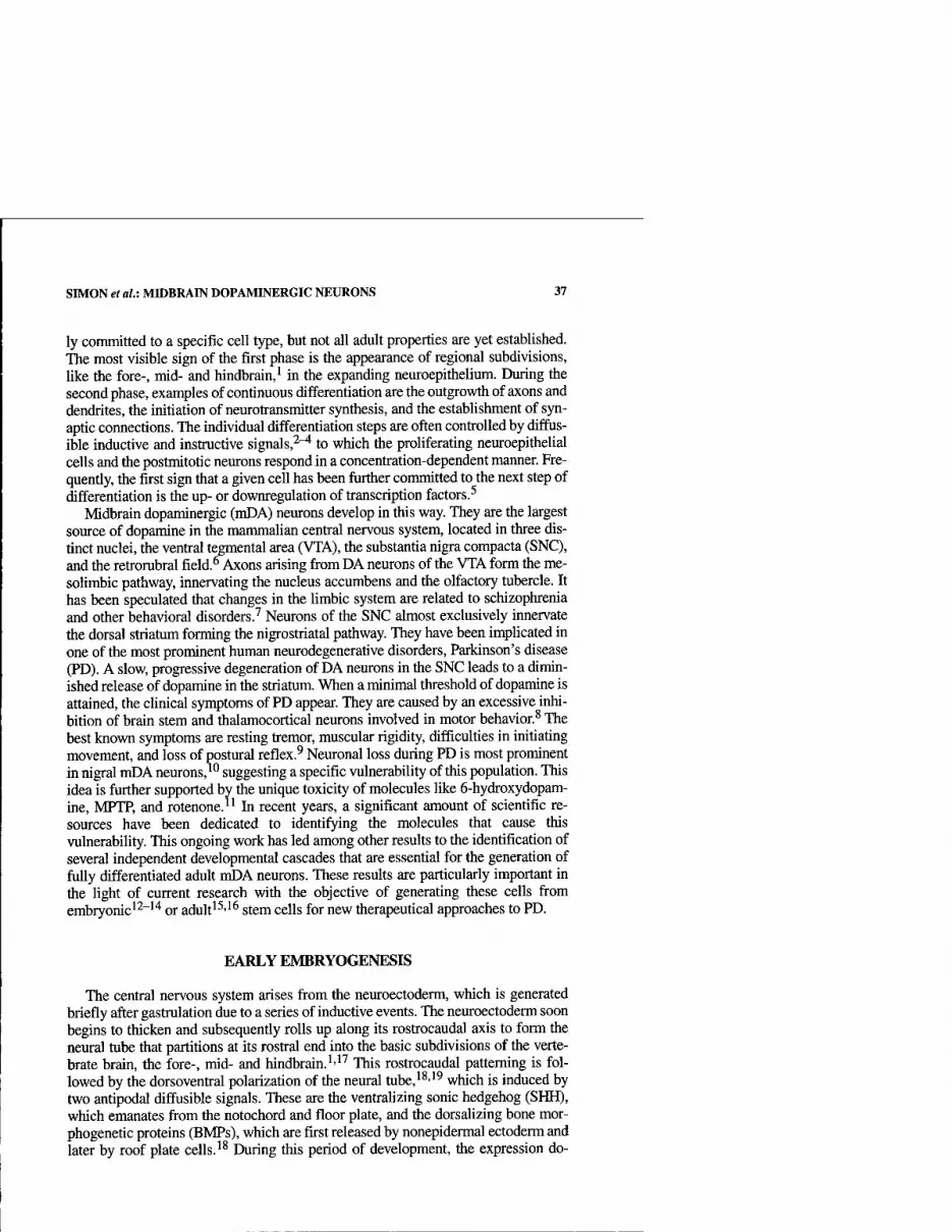
SIMON et al.: MIDBRAIN DOPAMINERGIC NEURONS 37
ly committed to a specific cell type, but not all adult properties are yet established. The most visible sign of the first phase is the appearance of regional subdivisions, like the fore-, mid- and hindbrain,^ in the expanding neuroepithelium. During the second phase, examples of continuous differentiation are the outgrowth of axons and dendrites, the initiation of neurotransmitter synthesis, and the estabUshment of syn- aptic connections. The individual differentiation steps are often controlled by difftis- ible inductive and instinctive signals,^'^ to which the proliferating neuroepitheUal cells and the postmitotic neurons respond in a concenttation-dependent manner. Fre- quently, the first sign that a given cell has been fiuther committed to the next step of differentiation is tiie up- or downregulation of ttanscription factors.^
Midbrain dopaminergic (mDA) neurons develop in this way. They are the largest source of dopamine in tiie mammalian centtal nervous system, located in three dis- tinct nuclei, the ventral t^gmental area (VTA), the substantia nigra compacta (SNC), and the reti-orubral field.^Axons arising fi-om DA neurons of the VTA form the me- solimbic pathway, innervating the nucleus accumbens and the olfactory tubercle. It has been speculated that changes in the Umbic system are related to schizophrenia and other behavioral disorders.^ Neurons of the SNC almost exclusively innervate the dorsal striatum forming the nigrostriatal pathway. They have been implicated in one of the most prominent human neurodegenerative disorders, Parkinson's disease (PD). A slow, progressive degeneration of DA neurons in the SNC leads to a dimin- ished release of dopamine in the striatum. When a minimal threshold of dopamine is attained, the chnical symptoms of PD appear. They are caused by an excessive inhi- bition of brain stem and thalamocortical neurons involved in motor behavior.^ The best knovm symptoms are resting tremor, muscular rigidity, difficulties in initiating movement, and loss of postural reflex.^ Neuronal loss during PD is most prominent in nigral mDA neurons,^'^ suggesting a specific vulnerability of this population. This idea is further supported by the unique toxicity of molecules like 6-hydroxydopam- ine, MPTP, and rotenone.^' In recent years, a significant amount of scientific re- sources have been dedicated to identifying the molecules that cause this vulnerability. This ongoing work has led among other results to the identification of several independent developmental cascades that are essential for the generation of fully differentiated adult mDA neurons. These results are particularly important in the Ught of current research witii the objective of generating tiiese cells from embryonic'^"''* or adult'^'^^ stem cells for new tiierapeutical approaches to PD.
EARLY EMBRYOGENESIS
The central nervous system arises from the neuroectoderm, which is generated briefly after gastrulation due to a series of inductive events. The neuroectoderm soon begins to thicken and subsequently rolls up along its rosttocaudal axis to form tiie neural tube that partitions at its rostial end into the basic subdivisions of the verte- brate brain, the fore-, mid- and hindbrain.'-^^ This rostrocaudal patterning is fol- lowed by the dorsoventral polarization of the neural tube,^^''^ which is induced by two antipodal diffusible signals. These are the venttalizing sonic hedgehog (SHH), which emanates from the notochord and floor plate, and the dorsaUzing bone mor- phogenetic proteins (BMPs), which are first released by nonepidermal ectoderm and later by roof plate cells.^^ During this period of development, the expression do-
38 ANNALS NEW YORK ACADEMY OF SCIENCES
Wnti
FIGURE 1. Expression pattern of Otx2, Pax2, Pax5, and Wnt-l at E8 and E9 of mouse development. The simplified scheme of the expression domains of Otx2, Pax2, Pax5, and Wnt-l, which are represented by bars above the embryo, shows an overlap of all four genes in the midbrain rostral to the isthmus. This is the region where the mDA neurons are induced by an interaction between SHH and FGF8. The scheme does not reflect the dynamic nature of the expression pattern at these ages. SHH = sonic hedgehog, FGF8 = fibroblast growth factor 8, fp = floor plate, is = isthmus, F = forebrain, M = midbrain, H = hindbrain.
mains of transcription factors begin to delineate the region that will give rise to mDA neurons. Probably the first to do so is the homeobox transcription factor Otx2, which initially appears in all three germ layers, regresses during further development, and from E7 onwards (mouse) defines the anterior neuroectoderm that will give rise to the fore- and midbrain. Shortly thereafter, Pca2 appears in the developing midbrain, followed by Pax5 at the same location. In parallel to Pax2, the secreted molecule Wnt] is expressed (FiG.l). Homozygous mutant mice null for Otx2, Wntl, and the double mutants for Pax2/5 all show the same phenotype with respect to the mid- brain—a complete deletion of it.^^^^ A consequence of midbrain deletion in Wntl- /- mutants is the lack of all mDA neurons (RG. 2). This is very likely also the case in 0£«:2-null mutants since the fore-, mid-, and anterior hindbrain are deleted. A de- pendence of Pax2, Pax5, and Wntl on each other after the initial onset of their ex- pression suggests the same for the Pax2/5 double mutants.^'* Further evidence that the Otx2, Pax2, and Pax5 expression domain is the source for mDA neurons is pro- vided by explant culture experiments and the analysis of transgenic mice.-^^"^^ The mDA neurons are induced at E9 (rat) by the combinatorial action of SHH and the fibroblast growth factor 8 (FGF8), released by the floor plate and isthmus, respec-

SIMON et al.: MmBRAIN DOPAMINERGIC NEURONS 39
0 SNCA/TA ^
^ » DMH
iDMH
FIGURE 2. Total loss of mDA neurons in Wntl-m\\ mutant. Midsagittal brain sections of PO wild-type and Wntl-/- mutant mouse stained against TH. (A) TH-positive cell bodies of SNC and VTA are detectable in the ventral midbrain of the wild type, whereas (B) the mutant mice have lost all mDA neurons (arrow). Staining in the dorsomedial hypothalamic nucleus (DMH) demonstrates that other dopaminergic populations are not affected.
tively. The floor plate spans almost the entire rostral-to-caudal neuroaxis, and the isthmus releases FGF8 into the midbrain and into the hindbrain. However, only a very restricted area of the developing neural tube, the midbrain directly anterior to the isthmus, is capable of generating DA neurons characteristic for the SNC and VTA. Further evidence was provided by the analysis of transgenic mice that express HNF3fi under the En2 promoter enhancer. These mice form an ectopic dorsal SHH- expressing floor plate in the midbrain and the anterior hindbrain, leading to an ec- topic induction of mDA neurons in the midbrain tissue, but not in the hindbrain. These findings demonstrated that only the tissue rostral to the isthmus is competent to form mDA neurons. The developmental stage when the mDA neurons are gener- ated corresponds to the timing of the Otx2, Pax2, and Pax5 expression; and the re- gion of the neural tube where the induction takes place is the area where these three transcription factors overlap. All this strongly suggests that the combination of the three specifies the region along the rostrocaudal axis of the neural tube where mDA can be generated, but the final conclusive experiments are still missing. If this hy- pothesis is true, a caudal shift of the Otx2 domain should increase the amount of mDA neurons or at least reallocate them in the posterior direction.^^
POSTMITOTIC DIFFERENTIATION OF MIDBRAIN DOPAMINERGIC NEURONS
Studies using tritiated thymidine on rat fetuses showed that the majority of mDA neurons become postmitotic at around E13 to E15.^^ This corresponds to approxi- mately El 1 to E13 in mouse (from here on we will use mouse embryonic ages even

40 ANNALS NEW YORK ACADEMY OF SCIENCES
if rat studies are mentioned, setting mouse age at rat - 2 days). Twelve to 24 hours later tyrosine hydroxylase (TH), the rate-Umiting enzyme of dopamine synthesis, is detectable by conventional immunohistochemistry.^° The exact start point of the TH expression is, however, controversial. Studies using an acrolein-based fixative were able to identify TH-positive cells on the ventricular side of the ventral midbrain as early as E8.5 to E9, suggesting that mDA neurons express TH before or shortly after they become postmotic.*^ All in all, the mDA neurons belong to one of the first neu- ronal populations generated in the developing CNS.^^ After becoming postmitotic, the next major developmental step is the formation of axonal connections to the bas- al ganglia and the frontal cortex. The first thin processes resembhng axons are de- tectable with an antibody against dopamine at Ell.^^ A day later, thick axonal bundles that have passed the diencephalons begin to enter the ventral telencephalon, where the forefront of the bundle reaches the ganglionic eminence and bends to- wards the cortical anlage. The formation of axonal terminals and the transient strio- somal patterning begin at El 8. Some of these differentiation steps are associated with the appearance of transcription factors, which probably regulate the individual events. TTiree independent regulatory pathways involved in the differentiation of the mDA neurons have been identified to date. The key transcription factors for these regulatory pathways are Nurrl, the engrailed genes, and Lmxlb.
THE ORPHAN STEROID RECEPTOR Nurrl
Nurrl (also known as RNRl, Not, or HZF-1) was first recognized by its expres- sion in the brain^'* and regenerating liver.^^ Its sequence suggests that it is an orphan nuclear receptor with a yet-unidentified ligand and acts as a ligand-activated tran- scription factor. It appears in the marginal and mantle zone of the ventral midbrain at E10.5, at the location where mDA neurons are generated, but 24 hours before they express any phenotypic markers such as TH?^ From this age continuing into adult- hood, Nurrl is expressed in at least 95% of all T/^-positive midbrain neurons.^^ The targeted deletion of Nurrl by homologous recombination leads to mutant mice that seem to develop normally during embryogenesis but are unable to feed and die short- ly after birth.'^-^^-^^ Detailed analysis of these mutant mice demonstrated that Nurrl controls the most prominent feature of mDA neurons, the use of dopamine as their neurotransmitter. Several genes that are involved in synthesis, axonal transport, stor- age, release, or reuptake of dopamine are not expressed in the mutant neurons. These genes are TH, AADC (aromatic aminoacid decarboxylase), VMAT (vesicle mem- brane-associated transporter), and /M7 (dopamine tt-ansporter).^^-^^'^^ Surprisingly, this requirement of Nurrl for the expression of the dopaminergic phenotype is re- stiicted to mDA neurons alone. Furthermore, the use of A^MrrV-independent markers for mDA neurons, such as AHD2, Enl, and a Nurrl riboprobe 5' to the deletion, re- vealed that the cells otherwise develop normally in the mutant embryos until E15.5. From this point of embryogenesis, the reports of two groups who analyzed the mu- tant phenotype conti-adict each other. One group claims a role of Nurrl in the migra- tion, striatal target innervation, and survival of these cells.^*^ The DA neurons in the Nurrl mutants were abnormally distiibuted, no axons were ti-aceable from the stria- tum, and a high amount of apoptotic nuclei were present in the ventral midbrain. The other group, doing essentially the same experiments, was unable to observe any of

SIMON et al.: MmBRAIN DOPAMINERGIC NEURONS 41
these alterations.^^ However, the two groups agreed in one respect, that there are still some mDA neurons left in the Nurrl mutant mice at birth. An explanation for these different observations could be that the investigated mutant mice are the results of two separate experiments using different constructs. The first group removed exons 2, 3, and part of 4, whereas the second group removed only exon 3. It is therefore plausible that a partial Nurrl protein is still translated in the latter case, resulting in a weaker phenotype.
THE HOMEODOMAIN TRANSCRIPTION FACTORS Engrailed-1 AND Engrailed-2
The mammalian engrailed genes, Enl and En2, are homeodomain transcription factors that were originally cloned on the basis of their sequence similarity to Droso- phila engrailed.'^'^'^^ As, in insects, they have two distinct ontogenetic roles: during early embryogenesis they take part in the regionalization of the embryo,"'^'"^^ where- as during later embryogenesis they are involved in the specification of neuronal phe- notypes.^^"^' Both engrailed genes appear at E8 in the anterior mouse neuroectoderm as patches, which subsequently fuse to mark a band of cells that will later give rise to the border region between midbrain and hindbrain. Between Ell and E12, the two genes emerge in postmitotic mDA neurons and are then continu- ously expressed throughout the entire life of these cells (FiG. 3).^^"^^ Mice homozy- gous for an Enl-mxW mutation die at birth and show a deletion of the inferior coUiculus and parts of the cerebellum; both these brain areas arise from the midbrain neuroepithelium, which expresses Enl during early embryogenesis."*' The En2 mu- tant phenotype is comparatively subtle despite the overlapping expression with Enl in the midbrain. These mice are viable and fertile, and show only a minor defect in cerebellar foliation.^^'^^ Neither of the two engrailed mutant strains, however, show a significant phenotype with respect to mDA neurons. The requirement for the en- grailed genes is apparent only in mutant mice deficient for both Enl and En2. Like the Enl-mxW mutants, the engrailed double mutants die at birth and exhibit a deletion of the midbrain and anterior hindbrain. The phenotype with respect to mDA neurons is, however, significantly different. The neurons are generated in the ventral mid- brain and start to express 777, but then fail to further differentiate and disappear. By E14, they are no longer detectable (FiG. 4D). Use of an En-1/tau-LacZ knock-in mouse as an autonomous marker demonstrated that the cells are lost in the double- mutant animals, unlike the Nurrl mutant mice, where only the neurotransmitter phe- notype is changed. Since the engrailed double-mutant mice show a deletion of the midbrain and anterior hindbrain,^''^^ two scenarios are possible. Either the genes are cell-autonomously required for the survival or, due to the missing midbrain tissue, an essential extemal support is absent in the mutant mice (non-cell-autonomous). Such questions are best addressed by experiments that mix wild-type and mutant cells. Engrailed double-mutant ES cells were injected into wild-type blastula and the chimeras raised to adulthood. These animals showed a normal brain morphology but a significant loss of DA neurons in the SNC and VTA, clearly demonstrating that the engrailed genes are cell autonomously required for the survival of these cells (un- pubhshed data). Furthermore, the mutant analysis also provided evidence that the expression of a-synuclein may be regulated by the two engrailed genes. a-Synuclein
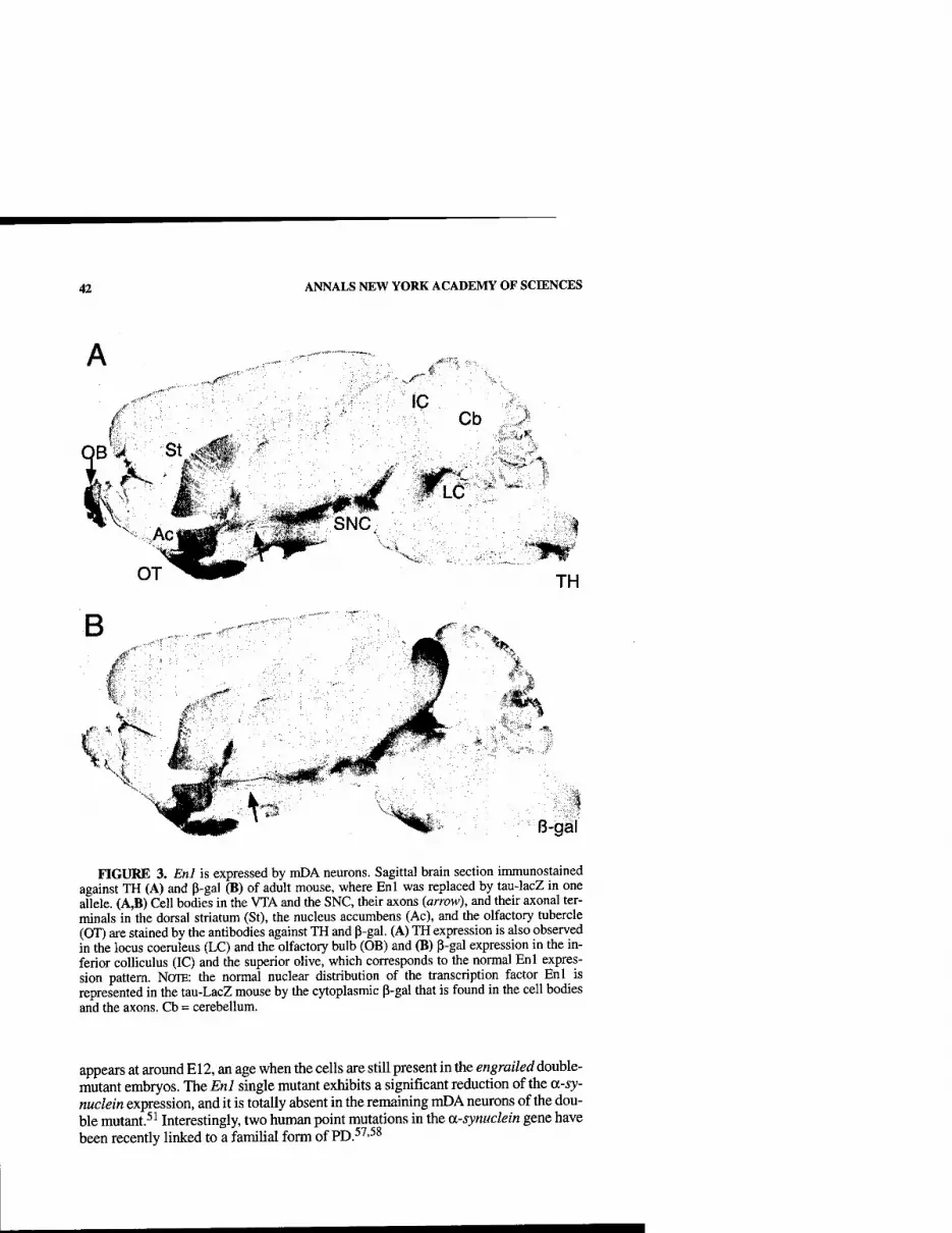
42 ANNALS NEW YORK ACADEMY OF SCIENCES
./r^ ^(
IC Cb
Wic'
TH
V.
B-gal
FIGURE 3. Enl is expressed by mDA neurons. Sagittal brain section immunostained against TH (A) and P-gal (B) of adult mouse, where Enl was replaced by tau-lacZ in one allele. (A,B) Cell bodies in the VTA and the SNC, their axons (arrow), and their axonal ter- minals in the dorsal striatum (St), the nucleus accumbens (Ac), and the olfactory tubercle (OT) are stained by the antibodies against TH and p-gal. (A) TH expression is also observed in the locus coeruleus (LC) and the olfactory bulb (OB) and (B) p-gal expression in the in- ferior colliculus (IC) and the superior olive, which corresponds to the normal Enl expres- sion pattern. NOTE: the normal nuclear distribution of the transcription factor Enl is represented in the tau-LacZ mouse by the cytoplasmic P-gal that is found in the cell bodies and the axons. Cb = cerebellum.
appears at around El2, an age when the cells are still present in the engraileddouhle- mutant embryos. The Enl single mutant exhibits a significant reduction of the a-sy- nuclein expression, and it is totally absent in the remaining mDA neurons of the dou- ble mutant.^ ^ Interestingly, two human point mutations in the a-synuclein gene have been recently linked to a familial form of PD.^^'^^

SIMON et al.: MroBRAIN DOPAMINERGIC NEURONS 43
A B E12
WT En1-/-;En2-/-
EIGURE 4. Loss of mDA neurons in the Engrailed double mutant. Immunohistochem- istry against TH on whole mounts of E12 embryo brains (A,B) and midsagittal sections of E14 embryo head (C,D) of wild type (A,C) and Enl-/-; En2-/- mutant (B,D). (A) AVhole- mount staining of wild type reveals TH-positive cells in the ventral midbrain that will give rise to the dopaminergic neurons of the SNC and VTA. (B) TH-positive cells are also present in the ventral midbrain of engrailed double mutants; however, the amount of cells is signif- icantly smaller than in the wild type. (C) Two days later at E14, the dopaminergic neurons, which begin to adopt an adult distribution, are detectable in the wild-type ventral midbrain. Cb = cerebellum; IC = inferior coUiculus; SC = superior coUiculus; VTA = ventral tegmental area. (D) The E14 mutant brain at the same plane of section shows no dopaminergic neurons at this position (arrow).
THE LIM-HOMEODOMAIN TRANSCRIPTION FACTOR IB (Lmxlb) AND THE PITUITARY HOMEOBOX 3 (Pitx3)
Lmxlb is a member of the LIM-homeodomain family of proteins. It is expressed in a wide range of different tissues and has been implicated in the development of diverse structures such as skeleton, eye, kidney, and limb.^^ In humans, loss-of-func-
44 ANNALS NEW YORK ACADEMY OF SCIENCES
tion mutations lead to nail patella syndrome,^'' defects that are very similar to those seen in mice homozygous for the Lnvclb null mutation.^^ Its role in the development of mDA neurons has recently come to Ught. It is expressed in the neural tube as early as E7.5, including the region of the Ventral midbrain that gives rise to dopamine cells.^' At least from E16 continuing into adulthood, it is colocalized with TH and Pitx3 (see below) in the ventral midbrain. The specificity of this expression was con- firmed by unilateral injection of 6-hydroxydopamine into the striatum. The function- al deletion of Lnvclb by homologous recombination has a profound effect on mDA neurons. Only a small amount of TH-positive cells are detectable in the ventral mid- brain at El 2.5, and they entirely disappear a few days later. Additionally, the expres- sion of Pitx3 is affected in these embryos. The wild-type expression of Pitx3 in the ventral midbrain always matches the distribution of TH; however, in the Lmxlb mu- tant embryos fewer cells were positive for Pitx3 than for TH, suggesting a regulatory role of Lmxlb. Unfortunately, the published data are rather Umited, and it is an open question whether the ablation of mDA neurons in the mutant is due to a cell-auton- omous requirement of Lmxlb in the neurons or is an effect of the large midbrain def- icit also observed in the mutant animals. In the chicken, the expression of Lmxlb is followed by the expression of Wntl; and gain-of-function experiments using a rep- lication-competent retroviral vector (Lmxlb/RCAS) suggest that Lmxlb is required for the onset and maintenance of the Wntl expression.^^ If this interaction also exists in mouse, then the Lmxlb mutant phenotype with respect to the mDA neurons is like- ly to be a consequence of the lack of Wntl expression in the midbrain (FiG. 2); this argues against the notion of a cell-autonomous requirement of Lmxlb. Furthermore, it is still unresolved whether the cells that express Lmxlb in the ventral midbrain dur- ing early embryogenesis are the precursor cells of adult mDA neurons or this expres- sion is purely coincidental.
Pitx3, also sometimes called Ptx3, (paired-like homeodomain transcription factor 3 or pituitary homeobox 3) is a homeodomain containing transcription factor with bind- ing activity to DNA similar to the Dwsophila bicoid. It is uniquely expressed in the brain by niDA neurons from E11.5 onwards and is maintained throughout the entire life of the animal.^^ Its relevance is connected to the appearance of 77/just briefly after Pitx3 is detected and the presence of a conserved bicoid response element (GGCTTT) just a few bases upstream of the TATA box of the TH gene in the rat, mouse, and hu- man.^ Gel shift experiments demonstrated an affinity of Pitx3 to this response element. Moreover, transient transfection experiments showed a cell type-dependent 8- to 12-fold increase of TH promoter activity when the Pitx3 protein was present. However, such in situ studies are only suggestive, and the precise role of PitxS in the development of the mDA neurons still needs to be determined. It is likely that the anal- ysis of the Aphakia mutant mice, which has been recently linked to a deletion in the Pitx3 gene,^^'^ may provide evidence pointing to its biological function.
REFERENCES
1. LuMSDEN, A. & R. KRUMLAUF. 1996. Patterning the vertebrate neuraxis. Science 274: 1109-1115.
2. MCALLISTER, A.K., L.C. KATZ & D.C. Lo. 1999. Neurotrophins and synaptic plastic- ity. Annu. Rev. Neurosci. 22: 295-318.
SIMON et al.: MmBRAIN DOPAMINERGIC NEURONS 45
3. MARTI, E. & P. BOVOLENTA. 2002. Sonic hedgehog in CNS development: one signal, multiple outputs. Trends Neurosci. 25: 89-96.
4. KENNEDY, T.E. 2000. Cellular mechanisms of netrin function: long-range and short- range actions. Biochem. Cell Biol. 78: 569-575.
5. EDLUND, T. & T.M. JESSELL. 1999. Progression from extrinsic to intrinsic signaling in cell fate specification: a view from the nervous system. Cell 96: 211-224.
6. NELSON, E.L., et al. 1996. Midbrain dopaminergic neurons in the mouse: computer- assisted mapping. J. Comp. Neurol. 369: 361-371.
7. MEYER-LINDENBERG, A., et al. 2002. Reduced prefrontal activity predicts exaggerated striatal dopaminergic function in schizophrenia. Nat. Neurosci. 5: 267-271.
8. OBESO, J. A., et al. 2000. Pathophysiology of the basal ganglia in Parkinson's disease. Trends Neurosci. 23: S8-19.
9. JENNER, P., A.H. SCHAPIRA & CD. MARSDEN. 1992. New insights into the cause of Par- kinson's disease. Neurology 42: 2241-2250.
10. HIRSCH, E.G., et al. 1997. Neuronal vulnerability in Parkinson's disease. J. Neural Transm. Suppl. 50: 79-88.
11. BEAL, M.F. 2001. Experimental models of Parkinson's disease. Nat. Rev. Neurosci. 2: 325-334.
12. LEE, S.H., et al. 2000. Efficient generation of midbrain and hindbrain neurons from mouse embryonic stem cells. Nat. Biotechnol. 18: 675-679.
13. KAWASAKI, H., et al. 2000. Induction of midbrain dopaminergic neurons from ES cells by stromal cell-derived inducing activity. Neuron 28: 31^0.
14. RoLLETSCHEK, A., et al. 2001. Differentiation of embryonic stem cell-derived dopam- inergic neurons is enhanced by survival-promoting factors. Mech. Dev. 105: 93-104.
15. SAKURADA, K., et al. 1999. Nurrl, an orphan nuclear receptor, is a transcriptional acti- vator of endogenous tyrosine hydroxylase in neural progenitor cells derived from the adult brain. Development 126: 4017-4026.
16. LIE, D.C, et al. 2002. The adult substantia nigra contains progenitor cells with neuro- genic potential. J. Neurosci. 22: 6639-6649.
17. RUBENSTEIN, J.L. & PA. BEACHY. 1998. Patterning of the embryonic forebrain. Curr. Opin. Neurobiol. 8: 18-26.
18. LEE, K.J. & T.M. JESSELL. 1999. The specification of dorsal cell fates in the vertebrate central nervous system. Annu. Rev. Neurosci. 22: 261-294.
19. SIMON, H., A. HORNBRUCH & A. LUMSDEN. 1995. Independent assignment of antero- posterior and dorso-ventral positional values in the developing chick hindbrain. Curr. Biol. 5: 205-214.
20. AcAMPORA, D., et al. 1995. Forebrain and midbrain regions are deleted in Otx2-/- mutants due to a defective anterior neuroectoderm specification during gastrulation. Development 121: 3279-3290.
21. ANG, S.L., et al. 1996. A targeted mouse Otx2 mutation leads to severe defects in gas- trulation and formation of axial mesoderm and to deletion of rostral brain. Develop- ment 122: 243-252.
22. ScHWARz, M., et al. 1997. Conserved biological function between Pax-2 and Pax-5 in midbrain and cerebellum development: evidence from targeted mutations. Proc. Natl. Acad. Sci. USA 94: 14518-14523.
23. MCMAHON, A.P & A. BRADUEY. 1990. The Wnt-1 (int-1) proto-oncogene is required for development of a large region of the mouse brain. Cell 62: 1073-1085.
24. RHINN, M. & M. BRAND. 2001. The midbrain-hindbrain boundary organizer. Curr. Opin. Neurobiol. 11: 34-42.
25. HYNES, M., et al 1995. Control of neuronal diversity by the floor plate: contact-medi- ated induction of midbrain dopaminergic neurons. Cell 80: 95-101.
26. HYNES, M., et al. 1995. Induction of midbrain dopaminergic neurons by Sonic hedge- hog. Neuron 15: 35-^M.
27. YE, W., et al. 1998. FGF and Shh signals control dopaminergic and serotonergic cell fate in the anterior neural plate. Cell 93: 755-766.
28. BROCCOLI, V., E. BONCINELLI & W. WURST. 1999. The caudal limit of Otx2 expression positions the isthmic organizer. Nature 401: 164-168.

46 ANNALS NEW YORK ACADEMY OF SCIENCES
29. ALTMAN, J. & S.A. BAYER. 1981. Development of the brain stem in the rat. V. Thymi- dine-radiographic study of the time of origin of neurons in the midbrain tegmentum. J. Comp. Neurol. 198: 677-716.
30. FOSTER, G.A., et al. 1988. Ontogeny of the dopamine and cyclic adenosine-3':5'-mono- phosphate-regulated phosphoprotein (DARPP-32) in the pre- and postnatal mouse central nervous system. Int. J. Dev. Neurosci. 6: 367-386.
31. Di PoRZio, U., et al. 1990. Early appearance of tyrosine hydroxylase immunoreactive cells in the mesencephalon of mouse embryos. Int. J. Dev. Neurosci. 8: 523-532.
32. SECHRIST, J. & M. BRONNER-FRASER. 1991. Birth and differentiation of reticular neu- rons in the chick hindbrain: ontogeny of the first neuronal population. Neuron 7: 947-963.
33. VOORN, P., et al. 1988. The pre- and postnatal development of the dopaminergic cell groups in the ventral mesencephalon and the dopaminergic innervation of the stria- tum of the rat. Neuroscience 25: 857-887.
34. LAW, S.W., et al. 1992. Identification of a new brain-specific transcription factor, NURRl. Mol. Endocrinol. 6: 2129-2135.
35. SCEARCE, L.M., et al. 1993. RNR-1, a nuclear receptor in the NGn-B/Nur77 family that is rapidly induced in regenerating liver. J. Biol. Chem. 268: 8855-8861.
36. ZETTER-STROM, R.H., et al. 1997. Dopamine neuron agenesis in Nurrl-deficient mice. Science 276: 248-250.
37. BACKMAN, C, et al. 1999. A selective group of dopaminergic neurons express Nurrl in the adult mouse brain. Brain Res. 851: 125-132.
38. CASTILLO, S.O., et al. 1998. Dopamine biosynthesis is selectively abolished in substan- tia nigra/ventral tegmental area but not in hypothalamic neurons in mice with tar- geted disruption of the Nurrl gene. Mol. Cell. Neurosci. 11: 36-46.
39. SAUCEDO-CARDENAS, O., et al. 1998. Nurrl is essential for the induction of the dopam- inergic phenotype and the survival of ventral mesencephalic late dopaminergic pre- cursor neurons. Proc. Natl. Acad. Sci. USA 95: 4013^018.
40. WALLEN, A., et al. 1999. Fate of mesencephalic AHD2-expressing dopamine progeni- tor cells in NURRl mutant mice. Exp. Cell Res. 253: 737-746.
41. WiTTA, J., et al. 2000. Nigrostriatal innervation is preserved in Nurrl-null mice, although dopaminergic neuron precursors are arrested from terminal differentiation. Mol. Brain Res. 84: 67-78.
42. JOYNER, A.L., et al. 1985. Expression during embryogenesis of a mouse gene with sequence homology to the Drosophila engrailed gene. Cell 43: 29-37.
43. JoYNER, A.L. & G.R. MARTTN. 1987. En-1 and En-2, two mouse genes with sequence homology to the Drosophila engrailed gene: expression during embryogenesis [pub- lished erratum appears in 1987. Genes Dev. 1(5): 521]. Genes Dev. 1: 29-38.
44. KORNBERG, T, et al. 1985. The engrailed locus of Drosophila: in situ localization of transcripts reveals compartment-specific expression. Cell 40: 45-53.
45. KORNBERG, T 1981. Engrailed: a gene controlling compartment and segment formation in Drosophila. Proc. Natl. Acad. Sci. USA 78: 1095-1099.
46. TABATA, T, et al. 1995. Creating a Drosophila wing de novo, the role of engrailed, and the compartment border hypothesis. Development 121: 3359-3369.
47. WURST, W., A.B. AUERBACH & A.L. JOYNER. 1994. Multiple developmental defects in Engrailed-1 mutant mice: an early mid-hindbrain deletion and patterning defects in forelimbs and sternum. Development 120: 2065-2075.
48. LUNDELL, M.J., et al. 1996. The engrailed and huckebein genes are essential for devel- opment of serotonin neurons in the Drosophila CNS. Mol. Cell. Neurosci. 7: 46-61.
49. CoNDRON, B.G., N.H. PATEL & K. ZINN. 1994. Engrailed controls glial/neuronal cell fate decisions at the midline of the central nervous system. Neuron 13: 541-554.
50. SAUERESSIG, H., J. BURRILL & M. GOULDING. 1999. Engrailed-1 and netrin-1 regulate axon pathfinding by association interneurons that project to motor neurons. Develop- ment 126: 4201-4212.
51. SIMON, H.H., et al. 2001. Fate of midbrain dopaminergic neurons controlled by the engrailed genes. J. Neurosci. 21: 3126-3134.

SIMON et aU: MTOBRAIN DOPAMINERGIC NEURONS 47
52. DAVIS, C.A. & A.L. JOYNER. 1988. Expression patterns of the homeo box-containing genes En-1 and En-2 and the proto-oncogene int-1 diverge during mouse develop- ment. Genes Dev. 2: 1736-1744.
53. GARDNER, C.A. & K.F. BARALD. 1992. Expression patterns of engrailed-like proteins in the chick embryo. Dev. Dyn. 193: 370-388.
54. MiLLEN, K.J., et al. 1994. Abnormal embryonic cerebellar development and patterning of postnatal foliation in two mouse Engrailed-2 mutants. Development 120: 695- 706.
55. JOYNER, A.L., et al. 1991. Subtle cerebellar phenotype in mice homozygous for a tar- geted deletion of the En-2 homeobox. Science 251: 1239-1243.
56. LIU, A. & A.L. JOYNER. 2001. EN and GBX2 play essential roles downstream of FGF8 in patterning the mouse mid/hindbrain region. Development 128: 181-191.
57. KRUGER, R., et al. 1998. Ala30Pro mutation in the gene encoding alpha-synuclein in Parkinson's disease. Nat. Genet. 18: 106-108.
58. POLYMEROPOULOS, M.H., et al. 1997. Mutation in the alpha-synuclein gene identified in families with Parkinson's disease. Science 276: 2045-2047.
59. CHEN, H., et al. 1998. Limb and kidney defects in Lmxlb mutant mice suggest an involvement of LMXIB in human nail patella syndrome. Nat. Genet. 19: 51-55.
60. KNOERS, N.V., et al. 2000. Nail-patella syndrome: identification of mutations in the LMXIB gene in Dutch families. J. Am. Soc. Nephrol. 11: 1762-1766.
61. MiDT, M.P., et al. 2000. A second independent pathway for development of mesen- cephalic dopaminergic neurons requires Lmxlb. Nat. Neurosci. 3: 337-341.
62. ADAMS, K.A., et al. 2000. The transcription factor Lmxlb maintains Wntl expression within the isthmic organizer. Development 127: 1857-1867.
63. SMIDT, M.P., et al. 1997. A homeodomain gene Ptx3 has highly restricted brain expres- sion in mesencephalic dopaminergic neurons. Proc. Natl. Acad. Sci. USA 94: 13305-13310.
64. CAZORLA, P., et al. 2000. A response element for the homeodomain transcription factor Ptx3 in the tyrosine hydroxylase gene promoter. J. Neurochem. 74: 1829-1837.
65. RIEGER, D.K., et al. 2001. A double-deletion mutation in the Pitx3 gene causes arrested lens development in aphakia mice. Genomics 72: 61-72.
66. SEMINA, E.V., et al. 2000. Deletion in the promoter region and altered expression of Pitx3 homeobox gene in aphakia mice. Hum. Mol. Genet. 9: 1575-1585.

Transcriptional Control of Dopamine Neuron Development
ASA WALLEN AND THOMAS PERLMANN
The Ludwig Institute for Cancer Research and Department of Cell and Molecular Biology, Karolinska Institutet, SE-171 77 Stockholm, Sweden
ABSTRACT: Recent studies have identified several factors that influence the de- velopment of midbrain dopamine (DA) neurons. The identity of early prolifer- ating DA progenitor cells are specified by the secreted factors sonic hedgehog and fibroblast growth factor 8, derived from the floor plate of the ventral mid- line and the mid/hindbrain border, respectively. While transcription factors specifically expressed in the proliferating DA progenitor cells remain to be identified, several transcription factors important for postmitotic DA cell de- velopment have been characterized. These include Nurrl, Lmxlb, Pitx3, and Enl/En2. The studies of these transcription factors have not only increased the understanding of how DA neurons are generated in vivo, but also allowed the development of new strategies using stem cells for engineering DA neurons in vitro, results that may have significance in future therapies of patients with Parkinson's disease.
KEYWORDS: dopamine neuron; development; progenitor cell; stem cell; Parkin- son's disease; transcription factor; nuclear receptor; Nurrl; Pitx3; Lmxlb; engrailed
ORGANIZATION OF DOPAMINE NEURONS
The original identification and localization of the brain dopamine (DA) cell groups was done by the Falck-Hillarp histofluorescence method,' which is based on the visualization of fluorescent monoamines upon formaldehyde treatment. It was shown that DA cells are localized in the diencephalon (part of the forebrain), in the mesencephalon (midbrain), and in the olfactory bulb and retina.^-^ The most promi- nent DA cell group resides in the ventral part of the mesencephalon, which contains approximately 75% of the total number of brain DA cells. Within the ventral mid- brain, the DA neurons are located in the lateral groups of the retrorubral field (RRF) and the substantia nigra pars compacta (SNc) as well as the medially located ventral tegmental area (VTA). Based on the initial mapping studies, these cell groups are also commonly referred to as A8, A9, and AlO, respectively.^ The DA neurons
Address for correspondence: Thomas Perlmann, The Ludwig Institute, Box 240, Karolinska Institutet, SE-17177 Stockholm, Sweden. Voice: +46-8-728-7106; fax: +46-8-332-812; or Asa Wallen, AstraZeneca R&D, Transgenics and Comparative Genomics, SE-42183 Molndal, Swe- den. Voice: +46-31-7065532; fax: +46-31-7763705.
[email protected] asa. wallen @ astrazeneca.com
Ann. N.Y. Acad. Sci. 991: 48-60 (2003). © 2003 New York Academy of Sciences.
48
WALLEN & PERLMANN: DOPAMINE NEURON DEVELOPMENT 49
project to different forebrain areas, forming the mesotelenecephalic system, where the target neurons are localized in the striatal, limbic, and cortical areas. The SNc neurons project to the dorsolateral striatum, the caudate putamen, forming die ni- grostriatal pathway involved in the control of voluntary movement. The neurons of the VTA project via the median forebrain bundle to the ventromedial striatum and the subcortical and cortical areas, forming the mesolimbocortical system, which is involved in emotional behavior and mechanisms of natural motivation and reward. Finally, RRF neurons project to the SNc and VTA and seem to be involved in inter- connecting these two areas. They also project to the dorsal striatum via the nigros- triatal pathway.'^'^
SPECIFICATION OF A PROLIFERATING DOPAMINE PROGENITOR CELL
The mesencephalic DA neurons are generated in the immediate vicinity of two organizing centers, the mid/hindbrain boundary (MHB; also called the isthmus or- ganizer) and the floor plate, a specialized cell type that lies along the CNS ventral midline. Expression of tyrosine hydroxylase (TH), the rate-limiting enzyme of DA synthesis and a frequently used marker for DA neurons, can be detected from ap- proximately embryonic day (E) 11 in the mouse midbrain. TH initially appears ros- trally of the MHB and close to the ventral midUne.* Several studies show that proliferating DA progenitor cells are specified well before El 1 by the combined ac- tions of two secreted signaling proteins, sonic hedgehog (Shh) and fibroblast growth factor 8 (FgfS).'''^ Shh is secreted from the floor plate and is instructive in positional information along the dorsoventral axis at many levels of the developing CNS.^'^"^ Fgf8 is locally produced in the narrow domain corresponding to the MHB. ^5"^''' Shh and Fgf8 expression domains intersect in the ventral MHB, and the combined sig- naling by these two proteins leads to the induction of DA progenitor cells rostrally of the MHB. Interestingly, Shh and Fgf8, together with Fgf4, are also required for specification of serotonergic neurons located caudally of the MHB.^^ A schematic illustration of the location of Shh, Fgf4, Fgf8, DA, and serotonergic neurons in tiie early mouse brain is shown in FIGURE 1.
Remarkably, only one marker specific for tiie proliferating DA progenitor cells has been reported. Aldhl (previously named AHD2), an aldehyde dehydrogenase capable of metaboUzing retinaldehyde into retinoic acid,^^ is expressed in the ventral midbrain akeady at E9.5.l^ Its expression is confined to proUferating cells of the ventral midbrain neuroepithelium, but it continues to be expressed after cells have stopped proUferating and is later colocalized with postmitotic markers including TH.^0 These findings raise the possibiUty that retinoic acid is an additional early sig- nal involved in DA cell differentiation. However, although retinoids have been shown to promote mattiration of a dopaminergic cell line,^^ any clear data supporting a role in DA cell development in vivo remains to be presented.
Several transcription factors are expressed in the early midbrain neuroepithelium, but tiieir expression patterns are not confined to die domain of DA progenitor cells. These factors include engrailed (Enl and En2), Lmxlb, Otx2, and Gbx2.6This early expression is a reflection of their roles in the establishment and patterning of the mid/hindbrain region ratiier than of tiieir functions in generating specific neuronal

50 ANNALS NEW YORK ACADEMY OF SCIENCES
MHB
metehcephalon
j? midbrain DA neurons (J hindbrain 5'HT neurons
rnombencephalon
/.
Shh area
Fgf8 area
Fgf4 area
FIGURE 1. Schematic representation of tlie developing mouse CNS. The figure shows a sagittal section of the embryonic mouse CNS where the floor-plate Shh-positive arease and the MHB Fgf8-postitive areas are marked. Note that the figure represents mouse CNS at approx- imately El 1.5; however, several of the described signaling events occur earUer, as described in the text and in RGURE 2. At the intersection of Shh and Fgf8 in the MHB, these secreted molecules give rise to DA neurons rostrally of the MHB and 5'HT neurons caudally of this boundary. In addition to Shh and Fgf8, 5'HT neurons require signaling by Fgf4, which might be derived from the primitive streak at earlier developmental stages. The intersection of telen- cephalic Fgf8 with the floor plate Shh is believed to induce forebrain DA neuron progenitor cells (reviewed in Hynes and Rosenthal 1999).^ The telencephalon and diencephalon are col- lectively called the forebrain, and the terras rhombencephalon and metencephalon are collec- tively called the hindbrain. (Drawing adapted from Wurst & Bally-Cuif.^)
identities.^^~^Thus, an important future goal should be to identify transcription fac- tors involved in specifying DA neuron identity in the developing midbrain.
POSTMITOTIC DIFFERENTIATION
The first postmitotic differentiating DA cells appear at approximately ElO-10.5 in the mouse.^^ The cessation of proliferation is followed by upregulation of general neuronal and specific dopaminergic markers such as TH. Newly formed neurons mi- grate into medial and lateral positions, to form the A8-A10 areas, and they also begin to initiate target innervation. Several transcription factors influencing these develop- mental processes have recently been characterized. These factors include the nuclear receptor Nurrl and the homeodomain transcription factors Lmxlb, Pitx3, and Enl/ En2. FIGURE 2 shows the temporal sequence whereby these and other dopaminergic markers are induced during DA neuron development. In the following, we will review our current understanding of how these factors influence DA neuron development.

WALLlfeN & PERLMANN: DOPAMINE NEURON DEVELOPMENT 51
Differentiated DA neuron
Neuroepitheliat Mitotic DA Post-mitotic stem celt progenitor cell differentiating DA neuron/ / -V y ^ ^-"ZT'-^ im >Nurr1,Pitx3 r ®^ ►(© ) ►( ® J ^ \~^ 1 Lmx1b, En1/2
Aldh1 Aldhi, Nurr1
Shh, Fgf8
O I Postmitotic neurons ffom E10 ~
E 9.5 10.5 11.5 13.5 15.5 18.5 P1
Aldhi ——— --——— —
Lmxib —— — —> Nurr1 ™™~——~—— -_---—.—►
En1/En2 ^ ^—■ -——————-> Pitx3 — •— -• — —^
TH ^ Ret — '—^ — ™— ^
FIGURE 2. (A) Midbrain DA cell development in the mouse. Shh and Fgf8 specify a mitotic DA progenitor cell expressing Aldhi. As these cells become postmitotic, Nurrl ex- pression is induced, followed by Enl and En2. (B) The expression of Lmxlb in the ventral midbrain starts at E7.5; however, it is uncertain if the early expression is confined to DA pro- genitor cells (broken line). At later stages, Lmxlb is expressed in DA neurons.^'* Represen- tation of the temporal sequence of gene induction in developing DA cells.
Nurrl
Among the transcription factors expressed in postmitotic developing DA cells, Nurrl has been the most extensively characterized and is therefore the main focus of this review. Nurrl is a member of the nuclear receptor superfamily of ligand-activat- ed transcription factors.^^ Together with NGFI-B and Norl, Nurrl forms a subgroup of three highly homologous receptors. The nuclear receptor family includes recep- tors for steroid hormones, retinoic acid, thyroid hormone, vitamin D, and several other small, lipophilic signaling molecules (FiG. 3).^'^ However, Nurrl, NGFIB, and Norl, as well as a relatively large number of additional members of this family, lack identified ligands and are therefore referred to as orphan receptors. Nuclear recep- tors have a common structural organization, with a conserved DNA binding domain and a somewhat less conserved Ugand binding domain (RG. 3). Like other nuclear

52 ANNALS NEW YORK ACADEMY OF SCIENCES
Nurr1
NH2 MiWilMMIiul^ LBD? COOH
AF1 AF2
B Ligand X <] v^
FIGURE 3. The orphan nuclear receptor Nurrl. The nuclear receptor structure with the centrally located DNA binding domain (DBD), flanked by a nonconserved amino-terminal re- gion containing a ligand-independent activatioin function (AFl) and a putative Ugand binding domain (LBD), also containing an activation function (AF2). (B) Ligand activation of nuclear receptors. Nuclear receptor ligands can be classical endocrine signaling molecules such as steroid hormones and thyroid hormone. They can also act more locally as para- or intracrine signaling molecules. An example of such a paracrine signaling Ugand is retinoic acid.
receptors, Nurrl binds to specific DNA binding sites in the vicinity of regulated genes. Nurrl can recognize DNA as a monomer, homodimer, or heterodimer with the common heterodimerization partner, the retinoid X receptor (RXR.)^^'^^ When binding as a monomer or as a homodimer, Nurrl can function as a constitutively ac- tive transcription factor, apparently in the absence of a ligand. The function of RXR is somewhat enigmatic as it seems to play dual roles. First, RXR is a dimerization partner of many nuclear receptors, including the retinoic acid receptor, the thyroid receptor, and the vitamin D receptor.^° Second, RXR is a bona fide receptor for 9- cis retinoic acid and fatty acids, such as the brain-enriched docosahexaenoic acid (DHA).3l'32
Apart from a few scattered A^wrri-expressing cells in developing limbs, adult tes- tis, adrenal gland, and thymus, Nurrl is exclusively localized in the CNS under nor- mal conditions.^^'^^"^^ Within the CNS, Nurrl is detected ab-eady at E10.5 in developing newly bom DA neurons of the ventral mesencephalon.^^ Nurrl expres- sion in these cells continues throughout development into adulthood.^^-^' Nurrl is expressed in a number of other developing CNS areas in addition to the mesenceph- alon, including the cortex, hippocampus, thalamus, and spinal cord. Expression con- tinues to be high in several of these brain areas also in adult animals. A unique,

WALLEN & PERLMANN: DOPAMINE NEURON DEVELOPMENT 53
interesting feature of Nurrl/NGFI-B/Norl is that they are encoded by immediate early response genes, and their expression is highly and transiently upregulated by various stimuli such as growth factors, ischemia, or kainic acid.^^^^ Such induction events are not solely confined to the CNS. For example, Nurrl has been shown to be expressed in regenerating liver and in activated T lymphocytes.'*^-'*^
Mouse knock-out studies from three laboratories have shown that Nurrl is essen- tial for the generation of midbrain DA neurons.^^'36.45 jjj jjjese animals, DA neuron markers such as TH cannot be detected at birth. In contrast, all other catecholamin- ergic cell groups are intact in Nurrl knock-out mice. Even nonmesencephalic DA neurons^n the olfactory bulb and the hypothalamus, for example—develop nor- mally in Nurrl knock-out animals, demonstrating that Nurrl is specifically promot- ing midbrain DA cell development. Nurrl expression is induced as progenitor cells stop proliferating (at approximately E10.5) and migrate from the ventricular neuro- epitheUal zone into the mantle layer. Although Nurrl can induce cell cycle arrest when expressed in certain cell lines,^^ analyses of proUferation in wild-type and Nurrl knock-out embryos indicate that Nurrl is not inducing cell cycle exit in vivo (our unpubUshed observation). In knock-out mice, several midbrain DA cell mark- ers—for instance, Pitx3, Enl, En2, GFRal, and Lmxlb—are normally induced even in the absence of Nurrl.l^'20'24.35 However, both TH and the receptor tyrosine kinase signaling subunit Ret are absent even fi-om early stages of development in mutant embryos.^O'^s jj j^as subsequently been demonstrated that the TH gene promoter is directly regulated by Nurrl.'*^''*'' Similarly, the dopamine transporter gene promoter is also regulated by Nurrl in vitro^^ a finding that has not yet been confirmed in vivo in knock-out mice. A corresponding direct regulation of the Ret gene promoter has not been reported. However, Nurrl and Ret are also coexpressed in the dorsal motor nucleus of the vagus nerve in the brain stem. These cells are generated even in the absence of Nurrl, but Ret expression is diminished, supporting the idea that Nurrl is somehow regulating Ret gene expression in vivoP
Several dopaminergic markers remain in a medial position in the developing Nurrl mutant midbrain, indicative of an early migration defect. Moreover, Nurrl- deficient neurons seem unable to innervate their normal forebrain target areas, as demonstrated by retrograde fluorogold ti-acing in newborn mutant pups.'^ It should be noted that one research group has reported preserved innervation and cellularity in newborn Nurrl knock-out mice.'*^ One explanation for this discrepancy might be their use of the Dil tracing method, which does not distinguish between ascending and decending pathways. Thus, adjacent nondopaminergic pafliways might have been detected. Finally, increased cell death is detected at late gestation in Nurrl knock-out mice.^^-^^ At this stage, essentially no dopaminergic markers are ex- pressed, cells have not migrated to their final destinations, and target innervation is undetectable. Thus, increased cell death is most Ukely an indirect consequence of these severe cellular deficiencies, and it remains unclear if Nurrl is required for long-term survival of DA neurons (also, see below).
Which are tiie target genes of Nurrl that can explain the disrupted DA neuron dif- ferentiation? Gene targeting of neither TH nor Ret, botii of which are absent in the Nurrl mutant midbrain, have resulted in abnormal DA cell development.^^'^^ Thus, additional Nurrl target genes must exist that can explain the drastic developmental phenotype, and identification of such genes should prove instrumental for our under- standing of DA neuron generation. However, it remains possible that Nurrl regula-
54 ANNALS NEW YORK ACADEMY OF SCIENCES
tion of Ret might be of significance in postnatal DA neuron development. The gUal cell line-derived neurotrophic factor (GDNF), which signals via Ret and an associ- ated coreceptor, has recently been impUcated in postnatal DA neuron development. Both GDNF and Ret knock-out mice die shortly after birth, possibly due to their in- ability to develop kidneys.^^"^** At birth, both types of knock-out mice have devel- oped apparently normal DA neurons and striatal innervation. Due to the early perinatal death of these knock-out animals, it has not been possible to determine the role of these genes in postnatal DA cell development and survival. To circumvent these limitations, Granholm and coworkers grafted embryonic midbrain tissue from GDNF knock-out donor embryos into wild-type 6-hydroxy DA-denervated adult rats and showed that GDNF influences the ability of grafts to survive and innervate the host striatal tissue.^^ Thus, the results point to a role for Ret in postnatal devel- opment and survival. Whether or not Nurrl, via regulation of Ret, influences these processes is unclear. Interestingly, however, Nurrl may be important for postnatal DA cell survival. Thus, heterozygous Nurrl mice, while otherwise apparently nor- mal, are significantly more sensitive than their wild-type littermates to the toxic ef- fects of l-methyl-4-phenyl-l,2,3,6-tetrahydropyridine (MPTP).^^ As mentioned above, Nurrl is strongly induced by various stressful stimuU such as ischemia and kainic acid. An intriguing possibility is thus that Nurrl is an essential component in a neuroprotective regulatory mechanism, possibly influenced by GDNF or other fac- tors stimulating Ret signal transduction.
Does Nurrl have an as-yet-unidentified endogenous ligand influencing the devel- opment and function of midbrain DA neurons? Such a possibility is intriguing, but it remains possible that Nurrl is not a "classical" Uganded signaling receptor. Ligands for nuclear receptors are often spatially or temporally restricted and bind to receptors whose expression is not highly regulated. It could be argued that the tight spatial distribution of Nurrl and its regulation as an immediate early gene would in- dicate that there is no need for a ligand. On the other hand, Nurrl has a conserved domain that is structurally homologous to Hgand binding domains of other nuclear receptors. It can only be concluded that the question remains open. Another realistic and equally interesting possbility is that Nurrl participates in signaling as a dimer- izing partner of RXR. Indeed, Nurrl-RXR heterodimers are very efficientiy activat- ed by RXR ligands.^^'^^ However, it should be noted that RXR is not required for Nurrl's ability to activate transcription as a monomer, and at least some functions of Nurrl in developing DA cells are apparently not dependent on Nurrl's ability to dimerize with RXR, as indicated from experiments in a dopaminergic differentiating cell Une.^' Nonetheless, it seems Ukely that Nurrl-RXR ligand-induced signaling might have functions in vivo, perhaps in mature DA neurons or in other unrelated cell types.
Ltnxlb, PitxS, and Enl/En2
Homeodomain transcription factors have been identified in developing DA cells. Lmxlb is detected from E7.5 in the mouse CNS including the venti-al mesencepha- lon,'^ whereas PitxS is specifically expressed in midbrain TH-positive cells from El 1.5 (see FiG. 2)P Gene targeting of the Lmxlb gene results in loss of embryonic Pitx3 expression, whereas Nurrl and TH were still detected at E12.5. Conversely, PitxS is initially expressed in Nurrl knock-out embryos, suggesting the existence of
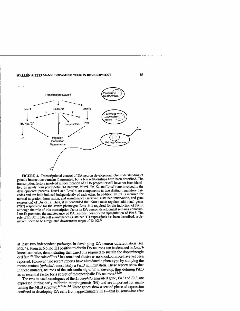
WALLEN & PERLMANN: DOPAMINE NEURON DEVELOPMENT 55
Transcription factors?
Nurri
TH, Ret, "X"
DA
FIGURE 4. Transcriptional control of DA neuron development. Our understanding of genetic interactions remains fragmented, but a few relationships have been described. The transcription factors involved in specification of a DA progenitor cell have not been identi- fied. In newly bom postmitotic DA neurons, Nurrl, Enl/2, and Lmxlb are involved in the developmental process. Nurrl and Lmxlb are components in two distinct regulatory cas- cades and are both induced independently of each other. In addition, Nurrl is required for normal migration, innervation, and maintenance (survival, sustained innervation, and gene expression) of DA cells. Thus, it is concluded that Nurrl must regulate additional genes ("X") responsible for the severe phenotype. Lmxlb is required for the induction of Pitx3, although the role of this transcription factor in DA neuron development remains unknovm. Lmxlb promotes the maintenance of DA neurons, possibly via upregulation of Pitx3. The role of Enl/2 in DA cell maintenance (sustained TH expression) has been described. a-Sy- nuclein seem to be a regulated downstream target of En 1/2.*^
at least two independent pathways in developing DA neuron differentiation (see RG. 4). From E16.5, no TH positive midbrain DA neurons can be detected in Lmxlb knock-out mice, demonstrating that Lmxlb is required to sustain the dopaminergic cell fate.^ The role of Pitx3 has remained elusive as no knockout mice have yet been reported. However, two recent reports have elucidated a phenotype by studying the mouse mutant (aphakia), most likely a Pitx3 null mutation. These reports show that in these mutants, neurons of the substantia nigra fail to develop, thus defining Pitx3 as an essential factor for a subset of mesencephalic DA neurons.^^-^^
The two mouse homologues of the Drosophila engrailed gene, Enl and En2, are expressed during early midbrain morphogenesis (E9) and are important for main- taining the MHB structure.^'^^'^'^^ These genes show a second phase of expression confined to developing DA cells firom approximately Ell—that is, somewhat after
56 ANNALS NEW YORK ACADEMY OF SCIENCES
Nurrl is induced. While single knockouts of these genes display no DA neuron de- ficiencies, TH is downregulated in compound knock-outs at late gestation and is ab- sent at E14.^^'^^ Nurrl, Lmxlb, or Pitx3 expression patterns have not been reported in these animals, and it is not known whether the loss ofTH is due to downregulation or an increase in cell death.
In conclusion, while the function of Pitx3 remains poorly investigated, homeobox transcription factors Lmxlb and Enl/En2, in addition to Nurrl, are clearly important for the normal differentiation and maintenance of the DA neuron phenotype. It should be noted, however, that the null mutations of Lmxlb and the En genes result in quite severe malformation of mid/hindbrain structures.^-^^ Thus, it remains pos- sible that the dopaminergic phenotypes could be indirect consequences of mid/hind- brain structural abnormalities.
ENGINEERING DA NEURONS FROM STEM CELLS
The elucidation of regulatory cascades influencing the specification and develop- ment of neuronal types is of relevance in stem cell research. This is a field attracting considerable interest, not least because of recent breakthroughs illustrating the in- creasingly realistic perspective of using stem cells and cell replacement strategies in treatment of neurodegenerative diseases.^''"^^ However, elucidating the relevant mechanisms involved in development of specific neuronal cell types will prove crit- ical in order to achieve such goals. Indeed, although our current understanding of factors influencing DA neuron development is quite limited, new knowledge has al- ready proved useful in attempts to engineer DA cells in vitro. In one recent study ex- pression of Nurrl in an immortalized neural stem cell hue derived from the embryonic cerebellum was shown to result in robust differentiation of TH-express- ing neurons when these cells were cocultured with type I astrocytes fi-om the ventral midbrain.^' Cultures of parental cells did not generate TH-positive neurons under these conditions, indicating an active instructive role of Nurrl. Other DA neuron markers were also expressed, and the cells were shown to synthesize DA. However, their ability to integrate in host brains and restore dopaminergic functions after graft- ing in rodent Parkinson's disease models remains to be demonstrated. The identity of the astrogUal factor is unknown but is evidently produced specifically by astro- cytes in the developing ventral midbrain, since glial cells from other regions of the developing CNS were unable to promote DA cell differentiation.^'
A second striking example of in v/fra-engineered DA neurons derives from work by McKay and coworkers on embryonic stem cells transfected with a Nurrl expres- sion vector.^^ By modifying a protocol previously developed by the same group, a large number of DA neurons could be generated in vitro. These neurons expressed several markers specific for midbrain DA neurons, including TH, Ret, Pitx3, and Enl. Impressively, grafting of these neurons into 6-hydroxy-DA-denervated host rat brains resulted in functional integration and restoration of parkinsonian symptoms. Although an ideal scenario would be to avoid the need of gene transfer and depend exclusively on extrinsic signals to manipulate embryonic stem cells in culture, both of these studies illusti-ate the importance of identifying the appropriate signals and factors that influence normal development of DA cells.
WALLEN & PERLMANN: DOPAMINE NEURON DEVELOPMENT 57
CONCLUSIONS
A great deal of information on transcriptional control of DA neuron development has been acquired during recent years. However, our understanding is still somewhat fragmented, and several areas will require attention in the years to come. For exam- ple, most of the current knowledge stems from studies of transcription factors ex- pressed in postmitotic maturing DA neurons. Similarly, a great deal is understood about the general patterning and morphogenetic events shaping the mid/hindbrain region. ^'^^ Signaling and the transcriptional events occuring in dividing progenitors leading to the generation of postmitotic DA neurons are much more poorly de- scribed. It will also be critical to define mechanisms generating diversity within the dopaminergic system and to define how functionally distinct dopaminergic cell groups are generated. It can be anticipated that characterization of these mechanisms will ultimately allow a complete description of genetic and epigenetic interactions required for generating these clinically important cells. As already shovm in previ- ous work, defining such mechanisms will be important in attempts to engineer DA neurons for transplantation and will likely identify mechanisms influencing the be- havior and survival of not only developing, but also mature DA neurons. Thus, the strong focus in this area holds excellent prospects of generating data of significance in the treatment of disorders such as Parkinson's disease.
ACKNOWLEDGMENTS
We thank Dr. Bertrand Joseph for critical comments on this manuscript and Dr. Alex Mata for additional help and comments.
REFERENCES
1. FALCK, B., et al. 1962. Fluorescence of catechol amines and related compounds con- densed with formaldehyde. J. Histochem. Cytochem. 10: 348-354.
2. DAHLSTROM, A. & K. FUXE. 1964. Evidence for the existence of monoamine-contain- ing neurones in the central nervous system. I. Demonstration of monoamines in the cell bodies of brain stem neurones. Acta Physiol. Scand. 62: 1-55.
3. BJORKLUND, A. & O. LiNDVALL. 1984. Dopamine-containing systems in the CNS, In Handbook of Chemical Neuroanatomy, Vol 2. Classical Transmitters in the CNS, Part I. BjOrklund, A. & T. Hokfelt, Eds. Elsevier Science Publishers.
4. ARTS, M.P.M., et al. 1996. Efferent projections of the retrorubral nucleus to the sub- stantia nigra and ventral tegmental area in cats as shown by anterograde tracing. Brain Res. Bull. 40: 219-228.
5. UNGERSTEDT, U. 1971. Stereotaxic mapping of the monoamine pathways in the rat brain. Acta Physiol. Scand. Suppl. 367: 1^8.
6. WURST, W. & L. BALLY-CUIF. 2001. Neural plate patterning: upstream and downstream of the isthmic organizer Nat. Rev. Neurosci. 2: 99-108.
7. HYNES, M. & A. ROSENTHAL. 1999. Specification of dopaminergic and serotonergic neurons in the vertebrate CNS. Curr. Opin. Neurobiol. 9: 26-36.
8. RoSENTHAL, A. 1998. Specification and survival of the dopaminergic neurons in the mammalian midbrain. Adv. Pharmacol. 42: 908-911.
9. HYNES, M., et al. 1995. Control of neuronal diversity by the floor plate: contact-medi- ated induction of midbrain dopaminergic neurons. Cell 80: 95-101.
10. HYNES, M., et al. 1995. Induction of midbrain dopaminergic neurons by sonic hedge- hog. Neuron 15: 33-44.
58 ANNALS NEW YORK ACADEMY OF SCIENCES
11. ERICSON, J., et al. 1995. Sonic hedgehog: a conunon signal for ventral patterning along the rostrocaudal axis of the neural tube. Int. J. Dev. Biol. 39: 809-816.
12. ERICSON, J., et al. 1997. Graded sonic hedgehog signaling and the specification of cell fate in the ventral neural tube. Cold Spring Harb. Symp. Quant. Biol. 62: 451-466.
13. ERICSON, J., et al. 1996. Two critical periods of sonic hedgehog signaling required for the specification of motor neuron identity. Cell 87: 661-673.
14. ERICSON, J., et al. 1995. Sonic hedgehog induces the differentiation of ventral fore- brain neurons: a common signal for ventral patterning within the neural tube Cell 81: 747-756.
15. CROSSLEY, P.H., S. MARTINEZ & G.R. MARTIN. 1996. Midbrain development induced by FGF8 in the chick embryo. Nature 380: 66-68.
16. LEE, S.M.K., et al. 1997. Evidence that FGF8 signalling from the midbrain-hindbrain junction regulates growth and polarity in the developing midbrain. Development 124: 959-969.
17. YE, W., et al. 1998. FGF and Shh signals control dopaminergic and serotonergic cell fate in the anterior neural tube. Cell 93: 755-766.
18. LiNDAHL, R. & S. EVCES. 1984. Rat liver aldehyde dehydrogenase. II. Isolation and characterization of four inducible isozymes. J. Biol. Chem. 259: 11991-11996.
19. WALL^N, A., et al. 1999. Fate of mesencephalic AHD2-expressing dopamine progeni- tor cells in NURRl mutant mice. Exp. Cell Res. 253: 737-746.
20. WALL^N, A., et al. 2001. Orphan nuclear receptor Nurrl is essential for Ret expression in the midbrain dopamine neurons and in the brain stem Mol. Cell. Neurosci. 18: 649-663.
21. CASTRO, D.S., et al. 2001. Induction of cell cycle arrest and morphological differentia- tion by Nurrl and retinoids in dopamine MN9D cells. J. Biol. Chem. 276: 43277- 43284.
22. JOYNER, A.L. 1996. Engrailed, Wnt and Pax genes regulate midbrain-hindbrain devel- opment. TIG 12: 15-20.
23. ADAMS, K.A., et al. 2000. The transcription factor Lmxlb maintains Wntl expression within the isthmic organizer Development 127: 1857-1867.
24. SMIDT, M.R, et al. 2000. A second independent pathway for development of mesen- cephalic dopaminergic neurons requires Lmxlb. Nat. Neurosci. 3: 337-341.
25. LAUDER, J.M. & F.E. BLOOM. 1974. Ontogeny of monoamine neurons in the locus coer- uleus, raphe nuclei and substantia nigra of the rat. I. Cell differentiation. J. Comp. Neurol. 155:469-^81.
26. LAW, S.W., et al. 1992. Identification of a new brain-specific transcription factor, NURRl. Mol. Endocrinol. 6: 2129-2135.
27. MANGELSDORF, D.J., et al. 1995. The nuclear receptor superfamily: the second decade. Cell 83: 835-839.
28 PERLMANN, T. & L. JANSSON. 1995. A novel pathway for vitamin A signaling mediated by RXR heterodimerization with NGFI-B and NURRl. Genes Dev. 9: 769-782.
29. FORMAN, B.M., et al. 1995. Unique response pathways are established by allosteric interactions among nuclear hormone receptors. Cell 81: 541-550.
30. MANGELSDORF, D.J. & R.M. EVANS. 1995. The RXR heterodimers and orphan recep- tors. Cell 83: 841-850.
31. HEVMAN, R.A., et al. 1992. 9-cis retinoic acid is a high affinity ligand for the retinoid X receptor. Cell 68: 397^06.
32. MATA DE URQUIZA, A., et al. 2000. Docosahexaenoic acid: a ligand for the retinoid X receptor in the mouse brain. Science 290: 2140-2144.
33. ZETTERSTROM, R.H., et al. 1996. Retinoid X receptor heterodimerization and develop- mental expression distinguish the orphan nuclear receptors NGFI-B, Nurrl and Norl. Mol. Endocrinol. 10: 1656-1666.
34. SAUCEDO-CARDENAS, O. & O.M. CONNEELY. 1996. Comparative distribution of nurrl and nur77 nuclear receptors in the mouse central nervous system J. Mol. Neurosci. 7: 51-63.
35. SAUCEDO-CARDENAS, O., et al. 1998. Nurrl is essential for the induction of the dopam- inergic phenotype and the survival of ventral mesencephalic late dopaminergic pre- cursor neurons. Proc. Natl. Acad. Sci. USA 95: 4013-4018.
WALL^N & PERLMANN: DOPAMINE NEURON DEVELOPMENT 59
36. ZETTERSTROM, R.H., et al. 1997. Dopamine neuron agenesis in Nurrl-deficient mice. Science 276: 248-250.
37. ZETTERSTROM, R.H., et al 1996. Cellular expression of the immediate early transcrip- tion factors Nurrl and NGFI-B suggests a gene regulatory role in several brain regions including the nigrostriatal dopamine system. Mol. Brain Res. 41: 111-120.
38. HoNKANiEMi, J., et al. 1997. Expression of zinc finger immediate early genes in rat brain after permanent middle cerebral artery occlusion. J. Cereb. Blood Flow Metab. 17: 636-646.
39. HONKAMEM, J. & RR. SHARP. 1996. Global ischemia induces immediate-early genes encoding zinc finger transcription factors. J. Cereb. Blood Flow Metab. 16: 557-565.
40. JOHANSSON, I.M., et al. 2000. Early and delayed induction of immediate early gene expression in a novel focal cerebral ischemia model in the rat. Eur. J. Neurosci. 12: 3615-3625.
41. LIN, T.N., et al. 1996. Expression of NGFl-B mRNA in a rat focal cerebral ischemia- reperfusion model. Mol. Brain Res. 43: 149-156.
42. CRISPING, M., et al. 1998. Nurrl mRNA expression in neonatal and adult rat brain fol- lowing kainic acid-induced seizure activity. Mol. Brain Res. 59: 178-188.
43. SCEARCE, L.M., et al. 1993. RNR-1, a nuclear receptor in the NGFI-B/Nur77 family that is rapidly induced in regenerating liver J. Biol. Chem. 268: 8855-8861.
44. MAGES, H.W., et al. 1994. NOT, A human immediate-early response gene closely related to the steroid/thyroid hormone receptor NAK1/TR3. Mol. Endocrinol. 8: 1583-1591.
45. CASTILLO, S.O., et al. 1998. Dopamine biosynthesis is selectively abolished in substan- tia nigra/ventral tegmental area but not in hypothalamic neurons in mice with tar- geted disruption of the Nurrl gene. Mol. Cell. Neurosci. 11: 36-46.
46. SAKURADA, K., et al. 1999. Nurrl, an orphan nuclear receptor, is a transcriptional acti- vator of endogenous tyrosine hydroxylase in neural progenitor cells derived from the adult brain. Development 126: 4017-^026.
47. IwAWAKi, T, K. KOHNO & K. KOBAYASHI. 2000. Identification of a potential nurrl response element that activates the tyrosine hydroxylase gene promoter in cultured cells. Biochem. Biophys. Res. Commun. 274: 590-595.
48. SACCHETTI, R, et al. 2001. Nurrl enhances transcription of the human dopamine trans- porter gene through a novel mechanism. J. Neurochem. 76: 1565-1572.
49. WiTTA, J., et al. 2000. Nigrostriatal innervation is preserved in Nurrl-null mice, although dopaminergic neuron precursors are arrested from terminal differentiation. Mol. Brain Res. 84: 67-78.
50. ZHOU, Q.Y. & R.D. PALMrmR. 1995. Dopamine-deficient mice are severely hypoactive, adipsic, and aphagic. Cell 83: 1197-1209.
51. MARCOS, C. & V. PACHNIS. 1996. The effect of the ret- mutation on the normal devel- opment of the central and parasympathetic nervous systems. Int. J. Dev. Biol. Suppl 1:137S-138S.
52. MOORE, M.W., et al. 1996. Renal and neuronal abnormalities in mice lacking GDNE Nature 382: 76-79.
53. SANCHEZ, M.R, et al. 1996. Renal agenesis and the absence of enteric neurons in mice lacking GONE Nature 382: 70-73.
54. SCHUCHARDT, A., et al. 1994. Defects in the kidney and enteric nervous system of mice lacking the tyrosine kinase receptor Ret [see comments]. Nature 367: 380-383.
55. GRANHOLM, A.C, et al. 2000. Glial cell line-derived neurotrophic factor is essential for postnatal survival of midbrain dopamine neurons. J. Neurosci. 20: 3182-3190.
56. LE, W.-D., et al. 1999. Reduced Nurrl expression increases the vulnerability of mesen- cephalic dopamine neurons to MPTP-induced injury. J. Neurochem. 73: 2218-2221.
57. SMIDT, M.P., et al. 1997. A homeodomain gene Ptx3 has highly restricted brain expression in mesencephalic dopaminergic neurons. Proc. Natl. Acad. Sci. USA 94:13305-13310.
58. NUNES, I., L.T. IbVAMASLW, R.M. SE.VA, et al. 2003. Pitx is required for development of substantia nigra dopaminergic neurons. Proc. Natl. Acad. Sci. USA 100: 4245-4250.
59. VAN DEN MUNCKHOF, P. K.C. LUK, L. STE.-MARIE, et al. 2003. Pitx is required for motor activity and for survival of a subset of midbrain dopaminergic neurons. Development 130: 2535-2542.

60 ANNALS NEW YORK ACADEMY OF SCffiNCES
60. DANIELIAN, P.D. & A.P. MCMAHON. 1996. Engrailed-l as a target of the wnt-1 signal- ing pathway in vertebrate midbrain development. Nature 383: 332-334.
61. JOYNER, A.L. & G.R. MARTIN. 1987. En-1 and En-2, two mouse genes with sequence homology to the Drosophila engrailed gene: expression during embryogenesis. Genes Dev. 1: 29-38.
62. SIMON, H.H., et al. 1998. En-1 and En-2 control the fate of the dopaminergic neurons in the substantia nigra and ventral tegmentum Eur. J. Neurosci. 10: 389.
63. SIMON, H.H., et al. 2001. Fate of midbrain dopaminergic neurons controlled by the engrailed genes. J. Neurosci. 21: 3126-3134.
64. GAGE, F.H. 2000. Mammalian neural stem cells. Science 287: 1433-1438. 65. ANDERSON, D.J. 2001. Stem cells and pattern formation in the nervous system: the pos-
sible versus the actual. Neuron 30: 19-35. 66. MOMMA, S., C.B. JOHANSSON & J. FRISEN. 2000. Get to know your stem cells. Curr
Opin. Neurobiol. 10: 45-^9. 67. WAGNER, J., et al. 1999. Induction of a midbrain dopaminergic phenotype in Nurrl-
overexpressing neural stem cells by type 1 astrocytes. Nat. Biotechnol. 17: 653-659. 68. KIM, J.H., et al. 2002. Dopamine neurons derived from embryonic stem cells function
in an animal model of Parkinson's disease. Nature 418: 50-56.
Transcription Factors in the Development of Midbrain Dopamine Neurons
J. PETER H. BURBACH, SIMONE SMITS, AND MARTEN P. SMIDT
Rudolf Magnus Institute of Neuroscience, Department of Pharmacology and Anatomy, University Medical Center Utrecht, Utrecht, the Netherlands
ABSTRACT: The development of midbrain dopamine (DA) neurons follows a number of stages marked by distinct events. After preparation of the region by signals that provide induction and patterning, at least two cascades of tran- scription factors contribute to the fully matured midbrain DA systems. One cascade involving the nuclear receptor Nurrl is required to synthesize the neurotransmitter DA; the enzyme tyrosine hydroxylase (TH) depends on it. The other cascade involves homeobox genes. Lmxlb and engrailed genes are expressed before the genesis of DA neurons and maintain their expression in these neurons. Lmxlb drives Ptx3, which is the latest transcription factor known to be induced. Its induction coincides with that of TH. Disruption of the function of Ptx3 affects the formation of the substantia nigra (SN) and alters the anatomical organization of the midbrain DA systems. While each cascade contributes to a specific aspect of DA neurons, both cascades are required for survival during development, indicating that the maintenance of DA neurons is delicately dependent on the appropriate activity of multiple transcriptional cascades.
KEYWORDS: transcription factors; midbrain; dopamine neurons
INTRODUCTION: MULTIPLICITY OF MIDBRAIN DOPAMINE NEURONS
Midbrain dopamine (DA) neurons comprise a heterogenous group of neurons that control elementary brain functions. First, they are involved in the control of move- ment; hence their prime role in Parkinson's disease. Second, they are part of circuits controling mood and reward, and thereby behavior. These two functions of midbrain DA systems are partly distinguished anatomically in the nigrostriatal system (move- ment) and the mesolimbic system (behavior).
Despite differentiation in anatomy and functions, all midbrain DA systems share the dopaminergic transmission machinery to communicate with postsynaptic ele- ments of the networks they are part of Dopaminergic transmission requires coordi- nated expression of specific gene products in order to synthesize and recycle the transmitter, to receive and control the chemical signal, and to transduce this into rel-
Address for correspondence: J.P.H. Burbach, Ph.D., Rudolf Magnus Institute of Neuroscience, Department of Pharmacology and Anatomy, University Medical Center Utrecht, Utrecht, the Netherlands. Voice: +31 (0)30 2538848; fax +31 (0)30 2539032.
Ann. N.Y. Acad. Sci. 991: 61-«8 (2003). © 2003 New York Academy of Sciences.
61

62 ANNALS NEW YORK ACADEMY OF SCIENCES
E10
PHOUFERATION
DIFFERBNTIA TION
E12
TRANSMITTER IDENTITY
FIGURE 1. Cascades of transcription factors enrolling in the development of midbrain DA neurons. Relationships are based on !oss-of-function studies in mice. All transcription factors affect the survival of developing DA neurons (see text).
evant cellular responses. Thus, midbrain neurons, despite their heterogeneous func- tions, have in common the transcriptional control to activate the gene sets necessary for dopaminergic transmission.
The distinction between functionally different groups of midbrain DA neurons is made at the level of the anatomy of cell bodies and the connectivity of axonal eflfer- ents with postsynaptic structures. DA signaling by substantia nigra (SN) neurons in- nervating the striatum modifies brain circuits controlling movement, while behavioral responses are triggered by ventral tegmental (VTA) DA neurons in the nucleus accumbens and other limbic areas. Thus, transcriptional control of genes in- volved in migration and axonal guidance is likely different from those involved in determination of the DA identity and is possibly variable between populations of midbrain DA neurons.
In this chapter we present our current view on how transcription factor cascades that unfold during development of midbrain DA neurons are linked to neurotrans- mission, connectivity, and survival. This view is illustrated in FIGURE 1.
THE FOREPLAY: PREPARATION OF THE EARLY MIDBRAIN
The development of midbrain dopamine neurons is largely dependent on the proper development of the ventral midbrain and the midbrain-hindbrain border that precedes the induction of midbrain dopamine neurons. Genes involved in boundary formation will have effects on the emergence of midbrain DA neurons. The first mid-

BURBACH et al: DEVELOPMENT OF MIDBRAIN DOPAMINE NEURONS 63
brain DA neurons arise at the most ventral rim of the neuroepithelium, lining up along the mesencephalic flexure of the ventral midbrain, where the expression of the growth factors Fgf8 and Shh interact.' Before the expression of DA-specific mark- ers, ventral midbrain markers are present in these cells. Among the earliest markers of the region are the homeobox genes engrailed-1 (Enl), engrailed-2 (En2), Pax2 and Pax5, and the growth factor Wntl. The expression of Enl and En2 is maintained by the expression of the signaling molecule Wntl.^ Another homeobox gene impli- cated in the early specification of the ventral midbrain is Lmxlb. This gene is ex- pressed at E7.5 in the ventral midbrain and diencephalon, and remains expressed in the adult in brain structures derived from these areas, including midbrain DA neurons.^
The first specific signs of the birth of midbrain DA neurons is the expression of the key enzyme in DA synthesis, tyrosine hydroxylase (TH), at El 1. This moment follows the induction of the nuclear receptor Nurrl (E10.5) and coincides with the expression of the midbrain DA-specific homeobox gene PtxS."*
THE HOMEOBOX PATHWAY: SPECIFICATION OF ARCHITECTURE AND CONNECTIVITY
As yet, four homeobox genes have been shown to be expressed in DA neurons: Enl, En2, Lmxlb, and Ptx3. Ptx3 differs from the others in both timing and space: Ptx3 is induced late, and its brain expression is confined to DA neurons. Enl, En2, and Lmxlb are expressed in the ventral midbrain, well before induction of TH; their expression extends largely beyond DA progenitors.^'^
In situ hybridization on mouse embryos from E8.5 to E16.5 showed the induction of PtxB expression with appearance of midbrain DA neurons. At El 1.5, a small layer at the ventral surface of the mesencephalic flexure expresses PtxB. At E12.5 a com- plete field of TH-positive, Ptx3-positive neurons has been obtained. These Ptx3-pos- itive cells are restricted to the marginal layer of the mesencephalic tegmentum. This group of about 50 cells corresponds to the first TH-expressing cells in the developing rodent brain. At later stages, the expression remains restricted to the midbrain DA system, and this association is conserved in adult rat brain and human SN tissue. This has also been demonstrated by 6-hydroxy DA lesions that take away all Ptx3- positive neurons.'* Extraneural Ptx3 expression is found in the developing skeletal muscle, in the tongue and in the developing eye lens.^'^
Lmxlb is a member of the LIM homeodomain family and is an essential regulator of dorsoventral patterning of the developing limbs. Lmxlb mutations evoke the nail- patella syndrome.^'^ Neural Lmxlb expression starts already at E7.5 (Johnson and Chen, personal communication). Early embryonic expression is extending anterior to the midbrain DA region into the ventral hypothalamic area, and posterior in the dorsal hindbrain and the dorsal spinal cord. In the ventral midbrain, Lmxlb is ex- pressed in midbrain DA neurons. Lmxlb expression is maintained in the adult SN and VTA of rodents and in the melanin-containing SN neurons of man.^ Engrailed genes also have an early (E8 onwards), regionally broad expression field in the mid- brain and hindbrain.^ Interesting, like Lmxlb, their expression is maintained in adult midbrain DA neurons.^ Enl is highly expressed in all adult midbrain DA neurons, and En2 in only a subset^ or is not detectable (unpublished).
64 ANNALS NEW YORK ACADEMY OF SCIENCES
The intrinsic potential of the Lmxlb and Enl/En2 genes as developmental regu- lators, together with the fact that these homeobox genes are expressed earlier than Nurrl, TH, and Ptx3, initially raised the hypothesis that Lmxlb and En genes may act genetically as upstream activators of these genes, in addition to preparing the ventral midbrain for genesis and differentiation of the midbrain DA system. To val- idate this hypothesis we analyzed expression of TH, Ptx3, and Nurrl in brain sec- tions of homozygous Lmxlb knock-out mice^ of stage E12.5, when the field of expression of TH and Ptx3 is first complete. Lmx 1 h~'~ embryos contained TH-pos- itive cells in the ventral tegmentum, but these cells did not express Ptx3, indicating that Ptx3 is not necessary for TH expression. Enl-null mutants are indistinguishable from wild-type mice, except for a high expression of En2 in all midbrain DA neu- rons.^ In Enl/En2-double-null mutants TH-positive neurons are initially generated, but soon lost.^ A similar observation has been made in Lmxlb-nuU mutants. Appar- ently, Lmxlb and Enl/En2 genes are each required for survival of midbrain DA neurons.
AN END STATION OF THE HOMEOBOX PATHWAY
To test the contribution of Ptx3 to the development of midbrain neurons, mice ho- mozygous for a defective Ptx3 allele were analyzed. These mice are blind, due to the function of Ptx3 in eye development, but grow and breed normally. These mice con- tain TH-positive neurons in the midbrain, but the anatomical organization is altered in two ways. First, TH-positive neurons are absent in the SN, and second, a central cell group exists fi-ontal to the VTA. The organization of TH neurons in the VTA ap- pears normal. Correlating with this phenotype is the lack of innervation of the dorsal striatum, which receives input from the SN DA neurons in wild-type mice. The in- nervation of the nucleus accumbens and olfactory tubercle exists as in wild-type mice. This phenotype suggests that Ptx3 disrupts processes required for expansion of DA neurons from El 1.5 onwards. These processes significantly affect neurons that will form the SN—that is, neurons that need to migrate laterally from the mes- encephalic flexure. Whether or not Ptx3 drives the survival, migration, or axonal pathfinding of this subset of neurons still needs to be established.
THE NURRl PATHWAY: INDUCTION OF THE DOPAMINERGIC TRANSMITTER PHENOTYPE AND SURVIVAL
Nurrl is an orphan member of the nuclear hormone receptor superfamily of tran- scription factors.'" Nurrl has a relatively wide field of expression in the embryonic midbrain, covering the entire ventral region.^ Only a small proportion of Nurrl-ex- pressing neurons overlap with the midbrain DA progenitor neurons (FIG. 2). Thus, brain expression of Nurrl is not uniquely linked to midbrain DA neurons, in contrast to the homeobox gene Ptx3. The onset of Nurrl expression in midbrain DA neurons is at E10.5 in the mouse, just before the induction of TH and Ptx3 atE 11.5 ."•'" • • ^ The expression of Nurrl is maintained in the adult stage, albeit in a more limited pattern, but including the midbrain DA system (FIG. 2). Multiple regions of the adult brain

BURBACH et al: DEVELOPMENT OF MIDBRAIN DOPAMINE NEURONS 65
EMBRYO E12.5
VTA SN CP DG CA SB
^ Sif
^A'
ADULT BRAIN
FIGURE 2. Expression of Nurrl in the embryonic and adult mouse brain. MB: midbrain; TS: thalamic subfield; DMN: dorsal motor nucleus; VTA: ventral tegmental area; SN: substan- tia nigra; CP: choroid plexus; CA: CAl, 2, and 3 field of the hippocampus; DG: dentate gyms; SB: subiculum.
express Nurrl.'^"'^ The Nurrl protein may therefore serve developmental and reg- ulatory functions that are not restricted to DA neurons.
The function of Nurrl in the brain has been addressed by the creation of null-mu- tant mice by three groups.'^''^'*^ Mice with a targeted deletion of the Nurrl gene die soon after birth. Analysis of the brain of these animals using such markers as Adh2, cRet, and TH showed that in the newborn animals no midbrain DA neurons are present. Although it was initially concluded that this is due to "agenesis" of midbrain DA neurons,'^ several additional studies demonstrated a more refined role of Nurrl in midbrain DA neurons. At E12.5 Nurrl-null mutants express other markers asso- ciated with midbrain DA neurons, including HNF 3p, Ptx3, and Lmxlb, but fail to induce TH.^ '''* CRet expression was also found to be absent in the ventral midbrain of Nurrl-null mutants.'* These findings link Nurrl to the growth factor glia-derived neurotrophic factor (GDNF), which promotes the survival of DA neurons.^"
The role of Nurrl as the key factor in the determination of the dopaminergic phenotype of midbrain DA neurons has been extended by two significant findings. First, it was shown that Nurrl*'" mice display diminished DA levels, suggesting that TH activity, which is rate limiting in neurotransmitter synthesis, is strictly regulated by Nurrl.'' Second, in humans a correlation between Nurrl and TH has been found in the age-related decrease of Nurrl in the SN.^' In cocain addicts Nurrl expression levels were decreased,^^ but this finding has not been compared to levels of TH.
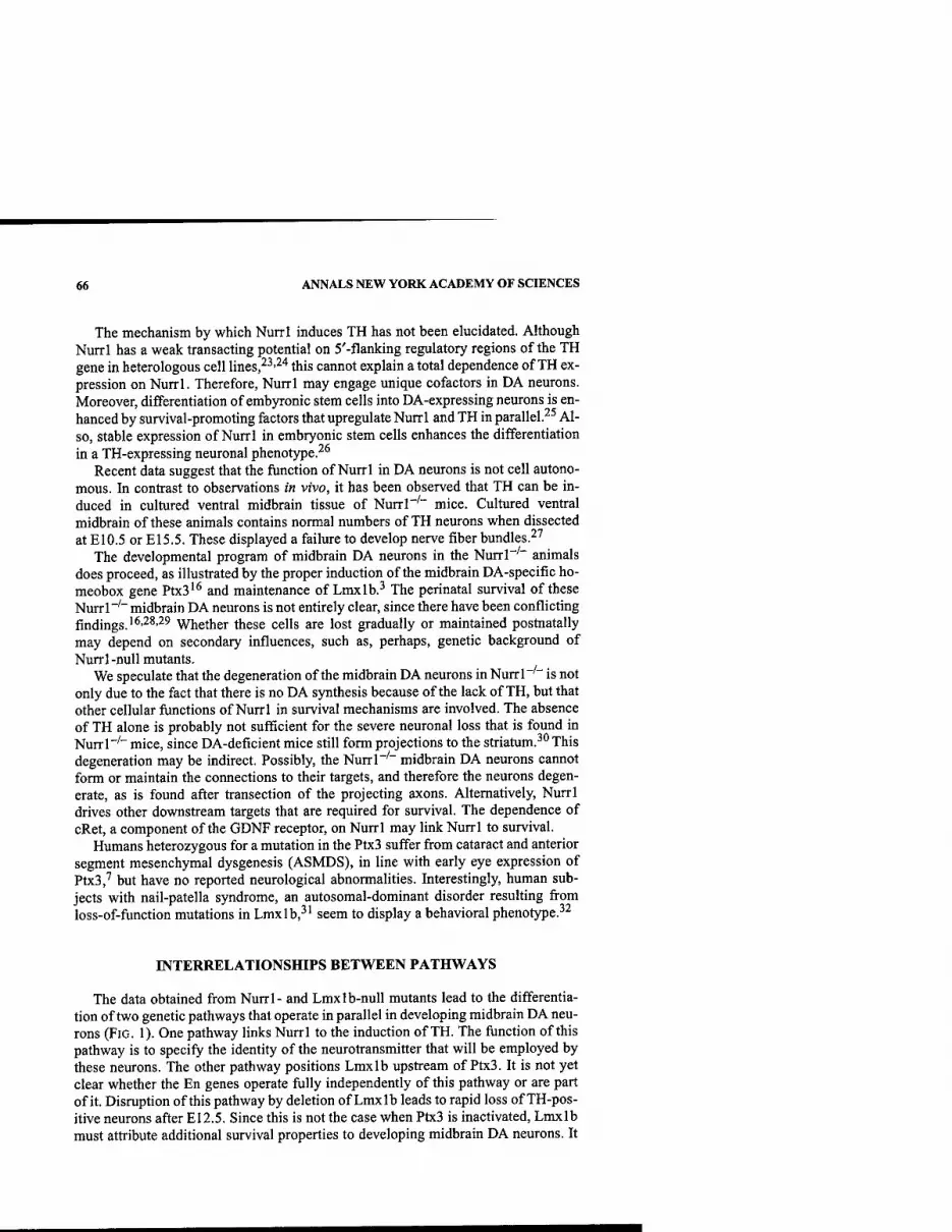
66 ANNALS NEW YORK ACADEMY OF SCIENCES
The mechanism by which Nurrl induces TH has not been elucidated. Although Nurrl has a weak transacting potential on 5'-flanking regulatory regions of the TH gene in heterologous cell lines,^^'^'* this cannot explain a total dependence of TH ex- pression on Nurrl. Therefore, Nurrl may engage unique cofactors in DA neurons. Moreover, differentiation of embyronic stem cells into DA-expressing neurons is en- hanced by survival-promoting factors that upregulate Nurrl and TH in parallel.^^ Al- so, stable expression of Nurrl in embryonic stem cells enhances the differentiation in a TH-expressing neuronal phenotype.^^
Recent data suggest that the function of Nurrl in DA neurons is not cell autono- mous. In contrast to observations in vivo, it has been observed that TH can be in- duced in cultured ventral midbrain tissue of Nurrl"'" mice. Cultured ventral midbrain of these animals contains normal numbers of TH neurons when dissected at E10.5 or E15.5. These displayed a failure to develop nerve fiber bundles.^''
The developmental program of midbrain DA neurons in the Nurrl"''" animals does proceed, as illustrated by the proper induction of the midbrain DA-specific ho- meobox gene Ptx3'^ and maintenance of Lmxlb.^ The perinatal survival of these Nurrl"'" midbrain DA neurons is not entirely clear, since there have been conflicting findings.'^'^^'■^^ Whether these cells are lost gradually or maintained postnatally may depend on secondary influences, such as, perhaps, genetic background of Nurrl-null mutants.
We speculate that the degeneration of the midbrain DA neurons in Nurrl"'" is not only due to the fact that there is no DA synthesis because of the lack of TH, but that other cellular functions of Nurrl in survival mechanisms are involved. The absence of TH alone is probably not sufficient for the severe neuronal loss that is found in Nurrl"'" mice, since DA-deficient mice still form projections to the striatum.^" This degeneration may be indirect. Possibly, the Nurrl"'" midbrain DA neurons cannot form or maintain the connections to their targets, and therefore the neurons degen- erate, as is found after transection of the projecting axons. Alternatively, Nurrl drives other downstream targets that are required for survival. The dependence of cRet, a component of the GDNF receptor, on Nurrl may link Nurrl to survival.
Humans heterozygous for a mutation in the Ptx3 suffer from cataract and anterior segment mesenchymal dysgenesis (ASMDS), in line with early eye expression of Ptx3,'' but have no reported neurological abnormalities. Interestingly, human sub- jects with nail-patella syndrome, an autosomal-dominant disorder resulting from loss-of-function mutations in Lmxlb,^' seem to display a behavioral phenotype.^^
INTERRELATIONSHIPS BETWEEN PATHWAYS
The data obtained from Nurrl- and Lmxlb-null mutants lead to the differentia- tion of two genetic pathways that operate in parallel in developing midbrain DA neu- rons (FIG. 1). One pathway links Nurrl to the induction of TH. The function of this pathway is to specify the identity of the neurotransmitter that will be employed by these neurons. The other pathway positions Lmxlb upstream of Ptx3. It is not yet clear whether the En genes operate fully independently of this pathway or are part of it. Disruption of this pathway by deletion of Lmxlb leads to rapid loss of TH-pos- itive neurons after E12.5. Since this is not the case when Ptx3 is inactivated, Lmxlb must attribute additional survival properties to developing midbrain DA neurons. It
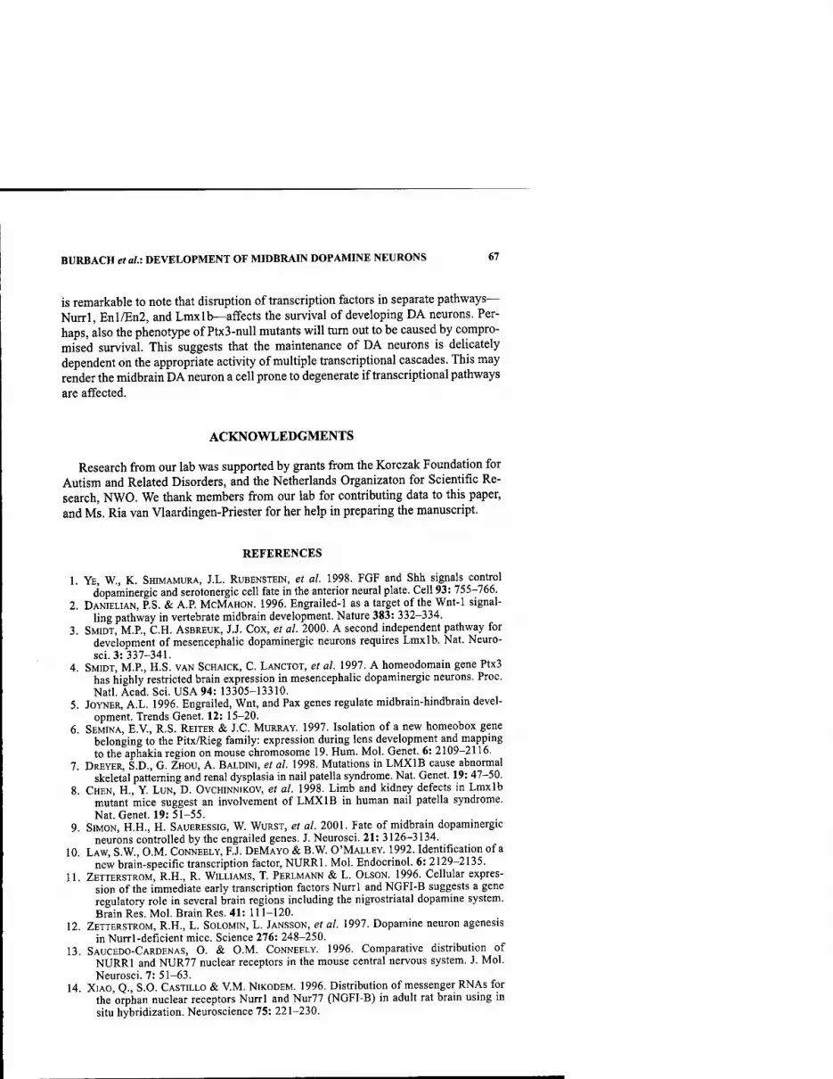
BURBACH et al: DEVELOPMENT OF MIDBRAIN DOPAMINE NEURONS 67
is remarkable to note that disruption of transcription factors in separate pathways— Nurrl, Enl/En2, and Lmxlb—affects the survival of developing DA neurons. Per- haps, also the phenotype of Ptx3-null mutants W\\\ turn out to be caused by compro- mised survival. This suggests that the maintenance of DA neurons is delicately dependent on the appropriate activity of multiple transcriptional cascades. This may render the midbrain DA neuron a cell prone to degenerate if transcriptional pathways
are affected.
ACKNOWLEDGMENTS
Research from our lab was supported by grants from the Korczak Foundation for Autism and Related Disorders, and the Netherlands Organizaton for Scientific Re- search, NWO. We thank members from our lab for contributing data to this paper, and Ms. Ria van Vlaardingen-Priester for her help in preparing the manuscript.
REFERENCES
1. YE, W., K. SHIMAMURA, J.L. RUBENSTEIN, et al. 1998. FGF and Shh signals control dopaminergic and serotonergic cell fate in the anterior neural plate. Cell 93: 755-766.
2. DANIELIAN, P.S. & A.P. MCMAHON. 1996. Engrailed-l as a target of the Wnt-1 signal- ling pathway in vertebrate midbrain development. Nature 383: 332-334.
3. SMIDT, M.P., C.H. ASBREUK, J.J. COX, et al. 2000. A second independent pathway for development of mesencephalic dopaminergic neurons requires Lmxlb. Nat. Neuro- sci. 3: 337-341.
4. SMIDT, M.P., H.S. VAN SCHAICK, C. LANCTOT, et al. 1997. A homeodomain gene Ptx3 has highly restricted brain expression in mesencephalic dopaminergic neurons. Proc. Natl. Acad. Sci. USA 94: 13305-13310.
5. JOYNER, A.L. 1996. Engrailed, Wnt, and Pax genes regulate midbrain-hindbrain devel- opment. Trends Genet. 12: 15-20.
6. SEMINA, E.V., R.S. REITER & J.C. MURRAY. 1997. Isolation of a new homeobox gene belonging to the Pitx/Rieg family: expression during lens development and mapping to the aphakia region on mouse chromosome 19. Hum. Mol. Genet. 6: 2109-2116.
7. DREYER, S.D., G. ZHOU, A. BALDINI, et al. 1998. Mutations in LMXIB cause abnormal skeletal patterning and renal dysplasia in nail patella syndrome. Nat. Genet. 19: 47-50.
8. CHEN, H., Y. LUN, D. OVCHINNIKOV, et al. 1998. Limb and kidney defects in Lmxlb mutant mice suggest an involvement of LMXIB in human nail patella syndrome. Nat. Genet. 19: 51-55.
9. SIMON, H.H., H. SAUERESSIG, W. WURST, et al. 2001. Fate of midbrain dopaminergic neurons controlled by the engrailed genes. J. Neurosci. 21: 3126-3134.
10. LAW, S.W., O.M. CoNNEELY, F.J. DEMAYO & B.W. O'MALLEY. 1992. Identification of a new brain-specific transcription factor, NURRl. Mol. Endocrinol. 6: 2129-2135.
11. ZETTERSTROM, R.H., R. WILLIAMS, T. PERLMANN & L. OLSON. 1996. Cellular expres- sion of the immediate early transcription factors Nurrl and NGFI-B suggests a gene regulatory role in several brain regions including the nigrostriatal dopamine system. Brain Res. Mol. Brain Res. 41: 111-120.
12. ZETTERSTROM, R.H., L. SOLOMIN, L. JANSSON, et al. 1997. Dopamine neuron agenesis in Nurrl-deficient mice. Science 276: 248-250.
13. SAUCEDO-CARDENAS, O. & O.M. CONNEELY. 1996. Comparative distribution of NURRl and NUR77 nuclear receptors in the mouse central nervous system. J. Mol. Neurosci. 7: 51-63.
14. XIAO, Q., S.O. CASTILLO & V.M. NIKODEM. 1996. Distribution of messenger RNAs for the orphan nuclear receptors Nurrl and Nur77 (NGFI-B) in adult rat brain using in situ hybridization. Neuroscience 75: 221-230.
68 ANNALS NEW YORK ACADEMY OF SCIENCES
15. BACKMAN, C, T. PERLMANN, A. WALLEN, et al. 1999. A selective group of dopaminer- gic neurons express Nurrl in the adult mouse brain. Brain Res. 851: 125-132.
16. SAUCEDO-CARDENAS, O., J.D. QUINTANA-HAU, W.D. LE, et a!. 1998. Nurrl is essential for the induction of the dopaminergic phenotype and the survival of ventral mesen- cephalic late dopaminergic precursor neurons. Proc. Natl. Acad. Sci. USA 95: 4013- 4018.
17. CASTILLO, S.O., J.S. BAFFI, M. PALKOVITS, et al. 1998. Dopamine biosynthesis is selec- tively abolished in substantia nigra/ventral tegmental area but not in hypothalamic neurons in mice with targeted disruption of the Nurrl gene. Mol. Cell Neurosci. 11: 36^6.
18. SAUCEDO-CARDENAS, O., R. KARDON, T.R. EDIGER, et al. 1997. Cloning and structural organization of the gene encoding the murine nuclear receptor transcription factor, NURRL Gene 187: 135-139.
19. WALLEN, A.A., D.S. CASTRO, R.H. ZETTERSTROM, et al. 2001. Orphan nuclear receptor Nurrl is essential for Ret expression in midbrain dopamine neurons and in the brain stem. Mol. Cell Neurosci. 18: 649-663.
20. BECK, K.D., J. VALVERDE, T. ALEXI, et al. 1995. Mesencephalic dopaminergic neurons protected by GDNF from axotomy-induced degeneration in the adult brain. Nature 373:339-341.
21. CHU, V., K. KOMPOLITI, E.J. COCHRAN, et al. 2002. Age-related decreases in Nurrl immunoreactivity in the human substantia nigra. J. Comp. Neurol. 450: 203-214.
22. BANNON, M.J., B. PRUETZ, A.B. MANNING-BOG, et al. 2002. Decreased expression of the transcription factor NURRl in dopamine neurons of cocaine abusers. Proc. Natl. Acad. Sci. USA 99: 6382-6385.
23. SAKURADA, K., M. OHSHIMA-SAKURADA, T.D. PALMER & F.H. GAGE. 1999. Nurrl, an orphan nuclear receptor, is a transcriptional activator of endogenous tyrosine hydrox- ylase in neural progenitor cells derived from the adult brain. Development 126: 4017-4026.
24. CAZORLA, P., M.P. SMIDT, K.L. O'MALLEY & J.P. BURBACH. 2000. A response element for the homeodomain transcription factor Ptx3 in the tyrosine hydroxylase gene pro- moter. J. Neurochera. 74: 1829-1837.
25. ROLLETSCHEK, A., H. CHANG, K. GUAN, et al. 2001. Differentiation of embryonic stem cell-derived dopaminergic neurons is enhanced by survival-promoting factors. Mech. Dev. 105: 93-104.
26. KIM, J.H., J.M. AUERBACH, J.A. RODRIGUEZ-GOMEZ, et al. 2002. Dopamine neurons derived from embryonic stem cells function in an animal model of Parkinson's dis- ease. Nature 418: 50-56.
27. ToRNQViST, N., E. HERMANSON, T. PERLMANN & I. STROMBERG. 2002. Generation of tyrosine hydroxylase-immunoreactive neurons in ventral mesencephalic tissue of Nurrl deficient mice. Dev. Brain Res. 133: 37-47.
28. LE, W., CM. CONNEELY, L. ZOU, et al. 1999. Selective agenesis of mesencephalic dopaminergic neurons in Nurrl-deficient mice. Exp. Neurol. 159: 451-458.
29. WALLEN, A., R.H. ZETTERSTROM, L. SOLOMIN, et al. 1999. Fate of mesencephalic AHD2-expressing dopamine progenitor cells in NURRl mutant mice. Exp. Cell Res. 253: 737-746.
30. ZHOU, Q.Y. & R.D. PALMITER. 1995. Dopamine-deficient mice are severely hypoactive, adipsic, and aphagic. Cell 83: 1197-1209.
31. BONGERS, E.M., M.C. GUBLER & N.V. KNOERS. 2002. Nail-patella syndrome. Over- view on clinical and molecular findings. Pediatr. Nephrol. 17: 703-712.
32. MCINTOSH, L, E. TIERNEY, I. BUKELIS & L. MARSH. 2001. Behavioral phenotype of nail-patella syndrome. Presented at the 51st Annual Meeting of the American Asso- ciation of Human Genetics, San Diego, CA, October 13-16, 2001. Abstr. 571/S.

Postnatal Developmental Programmed Cell Death in Dopamine Neurons
ROBERT E. BURKE Departments of Neurology and Pathology, The College of Physicians and Surgeons, Columbia University, New York, New York 10032. USA
ABSTRACT: The prenatal development of dopamine (DA) neurons of the sub- stantia nigra (SN) is characterized by their birth, specification, and migration to their final positions. Their postnatal development is characterized by the es- tablishment of contact and interactions between the SN and other neural nu- clei, particularly the striatal target, by extension of axons, terminal differentiation, and synapse formation. In this postnatal context there is a nat- ural cell death event, which is apoptotic in nature and biphasic in time course, with an initial peak on postnatal day (PND) 2, and a second on PND14. By PND20 the event has largely subsided. This natural cell death event is regulated in vivo by interaction with striatal target: it is augmented by axon-sparing tar- get lesion, DA terminal destruction, and medial forebrain bundle axotomy. This target dependence is present largely within only the first two postnatal weeks. The striatal target-derived neurotrophic factor(s) that regulate this death event are unknown. We have shown, in a postnatal primary culture mod- el of mesencephalic DA neurons, that glia-derived neurotrophic factor (GDNF) is unique in its ability to support their viability by suppressing apoptosis. We have also recently found that intrastriatal injection of GDNF in vivo suppresses apoptosis, and injection of neutralizing antibodies augments it. Thus, GDNF is a leading candidate for a striatum-derived neurotrophic factor for DA neurons during development.
KEYWORDS: apoptosis; programmed cell death; neurotrophic factors; Parkin- son's disease; striatum; substantia nigra
In evaluating research approaches to Parkinson's disease (PD), it is certainly reason- able to ask, how valuable is it to study developmental processes affecting dopamine (DA) neurons in relation to an adult-onset (and age-related) neurodegenerative dis- ease? There are several important reasons, related both to developing therapies for the treatment of patients today, and to furthering our understanding of pathogenesis for the development of prevention and cure. In relation to the treatment of patients today, some of the most promising approaches are based on cell replacement thera- pies. For all of these approaches, but especially for those based on the utilization of stem cells, critical issues in the developmental biology of dopamine neurons must be
Address for correspondence: Robert E. Burke, M.D., Department of Neurology, Room 308, Black Building, Columbia University, 650 West 168th Street, New York, NY 10032.Voice: +1- 212-305-7374; fax: -H-212-305-5450.
Ann. N.Y. Acad. Sci. 991: 69-79 (2003). © 2003 New York Academy of Sciences.
69

70 ANNALS NEW YORK ACADEMY OF SCIENCES
addressed in order to succeed: How is the mesencephalic dopamine neuron pheno- type specified? What extrinsic molecules and intrinsic signaling systems do the neu- rons depend upon to differentiate? At different stages in their development, what trophic factors and signaling pathways do they depend upon for viability? How do their axons find their way to appropriate targets and establish functional synaptic contacts?
In relation to the pathogenesis of PD, it is likely that the developmental neurobi- ology of dopamine neurons holds clues. Synuclein has been clearly implicated in the pathogenesis of familial and sporadic PD, and yet we do not know its normal fiinc-
DA Fibers in Siriatum
E14
Synapses Present in SNpc
E18
I TH Fibers'*
Vin Striatum E14.5
Largest increase „ Synapses in SNpc P15-P30
I 1
TH Cell Migration Ceases ^
E18-E20 I—I
IstTH 1st DA Cells >. ^ Cells E12.5 II E13
SN Cells Mitosis E11-E15
EO E5 E10 E15 E20;>
Largest increase Synapses in Striatum*
P13-P17 I 1
Str DA uptake 10% to 70% Adult level
Str. TH activity 10% to 75%^ Adult level
30% decrease in Striatal Neurons*
P4-P8 I 1
P5 P10 P15 P20 P25 P30
Birth
FIGURE 1. Important milestones in the development of the nigrostriatal dopaminergic system in rat. (1) SN dopamine neurons are bom between El 1 and E15, with a peak on £13 66,67 (2) Immunoreactivity for TH is first observed on E12.5;^^ for dopamine on E13.' (3) Prior to El8, dopaminergic neurons can be observed from the aqueduct to the ventral pial surface of the mesencephalon, in association with radial glia; they are likely to be migrating from their locus of origin to their final positions in the mesencephalon.^'By E20, dopamine neurons assume a topography similar to that of the adult brain,*' so it is likely that extra- nigral migration has ceased. (4) Dopaminergic fibers are first observed in the striatum by TH immunohistochemistry at E14.5;*^ by dopamine immunohistochemistry at E14.' (5) Synapses are first observed in SNpc at E18.'" (6) In the postnatal period, a natural cell death event occurs in striatum between PND4 and 8.'' (7) Differentiation of dopamine terminals takes place postnatally, indicated by large increases in TH activity and dopamine uptake be- tween birth and PNDSO.'" (8) Synapses form in the striatum postnatally, with the most rapid increase occurring between PNDsl3 and 17." (9) The largest increase in synapses in SNpc occurs between PI5 and 30.^"
BURKE: POSTNATAL DEVELOPMENTAL PROGRAMMED CELL DEATH 71
tion. One of our few clues is that it is highly upregulated during normal develop- ment.''^ The proteasome complex has been proposed to play a role in the pathogenesis of PD and other neurodegenerative disorders.^ What are the principal roles of this complex in the normal physiology of dopamine neurons? Again, devel- opment may hold clues: mRNAs for several components of the proteasome are high- ly upregulated.'* There is a growing consensus that programmed cell death (PCD) may play a role in the pathogenesis of neurodegenerative diseases. The concept of PCD derived in large part from developmental studies in neurobiology demonstrat- ing the universal occurrence of natural cell death.^ Many of the major classes of mol- ecules known today that regulate PCD, including the Bcl-2 family and the caspase family, were first identified by molecular studies of developmental cell death in C. elegans.^ Of all the features of development, the cell cycle would seem the least like- ly to play a role in adult-onset neurodegenerative diseases affecting postmitotic neu- rons, and yet components of the cell cycle have been implicated as PCD mediators^ and identified in Alzheimer brains.^ Surely we have much to learn about potential mechanisms of neurodegeneration by the study of developmental processes.
The development of dopamine neurons of the substantia nigra (SN) in rat can be thought of as proceeding in two broad phases (FIG. 1). In the prenatal phase, the pop- ulation of dopamine neurons is established, and the architecture of the SN as a nu- cleus is formed, by the processes of mitosis, phenotype specification, and neuronal migration. In this volume Drs. Simon, Perlmann, and Burbach address important ad- vances in the molecular basis of dopamine neuron phenotype specification. In the second phase of development, which begins in the perinatal period and extends into the postnatal period, the DA neurons of the SN establish interactions with other neu- ral groups by extending axons, forming synapses, and receiving afferents (FIG. 1). The first dopaminergic projections to reach the striatum do so by E14,^ but most of the innervation takes place between El 8 and postnatal day (PND) 4.^ During the postnatal period, dopaminergic terminals in the striatum undergo extensive differen- tiation, indicated by major increases in tyrosine hydroxylase (TH) activity and dopamine reuptake.'" During the postnatal period synapses form in the striatum, with most of the rapid rate of increase observed between PNDsl3 and 17." It is in this context of developing target interactions with the striatum that a natural cell death (NCD) event takes place among dopamine neurons of the SN.
NATURAL CELL DEATH IN DOPAMINE NEURONS OF THE SN
NCD in dopamine neurons of the SN begins shortly before birth in rat and achieves an initial peak on PND2'^''^ (FIG. 2). There is a second, smaller peak of NCD on PND 14, and by PND20 the event has largely abated. Qualitatively, the mor- phology of this NCD event is exclusively apoptotic. While other morphologies of cell death have been identified in the setting of developmental cell death,'"* we have observed only classic apoptotic morphology using a suppressed silver stain tech- nique.'^ We have previously shown that this technique will detect nonapoptotic, variant morphologies of programmed cell death,'^"'^ and yet none are observed in the SN during normal development.
We believe that this NCD event is likely to be a universal feature of nigral devel- opment in mammals. Based on counts of TH-immunostained neurons in SN, it had

72 ANNALS NEW YORK ACADEMY OF SCIENCES
been claimed that there is no evidence for NCD in mice.'' However, interpretation of such data is complicated by the fact that increasing expression of levels of TH dur- ing development add to the numbers of counted TH neurons; thus, loss of some of them could be masked.^ Indeed, Jackson-Lewis et al. have shown that a NCD event does occur in mice; classic nuclear apoptotic chromatin clumps are observed in TH- positive neurons.'^ This morphology was confirmed to be apoptotic at the light mi- croscope level by TUNEL labeling and immunostaining for the activated form of caspase-3. It was confirmed at the ultrastructural level by the appearance of electron- dense nuclear chromatin clumps and preservation of intracellular organelles as clas- sically described.'^
Our approach to quantifying the NCD event in SN warrants a brief methodolog- ical point. It will be noted in FIGURE 2 that two sets of counts are presented. Apop- totic nuclei meeting cellular criteria are surrounded by TH-positive cytoplasm, and are therefore defined as dopaminergic at the cellular level. It can be seen that only a small number of profiles meet this criterion. We had previously observed at an ultra- structural level that apoptotic neurons are stripped of their cytoplasm as they die,^" and it had previously been shown that neuronal cytoplasmic markers are diminished in their expression as cells die by apoptosis.^' It is therefore to be expected that only a small portion of dopamine neurons dying by apoptosis would be identifiable as such at the cellular level. In order to obtain an estimate of profiles derived from dopamine neurons, which otherwise would not be counted, we developed a regional criterion: any apoptotic profile in the SNpc, within 15 |xm of two TH-positive neu- rons, is counted. It can be seen in FIGURE 2 that the time courses of counts obtained by the cellular and regional criteria are identical.
Relatively little is known about the specific programmed cell death mechanisms in NCD in SN dopamine neurons. Of the three general pathways that mediate pro- grammed cell death, the extrinsic, the intrinsic, and the endoplasmic reticulum stress-mediated pathways,'^^ it is the intrinsic that is likely to be operative, given that NCD in SN appears to be regulated by the availability of trophic support (see below). In the intrinsic pathway, members of the Bcl-2 family of proteins act upstream to mitochondria to regulate release of cytochrome c and the resulting activation of
APOPTOTIC CELLS/SN
• TH + CELL ■ TH -)■ REGIONAL!
E19 T P4 BIRTH
P8 P12 P16 P20 AGE
P28
FIGURE 2. The time course of natural cell death in dopamine neurons in the SN. Counts for apoptotic profiles meeting cellular and regional criteria, as discussed in the text, are presented. Natural cell death in dopamine neurons has a biphasic time course, with an initial peak at PND2 and a second at PND14.

BURKE: POSTNATAL DEVELOPMENTAL PROGRAMMED CELL DEATH 73
caspases.^^ In transgenic mice overexpressing the antiapoptotic protein Bcl-2 under the control of the TH promoter, there is a reduction in the level of NCD in dopamine neurons and an increase in the number of SN TH-positive neurons surviving into adulthood.'* These animals are also resistant to the induction of developmental cell death due to striatal target lesion (see below). On the other hand, the proapoptotic protein Bax appears to be important for this induction, because it is reduced in Bax- null micc^"* While Bax-null mice show a trend for diminished levels of NCD, the effect does not reach significance, suggesting that mechanisms independent of Bax must also be operative.
Within the intrinsic pathway, mitochondrial release of cytochrome c, and its as- sociation with Apaf and pro-caspase-9 in the presence of ATP, results in the activa- tion of caspase-9, with the subsequent proteolytic cleavage, and activation of caspase-3 and other effector caspases.^^'^^ Activated caspase-3 is observed in apop- totic profiles in SN in a nuclear distribution during NCD,^' as are caspase protein cleavage products.'* However, whether caspase-3 is an essential mediator of NCD in dopamine neurons is unknovra. In many postmitotic neurons, it is not; caspase-3- null animals show diminished apoptotic nuclear chromatin changes, but no alteration in the levels of NCD.28
REGULATION OF NCD IN DOPAMINE NEURONS BY STRIATAL TARGET INTERACTIONS
According to classic neurotrophic theory, the magnitude of the NCD event within a projecting nucleus is regulated by interactions with its target.^^'^*^ The target provides a necessary trophic factor in limiting quantities by retrograde transport, such that only projecting neurons that successfully make target contact will survive. It is important to realize, however, that most of the experimental data upon which classic theory rests was developed fi-om systems with peripheral projections; far less is known about sys- tems with central projections. For dopamine neurons of the SN, it would appear likely that target interactions would regulate the NCD event, given that it occurs within the developmental period most characterized by elaboration of target contact and synapse formation (FIG. 1). In support of this concept, we have previously shown that axon- sparing lesions of the target striatum during development resuh in a reduction in the ultimate number of dopamine neurons at maturity.^' Furthermore, numerous investi- gations in tissue culture have demonstrated the ability of various striatal preparations to support the viability and differentiation of dopamine neurons.^^"^^
Based on these considerations, it would be predicted that disruption of striatal tar- get, or access to target, would result in an augmentation of NCD in dopamine neu- rons, and we have shown in a number of studies that this is indeed the case: developmental axon-sparing lesion of the striatum^** or selective disruption of dopamine terminals by 6-hydroxydopamine (60HDA)^^ or axotomy^^ lead to an in- duction of apoptotic death in dopamine neurons (FIG. 3).
For the striatal target lesion model and the 60HDA model, there is a developmen- tal window for this effect. In both models the effect is largely achieved within the first two postnatal weeks, corresponding, interestingly, to the period of NCD. There- after, the models differ; in the striatal lesion model, there is very little induction of death in SN pars compacta (pc) after PND14^* and none in adult.'^'^^ Thus, there is
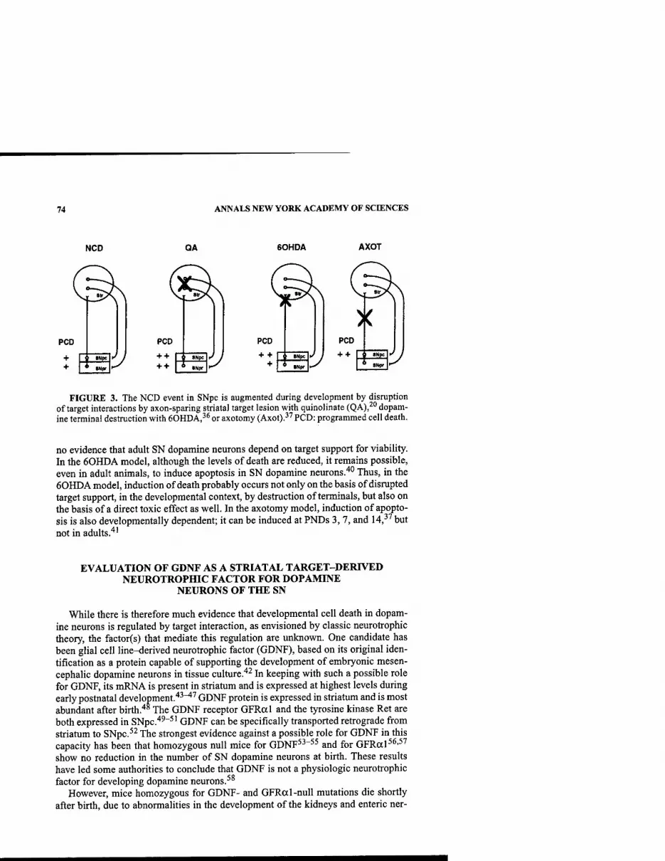
74 ANNALS NEW YORK ACADEMY OF SCIENCES
NCD QA 60HDA AXOT
PCD PCD
+ + + +
S»N|IC J J
FIGURE 3. The NCD event in SNpc is augmented during development by disruption of target interactions by axon-sparing striatal target lesion with quinolinate (QA),^" dopam- ine terminal destruction with 60HDA,^* or axotomy (Axot).^^ PCD: programmed cell death.
no evidence that adult SN dopamine neurons depend on target support for viability. In the 60HDA model, although the levels of death are reduced, it remains possible, even in adult animals, to induce apoptosis in SN dopamine neurons.'"' Thus, in the 60HDA model, induction of death probably occurs not only on the basis of disrupted target support, in the developmental context, by destruction of terminals, but also on the basis of a direct toxic effect as well. In the axotomy model, induction of apopto- sis is also developmentally dependent; it can be induced at PNDs 3, 7, and 14,-'^ but not in adults.'"
EVALUATION OF GDNF AS A STRIATAL TARGET-DERIVED NEUROTROPHIC FACTOR FOR DOPAMINE
NEURONS OF THE SN
While there is therefore much evidence that developmental cell death in dopam- ine neurons is regulated by target interaction, as envisioned by classic neurotrophic theory, the factor(s) that mediate this regulation are unknown. One candidate has been glial cell line-derived neurotrophic factor (GDNF), based on its original iden- tification as a protein capable of supporting the development of embryonic mesen- cephalic dopamine neurons in tissue culture.'*^ In keeping with such a possible role for GDNF, its mRNA is present in striatum and is expressed at highest levels during early postnatal development.''^"'*^ GDNF protein is expressed in striatum and is most abundant after birth.'*^The GDNF receptor GFRal and the tyrosine kinase Ret are both expressed in SNpc.''^"^' GDNF can be specifically transported retrograde from striatum to SNpc.^^ The strongest evidence against a possible role for GDNF in this capacity has been that homozygous null mice for GDNF^^"^^ and for GFRal^^•^'^ show no reduction in the number of SN dopamine neurons at birth. These results have led some authorities to conclude that GDNF is not a physiologic neurotrophic factor for developing dopamine neurons.^^
However, mice homozygous for GDNF- and GFRal-null mutations die shortly after birth, due to abnormalities in the development of the kidneys and enteric ner-
BURKE: POSTNATAL DEVELOPMENTAL PROGRAMMED CELL DEATH 75
vous system. Thus, they die quite early in the course of the NCD event in these neu- rons, and it is possible that they die before an effect on NCD can become apparent. Furthermore, none of these null mutations were regionally or temporally regulated; it is possible that compensatory changes occurred, masking a phenotype.
To reassess the possible role of GDNF as a physiologic trophic factor for dopam- ine neurons, we examined its ability to support their viability in a unique postnatal primary culture model,^' established during the period of NCD for these neurons. It is important to realize that all prior in vitro work on trophic factors for dopamine neurons, including that on GDNF itself, had been performed on early embryonic cul- tures, often derived at E14-E16. This is not a developmental period when dopamine neurons are making significant target contact (FIG. 1), so there is little reason to ex- pect that they should at this time be responsive to target-derived factors. The postna- tal culture model provides an opportunity to examine putative neurotrophic factors for dopamine neurons in a context in which target support is more likely to be oper- ative. In the postnatal cultures, we assessed GDNF in comparison to other factors that had previously been reported to support embryonic dopamine neurons, includ- ing brain-derived neurotrophic factor (BDNF),^^ transforming growth factors (TGF) P2 and [33,^' TGFa,^^ epidermal growth factor,^^ and basic fibroblast growth fac- tor. ^^ We found that GDNF alone was able to support the viability of postnatal dopamine neurons in vitro, and it did so by suppressing apoptosis.^^ This effect of GDNF was specific at the cellular level for dopamine neurons and at the regional lev- el for dopamine neurons derived from the SN.
We have recently evaluated effects of GDNF on levels of NCD in dopamine neu- rons in vivo. We have found that 1.0 \ig GDNF administered into the striatum of rats on PND2 is able to suppress levels of NCD, quantified using either cellular or re- gional criteria for apoptosis among dopamine neurons.*^ In support of a possible physiologic role for endogenous GDNF, we have also found, using a passive immu- nization approach, that acute intrastriatal neutralization of GDNF augments NCD among dopamine neurons.^^ This effect is developmentally dependent; it is observed on PNDs 2 and 6, but not PNDs 14 or 21. Thus, striatal GDNF appears to play a role primarily in the regulation of the first phase of NCD in dopamine neurons. This observation is in keeping with our assessment of the time course of GDNF mRNA expression in striatum during development. It is most abundant during the first post- natal week, and it declines thereafter (see Cho, Kholodilov, and Burke; this volume).
In order to be certain that endogenous striatal GDNF plays a physiologic role in regulating the NCD event and long-term survival of dopamine neurons, regional and temporally regulated knockout mice must be developed. To determine if GDNF alone is sufficient to regulate the NCD events in dopamine neurons, long-term over- expression in target areas must be achieved, and effects on the ultimate number of dopamine neurons in adult animals determined. We must also determine whether GDNF plays a role selectively in the early phase of NCD.
SUMMARY AND CONCLUSIONS
In summary, it is clear that a NCD event occurs during the normal development of dopamine neurons and that the magnitude of this event is regulated by early target interactions. It remains unknown what factor(s) derived from the striatal target reg-
76 ANNALS NEW YORK ACADEMY OF SCIENCES
ulate the NCD event, but in spite of observations in homozygous null mice, GDNF remains an important candidate.
Certainly this hypothesis does not exclude the possibility that there may be other equally important neurotrophic factors. To develop therapeutic approaches, and to better understand the pathogenesis of PD, the identification of physiologic trophic factors for dopamine neurons should be a foremost priority.
ACKNOWLEDGMENTS
The author is supported by NIH NS26836, NS38370, the Parkinson's Disease Foundation, and the Lovk^enstein Foundation.
REFERENCES
1. KHOLODILOV, N.G., M. NEYSTAT, T.F. OO, et al. 1999. Increased expression of rat synucleinl in the substantia nigra pars compacta identified by differential display in a model of developmental target injury. J. Neurochem. 73: 2586-2599.
2. PETERSEN, K., O.F. OLESEN & J.D. MIKKELSEN. 1999. Developmental expression of alpha-synuclein in rat hippocampus and cerebral cortex [in-process citation]. Neuro- science 91: 651-659.
3. Ii, K., H. ITO, K. TANAKA & A. HIRANO. 1997. Immunocytochemical co-localization of the proteasome in ubiquitinated structures in neurodegenerative diseases and the eld- erly. J. Neuropathol. Exp. Neurol. 56: 125-131.
4. EL-KHODOR, B.F., N.G. KHOLODILOV, O. YARVGINA & R.E. BURKE. 2001. The expres- sion of mRNAs for the proteasome complex is developmentally regulated in the rat mesencephalon. Dev. Brain Res. 129: 47-56.
5. OPPENHEIM, R.W. 1991. Cell death during development of the nervous system. Ann. Rev. Neurosci. 14: 453-501.
6. ELLIS, R.E., J. YUAN & H.R. HORVITZ. 1991. Mechanisms and functions of cell death. Annu. Rev Cell Biol. 7: 663-698.
7. PARK, D.S., E.J. MORRIS, R. BREMNER, et al. 2000. Involvement of retinoblastoma fam- ily members and E2F/DP complexes in the death of neurons evoked by DNA dam- age. J. Neurosci. 20: 3104-3114.
8. HusSEMAN, J.W., D. NocHLiN & I. ViNCENT. 2000. Mitotic activation: a convergent mech- anism for a cohort of neurodegenerative diseases. Neurobiol. Aging 21: 815-828.
9. VOORN, R, A. KALSBEEK, B. JORRITSMA-BYHAM & H.J. GROENEWEGEN. 1988. The pre- and postnatal development of the dopaminergic cell groups in the ventral mesen- cephalon and the dopaminergic innervation of the striatum of the rat. Neuroscience 25: 857-887.
10. CoYLE, J.T. 1977. Biochemical aspects of neurotransmission in the developing brain. Int. Rev Neurobiol. 20: 65-102.
11. HATTORI, T & PL. MCGEER. 1973. Synaptogenesis in the corpus striatum of infant rat. Exp. Neurol. 38: 70-79.
12. JANEC, E. & R.E. BURKE. 1993. Naturally occurring cell death during postnatal devel- opment of the substantia nigra of the rat. Mol. Cell Neurosci. 4: 30-35.
13. Oo, T.F. & R.E. BURKE. 1997. The time course of developmental cell death in pheno- typically defined dopaminergic neurons of the substantia nigra. Dev. Brain Res. 98: 191-196.
14. CLARKE, P.G.H. 1990. Developmental cell death: morphological diversity and multiple mechanisms. Anat. Embryol. 181: 195-213.
15. Go, T.F., R. BLAZESKI, S.M.W. HARRISON, et al. 1996. Neuron death in the substantia nigra of weaver mouse occurs late in development and is not apoptotic. J. Neurosci. 16:6134-6145.

BURKE: POSTNATAL DEVELOPMENTAL PROGRAMMED CELL DEATH 77
16. STEFANIS, L. & R.E. BURKE. 1996. Transneuronal degeneration in substantia nigra pars reticulata following striatal excitotoxic injury in adult rat: time course, distribution, and morphology of cell death. Neuroscience 74: 997-1008.
17. BLUM, M. 1998. A null mutation in TGF-alpha leads to a reduction in midbrain dopam- inergic neurons in the substantia nigra. Nat. Neurosci. 1: 374-377.
18. JACKSON-LEWIS, V., M. VILA, R. DJALDETTI, et al. 2000. Developmental cell death in dopaminergic neurons of the substantia nigra of mice. J. Comp. Neurol. 424: 476- 488.
19. KERR, J.F.R., G.C. GOBE, CM. WINTERFORD & B.V. HARMON. 1995. Anatomical meth- ods in cell death. In Methods in Cell Biology: Cell Death. L.M. Schwartz & B.A. Osbome, Eds.: 1-27. Academic Press. New York.
20. MACAYA, A., F. MUNELL, R.M. GUBITS & R.E. BURKE. 1994. Apoptosis in substantia nigra following developmental striatal excitotoxic injury. Proc. Natl. Acad. Sci. USA 91:8117-8121.
21. FREEMAN, R.S., S. ESTUS & E.M. JOHNSON. 1994. Analysis of cell cycle related gene expression in postmitotic neurons selective induction of cyclin Dl during pro- grammed cell death. Neuron 12: 343-355.
22. MEHMET, H. 2000. Caspases find a new place to hide [news]. Nature 403: 29-30. 23. ADAMS, J.M. & S. CORY. 1998. The Bcl-2 protein family: arbiters of cell survival. Sci-
ence 281: 1322-1326. 24. VILA, M., V. JACKSON-LEWIS, S. VUKOSAVIC, et al. 2001. Bax ablation prevents dopam-
inergic neurodegeneration in the l-methyl-4-phenyl-l,2,3,6-tetrahydropyridine mouse model of Parkinson's disease. Proc. Natl. Acad. Sci. USA 98: 2837-2842.
25. BUDIHARDJO, L, H. OLIVER, M. LUTTER, et al. 1999. Biochemical pathways of caspase activation during apoptosis. Annu. Rev. Cell Dev. Biol. 15: 269-290.
26. GREEN, D.R. & J.C. REED. 1998. Mitochondria and apoptosis. Science 281: 1309- 1312.
27. JEON, B.S., N.G. KHOLODILOV, T.F. GO, et al. 1999. Activation of caspase-3 in develop- mental models of programmed cell death in neurons of the substantia nigra. J. Neuro- chem. 73: 322-333.
28. OPPENHEIM, R.W., R.A. FLAVELL, S. VINSANT, et al. 2001. Programmed cell death of developing mammalian neurons after genetic deletion of caspases. J. Neurosci. 21: 4752-4760.
29. CLARKE, P.G.H. 1985. Neuronal death in the development of the vertebrate nervous system. Trends Neurosci. 8: 345-349.
30. BARDE, Y.A. 1989. Trophic factors and neuronal survival. Neuron 2: 1525-1534. 31. BURKE, R.E., A. MACAYA, D. DEVIVO, et al. 1992. Neonatal hypoxic-ischemic or exci-
totoxic striatal injury results in a decreased adult number of substantia nigra neurons. Neuroscience 50: 559-569.
32. PROCHIANTZ, A., U. DI PORZIO, A. KATO, et al. 1979. In vitro maturation of mesenceph- alic dopaminergic neurons from mouse embryos is enhanced in presence of their stri- atal target cells. Proc. Natl. Acad. Sci. USA 76: 5387-5391.
33. HEMMENDINOER, L.M., B.B. GARBER, P.C. HOFFMANN & A. HELLER. 1981. Target neu- ron-specific process formation by embryonic mesencephalic dopamine neurons in vitro. Proc. Natl. Acad. Sci. USA 78: 1264-1268.
34. HOFFMANN, P.C, L.M. HEMMENDINOER, C KOTAKE & A. HELLER. 1983. Enhanced dopam- ine cell survival in reaggregates containing target cells. Brain Res. 274: 275-281.
35. TOMOZAWA, Y. & S.H. APPEL. 1986. Soluble striatal extracts enhance development of mesencephalic dopaminergic neurons in vitro. Brain Res. 399: 111-124.
36. MARTI, M.J., CJ. JAMES, T.F. GO, et al. 1997. Early developmental destruction of ter- minals in the striatal target induces apoptosis in dopamine neurons of the substantia nigra. J. Neurosci. 17: 2030-2039.
37. EL-KHODOR, B.F. & R.E. BURKE. 2002. Medial forebrain bundle axotomy during devel- opment induces apoptosis in dopamine neurons of the substantia nigra and activation of caspases in their degenerating axons. J. Comp. Neurol. 452: 65-79.
38. KELLY, W.J. & R.E. BURKE. 1996. Apoptotic neuron death in rat substantia nigra induced by striatal excitotoxic injury is developmentally dependent. Neurosci. Lett. 220: 85-88.
78 ANNALS NEW YORK ACADEMY OF SCIENCES
39 LUNDBERG, C, K. WiCTORiN & A. BjORKLUND. 1994. Retrograde degenerative changes in the substantia nigra pars compacta following an excitotoxic lesion of the striatum. Brain Res. 644:205-212. . .
40. MARTI, M.J., J. SAURA, R.E. BURKE, et al. 2002. Striatal 6-hydroxydopamine induces apop'tosisofnigralneuronsin the adult rat. Brain Res. 958: 185-191.
41 CROCKER SJ N. WIGLE, R LISTON, et al. 2001. NAIF protects the nigrostriatal dopamine pathway in an intrastriatal 6-OHDA rat model of Parkinson's disease. Eur. J. Neurosci. 14:391^00. ,. ^ . ,
42 LIN, L.-RH., D.H. DOHERTY, J.D. LILE, et al. 1993. GDNF: a glial cell line-derived neurotrophic factor for midbrain dopaminergic neurons. Science 260: 1130-1132.
43. SCHAAR, D.G., B.A. SIBBER, C.R DREYFUS & I.B. BLACK. 1993. Regional and cell spe- cific expression of GDNF in rat brain. Exp. Neurol. 124: 368-371.
44 STROMBERG, I., L. BJORKLUND, M. JOHANSSON, et al. 1993. Glial cell line derived neu- rotrophic factor is expressed in the developing but not adult striatum and stimulates developing dopamine neurons in vivo. Exp. Neurol. 124: 401^12.
45 BLUM, M. & C.S. WEICKERT. 1995. GDNF mRNA expression in normal postnatal development, aging, and in weaver mutant mice. Neurobiol. Aging 16: 925-929
46 CHOI-LUNDBERG, D.L. & M.C. BOHN. 1995. Ontogeny and distribution of glial cell line-derived neurotrophic factor (GDNF) mRNA in rat. Dev. Brain Res. 85: 80-88.
47 GOLDEN J.R, J.A. DEMARO, RA. OSBORNE, et al. 1999. Expression of neurturm, GDNF, and GDNF family-receptor mRNA in the developing and mature mouse. Exp. Neurol. 158: 504-528.
48 LOPEZ-MARTIN E., H.J. CARUNCHO, J. RODRIGUEZ-PALLARES, et al. 1999. Stnatal dopaminergic afferents concentrate in GDNF-positive patches during development and in developing intrastriatal striatal grafts. J. Comp. Neurol. 406: 199-206.
49. TREANOR, J.J., L. GOODMAN, V. DE SAUVAGE, et al. 1996. Characterization of a multi- component receptor for GDNF. Nature 382: 80-83.
50 Yu T S. SCULLY, Y. YU, et al. 1998. Expression of GDNF family receptor components during development: implications in the mechanisms of interaction. J. Neurosci. 18:
4684-4696. . ^ ,.,„,• ^ ■ j 51 GLAZNER G W X. MU & J.E. SPRINGER. 1998. Localization of glial cell line-derived
neurotrophic factor receptor alpha and c-ret mRNA in rat central nervous system. J. Comp. Neurol. 391:42-49. . *• ■■ i
52 TOMAC A J WIDENFALK.L.H. LIN, e/a/. 1995. Retrograde axonal transport of glial cell line-derived neurotrophic factor in the adult nigrostriatal system suggests a trophic role in the adult. Proc. Natl. Acad. Sci. USA 92: 8274-8278.
53 MOORE, M.W., R.D. KLEIN, I. FARINAS, et al. 1996. Renal and neuronal abnormalities in mice lacking GDNR Nature 382: 76-79.
54. PiCHEL, J.G., L. SHEN, H.Z. SHENG, et al. 1996. Defects in enteric innervation and kid- ney development in mice lacking GDNF. Nature 382: 73-76.
55. SANCHEZ, M.R, I. SILOS-SANTIAGO, J. FRISEN, et al. 1996. Renal agenesis and the absence of enteric neurons in mice lacking GDNF. Nature 382: 70-73.
56 CACALANO, G., I. FARINAS, L.C. WANG, et al. 1998. GFRalphal is an essential receptor component for GDNF in the developing nervous system and kidney. Neuron 21: 53-
57. ENOMOTO, H., T. ARAKI, A. SACKMAN, et al. 1998. GFRalpha-1 deficient mice have deficits in the enteric nervous system and kidneys. Neuron 21: 317-324.
58. ROSENTHAL, A. 1999. The GDNF protein family: gene ablation studies reveal what they really do and how. Neuron 22: 201-203.
59 RAYPORT S., D. SULZER, W.X. SHI, et al. 1992. Identified postnatal mesolimbic dopamine neurons in culture morphology and electrophysiology. J. Neurosci. 12:
4264-4280. . ,■ c . r 60. HYMAN, C, M. HOFER, Y.A. BARDE, et al. 1991. BDNF is a neurotrophic factor for
doparninergic neurons of the substantia nigra. Nature 350:230-232. 61. POULSEN, K.T., M.R ARMANINI, R.D. KLEIN, et al. 1994. TGFP2 and TGF P3 are potent
survival factors for midbrain dopaminergic neurons. Neuron 13: 1245-1252. 62 ALEXI, T. & F. HEFTI. 1993. Trophic actions of transforming growth factor a on mesen-
ceph'alic dopaminergic neurons developing in culture. Neuroscience 55: 903-918.
BURKE: POSTNATAL DEVELOPMENTAL PROGRAMMED CELL DEATH 79
63. KNUSEL, B., P.P. MICHEL, J.S. SCHWABER & F. HEFTI. 1990. Selective and nonselective stimulation of central cholinergic and dopaminergic development in vitro by nerve growth factor, basic fibroblast growth factor, epidermal growth factor, insulin and the insulin-like growth factors I and II. J. Neurosci. 10: 558-570.
64. BURKE, R.E., M. ANTONELLI & D. SULZER. 1998. Glial cell line-derived neurotrophic growth factor inhibits apoptotic death of postnatal substantia nigra dopamine neu- rons in primary culture. J. Neurochem. 71: 517-525.
65. Oo, T.F., N. KHOLODILOV & R.E. BURKE. 2003. Regulation of natural cell death in dopaminergic neurons of the substantia nigra by striatal glial cell line-derived neu- rotrophic factor in vivo. J. Neurosci. 23: in press.
66. MARCHAND, R. & L.J. POIRER. 1983. Isthmic origin of neurons of the rat substantia nigra. Neuroscience 9: 373-381.
67. LAUDER, J.M. & F.E. BLOOM. 1974. Ontogeny of monoamine neurons in the locus coer- uleus, raphe nuclei and substantia nigra of the rat. J. Comp. Neurol. 155: 469-482.
68. SPECHT, L.A., V.M. PICKEL, T.H. JOH & D.J. REIS. 1981. Light-microscopic immunocy- tochemical localization of tyrosine hydroxylase in prenatal brain. I. Early ontogeny. J. Comp. Neurol. 199: 233-253.
69. SHULTS, C.W., R. HASIMOTO, R.M. BRADY & F.H. GAGE. 1990. Dopaminergic cells align along radial glia in the developing mesencephalon of the rat. Neuroscience 38: 427-436.
70. LAUDER, J.M. & F.E. BLOOM. 1975. Ontogeny of monoamine neurons in the locus coer- uleus, raphe nuclei and substantia nigra of the rat. J. Comp. Neurol. 163: 251-264.
71. FENTRESS, J.C, B.B. STANFIELD & W.M. COWAN. 1981. Observations on the develop- ment of the striatum in mice and rats. Anat. Embryol. 163: 275-298.
The Cast of Molecular Characters in Parkinson's Disease
Felons, Conspirators, and Suspects
KAH LEONG LIM," VALINA L. DAWSON,*''^''''' AND TED M. DAWSON*'^'''
"Neurodegeneration Research Laboratory, National Neuroscience Institute, 11 Jalan Tan Took Seng, Singapore 308433
^Institute for Cell Engineering and Departments of^Neurology, ^Neuroscience, and ^Physiology, Johns Hopkins University School of Medicine, Baltimore, Maryland 21287, USA
ABSTRACT: Parkinson's Disease (PD) is a common neurodegenerative disorder characterized by the progressive loss of dopamine neurons and the accumula- tion of Lewy bodies and neurites. Recent advances indicate that PD is due in some individuals to genetic mutations in a-synuclein, parkin, and ubiquitin C- terminal hydrolase LI (UCHLl). All three PD-linked gene products are related directly or indirectly to the functioning of the cellular ubiquitin proteasomal system (UPS), suggesting that UPS dysfunction may be important in PD patho- genesis. Indeed, emerging evidence indicates that derangements of the UPS may be one of the underlying mechanisms of PD pathogenesis. The function of parkin as an ubiquitin protein ligase positions it as an important player in both familial and idiopathic PD. We recently demonstrated that parkin mediates a nondegradative form of ubiquitination on synphilin-1 that could contribute to synphilin-1's aggregation in PD. Our results implicate parkin involvement in the formation of Lewy bodies associated with sporadic PD. This review discuss- es the role of the UPS, as well as the modus operandi of the three PD candidate felons (a-synuclein, parkin, and UCHLl) along with their conspirators in bringing about dopaminergic cell death in PD.
KEYWORDS: Parkinson's disease; a-synuclein; parkin; UCHLl; ubiquitin- proteasome system; ubiquitination
INTRODUCTION
Parkinson's disease (PD) is a progressive neurodegenerative disorder affecting 1- 2% of the population above the age of 65.' -^ Pathologically, the disease is character- ized by the relatively selective loss of midbrain dopaminergic neurons in the sub- stantia nigra pars compacta (SNpc) and the presence of Lewy bodies, eosinophilic
Address for correspondence: Ted M. Dawson, M.D., Ph.D., Institute for Cell Engineering, Departments of Neurology and Neuroscience, Johns Hopkins University School of Medicine, 600 N. Wolfe St., Carnegie 214, Baltimore, MD 21287. Voice: 410-614-3359; fax: 410-614- 9568.
Ann. N.Y. Acad. Sci. 991: 80-92 (2003). © 2003 New York Academy of Sciences.
80

LIM et al.i CAST OF MOLECULAR CHARACTERS IN PD 81
intracytoplasmic inclusions that are abundantly enriched in ubiquitin. The loss of ni- gral dopaminergic neurons results in a severe depletion of striatal dopamine and gives rise to a constellation of motoric deficits that include bradykinesia, rest tremor, rigidity, and postural instability. Although the etiology of PD remains unknown, the recent identification of several genetic mutations that cause familial PD has contrib- uted significantly to our understanding of the molecular pathogenesis of idiopathic PD. Mutations in a-synuclein, one of the major components of Lewy bodies, is linked to autosomal-dominant familial PD, and mutations in parkin cause autosomal recessive PD. Mutation in a third gene, encoding ubiquitin C-terminal hydrolase LI (UCHLl), may cause a rare autosomal-dominant form of PD.-' Interestingly, all the three PD-linked gene products are functionally associated directly or indirectly with the cellular ubiquitin proteasomal system (UPS).
The UPS plays an important role in maintaining cell homeostasis through the clearance of unwanted proteins. In this system, proteins destined for proteasomal degradation are covalently tagged with ubiquitin, a 76-amino acid residue protein, by the sequential actions of ubiquitin-activating (El), -conjugating (E2) and -ligat- ing (E3) enzymes. The ligation process is usually repeated many times to form a polyubiquitin chain in which the C terminal glycine residue of each ubiquitin unit is linked to a specific lysine (K) residue (most commonly K-48) of the previous ubiq- uitin.'* We and others have shown that parkin functions as an E2 dependent-E3 li- gase.^"^ Thus, one can envisage that a loss of parkin fiinction would lead to aberrations in the UPS and contribute to the pathogenesis of PD. Indeed, dysfunction of the UPS is emerging as a popular hypothesis to explain nigral degeneration in both familial and idiopathic PD.^ The formation of a-synuclein aggregates that are evi- dent in brain lesions in many neurodegenerative diseases including PD also suggests a derangement in intracellular protein handling. Finally, the function of UCHLl in the hydrolysis of polyubiquitin further implicates an involvement of the UPS ma- chinery in PD. We review here our current understanding of the various felons, con- spirators, and suspects involved in the molecular pathogenesis of PD.
a-SYNUCLEIN
The association of two missense mutations in a-synuclein (A30P and A53T) in a small subset of familial PD patients generated an intense and widespread interest among PD researchers.^'"^ Fueling this excitement was the discovery that Lewy bod- ies, the pathologic hallmark of sporadic PD, were strongly immunoreactive for a- synuclein.' ^ These findings made a-synuclein a compelling candidate felon in PD. Although the physiological function of a-synuclein remains to be elucidated, we now have a wealth of knowledge regarding its role in PD pathogenesis.
The a-synuclein protein belongs to a family of structurally related proteins that are expressed abundantly in the central nervous system. In humans, the synuclein family includes two other members, P- and y-synuclein. Structurally, synucleins have a highly conserved aminoterminal region containing repetitive imperfect re- peats that bear the consensus sequence KTKEGV, a hydrophobic middle section, and a less well conserved acidic carboxyl terminal. While a- and P-synucleins are con- centrated in close proximity to synaptic vesicles in nerve terminals, y-synuclein is more ubiquitously expressed throughout nerve cells.'^
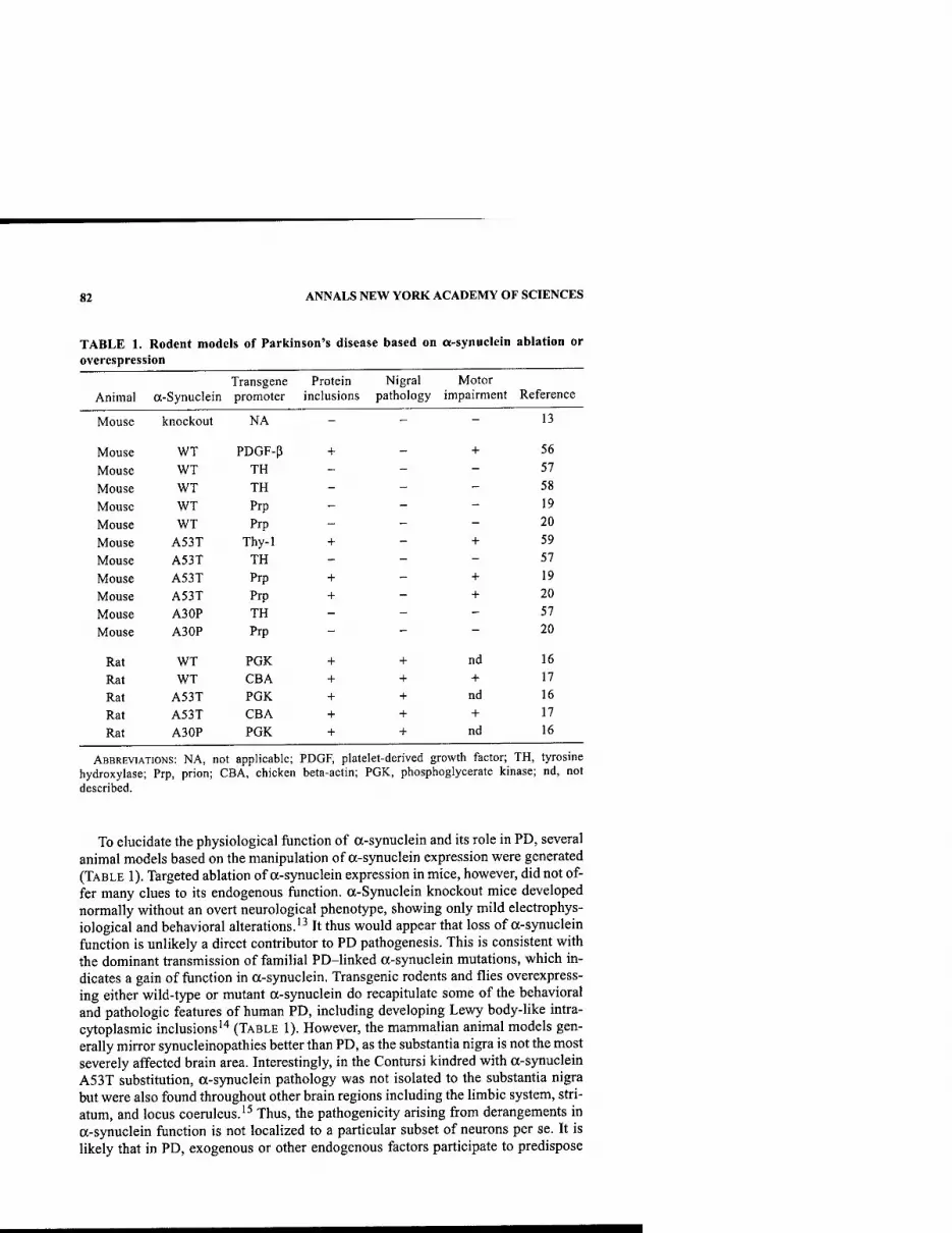
82 ANNALS NEW YORK ACADEMY OF SCIENCES
TABLE 1. Rodent models of Parkinson's disease based on a-synuclein ablation or overespression
Transgene Protein Nigral Motor Animal a-Synuclein promoter inclusions pathology impairment Reference
Mouse knockout NA - - - 13
Mouse WT PDGF-p + - + 56
Mouse WT TH - - - 57
Mouse WT TH - - - 58
Mouse WT Prp - - - 19
Mouse WT Prp - - - 20
Mouse A53T Thy-1 + - + 59
Mouse A53T TH - - - 57
Mouse A53T Prp + - + 19
Mouse A53T Prp + - + 20
Mouse A30P TH - - - 57
Mouse A30P Prp - - - 20
Rat WT PGK + + nd 16
Rat WT CBA + + + 17
Rat A53T PGK + + nd 16
Rat A53T CBA + + + 17
Rat A3 OP PGK + + nd 16
ABBREVIATIONS: NA, not applicable; PDGF, platelet-derived growth factor; TH, tyrosine hydroxylase; Prp, prion; CBA, chicken beta-actin; PGK, phosphoglycerate kinase; nd, not described.
To elucidate the physiological function of a-synuclein and its role in PD, several animal models based on the manipulation of a-synuclein expression were generated (TABLE 1). Targeted ablation of a-synuclein expression in mice, however, did not of- fer many clues to its endogenous function. a-Synuclein knockout mice developed normally without an overt neurological phenotype, showing only mild electrophys- iological and behavioral alterations.'^ It thus would appear that loss of a-synuclein function is unlikely a direct contributor to PD pathogenesis. This is consistent with the dominant transmission of familial PD-linked a-synuclein mutations, which in- dicates a gain of function in a-synuclein. Transgenic rodents and flies overexpress- ing either wild-type or mutant a-synuclein do recapitulate some of the behavioral and pathologic features of human PD, including developing Lewy body-like intra- cytoplasmic inclusions'"* (TABLE 1). However, the mammalian animal models gen- erally mirror synucleinopathies better than PD, as the substantia nigra is not the most severely affected brain area. Interestingly, in the Contursi kindred with a-synuclein A53T substitution, a-synuclein pathology was not isolated to the substantia nigra but were also found throughout other brain regions including the limbic system, stri- atum, and locus coeruleus.'^ Thus, the pathogenicity arising from derangements in a-synuclein function is not localized to a particular subset of neurons per se. It is likely that in PD, exogenous or other endogenous factors participate to predispose

LIM et al.: CAST OF MOLECULAR CHARACTERS IN PD 83
the nigral dopaminergic neurons to a-synuclein-mediated toxicity. To achieve a more localized effect of a-synuclein overexpression, direct intranigral injections of viral vectors expressing a-synuclein in rats induces Parkinson-like neurodegenera- tion including selective dopaminergic neuron loss and formation of a-synuclein pos- itive inclusions'^''^ (TABLE 1).
How do derangements in a-synuclein lead to neuronal death? Several lines of ev- idence seem to converge on a common theme: dysfunction in protein handling. The propensity of a-synuclein to aggregate and fibrillize into structures that are resistant to cellular degradation may be central to its neurotoxicity. The transformation of a- synuclein from a soluble state to a disease-associated fibrillar state involves a pro- gressive oligomerization of P-folded proteins known as protofibrils. Giasson and colleagues showed that the A53T a-synuclein has an increased tendency to polymer- ize into fibrils in vitro and that the polymerization is concentration dependent.'^ They proposed that a-synuclein fibrillization eventually leads to the formation of neuronal inclusions that would disrupt cellular function and result in cell death. The same group recently confirmed their hypothesis in mice overexpressing A53T a-sy- nuclein (TABLE 1), as mice overexpressing A53T a-synuclein develop a progressive neurodegenerative disorder characterized by the presence of insoluble aggregates of a-synuclein and fibrillar inclusions composed of a-synuclein.'^ In contrast, mice overexpressing wild-type a-synuclein lack Lewy body-like inclusions, and they do not develop a disease phenotype.'^ Lee and colleagues also recently observed that the A53T mutant a-synuclein is significantly more pathogenic than either wild-type a-synuclein or A30P mutant a-synuclein in transgenic mice.^" A53T mutant a-sy- nuclein-mediated neurodegeneration is associated with abnormal accumulation of detergent-insoluble a-synuclein that undergoes unique proteolytic processing.^*' Thus, proteolytic processing may in part drive the transformation of a-synuclein from a soluble state to a disease-associated fibrillar state through progressive oligo- merization. Clearly, additional conspirators are required to induce the formation of similar lesions from wild-type a-synuclein. Experimental cell culture models based on the ectopic expression of a-synuclein or its mutants have given us many clues to the nature of these conspirators. Agents that cause oxidative or nitrative damage can alter the solubility of a-synuclein and result in the formation of a-synuclein aggre- gates.^'"^^ Posttranslational modifications such as phosphorylation may also play a role in synuclein aggregation.^"* Phosphorylation of synuclein at Ser-129 promoted fibril formation in vitro, and this residue is selectively and extensively phosphorylat- ed in synucleinopathy lesions.^^ Interestingly, some evidence indicates the protofibril, rather than the fibril, to be the pathogenic species.^^ Biophysical studies conducted by Conway and colleagues showed that the two PD-linked a-synuclein mutations accelerated the rate of protofibril formation.-^' Furthermore, cytosolic dopamine seems to interact with a-synuclein to produce adducts that inhibit the con- version of protofibrils to fibrils.^* The stabilization of a-synuclein protofibrils by the dopamine-a-synuclein adducts might explain the specific neurodegeneration of ni- gral neurons in PD. Provoking further suspicion that dopamine may be an accom- plice in PD pathogenesis is the observation that accumulation of a-synuclein is toxic to cultured dopaminergic neurons, but not to nondopaminergic neurons.-^^ However, dopamine-a-synuclein adducts did not account for the selective degeneration of dopamine neurons; instead they showed that a-synuclein and 14-3-3 coexist in a 36- 83-kDa protein complex and that the levels of this complex are selectively increased

84 ANNALS NEW YORK ACADEMY OF SCIENCES
in the substantia nigra, but not in the cerebellum or cortex. The 14-3-3 family of pro- tein chaperones fulfill diverse roles in the cell, including the sequestration and inhi- bition of proapoptotic proteins.^" Complex formation between a-synuclein and 14- 3-3 is likely to decrease the free pool of cellular 14-3-3 to counteract apoptosis and to result in increased cellular susceptibility to stress.^' The involvement of dopamine in a-synuclein-mediated toxicity is a very appealing explanation on the selectivity of dopaminergic cell death in PD. It also highlights potential mechanistic differences in a-synuclein-mediated cell death among different subsets of the neuronal popula- tions in synucleinopathies.
PARKIN
Mutations in the parkin gene cause autosomal-recessive juvenile parkinsonism (ARJP). Pathologically, ARJP is characterized by a severe loss of nigral dopaminer- gic neurons and the absence of classic Lewy bodies. Affected individuals also tend to have an earlier mean age of onset (typically before 40 years of age). Following the discovery of parkin mutations in Japanese ARJP patients, several families with re- cessively inherited PD throughout the world were also found to carry parkin muta- tions. These findings are in stark contrast to the restricted occurrence of a-synuclein mutations. Indeed, mutations in parkin have become the main contributor not only to autosomal-recessive PD, but also to familial PD. It is thus important to understand how parkin acts as a felon in PD.
Parkin is a 465-amino acid ubiquitin protein ligase with a molecular mass of 52 kDa. The parkin protein has a modular structure consisting of an ubiquitin-like (UBL) domain at its N terminus, a RING (really mteresting new gene) box domain at its C terminus, and a unique middle segment that links the two domains. The RING box contains two RING finger motifs (termed RING 1 and RING 2) flanking a Cys-rich in-between RING (IBR). The RING box is important for the recruitment of E2s and represents the catalytic moiety of parkin.^''' Not surprisingly, the major- ity of familial point mutations in parkin reside in this region. The function of the UBL is currently unknown, although this domain shares 32% identity and 62% sim- ilarity with ubiquitin. Interestingly, the highly conserved lysine-48 (K-48) on the ubiquitin molecule is also conserved among UBL domains from different parkin orthologues (Lim et al., unpublished observations). During the synthesis of poly- ubiquitin chains, individual ubiquitin molecules are usually linked to one another through K-48-G76 isopeptide bonds."* We have shown that parkin promotes its own degradation through self-ubiquitination.'^ Potentially, K-48 on the UBL domain of parkin could represent a putative site on which the polyubiquitin chain is formed and, as such, may be important in the regulation of cellular parkin levels.
We and others have shown that familial associated mutations in parkin impair its catalytic function. These mutations disrupt parkin binding either to its cognate E2s or to its substrates. As a result, parkin-targeted substrates could not be ubiquitinat- ed.^"'' Our findings suggest that a loss of parkin fiinction would lead to the toxic ac- cumulation of its substrates, eventually killing the cell. However, it is not clear why dopaminergic neurons are preferentially susceptible. Undoubtedly, the identification of the spectrum of parkin substrates, together with the characterization of the bio- chemical pathways involved, will shed some light on the selective death of dopam-

LIM et al.: CAST OF MOLECULAR CHARACTERS IN PD 85
inergic neurons in PD. To date, six parkin substrate have been described: (1) CDCrel-1;'' (2) CDCrel2A (Choi etai, submitted); (3) an 0-glycosyIated form of a- synuclein;^^ (4) synphilin-!;■'■' (5) tau (Petrucelli et al., submitted); and (6) Pael-R (Parkin-associated endothelin receptor-/ike receptor).^'*
The synaptic vesicle-associated protein, CDCrel-1, was the first parkin substrate identified.^ Parkin interacts with and promotes the ubiquitination and degradation of CDCrel-1. Conversely, the familial parkin mutants Q31 Istop and T415N fail to af- fect CDCrel-1 turnover. Relatively little is known about CDCrel-1 function, but re- cent work from Trimble's group has suggested that it regulates synaptic vesicle release in the nervous system.^^ More recently, the same group generated CDCrel- 1- null mice and showed that CDCrel-l is not essential for neuronal development or function.^^ This observation is reminiscent of the mild neurological phenotype in a- synuclein knockout mice. Perhaps neuronal dysregulation results from CDCrel-l ac- cumulation rather than from its absence. Potentially, the elevation of CDCrel-1 in the absence of parkin-mediated degradation could disrupt dopamine release and eventu- ally lead to PD. Although this possibility remains to be evaluated, CDCrel 1 and a close homologue, CDCrel 2 A, were recently found to accumulate in the brain of pa- tients with ARJP (Choi et al., submitted). CDCrel 2A is a newly described parkin- binding protein that was identified using a yeast-two-hydrid screen (Choi et al., sub- mitted). Structurally, CDCrel 2A and CDCrel 1 share extensive homology except for their N terminal regions. Parkin interacts with and regulates the degradation of CD- Crel 2A. Parkin also colocalizes with CDCrel 2A in neuronal cell bodies and syn- apses (Choi et al., submitted). The association of both CDCrel members with parkin suggests an important fimctional relationship between parkin and the septins.
Since mutations in a-synuclein are associated with PD, it is tempting to link de- rangements in parkin and a-synuclein in a common pathogenic pathway. Naturally, this presynaptic protein became a much sought-after candidate substrate of parkin. However, in our coimmunoprecipitation studies we found that parkin neither inter- acts with nor ubiquitinates a-synuclein.^^ It turned out that a-synuclein needs to un- dergo a complex modification before it becomes a substrate of parkin. Shimura and colleagues described an 0-glycosylated form of a-synuclein (aSP22) that could in- teract with and be ubiquitinated by parkin. They fiirther showed that the level of aSP22 is elevated in ARJP brains and proposed that ubiquitination of aSP22 may be a prerequisite in the formation of Lewy bodies, which are generally absent in ARJP patients.^^ Parkin interaction with the glycosylated form of a-synuclein is highly selective, as the major nonglycosylated form of a-synuclein is totally inert to parkin.^^ This result is consistent with our inability to demonstrate an interaction be- tween parkin and the normal form of a-synuclein. Curiously, most laboratories have not been able to detect the glycosylated form of a-synuclein. Indeed, this type of complex glycosylation is more commonly found in extracellular proteins and is highly unusual for a cytoplasmic protein. Additionally, aSP22 to date has not been described in the several transgenic mouse models overexpressing a-synuclein. The relevance of aSP22 in the pathogenesis of PD thus awaits further characterization.
Synphilin-1, an interactor with a-synuclein, is a synaptic vesicle-enriched pro- tein that has previously been shown to form Lewy body-like inclusions when coex- pressed with a-synuclein.^^ Synphilin-1 is found in Lewy bodies, and abnormal accumulation of synphilin-1 is specific for brain lesions in which a-synuclein is a major component.^^ We recently identified synphilin-1 as a direct target for parkin-

86 ANNALS NEW YORK ACADEMY OF SCIENCES
mediated ubiquitination and further showed that parkin ubiquitinates proteins within cytosolic inclusions formed by coexpression of synphilin-1 and a-synuclein.-'^ Our results suggest a molecular link between the formation of Lewy bodies, the UPS, parkin, and synphilin-1 and support the speculation that functional parkin is required for Lewy body formation. Our results also linked the major nonglycosylated form of a-synuclein and parkin in a common biochemical pathway through their interactions with synphilin-1. Although the function of synphilin-1 is currently unknown, its ability to generate inclusions in the presence of a-synuclein indicates that it may play a role in Lewy body formation. We recently observed that parkin could mediate both K-48- and K-63-linked polyubiquitination of synphilin-1.^^ Unlike K-48- linked polyubiquitination, K-63-linked polyubiquitination of proteins is not associ- ated with proteasomal degradation.'* Indeed, while K-48-linked ubiquitination of synphilin-1 leads to a reduction in its steady state levels, the K-63-linked ubiquiti- nation of synphilin-1 did not affect its turnover rate. It is therefore conceivable that aberrant K-63-linked ubiquitination of synphilin-1 could lead to the intracytoplas- mic accumulation of protein. Interestingly, parkin-mediated ubiquitination of pro- teins present in cytoplasmic inclusions formed by coexpression of a-synuclein and synphilin-1 occurs via K-63 but not K-48 linkage.^^ Our results suggest that K-63- linked ubiquitination of synphilin-1 by parkin may be involved in Lewy body pathol- ogy associated with idiopathic PD.
The microtubule-associated protein tau is another protein present in Lewy bodies that is a parkin substrate (Petrucelli et al, submitted). While tauopathies and synu- cleinopathies are quite distinct phenomena, several recent genetic studies implicate tau in PD.'*^''" Furthermore, tau pathology was found in a case of autosomal-reces- sive early-onset PD caused by parkin mutations.'*^ Petrucelli et al. demonstrated physical and functional interactions between tau and parkin and, further, showed that tau accumulates in the brain of ARJP patients (Petrucelli et al, submitted). These findings indicate that tau pathology may contribute to the demise of nigral neurons in PD caused by parkin mutations.
A more general role for parkin in the ubiquitin degradation pathway is suggested by the observation that parkin is upregulated in response to cellular unfolded protein stress.^ Elevation of parkin relieves unfolded protein-mediated toxicity through ubiq- uitinating and targeting improperly folded protein to the proteasome for clearance. For this purpose, parkin recruits a different set of E2s, UBC6 and UBC7. Both UBC6 and UBC7 are resident to the endoplasmic recticulum (ER). Indeed, recent evidence showed that parkin ubiquitinates the putative G protein-coupled transmembrane re- ceptor Pael-R when it becomes unfolded.^"* When overexpressed in cells, Pael-R has a propensity to become unfolded and insoluble. The accumulation of unfolded Pael- R can be toxic to cells. Parkin protects against unfolded Pael-R-mediated cytotoxic- ity by facilitating unfolded Pael-R removal in the proteasome through ubiquitina- tion.^'* Consistent with this notion is the observation that there is an elevation of insoluble Pael-R in the brain of ARJP patients lacking functional parkin.
While more substrates of parkin undoubtedly will be identified with time, the cur- rent spectrum of parkin substrates is already indicative of the involvement of parkin in both familial and sporadic PD (FIG. 1). Under normal circumstances, parkin acts as an E3 ligase that will control the fate of the target proteins it ubiquitinates. Parkin- mediated K-48-linked ubiquitination will target its substrates for proteasomal clear- ance, while parkin-mediated K-63-linked ubiquitination is likely to modulate its tar-

LIM etal: CAST OF MOLECULAR CHARACTERS IN PD 87
CELLULAR STRESSES
UNFOLDED PROTEINS
REFOLDING (Hsp70 etc)
LOAD > DEGRADATION I
FAMILIAL PD-LINKED MUTATIONS
Aberrant K-63-linked ubiquitination?'
DEATH
PARKIN X POLYUBIQUITIN
K-63-linked
'^Hfflllffiifl K-48-linked
Toxic accumulation of substrates
(Tau, Pael-R, a-SP22, PROTEASOME synphilin-1, CDCrel-1)
ARJP
FIGURE 1. Depiction of the cast of molecular characters, felons, conspirators and sus- pects in PD.
get substrate's function—for example, in cell signaling. In ARJP patients lacking functional parkin, parkin substrates destined for degradation accumulate instead. The accumulation of these parkin substrates, such as aSP22, tau, and Pael-R, would be toxic to cells. It is likely that these cells fail to sequester the accumulated toxic proteins as inclusion bodies due to a loss in parkin-mediated ubiquitination.'*^''*'* However, Farrer and colleagues recently described the presence of Lewy bodies and Lewy neurites in an ARJP patient with compound heterozygous parkin mutations.'*^ This patient contains a null mutation in one parkin allele and a R275W substitution in the other. We have shown that the R275W mutation in parkin substantially reduc- es, but does not abolish, its catalytic competency.^-' Hence, the Lewy bodies and neu- ritic inclusions described by Farrer and colleagues could be related to the activity of parkin. This would be consistent with the speculation that functional parkin is re- quired for Lewy body formation and the finding that ARJP patients with homolo- gous null mutations in parkin lack Lewy body pathology. It is not known at this moment whether failure of parkin-mediated K-63-linked ubiquitination of proteins would contribute to ARJP. However, this mode of ubiquitination could potentially be linked to Lewy body formation in sporadic PD. The etiology of sporadic PD is likely complex, with environmental factors possibly playing an integral part, through the potential modulation of susceptibility genes for PD. Environmental insults that cause oxidative damage and other cellular stresses would, among other effects, increase the number of misfolded or unfolded proteins. The latter would trigger the unfolded pro- tein response (UPR) that would initially involve the upregulation of chaperone pro- teins such as members of the HspTO family. These chaperones would be instrumental in alleviating stresses due to unfolded proteins through the refolding of these pro- teins. Indeed, overexpression of Hsp70 rescues the motoric and neuropathologic fea-
88 ANNALS NEW YORK ACADEMY OF SCIENCES
tures of transgenic flies expressing normal and mutant forms of a-synuclein.'*^ In the presence of continuous or more intense insults, the unfolded protein load is likely to overwhelm chaperones. The UPS, whose response would include the elevation of parkin expression to aid ER-associated degradation, would then fulfill the second line of cellular defence against unfolded proteins. The recruitment of an alternative set of E2s resident in the ER by parkin may be dedicated to degradation-linked ubiq- uitination of proteins. Another collaborator of parkin function is a protein called CHIR Recent evidence from Takahashi's group showed that CHIP is a mammalian E4-like molecule that positively regulates parkin activity.'''^ CHIP and Hsp70 are both binding partners of parkin, and all three proteins exist as a complex with Pael- R. However, the amount of CHIP in the complex increases during ER stress. Appar- ently, CHIP facilitates parkin-mediated ubiquitination of Pael-R by promoting the dissociation of Hsp70 from parkin and Pael-R. Interestingly, the association of Hsp70 with Pael-R inhibits the E3 ligase activity of parkin."*' This observation sup- ports our model that Hsp70 chaperone activity represents the first line of defence against unfolded proteins. When Hsp70 activity becomes insufficient to cope with the unfolded protein stress, E3 ligases such as parkin would take over. We envisaged that if unfolded proteins accumulate beyond a certain threshold, leading to proteaso- mal impairment, the cell would sequester the ubiquitinated unfolded proteins in the form of Lewy body inclusions in an attempt to prolong its survival. Our model thus supports the general hypothesis that functional parkin is required for Lewy body for- mation and the notion that the formation of Lewy body-like inclusions is a cellular mechanism to sequester toxic products that would otherwise be an immediate threat to the cell. Although Lewy bodies may be beneficial initially, they are space-filling entities that would ultimately disrupt cellular function, leading to cell death. Addi- tionally, the sequestration of parkin in such inclusions would also deplete the cell of functional parkin and further contribute to neuronal death. In this light, the operating level of parkin in the cell is therefore crucial. Supporting this view is the increasing evidence that parkin haploinsufficiency may be sufficient for disease in some cas- es."** As ubiquitination and proteasomal degradation are both ATP-dependent pro- cesses, it is conceivable that events depleting the cell of its ATP could tax the UPS function. Blockage of UPS activity would, in turn, lead to increased oxidative stress. Thus, mitochondrion dysfunction and reduction of complex 1 activity in sporadic PD may be intimately linked with UPS derangements.
UCHLl AND OTHER SUSPECTS
UCHLl is a member of a gene family whose products hydrolyze small C terminal adducts of ubiquitin to generate the ubiquitin monomer. Expression of UCHLl is highly specific to neurons and to cells of neuroendocrine lineage. An I93M missense mutation in the UCHLl gene has been linked to PD in a sibling pair from a small German pedigree.'*^ The I93M mutation causes a partial loss of its ubiquitin hydro- lase activity in vitro.^^ Since hydrolysis of ubiquitin adducts is important in the prop- er funcfioning of the UPS, impairment of UCHLl activity could lead to aberrations in the proteolytic pathway and aggregation of proteins, including the UCHLl protein itself. Indeed, UCHLl is present in Lewy bodies. The occurrence of UCHLl in Lewy bodies and its function in the proteasome pathway thus make it a persuasive candi-

LIM et al.: CAST OF MOLECULAR CHARACTERS IN PD 89
date gene in PD. However, subsequent sequence analyses of UCHLl performed by others in several dominantly inherited PD cases have failed to detect the I93M sub- stitution.^"'^' Currently, the general view of this mutation is that it represents either a rare cause of PD or a benign substitution occurring in a single family. We speculate that an alternative way in which UCHLl could potentially act as a felon in PD is when its activity becomes overpromoted. Rapid and aberrant deubiquitination of polyubiquitinated proteins could stabilize proteins intended for degradation and con- tribute to their accumulation. A novel p53-interacting protein, herpesvirus-associat- ed ubiquitin-specific protease (HAUSP), has recently been shown to stabilize the levels of p53 by direct deubiquitination.^-^ HAUSP has intrinsic enzymatic activity that specifically deubiquitinates p53, leading to increased p53 levels both in vitro and in vivo. In contrast, expression of a catalytically inactive point mutant of HAUSP in cells increases the level of p53 ubiquitination and destabilizes p53. It is not known at this moment whether genetic mutations or environmental factors could modulate UCHLl activity or expression level. But aberrant UCHLl deubiquitination could be a mechanism contributing to protein aggregation.
There are many other suspects abetting PD pathogenesis, including (1) cyto- chrome P450 enzymes, which are involved in the detoxification of xenobiotics; (2) N-acetyltransferase 2, whose activity governs the rate of aromatic amine detoxifica- tion; and (3) apolipoprotein E (APOE), an extracellular ligand for receptors that clears remnants of chylomicrons and very-low-density lipoproteins.^^ Due to the lack of strong evidence for roles in PD, interest in these suspects has dissipated. Some newer suspects whose dysfunction may contribute to PD include transcription factors such as Nurr 1, Ptx-3, and Lmxlb, which regulate dopaminergic cell neuro- genesis, and the protein kinase PKCy. Comprehensive reviews of these newer PD candidates have appeared elsewhere.^'^"*
CONCLUSIONS
It is tempting to try to unify the multiple genes and sporadic causes of PD into one pathogenic biochemical pathway. The identification and characterization of the three PD gene products, a-synuclein, parkin, and UCHLl, has certainly helped us to conceive a general molecular pathway leading to PD. While we await the identity of the genes in other PD loci to be elucidated, the evolving picture of PD pathogenesis is one that converges onto cellular protein mishandling linked to UPS dysfunction. The firm establishment of UPS derangements as a major pathogenic biochemical pathway for PD would be instrumental in the design of rational therapeutic targets for PD patients. Potential draggable targets for PD could then include components of the protein handling squad, such as the chaperones, cochaperones, E3 ligases, and collaborators of E3 ligases. Other cellular components that help police the dopam- inergic activities would also be potential targets for PD therapy. For example, p-sy- nuclein has recently been suggested to be a natural negative regulator of a-synuclein aggregation.^^ Thus, the antiamyloidogenic property of p-synuclein might also pro- vide a novel strategy for the treatment of neurodegenerative disorders such as PD. Undoubtedly, with better understanding of the molecular mechanisms underlying PD, new and useful therapeutic targets will emerge. As we move into the postgen-
90 ANNALS NEW YORK ACADEMY OF SCIENCES
omic era, we certainly hope to move towards novel and effective treatment strategies for PD that go beyond L-DOPA.
ACKNOWLEDGMENTS
This work was supported by USPHS Grant NS38377 and the Edward D. and Anna Mitchell Family Foundation. K. L. Lim is a recipient of the National Science Scholarship International Fellowship awarded by A*STAR (Singapore).
REFERENCES
1. LANG, A.E. & A.M. LOZANO. 1998. Parkinson's disease. First of two parts. N. Engl. J. Med. 339: 1044-1053.
2. LANG, A.E. & A.M. LOZANO. 1998. Parkinson's disease. Second of two parts. N. Engl. J. Med. 339: 1130-1143.
3. LIM, K.L., V.L. DAWSON & T.M. DAWSON. 2002. The genetics of Parkinson's disease. Curr. Neurol. Neurosci. Rep. 2: 439-446.
4. PiCKART, CM. 2001. Mechanisms underlying ubiquitination. Annu. Rev. Biochem. 70: 503-533.
5. SHIMURA, H., N. HATTORI, S. KUBO, et al. 2000. Familial Parkinson disease gene prod- uct, parkin, is a ubiquitin-protein ligase. Nat. Genet. 25: 302-305.
6. iMAi, Y., M. SODA & R. TAKAHASHI. 2000. Parkin suppresses unfolded protein stress- induced cell death through its E3 ubiquitin-protein ligase activity. J. Biol. Chem. 275: 35661-35664.
7. ZHANG, Y., J. GAO, K.K. CHUNG, et al. 2000. Parkin functions as an E2-dependent ubiquitin- protein ligase and promotes the degradation of the synaptic vesicle-associ- ated protein, CDCrel-1. Proc. Natl. Acad. Sci. USA 97: 13354-13359.
8. KRUGER, R., O. EBERHARDT, O. RIESS, et al. 2002. Parkinson's disease: one biochemi- cal pathway to fit all genes? Trends Mol. Med. 8: 236-240.
9. PoLYMEROPOULOS, M.H., C. LAVEDAN, E. LEROY, et al. 1997. Mutation in the alpha- synuclein gene identified in families with Parkinson's disease. Science 276: 2045- 2047.
10. KRUGER, R., W. KUHN, T. MULLER, et al. 1998. Ala30Pro mutation in the gene encod- ing alpha-synuclein in Parkinson's disease. Nat. Genet. 18: 106-108.
11. SPILLANTINI, M.G., M.L. SCHMIDT, V.M. LEE, et al. 1997. Alpha-synuclein in Lewy bodies. Nature 388: 839-840.
12. GOEDERT, M. 2001. Alpha-synuclein and neurodegenerative diseases. Nat. Rev. Neuro- sci. 2: 492-501.
13. ABELIOVICH, A., Y. SCHMITZ, I. FARINAS, et al. 2000. Mice lacking alpha-synuclein dis- play functional deficits in the nigrostriatal dopamine system. Neuron 25: 239-252
14. DAWSON, T, A. MANDIR & M. LEE. 2002. Animal models of PD: pieces of the same puzzle? Neuron 35: 219-222.
15. SPIRA, P.J., D.M. SHARPE, G. HALLIDAY, et al. 2001. Clinical and pathological features of a Parkinsonian syndrome in a family with an Ala53Thr alpha-synuclein mutation. Ann. Neurol. 49:313-319.
16. Lo BIANCO, C, J.L. RIDET, B.L. SCHNEIDER, et al. 2002. alpha-Synucleinopathy and selective dopaminergic neuron loss in a rat lentiviral-based model of Parkinson's dis- ease. Proc. Natl. Acad. Sci. USA 99: 10813-10818.
17. KIRIK, D., C. ROSENBLAD, C. BURGER, et al. 2002. Parkinson-like neurodegeneration induced by targeted overexpression of alpha-synuclein in the nigrostriatal system. J. Neurosci. 22: 2780-2791.
18. GiASSON, B.I., K. URYU, J.Q. TROJANOWSKI, et al. 1999. Mutant and wild type human alpha-synucleins assemble into elongated filaments with distinct morphologies in vitro. J. Biol. Chem. 274: 7619-7622.

LIM et al.: CAST OF MOLECULAR CHARACTERS IN PD 91
19. GiASSON, B.I., J.E. DUDA, S.M. QUINN, et al. 2002. Neuronal alpha-synucleinopathy with severe movement disorder in mice expressing A53T human alpha-synuclein. Neuron 34: 521-533.
20. LEE, M.K., W. STIRLING, Y. XU, et al. 2002. Human alpha-synuclein-harboring familial Parkinson's disease-linked Ala-53 -> Thr mutation causes neurodegenerative disease with alpha-synuclein aggregation in transgenic mice. Proc. Natl. Acad. Sci. USA 99: 8968-8973.
21. PAXINOU, E., Q. CHEN, M. WEISSE, et al. 2001. Induction of alpha-synuclein aggrega- tion by intracellular nitrative insult. J. Neurosci. 21: 8053-8061.
22. SHERER, T.B., R. BETARBET, A.K. STOUT, et al. 2002. An in vitro model of Parkinson's disease: linking mitochondrial impairment to altered alpha-synuclein metabolism and oxidative damage. J. Neurosci. 22: 7006-7015.
23. LEE, H.J., S.Y. SHIN, C. CHOI, et al. 2002. Formation and removal of alpha-synuclein aggregates in cells exposed to mitochondrial inhibitors. J. Biol. Chem. 277: 5411- 5417.
24. OKOCHI, M., WALTER, J., KOYAMA, A., et al. 2000. Constitutive phosphorylation of the Parkinson's disease associated alpha-synuclein. J. Biol. Chem. 275: 390-397.
25. FUJIWARA, H., M. HASEGAWA, N. DOHMAE, et al. 2002. alpha-Synuclein is phosphory- lated in synucleinopathy lesions. Nat. Cell Biol. 4: 160-164.
26. GOLDBERG, M.S. & P.T. LANSBURY, JR. 2000. Is there a cause-and-effect relationship between alpha-synuclein fibrillization and Parkinson's disease? Nat. Cell Biol. 2: E115-119.
27. CoNWAY, K.A., S.J. LEE, J.C. ROCHET, et al. 2000. Acceleration of oligomerization, not fibrillization, is a shared property of both alpha-synuclein mutations linked to early-onset Parkinson's disease: implications for pathogenesis and therapy. Proc. Natl. Acad. Sci. USA 97: 571-576.
28. CoNWAY, K.A., J.C. ROCHET, R.M. BIEGANSKI, et al. 2001. Kinetic stabilization of the alpha-synuclein protofibril by a dopamine-alpha-synuclein adduct. Science 294: 1346-1349.
29. Xu, J., S.Y. KAO, F.J. LEE, et al. 2002. Dopamine-dependent neurotoxicity of alpha- synuclein: a mechanism for selective neurodegeneration in Parkinson disease. Nat. Med. 8: 600-606.
30. VAN HEMERT, M.J., H.Y. STBENSMA & G.P. VAN HEUSDEN. 2001. 14-3-3 proteins: key regulators of cell division, signalling, and apoptosis. Bioessays 23: 936-946.
31. WELCH, K. & J. YUAN. 2002. Releasing the nerve cell killers. Nat. Med. 8: 564-565. 32. SHIMURA, H., M.G. SCHLOSSMACHER, N. HATTORI, et al. 2001. Ubiquitination of a new
form of alpha-synuclein by parkin from human brain: implications for Parkinson's disease. Science 293: 263-269.
33. CHUNG, K.K., Y. ZHANG, K.L. LIM, et al. 2001. Parkin ubiquitinates the alpha-synu- clein-interacting protein, synphilin-1: implications for Lewy-body formation in Par- kinson disease. Nat. Med. 7: 1144-1150.
34. IMAI, Y, M. SODA, H. INOUE, et al. 2001. An unfolded putative transmembrane polypeptide, which can lead to endoplasmic reticulum stress, is a substrate of Parkin. Cell 105: 891-902.
35. BEITES, C.L., H. XlE, R. BOWSER, et al. 1999. The septin CDCrel-1 binds syntaxin and inhibits exocytosis. Nat. Neurosci. 2: 434-439.
36. PENG, X.R., Z. JIA, Y. ZHANG, et al. 2002. The septin CDCrel-1 is dispensable for nor- mal development and neurotransmitter release. Mol. Cell Biol. 22: 378-387.
37. ENGELENDER, S., Z. KAMINSKY, X. Guo, et al. 1999. Synphilin-1 associates with alpha- synuclein and promotes the formation of cytosolic inclusions. Nat. Genet. 22: 110- 114.
38. WAKABAYASHI, K., S. ENGELENDER, M. YOSHIMOTO, et al. 2000. Synphilin-1 is present in Lewy bodies in Parkinson's disease. Ann. Neurol. 47: 521-523.
39. LIM, K.L., Y. TANAKA & Y. ZHANG. 2002. Parkin mediates lysine-63 (K-63)-linked ubiquitination of synphilin-1: Implications for Lewy Body formation. Soc. Neurosci. Abst.
40. SCOTT, W.K., M.A. NANCE, R.L. WATTS, et al. 2001. Complete genomic screen in Par- kinson disease: evidence for multiple genes. JAMA 286: 2239-2244.

92 ANNALS NEW YORK ACADEMY OF SCIENCES
41. MARTIN, E.R., W.K. SCOTT, M.A. NANCE, etal. 2001. Association of single-nucleotide polymorphisms of the tau gene with late-onset Parkinson disease. JAMA 286: 2245- 2250.
42. VAN DE WARRENBURG, B.P., M. LAMMENS, C.B. LUCKING, et al. 2001. Clinical and pathologic abnormalities in a family with parkinsonism and parkin gene mutations. Neurology 56: 555-557.
43. DAWSON, T.M. 2000. New animal models for Parkinson's disease. Cell 101: 115-118. 44. CHUNG, K.K., V.L. DAWSON & T.M. DAWSON. 2001. The role of the ubiquitin-proteaso-
mal pathway in Parkinson's disease and other neurodegenerative disorders. Trends Neurosci. 24: S7-14.
45. FARRER, M., P. CHAN, R. CHEN, et al. 2001. Lewy bodies and parkinsonism in families with parkin mutations. Ann. Neurol. 50: 293-300.
46. AULUCK, P.K., H.Y. CHAN, J.Q. TROJANOWSKI, et al. 2002. Chaperone suppression of alpha-synuclein toxicity in a Drosophila model for Parkinson's disease. Science 295: 865-868.
47. IMAI, Y., M. SODA, S. HATAKEYAMA, et al. 2002. CHIP is associated with Parkin, a gene responsible for familial Parkinson's disease, and enhances its ubiquitin ligase activity. Mol. Cell 10: 55-67.
48. WEST, A.M. 2002. Complex relationship between Parkin mutations and Parkinson dis- ease. Am. J. Med. Genet. 114: 584-591.
49. LEROY, E., R. BOYER, G. AUBURGER, et al. 1998. The ubiquitin pathway in Parkinson's disease. Nature 395: 451-452.
50. LINCOLN, S., J. VAUGHAN, N. WOOD, et al. 1999. Low frequency of pathogenic muta- tions in the ubiquitin carboxy-terminal hydrolase gene in familial Parkinson's dis- ease. Neuroreport 10: 427—429.
51. MARAGANORE, D.M., M.J. FARRER, J.A. HARDY, et al. 1999. Case-control study of the ubiquitin carboxy-terminal hydrolase LI gene in Parkinson's disease. Neurology 53: 1858-1860.
52. LI, M., D. CHEN, A. SHILOH, et al. 2002. Deubiquitination of p53 by HAUSP is an important pathway for p53 stabilization. Nature 416: 648-653.
53. VAUGHAN, J.R., M.B. DAVIS & N.W. WOOD. 2001. Genetics of Parkinsonism: a review. Ann. Hum. Genet. 65: 111-126.
54. RAMSDEN, D.B., R.B. PARSONS, S.L. HO, et al. 2001. The aetiology of idiopathic Par- kinson's disease. Mol. Pathol. 54: 369-380.
55. HASHIMOTO, M., E. ROCKENSTEIN & M. MANTE. 2001. beta-Synuclein inhibits alpha- synuclein aggregation: a possible role as an anti-parkinsonian factor. Neuron 32: 213-223
56. MASLIAH, E., E. ROCKENSTEIN, I. VEINBERGS, et al. 2000. Dopaminergic loss and inclu- sion body formation in alpha-synuclein mice: implications for neurodegenerative disorders. Science 287: 1265-1269.
57. MATSUOKA, Y., M. VILA, S. LINCOLN, et al. 2001. Lack of nigral pathology in trans- genic mice expressing human alpha-synuclein driven by the tyrosine hydroxylase promoter. Neurobiol. Dis. 8: 535-539.
58. RICHFIELD, E.K., M.J. THIRUCHELVAM, D.A. CORY-SLECHTA, et al. 2002. Behavioral and neurochemical effects of wild-type and mutated human alpha-synuclein in trans- genic mice. Exp. Neurol. 175: 35-48.
59. VAN DER PUTTEN, H., K.H. WIEDERHOLD, A. PROBST, et al. 2000. Neuropathology in mice expressing human alpha-synuclein. J. Neurosci. 20: 6021-6029.
Oxidative Modifications of a-Synuclein
HARRY ISCHIROPOULOS
Stokes Research Institute, Department of Biochemistry and Biophysics, Children's Hospital of Philadelphia and the University of Pennsylvania, Philadelphia, Pennsylvania 19104, USA
ABSTRACT: Hallmark lesions of neurodegenerative synucleinopathies contain a-synuclein (a-syn) that is modifled by nitration of tyrosine residues and pos- sibly by dityrosine cross-linking to generated stable oligomers. Data gathered from in vitro experiments and from model systems of cells transfected with wild-type and mutant a-syn revealed that conditions resulting in a-syn nitra- tion also induce formation of a-syn inclusions with similar biochemical char- acteristics to protein extracted from human lesions. The detection of tyrosine- nitrated a-syn signifies the formation of reactive nitrogen species capable of both radical and electrophilic attack on aromatic residues as well as nucleo- philic additions and oxidations. The cellular sources and biochemical reactivity of reactive nitrogen species in the central nervous system remain largely un- known, but kinetically fast reactions of nitric oxide with superoxide to form peroxynitrlte as well as enzymatic one-electron oxidation of nitrite are two im- portant sources of reactive nitrogen species. Based on these findings a model is proposed where the process of fibrllization can be differentially affected by ox- idants and nitrating species. Posttranslational modifications of a-syn by reac- tive nitrogen species inhibits fibril formation and results in urea- and SDS- insoluble, protease-resistant a-syn aggregates that maybe responsible for cel- lular toxicity.
KEYWORDS: oxidative stress; tyrosine nitration; dityrosine; a-synuclein; pro- tein aggregation
OXIDATIVE AND NITRATIVE STRESSES IN NEURODEGENERATIVE DISORDERS
Partial reduction of oxygen by one electron produces superoxide, whereas two- electron reduction produces hydrogen peroxide. Enzymatic and other scavenging pathways rapidly remove superoxide and hydrogen peroxide to avoid formation of secondary strong oxidants. Under pathological conditions the overproduction of ox- idants may overwhelm the cellular antioxidant capacity, resulting in undesired oxi- dation of cellular molecules and ultimately cell death. Augmentation of intracellular defenses in humans as well as in model systems of neurodegenerative disorders has
Address for correspondence: Harry Ischiropoulos, Ph.D., Stokes Research Institute, Chil- dren's Hospital of Philadelphia, 416D Abramson Research Center, 34th St. and Civic Center Blvd., Philadelphia, PA 19104-4318. Voice: 215-590-5320; fax: 215-590-4267.
Ann. N.Y. Acad. Sci. 991: 93-100 (2003). © 2003 New York Academy of Sciences.
93
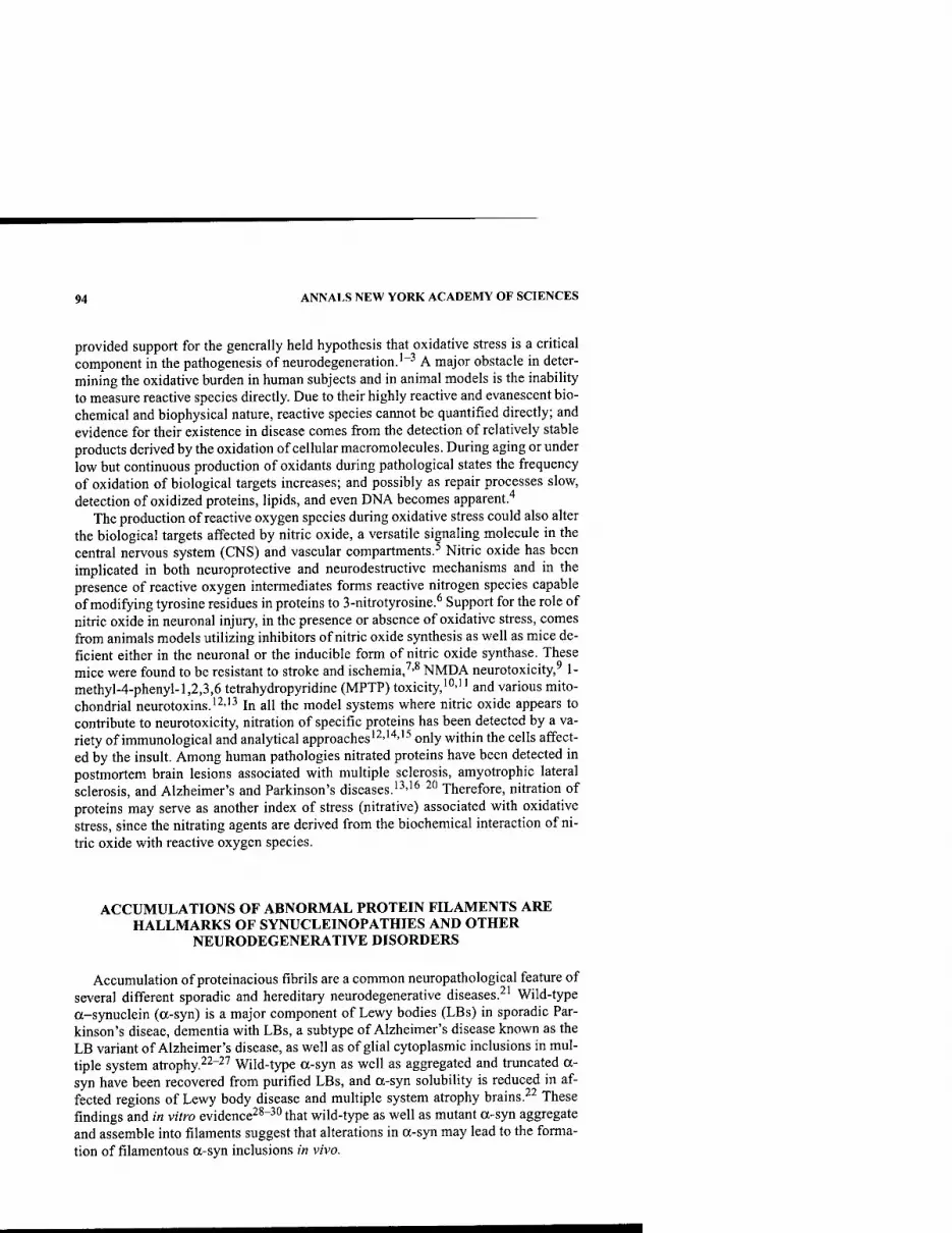
94 ANNALS NEW YORK ACADEMY OF SCIENCES
provided support for the generally held hypothesis that oxidative stress is a critical component in the pathogenesis of neurodegeneration.'"^ A major obstacle in deter- mining the oxidative burden in human subjects and in animal models is the inability to measure reactive species directly. Due to their highly reactive and evanescent bio- chemical and biophysical nature, reactive species cannot be quantified directly; and evidence for their existence in disease comes from the detection of relatively stable products derived by the oxidation of cellular macromolecules. During aging or under low but continuous production of oxidants during pathological states the frequency of oxidation of biological targets increases; and possibly as repair processes slow, detection of oxidized proteins, lipids, and even DNA becomes apparent."*
The production of reactive oxygen species during oxidative stress could also alter the biological targets affected by nitric oxide, a versatile signaling molecule in the central nervous system (CNS) and vascular compartments.^ Nitric oxide has been implicated in both neuroprotective and neurodestructive mechanisms and in the presence of reactive oxygen intermediates forms reactive nitrogen species capable of modifying tyrosine residues in proteins to 3-nitrotyrosine.^ Support for the role of nitric oxide in neuronal injury, in the presence or absence of oxidative stress, comes from animals models utilizing inhibitors of nitric oxide synthesis as well as mice de- ficient either in the neuronal or the inducible form of nitric oxide synthase. These mice were found to be resistant to stroke and ischemia,'''^ NMDA neurotoxicity,^ 1- methyl-4-phenyl-l,2,3,6 tetrahydropyridine (MPTP) toxicity,'"-'' and various mito- chondrial neurotoxins.'^''^ In all the model systems where nitric oxide appears to contribute to neurotoxicity, nitration of specific proteins has been detected by a va- riety of immunological and analytical approaches'2''''''^ only within the cells affect- ed by the insult. Among human pathologies nitrated proteins have been detected in postmortem brain lesions associated with multiple sclerosis, amyotrophic lateral sclerosis, and Alzheimer's and Parkinson's diseases.'^''^^0 Therefore, nitration of proteins may serve as another index of stress (nitrative) associated with oxidative stress, since the nitrating agents are derived from the biochemical interaction of ni- tric oxide with reactive oxygen species.
ACCUMULATIONS OF ABNORMAL PROTEIN FILAMENTS ARE HALLMARKS OF SYNUCLEINOPATHIES AND OTHER
NEURODEGENERATIVE DISORDERS
Accumulation of proteinacious fibrils are a common neuropathological feature of several different sporadic and hereditary neurodegenerative diseases.^' Wild-type a-synuclein (a-syn) is a major component of Lewy bodies (LBs) in sporadic Par- kinson's diseae, dementia with LBs, a subtype of Alzheimer's disease known as the LB variant of Alzheimer's disease, as well as of glial cytoplasmic inclusions in mul- tiple system atrophy.^^"^^ Wild-type a-syn as well as aggregated and truncated a- syn have been recovered from purified LBs, and a-syn solubility is reduced in af- fected regions of Lewy body disease and multiple system atrophy brains.^^ These findings and in vitro evidence^^"^^ that wild-type as well as mutant a-syn aggregate and assemble into filaments suggest that alterations in a-syn may lead to the forma- tion of filamentous a-syn inclusions in vivo.
ISCHIROPOULOS: OXIDATIVE MODIFICATIONS OF ot-SYNUCLEIN 95
MDVFMKGLS - KAKEGV-VfKAAE - KTKQGV-AEAAabG - KIKeCV-LYagV GS
R1 R2 (P) R3
KTKEGV-VHGVAsjT-VAE - KTKEQV-THVGG - AVVTGV-JA\/AQ- KTVEGA-
GSIAAA-TGFV
R4 (T) R5 R6 R7
NAG peptide
KKDQL-GKNEEGAPQE-GILEDMPVDP-DNEAY125EMPSE-EGY133QDY136EPEA
FIGURE 1. Amino acid sequence of a-synuclein. A unique feature of the peptide se- quence is the presence of 6 or possibly 7 imperfect 11-residue repeats of KTKEGV (R) in the conserved core in the N terminal (residues 10-86). The repeating domains consist of res- idues with a high tendency for helical conformation (A, L, K, and E). The first five repeats are hydrophilic and are predicted to form amphipathic a-helices that would promote lipid association.■'■' The last two repeats are part of the hydrophobic core of the protein, residues 61-95. This peptide sequence {underlined) has been identified as the non-AP component of amyloid (NAC peptide)."*^ The minimal peptide required for fibril formation is residues 71- 82.^^ Mutations responsible for familial forms of PD are A30P and A53T.''^''*^ In human brain inclusions a-syn is modified by tyrosine nitration in all four residues, and all four res- idue are capable of cross-linking via dityrosine formation.
a-SUNUCLEIN: RANDOM STRUCTURE AND NO APPARENT FUNCTION
a-Synuclein is a soluble protein consisting of 140 amino acids (sequence in FIG. 1) with a predicated molecular mass of 14,460.16 and an isoelectric point of 4.67. Spectroscopic techniques revealed that the wild-type a-syn contains as little as 3% a-helices and 23% P-sheets, whereas the rest of the protein assumes a random conformation. The function of a-syn remains uncertain, although the preferential lo- calization to presynaptic nerve terminals and its interaction with lipids and proteins suggest some regulatory fiinction associated with dopamine production and lipid vesicle trafficking.-'''^^ Of interest are the numerous interactions of a-syn with pro- teins, suggesting that the abundance of this relatively unstructured protein may be important in allowing stochiometric protein-protein interactions that may regulate the function of the interactive proteins.^^"^* A focus of our investigations has been the C terminus, which is rich in negatively charged residues and three tyrosine resi- dues. Sequence alignment of a ([either E or D]-X-X-[ED]-X-X-[ED]) motif revealed possible similarities with proteins such as tubulin, neurofilament, and the mitochon- drial chaperonin CNP60 protein; and published data indicated that a-syn, but not p- synuclein—another member of the synclein family, can associate with proteins in a manner consistent with a chaperone-like activity.^^ The chaperone-like function of a-s)Ti requires stochiometric amounts of protein, suggesting that this putative func- tion would require sufficiently high concentrations of a-syn. Interactions with syn- philin-l,^" parkin,'*' tyrosine hydroxylase,-'^ dopamine transporter,-'^ phospholipase D,^^ and P-synuclein^^ may be critical in understanding both the physiological and pathological function of this protein. In addition posttranslation modification such

96 ANNALS NEW YORK ACADEMY OF SCIENCES
Nitration a-syn fibrils ,
Membrane j ' ^ associated "^ monomer ^ oligomers
.1' ^ ^ ^ a-syn (dimer) ^
V Inclusions,
Lewy Bodies
5 5 ^^ ♦ y" Oxidation a-syn aggregates -^
Protein associated
FIGURE 2. Working model for the role of oxidative/nitrative modifications on a-synu- clein aggregation. The relative slow turnover rate of a-syn allows the protein to establish reversible associations with lipid membranes and proteins. (1) Monomeric a-synuclein ex- ists in equilibrium with vesicle lipids as well as in association with proteins. (2) Although the in vivo conditions that induce a-syn to dimerize have not been identified, dimerization of monomeric synuclein leads to the formation of oligomers that initiate the cascade of fibril formation (3) that ultimately leads to formation of inclusions and Lewy bodies.^^- (4) The process of fibril formation can be disrupted by tyrosine nitration of a-syn or by oxidative formation of dopamine adduct(s) but not by oxidation of the protein.'''''''^ However, oxidative cross-linking via dityrosine formation may initiate the cascade of fibril production. (5) Moreover, oxidants and nitrating species can covalently cross-link either a-syn-oligomers or fibers forming urea- and SDS-insoluble, protease-resistant a-syn aggregates.^ • If the oxidants and nitrating agents encounter a-syn in fibril form, then the aggregated synuclein can be incorporated in the inclusions or Lewy bodies. It remains unknown if protofibril oli- gomers, fibrils, or insoluble aggregates (or even all three) cause cellular toxicity Possibly toxicity is derived by multiple pathways: (a) loss of monomeric a-syn to fibrils may disturb the equilibrium and thus the association with lipids and proteins, resulting in disturbances of normal cellular functions; (b) the oligomers, fibrils, and covalently cross-linked post- translationally modified synuclein exhibit a cytotoxic gain of function; and finally (c) the cell becomes metabolically stressed by the energetic demand to repair oligomers, fibrils, and covalently cross-linked a-syn. This metabolic demand, coupled with the presence of oxida- tive/nitrative stresses in part derived from mitochondria or dopamine-mediated production of reactive intermediates,^^''""^^ results in cellular dysfunction. Alternatively the cell con- sumes considerable energy attempting to remove the modified synuclein, and as a result nor- mal turnover of cellular proteins is impaired or ignored, resulting in inappropriate signaling and dysfunction.
as glycosylation'" and serine phosphorylation^^''*^'''^ have been detected, and their potential role in the pathogenesis of synucleipathies is under investigation.
a-SYNUCLEIN IS POSTTRANSLATIONALLY MODIFIED BY NITRATION AND OXIDATION IN VIVO
Based on data gathered to understand the molecular basis of the selectivity of pro- tein tyrosine nitration in vivo we uncovered that tyrosine residues in the vicinity of charged amino acids (mostly acidic residues) are primary targets for nitration.''*''*'* Within the a-syn sequence the carboxyl terminus contains there tyrosine residues in an aspartate- and glutamate-rich environment, suggesting a reasonable site for mod- ification. Indeed, in vitro exposure of the protein to nitrating agents resulted in nitra- tion of these three tyrosine residues; and, moreover, in oxidation of tyrosine to form dityrosine that cross-linked the protein to form SDS and urea-stable dimer and multi- oligomeric forms."*^ A series of monoclonal antibodies were then raised and charac-

ISCHIROPOULOS: OXIDATIVE MODIFICATIONS OF a-SYNUCLEIN 97
terized by mutational analysis that recognizes only nitrated a-syn or only nitrated and oxidized a-syn.-^"'^^ These antibodies revealed a highly selective labeling of a- syn not only in LBs but also in dystrophic neurites, indicating a wide-spread but highly selective modification of a-syn in PD and other related synucleopathies.^"-^^ Selective nitration of a-syn but not of other presynaptic proteins has also been de- tected in MPTP-challenged mice.'*^ This selective targeting of a-syn for oxidative and nitrative modification clearly implies the formation of nitrating agents and is de- rived in part, as alluded to before, from the protein structure and not the biochemical nature of the nitrating agent, the abundance of the protein, or even the number of ty- rosine residues in the protein. This selective targeting may provide some clues re- garding the molecular and biochemical events that lead to the pathogenesis of PD, as an important determinant in the selection of protein(s) for nitration is the site of formation of the nitrating agent. Based on these observations a model is proposed that implicates oxidative/nitrative stress in the formation of a-syn inclusions and cel- lular death in PD.
ACKNOWLEDGMENTS
I am grateful to all members of my laboratory, the members of the synuclein group at the Center for Neurodegenerative Disorders (Virginia M-Y. Lee, John Q. Trojanowski, codirectors), for their support and stimulating discussions; and to Dr. Akiva Cohen for comments and critical reading of the manuscript. Major contribu- tions in this project have been made by Dr. Jose "Pepe" Souza, Dr. Qiping Chen, and Jenny Paxinou, with support from National Institute on Aging, National Institutes of Health.
REFERENCES
1. DESNUELLE, C, M. DiB, C. GARREL & A. FAVIER. 2001. A double-blind, placebo-con- trolled randomized clinical trial of alpha-tocopherol (vitamin E) in the treatment of amyotrophic lateral sclerosis. ALS Riluzole-Tocopherol Study Group. Amyotroph. Lateral Scler. Other Motor Neuron Disord. 2: 9-18.
2. SANO, M., C. ERNESTO, R.G. THOMAS, et al. 1997. A controlled trial of selegiline, alpha-tocopherol, or both as treatment for Alzheimer's disease. The Alzheimer's Dis- ease Cooperative Study. N. Eng. J. Med. 336: 1216-1222.
3. PRZEDBORSKI, S., Q. CHEN, M. VILA, et al. 2001. Oxidative post-translational modifica- tions of a-synuclein in the MPT? mouse model of Parkinson's disease. J. Neuro- chem. 76: 637-640.
4. STADTMAN, E.R. 1992. Protein oxidation and aging. Science 257: 1220-1224. 5. LiPTON, S.A, Y-B. CHOI, Z-H. PAN, et al. 1993. A redox-based mechanism for the neu-
roprotective and neurodestructive effects of nitric oxide and related compounds. Nature 364: 626-631.
6. IsCHlROPOULOS, H. 1998. Biological tyrosine nitration: a pathophysiological function of nitric oxide and reactive oxygen species. Arch. Biochem. Biophys. 356: 1-11.
7. HUANG, Z., P.L. HUANG, N. PANAHIAN, et al. 1994. Effects of cerebral ischemia in mice deficient in neuronal nitric oxide synthase. Science 265: 1883-1885.
8. ELIASSON, M.J., Z. HUANG, R.J. FERRANTE, et al. 1999. Neuronal nitric oxide synthase activation and peroxynitrite formation in ischemic stroke linked to neural damage. J. Neurosci. 19: 5910-5918.

98 ANNALS NEW YORK ACADEMY OF SCIENCES
9. AY ATA, C, G. AYATAM, H. HARA, et al. 1997. Mechanisms of reduced striatal NMDA excitotoxicity in type 1 nitric oxide synthase knock-out mice. J. Neurosci. 17: 6908- 6917.
10. PRZEDBORSKT, S., V. JACKSON-LEWIS, R. YOKOYAMA, et al. 1996. Role of neuronal nitric oxide in MPTP (l-methyl-4-phenyl-l,2,3,6-tetrahydropyridine)-induced dopaminer- gic neurotoxicity. Proc. Natl. Acad. Sci. USA 93: 4565-4571.
11. LiBERATORE, G.T., V. JACKSON-LEWIS, S. VUKOSAVIC, et al. 1999. Inducible nitric oxide synthase stimulates dopaminergic neurodegeneration in the MPTP model of Parkin- son disease. Nat. Med. 5: 1403-1409.
12. SCHULZ, J.B., P.L. HUANG, R.T. MATTHEWS, et al. 1996. Striatal malonate lesions are attenuated in nitric oxide oxide synthase knockout mice. J. Neurochem. 67: 430- 433.
13. SMITH, M.A., P.L. RICHEY HARRIS, L.M. SAYRE, et al. 1997. Widespread peroxynitrite- mediated damage in Alzheimer's disease. J. Neurosci. 17: 2653-2657.
14. ARA, J., S. PRZEDBORSKI, A.B. NAINI, et al. 1998. Inactivation of tyrosine hydroxylase following exposure to peroxynitrite and MPTP. Proc. Natl. Acad. Sci. USA 95: 7659-7663.
15. PENNATHUR S., V. JAKSON-LEWIS, S. PRZEDBORSKI & J.W. HEINECKE. 1999. Mass spec- trometric quantification of 3-nitrotyrosine, ortho-tyrosine, and o,o'-dityrosine in brain tissue of l-methyl-4-phenyl-l,2,3, 6-tetrahydropyridine-treated mice, a model of oxidative stress in Parkinson's disease. J. Biol. Chem. 274: 34621-34628.
16. BASARGA, O., F.H. MICHAELS, Y.M. ZHENG, et al. 1995. Activation of the inducible form of nitric oxide synthase in the brains of patients with multiple sclerosis. Proc. Natl. Acad. Sci. USA 92: 12041-12045.
17. BEAL, M.F., R.J. FERRANTE, S.E. BROWNE, et al. 1997. Increased 3-nitrotyrosine in both sporadic and familial amyotrophic lateral sclerosis. Ann. Neurol. 42: 644-654.
18. HENSLEY, K., M.L. MAIDT, Z.Q. YU, et al. 1998. Electrochemical analysis of protein nitrotyrosine and dityrosine in the Alzheimer brain indicates region-specific accumu- lation. J. Neurosci. 18: 8126-8132.
19. DUDA, J.E., B.I. GiASSON, Q. CHEN, et al. 2000. Widespread nitration of pathological inclusions in neurodegenerative synucleinopathies. Am. J. Pathol. 157: 1439-1445.
20. GiASSON, B.I., J.E. DUDA, I.V. MURRAY, et al. 2000. Oxidative damage linked to neuro- degeneration by selective alpha-synuclein nitration in synucleinopathy lesions. Sci- ence 290: 985-989.
21. TROJANOWSKI, J.Q., M. GOEDERT, T. IWATSUBO & V.M-Y. LEE. 1998. Fatal attractions: abnormal protein aggregation and neuron death in Parkinson's disease and Lewy body dementia. Cell Death Differ. 5: 832-837.
22. DUDA. J.E., B.l. GIASSON, M.E. MABON, et al. 2002. Novel antibodies to synuclein show abundant striatal pathology in Lewy body diseases. Ann. Neurol. 52: 205-210.
23. FuJlWARA, H., M. HASEGAWA, N. DOHMAE, et al. 2002. Alpha-synuclein is phosphory- lated in synucleinopathy lesions. Nat. Cell Biol. 4: 160-164.
24. IRIZARRY, M.C, W. GROWDON, T. GOMEZ-ISLA, et al. 1998. Nigral and cortical Lewy bodies and dystrophic nigral neurites in Parkinson's disease and cortical Lewy body disease contain a-synuclein. J. Neuropath. Exp. Neurol. 57: 334-337.
25. LANGSTON, J.W., S. SASTRY, P. CHAN, et al. 1998. Novel alpha-synuclein-immunoreac- tive proteins in brain samples from the Contursi kindred, Parkinson's, and Alzhe- imer's disease. Exp. Neurol. 154: 684-690.
26. SPILLANTINI, M.G, M.L. SCHMIDT, V.M-Y. LEE, et al. 1997. a-Synuclein in Lewy bod- ies. Nature 388: 839-840.
27. WAKABAYASHI, K., M. YOSHIMOTO, S. TSUJI & H. TAKAHASHI. 1998. Alpha-synuclein immunoreactivity in glial cytoplasmic inclusions in multiple system atrophy. Neuro- sci. Lett. 249: 180-182.
28. CONWAY, K.A., J.D. HARPER & P.T. LANSBURY. 1998. Accelerated in vitro fibril forma- tion by a mutant alpha-synuclein linked to early-onset Parkinson disease. Nature Med. 4: 1318-1320.
29. GiASSON, B.l, I.V. MURRAY, J.Q. TROJANOWSKI & V.M. LEE. 2001. A hydrophobic stretch of 12 amino acid residues in the middle of alpha-synuclein is essential for fil- ament assembly. J. Biol. Chem. 276: 2380-2386.

ISCHIROPOULOS: OXIDATIVE MODIFICATIONS OF a-SYNUCLEIN 99
30. UVERSKY, V.N., J. LI & A.L. FINK. 2001. Metal-triggered structural transformations, aggregation, and fibrillation of human alpha-synuclein. A possible molecular link between Parkinson's disease and heavy metal exposure. J. Biol. Chem. 276: 44284- 44296.
31. DAVIDSON, W.S., A. JONAS, D.F. CLAYTON & J.M. GEORGES. 1998. Stabilization of alpha-synuclein secondary structure upon binding to synthetic membranes. J. Biol. Chem. 273: 9443-9449.
32. LEE, F.J.S., F. Liu, Z.B. PRISTUPA & H.B. NIZNIK. 2001. Direct binding and functional coupling of a-synuclein to the dopamine transporters accelerate dopamine-induced apoptosis. FASEB J. 15: 916-926.
33. HASHIMOTO, M., A. TAKEDA, L.J. HSU, et al. 1999. Role of cytochrome c as a stimula- tor of a-synuclein aggregation in Lewy Body disease. J. Biol. Chem. 274: 28849- 28852.
34. HASHIMOTO, M., E. ROCKENSTEIN, M. MANTE, et al. 2001. beta-Synuclein inhibits alpha-synuclein aggregation: a possible role as an anti-parkinsonian factor. Neuron 32: 213-223.
35. JENCO, J.M., A. RAWLINGTON, B. DANIELS & A. J. MORRIS. 1998. Regulation of phosh- olipase D2: selective inhibition of mammalian phospholipase D isoenzyme by alpha- and beta-synucleins. Biochemistry 37: 4901-4909.
36. OSTREROVA-GOLTS, N., L. PETRUCELLI, M. FARER, et al. 1999. Alpha-synuclein shares physical and functional homology with 14-3-3 proteins. J. Neurosci. 19: 5782-5791.
37. PAYTON J.E., R.J. PERRIN, D.F. CLAYTON & J.M. GEORGE. 2001. Protein-protein inter- actions of alpha-synuclein in brain homogenates and transfected cells. Brain Res. Mol. Brain Res. 95: 138-145.
38. PEREZ, R.G., J.C. WAYMIRE, E. LIN, et al. 2002. A role for a-synuclein in the regulation of dopamine biosynthesis. J. Neurosci. 22: 3090-3099.
39. SouzA, J.M., B.I. GiASSON, V. M-Y. LEE & H. ISCHIROPOULOS. 2000. Chaperone-like activity of synucleins. FEBS Lett. 474: 116-119.
40. ENGELENDER, S., Z. KAMINSKY, X. Guo, et al. 1999. Synphilin-1 associates with a- synuclein and promotes the formation of cytosolic inclusions. Nat. Genet. 22: 110- 114.
41. SHIMURA, H., M.G. SCHLOSSMACHER, N. HATTORI, et al. 2001. Ubiquitination of a new form of alpha-synuclein by parkin from human brain: implications for Parkinson's disease. Science 293: 263-269.
42. ELLIS C.E., P.L. SCHWARTZBERG, T.L. GRIDER, et al. 2001. alpha-Synuclein is phospho- rylated by members of the Src family of protein-tyrosine kinases. J. Biol. Chem. 276: 3879-3884.
43. OKOCHI, M., J. WALTER, A. KOYAMA, et al. 2000, Constitutive phosphorylation of the Parkinson's disease associated a-synuclein. J. Biol. Chem. 275: 390-397.
44. SouzA, J.M., B.I. GIASSON, Q. CHEN, et al. 2000. Dityrosine cross-linking promotes formation of stable alpha-synuclein polymers. Implication of nitrative and oxidative stress in the pathogenesis of neurodegenerative synucleinopathies. J. Biol. Chem. 275: 18344-18349.
45. PRZEDBORSKI, S., V. KOSTIC, V. JACKSON-LEWIS, et al. 1992. Transgenic mice with increased Cu/Zn-superoxide dismutase activity are resistant to N-methyl-4-phenyl- 1,2,3,6-tetrahydropyridine-induced neurotoxicity. J. Neurosci. 12: 1658-1667.
46. TAKEDA, A., M. MALLORY, M. SUNDSMO, et al. 1998. Abnormal accumulation of NACP/alpha-synuclein in neurodegenerative disorders. Am. J. Pathol. 152: 367-372.
47. KRUGER, R., W. KUHN, T. MULLER, et al. 1998. Ala30-to-pro mutation in the gene encoding alpha-synuclein in Parkinson's disease. Nat. Genet. 18: 106-108.
48. POLYMEROPOULOS, M.H., C. LAVEDANM, E. LEROY, et al. 1997. Mutation in the alpha- synuclein gene identified in families with Parkinson's disease. Science 276: 2045- 2047.
49. CONWAY, K.A., J.C. ROCHET, R.M. BIEGANSKI & P.T. LANSBURY, JR. 2001. Kinetic sta- bilization of the alpha-synuclein protfibril by a dopamine-alpha-synuclein adduct. Science 294: 1396-1349.
50. PAXINOU, E., Q. CHEN, M. WEISSE, et al. 2001. Induction of alpha-synuclein aggrega- tion by intracellular nitrative insult. J. Neurosci. 21: 8053-8061.
100 ANNALS NEW YORK ACADEMY OF SCIENCES
51. LEE, H.J., S.Y. SHIN, C. CHOI, et at. 2002. Formation and removal of alpha-synuclein aggregates in cells exposesed to mitochondrial inhibitors. J. Bid. Chem. 277: 5411- 5417.
52. TABIRZI, S.J., M. ORTH, J.M. WILKINSON, et al. 2000. Expression of mutant alpha-synu- clein causes increased susceptibility to dopamine toxicity. Hum. Mol. Genet. 9: 2683-2689.
53. Xu, J., S.Y. KAO, F.J. LEE, et al. 2002. Dopamine-dependent neurotoxicity of alpha- synulcein: a mechanism for selective neurodegeneration in Parkinson's disease. Nat. Med. 8: 600-606.

Parkin and Endoplasmic Reticulum Stress
RYOSUKE TAKAHASHI,'' YUZURUIMAI," NOBUTAKA HATTORI,* AND YOSHIKUNIMIZUNO*
"Laboratory for Motor System Neurodegeneration, RIKEN Brain Science Institute (BSI), Saitama 351-0198, Japan
^Department of Neurology, Juntendo University School of Medicine, Bunkyo-ku, Tokyo, 113-0033, Japan
ABSTRACT: Autosomal-recessive juvenile parkinsonism (AR-JP) is caused by mutations in tlie parkin gene. Parlcin protein is cliaracterized by a ubiqultin- lilie domain at its NH2 terminus and by two RING finger motifs and one IBR (in between RING finger) motif at its COOH-terminus (RING-IBR-RING). We sliowed that the parlcin protein is an E3 ubiquitin ligase, which binds to ubiq- uitin-conjugating enzymes (E2s) through its RING-IBR-RING motif. The pathogenesis of AR-JP, therefore, was hypothesized to be accumulation of un- identified neurotoxic protein (a substrate of parlcin). On the basis of this hy- pothesis, the substrate of parkin was sought using a yeast two-hybrid system. A putative G protein-coupled transmembrane polypeptide, named Pael (par- kin-associated gndothelin receptor-/ike) receptor, was identified as a parkin binding protein. When overexpressed in cells, this receptor tends to become un- folded, insoluble, and ubiquitinated. The insoluble Pael receptor leads to endo- plasmic reticulum (ER) stress-induced cell death. Parkin specifically ubiquitinates this receptor in the presence of ER-resident E2s and promotes the degradation of unfolded Pael receptor, resulting in suppression of the cell death induced by the accumulation of unfolded Pael receptor in the ER. More- over, the insoluble form of Pael receptor accumulates in the brain of AR-JP pa- tients. This protein is highly expressed in the dopaminergic neurons in the substantia nigra, which is specifically affected in Parkinson's disease; although it is also widely expressed in oligodendroglias in the fiber tract. In conclusion, we showed that the accumulation of unfolded Pael receptor (a substrate of par- kin) may cause selective death of dopaminergic neurons in AR-JP.
KEYWORDS: AR-JP; parkin; Pael receptor; ER stress; ERAD
INTRODUCTION
Parkinson's disease (PD) is the second most common neurodegenerative disease among elderly people. More than 1% of people aged 65 or older are afflicted with PD. The main clinical feature of PD is progressive motor disturbances (tremor, ri- gidity, and akinesia). Although the rate of disease progression is variable, wheel chair or bed confinement usually occurs years after disease onset.
Address for correspondence; Ryosuke Takahashi, M.D., Ph.D., Laboratory for Motor System Neurodegeneration, RIKEN Brain Science Institute (BSI), 2-1 Hirosawa, Wako, Saitama 351- 0198, Japan. Voice: +81-48-467-6072; fax: +81-48-462-4796.
Ann. N.Y. Acad. Sci. 991:101-106 (2003). © 2003 New York Academy of Sciences.
101

102 ANNALS NEW YORK ACADEMY OF SCIENCES
Most PD cases are sporadic rather than familial (5-10%) and are presumably in- herited. Autosomal-recessive juvenile parkinsonism (AR-JP), the most frequent cause of familial PD, is characterized by several unique features including young age at onset (most under age 40) and superb responsiveness to L-dopa. The neuropatho- logical hallmark of AR-JP is selective degeneration of dopaminergic neurons in the substantia nigra, similar to that of the common form of Parkinson's disease. Howev- er, the pathology of AR-JP is not usually accompanied by Lewy bodies (cytoplasmic neuronal inclusions), which are the histopathological signature for common PD.'"^
In 1998, parkin was identified as the gene responsible for AR-JP.^ Here, the phys- iological and pathophysiological function of parkin in ER stress is discussed.
RESULTS AND DISCUSSION
The Structure and Function of the Parkin Protein
Parkin is a 465-amino acid protein characterized by a ubiquitin-like domain at its NH2 terminus and two RING finger motifs and one IBR (m between RING fingers) motif at its COOH-terminus (RING-IBR-RING). The RING finger motif is a variant of the zinc finger motif, which has metal binding activity and cysteine and histidine residues present at certain intervals.
Although the RING finger motif is found in a wide variety of proteins, the phys- iological function of this motif has been unknown until recently. However, recent findings implicate the RING domain in specific ubiquitination events.^ Protein ubiq- uitination begins with the formation of a thiol-ester linkage between the COOH ter- minus of ubiquitin and the active-site cysteine of the ubiquitin-activating enzyme (El). Ubiquitin then is transferred to a ubiquitin-conjugating enzyme (E2), again through a thiol-ester linkage. Ubiquitin-protein ligases (E3s), which are primarily responsible for providing specificity to ubiquitin conjugation, interact with E2 and the substrate, facilitating the formation of isopeptide bonds between the COOH-ter- minus of ubiquitin and lysines either on a target protein or on the last ubiquitin of a protein-bound multiubiquitin chain. Multiubiquitin chains are potent targeting sig- nals for protein degradation in proteasomes.^ A number of RING finger-containing proteins including Mdm2 and Siah are shown to be E3s. In RING-type E3s, RING finger motifs serve as recruiting motifs for specific E2 enzymes. These facts suggest that parkin, which contains the RING-IBR-RING motif, is a new E3 ubiquitin ligase.
Parkin actually turned out to be a RING-type E3 ubiquitin ligase.^""^ We have shown that wild-type parkin ubiquitinates an unidentified substrate protein. By con- trast, disease-related mutated parkin has lost its E3 activity, indicating a close inverse correlation between enzymatic activity and disease phenotype.^ These results sug- gest that a genetic defect in parkin causes the substrate of parkin to escape degrada- tion and accumulate, and may lead to PD. This hypothesis implies a key role for the substrate of parkin in the neurodegenerative process.
The Identification of Pael Receptor as a Substrate of Parkin
To identify the substrate of parkin, we carried out a yeast two-hybrid screening of human adult brain cDNA libraries using full-length parkin as bait. One of the iso- lated clones contained a COOH terminal portion of endothelin receptor-like protein.
TAKAHASHI et al.: PARKIN AND ER STRESS 103
which is a putative G protein-coupled receptor with its unidentified ligand, very sim- ilar to endothelin receptor type B in sequence. We renamed this receptor Pael (par- kin-associated endothelin receptor-/ike) receptor (Pael-R; GenBank accession number: AF502281).'0
Pael-R was specifically ubiquitinated by parkin in the presence of Ubc6 and Ubc7 in an in vitro ubiquitination assay. Moreover, the half-life of Pael-R in cultured neu- roblastoma cells transiently transfected with a Pael-R expression vector was dramat- ically shortened from one hour to less than 30 minutes by parkin coexpression. These results indicate that Pael-R is an in vivo substrate of parkin.
Pael-R Is a Substrate of Endoplasmic Reticulum-Associated Degradation
It has been shown that the ubiquitin-proteasome pathway plays an important role in the degradation of membrane or secretory protein at the level of endoplasmic reticulum (ER). The ER is an organelle that carries out the quality control of mem- brane or secretory protein folding. Newly synthesized secretory proteins enter the ER, where ER chaperones such as BiP bind and help the proper folding of these pro- teins. Properly folded proteins are then allowed to enter the normal secretory path- way composed of Golgi apparatus, plasma membrane, endosome, lysosome, and various intermediate transport compartments. However, the proteins that do not fold correctly are retrotranslocated to the cytosol, where these "failed" proteins are de- graded. This process is called endoplasmic reticulum-associated degradation (ERAD).'' ERAD substrates (i.e., ER luminal or ER transmembrane proteins) are transported through a channel-like protein complex termed a "translocon" back to the cytosol, where they are subject to ubiquitin-proteasome degradation.
Because parkin binds to Ubc6 and Ubc7 (ER-resident E2s involved in ERAD) and Pael-R was ubiquitinated in vitro in the presence of both parkin and Ubc6/7, it has been suggested that parkin is an E3 involved in ERAD and Pael-R is a substrate of ERAD. To test this idea, we caused overexpression of Pael-R in a dopaminergic neuroblastoma cell line, SH-SY5Y. When these cells were lysed with a nonionic de- tergent such as Triton X-100, properly folded and unfolded proteins substantially partitioned into detergent-soluble and -insoluble fractions, respectively.'^
Interestingly, a significant amount (up to 50%) of Pael-R was detected in the in- soluble fraction when cells transiently transfected with a plasmid containing a Pael- R cDNA were given no additional treatment, suggesting that the proper folding of Pael-R is inherently difficult. Treatment with ER stress-inducing reagents such as tu- nicamycin and 2-mercaptoethanol increased the level of insoluble Pael-R and de- creased the level of soluble Pael-R, indicating that the soluble and insoluble Pael-R correspond to properly folded and unfolded species of Pael-R. Moreover, treatment with lactacystin, a selective proteasomal inhibitor, dramatically increased the level of insoluble Pael-R without affecting the level of soluble Pael-R. This result indicates that inhibition of ERAD by lactacystin resulted in the accumulation of unfolded, in- soluble Pael-R and provides strong evidence that Pael-R is a substrate of ERAD.
Abnormal Accumulation of Pael-R and ER Stress-Induced Cell Death
To examine the effect of Pael-R accumulation on cell viability, Pael-R overex- pressing cells were treated with lactacystin to induce forced accumulation of Pael-
104 ANNALS NEW YORK ACADEMY OF SCIENCES
R. Immunofluorescence analysis revealed the accumulation of Pael-R in the ER 6 h after the addition of lactacystin. Prolonged lactacystin treatment led to Pael-R aggre- gate formation. As the aggregate formed, the cells became round and shrunken, sug- gesting apoptotic cell death and that accumulation of Pael-R causes cell death.
Then what is the mechanism underlying Pael-R-induced cell death? Abnormal accumulation of unfolded proteins in the ER constitutes a major threat to cell viabil- ity. This situation is called "unfolded protein stress" or ER stress. Cells make at- tempts to overcome ER stress in various ways including transcriptional upregulation of ER chaperones (such as GRP78/BiP) and components of ERAD (such as Ubc7), and general translational suppression. These cellular responses are collectively called the "unfolded protein response" (UPR). When the amount of accumulated un- folded protein exceeds the threshold level, the cells undergo cell death accompanied by activation of c-Jun N terminal kinase (JNK) and caspase-12.'^'''* At the early stage of Pael-R accumulation, GRP78/BiP was upregulated at the transcriptional level, indicating that accumulation of Pael-R indeed induced ER stress. Moreover, we have already found that parkin mRNA is increased when cells are subjected to tunicamycin- or 2-mercaptoethanol-induced ER stress and that the resulting parkin overexpression suppressed ER stress-induced cell death of the human neuroblasto- ma cell line SH-SY5Y expressing endogenous Pael-R. Parkin-mediated cell death suppression is at least partially explained by parkin-mediated degradation of Pael-R. Consistent with this idea, Pael-R overexpression-induced cell death is suppressed by coexpression of parkin, which is accompanied by a significant decrease of insoluble, but not soluble, Pael-R.
On the basis of these findings, accumulation of Pael-R is most likely the patho- genetic mechanism underlying AR-JP. Seeking direct evidence to support this idea, we examined brains from AR-JP patients to determined whether they contained ac- cumulated Pael-R. We prepared homogenates of AR-JP as well as control brain sam- ples, separated them into detergent-soluble and -insoluble fractions, and measured the amount of Pael-R in each fraction. The protein level of insoluble, but not soluble, Pael-R was 10-30-fold higher in AR-JP brains than that in non-AR-JP brains. More- over, in three out of four AR-JP brains examined, an increased level of GRP78/BiP was observed, suggesting that AR-JP brains were also under ER stress caused by the accumulation of unfolded Pael-R. Given that neurodegeneration in AR-JP is caused by ER stress-induced cell death, cytoplasmic inclusions composed of unfolded Pael- R may not be formed before cellular demise, providing reasonable explanation for the absence of Lewy bodies or other types of neuronal inclusion bodies in AR-JP.
Tissue Distribution Pattern of Pael-R
Pael-R mRNA is highly expressed in the central nervous system, especially in the substantia nigra and the corpus callosum, the latter being composed mainly of nerve fibers and not of neuronal cell bodies.'^-'^ Immunohistochemical analysis revealed that Pael-R is generally strongly expressed in oligodendrocytes and poorly expressed in neurons except hippocampal neurons and dopaminergic neurons in the substantia nigra. Assuming that postmitotic neurons are more vulnerable to ER stress than re- newable oligodendrocytes, selective degeneration of dopaminergic neurons in AR- JP can be partly explained by this unique distribution pattern of Pael-R. On the other hand, dopaminergic neurons are thought to be constitutively exposed to oxidative

TAKAHASHI et al.: PARKIN AND ER STRESS 105
Normal;
I Parkin ^ \
Unfolded Pael-R Constitutively degraded
Dopaminergic Neuron
\
^^ Pacl-R exerts its normal function
Properly folded Pael-R
AR-PD :
Unfolded Pael-R Abnormal accumulation of Pael-R
Dopaminergic Neuron
ER stress-induced ■♦ Cell death
FIGURE 1. Diagram showing (top) the normal functioning of Pael-R and (bottom) the pathogenic mechanism of AR-JP.
Stress and especially sensitive to various stresses, providing another factor contrib- uting to the lesion selectivity in AR-JP.
CONCLUSION
Taken together, our evidence strongly suggests that accumulation of Pael-R is causative in AR-JP (FIG. 1).'" Recently, the accumulation of misfolded proteins has been implicated in the pathogenesis of many neurodegenerative diseases including amyotrophic lateral sclerosis, Alzheimer's disease, Parkinson's disease, and poly- glutamine disease."''^ Our study showed that AR-JP is also one of these so-called "conformational diseases." Potentially cytotoxic misfolded protein is usually detox- ified either by refolding with molecular chaperones or degradation through the ubiquitin-proteasome pathway. Manipulations of such refolding or degrading ma- chineries may provide opportunities for treatment of currently intractable neuro- degenerative diseases.

106 ANNALS NEW YORK ACADEMY OF SCIENCES
ACKNOWLEDGMENTS
We would like to thank Ms. Mariko Soda and Dr. Haruhisa Inoue for their kind collaborative efforts. This study was supported by a Grant-in-Aid for Scientific Re- search from the Ministry of Education, Culture, Sports, Science and Technology, Ja- pan (R.T., N.H., and Y.M.); and a Grant-in-Aid for Encouragement of Young Scientists from the Japan Society for the Promotion of Science (Y.I.).
REFERENCES
1. YAMAMURA, Y., I. SoBUE, K. ANDO, et al. 1973. Paralysis agitans of early onset with marked diurnal fluctuation of symptoms. Neurology 23: 239-244.
2. ISHIKAWA, A. & S. TSUJI. 1996. Clinical analysis of 17 patients in 12 Japanese families with autosomal-recessive type juvenile parkinsonism. Neurology 47: 160-166.
3. YoKOCHi, M. 2000. Development of the nosological analysis of juvenile parkinsonism. Brain Dev. 22 Suppl. 1: S81-86.
4. KiTADA, T., S. ASAKAWA, N. HATTORI, et al. 1998. Mutations in the parkin gene cause autosomal recessive juvenile parkinsonism. Nature 392: 605-608.
5. JOAZEIRO, C.A. & A.M. WEISSMAN. 2000. RING finger proteins: mediators of ubiquitin ligase activity Cell 102: 549-552.
6. HERSHKO, A. & A. CIECHANOVER. 1998. The ubiquitin system. Annu. Rev. Biochem. 67: 425-479.
7. SHIMURA, H., N. HATTORI, S. KUBO, et al. 2000. Familial Parkinson disease gene prod- uct, parkin, is a ubiquitin-protein ligase. Nat. Genet. 25: 302-305.
8. IMAI, Y., M. SODA & R. TAKAHASHI. 2000. Parkin suppresses unfolded protein stress- induced cell death through its E3 ubiquitin-protein ligase activity. J. Biol. Chem. 275: 35661-35664.
9. ZHANG, Y, J. GAO, K.K. CHUNG, et al. 2000. Parkin functions as an E2-dependent ubiquitin-protein ligase and promotes the degradation of the synaptic vesicle-associ- ated protein, CDCrel-1. Proc. Natl. Acad. Sci. USA 97: 13354-13359.
10. IMAI, Y., M. SODA, H. INOUE, et al. 2001. An unfolded putative transmembrane polypeptide, which can lead to endoplasmic reticulum stress, is a substrate of parkin. Cell 105: 891-902.
11. PLEMPER, R.K. & D.H. WOLF. 1999. Retrograde protein translocation: ERADication of secretory proteins in health and disease. Trends Biochem. Sci. 24: 266-270.
12. WARD, C.L., S. OMURA & R.R. KOPITO. 1995. Degradation of CFTR by the ubiquitin- proteasome pathway. Cell 83: 121-127.
13. URANO, F., X. WANG, A. BERTOLOTTI, et al. 2000. Coupling of stress in the ER to acti- vation of JNK protein kinases by transmembrane protein kinase IREl. Science 287: 664-666.
14. NAKAGAWA, T, H. ZHU, N. MORISHIMA, et al. 2000. Caspase-12 mediates endoplas- mic-reticulum-specific apoptosis and cytotoxicity by amyloid-beta. Nature 403: 98- 103.
15. ZENG, Z., K. SU, H. KYAW, et al. 1997. A novel endothelin receptor type-B-like gene enriched in the brain. Biochem. Biophys. Res. Commun. 233: 559-567.
16. DONOHUE, P.J., H. SHAPIRA, S. A. MANTEY, et al. 1998. A human gene encodes a puta- tive G protein-coupled receptor highly expressed in the central nervous system. Brain Res. Mol. Brain Res. 54: 152-160.
17. SHERMAN, M.Y. & A.L. GOLDBERG. 2001. Cellular defenses against unfolded proteins: a cell biologist thinks about neurodegenerative diseases. Neuron 29: 15-32.
18. JuLlEN, J. 2001. Amyotrophic lateral sclerosis. Unfolding the toxicity of the misfolded. Celll04: 581-591.
Parkinson's Disease and Related a-SynucIeinopathies Are Brain Amyloidoses
JOHN Q. TROJANOWSKI AND VIRGINIA M-Y. LEE
Center for Neurodegenerative Disease Research, Department of Pathology and Laboratory Medicine, and the Institute on Aging, the University of Pennsylvania School of Medicine, Philadelphia, Pennsylvania 19104, USA
ABSTRACT: A paradigm shift in understanding Parkinson's disease (PD) and related disorders is emerging from studies showing that alpha-synuclein (AS) gene mutations cause familial PD; AS is abnormally nitrated, phosphorylated, and ubiquitinated; AS forms neuronal and glial inclusions; AS fibrillizes in vitro; and AS transgenic animals develop neurodegeneration with AS amyloid inclusions. Thus, PD and related synucleinopathies are brain amyloidoses that may share similar mechanisms and targets for drug discovery.
KEYWORDS: Parkinson's disease; alpha-synuclein; synucleinopathies; neuro- degenerative brain amyloidoses
Parkinson's disease (PD) is the most common neurodegenerative movement disor- der, but because its underlying mechanisms are poorly understood, only symptom- atic treatments that partially or transiently ameliorate some of the impairments associated with PD are available (reviewed in Ref. 1). However, recent research breakthroughs implicate a-synuclein (AS) abnormalities in mechanisms of PD (re- viewed in Refs. 2-7), and this will hasten development of more effective PD thera- pies. For example, AS pathologies are now recognized as the defining brain amyloid lesions of diverse neurodegenerative disorders known as a-synucleinopathies (also called synucleinopathies) that appear to share similar disease mechanisms (TABLE
1); and further insights into the role that AS plays in PD and other neurodegenerative brain amyloidoses are likely to provide new targets for the discovery of drugs to treat these diseases (reviewed in Refs. 2-9).
As summarized and discussed in more detail elsewhere,^"'^ a paradigm shift in understanding mechanisms of PD began in 1997, and this is resulting in the conver- gence of several lines of evidence implicating brain amyloid deposits formed by
Address for correspondence: John Q. Trojanowski, M.D., Ph.D., Center for Neurodegenera- tive Disease Research, Department of Pathology and Laboratory Medicine, University of Penn- sylvania School of Medicine, HUP, Maloney Building, 3rd Floor, Philadelphia, PA 19104. Voice: 215-662-6399/6427; fax: 215-349-5909.
Ann. N.Y. Acad. Sci. 991: 107-110 (2003). © 2003 New York Academy of Sciences.
107

108 ANNALS NEW YORK ACADEMY OF SCIENCES
TABLE 1. Neurodegenerative diseases with prominent filamentous AS lesions
Sporadic and familial Parkinson's disease Sporadic and familial Alzheimer's disease Dementia with Lewy bodies Multiple system atrophy Down's syndrome Neuronal degeneration with brain iron accumulation type 1 (Hallervorden-Spatz disease) Pure autonomic failure REM sleep behavior disorder
NOTE: Sporadic and hereditary neurodegenerative diseases characterized by filamentous AS brain lesions. Disease in which AS lesions are the predominant or sole form of brain amyloid are shown in italics. Notably, AD is a triple brain amyloidosis because it is associated with lesions formed by deposits of fibrillar tau, Ap, and AS.
abnormally aggregated AS filaments in these processes.'^"^^ The following key ob- servations support this new perspective on PD and related disorders:
(1) Mutations in the AS gene cause familial PD; (2) Lewy bodies (LBs) and dystrophic Lewy neuritis are detected by anti-AS
antibodies, but these hallmark PD amyloid lesions also occur in other dis- orders including Alzheimer's disease (AD), the LB variant of AD (LBVAD), and dementia with LBs (DLB);
(3) AS filaments are recovered from PD and DLB brains as well as from LBs purified from DLB brains;
(4) recombinant AS forms LB-like filaments with properties that are typical for amyloid fibrils and amino acids 71-82 in AS are essential for filament assembly;
(5) AS single transgenic (TG) mice and flies develop a neurodegenerative dis- ease phenotype with filamentous AS amyloid deposits;
(6) cortical LBs detected with antibodies to AS correlate with dementia in PD, DLB, and LBVAD;
(7) anti-AS antibodies detect more LBs in familial AD, sporadic AD, and Down's syndrome brains with AD pathology than previously described using other antibodies, suggesting that LBs are present in most cases of sporadic and hereditary AD;
(8) AS forms filaments that aggregate into amyloid-like glial cytoplasmic inclusions (GCIs), which are hallmarks of neurodegeneration with brain iron accumulation type 1 (NBIA-1) and multiple system atrophy (MSA);
(9) epitope mapping studies with anti-AS antibodies demonstrate that regions spanning the entire length of AS are present in LBs and GCIs;
(10) filamentous AS aggregates in LBs, GCIs, and related lesions contain abnormally nitrated, phopshorylated, and ubiquitinated residues;
(11) cells transfected with AS followed by treatment with nitric oxide genera- tors develop LB-like AS inclusions;
(12) double TG mice overexpressing mutant human amyloid-beta (Ab) precur- sor proteins and AS show an augmentation in AS inclusions;

TROJANOWSKI & LEE: BRAIN AMYLOIDOSES 109
(13) coexpression of heat shock proteins (HSPs) with AS in flies and of p-synu- clein with AS in mice leads to an amelioration of the disease phenotype seen in single AS TG animals, while a drug (geldanamycin) that induces HSPs in AS TG flies prevents degeneration of LB-containing neurons fol- lowing treatment of the TG flies with this drug.
Thus, prominent AS pathologies with the physical-chemical properties of amyloid deposits are the hallmark brain lesions of several neurodegenerative disorders that may represent different manifestations of nervous system degeneration due to com- mon underlying disease mechanisms. Accordingly, these disorders are classified to- gether as neurodegenerative synucleinopathies, and each of these disorders can be regarded as different forms of brain amyloidosis (TABLE 1).
The deleterious effects of AS aggregates on central nervous system (CNS) neu- rons (e.g., LBs) in PD or glial cells (e.g., GCIs) in MSA remain to be elucidated. However, LBs are likely to impair the function/viability of affected neurons based on studies of TG animal models of neurodegenerative diseases that have been engi- neered to overexpress AS in the CNS.^''^'"^ For example, AS filaments form a dense meshwork that could "trap" proteins destined for axonal transport, thereby depriving processes of trophic factors or other essential proteins. As a result of this cascade of events, axons arising from neurons harboring LBs may undergo a "dying back" pro- cess that disrupts synaptic transmission and eventually culminates in the degenera- tion of affected neurons. These speculations notwithstanding, the availability of TG animal models of LB-like inclusions generated by expressing mutant and wild-type human AS in the CNS will enable experimental tests of hypotheses about neurode- generation in PD as well as the screening of new drugs that might reverse or prevent PD and related synucleiopathies.
In addition to synucleinopathies, emerging data implicate amyloid deposits formed by diverse proteins and peptides in the onset/progression of many other spo- radic and hereditary neurodegenerative disorders (including AD, which is a triple brain amyloidosis, because it is associated with lesions formed by deposits of fibril- lar tau, Ap, and AS). Indeed, accumulating evidence suggests a mechanistic link be- tween abnormal filamentous amyloid aggregates and the degeneration of affected brain regions in neurodegenerative disorders. Inexplicably, many of these neurode- generative diseases share an enigmatic symmetry: that is, missense mutations in the gene encoding the disease protein cause an early-onset, highly aggressive familial disorder, as well as the hallmark brain amyloid lesions of the disease; but the same brain amyloid lesions also can be formed from the corresponding wild-type protein in sporadic variants of these conditions. Thus, clarification of this enigmatic symme- try in one disease could have a significant impact on understanding mechanisms un- derlying all of these disorders and on efforts to develop more effective therapies to treat them. Moreover, by addressing these important issues, it is likely that insights into the underlying mechanisms of PD and related synucleinopathies will emerge, and it may be possible to clarify the perplexing, but well-documented, cooccurrence of PD and AD in the same patient, which is more common than chance alone would predict.^''^ More significantly, this research is likely to stimulate the discovery of in- novative therapies designed to disrupt neurodegenerative mechanisms, including brain amyloidosis caused by the accumulation of filamentous AS aggregates; and thereby to block or retard the progression of PD and related synucleinopathies.

no ANNALS NEW YORK ACADEMY OF SCIENCES
ACKNOWLEDGMENTS
The authors thank members of the Center for Neurodegenerative Disease Re- search (CNDR)—especially Drs. J. E. Duda, B. I. Giasson, and K. Uryu—as well as collaborators within and outside the University of Pennsylvania for their important contributions to the studies reviewed here; and the families of the many patients studied in CNDR who have made our research possible. The studies summarized here from CNDR were supported by grants from the National Institute on Aging of the National Institutes of Health and the Alzheimer's Association. For more infor- mation on these diseases, see the CNDR website (http://www.med.upenn.edu/cndry
REFERENCES
1. LANG, A.E. & A.M. LOZANO. 1998. Parkinson's disease. New Engl. J. Med. 339: 1044-1053 and 1130-1143.
2. BONINI, N.M. 2002. Chaperoning brain degeneration. Proc. Natl. Acad. Sci. USA 99: 16407-16411.
3. CLAYTON, D.F. & J.M. GEORGE. 1999. Synucleins in synaptic plasticity and neurode- generative disorders. J. Neurosci. Res. 58: 120-129.
4. DiCKSON, D.W. 2001. Alpha-synuclein and the Lewy body disorders. Curr. Opin. Neu- rol. 14: 423-432.
5. FEANY, M.B. 2000. Studying neurodgenerative diseases in flies and worms. J. Neuro- path. Exper. Neurol. 59: 847-856.
6. GoEDERT, M. 2001. Alpha-synuclein and neurodegenerative diseases. Nat. Rev. Neuro- sci. 2: 492-501.
7. TROJANOWSKI, J.Q. & V.M-Y. LEE. 2002. Parkinson's disease and related synucleinopa- thies are a new class of nervous system amyloidoses. Neurotoxicology 23: 457-460.
8. AuLUCK, P.K., H.Y.E. CHAN, J.Q. TROJANOWSKI, et al. 2002. Chaperone suppression of alpha-synuclein toxicity in a Drosophila model of Parkinson's disease. Science 295: 865-868.
9. AULUCK, P.K. & N.M. BONINI. 2002. Pharmacological prevention of Parkinson's dis- ease in Drosophila. Nat. Med. 8: 1185-1186.
10. HASEGAWA, M., H. FUJIWARA, T. NONAKA, et al. 2002. Phosphorylated alpha-synuclein is ubiquitinated in alpha-synucleinopathy lesions. J. Biol. Chem. 277: 49071-49076.
11. HASHIMOTO, M., E. ROCKENSTEIN, M. MANTEE, et al. 2001. Beta-synuclein inhibits alpha-synuclein aggregation: a possible role as an anti-parkinsonian factor. Neuron 32:213-223.
12. MASLIAH, E., E. ROCKENSTEIN, I. VEINBERGS, et al. 2001. Beta-amyloid peptides enhance alpha-synuclein accumulation and neuronal deficits in a transgenic mouse model linking Alzheimer's disease and Parkinson's disease. Proc. Natl. Acad. Sci. USA 98: 12245-12250.
13. PERL, D.P., C.W. OLANOW & D. CALNE. 1998. Alzheimer's disease and Parkinson's dis- ease: distinct entities or extremes of a spectrum of neurodegeneration. Ann. Neurol. 44:S19-S31.

c
Mitochondrial Mechanisms of Neural Cell Death and Neuroprotective Interventions in Parkinson's Disease
GARY FISKUM," ANATOLY STARKOV,"* BRIAN M. POLSTER,"'^ AND CHRISTOS CHINOPOULOS"
"Department of Anesthesiology, University of Maryland School of Medicine, Baltimore, Maryland 21201, USA
'Department of Molecular Microbiology and Immunology, The Johns Hopkins University School of Public Health, SHPH5132, 615 N. Wolfe Street, Baltimore, Maryland 21205-2179, USA
ABSTRACT: Mitochondrial dysfunction, due to either environmental or genetic factors, can result in excessive production of reactive oxygen species, triggering the apoptotic death of dopaminergic cells in Parkinson's disease. Mitochondri- al free radical production is promoted by the inhibition of electron transport at any point distal to the sites of superoxide production. Neurotoxins that in- duce parkinsonian neuropathology, such as MPP* and rotenone, stimulate su- peroxide production at complex I of the electron transport chain and also stimulate free radical production at proximal redox sites including mitochon- drial matrix dehydrogenases. The oxidative stress caused by elevated mito- chondrial production of reactive oxygen species promotes the expression and (or) intracellular distribution of the proapoptotic protein Bax to the mitochon- drial outer membrane. Interactions between Bax and BH3 death domain pro- teins such as tBid result in Bax membrane integration, oligomerization, and permeabilization of the outer membrane to intermembrane proteins such as cytochrome c. Once released into the cytosol, cytochrome c together with other proteins activates the caspase cascade of protease activities that mediate the biochemical and morphological alterations characteristic of apoptosis. In addi- tion, loss of mitochondrial cytochrome c stimulates mitochondrial free radical production, further promoting cell death pathways. Excessive mitochondrial Ca^"^ accumulation can also release cytochrome c and promote superoxide pro- duction through a mechanism distinctly different from that of Bax. Ca^""" acti- vates a mitochondrial inner membrane permeability transition causing osmotic swelling, rupture of the outer membrane, and complete loss of mito- chondrial structural and functional integrity. While amphiphilic cations, such as dibucaine and propranolol, inhibit Bax-mediated cytochrome c release, transient receptor potential channel inhibitors inhibit mitochondrial swelling and cytochrome c release induced by the inner membrane permeability transi-
Address for correspondence: Dr. Gary Fiskum, Dept. of Anesthesiology, Univ. of Maryland School of Medicine, 685 W. Baltimore St., MSTF 5.34, Baltimore, MD 21201. Voice: 410-706- 3418; fax: 410-706-2550;
[email protected] *Current address: Department of Neurology, Weil Medical College, Cornell University, 510 E.
70th St., New York, NY 10021.
Ann. N.Y. Acad. Sci. 991: 111-119 (2003). © 2003 New York Academy of Sciences.
Ill

112 ANNALS NEW YORK ACADEMY OF SCIENCES
tion. These advances in the knowledge of mitochondrial cell death mechanisms and their inhibitors may lead to neuroprotective interventions applicable to Parkinsons's disease.
KEYWORDS: apoptosis; cytochrome c; calcium; excitotoxicity; Bax
INTRODUCTION
Parkinson's disease (PD) is a progressive neurodegenerative disease character- ized clinically by bradykinesia, rigidity, resting tremor, and ataxia. These symptoms are caused by decreased dopamine release in the striatum. Pathologically, PD is characterized primarily by the death of dopaminergic neurons in the substantia nigra pars compacta and the formation of ubiquitin- and a-synuclein-positive cytoplasmic inclusions (Lewy bodies). The molecular mechanisms responsible for these changes are not clearly understood. One theory is that mitochondrial dysfunction, due to ei- ther environmental or genetic factors, results in excessive oxidative stress that trig- gers apoptotic cell death.
EVIDENCE FOR A MITOCHONDRIAL ETIOLOGY OF PARKINSON'S DISEASE
Several lines of evidence support the hypothesis that mitochondrial dysfunction contributes to the etiology of Parkinson's disease. Electron transport chain complex I activity is reduced in PD substantia nigra autopsy specimens as well as in PD plate- lets.''^ A mitochondrial genomic etiology for defective complex I in PD is strongly suggested by the presence of altered complex I activity, abnormal mitochondrial morphology, and impaired mitochondrial energy-dependent activities in cybrid cell lines containing a normal nuclear genome but mitochondrial DNA from PD pa- tients.^"^ Parkinson's disease cybrids are also more sensitive to death induced by MPP"*", a dopaminergic neuron-selective toxin that induces Parkinson-like lesions and symptoms in both humans and animals.^ Additional evidence for a mitochondri- al etiology of PD is the finding that chronic systemic treatment of rats with rotenone, a highly specific complex I inhibitor, can induce Lewy body neuropathology in ad- dition to nigrostriatal dopaminergic degeneration and neurologic features of PD.^
MITOCHONDRIAL INITIATION OF NECROTIC AND APOPTOTIC CELL DEATH
Mitochondria have long been considered as mediators of cell death in neuro- degenerative disorders. The significance of mitochondrial injury was previously thought to be limited to the potential effects such injury has on maintaining sufficient cellular ATP to avoid necrotic cell death. However, we now understand that mito- chondria are the primary mediators of cell death caused by abnormal levels of intra- cellular Ca^"*" elicited during excitotoxicity^ and that mitochondrial mechanisms of neural cell death include oxidative stress and apoptosis in addition to metabolic fail- ure (FIG. 1). Relatively mild mitochondrial injury, where ATP levels are maintained
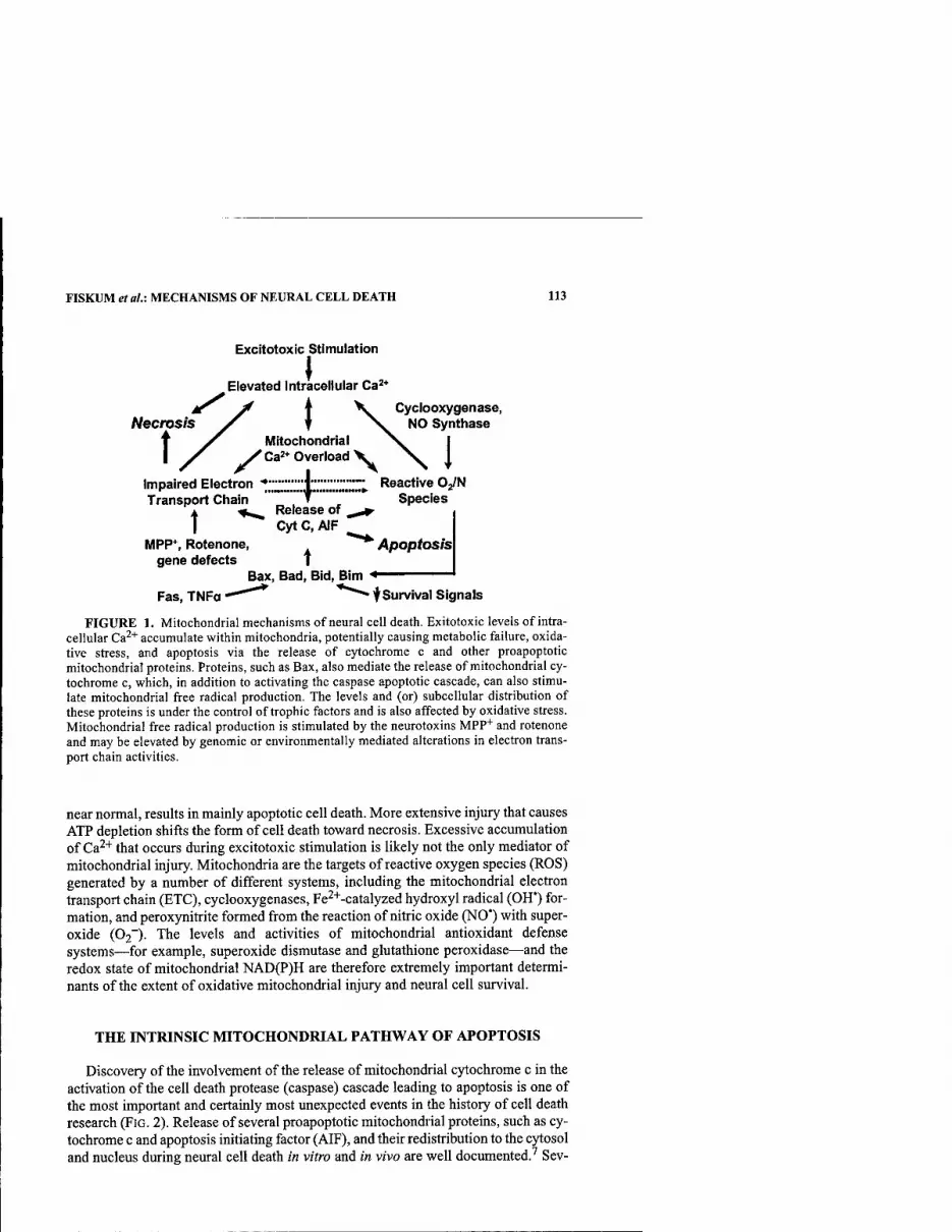
FISKUM et al.: MECHANISMS OF NEURAL CELL DEATH 113
Excitotoxic stimulation
\ Elevated Intracellular Ca^*
j^ y f \. Cyclooxygenase, Necmsis >^ f N^ NO Synthase
T y^ Mitochondrial I jT /Ca'* Overload V
Impaired Electron "tlZZJJlZJZZ.'.^ Reactive OJN Transport Chain """"*" " Species
^_ Release of ^ ^ t Cyt C, AIF
MPP*, Rotenone, ^'^ Apoptosis gene defects t
Bax, Bad, Bid, Bim '* Fas, TNFa "^""^ ^*"^ f Survival Signals
FIGURE 1. Mitochondrial mechanisms of neural cell death. Exitotoxic levels of intra- cellular Ca^'*' accumulate within mitochondria, potentially causing metabolic failure, oxida- tive stress, and apoptosis via the release of cytochrome c and other proapoptotic mitochondrial proteins. Proteins, such as Bax, also mediate the release of mitochondrial cy- tochrome c, which, in addition to activating the caspase apoptotic cascade, can also stimu- late mitochondrial free radical production. The levels and (or) subcellular distribution of these proteins is under the control of trophic factors and is also affected by oxidative stress. Mitochondrial free radical production is stimulated by the neurotoxins MPP""" and rotenone and may be elevated by genomic or environmentally mediated alterations in electron trans- port chain activities.
near normal, results in mainly apoptotic cell death. More extensive injury that causes ATP depletion shifts the form of cell death toward necrosis. Excessive accumulation of Ca^+ that occurs during excitotoxic stimulation is likely not the only mediator of mitochondrial injury. Mitochondria are the targets of reactive oxygen species (ROS) generated by a number of different systems, including the mitochondrial electron transport chain (ETC), cyclooxygenases, Fe^^-catalyzed hydroxyl radical (OH*) for- mation, and peroxynitrite formed from the reaction of nitric oxide (NO') with super- oxide (02~). The levels and activities of mitochondrial antioxidant defense systems—for example, superoxide dismutase and glutathione peroxidase—and the redox state of mitochondrial NAD(P)H are therefore extremely important determi- nants of the extent of oxidative mitochondrial injury and neural cell survival.
THE INTRINSIC MITOCHONDRIAL PATHWAY OF APOPTOSIS
Discovery of the involvement of the release of mitochondrial cytochrome c in the activation of the cell death protease (caspase) cascade leading to apoptosis is one of the most important and certainly most unexpected events in the history of cell death research (FIG. 2). Release of several proapoptotic mitochondrial proteins, such as cy- tochrome c and apoptosis initiating factor (AIF), and their redistribution to the cytosol and nucleus during neural cell death in vitro and in vivo are well documented.^ Sev-
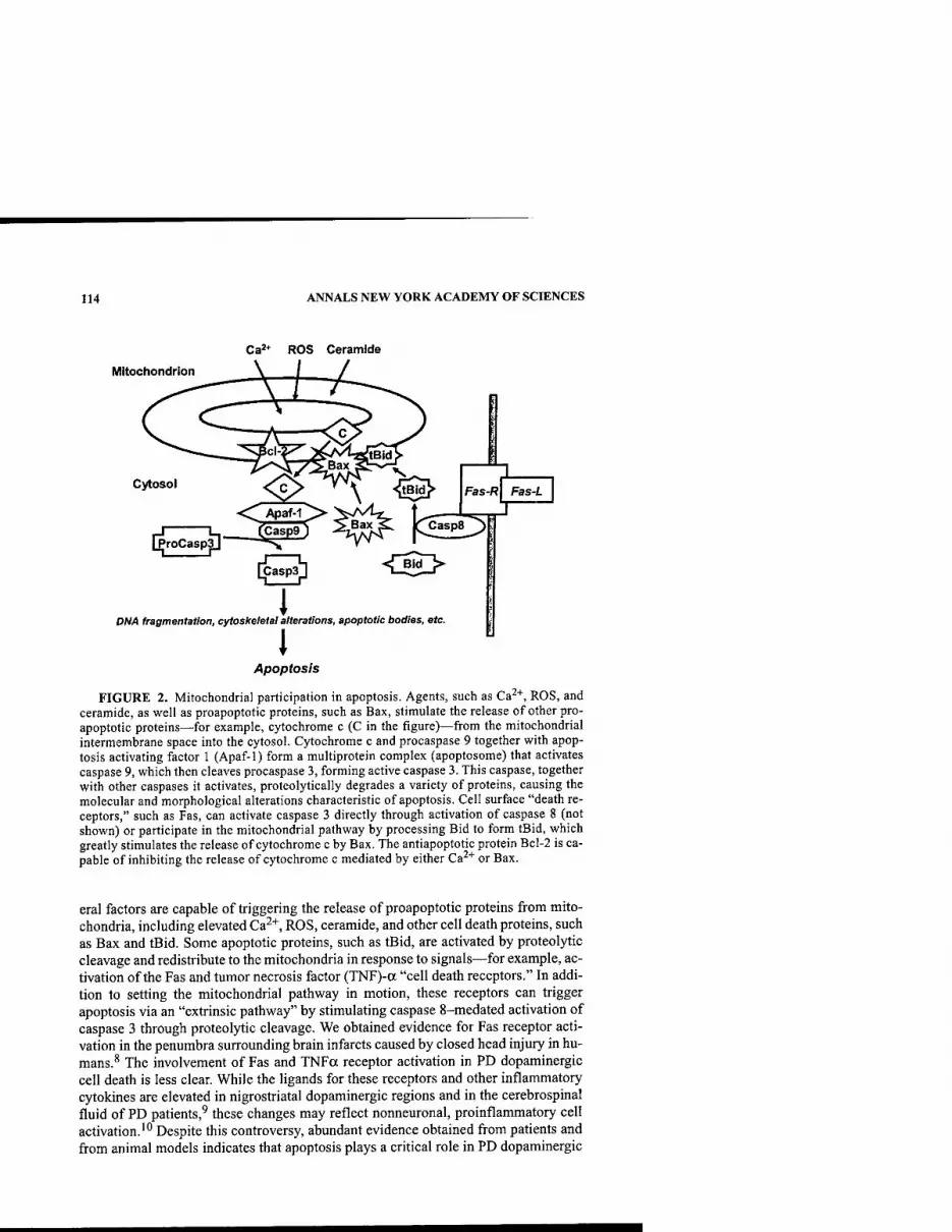
114 ANNALS NEW YORK ACADEMY OF SCIENCES
Ca'* ROS Ceramide
Mitochondrion
Cytosol Fas-R
Apaf-1 2> >A4^ ]^ U-
[ProCasp3] ^"=v '' '
Fas-/.
DW4 fragmentation, cytoskeletal alterations, apoptotic bodies, etc.
I Apoptosis
FIGURE 2. Mitochondrial participation in apoptosis. Agents, such as Ca^'^, ROS, and ceramide, as well as proapoptotic proteins, such as Bax, stimulate the release of other pro- apoptotic proteins—for example, cytochrome c (C in the figure)—from the mitochondrial intermembrane space into the cytosol. Cytochrome c and procaspase 9 together with apop- tosis activating factor 1 (Apaf-1) form a multiprotein complex (apoptosome) that activates caspase 9, which then cleaves procaspase 3, forming active caspase 3. This caspase, together with other caspases it activates, proteolytically degrades a variety of proteins, causing the molecular and morphological alterations characteristic of apoptosis. Cell surface "death re- ceptors," such as Fas, can activate caspase 3 directly through activation of caspase 8 (not shown) or participate in the mitochondrial pathway by processing Bid to form tBid, which greatly stimulates the release of cytochrome c by Bax. The antiapoptotic protein Bcl-2 is ca- pable of inhibiting the release of cytochrome c mediated by either Ca^"*" or Bax.
eral factors are capable of triggering the release of proapoptotic proteins from mito- chondria, including elevated Ca^+, ROS, ceramide, and other cell death proteins, such as Bax and tBid. Some apoptotic proteins, such as tBid, are activated by proteolytic cleavage and redistribute to the mitochondria in response to signals—for example, ac- tivation of the Fas and tumor necrosis factor (TNF)-a "cell death receptors." In addi- tion to setting the mitochondrial pathway in motion, these receptors can trigger apoptosis via an "extrinsic pathway" by stimulating caspase 8-medated activation of caspase 3 through proteolytic cleavage. We obtained evidence for Fas receptor acti- vation in the penumbra surrounding brain infarcts caused by closed head injury in hu- mans.^ The involvement of Fas and TNFa receptor activation in PD dopaminergic cell death is less clear. While the ligands for these receptors and other inflammatory cytokines are elevated in nigrostriatal dopaminergic regions and in the cerebrospinal fluid of PD patients,^ these changes may reflect nonneuronal, proinflammatory cell activation.'" Despite this controversy, abundant evidence obtained from patients and from animal models indicates that apoptosis plays a critical role in PD dopaminergic

FISKUM et al.: MECHANISMS OF NEURAL CELL DEATH 115
cell death and that the mitochondrial pathway of apoptosis is integrally involved.'' Therefore, the molecular mechanisms responsible for the release of proapoptotic pro- teins from mitochondria are potential targets for therapeutic intervention in PD.
There are two fundamentally different mechanisms of mitochondrial protein re- lease under consideration. The first involves physical disruption of the mitochondrial outer membrane and simple diffusion of cytochrome c from its normal exclusive lo- cation in the space located between the outer and inner mitochondrial membranes. The second mechanism involves transport of cytochrome c through a pore located in the outer membrane.
A likely cause for the physical disruption of the outer membrane is the osmotic swelling of the compartment surrounded by the inner membrane (matrix) due to an increased permeability of the inner membrane to small osmotically active solutes. There is a large body of literature describing this swelling phenomenon known as the mitochondrial membrane permeability transition (MPT).'^ This activity is triggered by abnormal mitochondrial Ca^"*" accumulation and is promoted by oxidative stress and reactive metabolites, such as peroxynitrite. The MPT is generally defined as a relatively nonspecific increase in inner membrane permeability that results in a sub- stantial increase in matrix volume and a decrease in mitochondrial membrane poten- tial that can be inhibited by the presence of the drug cyclosporin A (CsA) and by overexpression of the antiapoptotic gene Bcl-2,'^ both of which protect against MPP"''-induced dopaminergic cell death.''*''^ Although CsA is very effective at in- hibiting the MPT for non neural cell mitochondria, Ca^^-induced cytochrome c re- lease from brain mitochondria is relatively resistant to inhibition by CsA in the presence of physiologically realistic concentrations of the cytosolic components Mg2+andATP.'^
We recently found that 2-aminoethoxydiphenyl borate, an inhibitor of transient receptor potential (TRP) channels, is much more effective than CsA at protecting against Ca^"'"-induced cytochrome c release from brain mitochondria.'^ This obser- vation and others suggest that the MPT is associated with activation of Trp channels, a potential new class of targets for neuroprotection in PD and other brain disorders.
The MPT is an attractive mechanism of acute neural injury due to its activation by factors known to be associated with excitotoxicity and because of its sensitivity to inhibition by certain drugs and gene products known to be neuroprotective. How- ever, increasing evidence indicates that the mechanism by which proapoptotic pro- teins such as Bax and tBid release cytochrome c and cause other forms of mitochondrial dysfunction is independent of the MPT and involves either pore for- mation or lipid alterations at the mitochondrial outer membrane.'^~^'' The potential importance of Bax in PD is illustrated by the observation that Bax-knockout mice are resistant to nigrostriatal cell death induced by MPP"*".^'
The Bax-mediated mechanism of cytochrome c release is not inhibited by CsA but is inhibited by specific amphiphilic cations, such as dibucaine and propranolol, known to affect membrane lipid-protein interactions.^^ In addition to activation of proapoptotic proteins like Bid by cell death receptors, expression of the genes coding for these proteins and mitochondrial-protein interactions are promoted by high Ca^"'" and ROS through their stimulation of complex signal transduction cascades.^^ Therefore, Ca^"*" together with oxidative stress can promote cytochrome c release and apoptosis by both the MPT- and Bax-mediated molecular mechanisms that exhibit different pharmacologic sensitivities.
116 ANNALS NEW YORK ACADEMY OF SCIENCES
Mechanisms of mitochondrial proapoptotic protein release in addition to the MPT- and Bax-dependent pathways should also be considered. As AIF release ap- pears downstream of cytochrome c release and caspase activation,^'* it is possible that proteolytic cleavage of AIF or an anchoring protein might be necessary. Also, as cytochrome c release stimulates mitochondrial generation of reactive oxygen spe- cies,'^^ the oxidative modification of mitochondrial membrane lipids or proteins could be another event responsible for or promoting release of proapoptotic mito- chondrial proteins.
A possible key to the development of mitochondrial neuroprotective interven- tions is the understanding of the mechanisms by which the antiapoptotic, mitochon- drial protein Bcl-2 inhibits cytochrome c release mediated by both MPT-dependent and -independent pathways. Bcl-2 and its close relative BC1-XL exert some form of antioxidant activity that confers cytoprotection against ROS that may also inhibit ac- tivation of the MPT.'^'^^ In contrast, Bcl-2 inhibition of Bax appears to involve di- rect protein-protein interaction, impairment of Bax oligomerization, and consequently inhibition of pore formation.^' Stimulation of Bcl-2 expression has been demonstrated in several ischemic preconditioning paradigms and may repre- sent a primary mechanism of neuroprotection by estrogen.^^ Investigators have also utilized protein transduction domains to deliver exogenous BC1-XL to cells through- out the brain and have demonstrated neuroprotection when these fusion proteins were administered by intraperitoneal injection up to 1 hour following the occlusion of the middle cerebral artery of mice.'^^ Delivery of neuroprotective proteins, such as Bcl-2 and glial cell line-derived neurotrophic factor (GDNF), as a therapeutic ap- proach for neurodegenerative disorders like Parkinson's disease is therefore possible and may prove to be effective.
MITOCHONDRIAL MECHANISMS OF REACTFVE OXYGEN SPECIES GENERATION
As several lines of evidence suggest that elevated mitochondrial ROS production contributes to the etiology of Parkinson's disease and as oxidative stress is a potent activator of apoptosis, understanding the mechanisms of mitochondrial free radical production is critically important in elucidating the pathophysiology of Parkinson's and other neurodegenerative diseases. The two most commonly cited sites of mito- chondrial ROS production are ubisemiquinone (during complex III reduction) and complex I, although others, such as complex II, may also contributed^ The site of mitochondrial ROS production implicated most strongly in Parkinson's disease is complex I of the electron transport chain. Some neurotoxins that induce PD neuro- pathology in vivo, such as MPP"^ and rotenone, are inhibitors of complex I and stim- ulate ROS generation in vitro?^ Acting along with hydroxyl radical (OH') and peroxynitrite (HNOO~), these ROS species can cause oxidative damage and inhibi- tion of mitochondrial enzyme activities, including those of complex I and alpha-ke- toglutarate dehydrogenase complex (aKGDC) (FIG. 3).^^'^^ This inhibition can lead to metabolic failure through impairment of electron transport-dependent generation of the proton-motive force that drives the synthesis of ATP. Complete detoxification of superoxide depends on the enzymes superoxide dismutase (SOD), glutathione peroxidase (GPX), and glutathione reductase (GR) together with glutathione and a
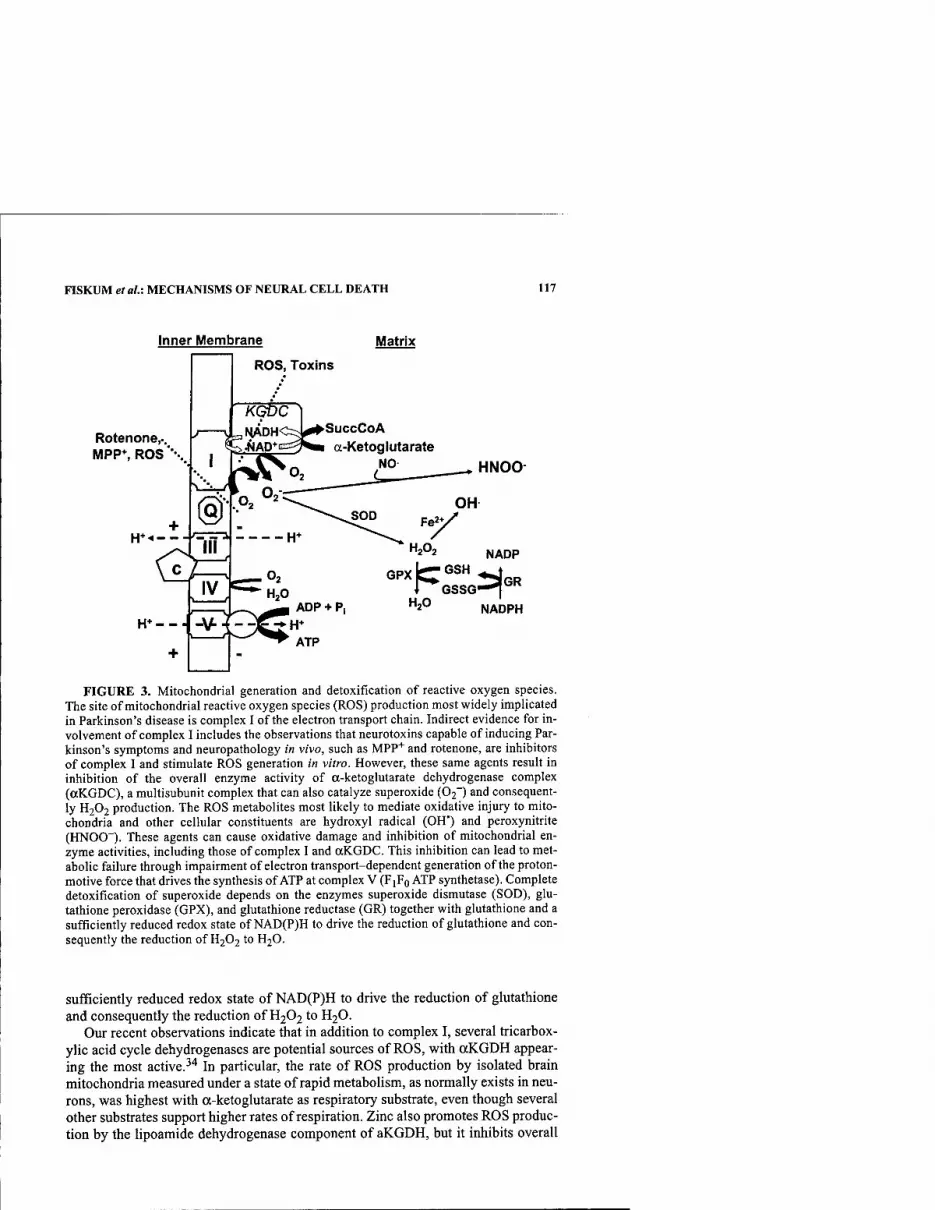
FISKUM etal: MECHANISMS OF NEURAL CELL DEATH 117
Inner Membrane
ROS, Toxins
Matrix
Rotenone,.. MPP*, ROS ■•••.
KGDC ' 3NlADHC=5^SuccCoA
IAD*ggj^M a-Ketoglutarate NO HNOO-
FIGURE 3. Mitochondrial generation and detoxification of reactive oxygen species. The site of mitochondrial reactive oxygen species (ROS) production most widely implicated in Parkinson's disease is complex I of the electron transport chain. Indirect evidence for in- volvement of complex I includes the observations that neurotoxins capable of inducing Par- kinson's symptoms and neuropathology in vivo, such as MPP"^ and rotenone, are inhibitors of complex I and stimulate ROS generation in vitro. However, these same agents result in inhibition of the overall enzyme activity of a-ketoglutarate dehydrogenase complex (aKGDC), a multisubunit complex that can also catalyze superoxide (02~) and consequent- ly H2O2 production. The ROS metabolites most likely to mediate oxidative injury to mito- chondria and other cellular constituents are hydroxyl radical (OH') and peroxynitrite (HNOO~). These agents can cause oxidative damage and inhibition of mitochondrial en- zyme activities, including those of complex I and aKGDC. This inhibition can lead to met- abolic failure through impairment of electron transport-dependent generation of the proton- motive force that drives the synthesis of ATP at complex V (FIFQ ATP synthetase). Complete detoxification of superoxide depends on the enzymes superoxide dismutase (SOD), glu- tathione peroxidase (GPX), and glutathione reductase (GR) together with glutathione and a sufficiently reduced redox state of NAD(P)H to drive the reduction of glutathione and con- sequently the reduction of H2O2 to H2O.
sufficiently reduced redox state of NAD(P)H to drive the reduction of glutathione and consequently the reduction of H2O2 to H2O.
Our recent observations indicate that in addition to complex I, several tricarbox- ylic acid cycle dehydrogenases are potential sources of ROS, with aKGDH appear- ing the most active.-''* In particular, the rate of ROS production by isolated brain mitochondria measured under a state of rapid metabolism, as normally exists in neu- rons, was highest with a-ketoglutarate as respiratory substrate, even though several other substrates support higher rates of respiration. Zinc also promotes ROS produc- tion by the lipoamide dehydrogenase component of aKGDH, but it inhibits overall
118 ANNALS NEW YORK ACADEMY OF SCIENCES
enzyme activity.^^ Considering the findings that environmental exposure to zinc is a risk factor for Parkinson's disease and that otKGDH enzyme activity is reduced in PD and in Alzheimer's disease,^^ the relationships between this enzyme complex, ROS production, and dopaminergic cell death in cellular and animal models of PD
require further investigation.
REFERENCES
1. PARKER, W.D.J., S.J. BOYSON & J.K. PARKS. 1989. Abnormalities of the electron trans- port chain in idiopathic Parkinson's disease. Ann. Neurol. 26: 719-723.
2. SHULTS, C.W., R.H. HAAS, D. PASSOV & M.F. BEAL. 1997. Coenzyme QIO levels corre- late with the activities of complexes I and II/III in mitochondria from parkinsonian and nonparkinsonian subjects. Ann. Neurol. 42: 261-264.
3. SwERDLOW, R.H., J.K. PARKS, S.W. MILLER, et al. 1996. Origin and functional conse- quences of the complex I defect in Parkinson's disease. Ann. Neurol. 40: 663-671.
4. SHEEHAN, J.P, R.H. SWERDLOW, S.W. MILLER, et al. 1997. Calcium homeostasis and reactive oxygen species production in cells transformed by mitochondria from indi- viduals with sporadic Alzheimer's disease. J. Neurosci. 17: 4612^622.
5. TRIMMER, P.A., R.H. SWERDLOW, J.K. PARKS, et al. 2000. Abnormal mitochondrial morphology in sporadic Parkinson's and Alzheimer's disease cybrid cell lines. Exp. Neurol. 162: 37-50.
6. BETARBET, R., T.B. SHERER, G. MACKENZIE, et al. 2000. Chronic systemic pesticide exposure reproduces features of Parkinson's disease. Nat. Neurosci. 3: 1301-1306.
7. FlSKUM, G. 2000. Mitochondrial participation in ischemic and traumatic neural cell death. J. Neurotrauma 17: 843-855.
8. Qiu, J., M.J. WHALEN, P. LOWENSTEIN, et al. 2002. Upregulation of the Fas receptor death-inducing signaling complex after traumatic brain injury in mice and humans. J. Neurosci. 22: 3504-3511.
9. NAGATSU, T., M. MOGI, H. ICHINOSE & A. TOGARI. 2000. Changes in cytokines and neurotrophins in Parkinson's disease. J. Neural Transm. Suppl. 277-290.
10. FERRER, I., R. BLANCO, B. CUTILLAS & S. AMBROSIO. 2000. Fas and Fas-L expression in Huntington's disease and Parkinson's disease. Neuropathol. Appl. Neurobiol. 26: 424-433.
11. DAWSON, T.M. & V.L. DAWSON. 2002. Neuroprotective and neurorestorative strategies for Parkinson's disease. Nat. Neurosci. 5 Suppl.: 1058-1061.
12. FRIBERG, H. & T. WIELOCH. 2002. Mitochondrial permeability transition in acute neu- rodegeneration. Biochimie 84: 241-250.
13. KOWALTOWSKI, A.J., A.E. VERCESI & G. FISKUM. 2000. Bcl-2 prevents mitochondrial permeability transition and cytochrome c release via maintenance of reduced pyri- dine nucleotides. Cell Death. Differ. 7: 903-910.
14. SEATON, T.A., J.M. COOPER & A.H. SCHAPIRA. 1998. Cyclosporin inhibition of apopto- sis induced by mitochondrial complex I toxins. Brain Res. 809: 12-17.
15. YANG, L., R.T. MATTHEWS, J.B. SCHULZ, et al. 1998. l-Methyl-4-phenyl-l,2,3,6-tet- rahydropyride neurotoxicity is attenuated in mice overexpressing Bcl-2. J. Neurosci. 18:8145-8152.
16. ANDREYEV, A. & G. FISKUM. 1999. Calcium induced release of mitochondrial cyto- chrome c by different mechanisms selective for brain versus liver. Cell Death. Differ. 6: 825-832.
17. CHINOPOULOS, C, A.A. STARKOV & G. FISKUM. 2003. Cyclosporin A-insensitive per- meability transition in brain mitochondria: inhibition by 2-aminoethoxydiphenyl borate. J. Biol. Chem. In press.
18. KoRSMEYER, S.J., M.C. WEI, M. SAITO, et al. 2000. Pro-apoptotic cascade activates BID, which oligomerizes BAK or BAX into pores that result in the release of cyto- chrome c. Cell Death Differ. 7: 1166^1173.
19. KuwANA, T., M.R. MACKEY, G. PERKINS, etal. 2002. Bid, Bax, and lipids cooperate to form supramolecular openings in the outer mitochondrial membrane. Cell 111: 331-342.
FISKUM et al.: MECHANISMS OF NEURAL CELL DEATH 119
20. PoLSTER, B.M., K.W. KiNNALLY & G. FiSKUM. 2001. Bh3 death domain peptide induces cell type-selective raitochondrial outer membrane permeability. J. Bid. Chem. 276: 37887-37894.
21. VILA, M., V. JACKSON-LEWIS, S. VUKOSAVIC, et al. 2001. Bax ablation prevents dopam- inergic neurodegeneration in the 1-methyl- 4-phenyl-l,2,3,6-tetrahydropyridine mouse model of Parkinson's disease. Proc. Natl. Acad. Sci. USA 98: 2837-2842.
22. PoLSTER, B.M. & G. FiSKUM. 2003. Inhibition of Bax-induced cytochrome c release from neural cell and brain mitochondria by dibucaine and propranolol. J. Neurosci. 23:2735-2743.
23. MARTIN, L.J. 2001. Neuronal cell death in nervous system development, disease, and injury. Int. J. Mol. Med. 7: 455-478.
24. ARNOULT, D., P. PARONE, J.C. MARTINOU, et al. 2002. Mitochondrial release of apopto- sis-inducing factor occurs downstream of cytochrome c release in response to several proapoptotic stimuli. J. Cell Biol. 159: 923-929.
25. STARKOV, A.A., B.M. POLSTER & G. FISKUM. 2002. Regulation of hydrogen peroxide production by brain mitochondria by calcium and Bax. J. Neurochem. 83: 220-228.
26. OuYANG, Y., S. CARRIEDO & R. GiFFARD. 2002. Effect of Bcl-X(L) overexpression on reactive oxygen species, intracellular calcium, and mitochondrial membrane poten- tial following injury in astrocytes. Free Radic. Biol. Med. 33: 544.
27. CHENG, E.H., M.C. WEI, S. WEILER, et al. 2001. BCL-2, BCL-X(L) sequester BH3 domain-only molecules preventing BAX- and BAK-mediated mitochondrial apopto- sis. Mol. Cell 8: 705-711.
28. ALKAYED, N.J., S. GOTO, N. SUGO, et al. 2001. Estrogen and Bcl-2: gene induction and effect of transgene in experimental stroke. J. Neurosci. 21: 7543-7550.
29. CAO, G., W. PEI, H. GE, et al. 2002. In vivo delivery of a Bcl-xL fusion protein con- taining the TAT protein transduction domain protects against ischemic brain injury and neuronal apoptosis. J. Neurosci. 22: 5423-5431.
30. BOVERIS, A. 1977. Mitochondrial production of superoxide radical and hydrogen per- oxide. Adv. Exp. Med. Biol. 78: 67-82.
31. LENAZ, G., C. BOVINA, M. D'AURELIO, et al. 2002. Role of mitochondria in oxidative stress and aging. Ann. N.Y. Acad. Sci. 959: 199-213.
32. HILLERED, L. & L. ERNSTER. 1983. Respiratory activity of isolated rat brain mitochon- dria following in vitro exposure to oxygen radicals. J. Cereb. Blood Flow Metab. 3: 207-214.
33. NULTON-PERSSON, A.C. & L.I. SZWEDA. 2001. Modulation of mitochondrial function by hydrogen peroxide. J. Biol. Chem. 276: 23357-23361.
34. STARKOV, A. & G. FISKUM. 2002. Generation of reactive oxygen species by brain mito- chondria mediated by alpha-ketoglutarate dehydrogenase. Soc. Neurosci. 194.17.
35. GAZARYAN, I.G., B.F. KRASNIKOV, G.A. ASHBY, et al. 2002. Zinc is a potent inhibitor of thiol oxidoreductase activity and stimulates reactive oxygen species production by lipoamide dehydrogenase. J. Biol. Chem. 277: 10064-10072.
36. GIBSON, G.E., L.C. PARK, K.F. SHEU, et al. 2000. The alpha-ketoglutarate dehydroge- nase complex in neurodegeneration. Neurochem. Int. 36: 97-112.

Mitochondria, Oxidative Damage, and Inflammation in Parkinson's Disease
M. FLINT BEAL
Department of Neurology and Neuroscience, Weill Medical College of Cornell University, New York Presbyterian Hospital, New York, New York 10021, USA
ABSTRACT: The pathogenesis of Parkinson's disease (PD) remains obscure, but there is increasing evidence that impairment of mitochondrial function, oxida- tive damage, and inflammation are contributing factors. The present paper re- views the experimental and clinical evidence implicating these processes in PD. There is substantial evidence that there is a deficiency of complex I activity of the mitochondrial electron transport chain in PD. There is also evidence for in- creased numbers of activated microglia in both PD postmortem tissue as well as in animal models of PD. Impaired mitochondrial function and activated microglia may both contribute to oxidative damage in PD. A number of thera- pies targeting inflammation and mitochondrial dysfunction are efficacious in the MPTP model of PD. Of these, coenzyme Qio appears to be particularly promising based on the results of a recent phase 2 clinical trial in which it sig- nificantly slowed the progression of PD.
KEYWORDS: inflammation; microglia; mitochondria; oxidative damage; creat- ine; coenzyme Q|o
THE ROLE OF FREE RADICALS AND IMPAIRED ENERGY METABOLISM IN PARKINSON'S DISEASE
All aerobic organisms are continually exposed to oxidative stress.' Oxidative phosphorylation involves the transfer of electrons, which can lead to the generation of free radicals, independently existing species that contain one or more unpaired electrons. Most free radicals are unstable reactive species that can extract an electron from neighboring molecules to complete their own orbital. This leads to oxidation of neighboring molecules. Critical biologic molecules including DNA, proteins, and membrane lipids are subject to oxidative damage.
Mitochondria are believed to be the most important cellular source of free radi- cals generating superoxide radicals at ubiquinone and NADH dehydrogenase (com- plex I).-^ Superoxide is normally converted by superoxide dismutase (SOD) to H2O2. H2O2 reacts with transition metals to generate hydroxyl radicals, the prime media- tors of cell damage. Superoxide can also react with nitric oxide to the form of per-
Address for correspondence: M. Flint Beal, M.D., Chairman, Department of Neurology and Neuroscience, Weill Medical College of Cornell University, 525 East 68th Street, Room F610, New York, NY 10021. Voice: 212-746-6575; fax: 212-746-8532.
Ann. N.Y. Acad. Sci. 991: 120-131 (2003). © 2003 New York Academy of Sciences.
120

BEAL: MITOCHONDRIA, OXIDATIVE DAMAGE, AND INFLAMMATION 121
oxynitrite (ONOO").^ This reaction occurs at the rate of 6.7 x 10^ M"' S"', threefold faster than the rate of superoxide dismutation by superoxide dismutase. Peroxyni- trite generation thus depends on the concentration of superoxide and nitric oxide in the cell. Peroxynitrite can exist in an activated "hydroxyl radical-like" transitional state. At physiologic pH, peroxynitrite may diffuse over several cell diameters to produce cell damage by oxidizing lipids, proteins, and DNA. Peroxynitrite can react with Cu,Zn SOD to form nitronium ion, which may then nitrate tyrosine residues."* The 3-nitrotyrosine so generated is an excellent biochemical marker of peroxyni- trite-mediated oxidative damage.
Much of the current interest in the association between neurodegeneration and mitochondrial dysfunction/oxidative damage stems from studies of l-methyl-4- phenyl-l,2,3,6-tetrahydropyridine (MPTP)-induced parkinsonism. The MPTP mod- el of PD results in a clinical syndrome that replicates the major features of PD in man and nonhuman primates.^ MPTP is metabolized to MPTP+ and MPP+ (l-methyl-4- phenylpyridinium) by the enzyme monoamine oxidase B. MPP"*" is subsequently se- lectively taken up by dopaminergic terminals and concentrated in neuronal mito- chondria in the substantia nigra. In vitro experiments demonstrated that nicotinamide dehydrogenase (NADH), complex I of the electron transport chain, is weakly and reversibly inhibited by MPP"*", leading to reductions in mitochondrial ATP production.^ MPP"*" can also cause irreversible inactivation of complex I by gen- erating free radicals and increasing lipid peroxidation.' Inhibition of NADH dehy- drogenase by MPP"*" leads to superoxide production in isolated bovine heart submitochondrial particles and in v/vo.^'^ Finally, MPTP-induced damage is attenu- ated in transgenic mice overexpressing superoxide dismutase.^^ We previously showed that mice deficient in either MnSOD or glutathione peroxidase show in- creased vulnerability to MPTP toxicity." •' ^ Thus, the MPTP model of parkinsonism has already set a precedent for a causal relationship between mitochondrial dysfunc- tion and oxidative cellular damage.
Evidence extending this hypothesis to the pathogenesis of idiopathic PD comes from a 30-40% decrease in complex I activity in the substantia nigra.'^"'^ Reduced staining for complex I subunits in PD substantia nigra, but preserved staining for subunits of the other electron transport complexes, has been demonstrated immuno- histochemically.'^ Strong support for a mitochondrial DNA-encoded defect comes from studies that showed that complex I defects from PD platelets are transferable into mitochondrial-deficient cell lines.*^''^ These defects are associated with in- creased free radical production, increased susceptibility to MPP"*", and impaired mi- tochondrial calcium buffering.^^ Direct sequencing of mitochondrial complex I and tRNA genes, however, failed to show homoplasmic mutations.^'
OXIDATIVE DAMAGE IN PD
A great deal of interest has been focused on the possibility that oxidative damage may play a role in the pathogenesis of PD. There are studies showing increased lev- els of malondialdehyde and cholesterol lipid hydroperoxides, markers for lipid per- oxidation, in PD substantia nigra.^^'^^ There are widespread increases in protein carbonyls in PD postmortem brain tissue.^'' Concentrations of 8-hydroxy-2-deoxy- guanosine, a marker of oxidative damage to DNA, are significantly increased in PD

122 ANNALS NEW YORK ACADEMY OF SCIENCES
substantia nigra and striatum.^^'-^^ However, attempts to detect increased free radi- cals using electron spin resonance have been unsuccessful. Evidence for nitrosyl radicals in PD substantia nigra was recently obtained.^^ Another means of looking for oxidative stress is to measure concentrations of reduced glutathione. Reduced glutathione is decreased in PD substantia nigra by approximately 50%.^^"^' Individ- uals with incidental Lewy body disease may have presymptomatic PD, and they have a 35% reduction in reduced glutathione as compared with age-matched controls.^^
INFLAMMATION AND PD
There is increasing evidence that inflammation may contribute to PD pathogene- sis. There is an increase in reactive microglia in the striatum and substantia nigra of patients with idiopathic PD.^^'^'* Although gliosis may in some circumstances be beneficial, at other times it exerts deleterious effects. Microglial cells, which are the resident macrophages in the brain, respond to many insults with rapid proliferation, hypertrophy, and expression of a number of cytokines.^^-^^ The brain area that en- compasses the substantia nigra has the highest density of microglia in the brain.^^ Activated microglia upregulate cell surface markers such as the macrophage antigen complex-I (MAC-1), and they produce a variety of proinflammatory cytokines. There appears to be a graded response with differential expression depending on the severity of the insult.^^ A consequence of activation of microglia is the production of reactive oxygen species. They can originate from NADPH oxidase, which produc- es superoxide (O2'); COX-2, which produces free radicals as a byproduct of prostag- landin synthesis; and from inducible nitric oxide synthase (iNOS), which generates NO*. iNOS, because it does not require Ca^* for activity, produces much higher lev- els of NO* for longer periods (hours) than do the endothelial and neuronal isoforms of NOS, which require Ca^"^ for activity. NADPH-oxidase is a major source of activ- ity-dependent superoxide.^^ The reaction of ©2' with NO" generates peroxynitrite ONOO", which is strongly implicated in PD pathogenesis. Prior studies showed that Lewy body-positive substantia nigra neurons in PD stain with antibodies to 3-nitro- tyrosine, which is thought to be a specific marker for peroxynitrite-mediated dam- age.^' More recent studies showed that Lewy bodies react with antibodies to nitrated a-synuclein, the major protein component of Lewy bodies.'*'*
It was reported in a small number of cases that the mRNA for the Clq and C9 components of complement are increased in PD substantia nigra."*' A number of cy- tokines including interleukin (IL)-l, IL-6, and tumor necrosis factor (TNF)-a con- tribute to inflammatory processes. Studies of TNFa showed it to be increased 366% in the striatum and 432% in the cerebrospinal fluid (CSF) of PD patients.''^ Further- more, an increase in TNFa imunoreactive glial cells was reported in the substantia nigra of PD patients, as compared to controls.''^''''*
The density of glial cells expressing IL-ip and interferon (IFN)-Ywas also in- creased in PD substantia nigra."*"* Others found increased levels of Il-lp and IL-6 in the caudate, putamen, and cerebral cortex of postmortem PD brains."*^ Both IL-ip and IL-6 were also found to be increased in the CSF of PD patients."*^ Another group found an increase in IL-6 in the CSF of PD patients and reported that levels inversely correlate with the severity of PD."''' Last, an IL-ip polymorphism was reported to in- crease the risk of PD."'^-''^

BEAL: MITOCHONDRIA, OXIDATIVE DAMAGE, AND INFLAMMATION 123
INFLAMMATION IN ANIMAL MODELS
Although there has been tremendous progress in genetic models of PD, they are not yet ideal in producing the cardinal features of PD.^ The most experience has been with toxin models of PD including MPTP, rotenone, and 6-hydroxydopamine. An activation of microglia in both the striatum and in the substantia nigra has been well documented to occur in the MPTP model of PD.^""^^ The microglial activation was associated with elevated IL-6 levels.^^ MPTP toxicity is also associated with in- creases in IL-1 p.^^ We demonstrated that mice deficient in IL-1 p converting enzyme are markedly resistant to MPTP neurotoxicity.^'* A recent study showed that mino- cycline prevented MPTP-induced activation of microglia, as well as the formation of IL-lp, activation of NADPH-oxidase, and induction of iNOS.^^ This was associated with reduced 3-nitrotyrosine formation, and reduced loss of substantia nigra dopam- inergic neurons.^^ Another group showed protection as well^^ but suggested that minocycline may also have direct neuroprotective effects. This was recently shown to be the case with direct effects on the mitochondrial permeability transition and cytochrome c release.^* The importance of inflammation was underscored by a study of humans exposed to MPTP in which persistent activation of microglia was associated with oxidative degeneration of substantia nigra neurons.^' Recently sys- temic administration of rotenone to rats was shown to produce an excellent animal model of PD.^^ Studies of rotenone toxicity in neuron/glia cultures from rat mesen- cephalon show enhanced neurodegeneration in the presence of glia.^^ A critical fea- ture of the toxicity was the release of O2* from microglia, which was attenuated by inhibitors of NADPH oxidase.
There is also substantial evidence for neuroinflammation playing a role in pro- gressive neurodegeneration following administration of 6-hydroxydopamine.^'' Studies using PET and PKl 195 (a ligand for activated microglia) showed that an in- crease in activated microglia occurs coincident with loss of dopaminergic neurons.^' Minocycline was reported to inhibit microglial activation and to protect nigral cells after 6-OHDA injection into mouse striatum.^^ Chronic infusion of lipopolysaccha- ride for 2 weeks into the substantia nigra results in rapid activation of microglia, fol- lowed by a delayed and gradual loss of nigral neurons beginning at 4-6 weeks and reaching 70% at 10 weeks.^-^
ANTIINFLAMMATORY AGENTS FOR NEUROPROTECTION
A number of antiinflammatory agents have shown promise for the amelioration of degeneration of dopaminergic neurons induced by MPTP. Approaches have uti- lized both transgenic mice as well as pharmacologic agents. MPTP-induced activa- tion of microglia is associated with an upregulation of iNOS and 3-nitrotyrosine formation.^^ Studies in iNOS-deficient mice show that degeneration of dopaminer- gic neurons is significantly attenuated; but there is no protection of dopaminergic terminals.^'''^^ However, there have been no reports of the effects of pharmacologic inhibition of iNOS on MPTP neurotoxicity. Another inflammatory pathway impli- cated in neurodegeneration is cyclooxygenase 2 (COX-2). COX-2 is expressed and regulated in glial cells by cytokines and lipopolysaccharide.^^ However, in excito- toxic lesions, synaptic excitation, apoptotic neuronal death, and cerebral ischemia

124 ANNALS NEW YORK ACADEMY OF SCIENCES
COX-2 is expressed primarily in neurons.^''"''° Cyclooxygenase catalyzes the forma- tion of prostaglandins, which involves reduction of the hydroperoxide resulting in the generation of free radicals. Superoxide radicals, a COX-2 reaction product, can react with NO' to form the strong oxidant peroxynitrite. A recent study showed that overexpression of COX-2 can activate cell cycle genes, which may contribute to ap- optotic cell death^' Mice overexpressing COX-2 show increased vulnerability to kainic acid and have elevated lipid peroxidation. COX-2 inhibitors inhibit lipo- polysaccharide-induced increases in TNF-a, which can stimulate astrocytic glutamate release^^'^^ Neuronal death mediated by Af-methyl-D-aspartate (NMDA) is diminished in a dose-dependent manner by COX-2 inhibitors in primary neuronal cultures.'^'' Transgenic mice that are deficient in COX-2 show reduced susceptibility to focal ischemia and to NMDA neurotoxicity.''^ The COX-2 inhibitor meloxicam significantly attenuated both the reduction of striatal dopamine levels and the deple- tion of substantia nigra dopaminergic neurons.'^ In COX-2-deficient mice, however, there was protection against loss of dopaminergic neurons in the substantia nigra, but no protective effect against lowering of striatal dopamine levels.'^
Another promising approach is to use thalidomide. Thalidomide reduces COX-2 levels and also inhibits TNFa production.■'^"^° Mice deficient in both TNFa recep- tors are resistant to MPTP toxicity.^' Thalidomide has been reported to attenuate MPTP-induced decreases in striatal dopamine levels, but the numbers of dopamin- ergic neurons in the substantia nigra were not examined.^^ Another approach is to use phosphodiesterase IV inhibitors, which inhibit the breakdown of cAMP. Phos- phodiesterase IV is selectively localized to immune cells and the central nervous sys- tem. Type IV phosphodiesterase inhibitors are effective against inflammatory lesions in the central nervous system, such as those produced in experimental auto- immune encephalomyelitis.^^'^"' Rolipram inhibits lipopolysaccharide-induced in- creases in plasma levels of TNFa in rats.^^ Phosphodiesterase IV inhibitors are also neuroprotective against MPTP neurotoxicity.^^ In these studies, several phosphodi- esterase IV inhibitors produced neuroprotection against both MPTP-induced deple- tion of striatal dopamine and as loss of substantia nigra neurons.
Last, peroxisome proliferator-activated receptor (PPAR)-Y agonists show good antiinflammatory effects. A broad range of proinflammatory genes are transcription- ally regulated by the nuclear hormone receptor PPAR-y. PPARs are a subfamily of ligand-activated transcription factors structurally related to the steroid and retinoic acid receptor families.^'^ The PPAR-y isoform is expressed in adipocytes, where it regulates lipid metabolism and reduces insulin resistance. Newly developed drugs of the thiazolidinedione class target the PPAR-y receptor and reduce plasma glucose in type 2 diabetics, although they do not alter glucose levels in nondiabetic animals and humans.^^ PPAR-y agonists suppress the expression of the proinflammatory cytok- ines IL-lp, TNFa, and IL-6,89,90 ^g well as of MMP-9 and iNOS.^'^^^ -phe PPAR- y agonist troglitazone reduces cell death in cultured cerebellar neurons after glutamate exposure.^'' The PPAR-y agonist pioglitazone reduced the severity and in- cidence of experimental allergic encephalomyelitis.'^ Pioglitazone was recently shown to exert neuroprotective effects against MPTP-induced degeneration of sub- stantia nigra neurons; however, it did not protect against striatal dopamine depletion. Lipopolysaccharide induces iNOS expression and release of nitric oxide in microglia, and COX-2 expression in neurons.^'' PPAR-y agonists inhibit lipopolysaccharide- induced cell death as well as increases in nitric oxide and COX-2 activity.^^

BEAL: MITOCHONDRIA, OXIDATIVE DAMAGE, AND INFLAMMATION 125
COENZYME Qio AND CREATINE AS POTENTIAL THERAPIES FOR PD
Coenzyme Qio, which is also known as ubiquinone, is an essential cofactor of the electron transport chain, where it accepts electrons from complexes 1 and 2. ^ Co- enzyme Qio also serves as an important antioxidant in both mitochondria and lipid membranes.^^''^" It is a lipid-soluble compound composed of redox-active quinone as well as a hydrophobic tail. It is soluble and mobile in the hydrophobic core of the phospholipid bilayer of the inner membrane of the mitochondria.
Considerable interest has been shown in the potential utility of coenzyme Qjo for treatment of mitochondrial disorders.'"^ There have been several reports of benefi- cial effects in patients with mitochondrial defects; and we have demonstrated that coenzyme QIQ increased brain mitochondrial coenzyme Qio concentrations in ma- ture and older animals.'*'^ It also decreased a-tocopherol concentrations in mito- chondria consistent with a sparing effect. We found that coenzyme QIQ exerts neuroprotective effects against the mitochondrial toxins malonate and 3-nitropropi- onic acid.^"^''"'' It also showed neuroprotective effects against MPTP toxicity in old- er mice.'"^ We found that coenzyme Qio levels were significantly reduced in mitochondria from PD patients.'^^ We also demonstrated that coenzyme Qio supple- mentation significantly increased plasma levels in PD patients. Recently a small phase II clinical trial was undertaken.^"' Patients were assigned to placebo or coen- zyme Qio at 300, 600, or 1200 mg daily. They were evaluated using the Unified Par- kinson's Disease Rating Scale at screening, baseline, and up to 16 months. In this pilot trial the administration of coenzyme Qio showed a significant slowing of dis- ease progression.
We also investigated whether creatine can exert neuroprotective effects. The cre- atine kinase system can shuttle high-energy phosphates such as phosphocreatine (PCr) to sites of energy usage in the cell, such as ion pumps in organelles, the plasma membranes, and the endoplasmic reticulum.'"^'**'^ Creatine may exert neuroprotec- tive effects by increasing PCr levels and thereby providing extra energy for ion ho- meostasis and the functional and structural integrity of mitochondria. Creatine exerts neuroprotective effects in vitro as well as in vivo. Creatine protects against both glutamate and p-amyloid toxicity in rat hippocampal neurons.''"'^^^ Creatine also protects against 3-nitropropionic acid and glutamate neurotoxicity in rat hippocam- pal and striatal neurons.''^ We found that creatine protects against both malonate and 3-nitropropionic acid striatal neurotoxicity in vivo'}^^ and against MPTP toxic- ity in a dose-dependent manner.''' Creatine administration also exerts neuroprotec- tive effects in transgenic mouse models of both amyotrophic lateral sclerosis and Huntington's disease.''^""^ We recently demonstrated that creatine can exert addi- tive neuroprotective effects when administered in combination with COX-2 inhibi- tors against MPTP neurotoxicity (unpublished data).
CONCLUSIONS
The pathogenesis of PD is unknown, but a substantial body of evidence impli- cates both mitochondrial dysfunction, oxidative damage, and inflammation in dis- ease pathogenesis. Agents targeting both mitochondrial dysfunction and inflammation are effective in attenuating damage to dopaminergic neurons in the
126 ANNALS NEW YORK ACADEMY OF SCIENCES
MPTP model of PD. One of these agents, coenzyme QIQ, has shown initial promise as an agent to slow the progression of PD. Clinical trials of other agents to slow the progress of PD are urgently needed. We suspect that combinations of agents target- ing different disease mechanisms may produce additive or synergistic neuroprotec-
tive effects that will lead to effective therapies to slow or halt PD.
ACKNOWLEDGMENTS
I wish to thank Sharon Melanson for secretarial assistance. This work was sup- ported by grants from The Department of Defense and the Parkinson's Disease
Foundation.
REFERENCES
1. HALLIWELL, B. 1992. Reactive oxygen species and the central nervous system. J. Neu- rochem. 59: 1609-1623.
2. TuRRENS, J.F., A. ALEXANDRE & A.L. LEHNINGER. 1985. Ubisemiquinone is the elec- tron donor for superoxide formation by complex III of heart mitochondria. Arch. Biochem. Biophys. 237: 408-414.
3. BECKMAN, J.S. & J.P. CROW. 1993. Pathological implications of nitric oxide superoxide and peroxynitrite formation. Biochem. Soc. Trans. 21: 330-334.
4. IscHiROPOULOS, H., L. ZHU, J. CHEN, et al. 1992. Peroxynitrite-mediated tyrosine nitra- tion catalyzed by superoxide dismutase. Arch. Biochem. Biophys. 298: 431-437.
5. BEAL, M.F. 2001. Experimental models of Parkinson's disease. Nat. Rev. Neurosci. 2: 325-334.
6. SINGER, TP., N. CASTAGNOLI, R.R. RAMSAY & A.J. TREVOR. 1987. Biochemical events in the development of parkinsonism induced by MPTP. J. Neurochem. 49: 1-8.
7. CLEETER, M.J.W., J.M. COOPER & A.H.V. SCHAPIRA. 1992. Irreversible inhibition of mitochondrial complex I by l-methyl-4-phenylpyridium: evidence for free radical involvement. J. Neurochem. 58: 786-789.
8. HASEGAWA, E., K. TAKESHIGE, T. OISHI, et al. 1990. MPP"^ induces NADH dependent superoxide formation and enhances NADH dependent lipid peroxidation in bovine heart submitochondrial particles. Biochem. Biophys. Res. Commun. 170: 1049-1055.
9. SRIRAM, K., K.S. PAI, M.R. BOYD & V. RAVINDRANATH. 1997. Evidence for generation of oxidative stress in brain by MPTP: in vitro and in vivo studies in mice. Brain Res. 749: 44-52.
10. PRZEDBORSKI, S., V. KOSTIC, V. JACKSON-LEWIS, el al. 1992. Transgenic mice with increased Cu/Zn-superoxide dismutase activity are resistant to N-methyl-4-phenyl- 1,2,3,6-tetrahydropyridine-induced neurotoxicity. J. Neurosci. 12: 1658-1667.
11. ANDREASSEN, O.A., R.J. FERRANTE, A. DEDEOGLU, etal. 2001. Mice with a partial defi- ciency of manganese superoxide dismutase show increased vulnerability to the mito- chondrial toxins malonate, 3- nitropropionic acid, and MPTP. Exp. Neurol. 167: 189-195.
12. KLIVENYI, P., O.A. ANDREASSEN, R.J. FERRANTE, et al. 2000. Inhibition of neuronal nitric oxide synthase protects against MPTP toxicity. Neuroreport 11: 1265-1268.
13. BiNDOFF, L.A., M. BIRCH-MARTIN, N.E.F. CARTLIDGE, et al. 1989. Mitochondrial func- tion in Parkinsons disease. Lancet 1: 49.
14. SCHAPIRA, A.H.V., J.M. COOPER, D. DEXTER, et al. 1990. Mitochondrial complex I defi- ciency in Parkinson's disease. J. Neurochem. 54: 823-827.
15. JANBTZKY, B., S. HAUCK, M.B.H. YOUDIM, et al. 1994. Unaltered aconitase activity, but decreased complex I activity, in substantia nigra pars compacta of patients with Par- kinson's disease. Neurosci. Lett. 169: 126-128.
16. MANN, V.M., J.M. COOPER, D. KRIGE, et at. 1992. Brain, skeletal muscle, and platelet homogenate mitochondrial function in Parkinson's disease. Brain 115: 333-342.
BEAL: MITOCHONDRIA, OXIDATIVE DAMAGE, AND INFLAMMATION 127
17. HATTORI, N., M. TANAKA, T. OZAWA & Y. MIZUNO. 1991. Immunohistochemical stud- ies on complexes I, II, III and IV of mitochondria in Parkinson's disease. Ann. Neu- rol. 30:563-571.
18. SwERDLOW, R.H., J.K. PARKS, S.W. MILLER, et al. 1996. Origin and functional conse- quences of the complex I defect in Parkinson's disease. Ann. Neurol. 40: 663-671.
19. Gu, M., J.M. COOPER, J.W. TAANMAN & A.H.V. SCHAPIRA. 1998. Mitochondrial DNA transmission of the mitochondrial defect in Parkinson's disease. Ann. Neurol. 44: 177-186.
20. SHEEHAN, J.P., R.H. SWERDLOW, W.D. PARKER, et al. 1997. Altered calcium homeosta- sis in cells transformed by mitochondria from individuals with Parkinson's disease. J.Neurochem. 68: 1221-1233.
21. SIMON, O.K., R. MAYEUX, K. MARDER, et al. 2000. Mitochondrial DNA mutations in complex I and tRNA genes in Parkinson's disease. Neurology 54: 703-709.
22. DEXTER, D.T., C.J. CARTER, F.R. WELLS, et al. 1989. Basal lipid peroxidation in sub- stantia nigra is increased in Parkinson's disease. J. Neurochem. 52: 381-389.
23. DEXTER, D.T., A.E. HOLLY, W.D. FLITTER, et al. 1994. Increased levels of lipid hydrop- eroxides in the Parkinsonian substantia nigra: an HPLC and ESR study. Mov. Disord. 9: 92-97.
24. ALAM, Z.I., S.E. DANIEL, A.J. LEES, et al. 1997. A generalised increase in protein car- bonyls in the brain in Parkinson's but not incidental Lewy body disease. J. Neuro- chem. 69: 1326-1329.
25. ALAM, Z.I., A. JENNER, S.E. DANIEL, et al. 1997. Oxidative DNA damage in the Parkin- sonian brain: an apparent selective increase in 8-hydroxyguanine levels in substantia nigra. J. Neurochem. 69: 1196-1203.
26. SANCHEZ-RAMOS, J.R., E. OVERVIK & B.N. AMES. 1994. A marker of oxyradical-medi- ated DNA damage (8-hydroxy-2' deoxyguanosine) is increased in nigro-striatum of Parkinson's disease brain. Neurodegeneration. 3: 197-204.
27. SHERGILL, J.K., R. CAMMACK, C.E. COOPER, et al. 1996. Detection of nitrosyl com- plexes in human substantia nigra, in relation to Parkinson's disease. Biochem. Bio- phys. Res. Commun. 228: 298-305.
28. PERRY, T.L., D.V. GODIN & S. HANSEN. 1982. Parkinson's disease: a disorder due to nigral glutathione deficiency? Neurosci. Lett. 33: 305-310.
29. PERRY, T.L. & V.W. YONG. 1986. Idiopathic Parkinson's disease, progressive supranu- clear palsy and glutathione metabolism in the substantia nigra of patients. Neurosci. Lett. 67: 269-274.
30. RIEDERER, P., J.-W. PARK & B. AMES. 1989. Transition metals, ferritin, glutathione, and ascorbic in parkinsonian brains. J. Neurochem. 52: 515-520.
31. SoFic, E., K.W. LANGE, K. JELLINGER & P. RIEDERER. 1992. Reduced and oxidized glu- tathione in the substantia nigra of patients with Parkinson's disease. Neurosci. Lett. 142: 128-130.
32. DEXTER, D.T., J. SIAN, S. ROSE, et al. 1994. Indices of oxidative stress and mitochondrial function in individuals with incidental Lewy body disease. Ann. Neurol. 35: 38-44.
33. MCGEER, P.L., S. ITAGAKI, B.E. BOYES & E.G. MCGEER. 1988. Reactive microglia are positive for HLA-DR in the substantia nigra of Parkinson's and Alzheimer's disease brains. Neurology 38: 1285-1291.
34. MiRZA, Z.N., M. KATO, H. KIMURA, et al. 2002. Fenoterol inhibits superoxide anion generation by human polymorphonuclear leukocytes via beta-adrenoceptor-depen- dent and -independent mechanisms. Ann. Allergy Asthma Immunol. 88: 494-500.
35. FLOYD, R.A. 1999. Antioxidants, oxidative stress, and degenerative neurological disor- ders. Proc. Soc. Exp. Biol. Med. 222: 236-245.
36. RAIVICH, G., L.L. JONES, A. WERNER, et al. 1999. Molecular signals for glial activation: pro- and anti-inflammatory cytokines in the injured brain. Acta Neurochir. Suppl. 73: 21-30.
37. KIM, W.G., R.P. MOHNEY, B. WILSON, et al. 2000. Regional difference in susceptibility to lipopolysaccharide-induced neurotoxicity in the rat brain: role of microglia. J. Neurosci. 20: 6309-6316.
38. BABIOR, B.M. 1999. NADPH oxidase: an update. Blood 93: 1464-1476.

128 ANNALS NEW YORK ACADEMY OF SCIENCES
39. GOOD, P.F., A. Hsu, P. WERNER, el al. 1998. Protein nitration in Parkinson's disease. J. Neuropathol. Exp. Neurol. 57: 338-339.
40. GiASSON, B.I., J.E. DUDA, I.V. MURRAY, et al. 2000. Oxidative damage linlced to neuro- degeneration by selective alpha-synuclein nitration in synucleinopathy lesions. Sci- ence 290: 985-989.
41. MCGEER, P.L., K. YASOJIMA & E.G. MCGEER. 2001. Inflammation in Parkinson's dis- ease. Adv. Neurol. 86: 83-89.
42. MOGI, M., M. HARADA, P. RIEDERER, et al. 1994. Tumor necrosis factor-alpha (TNF- alpha) increases both in the brain and in the cerebrospinal fluid from parkinsonian patients. Neurosci. Lett. 165: 208-210.
43. BOKA, G., P. ANGLADE, D. WALLACH, el al. 1994. Immunocytochemical analysis of tumor necrosis factor and its receptors in Parkinson's disease. Neurosci. Lett. 172:151-154.
44. HUNOT, S., N. DUGAS, B. FAUCHEUX, et al. 1999. FcepsilonRIl/CD23 is expressed m Parkinson's disease and induces, in vitro, production of nitric oxide and tumor necro- sis factor-alpha in glial cells. J. Neurosci. 19: 3440-3447.
45. MoGi, M., M. HARADA, T. KONDO, et al. 1994. lnterleukin-1 beta, interleukin-6, epi- dermal growth factor and transforming growth factor-alpha are elevated in the brain from parkinsonian patients. Neurosci. Lett. 180: 147-150.
46. BLUM-DEGEN, D., T. MULLER, W. KUHN, et al. 1995. Interleukin-1 beta and interleukin- 6 are elevated in the cerebrospinal fluid of Alzheimer's and de novo Parkinson's dis- ease patients. Neurosci. Lett. 202: 17-20.
47. MULLER, T., D. BLUM-DEGEN, H. PRZUNTEK & W. KUHN. 1998. Interleukin-6 levels in cerebrospinal fluid inversely correlate to severity of Parkinson's disease. Acta Neu- rol Scand. 98: 142-144.
48. MCGEER, PL., K. YASOJIMA & E.G. MCGEER. 2002. Association of interleukin-1 beta polymorphisms with idiopathic Parkinson's disease. Neurosci. Lett. 326: 67-69.
49. SCHULTE, T., L. SciiOLS, T. MULLER, el al. 2002. Polymorphisms in the interleukin-1 alpha and beta genes and the risk for Parkinson's disease. Neurosci. Lett. 326: 70-72.
50. KoHUTNlCKA, M., E. LEWANDOWSKA, I. KURKOWSKA-JASTRZEBSKA, el al. 1998. Micro- glial and astrocytic involvement in a murine model of Parkinson's disease induced by l-methyl-4-phenyl-l,2,3,6-tetrahydropyridine (MPTP). Immunopharmacology 39: 167-180.
51. KURKOWSKA-JASTRZEBSKA, 1., A. WRONSKA, M. KOHUTNICKA, et al. 1999. The mflam- matory reaction following l-methyl-4-phenyl-1,2,3, 6-tetrahydropyridine intoxica- tion in mouse. Exp. Neurol. 156: 50-61.
52. Wu, H., S.B. KANATOUS, F.A. THURMOND, et al. 2002. Regulation of mitochondrial biogenesis in skeletal muscle by CaMK. Science. 296: 349-352.
53. MoGi, M., A. TOGARI, M. OGAWA, el al. 1998. Effects of repeated systemic administra- tion of l-methyl-4-phenyl- 1,2,3,6-tetrahydropyridine (MPTP) to mice on interleu- kin-1 beta and nerve growth factor in the striatum. Neurosci. Lett. 250: 25-28.
54. KLIVENYI, P., R.J. FERRANTE, R.T. MATTHEWS, el al. 1999. Neuroprotective effects of creatine in a transgenic animal model of amyotrophic lateral sclerosis. Nature Med. 5: 347-350.
55. Du, Y., Z. MA, S. LIN, et al. 2001. Minocycline prevents nigrostriatal dopaminergic neurodegeneration in the MPTP model of Parkinson's disease. Proc. Natl. Acad. Sci. USA 98: 14669-14674.
56. ZHU, S., I.G. STAVROVSKAYA, M. DROZDA, et al. 2002. Minocycline inhibits cyto- chrome c release and delays progression of amyotrophic lateral sclerosis in mice. Nature 417: 74-78.
57. LANGSTON, J.W., L.S. FORNO, J. TETRUD, el al. 1999. Evidence of active nerve cell degeneration in the substantia nigra of humans years after l-methyl-4-phenyl- 1,2,3,6-tetrahydropyridine exposure. Ann. Neurol. 46: 598-605.
58. BETARBET, R., T.B. SHERER, G. MACKENZIE, el al. 2000. Chronic systemic pesticide exposure reproduces features of Parkinson's disease. Nat. Neurosci. 3: 1301-1306.
59. GAO, H.M., J. JIANG, B. WILSON, et al. 2002. Microglial activation-mediated delayed and progressive degeneration of rat nigral dopaminergic neurons: relevance to Par- kinson's disease. J. Neurochem. 81: 1285-1297.
BEAL: MITOCHONDRIA, OXIDATIVE DAMAGE, AND INFLAMMATION 129
60. AKIYAMA, H. & P.L. MCGEER. 1989. Microglial response to 6-hydroxydopamine- induced substantia nigra lesions. Brain Res. 489: 247-253.
61. CiccHETTi, E, A.L. BROWNELL, K. WILLIAMS, et a!. 2002. Neuroinflammation of the nigrostriatal pathway during progressive 6- OHDA dopamine degeneration in rats monitored by immunohistochemistry and PET imaging. Eur. J. Neurosci. 15: 991-998.
62. HE, Q.-P., M.-L. SMITH, P.-A. Li & B.K. SIESJO. 1997. Necrosis of the substantia nigra, pars reticulate, in flurothyl-induced status epilepticus is ameliorated by the spin trap a phenyl-N-?err-butyl nitrone. Free Radic. Biol. Med. 22: 917-922.
63. GAO, H.M., J.S. HONG, W. ZHANG & B. Liu. 2002. Distinct role for microglia in roten- one-induced degeneration of dopaminergic neurons. J. Neurosci. 22: 782-790.
64. LIBERATORE, G.T., V. JACKSON-LEWIS, S. VUKOSAVIC, el al. 1999. Inducible nitric oxide synthase stimulates dopaminergic neurodegeneration in the MPT? model of Parkin- son disease. Nature Med. 5: 1403-1409.
65. DEHMER, T., J. LINDENAU, S. HAID, et al. 2000. Deficiency of inducible nitric oxide synthase protects against MPTP toxicity in vivo. J. Neurochem. 74: 2213-2216.
66. CAO, X. & J. PHILLIS. 1994. a-Phenyl-tert-butyl nitrone reduced cortical infarct and edema in rats subjected to focal ischemia. Brain Res. 644: 267-272.
67. ADAMS, J., Y. COLLACO-MORAES & J. DE BELLEROCHE. 1996. Cyclooxygenase-2 induc- tion in cerebral cortex: an intracellular response to synaptic excitation. J. Neuro- chem. 66: 6-13.
68. Ho, L., H. OSAKA, P.S. AISEN & G.M. PASINETTI. 1998. Induction of cyclooxygenase (C0X)-2 but not COX-1 gene expression in apoptotic cell death. J. Neuroimmunol. 89: 142-149.
69. PLANAS, A.M., M.A. SORIANO, C. JUSTICIN & E. RODRIGUEZ-FARRE. 1999. Induction of cyclooxygenase-2 in the rat brain after a mild episode of focal ischemia without tis- sue inflammation or neural cell damage. Neurosci. Lett. 275: 141-144.
70. Tocco, G., J. FREIRE-MOAR, S.S. SCHREIBER, et al. 1997. Maturational regulation and regional induction of cyclooxygenase-2 in rat brain: implications for Alzheimer's disease. Exp. Neurol. 144: 339-349.
71. MlRJANY, M., L. Ho & G.M. PASINETTI. 2002. Role of cyclooxygenase-2 in neuronal cell cycle activity and glutamate-mediated excitotoxicity. J. Pharmacol. Exp. Then 301: 494-500.
72. ARAKI, E., C. FORSTER, J.M. DUBINSKV, et al. 2001. Cyclooxygenase-2 inhibitor ns-398 protects neuronal cultures from lipopolysaccharide-induced neurotoxicity. Stroke 32: 2370-2375.
73. BEZZI, P., M. DOMERCQ, L. BRAMBILLA, et al. 2001. CXCR4-activated astrocyte glutamate release via TNFalpha: amplification by microglia triggers neurotoxicity. Nat. Neurosci. 4:702-710.
74. HEWETT, S.J., T.F. ULIASZ, A.S. VIDWANS & J.A. HEWETT. 2000. Cyclooxygenase-2 contributes to N-methyl-D-aspartate-mediated neuronal cell death in primary cortical cell culture. J. Pharmacol. Exp. Ther. 293: 417-425.
75. IADECOLA, C, K. NIWA, S. NOGAWA, et al. 2001. Reduced susceptibility to ischemic brain injury and N-methyl-D- aspartate-mediated neurotoxicity in cyclooxygenase-2- deficient mice. Proc. Natl. Acad. Sci. USA. 98: 1294-1299.
76. TEISMANN, P. & B. FERGER. 2001. Inhibition of the cyclooxygenase isoenzymes COX-1 and COX-2 provide neuroprotection in the MPTP-mouse model of Parkinson's dis- ease. Synapse 39: 167-174.
77. FENG, Z., T. WANG, D. LI, et al. 2002. Cyclooxygenase-2-deficient mice are resistant to l-methyl-4-phenyll, 2, 3, 6-tetrahydropyridine-induced damage of dopaminergic neurons in the substantia nigra. Neurosci. Lett. 329: 354.
78. ENOMOTO, M. & S. NISHIGUCHI. 2002. SEN viruses and treatment response in chronic hepatitis C virus. Lancet 359: 1780-1781.
79. FUJITA, K., Y. KAWARADA, K. TERADA, et al. 2000. Quantitative detection of apoptotic thymocytes in low-dose X-irradiated mice by an anti-single-stranded DNA antibody. J. Radiat. Res. (Tokyo) 41: 139-149.
80. MOREIRA, A.L., L. TSENOVA-BERKOVA, J. WANG, et al. 1997. Effect of cytokine modu- lation by thalidomide on the granulomatous response in murine tuberculosis. Tuber. Lung Dis. 78:47-55.
130 ANNALS NEW YORK ACADEMY OF SCIENCES
81. SRIRAM, K., J.M. MATHESON, S.A. BENKOVIC, et al. 2002. Mice deficient in TNF receptors are protected against dopaminergic neurotoxicity: implications for Parkin- son's disease. FASEB J. 16: 1474-1476.
82. BoiREAU, A., F. BORDIER, P. DUBEDAT, et al. 1997. Thalidomide reduces MPTP- induced decrease in striatal dopamine levels in mice. Neurosci. Lett. 234: 123-126.
83. DiNTER, H., J. TSE, M. HALKS-MILLER, et al. 2000. The type IV phosphodiesterase specific inhibitor mesopram inhibits experimental autoimmune encephalomyelitis in rodents. J. Neuroimmunol. 108: 136-146.
84. MARTINEZ, 1., C. PUERTA, C. REDONDO & A. GARCIA-MERINO. 1999. Type IV phos- phodiesterase inhibition in experimental allergic encephalomyelitis of Lewis rats: sequential gene expression analysis of cytokines, adhesion molecules and the induc- ible nitric oxide synthase. J. Neurol. Sci. 164: 13-23.
85. DUTTA, P., D.E. RYAN & R. TABRIZCHI. 2001. The influence of phosphodiesterase inhibitor, rolipram, on hemodynamics in lipopolysaccharide-treated rats. Jpn. J. Pharmacol. 85: 241-249.
86. HuLLEY, P, J. HARTIKKA, S. ABDEL'AL, et al. 1995. Inhibitors of type IV phosphodi- esterases reduce the toxicity of MPTP in substantia nigra neurons in vivo. Eur. J. Neurosci. 7: 2431-2440.
87. LEMBERGER, T., O. BRAISSANT, C. JUGE-AUBRY, et al. 1996. PPAR tissue distribution and interactions with other hormone-signaling pathways. Ann. N.Y. Acad. Sci. 804: 231-251.
88. FUJISAWA, H., T. OGURA, A. HOKARI, el al. 1995. Inducible nitric oxide synthase in a human glioblastoma cell line. J. Neurochem. 64: 85-91.
89. JIANG, C, A.T. TING & B. SEED. 1998. PPAR-gamma agonists inhibit production of monocyte inflammatory cytokines. Nature 391: 82-86.
90. RicoTE, M., A.C. LI, T.M. WILLSON, et al. 1998. The peroxisome proliferator-acti- vated receptor-gamma is a negative regulator of macrophage activation. Nature 391: 79-82.
91. KITAMURA, Y., J. KAKIMURA, Y. MATSUOKA, et al. 1999. Activators of peroxisome proliferator-activated receptor-gamma (PPARgamma) inhibit inducible nitric oxide synthase expression but increase heme oxygenase-1 expression in rat glial cells. Neurosci. Lett. 262: 129-132.
92. MARX, N., F. MACH, A. SAUTY, et al. 2000. Peroxisome proliferator-activated recep- tor-gamma activators inhibit IFN-gamma-induced expression of the T cell-active CXC chemokincs IP-10, Mig, and 1-TAC in human endothelial cells. J. Immunol. 164: 6503-6508.
93. SHU, H., B. WONG, G. ZHOU, et al. 2000. Activation of PPARalpha or gamma reduces secretion of matrix metalloproteinase 9 but not interleukin 8 from human monocytic THP-1 cells. Biochem. Biophys. Res. Commun. 267: 345-349.
94. URYU, S., S. TOKUHIRO, T. MURASUGI & T. ODA. 2002. A novel compound, RS-1178, specifically inhibits neuronal cell death mediated by beta-amyloid-induced macroph- age activation in vitro. Brain Res. 946: 298-306.
95. FEINSTEIN, D.L., E. GALEA, V. GAVRILYUK, et al. 2002. Peroxisome proliferator-acti- vated receptor-gamma agonists prevent experimental autoimmune encephalomyeli- tis. Ann. Neurol. 51: 694-702.
96. BREIDERT, T., J. CALLEBERT, M.T. HENEKA, et al. 2002. Protective action of the perox- isome proliferator-activated receptor-gamma agonist pioglitazone in a mouse model of Parkinson's disease. J. Neurochem. 82: 615-624.
97. KIM, M.O., Q. Si, J.N. ZHOU, et al. 2002. Interferon-beta activates multiple signaling cascades in primary human microglia. J. Neurochem. 81: 1361-1371.
98. DALLNER, G. & P.J. SINDELAR. 2000. Regulation of ubiquinone metabolism. Free Radic. Biol. Med. 29: 285-294.
99. NoACK, H., U. KUBE & W. AUGUSTIN. 1994. Relations between tocopherol depletion and coenzyme Q during lipid peroxidation in rat liver mitochondria. Free Radic. Res. 20: 375-386.
100. LASS, A. & R.S. SOHAL. 1998. Electron transport-linked ubiquinone-dependent recy- cling of a-tocopherol inhibits autooxidation of mitochondrial membranes. Arch. Biochem. Biophys. 352: 229-236.

BEAL: MITOCHONDRIA, OXIDATIVE DAMAGE, AND INFLAMMATION 131
101. BEAL, M.F. 2002. Coenzyme QIO as a possible treatment for neurodegenerative dis- eases. Free Radio. Res. 36: 455—460.
102. MATTHEWS, R.T., L. YANG, B.G. JENKINS, et al. 1998. Neuroprotective effects of cre- atine and cyclocreatine in animal models of Huntington's disease. J. Neurosci. 18: 156-163.
103. BEAL, M.F., R. HENSHAW, B.G. JENKINS, et al. 1994. Coenzyme Q|o and nicotinamide block striatal lesions produced by the mitochondrial toxin malonate. Ann. Neurol. 36: 882-888.
104. MATTHEWS, R.T., L. YANG, S. BROWNE, et al. 1998. Coenzyme QIC administration increases brain mitochondrial concentrations and exerts neuroprotective effects. Proc. Natl. Acad. Sci. USA 95: 8892-8897.
105. BEAL, M.F., R.T. MATTHEWS, A. TIELEMAN & C.W. SHULTS. 1998. Coenzyme QIO attenuates the l-methyl-l,2,3-etrahydropyridine (MPTP) induced loss of striatal dopamine and dopaminergic axons in aged mice. Brain Res. 783: 109-114.
106. SHULTS, C.W., R.H. HAAS, D. PASSOV & M.F. BEAL. 1997. Coenzyme Qjo is reduced in mitochondria from parkinsonian patients. Ann. Neurol. 42: 261-264.
107. SHULTS, J. & A.L. MORROW. 2002. Use of quasi-least squares to adjust for two levels of correlation. Biometrics 58: 521-530.
108. DUNANT, Y, F. LOCTIN, J. MARSAL, et al. 1988. Energy metabolism and quantal ace- tylcholine release: effects of botulinum toxin, l-fluoro-2,4-dinitrobenzene, and dia- mide in the Torpedo electric organ. J. Neurochem. 50: 431-439.
109. HEMMER, W. & T. WALLIMANN. 1993. Functional aspects of creatine kinase in brain. Dev. Neurosci. 15: 249-260.
110. BREWER, G.J. & T.W WALLIMANN. 2000. Protective effect of the energy precursor cre- atine against toxicity of glutamate and (J-amyloid in rat hippocampal neurons. J. Neurochem. 74: 1968-1978.
111. MATTHEWS, R.T., R.J. FERRANTE, P. KLIVENYI, et al. 1999. Creatine and cyclocreatine attenuate MPTP neurotoxicity. Exp. Neurol. 157: 142-149.
112. BRUSTOVETSKY, N., T. BRUSTOVETSKY & J.M. DUBINSKY. 2001. On the mechanisms of neuroprotection by creatine and phosphocreatine. J. Neurochem. 76: 425-434.
113. FERRANTE, R.J., O.A. ANDREASSEN, B.G. JENKINS, et al. 2000. Neuroprotective effects of creatine in a transgenic mouse model of Huntington's disease. J. Neurosci. 20: 4389-4397.
114. KLEVENYI, P., O. ANDREASSEN, R.J. FERRANTE, et al. 1999. Transgenic mice express- ing a dominant negative mutant interleukin-lbeta converting enzyme show resistance to MPTP neurotoxicity. Neuroreport 10: 635-638.
115. ANDREASSEN, O.A., A. DEDEOGLU, R.J. FERRANTE, et al. 2001. Creatine increase sur- vival and delays motor symptoms in a transgenic animal model of Huntington's dis- ease. Neurobiol. Dis. 8: 479-491.

Apoptosis Inducing Factor and PARP- Mediated Injury in the MFTP Mouse Model of Parkinson's Disease
HONGMIN WANG, MIKA SHIMOJI, SEONG-WOON YU, TED M. DAWSON, AND VALINA L. DAWSON
Department of Neurology, Neuroscience, and Physiology, Institute for Cell Engineering, Johns Hopkins University School of Medicine, Baltimore, Maryland 21287, USA
ABSTRACT: Experimental intoxication models are used to study the more com- mon sporadic form of Parkinson's disease (PD). l-Methyl-4-phenyl-l,2,3,6- tetrahydropyrimidinc (MPTP) animal models of PD provide a valuable and predictive tool to probe the molecular mechanisms of dopamine neuronal cell death in PD. MPTP is a powerful neurotoxin that induces neuronal degenera- tion in the substantia nigra pars compacta and produces PD-like symptoms in several mammalian species tested, a feat not yet accomplished in genetically en- gineered mice expressing human genetic mutations. The mechanisms of MPTP-induced neurotoxicity are not yet fully understood but involve activa- tion of N-methyl-D-aspartate (NMDA) receptors by glutamate, production of NO by nNOS and iNOS, oxidativc injury to DNA, and activation of the DNA damage-sensing enzyme poly (ADP-ribose) polymerase (PARP). Recent exper- iments indicate that translocation of a mitochondrial protein apoptosis induc- ing factor (AIF) from mitochondria to the nucleus depends on PARP activation and plays an important role in excitotoxicity-induced cell death. This article briefly reviews the experimental findings regarding excitotoxicity, PARP acti- vation, and AIF translocation in MPTP toxicity and dopaminergic neuronal cell death.
KEYWORDS: Parkinson's disease; apoptosis inducing factor; MPTP mouse model
INTRODUCTION
Parkinson's disease (PD) is a neurological disorder characterized by slowness of movement, resting tremors, rigidity, and difficulty with balance.'"-' These clinical fea- tures are primarily due to the progressive loss of dopaminergic neurons in the substan- tia nigra pars compacta (SNpc).'* The degeneration of dopaminergic neurons is associated with the characteristic Lewy body formation (a-synuclein aggregation with eosinophilic intraneuronal inclusions).^"^ Although there are temporary therapeutic approaches available such as dopamine replacement and surgical treatment to alleviate PD symptoms, no current treatment strategy can stop the progression of this disease.
Address for correspondence: Valina L. Dawson, Ph.D., Department of Neurology, 600 North Wolfe Street, Carnegie 214, Baltimore, MD 21287. Voice: 410-614-3359; fax: 410-614-9568.
Ann. N.Y. Acad. Sci. 991: 132-139 (2003). © 2003 New York Academy of Sciences.
132
WANG et at: MPTP MOUSE MODEL OF PD 133
The identification of genetic mutants that lead to PD in familial cases is a won- derful advancement for PD research and allows the generation of genetically engi- neered mice to study the disease. However, it must not be forgotten that familial PD is a relatively rare event and the majority of patients suffer from sporadic PD. Exper- imental intoxication models have been to study the more common sporadic form of PD. l-Methyl-4-phenyl-l,2,3,6-tetrahydropyrimidine (MPTP) animal models of PD provide a valuable and predictive tool to probe the molecular mechanisms of dopam- ine neuronal cell death in PD. MPTP is a powerful neurotoxin that induces neuronal degeneration in SNpc and produces PD-like symptoms in several mammalian spe- cies tested,^'' a feat not yet accomplished in genetically engineered mice express- ing human genetic mutations. The mechanisms of MPTP induced neurotoxicity are not yet fully understood, but overexcitation of N-methyl-D-aspartate (NMDA) re- ceptors by glutamate plays an important role in MPTP-induced parkinsonism.'^"'^ Excessive NMDA-receptor stimulation leads to activation of neuronal nitric oxide synthase (nNOS) and production of NO and other free radicals that cause DNA dam- age and subsequent activation of the DNA damage sensing enzyme, poly (ADP-ri- bose) polymerase (PARP).'^"^' PARP overactivation in response to MPTP administration plays a critical role in striatal neuronal cell death. Our recent experi- ments indicate that translocation of a mitochondrial protein, apoptosis-inducing fac- tor (AIF) from mitochondria to the nucleus depends on PARP activation and plays an important role in excitotoxicity-induced cell death.^^ This article briefly reviews the experimental findings regarding excitotoxicity, PARP activation, and AIF trans- location in MPTP toxicity and dopaminergic neuronal cell death.
EXCITOTOXICITY, NO, AND ACTIVATION OF PARP IN MPTP-INDUCED NEURONAL CELL DEATH
MPTP is used to generate animal models for the investigation of sporadic PD as MPTP exposure in mammals replicates parkinsonian mortoric signs.^ MPTP pene- trates the blood-brain barrier and is converted, primarily in glial cells, into the active agent, l-methyl-4-phenylpyridinium (MPP+) by monoamine oxidase. MPP+ is se- lectively taken up into dopaminergic neurons via the high-affinity and energy-depen- dent dopamine transporters.^^'^'' Once in dopaminergic neurons, free cytosolic MPP+ enters mitochondria by an energy-dependent mechanism and is then concen- trated in the organelle.-^^'^^ MPP+ is a selective and potent mitochondrial complex I inhibitor of the electron transport chain and impairs mitochondrial functions leading to decrements in cellular ATP levels. Energetic failure in dopaminergic neurons in- duces depolarization of neuronal membrane potential and release of the excitotoxic neurotransmitter, glutamate. In addition, decrements in cellular ATP pools impair the uptake of extracellular glutamate. These functional changes trigger excessive glutamate accumulation in the extracellular space and cause overstimulation of glutamate receptors. In vivo experiments show that administration of certain selec- tive NMDA antagonists such as AP7, GPP, and MK801 provide significant protec- tion against MPP+ induced toxicity. Repeated application of these NMDA antagonists can also result in long-term protection after intranigral administration of MPP+.'^ These results strongly support the notion that excitotoxicity plays an im- portant role in MPTP-induced toxicity.

134 ANNALS NEW YORK ACADEMY OF SCIENCES
MPTP-induced excitotoxicity is mediated mainly via formation of NO and other free radicals. Overactivation of NMDA receptors causes a large amount of calcium influx into neuronal cells and activating nNOS to produce NO. NO can combine with superoxide anion to form the very toxic compound, peroxynitrite.^'^'^^^ There exists compelling evidence that the generation of NO radical plays a prominent role in MPTP-induced neurotoxicity. Inhibition of the activity of NOS can protect neurons from MPTP-induced reduction of striatal dopamine and its metabolites, homovanil- lic acid (HVA) and dihydroxyphenylacetic acid (DOPAC).^^ Studies using the NOS inhibitor, 7-nitroindasazole show that the protective effect is dose-dependent. NOS inhibition can protect mice against MPTP-induced loss of dopaminergic neurons in the MPTP mouse model.^^ Finally, mice lacking the gene for nNOS show significant resistance to MPTP-induced dopaminergic neurotoxicity.-^^ Taken together these data indicate that NO is key player in MPTP-induced neurotoxicity.
NO and peroxynitrite-mediated neurotoxicity in the MPTP mouse model is be- lieved to damage DNA, which subsequently activates the nuclear enzyme, PARP.^'^ ^' PARP is a DNA binding protein and it can sense the nick of DNA molecule in- duced by free radicals or other damaging reagents. The activity of PARP is com- pletely dependent on the number of DNA breaks and it is totally inactive in the absence of DNA nicks.'''^2' Among more than ten different PARPs, PARP-1 is the most abundant being expressed at approximately one molecule per 1,000 DNA base pairs.^^ The damaged DNA in dopaminergic neurons is detectable in both wild type and PARP-1 knockout (KO) neurons after MPTP administration.^^ Stimulation of NMDA receptors preferentially causes the activation of PARP in wild type of mice, whereas in our experiments we failed to observe the activation of PARP-1 in the mice lacking the gene for nNOS, suggesting that formation of NO is critical in activating PARP.^^'^' Activation of PARP plays an important role in MPTP-induced neurotox- icity in mice. Following MPTP administration, mitochondrial complex I of the elec- tron transport chain is inhibited, leading to decrements in intracellular ATP levels. When PARP is activated, NAD is depleted via poly(ADP-ribosyl)ation of nuclear proteins, and ATP is further depleted in an effort to resynthesize NAD. Some reports indicate that PARP inhibitors, such as benzamide and DHIQ, can significantly pro- tect the brain from MPTP-induced decreases in ATP and NAD levels, as well as dec- rements in striatal dopamine and its metabolites, DOPAC and HVA.^"'"^'' In the PARP-/- mice MPTP exposure fails to produce the reductions of dopamine, HVA, and DOPAC in striatum by 80-90% as observed in wild type mice.^' MPTP also causes a 60% reduction of both tyrosine hydroxylase (TH) and Nissl-stained neurons in SNpc in the wild type of mice, whereas these neurons are spared in PARP-/- mice. In addition, the time course of PARP activation and the presence of PAR polymer in cells correlates with dopaminergic neuronal cell death.^' These results taken togeth- er implicate PARP activation as a principal determinant of MPTP-induced dopamin- ergic cell death.
PARP ACTIVATION INDUCED NEURONAL CELL DEATH IS MEDIATED VIA APOPTOSIS INDUCING FACTOR TRANSLOCATION
Overactivation of PARP triggers a cell death cascade resulting in tissue damage. In wild-type mice, intrastriatal microinjection of NMDA produces well-delineated
WANG et al.: MPTP MOUSE MODEL OF PD 135
lesions. In contrast, microinjection of NMDA does not produce a visible lesion in the PARP-/- mice. In addition, cultured PARP-/- cortical neurons are remarkably resis- tant to excitotoxicity induced by NMDA-receptor stimulation.^^ These results link production of NO and other free radical species to PARP activation and to the cell death and tissue damage after an excitotoxic insult. However, it is still uncertain how activation of PARP triggers the cell death cascade. Recent investigations indicate that excitotoxicity followed by PARP activation is likely mediated via apoptosis in- ducing factor (AIF) and its translocation from mitochondria to the nucleus.^^
AIF is a mitochondrial flavoprotein that mediates caspase-independent cell death.^^ In response to toxic stimuli, AIF initiates cell death by translocation from mitochondria to the nucleus to induce nuclear condensation and DNA fragmenta- tion.^^"^' Results from our group support a link between AIF in PARP-mediated cell death.^^ We observe that NMDA mediated excitotoxicity is caspase independent and PARP dependent. In cultured cortical neurons, five-minutes of NMDA (500 nM) ex- posure elicits neurotoxicity in a delayed manner. Neuronal cell death induced by NMDA is significantly reduced by PARP inhibitors including, DPQ and completely blocked in cortical cultures from mice lacking PARP-1 gene. In contrast, neuronal cell death caused by NMDA receptor overstimulation cannot be prevented by the broad-spectrum caspase inhibitors, boc-aspartyl-fmk (BAF) and Z-VAD.fmk.^^'''^ Following PARP activation AIF translocates from mitochondria to the nucleus, pre- ceding cytochrome c release and activation of the major execution caspase, caspase 3.15 min after NMDA receptor stimulation, AIF translocation can be detected in the nucleus. In contrast, release of cytochrome c and activation of caspase-3 is detected 1-2 hours after NMDA treatment. The time course of AIF translocation is coincident with changes in nuclear morphology (nuclear condensation) and DNA damage. Ac- companying AIF translocation is mitochondrial membrane depolarization and phos- phatidylserine exposure on the cell surface. In response to NMDA receptor stimulation, AIF translocation is also coincident with PARP activation in NMDA treated neurons. PARP activation can be easily detected 15 minutes after NMDA re- ceptor stimulation. In contrast, in PARP-1 knockout cortical neurons AIF transloca- tion does not occur, the nucleus remains undisturbed and the cells do not die following NMDA-receptor stimulation.''2'43 These results taken together suggest that AIF and its translocation triggered via PARP activation may play a prominent role in NMDA excitotoxicity. To investigate the functional the role of AIF in excito- toxcity, AIF translocation was blocked by overexpression of Bcl-2, an anti-apoptotic protein that prevents AIF translocation.^^ Overexpression of Bcl-2 protein protects cells against PARP dependent death.^^'"*^ In a more direct experiment, addition of neutralizing antibodies to AIF provides substantial cytoprotection against PARP de- pendent cell death.^^ In total, these observations make a compelling argument for PARP-dependent death being mediated by via mitochondrial release of AIF and its translocation to the nucleus.
AIF IN THE MPTP MODEL OF PD
Consistent with the in vitro studies, our in vivo observations indicate that translo- cation of AIF into the nucleus may play an important role in MPTP induced toxicity. Exposure of mice to MPTP results in significant and persistent decreases in dopam-

136 ANNALS NEW YORK ACADEMY OF SCIENCES
MPP+ i-/fPolyADP-Ribosylation 1
IDecreased NAD & ATPJ
(2) Cyt-c Release
[ATPJ I
Caspase Activation—y>
DNA Damage
Cell Death
FIGURE I. Model of PARP AND AIF actions in the MPTP model of dopaminergic neurotoxicity. MPTP can penetrate the blood-brain barrier and is converted to MPP+. MPP+ is transported into dopaminergic neurons by the dopamine transporter and accumulates in mitochondria, where it inhibits mitochondria! complex I of the electron transport chain. The blockade of oxidative phosphorylation results in loss of cellular ATP levels, neuronal excit- ability, glutamate release, and activation of NMDA receptors. Calcium flows through NMDA receptors, activating nNOS to produce NO. NO reacts with superoxide anion to form peroxynitrite, which in turn damages DNA and activates PARP. PARP activation results in synthesis of PAR polymers, ribosylation of proteins, and consumption of NAD and ATP. These PARP-dependent events signal to the mitochondria to induce AIF release and trans- location to the nucleus. In the nucleus AIF triggers large-scale DNA fragmentation and nu- clear condensation. These nuclear changes in neurons are likely the final commitment point in the neurotoxic cascade.
ine and its metabolites in the striatum, and tyrosine hydroxylase (TH) immunoreac- tivity. MPTP exposure results in irreversible dopaminergic neurodegeneration in the SNcp.^"'' In the MPTP-treated mice, we find AIF in the nucleus in the TH positive neurons as soon as 2 hours after the last MPTP injection. However, AIF translocation is not seen in PARP-1 KO mice injected with MPTP.'*'' With the mounting evidence that AIF plays a critical role in PARP-dependent death and the observations for a ma- jor role for PARP activation in mediating MPTP-induced dopaminergic neurotoxic- ity, these observations of AIF translocation in wild type but not PARP-1 KO mice following MPTP exposure implicates AIF as a candidate molecule in the MPTP trig- gered death cascade. The MPTP neurotoxic cascade may proceed in the following manner (FiG. 1). When adininistered to mice, MPTP can penetrate the blood-brain barrier. In brain, MPTP is converted to MPP+ by monoamine oxidase BP'^^ MPP+ is selectively taken up by dopaminergic neurons via the dopamine transporter and

WANG et al.: MPTP MOUSE MODEL OF PD 137
accumulates in mitochondria of dopaminergic neurons. In mitochondria, MPP+ se- lectively inhibits the mitochondrial complex I of the electron transport chain and blocks the oxidative phosphorylation, leading to drop in cellular ATP levels, which indirectly causes neuronal depolarization, release of glutamate and overexcitation of NMDA receptors. Overstimulation of NMDA receptors by excessive extracellular glutamate results in opening the calcium channels and elevations in intracellular cal- cium. Elevated intracellular calcium activates nNOS to synthesize NO. NO reacts with superoxide anion to form peroxynitrite, which is a potent DNA damaging agent. DNA damage, in turn, activates PARR PARP activation produces PAR polymers and at the same time consumes NAD and ATP. The generation of PAR polymers, the ri- bosylation of proteins and the loss of NAD and ATP and/or other factors caused by PARP activation signal to the mitochondria to induce AIF release and translocation. These events precede cytochrome c release and caspase activation. The translocation of AIF is associated with DNA fragmentation and nuclear condensation. The dis- mantling of the nuclear structure is likely the final commitment point to cell death.
SUMMARY
Research in Parkinson's disease is entering a new era. Advances in understanding the molecular mechanisms of cell death and the pathogenesis of sporadic and famil- ial Parkinson's disease is creating new therapeutic opportunities for the treatment of this disease. It is likely that both neuroprotective and neurorestorative therapies will be necessary to elicit complete recovery from the devastating effects of PD. A better understanding of the basic processes involved in neurotoxicity is a prerequisite to de- rive new therapeutic interventions. AIF appears to be a central executioner of neu- ronal cell death and may be an engaging target for new therapeutic interventions to rescue neuronal cells.
ACKNOWLEDGMENTS
The secretarial assistance of Weza Cotman is gratefully acknowledged. This work was supported by the National Institutes of Health, the American Heart Asso- ciation, the Robert Packard Center for ALS Research and the Mary Lou Mcllhaney scholarship. We apologize to our colleagues whose work was not discussed in detail or referenced due to space limitations.
REFERENCES
1. DAWSON, T.M. 2000. New animal models for Parkinson's disease. Cell 101: 115-118. 2. LANG, A.E. & A.M. LOZANO. 1998. Parkinson's disease. First of two parts. N. Engl. J.
Med. 339: 1044-1053. 3. LANG, A.E. & A.M. LOZANO. 1998. Parkinson's disease. Second of two parts. N. Engl.
J. Med. 339: 1130-1143. 4. LEE, C.S., M. SCHULZER, E.K. MAK, et al. 1994. Clinical observations on the rate of
progression of idiopathic parkinsonism. Brain 117: 501-507. 5. DAWSON, V.L. & T.M. DAWSON. 1996. Nitric oxide neurotoxicity. J. Chem. Neuroanat.
10: 179-190.

138 ANNALS NEW YORK ACADEMY OF SCIENCES
6. FoRNO, L.S., J.W. LANGSTON, L.E. DELANNEY, et a!. 1986. Locus ceruleus lesions and eosinophilic inclusions in MPTP-treated monkeys. Ann. Neurol. 20: 449-455.
7. MizuNO, Y., N. SoNE & T. SAITOH. 1987. Effects of l-methyl-4-phenyl-l,2,3,6-tetrahy- dropyridine and l-methyl-4- phenylpyridinium ion on activities of the enzymes in the electron transport system in mouse brain. J. Neurochem. 48: 1787-1793.
8. DEXTER, D.T., I. NANAYAKKARA, M.A. GOSS-SAMPSON, et al. 1994. Nigral dopaminer- gic cell loss in vitamin E deficient rats. Neuroreport 5: 1773-1776.
9. SCHAPIRA, A.H. 1996. Neurotoxicity and the mechanisms of cell death in Parkinson's disease. Adv. Neurol. 69: 161-165.
10. LANGSTON, J.W. 1996. The etiology of Parkinson's disease with emphasis on the MPTP story. Neurology 47: SI53-160.
11. HEIKKILA, R.E., B.A. SIEBER, L. MANZINO & PK. SONSALLA. 1989. Some features of the nigrostriatal dopaminergic neurotoxin l-methyl-4-phenyl-1,2,3,6-tetrahydropyri- dine (MPTP) in the mouse. Mol. Chem. Neuropathol. 10: 171-183.
12. TuRSKi, L., K. BRESSLER, K.J. RETTIG, el al. 1991. Protection of substantia nigra from MPP+ neurotoxicity by N-methyl-D- aspartate antagonists. Nature 349: 414-418.
13. SONSALLA, P.K., D.S. ALBERS & G.D. ZEEVALK. 1998. Role of glutamate in neurode- generation of dopamine neurons in several animal models of parkinsonism. Amino Acids 14: 69-74.
14. BEAL, M.F. 1998. Excitotoxicity and nitric oxide in Parkinson's disease pathogenesis. Ann. Neurol. 44: Sl 10-114.
15. SMERER, T.B., R. BETARBET & J.T. GREENAMYRE. 2001. Pathogenesis of Parkinson's disease. Curr. Opin. Investig. Drugs 2: 657-662.
16. JUAREZ-SALINAS, H., J.L. SIMS & M.K. JACOBSON. 1979. Poly(ADP-ribose) levels in carcinogen-treated cells. Nature 282: 740-741.
17. BENJAMIN, R.C. & D.M. GILL. 1980. Poly(ADP-ribose) synthesis in vitro programmed by damaged DNA. A comparison of DNA molecules containing different types of strand breaks. J. Biol. Chem. 255: 10502-10508.
18. MiLAM, K.M. & J.E. CLEAVER. 1984. Inhibitors of poly(adenosine diphosphate-ribose) synthesis: effect on other metabolic processes. Science 223: 589-591.
19. BERGER, N.A. 1985. Poly(ADP-ribose) in the cellular response to DNA damage. Radiat. Res. 101:4-15.
20. THRAVES P.J., U. KASID & M.E. SMULSON. 1985. Selective isolation of domains of chromatin proximal to both carcinogen-induced DNA damage and poly-adenosine diphosphate-ribosylation. Cancer Res. 45: 386-391.
21. SZABO, C. & V.L. DAWSON. 1998. Role of poly(ADP-ribose) synthetase in inflamma- tion and ischaemia-reperfusion. Trends Pharmacol. Sci. 19: 287-298.
22. Yu, S.W., H. WANG, M.F. POITRAS, et al. 2002. Mediation of poly(ADP-ribose) poly- merase-1-dependent cell death by apoptosis-inducing factor. Science 297: 259-263.
23. PiFL, C, B. GiROS & M.G. CARON. 1993. Dopamine transporter expression confers cytotoxicity to low doses of the parkinsonism-inducing neurotoxin l-methyl-4-phe- nylpyridinium. J. Neurosci. 13: 4246-4253.
24. DUNNETT, S.B. & A. BJORKLUND. 1999. Prospects for new restorative and neuroprotec- tive treatments in Parkinson's disease. Nature 399: A32-39.
25. RAMSAY, R.R., S.O. SABLIN, S.O. BACHURIN & T.P SINGER. 1993. Oxidation of tetrahy- drostilbazole by monoamine oxidase A demonstrates the effect of alternate pathways in the kinetic mechanism. Biochemistry 32: 9025-9030.
26. BLUM, D., S. TORCH, M.F. Nissou & J.M. VERNA. 2001. 6-Hydroxydopamine-induced nuclear factor-kappa B activation in PC12 cells. Biochem. Pharmacol. 62: 473-481.
27. BECKMAN, J.S. & J.P. CROW. 1993. Pathological implications of nitric oxide, superox- ide and peroxynitrite formation. Biochem. Soc. Trans. 21: 330-334.
28. PRZEDBORSKI, S., V. JACKSON-LEWIS, R. YOKOYAMA, et al. 1996. Role of neuronal nitric oxide in l-methyl-4-phenyl-l,2,3,6- tetrahydropyridine (MPTP)-induced dopaminer- gic neurotoxicity. Proc. Natl. Acad. Sci. USA 93: 4565^571.
29. RADONS, J., B. HELLER, A. BURKLE, et al. 1994. Nitric oxide toxicity in islet cells involves poly(ADP-ribose) polymerase activation and concomitant NAD+ depletion. Biochem. Biophys. Res. Commun. 199: 1270-1277.
WANG et al.: MPTP MOUSE MODEL OF PD 139
30. ZHANG, J., V.L. DAWSON, T.M. DAWSON & S.H. SNYDER. 1994. Nitric oxide activation of poly(ADP-ribose) synthetase in neurotoxicity. Science 263: 687-689.
31. MANDIR, A.S., S. PRZEDBORSKI, V. JACKSON-LEWIS, et al. 1997. Poly(ADP-ribose) polymerase activation mediates l-methyl-4-phenyl-l, 2,3,6-tetrahydropyridine (MPTP)-induced parkinsonism. Proc. Natl. Acad. Sci. USA 96: 5774-5779.
32. CHIARUGI, A. & M.A. MOSKOWITZ. 2002. Cell biology. PARP-1—a perpetrator of apo- ptotic cell death? Science 297: 200-201.
33. TATTON, N.A. & S.J. KiSH. 1997. In situ detection of apoptotic nuclei in the substantia nigra compacta of l-methyl-4-phenyl-l,2,3,6-tetrahydropyridine-treated mice using terminal deoxynucleotidyl transferase labelling and acridine orange staining. Neuro- science77: 1037-1048.
34. Cosi, C, F. CoLPAERT, W. KOEK, et al. 1996. Poly(ADP-ribose) polymerase inhibitors protect against MPTP-induced depletions of striatal dopamine and cortical noradren- aline in C57B1/6 mice. Brain Res. 729: 264-269.
35. COSI, C, H. SUZUKI, S.D. SKAPER, et al. 1997. Poly(ADP-ribose) polymerase (PARP) revisited. A new role for an old enzyme: PARP involvement in neurodegeneration and PARP inhibitors as possible neuroprotective agents. Ann. N.Y. Acad. Sci. 825: 366-379.
36. COSI, C. & M. MARIEN. 1999. Decreases in mouse brain NAD+ and ATP induced by 1- methyl-4-phenyl-l, 2,3,6-tetrahydropyridine (MPTP): prevention by the poly(ADP- ribose) polymerase inhibitor, benzamide. Brain Res. 809: 58-67.
37. COSI, C. & M. MARIEN. 1999. Implication of poly (ADP-ribose) polymerase (PARP) in neurodegeneration and brain energy metabolism. Decreases in mouse brain NAD+ and ATP caused by MPTP are prevented by the PARP inhibitor benzamide. Ann. N.Y. Acad. Sci. 890: 227-239.
38. MANDIR, A.S., M.F. POITRAS, A.R. BERLINER, et al. 2000. NMDA but not non-NMDA excitotoxicity is mediated by Poly(ADP-ribose) polymerase. J. Neurosci. 20: 8005- 8011.
39. SUSIN, S.A., H.K. LORENZO, N. ZAMZAMI, et al. 1999. Molecular characterization of mitochondrial apoptosis-inducing factor. Nature 397: 441-446.
40. DAUGAS, E., S.A. SUSIN, N. ZAMZAMI, et al. 2000. Mitochondrio-nuclear translocation of AIF in apoptosis and necrosis. FASEB J. 14: 729-739.
41. JozA, N., S.A. SUSIN, E. DAUGAS, et al. 2001. Essential role of the mitochondrial apop- tosis-inducing factor in programmed cell death. Nature 410: 549-554.
42. WANG, H., S.W. YU, M.F. POITRAS, et al. 2001. Translocation of apoptosis-inducing factor is involved in NMDA excitotoxicity and is poly(ADP-ribose) polymerase dependent. Abstr. Soc. Neurosci. 96.16.
43. WANG, H., S.W. Yu, M.F. POITRAS, et al. 2002. Apoptosis-inducing factor mediates poly(ADP-ribose) polymerase-1 dependent NMDA excitotoxicity. Abstr. Soc. Neu- rosci. 199.4.
44. SHIMOJI, M., L. ZHANG, A.S. MANDIR, et al. 2002. Investigation of inclusion body for- mation in the MPTP mouse model of Parkinson's disease. Abstr. Soc. Neurosci. 485.8.

Cellular Models to Study Dopaminergic Injury Responses
TIMOTHY J. COLLIER, KATHY STEECE-COLLIER, SUSAN McGUIRE, AND CARYL E. SORTWELL Department of Neurological Sciences and the Research Center for Brain Repair, Rush Presbyterian-St. Luke's Medical Center, Chicago, Illinois 60612. USA
ABSTRACT: The study of immature midbrain dopamine (DA) neurons and dopaminergic cell lines in culture provides an opportunity to analyze mecha- nisms of cell death and avenues of potential intervention relevant to Parkin- son's disease (PD) in a controlled environment. Use of cell culture models has provided evidence for different sets of intracellular changes associated with DA neuron death following exposure to the neurotoxins 6-hydroxydopamine and MPP+, supporting roles for oxidative stress and impaired energy metabolism as significant factors endangering these cells. Interference with death of cul- tured DA neurons has provided an initial test system that has yielded all the identified neurotrophic factors for DA neurons. More recent work suggests that combinations of molecules secreted by myelinating glial cells and their precursors provide even greater neuroprotection for DA neurons. Most recent- ly, culture systems have been used to implicate microglial activation in DA neuron injury, providing impetus to the investigation of antiinflammatory agents as potential therapeutics for PD. Thus, cell culture models provide an important bidirectional link between mechanistic studies and clinically rele- vant observations.
KEYWORDS: culture; neurotoxin; apoptosis; oxidant stress; neurotrophic fac- tors; microglia; inflammation; pramipcxole
Symptoms of Parkinson's disease (PD) have been linked primarily to degeneration of dopamine (DA) neurons in the substantia nigra and the ensuing effects of striatal DA depletion on the activity of neurons within basal ganglia circuitry. While this rel- atively focal neurodegeneration is the culprit in PD, the cause of degeneration re- mains unknown. As inquiry into the etiology of PD goes on, one approach to prevention and therapy is identification of factors that specifically render DA neu- rons vulnerable to decay. While such studies are nearly impossible to conduct in populations of PD patients, models of the DA system, both in whole animals and iso- lated cells grown in culture, can be powerful tools. This brief review focuses upon
Address for correspondence: Timothy J. Collier, Ph.D., Dept. of Neurological Sciences, Rush Presbyterian-St. Luke's Medical Center, Tech 2000, Suite 200, 2242 W. Harrison St., Chicago, IL 60612. Voice: 312-563-3579; fax: 312-563-3571.
Ann. N.Y. Acad. Sci. 991: 140-151 (2003). © 2003 New York Academy of Sciences.
140

COLLIER et al.: DOPAMINERGIC INJURY RESPONSES 141
the use of DA neurons held in the controlled environment of cell culture as a system for analyzing mechanisms of cell death and avenues for potential intervention rele- vant to PD.
CHARACTERISICS OF THE CELL CULTURE MODEL
At the outset, it is important to acknowledge the characteristics of cell culture sys- tems that can impact the types of studies conducted and the interpretation of data ob- tained. Among these factors are types of cells, cell density, and culture environment.
Types of Cells
Cultures of both primary immature DA neurons and immortalized lines of cells exhibiting features of the DA neuron phenotype have been employed to study degen- eration and neuroprotection relevant to PD. Each type of culture presents attributes and limitations. Dopaminergic cell lines by their nature are altered from the normal state of the DA neuron to provide cells with the capacity for continuous proliferation. The impact of this proliferative state on the expression of differentiated features of the DA phenotype remains a question in studies using these cells. However, cell lines provide a homogeneous population of cells for study, allowing for reproducibility across experiments, and sufficient numbers of DA-like neurons to enable certain mo- lecular analyses that would be difficult on primary cultures. The ability to bank these cells promotes the ease and lowers the costs of experimentation. Primary cultures are derived from tissue harvested from embryonic or early postnatal donors. As such, the main advantage of these cultures is that they contain authentic DA neurons cultured in the context of their naturally occurring neighboring cell types. The region of the developing ventral mesencephalon containing the substantia nigra can be surgically isolated from donors, yielding cultures that include the specific type of DA neuron of relevance to PD. However, the use of primary cells is considerably more challeng- ing than working with cell lines. The numbers and types of cells comprising the cul- tures rely upon the precision and reproducibility of the surgical isolation. Even in the hands of skilled practitioners DA neurons comprise less than 5% of cells in primary culture. This lack of enrichment in DA neurons and heterogeneity of cells in culture complicate certain types of analysis. Measurements used must be able to detect the DA neuron "signal" against the background "noise" of many other cells. Primary DA neurons harvested are usually postmitotic, limiting the life span of the cultures and requiring new harvests with each new study. Cultured DA neurons are immature and may not accurately reflect responses of adult DA neurons.
Cell Density
One pervasive variable in cell culture work is the density at which cells are plated. Plating density is of particular importance in primary cultures containing DA neurons due to demonstrated trophic effects of the resident glial cells for DA neu- rons, ^"^ and DA neurons for each other.'' For example, we have found that the den- sity of cells in primary cultures significantly affects their response to insult. Microisland cultures,^ containing 30,000 cells, were significantly more resistant to withdrawal of serum from the culture medium than were cultures with 1/4 of this

142 ANNALS NEW YORK ACADEMY OF SCIENCES
density. Similarly, when one evaluates results of cell culture experiments one must understand that the ability to manipulate cell density can significantly impact the ex- perimental outcome. For example, the experimenter has the option to use low-den- sity cultures when investigating agents that may increase DA neuron survival in order to provide a greater opportunity to detect survival effects against the back- ground of suboptimal conditions. While this approach is common, it biases the ex- periment toward detection of modest effects and may exaggerate their biological significance. Work in our laboratory has shown that treatment of primary cultures plated at 7500 cells/well enables the detection of a significant enhancement of DA neuron survival in the presence of the caspase inhibitor acetyl-tyrosinyl-valyl- alanyl-aspartyl-chloro-methylketone (Ac-YVAD-cmk) that is not present in denser cultures plated at 30,000 cells/well. The two culture conditions yield entirely differ- ent conclusions concerning the potential efficacy of this agent in the prevention of DA neuron degeneration.
Culture Environment
The environment of cells in culture is accessible to manipulation in several ways, each of which can influence the experimental outcome. The physical interaction of cells with the culture well can yield notable differences in cell density within a single culture. In our experience, cells plated in fluid that fills the well produce preferential survival of DA neurons around the outer edge, with sparse survival in the center of the well. As noted above, cell density can impact experimental outcomes, and vari- able density within a single well can be problematic. An alternative method is the microisland culture technique,^ in which cells are plated in a drop of fluid in the cen- ter of the culture well, not contacting an edge. Cells settle and attach to the dish in small circular cultures that present greater uniformity of cell density and no "edge effect." Multiple types of culture medium are available that, depending upon the rec- ipe, can promote or inhibit the survival of glial cells and the general viability of cul- tured neurons. Filter floor inserts can be used to introduce an additional cell type and to study interactions by secreted diffusible factors as compared to physical cell-cell interactions. Cultured cells are bathed in a fluid environment through which experi- mental agents can be delivered. This provides direct access to the cells, but may not simulate delivery of agents to cells in the intact nervous system. It has become in- creasingly clear that all aspects of the culture environment may significantly impact experimental observations. Standard procedure has been to provide cultures with 20% oxygen within the incubators in which cells are maintained. It has become clear that this oxygen level is far in excess of the approximately 1-3% oxygen these cells experience in the environment of the developing brain, yielding a persistent environ- ment of oxidative stress while in culture. Any experimental manipulations are super- imposed upon this undefined background of cellular stress. Perhaps one of the most interesting examples of the effects of the culture environment on cells involves the concept of w vitro replicative senescence, or the Hayflick limit.^ This is the finding that proliferative cell types held in culture divide a finite number of times and then enter growth arrest, or cellular senescence. This phenomenon has long been believed to be a reflection of an intrinsic cellular mechanism of aging. Recent reports indicate that replicative senescence is a consequence of "culture shock," or suboptimal con-

COLLIER et al.: DOPAMINERGIC INJURY RESPONSES 143
ditions, and that alterations in the culture environment can yield unlimited prolifer- ation of normal cellsJ'^
With these caveats in mind, it remains true that cell culture models provide an im- portant research tool. Cultures provide direct access to the DA neuron, allovi'ing study of intracellular mechanisms of toxicity and neurotrophism, dose-response studies of experimental agents, and rapid screening of potential novel therapeutics. Cell culture provides an important bidirectional link between mechanistic studies and clinically relevant observations. The remainder of this review will discuss exam- ples of the use of primary DA neuron cultures for identifying factors that endanger DA neurons and may provide avenues for development of novel therapeutics.
NEUROTOXINS FOR DOPAMINE NEURONS
A variety of evidence suggests that two important factors that may mediate de- generation of DA neurons in PD are oxidative stress and impaired energy metabo- lism.^''" The neurotoxins 6-hydroxydopamine (6-OHDA) and l-methyl-4-phenyl- 1,2,3,6-tetrahydropyridine (MPTP) are widely used to generate animal models of PD and when administered in vivo can produce relatively selective degeneration of the mesostriatal DA system. Accordingly, better understanding of mechanisms of DA neuron degeneration in response to these toxins may provide clues to mechanisms of cell death in PD. Culture systems are well suited to this type of analysis, and detailed studies of the response of cultured primary DA neurons to these agents have been pursued.''"'^ The findings of these experiments suggest that 6-OHDA and MPTP endanger DA neurons via distinct mechanisms. Cultures of embryonic day 14 (El4) mouse ventral mesencephalon, containing the relevant developing DA neurons, were exposed to concentrations of each toxin determined to yield 50-70% death of DA neurons over 24^8 hours. To determine whether the toxins were producing apop- totic cell death, cultures were stained for the presence of annexin-V, an early marker of apoptosis and indicator of translocation of phosphatidylserine residues fi-om the inner to the outer leaflet of the plasma membrane. At 6 hours after exposure to the toxins, 83% of DA neurons exposed to 6-OHDA stained positive for annexin-V, while none were positive after exposure to the toxic metabolite of MPTP, MPP+. Similarly, treatment of toxin-exposed cultures with the caspase inhibitor BAF signif- icantly protected DA neurons exposed to 6-OHDA (78.7% recovery), with no effect on DA neurons exposed to MPP+. By these measures, 6-OHDA, but not MPP+, kills DA neurons via apoptotic mechanisms. Additional experiments used fluorophores to assess mitochondrial membrane potential and generation of reactive oxygen species. These measures indicated that both neurotoxins generate bursts of reactive oxygen species within minutes of exposure. Disparate effects were detected for mitochon- drial membrane potential. 6-OHDA induced a transient collapse of the mitochondri- al membrane potential followed by hyperpolarization, consistent with rapid oxidative inactivation of components of metabolic pathways and a pattern typical of apoptosis. In contrast, MPP-l- produced depolarization of the membrane potential that was delayed by 12-24 hours. This pattern is consistent with inhibition of com- plex I and a delayed metabolic death. Several other possible participants in MPP-i-- mediated DA neuron death did not appear to be significant. Blockade of excitotox- icity with a variety of glutamate receptor blockers and calcium channel blockers had
144 ANNALS NEW YORK ACADEMY OF SCIENCES
no effect on MPP+ toxicity. Treatment of cultures with alternate metabolic sources to address impaired energy metabolism and with nitric oxide synthase inhibitors pro- vided only modest protection from MPP+ toxicity. So while the findings are consis- tent with 6-OHDA endangering DA neurons via increased oxidative stress and induction of apoptosis, mechanisms associated with MPP+ appear to be different and less clear.
Interestingly, these culture findings did suggest a role of reactive oxygen species in the action of both neurotoxins. Accordingly, the investigators tested the protective effects of a potent antioxidant in the culture model, the carboxyfullerene compound C3. Effects were compared to the DA neurotrophic factor glial cell line-derived neu- rotrophic factor (GDNF). This molecule arguably has received the most attention as a potential therapeutic for PD. C3 treatment produced potent rescue of DA neurons in 6-OHDA-exposed cultures (92% recovery) and partial protection of MPP+ treat- ed cultures (37.5% recovery). This is consistent with the primary role of oxidant stress in 6-OHDA toxicity and its participation in MPP+ toxicity. In both instances GDNF was less effective, rescuing 38%) and 29%) of DA neurons treated with 6- OHDA and MPP+, respectively. Thus, studies of DA neurotoxin actions using cell culture models support the role of oxidant stress in the demise of DA neurons and suggest the potential efficacy of carboxyfullerene derivatives as potential therapeutic protectants in PD.
INFLAMMATION AND DOPAMINE NEURONS
Recently, interest has turned to the role of inflammation in a host of neurodegen- erative diseases. Large numbers of activated microglia are present in the substantia nigra in PD'^''^ and MPTP-induced parkinsonism.'^ Whether activated microglia participate in the pathogenesis of PD has been a matter of conjecture. Proinflamma- tory cytokines, including interleukin (IL)-1 p, interferon (INF)-y, and tumor necrosis factor (TNF)-a, are elevated in PD substantia nigra and cerebrospinal fluid.'^''^"'^ Still, it remains to be determined whether microglia can provoke or enhance DA neu- ron degeneration in PD, or whether they are present as phagocytes. Another use of cell culture models is in the study of such interactions between two defined cell populations.
In a clever series of experiments, investigators exploited the propensity of micro- glia to tightly adhere to plastic to generate a purified population of microglia from 3-4-day-old rat brain.^^ To determine conditions for activating microglia, cultures were exposed to either bacterial lipopolysaccharide (LPS) or immunoglobulins (IgGs) from serum of PD patients, and release of IL-lp and TNF-oc into culture me- dium were measured as markers of microglial activafion. After exposure to either ac- tivating agent, inflammatory cytokine levels increased approximately 20-fold, with an accompanying 15-fold increase in release of reactive oxygen species detected in culture medium. The investigators then cocultured either resting microglia or acti- vated microglia with cells of a dopaminergic cell line, MES 23.5 cells, or primary cultures of E14 rat ventral mesencephalon. Coculture with resting microglia had no effect on either type of dopaminergic neuron. However, coculture of MES 23.5 cells with activated microglia reduced tyrosine hydroxylase (TH) activity, a marker of the DA phenotype, by approximately 65%). Cocultures then were incubated with neutral-

COLLIER et al.: DOPAMINERGIC INJURY RESPONSES 145
izing antibodies to the inflammatory cytokines and selective inhibitors of reactive oxygen species to determine the factors contributing to microglial-mediated cytotox- icity. The results indicated that the reactive oxygen species nitric oxide (NO) and hy- drogen peroxide (H2O2), but not the cytokines, were responsible for dopaminergic cell injury. For primary cultures of ventral mesencephalon, at 2 days after coculture with activated microglia, approximately 85% of TH-positive DA neurons were lost. Thus, evidence derived from use of cell culture models supports the view that acti- vated microglia may initiate or amplify oxidative stress contributing to DA neuron degeneration in PD. In addition, therapeutic strategies that incorporate prevention of inflammation, and specifically microglial activation, may be beneficial in treatment ofPD.
NEUROPROTECXrVE STRATEGIES
Cell culture systems historically have provided the initial test system for novel therapeutic molecules. For PD, virtually every neurotrophic molecule for DA neu- rons has been characterized using cell cultures.^' Experimental agents are tested for their capacity to improve baseline survival and differentiated features of cultured DA neurons, as well as their capacity to protect DA neurons against insults such as serum deprivation and addition of toxins. Two examples of potential therapeutic approach- es developed in cell culture models are reviewed briefly here.
The first example is pramipexole (PPX), a D3-type DA receptor agonist. PPX has sevenfold higher affinity for the D3 receptor than for D2 or D4 receptors.^^ At low concentrations PPX decreases DA synthesis, release, and the firing of DA neurons, thereby decreasing extracellular DA concentrations.^^ PPX also has antioxidant ac- tivity.^"* Both actions would reduce oxidative stress and could benefit survival of DA neurons. In fact, when added to primary cultures of ventral mesencephalon exposed to neurotoxic concentrations of levodopa (l-dopa),^^'^^ PPX reduced DA neuron death in a dose-dependent manner.^'' In order to assess the individual contributions of actions via the D3 receptor versus antioxidant properties, a number of pharmaco- logical experiments were performed.^^'^^ Experiments utilized 7-OH-DPAT, a D3 agonist without intrinsic antioxidant properties, and antioxidants including toco- pherol. Addition of 7-OH-DPAT to cultures treated with 1-dopa and PPX potentiated the protection of DA neurons. Addition of the D3 antagonist U99194 decreased the protective effect of PPX. Addition of agents affecting D2 receptor function were without effect. In contrast, 7-OH-DPAT did not by itself protect DA neurons in 1- dopa-treated cultures but did provide protection when combined with low levels of tocopherol. Interestingly, treatment of cultures containing DA neurons with either PPX or 7-OH-DPAT produced conditioned medium that, when placed on sister cul- tures, increased the number and growth of DA neurons. Taken together, the effects of PPX appear to be attributable to a combination of antioxidant actions and actions at the D3 DA receptor that stimulates secretion of neurotrophic activity for DA neu- rons. Further studies suggest that the D3-stimulated neurotrophic activity is oxidant labile. Thus, PPX may present the unique capacity to stimulate DA neurotrophic ac- tivity and protect that activity from inactivation in the presence of prooxidants such as 1-dopa via its additional antioxidant properties.

146 ANNALS NEW YORK ACADEMY OF SCIENCES
The second example is the identification of DA neurotrophic activity produced by the glial progenitor cell type 02A (oligodendrocyte-type 2 astrocyte precursor). Several reports in the literature document that striatal target tissue exerts trophic ef- fects on DA neurons in coculture systems.''■■'^^^^ Our own experience in transplan- tation paradigms confirmed that implanting grafts of embryonic striatal tissue near grafts of DA neurons improved their survival and influenced the directionality of DA neurite extension.^^ A preliminary report in the literature suggested that striatal cul- tures enriched in 02A progenitor cells released activity into the culture medium that protected cultured DA neurons from death produced by serum deprivation.-'^ We pursued this finding in a series of culture experiments of our own.^^
We exploited a prior finding that exposure of cultures to a combination of the mi- togens platelet-derived growth factor (PDGF)-AA and basic fibroblast growth factor (bFGF) induced proliferation of 02A progenitor cells and enriched cultures in this cell type.-'^ Accordingly, we collected tissue from E14 rat striatum (lateral ganglion- ic eminence, LGE), dispersed the cells, plated them in culture, and exposed the cul- tures to fresh mitogens at 48-hour intervals for 1 month. As cultures became confluent during this interval cells were divided and replated. After 1 month of en- richment in 02A progenitors, mitogens were discontinued; and we collected culture medium containing factors secreted by the cells at 24-hour intervals for several days. Cultures stained with the 02A progenitor cell marker A2B5+ confirmed that mito- gen-treated cultures contained numerous dense clusters of 02A cells. Staining for markers of neurons, astrocytes, and mature oligodendrocytes confirmed their pres- ence as well, but with much reduced prevalence in comparison to 02A cells.
Striatal 02A (S02A) conditioned medium (CM) was aliquoted and diluted to achieve concentrations of 0, 25, 50, 75, and 100% S02A CM. When primary micro- island cultures of E14 rat ventral mesencephalon were exposed to increasing concen- trations of S02A CM during the first 4 days in culture, TH+ DA neurons increased in number in a dose-dependent manner, achieving a 400% increase at concentrations >75% S02A CM. CM from striatal cultures not enriched in 02A cells produced only modest increases in DA neuron survival that did not reach statistical signifi- cance. In addition to increasing the number of TH+ neurons in cultures, S02A CM increased the expression of TH in individual neurons by 43%.
As discussed earlier, culture conditions can be manipulated to yield different pop- ulations of cells. In the present experiments, mesencephalic cultures were main- tained in hormone-supplemented serum-free (HSSF) medium. These conditions do not support mesencephalic astrocytes, and staining for the marker glial fibrillary acidic protein (GFAP) confirmed that cultures were essentially free of astrocytes. Treatment with S02A CM did not increase the number of glia. Thus, the effects of CM on the number of TH+ neurons was not attributable to an indirect effect mediat- ed by astrocytes. Counts of all neurons, visualized by staining for microtuble-asso- ciated protein (MAP)-2, revealed no significant increase in numbers in cultures exposed to S02A CM. This finding supports a relatively specific effect of CM on TH-(- DA neurons and is consistent with their known prevalence at approximately 5% of the neurons in culture.
The increased number of TH+ cells in cultures exposed to S02A CM could rep- resent an effect on cell survival or be a function of increased TH expression. In- creased TH expression was documented and would allow visualization of cells with levels of TH below the limit of detection by the immuncytochemical technique. Ac-
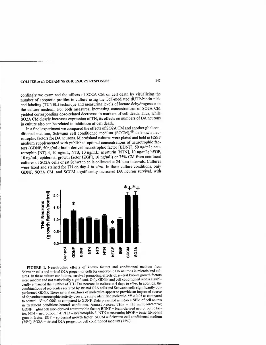
COLLIER et al.i DOPAMINERGIC INJURY RESPONSES 147
cordingly we examined the effects of S02A CM on cell death by visualizing the number of apoptotic profiles in culture using the TdT-mediated dUTP-biotin nick end labeling (TUNEL) technique and measuring levels of lactate dehydrogenase in the culture medium. For both measures, increasing concentrations of S02A CM yielded corresponding dose-related decreases in markers of cell death. Thus, while S02A CM clearly increases expression of TH, its effects on numbers of DA neurons in culture also can be related to inhibition of cell death.
In a final experiment we compared the effects of S02A CM and another glial con- ditioned medium, Schwann cell conditioned medium (SCCM),'*" to known neu- rotrophic factors for DA neurons. Microisland cultures were plated and held in HSSF medium supplemented with published optimal concentrations of neurotrophic fac- tors (GDNF, 50ng/mL; brain-derived neurotrophic factor [BDNF], 50 ng/mL; neu- rotrophin [NT]-4, 10 ng/mL; NT3, 10 ng/mL; neurturin [NTN], 10 ng/mL; bFGF, 10 ng/mL; epidermal growth factor [EOF], 10 ng/mL) or 75% CM fi-om confluent cultures of S02A cells or rat Schwann cells collected at 24-hour intervals. Cultures were fixed and stained for TH on day 4 in vitro. In these culture conditions, only GDNF, S02A CM, and SCCM significantly increased DA neuron survival, with
*j.* + '*=+ 2 2.0
go i^ 1.5 3 O
t £ 1.0
H O 0.5
8zZI-Hflooge5 roozzziLiuoO o O to .a w CO o
FIGURE 1. Neurotrophic effects of known factors and conditioned medium from Schwann cells and striatal 02A progenitor cells for embryonic DA neurons in microisland cul- tures. In these culture conditions, survival-promoting effects of several known growth factors were modest and not statistically significant. Only GDNF and cell conditioned media signifi- cantly enhanced the number of TH+ DA neurons in culture at 4 days in vitro. In addition, the combinations of molecules secreted by striatal 02A cells and Schwann cells significantly out- performed GDNF. These natural mixtures of molecules appear to provide an improved source of dopamine neurotrophic activity over any single identified molecule. *P < 0.05 as compared to control. +P < 0.0001 as compared to GDNF. Data presented is mean + SEM of cell counts in treatment conditions/control conditions. ABBREVIATIONS: THir = TH immunoreactive; GDNF = glial cell line-derived neurotrophic factor; BDNF = brain-derived neurotrophic fac- tor; NT4 = neurotrophin 4; NT3 = neurotrophin 3; NTN = neurturin; bFGF = basic fibroblast growth factor; EGF = epidermal growth factor; SCCM = Schwann cell condifioned medium (75%); S02A = striatal 02A progenitor cell conditioned medium (75%).

148 ANNALS NEW YORK ACADEMY OF SCIENCES
S02A CM and SCCM exhibiting significantly better effects than GDNF (FIG. 1). bPGF had a significant detrimental effect on the number of TH+ neurons. To our knowledge this is the only example of such a comparison of the effects of DA neu- rotrophic factors in microisland cultures, and it is an example of the impact of culture conditions on experimental outcome measures. As discussed earlier, potential surviv- al-promoting factors often are screened purposely in suboptimal culture conditions that favor detection of a positive effect. However, such conditions may exaggerate the biological significance of a given outcome. It is of interest that with microisland cul- tures, a condition in which the cultured cells are maintained in closer physical prox- imity to one another, effects of many of the identified neurotrophic molecules for DA neurons were modest and did not reach statistical significance. Of these, only GDNF exerted a significant effect on the number of DA neurons and was significantly out- performed by the natural combinations of molecules secreted by cultured S02A and Schwann cells. This comparison suggests that these cell-derived mixtures are more potent than any individual molecule and may be of significant therapeutic interest.
CONCLUSIONS
In this brief review we have summarized the main issues related to the use of cell culture systems to study DA neurons and have provided examples of the use of cul- ture models to examine mechanisms of DA neuron degeneration and to identify po- tential therapeutics. It is reasonable to ask whether cell culture findings effectively translate into valuable approaches in vivo. For the examples provided here, cell cul- ture findings have been predictive of results in animal models of PD and in some cas- es have been studied for relevance in the disease itself The response of cultured DA neurons to neurotoxins supports the important roles of oxidative stress and impaired mitochondrial energy metabolism. These continue to be important themes in the de- velopment of therapeutic approaches, although simple use of the antioxidant toco- pherol has proved ineffective in PD.'"''*^ The identification of activated microglia as active participants in DA neuron degeneration, as suggested by culture findings, has been confirmed in animal models of FD^^'^"* and generated interest in clinical appli- cation of antiinflammatory drugs for PD. For the neuroprotective strategies, prami- pexole has been shown to diminish DA neuron death in an animal model of PD^^ and has demonstrated efficacy as a monotherapy in mild-to-moderate PD.'*^ Finally, neu- rotrophic activity derived from S02A cells or Schwann cells has been shown to aug- ment survival and function of transplanted embryonic DA neurons in a rat model of p]r)47-49 ^j^j jp stimulate TH expression in adult DA neurons of rats and mon- j-gyg 50,51 jims^ the unique access to the DA neuron provided by cell culture systems serves as an important setting for analyzing cellular mechanisms, as well as a valu- able prelude to preclinical animal studies and clinical trials.
ACKNOWLEDGMENTS
The authors are grateful for the technical contributions of Brian Daley, Matthew Fleming, Michelle Gartland, and Mark Gallagher. This work was supported by Na-
COLLIER et al.: DOPAMINERGIC INJURY RESPONSES 149
tional Institutes of Health Grants AG08133, NS24032, AG10851, AG00844, and Department of Defense Grant DAMD 17-99-1-9543.
REFERENCES
1. ENGELE, J. & M.C. BoHN. 1991. The neurotrophic effects of fibroblast growth factors on dopaniinergic neurons in vitro are mediated by mesencephalic glia. J. Neurosci. 11: 3070-3078.
2. O'MALLEY, E.K., I.E. BLACK & C.F. DREYFUS. 1991. Local support cells promote sur- vival of substantia nigra dopaminergic neurons in culture. Exp. Neurol. 112: 40-48.
3. TAKESHIMA, T., J.M JOHNSTON & J.W. COMMISSIONG. 1994. Mesencephalic type 1 astrocytes rescue dopaminergic neurons from death induced by serum deprivation. J. Neurosci. 14: 4769-4779.
4. DAL TOSO, R., O. GIORGI, C. SORANZO, et al. 1988. Development and survival of neu- rons in dissociated fetal mesencphalic serum-free cell cultures: I. Effects of cell den- sity and of an adult mammalian striatal-derived neurontrophic factor (SDNF). J. Neurosci. 8: 733-745.
5. TAKESHIMA, T., K. SHIMODA, J.M. JOHNSTON, et al. 1996. Standardized methods to bioas- say neurotrophic factors for dopaminergic neurons. J. Neurosci Methods 67: 27-41.
6. HAVFLICK, L. 1965. The limited in vitro lifetime of human diploid cell strains. Exp. ' Cell Res. 37: 614-636.
7. MATHON, N.F., D.S. MALCOLM, M.C. HARRISINGH, et al. 2001. Lack of replicative senescence in normal rodent glia. Science 291: 872-875.
8. TANG, D.G., Y.M. TOKUMOTO, J.A. APPERLY, et al. 2001. Lack of replicative senes- cence in cultured rat oligodendrocyte precursor cells. Science 291: 868-871.
9. JENNER, R 1998. Oxidative mechanisms in nigral cell death in Parkinson's disease. Mov. Disord. 13: 24-34.
10. OLANOW, C.W. 1992. An introduction to the free radical hypothesis in Parkinson's dis- ease. Ann. Neurol. 32: S2-S9.
11. LOTHARIUS, J., L.L. DUGAN & K. L. O'MALLEY. 1999. Distinct mechanisms underlie neurotoxin-mediated cell death in cultured dopaminergic neurons. J. Neurosci. 19: 1284-1293.
12. CHOI, W.S., S.Y. YOON, T.H. OH, et al. 1999. Two distinct mechanisms are mvolved m 6-h'ydroxydopamine- and MPP+-induced dopaminergic neuronal cell death: role of caspases, ROS, and INK. J. Neurosci. Res. 57: 86-94.
13. OH, J.H., W.S. CHOI, J.E. KIM, et al. 1998. Overexpression of HA-Bax but not Bcl-2 or Bcl-XL attenutates 6-hydroxydopamine-induced neuronal apoptosis. Exp. Neurol. 154: 193-198.
14. MCGEER, P.L., S. ITAGAKI, B.E. BOYES, et al. 1988. Reactive microglia positive for HLA-DA in the substantia nigra of Parkinson's and Alzheimer's disease brain. Neu- rology 38: 1285-1291.
15. HiRSCH, E.C., S. HUNOT, R DAMIER, et al. 1998. Glia cells and inflammation in Parkin- son's disease: a role in neurodegeneration. Ann. Neurol. 44: SI 15-S120.
16. LANGSTON, J.W., L.S. FORNO, J. TETRUD, et al. 1999. Evidence of active nerve cell degeneration in the substantia nigra of humans years after I-methyl-4-phenyl- 1,2,3,6-tetrahydropyridine exposure. Ann. Neurol. 46: 598-605.
17. MOGI, M., M. HARADA, H. NARABAYASHI, et al. 1996. Interleukin (IL)-l beta, IL-2, IL- 4, IL-6 and transforming growth factor-alpha levels are elevated in ventriucular cere- brospinal fluid in juvenile parkinsonism and Parkinson's disease. Neurosci. Lett. 211: 13-16.
18. MOGI, M., M. HARADA, P. RIEDERER, et al. 1994. Tumor necrosis factor-alpha (TNF- alpha) increases both in the brain and in the cerebrospinal fluid from parkinsonian patients. Neurosci. Lett. 165: 208-210.
19. LE, W.D., D.B. ROWE, J. JANKOVIC, et al. 1999. Effects of cerebrospinal fluid from patiens with Parkinson disease on dopaminergic cells. Arch. Neurol. 56: 194-200.
20. LE, W.D., D. ROWE, W. XIE, et al. 2001. Microglial activation and dopaminergic cell injury: an in vitro model relevant to Parkinson's disease. J. Neurosci. 21: 8447-8455.

150 ANNALS NEW YORK ACADEMY OF SCIENCES
21. COLLIER, T.J. & C.E. SORTWELL. 1999. Therapeutic potential of nerve growth factors in Parkinson's disease. Drugs Aging 14: 261-287.
22. PiERCEY, M.F., W.E. HOFFMAN, M.W. SMITH, et al. 1996. Inhibition of dopamine neu- ron firing by pramipexole, a dopamine D3 receptor-preferring agonist: comparison to other dopamine receptor agonists. Eur. J. Pharmacol. 312: 35-44.
23. CARTER, A.J. & R.E. MULLER. 1991. Pramipexole, a dopamine D2 autoreceptor ago- nist, decreases the extracellular concentration of dopamine in vivo. Eur. J. Pharma- col. 200: 65-72.
24. PIERCEY, M.F. 1998. Pharmacology of pramipexole, a dopamine D3-preferring agonist useful in treating Parkinson's disease. Clin. Neuropharmacol. 21: 141-151.
25. ALEXANDER, T., C.E. SORTWELL, CD. SLADEK, et al. 1997. Comparison of neurotoxic- ity following repeated administration of L-DOPA, D-DOPA, and dopamine to embryonic mesencephalic dopamine neurons in cultures derived from Fisher 344 and Sprague-dawley donors. Cell Transplant. 6: 309-315.
26. Ziv, I., R. ZILKHA-FALB, D. OFFEN, et al. 1997. Levodopa induces apoptosis in cultured neuronal cells—a possible accelerator of nigrostriatal degeneration in Parkinson's disease? Mov. Disord. 12: 17-23.
27. CARVEY, P.M., S. FIERI & Z.D. LING. 1997. Attenuation of levodopa-induced toxicity in mesencephalic cultures by pramipexole. J. Neural Transm. 104: 209-228.
28. LING, Z.D., H.C. ROBIE, C.W. TONG, et al. 1999. Both the antioxidant and D3 agonist actions of pramipexole mediate its neuroprotective actions in mesencephalic cul- tures. J. Pharmacol. Exp. Ther. 289: 202-210.
29. CARVEY, P.M., S.O. MCGUIRE & Z.D. LING. 2001. Neuroprotective effects of D3 dopamine receptor agonists. Parkinsonism Relat. Disord. 7: 213-223.
30. PROCIIIANTZ, A., U. Di PORZIO, A. KATO, et al. 1979. In vitro maturation of mesen- cephalic dopaminergic neurons from mouse embryos is enhanced in the presence of their striatal target cells. Proe. Natl. Acad. Sci. USA 76: 5387-5391.
31. DAGUET, M.-C, U. DIPORZIO, A. PROCHIANTZ, et al. 1980. Release of dopamine from dissociated mesencephalic dopaminergic neurons in primary cultures in absence or presence of striatal target cells. Brain Res. 191: 564-568.
32. HEMMENDINGER, L.M., B.B. GARBER, P.C. HOFFMAN, et al. 1981. Target neuron-spe- cific process formation by embryonic mesencepahlic dopamine neurons in vitro. Proc. Natl., Acad. Sci. USA 78: 1264-1268.
33. HOFFMAN, P.C, L.M. HEMMENDINGER, C KOTAKE, et al. 1983. Enhanced dopamine cell survival in reaggregates containing telencephalic targets. Brain Res. 274: 275-281.
34. TOMOZAWA, Y. & S.H. APPEL. 1986. Soluble striatal extracts enhance development of mesencephalic dopaminergic neurons in vitro. Brain Res. 399: 111-124.
35. DONG, J.F., A. DETTA, M.H.M. BAKKER, et al. 1993. Direct interaction with target- derived glia enhances survival but not differentiation of human fetal mesencephalic dopaminergic neurons. Neuroscience 56: 53-60.
36. SORTWELL, C.E., T.J. COLLIER & J.R. SLADEK, JR. 1997. Co-grafted embryonic striatum increases the survival of grafted embryonic dopamine neurons. J. Comp. Neurol. 399: 530-540.
37. TAKESHIMA, T., J.M. JOHNSTON & J.W. COMMISSIONG. 1994. Oligodendrocyte-type-2 astrocyte (02A) progenitors increase the survival of rat mesencephalic, dopaminer- gic neurons from death induced by serum deprivation. Neurosci. Lett. 166: 178-182.
38. SORTWELL, C.E., B.F. DALEY, M.R. PITZER, et al. 2000. Oligodendroeyte-type 2 astro- cytc-derived trophic factors increase survival of developing dopamine neurons through the inhibition of apoptotic cell death. J. Comp. Neurol. 426: 143-153.
39. BOGLER, 0.,D. WREN, S.C BARNETT, et al. 1990. Cooperation between two growth fac- tors promotes extended self-renewal and inhibits differentiation of oligodendrocyte- type-2-astrocyte (02A) progenitor cells. Proc. Natl. Acad. Sci. USA 87: 6368-6372.
40. COLLIER, T.J., CD. SLADEK, M.J. GALLAGER, et al. 1990. Diffusible faetor(s) from adult rat sciatic nerve increases cell number and neurite outgrowth of cultured embryonic ventral mesencephalic tyrosine hydroxylase-positive neurons. J. Neuro- sci. Res. 27: 394-399.
41. THE PARKINSON STUDY GROUP. 1993. Effects of tocopherol and deprenyl on the pro- gression of disability in early Parkinson's disease. N. Engl. J. Med. 328: 176-183.
COLLIER et al: DOPAMINERGIC INJURY RESPONSES 151
42. SHOULSON, I. 1998. DATATOP; a decade of neuroprotective inquiry. Ann. Neurol. 44: S160-166.
43. Wu, D.C., V. JACKSON-LEWIS, M. VILA, et al. 2002. Blockade of microglial activation is'neuroprotective in the l-methyl-4-phenyl-l,2,3,6-tetrahydropyridine mouse model of Parkinson's disease. J. Neurosci. 22: 1763-1771.
44. HE, Y., W.-D. LE & S.H. APPEL. 2002. Role of Fc-gamma receptors in nigral cell injury induced by Parkinson disease immunoglobulin injection into mouse substantia nigra. Exp. Neurol. 176: 322-327.
45. Vu, T.Q., Z.D. LING, S.Y. MA, et a!. 2000. Pramipexole attenuates the dopammergic cell loss induced by intraventricular 6-hydroxydopamine. J. Neural Trans. 107: 159- 176.
46. SHANNON, K.M., J.RJ. BENNETT & J.H. FRIEDMAN. 1997. Efficacy of pramipexole, a novel dopamine agonist, as monotherapy in mild to moderate Parkinson's disease. Neurol. 49: 724-728.
47. VAN HORNE, C.G., L STROMBERG, D. YOUNG, et al. 1991. Functional enhancement of intrastriatal dopamine-containing grafts by the co-transplantation of sciatic nerve tis- sue in 6-hydroxydopamine-lesioned rats. Exp. Neurol. 113: 143-154.
48. COLLIER, T.J. & J.E. SPRINGER. 1991. Co-grafts of embryonic dopamine neurons and adult sciatic nerve into the denervated striatum enhance behavioral and morphologi- cal recovery in rats. Exper. Neurol. 114: 343-350.
49. SORTWELL, C.E., B.F. DALEY & T.J. COLLIER. 1998. Time course of apoptotic cell death in Mesencephalic suspension grafts: early trophic influence of 0-2A progenitor- enriched Striatal co-grafts. Soc. Neurosci. Abstr. 421.7, 24: 1056.
50. COLLIER, T.J. & P.N. MARTIN. 1993. Schwann cells as a source of neurotrophic activity for dopamine neurons. Exp. Neruol. 124: 129-133.
51. COLLIER, T.J., J.D. ELSWORTH, J.R. TAYLOR, et al. 1994. Peripheral nerve-dopamine neuron co-grafts in MPTP-treated monkeys: augmentation of tyrosine hydroxylase- positive fiber staining and dopamine content in host systems. Neurosci. 61: 875-889.
Convergent Pathobiologic Model of Parkinson's Disease
KATHLEEN A. MAGUIRE-ZEISS AND HOWARD J. FEDEROFF
Department of Neurology and the Center for Aging and Developmental Biology, University of Rochester School of Medicine and Dentistry, Rochester, New York 14642, USA
ABSTRACT: The etiology of Parkinson's disease (PD) has yet to be delineated. Human genetic studies as well as neurotoxicant and transgenic animal models of PD suggest that multiple events trigger the initiation of this progressive age- related neurodegenerative disorder. In addition, we propose that despite dis- parate disease triggers a convergent pathobiologic pathway exists leading to cell death. The common pathway model posits that both familial and sporadic forms of Parkinson's disease obligately share a common pathophysiological substrate. Herein we discuss the evidence for a common pathway model of Par- kinson's disease through a review of synuclein transgenic models and outline an approach for the idenfication of shared therapeutic targets. We end with a discussion of a potential alternative therapy for Parkinson's disease.
KEYWORDS: Parkinson's disease; synuclein; single-chain antibodies; transgenic animals
The etiologic basis of Parkinson's disease (PD), the second most prevalent neurode- generative disorder, is unknown. Several lines of evidence point to both genetic and environmental components in the development of this disease.'"'^ Originally de- scribed by James Parkinson as "shaking palsy," PD is typified by motoric dysftinc- tion resulting in both tremor and rigidity.'^ Neuropathologically, PD cases manifest intracytoplasmic inclusions known as Lewy bodies, gliosis, and neuronal loss spe- cific to the nigrostriatal system. Lewy bodies are composed of aggregated proteins, most notably a-synuclein and ubiquitin. a-Synuclein has received considerable at- tention in etiopathogensis following the description of several kindreds in which dominantly inherited mutations in the human gene result in early-onset PD.^""-^^ However, these familial forms account for a very small percentage of the total PD cases. Environmental factors including pesticides, herbicides, and industrial chemi- cals have also been flagged as potential risk factors for pD.i i.'4-16,26,27 Together, ge- netic and environmental factors may act synergistically to initiate a series of cellular events leading to the parkinsonian syndrome.
Address for correspondence: Howard J. Federoff, M.D,, Ph.D., Professor of Neurology, Center for Aging and Developmental Biology, Box 645, University of Rochester School of Medicine and Dentistry, 601 Elmwood Ave., Rochester, NY 14642. Voice: 716-273-2190; fax: 716-442- 6646.
Ann. N.Y. Acad. Sci. 991: 152-166 (2003). © 2003 New York Academy of Sciences.
152
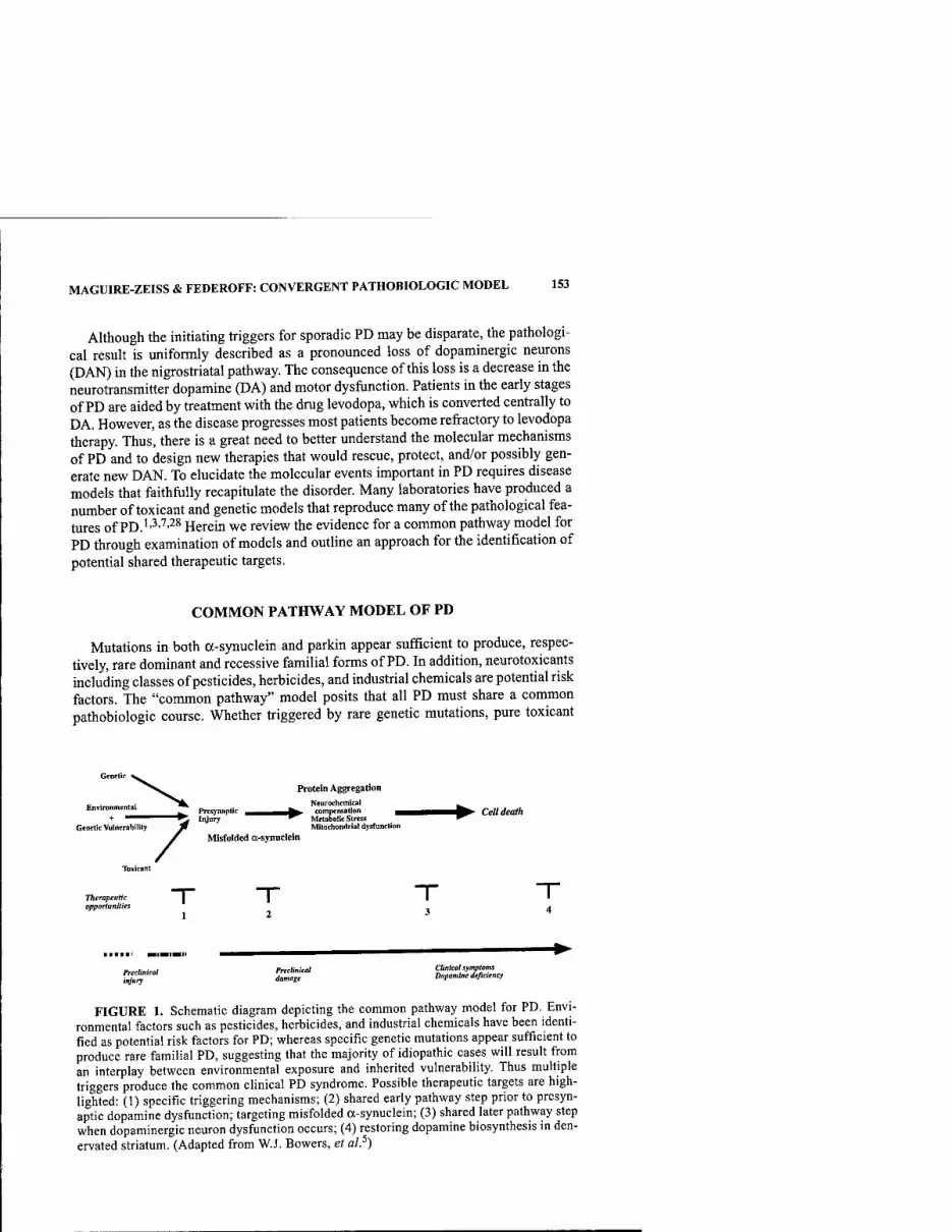
MAGUIRE-ZEISS & FEDEROFF: CONVERGENT PATHOBIOLOGIC MODEL 153
Although the initiating triggers for sporadic PD may be disparate, the pathologi- cal result is uniformly described as a pronounced loss of dopaminergic neurons (DAN) in the nigrostriatal pathway. The consequence of this loss is a decrease in the neurotransmitter dopamine (DA) and motor dysfunction. Patients in the early stages of PD are aided by treatment with the drug levodopa, which is converted centrally to DA. However, as the disease progresses most patients become refractory to levodopa therapy. Thus, there is a great need to better understand the molecular mechanisms of PD and to design new therapies that would rescue, protect, and/or possibly gen- erate new DAN. To elucidate the molecular events important in PD requires disease models that faithfully recapitulate the disorder. Many laboratories have produced a number of toxicant and genetic models that reproduce many of the pathological fea- tures of PD.''^'''^^ Herein we review the evidence for a common pathway model for PD through examination of models and outline an approach for the identification of potential shared therapeutic targets.
COMMON PATHWAY MODEL OF PD
Mutations in both a-synuclein and parkin appear sufficient to produce, respec- tively, rare dominant and recessive familial forms of PD. In addition, neurotoxicants including classes of pesticides, herbicides, and industrial chemicals are potential risk factors. The "common pathway" model posits that all PD must share a common pathobiologic course. Whether triggered by rare genetic mutations, pure toxicant
Protein Aggregation
Environmental ^ Prcsynaplic ^^,^_W compensation ^^H^K^^ Cett death + "^^"""^^^^ Iiyury ^^ Metabolic Stress ^
Genetic Vuinerabiiity Jf Mitochondria! dysfunction Misfolded a-synuclein
/
Therapeutic opportunities
Preclinical Preclitiical Oitiicohymptoms Z]ur) *""'«'• Vopammedefieumy
FIGURE 1. Schematic diagram depicting the common pathway model for PD. Envi- ronmental factors such as pesticides, herbicides, and industrial chemicals have been identi- fied as potential risk factors for PD; whereas specific genetic mutations appear sufficient to produce rare familial PD, suggesting that the majority of idiopathic cases will result from an interplay between environmental exposure and inherited vulnerability. Thus multiple triggers produce the common clinical PD syndrome. Possible therapeutic targets are high- lighted: (1) specific triggering mechanisms; (2) shared early pathway step prior to presyn- aptic dopamine dysfunction; targeting misfolded a-synuclein; (3) shared later pathway step when dopaminergic neuron dysfijnction occurs; (4) restoring dopamine biosynthesis in den- ervated striatum. (Adapted from W.J. Bowers, et al.^)

154 ANNALS NEW YORK ACADEMY OF SCIENCES
forms, or interactions between environmental toxicant and genetic vulnerability, all PD is postulated to be mechanistically convergent (FIG. 1 and Ref. 5). These multiple initiating mechanisms trigger cellular events, the first step of which results in pre- synaptic injury and dysfunction of the nigrostriatal tract. We surmise that presynap- tic injury is clinically silent. Presynaptic injury is envisioned to stimulate cellular compensation, during which we predict that intracellular proteins such as a-synucle- in initiate pathologic folding and consequent cell injury, perhaps by compromising proteosomal function. Progressive injury wrought by impaired disposal of possibly toxic abnormally folded and oxidized proteins are believed to evolve over years. As compensatory responses fail, neurochemical dysfunction (e.g., diminished DA re- lease, uptake, and vesicular loading) are attended by bioenergetic failure, marked re- active oxygen species (ROS) generation, and finally cell death.
Clinical and pathological evidence support this common pathway model indepen- dently of the triggering mechanism. First, all PD patients have a selective and profound loss of ventral midbrain dopamine neurons. Second, the invariant loss of DA-releasing nigrostriatal presynaptic terminals precedes cell body loss in the substantia nigra.^^"^^ Third, loss of these dopaminergic projection neurons results in the cardinal PD clinical features: bradykinesia, akinesia, tremor, rigidity, and postural reflex impairment (re- viewed in Ref 33). Fourth, neuropathology reveals the presence of a-synuclein and ubiquitin containing Lewy bodies.^"^"^^ Finally, systemic absorption of exogenous neurotoxicants best exemplified by MPTP induce in mouse, monkey, and man a clini- cal, biochemical, and pathological parkinsonian syndrome.'^'''^''*' Taken together, these data indicate that disparate triggers elicit a cellular, biochemical, and presumably molecular response that results in the familiar parkinsonian syndrome.
Additional support for a convergent model of PD and the role of a-synuclein de- rives from recently developed animal models of familial a-synuclein mutations. Al- though rare, mutations in the a-synuclein gene cause intracytoplasmic a-synuclein inclusions and early-onset disease.-^^^^^ These autosomal-dominant cases directly im- plicate specific coding region mutations (A30P, A53T) in a-synuclein aggregation and/or degradation. Moreover, diminished protein processing is further supported by mutations in parkin and ubiquitin carboxy-terminal-hydrolase-LI (UCHLl) two pro- teins involved in ubquitin-dependent proteosomal function.' 0.20,42-52 Linj^agg of tox- icant injury to protein aggregation has been provided by recent evidence that MPTP sdmulates production and oxidative modification of a-synuclein.^^"^^
TRANSGENIC MODELS OF PD
In our common pathway model a-synuclein plays an important role in DAN-spe- cific dysfunction and death. To better appreciate its role in PD several laboratories are using transgenic technology to overexpress wild-type and mutant a-synuclein (see TABLE 1 and Refs. 1,3,4,7).
Transgenic mouse models that use promoters capable of widespread expression such as PDGF, prion, and Thyl generally result in increased protein accumuladon, motor signs, abnormal axons, and neurites in multiple brain regions.^^"^^ Transgenic mice employing the Thy 1.2 promoter to drive a-synuclein overexpression display swollen neurites in the cerebellar Purkinje layer, nucleus dentatus, hippocampus, neocortex, and brain stem.^^ A second Thyl :a-synuclein transgenic mouse demon-

MAGUIRE-ZEISS & FEDEROFF: CONVERGENT PATHOBIOLOGIC MODEL 155
TABLE 1. Comparison of a-synuclein transgenic animals
Transgenic model human a-synuclein Promoter
NT function
Protein accum.
Neuronal abnorm.
Motor dysf. Ref.
WT
WT A53T A3 OP
human PDGF-p
ratTH
ND
(-) (+)
(+) (+)
{-)
(+)
(-)
58
60
WT A30P
mouse Thy 1.2 ND (+) ND (+) 56
WT A53T
mouse Thyl ND (+) (+) (+) 59
WT ratTH (+) (-) (-) (+) 61
A53T/A30P ratTH (+) (-) (+) (+) 61
WT A53T A30P
prion ND (+) (+) (+) 57
ABBREVIATIONS: NT = neurotransmitter; accum. = accumulation; Neuronal abnorm. = neuronal abnormalities, such as swollen neurites; Motor dysf. = motor dysfunction, such as alterations in behavioral tests of motor function.
strated neuropathologic a-synuclein aggregates and inclusions in an equally wide range of brain regions but most notably not in the substantia nigra pars compacta.^^ In fact, this transgene fails to express in ventral midbrain dopaminergic neurons, suggesting that these mice may be a more appropriate model for diffuse Lewy body disease rather than for PD (personal communication and Ref 59). Cytoplasmic in- clusions and a-synuclein aggregates, but no fibrillar material, were also observed in the neocortex, hippocampus, and substantia nigra of transgenic mice using the hu- man PDGF-p promoter.^* When driven by the mouse prion promoter, transgene product accumulation in the midbrain, cerebellum, brain stem, and spinal cord was correlated with an earlier onset of disease and death.^'
To more faithfully recapitulate pathogenesis including neuroanatomical, neuro- chemical, and neuropathological changes in DAN only, two laboratories have devel- oped transgenic animal models in which the catecholaminergic promoter tyrosine hydroxylase drives a-synuclein expression. One group reported modest a-synuclein accumulation without inclusions or neurochemical change when wild-type, A53T, or A3 OP a-synuclein was overexpressed.^^ On the other hand, our laboratory engi- neered two TH-driven transgenic lines that overexpress wild-type (ha-syn) or dou- bly mutated (hm^a-syn) human a-synuclein. The doubly-mutated a-synuclein transgene contains both the A53T and A30P mutations within the same open reading frame^' (see FIG. 2, panel A). Transgenic animals were evaluated neuroanatomically, neurochemically, behaviorally, and neuropathologically.^' Overexpression of either a-synuclein resulted in an age-dependent phenotype that included a range of motoric dysfunction, abnormal neurites, and neurotransmitter alterations, but no Lewy body inclusions. We have shown that a-synuclein mRNA is highly expressed in the sub- stantia nigra (SN) in these animals by in situ hybridization analysis (FIG. 2, panel B).

156 ANNALS NEW YORK ACADEMY OF SCIENCES
GSSC C209A
B
.^■■.-^
Cerebral Cortex
r X t ^
Cerebral Cortex H
Dorsal Striatum Dorsal Striatum pH + —♦■
FIGURE 2. Generation and characterization of human a-synuclein transgenic mice. (A) Map of the human double mutant a-synuclein construct used for engineering the trans- genic mice. (B) a-Synuclein niRNA Expression. Image of ;>? situ hybridization from hm^a- SYN at the level of the SN. (C-E) Immunohistochemistry using a human specific polyclonal antibody to a-synuclein (C,D) in hm^a-SYN line and (E) in nontransgenic littermates. This antibody detected only human a-synuclein protein but not mouse a-synuclein. Human a- synuclein was most highly expressed in the cell bodies and dendrites of the SN and VTA, their axons, and in the striatum. (F-H) Striatal protein was subjected to 2-D PAGE followed by Western blotting using a polyclonal Ab to a-syn that recognizes both mouse and human a-syn. (F) Nontransgenic littermate controls demonstrated a larger single spot migrating at approximately 15 kDa and a much smaller spot (arrow) migrating at a slightly smaller MW and at a slightly more basic pi suggestive of a dephosphorylated form of mouse a-syn. Lines hm^a-syn-39 (G) and hwa-syn-5 (H) demonstrated unique spots migrating at a similar MW to mouse a-syn, but at different and slightly more acidic pis. (Reproduced from E.K. Rich- field, et o/.^' with permission.)
Both ha-syn and hm-^a-syn express high levels of a-synuclein protein in midbrain DAN cell bodies and their striatal projections^' (FIG. 2, panels C,D). a-Synuclein colocalized with TH in cell bodies and dendrites of the SN, ventral tegmental area, locus ceruleus; and projection fields in the dorsal striatum, nucleus accumbens, ol- factory tubercle, and cerebral cortex. The subcellular distribution of a-synuclein is both nuclear and cytoplasmic compared to TH which is strictly cytoplasmic^' (FIG.
3, panels D-F). Whether a-synuclein nuclear localization identifies a previously un- disclosed action remains to be determined. Of interest, hm^a-syn axons in the me- dian forebrain bundle were pathologically dilated and beaded, resembling Lewy

MAGUIRE-ZEISS & FEDEROFF: CONVERGENT PATHOBIOLOGIC MODEL 157
3[^^^j|^y^-£^iS&ES
^^^^ ^S«^i^ ̂ ^pw#!f^ii®#li§'^
FIGURE 3. Colocalization of ha-SYN protein and abnormal axons and neurites in young ha-SYN transgenic mice. Double-label fluorescent IHC for TH (A,D,G), ha-SYN (B,E,H), both ha-SYN and TH (C,F,I) in a sagittal section from hm^a-SYN (A-C), a hwa- SYN SN neuron (D-F), or from hm^a-SYN median forebrain bundle (MFB, G-I). Confocal microscopy was used to image sections at 0.4-|im thickness using either the red or green fil- ter (D-I) or routine fluorescent microscopy at 1.25x (A-C). The ha-SYN protein was present in dopaminergic cell bodies and dendrites (B and E) and nigrostriatal axons and ter- minals (B and H) in a distribution that matched that of TH (A,D,G). The ha-SYN protein was present in both the cytoplasm and the nucleus of neurons (E). Abnormal neurites were

158 ANNALS NEW YORK ACADEMY OF SCIENCES
neurites. However, no Lewy body-like inclusions were appreciated^' (FIG. 3, panels J-M). Behavioral studies of these animals demonstrated an age-dependent decline in spontaneous activity and motor function, thus paralleling some of the clinical hall- marks of PD^' (data not shown).
Why do different transgenic lines exhibit varying degrees of neuropathology? Our hm^a-syn model may represent a slower progress and possibly a more reflective model of the human disease, possibly because of TH promoter use. Most interesting- ly, our mice support the notion that the toxicity of a-synuclein does not require Lewy body formation but rather another, misfolded, protein structural state. This observa- tion reinforces the concept of pathogenic stages as articulated in a common pathway model.
a-Synuclein transgenic animals are aiding in the identification of molecular mechanisms of PD pathogenesis, particularly the identification of cellular factors that regulate the aggregation and toxicity of a-synuclein. Although no model fully recapitulates all aspects of PD, the models appear to have great utility in addressing interactions between candidate neurotoxicants and a-synuclein itself Indeed, our laboratory has shown that ha-syn and hm^a-syn mice manifest enhanced sensitivity to MPTP treatment. When treated, the transgenic mice exhibited markedly decreased locomotor activity and DAT density compared with nontransgenic littermates.^'
To unravel the molecular basis of our staged convergent pathobiologic model we have embarked on an integrated and comparative bionomics analysis of striatum and substantia nigra in mouse models (a-synuclein transgenics and MPTP treatment) and human PD. This bipartite bionomics approach uses conventional high-density oligonucleotide microarray technology (Affymetrix) and a three-dimensional pro- teomics methodology to identify gene/gene product changes. Proteomics is accom- plished by 2D-gel electrophoresis, differential spot detection, recovery, and digestion, followed by SELDI-TOF mass spectroscopy.
To discriminate pathophysiologically important changes we assert that the iden- tified molecule must be expressed in the nigrostriatal pathway. Second, the molecule must be misregulated by neurotoxicant treatment and/or transgene expression in a temporally predictable manner. This combination of transcriptomic and proteomic methods will undoubtedly identify genes and gene products whose abundance is al- tered in PD and its models. Preliminary studies from the 2D-gel analysis followed by limited computer analysis demonstrates that five proteins are increased in hm^a- syn striatum, while four proteins are reduced compared to nontransgenic littermates (TABLE 2). Likewise five proteins were decreased in hm^a-syn substantia nigra
present in dendrites of SN and VTA neurons and axons to the striatum (G-I) and elsewhere. Fluorescent IHC for TH was performed in a nontransgenic littermate control mouse (J and K) or a hm^a-SYN mouse (L and M) in the MFB (J and L) or the nucleus accumbens (K and M). Confocal microscopy using a lOOx objective at 0.5-|J.m steps was performed and a total of 10 sections stacked to make the final image. TH is normally present in MFB axons of control mice in a discontinuous manner, with the majority of axons having a uniform di- ameter with occasional modest dilations (J). In hm^a-SYN mice TH axons were more bead- ed and dilated in appearance, with more discontinuities (L). In the Aeb, TH was present in smaller caliber processes and terminals of control mice (K). In hm^a-SYN mice the smaller caliber processes and terminals were more dilated and punctate (M). (Reproduced from E.K. Richfield, et al.^^ with permission.)
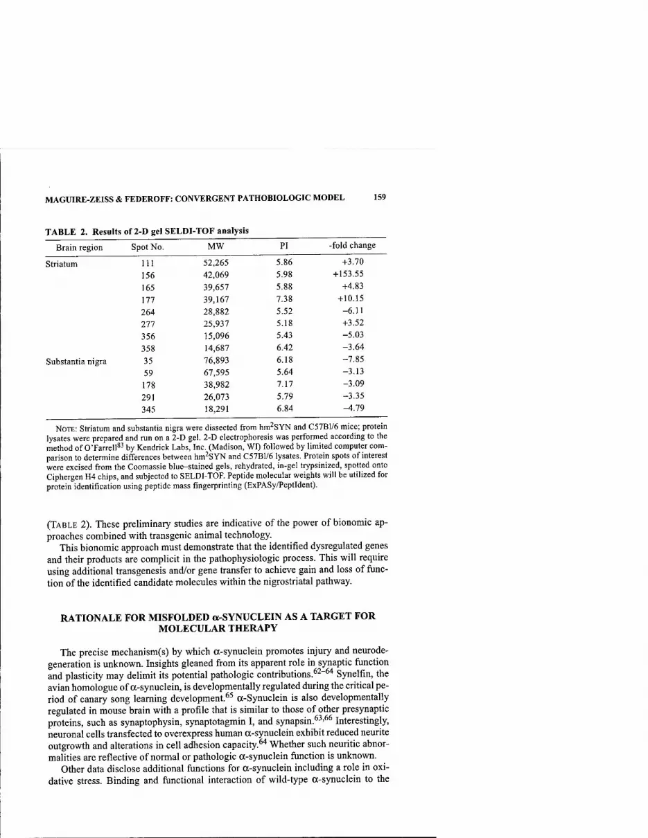
MAGUIRE-ZEISS & FEDEROFF: CONVERGENT PATHOBIOLOGIC MODEL 159
TABLE 2. Results of 2-D gel SELDI-TOF analysis
Brain region Spot No. MW PI -fold change
Striatum Ill 52,265 5.86 +3.70
156 42,069 5.98 +153.55
165 39,657 5.88 +4.83
177 39,167 7.38 + 10.15
264 28,882 5.52 -6.11
277 25,937 5.18 +3.52
356 15,096 5.43 -5.03
358 14,687 6.42 -3.64
Substantia nigra 35 76,893 6.18 -7.85
59 67,595 5.64 -3.13
178 38,982 7.17 -3.09
291 26,073 5.79 -3.35
345 18,291 6.84 -4.79
NOTE: Striatum and substantia nigra were dissected from hm^SYN and C57B1/6 mice; protein lysates were prepared and run on a 2-D gel. 2-D electrophoresis was performed according to the method of O'Farrell*^ by Kendrick Labs, Inc. (Madison, WI) followed by limited computer com- parison to determine differences between hm^SYN and C57B1/6 lysates. Protein spots of interest were excised from the Coomassie blue-stained gels, rehydrated, in-gel trypsinized, spotted onto Ciphergen H4 chips, and subjected to SELDI-TOF. Peptide molecular weights will be utilized for protein identification using peptide mass fingerprinting (ExPASy/Peptldent).
(TABLE 2). These preliminary studies are indicative of the power of bionomic ap- proaches combined with transgenic animal technology.
This bionomic approach must demonstrate that the identified dysregulated genes and their products are complicit in the pathophysiologic process. This will require using additional transgenesis and/or gene transfer to achieve gain and loss of func- tion of the identified candidate molecules within the nigrostriatal pathway.
RATIONALE FOR MISFOLDED a-SYNUCLEIN AS A TARGET FOR MOLECULAR THERAPY
The precise mechanism(s) by which a-synuclein promotes injury and neurode- generation is unknown. Insights gleaned from its apparent role in synaptic function and plasticity may delimit its potential pathologic contributions.^^"^'* Synelfin, the avian homologue of a-synuclein, is developmentally regulated during the critical pe- riod of canary song learning development.^^ a-Synuclein is also developmentally regulated in mouse brain with a profile that is similar to those of other presynaptic proteins, such as synaptophysin, synaptotagmin I, and synapsin.^^'^^ Interestingly, neuronal cells transfected to overexpress human a-synuclein exhibit reduced neurite outgrowth and alterations in cell adhesion capacity.^'' Whether such neuritic abnor- malities are reflective of normal or pathologic a-synuclein function is unknown.
Other data disclose additional functions for a-synuclein including a role in oxi- dative stress. Binding and functional interaction of wild-type a-synuclein to the
160 ANNALS NEW YORK ACADEMY OF SCIENCES
dopamine transporter (DAT) elicits membrane clustering of DAT, acceleration of DA uptake, and apoptosis.^^ This a-synuclein and DAT interaction could exacerbate tox- icant-induced apoptosis. Inducible expression of wild-type and mutant a-synuclein (G209A) levels in HEK293 cells did not induce cell death, but toxic synergism be- tween G209A mutant a-synuclein and dopamine was found.^^ Reports of DA-in- duced toxicity and cell death presumably results from the inherent reactivity of the neurotransmitter.^^'^" DA is readily oxidized to DA quinone resulting in the release of superoxide and hydrogen peroxide.^"-'' Furthermore, DA can covalently modify cysteine, forming S-cysteinyl dopamine on affected proteins, irreversibly modifying protein functionJ-^ DA terminals actively degenerate following DA-induced toxicity, and DA-derived orthoquinone (DAQ) forms covalent adducts with a-synuclein re- sulting in long-lived protofibril intermediates7^''^ Taken together, these studies sug- gest that DA conspires with a-synuclein resulting in the generation of ROS and DA adduct formation on proteins leading to synaptic terminal dysfunction.
a-Synuclein misfolding is favored by mutation or oxidative damage and thus ap- pears to be a central feature of the variously triggered forms of PD. Consistent with the idea that accumulation of aberrantly folded protein is integral to pathogenesis are the other familial forms that are characterized by loss of function mutations in the parkin and UCH-Ll genes. Since both gene products participate in the ubiquitin pro- teasome, it is evident that clearance of aberrantly folded proteins is slowed in par- kinsonian dopaminergic neurons (reviewed in Ref 46). Moreover, a nexus between toxicants such as MPTP and a-synuclein misfolding has also been established by ex- periments demonstrating that oxidative stress promotes the formation of a-synuclein aggregates and inclusions (reviewed in Refs. 17,75). This is also observed with other toxicants such as paraquat and rotenone.^'^ A key question remains unanswered: which form(s) of a-synuclein compromise(s) neuronal function?
STEADY STATE a-SYNUCLEIN MODEL AND ALTERNATIVE THERAPIES FOR PD
We postulate that within DAN a steady state exists between the native a-synucle- in (random coil), the preprotofibrillar conformer (P-sheet), and the aggregated form (FIG. 4). In this scenario either oxidative stress or the presence of A53T a-synuclein drives native conformers towards toxic oligomers. As a molecular instigator of PD, a-synuclein is analogous to amyloid beta (A(}), which forms extracellular deposits and is a pathological hallmark of Alzheimer's disease.'^~^^ Intracellular protein de- posits are relevant to other neurological diseases as well, including amyotrophic lat- eral sclerosis and expanded polyglutamine disorders (reviewed in Ref 79).
Under physiological conditions a-synuclein exists in a native random coil con- formation and is normally targeted for degradation. Under conditions of oxidative stress, a-synuclein adopts an altered conformation (P-sheet; preprotofibrillar) that can serve as the nidus for pathophysiological seeding of toxic protein aggregates (preprotofibrils). The alteration in steady state towards a pathogenic p-sheet (pre- protofibrillar)-containing population may represent a locus for therapeutic interven- tion. Furthermore, mutant a-synuclein relatively favors the P-sheet conformation without oxidative stress. Based on this evidence we suggest that therapies specifical- ly designed to interact and reduce toxic a-synuclein conformers will attenuate dis-

MAGUIRE-ZEISS & FEDEROFF: CONVERGENT PATHOBIOLOGIC MODEL 161
(A) Native/Random Coil Pre-protofibrillar Aggregates
7> •^
(B) Oxidative Stress
Genetic Mutations A53T
. Targeted Degradation
(C) Oxidative Stress
Targeted Degradation
Genetic Mutations A53T
FIGURE 4. Schematic diagram of the dynamics between a-synuclein conformers. (A) Under normal physiological conditions a steady state exists between random coil, pre- protofibrillar, and aggregated a-synuclein conformers. (B) Under conditions of oxidative stress (neurotoxicant treatment) the steady state shifts toward the preprotofibrillar and ag- gregated a-synuclein conformers. Likewise, mutant a-synuclein (A53T) demonstrates a similar steady state shift. (C) ScFv's directed against conformer-specific forms of a-synu- clein will either deplete the available pool of native or preprotofibrillar a-synuclein or target misfolded a-synuclein for degradation decreasing toxicity.
ease progression. Thus, preventing the formation of toxic preprotofibrillar forms of a-synuclein should be a goal of future therapeutics.
Our laboratory is pursuing a gene therapy approach whereby conformation-spe- cific single-chain Fvs (scFvs) are expressed and intended to interfere with a-synu- clein preprotofibrillar formation. ScFvs are composed of the minimal antibody binding site formed by noncovalent association of the Vjj and VL variable domains joined by a flexible polypeptide linker. Human scFv-phage libraries are available and allow for high-affinity human scFv antibodies to be selected from combinatorial libraries.*'' Antibody engineering makes it possible to manipulate the genes encod- ing the scFvs, allowing for antibody binding site expression within mammalian cells. Genetically fusing the scFv to intracellular targeting signals allows for specific sub-

162 ANNALS NEW YORK ACADEMY OF SCIENCES
cellular expression.**' These intracellular antibodies, termed intrabodies, are capable of modulating target protein function. Intrabodies have been used for both phenotyp- ic and functional knockdown of target molecules. Perhaps most relevant to PD ther- apy through a-synuclein targeting is the recent work using human scFv intrabodies to disrupt huntingtin aggregation.^^ Huntingtin-specific scFvs bound to the N termi- nal polyglutamine residues and promoted solubility of what otherwise would have been aggregated protein complexes.
In PD we suggest that protein aggregates forming within DAN are seeded, require the preprotofibrillar conformers of a-synuclein (p-sheet), and are cytotoxic. Identi- fication of scFvs that will selectively recognize the preprotofibrillar conformers of a-synuclein is anticipated to attenuate protein toxicity (FIG. 4). In one case, scFvs could interact with a-synuclein prior to aggregation, thereby depleting the pool of available protein. In a second scenario scFvs bind to and deplete preprotofibrillar a- synuclein, thus attenuating aggregation. A third possibility is that scFvs interact with aggregated a-synuclein and efficiently target the complex for degradation. Each of these proposed strategies would decrease protein toxicity and potentially mitigate pathophysiologic damage.
SUMMARY
Transgenic models of a-synuclein overexpression will help to elucidate a-synu- clein's role in PD pathogenesis. We postulate that combined genetic vulnerability and neurotoxicant exposure will provide mechanisms to examine sporadic PD. Our convergent pathobiologic model stipulates that disparate insults initiate a disease process that obligately share a common pathway leading to cell death. We further posit that a-synuclein misfolding is a proximal shared earlier step in the pathway. This critical step is suggested to induce cellular compensation that ulfimately fails. The delineation of the transition from adaptive compensatory signaling to prodeath signaling affords the opportunity for therapeutic intervention. Our bionomic ap- proach is formulated to identify both upstream (triggering stage) and downstream (compensatory or prodeath) targets involved in the convergent pathobiologic model. Genomic and proteomic methodologies in concert with innovative biologic ap- proaches such as scFv gene transfer in transgenic/neurotoxicant models provide a basis to validate underlying pathogenic mechanisms and hold promise in the longer term for therapy.
ACKNOWLEDGMENT
This work was supported by NIH Grant R01-NS36420A to H.J.F.
REFERENCES
1. BEAL, M.F. 2001. Experimental models of Parkinson's disease. Nat. Rev. Neurosei. 2: 325-334.
2. BETARBET, R., et al. 2000. Chronic systemic pesticide exposure reproduces features of Parkinson's disease. Nat. Neurosei. 3: 1301-1306.

MAGUIRE-ZEISS & FEDEROFF: CONVERGENT PATHOBIOLOGIC MODEL 163
3. BETARBET, R., et al. 2002. Animal models of Parkinson's disease. Bioessays 24: 308-318. 4. BOWERS, W.J., et al. 1997. Gene therapeutic strategies for neuroprotection: implica-
tions for Parkinson's disease. Exp Neurol. 144: 58-68. 5. BOWERS, W.J., et al. 2001. Gene therapeutic approaches to the treatment of Parkinson's
disease. Clin. Neurosci. Res. 1: 483-495. 6. BROOKS, A.I., et al. 1999. Paraquat elicited neurobehavioral syndrome caused by
dopaminergic neuron loss. Brain Res. 823: 1-10. 7. DAWSON, T., et al. 2002. Animal models of PD: pieces of the same puzzle? Neuron 35:
219-222. 8. KowALL, N.W., et al. 2000. MPTP induces alpha-synuclein aggregation in the substan-
tia nigra of baboons. Neuroreport 11: 211-213. 9. MANNING-BOG, A.B., et al. 2002. The herbicide paraquat causes up-regulation and
aggregation of alpha-synuclein in mice: paraquat and alpha-synuclein. J. Biol. Chem. 277: 1641-1644.
10. MizuNO, Y., et al. 1999. Genetic and environmental factors in the pathogenesis of Par- kinson's disease. Adv. Neurol. 80: 171-179.
11. OLANOW, C.W. & W.G. TATTON. 1999. Etiology and pathogenesis of Parkinson's dis- ease. Annu. Rev. Neurosci. 22: 123-144.
12. SCHMIDT, N. & B. FERGER. 2001. Neurochemical findings in the MPTP model of Par- kinson's disease. J. Neural Transm. 108: 1263-1282.
13. SCHWARTING, R.K. & J.P. HUSTON. 1996. The unilateral 6-hydroxydopamine lesion model in behavioral brain research. Analysis of functional deficits, recovery and treatments. Prog. Neurobiol. 50: 275-331.
14. THIRUCHELVAM, M., et al. 2000. Potentiated and preferential effects of combined paraquat and maneb on nigrostriatal dopamine systems: environmental risk factors for Parkinson's disease? Brain Res. 873: 225-234.
15. THIRUCHELVAM, M., et al. 2000. The nigrostriatal dopaminergic system as a preferen- tial target of repeated exposures to combined paraquat and maneb: implications for Parkinson's disease. J. Neurosci. 20: 9207-9214.
16. THIRUCHELVAM, M., et al. 2002. Developmental exposure to the pesticides paraquat and maneb and the Parkinson's disease phenotype. Neurotoxicology 23: 621-633.
17. VILA, M., et al. 2000. Alpha-synuclein up-regulation in substantia nigra dopaminergic neurons following administration of the parkinsonian toxin MPTP. J. Neurochem. 74: 721-729.
18. STEECE-COLLIER, K., et al. 2002. Etiology of Parkinson's disease: genetics and envi- ronment revisited. Proc. Natl. Acad. Sci. USA 99: 13972-13974.
19. PARKINSON, J. 1817. An Essay on the Shaking Palsy. Sherwood, Neely, and Jones. London.
20. KITADA, T., et al. 1998. Mutations in the parkin gene cause autosomal recessive juve- nile parkinsonism. Nature 392: 605-608.
21. KRUGER, R., et al. 1998. Ala30Pro mutation in the gene encoding alpha-synuclein in Parkinson's disease. Nat. Genet. 18: 106-108.
22. POLYMEROPOULOS, M.H., et al. 1996. Mapping of a gene for Parkinson's disease to chromosome 4q21-q23. Science 274: 1197-1199.
23. POLYMEROPOULOS, M.H., et al. 1997. Mutation in the alpha-synuclein gene identified in families with Parkinson's disease. Science 276: 2045-2047.
24. POLYMEROPOULOS, M.H. 2000. Genetics of Parkinson's disease. Ann. N.Y. Acad. Sci. 920: 28-32.
25. SVEINBJORNSDOTTIR, S., ef al. 2000. Familial aggregation of Parkinson's disease in Ice- land. N. Engl. J. Med. 343: 1765-1770.
26. GORELL, J.M., et al. 1998. The risk of Parkinson's disease with exposure to pesticides, farming, well water, and rural living. Neurology 50: 1346-1350.
27. SEIDLER, A., et al. 1996. Possible environmental, occupational, and other etiologic factors for Parkinson's disease: a case-control study in Germany. Neurology 46: 1275-1284.
28. CADET, J.L. 2001. Molecular neurotoxicological models of Parkinsonism: focus on genetic manipulation of mice. Parkinsonism Relat. Disord. 8: 85-90.
29. MORRISH, P.K., et al. 1995. Clinical and [18F] dopa PET findings in early Parkinson's disease. J. Neurol. Neurosurg. Psychiatry 59: 597-600.

164 ANNALS NEW YORK ACADEMY OF SCIENCES
30. PATE, B.D., et al. 1993. Correlation of striatal fluorodopa uptake in the MPTP monkey with dopaminergic indices. Ann. Neurol. 34: 331-338.
31. SANCHEZ-PERNAUTE, R., et al. 2002. Functional imaging of the dopamine system: in vivo evaluation of dopamine deficiency and restoration. Neurotoxicology 23: 469- 478.
32. MAREK, K., et al. 2001. [123I]beta-CIT SPECT imaging assessment of the rate of Par- kinson's disease progression. Neurology 57: 2089-2094.
33. CENTONZE, D., et al. 1999. Neurophysiology of Parkinson's disease: from basic research to clinical correlates. Clin. Neurophysiol. 110: 2006-2013.
34. MEZEY, E., et al. 1998. Alpha synuclein is present in Lewy bodies in sporadic Parkin- son's disease. Mol. Psych. 3: 493-499.
35. SPILLANTINI, M.G., et al. 1997. Alpha-synuclein in Lewy bodies. Nature 388: 839-840. 36. SPILLANTINI, M.G., et al. 1998. alpha-Synuclein in filamentous inclusions of Lewy
bodies from Parkinson's disease and dementia with lewy bodies. Proc. Natl. Acad. Sci. USA 95: 6469-6473.
37. TROJANOWSKI, J.Q. & V.M. LEE. 1998. Aggregation of neurofilament and alpha-synu- clein proteins in Lewy bodies: implications for the pathogenesis of Parkinson disease and Lewy body dementia. Arch. Neurol. 55: 151-152.
38. WAKABAYASHI, K., et al. 1998. Accumulation of alpha-synuclein/NACP is a cytopatho- logical feature common to Lewy body disease and multiple system atrophy. Acta Neuropathol. (Berl.) 96: 445^52.
39. WAKABAYASHI, K., et al. 1998. Alpha-synuclein immunoreactivity in glial cytoplasmic inclusions in multiple system atrophy. Neurosci. Lett. 249: 180-182.
40. LANGSTON, J.W., et al. 1983. Chronic Parkinsonism in humans due to a product of meperidine-analog synthesis. Science 219: 979-980.
41. LANGSTON, J.W., etal. 1999. Evidence of active nerve cell degeneration in the substan- tia nigra of humans years after l-methyl-4-phenyl-l,2,3,6-tetrahydropyridine expo- sure. Ann. Neurol. 46: 598-605.
42. ABBAS, N., et al. 1999. A wide variety of mutations in the parkin gene are responsible for autosomal recessive parkinsonism in Europe. French Parkinson's Disease Genet- ics Study Group and the European Consortium on Genetic Susceptibility in Parkin- son's Disease. Hum. Mol. Genet. 8: 567-574.
43. HARHANGI, B.S., et al. 1999. The Ile93Met mutation in the ubiquitin carboxy-terminal- hydrolase-Ll gene is not observed in European cases with familial Parkinson's dis- ease. Neurosci. Lett. 270: 1^.
44. HiLKER, R., et al. 2001. Positron emission tomographic analysis of the nigrostriatal dopaminergic system in familial parkinsonism associated with mutations in the par- kin gene. Ann. Neurol. 49: 367-376.
45. IsHiKAWA, A. & S. TsuJi. 1996. Clinical analysis of 17 patients in 12 Japanese families with autosomal-recessive type juvenile parkinsonism. Neurology 47: 160-166.
46. LEROY, E., et al. 1998. Deletions in the Parkin gene and genetic heterogeneity in a Greek family with early onset Parkinson's disease. Hum. Genet. 103: 424-427.
47. LUCKING, C.B., et al. 2000. Association between early-onset Parkinson's disease and mutations in the parkin gene. French Parkinson's Disease Genetics Study Group. N. Engl. J. Med. 2000. 342: 1560-1567.
48. RiESS, O. & R. KRUGER. 1999. Parkinson's disease—a multifactorial neurodegenerative disorder. J. Neural. Transm. Suppl. 56: 113-125.
49. TANAKA, Y., et al. 2001. Inducible expression of mutant alpha-synuclein decreases pro- teasome activity and increases sensitivity to mitochondria-dependent apoptosis. Hum. Mol. Genet. 10: 919-926.
50. VALENTE, E.M., et al. 2001. Localization of a novel locus for autosomal recessive early-onset parkinsonism, PARK6, on human chromosome Ip35-p36. Am. J. Hum. Genet. 68: 895-900.
51. VAN DE WARRENBURG, B.P., etal. 2001. Clinical and pathologic abnormalities in a fam- ily with parkinsonism and parkin gene mutations. Neurology 56: 555-557.
52. ZHANG, Y., et al. 2000. Oxidative stress and genetics in the pathogenesis of Parkinson's disease. Neurobiol. Dis. 7: 240-250.

MAGUIRE-ZEISS & FEDEROFF: CONVERGENT PATHOBIOLOGIC MODEL 165
53. DONG, Z., et al. 2002. Overexpression of Parkinson's disease-associated alpha- synucIeinA53T by recombinant adeno-associated virus in mice does not increase the vulnerability of dopaminergic neurons to MPTP. J. Neurobiol. 53: 1-10.
54. GOMEZ-SANTOS, C, et al. 2002. MPP+ increases alpha-synuclein expression and ERK/ MAP-kinase phosphorylation in human neuroblastoma SH-SY5Y cells. Brain Res. 935: 32-39.
55. RATHKE-HARTLIEB, S., et al. 2001. Sensitivity to MPTP is not increased in Parkinson's disease-associated mutant alpha-synuclein transgenic mice. J. Neurochem. 77: 1181-1184.
56. KAHLE, P.J., et al. 2000. Subcellular localization of wild-type and Parkinson's disease- associated mutant alpha-synuclein in human and transgenic mouse brain. J. Neuro- sci. 20: 6365-6373.
57. LEE, M.K., et al. 2002. Human alpha-synuclein-harboring familial Parkinson's dis- ease-linked Ala- 53-^Thr mutation causes neurodegenerative disease with alpha- synuclein aggregation in transgenic mice. Proc. Natl. Acad. Sci. USA 99: 8968- 8973.
58. MASLIAH, E., et al. 2000. Dopaminergic loss and inclusion body formation in alpha- synuclein mice: implications for neurodegenerative disorders. Science 287: 1265- 1269.
59. VAN DER PUTTEN, H., et al. 2000. Neuropathology in mice expressing human alpha- synuclein. J. Neurosci. 20: 6021-6029.
60. MATSUOKA, Y., et al. 2001. Lack of nigral pathology in transgenic mice expressing human alpha-synuclein driven by the tyrosine hydroxylase promoter. Neurobiol. Dis. 8: 535-539.
61. RICHFIELD, E.K., et al. 2002. Behavioral and neurochemical effects of wild-type and mutated human alpha-synuclein in transgenic mice. Exp. Neurol. 175: 35-48.
62. CLAYTON, D.F. & J.M. GEORGE. 1998. The synucleins: a family of proteins involved in synaptic function, plasticity, neurodegeneration, and disease. Trends Neurosci. 21: 249-254.
63. Hsu, L.J., et al. 1998. Expression pattern of synucleins (non-Abeta component of Alzheimer's disease amyloid precursor protein/alpha-synuclein) during murine brain development. J. Neurochem. '71: 338-344.
64. TAKENOUCHI, T., et al. 2001. Reduced neuritic outgrowth and cell adhesion in neuronal cells transfected with human alpha-synuclein. Mol. Cell. Neurosci. 17: 141-150.
65. GEORGE, J.M., et al. 1995. Characterization of a novel protein regulated during the crit- ical period for song learning in the zebra finch. Neuron 15: 361-372.
66. PRZEDBORSKI, S., et al. 1996. Role of neuronal nitric oxide in l-methyl-4-phenyl- 1,2,3,6- tetrahydropyridine (MPTP)-induced dopaminergic neurotoxicity. Proc. Natl. Acad. Sci. USA 93: 4565-4571.
67. LEE, F.J., et al. 2001. Direct binding and functional coupling of alpha-synuclein to the dopamine transporters accelerate dopamine-induced apoptosis. FASEB J. 15: 916- 926.
68. TABRIZI, S.J., et al. 2000. Expression of mutant alpha-synuclein causes increased sus- ceptibility to dopamine toxicity. Hum. Mol. Genet. 9: 2683-2689.
69. LAI, C.T. & P.H. Yu. 1997. Dopamine- and L-beta-3,4-dihydroxyphenylalanine hydro- chloride (L-Dopa)-induced cytotoxicity towards catecholaminergic neuroblastoma SH-SY5Y cells. Effects of oxidative stress and antioxidative factors. Biochem. Phar- macol. 53: 363-372.
70. SULZER, D. 2001. Alpha-synuclein and cytosolic dopamine: stabilizing a bad situation. Nat. Med. 7: 1280-1282.
71. LAVOIE, M.J. & T.G. HASTINGS. 1999. Dopamine quinone formation and protein modi- fication associated with the striatal neurotoxicity of methamphetamine: evidence against a role for extracellular dopamine. J. Neurosci. 19: 1484-1491.
72. HASTINGS, T.G., et al. 1996. Role of oxidation in the neurotoxic effects of intrastriatal dopamine injections. Proc. Natl. Acad. Sci. USA 93: 1956-1961.
73. CONWAY, K.A., et al. 2001. Kinetic stabilization of the alpha-synuclein protofibril by a dopamine-alpha-synuclein adduct. Science 294: 1346-1349.

166 ANNALS NEW YORK ACADEMY OF SCIENCES
74. RABINOVIC, A.D., et al. 2000. Role of oxidative changes in the degeneration of dopamine terminals after injection of neurotoxic levels of dopamine. Neuroscience 101: 67-76.
75. HASHIMOTO, M., et al. 1999. Oxidative stress induces amyloid-like aggregate forma- tion of NACP/alpha-synuclein in vitro. Neuroreport 10: 717-721.
76. MANN, D.M., et al. 1990. The prevalence of amyloid (A4) protein deposits within the cerebral and cerebellar cortex in Down's syndrome and Alzheimer's disease. Acta Neuropathol. (Berl.) 80: 318-327.
77. OGOMORI, K., et al. 1989. Beta-protein amyloid is widely distributed in the central ner- vous system of patients with Alzheimer's disease. Am. J. Pathol. 134: 243-251.
78. SuENAGA, T., et al. 1990. Modified Bielschowsky and immunocytochemical studies on cerebellar plaques in Alzheimer's disease. J. Neuropathol. Exp. Neurol. 1990. 49: 31-^0.
79. FORLONI, G., et al. 1995. Protein misfolding in Alzheimer's and Parkinson's disease: genetics and molecular mechanisms. Neurobiol. Aging 23: 957.
80. MALONE, J. & M.A. SULLIVAN. 1996. Analysis of antibody selection by phage display utilizing anti-phenobarbital antibodies. J. Mol. Recognit. 9: 738-745.
81. Zi-iu, Q., et al. 1999. Extended half-life and elevated steady-state level of a single- chain Fv intrabody are critical for specific intracellular retargeting of its antigen, caspase-7. J. Immunol. Methods 231: 207-222.
82. LECERF, J.M., et al. 2001. Human single-chain Fv intrabodies counteract in situ hun- tingtin aggregation in cellular models of Huntington's disease. Proc. Natl. Acad. Sci. USA 98: 4764^769.
83. O'FARRELL, PH. 1975. High resolution two-dimensional electrophoresis of proteins. J. Bid. Chem. 250:4007-4021.

The Relationship between hewy Body Disease, Parkinson's Disease, and Alzheimer's Disease
JOHN HARDY
Laboratory ofNeurogenetics, National Institute on Aging, National Institutes of Health, Bethesda, Maryland 20892, USA
ABSTRACT: The nosological relationship between Parliinson's disease, dement- ing syndromes with Lewy bodies, and Alzheimer's disease has been the subject of continuing debate. Here I argue, on the basis of recent data from families with hereditary versions of these diseases and from transgenic modeling, that these nosological debates are inevitable, Impossible to resolve, and a product of the fact that we define diseases as entities rather than processes.
KEYWORDS: Lewy body disease; Parkinson's disease; Alzheimer's disease
Lewy bodies are eosinophilic lesions that are found in (presumably) damaged neu- rons. They have been considered to be the pathognomic lesion for Parkinson's disease' and clearly show a predilection for nigral neurons. This predilection, how- ever, even in pure cases of clinically diagnosed Parkinson's disease, is not absolute. Lewy bodies in the nigra stain darkly, possibly because of the presence within them of oxidized catechols, and have been recognized there for many years. However, with the advent of, first, ubiquitin^ and, more recently, a-synuclein^ stains, it has be- come clear that their distribution is much wider and their frequency in brains much higher than was previously realized.
In 1984 Kosaka and colleagues'* described an entity characterized by dementia and by Lewy bodies in cortical regions with a varying amount of "senile changes" (plaques and tangles). Since this first description, the nosological confusion between these diseases and Alzheimer's disease has continued, with many different names being proposed for entities with overlapping pathological and clinical phenotypes, including diffuse Lewy body disease,'* Lewy body dementia,^ senile dementia of Lewy body type,^ and the Lewy body variant of Alzheimer's disease.^ A consensus conference went some way toward simplying the nomenclative confusion to demen- tia with Lewy bodies* but the boundaries of the disease with respect to Parkinson's disease and Alzheimer's disease remain unclear.^ A key observation was that Lewy bodies were a common copathology with AP pathology in those cases of dementia where there were few tangles.'^
Address for correspondence: John Hardy, Laboratory ofNeurogenetics, National Institute on Aging, National Institutes of Health, Building 10, Room 6C103, MSC1589, Bethesda, MD 20892. Voice: 301-451-3829; fax: 301-480-0335.
Ann. N.Y. Acad. Sci. 991:167-170 (2003). © 2003 New York Academy of Sciences.
167

168 ANNALS NEW YORK ACADEMY OF SCIENCES
Genetic analysis of kindred's autosomal-dominant diseases has shown the way that this conundrum can be understood. First, the realization that Lewy bodies can occur in families with amyloid precursor protein mutations" and in families with presenilin mutations'^ clearly shows that Lewy body formation can be initiated, rel- atively directly, by AP pathology. Second, genetic analysis of kindreds with Parkin- son's disease'^ identified mutations in the ot-synuclein gene'"* and led directly to the realization that a-synuclein was the primary constituent of Lewy bodies.^ However, clinical investigation of these kindreds showed that, while parkinsonism was usually the presenting feature, in some cases dementia was the presenting feature,'"* and the pathology of these cases showed extensive cortical pathology.' ^ In other words, from a genetic perspective, Parkinson's disease in these kindreds and "pure" dementia with Lewy bodies (with no amyloid pathology) are genetically closely related (one would say identical except that it must be likely that modifier genes alter the precise location of the pathology). Furthermore, genetic variability in a-synuclein expres- sion contributes to the risk of sporadic disease,'^ suggesting that while the autoso- mal-dominant disease is rare, the lessons learned from its analysis can be more generally applied.
The production of transgenic mice with a-synuclein transgenes has been infor- mative. While these mice do not develop Lewy bodies, they do develop subtle a-sy- nuclein pathology.'^ In agreement with the human data showing that individuals with APP mutations also have Lewy bodies, crossing these mice with mice with APP mutations also potentiated this a-synuclein pathology.'^
These data allow a simple synthesis (FIG. 1). In some individuals, for genetic rea- sons, Lewy bodies occur with a distribution that is often, but not always, nigracen-
AD/PD - Pathways to neurodegeneration
® APP mutations/
Trisomy 21 Synuclein mutations
or overexpresslon, or
other factors
FIGURE 1. The relationship between pathologies: in Alzheimer's disease the AP pa- thology is upstream of, and can potentiate, the Lewy body pathology. This latter can be ac- tivated directly by a-synuclein mutations (or other genetic lesions). If an individual close to the threshold for developing Lewy bodies starts to develop AP pathology, this can initiate the cortical Lewy body formation (see Ref. 20). ABBREVIATIONS: AD = Alzheimer's disease; PD = Parkinson's disease; DLB = dementia with Lewy bodies.
HARDY: LEWY BODY, PARKINSON'S, AND ALZHEIMER'S DISEASES 169
tric.'^ However, Lewy body formation can be potentiated by AP pathology. This pathology is largely cortical. If this pathology occurs, it can potentiate Lewy body formation, especially in the cortical regions in which it occurs.
Conceptually, these ideas resemble those we have previously promulgated for the tauopathies; in particular, FIGURE 1 is derived from Reference 20. However, the par- allels with tau pathologies are truly remarkable. Tangle pathology can be caused by APP^' and presenilin mutations^^ and thus occur in the presence of AP pathology. Tangle pathology can be caused directly by tau mutations.^^ Tangle pathology can be predisposed to by tau haplotypes.^'''^^ It is even the case that sporadic tangle dis- ease has two variants—a common parkinsonian variant, progressive supranuclear palsy, and a less common, more dementing illness, corticobasal degeneration. The production of mice with tau mutations that develop tangles in the midbrain^^ but, when crossed with mice with APP mutations, develop cortical tangles^' is particu- larly revealing and imitates the likely situation in the many cases of dementia with Lewy bodies, where amyloid pathology "pushes" cortical Lewy body pathology in someone who is genetically predisposed.
Under these circumstances, it can be seen that the nosological conundrum is in- surmountable in that there is a continuum between these disease processes.^^ How- ever, medicine is, of course, a pragmatic art; and making the best practical diagnosis is driven by the wish to ensure the best treatment. Arguments about nosology should be driven only by these pragmatic considerations.
Finally, this review does not take into account the likely cross-talk between tau and synuclein pathologies.'^^ It is clear that such cross-talk occurs, but what under- lies it is not yet understood.
REFERENCES
1. HUGHES, A.J., S.E. DANIEL & A.J. LEES. 1993. The clinical features of Parkinson's dis- ease in 100 histologically proven cases. Adv. Neurol. 60: 595-599.
2. LENNOX, G., et al. 1989. Anti-ubiquitin immunocytochemistry is more sensitive than conventional techniques in the detection of diffuse Lewy body disease. J. Neurol. Neurosurg. Psychiatry 52: 67-71.
3. SPILLANTINI, M.G., et al. 1997. Alpha-synuclein in Lewy bodies. Nature 388: 839-840. 4. KoSAKA, K., M. YOSHIMURA, K. IKEDA & H. BUDKA. 1984. Diffuse type of Lewy body
disease: progressive dementia with abundant cortical Lewy bodies and senile changes of varying degree—a new disease? Clin. Neuropathol. 3: 185-192.
5. GiBB, W.R., M.M. EsiRi & A.J. LEES. 1987. Clinical and pathological features of dif- fuse cortical Lewy body disease (Lewy body dementia). Brain 110: 1131-1153.
6. PERRY, R.H., et al. 1990. Senile dementia of Lewy body type. A clinically and neuro- pathologically distinct form of Lewy body dementia in the elderly. J. Neurol. Sci. 95: 119-139.
7. HANSEN, L., et al. 1990. The Lewy body variant of Alzheimer's disease: a clinical and pathologic entity. Neurology 40: 1-8.
8. MCKE[TH, LG., et al. 1996. Consensus guidelines for the clinical and pathologic diag- nosis of dementia with Lewy bodies (DLB): report of the consortium on DLB inter- national workshop. Neurology 47: 1113-1124.
9. MCKEITH, I.G., et al. 2003. Dementia with Lewy bodies. Semin. Clin. Neuropsychiatry 8: 46-57.
10. LANTOS, P.L., et al. 1994. Lewy bodies in the brain of two members of a family with the 717 (Val to He) mutation of the amyloid precursor protein gene. Neurosci. Lett. 172: 77-79.

170 ANNALS NEW YORK ACADEMY OF SCIENCES
11. HANSEN, L.A., E. MASLIAH, D. GALASKO & R.D. TERRY. 1993. Plaque-only Alzheimer disease is usually the lewy body variant, and vice versa. J. Neuropathol. Exp. Neurol. 52: 648-654.
12. HARDY, J. 1994. Lewy bodies in Alzheimer's disease in which the primary lesion is a mutation in the amyloid precursor protein. Neurosci. Lett. 180: 290-292.
13. LiPPA, C.F., et al. 1998. Lewy bodies contain altered alpha-synuclein in brains of many familial Alzheimer's disease patients with mutations in presenilin and amyloid pre- cursor protein genes. Am. J. Pathol. 153: 1365-1370.
14. MuENTER, M.D., et al. 1998. Hereditary form of parkinsonism-dementia. Ann. Neurol. 43:768-781.
15. GWINN-HARDY, K., et al. 2000. Distinctive neuropathology revealed by alpha-synu- clein antibodies in hereditary parkinsonism and dementia linked to chromosome 4p. Acta Neuropathol. (Bed.) 99: 663-672.
16. FARRER, M., et al. 2001. a-SynucIein gene haplotypes are associated with Parkinson's disease. Human Mol. Genet. 10: 1847-1851.
17. MASLIAH, E., et al. 2000. Dopaminergic loss and inclusion body formation in alpha- synuclein mice: implications for neurodegenerative disorders. Science 287: 1265- 1269.
18. MASLIAH, E., et al. 2001. P-Amyloid peptides enhance alpha-synuclein accumulation and neuronal deficits in a transgenic mouse model linking Alzheimer's disease and Parkinson's disease. Proc. Natl. Acad. Sci. USA 98: 12245-12250.
19. BRAAK, H., et al. 2003. Staging of brain pathology related to sporadic Parkinson's dis- ease. Neurobiol. Aging 24: 197-211.
20. HARDY, J., et al. 1998. Genetic dissection of Alzheimer's disease and related demen- tias: amyloid and its relationship to tau. Nat. Neurosci. 1: 95-99.
21. GOATE, A.M., e/ a/. 1991. Segregation of a missense mutation in the amyloid precursor protein gene with familial Alzheimer's disease. Nature 349: 704-706.
22. SHERRINGTON, R., et al. 1995. Cloning of a gene bearing missense mutations in early- onset familial Alzheimer's disease. Nature 375: 754-760.
23. HuTTON, M., et al. 1998. Coding and splice donor site mutations in tau cause autoso- mal dominant dementia (FTDP-17). Nature 393: 702-705.
24. BAKER, M., et al. 1999. Association of an extended haplotype in the tau gene with pro- gressive supranuclear palsy. Human Mol. Genet. 4: 711-715.
25. HouLDEN, H., et al. 2001. Corticobasal degeneration and progressive supranuclear palsy share a common tau haplotype. Neurology 56: 1702-1706.
26. LEWIS, J., et al. 2000. Neurofibrillary tangles, amyotrophy, and progressive motor dis- turbance in mice expressing mutant (P301L) tau protein. Nat. Genet. 25: 402-405.
27. LEWIS, J., et al. 2001. Enhanced neurofibrillary degeneration in transgenic mice expressing mutant tau and APR Science 293: 1487-1491.
28. HARDY J. & K. GWINN-HARDY. 1999. Neurodegenerative disease: a different view of diagnosis. Mol. Med. Today 5: 514-517.
29. DuDA, J.E., et al. 2002. Concurrence of alpha-synuclein and tau brain pathology in the Contursi kindred. Acta Neuropathol. (Berl.) 104: 7-11.

Transgenic Models of a-Synuclein Pathology
Past, Present, and Future
MAKOTO HASHIMOTO, EDWARD ROCKENSTEIN, AND ELIEZER MASLIAH
Departments ofNeurosciences and Pathology, University of California, San Diego, LaJolla, California 92093, USA
ABSTRACT: Accumulation and toxic conversion to protofibrils of a-synuclein has been associated with neurological disorders such as Parkinson's disease (PD), Lewy body disease, multiple system atrophy, neurodegeneration with brain iron accumulation type 1, and Alzheimer's disease. In recent years, mod- eling these disorders in transgenic (tg) mice and flies has helped improve un- derstanding of the pathogenesis of these diseases and has established the basis for the development of new experimental treatments. Overexpression of a-sy- nuclein in tg mice in a region- and cell-specific manner results in degeneration of selective circuitries accompanied by motor deficits and inclusion formation similar to what is found in PD and related disorders. Furthermore, studies in singly and doubly tg mice have shown that toxic conversion and accumulation can be accelerated by a-synuclein mutations associated with familial parkin- sonism, by amyloid P peptide 1-42 (Ap 1-42), and by oxidative stress. In con- trast, molecular chaperones such as Hsp70 and close homologues such as a- synuclein have been shown to suppress toxicity. Similar studies are underway to evaluate the effects of other modifying genes that might play a role in a-sy- nuclein ubiquitination. Among them considerable interest has been placed on the role of molecules associated with familial parkinsonism {Parkin, UCHL-1). Furthermore, studying the targeted overexpression of a-synuclein and other modifier genes in the nigrostriatal and limbic system by using regulatable pro- moters, lentiviral vectors, and siRNA will help improve understanding of the molecular mechanisms involved in selective neuronal vulnerability, and it will aid the development of new treatments.
KEYWORDS: a-synclein; transgenic models
INTRODUCTION
Abnormal accumulation and misfolding (toxic conversion) of synaptic proteins in the nervous system is being extensively explored as a key pathogenic event lead- ing to neurodegeneration in Parkinson's disease (PD), Lewy body disease (LBD), Alzheimer's disease (AD), and other neurological disorders.'"-' In PD and related conditions, such as LBD, abnormal accumulation of a-synuclein occurs in neuronal cell bodies, axons, and synapses;'*"^ while in AD, misfolded amyloid P peptide 1-42 (Ap 1-42), a proteolytic product of amyloid precursor protein metabolism, accumu-
Address for correspondence: Dr. E. Masliah, Department ofNeurosciences, University of Cal- ifornia San Diego, La JoUa, CA 92093-0624. Voice: 858-534-8992; fax 858-534-6232.
Ann. N.Y. Acad. Sci. 991: 171-188 (2003). © 2003 New York Academy of Sciences.
171
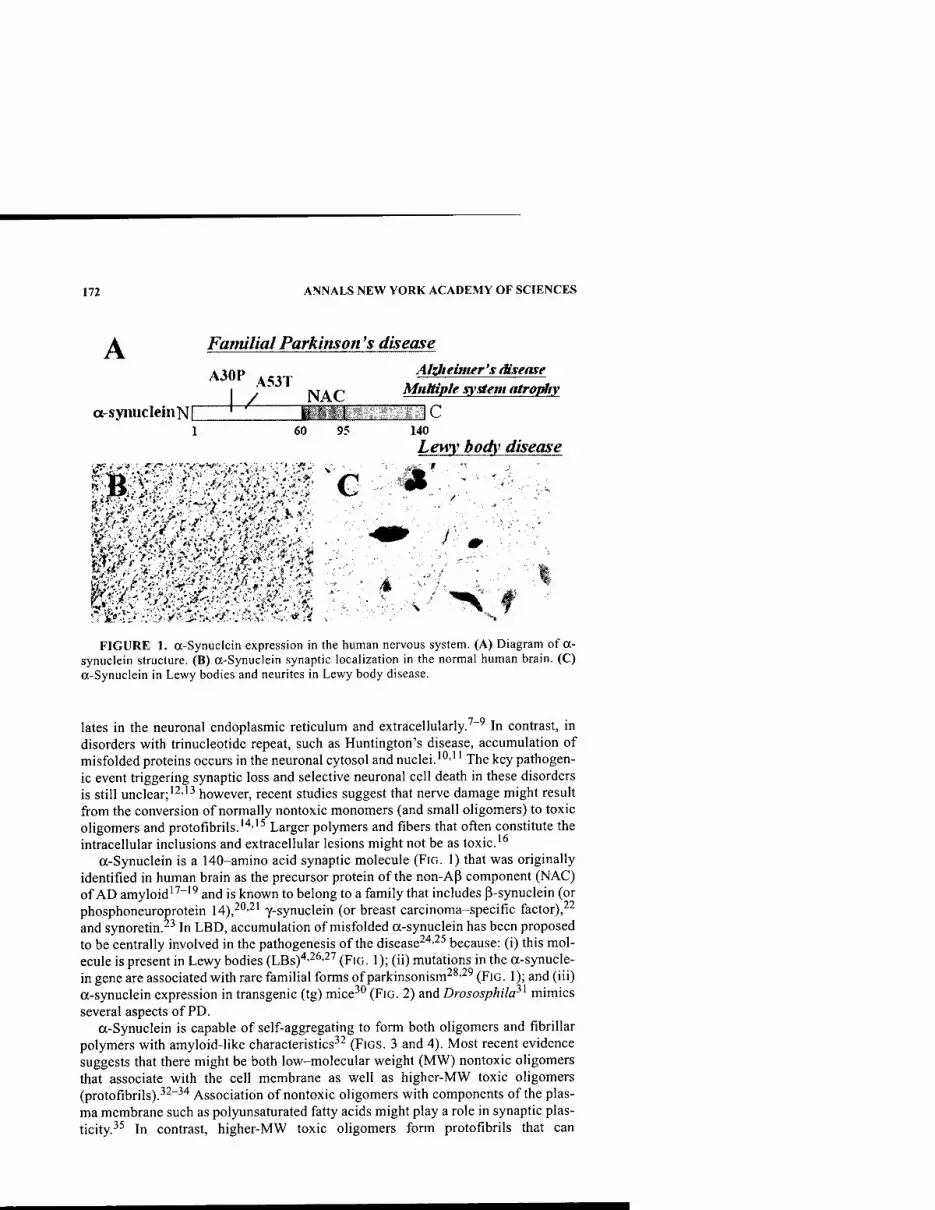
172 ANNALS NEW YORK ACADEMY OF SCIENCES
A Familial Parkinson's disease A30P 4C3J Alzfieimer'sdisease
I • NAT MtiJtiple system atropliy
a-syimcleinNI ' ilfrttf'l IC 1 60 95 140
Lewy body disease
FIGURE 1. a-Synuclein expression in the human nervous system. (A) Diagram of a- synuclein structure. (B) a-Synuclein synaptic localization in the normal human brain. (C) a-Synuclein in Lewy bodies and neurites in Lewy body disease.
lates in the neuronal endoplasmic reticulum and extracellularly.'"' In contrast, in disorders with trinucleotide repeat, such as Huntington's disease, accumulation of misfolded proteins occurs in the neuronal cytosol and nuclei.'"'" The key pathogen- ic event triggering synaptic loss and selective neuronal cell death in these disorders is still unclear; "^''^ however, recent studies suggest that nerve damage might result from the conversion of normally nontoxic monomers (and small oligomers) to toxic oligomers and protofibrils.'^''^ Larger polymers and fibers that often constitute the intracellular inclusions and extracellular lesions might not be as toxic.'^
a-Synuclein is a 140-amino acid synaptic molecule (FIG. 1) that was originally identified in human brain as the precursor protein of the non-AP component (NAC) of AD amyloid""" and is known to belong to a family that includes P-synuclein (or phosphoneuroprotein M),^^'^' y-synuclein (or breast carcinoma-specific factor),-^^ and synoretin.^^ In LBD, accumulation of misfolded a-synuclein has been proposed to be centrally involved in the pathogenesis of the disease^^'-^^ because: (i) this mol- ecule is present in Lewy bodies (LBs)'''^'''^''' (FIG. 1); (ii) mutations in the a-synucle- in gene are associated with rare familial forms of parkinsonism^^-^' (FIG. 1); and (iii) a-synuclein expression in transgenic (tg) mice^" (FIG. 2) and Drososphila^^ mimics several aspects of PD.
a-Synuclein is capable of self-aggregating to form both oligomers and fibrillar polymers with amyloid-like characteristics^^ (FIGS. 3 and 4). Most recent evidence suggests that there might be both low-molecular weight (MW) nontoxic oligomers that associate with the cell membrane as well as higher-MW toxic oligomers (protofibrils).^^"^"* Association of nontoxic oligomers with components of the plas- ma membrane such as polyunsaturated fatty acids might play a role in synaptic plas- ticity.^^ In contrast, higher-MW toxic oligomers form protofibrils that can
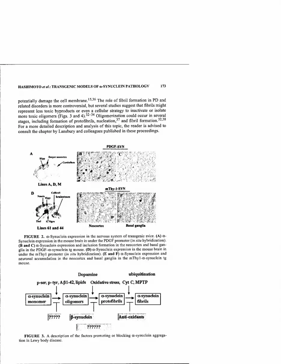
HASHIMOTO etal.: TRANSGENIC MODELS OF a-SYNUCLEIN PATHOLOGY 173
potentially damage the cell membrane.'^'^^ The role of fibril formation in PD and related disorders is more controversial, but several studies suggest that fibrils might represent less toxic byproducts or even a cellular strategy to inactivate or isolate more toxic oligomers (Figs. 3 and 4).^^"^'' Oligomerization could occur in several stages, including formation of protofibrils, nucleation,^' and fibril formation.^^'^^ For a more detailed description and analysis of this topic, the reader is advised to consult the chapter by Lansbury and colleagues published in these proceedings.
PDGF-SVN
Hipp Dtcp«r Btocortes
i y'
H. .* Lines A, D, M
0 ConkuH
Lines 61 and 44
mThy-1-SVN
0 ^ ' -U
,;<' I-
Neocortex Basal ganglia
FIGURE 2. a-Synuclein expression in the nervous system of transgenic mice. (A) a- Synuclein expression in the mouse brain in under the PDGF promoter {in situ hybridization). (B and C) a-Synuclein expression and inclusion formation in the neocortex and basal gan- glia in the PDGF-a-synuclein tg mouse. (D) a-Synuclein expression in the mouse brain in under the mXhyl promoter {in situ hybridization). (E and F) a-Synuclein expression and neuronal accumulation in the neocortex and basal ganglia in the mlhyl-a-synuclein tg
Dopamine ubiquitination
p-ser,p-tyr,Api-42,lipids Oxidative strefss, CytC,MPTP
1, _l, _l a-synudein monomer
T a-synudein oligomers
T a-synudein protofibrils
T a-synudein fibrils
If???? ||-^udein i^iiti-oxidants
FIGURE 3. A description of the factors promoting or blocking a-synuclein aggrega- tion in Lewy body disease.

174 ANNALS NEW YORK ACADEMY OF SCIENCES
a-synuclein oBgomers, protofibiUs
Synaptic and axonal damage
i Neiuological deficits
a-synuclein polymers, fibills
7 ■
T Inclusions,
LB's
i » IsolaUon ?
n
FIGURE 4. Proposed differential action of a-synuclein protofibrils and fibrils in Lewy body disease toxicity and pathogenesis.
Because of the implications for better understanding the disease pathogenesis and development of new treatments, conditions promoting or blocking a-synuclein ag- gregation and toxic conversion are now being extensively studied in tg animals and in vitro models. In general terms and for the purposes of this paper, the role of the following factors will be considered: (i) mutations associated with familial parkin- sonism;^'''**' (ii) posttranscriptional modifications associated with oxidative stress mediated by neurotoxins l-methyl-4-phenyl-l,2,3,6-tetrahydropyridine (MPTP, paraquat), iron, cytochrome C, copper (II), and dopamine;^^''"^^ (iii) posttranscrip- tional modifications associated with signaling events or conjugation (phosphoryla- tion, glycosylation, ubiquitination);'*^'''^ (iv) binding to lipid membrane vesicles;-'^'''^'^^ and (v) interactions with molecules that promote aggregation (such as NAC and Ap)'^'^*'"^^ or that block aggregation (such as (i-synuclein).^^
In this context, the main objectives of this paper are to provide a perspective with respect to the following efforts:
(1) the modeling of PD and related disorders in tg mice; (2) the definition of the in vivo role of factors promoting or blocking a-
synuclein aggregation in the pathogenesis of PD and related disorders.
Several reviews have been recently published on the subject of tg modeling of PD, and the reader is advised to consult them for additional information.^^^^^
DIFFERENTIAL EFFECTS OF WILD-TYPE AND MUTANT a-SYNUCLEIN IN tg MODELS OF TOXIC CONVERSION
Since progressive intraneuronal aggregation of a-synuclein has been proposed to play a central role in the pathogenesis of PD and related disorders,^''"''^'* most tg

HASHIMOTO etal\ TRANSGENIC MODELS OF a-SYNUCLEIN PATHOLOGY 175
n> (U
D
z > I
c O
(0
DQ
□ PE o
Is
K
2
(0
Is
g g
u |ui/6d ^ |Ui/6d
D) c «D O « 10 CB m
Bi>
PQ
■K - 1 s o Z
3
s I*
— 0)
Hi/
O
iJoqU OAH q pojSAo
If) c
sod )B0J8 % ti
uiiubs jad alias -t-HLlo -laquinu
J3
a >,
^ 3 > 6 . 0 — 00
la-- W go b MZ
w CO 5 03 ca w
1 HH ( ISO 11°
■'-^ •^ I—I
60 OH Z
7; o oi)
a hj §
'S cd » •^ 60 „ u c .S * 602 a — c 4^ 23 o

176 ANNALS NEW YORK ACADEMY OF SCIENCES
""" ,, ,,. m
1
1 Lui m m ■H m
ThiRmotflxis
Open Field
nZi Nontg
(i-syn tg (line D)
Untreated I Saline I Apomorphlme
FIGURE 6. Reversal of dopaminergic deficits in a-synuclein tg mice following apo- morphine treatment. (A) Diagram of the open field. (B) Assessment of thigmotaxis (percent- age in periphery of cage) before and after apomorphime treatment.
models have been focused on investigating the in vivo effects of a-synuclein accu- mulation using neuronal specific promoters (TABLE 1). Among these models, over- expression of wild-type (wt) a-synuclein under the regulatory control of the platelet- derived growth factor P (PDGPP) promoter (FIG. 2) has been shown to result in mo- tor deficits, dopaminergic loss, and formation of inclusion bodies.^" Mice with the highest levels of expression showed (line D) intraneuronal accumulation of a-synu- clein that started at 3 months of age and was accompanied by loss of tyrosine hy- droxylase (TH) fibers in the caudoputamen region and synapses in the temporal cortex. Although no apparent neuronal loss was detected in the substantia nigra (SN), measurements of dopamine levels in the caudoputamen region showed a 25- 50% decrease at 12 months of age (FIG. 5). Analysis of locomotor activity in the open field showed that these tg mice had increased thigmotaxis, a behavior that has been associated with dopaminergic activity. Supporting a role for alterations in the dopaminergic system, this abnormal behavior was ameliorated upon treatment with apomorphine, a compound known to promote dopamine release (FIG. 6). Further- more, at this age, tg mice showed mild-to-moderate motor deficits in the rotarod, particularly those mice with the greatest loss of dopamine, indicating that more sub- stantial deficits (>75%) of this transmitter might be necessary for more overt deficits to appear.
To determine whether different levels of human (h) a-synuclein expression from distinct promoters might result in neuropathology mimicking other synucleopathies, we also compared patterns of ha-synuclein accumulation in the brains of tg mice ex- pressing this molecule from the murine (m) Thy-1 and PDGFP promoters" (FIG. 2). In mThy-1-ha-synuclein tg mice, this protein accumulated in synapses and neurons throughout the brain, including the thalamus, basal ganglia, SN, and brain stem. Ex- pression of ha-synuclein from the PDGFp promoter resulted in accumulation in syn- apses of the neocortex, limbic system, and olfactory regions as well as formation of inclusion bodies in neurons in deeper layers of the neocortex (lines A and D). Fur- thermore, one of the intermediate expressor lines (line M) displayed ha-synuclein expression in glial cells mimicking some features of multiple system atrophy. These results show a more widespread accumulation of ha-synuclein in tg mouse brains. Taken together, these studies support the contention that ha-synuclein expression in

HASHIMOTO et al.i TRANSGENIC MODELS OF a-SYNUCLEIN PATHOLOGY 177
PDGF-us>n(wt)
Line D Line A A53T Nontg
kD
5J.4.
35.7 • syn-l 29.0-
«.7- «MMiiM«(M»
oligoTWT a-syn
I- -numomcT a-syn
123456789 10 11 13
anti-hcsyn 20.9-^^sj^a;»»*»■•*
*■ j. I /V *
*4*,' .#^; fi^ K^4 (is-'
8 . -V '1,- n u f 4
^ • r ^, V ■■^'f , ,f
* t '^•
^, . ^0*
V •i " > >
'i- ^ "• ,
Line D
Line A
A53T
FIGURE 7. Abnormal accumulation of in a-synuclein oligomers in PDGF-a-synucle- in (A53T) mutant mice. (A) Western blot analysis with 72-10 and syn-l antibodies against a-synuclein showing increased. (B-D) Immunocytochemical analysis with a human specific antibody against a-synuclein (72-10).
tg mice might mimic some neuropathological alterations observed in LBD and other synucleopathies, such as multiple system atrophy.^^
However, these studies did not clarify the potential relationship among synaptic damage, a-synuclein oligomerization and fibrillation, inclusion formation, and neu- rological deficits. Because previous studies have shown that mutations associated with familial parkinsonism accelerate a-synuclein aggregation and oligomeriza- tion,^^''*'' we compared the patterns of neurodegeneration, a-synuclein aggregation, and neurological alterations in tg mice expressing wt or mutant (mut) (A53T) ha- synuclein at comparable levels under the PDGpp promoter. For this purpose, two tg lines that express moderate levels of a-synuclein in a similar anatomical and cellular distribution were compared (line A, which expresses wt ha-synuclein,^*''^' and a newly developed line 8, which expresses mut ha-synuclein—that is, A53T) (FiG. 7). Additional comparisons were done against our well-described line D, which ex- presses higher levels of wt ha-synuclein-"''^' (FIG. 7). Lower expresser lines were se- lected for these experiments to avoid unintended effects of very high levels of expression as observed with other more strong promoters such as Thy-1 and PrP. These mice express about a third of the levels of ha-synuclein, compared to our higher expresser line D, which is approximately equivalent to a 1.5-to-l ratio with respect to endogenous levels. Remarkably, we found that mice expressing mut a-sy- nuclein developed progressive motor deficits and neurodegeneration, associated with a-synuclein accumulation in synapses and neurons; but very few or no inclu- sions were found (FIG. 7). In contrast, mice from line A (wt ha-synuclein) did not show neurodegenerative or neurological deficits, but they did display formation of inclusions. Analysis of the patterns of a-synuclein aggregation by Western blot us- ing the Syn-l antibody showed that in the mut line there was greater formation of a- synuclein oligomers compared to the wt line (FIG. 7). This is consistent with recent
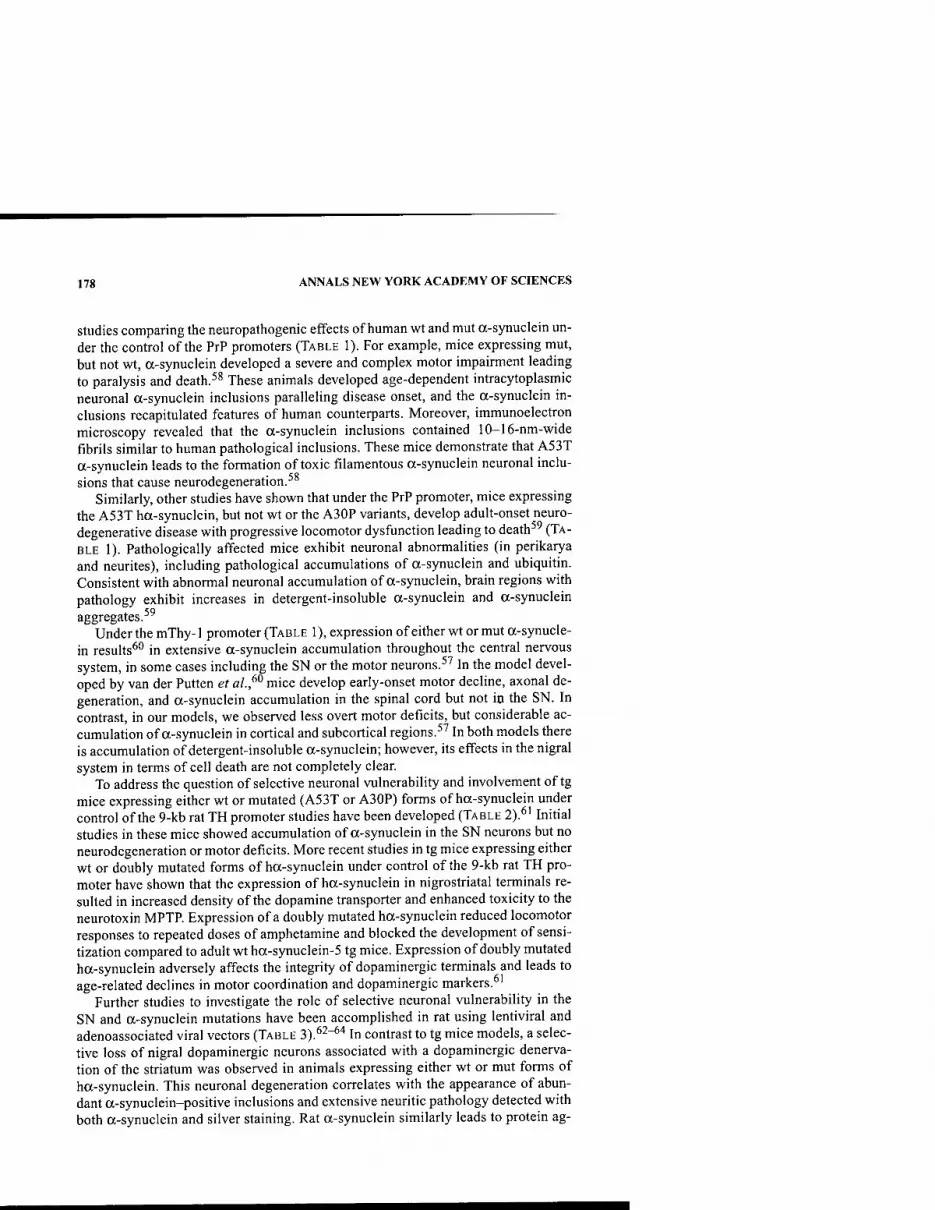
178 ANNALS NEW YORK ACADEMY OF SCIENCES
Studies comparing the neuropathogenic effects of human wt and mut a-synuclein un- der the control of the PrP promoters (TABLE 1). For example, mice expressing mut, but not wt, a-synuclein developed a severe and complex motor impairment leading to paralysis and death.^^ These animals developed age-dependent intracytoplasmic neuronal a-synuclein inclusions paralleling disease onset, and the a-synuclein in- clusions recapitulated features of human counterparts. Moreover, immunoelectron microscopy revealed that the a-synuclein inclusions contained 10-16-nm-wide fibrils similar to human pathological inclusions. These mice demonstrate that A53T a-synuclein leads to the formation of toxic filamentous a-synuclein neuronal inclu- sions that cause neurodegeneration.^^
Similarly, other studies have shown that under the PrP promoter, mice expressing the A53T ha-synuclein, but not wt or the A30P variants, develop adult-onset neuro- degenerative disease with progressive locomotor dysfiinction leading to death^ (TA- BLE 1). Pathologically affected mice exhibit neuronal abnormalities (in perikarya and neurites), including pathological accumulations of a-synuclein and ubiquitin. Consistent with abnormal neuronal accumulation of a-synuclein, brain regions with pathology exhibit increases in detergent-insoluble a-synuclein and a-synuclein aggregates.^^
Under the mThy-1 promoter (TABLE 1), expression of either wt or mut a-synucle- in results^'' in extensive a-synuclein accumulation throughout the central nervous system, in some cases including the SN or the motor neurons.^^ In the model devel- oped by van der Putten et al.,^^ mice develop early-onset motor decline, axonal de- generation, and a-synuclein accumulation in the spinal cord but not ip the SN. In contrast, in our models, we observed less overt motor deficits, but considerable ac- cumulation of a-synuclein in cortical and subcortical regions.^' In both models there is accumulation of detergent-insoluble a-synuclein; however, its effects in the nigral system in terms of cell death are not completely clear.
To address the question of selective neuronal vulnerability and involvement of tg mice expressing either wt or mutated (A53T or A30P) forms of ha-synuclein under control of the 9-kb rat TH promoter studies have been developed (TABLE 2).^' Initial studies in these mice showed accumulation of a-synuclein in the SN neurons but no neurodegeneration or motor deficits. More recent studies in tg mice expressing either wt or doubly mutated forms of ha-synuclein under control of the 9-kb rat TH pro- moter have shown that the expression of ha-synuclein in nigrostriatal terminals re- sulted in increased density of the dopamine transporter and enhanced toxicity to the neurotoxin MPTP. Expression of a doubly mutated ha-synuclein reduced locomotor responses to repeated doses of amphetamine and blocked the development of sensi- tization compared to adult wt ha-synuclein-5 tg mice. Expression of doubly mutated ha-synuclein adversely affects the integrity of dopaminergic terminals and leads to age-related declines in motor coordination and dopaminergic markers.^'
Further studies to investigate the role of selective neuronal vulnerability in the SN and a-synuclein mutations have been accomplished in rat using lentiviral and adenoassociated viral vectors (TABLE 3).*'^"^'* In contrast to tg mice models, a selec- tive loss of nigral dopaminergic neurons associated with a dopaminergic denerva- tion of the striatum was observed in animals expressing either wt or mut forms of ha-synuclein. This neuronal degeneration correlates with the appearance of abun- dant a-synuclein-positive inclusions and extensive neuritic pathology detected with both a-synuclein and silver staining. Rat a-synuclein similarly leads to protein ag-

HASHIMOTO etal: TRANSGENIC MODELS OF a-SYNUCLEIN PATHOLOGY 179
^_, u w C O
C8 U Ml <:
£> ■t-» w
e <u
J3 o O
5
^ o o s
>, 60
_o "o ^
U) M % £X -a O o t-<
E 3
u z VI S O S u
'B V en V3 B ,^, S o U g B w 2 "
c o O
S B >> w
ili Vi a VH
_o o *C o > <« VH
o PM
B O yi *£ « a & o C U 3
O ^^ O M >J n < H
JS CO
o "^
3 C
B
CO.
O Q
3
J3
ji a o T)
iC
•T3 R !" rt
KJ
O c3
::: b
c
C CO ••-I
CO bO
^ '53
3 3 e c
B 8 J3 J=
3
o o
I
3
60 60 03
< z
3 B
60 i2 !=; "
_^ -S =" 2 — o .2
•3 o 3
< ^; c B C
7) 73 ^ 3 3 ^ B B >. >. i> B 8 B
0- 3 6
^ in S <
3 3 B B >^ >.
I I
B B J3 J3
Xi 3 E
O o o
B
< z
3 B
2 •- o w CJ -is « .2 o^ 5 i2 B — TJ :S 3 3 u
o ;^ o c3 o B
py
B B B
3 3
I I 8 8
J3 J3
3 B >. 1/3
8 J3
•a 'p.
H 3 S
o
o (N
B CN O o
B !N
<D (U ja
O ed C^ fc^

180 ANNALS NEW YORK ACADEMY OF SCIENCES
■3 B
„ OJ w C 0
« 1) 00 <
&• Crt
E « JS 0 0
«
^ 0 0
:s
>^ 60
_o "o
HM ^ ra
.^ 0 (_ ;3 3 US 0) z ii e
■3 >• « e 0 k- 9 V 0
2 >■ Cfl
'€ C 0
a> U "Z fi
u 0 li- ^ a>
■0 e a>
a 0 6 0 *« l-H
PH "w 3 B >,
OJ) i
P. M 3
0 u ^
0 HJ tt < H
< z
o E D. CO
1- X
>- & 2 S o 2
u B c o c
a '5
3
u &
-§-§ 3 a, " 3 r^ o
H
^3
1 3
C^ in %_,■
is < IS OH
3 3 3 (1> u <;
0 0 3 3 SIT
0
<
0 3
0 ti?
3 C 3 t= rs >^ >. >. M W5
« H t5 ds - J3 ^ J3 J3
ffi ffi H H
„ B
^ u w 3 0
ro u 00 <
i> 00 <
0 0
S
>> 00
_o "o .c ra D.
^-v 0 t:
^-^ 3
>^ Z
.a M L. 0 B '3 >
B 0 0 u s 3 « a> c e 0 u U _> u .2 "oJ en u 0 (S M
"aS l-H
■0 0
0 0
E ;^ B u
"3 3 B >^
</l
Si D,
f<i 3 0 ^ u 0
.J oa < H
rt bfl
c
=3 3
Ti m <T1 « Tl
c on OIJ 3
3 c 0
c ed >%
C 0 X) u 1)
■a 0
0
•a E
E
3 0
la
E 3 0
cd
0 3
1 e 0
CO
tfl r/1 3 VI C r/l iTi 0 0
c 0 < 3
3 3 0
3 C
— H— o ^- o ^ o 3 3
3 O 3 P B 3 a c 8
: <
3 c >i OH
X! <
■^OH H
J= < <
> > < (D (-1 —
•^ o
HASHIMOTO et al.: TRANSGENIC MODELS OF a-SYNUCLEIN PATHOLOGY 181
gregation but without cell loss, suggesting that inclusions are not the primary cause of cell degeneration in PD.^^
In summary, under the PDGpp, both wt and mut a-synuclein tg mice develop be- havioral deficits and synaptic alterations following a limbic and cortical pattern; however, mut a-synuclein is more toxic, even though very few inclusions are formed compared to wt a-synuclein tg mice. Under the PrP promoter, mut a-synuclein is more toxic than wt, inclusion-like structures develop, and more widespread involve- ment of the lower motor system is observed. With the thy-1 construct, both wt and mut a-synuclein are toxic, and inclusion-like structures develop that can affect the cortex, subcortex, or spinal cord. Furthermore, these in vivo models support the con- tention that a-synuclein-dependent neurodegeneration is associated with abnormal accumulation of detergent-insoluble a-synuclein (probably representing oligomeric forms) rather than with inclusion formation representing fibrillar polymeric a-synu- clein. The specific accumulation of detergent-insoluble a-synuclein in these tg mice recapitulates a pivotal feature of LBD,^^ and it is of significance in the future devel- opment and evaluation of novel treatments.
DIFFERENTIAL EFFECTS OF POSTTRANSCRIPTIONAL MODIFICATIONS IN a-SYNUCLEIN
TOXIC CONVERSION
Although characterizing the role of a-synuclein mutations has improved our un- derstanding of the role of protein misfolding in the pathogenesis of PD, these muta- tions comprehend only a small fraction of the cases. It is estimated that over 95% of the cases of PD and related disorders are sporadic.^^ Thus, complex interactions be- tween environmental and genetic susceptibility factors might be at play. For exam- ple, the potential role of factors promoting oxidative stress is under intense investigation (FiG. 3). Factors such as iron, cytochrome C, copper (II), dopam- ine,^^'""^^'^^'^* and neurotoxins have been shown in vitro to alter a-synuclein con- formation leading to aggregation and toxic protofibril formation. Among such factors, the key discovery by Langston and colleagues of l-methyl-4-phenylpyridin- ium ion as a powerful dopaminergic neurotoxin led to the development of acute mod- els of parkinsonism in murine and simian models.^^
Since the neurotoxin MPTP promotes oxidative stress^" and degeneration of the dopaminergic system, mimicking some aspects of PD, we seek to determine the ef- fects of chronic MPTP treatment on tg mice overexpressing a-synuclein. For this purpose, tg mice expressing high levels of a-synuclein throughout the cortex and SN under the control of the Thy-1 promoter were treated with low doses of MPTP and analyzed ultrastructurally.^''^^ Electron microscopy analysis demonstrated that compared to control groups. Thy 1-a-synuclein tg mice treated with MPTP showed extensive mitochondrial alterations, invagination of the nuclear membrane, and for- mation of electrodense intracytoplasmic inclusions. Taken together, these studies suggest that oxidative stress and the associated mitochondrial damage might contrib- ute to aggregation of a-synuclein and to the pathogenesis of PD. Further supporting a role of oxidative stress in the pathogenesis of disorders with a-synuclein accumu- lation, recent studies have shown that this molecule is present in a nitrosylated form that promotes abnormal conformation.''^-^^ Consistent with this possibility, we have

182 ANNALS NEW YORK ACADEMY OF SCIENCES
observed increased 3-NT immunostaining in the a-synuclein tg mice. In neuronal cell lines overexpressing a-synuclein, there is also increased reactivity for markers of oxidative stress that are reversible with antioxidant treatments (for example, vitamin E)."*^
Another factor promoting oxidative damage and abnormal protein conformation that might play a role in a-synucleopathies is amyloid P-protein (FIGS. 3 and 4). This is interesting because AD and PD are associated with the cerebral accumulation of P-amyloid and a-synuclein, respectively.''^ Some patients have clinical and patho- logical features of both diseases, raising the possibility of overlapping pathogenetic pathways. We refer to this condition as the Lewy body variant (LBV) of AD;^"* how- ever, others prefer different terms such as combined AD+PD, senile dementia of the Lewy body type, or LBD.''^ To better understand the role of amyloid production in a-synucleopathies, we generated tg mice with neuronal expression of human AjJ, a- synuclein, or both.^^ The functional and morphological alterations in doubly tg mice resembled the LBV of AD. These mice had severe deficits in learning and memory, developed motor deficits before a-synuclein singly tg mice did, and showed promi- nent age-dependent degeneration of cholinergic neurons and presynaptic terminals. They also had more a-synuclein-immunoreactive neuronal inclusions than a-synu- clein singly tg mice. Ultrastructurally, some of these inclusions were fibrillar in dou- bly tg mice, whereas all inclusions were amorphous in a-synuclein singly tg mice. AP promoted aggregation of a-synuclein in a cell-free system and intraneuronal ac- cumulation of a-synuclein in cell culture. Ap may contribute to the development of LBD by promoting the aggregation of a-synuclein and exacerbating a-synuclein- dependent neuronal pathologies. Therefore, treatments that block the production or accumulation of AP could benefit a broader spectrum of disorders than previously anticipated.
Other posttranscriptional modifications that might promote a-synuclein aggrega- fion and toxic conversion include phosphorylation and conjugation (FIG. 3). In this regard, recent mass spectrometry analyses and studies with an antibody that specif- ically recognizes phospho-Ser 129 of a-synuclein have shown that this residue is se- lectively and extensively phosphorylated in synucleinopathy lesions, including those in patients with PD and related disorders and in a-synuclein tg mice."*^ Furthermore, phosphorylafion of a-synuclein at Ser 129 promoted fibril formation in vitro. These results highlight the importance of phosphorylation of filamentous proteins in the pathogenesis of neurodegenerative disorders. In Drosophila, synuclein overexpres- sion results in LB formation and dopaminergic neuronal loss.^' Furthermore, direct- ed expression of the molecular chaperone Hsp70 prevented dopaminergic neuronal loss associated with a-synuclein in Drosophila, and interference with endogenous chaperone activity accelerated a-synuclein toxicity.'^ The fact that LBs in human postmortem tissue immunostained for molecular chaperones suggests that chaper- ones may play a role in PD progression. Among them, ubiquitination appears to play an important role. This is significant because mutations in the Parkin gene, also known to be associated with familial PD, have been shown to result in deficient a- synuclein ubiquitination.''^"^' Studies in tg mice are underway in which a-synuclein tg mice are crossed with Parkin or UCHL-1 KO mice. The prediction is that this will result in defective a-synuclein ubiqutination and proteosome degradation, with worsening of the phenotype but probably with no inclusion formation.

HASHIMOTO et aU: TRANSGENIC MODELS OF a-SYNUCLEIN PATHOLOGY 183
ROLE OF ENDOGENOUS INHIBITORS IN PREVENTING AND MODULATING a-SYNUCLEIN TOXIC CONVERSION
Toxic conversion of a-synuclein is not only the result of factors promoting ag- gregation but might also be the consequence of a failure of factors preventing ag- gregation (FIG. 3). Thus a critical balance between aggregation and anti- aggregation factors might be at play. Endogenous mechanisms to prevent toxic con- version include clearance, proteolysis, and production of inhibitors of aggregation. Among these, we have characterized p-synuclein, the nonamyloidogenic homo- logue of a-synuclein, as an inhibitor of aggregation of a-synuclein.^^ For this pur- pose, doubly tg mice expressing both a- and P-synuclein were generated and analyzed in the rotarod and by confocal microscopy. These studies showed that P- synuclein ameliorated the motor deficits, neurodegenerative alterations, and neu- ronal accumulation of a-synuclein seen in a-synuclein tg mice. Similarly, cell lines stably transfected with P-synuclein were resistant to accumulating a-synuclein, when this later molecule was expressed under the control of the inducible muri- sterone A system. a-Synuclein coimmunoprecipitated with P-synuclein both in the brains of doubly tg mice and in the doubly transfected cell lines. Furthermore, p- synuclein has also been shown to reduce AP aggregation. Our results raise the in- triguing possibility that P-synuclein might be a natural negative regulator of a-sy- nuclein and Ap aggregation.^^
The synucleins are natively unfolded proteins.^" In support of an antiamy- loidogenic effect of p-synuclein, we^^ as well as others have shown that P-synuclein, which lacks 11 central hydrophobic residues compared to its homologues, exhibited the properties of a random coil, whereas a- and y-synucleins were slightly more compact and structured. y-Synuclein, unlike its homologues, formed a soluble oligo- mer at relatively low concentrations, which appears to be an off-fibrillation pathway species. Although they have biophysical properties similar to those of a-synuclein, p- and y-synucleins inhibit a-synuclein fibril formation. Complete inhibition of a- synuclein fibrillation was observed at 4:1 molar excess of P- and y-synucleins. No significant incorporation of P-synuclein into the fibrils was detected. The lack of fibrils formed by a-synuclein is most readily explained by the absence of a stretch of hydrophobic residues from the middle region of the protein.^'
The mechanisms by which P-synuclein might block a-synuclein aggregation in vivo are under investigation. One possible mechanism is that a-synuclein transloca- tion to the plasma and mitochondrial membranes might be regulated by P-synuclein. In this regard, it was recently described that aggregation of a-synuclein in the mem- brane fractions was stimulated by mitochondrial dysfunction, suggesting a patholog- ical relevance of a-synuclein in these fractions. Consistent with this view,*^ immunoblot analysis for the tg mice brain using sensitive a-synuclein-1 antibody showed that aggregation of a-synuclein in the particulate fractions in single a-synu- clein mice was significantly suppressed in doubly tg mice expressing a and p-synu- clein. Thus, pathological aggregation of a-synuclein in the membrane fractions may be negatively regulated by its nonamyloidogenic homologue, p-synuclein (FiG. 4). In- terestingly, the 50-kDa immunoreactive band in non-tg mice, which might represent physiological oligomers of a-synuclein,*^ was absent in both singly and doubly tg mice, suggesting that equilibrium of a-synuclein oligomerization under the physio-

184 ANNALS NEW YORK ACADEMY OF SCIENCES
logical conditions may be easily affected by different spices of a-synuclein as well as P-synuclein. Currently, it is unknown how interference with this physiological pro- cess might interfere with neuroplasticity. However, it may be ideal to design therapeu- tic drugs that would selectively act on the process of pathological aggregation of a- synuclein. Taken together, these findings suggest that P-synuclein homologues or de- rivatives might have a therapeutical potential for the treatment of AD, LBD, and PD.
CONCLUSIONS
Several tg mouse models for a-synucleopathy have now been developed. Models employing the PDGpp promoter display involvement of the neocortex, limbic sys- tem, and (to a lesser extent) the nigrostriatal pathways, with a-synuclein accumula- tion and inclusion formation. Under the mThy-1 and PrP promoters there is more widespread expression of a-synuclein throughout the brain, including the subcortex and spinal cord. In these animals, there is a more dramatic locomotor phenotype and increased lethality with involvement of the neuromuscular junction and motor sys- tem. The A53T mut a-synuclein appears to be more toxic, probably because of its increased susceptibility to forming toxic protofibrils. Models selectively targeting the nigral system have been developed using the TH promoter or viral vectors. In tg models, oxidative stress and amyloid P-protein accelerate a-synuclein accumula- tion, mimicking combined PD and AD. In contrast, molecular chaperones such as Hsp70 and close homologues such as a-synuclein have been shown to suppress tox- icity. Similar studies are underway to evaluate the effects of other modifying genes that might play a role in a-synuclein ubiquitination. For example, considerable in- terest has been focused on the role of molecules associated with familial parkin- sonism {Parkin, UCHL-1). With the discovery of a-synuclein and the advent of new experimental animal models for PD and related disorders, the potential for the de- velopment and discovery of new treatments has been significantly bolstered.
ACKNOWLEDGMENTS
This work was supported by the National Institutes of Health (Grants AG5131, AG10689, and AG18440) and by grants from the M. J. Fox Foundation for Parkin- son's Research and the Spencer family.
REFERENCES
1. Koo, E., P.J. LANSBURY & J. KELLY. 1999. Amyloid diseases: abnormal protein aggre- gation in neurodegeneration. Proc. Natl. Acad. Sci. USA 96: 9989-9990.
2. FERRIGNO, P. & R SILVER. 2000. Polyglutamine expansions: proteolysis, chaperones, and the dangers of promiscuity. Neuron 26: 9-12.
3. RAMASSAMY, C, et al. 1999. Oxidative damage and protection by antioxidants in the frontal cortex of Alzheimer's disease is related to the apolipoprotein E genotype. Free Rad. Biol. Med. 27: 544-553.
4. SPILLANTINI, M, et al. 1997. a-Synuclein in Lewy bodies. Nature 388: 839-840. 5. TAKEDA, A., et al. 1998. Abnormal accumulation of NACP/a-synuclein in neurodegen-
erative disorders. Am. J. Pathol. 152: 367-372.

HASHIMOTO et al.: TRANSGENIC MODELS OF a-SYNUCLEIN PATHOLOGY 185
6. IRIZARRY, M., et al. 1998. Nigral and cortical Lewy bodies and dystrophic nigral neu- rites in Parkinson's disease and cortical Lewy body disease contain alpha-synuclein immunoreactivity. J. Neuropathol. Exp. Neurol. 57: 334-337.
7. WALSH, D., et al. 2000. The oligomerization of amyloid beta-protein begins intracellu- larly in cells derived from human brain. Biochemistry 39: 10831-10839.
8. SELKOE, D.J., et al. 1996. The role of APP processing and trafficking pathways in the formation of amyloid beta-protein. Ann. N.Y. Acad. Sci. 777: 57-64.
9. TROJANOWSKI, J.Q. & V.M. LEE. 2000. "Fatal attractions" of proteins: a comprehensive hypothetical mechanism underlying Alzheimer's disease and other neurodegenera- tive disorders. Ann. N.Y. Acad. Sci. 924: 62-67.
10. MucHOWSKi, P.J. 2002. Protein misfolding, amyloid formation, and neurodegeneration: a critical role for molecular chaperones? Neuron 35: 9-12.
11. CuMMiNGS, C.J. & H.Y. ZoGHBi. 2000. Trinucleotide repeats: mechanisms and patho- physiology. Annu. Rev. Genomics Hum. Genet. 1: 281-328.
12. MASLIAH, E. 2000. The role of synaptic proteins in Alzheimer's disease. Ann. N.Y. Acad. Sci. 924: 68-75.
13. MASLIAH, E. 2001. Recent advances in the understanding of the role of synaptic proteins in Alzheimer's disease and other neurodegenerative disorders. J. Alz. Dis. 3: 1-9.
14. VOLLES, M.J. & P.T. LANSBURY, JR. 2002. Vesicle permeabilization by protofibrillar alpha-synuclein is sensitive to Parkinson's disease-linked mutations and occurs by a pore-like mechanism. Biochemistry 41: 4595-4602.
15. VOLLES, M.J., et al. 2001. Vesicle permeabilization by protofibrillar alpha-synuclein: implications for the pathogenesis and treatment of Parkinson's disease. Biochemistry 40: 7812-7819.
16. LANSBURY, P.T.J. 1999. Evolution of amyloid: what normal protein folding may tell us about fibrillogenesis and disease. Proc. Natl. Acad. Sci. USA 96: 3342-3344.
17. IWAI, A. 2000. Properties of NACP/alpha-synuclein and its role in Alzheimer's dis- ease. Biochim. Biophys. Acta 1502: 95-109.
18. MASLIAH, E., et al. 1996. Altered presynaptic protein NACP is associated with plaque formation and neurodegeneration in Alzheimer's disease. Am. J. Pathol. 148: 201-210.
19. UEDA, K., et al. 1993. Novel amyloid component (NAC) differentiates Alzheimer's disease from normal aging plaques. Soc. Neurosci. Abstr. 19: 1254.
20. JAKES, R., M. SPILLANTINI & M. GOEDERT. 1994. Identification of two disfinct synucle- ins from human brain. FEBS Lett. 345: 27-32.
21. NAKAJO, S., et al. 1993. A new brain-specific 14-kDa protein is a phosphoprotein: its complete amino acid sequence and evidence for phosphorylation. Eur. J. Biochem. 217: 1057-1063.
22. JiA, T., et al. 1999. Stimulation of breast cancer invasion and metastasis by synuclein gamma. Cancer Res. 59: 742-747.
23. SuRGUCHOV, A., et al. 1999. Synoretin—a new protein belonging to the synuclein fam- ily. Mol. Cell. Neurosci. 13: 95-103.
24. HASHIMOTO, M. & E. MASLIAH. 1999. Alpha-synuclein in Lewy body disease and Alzheimer's disease. Brain Pathol. 9: 707-720.
25. TROJANOWSKI, J. & V. LEE. 1998. Aggregation of neurofilament and alpha-synuclein proteins in Lewy bodies: implications for pathogenesis of Parkinson disease and Lewy body dementia. Arch. Neurol. 55: 151-152.
26. TAKEDA, A., et al. 1998. Abnormal distribution of the non-AP component of Alzhe- imer's disease amyloid precursor/a-synuclein in Lewy body disease as revealed by proteinase K and formic acid pretreatment. Lab. Invest. 78: 1169-1177.
27. WAKABAYASHI, K., et al. 1997. Neurofibrillary tangles in the dentate granule cells in Alzheimer's disease, Lewy body disease and progressive supranuclear palsy. Acta Neuropathol. 93: 7-12.
28. KRUGER, R., et al. 1998. Ala30Pro mutafion in the gene encoding a-synuclein in Par- kinsons's disease. Nat. Genet. 18: 106-108.
29. POLYMEROPOULOS, M., et al. 1997. Mutation in the a-synuclein gene identified in fam- ilies with Parkinson's disease. Science 276: 2045-2047.
30. MASLIAH, E., et al. 2000. Dopaminergic loss and inclusion body formation in alpha-synu- clein mice: implications for neurodegenerative disorders. Science 287: 1265-1269.
186 ANNALS NEW YORK ACADEMY OF SCIENCES
31. FEANY, M. & W. BENDER. 2000. A Dwsophila model of Parkinson's disease. Nature 404: 394-398.
32. HASHIMOTO, M., et al. 1998. Human recombinant NACP/a-synuclein is aggregated and fibrillated in vitro: relevance for Lewy body disease. Brain Res. 799: 301-306.
33. CoNWAY, K.A., et al. 2000. Acceleration of oligomerization, not fibrillization, is a shared property of both alpha-synuclein mutations linked to early-onset Parkinson's disease: implications for pathogenesis and therapy. Proc. Natl. Acad. Sci. USA 97: 571-576.
34. ROCHET, J., K. CONWAY & RJ. LANSBURY. 2000. Inhibition of fibrillization and accu- mulation of prefibrillar oligomers in mixtures of human and mouse alpha-synuclein. Biochemistry 39: 10619-10626.
35. PERRIN, R., et al. 2000. Interaction of human alpha-synuclein and Parkinson's disease variants with phospholipids: structural analysis using site-directed mutagenesis. J. Biol. Chem. 275: 34393-34398.
36. NARAYANAN, V. & S. SCARLATA. 2001. Membrane binding and self-association of alpha-synucleins. Biochemistry 40: 9927-9934.
37. WOOD, S.J., et al. 1999. alpha-Synuclein fibrillogenesis is nucleation dependent: impli- cations for the pathogenesis of Parkinson's disease. J. Biol. Chem. 274: 19509- 19512.
38. SERPELL, L., et al. 2000. Fiber diffraction of synthetic a-synuclein filaments shows amyloid-like cross-P conformation. Proc. Natl. Acad. Sci. USA 97: 4897-4902.
39. CONWAY, K., J. HARPER & P. LANSBURY. 1998. Accelerated in vitro fibril formation by a mutant alpha-synuclein linked to early-onset Parkinson disease. Nat. Med. 4: 1318-1320.
40. NARHI, L., et al. 1999. Both familial Parkinson's disease mutations accelerate alpha- synuclein aggregation. J. Biol. Chem. 274: 9843-9846.
41. HASHIMOTO, M., et al. 1999. Oxidative stress induces amyloid-like aggregate forma- tion of NACP/a-synuclein in vitro. Neuroreport 10: 717-721.
42. HASHIMOTO, M., et al. 1999. Role of cytochrome c as a stimulator of a-synuclein aggregation in Lewy body disease. J. Biol. Chem. 274: 28849-28852.
43. HSU, L.J., et al. 2000. a-Synuclein promotes mitochondria! deficiencies and oxidative stress. Am. J. Pathol. 157: 401-410.
44. PAIK, S.R., et al. 1999. Copper(II)-induced self-oligomerization of alpha-synuclein. Biochem. J. 340: 821-828.
45. SOUZA, J., et al. 2000. Dityrosine cross-linking promotes formation of stable alpha- synuclein polymers. J. Biol. Chem. 275: 18344-18349.
46. OKOCHI, M., et al. 2000. Constitutive phosphorylation of the Parkinson's disease asso- ciated a-synuclein. J. Biol. Chem. 275: 390-397.
47. FUJIWARA, H., et al. 2002. alpha-Synuclein is phosphorylated in synucleinopathy lesions. Nat. Cell Biol. 4: 160-164.
48. Jo, E., et al. 2000. Alpha-synuclein membrane iteractions and lipid specificity. J. Biol. Chem. 275: 34328-34334.
49. DAVIDSON, W., et al. 1998. Stabilization of alpha-synuclein secondary structure upon binding to synthetic membranes. J. Biol. Chem. 273: 9443-9449.
50. JENSEN, P., et al. 1997. Binding of AP to a- and P-synucleins: identification of seg- ments in a-synuclein/NAC precursor that bind AP and NAC. Biochem. J. 323: 539- 546.
51. PAIK, S., et al. 1998. Self-oligomerization of NACP, the precursor protien of the non- amyloid beta/A4 protein (A beta) component of Alzheimer's disease amyloid, observed in the presence of a C-terminal A beta fragment (residues 25-35). FEBS Lett. 421:73-76.
52. YosHiMOTO, M., et al. 1995. NACP, the precursor protein of non-amyloid p/A4 protein (AP) component of Alzheimer disease amyloid, binds AP and stimulates AP aggre- gation. Proc. Natl. Acad. Sci. USA 92: 9141-9145.
53. HASHIMOTO, M., et al. 2001. beta-Synuclein inhibits alpha-synuclein aggregation: a possible role as an anti-parkinsonian factor. Neuron 32: 213-223.
54. BETARBET, R., T.B. SHERER & J.T. GREENAMYRE. 2002. Animal models of Parkinson's disease. Bioessays 24: 308-318.
HASHIMOTO et al.: TRANSGENIC MODELS OF a-SYNUCLEIN PATHOLOGY 187
55. BEAL, M.F. 2001. Experimental models of Parkinson's disease. Nat. Rev. Neurosci. 2: 325-334.
56. DAWSON, T., A. MANDIR & M. LEE. 2002. Animal models of PD: pieces of the same puzzle? Neuron 35: 219-222.
57. ROCKENSTEIN, E., et al. 2002. Differential neuropathological alterations in transgenic mice expressing alpha-synuclein from the platelet-derived growth factor and Thy-1 promoters. J. Neurosci. Res. 68: 568-578.
58. GIASSON, B.I., et al. 2002. Neuronal alpha-synucleinopathy with severe movement dis- order in mice expressing A53T human alpha-synuclein. Neuron 34: 521-533.
59. LEE, M.K., et al. 2002. Human alpha-synuclein-harboring familial Parkinson's dis- ease-linked Ala-53—'Thr mutation causes neurodegenerative disease with alpha- synuclein aggregation in transgenic mice. Proc. Natl. Acad. Sci. USA 99: 8968- 8973.
60. VAN DER PUTTEN, H., et al. 2000. Neuropathology in mice expressing human alpha- synuclein. J. Neurosci. 20: 6021-6029.
61. RICHFIELD, E.K., et al. 2002. Behavioral and neurochemical effects of wild-type and mutated human alpha-synuclein in transgenic mice. Exp. Neurol. 175: 35-48.
62. Lo BIANCO, C, et al. 2002. alpha-Synucleinopathy and selective dopaminergic neuron loss in a rat lentiviral-based model of Parkinson's disease. Proc. Natl. Acad. Sci. USA 99: 10813-10818.
63. KiRiK, D., et al. 2002. Parkinson-like neurodegeneration induced by targeted overex- pression of alpha-synuclein in the nigrostriatal system. J. Neurosci. 22: 2780-2791.
64. KLEIN, R.L., et al. 2002. Dopaminergic cell loss induced by human A30P alpha-synu- clein gene transfer to the rat substantia nigra. Hum. Gene Ther. 13: 605-612.
65. KAHLE, P.J., et al. 2001. Selective insolubility of alpha-synuclein in human Lewy body diseases is recapitulated in a transgenic mouse model. Am. J. Pathol. 159: 2215- 2225.
66. GoEDERT, M. 2001. Alpha-synuclein and neurodegenerative diseases. Nat. Rev. Neuro- sci. 2: 492-501.
67. CONWAY, K.A., et al. 2001. Kinetic stabilization of the alpha-synuclein protofibril by a dopamine-alpha-synuclein adduct. Science 294: 1346-1349.
68. GiASSON, B.I., et al. 2000. Oxidative damage linked to neurodegeneration by selective alpha-synuclein nitration in synucleinopathy lesions. Science 290: 985-989.
69. LANGSTON, J.W., E.B. LANGSTON & I. IRWIN. 1984. MPTP-induced parkinsonism in human and non-human primates—clinical and experimental aspects. Acta Neurol. Scand. Suppl. 100: 49-54.
70. GRUNBLATT, E., S. MANDEL & M.B. YOUDIM. 2000. MPTP and 6-hydroxydopamine- induced neurodegeneration as models for Parkinson's disease: neuroprotective strate- gies. J. Neurol. 247(Suppl. 2): 95-102.
71. RATHKE-HARTLIEB, S., et al. 2001. Sensitivity to MPTP is not increased in Parkinson's disease-associated mutant alpha-synuclein transgenic mice. J. Neurochem. 77: 1181-1184.
72. SONG et al. 2002. MPTP promotes mitochondrial pathology and aggregation in human a-synuclein transgenic mice. Soc. Neurosci. Abstr. 226.1.
73. MASLIAH, E., et al. 2001. beta-Amyloid peptides enhance alpha-synuclein accumula- tion and neuronal deficits in a transgenic mouse model linking Alzheimer's disease and Parkinson's disease. Proc. Natl. Acad. Sci. USA 98: 12245-12250.
74. HANSEN, L., et al. 1990. The Lewy body variant of Alzheimer's disease: A clinical and pathologic entity. Neurology 40: 1-7.
75. MCKEITH, I., et al. 1996. Clinical and pathological diagnosis of dementia with Lewy bodies (DLB): Report of the CDLB International Workshop. Neurology 47: 1113- 1124.
76. AuLUCK, P.K., et al. 2002. Chaperone suppression of alpha-synuclein toxicity in a Drosophila model for Parkinson's disease. Science 295: 865-868.
77. HATTORI, N., et al. 1998. Point mutations (Thr240Arg and Ala311Stop) in the Parkin gene. Biochem. Biophys. Res. Commun. 249: 754-758.
78. KITADA, T., et al. 1998. Mutations in the Parkin gene cause autosomal recessive juve- nile parkinsonism. Nature 392: 605-608.

188 ANNALS NEW YORK ACADEMY OF SCIENCES
79. SHIMURA, H., et a!. 2000. Familial Parkinson disease gene product, Parkin, is a ubiq- uitin-protein ligase. Nat. Gen. 25: 302-305.
80. WEINREB, P., et al. 1996. NACP, a protein implicated in Alzheimer's disease and learn- ing, is natively unfolded. Biochemistry 35: 13709-13715.
81. UVERSKY, V.N., et al. 2002. Biophysical properties of the synucleins and their propen- sities to fibrillate: inhibition of alpha-synuclein assembly by beta- and gamma-synu- cleins. J. Biol. Chem. 277: 11970-11978.
82. LEE, H.J., et al. 2001. Formation and removal of alpha-synuclein aggregates in cells exposed to mitochondrial inhibitors. J. Biol. Chem. 277: 5411-5417.
83. LENG, V., T.N. CHASE & M.C. BENNETT. 2001. Muscarinic receptor stimulation induces translocation of an alpha-synuclein oligomer from plasma membrane to a light vesi- cle fraction in cytoplasm. J. Biol. Chem. 276: 28212-28218.

The l-Methyl-4-Phenyl-l,2,3,6- Tetrahydropyridine Mouse Model
A Tool to Explore the Pathogenesis of Parkinson's Disease
SERGE PRZEDBORSKI"'''^ ANDMIQUEL VILA"
Neuroscience Research Laboratories of the Movement Disorder Division, Departments of "Neurology and ''Pathology and the '^Center for Neurobiology and Behavior, Columbia University, New York, New York 10032, USA
ABSTRACT: Experimental models of dopamlnergic neurodegeneration play a critical role in our quest to elucidate the cause of Parkinson's disease (PD). De- spite the recent development of "genetic models" that have followed upon the discovery of mutations causing rare forms of familial PD, toxic models remain at the forefront when it comes to exploring the pathogenesis of sporadic PD. Among these, the model produced by the neurotoxin l-methyl-4-phenyl- 1,2,3,6-tetrahydropyridine (MPTP) has a competitive advantage over all other toxic models because once this neurotoxin causes intoxication, it induces in hu- mans a syndrome virtually identical to PD. For the past two decades, the com- plex pharmacology of MPTP and the key steps in the MPTP neurotoxic process have been identified. These molecular events can be classified into three groups: First, those implicated in the initiation of toxicity, which include ener- gy failure due to ATP depletion and oxidatlve stress mediated by superoxide and nitric oxide; second, those recruited subsequently in response to the initial neuronal perturbations, which include elements of the molecular pathways of apoptosis such as Bax; and, third, those amplifying the neurodegenerative in- sult, which include various proinflammatory factors such as prostaglandins. Herein, these different contributing factors are reviewed, as is the sequence in which it is believed these factors are acting within the cascade of events respon- sible for the death of dopamlnergic neurons in the MPTP model and in PD. How to target these factors to devise effective neuroprotective therapies for PD is also discussed.
KEYWORDS: apoptosis; cell death; nitric oxide; neurotoxicity; neurodegenera- tion; MPTP; Parkinson's disease (PD); reactive oxygen species; superoxide dismutase
INTRODUCTION
l-Methy-4-phenyl-l,2,3,6-tetrahydropyridine (MPTP) is a byproduct of the chemical synthesis of a meperidine analog with potent heroin-like effects. MPTP can induce a parkinsonian syndrome in humans almost indistinguishable from Par-
Address for correspondence: Dr. Serge Przedborski, BB-307, Columbia University, 650 West 168th Street, New York, New York 10032. Voice: 212-305-1540; fax: 212-305-5450.
Ann. N.Y. Acad. Sci. 991: 189-198 (2003). © 2003 New York Academy of Sciences.
189

190 ANNALS NEW YORK ACADEMY OF SCIENCES
kinson's disease (PD).' Recognition of MPTP as a neurotoxin occurred early in 1982, when several young drug addicts mysteriously developed a profound parkin- sonian syndrome after the intravenous use of street preparations of meperidine an- alogs that, unknown to anyone, were contaminated with MPTP.^ In humans and nonhuman primates, depending on the regimen used, MPTP produces an irrevers- ible and severe parkinsonian syndrome that replicates almost all of the features of PD; in nonhuman primates, however, a resting tremor characteristic of PD has been demonstrated convincingly only in the African green monkey.^ It is believed that in PD the neurodegenerative process occurs over several years, while the most active phase of neurodegeneration is completed within a few days following MPTP admin- istration."*'^ However, recent data suggest that, following the main phase of neuronal death, MPTP-induced neurodegeneration may continue to progress "silently" over several decades, at least in humans intoxicated with MPTP.^'^ Except for four cases,^'** no human pathological material has been available for study; thus, the comparison between PD and the MPTP model is limited largely to nonhuman pri- mates.^ Neuropathological data show that MPTP administration causes damage to the nigrostriatal dopaminergic pathway identical to that seen in PD,'" yet there is a resemblance that goes beyond the loss of substantia nigra pars compacta (SNpc) dopaminergic neurons. Like PD, MPTP causes a greater loss of dopaminergic neu- rons in the SNpc than in the ventral tegmental area' '''^ and, in monkeys treated with low doses of MPTP (but not in humans), a greater degeneration of dopaminergic nerve terminals in the putamen than in the caudate nucleus.'^''"^ However, two typ- ical neuropathologic features of PD have, until now, been lacking in the MPTP mod- el. First, except for the SNpc, pigmented nuclei such as the locus coeruleus have been spared, according to most published reports. Second, the eosinophilic intra- neuronal inclusions called Lewy bodies, so characteristic of PD, have not, thus far, been convincingly observed in MPTP-induced parkinsonism;' however, in MPTP- injected monkeys, intraneuronal inclusions reminiscent of Lewy bodies have been described.' ^ Despite these imperfections, MPTP continues to be regarded as an excel- lent animal model of sporadic PD, and the belief is that studying MPTP toxic mecha- nisms will shed light on meaningful pathogenic mechanisms implicated in PD.
Over the years, MPTP has been used in a large variety of animal species, ranging from worms to mammals. To date, the most frequently used animals for MPTP stud- ies have been monkeys, rats, and mice.'^ The administration of MPTP through a number of different routes using different dosing regimens has led to the develop- ment of several distinct models, each characterized by some unique behavioral and neuropathological features. Herein, we will restrict our discussion to mice, since they have emerged as the preferred animals to explore cellular and molecular alter- ations produced by MPTP, in part because lines of engineered animals that are so critical to these types of investigations are available only in mice.'^
MPTP MODE OF ACTION
As illustrated in FIGURE 1, the metabolism of MPTP is a complex, multistep pro- cess.'^ After its systemic administration, MPTP, which is highly lypophilic, rapidly crosses the blood-brain barrier. Once in the brain, the protoxin MPTP is metabolized to l-methyl-4-phenyl-2,3-dihydropyridinium (MPDP+) by the enzyme monoamine
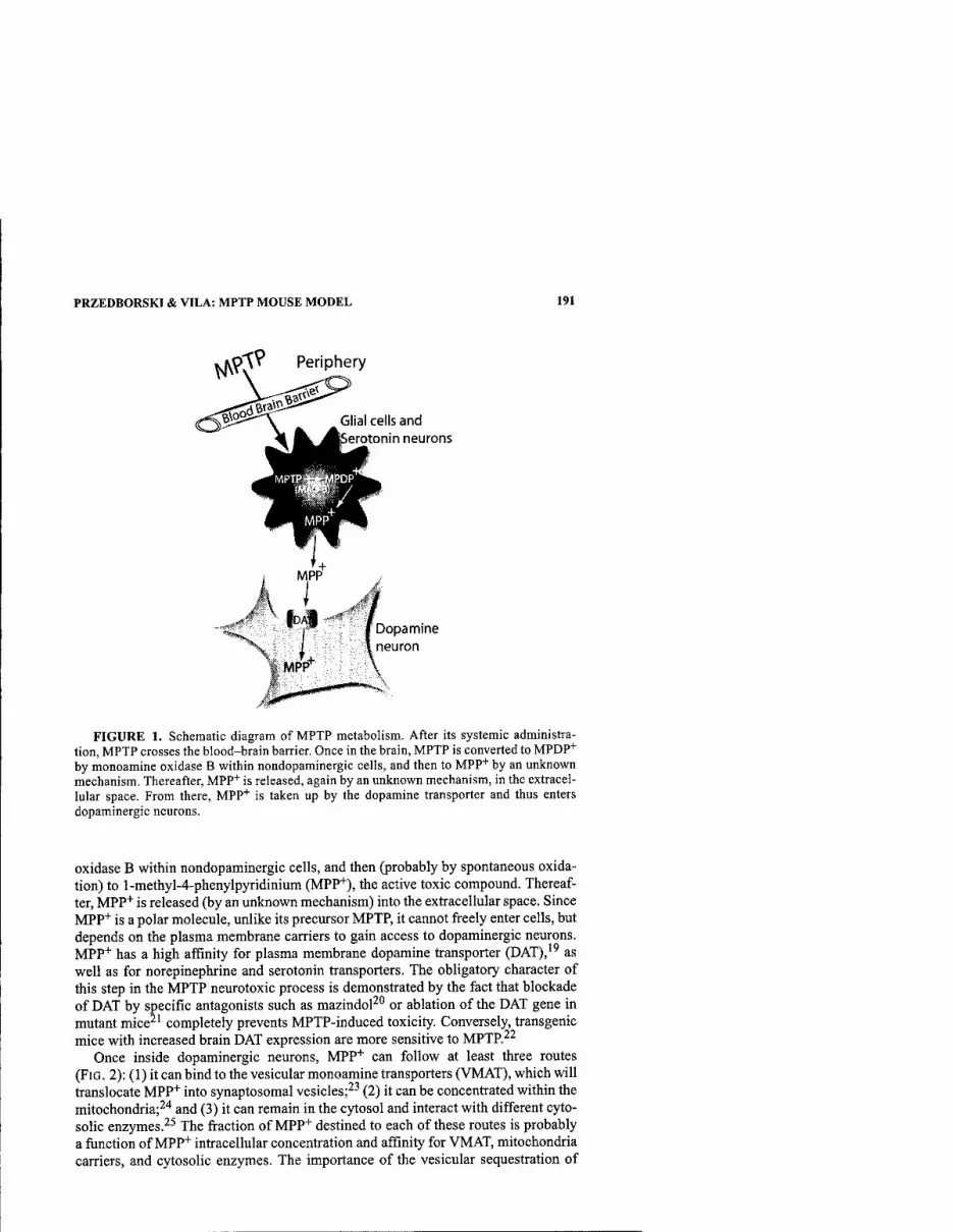
PRZEDBORSKI & VILA: MPTP MOUSE MODEL 191
Periphery
Glial cells and erotonin neurons
Dopamine neuron
MPF
FIGURE 1. Schematic diagram of MPTP metabolism. After its systemic administra- tion, MPTP crosses the blood-brain barrier. Once in the brain, MPTP is converted to MPOP"*" by monoamine oxidase B within nondopaminergic cells, and then to MPP* by an unknown mechanism. Thereafter, MPP+ is released, again by an unknown mechanism, in the extracel- lular space. From there, MPP"^ is taken up by the dopamine transporter and thus enters dopaminergic neurons.
oxidase B within nondopaminergic cells, and then (probably by spontaneous oxida- tion) to l-methyl-4-phenylpyridinium (MPP+), the active toxic compound. Thereaf- ter, MPP"*" is released (by an unknown mechanism) into the extracellular space. Since MPP* is a polar molecule, unlike its precursor MPTP, it cannot freely enter cells, but depends on the plasma membrane carriers to gain access to dopaminergic neurons. MPP* has a high affinity for plasma membrane dopamine transporter (DAT),'' as well as for norepinephrine and serotonin transporters. The obligatory character of this step in the MPTP neurotoxic process is demonstrated by the fact that blockade of DAT by specific antagonists such as mazindol^*^ or ablation of the DAT gene in mutant mice^' completely prevents MPTP-induced toxicity. Conversely, transgenic mice with increased brain DAT expression are more sensitive to MPTP.^^
Once inside dopaminergic neurons, MPP"*" can follow at least three routes (FIG. 2): (1) it can bind to the vesicular monoamine transporters (VMAT), which will translocate MPP"*" into synaptosomal vesicles;^^ (2) it can be concentrated within the mitochondria;^"* and (3) it can remain in the cytosol and interact with different cyto- solic enzymes.^^ The fraction of MPP+ destined to each of these routes is probably a function of MPP"*" intracellular concentration and affinity for VMAT, mitochondria carriers, and cytosolic enzymes. The importance of the vesicular sequestration of
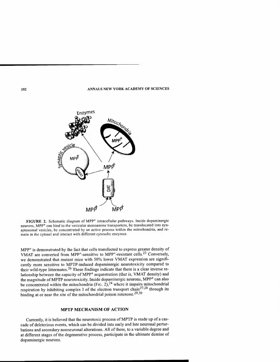
192 ANNALS NEW YORK ACADEMY OF SCIENCES
Enzymes
MPI^ MP^
FIGURE 2. Schematic diagram of MPP+ intracellular pathways. Inside dopaminergic neurons, MPP"*" can bind to the vesicular monoamine transporters, be translocated into syn- aptosomal vesicles, be concentrated by an active process within the mitochondria, and re- main in the cytosol and interact with different cytosolic enzymes.
MPP+ is demonstrated by the fact that cells transfected to express greater density of VMAT are conveiled from MPP"^-sensitive to MPP+-resistant cells.^^ Conversely, we demonstrated that mutant mice with 50% lower VMAT expression are signifi- cantly more sensitive to MPTP-induced dopaminergic neurotoxicity compared to their wild-type littermates.^* These findings indicate that there is a clear inverse re- lationship between the capacity of MPP+ sequestration (that is, VMAT density) and the magnitude of MPTP neurotoxicity. Inside dopaminergic neurons, MPP"*" can also be concentrated within the mitochondria (FIG. 2),^"* where it impairs mitochondrial respiration by inhibiting complex I of the electron transport chain^^-^^ through its binding at or near the site of the mitochondrial poison rotenone.^^'^"
MPTP MECHANISM OF ACTION
Currently, it is believed that the neurotoxic process of MPTP is made up of a cas- cade of deleterious events, which can be divided into early and late neuronal pertur- bations and secondary nonneuronal alterations. All of these, to a variable degree and at different stages of the degenerative process, participate in the ultimate demise of dopaminergic neurons.
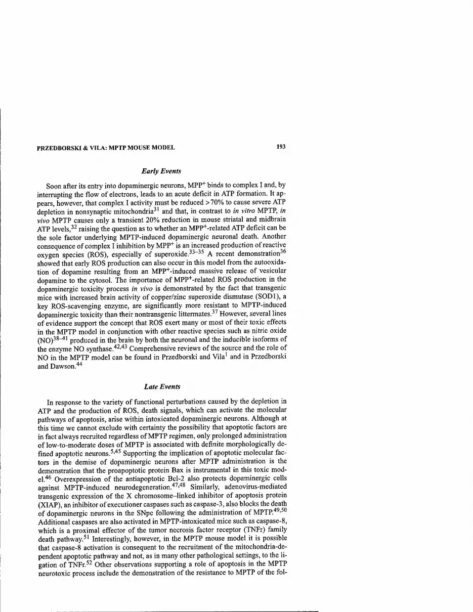
PRZEDBORSKI & VILA: MPTP MOUSE MODEL 193
Early Events
Soon after its entry into dopaminergic neurons, MPP"*" binds to complex I and, by interrupting the flow of electrons, leads to an acute deficit in ATP formation. It ap- pears, however, that complex I activity must be reduced >70% to cause severe ATP depletion in nonsynaptic mitochondria^' and that, in contrast to in vitro MPTP, in vivo MPTP causes only a transient 20% reduction in mouse striatal and midbrain ATP levels,^^ raising the question as to whether an MPP+-related ATP deficit can be the sole factor underlying MPTP-induced dopaminergic neuronal death. Another consequence of complex I inhibition by MPP+ is an increased production of reactive oxygen species (ROS), especially of superoxide.^^"^^ A recent demonstration^^ showed that early ROS production can also occur in this model from the autooxida- tion of dopamine resulting from an MPP^-induced massive release of vesicular dopamine to the cytosol. The importance of MPP+-related ROS production in the dopaminergic toxicity process in vivo is demonstrated by the fact that transgenic mice with increased brain activity of copper/zinc superoxide dismutase (SODl), a key ROS-scavenging enzyme, are significantly more resistant to MPTP-induced dopaminergic toxicity than their nontransgenic littermates.^^ However, several lines of evidence support the concept that ROS exert many or most of their toxic effects in the MPTP model in conjunction with other reactive species such as nitric oxide (NO)^^^' produced in the brain by both the neuronal and the inducible isoforms of the enzyme NO synthase.'*^''*^ Comprehensive reviews of the source and the role of NO in the MPTP model can be found in Przedborski and Vila' and in Przedborski and Dawson.^'*
Late Events
In response to the variety of functional perturbations caused by the depletion in ATP and the production of ROS, death signals, which can activate the molecular pathways of apoptosis, arise within intoxicated dopaminergic neurons. Although at this time we cannot exclude with certainty the possibility that apoptotic factors are in fact always recruited regardless of MPTP regimen, only prolonged administration of low-to-moderate doses of MPTP is associated with definite morphologically de- fined apoptotic neurons.^'"*^ Supporting the implication of apoptotic molecular fac- tors in the demise of dopaminergic neurons after MPTP administration is the demonstration that the proapoptotic protein Bax is instrumental in this toxic mod- el.''^ Overexpression of the antiapoptotic Bcl-2 also protects dopaminergic cells against MPTP-induced neurodegeneration.'*'^''*^ Similarly, adenovirus-mediated transgenic expression of the X chromosome-linked inhibitor of apoptosis protein (XIAP), an inhibitor of executioner caspases such as caspase-3, also blocks the death of dopaminergic neurons in the SNpc following the administration of MPTP."*^'^" Additional caspases are also activated in MPTP-intoxicated mice such as caspase-8, which is a proximal effector of the tumor necrosis factor receptor (TNFr) family death pathway.^' Interestingly, however, in the MPTP mouse model it is possible that caspase-8 activation is consequent to the recruitment of the mitochondria-de- pendent apoptotic pathway and not, as in many other pathological settings, to the li- gation of TNFr.^^ Other observations supporting a role of apoptosis in the MPTP neurotoxic process include the demonstration of the resistance to MPTP of the fol-
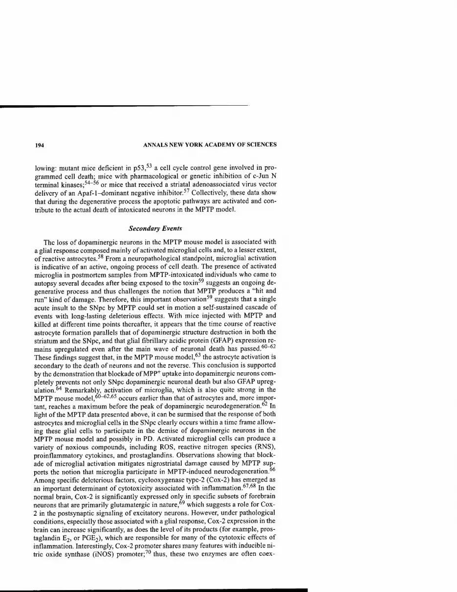
194 ANNALS NEW YORK ACADEMY OF SCIENCES
lowing: mutant mice deficient in p53,^^ a cell cycle control gene involved in pro- grammed cell death; mice with pharmacological or genetic inhibition of c-Jun N terminal kinases;''*"^^ or mice that received a striatal adenoassociated virus vector delivery of an Apaf-1-dominant negative inhibitor.^^ Collectively, these data show that during the degenerative process the apoptotic pathways are activated and con- tribute to the actual death of intoxicated neurons in the MPTP model.
Secondary Events
The loss of dopaminergic neurons in the MPTP mouse model is associated with a glial response composed mainly of activated microglial cells and, to a lesser extent, of reactive astrocytes.^^ From a neuropathological standpoint, microglial activation is indicative of an active, ongoing process of cell death. The presence of activated microglia in postmortem samples from MPTP-intoxicated individuals who came to autopsy several decades after being exposed to the toxin^^ suggests an ongoing de- generative process and thus challenges the notion that MPTP produces a "hit and run" kind of damage. Therefore, this important observation^^ suggests that a single acute insult to the SNpc by MPTP could set in motion a self-sustained cascade of events with long-lasting deleterious effects. With mice injected with MPTP and killed at different time points thereafter, it appears that the time course of reactive astrocyte formation parallels that of dopaminergic structure destruction in both the striatum and the SNpc, and that glial fibrillary acidic protein (GFAP) expression re- mains upregulated even after the main wave of neuronal death has passed.^*'~°^ These findings suggest that, in the MPTP mouse model,^^ the astrocyte activation is secondary to the death of neurons and not the reverse. This conclusion is supported by the demonstration that blockade of MPP"*" uptake into dopaminergic neurons com- pletely prevents not only SNpc dopaminergic neuronal death but also GFAP upreg- ulation.^'* Remarkably, activation of microglia, which is also quite strong in the MPTP mouse model,^''"^^•^^ occurs earlier than that of astrocytes and, more impor- tant, reaches a maximum before the peak of dopaminergic neurodegeneration.^^ In light of the MPTP data presented above, it can be surmised that the response of both astrocytes and microglial cells in the SNpc clearly occurs within a time frame allow- ing these glial cells to participate in the demise of dopaminergic neurons in the MPTP mouse model and possibly in PD. Activated microglial cells can produce a variety of noxious compounds, including ROS, reactive nitrogen species (RNS), proinflammatory cytokines, and prostaglandins. Observations showing that block- ade of microglial activation mitigates nigrostriatal damage caused by MPTP sup- ports the notion that microglia participate in MPTP-induced neurodegeneration.^* Among specific deleterious factors, cyclooxygenase type-2 (Cox-2) has emerged as an important determinant of cytotoxicity associated with inflammation.*^'^^ In the normal brain, Cox-2 is significantly expressed only in specific subsets of forebrain neurons that are primarily glutamatergic in nature,^^ which suggests a role for Cox- 2 in the postsynaptic signaling of excitatory neurons. However, under pathological conditions, especially those associated with a glial response, Cox-2 expression in the brain can increase significantly, as does the level of its products (for example, pros- taglandin E2, or PGE2), which are responsible for many of the cytotoxic effects of inflammation. Interestingly, Cox-2 promoter shares many features with inducible ni- tric oxide synthase (iNOS) promoter;^^ thus, these two enzymes are often coex-
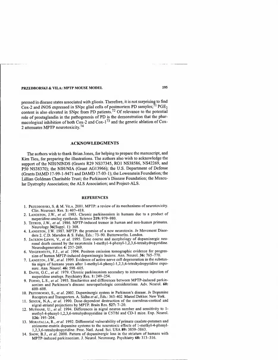
PRZEDBORSKI & VILA: MPTP MOUSE MODEL 195
pressed in disease states associated with gliosis. Therefore, it is not surprising to find Cox-2 and iNOS expressed in SNpc glial cells of postmortem PD samples;'' PGE2 content is also elevated in SNpc from PD patientsJ^ Of relevance to the potential role of prostaglandin in the pathogenesis of PD is the demonstration that the phar- macological inhibition of both Cox-2 and Cox-l'^ and the genetic ablation of Cox- 2 attenuates MPTP neurotoxicity.'^
ACKNOWLEDGMENTS
The authors wish to thank Brian Jones, for helping to prepare the manuscript, and Kim Tieu, for preparing the illustrations. The authors also wish to acknowledge the support of the NIH/NINDS (Grants R29 NS37345, ROl NS38586, NS42269, and P50 NS38370); the NIH/NIA (Grant AG13966); the U.S. Department of Defense (Grants DAMD 17-99-1-9471 and DAMD 17-03-1); the Lowenstein Foundation; the Lillian Goldman Charitable Trust; the Parkinson's Disease Foundation; the Muscu- lar Dystrophy Association; the ALS Association; and Project-ALS.
REFERENCES
1. PRZEDBORSKI, S. & M. ViLA. 2001. MPTP: a review of its mechanisms of neurotoxicity. Clin. Neurosci. Res. 1: 407-418.
2. LANGSTON, J.W., et al. 1983. Chronic parkinsonism in humans due to a product of meperidine-analog synthesis. Science 219: 979-980.
3. TETRUD, J.W., et al. 1986. MPTP-induced tremor in human and non-human primates. Neurology 36(Suppl. 1): 308.
4. LANGSTON, J.W. 1987. MPTP: the promise of a new neurotoxin. In Movement Disor- ders 2. CD. Marsden & S. Fahn, Eds.: 73-90. Butterworths. London.
5. JACKSON-LEWIS, V., et al. 1995. Time course and morphology of dopaminergic neu- ronal death caused by the neurotoxin l-methyl-4-phenyl-l,2,3,6-tetrahydropyridine. Neurodegeneration 4: 257-269.
6. ViNGERHOETS, F.J., et al. 1994. Positron emission tomographic evidence for progres- sion of human MPTP-induced dopaminergic lesions. Ann. Neurol. 36:165-110.
1. LANGSTON, J.W., et al. 1999. Evidence of active nerve cell degeneration in the substan- tia nigra of humans years after l-methyl-4-phenyl-l,2,3,6-tetrahydropyridine expo- sure. Ann. Neurol. 46: 598-605.
8. DAVIS, G.C, et al. 1979. Chronic parkinsonism secondary to intravenous injection of meperidine analogs. Psychiatry Res. 1: 249-254.
9. FORNO, L.S., et al. 1993. Similarities and differences between MPTP-induced parkin- sonism and Parkinson's disease: neuropathologic considerations. Adv. Neurol. 60: 600-608.
10. PRZEDBORSKI, S., et al. 2002. Dopaminergic system in Parkinson's disease. In Dopamine Receptors and Transporters. A. Sidhu et al, Eds.: 363^02. Marcel Dekker. New York.
11. SENIUK, N.A., et al. 1990. Dose-dependent destruction of the coeruleus-cortical and nigral-striatal projections by MPTR Brain Res. 527: 7-20.
12. MUTHANE, U., et al. 1994. Differences in nigral neuron number and sensitivity to 1- methyl-4-phenyI-l,2,3,6-tetrahydropyridine in C57/bl and CD-I mice. Exp. Neurol. 126: 195-204.
13. MORATALLA, R., et al. 1992. Differential vulnerability of primate caudate-putamen and striosome-matrix dopamine systems to the neurotoxic effects of l-methyl-4-phenyl- 1,2,3,6-tetrahydropyridine. Proc. Natl. Acad. Sci. USA 89: 3859-3863.
14. SNOW, B.J., et al. 2000. Pattern of dopaminergic loss in the striatum of humans with MPTP-induced parkinsonism. J. Neurol. Neurosurg. Psychiatry 68: 313-316.
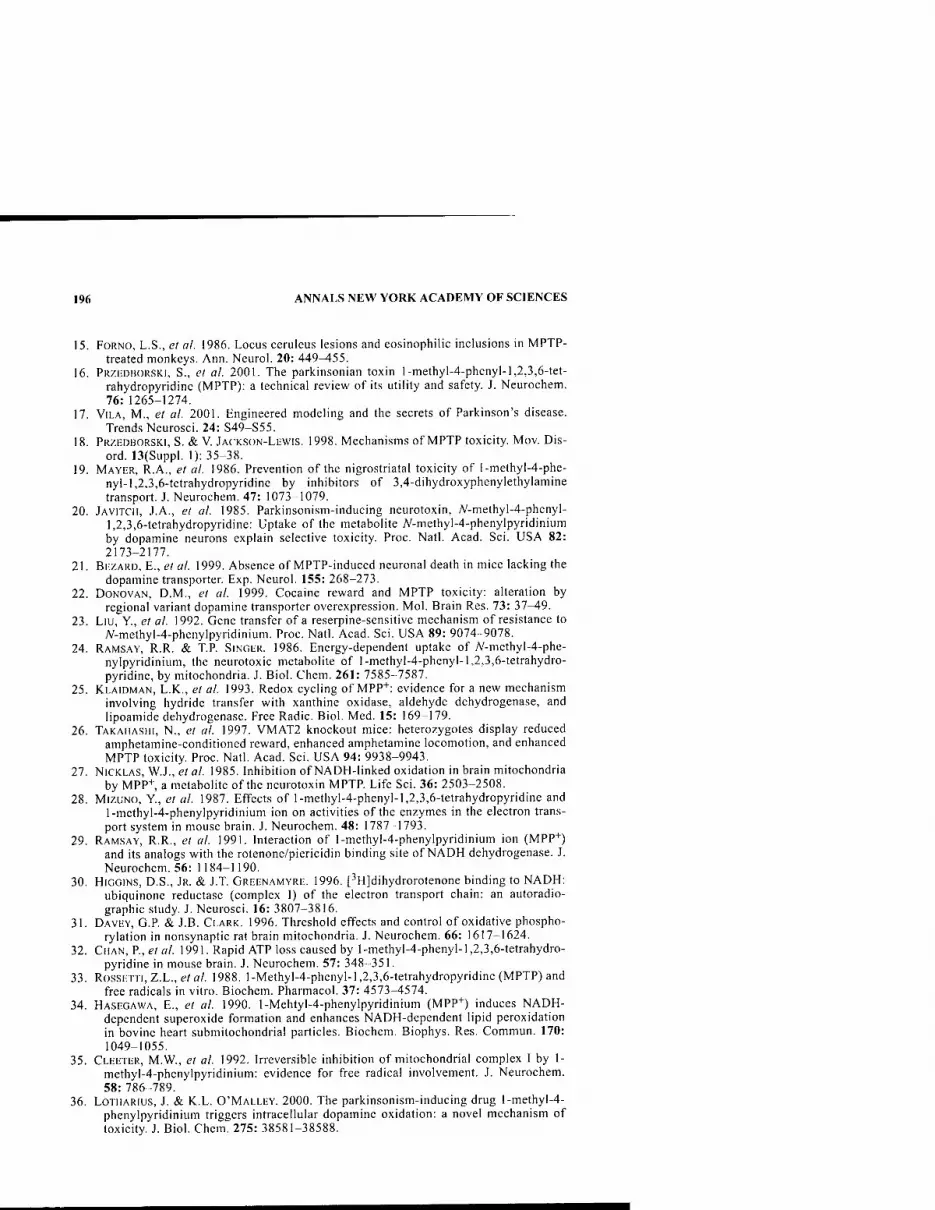
196 ANNALS NEW YORK ACADEMY OF SCIENCES
15. FoRNO, L.S., el al. 1986. Locus cerulcus lesions and eosinophilic inclusions in MPTP- treated monkeys. Ann. Neurol. 20: 449-455.
16. PRZEDBORSKI, S., et al. 2001. The parkinsonian toxin l-methyl-4-phenyl-1,2,3,6-tet- rahydropyridinc (MPTP): a technical review of its utility and safety. J. Neurochem. 76: 1265-1274.
17. VILA, M., et al. 2001. Engineered modeling and the secrets of Parkinson's disease. Trends Neurosci. 24: S49-S55.
18. PRZEDHORSKI, S. & V. JACKSON-LEWIS. 1998. Mechanisms of MPTP toxicity. Mov. Dis- ord. 13(Suppl. 1): 35-38.
19. MAYER, R.A., el al. 1986. Prevention of the nigrostriatal toxicity of l-methyl-4-phe- nyl-l,2,3,6-tetrahydropyridinc by inhibitors of 3,4-dihydroxyphenylethylamine transport. J. Neurochem. 47: 1073-1079.
20. jAViTCll, J.A., et al. 1985. Parkinsonism-inducing neurotoxin, A'-methyl-4-phenyl- 1,2,3,6-tetrahydropyridine: Uptake of the metabolite 7V-methyl-4-phenylpyridinium by dopamine neurons explain selective toxicity. Free. Natl. Acad. Sci. USA 82: 2173-2177.
21. BEZARD, E., el al. 1999. Absence of MPTP-induced ncuronal death in mice lacking the dopamine transporter. Exp. Neurol. 155: 268-273.
22. DONOVAN, D.M., el al. 1999. Cocaine reward and MPTP toxicity: alteration by regional variant dopamine transporter overexpression. Mol. Brain Res. 73: 37^9.
23. LIU, Y., et al. 1992. Gene transfer of a reserpine-sensitive mechanism of resistance to A'-methyl-4-phenylpyridinium. Proc. Natl. Acad. Sci. USA 89: 9074-9078.
24. RAMSAY, R.R. & T.P. SINGER. 1986. Energy-dependent uptake of A'-mcthyl-4-phe- nylpyridinium, the neurotoxic metabolite of l-methyl-4-phenyl-l,2,3,6-tetrahydro- pyridinc, by mitochondria. J. Biol. Chem. 261: 7585-7587.
25. KLAIDMAN, L.K., el al. 1993. Redox cycling of MPP"''; evidence for a new mechanism involving hydride transfer with xanthine oxidase, aldehyde dehydrogenase, and lipoaniide dehydrogenase. Free Radic. Biol. Med. 15: 169-179.
26. TAKAIIASIII, N., el al. 1997. VMAT2 knockout mice: heterozygotes display reduced amphetamine-conditioned reward, enhanced amphetamine locomotion, and enhanced MPTP toxicity. Proc. Natl. Acad. Sci. USA 94: 9938-9943.
27. NiCKLAS, W.J., et al. 1985. Inhibition of NADH-linked oxidation in brain mitochondria by MPP+, a metabolite of the neurotoxin MPTR Life Sci. 36: 2503-2508.
28. MIZUNO, Y., el al. 1987. Effects of l-methyl-4-phenyl-l,2,3,6-tetrahydropyridine and l-methyl-4-phenylpyridinium ion on activities of the enzymes in the electron trans- port system in mouse brain. J. Neurochem. 48: 1787-1793.
29. RAMSAY, R.R., el al. 1991. Interaction of l-methyl-4-phenylpyridinium ion (MPP+) and its analogs with the rotenonc/piericidin binding site of NADH dehydrogenase. J. Neurochem. 56: 1184-1190.
30. HiGGiNS, D.S., JR. & J.T. GREENAMYRE. 1996. ['H]dihydrorotenone binding to NADH: ubiquinone reductasc (complex I) of the electron transport chain: an autoradio- graphic study. J. Neurosci. 16: 3807-3816.
31. DAVEY, G.P. & J.B. CLARK. 1996. Threshold effects and control of oxidative phospho- rylation in nonsynaptic rat brain mitochondria. J. Neurochem. 66: 1617-1624.
32. CHAN, ?.,el al. 1991. Rapid ATP loss caused by l-methyl-4-phenyl-l,2,3,6-tetrahydro- pyridine in mouse brain. J. Neurochem. 57: 348-351.
33. RosSETTi, Z.L., e/o/. 1988. l-Methyl-4-phenyl-l,2,3,6-tetrahydropyridine (MPTP) and free radicals in vitro. Biochem. Pharmacol. 37: 4573-4574.
34. HASEGAWA, E., el al. 1990. l-Mchtyl-4-phenylpyridinium (MPP"^) induces NADH- dependent superoxide formation and enhances NADH-dependent lipid peroxidation in bovine heart submilochondrial particles. Biochem. Biophys. Res. Commun. 170: 1049-1055.
35. CLEETER, M.W., et al. 1992. Irreversible inhibition of mitochondrial complex 1 by 1- methyl-4-phcnylpyridinium: evidence for free radical involvement. J. Neurochem. 58: 786-789.
36. LoTiiARius, J. & K.L. O'MALLEY. 2000. The parkinsonism-inducing drug l-methyl-4- phenylpyridinium triggers intracellular dopamine oxidation: a novel mechanism of toxicity. J. Biol. Chcm. 275: 38581-38588.

PRZEDBORSKI & VILA: MPTP MOUSE MODEL 197
37. PRZEDBORSKI, S., et al. 1992. Transgenic mice with increased Cu/Zn-superoxide dis- mutase activity are resistant to Af-methyl-4-phenyl-1,2,3,6-tetrahydropyridine- induced neurotoxicity. J. Neurosci. 12: 1658-1667.
38. SCHULZ, J.B., et al. 1995. Inhibition of neuronal nitric oxide synthase by 7-nitroindazole protects against MPTP-induced neurotoxicity in mice. J. Neurochem. 64: 936-939.
39. PENNATHUR, S., et al. 1999. Mass spectrometric quantification of 3-nitrotyrosine, ortho-tyrosine, and o,o'-dityrosine in brain tissue of l-methyl-4-phenyl-l,2,3,6-tet- rahydropyridine-treated mice, a model of oxidative stress in Parkinson's disease. J. Biol. Chem. 274: 34621-34628.
40. ARA, J., et al. 1998. Inactivation of tyrosine hydroxylase by nitration following expo- sure to peroxynitrite and l-methyl-4-phenyl-l,2,3,6-tetrahydropyridine (MPTP). Proc. Natl. Acad. Sci. USA 95: 7659-7663.
41. PRZEDBORSKI, S., etal. 2001. Oxidative post-translational modifications of alpha-synu- clein in the l-methyl-4-phenyI-l,2,3,6-tetrahydropyridine (MPTP) mouse model of Parkinson's disease. J. Neurochem. 76: 637-640.
42. LiBERATORE, G., Bt al. 1999. Inducible nitric oxide synthase stimulates dopaminergic neurodegeneration in the MPTP model of Parkinson's disease. Nat. Med. 5: 1403- 1409.
43. PRZEDBORSKI, S., et al. 1996. Role of neuronal nitric oxide in MPTP (l-methyl-4-phe- nyl-l,2,3,6-tetrahydropyridine)-induced dopaminergic neurotoxicity. Proc. Natl. Acad. Sci. USA 93: 4565-4571.
44. PRZEDBORSKI, S. & T.M. DAWSON. 2001. The role of nitric oxide in Parkinson's dis- ease. In Parkinson's Disease. Methods and Protocols. M.M. Mouradian, Ed.: 113- 136. Humana Press. Totowa, NJ.
45. TATTON, N.A. & S.J. KiSH. 1997. In situ detection of apoptotic nuclei in the substantia nigra compacta of l-methyl-4-phenyl-l,2,3,6-tetrahydropyridine-treated mice using terminal deoxynucleotidyl transferase labelling and acridine orange staining. Neuro- science77: 1037-1048.
46. VILA, M., et al. 2001. Bax ablation prevents dopaminergic neurodegeneration in the 1- methyl-4-phenyl-l,2,3,6-tetrahydropyridine mouse model of Parkinson's disease. Proc. Natl. Acad. Sci. USA 98: 2837-2842.
47. YANG, L., et al. 1998. l-Methyl-4-phenyl-l,2,3,6-tetrahydropyride neurotoxicity is attenuated in mice overexpressing Bcl-2. J. Neurosci. 18: 8145-8152.
48. OFFEN, D., et al. 1998. Transgenic mice expressing human Bcl-2 in their neurons are resistant to 6-hydroxydopamine and l-methyl-4-phenyl-l,2,3,6-tetrahydropyridine neurotoxicity. Proc. Natl. Acad. Sci. USA 95: 5789-5794.
49. Xu, D., et al. 1999. Attenuation of ischemia-induced cellular and behavioral deficits by X chromosome-linked inhibitor of apoptosis protein overexpression in the rat hip- pocampus. J. Neurosci. 19: 5026-5033.
50. EBERHARDT, O., et al. 2000. Protection by synergistic effects of adenovirus-mediated X-chromosome-linked inhibitor of apoptosis and glial cell line-derived neurotrophic factor gene transfer in the l-methyl-4-phenyl-l,2,3,6-tetrahydropyridine model of Parkinson's disease. J. Neurosci. 20: 9126-9134.
51. HARTMANN, A., et al. 2001. Caspase-8 is an effector in apoptotic death of dopaminer- gic neurons in Parkinson's disease, but pathway inhibition results in neuronal necro- sis. J Neurosci. 21: 2247-2255.
52. ViswANATH, v., et al. 2001. Caspase-9 activation results in downstream caspase-8 acti- vation and bid cleavage in l-methyl-4-phenyl-l,2,3,6-tetrahydropyridine-induced Parkinson's disease. J. Neurosci. 21: 9519-9528.
53. TRIMMER, P.A., et al. 1996. Dopamine neurons from transgenic mice with a knockout of the p53 gene resist MPTP neurotoxicity. Neurodegeneration 5: 233-239.
54. SAPORITO, M.S., et al. 2000. MPTP activates c-Jun NH(2)-terminal kinase (JNK) and its upstream regulatory kinase MKK4 in nigrostriatal neurons in vivo. J. Neurochem. 75: 1200-1208.
55. SAPORITO, M.S., et al. 1999. CEP-1347/KT-7515, an inhibitor of c-jun N-terminal kinase activation, attenuates the l-methyl-4-phenyl tetrahydropyridine-mediated loss of nigrostriatal dopaminergic neurons in vivo. J. Pharmacol. Exp. Ther. 288: 421- 427.

198 ANNALS NEW YORK ACADEMY OF SCIENCES
56. XiA, X.G., et al. 2001. Gene transfer of the JNK interacting protein-1 protects dopam- inergic neurons in the MPTP model of Parkinson's disease. Proc. Natl. Acad. Sci. USA 98: 10433-10438.
57. MOCHIZUKI, H., et al. 2001. An AAV-derived Apaf-1 dominant negative inhibitor pre- vents MPTP toxicity as antiapoptotic gene therapy for Parkinson's disease. Proc. Natl. Acad. Sci. USA 98: 10918-10923.
58. VILA, M., et al. 2001. The role of glial cells in Parkinson's disease. Curr. Opin. Neurol. 14: 483-489,
59. LANGSTON, J.W., et al. 1999. Evidence of active nerve cell degeneration in the substan- tia nigra of humans years after l-methyl-4-phenyl-l,2,3,6-tetrahydropyridine expo- sure. Ann. Neurol. 46: 598-605.
60. CzLONKOWSKA, A., el al. 1996. Microglial reaction in MPTP (l-methyl-4-phenyl- 1,2,3,6-tetrahydropyridine) induced Parkinson's disease mice model. Neurodegener- ationS: 137-143.
61. KOHUTNICKA, M., et al. 1998. Microglial and astrocytic involvement in a murine model of Parkinson's disease induced by l-methyl-4-phenyl-l,2,3,6-tetrahydropyridine (MPTP). Immunopharmacology 39: 167-180.
62. LiBERATORE, G.T., et al. 1999. Inducible nitric oxide synthase stimulates dopaminergic neurodegeneration in the MPTP model of Parkinson disease. Nat. Med. 5: 1403- 1409.
63. PRZEDBORSKI, S., et al. 2000. The parkinsonian toxin MPTP: Action and mechanism. Restor. Neurol. Neurosci. 16: 135-142.
64. O'CALLAGHAN, J.P., et al. 1990. Characterization of the origins of astroeyte response to injury using the dopaminergic neurotoxicant, l-methyl-4-phenyl-l,2,3,6-tetrahy- dropyridine. Brain Res. 521: 73-80.
65. DEHMER, T., et al. 2000. Deficiency of inducible nitric oxide synthase protects against MPTP toxicity in vivo. J. Neurochem. 74: 2213-2216.
66. Wu, D.C., et al. 2002. Blockade of microglial activation is neuroprotective in the 1- methyl-4-phenyl-l,2,3,6-tetrahydropyridine mouse model of Parkinson disease. J. Neurosci. 22: 1763-1771.
67. SEIBERT, K., et al. 1995. Mediation of inflammation by cyclooxygenase-2. Agents Actions Suppl. 46: 41-50.
68. O'BANION, M.K. 1999. Cyclooxygenase-2: molecular biology, pharmacology, and neu- robiology. Crit. Rev, Neurobiol. 13: 45-82.
69. KAUFMANN, W.E., et al. 1996, COX-2, a synaptically induced enzyme, is expressed by excitatory neurons at postsynaptic sites in rat cerebral cortex. Proc. Natl. Acad. Sci. USA 93: 2317-2321,
70. NATHAN, C. & Q.W. XIE. 1994, Regulation of biosynthesis of nitric oxide. J. Bid. Chem. 269: 13725-13728,
71. KNOTT, C, et al. 2000. Inflammatory regulators in Parkinson's disease: iNOS, lipocor- tin-1, and cyclooxygenases-1 and -2. Mol. Cell. Neurosci. 16: 724-739.
72. MATTAMMAL, M.B,, et al. 1995. Prostaglandin H synthetase-mediated metabolism of dopamine: Implication for Parkinson's disease. J. Neurochem. 64: 1645-1654.
73. TEISMANN, P, & B, FERGER. 2001. Inhibition of the cyclooxygenase isoenzymes COX-1 and COX-2 provide neuroprotection in the MPTP-mouse model of Parkinson's dis- ease. Synapse 39: 167-174.
74. FENG, Z., et al. 2002. Cyclooxygenase-2-deficient mice are resistant to l-methyI-4- phenyl-l,2,3,6-tefrahydropyridine-induced damage of dopaminergic neurons in the substantia nigra. Neurosci. Lett. 329: 354.
Pathophysiology of Parkinson's Disease: The MPTP Primate Model of the Human Disorder
THOMAS WICHMANN AND MAHLON R. DELONG
Department of Neurology, Emory University School of Medicine,
Atlanta Georgia 30322, USA
ABSTRACT: The striatum Is viewed as the principal input structure of the basal ganglia, while the internal pallidal segment (GPi) and the substantia nigra pars reticulata (SNr) are output structures. Input and output structures are linked via a monosynaptic "direct" pathway and a polysynaptic "Indirect" pathway involving the external pallidal segment (GPe) and the subthalamic nucleus (STN). According to current schemes, striatal dopamine (DA) enhances trans- mission along the direct pathway (via Dl receptors), and reduces transmission over the indirect pathway (via D2 receptors). DA also acts on receptors in GPe, GPi, SNr, and STN. Electrophysiologic and other studies in primates rendered parkinsonian by treatment with the dopaminergic neurotoxin MPTP have demonstrated a reduction of neuronal activity of GPe and an increase of neu- ronal discharge in STN, GPi. and SNr. These findings are compatible with the view that striatal DA loss results in increased activity over the indirect path- way. Prominent bursting, oscillatory discharge patterns, and increased syn- chronization of neighboring neurons are found throughout the basal ganglia. These may result from changes in the activity of local circuits (e.g., the GPe-STN "pacemaker") or from more global abnormalities of the basal gan- glia-thalamocortical network. These findings have been replicated in human patients undergoing microelectrode-guided stereotactic procedures targeted at GPi or STN. PET studies in patients with Parkinson's disease have lent further support to the proposed circuit abnormalities. The current models of basal ganglia function have recently been criticized. For instance, the strict separa- tion of direct and indirect pathways and the segregation of Dl and D2 receptors have been questioned, and the almost complete absence of motor side effects of pallidal or thalamic lesions in human patients and animals is inconsistent. These results suggest that changes in discharge patterns and synchronization between basal ganglia neurons, abnormal network interactions, and compen- satory mechanisms are at least as important in the pathopohysiology of parkin- sonism as changes in discharge rates in individual basal ganglia nuclei. Lesions of GPi or STN are effective in treating parkinsonism, because they reduce or abolish abnormal basal ganglia output, enabling remaining circuits to function more normally.
KEYWORDS: MPTP; primate; pathophysiology; pallidum; subthalamic nucle- us; substantia nigra
Address for correspondence: Thomas Wichmann, M.D., Department of Neurology, Emory University, Suite 6000, Woodruff Memorial Research Building, 1639 Pierce Drive, Atlanta, GA 30322. Voice: 404-727-3818; fax: 404-727-3157.
Ann. N.Y. Acad. Sci. 991: 199-213 (2003). © 2003 New York Academy of Sciences.
199
200 ANNALS NEW YORK ACADEMY OF SCIENCES
INTRODUCTION
Recent progress in neuroscience research has led to major insights into the struc- ture and function of the basal ganglia and into the pathophysiologic basis of disor- ders of basal ganglia origin, such as Parkinson's disease.'"^ The availability of suitable animal models, in particular the MPTP model of primate parkinsonism, has been crucial in this progress.'*'^ In addition, the renaissance of stereotactic surgery for Parkinson's disease and other movement disorders has provided valuable neu- ronal recording and imaging data from human subjects. In the following, we sum- marize, from a systems perspective, the pathophysiologic concepts that have arisen from the animal models and from work in patients with Parkinson's disease.
PATHOLOGIC SUBSTRATE IN PARKINSON'S DISEASE
Idiopathic Parkinson's disease is characterized by the cardinal signs of akinesia (impaired movement initiation and poverty of movement), bradykinesia (slowness of movement), muscular rigidity, and tremor at rest. The etiology of the disease is most likely multifactorial, with both genetic and environmental/toxic factors resulting in a relatively selective degeneration of dopaminergic neurons in the substantia nigra pars compacta (SNc), which project to the striatum (e.g., Ref 6), and, to a lesser ex- tent, to other basal ganglia nuclei such as the external and internal segments of the globus pallidus (GPe, GPi, respectively), the subthalamic nucleus (STN), and the substantia nigra, pars reticulata (SNr).^ In early phases of the disease, dopamine loss affects particularly the sensorimotor portion of the striatum, the putamen, resulting in the appearance of motor disturbances early in the disease (e.g., Ref. 7). In later stages, more widespread dopamine loss affecting other regions of the basal ganglia (such as the caudate nucleus and the pallidum) and extrabasal ganglia areas (such as cortex, hypothalamus, and thalamus) as well as spread of neuronal degeneration to nondopaminergic systems (such as the locus coeruleus and the raphe nuclei) may cause the development of additional signs and symptoms (such as cognitive disabil- ities, sleep disorders, and mood disturbances).
ANATOMICAL SUBSTRATE FOR CIRCUIT DYSFUNCTION IN PARKINSONISM
In order to understand how the loss of dopamine in the basal ganglia leads to the signs and symptoms of parkinsonism, it is necessary first to consider some of the an- atomic and physiologic details of the basal ganglia and related structures.
The basal ganglia are a group of functionally related subcortical nuclei that in- clude the neostriatum (comprised of the caudate nucleus and the putamen), ventral striatum, GPe, STN, GPi, SNr and SNc. They are anatomically related to large por- tions of the cerebral cortex, thalamus, and brain stem. The corticobasal ganglia- thalamocortical circuits appear to be organized in an orderly arrangement of segre- gated reentrant circuits that is thought to significantly enhance the efficiency and speed of cortical processing (see left half of FIG. 1). Most authors believe, however, that the range of behaviors seen in humans, nonhuman primates, and other species

WICHMANN & DELONG: MPTP PRIMATE MODEL OF PD
Normal Parkinsonism
201
CORTEX CORTEX
mm/ANU ■CMVAA/L
Brain stem/ j ' f Hi < ' ^''^'" ®'®'^'" Spinal cord *=^ ' Spinal cord
FIGURE 1. Simplified schematic diagram of the basal ganglia-thalamocortical circuit- ry under normal conditions (left) and rate changes in parkinsonism (right). Inhibitory con- nections are shown as filled arrows, excitatory connections as open arrows. The principal input nuclei of the basal ganglia, the striatum and the STN, are connected to the output nu- clei, GPi and SNr. Basal ganglia output is directed at several thalamic nuclei (VA/VL and CM) and at brain stem nuclei (PPN and others). In parkinsonism, dopaminergic neurons in the SNc degenerate, which results, via a cascade of changes in the other basal ganglia nuclei, in increased basal ganglia output from GPi and SNr. This, in turn, is thought to lead to inhi- bition of related thalamic and cortical neurons. In addition to the changes shown here, there are prominent alterations in discharge patterns (see text). For abbreviations, see text.
requires some intermodular transfer of information. Such cross-talk may occur through interactions of the basal ganglia with some of the brain stem areas or the SNc, which are not as distinctly organized into functional territories.
In primates, projections from the somatosensory, motor, and premotor cortices terminate in the postcommissural putamen, the "motor portion" of the striatum;^-' while associative cortical areas project to the caudate nucleus and the precommis- sural putamen;'^''' and projections from limbic cortices, amygdala, and hippocam- pus terminate preferentially in the ventral striatum (e.g., Ref. 12). Cortical inputs also terminate in the STN.'^'^'' This projection originates in the primary motor, pre- frontal, and premotor cortices.'^''''
A second major group of inputs to striatum and STN arises from the intralaminar thalamic nuclei, the centromedian nucleus (CM) and the parafascicular nucleus (Pf) (e.g., Refs. 15-17). In primates, CM projects to the motor portions of putamen and STN, whereas Pf projects largely to the associative and limbic territories.'^"^*'
Topographically segregated cortical information is conveyed from the striatum to the output nuclei of the basal ganglia (GPi and SNr). Striatofugal projections main-
202 ANNALS NEW YORK ACADEMY OF SCIENCES
tain the striatal organization into motor, limbic, associative, and oculomotor territo- ries.^' The connections between the striatum and the basal ganglia output nuclei are thought to be organized into a direct and an indirect pathway.^'^^'^^ The direct path- way arises from striatal neurons that project monosynaptically to neurons in GPi and SNr, whereas the indirect pathway arises from a different set of neurons that projects to GPe (see Ref. 24 for a review). Some striatofugal neurons may also collateralize more extensively, reaching GPe, GPi, and SNn'^^ GPe conveys the information it re- ceives either directly to GPi/SNr or via the STN.
The population of striatal neurons that gives rise to the direct pathway can be fur- ther characterized by the presence of the neuropeptides substance P and dynorphin, by the preferential expression of the dopamine Dl receptors, and by the fact that these neurons (as well as most striatal interneurons) appear to be the targets of tha- lamic inputs from the centromedian nucleus.^^'^^ The population that gives rise to the indirect pathway expresses preferentially enkephalin and dopamine D2 receptors^^'^' and may be the principal target of cortical inputs.^^'^^ The functional- ly important segregation of Dl and D2 receptors between the direct and indirect pathways has been most clearly demonstrated in dopamine-depleted animals, while several studies in normal animals have supported the existence of a degree of overlap between Dl- and D2-positive cell populations (e.g., Refs. 29, 30). However, the Dl/ D2 dichotomy may still serve to explain the apparent dual action of dopamine, re- leased from the nigrostriatal pathway arising in the substantia nigra pars compacta, on striatal output. Dopamine appears to modulate the activity of the basal ganglia output neurons in GPi and SNr by facilitation of transmission over the direct path- way and inhibition of transmission over the indirect pathway (e.g., Ref 31). The net effect of striatal dopamine release appears to be to reduce basal ganglia output. A reduction of dopamine release as is seen in Parkinson's disease will therefore result in a net increase in basal ganglia output.
Basal ganglia output arises from both GPi and SNr. The division of GPi into a caudoventral "motor" portion and rostromedial associative and limbic areas^^ is maintained in the pallidothalamic projections.^^ The motor territory of GPi projects almost exclusively to the posterior part of the ventrolateral nucleus (VLo in macaques), which in turn sends projections towards the supplementary motor area (SMA),^^'-'^ the primary motor cortex (Ml),-'-'"^^ and the premotor (PM) cortical ar- eas.^^ Associative and limbic areas project preferentially to the parvocellular part of the ventral anterior (VA) and the dorsal VL nucleus (VLc in macaques)^^ and may be transmitted in turn to prefrontal cortical areas,■'^'^^ as well as to motor and sup- plementary motor regions^^).
The SNr can be broadly subdivided into a dorsolateral sensorimotor and a ventro- medial associative territory (e.g., Refs. 40, 41). Projections from the SNr to the thal- amus terminate in the magnocellular division of the ventral anterior nucleus (VAmc) and in the mediodorsal nucleus (MDmc). These nuclei, in turn, innervate anterior re- gions of the frontal lobe, including the principal sulcus (area 46) and the orbital cor- tex (area 11),"*^ as well as premotor areas and the frontal eye field.^^
Both GPi and SNr also send projections to the noncholinergic neurons of the pp-[^43^5 ^jjj fjjg CM/Pf nuclei.■^'^'^■' Additional projections from the SNr reach the superior colliculus; these may play a critical role in the control of saccades and ori- enting behaviors.'*^

WICHMANN & DELONG: MPTP PRIMATE MODEL OF PD 203
MOVEMENT-RELATED BASAL GANGLIA ACTIVITY IN NORMAL AND PARKINSONIAN ANIMALS
Voluntary movements appear to be initiated at the cortical level of the motor cir- cuit, with output to brain stem and spinal cord, and to multiple subcortical targets including the thalamus, putamen, and the SIN. Cortical activation of an ensemble of striatal motor territory neurons that give rise to the direct pathvi^ay leads to a reduc- tion of inhibitory basal ganglia output and subsequent disinhibition of related thalamocortical neurons'*'' and facilitation of the movement. In contrast, activation of striatal neurons that give rise to the indirect pathway will lead to increased basal ganglia output and, presumably, to suppression of movement. Since the majority of neurons in GPi increase their firing rate with movement,''^'''^ it has been speculated that the main role of the basal ganglia is to inhibit and stabilize the activity of the thalamocortical network. As far as motor function is concerned, the basal ganglia may play a role in the control of specific kinematic parameters, such as amplitude, velocity, and direction (see, e.g., Refs. 47, 50-52) or may "focus" movements," al- lowing intended movements to proceed and suppressing unintended movement (see discussion in Ref. 1). Besides these elemental functions in motor control, a multitude of other motor functions have been proposed, such as a role in self-initiated (inter- nally generated) movements, in motor (procedural) learning, and in movement se- quencing (e.g., Refs. 54-56). The functions of the nonmotor portions of the basal ganglia may be analogous to the motor functions but are less well explored.
Global Activity Changes in the Basal Ganglia
The study of pathophysiologic changes in the basal ganglia that result from loss of dopaminergic transmission has been greatly facihtated by the discovery that primates treated with MPTP develop behavioral and anatomic changes that closely mimic the features of Parkinson's disease in humans.^'"'^* Early studies in this model suggested that the metabolic activity (as measured with the 2-deoxy-glucose technique) is in- creased in both pallidal segments (e.g., Refs. 59,60). This was interpreted as evidence for increased activity of the striatum-GPe connection and the STN-GPi pathway; or, alternatively, as evidence for increased activity via the projections from the STN to both pallidal segments. It was then shovra with microelectrode recordings of neuronal activity that MPTP-induced parkinsonism in primates is associated with reduced tonic neuronal discharge in GPe and increased discharge in STN, GPi, and SNr, as compared to normal controls (see example recordings in FIG. 2 and Refs. 61-64). In parkinsonian patients undergoing pallidotomy it has been shown that the average discharge rate in GPe is significantly lower than that in GPi.^^"*''
The changes in discharge rates in the basal ganglia nuclei have been interpreted as indicating that striatal dopamine depletion leads to increased activity of striatal neu- rons of the indirect pathway, resulting in inhibition of GPe, and subsequent disinhibi- tion of STN and GPi/SNr. In addition, loss of dopamine in the striatum should also lead to reduced activity via the inhibitory direct pathway. Increased basal ganglia output to the thalamus and increased inhibition of thalamocortical neurons have been corrobo- rated by 2-deoxy-glucose studies in which increased (synaptic) activity in the VA and VL nucleus of thalamus was demonstrated.^^-^" PET studies in parkinsonian patients
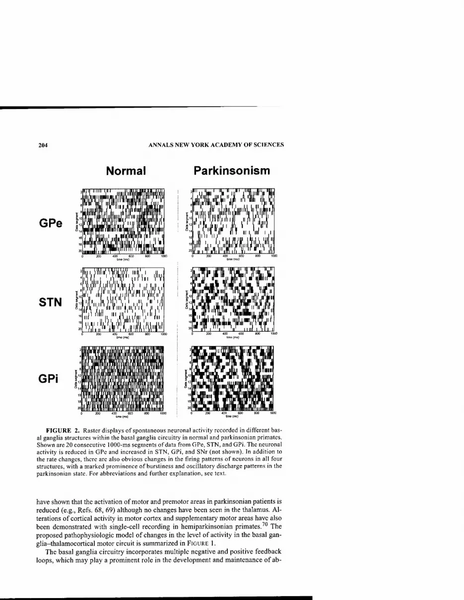
204 ANNALS NEW YORK ACADEMY OF SCIENCES
Normal Parkinsonism
GPe
STN
GPi
liiii,Viyiii''iVM, iiiil',1 '■'■'«>"■iMii 'i"V
iijifiii/'|i;'|'wiil!'iiUiii/ ri'i, ill' 1,11 fil Pill Will',I ill I liLV\ I 111 II' I,
v. ill niii 1,111,111 ji i|i iir lilK lllJlLiJ,,, iJAl
il! mil
^^iESTl^i
111 I, nil I I
m
■ "' II ll' 11 III I 'ill 0 20) MO 600 800 1000
lime (ms)
FIGURE 2. Raster displays of spontaneous neuronal activity recorded in different bas- al ganglia structures within the basal ganglia circuitry in normal and parkinsonian primates. Shown are 20 consecutive 1000-ms segments of data from GPe, STN, and GPi. The neuronal activity is reduced in GPe and increased in STN, GPi, and SNr (not shown). In addition to the rate changes, there are also obvious changes in the firing patterns of neurons in all four structures, with a marked prominence of burstiness and oscillatory discharge patterns in the parkinsonian state. For abbreviations and further explanation, see text.
have shown that the activation of motor and premotor areas in parkinsonian patients is reduced (e.g., Refs. 68, 69) although no changes have been seen in the thalamus. Al- terations of cortical activity in motor cortex and supplementary motor areas have also been demonstrated with single-cell recording in hemiparkinsonian primates.^** The proposed pathophysiologic model of changes in the level of activity in the basal gan- glia-thalamocortical motor circuit is summarized in FIGURE 1.
The basal ganglia circuitry incorporates multiple negative and positive feedback loops, which may play a prominent role in the development and maintenance of ab-
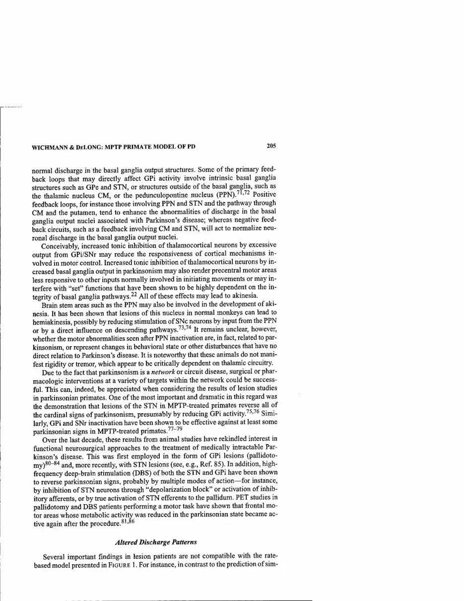
WICHMANN & DELONG: MPTP PRIMATE MODEL OF PD 205
normal discharge in the basal ganglia output structures. Some of the primary feed- back loops that may directly affect GPi activity involve intrinsic basal ganglia structures such as GPe and STN, or structures outside of the basal ganglia, such as the thalamic nucleus CM, or the pedunculopontine nucleus (PPN).''''''^ Positive feedback loops, for instance those involving PPN and STN and the pathway through CM and the putamen, tend to enhance the abnormalities of discharge in the basal ganglia output nuclei associated with Parkinson's disease; whereas negative feed- back circuits, such as a feedback involving CM and STN, will act to normalize neu- ronal discharge in the basal ganglia output nuclei.
Conceivably, increased tonic inhibition of thalamocortical neurons by excessive output from GPi/SNr may reduce the responsiveness of cortical mechanisms in- volved in motor control. Increased tonic inhibition of thalamocortical neurons by in- creased basal ganglia output in parkinsonism may also render precentral motor areas less responsive to other inputs normally involved in initiating movements or may in- terfere with "set" functions that have been shown to be highly dependent on the in- tegrity of basal ganglia pathways.^^ All of these effects may lead to akinesia.
Brain stem areas such as the PPN may also be involved in the development of aki- nesia. It has been shown that lesions of this nucleus in normal monkeys can lead to hemiakinesia, possibly by reducing stimulation of SNc neurons by input from the PPN or by a direct influence on descending pathways.'^'"''* It remains unclear, however, whether the motor abnormalities seen after PPN inactivation are, in fact, related to par- kinsonism, or represent changes in behavioral state or other disturbances that have no direct relation to Parkinson's disease. It is noteworthy that these animals do not mani- fest rigidity or tremor, which appear to be critically dependent on thalamic circuitry.
Due to the fact that parkinsonism is a network or circuit disease, surgical or phar- macologic interventions at a variety of targets within the network could be success- ful. This can, indeed, be appreciated when considering the results of lesion studies in parkinsonian primates. One of the most important and dramatic in this regard was the demonstration that lesions of the STN in MPTP-treated primates reverse all of the cardinal signs of parkinsonism, presumably by reducing GPi activity.''^•''^ Simi- larly, GPi and SNr inactivation have been shown to be effective against at least some parkinsonian signs in MPTP-treated primates.''^"^^
Over the last decade, these results from animal studies have rekindled interest in fiinctional neurosurgical approaches to the treatment of medically intractable Par- kinson's disease. This was first employed in the form of GPi lesions (pallidoto- jjjy)80-84 ajj(j^ jjjoj.e recently, with STN lesions (see, e.g., Ref 85). In addition, high- frequency deep-brain stimulation (DBS) of both the STN and GPi have been shown to reverse parkinsonian signs, probably by multiple modes of action—for instance, by inhibition of STN neurons through "depolarization block" or activation of inhib- itory afferents, or by true activation of STN efferents to the pallidum. PET studies in pallidotomy and DBS patients performing a motor task have shown that frontal mo- tor areas whose metabolic activity was reduced in the parkinsonian state became ac- tive again after the procedure.^'*^^
Altered Discharge Patterns
Several important findings in lesion patients are not compatible with the rate- based model presented in FIGURE 1. For instance, in contrast to the prediction of sim-
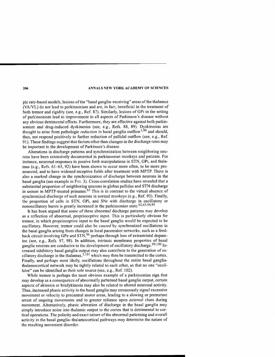
206 ANNALS NEW YORK ACADEMY OF SCIENCES
pie rate-based models, lesions of the "basal ganglia-receiving" areas of the thalamus (VA/VL) do not lead to parkinsonism and are, in fact, beneficial in the treatment of both tremor and rigidity (see, e.g., Ref 87). Similarly, lesions of GPi in the setting of parkinsonism lead to improvement in all aspects of Parkinson's disease without any obvious detrimental effects. Furthermore, they are effective against both parkin- sonism and drug-induced dyskinesias (see, e.g., Refs. 88, 89). Dyskinesias are thought to arise from pathologic reduction in basal ganglia outflow''^" and should, thus, not respond positively to further reduction of pallidal outflow (see, e.g., Ref 91). These findings suggest that factors other than changes in the discharge rates may be important in the development of Parkinson's disease.
Alterations in discharge patterns and synchronization between neighboring neu- rons have been extensively documented in parkinsonian monkeys and patients. For instance, neuronal responses to passive limb manipulations in STN, GPi, and thala- mus (e.g., Refs. 61-63, 92) have been shown to occur more often, to be more pro- nounced, and to have widened receptive fields after treatment with MPTP. There is also a marked change in the synchronization of discharge between neurons in the basal ganglia (see example in FIG. 3). Cross-correlation studies have revealed that a substantial proportion of neighboring neurons in globus pallidus and STN discharge in unison in MPTP-treated primates.^^ This is in contrast to the virtual absence of synchronized discharge of such neurons in normal monkeys (e.g., Ref 93). Finally, the proportion of cells in STN, GPi, and SNr with discharge in oscillatory or nonoscillatory bursts is greatly increased in the parkinsonian state.^^'^■''^'''^^
It has been argued that some of these abnormal discharge patterns may develop as a reflection of abnormal, proprioceptive input. This is particularly obvious for tremor, in which proprioceptive input to the basal ganglia would be expected to be oscillatory. However, tremor could also be caused by synchronized oscillations in the basal ganglia arising from changes in local pacemaker networks, such as a feed- back circuit involving GPe and STN,^^ perhaps through loss of extrastriatal dopam- ine (see, e.g., Refs. 97, 98). In addition, intrinsic membrane properties of basal ganglia neurons are conducive to the development of oscillatory discharge.'^''"" In- creased inhibitory basal ganglia output may also contribute to the generation of os- cillatory discharge in the thalamus,'-"" which may then be transmitted to the cortex. Finally, and perhaps most likely, oscillations throughout the entire basal ganglia- thalamocortical network may be tightly related to each other, so that no one "oscil- lator" can be identified as their sole source (see, e.g., Ref 102).
While tremor is perhaps the most obvious example of a parkinsonian sign that may develop as a consequence of abnormally patterned basal ganglia output, certain aspects of akinesia or bradykinesia may also be related to altered neuronal activity. Thus, increased phasic activity in the basal ganglia may erroneously signal excessive movement or velocity to precentral motor areas, leading to a slowing or premature arrest of ongoing movements and to greater reliance upon external clues during movement. Alternatively, phasic alteration of discharge in the basal ganglia may simply introduce noise into thalamic output to the cortex that is detrimental to cor- tical operations. The polarity and exact nature of the abnormal patterning and overall activity in the basal ganglia-thalamocortical pathways may determine the nature of the resulting movement disorder.

WICHMANN & DELONG: MPTP PRIMATE MODEL OF PD 207
<S
c o s
3 o
o S w
o ^ bi: aj e, D.
TJ j:
O D ^ K X5
r^ -1 O a a> fi ^ P N
E Pi
a o w •o 1> & e s
tn M
§ » OH H R.1 0 s
b J3

208 ANNALS NEW YORK ACADEMY OF SCIENCES
Consistent with the notion that changes in basal ganglia discharge may result in tremor, lesions of the STN or GPi significantly reduce tremor in MPTP-treated Af- rican green monkeys and in patients with parkinsonism.^^'^"''^'
Besides the skeletomotor abnormalities, parkinsonism is also associated with oc- ulomotor abnormalities, such as hypometric and slow saccades (e.g., Ref. 103), au- tonomic dysfunction, depression, anxiety, sleep disturbances, impaired visuospatial orientation, and cognitive abnormalities (e.g., Ref. 104). It is likely that at least some of these abnormalities rely on abnormal discharge in nonmotor circuits of the basal ganglia, which may be affected by dopamine loss in much the same way as the motor circuit. This is particularly true for oculomotor abnormalities that may directly result from dopamine depletion in the caudate nucleus (see, e.g., Refs. 105, 106). Similar- ly, some of the cognitive and psychiatric disturbances seen in parkinsonian patients are reminiscent of syndromes seen after lesions of the dorsolateral prefrontal cortex (problems with executive functions) or of the anterior cingulate (apathy, personality changes) and may be the result of loss of dopamine in the dorsolateral or ventral cau- date nucleus, respectively.'"^
CONCLUSIONS
From the considerations above, a complex model of parkinsonism emerges in which relatively selective dopamine depletion in the striatum and other basal ganglia nuclei results in increased and disordered discharge and synchronization in motor ar- eas of the basal ganglia thalamocortical motor loops. In fact, the motor circuit in Par- kinson's disease may be "taken hostage" by widespread discharge abnormalities that greatly interfere with its normal functions. Abnormal activity in the basal ganglia feedback loops may contribute to the development of parkinsonism. Individual par- kinsonian motor signs appear to be caused by distinct abnormalities in basal ganglia discharge. It is probable that progressive loss of dopamine in nonmotor areas of the striatum and other basal ganglia nuclei may underlie the nonmotor abnormalities of Parkinson's disease. The development of the different signs of movement disorders may be the consequence of changes in the rate, patterns, and degree of synchroniza- tion of discharge; of altered proprioceptive feedback; and of the appearance of "noise" in the basal ganglia output signal.
The current models of basal ganglia pathophysiology are incomplete and should be taken as a first draft of basal ganglia dysfunction in the different disease states. Most pertinently, changes in phasic discharge patterns and new anatomical connec- tions need to be better incorporated into any new concept of basal ganglia function, and greater emphasis needs to be placed on the manner in which thalamic, brain stem, and cortical neurons utilize basal ganglia output.
REFERENCES
1. WiCHMANN, T. & M.R. DELONG. 1996. Functional and pathophysiological models of the basal ganglia. Curr. Opin. Neurobiol. 6: 751-758.
2. ALBIN, R.L., et al. 1989. The functional anatomy of basal ganglia disorders. Trends Neurosci. 12: 366-375.
3. GRAYBIEL, A.M. 1996. Basal ganglia: new therapeutic approaches to Parkinson's dis- ease. Curr. Biol. 6: 368-371.

WICHMANN & DELONG: MPTP PRIMATE MODEL OF PD 209
4. LANGSTON, J.W., et al. 1984. Selective nigral toxicity after systemic administration of l-methyl-4-phenyl-l,2,3,6-tetrahydropyrine (MPTP) in the squirrel monkey. Brain Res. 292: 390-394.
5. BANKIEWICZ, K.S., et al. 1986. Hemiparkinsonism in monkeys after unilateral internal carotid artery infusion of l-methyl-4-phenyl-l,2,3,6-tetrahydropyridine (MPTP). Life Sciences 39: 7-16.
6. FoRNO, L.S. 1996. Neuropathology of Parkinson's disease. J. Neuropathol. Exp. Neu- rol. 55: 259-272.
7. KiSH, S.J., et al. 1988. Uneven pattern of dopamine loss in the striatum of patients with idiopathic Parkinson's disease. N. Engl. J. Med. 318: 876-880.
8. KuNZLE, H. 1975. Bilateral projections from precentral motor cortex to the putamen and other parts of the basal ganglia. An autoradiographic study in macaca fascicu- laris. Brain Res. 88: 195-209.
9. FLAHERTY, A.W. & A.M. GRAYBIEL. 1991. Corticostriatal transformations in the pri- mate somatosensory sstem. Projections from physiologically mapped body-part rep- resentations. J. Neurophysiol. 66: 1249-1263.
10. SELEMON, L.D. & P.S. GOLDMAN-RAKIC. 1985. Longitudinal topography and interdigi- tation of cortico-striatal projections in the rhesus monkey. J. Neurosci. 5: 776-794.
11. YETERIAN, E.H. & D.N. PANDYA. 1998. Corticostriatal connections of the superior tem- poral region in rhesus monkeys. J. Comp. Neurol. 399: 384^02.
12. HABER, S.N., et al. 1995. The orbital and medial prefrontal circuit through the primate basal ganglia. J. Neurosci. 15: 4851^867.
13. HARTMANN-VON MONAKOW, K. et al. 1978. Projections of the precentral motor cortex and other cortical areas of the frontal lobe to the subthalamic nucleus in the monkey. Exp. Brain Res. 33: 395-403.
14. NAMBU, A., et al. 1996. Dual somatotopical representations in the primate subthalamic nucleus: evidence for ordered but reversed body-map transformations from the pri- mary motor cortex and the supplementary motor area. J. Neurosci. 16: 2671-2683.
15. WILSON, C.J., et al. 1983. Origins of post synaptic potentials evoked in spiny neostriatal projection neurons by thalamic stimulation in the rat. Exp. Brain Res. 51: 217-226.
16. SADIKOT, A.F., et al. 1992. Efferent connections of the centromedian and parafascicu- lar thalamic nuclei in the squirrel monkey: a light and electron microscopic study of the thalamostriatal projection in relation to striatal heterogeneity. J. Comp. Neurol. 320: 228-242.
17. FEGER, J., et al. 1994. The projections from the parafascicular thalamic nucleus to the subthalamic nucleus and the striatum arise from separate neuronal populations: a comparison with the corticostriatal and corticosubthalamic efferents in a retrograde double-labelling study. Neuroscience 60: 125-132.
18. SMITH, Y. & A. PARENT. 1986. Differential connections of caudate nucleus and puta- men in the squirrel monkey (Saimiri sciureus). Neuroscience 18: 347-371.
19. NAKANO, K., et al. 1990. Topographical projections from the thalamus, subthalamic nucleus and pedunculopontine tegmental nucleus to the striatum in the Japanese monkey, Macaca fuscata. Brain Res. 537: 54-68.
20. SiDlBE, M., et al. 2002. Nigral and pallidal inputs to functionally segregated thalamos- triatal neurons in the centromedian/parafascicular intralaminar nuclear complex in monkey. J. Comp. Neurol. 447: 286-299.
21. ALEXANDER, G.E., et al. 1986. Parallel organization of functionally segregated circuits linking basal ganglia and cortex. Ann. Rev. Neurosci. 9: 357-381.
22. ALEXANDER, G.E. & M.D. CRUTCHER. 1990. Functional architecture of basal ganglia circuits: neural substrates of parallel processing. Trends Neurosci. 13: 266-271.
23. BERGMAN, H., et al. 1990. Amelioration of parkinsonian symptoms by inactivation of the subthalamic nucleus (STN) in MPTP treated green monkeys. Movement Disor- ders 5: 79.
24. GERFEN, C.R. & C.J. WILSON. 1996. The basal ganglia. In Handbook of Chemical Neu- roanatomy, Integrated Systems of the CNS, Part III. A. Bjorklund, T. Hokfeld & L. Swanson, Eds.: 369. Elsevier. Amsterdam.
25. PARENT, A., et al. 1995. Single striatofugal axons arborizing in both pallidal segments and in the substantia nigra in primates. Brain Res. 698: 280-284.

210 ANNALS NEW YORK ACADEMY OF SCIENCES
26. SiDiBE, M. & Y. SMITH. 1996. Differential synaptic innervation of striatofugal neurones projecting to the internal or external segments of the globus pallidus by thalamic afferents in the squirrel monkey. J. Comp. Neurol. 365: 445-465.
27. PARTHASARATHY, H.B. & A.M. GRAYBIEL. 1997. Cortically driven immediate-early gene expression reflects influence of sensorimotor cortex on identified striatal neu- rons in the squirrel. J. Neurosci. 17: 2477-2491.
28. GERFEN, C.R., et al. 1990. Dl and D2 dopamine receptor-regulated gene expression of striatonigral and striatopallidal neurons. Science 250: 1429-1432.
29. SuRMEiER, D.J., et al. 1996. Coordinated expression of dopamine receptors in neostri- atal medium spiny neurons. J. Neurosci. 16: 6579-6591.
30. AiZMAN, O., et al. 2000. Anatomical and physiological evidence for Dl and D2 dopamine receptor colocalization in neostriatal neurons. Nat. Neurosci. 3: 226-230.
31. GERFEN, C.R. 1995. Dopamine receptor function in the basal ganglia. Clin. Neurophar- macol. 18:S162-S177.
32. PARENT, A. 1990. Extrinsic connections of the basal ganglia. Trends Neurosci. 13: 254-258.
33. SiDiBE, M., et al. 1997. Efferent connections of the internal globus pallidus in the squirrel monkey: I. Topography and synaptic organization of the pallidothalamic pro- jection. J. Comp. Neurol. 382: 323-347.
34. ScHELL, G.R. & P.L. STRICK. 1984. The origin of thalamic inputs to the arcuate premo- tor and supplementary motor areas. J. Neurosci. 4: 539-560.
35. INASE, M. & J. TANJI. 1995. Thalamic distribution of projection neurons to the primary motor cortex relative to afferent terminal fields from the globus pallidus in the macaque monkey. J. Comp. Neurol. 353: 415^26.
36. NAMBU, A., et al. 1988. Projection on the motor cortex of thalamic neurons with pal- lidal input in the monkey. Exp. Brain Res. 71: 658-662.
37. HOOVER, J.E. & P.L. STRICK. 1993. Multiple output channels in the basal ganglia. Sci- ence 259: 819-821.
38. GOLDMAN-RAKIC, P.S. & L.J. PORRINO. 1985. The primate mediodorsal (MD) nucleus and its projection to the frontal lobe. J. Comp. Neurol. 242: 535-560.
39. MiDDLETON, F.A. & P.L. STRICK. 1994. Anatomical evidence for cerebellar and basal ganglia involvement in higher cognitive function. Science 266: 458^61.
40. HEDREEN, J.C. & M.R. DELONG. 1991. Organization of striatopallidal, striatonigral and nigrostriatal projections in the macaque. J. Comp. Neurol. 304: 569-595.
41. MiDDLETON, F.A. & P.L. STRICK. 2002. Basal-ganglia "projections" to the prefrontal cortex of the primate. Cerebr. Cortex 12: 926-935.
42. ILINSKY, I.A., et al. 1985. Organization of the nigrothalamocortical system in the rhesus monkey. J. Comp. Neurol. 236: 315-330.
43. GROFOVA, I. & M. ZHOU. 1998. Nigral innervation of cholinergic and glutamatergic cells in the rat mesopontine tegmentum: light and electron microscopic anterograde tracing and immunohistochemical studies. J. Comp. Neurol. 395: 359-379.
44. RYE, D.B., et al. 1988. Medullary and spinal efferents of the pedunculopontine teg- mental nucleus and adjacent mesopontine tegmentum in the rat. J. Comp. Neurol. 269:315-341.
45. STEININGER, T.L., et al. 1992. Afferent projections to the cholinergic pedunculopontine tegmental nucleus and adjacent midbrain extrapyramidal area in the albino rat. I. Retrograde tracing studies. J. Comp. Neurol. 321: 515-543.
46. WuRTZ, R.H. & O. HiKOSAKA. 1986. Role of the basal ganglia in the initiation of sac- cadic eye movements. Prog. Brain Res. 64: 175-190.
47. INASE, M., et al. 1996. Changes in the control of arm position, movement, and thalamic discharge during local inactivation in the globus pallidus of the monkey. J. Neuro- physiol.75: 1087-1104.
48. GEORGOPOULOS, A.P., et al. 1983. Relations between parameters of step-tracking movements and single cell discharge in the globus pallidus and subthalamic nucleus of the behaving monkey. J. Neurosci. 3: 1586-1598.
49. MITCHELL, S.J., et al. 1987. The primate globus pallidus: neuronal activity related to direction of movement. Exp. Brain Res. 68: 491-505.
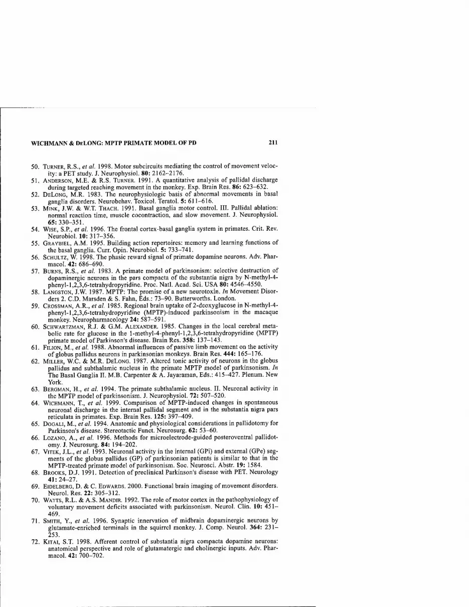
WICHMANN & DELONG: MPTP PRIMATE MODEL OF PD 211
50. TURNER, R.S., et al. 1998. Motor subcircuits mediating the control of movement veloc- ity: a PET study. J. Neurophysiol. 80: 2162-2176.
51. ANDERSON, M.E. & R.S. TURNER. 1991. A quantitative analysis of pallidal discharge during targeted reaching movement in the monkey. Exp. Brain Res. 86: 623-632.
52. DELONG, M.R. 1983. The neurophysiologic basis of abnormal movements in basal ganglia disorders. Neurobehav. Toxicol. Teratol. 5: 611-616.
53. MINK, J.W. & W.T. THACH. 1991. Basal ganglia motor control. III. Pallidal ablation: normal reaction time, muscle cocontraction, and slow movement. J. Neurophysiol. 65:330-351.
54. WISE, S.P., et al. 1996. The frontal cortex-basal ganglia system in primates. Crit. Rev. Neurobiol. 10:317-356.
55. GRAYBIEL, A.M. 1995. Building action repertoires: memory and learning functions of the basal ganglia. Curr. Opin. Neurobiol. 5: 733-741.
56. ScHULTZ, W. 1998. The phasic reward signal of primate dopamine neurons. Adv. Phar- macol. 42: 686-690.
57. BURNS, R.S., et al. 1983. A primate model of parkinsonism: selective destruction of dopaminergic neurons in the pars compacta of the substantia nigra by N-methyl-4- phenyl-l,2,3,6-tetrahydropyridine. Proc. Natl. Acad. Sci. USA 80: 4546-4550.
58. LANGSTON, J.W. 1987. MPTP: The promise of a new neurotoxin. In Movement Disor- ders 2. CD. Marsden & S. Fahn, Eds.: 73-90. Butterworths. London.
59. GROSSMAN, A.R., et al. 1985. Regional brain uptake of 2-deoxyglucose in N-methyl-4- phenyl-l,2,3,6-tetrahydropyridine (MPTP)-induced parkinsonism in the macaque monkey. Neuropharmacology 24: 587-591.
60. SCHWARTZMAN, R.J. & G.M. ALEXANDER. 1985. Ghanges in the local cerebral meta- bolic rate for glucose in the l-methyl-4-phenyl-l,2,3,6-tetrahydropyridine (MPTP) primate model of Parkinson's disease. Brain Res. 358: 137-143.
61. FiLiON, M., et al. 1988. Abnormal influences of passive limb movement on the activity of globus pallidus neurons in parkinsonian monkeys. Brain Res. 444: 165-176.
62. MILLER, W.C. & M.R. DELONG. 1987. Altered tonic activity of neurons in the globus pallidus and subthalamic nucleus in the primate MPTP model of parkinsonism. In The Basal Ganglia II. M.B. Garpenter & A. Jayaraman, Eds.: 415-427. Plenum. New York.
63. BERGMAN, H., et al. 1994. The primate subthalamic nucleus. II. Neuronal activity in the MPTP model of parkinsonism. J. Neurophysiol. 72: 507-520.
64. WiCHMANN, T., et al. 1999. Gomparison of MPTP-induced changes in spontaneous neuronal discharge in the internal pallidal segment and in the substantia nigra pars reticulata in primates. Exp. Brain Res. 125: 397-409.
65. DOGALI, M., et al. 1994. Anatomic and physiological considerations in pallidotomy for Parkinson's disease. Stereotactic Funct. Neurosurg. 62: 53-60.
66. LozANO, A., et al. 1996. Methods for microelectrode-guided posteroventral pallidot- omy. J. Neurosurg. 84: 194-202.
67. ViTEK, J.L., et al. 1993. Neuronal activity in the internal (GPi) and external (GPe) seg- ments of the globus pallidus (GP) of parkinsonian patients is similar to that in the MPTP-treated primate model of parkinsonism. Soc. Neurosci. Abstr. 19: 1584.
68. BROOKS, D.J. 1991. Detection of preclinical Parkinson's disease with PET. Neurology 41:24-27.
69. EiDELBERG, D. & G. EDWARDS. 2000. Functional brain imaging of movement disorders. Neurol. Res. 22:305-312.
70. WATTS, R.L. & A.S. MANDIR. 1992. The role of motor cortex in the pathophysiology of voluntary movement deficits associated with parkinsonism. Neurol. Glin. 10: 451- 469.
71. SMITH, Y., et al. 1996. Synaptic innervation of midbrain dopaminergic neurons by glutamate-enriched terminals in the squirrel monkey. J. Gomp. Neurol. 364: 231- 253.
72. KITAI, S.T. 1998. Afferent control of substantia nigra compacta dopamine neurons: anatomical perspective and role of glutamatergic and cholinergic inputs. Adv. Phar- macol. 42: 700-702.

212 ANNALS NEW YORK ACADEMY OF SCIENCES
73. KOJIMA, J., et al. 1997. Excitotoxic lesions of the pedunculopontine tegmental nucleus produce contralateral hemiparkinsonism in the monkey. Neurosci. Lett. 226: 111- 114.
74. MUNRO-DAVIES, L.E., el al. 1999. The role of the pedunculopontine region m basal- ganglia mechanisms of akinesia. Exp. Brain Res. 129: 511-517.
75. BERGMAN, H., el al. 1990. Reversal of experimental parkinsonism by lesions of the subthalamic nucleus. Science 249: 1436-1438.
76. AZIZ, T.Z., el al. 1991. Lesion of the subthalamic nucleus for the alleviation of 1- methyl-4-phenyl-l,2,3,6-tetrahydropyridine (MPTP)-induced parkinsonism in the primate. Mov. Disord. 6: 288-292.
77. WlCHMANN, T., el al. 1994. Local inactivation of the sensorimotor territories of the internal segment of the globus pallidus and the subthalamic nucleus alleviates par- kinsonian motor signs in MPTP treated monkeys. In The Basal Ganglia IV: New Ideas and Data on Structure and Function. G. Percheron, J.S. McKenzie & J. Feger, Eds.: 357-363. Plenum. New York.
78. WlCHMANN, T., et al. 2001. Antiparkinsonian and behavioral effects of inactivation of the substantia nigra pars reticulata in hemiparkinsonian primates. Exp. Neurol. 167: 410-424.
79. LONSER, R.R., el al. 1999. Convection-enhanced selective excitotoxic ablation of the neurons of the globus pallidus internus for treatment of parkinsonism in nonhuman primates [see comments]. J. Neurosurg. 91: 294-302.
80. BARON, M.S., et al. 1996. Treatment of advanced Parkinson's disease by GPi pallidot- omy: 1 year pilot-study results. Ann. Neurol. 40: 355-366.
81. DOGALI, M., er a/. 1995. Stereotactic ventral pallidotomy for Parkinson's disease. Neu- rology 45: 753-761.
82. LAITINEN, L.V., el al. 1992. Leksell's posteroventral pallidotomy in the treatment of Parkinson's disease. J. Neurosurg. 76: 53-61.
83. LOZANO, A.M., etal. 1995. Effect of GPi pallidotomy on motor function in Parkinson's disease. Lancet 346: 1383-1387.
84. ViTEK, J.L., et al. 1997. Microelectrode-guided pallidotomy for medically intractable Parkinson's disease. Adv. Neurol. 74: 183-198.
85. GILL, S.S. & P. HEYWOOD. 1998. Bilateral subthalamic nucleotomy can be accom- plished safely. Mov. Disord. 13: 201.
86. CEBALLOS-BAUMAN, A.O. el al. 1994. Restoration of thalamocortical activity after pos- teroventrolateral pallidotomy in Parkinson's disease. Lancet 344: 814.
87. TASKER, R.R., el al. 1997. Pallidal and thalamic surgery for Parkinson's disease. Exp. Neurol. 144: 35-40.
88. RABEY, J.M., et al. 1995. Levodopa-induced dyskinesias are the main feature improved by contralateral pallidotomy in Parkinson's disease. Neurology 45: A377.
89. OBESO, J.A., et al. 2000. Pathophysiology of levodopa-induced dyskinesias in Parkin- son's disease: problems with the current model. Ann. Neurol. 47: S22-32; discussion S32-34.
90. PAPA, S.M., et al. 1999. Internal globus pallidus discharge is nearly suppressed during levodopa-induced dyskinesias. Ann. Neurol. 46: 732-738.
91. MARSDEN, CD. & J.A. OBESO. 1994. The functions of the basal ganglia and the para- dox of stereotaxic surgery in Parkinson's disease. Brain 117: 877-897.
92. ViTEK, J.L., el al. 1990. Altered somatosensory response properties of neurons in the "motor" thalamus of MPTP treated parkinsonian monkeys. Soc. Neurosci. Abstr. 16: 425.
93. WlCHMANN, T, et al. 1994. The primate subthalamic nucleus. I. Functional properties in intact animals. J. Neurophysiol. 72: 494-506.
94. WlCHMANN, T, et al. 1996. Comparison of the effects of experimental parkinsonism on neuronal discharge in motor and non-motor portions of the basal ganglia output nuclei in primates. Soc. Neurosci. Abstr. 22: 415.
95. FILION, M. & L. TREMBLAY. 1991. Abnormal spontaneous activity of globus pallidus neurons in monkeys with MPTP-induced parkinsonism. Brain Res. 547: 142-151.
96. PLENZ, D. & S. KiTAi. 1999. A basal ganglia pacemaker formed by the subthalamic nucleus and external globus pallidus. Nature 400: 677-682.
WICHMANN & DELONG: MPTP PRIMATE MODEL OF PD 213
97. BERGMAN, H., el al. 1998. Physiology of MPTP tremor. Mov. Disord. 13: 29-34. 98. BERNHEIMER, H., et al. 1973. Brain dopamine and the syndromes of Parkinson and
Huntington. J. Neurol. Sci. 20: 415-455. 99. NAMBU, A. & R. LLINAS. 1990. Electrophysiology of the globus pallidus neurons: an
in vitro study in guinea pig brain slices. Soc. Neurosci. Abstr. 16: 428. 100. BEURRIER, C, et al. 1999. Subthalamic nucleus neurons switch from single-spike
activity to burst-firing mode. J. Neurosci. 19: 599-609. 101. WiCHMANN, T., et al. 1994. The primate subthalamic nucleus. III. Changes in motor
behavior and neuronal activity in the internal pallidum induced by subthalamic inac- tivation in the MPTP model of parkinsonism. J. Neurophysiol. 72: 521-530.
102. MAGILL, P.J., et al. 2000. Relationship of activity in the subthalamic nucleus-globus pallidus network to cortical electroencephalogram. J. Neurosci. 20: 820-833.
103. VlDAlLHET, M., et al. 1999. Saccades and antisaccades in parkinsonian syndromes. Adv. Neurol. 80: 377-382.
104. CuMMiNGS, J.L. & D.L. MASTERMAN. 1999. Depression in patients with Parkinson's disease. Int. J. Geriatr. Psychiatry 14: 711-718.
105. KORI, A., et al. 1995. Eye movements in monkeys with local dopamine depletion in the caudate nucleus. II. Deficits in voluntary saccades. J. Neurosci. 15: 928-941.
106. KATO, M., et al. 1995. Eye movements in monkeys with local dopamine depletion in the caudate nucleus. I. Deficits in spontaneous saccades. J. Neurosci. 15: 912-927.
107. CuMMiNGS, J.L. 1993. Frontal-subcortical circuits and human behavior. Arch. Neurol. 50: 873-880.
108. RAZ, A., et al. 2000. Firing patterns and correlations of spontaneous discharge of pal- lidal neurons in the normal and the tremulous l-methyl-4-phenyl-l,2,3,6-tetrahydro- pyridine vervet model of parkinsonism. J. Neurosci. 20: 8559-8571.
The Role of dial Reaction and Inflammation in Parkinson's Disease
E. C. HIRSCH, T. BREIDERT, E. ROUSSELET, S. HUNOT, A. HARTMANN, AND P. P. MICHEL
INSERM U289, Experimental Neurology and Therapeutics, Hopital de la Salpetriere,
75651 Paris Cedex 13. France
ABSTRACT: The glial reaction is generally considered to be a consequence of neuronal death in neurodegcnerative diseases such as Alzheimer's disease, Huntington's disease, and Parkinson's disease. In Parkinson's disease, post- mortem examination reveals a loss of dopaminergic neurons in the substantia nigra associated with a massive astrogliosis and the presence of activated microglial cells. Recent evidence suggests that the disease may progress even when the initial cause of neuronal degeneration has disappeared, suggesting that toxic substances released by the glial cells may be involved in the propaga- tion and perpetuation of neuronal degeneration. Glial cells can release delete- rious compounds such as proinflammatory cytokines (TNF-a, Il-lp, IFN-7), which may act by stimulating nitric oxide production in glial cells, or which may exert a more direct deleterious effect on dopaminergic neurons by activat- ing receptors that contain intracytoplasmic death domains involved in apopto- sis. In line with this possibility, an activation of proteases such as caspasc-3 and caspase-8, which are known effectors of apoptosis, has been reported in Par- kinson's disease. Yet, caspase inhibitors or invalidation of TNF-a receptors does not protect dopaminergic neurons against degeneration in experimental models of the disease, suggesting that manipulation of a single signaling path- way may not be sufficient to protect dopaminergic neurons. In contrast, the antiinflammatory drugs pioglitazone, a PPAR-7 agonist, and the tetracycline derivative minocycline have been shown to reduce glial activation and protect the substantia nigra in an animal model of the disease. Inhibition of the glial reaction and the inflammatory processes may thus represent a therapeutic tar- get to reduce neuronal degeneration in Parkinson's disease.
KEYWORDS: cytokines; neuroprotection; apoptosis; dopamine neuron; substan- tia nigra
SUBPOPULATIONS OF DOPAMINERGIC NEURONS ARE SELECTIVELY AFFECTED IN PARKINSON'S DISEASE
Parkinson's disease is characterized by a slow and progressive degeneration of dopaminergic neurons in the mesencephalon. Yet, evidence suggests that the loss of these neurons is heterogeneous across different catecholaminergic cell groups. In-
Address for correspondence: E.G. Hirsch, INSERM U289, Experimental Neurology and Ther- apeutics, Hopital de la Salpetriere, 47 boulevard de I'Hopital, 75651 Paris Cedex 13, France. Voice: +33 (0) 1 42 16 22 02; fax: + 33 (0) 1 44 24 36 58.
Ann. N.V. Acad. Sci. 991: 214-228 (2003). © 2003 New York Academy of Sciences.
214

HIRSCH et at.: ROLE OF GLIAL REACTION AND INFLAMMATION 215
deed, neuronal loss is extremely severe in the substantia nigra pars compacta, mod- erate in the ventral tegmental area and catecholaminergic cell group A8, and almost nil in the central gray substance.' Furthermore, within the substantia nigra itself, the most severe loss occurs in the ventrolateral part of the substantia nigra pars compac- ta.^'-' More recently, when compartmental patterns of calbindin D28K immunostain- ing were used to subdivide the substantia nigra with landmarks independent of the degenerative process, zones of very high vulnerability were identified.'* Dopamine- containing neurons were identified in the calbindin-rich regions (matrix) and in five calbindin-poor pockets (nigrosomes) defined by analysis of the three-dimensional networks formed by the calbindin-poor zones. Within these zones, cell loss followed a strict order, depletion being maximal in the main pocket located in the caudal and mediolateral part of the substantia nigra pars compacta. Furthermore, the degree of loss of dopamine-containing neurons in the substantia nigra pars compacta was re- lated to the duration of the disease, with a pattern of neuronal loss consistent from one parkinsonian substantia nigra to another. The reason for such a differential vul- nerability of dopaminergic neurons within the parkinsonian substantia nigra is not known. Yet, the different nigral compartments may differ in terms of their content of growth factor, receptors, compounds related to excitotoxicity, agents involved in ox- idative metabolism, and activity of potentially predisposing genes such as those for a-synuclein^ and parkin.^
In addition to their heterogeneity in terms of susceptibility to the as yet unknown agent causing their death, dopaminergic neurons in the parkinsonian substantia nigra may also differ with regard to their individual status. Indeed, at least three types of dopaminergic neurons can be described in the parkinsonian substantia nigra. The first type of dopaminergic neurons, probably located mostly in the matrix, is still un- affected by the pathological process and corresponds to "healthy" surviving neurons. In contrast, the second type of dopaminergic neurons is already engaged in a degen- erative process and displays "typical features" of apoptosis or autophagic degenera- tion.' Whether such severely affected neurons can be protected from neurodegeneration using a therapeutic strategy is not knovra, but in vitro experi- ments using caspase inhibitors suggest this goal will be difficult to attain.^
Finally, the last type of dopaminergic neurons in the parkinsonian substantia nigra consists in "suffering" neurons. These neurons may not function adequately and in that respect differ from the still healthy neurons. Yet, unlike apoptotic neurons, they may not be engaged in the irreversible final stage of degeneration. Some of these dopaminergic neurons in the substantia nigra may contain Lewy bodies, which are the histopathological hallmarks of the disease (for review, see Fomo^). The concept of suffering neurons in the parkinsonian substantia nigra is further supported by the identification of another cell type in the substantia nigra of parkinsonian patients but not in that of normal individuals. These cells contain neuromelanin (the end product in the catabolism of catecholamines by autoxidation) in the absence of tyrosine hy- droxylase immunoreactivity.' These pigmented tyrosine hydroxylase-negative neu- rons may represent "wounded" neurons that once contained tyrosine hydroxylase. Within the parkinsonian substantia nigra, these neurons can represent as much as 20% of the surviving melanized neurons, indicating that there are many suffering neurons in the brain of the patients that have ceased to function long before their ac- tual death. If so, it suggests that we should be less concerned about those neurons that are dead, and instead focus our attention more on those in the early stages of de-

216 ANNALS NEW YORK ACADEMY OF SCIENCES
generation. These neurons may, on the one hand, be a suitable target for neurorestor- ative and neuroprotective strategies and, on the other hand, be particularly sensitive to a final common pathway of neuronal degeneration. We therefore need to consider the mechanisms involved in the degeneration of these wounded neurons.
IS THE GLIAL REACTION INVOLVED IN THE PROGRESSION OF NEURONAL DEGENERATION IN PARKINSON'S DISEASE?
The mechanism by which dopaminergic neurons degenerate in Parkinson's dis- ease is not yet known. Yet, several lines of evidence suggest that oxidative stress, ex- citotoxic mechanisms, and altered protein catabolism participate in the cascade of events leading to neuronal degeneration in this disease.'""'^ These cellular and mo- lecular alterations ultimately lead to apoptosis.''''^'''* In addition, degeneration of dopaminergic neurons is associated with a strong glial reaction, which is generally considered to be a nonspecific consequence of neuronal degeneration. In the follow- ing paragraphs, we will review the data suggesting that a subset of reactive glial cells may participate actively in dopaminergic cell demise during Parkinson's disease by virtue of their inflammatory properties.
The astroglial reaction in the substantia nigra of patients with Parkinson's disease is a well-known neuropathological characteristic of the disease. Furthermore, in their seminal study, McGeer and coworkers reported a large number of reactive hu- man leucocyte antigen-DR (HLA-DR)-positive microglial cells in the substantia ni- gra of patients with Parkinson's disease.'^ Such a glial reaction has also been described in the affected brain regions in other neurological disorders such as Alz- heimer's disease and brain infarct (for review, see McGeer and McGeer'^) as well as in animal models of Parkinson's disease.'''''^ These data suggest that glial activation is not specific to Parkinson's disease and that it likely represents a common phenom- enon in neurodegenerative disorders. However, recent evidence supports the notion that a subpopulation of activated glial cells may be deleterious in Parkinson's disease and particularly so for suffering or wounded neurons. Strong support for this hypoth- esis came from the study of young drug addicts who developed a parkinsonian syn- drome after l-methyl-4-phenyl-l,2,3,6-tetrahydropyridine (MPTP) intoxication.'^ In a recent study, these authors reported a postmortem neuropathological study of three subjects with MPTP-induced parkinsonism.^" Interestingly, gliosis and clus- tering of microglial cells around nerve cells were detected despite survival times ranging from 3 to 16 years. These findings not only indicate ongoing nerve cell loss after a time-limited insult, but suggest that activated microglial cells may perpetuate neuronal degeneration. One may thus speculate that after a primary insult of envi- ronmental or genetic origin, the glial reaction may enhance the degeneration of dopaminergic neurons. However, this question cannot be resolved by postmortem studies alone. Much insight into the potential involvement of inflammation-mediat- ed nerve cell death in Parkinson's disease has in fact come from experimental models of the disease. Indeed, lipopolysaccharides (LPS), which activate glial cells and in- duce the expression of proinflammatory cytokines, have been shown to kill dopam- inergic neurons in mixed neuron-glial cell cultures but not in pure neuronal cultures.2' This indicates that the production of a glial factor induced by LPS (very likely cytokines) is able to kill dopaminergic neurons at least in vitro. These results

HIRSCH et al: ROLE OF GLIAL REACTION AND INFLAMMATION 217
were recently confirmed by McNaught and Jenner in experiments in which they showed that LPS-activated astrocytes could cause neuronal death in a time-depen- dent manner in primary ventral mesencephalic neuronal cultures.-^^ Furthermore, these authors extended this finding by showing that the toxicity of agents capable of inducing the death of dopaminergic neurons, such as MPP"*" or 6-hydroxydopamine, was enhanced when the dopaminergic neurons were cultured with LPS-activated as- trocytes. In vivo models of Parkinson's disease also support the notion that activated glial cells may participate in the degeneration of dopaminergic neurons. Indeed, Her- rera and coworkers reported that a single intranigral injection of LPS induces dam- age to dopaminergic neurons in the substantia nigra with preservation of GABAergic or serotoninergic neurons.^^'-^'* More recently, the same authors showed that dexam- ethasone, a potent antiinflammatory drug that interferes with many of the features characterizing proinflammatory glial reaction, prevented the loss of catecholaminer- gic content, tyrosine hydroxylase activity, and tyrosine-hydroxylase immunostain- ing induced by LPS injection and also the bulk activation of microglia- macrophages.^^
Taken together, these data clearly show that activated glial cells can participate in the death of dopaminergic neurons. The mechanism by which microglial cells can amplify injury to the nigral dopaminergic neurons is not yet known. However, the factors involved in this deleterious effect are very likely cytokines, including tumor necrosis factor-a (TNF-a), interleukin-ip (IL-IP), and interferon-y (IFN-y).
PROINFLAMMATORY CYTOKINES IN PARKINSON'S DISEASE
Several studies have reported a marked increase of cytokine levels in the brain and cerebrospinal fluid (CSF) of parkinsonian patients.^^ In addition, a higher den- sity of glial cells expressing TNF-a, IL-ip, and IFN-y was observed in the substantia nigra of patients with Parkinson's disease as compared to age-matched control sub- jects.^^'^^ Some of these cells were sometimes in close proximity to blood vessels and degenerating dopaminergic neurons, suggesting their involvement in the patho- physiology of Parkinson's disease. Two mechanisms that are not mutually exclusive may explain the deleterious role of cytokines in the parkinsonian substantia nigra.
First, proinflammatory cytokines can induce the production of nitric oxide in glial cells.-^^ This mechanism involves the induction of a low-affinity receptor for immunoglobulin E (CD23) and its activation by an as yet unknown ligand. Nitric ox- ide may diffiise towards dopaminergic neurons, where it may play a deleterious role by inducing lipid peroxidation and DNA strand breaks and inhibiting mitochondrial respiration and energy metabolism.^' In support of this hypothesis, an increased density of glial cells expressing the inducible isoform of nitric oxide synthase and CD23 has been reported in the substantia nigra of patients with Parkinson's disease as compared to age-matched control subjects.-^^'^^ A role of nitric oxide in cell de- mise in Parkinson's disease is further supported by the increased nitrite levels ob- served in the CSF of patients with Parkinson's disease^' and detectable immunoreactivity for nitrotyrosine in Lewy bodies.^^
The second mechanism by which proinflammatory cytokines may play a delete- rious role in Parkinson's disease may be more direct. Indeed, dopaminergic neurons in the substantia nigra express specific receptors for these molecules. For instance,
218 ANNALS NEW YORK ACADEMY OF SCIENCES
type 1 TNF-a receptors are expressed on dopaminergic neurons of the human sub- stantia nigra." Two types of TNF-a receptors, type 1 (TNFRl) and type 2 (TNFR2), which are encoded by different genes and are coupled to two distinct transduction pathways, may mediate distinct effects of TNF-a. TNFRl has the potential to induce apoptotic cytotoxicity,^^ fibroblast proliferation, antiviral activity, NFKB activation, and cell adhesion molecule activation.^'''^^ TNFR2 seems to be involved in cellular proliferation processes^^'^'' and also to potentiate the effect of TNFRl.^^'^' The transduction pathways coupled to TNFRl are of great interest in terms of degenera- tion of dopaminergic neurons. Indeed, TNF-a induces trimerization of TNFRl on binding that leads, through the adaptor molecules TNFRl-associated protein with a death domain (TRADD) and FAS-associated protein with a death domain (FADD), to the autoproteolytical activation of caspase-8.'*° Caspase-8 may in turn either cleave effector caspases, such as caspase-3, directly or amplify the death signal through the translocation of BID, a proapoptotic member of the Bel-family, to the mitochondria and the subsequent release of cytochrome c from the mitochondrial in- tennembrane space into the cytosol."*' Cytochrome c release also eventually triggers caspase-3 activation, which plays an important role in cell degeneration. Finally, caspase-8 can also be activated downstream of mitochondrial cytochrome c release, possibly to amplify the BID-induced cytochrome c release."'^'''^ Such a TNF-a re- ceptor transduction pathway is of particular relevance for Parkinson's disease, as it is very likely to be activated in the neurons most vulnerable to the disease process. Indeed, in a human postmortem study, we showed a significant decrease in the per- centage of FADD-immunoreactive dopaminergic neurons in the substantia nigra pars compacta of patients with Parkinson's disease compared to control subjects.'*" Furthermore, this decrease correlated with the known selective vulnerability of ni- gral dopaminergic neurons in Parkinson's disease, suggesting that this pathway con- tributes to the susceptibility of dopaminergic neurons to TNF-mediated apoptosis in Parkinson's disease. Similar results were obtained for caspase-3. Indeed, we ob- served a positive correlation between the degree of neuronal loss in dopaminergic cell groups affected in the mesencephalon of parkinsonian patients and the percent- age of caspase-3-positive neurons in these cell groups in control subjects. Further- more, we also found a significant decrease of caspase-3-positive pigmented neurons in the substantia nigra pars compacta of parkinsonian patients compared to control subjects.'''* Taken together, these findings suggest that the melanized dopaminergic neurons expressing the TNFRl transduction pathway are particularly prone to de- generation in Parkinson's disease if this pathway is activated during the course of the disease. A crucial finding in support of the activation of this pathway came from a postmortem study of caspase activation in Parkinson's disease. Using an antibody raised against activated caspase-3, the percentage of active caspase-3-positive neu- rons among dopaminergic neurons was shown to be significantly higher in parkin- sonian patients than in control subjects.'*'' Similarly, the proportion of melanized neurons displaying caspase-8 activation in Parkinson's disease was also higher than in control subjects.^ Interestingly, the proportion of activated caspase-3-positive neurons among neurons containing Lewy bodies was higher than in neurons that did not contain Lewy bodies.'*'' In contrast, immunoreactivity for activated caspase-3 was not observed in neurons displaying the typical features of apoptosis. These data suggest that caspase-3 activation occurs in suffering neurons just before degenera- tion by apoptosis. This concept is further supported by experimental models of Par-

HIRSCH et al.: ROLE OF GLIAL REACTION AND INFLAMMATION 219
kinson's disease, in which the exact time course of caspase activation and apoptosis could be studied. Thus, in mice intoxicated by MPTP, caspase-3 activation was shown to slightly precede the death of dopaminergic neurons by apoptosis."*^ Taken together, these data suggest that the activation of the TNF-a transduction pathway is very likely involved in the pathophysiology of Parkinson's disease and especially in the induction of apoptosis, which has already been described in this disease (for a review, see Hartmann and Hirsch"*^).
The data reviewed in the preceding paragraph suggest that proinflammatory cy- tokines may participate in the pathophysiology of Parkinson's disease either by pro- ducing nitric oxide, which may subsequently have a deleterious effect on dopaminergic neurons, or by activating specific transduction pathways coupled to receptors for cytokines. Nevertheless, it is very likely that these pathophysiological events are not specific to Parkinson's disease, as similar changes have been de- scribed in other neurodegenerative diseases. However, this lack of specificity does not preclude the development of neuroprotective strategies based on inhibition of the inflammatory processes in Parkinson's disease.
ARE INFLAMMATORY PROCESSES A POTENTIAL THERAPEUTIC TARGET IN PARKINSON'S DISEASE?
The use of antiinflammatory drugs to prevent dopaminergic degeneration in Par- kinson's disease has not yet been formally tested in patients. The concept that anti- inflammatory agents may be beneficial in Parkinson's disease thus relies on preclinical studies of w vitro or in vivo models of the disease. In vivo, various com- pounds such as nonsteroidal antiinflammatory drugs (NSAIDs) with their targets COX, NFKB, and others, including steroids, immunophilins, thalidomide, and phos- phodiesterase IV inhibitors, have been studied with variable results (TABLE 1). Fur- thermore, gene manipulation in mice has shown that C0X2 and iNOS are of particular interest in such strategies (TABLE 2). However, fi-om our own data, it ap- pears that manipulation of a single specific pathway or a downstream pathway in- volved in the inflammatory processes does not protect dopaminergic neurons against degeneration in models of Parkinson's disease. In contrast, agents with a broader spectrum of action on the inflammatory processes are more likely to protect dopam- inergic neurons against degeneration. To analyze the putative role of TNF-a receptor transduction pathways in parkinsonism, we compared the effect of the parkinsonian drug MPTP in mice lacking TNFRl, TNFR2, or both receptors and in wild-type lit- termates.'*' Postmortem analysis revealed no difference in the number of nigral dopaminergic neurons, whatever the group intoxicated by MPTP. These data suggest that a simple manipulation of the transduction pathway coupled to TNF-a receptors in Parkinson's disease is unlikely to prevent the progressive degeneration of dopam- inergic neurons in the disease. Such negative results may be explained by the fact that the other proinflammatory cytokines produced in the parkinsonian substantia ni- gra and in experimental models of the disease may also participate in the death of dopaminergic neurons. Moreover, experiments in primary dopamine cultures of rat embryo showed that broad-spectrum and specific caspase-8 inhibitors did not result in neuroprotection against MPP"*" (the active metabolite of MPTP) intoxication.* Not only did the caspase inhibitors not protect dopamine cultures against MPP"*" toxicity.
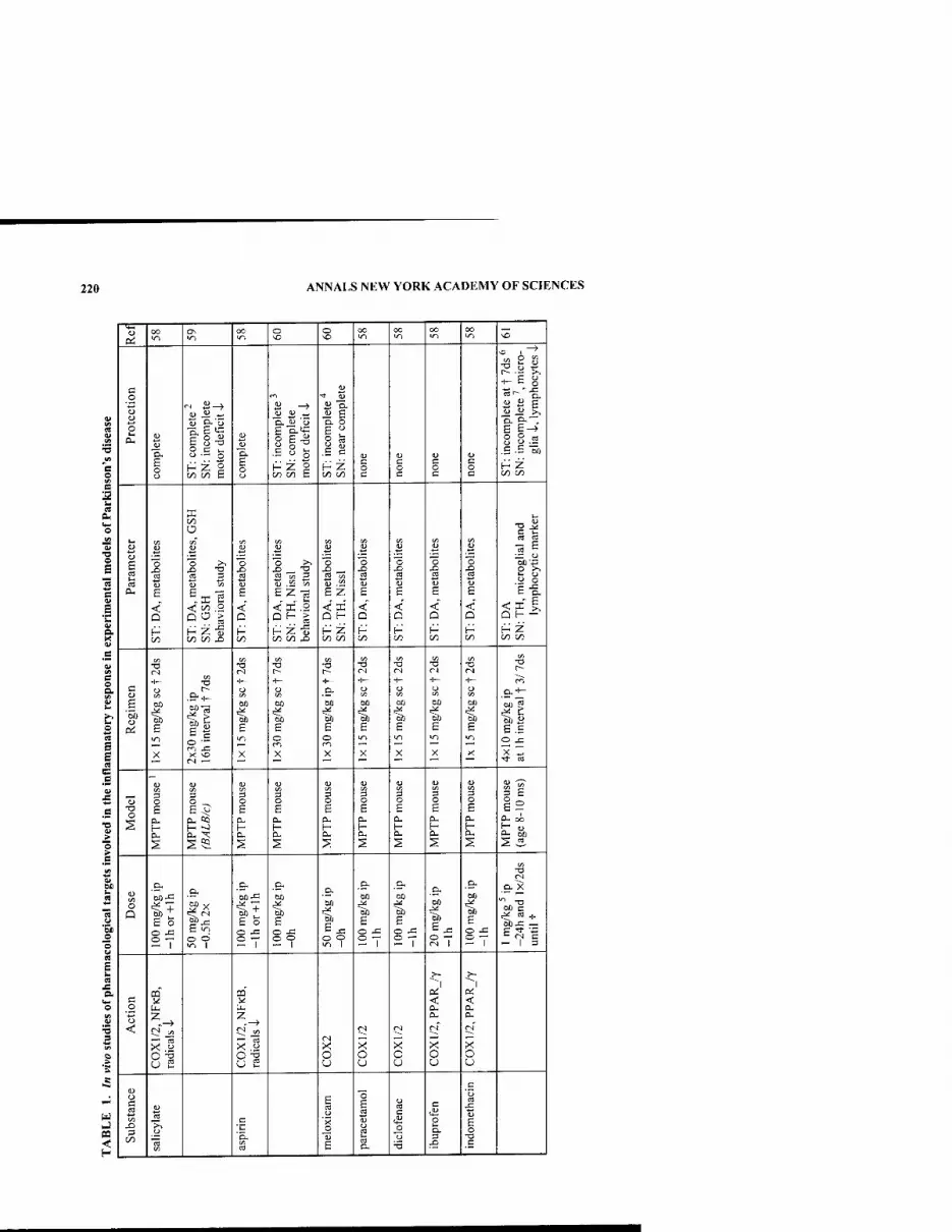
220 ANNALS NEW YORK ACADEMY OF SCIENCES
Pi oo in
ON in
oo o o NO
oo 00 IT)
oo oo ■n s
o
c o
o
e AH t)
B o o
o C i_
H Z g £ o o
if- H Z g CO on c
ii E a o E " S « J S H Z 1/5 C/)
c o c
OJ
§ o c o c
" B g
S 8.2 .S-E -5) H Z C/3 C«
o
o E
c 0)
E *C Q. X V
O
E 2 0-
V3
"o
E
< Q
O (/T
!§ t S 3
<C c/3 2 QO >
H Z -S on c/: j3
'o « B
<
u
EZ-g < X .2 D^ ^ /_■ ^ j:
00 C/) XJ
S "5 •S -
E Z < X D f-
H Z C/3 C/}
on
X)
< Q
H
1 cd u
E
< D
H
B OJ
E
< Q
H C/3
en
-2
E
< Q
H
LH
Cd «
s & .a o E-g.
.s B O a
o « E
e
Di e in
X
W5
X ^
-a
-!— O
GO
B
X
-a
-I-
an
B o
X
XI
■I—
e- M
=^ E o
X
(N
•i- O
B m
X
•a
bO
E in
X
-o
U
'I, E in
E
X
-o r-
oo ^
MB £ .£ O X
X I^
B
B
i >
■a o
o 6
H P-
2
o ^
a, Ki
OH X
S5i
O
s
i E
H PH
s
O
B 0-
s
§ E 0. H PH
2
o E p. [- p.
2
§ E p-
2
o £
H Cu
12 D- oo
a. 60
2 3. E
& ■2
o
*OJD O
s
a CM o
S
i
o
e o o j:
27
H'x |s B ^ o o "n 1
.9-
E o o ^ 27
DO
E
2?
.D.
an 00
E o -c 27
a,
t B o x:
S7
00
£ o ^ 27
D. X
-7 i
.2
<
U E
UH
X.2
u E X o o
u
X o u
1
< a.
ri
>< o u
>• 1
ai < a. 0-
(N
>< o u
oa <
ID O c cd
1/5
on Vi
a ■c
'E. CO
E cd
o
B
3 E B ij (j ca
cd
o cd c
o
•5
c
e a.
Xi
c
E o
T3

HIRSCH etal.: ROLE OF GLIAL REACTION AND INFLAMMATION 221
OJ (N CO (N (^ m m ^ in VD
(S •rj in >o M3 ^ vo ^ >o 'O
<« Wl ^ s u 0
a ■3 w B
c .0
0 u
s OH
c 0
It
S 0
Q g. C M 0 U
111 c 6 rt
0
i
CN
■E.'H.
.g.s HZ
•2 t
•g §
"Hb 0 0
u ■3. s 0
g izi on c on qfl c 0
B
fc. a
eu =» t- g; "S u D
CA VH 1 " C3 « » en « sf ^ « •3
g 1 a ^ .a 0
■3
s
■2^
x> -2 ^t .s 3 1
"BS rt u u u 1> CJ "Ho u B PH E B-g. E e B B g Z-3 a 1 ffil ffi < KI < < <^'
0 < S.2 <f •c H eoH Q H — 0 Q Q 0 0 c >
cd cd Q 0. H Z H z H H HZ H H-8 H g c/3 « M w (/I 00 on tzi M c/a ^ on
_B w
i B
1 00
•a
•1-
•S* — 13
•a CA
2 "0 ^~"
•a
s W)-!—
W)"^
s ^ 1 60 5 CO
B.SC
■1- > H 60 •" 1"^
•B fe
60 g
3 (S 6 5 $:> w B^ C3
g E 5
2,1 X J3 X
0^^
X " '^ 'i 0
i 0 0
60 a B^
in in
Tf <N -a- C8 •!— 00 00 (N (N —' m 3 m CO
c: _B
■3 T3 S <l <i <
i 0
£ g CL, 0 0_ « *N ii §i
Q CL, ID
^ 3 >■ a- 0 PH 0 60 0 5 0 0 0 „ eu 5 PH 0 K 0
s a S S S B^ ^ B vO g vi 2 S B s a S B B rz3 J3
1 0 1 :s -0 <n J 1 U 5 -1- S^ .&! CO
0 <n
.&^ a p.iri -S Q ^ — bO 60 + (30 >n 605 M)3 5
3) 3 -H
60 04
2 Q "§.1 "§> bOT3 ^=n
1:^0 t3 ^B 1^ a "n •a e^ 6? 6:s B4 11 s:s B § &
0 (N ^ e 0 <^ 0 — 2.B- 0 -- 0 -7 0 -7 °9"S 1 <N 1 <^ 1 — 1 -I- <N 1 (N 1 ^ W ^ ! •^ 1 + in 1 .3
u at (>J ji .C C .c B. •s 1 2 !S IS r^
o< & D. rt w 0 0 0
1 0 <
S B S
i. 1 z
1 z BH Z z P
c 0
1 .5
C
i
g
8 p
< c
■c a §
m
0 1 3
< 3
C/3 2 'a
C<3
13
•^ ^ 0 0
15 J3
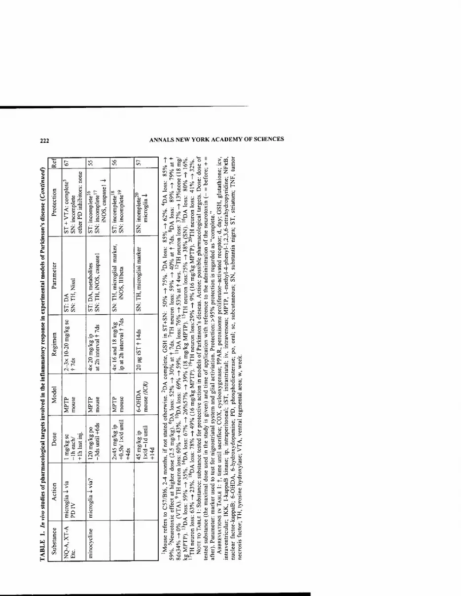
222 ANNALS NEW YORK ACADEMY OF SCIENCES
O -s
Pi
c o
o ID
£ a-
c
^ id
Ml H o Q
+ •- S3
on cfl o
s s s
l.si H Z
If u 00 Ol
OJ IS
Is •S E
Z
E
iz
D H
t/2 00
ii < K D H
H Z
II li z
S3
■1 E
"3) a o E
H
g g 5b oi
1 o
O
V)
6013
li 00 C — u •a c
y "
H
i o
-a o
If 11 si o § vi E
en O
— 1 +
CO
a? ||
■e-l
fl X 9 f <N + +
E:g5 ■* — +
c .2 o <
6 cu
2 ■>
-> .2
£
o c
CO
<
< 2 w
(D
C
00'^
S T ° o 2: -" ^ N© "^
S 2; '^ " ^ Q 00 — « Jn
o oT
00.
c o
"ob
o ^
T Q §ZK
— on H
is
CO 2 ID -^
^C- )^^ h O w *:; ' H — cS P- 2 C ^^ (D ■ ^ O TJ O &■•— (U u
a'E .2 ■= •
Z "
&2
U t/5
^. M
ca
on g
on K
t K ca o '-^—■ t " S3 ^ fc ■
^ ;= ,<u c^ cn <u
' cu
^S < 60 5 I- 0
o «
<-'^ ^_r M) ^
r> " i? ,^ H
2 a> o D.
S CU
0:S
E c T
c < "' M..2 ^
o >*- >
TS E J!
en
" a DO
._ ^
00 > w C ° <
a a
§ a c u . ►
CO I.- i: o c , •" o
•■ D. > ^^
ca « ,
I .H ^. W "O cd
■tow
, in -
: ^ •= " E
s-2 ^8 ^^ 0^ ocrt r-
t^ op 1 ^ g ^0 CO 0
c l*-l ID 0 *—
<-i^< « n 0 — .
' iS b" ! 5. >, ■ -2 I .2 ^
■Ipi <
crt 1-7.
S sO r^
ON -O
o^ ON
■• ^ CO Ci^
> -2 "^ :^
Xi <U (D C 3 en ■*-■ '^
" — -a ..
i E g u JD ca S3 5
.. ^ 2 :7
F- o 2 00 -S 1=
C _ P Ju ca
•c .S •t' "Eg o- S ^ ■a 5 ■* ■^ o <£
.&§^
5 ■? ">.
•^ ' S
=° . .£ •-f- V^ CO ^ O
. - 1-
M ca "
s sffi U4 u ■<-■ 'wi ;-,
^2 E^g
Sffi
I- -g 3 ^
3 ° S
m ^ (D o

HIRSCH etal.: ROLE OF GLIAL REACTION AND INFLAMMATION 223
<L} en in
oo' 00 0\ o r~-
(N
0) Vi a
Vi
■3
e o S
'S u a
o
•a
1 S ~4>
g.s HZ M on
s s GO on
HZ M on
=! g.s H Z CO w
s §
II i J
H Z M on
i o
d
1 5 6
o S
1 s-
•o
<
£ S 5 -S .S ^ g
o 5 S e E
B. X (U
V
B O B.
(2
1 „ s
If II
„ a
I"! 11 s 1 1
6
1 3
ffi" H
< Q
"a in ^
3.2 l| ll it H Z on on
H 00
en u
1 a
CO Ol p on on H Z on w
H Z on M
H Z on on
H Z on i/3
H on
C9
E E « c B
B
■O V
1
g Dn
e
1 ll X J3 1- <N
1 to
o '^
X -o
1 u
Of)
CN -1- X -o
u
60
Ml-a G 2
CN -1- X -a
1 S
(N
X
T3
.&
E in
X ■5f
u CO
60
X
1 o
X
O
si 11 II si OH U
li li li li CM o
'■3
S
1
1>
1 G
1 1 1
+ 1
■§ cd
4
+ T 1
1
So S ■"
1 -a
TS C ca o O
.g <t>
II ^ 1
B
rj
« <
1 on O
CN <N
<
(N
s o in Ci.
o
d
a ^'
ts a
o
•So."
c o h S ° ' M — S m OT >^
• S ca „H •iS-o-a _ <3 iJ 9 "O
<« C3 U -^ O 60 g U ^ V< ^ -Vi
S o
3 ~ O H- 60 f^ >>H s a U 3
^ (D oj in JJ -c
+■ "
I H
: a.
t. a M 60 ,P P b-fi
t T t i I N? sO sO 0-- QJ O^ O^ O^ T* "t-» ^ -^ o -i " r- \o -^
www www
_o _o _o
a-a o e
tH k. 1.4 3 3 3 lU cu u C C C
S W W H H H
T u ' 60
00 (L> v^ 60 .. t. w ,=0 w H
« w J2;
2 o "^ •aP «
■§ > s S M C „ « M a s 3 w < .O

224 ANNALS NEW YORK ACADEMY OF SCIENCES
but they even seemed to enhance the toxic properties of MPP+. Furthermore, these molecules induced a switch from apoptosis to necrosis. Indeed, apoptotic features could be readily detected in dopaminergic cultures treated with MPP"^ alone, where- as the morphological changes observed in the cultures co-treated with caspase inhib- itors were compatible with necrosis with regard to membrane leakage. In contrast, the presence of a high concentration of glucose in the culture medium alone or in conjunction with caspase inhibitors afforded a partial protection of dopamine neuron cultures against MPP+. Thus, these data indicate that it will be extremely difficult to save neurons with strongly impaired functioning using neuroprotective strategies. The data rather suggest that a cocktail of neuroprotective agents (in our case, caspase inhibitors and glucose) or agents with a broader spectrum of action should be used to protect dopaminergic neurons. We recently tested this hypothesis by activating peroxisome proliferator-activated receptor-y (PPAR-y), a member of the nuclear re- ceptor superfamily that has recently been shown to inhibit inflammatory processes (probably by counteracting NFKB activation) in a variety of cell types in vitro—in- cluding monocytes-macrophages'***'''' and microglial cells^"—and in vivo.^' We showed that in mice intoxicated by MPTP, orally administered pioglitazone, a selec- tive PPAR-y agonist currently used in the treatment of diabetes, attenuated the MPTP-induced microglial activation and prevented the dopaminergic cell loss in the substantia nigra.^^ j^ contrast, pioglitazone did not influence the microglial response in the striatum nor did it affect the loss of striatal tyrosine hydroxylase immunoreac- tivity induced by MPTP. Similarly, other neuroprotective strategies that efficiently protect dopaminergic neurons in the substantia nigra, such as the invalidation of the inducible form of nitric oxide synthase, have also been shown to have little effect on dopaminergic terminals in the striatum."-^'* These data suggest that after MPTP in- toxication the mechanisms regulating microglial activation may be different in dopaminergic terminals and in dopaminergic cell bodies. Furthermore, they indicate that combined neuroprotective strategies should be developed for dopaminergic cell bodies and terminals. Fulfilling these criteria, minocycline, a tetracycline derivative that inhibits microglial activation, was recently shown to reduce glial activation and protect dopaminergic structures in the entire striatonigral system in the MPTP mouse model^^'^** or 6-hydroxydopamine rat model.^'' The experimental data suggest that the minocycline-induced suppression of microglial activation is essential for its neu- roprotective effect. These findings demonstrate the potentially deleterious role of ac- tivated glia for the pathogenesis of Parkinson's disease.
In conclusion, the data reviewed here suggest an involvement of the glial reaction and inflammatory processes in the progression of neuronal degenerafion in parkin- sonian syndromes. Pharmacological manipulation of these pathways affords a rela- tive degree of neuroprotection. Yet, it remains to be determined if such molecules will be neuroprotective in Parkinson's disease. Epidemiological studies concentrat- ing on the effects of nonsteroidal antiinflammatory drugs in Parkinson's disease may help to answer this question before clinical trials are initiated.
REFERENCES
1. HiRSCH, E.G., A.M. GRAYBIEL & Y. AGIO. 1988. Melanized dopaminergic neurons are differentially susceptible to degeneration in Parkinson's disease. Nature 334: 345- 348.
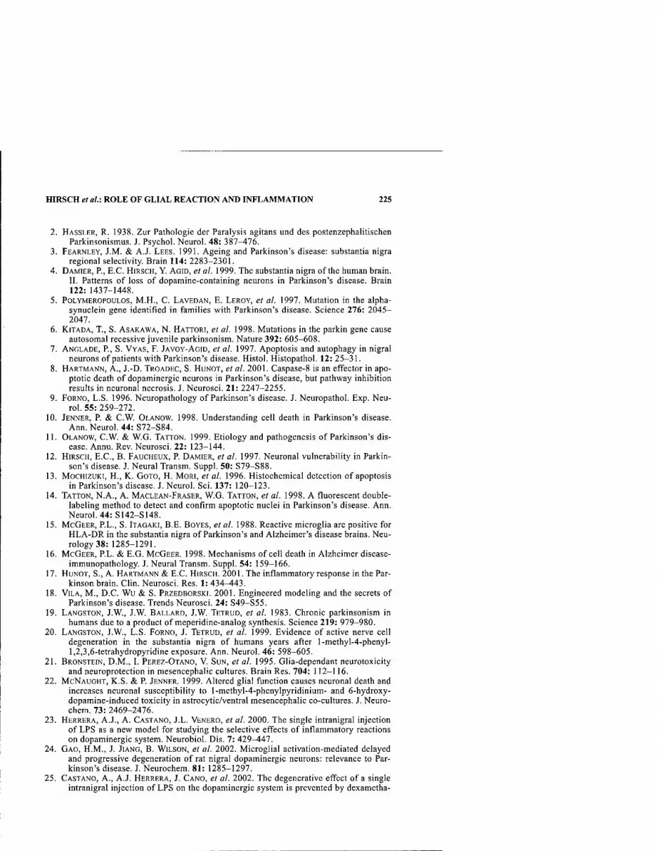
HIRSCH et al: ROLE OF GLIAL REACTION AND INFLAMMATION 225
2. HASSLER, R. 1938. Zur Pathologie der Paralysis agitans und des postenzephalitischen Parkinsonismus. J. Psychol. Neurol. 48: 387-476.
3. FEARNLEY, J.M. & A.J. LEES. 1991. Ageing and Parkinson's disease: substantia nigra regional selectivity. Brain 114: 2283-2301.
4. DAMIER, P., E.C. HIRSCH, Y. AGID, et al. 1999. The substantia nigra of the human brain. II. Patterns of loss of dopamine-containing neurons in Parkinson's disease. Brain 122: 1437-1448.
5. PoLYMEROPOULOS, M.H., C. LAVEDAN, E. LEROY, et al. 1997. Mutation in the alpha- synuclein gene identified in families with Parkinson's disease. Science 276: 2045- 2047.
6. KiTADA, T., S. ASAKAWA, N. HATTORI, et al. 1998. Mutations in the parkin gene cause autosomal recessive juvenile parkinsonism. Nature 392: 605-608.
7. ANGLADE, P., S. VYAS, F. JAVOY-AGID, et al. 1997. Apoptosis and autophagy in nigral neurons of patients with Parkinson's disease. Histol. Histopathol. 12: 25-31.
8. HARTMANN, A., J.-D. TROADEC, S. HUNOT, et al. 2001. Caspase-8 is an effector in apo- ptotic death of dopaminergic neurons in Parkinson's disease, but pathway inhibition results in neuronal necrosis. J. Neurosci. 21: 2247-2255.
9. FORNO, L.S. 1996. Neuropathology of Parkinson's disease. J. Neuropathol. Exp. Neu- rol. 55: 259-272.
10. JENNER, P. & C.W. OLANOW. 1998. Understanding cell death in Parkinson's disease. Ann. Neurol. 44: S72-S84.
11. OLANOW, C.W. & W.G. TATTON. 1999. Etiology and pathogenesis of Parkinson's dis- ease. Annu. Rev. Neurosci. 22: 123-144.
12. HIRSCH, E.C, B. FAUCHEUX, P. DAMIER, et al. 1997. Neuronal vulnerability in Parkin- son's disease. J. Neural Transm. Suppl. 50: S79-S88.
13. MocHizuKi, H., K. GOTO, H. MORI, et al. 1996. Histochemical detection of apoptosis in Parkinson's disease. J. Neurol. Sci. 137: 120-123.
14. TATTON, N.A., A. MACLEAN-FRASER, W.G. TATTON, et al. 1998. A fluorescent double- labeling method to detect and confirm apoptotic nuclei in Parkinson's disease. Ann. Neurol. 44: S142-S148.
15. MCGEER, P.L., S. ITAGAKI, B.E. BOYES, et al. 1988. Reactive microglia are positive for HLA-DR in the substantia nigra of Parkinson's and Alzheimer's disease brains. Neu- rology 38: 1285-1291.
16. MCGEER, P.L. & E.G. MCGEER. 1998. Mechanisms of cell death in Alzheimer disease- immunopathology. J. Neural Transm. Suppl. 54: 159-166.
17. HUNOT, S., A. HARTMANN & E.C. HIRSCH. 2001. The inflammatory response in the Par- kinson brain. Clin. Neurosci. Res. 1: 434-443.
18. VILA, M., D.C. WU & S. PRZEDBORSKI. 2001. Engineered modeling and the secrets of Parkinson's disease. Trends Neurosci. 24: S49-S55.
19. LANGSTON, J.W., J.W. BALLARD, J.W. TETRUD, et al. 1983. Chronic parkinsonism in humans due to a product of meperidine-analog synthesis. Science 219: 979-980.
20. LANGSTON, J.W., L.S. FORNO, J. TETRUD, et al. 1999. Evidence of active nerve cell degeneration in the substantia nigra of humans years after l-methyl-4-phenyl- 1,2,3,6-tetrahydropyridine exposure. Ann. Neurol. 46: 598-605.
21. BRONSTEIN, D.M., I. PEREZ-OTANO, V. SUN, et al. 1995. Glia-dependant neurotoxicity and neuroprotection in mesencephalic cultures. Brain Res. 704: 112-116.
22. MCNAUGHT, K.S. & P. JENNER. 1999. Altered glial function causes neuronal death and increases neuronal susceptibility to l-methyl-4-phenylpyridinium- and 6-hydroxy- dopamine-induced toxicity in astrocytic/ventral mesencephalic co-cultures. J. Neuro- chem. 73: 2469-2476.
23. HERRERA, A.J., A. CASTANO, J.L. VENERO, et al. 2000. The single intranigral injection of LPS as a new model for studying the selective effects of inflammatory reactions on dopaminergic system. Neurobiol. Dis. 7: 429-447.
24. GAO, H.M., J. JIANG, B. WILSON, et al. 2002. Microglial activation-mediated delayed and progressive degeneration of rat nigral dopaminergic neurons: relevance to Par- kinson's disease. J. Neurochem. 81: 1285-1297.
25. CASTANO, A., A.J. HERRERA, J. CANO, et al. 2002. The degenerative effect of a single intranigral injection of LPS on the dopaminergic system is prevented by dexametha-
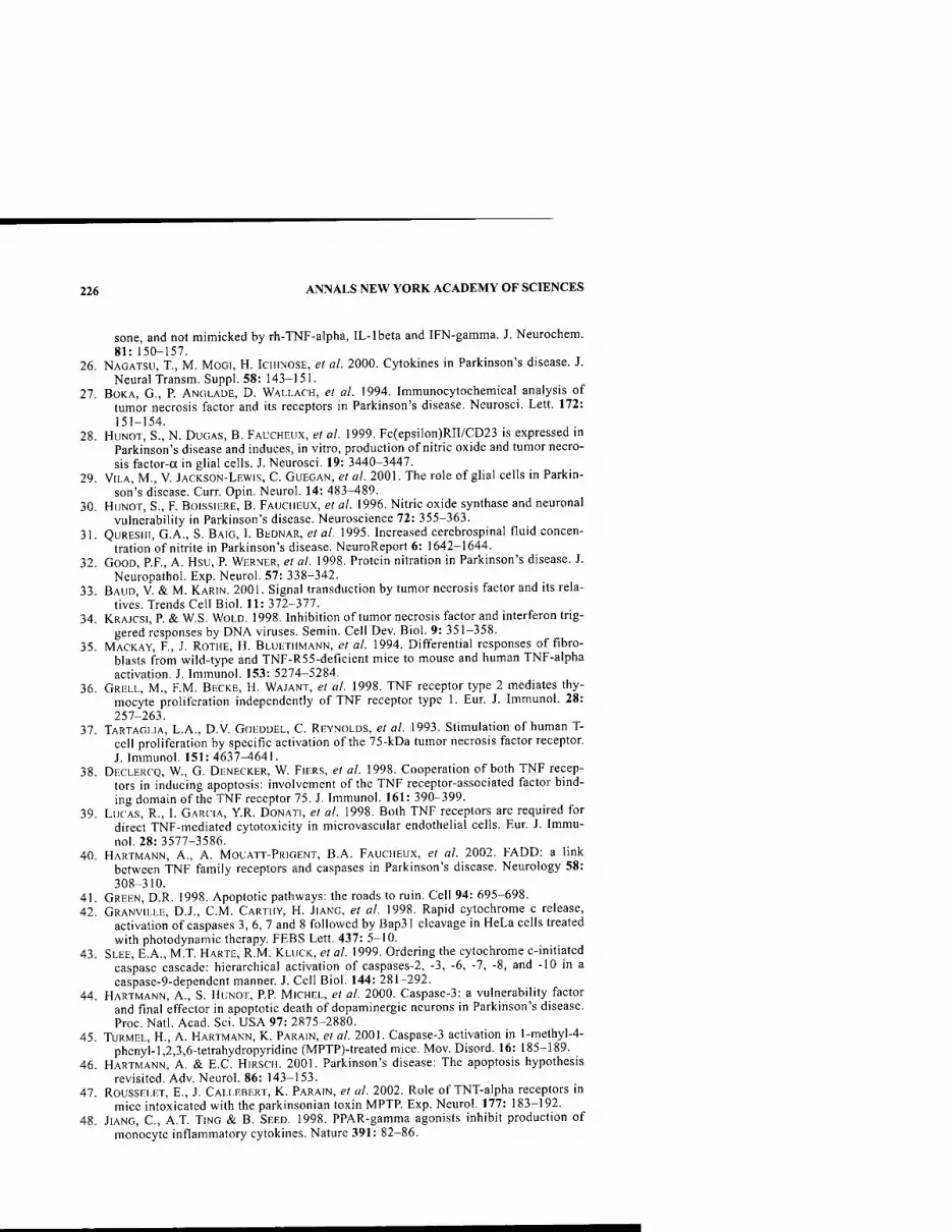
226 ANNALS NEW YORK ACADEMY OF SCIENCES
sone, and not mimicked by rh-TNF-alpha, IL-lbeta and IFN-gamma. J. Neurochem. 81: 150-157.
26. NAGATSU, T., M. MOGI, H. ICHINOSE, et al. 2000. Cytokines in Parkinson's disease. J. Neural Transm. Suppl. 58: 143-151.
27. BOKA, G., P. ANGLADE, D. WALLACH, et al. 1994. Immunocytochemical analysis of tumor necrosis factor and its receptors in Parkinson's disease. Neurosci. Lett. 172: 151-154.
28. HUNOT, S., N. DuGAS, B. FAUCHEUX, et al. 1999. Fc(epsilon)RII/CD23 is expressed in Parkinson's disease and induces, in vitro, production of nitric oxide and tumor necro- sis factor-a in glial cells. J. Neurosci. 19: 3440-3447.
29. VILA, M., V. JACKSON-LEWIS, C. GUEGAN, et al. 2001. The role of glial cells in Parkin- son's disease. Curr. Opin. Neurol. 14: 483^89.
30. HuNOT, S., F. BOISSIERE, B. FAUCHEUX, et al. 1996. Nitric oxide synthase and neuronal vulnerability in Parkinson's disease. Neuroscience 72: 355-363.
31. QuRESlli, G.A., S. BAIG, L BEDNAR, et al. 1995. Increased cerebrospinal fluid concen- tration of nitrite in Parkinson's disease. NeuroReport 6: 1642-1644.
32. GOOD, P.F., A. Hsu, P. WERNER, el al. 1998. Protein nitration in Parkinson's disease. J. Neuropathol. Exp. Neurol. 57: 338-342.
33. BAUD, V. & M. KARIN. 2001. Signal transduction by tumor necrosis factor and its rela- tives. Trends Cell Biol. 11: 372-377.
34. KRAJCSI, P & W.S. WOLD. 1998. Inhibition of tumor necrosis factor and interferon trig- gered responses by DNA viruses. Semin. Cell Dev. Biol. 9: 351-358.
35. MACKAY, F., J. ROTHE, H. BLUETHMANN, et al. 1994. Differential responses of fibro- blasts from wild-type and TNF-R55-deficient mice to mouse and human TNF-alpha activation. J. Immunol. 153: 5274-5284.
36. GRELL, M., F.M. BECKE, H. WAJANT, et al. 1998. TNF receptor type 2 mediates thy- mocyte proliferation independently of TNF receptor type 1. Eur. J. Immunol. 28: 257-263.
37. TARTAGLIA, L.A., D.V. GOEDDEL, C. REYNOLDS, et al. 1993. Stimulation of human T- cell proliferation by specific activation of the 75-kDa tumor necrosis factor receptor. J. Immunol. 151:4637^641.
38. DECLERCO, W., G. DENECKER, W. FIERS, et al. 1998. Cooperation of both TNF recep- tors in inducing apoptosis: involvement of the TNF receptor-associated factor bind- ing domain of the TNF receptor 75. J. Immunol. 161: 390-399.
39. LUCAS, R., I. GARCIA, Y.R. DONATI, et al. 1998. Both TNF receptors are required for direct TNF-mediated cytotoxicity in microvascular endothelial cells. Eur. J. Immu- nol. 28: 3577-3586.
40. HARTMANN, A., A. MOUATT-PRIGENT, B.A. FAUCHEUX, et al. 2002. FADD: a link between TNF family receptors and caspases in Parkinson's disease. Neurology 58: 308-310.
41. GREEN, D.R. 1998. Apoptotic pathways: the roads to ruin. Cell 94: 695-698. 42. GRANVILLE, D.J., CM. CARTHY, H. JIANG, et al. 1998. Rapid cytochrome c release,
activation of caspases 3, 6, 7 and 8 followed by Bap31 cleavage in HeLa cells treated with photodynamic therapy. FEBS Lett. 437: 5-10.
43. SLEE, E.A., M.T. HARTE, R.M. KLUCK, et al. 1999. Ordering the cytochrome c-initiated caspase cascade: hierarchical activation of caspases-2, -3, -6, -7, -8, and -10 in a caspase-9-dependent manner. J. Cell Biol. 144: 281-292.
44. HARTMANN, A., S. HUNOT, RP MICHEL, et al. 2000. Caspase-3: a vulnerability factor and final effector in apoptotic death of dopaminergic neurons in Parkinson's disease. Proc. Natl. Acad. Sci. USA 97: 2875-2880.
45. TURMEL, H., A. HARTMANN, K. PARAIN, et al. 2001. Caspase-3 activation in l-methyl-4- phenyl-l,2,3,6-tetrahydropyridine (MPTP)-treated mice. Mov. Disord. 16: 185-189.
46. HARTMANN, A. & E.C. HIRSCH. 2001. Parkinson's disease: The apoptosis hypothesis revisited. Adv. Neurol. 86: 143-153.
47. RoussELET, E., J. CALLEBERT, K. PARAIN, et al. 2002. Role of TNT-alpha receptors in mice intoxicated with the parkinsonian toxin MPTP. Exp. Neurol. 177: 183-192.
48. JIANG, C, A.T. TING & B. SEED. 1998. PPAR-gamma agonists inhibit production of monocyte inflammatory cytokines. Nature 391: 82-86.
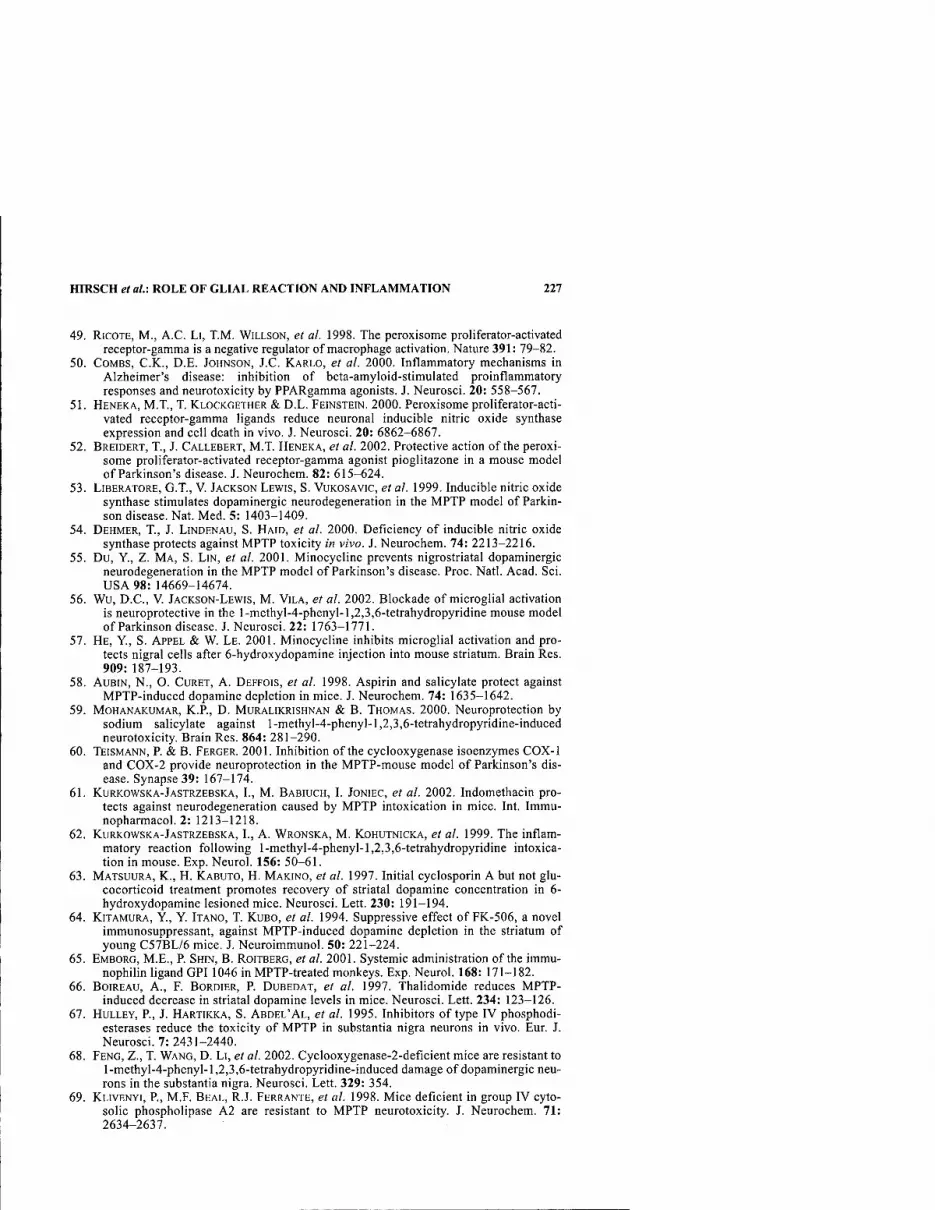
HIRSCH et al: ROLE OF GLIAL REACTION AND INFLAMMATION 227
49. RiCOTE, M., A.C. LI, T.M. WILLSON, et al. 1998. The peroxisome proliferator-activated receptor-gamma is a negative regulator of macrophage activation. Nature 391: 79-82.
50. COMBS, C.K., D.E. JOHNSON, J.C. KARLO, et al. 2000. Inflammatory mechanisms in Alzheimer's disease: inhibition of beta-amyloid-stimulated proinflamraatory responses and neurotoxicity by PPARgamma agonists. J. Neurosci. 20: 558-567.
51. HENEKA, M.T., T. KLOCKGETHER & D.L. FEINSTEIN. 2000. Peroxisome proliferator-acti- vated receptor-gamma ligands reduce neuronal inducible nitric oxide synthase expression and cell death in vivo. J. Neurosci. 20: 6862-6867.
52. BREIDERT, T., J. CALLEBERT, M.T. HENEKA, et al. 2002. Protective action of the peroxi- some proliferator-activated receptor-gamma agonist pioglitazone in a mouse model of Parkinson's disease. J. Neurochem. 82: 615-624.
53. LIBERATORE, G.T., V. JACKSON LEWIS, S. VUKOSAVIC, et al. 1999. Inducible nitric oxide synthase stimulates dopaminergic neurodegeneration in the MPTP model of Parkin- son disease. Nat. Med. 5: 1403-1409.
54. DEHMER, T., J. LINDENAU, S. HAID, et al. 2000. Deficiency of inducible nitric oxide synthase protects against MPTP toxicity in vivo. J. Neurochem. 74: 2213-2216.
55. Du, Y., Z. MA, S. LIN, et al. 2001. Minocycline prevents nigrostriatal dopaminergic neurodegeneration in the MPTP model of Parkinson's disease. Proc. Natl. Acad. Sci. USA 98: 14669-14674.
56. Wu, D.C., V. JACKSON-LEWIS, M. VILA, et al. 2002. Blockade of microglial activation is neuroprotective in the l-methyl-4-phenyl-l,2,3,6-tetrahydropyridine mouse model of Parkinson disease. J. Neurosci. 22: 1763-1771.
57. HE, Y., S. APPEL & W. LE. 2001. Minocycline inhibits microglial activation and pro- tects nigral cells after 6-hydroxydopamine injection into mouse striatum. Brain Res. 909: 187-193.
58. AUBIN, N., O. CuRET, A. DEFFOIS, et al. 1998. Aspirin and salicylate protect against MPTP-induced dopamine depletion in mice. J. Neurochem. 74: 1635-1642.
59. MOHANAKUMAR, K.P., D. MURALIKRISHNAN & B. THOMAS. 2000. Neuroprotection by sodium salicylate against l-methyl-4-phenyl-l,2,3,6-tetrahydropyridine-induced neurotoxicity Brain Res. 864: 281-290.
60. TEISMANN, P. & B. FERGER. 2001. Inhibition of the cyclooxygenase isoenzymes COX-1 and COX-2 provide neuroprotection in the MPTP-mouse model of Parkinson's dis- ease. Synapse 39: 167-174.
61. KURKOWSKA-JASTRZEBSKA, I., M. BABIUCH, I. JONIEC, et al. 2002. Indomethacin pro- tects against neurodegeneration caused by MPTP intoxication in mice. Int. Immu- nopharmacol. 2: 1213-1218.
62. KURKOWSKA-JASTRZEBSKA, I., A. WRONSKA, M. KOHUTNICKA, et al. 1999. The inflam- matory reaction following l-methyl-4-phenyl-l,2,3,6-tetrahydropyridine intoxica- tion in mouse. Exp. Neurol. 156: 50-61.
63. MATSUURA, K., H. KABUTO, H. MAKINO, et al. 1997. Initial cyclosporin A but not glu- cocorticoid treatment promotes recovery of striatal dopamine concentration in 6- hydroxydopamine lesioned mice. Neurosci. Lett. 230: 191-194.
64. KITAMURA, Y, Y. ITANO, T. KUBO, et al. 1994. Suppressive effect of FK-506, a novel immunosuppressant, against MPTP-induced dopamine depletion in the striatum of young C57BL/6 mice. J. Neuroimmunol. 50: 221-224.
65. EMBORG, M.E., P. SHIN, B. ROITBERG, et al. 2001. Systemic administration of the immu- nophilin ligand GPI 1046 in MPTP-treated monkeys. Exp. Neurol. 168: 171-182.
66. BoiREAU, A., F. BORDIER, P. DUBEDAT, et al. 1997. Thalidomide reduces MPTP- induced decrease in striatal dopamine levels in mice. Neurosci. Lett. 234: 123-126.
67. HULLEY, P., J. HARTIKKA, S. ABDEL'AL, et al. 1995. Inhibitors of type IV phosphodi- esterases reduce the toxicity of MPTP in substantia nigra neurons in vivo. Eur. J. Neurosci. 7: 2431-2440.
68. FENG, Z., T. WANG, D. LI, et al. 2002. Cyclooxygenase-2-deficient mice are resistant to l-methyl-4-phenyl-l,2,3,6-tetrahydropyridine-induced damage of dopaminergic neu- rons in the substantia nigra. Neurosci. Lett. 329: 354.
69. KLIVENYI, P., M.F. BEAL, R.J. FERRANTE, et al. 1998. Mice deficient in group IV cyto- solic phospholipase A2 are resistant to MPTP neurotoxicity. J. Neurochem. 71: 2634-2637.
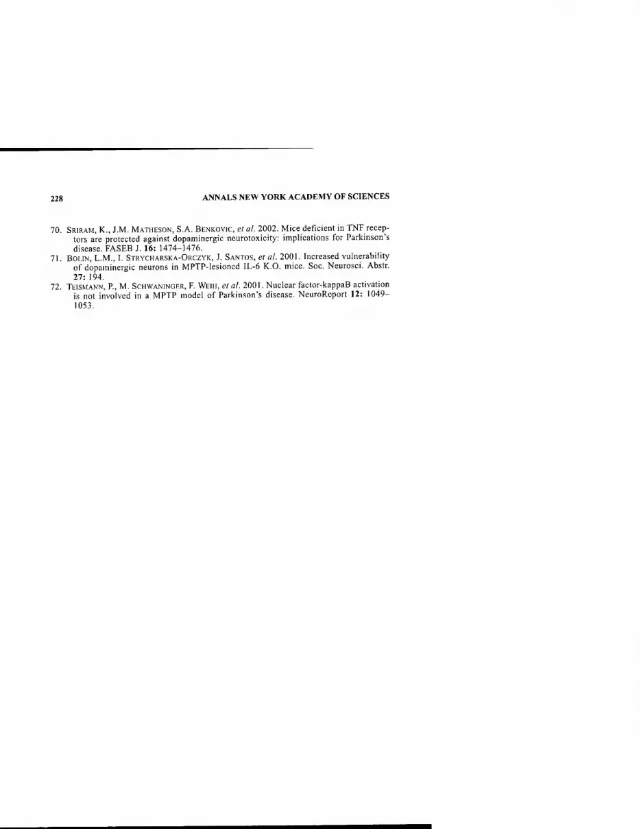
228 ANNALS NEW YORK ACADEMY OF SCIENCES
70. SRIRAM, K., J.M. MATHESON, S.A. BENKOVIC, et al. 2002. Mice deficient in TNF recep- tors are protected against dopaminergic neurotoxicity: implications for Parkinson's disease. FASEB J. 16: 1474-1476.
71. BoLiN, L.M., I. STRYCHARSKA-ORCZYK, J. SANTOS, et al. 2001. Increased vulnerability of dopaminergic neurons in MPTP-lesioned IL-6 K.O. mice. Soc. Neurosci. Abstr. 27: 194.
72. TEISMANN, P., M. SCHWANINGER, F. WEIH, et al. 2001. Nuclear factor-kappaB activation is not involved in a MPTP model of Parkinson's disease. NeuroReport 12: 1049- 1053.

Induction of Adult Neurogenesis
Molecular Manipulation of Neural Precursors in Situ
PAOLA ARLOTTA, SANJAY S. MAGAVI, AND JEFFREY D. MACKLIS
MGH-HMS Center for Nervous System Repair, Massachusetts General Hospital Departments ofNeurosurgery and Neurology and Program in Neuroscience, Harvard Medical School, Boston, Massachusetts 02114, USA
ABSTRACT: Over most of the past century, it was thought that the adult brain was completely incapable of generating new neurons. However, in the last de- cade, the development of new techniques has resulted in an explosion of new research showing that (i) neurogenesis, the birth of new neurons, is not restrict- ed to embryonic development, but normally also occurs in two limited regions of the adult mammalian brain (the olfactory bulb and the dentate gyrus of the hippocampus); (ii) that there are significant numbers of multipotent neural precursors in many parts of the adult mammalian brain; and (iii) that it is pos- sible to induce neurogenesis even in regions of the adult mammalian brain, like the neocortex, where it does not normally occur, via manipulation of endoge- nous multipotent precursors in situ. In the neocortex, recruitment of small numbers of new neurons can be induced in a region-specific, layer-specific, and neuronal type-specific manner, and newly recruited neurons can form long-dis- tance connections to appropriate targets. This suggests that elucidation of the relevant molecular controls over adult neurogenesis from endogenous neural precursors/stem cells may allow the development of neuronal replacement therapies for neurodegenerative disease and other central nervous system inju- ries that may not require transplantation of exogenous cells.
KEYWORDS: neurogenesis; neocortex; neural precursors; neural stem cells; tar- geted apoptosis; neuronal recruitment
Neural cell replacement therapies are based on the idea that neurological function lost due to injury or neurodegenerative disease can be improved by introducing new cells that can replace the function of lost neurons. Theoretically, the new cells can do this in one of two general ways.' New neurons could anatomically integrate into the host brain, becoming localized to the diseased portion of the brain, receiving af- ferents, expressing neurotransmitters, and forming axonal projections to relevant portions of the brain. Alternatively, newly introduced cells could constitutively se- crete neurotransmitters into local central nervous system (CNS) tissue, or they could
Address for correspondence: Jeffrey D. Macklis, MGH-HMS Center for Nervous System Repair, Massachusetts General Hospital, Edwards 4, 55 Fruit Street, Boston, MA 02114. Voice: 617-355-7185; fax: 617-734-1646.
Ann. N.Y. Acad. Sci. 991: 229-236 (2003). © 2003 New York Academy of Sciences.
229

230 ANNALS NEW YORK ACADEMY OF SCIENCES
be engineered to produce growth factors to support the survival or regeneration of existing neurons. Growing knowledge about the normal role of endogenous neural precursors, their potential differentiation fates, and their responsiveness to a variety of cellular and molecular controls suggests that neuronal replacement therapies based on manipulation of endogenous precursors either in situ or ex vivo may be pos- sible.
Neuronal replacement therapies based on the manipulation of endogenous pre- cursors in situ may have advantages over transplantation-based approaches, but they may have several limitations as well. The most obvious advantage of manipulating endogenous precursors in situ is that there is no need for external sources of cells. Cells for transplantation are generally derived from embryonic tissue, nonhuman species (xenotransplantation), or cells grown in culture. Use of embryo-derived tis- sue aimed towards human diseases is complicated by limitations in availability and by both political and ethical issues; for example, current transplantation therapies for Parkinson's disease require tissue from several embryos. Xenotransplantation of an- imal cells carries potential risks of introducing novel diseases into humans and raises questions about how well xenogenic cells will integrate into the human brain. In many cases, cultured cells need to be immortalized by oncogenesis, increasing the risk that the cells may become tumorigenic. In addition, transplantation of cells from many of these sources risks immune rejection and may require immunosuppression.
However, there are also potential limitations to manipulating endogenous precur- sor cells as a neuronal replacement therapy. First, such an approach may be practi- cally limited to particular regions of the brain, since multipotent neural precursors are densely distributed only in particular subregions of the adult brain (for example, the subventricular zone [SVZ]/subependimal zone [SEZ] and hippocampal subgran- ular zone). In addition, the potential differentiation fates of endogenous precursors may be too limited to allow their integration into varied portions of the brain. Anoth- er potential difficulty is that it may be difficult to provide the precise combination and sequence of molecular signals necessary to induce endogenous precursors to ef- ficiently and precisely proliferate and differentiate into appropriate types of neurons deep in the brain; this will likely be a critical issue to achieve proper integration and function by the newborn neurons.
The substantial amount of prior research regarding constitutively occurring neu- rogenesis provides insight into the potential and limitations of endogenous precursor based neuronal replacement therapies. In this chapter, we will first review results from several laboratories on the nature and location of endogenous neural precursors and then review some of our experiments demonstrating that endogenous neural pre- cursors can be induced to differentiate into neurons in regions of the adult brain that do not normally undergo neurogenesis, such as the cerebral cortex. Because of space limitations, we refer readers to other review articles that cover the broad field of con- stitutive adult neurogenesis in more detail.
IDENTITY OF ADULT NEURAL PRECURSORS
The effort to identify the neural precursors that contribute to olfactory bulb neu- rogenesis has generated a great deal of controversy. Research took an interesting turn with the provocative discovery that "glia-like" cells (for example, cells showing phe-
ARLOTTA et al.: INDUCTION OF ADULT NEUROGENESIS 231
notypic and antigenic features of glia, including cytoplasmic glycogen inclusions and expression of the intermediate filament protein GFAP, glial acidic fibrillary pro- tein) from the adult SVZ/SEZ can give rise to neurons and may therefore be neural precursors/stem cells.^ In a series of elegant experiments, Alvarez-Buylla and col- leagues showed that some SVZ astrocytes in the adult mammalian brain are able to form multipotent neurospheres in vitro (by many considered a property of neural stem cells) and are the source of new neurons of the olfactory bulb. These data strongly suggest that at least a subset of SVZ astrocyte-like cells are neural precur- sors. They selectively labeled SVZ astrocytic cells and traced the label via selective viral infection, using a receptor for an avian leukosis virus under the control of the GFAP promoter in transgenic mice, to newly generated neurons of the olfactory bulb.^ Similarly, GFAP-positive astrocyte-like cells in the subgranular layer (SGL) of the adult hippocampus (the other major site of adult neurogenesis) divide and gen- erate new neurons.-' These data support the idea that a subset of astrocyte-like cells have the properties of neural precursors and suggests discussion regarding the proper name to assign to these cells. Are they really bonafide astrocytes? It appears that only a very small fraction of astrocyte-like cells in the SVZ behave as neural precur- sors. Could related cells exist in other regions of the postnatal and adult brain? Or rather, is this subset of SVZ astrocytes a unique class of GFAP-expressing precursors that should be considered distinct and potentially given a new name?
In contrast with these results, Frisen's group published experiments reporting that ependymal cells lining the lumenal surface of the adult ventricular zone are adult neural precursors.'* These results contrast with those of Doetsch et al., who reported that ependymal cells did not divide in vivo or under their culture conditions in vitro.^ A parallel report from van der Kooy's lab reported that, while ependymal cells divide as spheres in vitro, they do not possess multipotential precursor properties.^ It re- mains to be established whether experimental differences between the three groups account for their different results (reviewed in Barres^). The idea that some subset of astrocyte-like cells can behave as neural precursors is supported by later studies suggesting that 1-10% of astrocytes isolated from several regions of the early post- natal brain (SVZ, cerebral cortex, cerebellum, spinal cord) and grown as monolayers are able to form "neurospheres" that can give rise to both neurons and glia in vitro.^ Moreover, retrovirally mediated expression of exogenous genes can drive some post- natal astrocyte-like cells to become neurons in vitro.^ Interestingly, there are devel- opmental and spatial constraints to astrocyte-like cells, specifically their multipotency, which is normally restricted to the first two postnatal weeks in mice, with the apparent exception of a subset of astrocyte-like cells from the SVZ that ap- pear to retain their multipotency during adulthood.^ This could mean that such cells in young animals may still be immature and retain their precursor attributes initially, until approximately PI0-11.^ However, it may be that the neurogenic environment of the SVZ can support and maintain the multipotency of a subset of astrocyte-like precursors, even in the adult brain.
Although the idea that glial cells could be neural precursors may sound unusual, it is supported by previous studies (long ignored) that in lizards and newts a special class of radial ependymoglia that extend from the ventricular zone (VZ) lumen to the pial surface can divide and give rise to both neurons and glia after injury, critical for spinal cord regeneration.'" In line with these observations, similar radial glial cells are present in the adult brain of the canary in regions of active neurogenesis, suggest-

232 ANNALS NEW YORK ACADEMY OF SCIENCES
ing that they may be the progenitors of newly generated neurons.'' Moreover, results from our laboratory show that adult astrocytes from the cerebral cortex can differen- tiate into transitional radial glia in response to signals that induce neurogenesis from transplanted immature precursors.'^ In general, the notion that cells of the radial glia lineage may have features of neural precursors during development and thus be able to generate both glia and neurons is not new.'^"'' We may imagine adult astrocyte- like cells as precursors with broad potential in neurogenic regions of the CNS (SVZ and dentate gyrus) and more restricted potential in nonneurogenic regions (cerebral cortex, spinal cord). If this is true, one future challenge will be to understand whether astrocyte-like cells from nonneurogenic regions can be induced to assume multipo- tential neuronal precursors properties, towards the goal of neuronal repopulation and CNS repair in situ.
THE LOCATION OF ADULT MAMMALIAN MULTIPOTENT PRECURSORS
Neurogenesis is normally restricted mainly to two specific regions of the adult mammalian brain: the SVZ or the SEZ, which provides neurons to the olfactory bulb, and the dentate gyrus of the hippocampus. If adult multipotent precursors were lim- ited to these two neurogenic regions of the adult CNS, it would severely limit the po- tential of neuronal replacement therapies based on in situ manipulation of endogenous precursors. However, adult multipotent precursors have been isolated and cultured in vitro from several other regions of the adult brain, including caudal portions of the SVZ, spinal cord,'^'*'' septum,'^ striatum,'^ neocortex,'^ optic nerve,'^ and retina.^" The precursors derived from all these regions can self-renew and differentiate into neurons, astroglia, and oligodendroglia (the three main cell types of the brain) in vitro. In nonneurogenic regions of the mature brain, it is thought that they normally differentiate only into glia or die. Cells from each region have different requirements for their proliferation and differentiation. Understanding the similarities and differences between the properties of multipotent precursors de- rived from different regions of the brain will be instrumental in potentially develop- ing neuronal replacement therapies based on manipulation of endogenous precursors.
MANIPULATION OF AN INHIBITORY ENVIRONMENT
The fact that new neurons are born only in very restricted areas of the adult mam- malian brain, even though neural precursors are found quite ubiquitously in the adult CNS, suggests that precursors from different regions may have different potential. Alternatively or in addition, local environments may differ in their ability to support/ allow neurogenesis from these precursors. Data from several groups have shown that multipotent precursors from several regions of the adult brain have a broad potential and can differentiate into at least the three different broad cell types, astroglia, oli- godendroglia, and neurons, given an appropriate in vitro or in vivo environment. This led us to explore the fate of multipotent precursors in an adult cortical environment, which does not normally support de nova neurogenesis.

ARLOTTA et al.i INDUCTION OF ADULT NEUROGENESIS 233
Our lab has previously shown that cortex undergoing synchronous apoptotic de- generation of projection neurons forms an instructive environment that can guide the differentiation of transplanted immature neurons or neural precursors.^'"^^ Imma- ture neurons or multipotent neural precursors transplanted into targeted cortex can migrate selectively to layers of cortex undergoing degeneration of projection neu- rons, differentiate into projection neurons, receive afferent synapses, express appro- priate neurotransmitters and receptors, and reform appropriate long-distance connections to the original contralateral targets of the degenerating neurons in adult murine neocortex.'^'^^"^^ Together, these results suggested to us that cortex under- going targeted apoptotic degeneration could direct endogenous multipotent precur- sors to integrate into adult cortical microcircuitry. In agreement with this hypothesis, we found, for the firs time, that endogenous multipotent precursors, normally locat- ed in the adult brain, can be induced to differentiate into neurons in the adult mam- malian neocortex, without transplantation.^' We induced synchronous apoptotic degeneration of corticothalamic neurons in layer 6 of anterior cortex and examined the fates of dividing cells within cortex, using BrdU and markers of progressive neu- ronal differentiation. BrdU"*" newborn cells expressed NeuN, a mature neuronal marker, and survived at least 28 weeks, while no new neurons were observed in con- trol, intact cortex. Moreover, some newborn neurons displayed typical pyramidal neuron morphology (large, 10-15 |im diameter somata with apical processes) char- acteristic of neurons that give rise to long-distance projections. Retrograde labeling from thalamus demonstrated that newborn BrdU"'" neurons can form appropriate long-distance corticothalamic connections.^' Together, these results demonstrate that endogenous neural precursors can be induced in situ to differentiate into cortical neurons, survive for many months, and form appropriate long-distance connections in the adult mammalian brain. The same microenvironment that supports the migra- tion, neuronal differentiation, and appropriate axonal extension of transplanted neu- roblasts and precursors also supports and instructs the neuronal differentiation and axon extension of endogenous precursors. These results demonstrate that the normal absence of constitutive cortical neurogenesis does not reflect an intrinsic limitation of the endogenous neural precursors' potential, but more likely results from a lack of appropriate microenvironmental signals necessary for neuronal differentiation or survival. Elucidation of these signals could enable CNS repair. Recently, other groups have reported similar and complementary results in hippocampus-'^ and stri- atum,^^ confirming and further supporting this direction of research.
CONCLUSION
Recent research suggests that it may be possible to manipulate endogenous neural precursors in situ so that they undergo neurogenesis in the adult brain, towards future neuronal replacement therapy for neurodegenerative disease and other CNS injury. Multipotent precursors, capable of differentiating into astroglia, oligodendroglia, and neurons exist in many portions of the adult brain. These precursors have consid- erable plasticity; and although they may have limitations in their integration into some host sites, they are, when heterotopically transplanted, capable of differentia- tion into neurons appropriate to a wide variety of recipient regions. Many adult pre- cursors are capable of migrating long distances, using both tangential and radial
234 ANNALS NEW YORK ACADEMY OF SCIENCES
forms of migration. Endogenous adult neural precursors are also capable of extend- ing axons significant distances through the adult brain. In addition, in vitro and in vivo experiments have begun to elucidate the responses of endogenous precursors to both growth factors and behavioral manipulations and are beginning to provide key information towards manipulation of their proliferation and differentiation. Recent experiments from our lab have shown that, under appropriate conditions, endoge- nous precursors can differentiate into neurons, extend long-distance axonal projec- tions, and survive for long periods of time in regions of the adult brain that do not normally undergo neurogenesis. Other laboratories have recently reported similar results in other systems. These results indicate that there exists a sequence and com- bination of molecular signals by which neurogenesis can be induced in the adult mammalian cerebral cortex and other regions where it does not normally occur.
Together, these data suggest that neuronal replacement therapies based on manip- ulation of endogenous precursors may be possible in the future. However, several questions must be answered before neuronal replacement therapies using endoge- nous precursors become a reality. The multiple signals that are responsible for en- dogenous precursor division, migration, and differentiation and axon extension and survival will need to be elucidated for such therapies to be developed efficiently.^"' These challenges also exist for neuronal replacement therapies based on transplan- tation of precursors, since donor cells, whatever their source, must interact with the mature CNS's environment to integrate into the brain. In addition, it remains an open question whether potential therapies manipulating endogenous precursors in situ would necessarily be limited to portions of the brain near adult neurogenic regions. However, even if multipotent precursors are not located in very high numbers out- side of neurogenic regions of the brain, it may be possible to induce them to prolif- erate from the smaller numbers that are more widely distributed throughout the neuraxis. Potentially, it may be possible to induce the repopulation of diseased mam- malian brain via the specific activation and instructive control over endogenous neu- ral precursors along desired neuronal lineages. However, the field is just beginning to understand the complex interplay between particular neural precursors' potential, their heterogeneity, and how to take advantage of what may be partial cell type re- striction, permissive and instructive developmental signals, and modulation of spe- cific aspects of neuronal differentiation and survival. Progress over the past decade has been great, and the coming decades promise to offer significant insight into these and other critical issues for the field.
ACKNOWLEDGMENTS
This work was supported in part by grants from the National Insfitutes of Health (NS41590, NS45523, HD28478, and MRRC HD18655), the Alzheimer's Associa- tion, the Human Frontiers Science Program, and the National Science Foundation to J.D.M. RA. was supported by a Wills Foundation fellowship. S.S.M. was partially supported by a National Institutes of Health predoctoral training grant and fellow- ships from the Leopold Schepp Foundation and the Lefler Foundation. Parts of this review were updated and modified from a similar review by the same authors for a different readership.^^

ARLOTTA et al.: INDUCTION OF ADULT NEUROGENESIS 235
REFERENCES
1. BJORKLUND, A. & O. LiNDVALL. 2000. Cell replacement therapies for central nervous system disorders. Nat. Neurosci. 3: 537-544.
2. DOETSCH, R, J.M. GARCIA-VERDUGO & A. ALVAREZ-BUYLLA. 1999. Regeneration of a germinal layer in the adult mammalian brain. Proc. Natl. Acad. Sci. USA 96: 11619- 11624.
3. SERI, B., J.M. GARCIA-VERDUGO, B.S. MCEWEN & A. ALVAREZ-BUYLLA. 2001. Astro- cytes give rise to new neurons in the adult mammalian hippocampus. J. Neurosci. 21: 7153-7160.
4. JOHANSSON, C.B. et al. 1999. Identification of a neural stem cell in the adult mamma- lian central nervous system. Cell 96: 25-34.
5. DOETSCH, R, I. CAILLE, D.A. LIM, el al. 1999. Subventricular zone astrocytes are neu- ral stem cells in the adult mammalian brain. Cell 97: 703-716.
6. CHIASSON, B.J., V. TROPEPE, CM. MORSHEAD & D. VAN DER KOOY. 1999. Adult mam- malian forebrain ependymal and subependymal cells demonstrate proliferative potential, but only subependymal cells have neural stem cell characteristics. J. Neu- rosci. 19: 4462-^471.
7. BARRES, B.A. 1999. A new role for glia: generation of neurons! 1999. Cell 97: 667-670. 8. LAYWELL, E.D., R RAKIC, V.G. KUKEKOV, et al. 2000. Identification of a multipotent
astrocytic stem cell in the immature and adult mouse brain. Proc. Natl. Acad. Sci. USA 97: 13883-13888.
9. HEINS, N. et al. 2002. Glial cells generate neurons: the role of the transcription factor Pax6. Nat. Neurosci. 5: 308-315.
10. CHERNOFF, E.A. 1996. Spinal cord regeneration: a phenomenon unique to urodeles? Int. J.Dev.Biol. 40:823-831.
11. ALVAREZ-BUYLLA, A., J.R. KIRN & R NOTTEBOHM. 1990. Birth of projection neurons in adult avian brain may be related to perceptual or motor learning [published erra- tum appears in Science 1990 Oct 19;250(4979):360]. Science 249: 1444-1446.
12. LEAVITT, B.R., C.S. HERNIT-GRANT & J.D. MACKLIS. 1999. Mature astrocytes trans- form into transitional radial glia within adult mouse neocortex that supports directed migration of transplanted immature neurons. Exp. Neurol. 157: 43-57.
13. MALATESTA, P., E. HARTFUSS & M. GOTZ. 2000. Isolation of radial glial cells by fluo- rescent-activated cell sorting reveals a neuronal lineage. Development 127: 5253- 5263.
14. NOCTOR, S.C, A.C. FLINT, T.A. WEISSMAN, et al. 2001. Neurons derived from radial glial cells establish radial units in neocortex. Nature 409: 714-720.
15. NocTOR, S.C. et al. 2002. Dividing precursor cells of the embryonic cortical ventricu- lar zone have morphological and molecular characteristics of radial glia. J. Neurosci. 22:3161-3173.
16. SHIHABUDDIN, L.S., J. RAY & RH. GAGE. 1997. FGF-2 is sufficient to isolate progeni- tors found in the adult mammalian spinal cord. Exp. Neurol. 148: 577-586.
17. WEISS, S. et al. 1996. Multipotent CNS stem cells are present in the adult mammalian spinal cord and ventricular neuroaxis. J. Neurosci. 16: 7599-7609.
18. PALMER, T.D., E.A. MARKAKIS, A.R. WILLHOITE, et al. 1999. Fibroblast growth factor- 2 activates a latent neurogenic program in neural stem cells from diverse regions of the adult CNS. J. Neurosci. 19: 8487-8497.
19. PALMER, T.D., J. RAY & RH. GAGE. 1995. FGF-2-responsive neuronal progenitors reside in proliferative and quiescent regions of the adult rodent brain. Mol. Cell. Neurosci. 6: 474-486.
20. TROPEPE, V. et al. 2000. Retinal stem cells in the adult mammalian eye. Science 287: 2032-2036.
21. MACKLIS, J.D. 1993. Transplanted neocortical neurons migrate selectively into regions of neuronal degeneration produced by chromophore-targeted laser photolysis. J. Neurosci. 13: 3848-3863.
22. MADISON, R. & J.D. MACKLIS. 1993. Noninvasively induced degeneration of neocorti- cal pyramidal neurons in vivo: selective targeting by laser activation of retrogradely transported photolytic chromophore. Exp. Neurol. 121: 153-159.

236 ANNALS NEW YORK ACADEMY OF SCIENCES
23. SciiARFF, C, J.R. KIRN, M. GROSSMAN, el al. 2000. Targeted neuronal death affects neuronal replacement and vocal behavior in adult songbirds [see comments]. Neuron 25:481-492.
24. SHEL'N, V.L., E.B. DREYER & J.D. MACKLIS. 1992. Calcium-mediated neuronal degen- eration following singlet oxygen production. NeuroReport 3: 705-708.
25. SHEEN, V.L. & J.D. MACKLIS. 1994. Apoptotic mechanisms in targeted neuronal cell death by chromophore-activated photolysis. Exp. Neurol. 130: 67-81.
26. HERNIT-GRANT, C.S. & J.D. MACKLIS. 1996. Embryonic neurons transplanted to regions of targeted photolytic cell death in adult mouse somatosensory cortex re- form specific callosal projections. Exp. Neurol. 139: 131-142.
27. SHEEN, V.L. & J.D. MACKLIS. 1995. Targeted neocortical cell death in adult mice guides migration and differentiation of transplanted embryonic neurons. J. Neurosei. 15: 8378-8392.
28. SHIN, J.J. et al. 2000. Transplanted neuroblasts differentiate appropriately into projec- tion neurons with correct neurotransmitter and receptor phenotype in neoeortex undergoing targeted projection neuron degeneration. J. Neurosei. 20: 7404-7416.
29. SNYDER, E.Y., C. YOON, J.D. FLAX & J.D. MACKLIS. 1997. Multipotent neural precur- sors can differentiate toward replacement of neurons undergoing targeted apoptotic degeneration in adult mouse neoeortex. Proc. Natl. Acad. Sei. USA 94: 11663- 11668.
30. FRICKER-GATES, R.A., J.J. SHIN, C.C. TAI, et al. 2002. Late-stage immature neocortical neurons reconstruct interhemispheric connections and form synaptic contacts with increased efficiency in adult mouse cortex undergoing targeted neurodegeneration. J. Neurosei. 22: 4045^056.
31. MAGAVI, S.S., B.R. LEAVITT & J.D. MACKLIS. 2000. Induction of neurogenesis in the neoeortex of adult mice [see comments]. Nature 405: 951-955.
32. NAKATOMI, H., T. KURIU, S. OKABE, et al. 2002. Regeneration of hippocampal pyrami- dal neurons after ischemic brain injury by recruitment of endogenous neural progen- itors. Cell 110:429-441.
33. CHICHUNG LIE, D., G. DZIEWCZAPOLSKI, A.R. WILLHOITE, et al. 2002. The adult sub- stantia nigra contains progenitor cells with neurogenic potential. J. Neurosei. 22: 6639-6649.
34. CATAPANO, L.A., M.W. ARNOLD, F.A. PEREZ & J.D. MACKLIS. 2001. Specific neu- rotrophic factors support the survival of cortical projection neurons at distinct stages of development. J. Neurosei. 21: 8863-8872.
35. MAGAVI, S.S. & J.D. MACKLIS. 2001. Manipulation of neural precursors in situ: induc- tion of neurogenesis in the neoeortex of adult mice. Neuropsychopharmacology 25: 816-835.

Molecular Mechanisms of Neuronal Cell Death
KIM A. HEIDENREICH
Department of Pharmacology and Neuroscience Program, University of Colorado Health Sciences Center, and the Denver Veterans Affairs Medical Center, Denver, Colorado 80262, USA
ABSTRACT: Chronic neurodegenerative diseases, including Parkinson's disease, are characterized by a selective loss of specific subsets of neuronal populations over a period of years or even decades. While the underlying causes of the var- ious neurodegenerative diseases are not clear, the death of neurons and the loss of neuronal contacts are key pathological features. Pinpointing molecular events that control neuronal cell death is critical for the development of new strategies to prevent and treat neurodegenerative disorders.
KEYWORDS: neuron; cell death; apoptosis
APOPTOTIC DEATH PATHWAYS
Recent data obtained from studies of human postmortem brains, as well as animal and cell culture models, suggest that apoptosis and other forms of programmed cell death contribute to neurodegeneration (TABLE 1, Refs. 1-21). Necrosis may also play a role in neurodegenerative disorders but is probably more important in acute and ex- treme insults to the brain. Apoptosis is a type of programmed cell death character- ized by a cascade of proteolytic events orchestrated by the caspase family of cysteine proteases (see FIG. 1). There are two principal pathways leading to apoptotic cell death. These include the "extrinsic" or death receptor-initiated pathway and the "in- trinsic" or mitochondrial pathway.^^'-^^ The extrinsic pathway originates with bind- ing of death-promoting ligands (such as Fas ligand, FasL) to their cognate death receptors (such as Fas).^^ There is a large family of death ligands and death recep- tors, and the FasL/Fas system is outlined as a prototype. Ligand binding induces oli- gomerization of death receptors and promotes their association with adapter molecules such as Fas-associated death domain protein (FADD).^^ The receptor- FADD interaction occurs via a protein-protein binding motif known as the death do- main.^^ The initiator caspase, procaspase-8, is then recruited to the death-inducing signaling complex via binding to the death effector domain of FADD.^' The result- ing proximity of multiple procaspase-8 molecules facilitates their autocatalytic cleavage to the active protease caspase-8.^^
The intrinsic pathway is initiated by the release of cytochrome c from mitochon- dria and its subsequent association with apoptosis activating factor-! and pro-
Address for correspondence: Kim A. Heidenreich, Ph.D., Denver VAMC-lllH, 1055 Cler- mont Street, Denver, Colorado 80220. Voice: 303-399-8020 (3891); fax: 303-393-5271.
Ann. N.Y. Acad. Sci. 991: 237-250 (2003). © 2003 New York Academy of Sciences.
237

238 ANNALS NEW YORK ACADEMY OF SCIENCES
TABLE 1. Evidence that programmed cell death contributes to neurodegenerative diseases
Disease Type of neuron Reference
Parkinson's disease dopamine 1-4
Alzheimer's disease hippocampal/cortieal 5-9
Huntington's disease striatal/cortical 1-13
Amyotrophic lateral sclerosis motor 14-18
Spinoeerebellar ataxias cerebellar 19-21
caspase-9.2^ This large protein complex (the apoptosome) promotes the activation of caspase-9.^'' Each of the above initiator caspases, 8 and 9, cleave downstream exe- cutioner caspases, such as caspase-3, from the pro-form to the active protease. Acti- vation of the executioner caspases then results in the cleavage of critical cellular proteins and apoptosis.^'-^^
The intrinsic death pathway is regulated by both pro- and antiapoptotic members of the Bcl-2 family.^^ Bax and Bak are pro-apoptotic members of the Bcl-2 family that appear to serve a redundant function in making the mitochondrial membrane permeable to cytochrome c.^'' Cytochrome c release from mitochondria occurs by formation of a Bax- or Bak-containing "pore" in the outer mitochondrial membrane that permits passage of small proteins.^^ The proapoptotic function of Bax is atten- uated by antiapoptotic members of the Bcl-2 family (Bcl-2, BC1-XL) that het- erodimerize with Bax and sequester it away from mitochondria.^^ Conversely, BH3 domain-only Bcl-2 family members, including Bim, Bid, Dp5/Hrk, and Bad, pro- mote the proapoptotic effects of Bax by binding to Bcl-2, thus freeing Bax to incor- porate into the mitochondrial membrane.^^
pro^aspS
APOPTOSIS
FIGURE 1. Extrinsic and intrinsic death signaling cascades.
HEIDENREICH: MOLECULAR MECHANISMS OF NEURONAL CELL DEATH 239
Besides the sequestration of Bcl-2 away from Bax by BH3-only proteins, anoth- er critical step required for initiation of cytochrome c release is the active translo- cation of Bax from the cytoplasm to mitochondria. The factors regulating this process are unclear at present, but recent data indicate that opening of the mitochon- drial permeability transition pore (mitoPTP) stimulates Bax movement to mito- chondria.3^ The mitoPTP is a heteromeric protein complex that includes the voltage-dependent anion channel, the adenine nucleotide translocator, and cyclo- philin D, as well as several other proteins.^^ The mitoPTP is localized at contact sites between the inner and outer mitochondrial membranes. Some apoptotic stimuli are capable of opening the mitoPTP, resulting in disruption of the mitochondrial membrane potential (depolarization), a decline in ATP production, and entry of sol- utes and water into the mitochondrial matrix. Ultimately, mitochondrial swelling and rupture of the outer mitochondrial membrane can occur. Precisely how opening of the mitoPTP induces Bax translocation to mitochondria is unknown, but an in- hibitor of the pore, cyclosporin A (which binds to cyclophilin D in the pore), blocks Bax localization to mitochondria.'"* Thus, cytochrome c release is regulated by mul- tiple signals that involve Bcl-2 family members: (1) opening of the mitoPTP stim- ulates Bax translocation to mitochondria; (2) increased expression of BH3-only proteins, such as Bim, results in sequestration of Bcl-2 away from Bax; and 3) Bax, once free from Bcl-2, inserts into the mitochondrial outer membrane to form a pore for cytochrome c release.
In some cells, the extrinsic death pathway plays the major role in the execution of apoptosis. The death-inducing signaling complex (DISC) forms very efficiently, generating large amounts of active caspase-8, which directly activates caspase-3. In other cells, small amounts of caspase-8 are generated because of insufficient DISC formation, requiring a mitochondrial contribution to the execution of apoptosis. The extrinsic pathway can activate the mitochondrial pathway by the cleavage of Bid by active caspase-8. Truncated Bid (tBid) sequesters Bcl-2 and promotes cytochrome c release and caspase-9 activation. Still, in other cell types, the mitochondrial pathway appears to be the dominant mechanism for executing apoptosis. The signaling events that regulate activation of the mitochondrial death pathway are poorly understood.
APOPTOSIS AND NEURODEGENERATION
Aberrant apoptotic mechanisms are thought to contribute significantly to many neurodegenerative disorders, including Parkinson's disease and Alzheimer's dis- ease.'"^ Recent findings indicate that components of both the extrinsic (death recep- tors or their ligands) and intrinsic (Bcl-2 family members) death pathways are regulated at the level of expression during neurodegeneration or neuronal injury in yly^AXM Moreover, transgenic animal models or spontaneously occurring mutants of specific death receptor signaling molecules or Bcl-2 family members provide fur- ther evidence that these pathways are involved in neuronal injury.'*^-'''*
In vitro studies using cultured neurons have shown that the BH3-only protein Bim is upregulated following either nerve growth factor (NGF) withdrawal from primary sympathetic neurons'*^ or serum and potassium withdrawal from primary cerebellar granule neurons.''^ Moreover, overexpression of Bim or related BH3-only family members promotes neuronal apoptosis in a Bax-dependent manner,'''' and neurons
240 ANNALS NEW YORK ACADEMY OF SCIENCES
isolated from Bim knock-out mice show increased resistance to apoptosis.^^ These observations support a general mechanism by which BH3-only protems, such as Bim block the Bcl-2-mediated repression of Bax-dependent cytochrome c release, thus'initiating the intrinsic or mitochondrial death pathway in some neurons.
In contrast to the above findings implicating a role of the intrmsic death pathway in neurodegenerative conditions, there have been fewer in vitro studies examming the involvement of the extrinsic death pathway in promotmg neuronal apoptosis. Le- Niculescu et al. showed that FasL mRNA is induced following trophic factor with- drawal in cerebellar granule neurons; and furthermore, sequestration of FasL with FasFc attenuates granule neuron apoptosis.^^ However, additional data supporting a role for the extrinsic death pathway in neuronal apoptosis have not been forthcom- ing Recently, our laboratory has investigated the role of both extrinsic and intrinsic signaling pathways in regulating neuronal death in an in vitro model system, which is described in the text that follows. We have learned two general features from our studies- (1) Some types of neurons die using the classic apoptotic machinery de- scribed in the preceding text; whereas other neurons die by programmed mecha- nisms distinct from apoptosis. (2) Multiple signaling pathways are either activated or interrupted during neuronal death, but interruption of a single pathway is some- times sufficient to block apoptosis.
MODEL SYSTEM FOR STUDYING NEURONAL CELL DEATH
We have investigated signaling pathways that control neuronal death using pri- mary cerebellar neuronal cultures. These cultures, derived from the cerebella of ear- ly postaatal rats, survive and differentiate when maintained in media containing 10 /o serum and 25 mM potassium.^O Granule neurons, which constitute -95% of the cell population, undergo apoptosis when they are switched to serum-free medium con- taining 5 mM potassium. Purkinje neurons, which make up -3% of the culture, un- dergo delayed cell death when serum factors are removed from their culture media.
INVOLVEMENT OF EXTRINSIC PATHWAYS IN NEURONAL CELL DEATH
To investigate whether death receptor signaling is involved in the apoptotic cell death of cerebellar granule neurons subjected to trophic factor withdrawal, we used adenoviral gene transfer to introduce a gene encoding a mutant protein that bbcks death receptor signaling. The gene encoded AFADD, a truncated protein that lacks the death effector domain, and therefore inhibits coupling of liganded death recep- tors to the inifiator caspase-8.5'.52 Cerebellar cell cultures were infected with in- creasing fiters of adenoviral, AUl-tagged, dominant-negative FADD (Ad-AUl- AFADD) Forty-eight hours following infection, cells were switched from control medium containing serum and 25 mM potassium to apoptotic medium lacking serum and containing 5 mM potassium. After an additional 24-h incubation, cells were fixed and nuclei were stained with Hoechst dye. Granule neurons containing con- densed and/or fragmented nuclei were scored as apoptotic. In most experiments, apoptosis increased from about 6% in control medium to approximately 70/o after
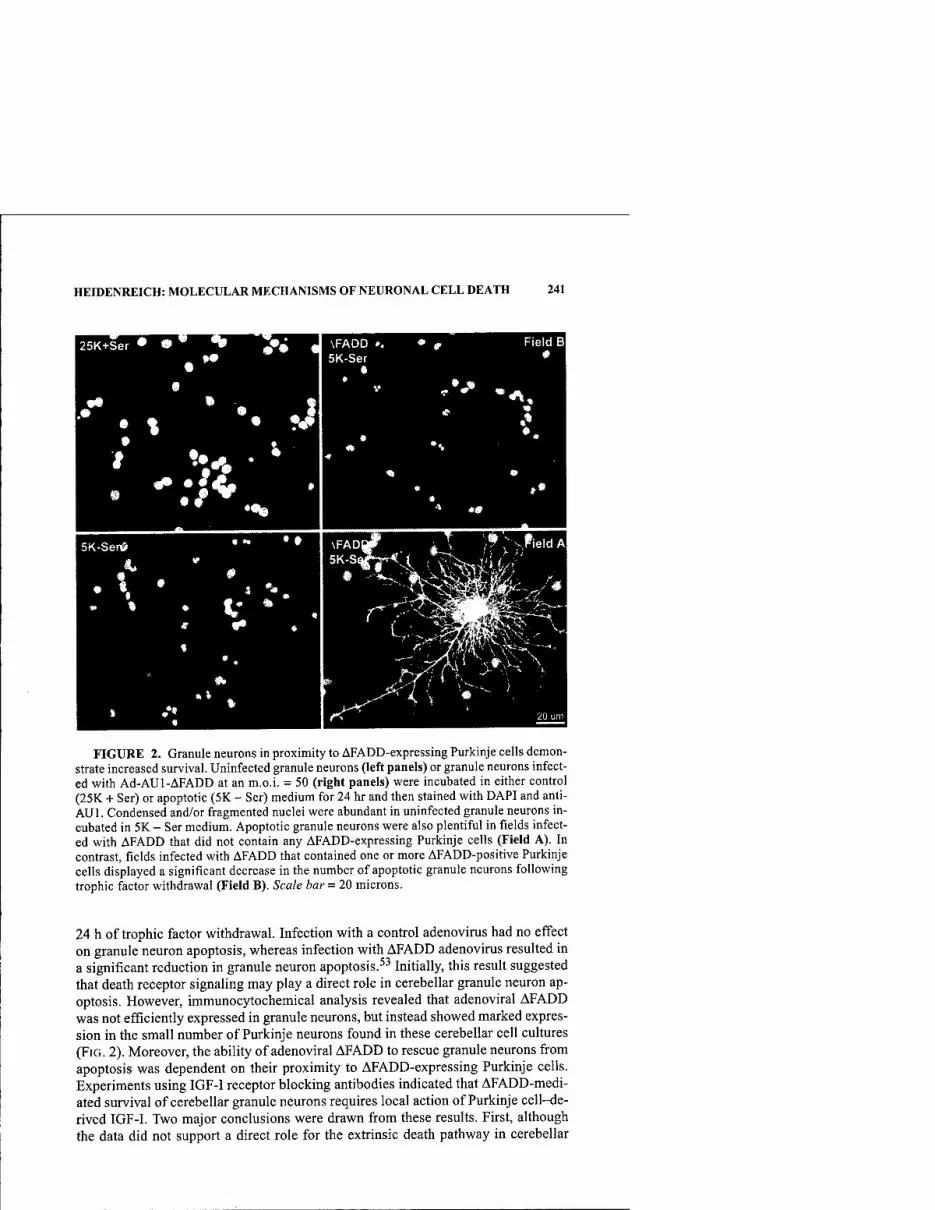
HEIDENREICH: MOLECULAR MECHANISMS OF NEURONAL CELL DEATH 241
FIGURE 2. Granule neurons in proximity to AFADD-expressing Purkinje cells demon- strate increased survival. Uninfected granule neurons (left panels) or granule neurons infect- ed with Ad-AUl-AFADD at an m.o.i. = 50 (right panels) were incubated in either control (25K + Ser) or apoptotic (5K - Ser) medium for 24 hr and then stained with DAPI and anti- AUl. Condensed and/or fragmented nuclei were abundant in uninfected granule neurons in- cubated in 5K - Ser medium. Apoptotic granule neurons were also plentiful in fields infect- ed with AFADD that did not contain any AFADD-expressing Purkinje cells (Field A). In contrast, fields infected with AFADD that contained one or more AFADD-positive Purkinje cells displayed a significant decrease in the number of apoptotic granule neurons following trophic factor withdrawal (Field B). Scale bar = 20 microns.
24 h of trophic factor withdrawal. Infection with a control adenovirus had no effect on granule neuron apoptosis, whereas infection with AFADD adenovirus resulted in a significant reduction in granule neuron apoptosis.^^ Initially, this result suggested that death receptor signaling may play a direct role in cerebellar granule neuron ap- optosis. However, immunocytochemical analysis revealed that adenoviral AFADD was not efficiently expressed in granule neurons, but instead showed marked expres- sion in the small number of Purkinje neurons found in these cerebellar cell cultures (FIG. 2). Moreover, the ability of adenoviral AFADD to rescue granule neurons from apoptosis was dependent on their proximity to AFADD-expressing Purkinje cells. Experiments using IGF-I receptor blocking antibodies indicated that AFADD-medi- ated survival of cerebellar granule neurons requires local action of Purkinje cell-de- rived IGF-I. Two major conclusions were drawn from these results. First, although the data did not support a direct role for the extrinsic death pathway in cerebellar
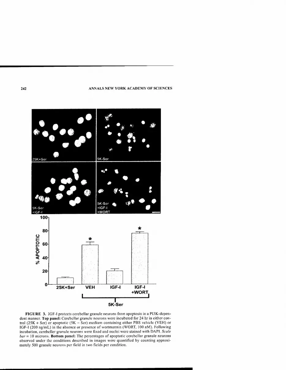
242 ANNALS NEW YORK ACADEMY OF SCIENCES
100i
5K-Ser +IGF-I +WORT
25K+Ser VEH IGF-I IGF-I +WORT.
. 1 5K-Ser
FIGURE 3. IGF-I protects cerebellar granule neurons from apoptosis in a PI3K-depen- dent manner. Top panel: Cerebellar granule neurons were incubated for 24 hr in either con- trol (25K -I- Ser) or apoptotic (5K - Ser) medium containing either PBS vehicle (VEH) or IGF-I (200 ng/mL) in the absence or presence of wortmannin (WORT, 100 nM). Following incubation, ccrebcller granule neurons were fixed and nuclei were stained with DAPI. Scale bar = 10 microns. Bottom panel: The percentages of apoptotic cerebellar granule neurons observed under the conditions described in images were quantified by counting approxi- mately 500 granule neurons per field in two fields per condition.

HEIDENREICH: MOLECULAR MECHANISMS OF NEURONAL CELL DEATH 243
granule neuron apoptosis, the results were the first to demonstrate that death receptor signaling in Purkinje neurons indirectly influences the survival of granule neurons. Second, selective suppression of the extrinsic death pathway in Purkinje cells is suf- ficient to rescue neighboring granule neurons that depend on Purkinje cell-derived trophic support including IGF-I.
INTRINSIC DEATH PATHWAYS AND NEURONAL DEATH
Recent data from a number of laboratories indicate that the intrinsic death path- way plays an important role in executing apoptosis of granule neurons.^'*'^^ To de- termine if IGF-I protected granule neurons from apoptosis by blocking components of the intrinsic pathway, we examined the effects of IGF-I on the various components of the intrinsic pathway working from the executioner caspases backward to the ini- tiator caspases and mitochondrial regulators (see FIG. 1). Apoptosis was induced in the cerebellar granule neurons by removing serum from the medium and lowering the potassium concentration from 25 mM to 5 mM. FIGURE 3 demonstrates the neu- roprotective effects of IGF-I in granule neurons. Apoptosis was quantified by count- ing the percentage of Hoesht-stained nuclei that were condensed or fragmented. In culture medium containing 25 mM potassium and serum, less than 10% of the gran- ule neurons are apoptotic. When the extracellular potassium is reduced to 5 mM and serum is removed from the medium, about 80% of the neurons are apoptotic by 24 h. IGF-I markedly blocks apoptosis in a PI3-kinase (PI3K)-dependent manner. Us- ing Western blotting techniques with antibodies against activated caspase 3, we de- tected caspase 3 activation within 6 h of serum and potassium deprivation. IGF-I totally inhibited caspase-3 activation in a PI3K-dependent manner. Immediately up- stream of caspase-3 cleavage in the intrinsic pathway is the activation of the initiator caspase-9. Recently, caspase-9 activation was shown to be required for caspase-3 cleavage in cerebellar granule neurons deprived of serum and depolarizing potassi- um.^^ Consistent with this finding, we observed marked cleavage of caspase-9 indic- ative of its activation in granule neurons deprived of trophic support. As was observed for caspase-3 cleavage, activation of caspase-9 was significantly inhibited by IGF-I in a PI3K-dependent manner, demonstrating that IGF-I suppresses a key component of the intrinsic death pathway in granule neurons.
Caspase-9 is activated following its association with Apaf-1 and cytochrome c, which assemble into a large oligomeric complex known as the apoptosome.^" For- mation of the apoptosome occurs after release of the mitochondrial protein cyto- chrome c into the cytoplasm. Our studies showed that in granule neurons maintained in the presence of serum and depolarizing potassium, cytochrome c was localized predominantly in mitochondria, with only diffuse staining observed in neuronal pro- cesses. Removal of serum and depolarizing potassium for 4 h resulted in a rapid re- distribution of cytochrome c from mitochondria to a diffuse staining throughout the cytoplasm (FiG. 4). This redistribution was accompanied by the formation of many pronounced punctate areas of cytochrome c staining. The latter were observed pri- marily, although not exclusively, in distinct focal complexes localized to neuronal processes. In contrast to cytochrome c staining, no detectable redistribution of the mitochondrial marker Mitotracker Green was observed in neuronal processes under apoptotic conditions, indicating that the punctate areas of cytochrome c staining
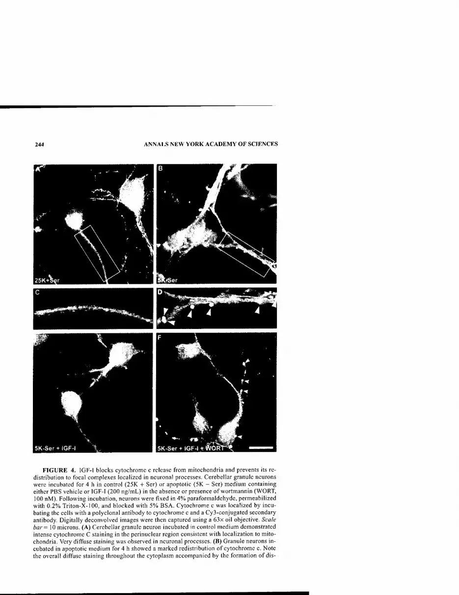
244 ANNALS NEW YORK ACADEMY OF SCIENCES
FIGURE 4. IGF-I blocks cytochrome c release from mitochondria and prevents its re- distribution to focal complexes localized in neuronal processes. Cerebellar granule neurons were incubated for 4 h in control (25K + Ser) or apoptotic (5K - Ser) medium containing either PBS vehicle or IGF-I (200 ng/niL) in the absence or presence of wortmannin (WORT, 100 nM). Following incubation, neurons were fixed in 4% paraformaldehyde, permeabilized with 0.2% Triton-X-100, and blocked with 5% BSA. Cytochrome c was localized by incu- bating the cells with a polyclonal antibody to cytochrome c and a Cy3-conjugated secondary antibody. Digitally deconvolved images were then captured using a 63x oil objective. Scale bar = 10 microns. (A) Cerebellar granule neuron incubated in control medium demonstrated intense cytochrome C staining in the perinuclear region consistent with localization to mito- chondria. Very diffuse staining was observed in neuronal processes. (B) Granule neurons in- cubated in apoptotic medium for 4 h showed a marked redistribution of cytochrome c. Note the overall diffuse staining throughout the cytoplasm accompanied by the formation of dis-

HEIDENREICH: MOLECULAR MECHANISMS OF NEURONAL CELL DEATH 245
were not associated with intact mitochondria. Inclusion of IGF-I during trophic fac- tor withdrawal prevented the release and redistribution of cytochrome c from mito- chondria. Addition of wortmannin in combination with IGF-I restored the release of cytochrome c from mitochondria and its redistribution to focal complexes in neu- ronal processes, indicating that the effects of IGF-I on cytochrome c release were PI3K-dependent (FIG. 4). Thus, IGF-I inhibits the release of cytochrome c from mi- tochondria, and in this manner blocks the subsequent activation of the intrinsic ini- tiator caspase-9.
A potential mechanism by which cytochrome c release is regulated involves the formation of a Bax- or Bak-containing "pore" in the outer mitochondrial membrane that permits passage of proteins.^"*'^' The BH3-only Bcl-2 family members, includ- ing Bad, Bid, Dp5/Hrk, and Bim, promote the proapoptotic effects of Bax and Bak, while concomitantly suppressing the prosurvival function of Bcl-2.^^'^^ Recently, Bim has been shown to be upregulated following both nerve growth factor (NGF) withdrawal from primary sympathetic neurons, and serum and potassium withdraw- al from granule neurons.''^'^^ Moreover, overexpression of Bim has been shown to promote apoptosis of cerebellar granule neurons.'*' Our laboratory examined wheth- er IGF-I was capable of regulating Bim induction in cerebellar granule neurons. Im- munoblotting for Bim following acute trophic factor withdrawal in granule neurons (FIG. 5) demonstrated a marked increase in the expression of Bim short, the most proapoptotic splice variant of this protein family. ^^ IGF-I completely blunted the in- duction of Bim, indicating that suppression of Bim is one mechanism by which IGF- I inhibits cj^ochrome c release from mitochondria.
Bim expression is regulated by multiple transcription factors. In NGF-deprived sympathetic neurons, dominant-negative c-Jun partially attenuated the induction of Bim mRNA and Bim^L protein, inhibited cytochrome c release, and rescued sympa- thetic neurons from apoptosis.''^ c-Jun has also been implicated in the apoptosis of cerebellar granule neurons,^' and an inhibitor of the JNK signaling pathway (CEP- 1347) was recently shown to partially blunt the induction of Bim mRNA in cerebel- lar granule neurons subjected to trophic factor withdrawal.'*'' In agreement with these findings, we found that the p38/JNK inhibitor SB203580 significantly attenu- ated both the activation of c-Jun and the increase in Bimg expression in cerebellar granule neurons deprived of trophic support. However, IGF-I failed to inhibit c-Jun activation under conditions where it significantly blocked induction of Bimg. These results indicated that c-Jun plays a role in the regulation of Bim expression during granule neuron apoptosis, but IGF-I suppresses the induction of Bim via a mecha-
tinct, brightly fluorescent focal complexes on the cell bodies and processes. (C) The area de- marcated by the box in A is enlarged to show the diffuse cytochrome c staining in a control neuronal process. (D) The area demarcated by the box in B is enlarged to show the intense cytochrome c staining localized to discrete focal complexes (indicated by arrowheads) in neuronal processes. (E) Granule neurons incubated in apoptotic medium containing exoge- nous IGF-I displayed cytochrome c localization to mitochondria similar to controls. (F) Granule neurons incubated in apoptotic medium containing both IGF-I and wortmannin showed cytochrome c staining similar to granule neurons incubated in apoptotic medium alone. Focal complexes of cytochrome c staining are indicated by arrowheads.
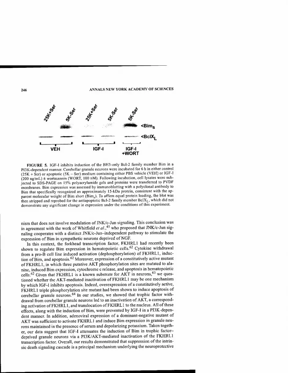
246 ANNALS NEW YORK ACADEMY OF SCIENCES
A
V ^ <3^ <Bims
<BCIXL
VEH IGF-I IGF-I +WORT
FIGURE 5. IGF-I inhibits induction of the BH3-only Bcl-2 family member Bim in a PI3K-dependent manner. Cerebellar granule neurons were incubated for 6 h in either control (25K + Ser) or apoptotic (5K - Ser) medium containing either PBS vehicle (VEH) or IGF-I (200 ng/mL) ± wortmannin (WORT, 100 nM). Following incubation, cell lysates were sub- jected to SDS-PAGE on 15% polyacrylamide gels and proteins were transferred to PVDF membranes. Bim expression was assessed by immunoblotting with a polyclonal antibody to Bim that specifically recognized an approximately 15-kDa protein, consistent with the ap- parent molecular weight of Bim short (BimJ. To affirm equal protein loading, the blot was then stripped and reprobed for the antiapoptotic Bcl-2 family member BCIXL, which did not demonstrate any significant change in expression under the conditions of this experiment.
nism that does not involve modulation of JNK/c-Jun signaling. This conclusion was in agreement with the work of Whitfield et al.,'^^ who proposed that JNKyc-Jun sig- naling cooperates with a distinct JNK/c-Jun-independent pathway to stimulate the expression of Bim in sympathetic neurons deprived of NGF.
In this context, the forkhead transcription factor, FKHRLl had recently been shown to regulate Bim expression in hematopoietic cells.^^ Cytokine withdrawal from a pro-B cell line induced activation (dephosphorylation) of FKHRLl, induc- tion of Bim, and apoptosis.^^ Moreover, expression of a constitutively active mutant of FKHRLl, in which three putative AKT phosphorylation sites are mutated to ala- nine, induced Bim expression, cytochrome c release, and apoptosis in hematopoietic cells.^2 Given that FKHRLl is a known substrate for AKT in neurons,^^ we ques- tioned whether the AKT-mediated inactivation of FKHRLl may be one mechanism by which IGF-I inhibits apoptosis. Indeed, overexpression of a constitutively active, FKHRLl triple phosphorylation site mutant had been shown to induce apoptosis of cerebellar granule neurons.^"* In our studies, we showed that trophic factor with- drawal from cerebellar granule neurons led to an inactivation of AKT, a correspond- ing activation of FKHRLl, and translocation of FKHRLl to the nucleus. All of these effects, along with the induction of Bim, were prevented by IGF-I in a PI3K-depen- dent manner. In addition, adenoviral expression of a dominant-negative mutant of AKT was sufficient to activate FKHRLl and induce Bim expression in granule neu- rons maintained in the presence of serum and depolarizing potassium. Taken togeth- er, our data suggest that IGF-I attenuates the induction of Bim in trophic factor- deprived granule neurons via a PI3K/AKT-mediated inactivation of the FKHRLl transcription factor. Overall, our results demonstrated that suppression of the intrin- sic death signaling cascade is a principal mechanism underlying the neuroprotective
HEIDENREICH: MOLECULAR MECHANISMS OF NEURONAL CELL DEATH 247
effects of IGF-I in granule neurons. IGF-I blocks Bim induction, cytochrome cre- lease, and activation of the intrinsic initiator caspase-9 and the executioner caspase- 3 in granule neurons deprived of trophic support. Moreover, IGF-I inhibits the ac- tions of FKHRLl, a transcriptional regulator of Bim, suggesting a novel c-Jun-in- dependent mechanism for the modulation of Bim in neurons.
PROGRAMMED CELL DEATH DISTINCT FROM APOPTOSIS
The data discussed above indicate that cerebellar granule neurons, when deprived of trophic support, die by classic apoptotic mechanisms involving the mitochondrial death pathway. The extrinsic or death receptor pathway indirectly regulates the sur- vival of granule neurons by its effects on Purkinje cells that provide trophic support such as IGF-I to the granule neurons. Ongoing studies in our laboratory indicate that Purkinje neurons, in contrast to the granule neurons, die by a cell death program that is caspase independent and morphologically distinct from apoptosis (manuscript in preparation). Death is not characterized by nuclear condensation but rather by exten- sive autophagic-lysosomal vacuolation. Thus, even under the same culture condi- tions with the same apoptotic stimulus, some neurons die using the classic apoptotic machinery described above; whereas other types of neurons die by programmed mechanisms distinct from apoptosis. Recent data from other laboratories support the notion that, in addition to apoptosis, other forms of programmed cell death may participate in neurodegeneration. In light of numerous unique mechanisms of neu- ronal cell death, care must be taken in making broad general conclusions about mechanisms of neuronal cell death as they relate to specific disease pathology. It will be necessary to investigate the molecular mechanisms of cell death in specific neu- ronal subtypes to define better therapeutic strategies for the various neurodegenera- tive disorders.
ACKNOWLEDGMENTS
The author would like to thank Dr. Daniel Linseman, Maria McClure, Reid Phelps, and Ron Bouchard for their contributions to this manuscript. This research was supported by grants from the U.S. Army Medical Research Command (DAMD17-99-1-9481), the National Institutes of Health (NS38619-01A1), and the Department of Veterans Affairs (Merit Award and REAP Award) to K.A.H.
REFERENCES
1. MoCHizuKi, H., et al. 1996. Histochemical detection of apoptosis in Parkinson's dis- ease. J. Neurol. Sci. 137: 120-123.
2. ANGLADE, P., et al. 1997. Apoptosis and autophagy in nigral neurons of patients with Parkinson's disease. Histol. Histopathol. 12: 25-31.
3. TATTON, N.A., et al. 1998. A fluorescent double-labling method to detect and confirm apoptotic nuclei in Parkinson's disease. Ann. Neurol. 44: S142-S148.
4. HiRSCH, E.G., et al. 1999. Dopaminergic neurons degenerate by apoptosis in Parkin- son's disease. Mov. Disord. 14: 383-385.

248 ANNALS NEW YORK ACADEMY OF SCIENCES
5. SMALE, G., el al. 1995. Evidence for apoptotic cell death in Alzheimer's disease. Exp. Neurol. 133: 225-230.
6. LASSMANN, H., el al. 1995. Cell death in Alzheimer's disease evaluated by DNA frag- mentation in situ. Acta Neuropathol. (Berlin) 89: 35-41.
7. ANDERSON, A.J., J.H. Su & C.W. COTMAN. 1996. DNA damage and apoptosis in Alzhe- imer's disease: colocalization with c-Jun immunoreactivity, relationship to brain area, and effect of postmortem delay. J. Neurosci. 16: 1710-1719.
8. TRONCOSO, J.C, el al. 1996. In situ labeling of dying cortical neurons in normal aging and in Alzheimer's disease: correlations with senile plaques and disease progression. J. Neuropathol. Exp. Neurol. 55: 1134-1142.
9. Su, J.H., G. DENG & C.W. COTMAN. 1997. Bax protein expression is increased in Alzheimer's brain: Correlations with DNA damage, Bcl-2 expression, and brain pathology. J. Neuropathol. Exp. Neurol. 56: 86-93.
10. DRAGUNOW, M., et al. 1995. In situ evidence for DNA fragmentation in Huntington's disease striatum and Alzheimer's disease temporal lobes. Neuroreport 6: 1053-1057.
11. THOMAS, L.B., el al. 1995. DNA end labeling (TUNEL) in Huntington's disease and other neuropathological conditions. Exp. Neurol. 133: 265-272.
12. ZEITLIN, S., el al. Increased apoptosis and early embryonic lethality in mice nullizy- gous for the Huntington's disease gene homologue. Nat. Genet. 11: 155-163.
13. GOLDBERG, Y.P., el al. 1996. Cleavage of huntingtin by apopain, a proapoptotic cys- teine protease, is modulated by the polyglutamine tract. Nat. Genet. 13: 442-449.
14. TROOST, D., el al. 1995. Apoptosis in amyotrophic lateral sclerosis is not restricted to motor neurons. Bcl-2 expression is increased in unaffected post-central gyrus. Neu- ropathol. Appl. Neurobiol. 21: 498-504.
15. RABIZADEH, S., el al. 1995. Mutations associated with amyotrophic lateral sclerosis convert superoxide dismutase from an antiapoptotic gene to a proapoptotic gene: studies in yeast and neural cells. Proc. Natl. Acad. Sci. USA 92: 3024-3028.
16. Mu, X., et al. 1996. Altered expression of bcl-2 and bax mRNA in amyotrophic lateral sclerosis spinal chord motor neurons. Ann. Neurol. 40: 379-386.
17. GHADGE, G.D., et al. 1997. Mutant superoxide dismutase-1-linked familial amyo- trophic lateral sclerosis: molecular mechanisms of neuronal death and protection. J. Neurosci. 17: 8756-8766.
18. PASINELLI, P., el al. 1998. Caspase-1 is activated in neural cells and tissue with amyo- trophic lateral sclerosis-associated mutations in copper-zinc superoxide dismutase. Proc. Natl. Acad. Sci. USA 95: 15763-15768.
19. IKEDA, H., et al. 1996. Expanded polyglutamine in the Machado-Joseph disease protein induces cell death in vitro and in vivo. Nat. Genet. 13: 196-202.
20. WELLINGTON, C.L., et al. 1998. Caspase cleavage of gene products associated with trip- let expansion disorders generates truncated fragments containing the polyglutamine tract. J. Biol. Chem. 273: 9158-9167.
21. WARRICK, J.M., el al. 1998. Expanded polyglutamine protein forms nuclear inclusions and causes neural degeneration in Drosophila. Cell. 93: 939-949.
22. STRASSER, A., L. O'CONNOR & V.M. DIXIT. 2000. Apoptosis signaling. Annu. Rev. Biochem. 69: 217-245.
23. GREEN, D.R. 1998. Apoptotic pathways: the roads to ruin. Cell 94: 695-698. 24. PiNKOSKi, M.J. & D.R. GREEN. 1999. Fas ligand, death gene. Cell Death Differ. 6:
1174-1181. 25. CHINNAIYAN, A.M., et al. 1996. FADD/MORTl is a common mediator of CD95 (Fas/
APO-1) and tumor necrosis factor receptor-induced apoptosis. J. Biol. Chem. 271: 4961-4965.
26. FEINSTEIN, E., et al. 1995. The death domain: a module shared by proteins with diverse cellular functions. Trends Biochem. Sci. 20: 342-344.
27. Muzio, M., el al. 1996. FLICE, a novel FADD-homologous ICE/CED-3-like protease, is recruited to the CD95 (Fas/APO-1) death-inducing signaling complex. Cell 85: 817-827.
28. MUZIO, M., et al. 1998. An induced proximity model for caspase-8 activation. J. Biol. Chem. 273: 2926-2930.
29. CAI, J., J. YANG & D.P. JONES. 1998. Mitochondrial control of apoptosis: the role of cytochrome c. Biochim. Biophys. Acta 1366: 139-149.
HEIDENREICH: MOLECULAR MECHANISMS OF NEURONAL CELL DEATH 249
30. Zou, H., et al. 1999 An APAF-1/cytochrome c multimeric complex is a functional apo- ptosome that activates caspase-9. J. Biol. Chem. 274: 11549-11556.
31. STENNICKE, H.R. 1998. Pro-caspase-3 is a major physiologic target of caspase-8. J. Biol. Chem. 273: 27084-27090.
32. PAN, G., E.W. HUMKE & V.W. DIXIT. 1998. Activation of caspases triggered by cyto- chrome c in vitro. FEES Lett. 426: 151-154.
33. TsuJiMOTO, Y. 1998. Role of Bcl-2 family proteins in apoptosis: apoptosomes or mito- chondria? Genes Cells 3: 697-707.
34. WEI, M.C, et al. 2001. Proapoptotic BAX and BAK: a requisite gateway to mitochon- drial dysfunction and death. Science 292: 727-730.
35. POLSTER, B.M., K.W. KiNALLY & G. FiSKUM. 2001. BH3 death domain peptide induces cell type-selective mitochondrial outer membrane permeability. J. Biol. Chem. 276: 37887-37894.
36. OTTER, I., et al. 1998. The binding properties and biological activities of Bcl-2 and Bax in cells exposed to apoptotic stimuli. J. Biol. Chem. 273: 6110-6120.
37. ZONG, W.X., et al. 2001. BH3-only proteins that bind pro-survival Bcl-2 family members fail to induce apoptosis in the absence of Bax and Bak. Genes Dev. 15: 1481-1486.
38. DE GlORGi, F., et al. 2002. The permeability transition pore signals apoptosis by direct- ing Bax translocation and multimerization. FASEB J. 16: 607-609.
39. HALESTRAP, A.P., G.P. MCSTAY & S.J. CLARK. 2002. The permeability transition pore complex: another view. Biochimie 84:153-166.
40. WALDMEIER, R C, et al. 2002. Inhibition of the mitochondrial permeability transition by the nonimmunosuppressive cyclosporin derivative N1M811. Mol. Pharmacol. 62: 22-29.
41. KITAMURA, Y., et al. 1998. Alteration of proteins regulating apoptosis, Bcl-2, Bcl-x, Bax, Bak, Bad, ICH-1 and CPP32, in Alzheimer's disease. Brain Res. 780: 260-269.
42. FELDERHOFF-MUESER, U., et al. 2000. Fas/CD95/APO-l can function as a death recep- tor for neuronal cells in vitro and in vivo and is upregulated following cerebral hypoxic-ischemic injury to the developing rat brain. Brain Pathol. 10: 17-29.
43. MARTIN-VILLALBA, A., et al. 1999. CD95 ligand (Fas-L/APO-lL) and tumor necrosis factor-related apoptosis-inducing ligand mediate ischemia-induced apoptosis in neu- rons. J. Neurosci. 19: 3809-3817.
44. PARSADANIAN, A.S., et al. 1998. Bcl-xL is an antiapoptotic regulator for postnatal CNS neurons. J. Neurosci. 18: 1009-1019.
45. WHITFIELD, J., et al. 2001. Dominant-negative c-Jun promotes neuronal survival by reducing BIM expression and inhibiting mitochondrial cytochrome c release. Neuron 29: 629-643.
46. LiNSEMAN, D.A., et al. 2002. Insulin-like growth factor-I blocks Bcl-2 mteractmg mediator of cell death (Bim) induction and intrinsic death signaling in cerebellar granule neurons. J. Neurosci. 22: 9287-9297.
47. HARRIS C.A. & E.M. JOHNSON, JR. 2001. BH3-only Bcl-2 family members are coordi- nately regulated by the JNK pathway and require Bax to induce apoptosis in neurons. J. Biol. Chem. 276: 37754-37760.
48. BouiLLET, P., et al. 2001. Degenerative disorders caused by Bcl-2 deficiency prevented by loss of its BH3-only antagonist Bim. Dev. Cell. 5: 645-653.
49. LE-NICULESCU, H., et al. 1999. Withdrawal of survival factors results in activation of the JNK pathway in neuronal cells leading to Fas ligand induction and cell death. Mol. Cell. Biol. 19:751-763.
50. D'MELLO, S.R., et al. 1993. Induction of apoptosis in cerebellar granule neurons by low potassium: inhibition of death by insulin-like growth factor I and cAMP. Proc. Natl. Acad. Sci. USA 90: 10989-10993.
51. STREETZ, K., et al. 2000. Tumor necrosis factor alpha in the pathogenesis of human and murine fulminant hepatic failure. Gastroenterology 119: 446-460.
52. BRADHAM, C.A., et al. 1998. The mitochondrial permeability transition is required for tumor necrosis factor alpha-mediated apoptosis and cytochrome c release. Mol. Cell. Biol. 18: 6353-6364.
53. LlNSEMAN, D.A., et al. 2002. Suppression of death receptor signaling in cerebellar Purkinje neurons protects neighboring granule neurons from apoptosis via an insu- lin-like growth factor-I dependent mechanism. J. Biol. Chem. 277: 24546-24553.

250 ANNALS NEW YORK ACADEMY OF SCIENCES
54. MILLER, T.M., et al. 1997. Bax deletion further orders the cell death pathway in cere- bellar granule cells and suggests a caspase-independent pathway to cell death. J. Cell. Bid. 139:205-217.
55. SHLIMI, F., et al. 2000. Target-related and intrinsic neuronal death in Lurcher mutant mice are both mediated by caspase-3 activation. J. Neurosci. 20: 992-1000.
56. GERHARDT, E., et al. 2001. Cascade of caspase activation in potassium-deprived cere- bellar granule neurons: targets for treatment with peptide and protein inhibitors of apoptosis. Mol. Cell. Neurosci. 17: 717-731.
57. KoRSMEYER, S.J. 2000. Pro-apoptotic cascade activates BID, which oligomerizes BAK or BAX into pores that result in the release of eytochrome c. Cell Death Differ. 7: 1166-1173.
58. DESAGHER, S., et al. 1999. Bid-induced eonformational change of Bax is responsible for mitochondrial eytochrome c release during apoptosis. J. Cell. Biol. 144: 891- 901.
59. PUTCHA, G.V., et al. 2001. Induction of BIM, a proapoptotic BH3-only BCL-2 family member, is critical for neuronal apoptosis. Neuron 29: 615-628.
60. O'CONNOR, L., et al. 1998. Bim: A novel member of the Bcl-2 family that promotes apoptosis. EMBO J. 17: 384-395.
61. WATSON, A., et al. 1998. Phosphorylation of c-Jun is necessary for apoptosis induced by survival signal withdrawal in cerebellar granule neurons. J. Neurosci. 18: 751- 762.
62. DiJKERS, P.F., et al. 2002. FKHR-Ll can act as a critical effector of cell death induced by cytokine withdrawal: protein kinase B-enhanced cell survival through mainte- nance of mitochondrial integrity. J. Cell. Biol. 156: 531-542.
63. ZHENG, W.H., S. KAR & R, QUIRION. 2000. Insulin-like growth factor-1-induced phos- phorylation of the forkhead family transcription factor FKHRLl is mediated by Akt kinase in PC12 cells. J. Biol. Chem. 275: 39152-39158.
64. BRUNEI, A., et al. 1999. Akt promotes cell survival by phosphorylating and inhibiting a Forkhead transcription factor. Cell 96: 857-868.
Redox State as a Central Modulator of Precursor Cell Function
MARK NOBLE," JOEL SMITH,* JENNIFER POWER,'^ AND MARGOT MAYER-PROSCHEL"
"Department of Biomedical Genetics, University of Rochester School of Medicine, Rochester, New York 14642, USA
^Memorial Sloan-Kettering Hospital, Laboratory of Developmental Hematopoiesis, New York, New York 10021, USA
'^Columbia University, Center for Neurobiology and Behavior, New York, New York 10032, USA
ABSTRACT: In our attempts to understand how the balance between self-renew- al and differentiation is regulated in dividing precursor cells, we have discov- ered that intracellular redox state appears to be a critical modulator of this balance in oligodendrocyte-type-2 astrocyte (0-2A) progenitor cells. The in- tracellular redox state of freshly isolated progenitor cells allows prospective isolation of cells with different self-renewal characteristics, which can be fur- ther modulated in opposite directions by prooxidants and antioxidants. Redox state is itself modulated by cell-extrinsic signaling molecules that alter the bal- ance between self-renewal and differentiation: growth factors that promote self-renewal cause progenitors to become more reduced, while exposure to sig- naling molecules that promote differentiation causes progenitors to become more oxidized. Moreover, pharmacological antagonists of the redox effects of these cell-extrinsic signaling molecules antagonize their effects on self-renewal and differentiation, further suggesting that cell-extrinsic signaling molecules that modulate this balance converge on redox modulation as a critical compo- nent of their effector mechanism. A further example of the potential relevance of intracellular redox state to development processes emerges from our at- tempts to understand why different central nervous system (CNS) regions ex- hibit different temporal patterns of oligodendrocyte generation and myelinogenesis. Characterization of 0-2A progenitor cells (0-2A/0PCs) iso- lated from different regions indicates that these developmental patterns are consistent with properties of the specific 0-2A/0PCs resident in each region. Marked differences were seen in self-renewal and differentiation characteris- tics of 0-2A/0PCS isolated from cortex, optic nerve, and optic chiasm. In con- ditions where optic nerve-derived 0-2A/0PCs generated oligodendrocytes within 2 days, oligodendrocytes arose from chiasm-derived cells after 5 days and from cortical 0-2A/0PCs after only 7-10 days. These differences, which appear to be cell intrinsic, were manifested both in reduced percentages of clones producing oligodendrocytes and in a lesser representation of oligoden- drocytes in individual clones. In addition, responsiveness of optic nerve-, chi-
Address for correspondence: Mark Noble, Department of Biomedical Genetics, University of Rochester School of Medicine, 601 Elmwood Avenue, Box 633, Rochester, New York 14642. Voice: 716-273-1448; fax: 716-273-1450.
Ann. N.Y. Acad. Sci. 991: 251-271 (2003). © 2003 New York Academy of Sciences.
251
252 ANNALS NEW YORK ACADEMY OF SCIENCES
asm-, and cortex-derived 0-2A/0PCs to thyroid hormone (TH) and ciliary neurotrophic factor (CNTF), well-characterized induccrs of oligodendrocyte generation, was inversely related to the extent of self-renewal observed in basal division conditions. These results demonstrate hitherto unrecognized complex- ities among the precursor cells thought to be the immediate ancestors of oligo- dendrocytes and suggest that the properties of these different populations may contribute to the diverse time courses of myelination in different CNS regions. Strikingly, 0-2A/0PCs isolated from cortex and analyzed immediately upon isolation were more reduced in their redox state than were optic nerve-derived cells, precisely as would be predicted from our analysis of the role of redox state in modulating the balance between self-renewal and differentiation. Chiasm- derived cells, which exhibited self-renewal properties intermediate between cortex- and optic nerve-derived cells, were more reduced than optic nerve cells but more oxidized that cortical 0-2A/0PCs.
KEYWORDS: myelination; development; oligodendrocyte-type-2 astrocyte pro- genitor; oligodendrocyte precursor cell; oligodendrocyte; self-renewal; pre- cursor cell
INTRODUCTION
Modulation of the balance between self-renewing divisions and differentiation is at the heart of precursor cell function in development, tissue repair, and tissue ho- meostasis, yet relatively little is known about physiological mechanisms central to such modulation. For example, on a biochemical level, one of the only properties re- ported to be predictive of self-renewal characteristics in precursor cells of different lineages (i.e., hematopoietic stem cells and hepatic precursor cells, as described, e.g., in Refs. 1-3) was that cells with a greater tendency to undergo self-renewing divisions showed a lesser extent of labeling with such mitochondrial dyes as rhodamine-123. This reduced labeling was originally thought to be due to expres- sion of P-glycoprotein by cells with a greater self-renewal potential, but subsequent studies on hematopoietic stem cells raised the possibility that it may instead be re- flective of mitochondrial activity and intracellular redox state."* Such a correlation of such an important physiological state with self-renewal characteristics is poten- tially of great interest. It is not known, however, whether this correlation is coinci- dental or whether it provides an important clue to understanding this critical feature of precursor cell biology.
To examine the possibility that intracellular redox regulation plays an important functional role in modulating the self-renewal characteristics of precursor cells, we studied the correlation between redox state and the balance between self-renewal and differentiation in dividing oligodendrocyte-type-2 astrocyte (0-2A) progenitor cells (also referred to has oligodendrocyte precursor cells, or OPCs, and here abbreviated as 0-2A/0PCs).^ This population, which gives rise to the myelin-forming oligoden- drocytes of the central nervous system (CNS) (see, e.g., Refs. 6-8), provides one of the most extensively characterized and tractable precursor cell systems available for such studies. It is possible to obtain pure populations of 0-2A/OPCs that can be in- duced to undergo division and differentiation by growth in simple chemically de- fined medium supplemented with platelet-derived growth factor (PDGF, the best characterized 0-2A/OPC mitogen; see, e.g., Refs. 9 and 10).
NOBLE et al.: REDOX STATE AS A CENTRAL MODULATOR 253
0-2AyOPCs obtained from the optic nerves of 7-day-old (P7) rat pups and grown in the presence of saturating levels of PDGF, one of the important mitogens for these cells^'" exhibit an approximately equal probability of undergoing a self-re- newing division or exiting the cell cycle and differentiating into an oligodendro- cyte.'^ The tendency of dividing 0-2A/0PCs to generate oligodendrocytes is enhanced if cells are coexposed to such signaling molecules as thyroid hormone, ciliary neurotrophic factor, or retinoic acid (see, e.g., Refs. 13-15). Signaling mol- ecules of the bone morphogenetic protein family induce differentiation along oligo- dendrocyte and astrocyte pathways, respectively.'^'^^-'^ In contrast, coexposure to neurotrophin-3 or basic fibroblast growth factor (bFGF) inhibits differentiation and is associated with increased precursor cell division and self-renewal.'^''^''^
Previous studies have shown that the degree to which exposure to signaling mol- ecules alters the balance between self-renewal and differentiation in dividing 0-2A/ OPCs is very extensive. It is possible to induce nearly synchronous differentiation of all clonally related cells into oligodendrocytes when cells are exposed to thyroid hormone (TH) and type-1 astrocytes;^^'^*' varying degrees of asymmetric division and differentiation are seen in the presence of other combinations of factors;^^ and almost complete suppression of differentiation with continuous promotion of self- renewal occurs when cells are grown in the presence of both PDGF and bFGF. '^ At least a subset of these signaling molecules is also known to be of importance in vi- vo. For example, hypothyroid animals show reduced oligodendrocyte genera- tion,'^'^' and animals in which neurotrophin-3 (NT-3) levels are artificially increased show increases in 0-2A/0PC proliferation.'^ Both of these results are consistent with the outcomes of in vitro studies on the effects of TH and NT-3 on 0-2A/0PC division and differentiation.'^''^''^
In a consideration of situations in which plasticity of precursor cell behavior might play a role, one possibility is seen in the striking phenomenon that different regions of the CNS develop according to different schedules, with great variations seen in the timing of both neurogenesis and gliogenesis.-^^ For example, neuron pro- duction in the rat spinal cord is largely complete by the time of birth, is still ongoing in the rat cerebellum for at least several days after birth, and continues in the olfac- tory system and in some regions of the hippocampus of multiple species throughout life. Similarly, myelination has long been known to progress in a rostral-caudal di- rection, beginning in the spinal cord significantly earlier than in the brain (see, e.g., Refs. 23-25). Even within a single CNS region, myelination is not synchronous. In the rat optic nerve, for example, myelinogenesis occurs with a retinal-to-chiasmal gradient, with regions of the nerve nearest the retina becoming myelinated first.^^''^^ The cortex itself shows the widest range of timing for myelination, both initiating later than many other CNS regions (see, e.g., Refs. 23-25) and exhibiting an ongoing myelinogenesis that can extend over long periods of time. This latter characteristic is seen perhaps most dramatically in the human brain, for which it has been suggest- ed that myelination may not be complete until after several decades of life.^^'^^
Variant time courses of development in different CNS regions could be due to two fundamentally different reasons. One possibility is precursor cells are sufficiently plas- tic in their developmental programs that local differences in exposure to modulators of division and differentiation may account for these variances. Alternatively, it may be that the precursor cells resident in particular tissues express differing biological prop- erties related to the timing of development in the tissue to which they contribute.
254 ANNALS NEW YORK ACADEMY OF SCIENCES
Taken together, all of the above observations lead to a series of potentially related questions:
(1) The balance between self-renewal and differentiation in dividing 0-2A/ OPCs can be modified by a wide range of signaling molecules, acting through diverse receptor systems. Is it possible to identify points at which these differing pathways converge, at least to the extent of identifying com- mon elements by which different kinds of signaling molecules can promote self-renewing division, with perhaps other elements common to factors that induce differentiation? Or is it possible that there are biochemical or molecular pathways within the cell at which all these influences converge?
(2) Are the diverse signaling molecules that have been found to modulate the balance between self-renewal and differentiation in dividing 0-2A/0PCs of importance in understanding the different timing of oligodendrocyte generation in diverse CNS regions in vivol Knowledge of the role of these molecules may help in identifying means of modifying precursor cell func- tion in vivo.
(3) To what extent do previously unrecognized differences in precursor cell behavior contribute to the variability in oligodendrocyte generation in vivol If precursor cells themselves exhibit substantial regional heterogene- ity in their function, this would indicate a hitherto unsuspected complexity in precursor cell biology. Moreover, different populations might express properties of greater or lesser value in the context of using them to repair damaged tissue following cell transplantation.
(4) Do the observations that hematopoietic stem cells and liver precursor cells that show less labeling with rhodamine-123—and thus may be more reduced—provide an important clue to understanding the above problems?
THE ROLE OF REDOX STATE IN MODULATING SELF-RENEWAL CHARACTERISTICS OF 0-2A/0PCs DERIVED FROM
THE OPTIC NERVE OF P7 RATS
Cellular Redox State Allows Prospective Isolation of Cells with Different Self-Renewal Characteristics
To determine whether intracellular redox state might be associated with the self- renewal potential of 0-2A/0PCs developing in vivo, we determined whether low levels of labeling with redox-sensitive dyes were indicative of enhanced self-renewal capacity in freshly isolated 0-2A/0PCs.^ Different dyes provide information on dif- ferent aspects of redox state, which ultimately reflects the balance between reducing and oxidative equivalents within the cell. Redox state, however, is influenced in many ways, including by the state of mitochondrial activation, levels of reduced glu- tathione and other thiols, levels of NADH and NADPH, and levels of thioredoxin and other factors. In our experiments, cells were labeled with JC-1, rhodamine-123, or dihydrotetramethylrosamine (reduced rosamine), and FAGS sorting was used to pro- vide enriched populations of the upper and lower quintiles of labeled cells. Cells from each group were then plated at clonal density in the presence of PDGF, and bas- al self-renewal characteristics were determined. All dyes gave similar results, but ro-

NOBLE et al.\ REDOX STATE AS A CENTRAL MODULATOR 255
samine was consistently associated with the best postsort viability. As rosamine fluorescence is most directly related to the balance between oxidative and reducing equivalents, this dye also provides one of the broadest measures of redox state.^^
Results obtained using this purification procedure were striking, rosamine'"^ cells grown in the presence of 10 ng/mL PDGF generated clones in which self- renewing divisions were prevalent. Five days after plating, 70 ± 4.3% of these con- sisted only of dividing progenitor cells, and the average number of cells per clone was 11.1 ± 0.9. In contrast, rosamine'''^'' cells, which would have had a higher intra- cellular level of oxidizing equivalents in vivo, underwent very little division in these same conditions. With rare exceptions, the largest clones consisted of four cells (mean = 2.5 ± 0.4 cells/clone), rosamine'^'^'' cells also demonstrated a greatly in- creased tendency to generate oligodendrocytes. At day 5, 47 + 4.6% of clones de- rived from rosamine'''^'' cells consisted of one oligodendrocyte and no progenitor cells, and only 29 ± 5.4% of clones contained any progenitors at all. Thus, in agree- ment with results obtained for hematopoietic stem cells and hepatic precursor cells (see, e.g., Refs. 1-4), rosamine'"* 0-2A/0PCs appeared better able to undergo self- renewing divisions than rosamine*"^'' cells. It is important to note that the viability of all populations was identical, and both rosamine'"* and rosamine'"^'' cells were able to undergo division and differentiation without undergoing any extensive cell death for at least a week after isolation. Cell morphology and antigenic phenotype were as seen in several previous publications.
Antioxidant and Prooxidant Drugs Modulate Self-Renewal and Differentiation Characteristics in Opposite Directions
Despite their low frequency of division, rosamine'^'s'^ 0-2A/0PCs had not already irreversibly committed to differentiation in vivo. This was demonstrated by altering intracellular redox state through exposure to Af-acetyl-L-cysteine (NAC), a potent antioxidant that also acts as a cysteine prodrug, thus enhancing production of glutathione, which is the most prevalent reduced thiol within cells and is crucial for maintaining a reduced intracellular environment.^^'-" Self-renewal of rosamine'''^'' cells was markedly enhanced in these conditions. The percentage of progenitor cell- containing clones at day 5 increased from 29 ± 5.4% to 65 ± 3.4%, and the average clone size increased almost threefold, from 2.5 ± 0.4 cells to 7.0 ± 1.2 cells per clone. These results suggest that aheration of intracellular redox state is a dynamic modu- lator of the balance between self-renewal and differentiation rather than merely be- ing a secondary consequence of an irrevocable fate determination.
Further studies confirmed that the balance between self-renewal and differentia- tion in dividing 0-2A/0PCs could be modulated by manipulation of intracellular re- dox state. In these experiments, clonal 0-2A/OPCs were grown in the presence of PDGF with or without compounds known to alter intracellular redox state. To in- crease oxidative levels, we used tert-butylhydroperoxide (^BuOOH) and buthionine sulfoximine (BSO). f-BuOOH is a potent prooxidant,^^ while BSO inhibits synthesis of glutathione.^^ To render cells more reduced, the medium of dividing cells was supplemented with NAC. We also examined the effects of three polyhydroxyalkyl thiazolidine carboxylic acid drugs (GlcCys, ProCys, and RibCys). These procysteine drugs contribute to glutathione biosynthesis when they are taken up by cells and their
256 ANNALS NEW YORK ACADEMY OF SCIENCES
thilozolidine ring is opened enzymatically.^'' Thus, the antioxidant activity of these compounds, unlike NAC, is targeted to intracellular thiol pools.
Treatment of dividing 0-2A/0PCs with PDGF plus 1 mM BSO or 50 nM t- BuOOH was associated with diminished progenitor cell division and increased oligodendrocyte generation. In the presence of either BSO or ?-BuOOH, approxi- mately half of the founder cells differentiated in the absence of division, with the rest undergoing one to two divisions and yielding clones containing two to seven cells after 5 days. Average clone sizes at this time point, in either condition, were 2.1 ± 0.4 cells per clone, less than half that seen in basal division conditions (5.7 ± 0.8 cells/clone). Exposure to BSO or t-BuOOH also was associated with both a reduc- tion in the number of clones containing only progenitor cells and an increase in the proportion of clones containing only oligodendrocytes. For example, when clones were grown in the presence of PDGF for 5 days, 43 ± 3.0% of the clones consisted wholly of progenitor cells. In contrast, in cultures exposed to PDGF plus BSO or t- BuOOH for 5 days, only 5 ± 2.4% and 7 + 3.7% of clones consisted solely of pro- genitor cells, respectively. Complementary changes were seen in the proportion of clones consisting solely of oligodendrocytes. After 5 days, 88 ± 8.3% of clones ex- posed to BSO or 83 ± 6.0%o of clones exposed to ^BuOOH consisted wholly of oli- godendrocytes, as compared with complete differentiation of only 35 ± 6.6%) of clones in basal division conditions.
In contrast with the effects of exposure to prooxidants, clones of 0-2A/OPCs cul- tured in PDGF plus 1 mM NAC exhibited enhanced self-renewal and a marked re- duction in oligodendrocyte generation. After 5 days in culture, clones growing in medium supplemented with NAC contained an average of 11.3 ± 2.2 cells per clone, almost twice the value seen in clones growing in the presence of PDGF alone, with many clones containing more than 15 cells. In cultures grown in the presence of NAC for 3 days, 80 + 2.1% of clones contained only progenitor cells. Even after 5 days, 60 ± 2.8% of clones contained only progenitor cells in these conditions. In con- trast, when cells were grown in basal division conditions for 3 days, 47 ± 1.1 %i of clones consisted only of progenitor cells, with similar values (43 + 3.0%) observed after 5 days. Cells exposed to NAC alone (i.e., no PDGF present) differentiated into oligodendrocytes without undergoing division.
Enhanced self-renewal of 0-2A/0PCs grown in the presence of NAC continued for at least 7 days. Clones examined at this time were on average six times larger than clones exposed to PDGF alone. Moreover, in cultures exposed to PDGF plus NAC for 7 days, 40 ± 5.9%) of clones still contained only progenitors. In contrast, only 5 ± 2.6%) of PDGF-stimulated clones consisted solely of progenitor cells after 7 days. It is important to note that in cultures exposed to PDGF plus NAC for this time, in those clones in which division occurred, 49 + 8.5%) contained one or more oligoden- drocytes. Moreover, absolute numbers of oligodendrocytes in either condition were similar (151 ±21 and 116 ± 38 for control and NAC-treated cultures, respectively), demonstrating that exposure to NAC did not preclude eventual differentiation. Sup- plementation of basal division medium with the cysteine prodrugs GlcCys, ProCys, or RibCys had virtually identical effects to NAC, thus suggesting that the capacity of NAC to modify intracellular thiol balance was more important to its activity than its ability to function as an extracellular antioxidant. All of the prooxidants and cys- teine prodrugs examined caused the expected direction of change in intracellular re- dox state, as determined by rosamine fluorescence.
NOBLE et al: REDOX STATE AS A CENTRAL MODULATOR 257
Signaling Molecules That Promote Differentiation Cause Greater Intracellular Oxidative Turnover, While Signaling Molecules Which Promote
Self-Renewal Cause More Reduced Intracellular States
While it is possible that differences in intracellular redox state in vivo might be due to cell-intrinsic properties or to direct exposure to pro- or antioxidants, it is also possible that the cell-extrinsic signaling molecules that have been previously found to modulate the balance between self-renewal and differentiation have as part of their activity the modulation of redox state. We therefore next determined whether redox modulation might be of any relevance in the action of cell-extrinsic signaling molecules upon 0-2A/0PCs.
Growth of cells in the presence of PDGF supplemented with factors that promote self-renewal was associated with a more reduced intracellular redox state, as indicat- ed by rosamine fluorescence; while supplementation with inducers of differentiation was associated with increased rosamine fluorescence, indicating an increase in oxi- dative levels. Such effects were seen within 18 h, well before it is possible to observe any differences in progenitor or oligodendrocyte numbers. For example, after expo- sure of cells to NT-3 plus PDGF for 12 h, aggregate rosamine fluorescence was 15 ± 6% lower than in the presence of PDGF alone. This compares to treatment with either 1 mM NAC or 1 mM ProCys, which after 6 h in culture led to a decrease in rosamine fluorescence by 12 ± 4% and 18 ± 7%, respectively, relative to control val- ues (P < 0.05). By 18 h after exposure to NT-3, relative fluorescence was 29 ± 3% lower than for control cells (P = 0.0075). Similarly, 0-2A/0PCs exposed to PDGF plus bFGF exhibited a 23 ± 8% reduction in aggregate fluorescence values {P < 0.05; 18-h time point) as compared with cells exposed only to PDGF. The effect of NT-3 on redox levels was inhibited by simultaneous treatment with BSO. After exposure of cells to 1 mM BSO plus NT-3 plus PDGF for 12 h, mean rosamine fluorescence was increased by 6 ± 7% relative to growth in PDGF alone, a difference that was not significant.
Exposure to TH, which induces oligodendrocyte generation, had the opposite ef- fect with respect to NT-3 and was associated with greater rosamine fluorescence, in- dicating an increase in oxidative levels. Exposure of cells to TH was associated with an 11 ± 4% increase in fluorescence after 3 h and a statistically significant (23 ± 4%; P < 0.01) increase in fluorescence by 12 h after initiation of treatment. This effect was significantly diminished with simultaneous exposure to 1 mM NAC: after 12 h in the presence of NAC plus TH plus PDGF, cells displayed mean rosamine fluores- cence 4 ± 13% greater than control compared with the 23 ± 4% increase observed for cells grown in just TH plus PDGF {P < 0.01). By comparison, cells treated with either 1 mM BSO or 50 nM ^BuOOH for 6 h displayed an increase in rosamine flu- orescence of 6 + 3% (P = 0.0723) and 9 ± 2% (P < 0.05) over control cells, respec- tively (data not shown). An effect of increased oxidative activation of rosamine was also associated with exposure to bone morphogenetic protein-4 (BMP-4), even though this protein causes 0-2A/0PCs to differentiate into type-2 astrocytes'^ and is thought to act through receptor and signaling systems very different from those relevant to TH action. 0-2A/0PCs exposed to PDGF plus BMP-4 exhibited a signif- icant increase in fluorescence by 12 h (25 ± 11%; P < 0.05), and a highly significant increase (38 ± 13%; P = 0.004) after 18 h. Thus, exposure to inducers of differenti- ation caused an increase in rosamine fluorescence, indicative of an increase in oxi-

258 ANNALS NEW YORK ACADEMY OF SCIENCES
dative levels, independent of whether oligodendrocyte or astrocyte differentiation was being stimulated.
We also noted that the changes in geometric means of rosamine fluorescence val- ues were associated with a redistribution of cells within the normal range of redox values, rather than an alteration of the range itself A lower mean is associated with more cells exhibiting low levels of rosamine fluorescence, while a higher value is as- sociated with a redistribution of cells to a more oxidatively active portion of the range. Thus, exposure to NT-3, bFGF, TH, and BMP-4 appears to alter intracellular redox profiles within a distribution that is otherwise tightly regulated as to its upper and lower boundaries.
Further demonstration of the effects on redox state of NT-3 and TH, the two sig- naling molecules for which the best evidence for an in vivo role has been provid- gj 15,18,21 ^gg obtained by analysis of cells with the JC-1 dye, which provides information on mitochondrial inner membrance potential (AT). Treatment with TH for 18 h resulted in a 18 ± 11% decrease in AT (aggregate red:green fluorescence ratio; P < 0.05), indicative of a more oxidative state within the cells.^^ In contrast, treatment with NT-3 for 18 h was associated with a 16 ± 11% increase in AT (P < 0.05), indicative of a more reduced state within the cells.
Pharmacological Agents That Antagonize the Redox Effects of NT-3 and TH Also Antagonize Their Effects on Self-Renewal and Differentiation
We next asked whether the effects of cell-extrinsic signaling molecules on self- renewal and differentiation could be blocked by pharmacological manipulation of redox state in the opposite direction to that induced by the signaling molecule itself. As the best evidence for an in vivo role of such compounds in 0-2A/OPC develop- ment has been provided for NT-3 and TH,'^''^'^' we specifically studied the action of these agents.
Cells grown in the presence of PDGF supplemented with TH and NAC exhibited a similar profile of self-renewal to cells grown in the presence of PDGF alone, indi- cating that NAC countered the effects of TH. After 5 days, 60 ± 4.8% of clones con- sisted of only progenitor cells in cultures exposed to TH plus NAC, a proportion even greater than that observed in clones exposed only to PDGF (i.e., 43 ± 3.0%.). In con- trast, only 10 ± 1.4% of clones consisted entirely of progenitor cells when cells were exposed to PDGF plus TH alone. Progenitor cells were able to undergo more self- renewal when exposed to TH plus NAC, and the average clone size was 4.7 ± 1.1 cells after 5 days (as compared with 2.1 ± 0.4 cells per clone in cultures exposed to PDGF plus TH). Just under half of the clones differentiated without division when TH was present, and 81 ± 10.8% of the clones in which at least one division occurred contained one or more oligodendrocytes. When NAC was also present, in contrast, only 30 ± 4.0% of clones consisted wholly of oligodendrocytes (and most of these clones consisted of a single oligodendrocyte), and only 17 ± 4.2% of clones in which at least one division occurred contained at least one oligodendrocyte.
In complementary experiments, exposure of cells to PDGF plus 1 |aM BSO inhib- ited the ability of NT-3 to enhance self-renewal, in agreement with our observations that BSO antagonizes the NT-3-associated alterations in redox state. Clones grown in the presence of PDGF plus NT-3 showed an increase in self-renewing divisions as

NOBLE et al.: REDOX STATE AS A CENTRAL MODULATOR 259
determined by average clonal size (9.7 ± 1.2 cells), a 70% increase over control val- ues. In contrast, in cultures treated also with 1 |iM BSO, mean clone size after 5 days vi^as 2.2 ± 0.4 cells, and half of the clones were single cells that differentiated into oligodendrocytes in the absence of division. NT-3 exposure also was associated with a decrease in the proportion of clones containing at least one oligodendrocyte (36 + 2.6% by day 5, as compared with 57 ± 3.0% in basal division medium). BSO inhib- ited this effect, and 74 ± 5.7% of clones contained at least one oligodendrocyte in cultures treated with NT-3 plus BSO.
The above results indicate strongly that the redox alterations caused by NT-3 and TH are essential to the mechanism by which these cell-signaling molecules modulate the balance between self-renewal and differentiation. Because BSO inhibits glu- tathione production, thus making cells more oxidized, our results suggest that the ability of NT-3 to render cells more reduced is necessary for this signaling molecule to promote self-renewal. Similarly, the ability of NAC to protect against oxidative agents suggests that the increased intracellular oxidation associated with exposure to TH may be a necessary aspect of the mechanism by which TH enhances oligoden- drocyte generation.
BALANCE BETWEEN SELF-RENEWAL AND DIFFERENTION IN 0-2A/ OPCs FROM DIVERSE CNS REGIONS REVEALS REDOX-
ASSOCIATED, CELL-INTRINSIC DIFFERENCES IN TIMING OF MYELINATION
A2B5^ Precursor Cells Isolated from P7 Rat Cortex, Optic Chiasm, and Optic Nerve Exhibit Identical Patterns of Response to Inducers of
Astrocyte Generation
One of the most critical means by which a precursor cell population is defined is by determining what kind of differentiated progeny it may generate. In particular, in respect to glial precursor cells of the CNS, the characterization of a novel A2B5'*' tri- potential glial-restricted progenitor (GRP) cell from embryonic spinal cord has demonstrated the importance of identifying the kinds of astrocytes to which a pre- cursor cell may give rise. GRP cells and 0-2A/0PCs differ markedly in their re- sponse to inducers of astrocyte generation. Specifically, GRP cells exposed to either fetal calf serum or bone morphogenetic proteins give rise predominantly to A2B5~ astrocytes with a flattened fibroblast-like morphology (i.e., type-1 astrocytes) while 0-2A/0PCs exposed to these same inducers of differentiation give rise exclusively to A2B5+ stellate (i.e., type-2) astrocytes.^^'^''
Examination of the response of A2B5''" cells purified from cortex (CX), optic ch- iasm (OC), and optic nerve (ON) of P7 rats to 3 days of exposure to FCS or BMP-4 confirmed that all of these populations were 0-2A/0PCs. All astrocytes generated expressed the stellate A2B5"*'GFAP"'" phenotype of type-2 astrocytes (as described previously for ON^^'^^) with no generation of type-1 astrocytes. All of these popu- lations also give rise to oligodendrocytes.
260 ANNALS NEW YORK ACADEMY OF SCIENCES
Optic Chasm 0-2A/0PCs and Cortex 0-2A/0PCs Differ from Optic Nerve 0-2A/0PCS in Their Capacity to Undergo Self-Renewal When Grown
in the Presence of Platelet-Derived Growth Factor
Examination of the in vitro development of purified 0-2A/0PCs (ON), 0-2A/ OPCs (OC), and 0-2A/0PCs (CX) progenitor cells in cultures exposed to PDGF re- vealed that although all populations were responsive to this mitogen, strikingly dif- ferent patterns of differentiation were seen.^^ Both 0-2A/0PCs (OC) and 0-2A/ OPCs (CX) displayed much greater tendencies than 0-2A/0PCs (ON) to undergo self-renewing divisions (in contrast to generating nondividing oligodendrocytes).
0-2A/0PCS (ON) grown in the basal growth conditions that we have previously described to promote cell division and allow oligodendrocyte generation for these cells (i.e., DMEM-BS/PDGF+, TH", as described in Ref 15) yielded results similar to those seen previously. In addition, 57 ± 11% (mean ± SEM) of clones of 0-2A/ OPCs (ON) contained at least one oligodendrocyte after 3 days of/« vitro growth in these conditions, and this proportion increased to 86 ± 8% after 6 days of in vitro growth and 94 ± 4% after 7 days of/« vitro growth. The percentage of oligodendro- cytes in these cultures was 21 ± 11% on day 3, 53 ± 15% on day 6, and 56 ± 15% on day 7.
0-2A/0PCS (OC) exhibited markedly reduced generation of oligodendrocytes as compared with 0-2A/0PCs (ON). After 3 days of/« vitro growth, 25 ± 8% of clones of 0-2A/OPCS (OC) contained oligodendrocytes, less than half the value obtained for 0-2A/0PCS (ON). Even after 6 days of m vitro growth, only 36 ± 13% of clones of 0-2A/0PCS (OC) contained oligodendrocytes. Indeed, if we specifically exclud- ed those cells that differentiated in the absence of any division (and those yielded clones consisting of one oligodendrocyte), only 2 ± 1% of the clones of 0-2A/0PCs (OC) contained oligodendrocytes at this time point. The relative absence of oligo- dendrocyte generation at the clonal level was associated with a greatly reduced con- tribution of oligodendrocytes to the total culture of 0-2A/0PCs (OC) as compared with 0-2A/0PCS (ON).
Self-renewal was even more enhanced, and generation of oligodendrocytes even more reduced, in populations of 0-2A/0PCs (CX) than in cells derived from the optic nerve or optic chiasm. In clones of 0-2A/0PCs (CX) grown in DMEM-BS/ TH", only 3 ± 2% of clones contained at least one oligodendrocyte after 3 days of w vitro growth, and this proportion increased only to 6 ± 6% after 7 days of in vitro growth. Even after 10 days of;« vitro growth, less than 20 ± 1% of 0-2A/0PC (CX) clones grown in PDGF contained one or more oligodendrocytes. Similarly, the over- all percentage of oligodendrocytes in 0-2A/0PC (CX) cultures was markedly lower in 0-2A/0PC (CX) clones than in clones of 0-2A/OPCs (ON) at all time points an- alyzed. The percentage of oligodendrocytes seen in clonal cultures of 0-2A/0PCs (CX) was <2% on days 3, 7, and 10 in these basal division conditions. Even in those clones that did contain oligodendrocytes, the proportion of oligodendrocytes in these clones was still low: these clones rarely contained more than one to two oligoden- drocytes regardless of the number of 0-2A/0PCs found within the clone.
The ability of 0-2A/0PCs (CX) to undergo extended self-renewal when exposed to PDGF was associated with the generation of large clonal sizes and with division that continued for several weeks of/« vitro growth. 0-2A/0PCs (CX) isolated as sin-
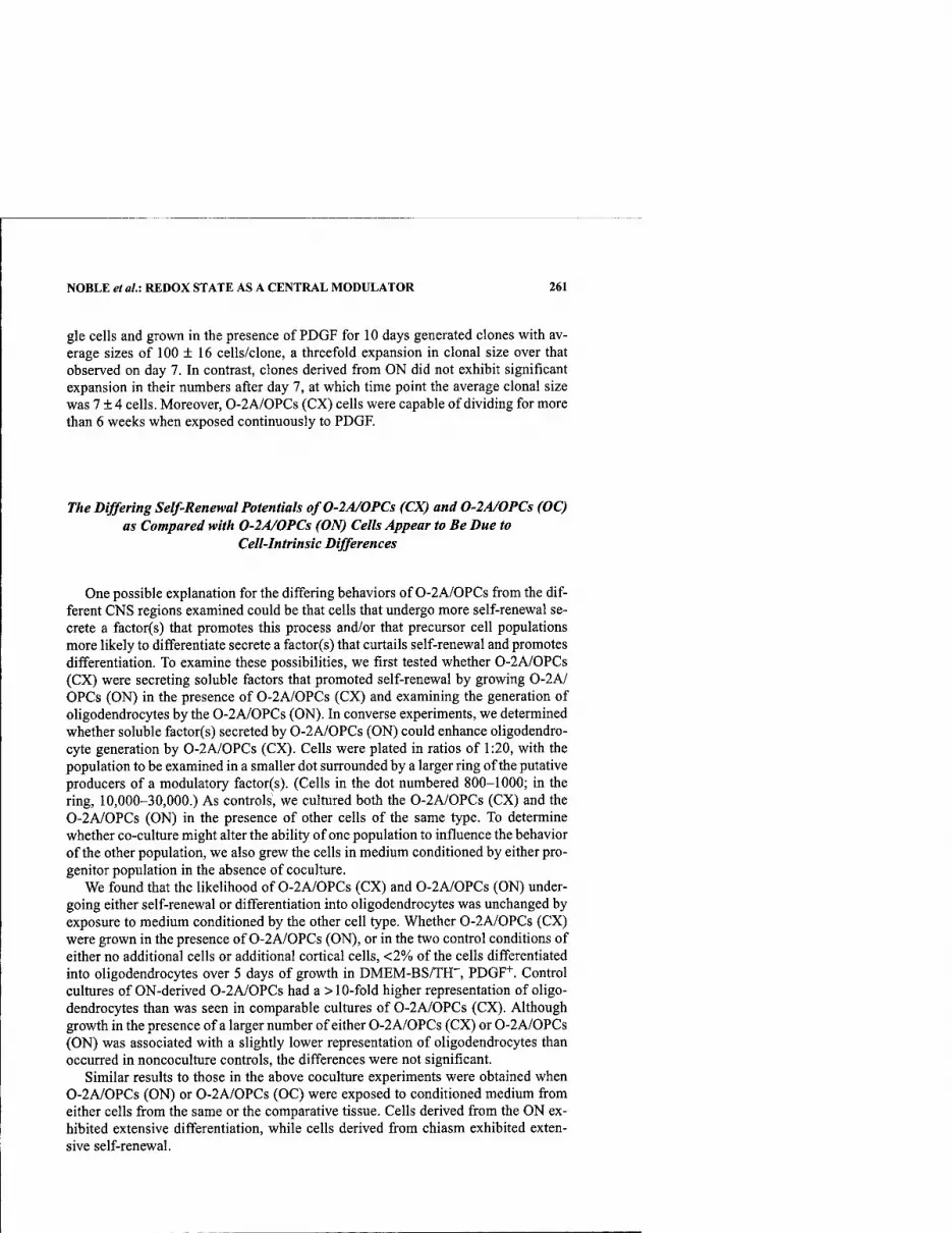
NOBLE et al.i REDOX STATE AS A CENTRAL MODULATOR 261
gle cells and grown in the presence of PDGF for 10 days generated clones with av- erage sizes of 100 ± 16 cells/clone, a threefold expansion in clonal size over that observed on day 7. In contrast, clones derived from ON did not exhibit significant expansion in their numbers after day 7, at which time point the average clonal size was 7 ± 4 cells. Moreover, 0-2A/0PCs (CX) cells were capable of dividing for more than 6 weeks when exposed continuously to PDGF.
The Differing Self-Renewal Potentials ofO-2A/OPCs (CX) and 0-2A/0PCs (OC) as Compared with 0-2A/0PCs (ON) Cells Appear to Be Due to
Cell-Intrinsic Differences
One possible explanation for the differing behaviors of 0-2A/0PCs from the dif- ferent CNS regions examined could be that cells that undergo more self-renewal se- crete a factor(s) that promotes this process and/or that precursor cell populations more likely to differentiate secrete a factor(s) that curtails self-renewal and promotes differentiation. To examine these possibilities, we first tested whether 0-2A/0PCs (CX) were secreting soluble factors that promoted self-renewal by growing 0-2AJ OPCs (ON) in the presence of 0-2A/0PCs (CX) and examining the generation of oligodendrocytes by the 0-2A/0PCs (ON). In converse experiments, we determined whether soluble factor(s) secreted by 0-2A/0PCs (ON) could enhance oligodendro- cyte generation by 0-2A/0PCs (CX). Cells were plated in ratios of 1:20, with the population to be examined in a smaller dot surrounded by a larger ring of the putative producers of a modulatory factor(s). (Cells in the dot numbered 800-1000; in the ring, 10,000-30,000.) As controls, we cultured both the 0-2A/0PCs (CX) and the 0-2A/0PCS (ON) in the presence of other cells of the same type. To determine whether co-culture might alter the ability of one population to influence the behavior of the other population, we also grew the cells in medium conditioned by either pro- genitor population in the absence of coculture.
We found that the likelihood of 0-2A/OPCs (CX) and 0-2A/0PCs (ON) under- going either self-renewal or differentiation into oligodendrocytes was unchanged by exposure to medium conditioned by the other cell type. Whether 0-2A/0PCs (CX) were grown in the presence of 0-2A/0PCs (ON), or in the two control conditions of either no additional cells or additional cortical cells, <2% of the cells differentiated into oligodendrocytes over 5 days of growth in DMEM-BS/TH", PDOF"*". Control cultures of ON-derived 0-2A/0PCs had a > 10-fold higher representation of oligo- dendrocytes than was seen in comparable cultures of 0-2A/0PCs (CX). Although growth in the presence of a larger number of either 0-2A/0PCs (CX) or 0-2A/0PCs (ON) was associated with a slightly lower representation of oligodendrocytes than occurred in noncoculture controls, the differences were not significant.
Similar results to those in the above coculture experiments were obtained when 0-2A/0PCS (ON) or 0-2A/0PCs (OC) were exposed to conditioned medium from either cells from the same or the comparative tissue. Cells derived from the ON ex- hibited extensive differentiation, while cells derived from chiasm exhibited exten- sive self-renewal.
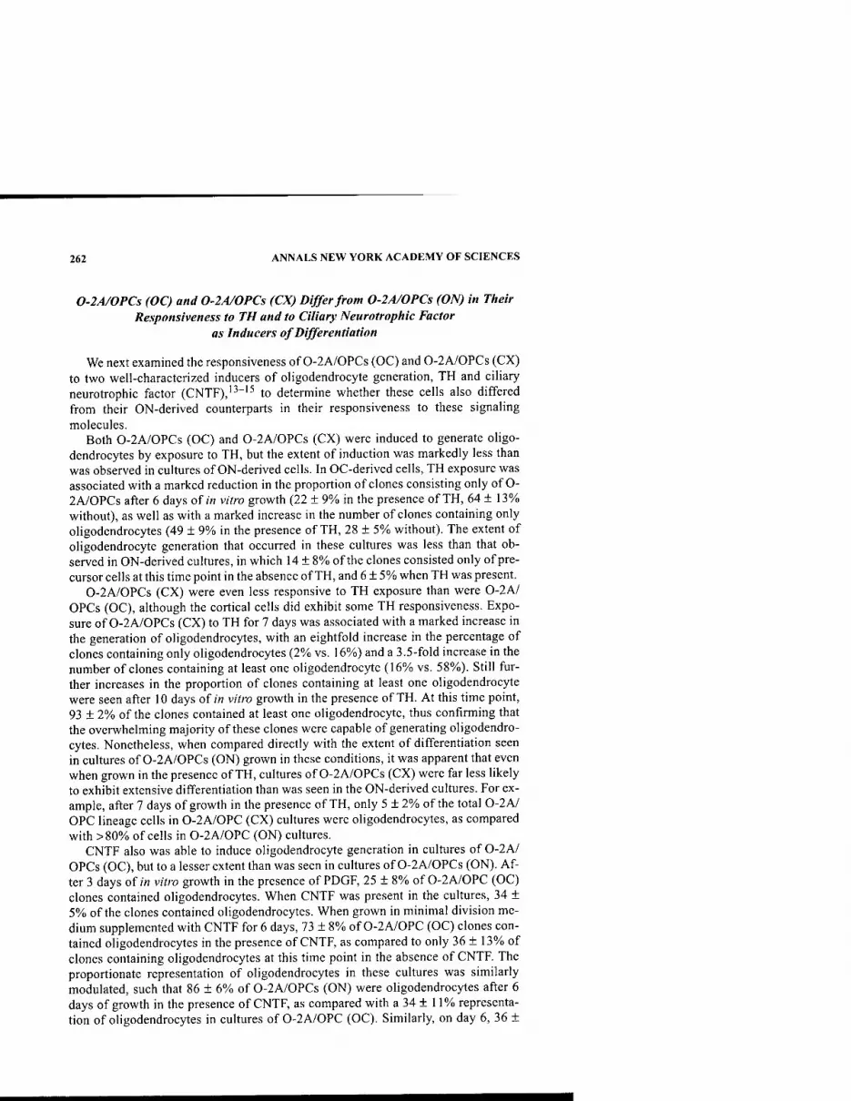
262 ANNALS NEW YORK ACADEMY OF SCIENCES
0-2A/0PCS (OC) and 0-2A/0PCs (CX) Differ from 0-2A/0PCs (ON) in Their Responsiveness to TH and to Ciliary Neurotrophic Factor
as Inducers of Differentiation
We next examined the responsiveness of 0-2A/0PCs (OC) and 0-2A/0PCs (CX) to two well-characterized inducers of oligodendrocyte generation, TH and ciliary neurotrophic factor (CNTF),'^"'^ to determine whether these cells also differed from their ON-derived counterparts in their responsiveness to these signaling molecules.
Both 0-2A/0PCS (OC) and 0-2A/0PCs (CX) were induced to generate oligo- dendrocytes by exposure to TH, but the extent of induction was markedly less than was observed in cultures of ON-derived cells. In OC-derived cells, TH exposure was associated with a marked reduction in the proportion of clones consisting only of O- 2A/0PCS after 6 days of/n vitro growth (22 ± 9% in the presence of TH, 64 ± 13% without), as well as with a marked increase in the number of clones containing only oligodendrocytes (49 ± 9% in the presence of TH, 28 ± 5% without). The extent of oligodendrocyte generation that occurred in these cultures was less than that ob- served in ON-derived cultures, in which 14 ± 8% of the clones consisted only of pre- cursor cells at this time point in the absence of TH, and 6 ± 5% when TH was present.
0-2A/0PCS (CX) were even less responsive to TH exposure than were 0-2A/ OPCs (OC), although the cortical cells did exhibit some TH responsiveness. Expo- sure of 0-2A/0PCS (CX) to TH for 7 days was associated with a marked increase in the generation of oligodendrocytes, with an eightfold increase in the percentage of clones containing only oligodendrocytes (2% vs. 16%) and a 3.5-fold increase in the number of clones containing at least one oligodendrocyte (16%o vs. 58%)). Still fur- ther increases in the proportion of clones containing at least one oligodendrocyte were seen after 10 days of m vitro growth in the presence of TH. At this time point, 93 ± 2% of the clones contained at least one oligodendrocyte, thus confirming that the overwhelming majority of these clones were capable of generating oligodendro- cytes. Nonetheless, when compared directly with the extent of differentiation seen in cultures of 0-2A/OPCs (ON) grown in these conditions, it was apparent that even when grown in the presence of TH, cultures of 0-2A/OPCs (CX) were far less likely to exhibit extensive differentiation than was seen in the ON-derived cultures. For ex- ample, after 7 days of growth in the presence of TH, only 5 ± 2%. of the total 0-2A/ OPC lineage cells in 0-2A/0PC (CX) cultures were oligodendrocytes, as compared with >80% of cells in 0-2A/0PC (ON) cultures.
CNTF also was able to induce oligodendrocyte generation in cultures of 0-2A/ OPCs (OC), but to a lesser extent than was seen in cultures of 0-2A/0PCs (ON). Af- ter 3 days of/« vitro growth in the presence of PDGF, 25 ± 8% of 0-2A/0PC (OC) clones contained oligodendrocytes. When CNTF was present in the cultures, 34 ± 5%) of the clones contained oligodendrocytes. When grown in minimal division me- dium supplemented with CNTF for 6 days, 73 ± 8% of 0-2A/0PC (OC) clones con- tained oligodendrocytes in the presence of CNTF, as compared to only 36 ± 13%> of clones containing oligodendrocytes at this time point in the absence of CNTF. The proportionate representation of oligodendrocytes in these cultures was similarly modulated, such that 86 ± 6% of 0-2A/0PCs (ON) were oligodendrocytes after 6 days of growth in the presence of CNTF, as compared with a 34 ± 11% representa- tion of oligodendrocytes in cultures of 0-2A/0PC (OC). Similarly, on day 6, 36 ±

NOBLE et al.: REDOX STATE AS A CENTRAL MODULATOR 263
13% of clones contained oligodendrocytes in the absence of CNTF, and 73 ± 18% of clones contained oligodendrocytes in the presence of CNTF.
In contrast with effects on 0-2A/0PC (ON) and 0-2A/0PC (OC) populations, CNTF was far less effective at inducing oligodendrocyte generation in 0-2A/0PC (CX) cultures. When these cells were grown in the presence of CNTF for 7 days, ~2 ±1% differentiated into oligodendrocytes, as compared with a figure of < 1 ± 0% in cultures exposed to PDGF alone. The proportion of clones containing oligodendro- cytes in the absence and presence of CNTF was also approximately the same after 7 days in culture (6 ± 6% in the absence of CNTF, and 7 ± 1% when CNTF was present). However, CNTF did have an effect on 0-2A/OPC (CX) cultures by reduc- ing cell numbers within a clone, presumably by slowing the rate of cell division. The average clone size in the presence of CNTF was 8 ± 2 cells, as compared with 31 ± 9 cells in its absence. CNTF also seemed slightly to enhance differentiation induc- tion by TH. As mentioned above, the percentage of oligodendrocytes generated by 0-2A/0PC (CX) after 7 days in vitro growth was 5 + 2% in the presence of TH and 2+1%) in the presence of CNTF. In contrast, exposure to both factors induced 12 ± 4% of 0-2A/0PC (CX) progenitors to become oligodendrocytes, a very marked in- crease over the value of 0.2 ± 0.2% observed in cultures growing in the presence of PDGF alone.
Freshly Isolated 0-2A/0PCs (ON), 0-2A/0PCs (OC), and 0-2A/0PCs (CX) Exhibit Differences in Their Intracellular Redox States, with the Degree of Oxidation Correlating Inversely with the Capacity for Self-Renewal
The differences between 0-2A/0PCs from ON, OC, and CX bear a striking re- semblance to the differences we found to be associated with the redox balance of ON-derived 0-2A/0PCs. As discussed earlier, in our studies on ON-derived cells, we found that freshly isolated 0-2A/0PCs that possessed a relatively reduced intra- cellular environment were most likely to undergo continued self-renewing divisions when grown in the presence of PDGF. In contrast, when grown in precisely the same conditions, cells that were more oxidized were more likely to differentiate into oli- godendrocytes in the absence of extensive in vitro division. Moreover, we found that pharmacological manipulation to make cells more reduced antagonized the differen- tiation-inducing abilities of TH. Thus, in several respects, the behavior of 0-2A/ OPCs (OC) and, to an even greater degree 0-2A/0PCs (CX), resembled the behavior of 0-2A/0PCs (ON) with a more reduced intracellular redox state. Therefore, to de- termine whether 0-2A/0PCs isolated from ON, OC, or CX differed in their intrac- ellular redox state in a manner consistent with their self-renewal characteristics, we labeled freshly isolated cells with rosamine and analyzed them by flow cytometry.
0-2A/0PCs (CX) progenitors displayed significantly lower levels of oxidative equivalents (as determined by rosamine fluorescence) than either 0-2A/0PCs (ON) or 0-2A/0PCs (OC) {P < 0.05), indicating a more reducing intracellular environ- ment in 0-2A/0PCs (CX). Data from six independent trials were compiled and stan- dardized so that the fluorescence level of 0-2A/0PCs (CX) was set equal to 100 units (±21 units SEM). In comparison, average fluorescence of 0-2A/0PCs (ON) progenitors was 207 ± 41 units, indicating greater oxidative turnover of the fluores- cent probe. 0-2A/0PCs (OC) had an intermediate level of mean fluorescence at 180 ± 39 units, significantly higher than the mean fluorescence of 0-2A/0PCs (CX).

264 ANNALS NEW YORK ACADEMY OF SCIENCES
This value was, in addition, lower than that for 0-2A/0PCs (ON), though not signif- icantly {P = 0.212). Thus, these results point to a trend in intracellular redox state between freshly isolated 0-2A/0PCs (CX), 0-2A/0PCs (OC), and 0-2A/0PCs (ON), such that those cells that undergo the most self-renewal (i.e., 0-2A/0PCs [CX]) were the most reduced, those that undergo the least self-renewal (i.e., 0-2A/ OPCs [ON]) were the most oxidized, and 0-2A/0PCs (OC) were intermediate both in respect to self-renewal and intracellular redox state.
DISCUSSION
The results presented point to a remarkable association between redox regulation and precursor cell function. In the first portion of these studies,^ we discovered that intracellular redox state modulation appears to be a central biochemical/molecular regulator of the balance between self-renewal and differentiation. In particular, re- dox state modulation satisfies all of the following criteria required to support such a conclusion:
(1) The proposed regulator should be altered in its level and/or function by cell-extrinsic signaling molecules that modulate the balance between self- renewal and differentiation, with signaling molecules that have opposite effects on self-renewal and differentiation exerting opposite effects on the proposed regulator.
(2) Alterations like those caused by exposure to these signaling molecules should have the same effect as the signaling molecules themselves.
(3) Substances that antagonize the alterations in the regulator caused by the cell-extrinsic signaling molecules should block their effects on this balance.
(4) Progenitor cell populations isolated from developing animals on the basis of the state of the proposed regulator should exhibit predictably different self-renewal characteristics consistent with the outcome of the other analyses.
Identification of a biochemical/molecular regulator that meets all of the above criteria is a strikingly different outcome from past efforts to understand how the bal- ance between self-renewal and differentiation is controlled. For example, the stron- gest evidence for a protein that might represent such a regulator has been reported for the cyclin-dependent kinase inhibitor p27'^'P'. P27 levels progressively accumu- late as 0-2A/0PCS proliferate,^^ and 0-2A/0PCs isolated from p27"'" mice under- go more division than do wild-type cells.^^'''° Ectopic p27 expression, however, causes 0-2A/0PCs to undergo cell cycle arrest in the absence of differentiation,'" a different outcome from that of rendering dividing 0-2A/0PCs slightly more oxi- dized by exposure to TH or pharmacological prooxidants. Moreover, no data has been provided that p27 levels are predictive of self-renewal potential or that experi- mental alterations in p27 levels yield the characteristic outcomes caused by TH, NT- 3, or redox manipulation. Thus, while p27 may be a part of the mechanism that mod- ulates the balance between self-renewal and differentiation, the evidence for intrac- ellular redox state as a central modulator appears to be more comprehensive.
The second portion of these studies^^ initially were focused on what appeared to be a quite separate issue, that of whether differing times of myelination in different

NOBLE et al.: REDOX STATE AS A CENTRAL MODULATOR 265
regions of the CNS might in any way be associated with the properties of the resident precursor cell populations. Characterization of precursor cells for oligodendrocytes isolated from ON, OC, and CX of P7 rats has revealed that each of these populations expresses distinct biological properties. In particular, cells isolated from ON, OC, and CX of identically aged rats show marked differences in their tendency to under- go self-renewing division and in their sensitivity to known inducers of oligodendro- cyte generation. Precursor cells isolated from the CX, a CNS region where myelination is a more protracted process than in the ON, appear to be intrinsically more likely to begin generating oligodendrocytes at a later stage and over a longer time period than cells isolated from the ON, raising the possibility that the different time courses of myelination in these CNS regions may reflect different biological properties of the resident precursor cell population.
It was striking that the redox state of 0-2A/0PCs from different CNS regions showed an excellent correlation with the properties expressed by cells of the ON in which redox state was experimentally manipulated. In particular, those cells with the greatest self-renewal potential (i.e., cortical 0-2A/0PCs), and the least response to inducers of oligodendrocyte generation, exhibited the most reduced redox state when examined as freshly isolated cells.
Previous studies had already demonstrated that 0-2A/0PCs isolated from ani- mals of different ages can have different properties.^^"^^ The results described here extend such observations on heterogeneity to demonstrate that the CNS of early post- natal rats contains multiple 0-2A/0PCs that exhibit markedly different properties, both in respect to their intrinsic tendency to undergo self-renewing divisions and in their response to inducers of oligodendrocyte generation. These differences, which appear to be cell intrinsic and also appear to be correlated with intracellular redox state, indicate a previously unanticipated complexity of phenotypes among precursor cells that give rise to the same differentiated cell type. The characteristics of these populations are such as to be consistent with the hypothesis that differences in the timing of myelinogenesis may be due, at least in part, to the local utilization of oligo- dendrocyte precursor cell populations with ftindamentally different properties.
One of the surprising findings to emerge from our studies was the extent to which self-renewal characteristics and response to inducers of differentiation differed in precursor cells that all give rise to oligodendrocytes and are isolated from postnatal animals of a single age. For example, 0-2A/0PCs isolated even from adjoining CNS regions, the ON and the OC, exhibited marked differences in their probability of un- dergoing differentiation when grown in the presence of PDGF sans inducers of dif- ferentation (such as TH and CNTF). Still more different from ON-derived cells were 0-2A/0PCs (CX), which were able to undergo continuous self-renewal for many days when grown in basal division conditions, generating large clones of up to 300 cells that consisted predominantly of 0-2A/0PCs and containing very few oligoden- drocytes. Even after 10 days of/« vitro growth in these conditions, the average clonal composition in 0-2A/0PC (CX) cultures consisted of 99.5% progenitors and 0.5% oligodendrocytes. The average clonal composition in 0-2A/0PC (ON) cultures at this time point, in contrast, was 37% 0-2A/0PCs and 63% oligodendrocytes. In- deed, OPCs (CX) derived from P7 rats could be maintained as dividing cells in basal division medium for at least 6 weeks (unpublished observations), a continuation of division that we have never observed in cultures of 0-2A/0PCs (ON) grown in these conditions.
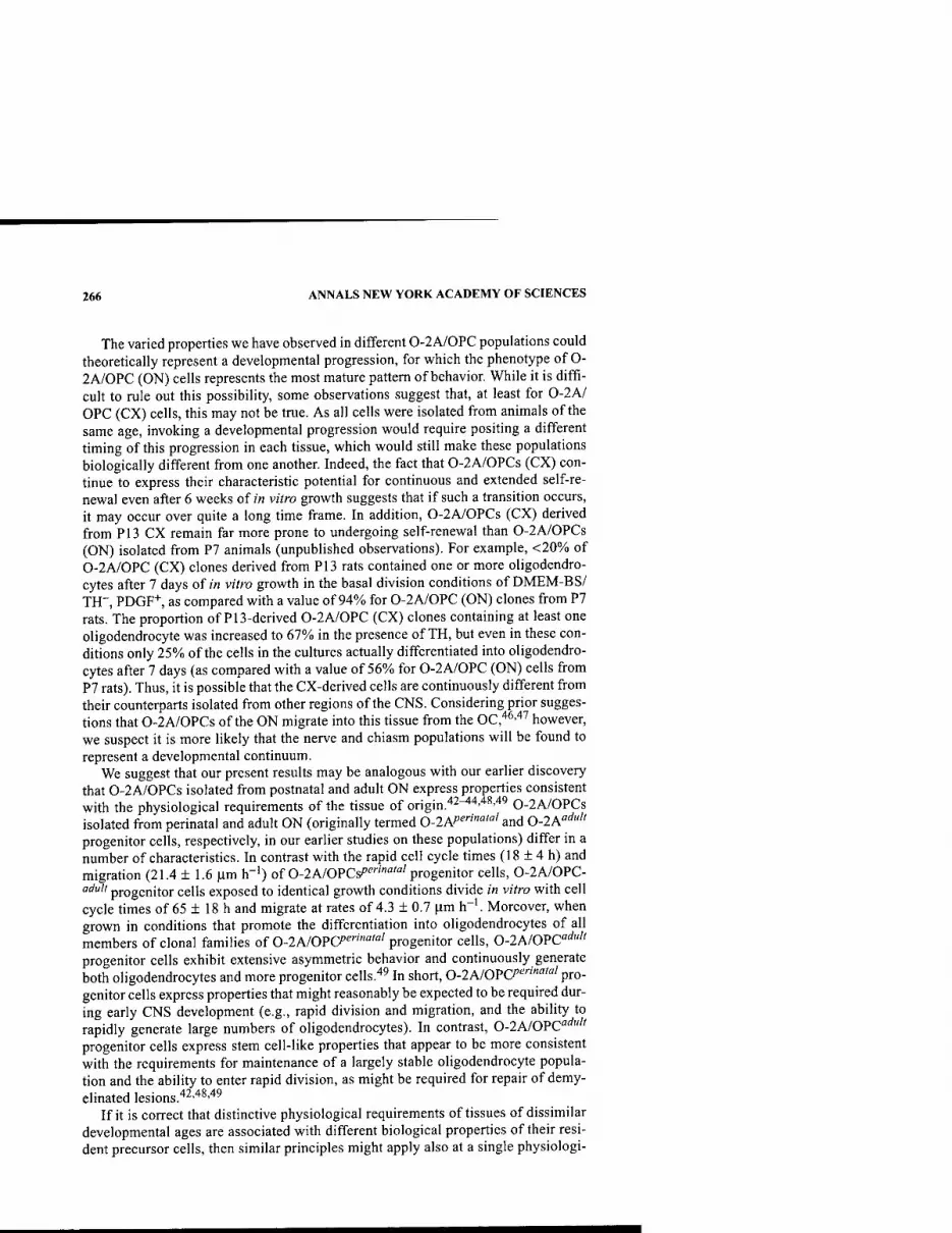
266 ANNALS NEW YORK ACADEMY OF SCIENCES
The varied properties we have observed in different 0-2A/0PC populations could theoretically represent a developmental progression, for which the phenotype of O- 2A/0PC (ON) cells represents the most mature pattern of behavior. While it is diffi- cult to rule out this possibility, some observations suggest that, at least for 0-2A/ OPC (CX) cells, this may not be true. As all cells were isolated from animals of the same age, invoking a developmental progression would require positing a different timing of this progression in each tissue, which would still make these populations biologically different from one another. Indeed, the fact that 0-2A/0PCs (CX) con- tinue to express their characteristic potential for continuous and extended self-re- newal even after 6 weeks of zn vitro growth suggests that if such a transition occurs, it may occur over quite a long time frame. In addition, 0-2A/0PCs (CX) derived from P13 CX remain far more prone to undergoing self-renewal than 0-2A/OPCs (ON) isolated from P7 animals (unpublished observations). For example, <20% of 0-2A/0PC (CX) clones derived from P13 rats contained one or more oligodendro- cytes after 7 days of w vitro growth in the basal division conditions of DMEM-BS/ TH", PDGF+, as compared with a value of 94% for 0-2A/0PC (ON) clones from P7 rats. The proportion of P13-derived 0-2A/OPC (CX) clones containing at least one oligodendrocyte was increased to 67% in the presence of TH, but even in these con- ditions only 25% of the cells in the cultures actually differentiated into oligodendro- cytes after 7 days (as compared with a value of 56% for 0-2A/0PC (ON) cells from P7 rats). Thus, it is possible that the CX-derived cells are continuously different from their counterparts isolated from other regions of the CNS. Considering prior sugges- tions that 0-2A/0PCS of the ON migrate into this tissue from the OC,'*^''''' however, we suspect it is more likely that the nerve and chiasm populations will be found to represent a developmental continuum.
We suggest that our present results may be analogous with our earlier discovery that 0-2A/0PCs isolated from postnatal and adult ON express properties consistent with the physiological requirements of the tissue of origin.'*^^''''*^''*^ 0-2A/0PCs isolated from perinatal and adult ON (originally termed 0-2A/'^"™"'' and 0-2A'"'"" progenitor cells, respectively, in our earlier studies on these populations) differ in a number of characteristics. In contrast with the rapid cell cycle times (18 ± 4 h) and migration (21.4 ± 1.6 |im h"') of 0-2A/0PCs^''"'""'''progenitor cells, 0-2A/OPC- aduli progenitor cells exposed to identical growth conditions divide in vitro with cell cycle times of 65 ± 18 h and migrate at rates of 4.3 ± 0.7 \im h"'. Moreover, when grown in conditions that promote the differentiation into oligodendrocytes of all members of clonal families of 0-2A/0Pa'^"'""'''progenitor cells, 0-2A/0PC'"^"" progenitor cells exhibit extensive asymmetric behavior and continuously generate both oligodendrocytes and more progenitor cells.^^ In short, 0-2A/0Pa"'"'"'"'' pro- genitor cells express properties that might reasonably be expected to be required dur- ing early CNS development (e.g., rapid division and migration, and the ability to rapidly generate large numbers of oligodendrocytes). In contrast, 0-2A/0PC'"'''" progenitor cells express stem cell-like properties that appear to be more consistent with the requirements for maintenance of a largely stable oligodendrocyte popula- tion and the ability to enter rapid division, as might be required for repair of demy- elinated lesions.'*^''*^''*^
If it is correct that distinctive physiological requirements of tissues of dissimilar developmental ages are associated with different biological properties of their resi- dent precursor cells, then similar principles might apply also at a single physiologi-

NOBLE et al: REDOX STATE AS A CENTRAL MODULATOR 267
cal age. In analogy with our proposal that the differences between 0-2A/0PC'"'"'' and O-IA/OPCP'^""""'' progenitor cells of the rat ON are reflective of the differing physiological requirements of tissue development and tissue homeostasis, we pro- pose that the differences that distinguish 0-2A/0PCs (ON), 0-2A/0PCs (OC), and 0-2A/0PCs (CX) reflect differing physiological requirements of the tissues to which these cell contribute. For example, a variety of experiments have indicated that the 0-2A/OPC population of the ON arises from a germinal zone located in or near the OC and enters the nerve by migration.^^'^'' Thus, it would not be surprising if progenitor cells of the OC expressed properties expected of cells at a potentially earlier stage of development than those cells that are isolated from ON of the same physiological age. Such properties would be expected to include the capacity to un- dergo a greater extent of self-renewal, much as has been seen when the properties of 0-2A/0PCs from ONs of embryonic rats and postnatal rats have been compared.^^ In respect to the properties of cortical progenitor cells, physiological considerations also appear to be consistent with our observations. The CX is one of the last regions of the CNS in which myelination is initiated, and the process of myelination also can continue for extended periods in this region.^-'"^^ If the biology of a precursor cell population is reflective of the developmental characteristics of the tissue in which it resides, then one might expect that 0-2A/0PCs isolated from this tissue would not initiate oligodendrocyte generation until a later time than occurs with 0-2A/0PCs isolated from structures in which myelination occurs earlier. In addition, cortical O- 2A/0PCs might be physiologically required to make oligodendrocytes for a longer time due to the long period of continued development in this tissue, at least as this has been defined in the human CNS (see, e.g., Refs. 27 and 28).
Our findings extend upon our previous analyses of oligodendrocyte development by demonstrating expression of tissue-specific properties as well as age-specific properties by cells that share the common property of being able to generate oligo- dendrocytes. Moreover, the observation that cell-intrinsic differences might be asso- ciated with different levels of responsiveness to inducers of differentiation adds a new degree of complexity to our understanding of the means by which the balance between self-renewal and differentiation is modulated in dividing 0-2A/0PCs once oligodendrocyte generation begins. We previously proposed that once this stage of oligodendrocyte creation is initiated, the regulation of differentiation is shifted from control by a cell-intrinsic biological clock to regulation by the environment.'^ Our present results suggest that the extent of environmental regulation that occurs is modulated by cell-intrinsic properties that modify responsiveness to environmental signals.
It will be important to determine whether the differences we have observed will be associated with differing patterns and extents of oligodendrocyte production fol- lowing transplantation of these various populations in vivo. Although it is well es- tablished that transplanted 0-2A/0PCs can readily repair demyelinating damage (see, e.g., Refs. 8, 51, and 52), it might be that cells with the characteristics of 0-2A/ OPCs (CX) can generate a larger number of oligodendrocytes from each injected cell than would be the case for 0-2A/0PCs (ON), an outcome that would indicate a greater potential utility of 0-2A/0PC (CX)-like cells in such applications.
Our results raise the intriguing possibility that at least one of the cell-intrinsic properties that regulate self-renewal and responsiveness to environmental factors is intracellular redox state. It seems particularly striking that the differences between

268 ANNALS NEW YORK ACADEMY OF SCIENCES
0-2A/OPC (CX), 0-2A/0PC (OC), and 0-2A/OPC (ON) populations are very much like differences we found being modulated by intracellular redox state in studies on purified ON-derived 0-2A/0PCs.^'^^ In this regard, we were particularly intrigued to find that the comparative redox states of these three populations were precisely as would be predicted from our other analyses. Such results make it seem an attractive hypothesis that cell-intrinsic mechanisms contributing to redox state modulation play an important role in regulating fundamental aspects of precursor cell function. If this is correct, then deciphering what these cell-intrinsic mechanisms might be should prove of considerable interest.
Available data from the study of several biological systems provides strong rea- son to believe that redox modulation functions as a central integrator of multiple pro- cesses related to self-renewal and differentiation, rather than as a controller of a sin- gle unique downstream effector pathway. Many different signaling pathways appear to converge on regulation of redox state, and redox alterations can in turn modulate several different pathways of possible relevance in modulation of self-renewal and differentiation. Several components of the redox regulatory network can be altered by exposure of cell to such cell-extrinsic signaling molecules as neurotrophins,^^'^'* type 1 interferon,^^ stem cell factor,^* transforming growth factor-P,^^ inflammatory cytokines (see, e.g., Ref 58), and thyroid hormone,^^ and also by ras activation.^** Increased levels of oxidative stress can in turn induce elevations in proteins (such as the cyclin-dependent kinase inhibitor p21 *^"'^'P'; see Ref 61) and could potentially modify function of transcription complexes (such as AP-1; see Ref 62), thus affect- ing cell division and/or differentiation. In addition, intracellular redox state can itself impinge on many aspects of cell signaling (see, e.g., Ref 63 for review). In this con- text, one of the more intriguing aspects of our studies is the observation that what appear to be relatively small changes in intracellular redox state are associated with profound differences in cellular behavior. Such observations are consistent with findings that as little as a 10% decrease in average glutathione levels significantly decreases calcium influx in peripheral blood lymphocytes stimulated with anti-CD3 antibody.^'' An ability of relatively small changes in redox state to modulate cell function would enable this fundamental component of cellular physiology to func- tion as a highly sensitive central rheostat that integrates cell-intrinsic states with cell- extrinsic signals.
REFERENCES
1. BERTONCELLO, I., G. HODGSON & T. BRADLEY. 1985. Multiparameter analysis of trans- plantable hemopoietic stem cells: I. The separation and enrichment of stem cells homing to marrow and spleen on the basis of rhodamine-123 fluorescence. Exp. Hematol. 13: 999-1006.
2. MULDER, A. & J. VISSER. 1987. Separation and functional analysis of bone marrow cells separated by rhodamine-123 fluorescence. Exp. Hematol. 15: 99-104.
3. REID, L. 1996. Stem cell/lineage biology and lineage-dependent extracellular matrix chemistry. In Textbook of Tissue Engineering. R. Lanza, R. Langer & W. Chick, Eds. R.G. Landes. Austin, TX.
4. KIM, M., D. COOPER, S. HAYES & G. SPANGRUDE. 1998. Rhodamine-123 staining in hematopoietic stem cells of young mice indicates mitochondrial activation rather than dye efflux. Blood 91: 4106-4117.
5. SMITH, J., E. LADI, M. MAYER-PROSCHEL & M. NOBLE. 2000. Redox state is a central modulator of the balance between self-renewal and differentiation in a dividing glial precursor cell. Proc. Natl. Acad. Sci. USA 97: 10032-10037.
NOBLE et al.: REDOX STATE AS A CENTRAL MODULATOR 269
6. MILLER, R.H., C. FRENCH CONSTANT & M.C. RAFF. 1989. The macroglial cells of the rat optic nerve. Annu. Rev. Neurosci. 12: 517-534.
7. RAFF, M.C, R.H. MILLER & M. NOBLE. 1983. dial cell lineages in the rat optic nerve. Cold Spring Harbor Symp. Quant. Bid. 48: 569-572.
8. GROVES, A.K. et al. 1993. Repair of demyelinated lesions by transplantation of puri- fied 0-2A progenitor cells. Nature 362: 453-455.
9. NOBLE, M., K. MURRAY, R STROOBANT, et al. 1988. Platelet-derived growth factor pro- motes division and motility and inhibits premature differentiation of the oligoden- drocyte/type-2 astrocyte progenitor cell. Nature 333: 560-562.
10. RICHARDSON, W.D., N. PRINGLE, M.J. MOSLEY, et al. 1988. A role for platelet-derived growth factor in normal gliogenesis in the central nervous system. Cell 53: 309-319.
11. RICHARDSON, W.D., N. PRINGLE, M. MOSLEY, et al. 1988. A role for platelet-derived growth factor in normal gliogenesis in the central nervous system. Cell 53: 309-319.
12. YAKOVLEV, A.Y., K. BOUCHER, M. MAYER-PROSCHEL & M. NOBLE. 1998. Quantitative insight into proliferation and differentiation of 0-2A progenitor cells in vitro: the clock model revisited. Proc. Natl. Acad. Sci. USA 95: 14164-14167.
13. BARRES, B.A., M.A. LAZAR & M.C. RAFF. 1994. A novel role for thyroid hormone, glu- cocorticoids and retinoic acid in timing oligodendrocyte development. Development 120: 1097-1108.
14. MAYER, M., K. BHAKOO & M. NOBLE. 1994. Ciliary neurotrophic factor and leukemia inhibitory factor promote the generation, maturation and survival of oligodendro- cytes in vitro. Development 120: 142-153.
15. IBARROLA, N., M. MAYER-PROSCHEL, A. RODRIGUEZ-PENA & M. NOBLE. 1996. Evi- dence for the existence of at least two timing mechanisms that contribute to oligo- dendrocyte generation in vitro. Dev. Biol. 180: 1-21.
16. MABIE, P. et al. 1997. Bone morphogenetic proteins induce astroglial differentiation of oligodendroglial-astroglial progenitor cells. Neuroscience 17: 4112-4120.
17. BOGLER, O., D. WREN, S.C. BARNETT, et al. 1990. Cooperation between two growth factors promotes extended self-renewal and inhibits differentiation of oligodendro- cyte-type-2 astrocytes (0-2A) progenitor cells. Proc. Natl. Acad. Sci. USA 87: 6368-6372.
18. BARRES, B.A., et al. 1994. A crucial role for neurotrophin-3 in oligodendrocyte devel- opment. Nature 367: 371-375.
19. TEMPLE, S. & M.C. RAFF. 1986. Clonal analysis of oligodendrocyte development in culture: evidence for a developmental clock that counts cell division. Cell 44: 773- 779.
20. RAFF, M.C, L.E. LILLIEN, W.D. RICHARDSON, et al. 1988. Platelet-derived growth fac- tor from astrocytes drives the clock that times oligodendrocyte development in cul- ture. Nature 333: 562-565.
21. AHLGREN, S., H. WALLACE, J. BISHOP, et al. 1997. Effects of thyroid hormone on embryonic oligodendrocyte precursor cell development in vivo and in vitro. Mol. Cell. Neurosci. 9: 420-432.
22. POWER, J., M. MAYER-PROSCHEL, J. SMITH & M. NOBLE. 2002. Oligodendrocyte pre- cursor cells from different brain regions express divergent properties consistent with the differing time courses of myelination in these regions. Dev. Biol. 245: 362-375.
23. MACKLIN, W.B. & CL. WEILL. 1985. Appearance of myelin proteins during develop- ment in the chick central nervous system. Dev. Neurosci. 7: 170-178.
24. KiNNEY, H.C, B.A. BRODY, A.S. KLOMAN & F.H. GILLES. 1988. Sequence of central nervous sytem myelination in human infancy. II. Patterns of myelination in autopsied infants. J. Neuropath. Exp. Neurol. 47: 217-234.
25. FORAN, D.R. & A.C. PETERSON. 1992. Myelin acquisition in the central nervous system of the mouse revealed by an MBP-LacZ transgene. J. Neurosci. 12: 4890-4897.
26. SKOFF, R.P., D. TOLAND & E. NAST. 1980. Pattern of myelination and distribution of neuroglial cells along the developing optic system of the rat and rabbit. J. Comp. Neurol. 191: 237-253.
27. YAKOVLEV, P.L. & A.R. LECOURS. 1967. The myelogenetic cycles of regional matura- tion of the brain. In Regional Development of the Brain in Early Life. A. Minkowski et al., Eds.: 3-70. Blackwell, Oxford.

270 ANNALS NEW YORK ACADEMY OF SCIENCES
28. BENES, F.M., M. TURTLE, Y. KHAN & P. FAROE. 1994. Myelination of a key relay zone in the hippocampal formation occurs in the human brain during childhood, adoles- cence and adulthood. Arch. Gen. Psychiatry 51: 477-484.
29. WHITAKER, J.E., P.L. MOORE, R.P. HAUGLAND & R.R HAUGLAND. 1991. Dihydrotet- ramethylrosamine: a long wave-length fluorogenic peroxidase substrate evaluated in vitro and in a model phagocyte. Biochem. Biophys. Res. Commun. 175: 387-393.
30. MEISTER, A., M.E. ANDERSON & O. HWANG. 1986. Intracellular cysteine and glu- tathione delivery systems. J. Am. Coll. Nutr. 5: 137-151.
31. MEISTER, A. & M.E. ANDERSON. 1983. Glutathione. Annu. Rev. Biochem. 52: 711- 760.
32. OCHI, T. 1993. Mechanism for the changes in the levels of glutathione upon exposure of cultured mammalian cells to tertiary-butylhydroperoxide and diamide. Arch. Tox- icol. 67:401-410.
33. MARTENSSON, J. et al. 1991. Inhibition of glutathione synthesis in the newborn rat: a model for endogenously produced oxidative stress. Proc. Natl. Acad. Sci. USA 88: 9360-9364.
34. ROBERTS, J., H. NAGASAWA, R. ZERA, et al. 1987. Prodrugs of L-cysteine as protective agents against acetaminophen-induced hepatotoxicity. 2-(Polyhydroxyalkyl)- and 2- (polyacetoxyalkyl)thiazolidine-4(R)-carboxylic acids. J. Med. Chem. 30: 1891- 1896.
35. SMILEY, S.T. et al. 1991. Intracellular heterogeneity in mitochondrial membrane poten- tials revealed by a J aggregate forming lipophilic cation JC-1. Proc. Natl. Acad. Sci. USA 88: 3671-3675.
36. RAFF, M.C, R.H. MILLER & M. NOBLE. 1983. A glial progenitor cell that develops in vitro into an astrocyte or an oligodendrocyte depending on the culture medium. Nature 303: 390-396.
37. RAO, M., M. NOBLE & M. MAYER-PROSCHEL. 1998. A tripotential glial precursor cell is present in the developing spinal cord. Proc. Natl. Acad. Sci. USA 95: 3996^001.
38. DURAND, B., F. GAO & M. RAFF. 1997. Accumulation of the cyclin-dependent kinase inhibitor p27/Kipl and the timing of oligodendrocyte differentiation. EMBO J. 16: 306-317.
39. CASACCIA-BONNEFIL, P et al. 1997. Oligodendrocyte precursor differentiation is per- turbed in the absence of the cyclin-dependent kinase inhibitor p27'*''P'. Genes Dev. 11:2335-2346.
40. DURAND, B., M. FERO, J. ROBERTS & M. RAFF. 1998. p27'^'P' alters the response of cells to mitogen and is part of a cell-intrinsic timer that arrests the cell cycle and ini- tiates differentiation. Curr. Biol. 6: 431-440.
41. TiKOO, R. et al. 1998. Ectopic expression of p27'^'P' in oligodendrocyte progenitor cells results in cell cycle growth arrest. J. Neurobiol. 36: 431-440.
42. WOLSWUK, G. & M. NOBLE. 1989. Identification of an adult-specific glial progenitor cell. Development 105: 387-400.
43. WOLSWIJK, G., P.N. RIDDLE & M. NOBLE. 1990. Coexistence of perinatal and adult forms of a glial progenitor cell during development of the rat optic nerve. Develop- ment 109: 691-698.
44. WoLSWiJK, G., P.N. RIDDLE & M. NOBLE. 1991. Platelet-derived growth factor is mito- genic for 0-2A'"'"'' progenitor cells. Glia 4: 495-503.
45. GAO, F. & M. RAFF. 1997. Cell size control and a cell-intrinsic maturation program in proliferating oligodendrocyte precursor cells. J. Cell. Biol. 138: 1367-1377.
46. SMALL, R.K., P. RIDDLE & M. NOBLE. 1987. Evidence for migration of oligodendro- cyte-type-2 astrocyte progenitor cells into the developing rat optic nerve. Nature 328: 155-157.
47. ONO, K., Y. YASUI, U. RUTISHAUSER & R.H. MILLER. 1997. Focal ventricular origin and migration of oligodendrocyte precursors into the chick optic nerve. Neuron 19: 283-292.
48. WoLSWiJK, G. & M. NOBLE. 1992. Cooperation between PDGF and FGF converts slowly dividing 0-2Aadult progenitor cells to rapidly dividing cells with characteris- tics of 0-2Aperinatal progenitor cells. J. Cell. Biol. 118: 889-900.
49. WREN, D., G. WOLSWIJK & M. NOBLE. 1992. In vitro analysis of the origin and mainte- nance of 0-2A'"'"" progenitor cells. J. Cell. Biol. 116: 167-176.

NOBLE et al.: REDOX STATE AS A CENTRAL MODULATOR 271
50. ONO, K., R. BANSAL, J. PAYNE, et al. 1995. Early development and dispersal of oligo- dendrocyte precursors in the embryonic chick spinal cord. Development 121: 1743- 1754.
51. WARRINGTON, A.E., E. BARBARESE & S.E. PFEIFFER. 1993. Differential myelinogenic capacity of specific developmental stages of the oligodendrocyte lineage upon trans- plantation into hypomyelinating hosts. J. Neurosci. Res. 34: 1-13.
52. UTZSCHNEIDER, D.A., D.R. ARCHER, J.D. KOCSIS, et al. 1994. Transplantation of glial cells enhances action potential conduction of amyelinated spinal cord axons in the myelin-deficient rat. Proc. Natl. Acad. Sci USA 53-57.
53. SAMPATH, D. & R. PEREZ-POLO. 1997. Regulation of antioxidant enzyme expression by NGF. Neurochem. Res. 22: 351-362.
54. GABAIZADEH, R., H. STAECKER, W. LIU & T. VAN DE WATER. 1997. BDNF protection of auditory neurons from cisplatin involves changes in intracellular levels of both reac- tive oxygen species and glutathione. Brain Res. Mol. Brain Res. 50: 71-78.
55. LEWIS, J., A. HUQ & P. NAJARRO. 1996. Inhibition of mitochondrial function by inter- feron. J. Biol. Chem. 271: 184-190.
56. LEE, J. 1998. Inhibition of p53-dependent apoptosis by the KIT tyrosine kinase: regu- lation of mitochondrial permeability transition and reactive oxygen species genera- tion. Oncogene 17: 1653-1662.
57. ISLAM, K. et al. 1997. TGF-betal triggers oxidative modifications and enhances apop- tosis in HIT cells through accumulation of reactive oxygen species by suppression of catalase and glutathione peroxidase. Free Radic. Biol. Med. 22: 1007-1017.
58. HAMPTON, M., B. FADEEL & S. ORRENIUS. 1998. Redox regulation of the caspases dur- ing apoptosis. Ann. N.Y. Acad. Sci. 854: 328-335.
59. PILLAR, T. & H. SEITZ. 1997. Thyroid hormone and gene expression in the regulation of mitochondrial respiratory function. Eur. J. Endocrinol. 136: 231-239.
60. LEE, A. et al. 1999. Ras proteins induce senescence by altering the intracellular levels of reactive oxygen species. J. Biol. Chem. 274: 7936-7940.
61. EsposiTO, F. et al. 1997. Redox-mediated regulation of p21(wafl/cipl) expression involves a post-transcriptional mechanism and activation of the mitogen-activated protein kinase pathvi^ay. Eur. J. Biochem. 245: 730-737.
62. XANTHOUDAKIS, S. & T. CURRAN. 1992. Identification and characterization of Ref-1, a nuclear protein that facilitates AP-1 DNA-binding activity. EMBO J. 11: 653-665.
63. KAMATA, H. & H. HiRATA. 1999. Redox regulation of cellular signaling. Cell Signal 11: 1-14.
64. STAAL, F. et al. 1994. Redox regulation of signal transduction: tyrosine phosphoryla- tion and calcium influx. Proc. Natl. Acad. Sci. USA 91: 3619-3622.

COX-2 and Neurodegeneration in Parkinson's Disease
p. TEISMANN," M. VILA," D.-K. CHOI," K. TIEU,'' D. C. WU," V. JACKSON-LEWIS," AND S. PRZEDBORSKF*'^
Neuroscience Research Laboratories of the Movement Disorder Division, Departments of Neurology and ''Pathology and the ''Center for Neurobiology and Behavior, Columbia University, New York, New York 10032, USA
ABSTRACT: Parkinson's disease (PD) is a common neurodegenerative disorder characterized by a progressive loss of dopaminergic neurons in the substantia nigra pars compacta. Recent observations linli cyclooxygenase type-2 (COX-2) to the progression of the disease. Consistent with this notion, studies with the dopaminergic neurotoxin l-methyl-4-phenyl-l,2,3,6-tetrahydropyridine (MPTP) show that inhibition and ablation of COX-2 markedly reduce the del- eterious effects of this toxin on the nigrostriatal pathway. The similarity be- tween this experimental model and PD strongly supports the possibility that COX-2 expression is also pathogenic in PD.
KEYWORDS: inflammation; neurotoxicity; neurodegeneration; MPTP; Parkin- son's disease; reactive oxygen species; superoxide dismutase
INTRODUCTION
Inflammatory processes associated with an increased expression of cyclooxyge- nase (COX) and elevated prostaglandin Ej (PGE2) levels have been linked to a vari- ety of neurodegenerative disorders, including Parkinson's disease (PD), amyotrophic lateral sclerosis, and Alzheimer's disease.' COX, which converts arachidonic acid to PGH2, the prostaglandin precursor of PGE2 and several others, comes in eukaryotic cells in two main isoforms: COX-1, which is constitutively ex- pressed, and COX-2, which is inducible.^ COX-2 is rapidly upregulated at inflam- matory sites and appears to be responsible for the formation of proinflammatory PGs.-^ Thus, COX-2 may contribute to the neurodegenerative process that is seen in Parkinson's disease^ and that are the focus of this manuscript.
COX-2 BRAIN LOCALIZATION: EMPHASIS ON THE NIGROSTRIATAL PATHWAY
COX-2 mRNA or protein is usually not detectable outside of a handful of discrete areas of the brain, where it is found primarily in neurons.'*'^ COX-2 immunoreactiv-
Address for correspondence: Dr. Serge Przedborski, BB-307, Columbia University, 650 West 168th Street, New Yorlc, NY 10032. Voice: 212-305-1540; fax: 212-305-5450.
Ann. N.Y. Acad. Sci. 991: 272-277 (2003). © 2003 New York Academy of Sciences.
272
TEISMANN et al.: COX-2 AND NEURODEGENERATION IN PD 273
ity is observed mainly in distal dendrites and dendritic spines and, apparently, exclu- sively in excitatory neurons such as glutamatergic neurons.'* Consistent with this description, we found no evidence of definite COX-2 immunoreactivity either in normal mice or in human postmortem dopaminergic structures at the level of both the substantia nigra and the striatum, which correspond to the site of origin and of projection of the nigrostriatal neurons.
Conversely, COX-2 becomes expressed in most neurons following a variety of in- sults.^ To a much lesser extent, COX-2 can also be upregulated by injury and disease in astrocytes and, more rarely, in microglial and endothelial cells.^ These consider- ations are perfectly in agreement with our COX-2 immunostaining data in PD post- mortem samples and in the l-methyl-4-phenyl-l,2,3,6-trerahydropyridine (MPTP) mouse model of PD.^ Indeed, although we did not see clear COX-2 immunoreactiv- ity in any of the controls, except over the neurophil, strong COX-2 immunostaining was seen in cells in PD and MPTP midbrain samples. Almost all positive cells exhibited a neuronal morphology, and only a few resembled astrocytes. None ap- peared as microglial cells. By double immunostaining, we were able to show that the majority of COX-2-positive neurons in the MPTP mice were dopaminergic. Al- though Knott et al? have found more abundant astrocytic COX-2 immunoreactivity in PD samples than we have (which could be related to technical differences), the two studies appear to agree that the majority of COX-2-positive cells in PD brains are neuronal.
COX-2 INDUCTION IN PD AND MPTP BRAINS
Among the many factors capable of inducing COX-2 expression are found many inflammatory cytokines, such as tumor necrosis factor (TNF)-a and interleukin (IL)-ip, as well as glutamate through the activation of the NMDA receptor and, pre- sumably, inducible nitric oxide synthase (iNOS).^ Relevant to PD and its experimen- tal model MPTP are the demonstrations that many of the aforementioned factors are significantly increased in the cerebrospinal fluid and the substantia nigra in these two pathological situations.^ It is worth noting, however, that while COX-2 upregu- lation occurs mainly in neurons, the factors potentially responsible for this induc- tion, as suggested above, may emanate from glial cells. This raises the possibility of an interesting deleterious interplay between neuron and glia in which the first neuron to die in PD would trigger a glial response that would, by releasing proinflammatory factors, induce the expression of COX-2 in neurons, enhancing the susceptibility of dopaminergic neurons to the degenerative process. According to this scenario, COX- 2 would not initiate the demise of dopaminergic neurons, but rather facilitate it. If one were to accept this scenario, one might wonder how COX-2 participates in the neurodegenerative process. In the most obvious scenario, upon COX-2 induction in dopaminergic neurons, these cells start to produce significant amounts of PGE2 that would amplify the glial response and the production of glia-derived deleterious me- diators such as reactive oxygen species and proinflammatory cytokines. Relevant to this hypothesis is our demonstration that following MPTP administration a robust glial response develops^ and that mitigating this reaction attenuates dopaminergic neuronal loss.^ At this point, however, we are not aware of any demonstration that

274 ANNALS NEW YORK ACADEMY OF SCIENCES
COX-2-derived PGEj plays a signaling role in linking injured neurons to the activa-
tion of glial cells. Another route by which COX-2 could contribute to the progression of nigrostn-
atal neurodegeneration is via the oxidation of dopamine by COX-2 and the conse- quent production of dopamine quinones. In this case, glia-derived inflammatory events would lead to COX-2 induction in neurons that would employ dopamine to generate reactive quinones.'^ These reactive dopamine quinones, which are widely known to react with nucleophiles,'' can bind covalently with cystein residues in pro- teins to form protein-bound 5-S-cysteinyl dopamine, a type of posttranslational modification that can seriously affect protein functions.
A
« V
NaCI + NaCI Mel 2 + MPTP
B
• ; ■■ * '
NaCI + MPTP
FIGURE 1. Effect of meloxicam on MPTP-induced SNpc dopaminergic neuronal death. (A) In saline-injected control mice, there are numerous SNpc tyrosine hydroxylase (TH)-positive neurons. (B) In mice treated with MPTP (30 mg/kg subcutaneous injection), the number of SNpc TH-positive neurons is reduced. (C-E) In mice treated with both MPTP and meloxicam, there is a noticeable attenuation of SNpc TH-positive neuronal loss. Scale bar. 200 p.m. (Reproduced from Teismann & Ferger'^ with permission.)

TEISMANN et al.: COX-2 AND NEURODEGENERATION IN PD 275
TABLE 1. Effect of meloxicam on MPTP toxicity
Saline
MPTP + MPTP + MPTP + /[PTP + meloxicam meloxicam meloxicam saline (2 mg/kg) (7.5 mg/kg) (50 mg/kg)
Tyrosine hydrox- ylase (cells/ section)
Nissl (cells/ section)
76 ±2
94 ±2
36 ±4*
54 ±6*
34 ±6*
56 ±9*
67±4# 69±3#
99±3# 96±56±3#
Dopamine 13.91 ± 0.73 2.21 ±0.40* 2.22 ± 0.53*
DOPAC 0.99 ±0.06 0.23 ± 0.03* 0.31 ±0.04*
HVA 1.30 ±0.07 0.53 ±0.06* 0.63 ± 0.08*
5.01±0.47# 5.61±0.35#
0.43±0.03# 0.39±0.03#
0.84±0.05# 0.86±0.04#
NOTE: Counts of tyrosine-hydroxylase positive neurons and Nissl in three sections at the third cranial nerve and dopamine, DOPAC, HVA content after saline or MPTP (30 mg/kg s.c.) in meloxicam (0, 2, 7.5, 50 mg/kg i.p.)-pretreated mice. *P < 0.05, fewer than saline-control mice. #P < 0.05, more than MPTP-injected mice and not different from control mice. Values are means ± SEM (n = 8-12 per group). (From Teismann & Ferger;13 used with permission.)
ROLE OF COX-2 IN THE MPTP MOUSE MODEL OF PD
To demonstrate whether COX-2 actually plays a deleterious role in PD, we and other investigators have examined the effects of COX-2 inhibition on MPTP-induced dopaminergic neurotoxicity. In an earlier report, acetylsalicylic acid and salicylic acid provided protection against MPTP neurotoxicity in mice, whereas diclofenac failed to do so.''^ Because the failure to observe neuroprotection by diclofenac could be due to poor brain entry, we have revisited the issue using meloxicam, a specific COX-2 inhibitor with better brain penetration.*-' In this subsequent study, the au- thors found that MPTP caused a significant reduction in striatal dopamine levels as well as in dopaminergic neuron numbers in the substantia nigra, which was markedly attenuated by meloxicam (FIG. 1 and TABLE 1). In another MPTP study, mutant mice deficient in COX-2 were used instead of pharmacological inhibitors. This study gen- erated essentially the same outcomes.''* These latter data confirm the significant role of COX-2 in MPTP-induced neurodegeneration.
CONCLUSION
COX-2 has emerged as a potential pathogenic factor in several neurodegenerative disorders, including PD. Several studies have shown that COX-2 protein is upregu- lated in dopaminergic neurons in PD and in its experimental model, MPTP. Although the actual mechanism by which COX-2 is involved in the nigrostriatal neurodegen- eration remains to be elucidated, the fact that both the inhibition and abrogation of COX-2 in MPTP-treated mice attenuates significantly the loss of dopaminergic neu- rons provides compelling evidence of its role in the pathogenesis of PD.
276 ANNALS NEW YORK ACADEMY OF SCIENCES
ACKNOWLEDGMENTS
The authors wish to thank Mr. Brian Jones for his help in the preparation of this manuscript. The authors wish also to acknowledge the support of the NIH/NIHNDS (Grants R29 NS37345, ROl NS38586 and NS42269, and P50 NS38370); the NIH/ NIA (Grant AG13966); the U.S. Department of Defense (Grants DAMD 17-99-1- 9471 and DAMD 17-03-1); the Lowenstein Foundation; the Lillian Goldman Char- itable Trust; the Parkinson's Disease Foundation; the Muscular Dystrophy Associa- tion; the ALS Association; and Project ALS. RT is a recipient of a scholarship from the German Research Foundation (DFG TE 343/1-1).
NOTE ADDED IN PROOF
Since the submission of this manuscript, we have published an article [Teismann, P., etal. 2003. Cyclooxygenase-2 is instrumental in Parkinson's disease neurodegen- eration. Proc. Natl. Acad. Sci. USA 100(9): 5473-5478] further supporting a critical role for COX-2 in both the pathogenesis and selectivity of the PD neurodegenerative process. In this paper, we show that COX-2 is upregulated in brain dopaminergic neurons of both PD and MPTP mice and that COX-2 induction occurs through a JNKc-Jun-dependent mechanism after MPTP administration. We demonstrate that targeting COX-2 does not protect against MPTP-induced dopaminergic neurodegen- eration by mitigating inflammation. Instead, we provide evidence that COX-2 inhi- bition/ablation prevents the formation of the oxidant species dopamine-quinone, which has been implicated in the pathogenesis of PD.
REFERENCES
1. WYSS-CORAY, T. & L. MucKE. 2002. Inflammation in neurodegenerative disease: a double-edged sword. Neuron 35: 419-432.
2. O'BANION, M.K. 1999. Cyclooxygenase-2: moleeular biology, pharmacology, and neu- robiology. Grit. Rev. Neurobiol. 13: 45-82.
3. VILA, M. et al. 2001. The role of glial cells in Parkinson's disease. Curr. Opin. Neurol. 14:'483-489.
4. KAUFMANN, W.E. et al. 1997. Cyelooxygenases and the central nervous system. Pros- taglandins 54: 601-624.
5. HURLEY, S.D. et al. 2002. Cyclooxygenase inhibition as a strategy to ameliorate brain injury. J. Neurotrauma 19: 1-15.
6. PRZEDBORSKI, S. & M. VILA. 2001. MPTP: a review of its mechanisms of neurotoxicity. Clin. Neurosci. Res. 1: 407-418.
7. KNOTT, C. et al. 2000. Inflammatory regulators in Parkinson's disease: iNOS, lipocor- tin-1, and cyelooxygenases-1 and -2. Mol. Cell. Neurosci. 16: 724-739.
8. LIBERATORE, G.T. et al. 1999. Indueible nitric oxide synthase stimulates dopaminergic neurodegeneration in the MPTP model of Parkinson disease. Nat. Med. 5: 1403-1409.
9. Wu, D.C. et al. 2002. Blockade of microglial activation is neuroprotective in the I- m'ethyl-4-phenyl-l,2,3,6-tetrahydropyridine mouse model of Parkinson disease. J. Neurosci. 22: 1763-1771.
10. HASTINGS, TO. 1995. Enzymatic oxidation of dopamine: the role of prostaglandin H synthase. J. Neurochem. 64: 919-924.
11. GRAHAM, D.G. 1978. Oxidative pathways for catecholamines in the genesis of neu- romelanin and cytotoxic quinones. Mol. Pharmacol. 14: 633-643.

TEISMANN et al.: COX-2 AND NEURODEGENERATION IN PD 277
12. AUBIN, N. et al. 1998. Aspirin and salicylate protect against MPTP-induced dopamine depletion in mice. J. Neurochem. 71: 1635-1642.
13. TEISMANN, P. & B. FERGER. 2001. Inhibition of the cyclooxygenase isoenzymes COX-1 and COX-2 provide neuroprotection in the MPTP-mouse model of Parkinson's dis- ease. Synapse 39: 167-174.
14. FENG, Z. et al. 2002. Cyclooxygenase-2-deficient mice are resistant to l-methyl-4-phe- nyl-l,2,3,6-tetrahydropyridine-induced damage of dopaminergic neurons in the sub- stantia nigra. Neurosci. Lett. 329: 354.
Striatopallidal Changes in a Preclinical Rat Model of Parkinson's Disease
A. E. GRISSELL, T. M. BUCHANAN, AND M. A. ARIANO
Department ofNeuroscience, The Chicago Medical School, North Chicago, Illinois 60064, USA
KEYWORDS: Parkinson's disease; dopamine; striatum
INTRODUCTION
Parkinson's disease (PD) is characterized by motor symptoms due to substantial nigrostriatal dopamine (DA) loss (-80%). To investigate the neurochemical alter- ations that occur prior to motor symptoms, an early preclinical model of PD has been produced. Malonate was infused into one substantia nigra causing a partial, unilat- eral striatal DA depletion. HPLC analysis detected striatal DA loss (-50%) at 4 weeks. Subtle behavioral changes, indicated by asymmetry of limb use, confirmed a DA imbalance.' Morphological analysis of the DA-depleted striatum showed en- hanced D2 DA receptor staining, signifying receptor upregulation due to partial DA loss;^ activated (cleaved) caspase-3, an indication of cellular homeostatic imbalanc- es; and 8-oxoguanine, a marker of oxidative nucleic acid damage. Further, alter- ations occurred specifically in the striatopallidal projection system, based upon coincident expression of enkephalin as a phenotypic marker of the efferents with the neurochemical markers.
ANIMAL PREPARATION
Twenty-eight male Sprague-Dawley rats (-225 g) were lesioned unilaterally us- ing malonate infusion (3 ixmoles in 1 \i\) into the right substantia nigra. Rats were killed 4 weeks after malonate infusions.
HPLC ANALYSIS
Striata were dissected, frozen on dry ice, and stored at -86°C until HPLC analy- sis.3 Striatal DA content was averaged (±SEM): intact 14.09 ± 2.22 ng/mg protein versus DA-depleted 8.01 + 3.38 ng/mg protein {P < 0.001, Ar= 14). DOPAC:DA ra- tios: intact 0.26 ± .03; DA-depleted 0.32 ± .07 (P = .008).
Address for correspondence: Dr. Marjorie A. Ariano, Department ofNeuroscience, The Chi- cago Medical School, 3333 Green Bay Road, North Chicago, Illinois 60064. Voice: 847-578- 3412; fax: 847-578-8515.
Ann. N.Y. Acad. Sci. 991: 278-280 (2003). © 2003 New Yorli Academy of Sciences.
278
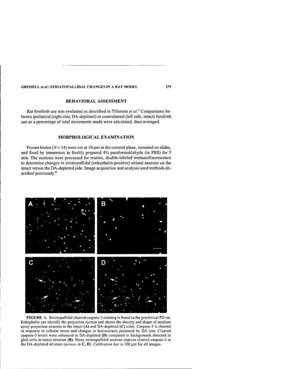
GRISSELL et al.: STRIATOPALLIDAL CHANGES IN A RAT MODEL 279
BEHAVIORAL ASSESSMENT
Rat forelimb use was evaluated as described in Tillerson et al.' Comparisons be- tween ipsilateral (right side, DA-depleted) or contralateral (left side, intact) forelimb use as a percentage of total movements made were calculated, then averaged.
MORPHOLOGICAL EXAMINATION
Frozen brains (A^= 14) were cut at 10 |Xm in the coronal plane, mounted on slides, and fixed by immersion in freshly prepared 4% paraformaldehyde (in PBS) for 5 min. The sections were processed for routine, double-labeled immunofluorescence to determine changes in striatopallidal (enkephalin-positive) striatal neurons on the intact versus the DA-depleted side. Image acquisition and analysis used methods de- scribed previously.^
FIGURE 1. Striatopallidal cleaved caspase-3 staining is found in the preclinical PD rat. Enkephalin can identify the projection system and shows the density and shape of medium spiny projection neurons in the intact (A) and DA-depleted (C) sides. Caspase-3 is cleaved in response to cellular stress and changes in homeostasis produced by DA loss. Cleaved caspase-3 levels were enhanced in DA-depleted (D) compared to backgrounds detected in glial cells in intact striatum (B). Many striatopallidal neurons express cleaved caspase-3 in the DA-depleted striatum (arrows in C, D). Calibration bar is 100 ]xm for all images.

280 ANNALS NEW YORK ACADEMY OF SCIENCES
RESULTS
The preclinical PD rat provides a credible model of preclinical stages of PD. Rats have subtle behavior imbalances correlated to the partial striatal DA loss, which showed preferential use of the forepaw ipsilateral to the DA depletion (27.78%, 29.44%, 31.67%) versus the intact side (11.67%, 8.89%, 5.56%) at 2 weeks, 3 weeks, and 4 weeks postinfusion (A^= 9). Morphological correlates include slight el- evation in D2 DA receptor staining and significant {P < .001) elevation in expression of cleaved caspase-3 and 8-oxoguanine within the striatopallidal projection pathway ipsilateral to the nigrostriatal DA loss (FIGURE 1), evaluated using cellular luminos- ity histogram analyses.'*
CONCLUSIONS
These neurochemical enhancements were unexpected in light of the level (-50%) and duration (4 weeks) of the striatal DA loss. These findings suggest substantial postsynaptic (striatal) changes occur in the early stages of PD and provide a mecha- nism to evaluate initial neurochemical alterations that may be modified to retard or stop the insidious progression of PD. These results demonstrate the complexity of PD and caution our thinking of the disorder as principally dopaminergic in scope.
ACKNOWLEDGMENT
This work was supported by Department of Defense Grant DAMD17-99-1-9542.
REFERENCES
TiLLERSON, J.L., A.D. COHEN, J. PHILHOWER, etal. 2001. Forced limb-use effects on the behavioral and neurochemical effects of 6-hydroxydopamine. J. Neurosci. 21: 4427- 4435.
GERFEN, C.R. & W.S. YOUNG 3RD. 1988. Distribution of striatonigral and striatopallidal peptidergic neurons in both patch and matrix compartments: an in situ hybridization histochemistry and fluorescent retrograde tracing study. Brain Res. 460: 161-167.
MORROW, B.A., A.J. REDMOND, R.H. ROTH & J.D. ELSWORTH. 2000. The predator odor, TMT, displays a unique, stress-like pattern of dopaminergic and endocrinological activation in the rat. Brain Res. 864: 146-151.
ARIANO, M.A., N. ARONIN, M. DIFIGLIA, et al. 2002. Striatal neurochemical changes in transgenic models of Huntington's disease. J. Neurosci. Res. 68: 716-729.

Neurotoxicity-Induced Changes in Striatal Dopamine Receptor Function
ANNA-LIISA BROWNELL, IRIS Y. CHEN, XUKUI WANG, MEIXIANG YU, AND BRUCE G. JENKINS
Department of Radiology, Massachusetts General Hospital, Boston, Massachusetts 02114, USA
KEYWORDS: dopamine receptor; dopamine transporter; striatum
Impairment of dopaminergic neurotransmission can be primary, as in Parkinson's disease or secondary, as in Huntington's disease (HD). The secondary dopamine dysfunction is related to the progressive loss of the striatal neurons bearing the postsynaptic dopamine DI and D2 receptors. PET studies have shown a significant decrease, at an annual rate of 2-6.5%^, in striatal glucose metabolism' and in dopamine Dl and D2 receptor binding in both asymptomatic and symptomatic HD patients.
Huntington's disease can be modeled by 3-nitropropionic acid (3-NP), which is an inhibitor of succinate dehydrogenase.^ It causes mitochondrial inhibition and striatal degeneration. We used in vivo PET studies to investigate 3-NP-induced acute and prolonged neurotoxic effects on striatal dopamine receptors and transporters in a rat model. 3-NP was administered twice a day at a dose of 10 mg/kg i.p. to eight rats (male Spraque-Dawley rats from Charles River Laboratories, average weight 300 g) until symptomatic gait was observed or for a maximum of 5 days. Imaging studies of dopamine Dl and D2 receptors and transporters were conducted before 3-NP admin- istration and 2 and 7 days and 4 and 16 weeks after 3-NP administration. To validate the striatal deficit, an additional PET study of glucose metabolism was done two days after the cessation of 3-NP using '*F-2-fluorodeoxy-D-glucose ('^F-FDG). Studies of dopamine Dl receptors were done using "C-SCH (Schoering 23660) as a tracer. Dopamine D2 receptors were imaged by '*C-raclopride, and dopamine transporters using 2p-carbomethoxy-3p-4-fluorophenyl tropane ("C-CFT). All the PET imaging studies were conducted using an in-house built tomographic instrument.'*
FIGURE 1 shows PET studies conducted 2 days after 3-NP administration. The glucose study revealed large striatal lesions. At that time a moderate decrease of dopamine Dl and D2 receptor binding and an increase in dopamine transporter bind- ing were observed. After that, progressive decrease was observed in pre and post- synaptic dopamine receptor function (TABLE 1, FIG. 1).
Address for correspondence: Anna-Liisa Brownell, Department of Radiology, Massachusetts General Hospital, 55 Fruit St., Boston, MA 02114.
Ann. N.Y. Acad. Sci. 991: 281-283 (2003). © 2003 New Yorli Academy of Sciences.
281
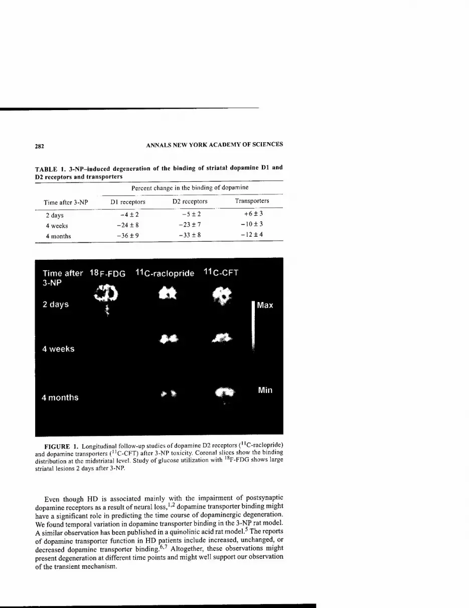
282 ANNALS NEW YORK ACADEMY OF SCIENCES
TABLE 1. 3-NP-induced degeneration of the binding of striatal dopamine Dl and D2 receptors and transporters
Percent change in the binding of dopamine
Time after 3-NP Dl receptors D2 receptors Transporters
2 days -4 ±2 -5 ±2 + 6 ±3
4 weeks -24 ±8 -23 ±7 -10±3
4 months -36 ±9 -33 ±8 -12±4
FIGURE 1. Longitudinal follow-up studies of dopamine D2 receptors ("C-raclopride) and dopamine transporters ("C-CFT) after 3-NP toxicity. Coronal slices show the binding distribution at the midstriatal level. Study of glucose utilization with '^F-FDG shows large striatal lesions 2 days after 3-NP.
Even though HD is associated mainly with the impairment of postsynaptic dopamine receptors as a result of neural loss,''^ dopamine transporter binding might have a significant role in predicting the time course of dopaminergic degeneration. We found temporal variation in dopamine transporter binding in the 3-NP rat model. A similar observation has been published in a quinolinic acid rat model.^ The reports of dopamine transporter function in HD patients include increased, unchanged, or decreased dopamine transporter binding.^-'' Altogether, these observations might present degeneration at different time points and might well support our observation of the transient mechanism.

BROWNELL et al.: STRIATAL DOPAMINE RECEPTOR FUNCTION 283
ACKNOWLEDGMENT
This work was supported by the DOD Grant DAMD17-99-1-9555.
REFERENCES
1. ANDREWS, T.C, et al. 1998. Advances in the understanding of early Huntington's dis- ease using the functional imaging techniques of PET and SPECT. Mol. Med. Today 4(12); 532-539.
2. ANDREWS, T.C, et al. 1999. Huntington's disease progression, PET and clinical obser- vations. Brain 122(Pt. 12): 2353-2363.
3. ALSTON, T.A., et al. 1977. 3-Nitropropionate, the toxic substance of Indigofera, is a suicide inactivator of succinate dehydrogenase. Proc. Natl. Acad. Sci. USA 74: 3767-3771.
4. BROWNELL, G.L., et al. 1985. High resolution tomograph using analog coding. In The Metabolism of the Human Brain. Studies with Positron Emssion Tomography. T. Greitz et al., Eds.; 13-19. Raven Press. New York.
5. ARAUJO, M.D. et al. 2000. Deficits in striatal dopamine D2 receptors and energy metabolism detected by in vivo mirco PET imaging in a rat model of Huntington's disease. Exp. Neurol. 166: 287-297.
6. SUZUKI, M. et al. 2001. Vesicular neurotransmitter transporters in Huntington's dis- ease: initial observations and comparison with traditional synaptic markers. Synapse 41(4): 329-336.
7. GiNOVART, N. et al. 1997. PET study of pre- and post-synaptic dopaminergic markers for neurodegenerative process in Huntington's disease. Brain 120: 503-514.

The Developmental Time Course of dial Cell Line-Derived Neurotrophic Factor (GDNF) and GDNF Receptor a-1 mRNA Expression in the Striatum and Substantia Nigra
JINWHAN CHO," NIKOLAI G.KHOLODILOV," AND ROBERT E. BURKE"-*
Departments of Neurology and ''Pathology, The College of Physicians and Surgeons, Columbia University, New York, New York 10032, USA
KEYWORDS: glial cell line-derived neurotrophic factor (GDNF); GDNF recep- tor a-1 (GFR a-1); natural cell death; Northern analysis; phosphoimager
INTRODUCTION
In most developing neural systems, a natural cell death (NCD) event occurs that eliminates 50% or more of the neuronal population. One central neuronal population of particular interest consists of the dopamine (DA) neurons of the substantia nigra (SN), the population that degenerates to the greatest extent in Parkinson's disease. Of central interest to the neurobiology of this disease are the neurotrophic factors that support the viability of these neurons during development and that ultimately determine their number in the mature brain. There is evidence that NCD in dopamine neurons of the SN is regulated during development by interaction with their target, the striatum, but the neurotrophic factors that mediate this regulation are unknown. One candidate is glial cell line-derived neurotrophic factor (GDNF), which was originally identified on the basis of its ability to support the development of embry- onic mesencephalic DA neurons in culture.' However, little is known about the time course of developmental expression of GDNF and its receptor GFR a-1 in the devel- oping rat brain in relation to the time course of the postnatal NCD event. We there- fore examined GDNF and GFR a-1 expression to understand better the possible role of GDNF as a physiologic striatal target-derived neurotrophic factor in relation to the NCD event in SN DA neurons.
RESULTS
The expression levels of GDNF and GFR a-1 in the striatum and SN were studied by Northern analysis for rats at postnatal days 2, 6, 10, 14, and 28, as well as for
Address for correspondence: Robert E. Burke, Department of Neurology, Room 308, Black Building, Columbia University, 650 West 168th Street, New York, New York 10032. Voice: 212- 305-7374; fax: 212-305-5450.
Ann. N.Y. Acad. Sci. 991: 284-287 (2003). © 2003 New York Academy of Sciences.
284

CHO et al: TIME COURSE OF GDNF AND GFR a-1 mRNA EXPRESSION 285
100
80
60
40
20
I 1
-//-
ANOVA P<0.001 1: P<0.01 vs PND10,14,28, Adult 2:P<0.005vsPND28, Adult
—*
—r- 10
—I— 14
—1—
IB 22 —r—
26 -rV-
ADULT
B POSTrWTAL DAY
100
80
60
40
^^
ANOVA P<0.001 1: P<0.001 VS PND6,10,14,28, Adult 2: P<0.01 vs PND10,14,28, Adult 3: P<0.001 vs PND28, Adult
—i
10 14 18 22 26
POSTNATAL DAY
^/- ADULT
FIGURE 1. Developmental time course of GDNF mRNA expression in the striatum (A) and in the SN (B). Expression of GDNF is highest in the striatum at postnatal day 2 and progressively decreases after postnatal day 6. In the SN, GDNF mRNA also decreases after postnatal day 2 and shows a small plateau between postnatal days 10 and 14.
adult rats. Northern blot analysis showed a single major band at 900 bp for GDNF, and two major bands were identified for GFR a-1, at about 4.0 and 8.0 kb. We ana- lyzed radioactive bands using a phosphoimager and expressed them as a relative per- centage of the radioactivity at postnatal day 2. Expression of GDNF is highest in the striatum at postnatal day 2 and progressively decreases after postnatal day 6 (FIG.
1 A). In the SN, GDNF mRNA also decreases after postnatal day 2 and shows a small
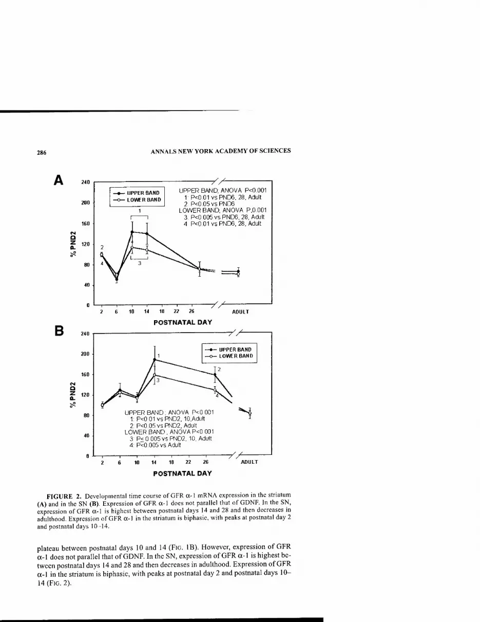
286 ANNALS NEW YORK ACADEMY OF SCIENCES
240
200
160
a ? 120
UPPER BAND LOWER BAND
r / UPPER BAND, ANOVA P<0,001
1 P<0,01vsPND6. 28, Adult 2: P<0,05vsPND6
LOWER BAND, ANOVA P,0.001 3 P<0 005 vs PND6, 28, Adult 4 P<0.01 vs PND6. 28, Adult
=i
B 10 14 18 22 26
POSTNATAL DAY
-/^ ADULT
CM Q
240
200
160
120
40
UPPER BAND LOWER BAND
UPPER BAND, ANOVA P<0 001 1: P<0 01 VSPND2, 10,Adult 2 P<0 05 VSPND2, Adult
LOWER BAND , ANOVA P<0 001 3: P< 0 005 vs PND2, 10, Adult 4: P<0 005 vs Adult
"^
10 14 18 22 26 -y/^
ADULT
POSTNATAL DAY
FIGURE 2. Developmental time course of GFR a-1 mRNA expression in the striatum (A) and in the SN (B). Expression of GFR a-1 does not parallel that of GDNF. In the SN, expression of GFR a-1 is highest between postnatal days 14 and 28 and then decreases in adulthood. Expression of GFR a-1 in the striatum is biphasic, with peaks at postnatal day 2 and postnatal days 10-14.
plateau between postnatal days 10 and 14 (FIG. IB). However, expression of GFR a-1 does not parallel that of GDNF. In the SN, expression of GFR a-1 is highest be- tween postnatal days 14 and 28 and then decreases in adulthood. Expression of GFR a-1 in the striatum is biphasic, with peaks at postnatal day 2 and postnatal days 10- 14 (FIG. 2).

CHO et al.: TIME COURSE OF GDNF AND GFR a-1 mRNA EXPRESSION 287
CONCLUSIONS
In both striatum and SN, GDNF mRNA expression is highest during the first postnatal week, suggesting that this is the time period when it is most likely to play a role in regulating dopamine neuron development. In the nigrostriatal axis, its ex- pression is highest in the striatum, most consistent with a role as a target-derived fac- tor. Nevertheless, abundant expression is also observed in the SN, suggesting that it may also have a local role in regulating development.
The expression of GFR a-1 mRNA is also developmentally regulated, but its tem- poral pattern of expression is more complex and does not parallel that of GDNF. In the nigrostriatal axis, its expression is higher in SN than striatum, as would be ex- pected were SN DA neurons responsive to GDNF derived from the striatal target. However, in SN expression peaks at postnatal day 14, when GDNF striatal mRNA begins to wane. The reasons for this discrepancy are unknown. It is possible that GFR a-1 mRNA rises at postnatal day 14 as a compensatory response when compe- tition for striatum-derived GDNF becomes most intense. Alternatively, it is possible that SNpc neurons produce GFR a-1 for presentation to other neurons in traits.^
ACKNOWLEDGMENTS
This work was supported by NIH/NINDS Grants NS26836 and NS38370 and by the Parkinson's Disease Foundation and the Lowenstein Foundation.
REFERENCES
1. LIN, L.H. et al. 1993. GDNF: a glial cell line-derived neurotrophic factor for midbrain dopaminergic neurons. Science 260: 1130-1132.
2. SAARMA, M. 2001. GDNF recruits the signaling crew into lipid rafts. Trends Neurosci. 24: 427^29.
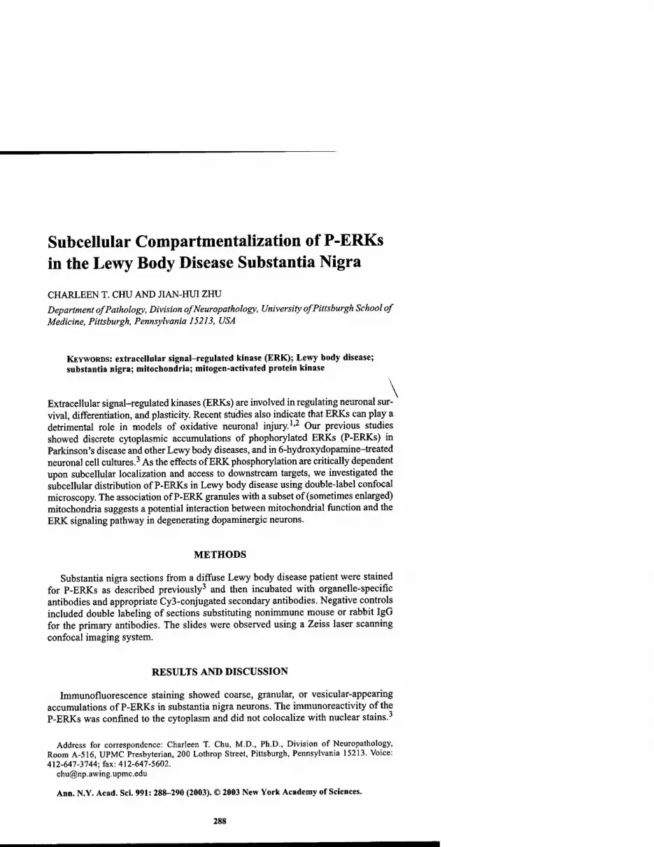
Subcellular Compartmentalization of P-ERKs in the Lewy Body Disease Substantia Nigra
CHARLEEN T. CHU AND JIAN-HUIZHU Department of Pathology, Division ofNeuropathology, University of Pittsburgh School of Medicine, Pittsburgh, Pennsylvania 15213, USA
KEYWORDS: extracellular signal-regulated kinase (ERK); Lewy body disease; substantia nigra; mitochondria; mitogen-activated protein kinase
Extracellular signal-regulated kinases (ERKs) are involved in regulating neuronal sur- vival, differentiation, and plasticity. Recent studies also indicate that ERKs can play a detrimental role in models of oxidative neuronal injury.'-^ Our previous studies showed discrete cytoplasmic accumulations of phophorylated ERKs (P-ERKs) in Parkinson's disease and other Lewy body diseases, and in 6-hydroxydopamine-treated neuronal cell cultures.^ As the effects of ERKphosphorylation are critically dependent upon subcellular localization and access to downstream targets, we investigated the subcellular distribution of P-ERKs in Lewy body disease using double-label confocal microscopy. The association of P-ERK granules with a subset of (sometimes enlarged) mitochondria suggests a potential interaction between mitochondrial function and the ERK signaling pathway in degenerating dopaminergic neurons.
METHODS
Substantia nigra sections from a diffuse Lewy body disease patient were stained for P-ERKs as described previously^ and then incubated with organelle-specific antibodies and appropriate Cy3-conjugated secondary antibodies. Negative controls included double labeling of sections substituting nonimmune mouse or rabbit IgG for the primary antibodies. The slides were observed using a Zeiss laser scanning confocal imaging system.
RESULTS AND DISCUSSION
Immunofluorescence staining showed coarse, granular, or vesicular-appearing accumulations of P-ERKs in substantia nigra neurons. The immunoreactivity of the P-ERKs was confined to the cytoplasm and did not colocalize with nuclear stains.-'
Address for correspondence: Charleen T. Chu, M.D., Ph.D., Division of Neuropathology, Room A-5I6, UPMC Presbyterian, 200 Lothrop Street, Pittsburgh, Pennsylvania 15213. Voice: 412-647-3744; fax: 412-647-5602.
Ann. N.Y. Acad. Sci. 991: 288-290 (2003). © 2003 New York Academy of Sciences.
288
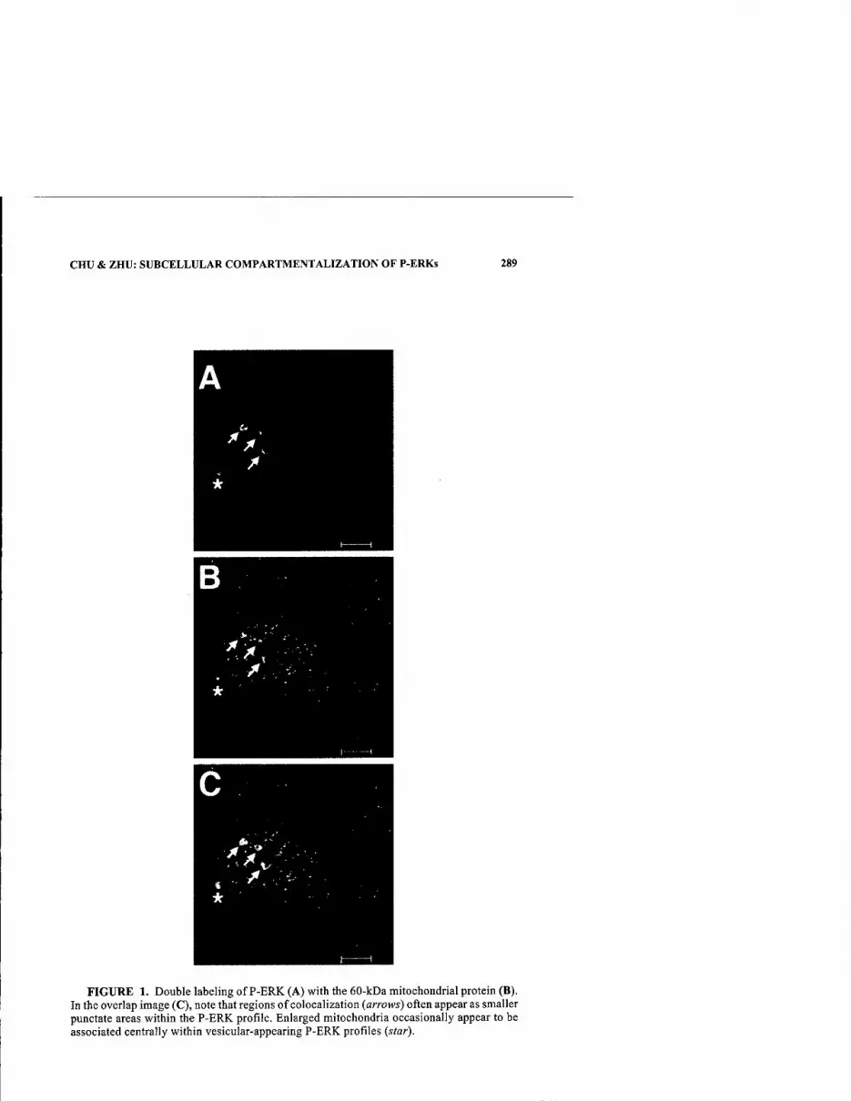
CHU & ZHU: SUBCELLULAR COMPARTMENTALIZATION OF P-ERKs 289
FIGURE 1. Double labeling of P-ERK (A) with the 60-kDa mitochondrial protein (B). In the overlap image (C), note that regions of colocalization (arrows) often appear as smaller punctate areas within the P-ERK profile. Enlarged mitochondria occasionally appear to be associated centrally within vesicular-appearing P-ERK profiles (star).
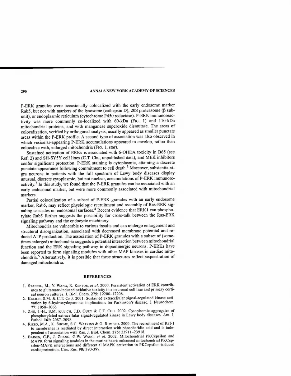
290 ANNALS NEW YORK ACADEMY OF SCIENCES
P-ERK granules were occasionally colocalized with the early endosome marker Rab5, but not with markers of the lysosome (cathepsin D), 20S proteasome (p sub- unit), or endoplasmic reticulum (cytochrome P450 reductase). P-ERK immunoreac- tivity was more commonly co-localized with 60-kDa (FIG. 1) and 110-kDa mitochondrial proteins, and with manganese superoxide dismutase. The areas of colocalization, verified by orthogonal analysis, usually appeared as smaller punctate areas within the P-ERK profile. A second type of association was also observed in which vesicular-appearing P-ERK accumulations appeared to envelop, rather than colocalize with, enlarged mitochondria (FIG. 1, star).
Sustained activation of ERKs is associated with 6-OHDA toxicity in B65 (see Ref 2) and SH-SY5Y cell lines (C.T. Chu, unpublished data), and MEK inhibitors confer significant protection. P-ERK staining is cytoplasmic, attaining a discrete punctate appearance following commitment to cell death.^ Moreover, substantia ni- gra neurons in patients with the full spectrum of Lewy body diseases display unusual, discrete cytoplasmic, but not nuclear, accumulations of P-ERK immunore- activity.^ In this study, we found that the P-ERK granules can be associated with an early endosomal marker, but were more commonly associated with mitochondrial markers.
Partial colocalization of a subset of P-ERK granules with an early endosome marker, Rab5, may reflect physiologic recruitment and assembly of Ras-ERK sig- naling cascades on endosomal surfaces.'' Recent evidence that ERKl can phospho- rylate Rab5 further suggests the possibility for cross-talk between the Ras-ERK signaling pathway and the endocytic machinery.
Mitochondria are vulnerable to various insults and can undergo enlargement and structural disorganization, associated with decreased membrane potential and re- duced ATP production. The association of P-ERK granules with a subset of (some- times enlarged) mitochondria suggests a potential interaction between mitochondrial function and the ERK signaling pathway in dopaminergic neurons. P-ERKs have been reported to form signaling modules with other MAP kinases in cardiac mito- chondria.^ Alternatively, it is possible that these structures reflect sequestration of damaged mitochondria.
REFERENCES
1. STANCIU, M., Y. WANG, R. KENTOR, et al. 2000. Persistent activation of ERK contrib- utes to glutamate-induced oxidative toxicity in a neuronal cell line and primary corti- cal neuron cultures. J. Biol. Chem. 275: 12200-12206.
2. KULICH, S.M. & C.T. CHU. 2001. Sustained extracellular signal-regulated kinase acti- vation by 6-hydroxydopamine: implications for Parkinson's disease. J. Neurochem. 77: 1058-1066.
3. ZHU, J.-H., S.M. KULICH, T.D. OURY & C.T. CHU. 2002. Cytoplasmic aggregates of phosphorylated extracellular signal-regulated kinase in Lewy body diseases. Am. J. Pathol. 161:2087-2098.
4. Rizzo, M.A., K. SHOME, S.C. WATKINS & G. ROMERO. 2000. The recruitment of Raf-1 to membranes is mediated by direct interaction with phosphatidic acid and is inde- pendent of association with Ras. J. Biol. Chem. 275: 23911-23918.
5. BAINES, C.P., J. ZHANG, G.W. WANG, et al. 2002. Mitochondrial PKCepsilon and MAPK form signaling modules in the murine heart: enhanced mitochondrial PKCep- silon-MAPK interactions and differential MAPK activation in PKCepsilon-induced cardioprotection. Circ. Res. 90: 390-397.

Altered Striatal Neuronal Morphology Is Associated with Astrogliosis in a Chronic Mouse Model of Parkinson's Disease
A. G. DERVAN," S. TOTTERDELL,* Y.-S. LAU,'^ AND G. E. MEREDITH" "Department of Cellular and Molecular Pharmacology, Chicago Medical School, North Chicago, Illinois 60064, USA ''Department of Pharmacology, University of Oxford, Oxford 0X1 3QT, United Kingdom
"Department of Pharmacology, University of Missouri-Kansas City, Kansas City, Missouri 64108, USA
KEYWORDS: astrocyte; dendrite; spine; MPTP; dopamine; gllosis; medium spiny neuron; glutamate
INTRODUCTION
Neostriatal medium spiny neurons (MSNs) lose their dopaminergic (DA) inner- vation in Parkinson's disease (PD). This loss is followed by a cascade of events that ultimately changes the structure of MSNs and the activity of basal ganglia circuits, resulting in the development of PD symptomatology. The specific mechanisms lead- ing to MSN morphological change are not well understood but may involve glutamate hyperactivity.' Astrocytes provide trophic support for neurons and their associated synapses. They also possess receptor systems for recycling glutamate.^ With central nervous system insults, astrocytes react, presumably to restore homeo- stasis. However, once reactive, their ability to carry out normal supportive functions is impaired.^ In the present study, we examined the association between resident as- trocytes and MSN morphology after DA loss in a chronic mouse model of PD.'*'^
METHODS
Adult C57/bl mice were treated with l-methyl-4-phenyl-1,2,3,6-tetrahydropyri- dine (MPTP) hydrochloride (25 mg/kg, sc in saline) and probenecid (250 mg/kg, ip in DMSO) or probenecid alone (controls) for 5 weeks (10 doses). All animals were assessed with an acceleration test on a rotarod; 3-5 weeks after treatment, the ani- mals were anesthetized (pentobarbital, 130 mg/kg) and perfused transcardially with
Address for correspondence: Dr. G.E. Meredith, Department of Cellular and Molecular Phar- macology, Chicago Medical School, Finch University of Health Sciences, 3333 Green Bay Road, North Chicago, Illinois 60064. Voice: 847-578-8680; fax: 847-578-3268.
Ann. N.Y. Acad. Sci. 991: 291-294 (2003). © 2003 New York Academy of Sciences.
291

292 ANNALS NEW YORK ACADEMY OF SCIENCES
B
E F
^ **', i/*
FIGURE 1. See following page for legend.
DERVAN el al.: STRIATAL NEURONAL MORPHOLOGY AND ASTROGLIOSIS 293
fixative; 300-|im slices through the striatum were cut alternately with 50-|im sec- tions. In slices, neurons counterstained with DAPI (0.001%) were iontophoretically injected with biotinylated Lucifer yellow. Slices were then resectioned (40 lira) and reacted with nickel-enhanced DAB. Adjacent sections were immunoreacted for ty- rosine hydroxylase (TH) or glial fibrillary acidic protein (GFAP). Reconstructed in- tracellularly filled neurons were morphometrically analyzed, and the density of GFAP-immunopositive (+) cells and fibers was studied. Control and treated groups were compared with standard statistics.
RESULTS AND DISCUSSION
When compared to controls, MPTP/probenecid (MPTP/p)-treated mice were sig- nificantly impaired on the rotarod {P < 0.01), and showed reduced striatal TH inner- vation and few TH+ neurons in the substantia nigra (FIGS. 1A,B). MSNS
(FIGS. 1C,D) did not differ between groups in dendritic length, surface area, or spine density. However, distal dendrites were significantly more tortuous (P < 0.05), had fewer spines (P < 0.05), and tended (P = 0.1) to be longer in MPTP/p mice than in controls. As for astrocytes, GFAP-l- cells typically had short, fine processes in con- trols (FIG. IE), but numerous long, thick extensions (FIG. IF), typical of reactive as- trocytes, after MPTP/p treatment. GFAP+ cell and fiber densities were significantly increased (P < 0.01) by 40% and 36%>, respectively, compared to controls.
Morphological change seen in experimental models of PD appears to depend upon the severity of DA depletion and, as data here suggest, changes in resident as- trocytes. Complete 6-OHDA lesions produce atrophy and major spine loss.^-'' How- ever, more partial lesions remodel distal dendrites with little spine loss (present results and Meredith et al?). Astrocytes, which are vital for brain homeostasis be- cause they clear glutamate and influence calcium dynamics,^ proliferate after injury and release growth factors. Such factors may help maintain the integrity of dendrites, especially small distal processes vulnerable to calcium overload.^ Nevertheless, stri- atal glutamate levels rise following DA loss,^'^ and astrocytes, once reactive, essen- tially lose their ability to recycle it.^ They also activate microglia that produce pro- inflammatory cytokines.^'^ In PD, astrogliosis is rarely observed in the striatum, yet postmortem studies show that striatal neurons are severely altered structurally.'^ This may mean that inflammation occurs early in the disease process, a hypothesis that is strongly supported by the present results.
FIGURE 1. TH immunoreactivity in the midbrain of (A) probenecid control and (B) 4 weeks post-MPTP/probenecid-treated mouse. (C) Intracellularly injected MSN of an MPTP/probenecid-treated mouse and (D) its digitized reconstruction. Note the sparse spines on dendrites {arrows). Typical astrocytes in (E) control and (F) MPTP/probenecid-treated striata. Note numerous fine processes {arrows) in (F). Scale bars: A,B = 200 |im, C = 20 |tm, E,F= 10|lm.

294 ANNALS NEW YORK ACADEMY OF SCIENCES
ACKNOWLEDGMENTS
This study was funded by USPHS Grant ROl NS41799 from NIH/NINDS and a Wellcome Trust Biomedical Collaboration Grant.
REFERENCES
1. INGHAM, C.A. et al. 1998. Plasticity of synapses in the rat neostriatum after unilateral lesion of the nigrostriatal dopaminergic pathway. J. Neurosci. 18: 4732^743.
2. AMUNDSON, R.H. et al. 1992. Uptake of [%]serotonin and [^H]glutamate by primary astrocyte cultures. II. Differences in cultures prepared from different brain regions. GIia6:9-18.
3. RiDET, J.L. et al. 1997. Reactive astrocytes: cellular and molecular cues to biological function. Trends Neurosci. 20: 570-577.
4. PETROSKE E. et al. 2001. Mouse model of parkinsonism: a comparison between sub- acute MPTP and chronic MPTP/p treatment. Neuroscience 106: 589-601.
5. MEREDITH, G.E. et al. 2002. Lysosomal malfunction accompanies alpha-synuclein aggregation in a progressive mouse model of Parkinson's disease. Brain Res. 956: 156-165.
6. INGHAM, C.A. et al. 1989. Spine density on neostriatal neurons changes with 6- hydroxydopamine lesions and with age. Brain Res. 503: 334-338.
7. MEREDITH G.E. et a/.1995. The effects of dopamine depletion on the morphology of medium spiny neurons in the shell and core of the rat nucleus accumbens. J. Neuro- sci. 15: 3808-3820.
8. EDDELSTON, M. & L. MucKE. 1993. Molecular profile of reactive astrocytes—implica- tions for their role in neurologic disease. Neuroscience 54: 15-36.
9. ROBINSON, S. et al. 2001. Blockade of NMDA receptors by MK-801 reverses the changes in striatal glutamate immunolabeling in 6-OHDA-lesioned rats. Synapse 42: 54-61.
10. MCNEILL, T.H. et al. 1988. Atrophy of medium spiny I striatal dendrites in advanced Parkinson's disease. Brain Res. 455: 148-152.

Is the Initial Insult in Parkinson's Disease and Dementia with Lewy Bodies a Neuritic Dystrophy?
JOHN E. DUDA,"'* BENOIT I. GIASSON," VIRGINIA M-Y. LEE," AND JOHN Q. TROJANOWSKI" "Center for Neurodegenerative Disease Research, University of Pennsylvania, Philadelphia, Pennsylvania 19104, USA ^Parkinson's Disease Research, Education and Clinical Center, Philadelphia VA Medical Center, Philadelphia, Pennsylvania 19104, USA
KEYWORDS: Parkinson's disease; dementia with Lewy bodies; neuritic dystro- pliy; alpha-synuclein
The role that intracytoplasmic a-synuclein (a-syn) inclusions including Lewy bod- ies (LBs) and Lewy neurites (LNs) play in the neurodegenerative process in Parkin- son's disease (PD) and dementia with Lewy bodies (DLB) remains controversial. Whether they are a primary insult that begins a cascade towards eventual neuronal death or a secondary adaptive response is still debated. Recently, we have published three lines of evidence suggesting that LNs, and the interruption of axonal transport that they may engender, is an integral component in this neurodegenerative process. First, with novel antibodies to a-syn, we have demonstrated a burden of neuritic pa- thology in PD, DLB, and concomitant DLB and AD (AD/DLB) that is far greater than previously recognized.' In this report, we documented a large, previously un- recognized burden of neuritic pathology in the striatum of the Lewy body disorders. Moreover, with immunofluorescent double labeling we were able to confirm that at least some of the neuritic aggregates were located within axons of nigrostriatal path- way neurons. In addition, we have also examined the burden of this pathology in the cerebral cortex of the Lewy body disorders. When we performed a similar semi- quantitative analysis of neuritic burden in the cingulate cortex from patients repre- senting the complete range of synucleinopathies as well as several control groups, we observed a similar density pattern to what we observed in the striatum. Specifi- cally, the highest density was seen in cases of AD/DLB, followed by DLB, and then PD; little or no pathology was seen in multiple system atrophy, AD, progressive su-
Address for correspondence: John Q. Trojanowski, Center for Neurodegenerative Disease Research, University of Pennsylvania, 3600 Spruce St., 3 Maloney, Philadelphia, PA 19104. Voice: 215-662-6399; fax: 215-349-5909.
Ann. N.Y. Acad. Sci. 991: 295-297 (2003). © 2003 New Yoric Academy of Sciences.
295

296 ANNALS NEW YORK ACADEMY OF SCIENCES
3 - 3 •
17^-^
2 9^-^^ 1
•
1 15^-^ 4
•
n( \^^ 2 •
VI c 01 o >t
1 m
0 12 3 Neurite Density
FIGURE 1. Independent semiquantitative analysis of Lewy neurite (LN) density and Lewy body (LB) density was performed on the anterior cingulate gyrus from 53 patients with DLB/AD, DLB, or PD. Sections were immunostained with an anti-synuclein mono- clonal antibody, Syn 514.' The following assessment strategy was used to estimate the den- sity of a-syn inclusions: 3 = frequent; 2 = moderate; 1= sparse; and 0 = few to none. When the density of LN (x axis) in the cingulate cortex is correlated with LB density (y axis) in the same cortex, a correlation of/?^ = 0.906, P < 0.0001 is achieved. Numbers represent the numbers of cases at each data point.
pranuclear palsy, corticobasal degeneration, idiopathic striatonigral degeneration, and normal controls. In addition, we performed a semiquantitative analysis of the abundance of neuritic pathology and an independent assessment of LB density in the cingulate cortex of 53 cases of AD/DLB, DLB, and PD and found a strong correla- tion (R^ - 0.906, P < 0.0001) (FIG. 1) between the two. Taken together with many recent studies correlating cortical LB density and the dementia of LB disorders,^^ it is reasonable to hypothesize a similar correlation between cortical neurite density and dementia.
The second line of evidence supporting the role of neuritic dystrophy in LB dis- orders is the observation that neuritic aggregates are the predominant pathology in a case of familial PD from the Contursi kindred with a pathogenic A53T mutation in the a-syn gene.^ We reexamined the initial autopsy from the kindred^ using a-syn and tau immunochemistry and found an overwhelming burden of neuritic pathology and virtual absence of LBs in this patient. This patient is particularly interesting in that he died an accidental death prior to the end stage of the disease and therefore gives us a pathological "snapshot" earlier in the disease process. When compared to the pathology at the end stage of this kindred in other cases (unpublished results and personal communication with Dr. D.W. Dickson) that have more typical PD pathol- ogy, it is tempting to postulate widespread neuritic pathology as an early pathologi- cal aberration.
Finally, we have developed an A53T transgenic mouse model that displays an age-dependent neurodegenerative process that includes development of primarily neuritic a-syn aggregates and a concomitant motor phenotype with rapid progres- sion to paralysis and death.^ The close temporal association between the develop- ment of a-syn aggregates and the onset of the motor phenotype in these mice support a pathogenic role for these inclusions and allow us to examine the consequences of
DUDA ei al.: NEURITIC DYSTROPHY IN LEWY BODY DISORDERS 297
neuritic inclusions. With a-syn immunoelectron microscopy of the ventral roots of affected mice, we are able to confirm the deposition of fibrillar a-syn aggregates within the axonal compartment; the intraaxonal accumulation of vesicles, mitochon- dria, vacuoles, and neurofilaments, suggesting an interruption of axonal transport; and axonal degeneration. Therefore, it is possible to hypothesize that intraaxonal ag- gregates are an early component of the disease process and lead to an interruption of axonal transport with resultant axonal degeneration that is the ultimate pathological cause of the motor phenotype observed in these animals.
Collectively, these observations engender a "neuritic dystrophy hypothesis" of pathophysiology for the Lewy body disorders wherein various environmental, genet- ic, or intrinsic factors cause a conformational change in the structure of a-syn to P- pleated sheet and a propensity to aggregate into pathological inclusions. These in- clusions can occur in the cell soma as LBs or in the axonal compartment and grow in size by accumulating additional a-syn and trapping other proteins and/or or- ganelles. In the axonal compartment, these inclusions expand until they congest the axonal compartment, resulting in an interruption of axonal transport. This cessation of axonal transport disallows distal axonal viability and eventually leads to axonal degeneration with the possibility of resultant neuronal degeneration. Further exam- ination of the human diseases as well as many of our current models of the Lewy body disorders may improve our understanding of the role of neuritic dystrophy and eventually lead to novel therapeutic interventions to prevent disease progression in these disorders.
REFERENCES
1. DuDA, J.E., B.I. GiASSON, M.E. MABON, et al. 2002. Novel antibodies to synuclein show abundant striatal pathology in Lewy body diseases. Ann. Neurol. 52: 205-210.
2. HARDING, A.J. & G.M. HALLIDAY. 200 L Cortical Lewy body pathology in the diagno- sis of dementia. Acta Neuropathol. 102: 355-363.
3. HuRTiG, H.L, J.Q. TROJANOWSKI, J. GALVIN, et al. 2000. Alpha-synuclein cortical Lewy bodies correlate with dementia in Parkinson's disease. Neurology 54: 1916- 1921.
4. MATTILA, P.M., J.O. RINNE, H. HELENIUS, D.W. DICKSON & M. ROYTTA. 2000. Alpha- synuclein-immunoreactive cortical Lewy bodies are associated with cognitive impairment in Parkinson's disease. Acta Neuropathol. 100: 285-290.
5. DUDA, J.E., B.L GiASSON, M.E. MABON, D.C. MILLER, et al. 2002. Concurrence of alpha-synuclein and tau brain pathology in the Contursi kindred. Acta Neuropathol. 104:7-11.
6. GOLBE, L.I., G. Di lORiO, V. BoNAViTA, et al. 1990. A large kindred with autosomal dominant Parkinson's disease. Ann. Neurol. 27: 276-282.
7. GiASSON, B.I., J.E. DUDA, S.M. QUINN, et al. 2002. Neuronal alpha-synucleinopathy with severe movement disorder in mice expressing A53T human alpha-synuclein. Neuron 34: 521-533.

Neuroplasticity in the MPTP-Lesioned Mouse and Nonhuman Primate
MICHAEL W. JAKOWEC," BETH FISHER," KERRY NIXON," ELIZABETH HOGG," CHARLES MESHUL,* SAMUEL BREMMER,* TOM McNEILL," AND GISELLE M. PETZINGER"
"Department of Neurology, University of Southern California, Los Angeles, California 90033, USA
''Department of Behavioral Neuroscience, VA Medical Center/Oregon Health and Science University, Portland, Oregon 97201, USA
KEYWORDS: neuroplasticity; MPTP-lesioned mouse; nigrostriatal dopaminer- gic neurons
INTRODUCTION
Systemic administration of the neurotoxicant MPTP (I-methyl-4-phenyl-I,2,3,6- tetrahydropyridine) to C57 BL/6J mice and squirrel monkey (Saimiri sciureus) leads to the destruction of nigrostriatal dopaminergic neurons, the depletion of striatal dopamine, and the onset of parkinsonian features including bradykinesia. Interest- ingly, both mice and nonhuman primates demonstrate neurochemical and behavioral recovery from this neurotoxic-induced injury to the basal ganglia. The molecular mechanisms underlying neuroplasticity in these models are not fully understood. The focus of our studies is to elucidate the molecular, morphological, and neuro- chemical basis for recovery including interventions, such as exercise, that may en- hance recovery.
METHODS
Male 8-week-old mice were administered MPTP in a series of four injections (20 mg/kg, ip, free-base, every 2 h). Brain tissue was collected at postlesion days 7, 14, 30, 60, and 90. Another group of mice were subjected to treadmill exercise starting 4 days after MPTP lesioning, recording velocity and duration (maximum 1 h). Brain tissue was collected after 30 days of treadmill training. Squirrel monkeys were ad- ministered MPTP in a series of six injections (2 mg/kg, sc, free-base, 2 weeks be- tween each injection). Motor behavior was assessed using a modified Parkinson's
Address for correspondence: Michael W. Jakowec, Ph.D., Department of Neurology, MCH- 142, 1333 San Pablo Street, University of Southern California, Los Angeles, California 90033. Voice: 323-442-1057; fax: 323-442-1055.
Ann. N.Y. Acad. Sci. 991: 298-301 (2003). © 2003 New Yorlc Academy of Sciences.
298
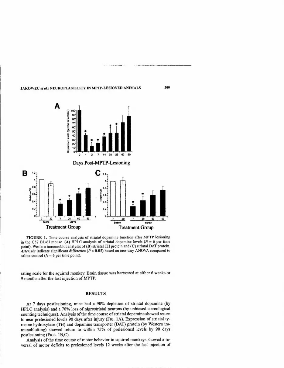
JAKOWEC et al.: NEUROPLASTICITY IN MPTP-LESIONED ANIMALS 299
7 14 21 28
Days Post-MPTP-Lesioning
c
Salne MPTP
Treatment Group Treatment Group
FIGURE 1. Time course analysis of striatal dopamine function after MPTP lesioning in the C57 BL/6J mouse. (A) HPLC analysis of striatal dopamine levels (N = 6 per time point). Western immunoblot analysis of (B) striatal TH protein and (C) striatal DAT protein. Asterisks indicate significant difference (P < 0.05) based on one-way ANOVA compared to saline control (N=6 per time point).
rating scale for the squirrel monkey. Brain tissue was harvested at either 6 weeks or 9 months after the last injection of MPTP.
RESULTS
At 7 days postlesioning, mice had a 90% depletion of striatal dopamine (by HPLC analysis) and a 70% loss of nigrostriatal neurons (by unbiased stereological counting techniques). Analysis of the time course of striatal dopamine showed return to near prelesioned levels 90 days after injury (FIG. lA). Expression of striatal ty- rosine hydroxylase (TH) and dopamine transporter (DAT) protein (by Western im- munoblotting) showed return to within 15% of prelesioned levels by 90 days postlesioning (FIGS. 1B,C).
Analysis of the time course of motor behavior in squirrel monkeys showed a re- versal of motor deficits to prelesioned levels 12 weeks after the last injection of

300 ANNALS NEW YORK ACADEMY OF SCIENCES
(A)
is-
le-
14-
"3 12-
00 10- c '■§ 8- Pi H 6- o
I 4- O
2-
0-
\ -•-Normal
-«- Moderate
\ -^k-*-
--•-„■' •••■•<,^**«- •■ ■».- >-» ♦-<
I I I I I I I I I I I I I >l I I I I I I I I I I I I -rNif^-fl-i^t©r-TOceo^ryro^t/va>r*05C603r-c\i<r[3-iA<Dr'cpg>Oj-o)C2
,-^^T-T-r-T-^T-T-OJ(MfMfMOjrv)<M(N<SjrvjrOO)rOCO
Weeks
(B) MPTP
Saline 6 Weeks
CN
i^
9 Months
20X Mag
CN
M'JJ^rs
FIGURE 2. The time course of motor behavior in the MPTP-Iesioned squirrel monkey. (A) Analysis using a clinical rating scale indicates the recovery of motor deficit 12 weeks after the last injection of MPTP (A^ = 4 animals per group). (B) Immunocytochemical anal- ysis of TH protein in the caudate nucleus (CN) indicates severe depletion at 6 weeks after the last injection of MPTP but only partial return 9 months after the last injection of MPTP. The upper three panels in B are derived from the dorsal caudate nucleus, while the lower three panels are derived from theuppermost dorsal-lateral quadrant of the caudate nucleus/ white matter junction. The scale bar in upper right panel represents 1 mm.

JAKOWEC et al.: NEUROPLASTICITY IN MPTP-LESIONED ANIMALS 301
MPTP (FIG. 2). The expression of TH and DAT proteins in the caudate and putamen showed a 90% depletion at 6 weeks but returned to only 35% of prelesioned levels at 9 months. Despite the partial recovery of TH, monkeys demonstrated normal mo- tor behavior. Unbiased stereological analysis of SNpc neurons based on TH immu- noreactivity with Nissl substance staining in both parkinsonian (harvested at 6 weeks postlesioning) and recovered (harvested at 9 months postlesioning) monkeys is being used to determine if there is a significantly different number of midbrain dopaminergic neurons between these groups (Petzinger et al., in preparation).
In the treadmill exercise paradigm, MPTP-lesioned mice showed enhanced be- havioral recovery. After 30 days of exercise, both velocity and endurance reached levels similar to the normal exercise animals. Molecular analysis of the exercised MPTP-lesioned animals showed suppression of striatal DAT protein compared to the nonexercised MPTP-lesioned control group. Immunoelectron microscopy analysis showed that MPTP alone increased the density of nerve terminal glutamate and that exercise reversed this increase (Fisher et al., in preparation).
DISCUSSION
Resuks from these studies show that both TH and DAT protein return participate in behavioral recovery after MPTP lesioning. The partial return of TH and DAT pro- tein in the squirrel monkey in conjunction with the complete behavioral recovery suggests that nondopaminergic systems may be involved in neuroplasticity after in- jury to the basal ganglia. One important system may involve glutamate.
The treadmill exercise paradigm shows that exercise alters both the glutamatergic and dopaminergic systems as part of neuroplasticity in the injured basal ganglia. These alterations that accompany behavioral recovery can take place at either the molecular level (DAT protein expression) or morphological level (changes in glutamate density of nerve terminal glutamate immunolabeling). Ongoing studies indicate that expression of TH and dopamine receptors (Dl and D2 subtypes) are also altered in our exercise paradigm (Fisher et al., in preparation).
The elucidation of the molecular mechanisms underlying neuroplasticity in the MPTP-lesioned mouse and nonhuman primate will identify novel therapeutic targets and will provide insight into the application of current strategies including transplantation.
ACKNOWLEDGMENTS
These studies are supported by the National Institutes of Health, the U.S. Army, the Parkinson's Disease Foundation, the Lisette and Norman Ackerberg Foundation, the Baxter Foundation, the Zumberge Foundation, and the VA Merit Review Pro- gram (to CM.).

An Experimental Infection by Filterable Forms of Nocardia asteroides and Late-Onset Movement Disorder in Mice
S. KOHBATA" AND C. KADOYA*
"Department of Microbiology, Gifu University School of Medicine, Gifu City, Gifu 500-8076, Japan
^Department ofNeurosurgery, University of Occupational and Environmental Health, Kitakyushu, Fukuoka 807-8555, Japan
KEYWORDS: Nocardia asteroides; filterable nocardiae; Parkinson's disease
Filterable nocardiae, morphologically identical to lipochrome bodies in Parkinson's disease,' are not gram-positive bacteria isolated from broth cultures of Nocardia as- teroides by filtration.2'^ They fluoresced brilliantly green-yellow under ultraviolet light when stained with acridine orange and were strongly periodic acid-Schiff stain (PAS) positive on day 1 of incubation in broth cultures. They became acid fast after day 2 of incubation. The 5-h cultures were used as inoculum for the experimental infection. The 14-h cultures were used as antigen for the preparation of anti-filter- able nocardiae antiserum according to the modified method.''
After the mice were innoculated via the tail vein, they appeared to be healthy and to move as quickly as control mice. By early in month 5, the mice were moving slow- ly. When the tail was picked up, they failed to respond abruptly, and when they did move, they moved slowly. They appeared to have difficulty with a particular motion, the twisting of the body trunk, at month 5. At month 6, they hung limply when sus- pended. Soon after, they bent their backs and held their forepaws, assuming a pecu- liar posture, resembling sleeping bats, which they maintained.
Their brains were collected, fixed, and embedded in paraffin. Serial coronal sec- tions were prepared. The sections were stained forNissl substance with cresyl violet for dopamine neurons with anti-tyrosine hydroxylase antibody,^ or for filterable no- cardiae with carbolfuchsin, PAS, and anti-filterable nocardiae antiserum. The mid- brain section revealed that the pars compacta dopamine neurons were severely damaged at month 5 and lost bilaterally at month 6. Filterable nocardiae were dem- onstrated as acid-fast particles within many macroglia in nearby sinusoidal vessels
Address for correspondence: S. Kohbata, Department of Microbiology, Gifu University School of Medicine, Tsukasa-machi 40, Gifu City, Gifu 500-8076, Japan. Voice: 81-582-65- 1241; fax: 81-562-67-0156.
Ann. N.Y. Acad. Sci. 991: 302-303 (2003). © 2003 New York Academy of Sciences.
302

KOHBATA & KADOYA: EXPERIMENTAL INFECTION BY Nocardia asteroides 303
of the midbrain. These were apparent early in month 5 and continued to be evident during the month. At month 6, they were seen as immunoreactive deposits within many macroglia distributed through the substantia nigra. The acid-fast filterable no- cardiae, observed early in month 5, are reminiscent of acid-fast lipochrome bodies in the early phases of Parkinson's disease.' The immunological detection of filter- able nocardiae in Parkinson's disease is now under study.^ Filterable nocardiae were also detected as immunopositive deposits within many macroglia distributed through the substantia nigra at week 5, but they were not detected later.
Filterable nocardiae, which may be able to invade the midbrain substantia nigra, were probably eliminated by host immune responses. Reinvasion of the midbrain by filterable nocardiae, early in month 5 through month 6, indicates their extraneural focus of infection rather than their neural persistence in host tissues.
The results suggest that an infection of the midbrain with filterable nocardiae may occur and cause the severe damage of the pars compacta dopamine neurons related to the movement disorder.
REFERENCES
KoHBATA. S., etal. 1998. Accumulation of acid-fast lipochrome bodies in glial cells of the midbrain nigral lesion in Parkinson's disease. Clin. Diag. Lab. Immunol. 5: 888- 893.
KoHBATA, S. 1996. Acid-fastness of Nocardia asteroides GUH-2 cultured in brain heart infusion broth supplemented with paraffin. Microbiol. Immunol. 40: 711-716.
KoHBATA, S. 1998. Tinctorial properties of spherical bodies in broth cultures of Nocar- dia asteroides GUH-2. Microbiol. Immunol. 42: 151-157.
KoHBATA, S., et al. 1986. Cytopathogenic effect of Salmonella typhi GIFU 10007 on M cells of murine ileal Peyer's patches in ligated ileal loops: An ultra-structural study. Microbiol. Immunol. 30: 1225-1237.
KoHBATA, S. & B.L. BEAMAN. 1991. L-Dopa-responsive movement disorder caused by Nocardia asteroides localized in the brains of mice. Infect. Immunity 59: 181-191.
KOHBATA, S., et al. 2002. An infection with filterable forms of Nocardia asteroides in the midbrain nigral lesion in Parkinson's disease. Paper presented at the Seventh International Congress of Parkinson's Disease and Movement Disorders, Miami, FL, November 11,2002.

A Characterization of Dopaminergic Neurodegeneration in Organotypic Cultures
GERALDINE J. KRESS AND IAN J. REYNOLDS
Department of Pharmacology, University of Pittsburgh, Pittsburgh, Pennsylvania 15261, USA
KEYWORDS: Parkinson's disease; 6-hydroxydopaniine; substantia nigra; cell death
Parkinson's disease is a neurodegenerative disorder involving the progressive dopaminergic degeneration of the nigrostriatal pathway. A major obstacle in the pre- vention and treatment of this disease is determining the cause of degeneration. Many studies have linked oxidative stresses, such as those arising from dysfunctional mitochondria, as playing a role.' Additionally, compounds such as 6-hydroxy- dopamine (6-OHDA) also cause oxidative stresses by accumulating in dopaminergic neurons, oxidizing, and producing free radicals and hydrogen peroxide. Interesting- ly, 6-OHDA selectively harms dopamine cells of the substantia nigra but not those of the ventral tegmental area.
Several studies of dopaminergic neurotoxins have been performed in whole animal models and in dissociated cell culture, but none in organotypic cultures. Therefore, we propose to study dopaminergic cell death in cortex-striatum-substan- tia nigra organotypic cultures prepared from PI-3 rats. Organotypic cultures provide a useful intermediate between dissociated cell cultures and in vivo models because (1) they possess mature dopaminergic neurons and supporting glial cells; (2) they form synaptic connections; (3) they are relatively long-lived; and (4) they retain the simplicity of in vitro systems. In our organotypic culture, adapted from Plenz and Kitai,^ the slices were grown for 18-23 days before experimentation.
Prior to 6-OHDA application, slices were stained with 5,7-dihydroxytryptamine (DHT). DHT is a fluorophore selectively taken up by dopaminergic cells via the dopamine transporter without displaying any visible toxic effects. DHT was used to locate and quantify the number of dopaminergic cells in live slices. Moreover, prior to 6-OHDA application, the viability of the slice was examined with propidium iodide (PI), a fluorophore that stains dead cells. Slices were excluded from the study if the initial amount of PI staining was high. After PI and DHT staining, 6-OHDA was applied, then washed extensively, and returned to the incubator. After 6-OHDA
Address for correspondence: Ian J. Reynolds, Ph.D., Department of Pharmacology, School of Medicine, University of Pittsburgh, Pittsburgh, PA 15261. Voice: 412-648-2134; fax: 412-624- 0794.
Ann. N.Y. Acad. Sci. 991: 304-306 (2003). © 2003 New York Academy of Sciences.
304
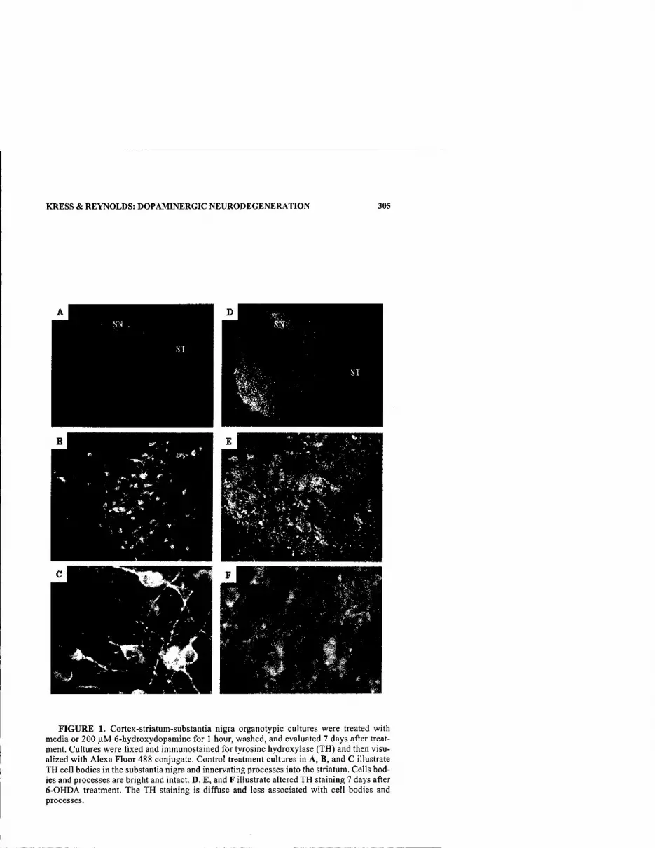
KRESS & REYNOLDS: DOPAMINERGIC NEURODEGENERATION 305
*v
■^li.;
FIGURE 1. Cortex-striatum-substantia nigra organotypic cultures were treated with media or 200 jlM 6-hydroxydopamine for 1 hour, washed, and evaluated 7 days after treat- inent. Cultures were fixed and immunostained for tyrosine hydroxylase (TH) and then visu- alized with Alexa Fluor 488 conjugate. Control treatment cultures in A, B, and C illustrate TH cell bodies in the substantia nigra and innervating processes into the striatum. Cells bod- ies and processes are bright and intact. D, E, and F illustrate altered TH staining 7 days after 6-OHDA treatment. The TH staining is diffuse and less associated with cell bodies and processes.

306 ANNALS NEW YORK ACADEMY OF SCIENCES
treatment, the slices were examined with PI staining to assess nonspecific death and then fixed for immunohistochemical determinations.
Control slices illustrated in FIGURE 1A, B, and C show bright, intact tyrosine hydroxylase (TH)-positive cell bodies in the substantia nigra with processes inner- vating the striatum. The PI staining of control slices is nearly undetectable (data not shown). With high 6-OHDA concentrations (5 mM) for short exposure times (15 minutes), gross injury was evident by the detachment of the slice from the substrate. The determined amount of 6-OHDA applied to the slice to produce low nonspecific death was 200 |J,M for 1 hour; the slice was then visualized 7 days post stimulus with PI staining (data not shown). FIGURE ID, E, and F show a type of TH staining very different from that of the control. There is an overall loss of TH staining in the substantia nigra and striatum, which is diffuse and less associated with cell bodies and processes. This altered pattern of staining may be indicative of a selective injury process. Also, this altered pattern of TH staining after 6-OHDA is not what we expected on the basis of studies in vivo and in dissociated cell cultures where TH staining is lost using this kind of injury paradigm. Moreover, it is difficult to do an objective quantitative assessment of this kind of rearrangement of staining using simple measures of fluorescence intensity.
In conclusion, we have demonstrated that a change occurs in TH staining with low nonspecific cell death that may reflect selective dopaminergic injury after 6- OHDA. However, additional studies, perhaps with other markers of cell injury, will be necessary to establish whether alterations in TH staining following 6-OHDA actually reflect cell injury.
REFERENCES
1. BETARBET, R., et al. 2002. Animal models of Parkinson's disease. Bioessays 24: 308- 318.
2. PLENZ, D. & S.T. KiTAi. 1996. Organotypic cortex-striatum-mesencephalon cultures: nigrostriatal pathway. Neurosci. Lett. 209: 177-180.

Developmental Cell Death and Oxidative Stress
Lessons from Nigral Dopamine Neurons
LAURENT GROC,"-''TANGELLA JACKSON HUNTER,''HAD JIANG,«* LAURENT BEZIN,''AND ROBERT A. LEVINE""^
"William T. Gossett Neurology Laboratories, Henry Ford Health System. Detroit, Michigan 48202, USA
''John D. Dingell Veterans Administration Medical Center, Detroit, Michigan 48201, USA
'^Department of Pharmaceutical Sciences, Wayne State University, Detroit, Michigan 48201, USA
^CNRS-UMR5578, Lyon, France
KEYWORDS: developmental cell death; dopamine; oxidative stress; substantia nigra
Neuronal death during development appears to be a key event for the maturation of the nervous system. The mechanisms underlying developmental death are poorly understood, although several in vitro studies have suggested the involvement of oxidative stress.' As shown in FIGURE 1, oxidative stress in neurons (e.g., dopamin- ergic) can be mediated by reactive oxygen species (ROS) that induce cellular dam- age such as lipid peroxidation (lipid byproducts derived from the breakdown of polyunsaturated fatty acids). Oxidative stress may also involve nitric oxide metabolism. ROS can be scavenged by the antioxidant enzymes superoxide dismu- tase, catalase, and glutathione peroxidase (GPx),^ which are usually activated during periods of oxidative stress. High oxidative stress exceeds the capacity of antioxidant enzymes and leads to ROS-induced cellular damage, particularly lipid peroxidation. On the contrary, when the activity of these enzymes is upregulated sufficiently, the amount of lipid peroxidation is kept at a low level (FIG. 1).
Developmental apoptotic death of dopamine neurons in the rat substantia nigra occurs mainly around birth, with a major peak at postnatal day (P) 3.^ Understanding intrinsic mechanisms causing developmental apoptotic death that is not induced by external insults (e.g., neurotoxins) may shed light on the causes of apoptotic death of nigral dopamine neurons in Parkinson's disease, because the same mechanisms may mediate the premature loss of nigral dopamine neurons in Parkinson's disease.
Address for correspondence: Robert A. Levine, Ph.D., W.T. Gossett Neurology Laboratories, Henry Ford Health System, 1 Ford Place, Detroit, MI 48202. Voice: 313-874-3771; fax: 208- 545-3455.
[email protected] or [email protected]
Ann. N.Y. Acad. Sci. 991: 307-310 (2003). © 2003 New York Academy of Sciences.
307
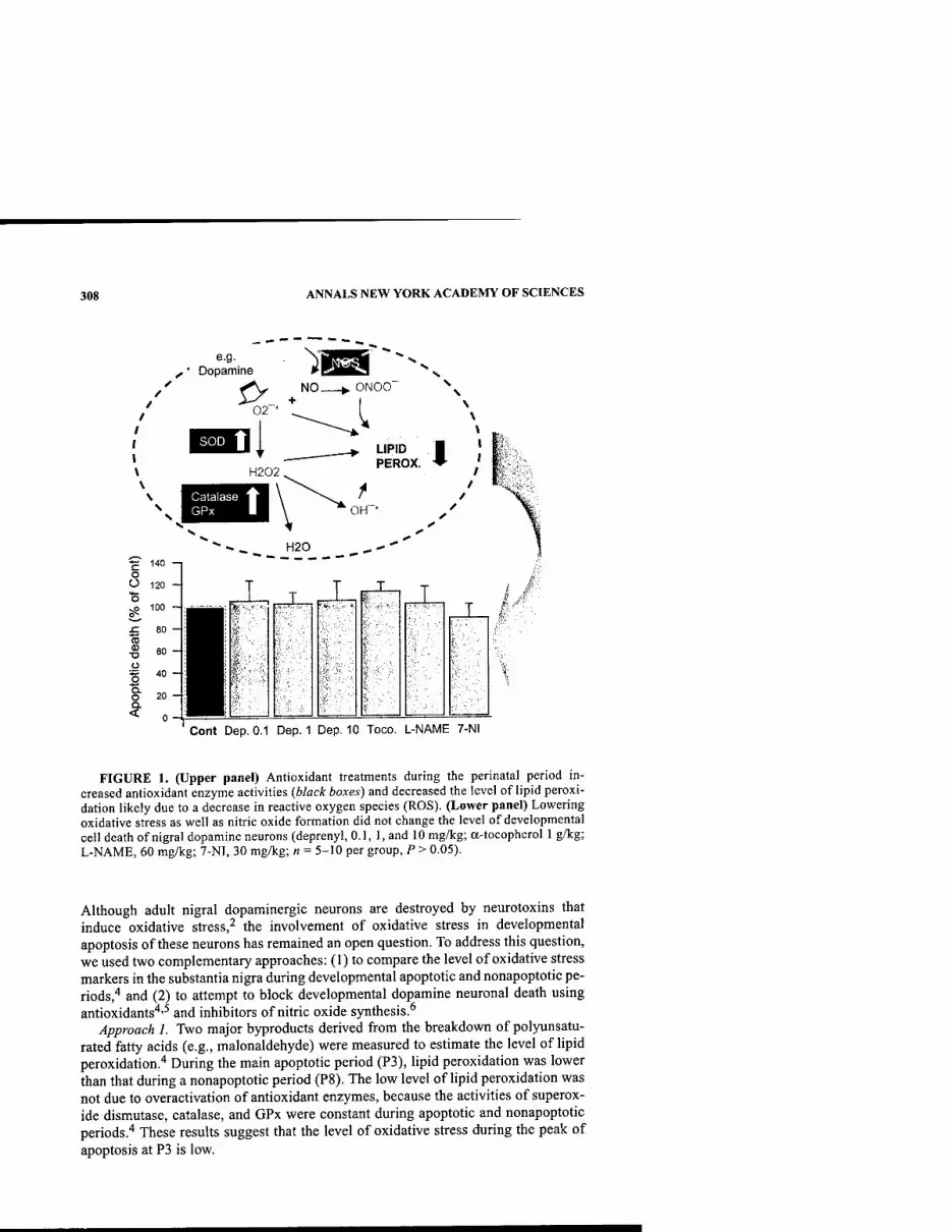
308 ANNALS NEW YORK ACADEMY OF SCIENCES
^1 e.g. ^ • Dopamine
02"
ONOO
LIPID PEROX.
H20 ■£ 140 —1 O O 120 - T
T T
■B ^ go 100 - ■ »»I - ' r ^M'
x: 80 - (0
■g 60- :■ i o i|t
1 '°- ■ f O 20 - :^^H I - D. < n- :H^H
Cont Dep. 0.1 Dep. 1 Dep. 10 Toco. L-NAME 7-NI
FIGURE 1. (Upper panel) Antioxidant treatments during the perinatal period in- creased antioxidant enzyme activities (black boxes) and decreased the level of lipid peroxi- dation likely due to a decrease in reactive oxygen species (ROS). (Lower panel) Lowering oxidative stress as well as nitric oxide formation did not change the level of developmental cell death of nigral dopamine neurons (deprenyl, 0.1, 1, and 10 mg/kg; a-tocopherol 1 g/kg; L-NAME, 60 mg/kg; 7-NI, 30 mg/kg; « = 5-10 per group, P > 0.05).
Although adult nigral dopaminergic neurons are destroyed by neurotoxins that induce oxidative stress,^ the involvement of oxidative stress in developmental apoptosis of these neurons has remained an open question. To address this question, we used two complementary approaches: (1) to compare the level of oxidative stress markers in the substantia nigra during developmental apoptotic and nonapoptotic pe- riods,"* and (2) to attempt to block developmental dopamine neuronal death using antioxidants'*'^ and inhibitors of nitric oxide synthesis.^
Approach 1. Two major byproducts derived from the breakdown of polyunsatu- rated fatty acids (e.g., malonaldehyde) were measured to estimate the level of lipid peroxidation.'* During the main apoptotic period (P3), lipid peroxidation was lower than that during a nonapoptotic period (P8). The low level of lipid peroxidation was not due to overactivation of antioxidant enzymes, because the activities of superox- ide dismutase, catalase, and GPx were constant during apoptotic and nonapoptotic periods.'' These results suggest that the level of oxidative stress during the peak of apoptosis at P3 is low.

CROC et al.: DEATH OF NIGRAL DOPAMINE NEURONS 309
Approach 2. To test further if oxidative stress triggers apoptotic death of nigral dopamine neurons at crucial stages during perinatal development, we examined whether antioxidant drug treatments during pre- and postnatal periods could prevent this death process.'^"^ Antioxidant treatments with deprenyl and a-tocopherol in- creased the activity of antioxidative enzymes and reduced lipid peroxidation,^'^'' and nitric oxide synthase inhibitors (L-NAME, 7-NI) significantly reduced the for- mation of nitric oxide (FIG. 1, upper panel). However, none of those treatments re- duced the developmental death of nigral dopamine neurons (FIG. 1, lower panel). Moreover, no change in the structure of the substantia nigra, as monitored by ty- rosine hydroxylase immunocytochemistry, was observed after antioxidant treat- ments.^'^ These treatments also had no effect on the locomotor activity of adult rats.^
Our findings suggest that neither ROS nor nitric oxide is a major inducer or mediator of in vivo developmental death of nigral dopamine neurons.'*"^ In related studies, we have also shown that oxidative stress is unlikely to be involved in the apoptotic death of PC 12 cells after withdrawal of trophic support.^ It therefore seems unlikely that ROS, which are highly reactive and rather nonspecific mole- cules, mediate the highly controlled process of developmental cell death.^ The increased levels of ROS previously observed in different in vitro or in vivo neuro- degenerative models of developmental cell death' may be a passive result rather than a causal factor in apoptosis. Which cellular pathways, then, may be responsible for the death of the dopamine neurons during development? There is evidence for the involvement of caspase 3,'" Bcl-2 family members,"''^ as well as the proteasome complex.'^ Moreover, the involvement of transcription factors, such as Nurrl, in the late postnatal cell death process has been hypothesized.'^ However, more investiga- tions are needed to fully clarify the relevant pathways. Once the underlying mecha- nisms causing apoptosis of nigral dopamine neurons during development are elucidated, it will be possible to investigate whether these same mechanisms are inappropriately activated in Parkinson's disease.
REFERENCES
1. CASTAGNE, V., M. GAUTSCHI, K. LEFEVRE, et al. 1999. Relationships between neuronal death and the cellular redox status. Focus on the developing nervous system. Prog. Neurobiol. 59: 397^23.
2. CADET, J.L. & C. BRANNOCK. 1998. Free radicals and the pathobiology of brain dopamine systems. Neurochem. Int. 32: 117-131.
3. Go, T.F. & R.E. BURKE. 1997. The time course of developmental cell death in pheno- typically defined dopaminergic neurons of the substantia nigra. Dev. Brain Res. 98: 191-196.
4. GROC, L., L. BEZIN, J.A. FOSTER, et al. 2001. Lipid peroxidation-mediated oxidative stress and dopamine neuronal apoptosis in the substantia nigra during development. Neurochem. Int. 39: 127-133.
5. GROC, L., R.A. LEVINE, J.A. FOSTER, et al. 2000. Evidence of deprenyl-insensitive apoptosis of nigral dopamine neurons during development. Brain Res. Dev. Brain Res. 120: 95-98.
6. GROC, L., T. JACKSON HUNTER, H. JIANG, et al. 2002. Nitric oxide synthase inhibition during development: effect on apoptotic death of dopamine neurons. Brain Res. Dev. Brain Res. 138: 147-153.
7. GROC, L., L. BEZIN, H. JIANG, et al. 2002. Inhibition of oxidative stress during develop- mental cell death: cellular and behavioral effects. Chem. Biol. Pteridines Folates 399-404. Kluwer Academic Publishers. The Netherlands.
310 ANNALS NEW YORK ACADEMY OF SCIENCES
8. ANASTASIADIS, P.Z., H. JIANG, L. BEZIN, el al. 2001. Tetrahydrobiopterin enhances apoptotic PC 12 cell death following withdrawal of trophic support. J. Biol. Chem. 276: 9050-9058.
9. JACOBSON, M.D. 1996. Reactive oxygen species and programmed cell death. Trends Biochem. Sci. 21: 83-86.
10. JEON, B.S., N.G. KHOLODILOV, T.F. OO, et al. 1999. Activation of caspase-3 m develop- mental models of programmed cell death in neurons of the substantia nigra. J. Neuro- chem. 73: 322-333.
11. GROC, L., L. BEZIN, H. JIANG, etal. 2001. Bcl-2 and cyclin expression and apoptosis m rat substantia nigra during development. Neurosci. Lett. 306: 198-202.
12. JACKSON-LEWIS, V., M. VILA, R. DJALDETTI, et al. 2000. Developmental cell death in dopaminergic neurons of the substantia nigra of mice. J. Comp. Neurol. 424: 476- 488.
13. EL-KHODOR, B.F., N.G. KHOLODILOV, O. YARVGINA & R.E. BURKE. 2001. The expres- sion of mRNAs for the proteasome complex is developmentally regulated in the rat mesencephalon. Brain Res. Dev. Brain Res. 129: 47-56.
14. SAUCEDO-CARDENAS, O., J.D. QUINTANA-HAU, W.-D. LE, et al. 1998. Nurrl is essential for the induction of the dopaminergic phenotype and the survival of ventral mesen- cephalic late dopaminergic precursor neurons. Proc. Natl. Acad. Sci. USA 95: 4013- 4018.

Identification of a Novel Gene Linked to Parkin via a Bidirectional Promoter
PAUL J. LOCKHART," ANDREW B. WEST,* CASEY A. O'FARRELL," AND MATTHEW J. FARRER'' "Laboratories ofNeuroscience and ^Program for Molecular Neuroscience, Mayo Clinic Jacksonville, Jacksonville, Florida 32224, USA
KEYWORDS: Parkinson's disease; Parkin; transcription regulation; bi- directional promoters
Mutations of the Parkin gene (Parkin) on chromosome 6q25-27 are the predominant cause of familial early-onset, autosomal-recessive parkinsonism (AR-JP). Parkin is a multidomain protein, with ubiquitin-protein (E3) ligase activity, that has a role in the proteasome-mediated degradation of target substrates. Previously, we identified and characterized the promoter structure and regulatory regions of Parkin.^ In the process, we identified a novel gene that initiates 204 bp upstream of Parkin and spans over 0.6 Mb of genomic DNA, antisense to Parkin. We have named this novel gene Parkin coregulatedgene or PACRG? Northern blot analysis demonstrated that PACRG was expressed in many tissues and was co-expressed with Parkin in several, including brain, heart, and muscle (FiG. 1). Database analysis identified homologous proteins encoded in the mouse and the fly databases (FIG. 2); however, no functional domains were detected across the complete protein sequence. Currently, the function of PACRG is unknown.
Electronic prediction analysis suggested that Parkin and PACRG shared a bi- directional promoter with common transcription factor binding sites (data not shown). To test this hypothesis, we designed a series of overlapping promoter frag- ments and inserted them (in both orientations) upstream of a promoterless luciferase reporter construct (pGL3-Basic, Promega). Dopaminergic BE(2)-M17 neuroblas- toma cells were transfected with the constructs, and promoter activity was assessed by a dual-luciferase assay (Promega). Transcriptional activation of the reporter was observed 72 bp upstream/sense of the Parkin transcriptional start site (TSS) and 173
Address for correspondence: Dr. Paul J. Lockhart, Laboratories ofNeuroscience, Mayo Clinic Jacksonville, 4500 San Pablo Road, Jacksonville, FL 32224. Voice: 904-953-2483; fax: 904-953- 7370.
Ann. N.Y. Acad. Sci. 991: 311-314 (2003). © 2003 New York Academy of Sciences.
311

312 ANNALS NEW YORK ACADEMY OF SCIENCES
■>r
• ^^ <^ s.* c ^ ^^ <■
.a ■■4 4kbp
a ■*
FIGURE 1. Expression analysis ofPACRG. Probes for Parkin, PACRG, and beta-actin were hybridized to a multiple human tissue blot (Clontech).
bp upstreain/sense of the PACRG TSS (FIG. 3). These results suggest that a common bidirectional region of transcription activation for Parkin and PACRG is located in the region -72 to -38 bp upstream/sense of the Parkin TSS. This sequence contains a noncanonical myc site (CGCGTG), recently demonstrated to bind N and C-myc/ Max transcription factors and mediate expression of Pax3.^ We are currently testing the possibility that ParkinlPACRG transcription might be regulated by a similar mechanism.
To confirm that PACRG was expressed and translated in vivo, we generated peptide antibodies specific for PACRG and performed Western analysis of mouse and human brain extracts. Two independent antibodies gave similar results. A pro- tein of the predicted size (approximately 30 kDa) was detected in human and mouse brain extracts and in HEK293 cells transfected with PACRG expression constructs (data not shown). This protein was not observed when the antibody was incubated
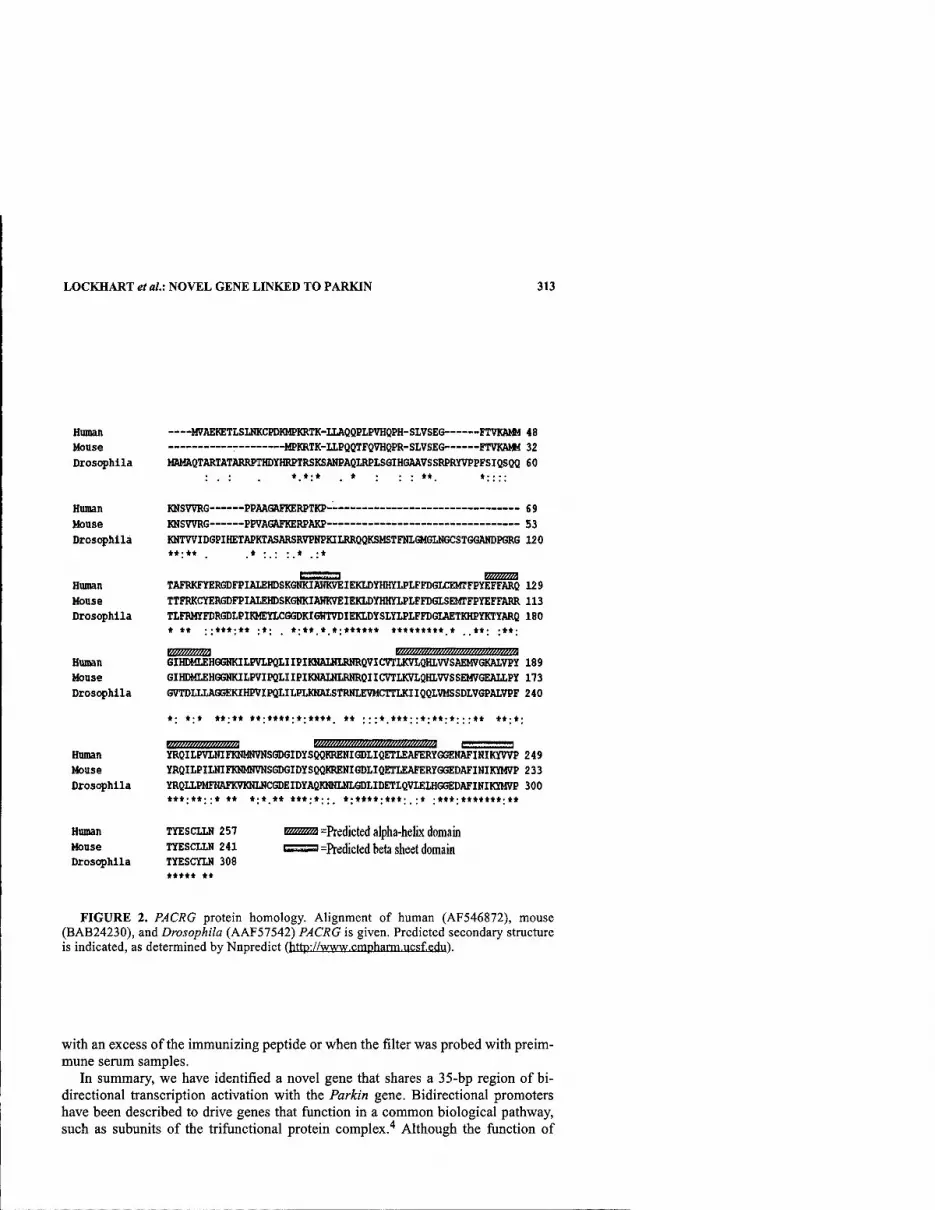
LOCKHART etal.: NOVEL GENE LINKED TO PARKIN 313
Human M7AEKETLSLHKCPDKMPKRTK-ILAQQPLPVHQPH-SLVSEG FTVKAMM 48 Mouse ^MPKRTK-ILPQOTFQVHQPR-SLVSEG FTVKAMM 32 Drosophila MAMAQTARTATARRFTHDYHRPTRSKSAHPAQLRPISGIHGAAVSSRPRYVPFFSIQSQQ 60
Hunan Mouse Drosophila
Human House Drosophila
Haman Mouse Drosophila
KNSWRG PPAAGaFKERPTKP 69 KHSWRG PPVAGRFKERPAKP 53 KNTWIDGPIHETAPKIASARSR7PNPKIIRRQQKSMSTFNLGH6UIGCSTGGANDP6RG 120 *♦•*♦ ♦•••*•♦
TAFBKFYGRGDFPIAIEHDSKGNKIAnKVEIEKLDYHHYLPLFFDGLCEUTFPVEFFARQ 129 TTFRKCTERGDFPIMJEHDSKGHKIAfflWEIEKLDYHHYlPLFFDGLSEMrFPYEFFARR 113 TlFRMyFDRGDI.PIKMEYlCGGDKIGHTVDIEKLDYSLYlPI,FFDGIAETKHPirKTYflRQ 180 ♦ ♦• •■♦♦*■♦» ••• ♦•»« ♦ «•«♦***♦ ♦♦♦*♦♦*♦♦ * **• .*♦■
GIHDHLEHGQnCILFVLPQIIIPIKNAUHRNRQVICVTLKVIQHLVVSAEMVGKALVPY 189 GIHDMLEHGGHKILPVIPQLIIPIKHALNIRHROIICVTLKVIQHLWSSEMVGEAIXPY 173 GVTDIXIAGGEKIHPVIPQLIIPLKHAISTRNIEVMCTTLKIIQQLVMSSDLVGPALVPF 240
♦• ♦•♦ ♦*•*♦ *♦•*«♦♦•♦•♦♦«♦ ♦♦ •••♦ ♦*♦••♦•♦*•♦•••♦* ♦♦•♦•
m/im/»nm7777!7m wmmwwMm/m///M/im»m c a Human YRQILPVIHIFKHMNVNSGDGIDYSQQKREHIGDIIQETLEAFERYGGEHAFIHIKYWP 249 Mouse YRQILPILNIFKNMNVNSGDGIDYSQQKREHIGDLIQETLEAFERYGGEDAFIHIKYMVP 233 Drosc^hlla YRQUPMFlIAFK9KHINCGDEIDYAQKtlHLl)IGDIlDETI.QVIEIHGGEDAFINIKYM7P 300
***■**•■* ♦♦ ♦■♦ ♦* ***•♦•• ♦■♦♦♦♦•♦♦*■ •♦ •*♦♦•♦**♦»*♦.♦♦
Human Mouse Drosophila
TYESCLLH 257 TYESCIiN 241 TYESCYLH 308 ***** **
=Predicted alpha-helLx domain =Predicted beta sheet domain
FIGURE 2. PACRG protein homology. Alignment of human (AF546872), mouse (BAB24230), and Drosophila (AAF57542) PACRG is given. Predicted secondary structure is indicated, as determined by Nnpredict (http://www.cmpharm.ucsf.edu).
with an excess of the immunizing peptide or when the filter was probed with preim- mune serum samples.
In summary, we have identified a novel gene that shares a 35-bp region of bi- directional transcription activation with the Parkin gene. Bidirectional promoters have been described to drive genes that function in a common biological pathway, such as subunits of the trifiinctional protein complex.'* Although the function of

314 ANNALS NEW YORK ACADEMY OF SCIENCES
4 3 2 10 0 I J 4 5 6
pas-Basic (-)
pa3-S\^0Caitrol
PACKOCat-lOSbp)
p(X3-Baac (-)
pa3-SV40Co(itiol
Paikiu (j( +11)1))
•IMbp
m Wi, fUbf
t •2«'bp
•IMbp t -WO
p%'^,ri'.
FIGURE 3. Dual luciferase assay of the Parkin/PACRG promoter region. Deletion con- structs were inserted into the pGL3-Basic vector in the sense {dark bar) or antisense {light bar) orientation relative to Parkin. Resulting luciferase activity in transiently transfected BE(2)-M17 neuroblastoma cells is given adjacent to each construct. Units are relative to the SV-40 control promoter, defined as one. Data are representative of six independent experi- ments, and error bars are 2 x SEM. Positions of deletion constructs are given relative to the Parkin transcriptional start site.
PACRG is currently unknown, we are currently investigating the possibility that
PACRG may function in the same biological pathway as Parkin.
REFERENCES
1. WEST, A., et al. 2001. Identification and characterization of the human parkin gene promoter. J. Neurochem. 78: 1146-1152.
2. WEST, A., et al. 2003. Identification of a novel gene linked to parkin via a bi-direc- tional promoter. J. Mol. Biol. 326: 11-19.
3. BLACKWELL, T.K., et al. 1993. Binding of myc proteins to canonical and noncanonical DNA sequences. Mol. Cell Biol. 13: 5216-5224.
4. ORII, K.E., et al. 1999. Genes for the human mitochondrial trifunctional protein alpha- and beta-subunits are divergently transcribed from a common promoter region. J. Biol. Chem. 274: 8077-8084.

Gene Expression Analysis of the MPTP- Lesioned Substantia Nigra in Mice
R. M. MILLER, C. CASACELI, L. CHEN, AND H. J. FEDEROFF
Department of Neurology, Center for Aging and Developmental Biology, University of Rochester School of Medicine, Rochester, New York 14642, USA
KEYWORDS: genes; Parkinson's disease; MPTP; substantia nigra
INTRODUCTION
Parkinson's disease (PD) is a clinical syndrome triggered by disparate mecha- nisms. That the clinical and neuropathological features are indistinguishable among the different forms suggests a convergent, shared pathway. The earliest shared steps of this cascade, including compensatory responses, represent ideal targets for thera- peutic development.
One approach is the use of robust exploratory methodologies to identify candi- date molecules or pathways that can be implicated in one or more models of PD. An assortment of new technologies is available to assist in this discovery. To increase the reliability of target identification and then confirm involvement of a particular molecule in any biologic process, additional strategies are needed. To this end, we have undertaken an integrative bionomic approach whereby multiple methodologies are used to establish the involvement of candidate molecules in dopamine neuron cell injury, compensation, and cell death. We applied the integrated bionomics approach to the discovery of new molecular targets for PD in the l-methyl-4-phenyl- 1,2,3,6-tetrahydropyridine (MPTP)-treated mouse model (30 mg/kg MPTP-HCl; 4 doses ip over 8 days). Two time points following MPTP intoxication were examined. Using four data mining methods (MAS 5.0 and DMT, dChip,' and SAM^), we found that the expression of many genes decreased in MPTP-treated mice relative to saline- treated controls at the 24-hour time point. At 7 days, there were both increased and decreased genes.
RESULTS
Gene expression analysis using Affymetrix U74A oligonucleotide arrays of MPTP- and saline-treated mice was employed to discover novel and potentially im- portant molecular changes in the substantia nigra (SN) following dopaminergic cell injury. Raw intensity data (.eel files) were analyzed using two independent methods
Address for correspondence: H.J. Federoff, M.D., Ph.D., University of Rochester School of Medicine, 601 Elmwood Ave., Rochester, NY 14642. Voice: 585-273-2190; fax: 585-506-1957.
Ann. N.Y. Acad. Sci. 991: 315-318 (2003). © 2003 New York Academy of Sciences.
315

316 ANNALS NEW YORK ACADEMY OF SCIENCES
Microarray Suite 5.0 computes
SIGNAL (from GEL file)
dChip
computes Model Based Expression Index
T-test P< 0.01
Statistical computation by Affyitietrix DMT (Data Mining Tool) of significantly different gene expression signals.
Count & Percent
Ranks DMT-identified expression changes (increase or decrease) by the number of pairwise comparisons
Changed the gene is changed in. (>75%)
Normalizes, uses probe sensitivity irulex to determine expression level with SEM, and calculates fold change with confidence intervals. (Li and Wong, 2001)
Significant Genes Identifies genes with potentially significant
SAM expression changes by computing multiple (Sienificance ^^"^ specific t-lests. Genes exceeding a user- \ , ■ f defined thresMd, 5, are deemed significant. Analysis ot increasing S lowers the false discovery rate (FDR). Microarrays) ^^^er et al.. 2001).
FIGURE I. Affymetrix .eel files were analyzed with MAS 5.0 or dChip to generate gene expression levels for each sample. Gene expression levels in saline- and MPTP-treated samples were then compared to identify significant changes. Each method (Mest, pair-wise comparisons, SAM, and dChip) was used to create a list of genes whose difference exceeded a predefined significance threshold.
to generate expression levels for each gene, which were then used to compare levels between MFTP and saline arrays (FIG. 1). As expected, we found that the expression of several genes related to the dopaminergic phenotype, including tyrosine hydroxy- lase (TH) and dopamine transporter (DAT), was decreased in MPTP-treated mice. Other functional classes were affected as well, notably transcription factors, kinases, and phosphatases, and messages coding for proteins involved with protein transport and degradation (TABLE 1). We found that the expression of 126 genes was
TABLE I. Functional classification of mRNAs
Functional class MML genes:
126 total in 4/4 methods MME & MML genes:
23 total in 4/4 methods
Transcription/translation 15 4
Kinase/phosphatase 9 3
Signaling 11 2
Developmental 3 0
Bioenergetics 5 2
Protein turnover/transport 8 2
Other 11 3
Unknown 63 8

MILLER etal: MPTP IN PARKINSON'S DISEASE 317
A. Analysis of MPTP 24 hr (MME) vs. Saline-Treated Mice
T-test IncTDec. SAM dCMp
# significant genes: 3258 1073 218 1161
MERGE:
Mgenes in 4/4 methods: 126
B. Analysis of MPTP 7 day (MML) vs. Saline-Treated Mice
T-test IncAJec. SAM dChip
# significant genes: 558 328 221 638
MERGE: Mgenes in 4/4 methods: 44
FIGURE 2. Summary of significantly different gene totals for each method separately and merged. MPTP-treated mice sacrificed at 24 hours following the last dose were com- pared to saline-treated littermates (A). MPTP-treated mice sacrificed 7 days after the last dose were compared to saline-treated littermates (B).
decreased, as determined by all four methods employed, 24 hours after the final MPTP injection (FIG. 2). Seven days after the final MPTP dose, 23 of these genes were still decreased, whereas an additional 21 genes were significantly different (up or down, by all four methods) in the MPTP-treated mouse SN (FiG. 2B). In addition to the use of multiple data analysis techniques, we also verified our gene expression findings using quantitative real-time polymerase chain reaction (PCR) (TABLE 2). As predicted by the microarray results (genes called significant in four of four analysis
TABLE 2. Validation of MML and MME by quantitative RT-PCR
Primer and probe set Confirmed Direction of change
Merged significant DAT yes D genes: 4/5 or 5/5 TH yes D
ElkL motif kinase yes D
AchE yes D
PIP, D type not detected D
Single-method signifi- Kinesin 3 a no NC cant genes VAMP 2 no NC
MAP-6 no NC
318 ANNALS NEW YORK ACADEMY OF SCIENCES
methods), expression of TH, DAT, emk, and AchE was significantly decreased by re- al- time PCR. Further validation of these candidates is in progress. We have been un- able to validate genes called significant in three or fewer methods (i.e., kinesin 3A, VAMP-2, and MAP-6).
REFERENCES
1. LI, C & W.H. WONG. 2001. Model-based analysis of oligonucleotide arrays: model val- idation, design issues and standard error application. Genome Biol. 2: 32.1-32.11.
2. TUSHER, V.G., R. TiBSHiRANi & G. CHU. 2001. Significance analysis of microarrays applied to the ionizing radiation response. Proc. Natl. Acad. Sci. USA 98: 5116- 5121.

Neuroimaging and Proteomic Tracking of Neurodegeneration in MPTP-Treated Mice
HOWARD E. GENDELMAN,'''* CHRISTOPHER J. DESTACHE, "■' MARINA L. ZELIVYANSKAYA,"'* JAY A. NELSON,"'''MICHAEL D. BOSKA,"''' TONIM. BISKUP,"'*MICHAEL K. MCCARTHY,"* KIMBERLY A. CARLSON,"* CRAIG NEMECHEK,"* ERIC J. BENNER,"* AND R. LEE MOSLEY"*
"Center for Neurovirology and Neurodegenerative Disorders, University of Nebraska Medical Center, Omaha, Nebraska 68198-5215, USA
"Department of Pathology and Microbiology, University of Nebraska Medical Center, Omaha, Nebraska 68198-5215, USA
''Department of Pharmacy Practice, Creighton University, Omaha, Nebraska 68178, USA
"Department of Radiology, University of Nebraska Medical Center, Omaha, Nebraska 68198-5215, USA
KEYWORDS: mouse; dopamine; neurodegeneration; l-niethyl-4-phenyl-l,2,3,6- tetrahydropyridine (MPTP); single-photon emission computed tomography (SPECT); magnetic resonance imaging (MRI); magnetic resonance spectro- scopic imaging (MRSI); proteomics; tyrosine hydroxylase; dopamine trans- porter (DAT); 2p-carboxymethoxy-3P-(4-iodophenyI)tropane (p-CIT); D2 receptors; iodobenzamide (IBZM)
Our laboratory is developing an integrated testing paradigm for human Parkinson's disease aimed at early diagnosis and therapeutic monitoring of disease utilizing l-methyl-4-phenyl-l,2,3,6-tetrahydropyridine (MPTP)-treated mice. Such MPTP- treated animals mirror the biochemical and histopathological aspects of human Parkinson's disease.' In our studies, male 8-11-week-old C57BL/6NCrlBR mice from Charles River Laboratories were administered four intraperitoneal (ip) injec- tions (1 every 2 hours) of 21.8 mg MPTP-HCl (18 mg free base)/10 mL phosphate- buffered saline (PBS)/kg body weight. Excipient control mice received 10 mL PBS/ kg body weight intraperitoneally over the same time period. MPTP induced typical dopaminergic neurodegeneration in the substantia nigra pars compacta (SNpc) and the striata, as determined by immunohistochemistry with antibody to tyrosine hydroxylase (TH).^ A 63% loss of TH immunoreactive neurons was observed in the SNpc by 7 days after MPTP treatment as compared to controls (P = 0.001). Reverse
Address for correspondence: R. Lee Mosley, Ph.D., University of Nebraska Medical Center, 985215 Nebraska Medical Center, Omaha, NE 68198-5215. Voice: 402-559-2510; fax: 402-559- 8922.
Ann. N.Y. Acad. Sci. 991: 319-321 (2003). © 2003 New York Academy of Sciences.
319

320 ANNALS NEW YORK ACADEMY OF SCIENCES
phase-high performance liquid chromatography (HPLC) with electrochemical detection^ of striatal lysates showed a 70% reduction in dopamine levels of MPTP mice {P < 0.03) with no significant differences in dopamine metabolites. Addition- ally, the loss of striatal termini and nigral neurons was evaluated by coregistration of single-photon emission computed tomography (SPECT)'*'^ and magnetic resonance (MR)/MR spectroscopic imaging (MRI/MRSI). SPECT analysis of striatal D2 re- ceptors with ['23i].iodobenzamide (['^^IJ-IBZM)^ and dopamine transporter (DAT) with ['23i].2|3-carboxymethoxy-3P-(4-iodophenyl)tropane (['2^I]-|i-CIT)'' demon- strated 30% and 32% reductions, respectively, in MPTP-treated mice compared to controls. Coregistration of SPECT and MRI confirmed the striatal location of ['^^I]- IBZM and ['^^ij-p-CIT ligand-receptor specificity. MRS! evaluation of 36-mm^ midbrain volumes circumscribing the SN from day 5 MPTP-treated mice showed significantly reduced levels of A^-acetylaspartate (42%), choline (25%), and glutamate (26%) and increased myoinositol (34%) compared to those in control an- imals. These meta-bolic changes are consistent with neuronal degradation and prom- inent gliosis.
To detect alterations in protein profiles induced by MPTP treatment, lysates from the midbrain of mice at 1, 2, 3, 4, and 7 days after intoxication were evaluated by proteomic tests using surface-enhanced laser desorption/ionization (SELDI) from adsorbed NP20 protein chips (Ciphergen Biosystems, Fremont, CA). Duplicate de- terminations from three mice per time point were compared to samples obtained from excipient controls treated on day 0, and phenomic profiles were generated by the evaluation of relative protein concentrations as a function of mass/charge. Pro- tein concentrations of each molecular weight species at each time point that differed significantly were identified by Student's t test as candidates for further evaluation as a function of time. ANOVA indicated the existence of 45 different proteins for which the variance in protein expression was significantly affected over time post- MPTP treatment. Kinetic analysis of protein expression and histopathology obtained from the substantia nigra revealed that 35 of these midbrain protein species present- ed kinetic profiles that correlated with the frequency of silver-stained degenerating neurons, which peak by day 2 and, similar to TH immunoreactive neurons, reach a nadir by day 4 post-MPTP treatment.^ Four protein profiles correlated with CD3"*' T cell infiltration into the substantia nigra; however, because of the small population of T cells found in the brain of MPTP-treated mice and consequent low contribution of proteins in midbrain lysates, the possibility could not be ruled out that these pro- files may reflect mechanistic responses from other cell types. The remaining proteins exhibited expression kinetics for which no correlations could be made with the kinetic profile of any one cell type and appear to reflect a combination of kinetic profiles, suggesting that these protein species may be commonly expressed by a variety of MPTP-responsive cell types. We have recently initiated studies integrating SELDI proteomic and microsequence (ProteomiX, Thermo Finnigan) analyses to determine the identities of neural proteins whose expression is altered by MPTP treatment.
Overall, the utility of SPECT, MRI, and MRSI and the coregistration of these neuroimages with histopathological techniques in the MPTP model will provide additional information concerning disease processes during dopaminergic neuro- degeneration. Moreover, the elucidation of noninvasive definitive neuroimages and unique protein profiles in the brain during the degenerative process could provide
GENDELMAN et al.: TRACKING DOPAMINERGIC NEURODEGENERATION 321
novel insights into the neuropathogenesis of disease as well as uncovering proteins that could be used for diagnosis and/or therapeutic monitoring of human Parkinson's disease.
REFERENCES
HEIKKILA, R.E., A. HESS & R.C. DUVOISIN. 1984. Dopaminergic neurotoxicity of 1- methyl-4-phenyl-l,2,5,6- tetrahydropyridine in mice. Science 224: 1451-1453.
JACKSON-LEWIS, V., M. JAKOWEC, R.E. BURKE & S. PRZEDBORSKI. 1995. Time course and morphology of dopaminergic neuronal death caused by the neurotoxin 1-methyl- 4-phenyl-l,2,3,6-tetrahydropyridine. Neurodegeneration 4: 257-269.
JACKSON-LEWIS, V. & G. LIBERATORE. 2000. Effects of a unilateral stereotaxic injection of Tinuvin 123 into the substantia nigra on the nigrostriatal dopaminergic pathway in the rat. Brain Res. 866: 197-210.
GOODMAN, M.M., M.P. KUNG, G.W. KABALKA, et al. 1994. Synthesis and characteriza- tion of radioiodinated N-(3-iodopropen-l-yl)-2 beta-carbomethoxy-3 beta-(4-chloro- phenyl)tropanes: potential dopamine reuptake site imaging agents. J. Med. Chem. 37: 1535-1542.
KUNG, H.F., R. KASLIWAL, S.G. PAN, et al. 1988. Dopamine D-2 receptor imaging radiopharmaceuticals: synthesis, radiolabeling, and in vitro binding of (R)-(-l-)- and (S)-(-)-3-iodo-2-hydroxy-6-methoxy-Af-[(l-ethyl-2-pyrrolidinyl)methyl]benzamide. J. Med. Chem. 31: 1039-1043.

PCBs and Dopamine Function Neurological Effects of Poly chlorinated Biphenyls: Does Occupational Exposure Alter Dopamine-Mediated Function?
RICHARD F. SEEGAL
Wadsworth Center, New York State Department of Health and School of Public Health, University at Albany, Albany, New York 12201, USA
KEYWORDS: polychlorinated biphenyls; occupational exposure; dopamine; neurological function
Polychlorinated biphenyls (PCBs), once heavily used in industry, are now wide- spread environmental contaminants that are suggested to be associated with devel- opmental and cognitive deficits in infants and children bom to mothers who consumed food products contaminated with PCBs and other toxicants.' These data, supported by studies of laboratory animals exposed during development to PCBs, as reviewed by Seegal,^ suggest that the developing central nervous system (CNS) is sensitive to this toxicant. However, both in vitro and in vivo data (to be described) suggest that: (1) susceptibility extends beyond the perinatal period, and (2) PCBs induce long-term alterations in dopamine (DA) function, including reductions in de novo synthesis and inhibition of monoamine transporter function. These findings, in turn, provide a biologically based rationale for examining neurological function in a cohort of aging former capacitor workers who had been exposed to extraordi- narily high levels of PCBs.
PCBs ALTER DOPAMINE FUNCTION IN VITRO
PCBs reduce DA content in tissues derived from laboratory rodents, including pheochromocytoma (PC 12) cells, adult striatal tissue, and synaptosomes exposed ex vivo to PCBs, and they elevate media DA concentrations.^ These changes in DA function may, in part, involve PCB-induced inhibition of monoamine transporters, including the membrane dopamine transporter (DAT) and the intracellular vesicular
Address for correspondence: Dr. Richard F. Seagal, Wadsworth Center, New York State Department of Health and School of Public Health, University at Albany, Albany, NY 12201. Voice: 518-473-4378; fax: 518-486-1505.
Ann. N.Y. Acad. Sci. 991: 322-325 (2003). © 2003 New York Academy of Sciences.
322

SEEGAL: PCBS AND DOPAMINE FUNCTION 323
monoamine transporter (VMAT).'*'^ The consequences of such transporter inhibition include increased free cytosolic DA, elevations in both tissue and media concentra- tions of 3,4-dihydroxyphenylacetic acid (DOPAC), and enhanced formation of DA quinones and semiquinones, leading to generation of hydrogen peroxide and other reactive oxygen species.^
PCBs ALTER DOPAMINE FUNCTION IN VIVO
The alterations in DA function just described may explain the neurochemical and neuropathological consequences of long-term exposure of the adult nonhuman primate to PCBs. Thus, in adult nonhuman primates exposed to PCBs for 20 weeks, an exposure that results in serum concentrations similar to those seen in former workers, basal ganglia DA concentrations were significantly reduced.^ Furthermore, when additional nonhuman primates were exposed to similar levels of PCBs but were then removed from exposure for 24 or 44 weeks prior to sacrifice, brain DA concentrations remained depressed and were not significantly different from DA levels in nonhuman primates sacrificed during exposure,^ despite dramatic reduc- tions in serum PCB levels (FIG. 1). This pattern suggests long-term, if not perma- nent, changes in DA function. A likely explanation for these persistent reductions in basal ganglia DA concentrations is based on our findings that the number of tyrosine hydroxylase-positive neurons in the nonhuman primate substantia nigra pars com- pacta was reduced by approximately 50% (FiG. 2).
These laboratory findings, incorporating both in vitro and in vivo techniques, pro- vide compelling evidence that PCBs alter central DA function, including inhibition of monoamine transporter function, leading to premature death of central DA neurons. When combined with the aforedescribed studies in nonhuman primates, the
^ 22
E 20 - T
2.0
1 r
1.5 ^ c 18
LU 16
-
^V
- 2 -lA
/
**
A T E
Q- CL
1-0 ^ CQ o a.
0.5 S =) DC LU C/3
0.0
- 14
< 12
O 10 Q 8 _l
< 4
c/5 n
**
> -
Control 20-Week 24-Week Exposure Post-Exposure
FIGURE 1. Striatal dopamine and serum PCB concentrations in nonhuman primates (macaques, Macaca nemestrina) exposed as adults to PCBs (Aroclor 1016, 20 weeks, 3.2 mg/kg/day) and sacrificed either immediately or 24 weeks following exposure. **P < 0.01 compared to controls; n = 3-5 animals per exposure condition.
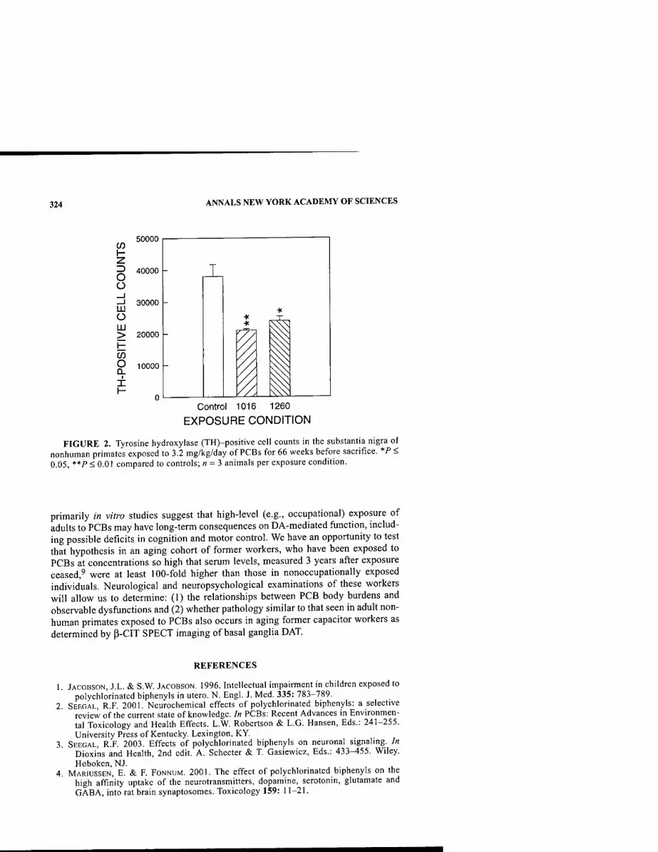
324 ANNALS NEW YORK ACADEMY OF SCIENCES
\- ■z. z> O O
Ul o
50000
40000 -
30000
LU > 20000
o I
X
10000 -
- J s Control 1016 1260
EXPOSURE CONDITION
FIGURE 2. Tyrosine hydroxylase (TH)-positive cell counts in the substantia nigra of nonhuman primates exposed to 3.2 mg/kg/day of PCBs for 66 weeks before sacrifice, *P < 0.05, **P < 0.01 compared to controls; « = 3 animals per exposure condition.
primarily in vitro studies suggest that high-level (e.g., occupational) exposure of adults to PCBs may have long-term consequences on DA-mediated function, includ- ing possible deficits in cognition and motor control. We have an opportunity to test that hypothesis in an aging cohort of former workers, who have been exposed to PCBs at concentrations so high that serum levels, measured 3 years after exposure ceased,^ were at least 100-fold higher than those in nonoccupationally exposed individuals. Neurological and neuropsychological examinations of these workers will allow us to determine: (1) the relationships between PCB body burdens and observable dysfunctions and (2) whether pathology similar to that seen in adult non- human primates exposed to PCBs also occurs in aging former capacitor workers as determined by p-CIT SPECT imaging of basal ganglia DAT.
REFERENCES
JACOBSON, J.L. & S.W. JACOBSON. 1996. Intellectual impairment in children exposed to polychlorinated biphenyls in utero. N. Engl. J. Med. 335: 783-789.
SEEGAL, R.F. 2001. Neurochemical effects of polychlorinated biphenyls: a selective review of the current state of knowledge. In PCBs: Recent Advances in Environmen- tal Toxicology and Health Effects. L.W. Robertson & L.G. Hansen, Eds.: 241-255. University Press of Kentucky. Lexington, KY.
SEEGAL, R.F. 2003. Effects of polychlorinated biphenyls on neuronal signaling. In Dioxins and Health, 2nd edit. A. Schecter & T. Gasiewicz, Eds.: 433-455. Wiley. Hoboken, NJ.
MARIUSSEN, E. & F. FONNUM. 2001. The effect of polychlorinated biphenyls on the high affinity uptake of the neurotransmitters, dopamine, serotonin, glutamate and GABA, into rat brain synaptosomes. Toxicology 159: 11-21.
SEEGAL: PCBS AND DOPAMINE FUNCTION 325
5. MARIUSSEN, E., P.L. ANDERSSON, M. TYSKLIND & F. FONNUM. 2001. Effect of poly- chlorinated biphenyls on the uptake of dopamine into rat brain synaptic vesicles: a structure-activity study. Toxicol. Appl. Pharmacol. 175: 176-183.
6. LAVOIE, M.J. & T.G. HASTINGS. 1999. Dopamine quinone formation and protein modi- fication associated with the striatal neurotoxicity of methamphetamine: evidence against a role for extracellular dopamine. J. Neurosci. 19: 1484-1491.
7. SEEGAL, R.F., B. BUSH & K.O. BROSCH. 1991. Comparison of effects of Aroclors 1016 and 1260 on nonhuman primate catecholamine function. Toxicology 66: 145-163.
8. SEEGAL, R., B. BUSH & K.O. BROSCH. 1994. Decreases in dopamine concentrations in adult non-human primate brain persist following removal from polychlorinated biphenyls. Toxicology 86: 71-87.
9. LAWTON, R.W., M.R. Ross, J. FEINGOLD & J.F. BROWN, JR. 1985. Effects of PCB expo- sure on biochemical and hematological findings in capacitor workers. Environ. Health Perspect. 60: 165-184.
A Murine Model of ALS-PDC with Behavioral and Neuropathological Features of Parkinsonism
J. D. SCHULZ," J. M. B. WILSON," AND C. A. SHAW*'^''
"Neuroscience Graduate Program and Departments of ^Ophthalmology, '^Physiology, and ^Experimental Medicine, University of British Columbia, Vancouver, British Columbia 6T1Z4, Canada
KEYWORDS: parkinsonism; amyotrophic lateral sclerosis (ALS); dementia
Amyotrophic lateral sclerosis-parkinsonism dementia complex (ALS-PDC) is an unusual neurological disease that occurs among the Chamorro people of Guam. It can express as ALS, an Alzheimer's disease-like disorder with parkinsonism features, or as a combined form with both motor and cognitive deficits. Kurland and others recognized the importance of understanding the Guamanian disorders in the hopes of unlocking crucial clues to related neurodegenerative disorders worldwide. The strongest epidemiologic link to ALS-PDC is the correlation to consumption of flour made from the seed of C. circinalis, a species of cycad.'^ Cycad is known to contain potent toxins, some of which can be removed by the extensive washing pro- cedures used by the Chamorros.' Early work using unwashed cycad and isolated components of cycad seeds in animal models failed to support a role for cycad as a causal agent in ALS-PDC.^ However, to our knowledge, the critical in vivo animal experiments using washed cycad had never been performed.
To test the hypothesis that cycad neurotoxicity could be causal to ALS-PDC, we fed washed cycad flour to mice as part of their diet and used a battery of behavioral tests and histological assays to assess developing and end-state neural degeneration. (See Wilson et al^ for a complete description of the experimental methods.)
Cycad-fed mice showed significant decreases in motor, olfactory, and cognitive functions that correlated with neuron degeneration in substantia nigra, spinal cord, olfactory bulb, neocortex, and the hippocampus, as well as a decrease of dopamin- ergic terminals in the striatum. In regard to motor function, deficits were demonstrat- ed by a significant loss of the leg extension reflex, which is indicative of motor neuron dysfunction (FIG. 1 A). In addition, measurements of gait length, a marker of basal ganglia function that correlates significantly with cell loss in the substantia
Address for correspondence: Dr. C.A. Shaw, Neuroscience Graduate Program, Departments of Ophthalmology, Physiology, and Experimental Medicine, University of British Columbia, Vancouver, BC 6T 1Z4, Canada. Voice: 604-875-4111, ext. 68375; fax: 604-874-4376.
Ann. N.Y. Acad. Sci. 991: 326-329 (2003). © 2003 New York Academy of Sciences.
326

SCHULZ et al.: ALS-PDC AND PARKINSONISM 327
U
+ HI
m (uio) asueista
W
pspuspo sSdi pujM ;o # D
6 Q o 1
c o o
■•-' c .s J3
C/1 (U X: O ft 3
r/l O
•a 61)
13 C) ^
o ' bH o
■o o o P, o ^
o c 11 o 3 m T3
1 J3
c O, o > o o
o •a s oO
> •a c o £ -
■a a a (0
a * w ^^ ■^
.w w o r) t; (L> u o 5> ̂ (1>
o V
1*^ d w ^ ,» CO <<- « CO ,-H
O o e 'M «J o 1 .
3 en o A V
a SO, rl
(3 (U <ji P3 «
x^ r/l '°. 50
.g M)
o feo
H-l T)
d 1*"^
a
a Is ^ -S ?n>S
c2 d
^ T3
o C O o
a a
3 O a a 60 rl
y:3 o T)
E .a B u _> *
V, ^ X s
o o cd
^a •a o
a w OD Tl •o ftii O „ g o CO ^2 03 13) ",' Vi i-i P
1 a> *:;
o
C3 J3 a O 60
O ifl 60 J3
O
U -o a o
M ti ^ E a
'S 1 ^ 60
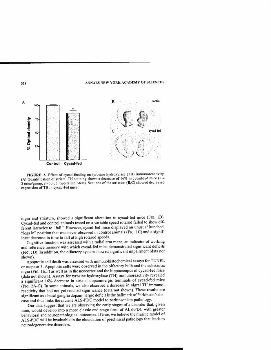
328 ANNALS NEW YORK ACADEMY OF SCIENCES
control 100-1
i? ■ w c
■D
s '•Si o. O
t.
r.
^^v'..^V. Control Cycad-fed
FIGURE 2. Effect of cycad feeding on tyrosine hydroxylase (TH) immunoreactivity. (A) Quantification of striatal TH staining shows a decrease of 16% in cycad-fed mice (n = 3 mice/group, P < 0.05, two-tailed /-test). Sections of the striatum (B,C) showed decreased expression of TH in cycad-fed mice.
nigra and striatum, showed a significant alteration in cycad-fed mice (FiG. IB). Cycad-fed and control animals tested on a variable speed rotarod failed to show dif- ferent latencies to "fall." However, cycad-fed mice displayed an unusual hunched, "legs in" position that was never observed in control animals (FIG. IC) and a signif- icant decrease in time to fall at high rotarod speeds.
Cognitive function was assessed with a radial arm maze, an indicator of working and reference memory with which cycad-fed mice demonstrated significant deficits (FIG. ID). In addition, the olfactory system showed significant impairment (data not shown).
Apoptotic cell death was assessed with immunohistochemical assays for TUNEL or caspase-3. Apoptotic cells were observed in the olfactory bulb and the substantia nigra (FIG. 1E,F) as well as in the neocortex and the hippocampus of cycad-fed mice (data not shown). Assays for tyrosine hydroxylase (TH) immunoreactivity revealed a significant 16% decrease in striatal dopaminergic terminals of cycad-fed mice (FIG. 2A-C). In some animals, we also observed a decrease in nigral TH immuno- reactivity that had not yet reached significance (data not shown). These results are significant as a basal ganglia dopaminergic deficit is the hallmark of Parkinson's dis- ease and thus links the murine ALS-PDC model to parkinsonism pathology.
Our data suggest that we are observing the early stages of a disorder that, given time, would develop into a more classic end-stage form of ALS-PDC with greater behavioral and neuropathological outcomes. If true, we believe the murine model of ALS-PDC will be invaluable in the elucidation of preclinical pathology that leads to neurodegenerative disorders.

SCHULZ et al.: ALS-PDC AND PARKINSONISM 329
ACKNOWLEDGMENTS
This work was supported by the ALS Association, Parkinson's Disease Founda- tion, Scottish Rite Charitable Foundation of Canada, Natural Science and Engineer- ing Research Council of Canada, and the U.S. Army Medical Research and Materiel Command (#DAMD 17-02-1-0678) (to C.A.S.).
REFERENCES
1. KuRLAND, L.T. 1988. Amyotrophic lateral sclerosis and Parkinson's disease complex on Guam linked to an environmental toxin. Trends Neurosci. 11: 51-53.
2. KuRLAND, L.T. et al. 1994. Amyotrophic lateral sclerosis-parkinsonism dementia com- plex on Guam: epidemiological and etiological perspectives. In Motor Neuron Dis- ease. A. Willams, Ed.: 109-130. Chapman & Hall. London.
3. LiLENFELD, D.E. et al. 1994. Guam neurodegeneration. In Neurodegenerative Diseases. D.B. Calne, Ed.: 895-908. W.B. Saunders. Philadelphia.
4. WILSON, J.M.B. et al. 2002. Behavioural and neurological correlates of ALS-parkin- sonism dementia complex in adult mice fed washed cycad flour. Neuromolecular Med. 1:207-221.
Neuroprotective Ganglioside Derivatives
K. CONN,** S. DOHERTY,^P. EISENHAUER,"*-^R. FINE,** J. WELLS,"'* AND M, D. ULLMAN"'^
"VA Hospital, Bedford, Massachusetts 01730, USA
^Boston University School of Medicine, Boston, Massachusetts 02118. USA
'^University of Massachusetts Medical School Shriver Center, Waltham, Massachusetts 02452, USA
KEYWORDS: gangliosides; neuroprotective agents
Natural and semisynthetic gangliosides protect neurons from injury. The hydrophilic property of gangliosides, however, restricts their blood-brain barrier (BBB) perme- ability when given peripherally This hinders their use as neuroprotective agents. Gangliosides are amenable to chemical derivatization, so that semisynthetic deriva- tives with both cytoprotective properties and improved ability to cross the BBB can be produced. Therefore, ganglioside functional group derivatives that provide cytoprotection and effectively cross the BBB are being sought. This will provide a basis to understand neuroprotective mechanisms. Insight into neuroprotective mechanisms also requires an understanding of cell death processes. Thus, studies into l-methyl-4-phenylpyridinium (MPP+)-induced changes in gene expression are also ongoing.
Semisynthetic GMl derivatives were synthesized and tested for their ability to protect SH-SY5Y human neuroblastoma cells from MPP+ toxicity. SH-SY5Y cells were cultured at 37°C in a 95% air, 5% CO2 humidified incubator and maintained in DMEM-high glucose supplemented with 10% fetal bovine serum. For cytoprotec- tion experiments, 10x10^ cells per well were plated in a 48-well cell culture plate and differentiated in the presence of 10 |.lM retinoic acid. After 4 days, medium was replaced with that containing 0.5% fetal calf serum and the test ganglioside (690 nM) I hour before exposure to MPP"^. Ganglioside was again added on the day after MPP"*" treatment.
For experiments on gene expression, 5 x 10^ cells were plated in 100 mm^ culture dishes in 10 mL of DMEM medium containing 10% fetal bovine serum, 100 units/ mL penicillin, and 100 mg/mL streptomycin and cultured for 4 days. Freshly pre- pared toxin was added to the cultures and incubated for the requisite times. RNA iso-
Addrcss for correspondence: M. David Ullman, VA Hospital (182B), 200 Springs Road, Bed- ford, MA 01730. Voice: 781-687-2636; fax: 781-687-3515.
Ann. N.Y. Acad. Sci. 991: 330-332 (2003). © 2003 New York Academy of Sciences.
330
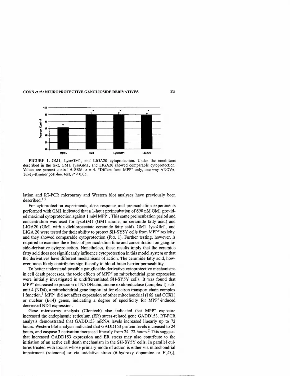
CONN et al: NEUROPROTECTIVE GANGLIOSIDE DERIVATIVES 331
PPPf MPP* CM1 LysoGMI UGA20
FIGURE 1. GMl, LysoGMI, and LIGA20 cytoprotection. Under the conditions described in the text, GMl, lysoGMl, and LIGA20 showed comparable cytoprotection. Values are percent control + SEM. n = 4. *Differs from MPP"*" only, one-way ANOVA, Tukey-Kramer post-hoc test, P < 0.05.
lation and RT-PCR microarray and Western blot analyses have previously been described.''^
For cjrtoprotection experiments, dose response and preincubation experiments performed with GMl indicated that a 1-hour preincubation of 690 nM GMl provid- ed maximal cytoprotection against 1 mM MPP"*". This same preincubation period and concentration was used for lysoGMl (GMl amine, no ceramide fatty acid) and LIGA20 (GMl with a dichloroacetate ceramide fatty acid). GMl, lysoGMl, and LIGA 20 were tested for their ability to protect SH-SY5Y cells from MPP+ toxicity, and they showed comparable cytoprotection (FIG. 1). Further testing, however, is required to examine the effects of preincubation time and concentration on ganglio- side-derivative cytoprotection. Nonetheless, these results imply that the ceramide fatty acid does not significantly influence cytoprotection in this model system or that the derivatives have different mechanisms of action. The ceramide fatty acid, how- ever, most likely contributes significantly to blood-brain barrier permeability.
To better understand possible ganglioside-derivative cytoprotective mechanisms in cell death processes, the toxic effects of MPP"*" on mitochondrial gene expression were initially investigated in undifferentiated SH-SY5Y cells. It was found that MPP'^ decreased expression of NADH:ubiquinone oxidoreductase (complex I) sub- unit 4 (ND4), a mitochondrial gene important for electron transport chain complex I function.' MPP"*" did not affect expression of other mitochondrial (16S and COXl) or nuclear (B14) genes, indicating a degree of specificity for MPP"'"-induced decreased ND4 expression.
Gene microarray analysis (Clontech) also indicated that MPP"*" exposure increased the endoplasmic reticulum (ER) stress-related gene GADD153. RT-PCR analysis demonstrated that GADD153 mRNA levels increased linearly up to 72 hours. Western blot analysis indicated that GADD153 protein levels increased to 24 hours, and caspase 3 activation increased linearly fi-om 24-72 hours.^ This suggests that increased GADD153 expression and ER stress may also contribute to the initiation of an active cell death mechanism in the SH-SY5Y cells. In parallel cul- tures treated with toxins whose primary mode of action is either via mitochondrial impairment (rotenone) or via oxidative stress (6-hydroxy dopamine or H2O2),
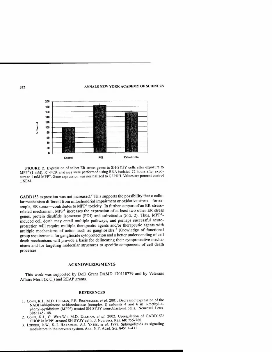
332 ANNALS NEW YORK ACADEMY OF SCIENCES
o
Control
FIGURE 2. Expression of select ER stress genes in SH-SY5Y cells after exposure to MPP"^ (1 mM). RT-PCR analyses were performed using RNA isolated 72 hours after expo- sure to 1 mM MPP"^. Gene expression was normalized to G3PDH. Values are percent control ±SEM.
GADD153 expression was not increased.^ This supports the possibility that a cellu- lar mechanism different from mitochondrial impairment or oxidative stress—for ex- ample, ER stress—contributes to MPP+ toxicity. In further support of an ER-stress- related mechanism, MPP+ increases the expression of at least two other ER stress genes, protein disulfide isomerase (PDI) and calreticulin (FIG. 2). Thus, MPP+- induced cell death may entail multiple pathways, and perhaps successful neuro- protection will require multiple therapeutic agents and/or therapeutic agents with multiple mechanisms of action such as gangliosides.^ Knowledge of functional group requirements for ganglioside cytoprotection and a better understanding of cell death mechanisms will provide a basis for delineating their cytoprotective mecha- nisms and for targeting molecular structures to specific components of cell death processes.
ACKNOWLEDGMENTS
This work was supported by DoD Grant DAMD 170110779 and by Veterans Affairs Merit (K.C.) and REAP grants.
REFERENCES
CONN, K.J., M.D. ULLMAN, P.B. EISENHAUER, et al. 2001. Decreased expression of the NADH-ubiquinone oxidoreductase (complex I) subunits 4 and 6 in l-methyl-4- phenyl-pyridinium (MPP+)-treated SH-SY5Y neuroblastoma cells. Neurosci. Letts. 306: 145-148.
CONN, K.J., G. WEN-WU, M.D. ULLMAN, et al. 2002. Upregulation of GADD153/ CHOP in MPP+-treated SH-SY5Y cells. J. Neurosci. Res. 68: 755-760.
LEDEEN, R.W., S.-I. HAKAMORI, A.J. YATES, et al. 1998. Sphingolipids as signaling modulators in the nervous system. Ann. N.Y. Acad. Sci. 845: 1^31.

Redox- and Metalloregulated RNA Bioaptamer Targets of Proteins Associated with Parkinson's and Other Neurodegenerative Diseases
Factors of Relevance in the Life Cycle of Cells
JOSEF H. WISSLER
ARCONS Applied Research Institute, D-61231 Bad Nauheim, Germany
KEYWORDS: Parkinson's parkin; Alzlieimer's amyloid precursor; Huntington's liuntingtin; prion PrP; SlOO EF-liand; calgranulin; psoriasin proteins; RNA bioaptamer; angiotropin CuRNP ribokine; copper-ribonucleoprotein; angio- genesis; morphogenesis; Cu, zinc transition metalloregulation; nucleic acid- binding R3H domain; Fenton redox reaction; receptor for advanced glycosyla- tion end products (RAGE); amyotrophic lateral sclerosis; fragile X syndrome; ubiquitinylation; proteosome; RNA-chaperone; 3D-rapid prototyping molecu- lar model
INTRODUCTION
Some proteins of neuronal tissue and their genes involved in Parkinson's and other neurodegenerative diseases with complex clinical features (synucleopathies) have been considered unrelated, such as Parkinson's parkin, Alzheimer's amyloid precursor, prion (PrP), Huntington's huntingtin, SlOO EF-hand, and other amy- loidogenic proteins. Their physiological functions are still under debate. Parkin was shown to have conserved Cu,Zn-metalloregulator and also nucleic acid binding sites''^ in terms of short basic residue, SR/K/RS peptide segments, and conserved compact R3H (RxxxH) motifs besides known ring- and in-between-ring (IBR) fin- ger domains (FIG. 1). R3H is a domain that binds single-stranded nucleic acids; it is related to regulatory canonical metal binding domains.^"^ Secondary structure pre- dictions and patterns of residue conservation suggest nucleation of helix-p-hairpin and adoption of helix structures upon binding of transition metal (Cu,Zn) ions and RNA.^~^ Findings for SlOO EF-hand, Alzheimer's precursor, huntingtin, and prion proteins were similar.''-^'^'^ This suggests relationships in functions on a common structural basis in proteins by binding metalloregulated RNA or oligonucleotides. As shown,^"'^ some sequence-defined RNA bioaptamers (natural RNA targets selected by evolution) impart novel bioftinctions to certain proteins that these do not have on their own.
Address for correspondence: Professor Josef H. Wissler, Ph.D., ARCONS Institute for Applied Research, Education and Didactics, Postfach 1327, D-61231 Bad Nauheim, Germany. Voice: +49-6032-31716; fax: +49-6032-31725.
Ann. N.Y. Acad. Sci. 991: 333-338 (2003). © 2003 New York Academy of Sciences.
333
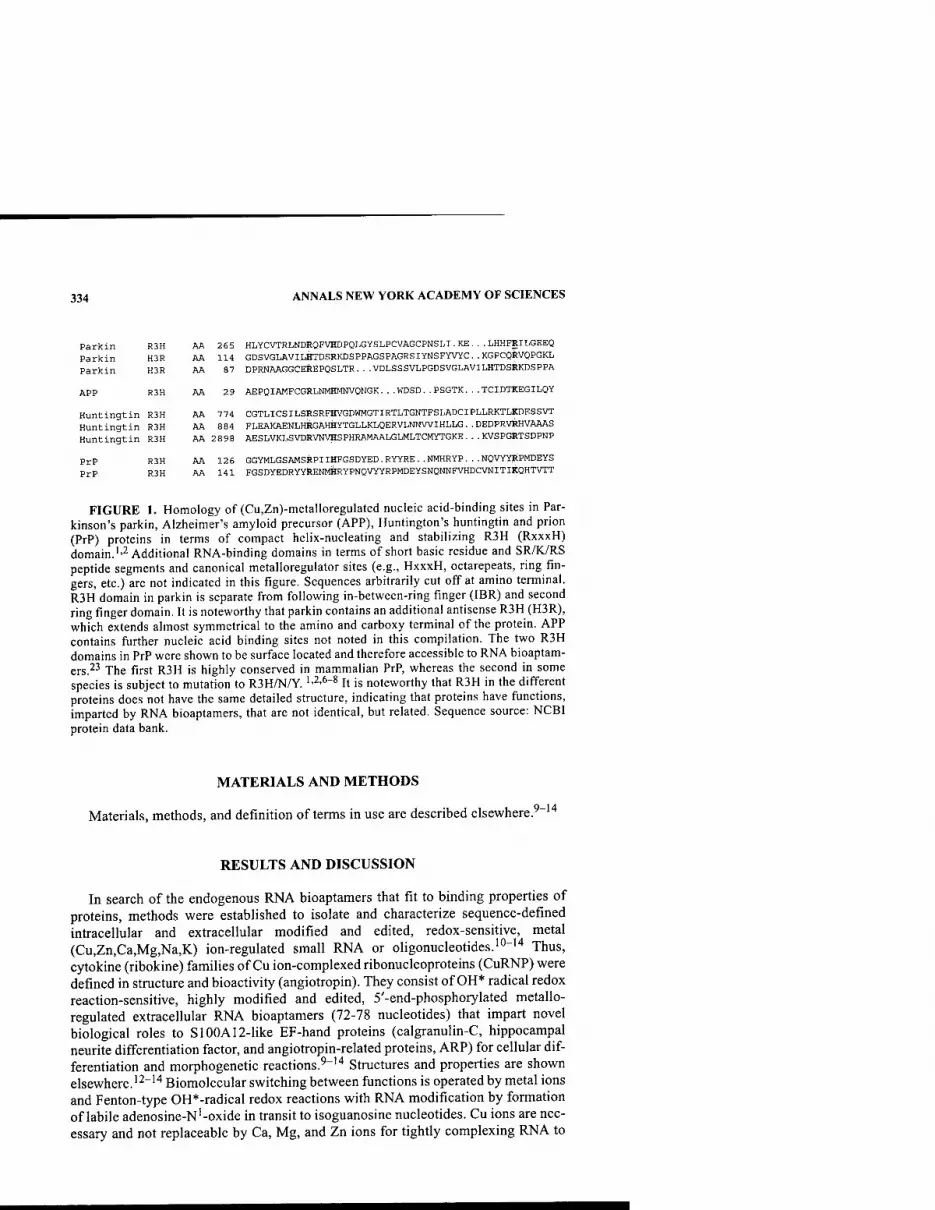
Huntingtin R3H
PrP R3H PrP R3H
334 ANNALS NEW YORK ACADEMY OF SCIENCES
Parkin R3H AA 265 HLYCVTRLNDRQFVHDPQLGYSLPCVAGCPNSLI.KE...LHHFRILGEEQ Parkin H3R AA 114 GDSVGLRVILHTDSRKDSPPAGSPAGRSIYNSFYVYC. . KGPCQRVQPGKL Parkin H3R AA 87 DPRNAAGGCEREPQSLTR...VDLSSSVLPGDSVGLAVILBTDSRKDSPPA
APP R3H AA 29 AEPQIAMFCGRLNMHMNVQNGK. . .KDSD. . PSGTK. . .TCIDTKEGILQY
Huntingtin R3H AA 774 CGTLICSILSRSRFHVGDWMGTIRTLTGNTFSLADCIPLLRKTLKDESSVT Huntingtin R3H AA 884 FLEAKAENLHRGAHHYTGLLKLQERVLNNWIHLLG. .DEDPRV^HVAAAS
AA 2898 AESLVKLSVDRVNVHSPHRAMAALGLMLTCMYTGKE...KVSPGRTSDPNP
AA 126 GGYMLGSAMSRPIIHFGSDYED.RYYRE..NMHRYP...NOVYYRPMDEYS AA 141 FGSDYEDRYYRENMHRYPNQVYYRPMDEYSNQNNFVHDCVNITIKQHTVTT
FIGURE 1. Homology of (Cu,Zn)-metalloregulated nucleic acid-binding sites in Par- kinson's parkin, Alzheimer's amyloid precursor (APP), Huntington's huntingtin and prion (PrP) proteins in terms of compact helix-nucleating and stabilizing R3H (RxxxH) domain.''2 Additional RNA-binding domains in terms of short basic residue and SR/K/RS peptide segments and canonical metalloregulator sites (e.g., HxxxH, octarepeats, ring fin- gers, etc.) are not indicated in this figure. Sequences arbitrarily cut off at amino terminal. R3H domain in parkin is separate from following in-between-ring finger (IBR) and second ring finger domain. It is noteworthy that parkin contains an additional antisense R3H (H3R), which extends almost symmetrical to the amino and carboxy terminal of the protein. APP contains further nucleic acid binding sites not noted in this compilation. The two R3H domains in PrP were shown to be surface located and therefore accessible to RNA bioaptam- ers.^' The first R3H is highly conserved in mammalian PrP, whereas the second in some species is subject to mutation to R3H/N/Y. '•^•^"^ It is noteworthy that R3H in the different proteins does not have the same detailed structure, indicating that proteins have functions, imparted by RNA bioaptamers, that are not identical, but related. Sequence source: NCBI protein data bank.
MATERIALS AND METHODS
Materials, methods, and definition of terms in use are described elsewhere.^"''*
RESULTS AND DISCUSSION
In search of the endogenous RNA bioaptamers that fit to binding properties of proteins, methods were established to isolate and characterize sequence-defined intracellular and extracellular modified and edited, redox-sensitive, metal (Cu,Zn,Ca,Mg,Na,K) ion-regulated small RNA or oligonucleotides.'"-''* Thus, cytokine (ribokine) families of Cu ion-complexed ribonucleoproteins (CuRNP) were defined in structure and bioactivity (angiotropin). They consist of OH* radical redox reaction-sensitive, highly modified and edited, 5'-end-phosphorylated metallo- regulated extracellular RNA bioaptamers (72-78 nucleotides) that impart novel biological roles to S100A12-like EF-hand proteins (calgranulin-C, hippocampal neurite differentiation factor, and angiotropin-related proteins, ARP) for cellular dif- ferentiation and morphogenetic reactions.^"'"* Structures and properties are shown elsewhere. '^"''' Biomolecular switching between functions is operated by metal ions and Fenton-type OH'^-radical redox reactions with RNA modification by formation of labile adenosine-N'-oxide in transit to isoguanosine nucleotides. Cu ions are nec- essary and not replaceable by Ca, Mg, and Zn ions for tightly complexing RNA to
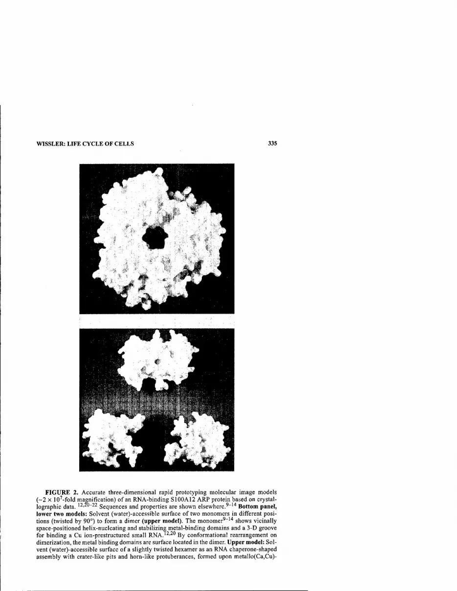
WISSLER: LIFE CYCLE OF CELLS 335
FIGURE 2. Accurate three-dimensional rapid prototyping molecular image models (~2 X 10^-fold magnification) of an RNA-binding S100A12 ARP protein based on crystal- lographic data. 12,20-22 Sequences and properties are shown elsewhere.'"''* Bottom panel, lower two models: Solvent (water)-accessible surface of two monomers in different posi- tions (twisted by 90°) to form a dimer (upper model). The monomer'"''' shows vicinally space-positioned helix-nucleating and stabilizing metal-binding domains and a 3-D groove for binding a Cu ion-prestructured small RNA.'^'^" By conformational rearrangement on dimerization, the metal binding domains are surface located in the dimer. Upper model: Sol- vent (water)-accessible surface of a slightly twisted hexamer as an RNA chaperone-shaped assembly with crater-like pits and horn-like protuberances, formed upon metallo(Ca,Cu)-

336 ANNALS NEW YORK ACADEMY OF SCIENCES
proteins and OH* radical formation by Fenton-type redox reactions. Findings were similar for intracellular oligo RNA bioaptamers as targets for proteins, such as trans- fer factors of delayed-type hypersensitivity reactions, psoriasin.'''^''^"'^
To investigate functions, a novel method was applied.'^'^° Accurate three-dimen- sional rapid prototyping molecular image models (3-D RPM) of metalloregulated proteins can now be constructed when structural data of proteins are available in terms of X-ray crystallographic or NMR data—that is, retrievable from the Brookhaven Data Bank.'^ This allows detailed surface topography analysis of mol- ecules in ~2 X lO'^-fold magnified 3-D scale plastic image. Thus, for example, based on crystallographic data,^''^^ a 3-D RPM (FiG. 2) of Ca,Zn,Mg,Cu ion-regulated RNA-binding S100A12 EF-hand ARP monomer^"'"* shows vicinally space- positioned helix-nucleating and stabilizing metal-binding domains and a 3-D groove for binding a Cu ion-prestructured small RNA.'2'^° Upon metalloregulated oligo- merization via dimers to hexamers, slightly twisted chaperone-shaped assemblies with crater-like pits and horn-like protuberances are formed, having an elliptically shaped internal tubular channel of-4 nm in length. The internal diameter enlarging from ~1 nm at two entries to -2.5 nm at the center allows transient homing and re- forming of small RNA. It suggests RNA chaperone-shaped assemblies at cellular re- ceptors in dependence of metal (Ca,Cu) ions.^O Based on NMR-derived structure data, similar functional relationships are suggested for other (prion) proteins associ- ated with neurodegenerative disease.'-^'^^ Because crystallography or NMR-derived structure data for parkin and other proteins are unavailable so far, such studies have to wait to be applied to them. However, in view of the known role of parkin in ubiquitinylation and possibly proteasomal processes in which involvement of RNA was shown, metallo(Cu,Zn)-regulated functions of parkin in supramolecular struc- ture reformation/degradation comparable to chaperones likewise may be regulated or imparted by RNA bioaptamers. The considerations show the proteins also sharing properties with RNA-binding proteins in amyotrophic lateral sclerosis and fragile X syndrome.^^
SUMMARY AND CONCLUSIONS
The results suggest proteins in neuronal tissues having common sequence homol- ogies in helix-nucleating and stabilizing domains associated with defined functions (RNA chaperones and reformation). They may bind endogenous Cu ion-prestruc- tured, OH* radical redox-sensitive, modified and edited oligo-RNA. Therefore, neurodegenerative disorders may be seen as related to disorders or impairment of functions imparted by binding of endogenous metalloregulated RNA bioaptamers to proteins that these do not have on their own. Thus, synucleopathies in which these
regulated oligomerization from monomers via dimers at cellular receptors for advanced gly- cosylation end products (RAGE). It has an elliptically shaped internal tubular channel of ~4 nm in length. Enlargement of the internal diameter from ~ 1 nm at two entries to -2.5 nm at the center allows transient homing and reforming of small RNA.'^'^" The entries to the channel allow passage of reformed extracellular single-stranded oligoRNA (ssRNA) into the cell via RAGE to induce a cellular differentiation process.

WISSLER: LIFE CYCLE OF CELLS 337
proteins are involved may result from impaired redox- and metalloregulated RNA- protein interactions and reformation. Such disorders or impairment may deviate met- al ion- and RNA-devoid protein structures to unfolding/misfolding and, thus, to dis- ease. The propensity of some proteins to form redox- and metalloregulated RNA- chaperone-shaped assemblies for reforming RNA structures in cellular processes emphasizes the kinetics and thermodynamics of cellular topochemistry, binding, and turnover of RNA bioaptamers as factors of relevance in the life cycle of (neuronal) cells.
REFERENCES
1. WissLER, J.H. & E. LOGEMANN. 2000. Bioinformatics applied to neurodegenerative dis- eases suggests physiological priori (PrP), Alzheimer's amyloid precursor (APP), Par- kinson's parkin (P) and Huntington's huntingtin (H) proteins being (Cu/Zn)- metalloregulated RNA-binding protein families. Mol. Biol. Cell Suppl. 11: 132a.
2. WissLER, J.H. & E. LoGEMANN. 2001. Proteins of neurodegenerative diseases scrapie (cellular prion, PrP), Alzheimer (amyloid precursor, APP), Parkinson (parkin, P) and Huntington (huntingtin, H) are related in canonical (Cu/Zn)-metalloregulator and RNA-binding (R3H) domains. Biophys. J. 80: 566a.
3. HiGAKi, J.N., R.J. FLETTERICK & C.S. CRAIK. 1992. Engineered metalloregulation in enzymes. Trends Biochem. Sci. 17: 100-104.
4. VARANI, G. 1997. RNA-protein intermolecular recognition. Ace. Chem. Res. 30: 189- 195.
5. GRISHIN, N.V. 1998. The R3H motif: a domain that binds single-stranded nucleic acids. Trends Biochem. Sci. 23: 329-330.
6. WissLER, J.H. & E. LoGEMANN. 2000. Prion proteins (PrP) have conserved nucleic acid-binding (R3H) motifs: leads of ribokine structure to function and transfer of bioinformation in PrP infectivity. Biophys. J. 78: 290A.
7. WissLER, J.H. & E. LOGEMANN. 2000. Prion proteins (PrP) have conserved (HxxxH/N/ G) metalloregulator (Cu/Zn) and nucleic acid-binding (R3H) domains: leads of nutri- tion factors and transfer of bioinformation in PrP infectivity. FASEB J. 14: A794.
8. WissLER, J.H. & E. LOGEMANN. 2000. Bioinformatics applied to prion proteins (PrP) suggest metalloregulated RNA-binding protein families with helix-nucleating and stabilizing canonical metalloregulator (HxxxH/N/G) and RNA-binding (R3H) domains. Biol. Chem. 381: S234.
9. WissLER, J.H., W.M. AMSELGRUBER, M. SCHWEIGER & E. LOGEMANN. 1997. Structure, function and cellular localization of angiotropin, a non-mitogenic leukocytic endo- thelial cell RNP-morphogen (ribokine) for organoid capillary pattern formation. Mol. Biol. Cell Suppl. 8: 231a.
10. WissLER, J.H. & E. LOGEMANN. 1998. RNA structure and modification of copper-RNP complex of angiotropin ribokines (non-mitogenic leukocytic endothelial cell mor- phogens). FASEB J. 12: A1463.
U. WissLER, J.H. & E. LOGEMANN. 2000. Ribokines with modified and edited extracellular eRNA: endogenous metallo-regulated copper-ribonucleoprotein cytokines (CuRNP) and their components (SlOO-EF-hand protein, RNA, precursors) in cellular signal transduction. Biol. Chem. 381: S246.
12. WISSLER, J.H. 2001. Engineering of blood vessel patterns by angio-morphogens (angiotropins): non-mitogenic copper-ribonucleoprotein cytokines (CuRNP ribo- kines) with their metalloregulated constituents of RAGE-binding SlOO-EF-hand pro- teins and extracellular RNA bioaptamers in vascular remodeling of tissue and angiogenesis in vitro. Materialwiss. Werkstofftech. (Mat. Sci. Eng. Technol.) 32: 984-1008.
13. WISSLER, J.H. 2002. Engineering of capillary patterns in muscle by a nonmitogenic copper-ribonucleoprotein angiomorphogen (angiotropin CuRNP ribokine). Ann. N.Y. Acad. Sci. 961: 292-297.
338 ANNALS NEW YORK ACADEMY OF SCIENCES
14. WissLER, J.H., E. LOGEMANN, H.E. MEYER, et al. 1986. Structure and function of a monocytic blood vessel morphogen (angiotropin) for angiogenesis in vivo and in vitro: a copper-containing metallo-polyribonucleo-polypeptide as a novel and unique type of monokine. In Protides of the Biological Fluids. H. Peeters, Ed. 34: 517-536. Pergamon Press. Oxford.
15. WisSLER, J.H. 1999. Prion modeling: potential of copper-mediated ribokine formation and relation of molecular mechanisms in bioinformation transfer of prion "infectiv- ity" to virus-independent factors of transfer of (delayed-type) cell-mediated immu- nity. FASEB J. 13: A1510 and Biol. Chem 380: S208.
16. WissLER, J.H. & E. LOGEMANN. 1999. Transfer factors of delayed-type hypersensitivity (TF-DTH): structure of copper-RNP cytokines (ribokines) and cellular and enzy- matic biofunctions of their SlOO-EF-hand protein and oligonucleotide (RNA, dsDNA) units. FASEB J. 13: A1472 and Biol. Chem. 380: S208.
17. WissLER, J.H. & E. LOGEMANN. 2001. Priou proteins (PrP) that (Cu,Zn)-metalloregu- lated bind RNA are related to transfer factors of delayed-type hypersensitivity: mechanisms of transfer of bioinformation and PrP infectivity. FASEB J. 15: A938.
18. WissLER, J.H. 2001. Clonal selection of bioaptamers and diversity of oligonucleotide biolibraries in antibody-independent, antigen-specific delayed-type hypersensitivity (DTH), spongiform encephalopathy (TSE) and related transfer ("infectivity") reac- tions. Biol. Chem. 382: S99.
19. LAUB, M., T. SEUL, E. SCHMACHTENBERG & H.P. JENNISSEN. 2001. Molecular modelling of bone morphogenetic protein-2 (BMP-2) by 3D-rapid prototyping. Materialwiss. Werkstofftech, (Mat. Sci. Eng. Technol.) 32: 926-930.
20. WISSLER, J.H., M. LAUB & H.P. JENNISSEN. 2002. Non-mitogenic angio-morphogenesis by angiotropin: 3D-rapid-prototyping molecular modeling suggests RNA-chaperone- shaped assemblies of SlOO-proteins (calgranulins) at receptors for advanced glyco- sylation end products (RAGE) of endothelial cells. Mol. Biol. Cell Suppl. 13: 69a.
21. MOROZ, O.V., A.A. ANTSON, G.N. MURSHUDOV, et al. 2001. The three-dimensional structure of human S100A12. Acta Crystallogr. D Biol. Crystallogr. 57: 20-29.
22. MOROZ, O.V., A.A. ANTSON, E.J. DODSON, et al. 2002. The structure of S100A12 in a hexameric form and its proposed role in receptor signalling. Acta Crystallogr. D Biol. Crystallogr. 58: 407-413.
23. WISSLER, J.H., M. LAUB & H.P JENNISSEN. 2003. 3D-Rapid prototyping molecular image models of cellular prion proteins (PrP^) based on NMR data: metalloregulated interactions with copper-structured RNA bioaptamers and relation to RNA chaper- ones. FASEB J. 17: A595.
24. KAYTOR, M.D. & H.T. ORR. 2001. RNA targets of the fragile X protein. Cell 107: 555- 557.

Dopaminergic Stimulatory Polypeptides from Immortalized Striatal Cells
ALFRED HELLER," MARTIN GROSS,* SUZANNE HESSEFORT,« NANCY BUBULA," AND LISA WON" "Department ofNeurobiology, Pharmacology and Physiology, and''Department of Pathology, The University of Chicago, Chicago, Illinois 60637, USA
KEYWORDS: dopamine; serotonin; trophic factor; cell lines; reaggregate culture; mesencephalon; corpus strlatum; nigrostriatal projection
Somatic cell fusion has been utilized as an approach to the immortalization of neurons for the purpose of obtaining monoclonal cell lines expressing neurotrophic factors.' Such fiisions have permitted the generation of monoclonal hybrid cells derived from neurons of the nigrostriatal projection expressing specific transmitter phenotypes.'"^ The cells include a striatal cell line (X61) that served as a source of possible trophic agents^ and a mesencephalic cell line (MN9D) expressing a dopa- minergic (DA) phenotype that was used as a test object.^
Cell lysates of the striatal X61 line have been shown to contain factors that have a stimulatory effect on both immortalized DA hybrid cells and primary DA neurons.^ The DA stimulatory activity resides in a low-molecular-weight polypeptide fraction of less than 5 kDa.
The effect of this polypeptide fraction was examined on primary neurons using the three-dimensional reaggregate system that permits culture of mesencephalic DA and serotonergic (5-HT) cells in the presence or absence of appropriate target cells.' In the absence of target cells (mesencephalic-tectal aggregates), no axonal arbors are formed and most monoaminergic neurons disappear, presumably secondary to cell death. Some neurons survive and form fairly large processes, which appear to be dendritic in character and make autotypic connections with other DA neurons. We have already reported on the effect of the crude X61 stimulatory factor on DA neurons in such cultures in terms of increased DA content.^ Here we describe the effect of the partially purified stimulatory factor on the morphology of both DA and 5-HT neurons by means of immunocytochemical methods.
A partially purified preparation (UF4) from X61 cell lysate was added (20 |i,L/mL) to the aggregate culture medium from day 1 to day 15 of culture. Aggregate sections were examined for DA neurons using an antibody against tyrosine hydroxylase, and
Address for correspondence: Alfred Heller, M.D., Ph.D., Department ofNeurobiology, Phar- macology and Physiology, The University of Chicago, 947 East 58th Street, Chicago, IL 60637. Voice: 773-702-3513; fax: 773-702-3774.
Ann. N.Y. Acad. Sci. 991: 339-341 (2003). © 2003 New York Academy of Sciences.
339

340 ANNALS NEW YORK ACADEMY OF SCIENCES
DA CONTROL DA TREATED
3' H^v«, ■
''' ■%'
5HT CONTROL 5HT TREATED
FIGURE 1. Immunocytochemical visualization of monoaminergic neurons in mesen- cephalic-tectal aggregates following treatment with UF4, a partially purified fraction obtained from the lysate of an immortalized clonal hybrid cell (X61) derived from embry- onic murine corpus striatum.
for 5-HT neurons by an antibody against 5-HT. The results of this experiment are shown in FIGURE 1. With UF4 treatment, substantial numbers of densely stained DA neurons with extensive processes are observed. By contrast, while DA neurons are present in the untreated controls, such dense groupings of heavily stained cells with extensive processes are, at best, extremely rare. UF4-treated aggregates also contain 5-HT neurons and axons that are more densely stained than the cells observed in the untreated controls. Neurochemical analysis of the aggregates revealed a 30% increase in aggregate DA (P < 0.001) and a 52% increase in aggregate 5-HT follow-

HELLER et ah: DOPAMINERGIC STIMULATORY POLYPEPTIDES 341
ing treatment with the UF4 partially purified preparation. Homovanillic acid, a major metabolite of DA, was increased by 75% (/" < 0.001) in the media from UF4- treated aggregates.
A low-molecular-weight polypeptide fraction, obtained from lysate of immortal- ized monoclonal cells derived from the striatum, is, therefore, capable of increasing DA levels of both a monoclonal cell line (MN9D) and primary DA and 5-HT neurons in three-dimensional reaggregate culture. In addition, the polypeptide fraction increases the immunocytochemical staining of both cell bodies and processes of the monoaminergic neurons. Purification and sequencing of the active polypeptides will permit assessment of their efficacy in the reversal of the motor dysfunction that occurs secondary to degeneration of the DA nigrostriatal projection.
ACKNOWLEDGMENT
This research was supported by DAMD 17-01-1-0819.
REFERENCES
. HELLER, A., et al. 1992. Study of dopaminergic neuronal differentiation and survival by use of three-dimensional reaggregate tissue culture and monoclonal hybrid cell lines. In Progress in Parkinson's Disease Research. F. Hefti & W.J. Weiner, Eds.: 403-426. Futura. New York.
. CHOI, H.K., e/a/. 1991. Immortalization of embryonic mesencephalic dopaminergic neurons by somatic cell fusion. Brain Res. 552: 67-76.
. WAINWRIGHT, M.S., et al. 1995. Immortalized murine striatal neuronal cell lines expressing doparaine receptors and cholinergic properties. J. Neurosci. 15: 676-688.
. HELLER, A., et al. 2000. Cellular dopamine is increased following exposure to a factor derived from immortalized striatal neurons. Neurosci. Lett. 295: 1^.
Glutathione and Ascorbate
Their Role in Protein Glutathione Mixed Bisulfide Formation during Oxidative Stress and Potential Relevance to Parkinson's Disease
GAIL D. ZEEVALK, LAURA P. BERNARD, AND JULIE EHRHART
Department of Neurology, UMDNJ-Robert WoodJohnson Medical School, Piscataway, New Jersey 08854, USA
KEYWORDS: glutathione; ascorbate; protein-glutathione-mixed disulfide; oxi- dative stress
Recent advances have identified several genetic mutations as causative factors in fa- milial Parkinson's disease (PD). Yet, the sporadic form of the disease, representing 85-95% of the Parkinson population, has had no such evidence forthcoming. In- stead, sporadic PD is associated with a selective loss of total glutathione in the sub- stantia nigra, deficits in mitochondrial metabolism, and clear evidence of ongoing oxidative damage. The glutathione system and its associated enzymes are one of the cell's major defenses against oxidative damage. Given the 40-50% loss of total glu- tathione in sporadic PD,' the consequences of this deficit on the remaining nigral population and progression of the disease need to be considered. An appreciation of this, however, requires an understanding of the various functions carried out by the glutathione system. The most widely studied functions of glutathione are H2O2 re- moval, catalyzed by glutathione peroxidase, and toxin conjugation, catalyzed by a family of glutathione S-transferases. Less studied is the role of glutathione in protein glutathione mixed disulfide (PrSSG) formation, where the sulfhydryl moiety on a cysteine residue on a protein can undergo reversible sulfhydryl linkage with glu- tathione (see Ref 2 for review). Both thiolation and dethiolation of proteins with glutathione are catalyzed by glutaredoxin. This enzyme was recently identified in the cytosol of brain neurons.^ We'* and others^ have demonstrated a mitochondrial form of the enzyme. Glutaredoxin also contains dehydroascorbate reductase activi- ty,^ which is needed to maintain the reduced form of the antioxidant ascorbate in cells. Oxidative stress can increase PrSSG formation in cell cytosol and mitochon- dria (see Ref 7 for discussion). The function of this increase in glutathionylation of proteins during oxidative stress remains controversial. It has been suggested to pro- vide a beneficial role in protecting sulfhydryl groups on proteins from irreversible
Address for correspondence: Gail D. Zeevalk, Department of Neurology, UMDNJ-Robert Wood Johnson Medical School, Building UBHC, 675 Hoes Lane, Piscataway, NJ 08854. Voice: 732-235-3494; fax: 732-235-5295.
Ann. N.Y. Acad. Sci. 991: 342-345 (2003). © 2003 New York Academy of Sciences.
342

ZEEVALK etal.: GLUTATHIONE AND ASCORBATE 343
a,b ■ Control ^H202 □ H202/ASC
B Control H202
□ H202/ASC
Dopamine GABA
FIGURE 1. Rat mesencephalic cultures were exposed to 100 |xM H2O2 in the presence or absence of 400 ^M ascorbate for 6 h and (A) analyzed for protein glutathione mixed dis- ulfides (PrSSG) or (B) returned to toxin-free medium and recovery for 72 h prior to assessing toxicity by measurement of the high-affmity uptake of ^H-dopamine and '^C-GABA. Black bars = control; hatched bars = exposed to H2O2 only; white bars = exposed to H2O2 in the presence of ascorbate. a = different from control; b = different from H2O2 alone (ANOVA, Tukey post hoc test).

344 ANNALS NEW YORK ACADEMY OF SCIENCES
oxidation as well as a detrimental role caused by alteration of protein function (see Ref. 7 for discussion). Given the dual fianction of glutaredoxin in thiolation/dethiola- tion and ascorbate reduction, the present study was carried out to examine interac- tions between ascorbate and glutathione protein thiolation to provide insight into the role of this enzyme and PrSSG formation during oxidative stress.
An oxidative stress was imposed in embryonic day 15 rat mesencephalic cultures by exposure to 100 |iM H2O2 for 6 h in a bicarbonate-buffered Krebs-Ringer in the presence or absence of 400 \iM ascorbate. Cultures were either immediately pro- cessed for total PrSSG levels (FIG. 1 A) as described previously^ or returned to toxin- free medium and allowed to recover for 72 h prior to assessing toxicity of dopamine and GABA neurons by a functional assay to measure dopamine and GABA trans- porter activity (FIG. IB) as described in detail elsewhere.^ H2O2 was equally toxic to mesencephalic dopamine and GABA neurons, and toxicity was partially attenuat- ed by ascorbate. Mixed disulfide levels increased twofold with H2O2 exposure. Pro- tection by ascorbate was accompanied by a further stimulation of PrSSG formation. No elevation in mixed disulfides was observed with ascorbate in the absence of an oxidative stress (data not shown).
To provide a more comprehensive picture of the effects of ascorbate on glu- tathione status during oxidative stress, cultures were exposed to 100 |iM H2O2 plus or minus ascorbate for 6 h, and intra- and extracellular oxidized glutathione (GSSG) and reduced glutathione (GSH) and PrSSG levels were determined (TABLE 1). H2O2 exposure significantly decreased intracellular GSH, increased GSSG, and subse- quently increased the oxidation status of the redox pair GSSG/GSH. The decrease in intracellular GSH could be accounted for by a large efflux of GSH into the extracel- lular space. Extracellular GSSG was also elevated. Ascorbate spared intracellular GSH and abrogated the rise in GSSG, thus maintaining the GSSG/GSH redox status. Evaluation of the effects of ascorbate suggested that maintenance of intracellular GSH and GSSG by ascorbate was due to an attenuation of efflux of GSH and stim- ulated incorporation of intracellular GSSG into mixed disulfides. In contrast, efflux of GSSG was not diminished by ascorbate. Glutaredoxin with its dual action as a de- hydroascorbate reductase and thioltransferase could provide a link to coordinate these events as shown schematically in FIGURE 2. The reaction sequence depicted in FIGURE 2 would provide a threefold benefit to cells during oxidative stress. It would (1) recycle DHA back to ascorbate, (2) remove excess intracellular GSSG by incor-
TABLE 1. Effect of ascorbate on glutathione status during oxidative stress
Intracellular Extracellular
GSSG/ GSH GSSG GSH PrSSG GSH GSSG
Control 24.0 ±1.0 0.79 ± 0.06 0.033 0.27 + 0.04 4.010.35 0.35 10.09
H2O2 19.4*0.9" 1.4210.12" 0.073 0.5810.05" 12.81 1.1" 1.25 ± 0.22"
ASC + H2O2 22.711.1* 1.0510.17 0.046 0.8110.09"'* 7.71 1.3"''' 1.0910.19"
NOTE: Results (nmol/mg protein + SEM) are from five experiments run in duplicate. "Different from control. *Different from H2O2 alone.

ZEEVALK et al.: GLUTATHIONE AND ASCORBATE 345
^^ DHA ^^1 ^^-^^^^^ ^^^ r^ ^ glutaredoxin T
GSSG
Protein •S-SG
FIGURE 2. Schematic diagram of the coupled reduction of ascorbate and stimulation of glutathione protein mixed disulfides by glutaredoxin during oxidative stress. Ascorbate (ASC), by serving to remove free radicals such as superoxide and hydroxyl radicals, be- comes oxidized to dehydroascorbate (DHA). Glutaredoxin, using GSH as an electron donor, reduces DHA back to ascorbate and forms GSSG in the process. GSSG can then serve as substrate for glutaredoxin in the formation of a protein mixed disulfide.
poration into protein, and (3) through mixed disulfide formation protect vulnerable sulfhydryl groups from irreversible oxidation to sulfinic or sulfonic acids, thus re- ducing the oxidized protein burden. Once the oxidative stress had abated, glutare- doxin, using GSH as electron donor, could further serve to reverse protein thiolation and return proteins to their reduced state. The significant loss of glutathione in the substantia nigra in PD will likely affect the ability of the glutathione system to carry out its various functions. In addition to H2O2 and toxin removal, the effects on pro- tein glutathionylation and dethiolation should also be considered, as effective anti- oxidant therapy will need to carry out all the roles played by glutathione.
REFERENCES
1. JENNER, P, D.T. DEXTER, J. SIAN, et al. 1992. Oxidative stress as a cause of nigral cell death in Parkinson's disease and incidental lewy body disease. Ann. Neurol. 32: S82-S87
2. MiEYAL, J.J., U. SRINIVASAN, D.W. STARKE, et al. 1995. Glutathionyl specificity of thioltransferases: mechanistic and physiological implication. In Biothiols in Health and Disease. L. Packer & E. Cadenas, Eds.: 305-372. Marcel Decker. New York
3. BALIJEPALLI, S, P.S. TIRUMALAI, K.V. SWAMY, et al. 1999. Rat brain thioltransferase: regional distribution, immunological characterization, and localization by fluores- cent in situ hybridization. J. Neurochem. 72: 1170-1178.
4. EHRHART, J., M. GLUCK, J. MIEYAL & G.D. ZEEVALK. 2002. Functional glutaredoxin (thioltransferase) activity in rat brain and liver mitochondria. Parkinsonism Relat. Disord. 8(6): 395^00.
5. LuNDBERG, M., C. JOHANSSON, J. CHANDRA, et al. 2001. Cloning and expression of a novel human glutaredoxin (grx2) with mitochondrial and nuclear isoforms. J. Bid. Chem. 276(28): 26269-26275.
6. WASHBURN, M.P. & W.W. WELLS. 1999. The catalytic mechanism of the glutathione- dependent dehydroascorbate reductase activity of thioltransferase (glutaredoxin). Biochemistry 38(1): 268-274.
7. EHRHART, J. & G.D. ZEEVALK. 2001. Hydrogen peroxide removal and glutathione mixed disulfide formation during metabolic inhibition in mesencephalic cultures. J. Neurochem. 77: 1496-1507.

Dopaminergic Neurons in Human Striatum and Neurogenesis in Adult Monkey Striatum
MARTINE COSSETTE, ANDR^ANNE BfiDARD, AND ANDRE PARENT
Centre de recherche Universite Laval Robert-Giffard, Beauport, Quebec, Canada, G1J2G3
KEYWORDS: basal ganglia; dopamine; Parkinson's disease; human brain; movement disorders; adult neurogenesis
INTRODUCTION
Dopaminergic neurons have been detected in the striatum (STR) of rats and mon- keys, and their number was shown to increase in dopamine-denervated animals.'^ We also demonstrated the presence of such neurons in the human STR.^ Because of its possible functional importance, we decided to characterize morphologically this unique striatal cell population in humans and investigate the possibility that new DA neurons are produced throughout adult life in the STR of monkeys.
METHODS
In a first set of experiments, quantitative stereological methods and immunoflu- orescence procedures were applied to postmortem tissues from 7 normal individuals (age range: 36-76 years; postmortem delay range: 3-30 hours). The chemical phe- notype of the STR DA neurons was determined by using antibodies against dopam- ine-P-hydroxylase, dopamine transporter, neuronal nuclear protein (NeuN), and tyrosine hydroxylase (TH). In a second set of experiments, three adult male squirrel monkeys (Saimiri sciureus) were sacrificed 3 weeks following injections of bro- modeoxyuridine (BrdU), a thymidine analogue; and their striata were examined for the presence of BrdU-labeled cells that express the neuronal marker NeuN. In both sets of experiments, a Zeiss LMS 510 confocal laser-scanning microscope served to image the fluorescence signals (see Ref 6 for further technical details).
Address for correspondence: Andre Parent, Ph.D., Centre de recherche Universite Laval Rob- ert-Giffard, 2601, Chemin de la Canardiere, Local F-6500, Beauport, Quebec, Canada, GIJ 2G3. Voice: 418-663-5747; fax: 418-663-8756.
Ann. N.Y. Acad. Sci. 991: 346-349 (2003). © 2003 New Yoric Academy of Sciences.
346
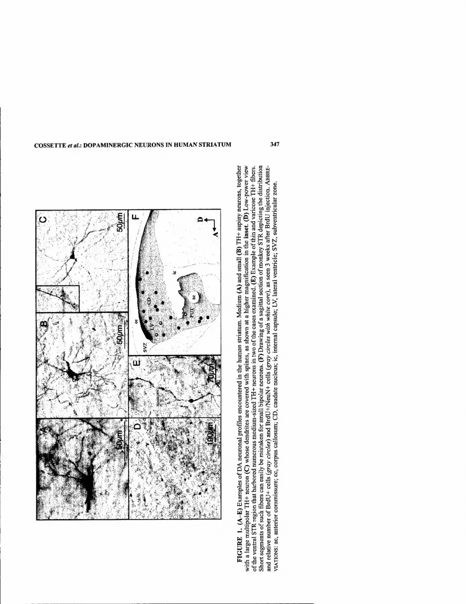
COSSETTE etal.: DOPAMINERGIC NEURONS IN HUMAN STRIATUM 347
o
■X'
;
-'y'^^
I, ..IIH.,iii.y.i I. 1^ i«iH||i« I... ..M.i.i.i.i
••■■';'""^fe", 'fit ► ? < ' 'i .,'»' ■ ■.
. .. • -< ,i;. • • »■
u ? S" o m
U > -D 3 S bO I, tB ^ 5 2 S + -fi ^ ' 5 K .2 d
S.H T! .2 J <U (U ■
o o ■
o
<1> B o N S
00 g o 3
>^ ?^^ ? « J3 -e
f S 5 -a V. 3 + B .S od (§ " ^ 'o - H a N ^ t^H on >,
<a
s a ^ o S o ^ 0 a t; ^ c _ e T3^ a r! ca - '|B|
O D Q u
^,|-^ ,S-
c u >
IJI a o ^ «:s ><'&.Si 1 J3 S a u
a
a o « 5 g
fl M 2 2 g a
J3
.g •- ^ c .,
O O w u
"O ^ - - j5 "^ 0 O , "
g S p o 3 -q
w a
o TS •= Z a M N — + > f^ w a :D Q
3 t. P3 o
D.-5
§ S 3 O S -a 1^
S ^ <S-3
a u ^ ^ ^ "- o 3 o a M
i + i S25 a g«ffl § .2 ja t4- o 60 O O u " 3 tH 0
«p
S » 2 I S S S « § g^ g s ri ^ " (U - « « U f g
'S t« ^ a ? o ai a

348 ANNALS NEW YORK ACADEMY OF SCIENCES
RESULTS
Three types of DA neurons were encountered within the human STR. The most frequent ones had a round or oval perikarya of medium size (diameter: 20 x 15-20 H-m) from which emerged 3-5 varicose aspiny dendrites (FIG. 1A). Neurons of the second type, which were about 50% less abundant than those of the first type, had a small cell body (diameter: 10 x 10-15 ^m) with 3 or more varicose aspiny dendrites (FIG. IB). Neurons of the third type, which represented less than 1% of the total pop- ulation of STR DA neurons, had a large polygonal perikarya (diameter: 25-30 ftm) with 4 or more dendrites endowed with spines (FIG. IC). These 3 types of DA neu- rons were scattered throughout the entire extent of the caudate nucleus and putamen. However, in 2 of the 7 cases analyzed, a prominent cluster comprising 20-50 DA neurons per section occurred in the ventral STR, near the anterior commissure (FIG. ID). Besides neurons, numerous TH-immunoreactive processes were present in the human STR (FIG. IE). Short segments of these processes often displayed large swol- len varicosities (diameter: 10-12 ^m) and could thus easily be mistaken for small bipolar DA neurons. Such profiles were not taken into account in the present study because their varicosities lack NeuN immunoreactivity.
Various quantitative methods were used to evaluate the density of the STR DA neurons. The area of striatum was calculated with an image analysis system, and the neuronal densities were assessed by dividing the number of TH-positive neurons by the area of striatum surveyed. Because of the unequal size of our sample, factorial one-way ANOVA and Fisher's LSD methods were applied to compare the various density values. The Grubb's test was also used to look for the presence of outliers. The density of STR DA neurons varied significantly in the 7 individuals analyzed in the present study. The total number of DA neurons in the human STR ranged from 173 to 717, with a mean value estimated at 332 ± 199 neurons. However, this value is most likely an underestimate of the actual number of STR DA neurons, because only neuronal profiles that meet several stringent criteria were included in our sam- ple. Furthermore, we did not include in the density measurements the ventral STR prolific zones that were encountered in 2 individuals and which could have raised the total number of STR DA neurons to about 3000.
Finally, numerous BrdU-labeled cells were visualized in the STR of monkeys in- jected with this thymidine analogue. Their number ranged from 5-50 per 40-|xm- thick section. These BrdU+ cells were more abundant medially than laterally and displayed a rostrocaudal-decreasing gradient in caudate nucleus and putamen. Dou- ble immunofluorescence confocal studies have revealed that about 5-10% of the Br- dU+ striatal cells expressed NeuN, a marker for mature neurons.
DISCUSSION
This study has allowed us to: (1) confirm the presence of DA neurons in the hu- man STR; (2) characterize their morphological phenotype; (3) establish their topo- graphical localization; and (4) estimate their number. Despite their relatively small number, these DA neurons could play an important role in the functional organiza- tion of the human STR in both health and disease. For example, they could compen- sate for the loss extrinsic DA innervation of the STR seen in Parkinson's disease by

COSSETTE et al.: DOPAMINERGIC NEURONS IN HUMAN STRIATUM 349
either upregulating their DA metabolic machinery or by increasing their number, as suggested by experimental studies in monkeys.^ The absence of the aging pigment lipofuscin in the DA cells of the monkey SIR has lead to the suggestion that prolif- erative progenitor cells might contribute to the increase in intrinsic STR DA cells seen in Parkinsonian monkeys.^ This hypothesis is supported by the fact that new neurons are produced throughout adulthood in the STR of normal monkeys, as dem- onstrated here and elsewhere.^ These findings raise the possibility of experimentally enhancing the recruitment of newborn DA neurons as a means to alleviate the symp- toms of neurodegenerative diseases that affect the STR.
REFERENCES
1. DuBACH, M., et al. 1987. Primate neostriatal neurons containing tyrosine hydroxylase: immunohistochemical evidence. Neurosci. Lett. 75: 205-210.
2. BETARBET, R., et al. 1997. Dopaminergic neurons intrinsic to the primate striatum. J. Neurosci. 17: 6761-6768.
3. TASHIRO, Y., et al. 1989. Tyrosine hydroxylase-like immunoreactive neurons in the stri- atum of the rat. Neurosci. Lett. 97: 6-10.
4. MEREDITH, G.E., et al. 1999. Immunocytochemical characterization of catecholaminer- gic neurons in the rat striatum following dopamine-depleting lesions. Eur. J. Neuro- sci. 11: 3585-3596.
5. CossETTE, M., et al. 1999. Extrastriatal dopaminergic innervation of human basal gan- glia. Neurosci. Res. 34: 51-54.
6. BfiDARD, A., et al. 2002. Proliferating cells can differentiate into neurons in the stria- tum of adult monkey. Neurosci. Lett. 328: 213-216.

Secreted Factors from Primary Midbrain Glia Regulate Nurrl Activity
Y. LUO" AND H. J. FEDEROFF*
"Department of Environmental Medicine and '^Department of Neurology, Center for Aging and Developmental Biology, University of Rochester School of Medicine, Rochester, New York 14642, USA
KEYWORDS: dopamine; Nurrl; ligand; nuclear receptor; dopaminergic neu- rons; transcription factor
INTRODUCTION
Nurrl is a member of the orphan nuclear receptor family. Previous studies have demonstrated that Nurrl is essential for the development of midbrain dopaminergic (DA) neurons. However, the underlying mechanisms through which Nurrl works are still unknown. Overexpression of Nurrl in neuronal progenitor cells is sufficient to induce the dopaminergic phenotype only in the presence of secreted factors from midbrain glia cells. Other studies also have demonstrated that the ligand binding do- main of Nurrl has the intrinsic capacity for transcriptional activation, depending on cell type and mode of DNA binding. These data suggest that activity of Nurrl might be modulated by an unidentified coactivator and/or ligand in the developing and ma- ture central nervous system.
In this study, we used different truncated forms of Nurrl protein and a series of reporter constructs to look at the regulation of Nurrl by factors secreted by local glia cells. Progenitor cells (C17.2) were transfected with full-length or truncated forms of Nurrl constructs and the reporter constructs. Cells were then incubated with ei- ther serum-free medium (SFM) or SFM combined with different percentages of con- ditioned medium (CM) from primary midbrain glia cells. The activity of Nurrl was assessed by the expression of reporter gene (lacZ or luciferase).
METHODS
Plasmids
Constructs containing full-length or truncated forms of Nurrl are as shown in FIGURES 1 and 2.
Address for correspondence: Howard J. Federoff, M.D., Ph.D., University of Rochester School of Medicine, 601 Elmwood Ave., Rochester, NY 14642. Voice: 585-273-2190; fax: 585- 506-1957.
Ann. N.Y. Acad. Sci. 991: 350-353 (2003). © 2003 New Yorit Academy of Sciences.
350
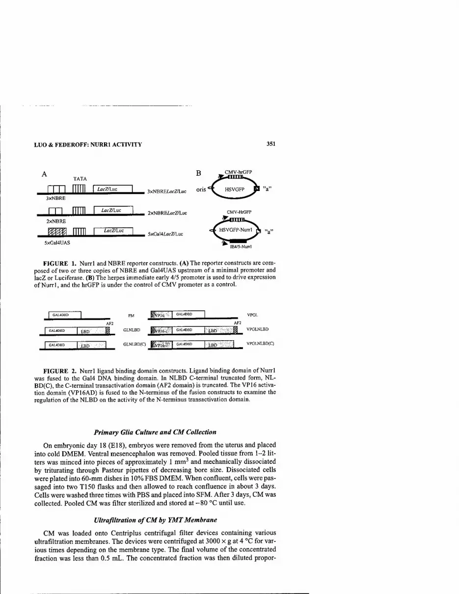
LUO & FEDEROFF: NURRl ACTIVITY 351
TATA
iflcZ/Luc
3XNBRE
m mm 2xNBRE
5xGal4UAS
lacZ/Luc
B
3xNBREincZ/Luc O^S
2xNBREiflcZ/Luc
CMV-hrGFP
incZ/Luc 5xGal4iocZ/Luc
=4 HSVGFP dl "a"
CMV-HrGFP
<^HSVGFP-Nurrl^ ..^
IE4/5-Nurrl
FIGURE 1. Nurrl and NBRE reporter constructs. (A) The reporter constructs are com- posed of two or three copies of NBRE and Gal4UAS upstream of a minimal promoter and lacZ or Luciferase. (B) The herpes immediate early 4/5 promoter is used to drive expression of Nurrl, and the hrGFP is under the control of CMV promoter as a control.
1 GAUDBD j
AF2
1 GAUDBD |. LBD ' m 1 GAL4DBD t-IBD :' .:.]
PM iWis 1 GAUDBD 1 AF2
,NLBD RWHIS 1 GAL4DBD LBD J_ GLNLBD(C) I'vplfti' ''I GAUDBD LBO VPOLNLBD(C)
FIGURE 2. Nurrl ligand binding domain constructs. Ligand binding domain of Nurrl was fused to the Gal4 DNA binding domain. In NLBD C-terminal truncated form, NL- BD(C), the C-terminal transactivation domain (AF2 domain) is truncated. The VP16 activa- tion domain (VP16AD) is fused to the N-terminus of the fusion constructs to examine the regulation of the NLBD on the activity of the N-terminus transactivation domain.
Primary Glia Culture and CM Collection
On embryonic day 18 (El8), embryos were removed from the uterus and placed into cold DMEM. Ventral mesencephalon was removed. Pooled tissue from 1-2 lit- ters was minced into pieces of approximately 1 mm^ and mechanically dissociated by triturating through Pasteur pipettes of decreasing bore size. Dissociated cells were plated into 60-mm dishes in 10% FBS DMEM. When confluent, cells were pas- saged into two T150 flasks and then allowed to reach confluence in about 3 days. Cells were washed three times with PBS and placed into SFM. After 3 days, CM was collected. Pooled CM was filter sterilized and stored at -80 °C until use.
Ultrafiltration of CM by YMT Membrane
CM was loaded onto Centriplus centrifugal filter devices containing various ultrafiltration membranes. The devices were centrifiiged at 3000 x g at 4 °C for var- ious times depending on the membrane type. The final volume of the concentrated fraction was less than 0.5 mL. The concentrated fraction was then diluted propor-
352 ANNALS NEW YORK ACADEMY OF SCIENCES
8000
7000
_ 6000
S 5000
■S 4000 3 3000
2000
1000
0
□ GFP pGFPNurTI
FIGURE 3. NurrI activity via NBRE is regulated by CM from midbrain glia. (A) Full- length Nurrl induces the reporter gene expression in a NBRE-specific manner. Three copies of NBRE are more potent than two copies. GaMUAS is not induced, demonstrating that the activation of NBRE is specific. (B) CM from midbrain glia cells enhances the activity of Nurrl on 3NBRE constructs in a dose-dependent manner. GFP only was used as a control.
600 DSFM nSFM+20%CM
S 500- 10 ■ SFM+50%CM 01
S: 400 1 ■ SFM+80%CM
RLU
of
luc
T
r. 100-
n - rrt^ GLNLBD VPGLNLBD VPGLNLBD(C)
FIGURE 4. Conditioned medium regulates transcriptional activity through the ligand binding domain of Nurrl. Reporter gene expression after transfection of different constructs and treatment with CM is shown. The intrinsic AF2 activity of NLBD is demonstrated by the expression of reporter gene. Deletion of the AF2 core sequence completely abolished the activity in C17.2 cells (data not shown). The activity of NLBD was enhanced by CM in a dose-dependent manner. Moreover, although the NLBD(C) did not have transcriptional ac- tivity, it can regulate the VP16 AD activity in the presence of CM.
tionally into SFM and added to transfected cells. The filtratration fraction was also collected and tested.
RESULTS
Full-length Nurrl protein activates reporter gene expression via a Nurrl-respon- sive element (NBRE), and CM from midbrain glia cells enhances the activity of Nurrl in a dose-dependent manner (FIG. 3). Furthermore, the ligand binding domain
LUO & FEDEROFF: NURRl ACTIVITY 353
B 9000
8000
70O0-
6000
5000
4000
300O
2000
1000
0
DSFM ■ YM-aoCon BYM30Fillr ■ 80%CM
SOOO
6000
4000
2000
DSFM ■ YM-3C0O QYM.3Fillr ■ac%CM
FIGURE 5. Concentrated and ultrafiltrated CM retained the enhancing activity for the Nurrl ligand binding domain. C17.2 cells were cotransfected with the GaWLuc reporter construct and GLNLBD. Conditioned medium was ultrafiltered through two types of YMT membrane (YM-3 retains molecules larger than 3KD, and YM-30 retains molecules larger than 30KD). The enhancing activity was effectively retained in concentrated fraction from both the YM30 (A) and YM-3 (B) membranes.
has an intrinsic transcriptional activity, which can be enhanced by the CM (FIG. 4). Our data demonstrate that a factor(s) in the CM from midbrain glia cells regulates the activity of Nurrl through the ligand binding domain (LBD) and that the factor(s) can be concentrated by ultrafiltration (FiG. 5).

Development of Nurrl Stable Cell Lines for the Identification of Downstream Targets
YU LUO," LEIGH A. HENRICKSEN,* KATHLEEN A. MAGUIRE-ZEISS,* AND HOWARD J. FEDEROFF*
"Department of Environmental Medicine and ''Department of Neurology, Center for Aging and Developmental Biology, University of Rochester School of Medicine, Rochester, New York 14642, USA
KEYWORDS: dopamine; differential display; Nurrl; nuclear receptor; dopam- inergic neurons; transcription factor
INTRODUCTION
Current evidence indicates that the transcription factor Nurrl is critical for the de- velopment of midbrain dopaminergic (DA) neurons. First, transgenic mice lacking Nurrl function are devoid of DA neurons in the midbrain. Second, overexpression of Nurrl, in combination with an unknown secretory factor from glia cells, is suffi- cient to induce the DA phenotype in neuronal progenitor cells (cl7.2). However, the cellular mechanism by which Nurrl promotes the DA phenotype is still unknown. Since Nurrl is a transcription factor, we speculate that Nurrl may exert its influence on DA neurons by regulating the expression of a specific group of target genes. We have developed cell lines with regulated Nurrl expression and have begun to identify potential downstream targets of Nurrl. The development of these cell lines will fur- ther our understanding of the role of Nurrl in the development of DA neurons by providing insight into the set(s) of genes regulated by Nurrl function.
METHODS AND RESULTS
Autoreglated Bidirectional Expression Vector for Nurrl
Our laboratory has utilized an autoregulated bidirectional tetracycline-responsive pBIG2i expression vector (FIG. 1) to establish stable expression of Nurrl in the mes- encephalic dopaminergic cell line MN9D. The bidirectional tetracycline-responsive promoter is comprised of a tetOy element, a stronger CMV element that drives cDNA expression, and a weak TK element to drive the transactivator (rtTA) expres- sion. In addition, a selectable marker is added to allow for the production of stable
Address for correspondence: Howard J. Federoff, M.D., Ph.D., University of Rochester School of Medicine, 601 Elmwood Ave., Rochester, NY 14642. Voice: 585-273-2190; fax: 585- 506-1957,
Ann. N.Y. Acad. Sci. 991: 354-358 (2003). © 2003 New Yorli Academy of Sciences.
354

LUO et al.: IDENTIFICATION OF DOWNSTREAM TARGETS 355
CMV* f TK* ^
Nurrl
IRES
eGFP HygroB
FIGURE 1. Schematic diagram of tetracycline-responsive vector pBig2I with Nurrl. (Adapted from Strathdee et a/.')
A B dox 24 hr 48 hr 72 hr 24 hr 48 hr 72 hr
FIGURE 2. eGFP expression is induced following treatment with doxycycline in MN9D/NurrMRES'eGFP cells, MN9D/NurrMRES«eGFP cells were plated onto 24-welI plates and induced with 2 |ig/mL doxycycline for 24, 48, and 72 hours. Cells were plated at a density of 4 x 10'' for 24- and 48-h time points and 1 x lO"* for a 72-h time point. Following induction, cells were examined under phase contrast (A) and green fluorescence (B) to detect eGFP expression.
cell lines and an internal ribosome entry site in front of a codon-optimized enhanced green fluorescent protein coding region (human codon optimized eGFP), which al- lows for the monitoring of protein expression in live cells.
Production of Tetracycline-Responsive Autoregulated Stable MN9D
After establishing the appropriate concentration of hygromycin B for selection, MN9D cells were transfected with pBig2i containing either an eGFP or a Nurrl •IRES'eGFP insert. Following selection for 14 days, cells were isolated and expanded. Positive stable clones were detected by eGFP fluorescence after induction with doxycycline. Using both a cloning ring and the autoclone cell sorting method, we obtained a number of stable cell lines. The autoclone method produced pure clonal lines. FIGURE 2 demonstrates the effectiveness of this system for the regulated expression of eGFP and Nurrl in MN9Ds. In the presence of doxycycline (Dox) eGFP expression is activated, while in the absence of Dox no eGFP fluorescence is observed.

356 ANNALS NEW YORK ACADEMY OF SCIENCES
0.03 ■ ^ 0.025' Z 0.02' M 0.015'
0.01' 0.005'
0
■ -DOX D + DOX
24 48 Time (hour)
72
FIGURE 3. Nurrl mRNA levels increase in a time-dependent manner following expo- sure to doxycycline. MN9D/Nurrl-eGFP cells were treated with Dox for 24,48, or 72 hours. RNA was isolated, DNase I treated, and reverse transcribed. The amount of Nurrl mRNA present was determined by qRT^-PCR.
Nurrl
DOX- +
I OS Z
SN
Lanel 2 3 4 5 6 7
FIGURE 4. Following exposure to Dox, induction of Nurrl is detected by Western blot analysis. Protein samples from two independent cell lines grown in the presence or absence of DOX were collected, separated by SDS-PAGE, and subjected to Western blot analysis. Nurrl protein is detected only in Nurrl stable cell lines following treatment with 2 ng/mL Dox {arrow; compare lanes 1 and 3 to lanes 2 and 4). Positive controls include a BHK lysate containing Nurrl fused to a Flag tag and transiently expressed (lane 5) and total protein from mouse substantia nigra (SN) (lane 7). Nurrl is not detected in the parental MN9D cell line (lane 6).
Characterization of Nurrl Stable Cell Lines
Nurr 1 mRNA expression was activated in this cell line after Dox treatment as de- termined by quantitative real-time PCR (qRT^-PCR). The mRNA for Nurrl increas- es approximately 15- to 25-fold after treatment with doxycycline for 48 hours (FIG.
3). As further confirmation, Nurrl protein was induced as determined by Western blot analysis (FiG. 4). We have successfully used this approach to obtain multiple stable cell lines derived from MN9D cells.
Identification of Nurrl Downstream Target Genes by Differential Display Analysis
Total RNA was extracted from MN9D Big2iNurrl and Big2ieGFP cells with and without Dox (1 \iglraV) induction. Total RNA was subjected to differential display

LUO etal.: IDENTIFICATION OF DOWNSTREAM TARGETS 357
Nurrl eGFP _ + _ + _
I
1 2 3 4 5 6 Lane
FIGURE 5. Differential display: treatment with DOX results in altered levels of select- ed mRNAs in stable Nurrl cell lines. After 48-h incubation with 2 |ig/inL Dox, mRNA was isolated and selectively amplified by PCR using primers designed for differential display in the presence of [^^P]dATP. Reaction products were separated on a 6% denaturing PAG and detected by phosphorimager. Shown is a representative gel. Transcripts that are either de- creased or increased {arrows) are detected by comparing samples without (lanes 1 and 3) or with (lanes 2 and 4) Nurrl expression (i.e., +/- Dox). These changes are absent in control lanes showing the parent expression vector containing only eGFP (lanes 5 and 6). Typical changes range from 1.5- to 2-fold as quantitated by Imagequant (Molecular Dynamics).
I O 14
H''\
Increasing Decreasing Trend in Transcript Expression
FIGURE 6. Summary of transcripts isolated by differential display. Bands iden- tified by differential display with either an increase or decrease were excised and reamplified.
analysis according to the manufacturer (GenHunter; Nashville, TN). Following am- plification, the samples were resolved on a denaturing PAGE and compared to each other. Transcripts more (or less) abundant were excised, reamplified, and isolated for further analysis and verification.
As shown in FIGURE 5, using one set of primers we visualize at least one differ- entially expressed gene following Nurrl induction (arrows). This technique will al- low for the identification and cloning of these differentially expressed genes. To date we have isolated 26 distinct transcripts (FIG. 6). As expected, comparison of these sequences with available databases results in both known genes and mRNAs of un- known function.
358 ANNALS NEW YORK ACADEMY OF SCIENCES
SUMMARY
We have employed the use of a vector containing a modified form of the tetracy- cline-inducible promoter system. There is minimal expression of Nurrl in the "off" state. However, addition of doxycycline permits the expression of the reverse tetra- cycline transactivator (rtTA) gene and a Nurrl-IRES-eGFP transcriptional cassette from a bidirectional promoter. Thus, we are able to temporally follow downstream transcription events resulting from Nurrl gain of function. We have established sev- eral Nurrl clonal cell lines from the dopaminergic cell line MN9D. Currently, we are using differential display to identify changes in gene expression following the induc- tion of Nurrl for 24,48, and 72 hours. This approach detects genes whose expression is either increased or decreased following Nurrl expression. We have detected tran- scripts with altered expresssion levels and are currently identifying the putative tar- get genes by sequencing.
REFERENCE
1. STRATHDEE, C.A., M.R. MCLEOD & J.R. HALL. 1999. Efficient control of tetracycline- responsive gene expression from an autoregulated bi-directional expression vector. Gene 229: 21-29.

Index of Contributors
Alben, L., 36-47 Ariano, M.A., 278-280 Arlotta, P., 229-236
Bed, M.F., 120-131 Bddard, A., 346-349 Benner, E.J., 319-321 Bernard, L.P., 342-345 Bezin, L., 307-310 Bhatt, L., 36-47 Biskup,T.M., 319-321 Boska, M.D., 319-321 Breidert, T., 214-228 Bremmer, S., 298-301 Brooks, D.J., 22-35 Brownell,A.-L., 281-283 Bubula, N., 339-341 Buchanan, T.M., 278-280 Burbach, J.P.H., 61-68 Burke, R.E., 69-79,284-287
Carlson, K.A., 319-321 Casaceli, C, 315-318 Chen, I.Y., 281-283 Chen, L., 315-318 Chinopoulos, C, 111-119 Cho, J.W., 284-287 Choi, D.-K., 272-277 Chu, C.T., 288-290 Collier, TJ., 140-151 Conn, K., 330-332 Cossette, M., 346-349
Dawson, T.M., 80-92,132-139 Dawson, V.L., 80-92,132-139 DeLong, M.R., 199-213 Dervan, A.G., 291-294 Destache, CI, 319-321 Doherty, S., 330-332 Duda, J.E., 295-297
Ehrhart, J., 342-345 Eisenhauer, P., 330-332
Fahn, S., 1-14 Farrer,M.J., 311-314 Federoff, H.J., ix, 152-166, 315-318,
350-353, 354-358 Fine, R., 330-332 Fisher, B., 298-301 Fiskum,G., 111-119
Gendelman, H.E., 319-321 Gherbassi, D., 36-47 Giasson, B.I., 295-297 Grissell, A.E., 278-280 Groc,L., 307-310 Gross, M., 339-341
Hamani, C, 15-21 Hardy, J., 167-170 Hartmann, A., 214-228 Hashimoto, M., 171-188 Hattori, N., 101-106 Heidenreich, K.A., 237-250 Heller, A., 339-341 Henricksen, L.A., 354-358 Hessefort, S., 339-341 Hirsch, E.C., 214-228 Hogg, E., 298-301 Hunot, S., 214-228 Hunter, T.J., 307-310
Imai, Y., 101-106 Ischiropoulos, H., 93-100
Jackson-Lewis, V., 272-277 Jakowec, M.W., 298-301 Jenkins, B.G., 281-283 Jiang, H., 307-310
Kadoya, C, 302-303 Kholodilov, N.G., 284-287 Kohbata, S., 302-303 Kress, G.J., 304-306
Lau, Y.-S., 291-294
359

360 ANNALS NEW YORK ACADEMY OF SCIENCES
Lee, V. M-Y., 107-110, 295-297 Levine, R.A., 307-310 Lim, K.L., 80-92 Lockhart,P.J., 311-314 Lozano, A.M., 15-21 Luo, Y., 350-353, 354-358
Macklis, J.D., 229-236 Magavi, S.S., 229-236 Maguire-Zeiss, K.A., 152-166,354-358 Masliah, E., 171-188 Mayer-Proschel, M., 251-271 McCarthy, M.K., 319-321 McGuire,S., 140-151 McNeill, T., 298-301 Meredith, G.E., 291-294 Meshul, C, 298-301 Michel, P.P., 214-228 Miller, R.M., 315-318 Mizuno, Y., 101-106 Mosley, R.L., 319-321
Nelson, J.A., 319-321 Nemechek, C, 319-321 Nixon, K., 298-301 Noble, M., 251-271
0'Farrell,C.A., 311-314
Parent, A., 346-349 Perlmann, T., 48-60 Petzinger, G.M., 298-301 Polster,B.M., 111-119 Power, J., 251-271 Przedborski, S., 189-198,272-277
Reynolds, I.J., 304-306 Rockenstein,E., 171-188 Rousselet, E., 214-228
Schulz, J.D., 326-329 Seegal, 322-325 Sgad6, P., 36-47 Shaw, C.A., 326-329 Shimoji, M., 132-139 Simon, H.H., 36^7 Smidt, M.P., 61-68 Smith,!., 251-271 Smits, S., 61-68 Sortwell, C.E., 140-151 Starkov, A., 111-119 Steece-Collier, K., 140-151
Takahashi, R., 101-106 Teismann, P., 272-277 Tieu, K., 272-277 Totterdell, S., 291-294 Trojanowski, J.Q., 107-110, 295-297
Ullman, M.D., 330-332
Vila, M., 189-198, 272-277
Wall^n, A., 48-60 Wang, H., 132-139 Wang, X., 281-283 Wells, J., 330-332 West, A.B., 311-314 Wichmann, T., 199-213 Wilson, J.M.B., 326-329 Wissler, J.H., 333-338 Won, L, 339-341 Wu, D.C., 272-277
Yu, M., 281-283 Yu, S.-W., 132-139
Zeevalk, G.D., 342-345 Zelivyanskaya, M.L., 319-321 Zhu, J.-H., 288-290