bac library for the amphipod crustacean, parhyale hawaiensis
TRANSCRIPT
Genomics 95 (2010) 261–267
Contents lists available at ScienceDirect
Genomics
j ourna l homepage: www.e lsev ie r.com/ locate /ygeno
BAC library for the amphipod crustacean, Parhyale hawaiensis
Ronald J. Parchem a,1, Francis Poulin b,1,2, Andrew B. Stuart d, Chris T. Amemiya d, Nipam H. Patel a,b,c,⁎a Department of Molecular and Cell Biology, University of California, Berkeley, CA 94720-3200, USAb Department of Integrative Biology, University of California, Berkeley, CA 94720-3140, USAc Center for Integrative Genomics, University of California, Berkeley, CA 94720-3200, USAd Genome Resource Center, Benaroya Research Institute, 1201 Ninth Avenue, Seattle, WA 98101, USA
⁎ Corresponding author. University of California at Berkand Cell Biology, 519A LSA # 3200, Berkeley, California643 5022.
E-mail address: [email protected] (N.H. Pa1 These authors contributed equally to this work.2 Present address: Genzyme Corporation, One The Mo
01701, USA.
0888-7543/$ – see front matter © 2010 Elsevier Inc. Adoi:10.1016/j.ygeno.2010.03.005
a b s t r a c t
a r t i c l e i n f oArticle history:Received 19 August 2009Accepted 9 March 2010Available online 16 March 2010
Keywords:EvolutionEcdysozoaCrustaceanArthropodAmphipodBAC library
Bacterial artificial chromosomes (BACs) are capable of propagating large fragments of DNA and have becomean invaluable tool for studying genome biology. To fill a phylogenetic gap in available genomic sequence andto complement ongoing molecular and genetic studies, we have generated a BAC library for the marineamphipod crustacean, Parhyale hawaiensis. The library was generated from genomic DNA isolated fromwhole adult animals and comprises 129,024 individual clones. The median insert size is ∼140 kb and thegenomic coverage is approximately five genome equivalents. We have screened the Parhyale BAC library fordevelopmentally relevant genes and characterized the genomic organization of four genes: a hedgehogortholog, and three Pax3/7 paralogs. Preliminary analysis suggests that introns are larger and more prevalentin Parhyale than in other arthropods whose genomes have been sequenced, which may partly account for thelarge genome size in Parhyale.
eley, Department ofMolecular94720-200, USA. Fax: +1 510
tel).
untain Road, Framingham, MA
ll rights reserved.
© 2010 Elsevier Inc. All rights reserved.
Introduction
Bilaterian animals are separated into two main clades, the deu-terostomes and the protostomes. The deuterostomes include thechordates (e.g. humans) and echinoderms (e.g sea urchins), while theprotostomes are divided into two large clades, the lophotrochozoansand the ecdysozoans. Molluscs (e.g. snails) and annelids (e.g.earthworms) are part of the lophotochozoans, while nematodes(e.g. C. elegans) and arthropods (e.g. insects, crustaceans) areecdysozoans. Of these groups, the ecdysozoans contain the vastmajority of described animal species, due in large part to the diversityof arthropods. Our current understanding of the phylogeneticrelationship of some of the major groups within the Ecdysozoa isshown in Fig 1.
Within the Ecdysozoa lie two powerful genetic model systems,Drosophila melanogaster and C. elegans. These two animals wereamong the first to have their genome completely sequenced [1,2], andthe genomes of several closely related species have been sequenced inorder to complement and leverage the large body of work involvingthese two model species [3,4]. While Drosophila and C. elegans remainpremier genetic systems for studies in many fields of biology, our
initial reliance upon these two organisms may have biased some ofour views of animal evolution and development.
For example, the genomes of Drosophila and C. elegans are some-what unusual in that they are relatively small, which undoubtedlycontributed to the decision to sequence them. However, many studiessuggest that these two species may not be representative of typicalextant bilaterians. For instance, WNT genes are a family of highlyconserved cell-cell signaling molecules whose founding member iswingless. In Drosophila there are seven (7), and in C. elegans there arefive (5) WNT family members [5]. In contrast, the human genomecontains nineteen (19)WNT genes, which at first glancemight suggestan expansion of this family in the vertebrate lineage. However, morerecent analyses of WNT family members in lophotrochozoans and thephylogenetically basal cnidarian Nematostella vectensis has revealedthirteen (13) ancestral WNT subfamilies [6–8]. This strongly suggeststhat the small number of WNTs in Drosophila and C. elegans is due togene loss. More extensive evidence of gene loss for these two modelspecies was provided by the finding of several important geneticpathways in Cnidaria that are absent in Drosophila and C. elegans [9].The question of when these losses occurred in the evolution ofEcdysozoa remains largely unanswered, and it is quite possible that atleast some of these losses represent cases of independent loss withinthe nematode and insect lineages. In addition to gene loss, thegenomes of Drosophila and C. elegans are peculiar in that they appearto have undergone compaction, as evidenced by the loss andshortening of introns, and a general decrease in intergenic distance[10,11].
Understanding when these genomic changes occurred and whatrole they played in the evolution of extant animals requires genomic

Fig. 1. Evolutionary relationships. Phylogenetic relationships of the major groups of animals within Ecdysozoa. Crustaceans are paraphyletic and Parhyale is a member of theVericrustacea lineage [21]. Tree topology and nomenclature based upon Regier et al, 2010 [21].
262 R.J. Parchem et al. / Genomics 95 (2010) 261–267
sequence from a broader set of species. Recent advancements insequencing and bioinformatics have enabled genomic studies of amore diverse set of animals, without having to focus exclusively onestablished model species with reduced genome size. Indeed, whilethe genomes of several ecdysozoans have been sequenced, the vastmajority belong to two clades: Insecta, with a heavy bias towardholometabolous insects, especially dipterans [3,12–18], and Nema-toda [2,4,19]. Therefore, our understanding of genome evolutionwithin Ecdysozoa will benefit greatly from obtaining genomic se-quence from additional species representing a broader taxonomicsampling [20]. As a case in point, the phylogenomic analysis of 2.6 Mbof sequence from 62 single-copy genes of 75 arthropods was requiredto resolve the deep phylogenetic history of the major arthropodlineages [21].
Since arthropods are not only species-rich, but also morphologi-cally and developmentally diverse, they are critically important forthe study of evolution and development (evo-devo). A great deal isknown about Drosophila development due to its powerful genetictools and large research community. In comparison we know far lessabout the development of other arthropods, thus more taxonomicsampling is necessary to help us understand the forces underlying theradiation of this group [21]. To this end, the marine amphipod,Parhyale hawaiensis, has been developed as a new model system forevo-devo studies [22–24]. Part of a morphologically diverse group ofcrustaceans termed malacostracans, Parhyale is closely related toeconomically important animals such as shrimps, crabs, and lobsters,and its phylogenetic position allows important comparisons to recon-struct ancestral features within insects and Pancrustacea [21,25].
In an effort to develop a more powerful genetic system for evo-devo studies, and to begin to address issues of genome evolutionwithin arthropods and, more broadly, in Ecdysozoa, we have gen-erated a bacterial artificial chromosomes (BAC) library for Parhyale.BACs are a plasmid vector-based system capable of accommodatinglarge inserts of DNA (N100 kb) [26–28]. Using an F-factor origin ofreplication, these bacterial plasmids are maintained as a single copywithin E. coli and allow for faithful propagation of large DNA frag-ments. These libraries can then be used for molecular and genomicstudies such as positional cloning [29] or comparing gene structuresand synteny across different species [30]. Moreover, BACs are a con-venient tool for the initial physical and genetic mapping of genomicregions without whole genome sequencing. Thus, BAC libraries are anexcellent means of studying genome level questions, particularly for
those interested in comparative studies of specific genomic regions.Highlighting their usefulness, BAC libraries have now been con-structed for hundreds of species ranging from bacteria to plants toanimals. Here we report the building and initial characterization of aParhyale hawaiensis BAC library.
Results and discussion
We have constructed a BAC library for the amphipod crustacean,Parhyale hawaiensis, that consists of 129,024 clones with an averageinsert size of 140 kb. The genome size of P. hawaiensis is estimated tobe 3.6 Gb (N.H.P.and Aziz Aboobaker, unpublished data), and there-fore the coverage of our library is estimated to be close to five genomeequivalents. The Parhyale BAC clones were robotically picked, grown,and stored in 384-well plates and they were arrayed onto several setsof nylon filters for screening. To assess the quality of the library, wescreened the filters with gene-specific probes derived from cDNAs wecloned from Parhyale embryos for developmental and evolutionarystudies. Probes were designed outside of conserved domains tominimize their potential for cross-hybridization. The identity of BACclones identified by screening was confirmed by PCR and/or Southernanalysis. Table 1 summarizes the results of screening the Parhyale BAClibrary with probes for twenty (20) single-copy genes. This initialstudy confirmed the anticipated 5X coverage of the library. Based onthe success of our initial screening, we identified an additional 50BACs and analyzed their insert size by pulse-field gel electrophoresis.This analysis revealed the average insert size to be 140 kb (Fig. 2),with inserts ranging in size from 95 kb to 170 kb. The majority ofclones (46 of 70) have inserts of 130 to 150 kb (Fig. 3). Importantly,our results demonstrate the quality of this BAC library, which containsinserts of expected size, and possesses enough genomic coverage toidentify several BACs spanning a genomic region of interest.
One reason for investigating the genomeof Parhyale is that it is largerthan that of the classic ecdysozoan models, Drosophila and C. elegans.These organisms, potentially as a consequence of their rapid life cycle,appear to have undergone a reduction of their genome size resulting inintron loss and smaller gene size. To address this issue specifically, wehave used BAC mapping and sequencing to verify whether genes arelarger and intronsmore prevalent in Parhyale. Sequence analysis of fourdifferent BACs provides preliminary evidence that introns have bothgreater size andnumber inParhyalewhencompared to their ortholog(s)in other species.
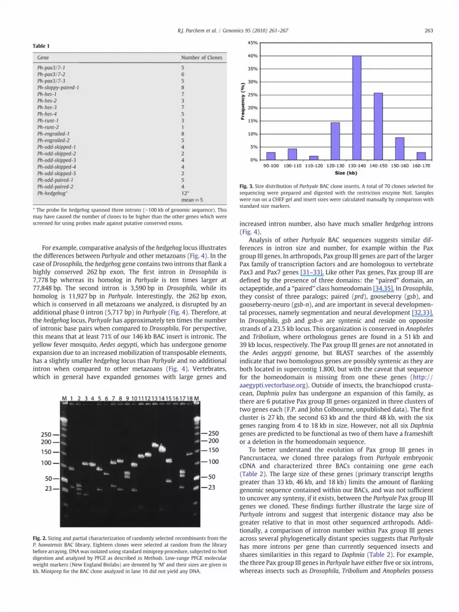
Table 1
Gene Number of Clones
Ph-pax3/7-1 5Ph-pax3/7-2 6Ph-pax3/7-3 5Ph-sloppy-paired-1 8Ph-hes-1 7Ph-hes-2 3Ph-hes-3 7Ph-hes-4 5Ph-runt-1 3Ph-runt-2 1Ph-engrailed-1 8Ph-engrailed-2 5Ph-odd-skipped-1 4Ph-odd-skipped-2 2Ph-odd-skipped-3 4Ph-odd-skipped-4 4Ph-odd-skipped-5 2Ph-odd-paired-1 5Ph-odd-paired-2 4Ph-hedgehog* 12*
mean=5
* The probe for hedgehog spanned three introns (N100 kb of genomic sequence). Thismay have caused the number of clones to be higher than the other genes which werescreened for using probes made against putative conserved exons.
Fig. 3. Size distribution of Parhyale BAC clone inserts. A total of 70 clones selected forsequencing were prepared and digested with the restriction enzyme NotI. Sampleswere run on a CHEF gel and insert sizes were calculated manually by comparison withstandard size markers.
263R.J. Parchem et al. / Genomics 95 (2010) 261–267
For example, comparative analysis of the hedgehog locus illustratesthe differences between Parhyale and other metazoans (Fig. 4). In thecase of Drosophila, the hedgehog gene contains two introns that flank ahighly conserved 262 bp exon. The first intron in Drosophila is7,778 bp whereas its homolog in Parhyale is ten times larger at77,848 bp. The second intron is 3,590 bp in Drosophila, while itshomolog is 11,927 bp in Parhyale. Interestingly, the 262 bp exon,which is conserved in all metazoans we analyzed, is disrupted by anadditional phase 0 intron (5,717 bp) in Parhyale (Fig. 4). Therefore, atthe hedgehog locus, Parhyale has approximately ten times the numberof intronic base pairs when compared to Drosophila. For perspective,this means that at least 71% of our 146 kb BAC insert is intronic. Theyellow fever mosquito, Aedes aegypti, which has undergone genomeexpansion due to an increased mobilization of transposable elements,has a slightly smaller hedgehog locus than Parhyale and no additionalintron when compared to other metazoans (Fig. 4). Vertebrates,which in general have expanded genomes with large genes and
Fig. 2. Sizing and partial characterization of randomly selected recombinants from theP. hawaiensis BAC library. Eighteen clones were selected at random from the librarybefore arraying. DNAwas isolated using standardminiprep procedure, subjected toNotIdigestion and analyzed by PFGE as described in Methods. Low-range PFGE molecularweight markers (New England Biolabs) are denoted by ‘M’ and their sizes are given inkb. Miniprep for the BAC clone analyzed in lane 16 did not yield any DNA.
increased intron number, also have much smaller hedgehog introns(Fig. 4).
Analysis of other Parhyale BAC sequences suggests similar dif-ferences in intron size and number, for example within the Paxgroup III genes. In arthropods, Pax group III genes are part of the largerPax family of transcription factors and are homologous to vertebratePax3 and Pax7 genes [31–33]. Like other Pax genes, Pax group III aredefined by the presence of three domains: the “paired” domain, anoctapeptide, and a “paired” class homeodomain [34,35]. In Drosophila,they consist of three paralogs; paired (prd), gooseberry (gsb), andgooseberry-neuro (gsb-n), and are important in several developmen-tal processes, namely segmentation and neural development [32,33].In Drosophila, gsb and gsb-n are syntenic and reside on oppositestrands of a 23.5 kb locus. This organization is conserved in Anophelesand Tribolium, where orthologous genes are found in a 51 kb and39 kb locus, respectively. The Pax group III genes are not annotated inthe Aedes aegypti genome, but BLAST searches of the assemblyindicate that two homologous genes are possibly syntenic as they areboth located in supercontig 1.800, but with the caveat that sequencefor the homeodomain is missing from one these genes (http://aaegypti.vectorbase.org). Outside of insects, the branchiopod crusta-cean, Daphnia pulex has undergone an expansion of this family, asthere are 6 putative Pax group III genes organized in three clusters oftwo genes each (F.P. and John Colbourne, unpublished data). The firstcluster is 27 kb, the second 63 kb and the third 48 kb, with the sixgenes ranging from 4 to 18 kb in size. However, not all six Daphniagenes are predicted to be functional as two of them have a frameshiftor a deletion in the homeodomain sequence.
To better understand the evolution of Pax group III genes inPancrustacea, we cloned three paralogs from Parhyale embryoniccDNA and characterized three BACs containing one gene each(Table 2). The large size of these genes (primary transcript lengthsgreater than 33 kb, 46 kb, and 18 kb) limits the amount of flankinggenomic sequence contained within our BACs, and was not sufficientto uncover any synteny, if it exists, between the Parhyale Pax group IIIgenes we cloned. These findings further illustrate the large size ofParhyale introns and suggest that intergenic distance may also begreater relative to that in most other sequenced arthropods. Addi-tionally, a comparison of intron number within Pax group III genesacross several phylogenetically distant species suggests that Parhyalehas more introns per gene than currently sequenced insects andshares similarities in this regard to Daphnia (Table 2). For example,the three Pax group III genes in Parhyale have either five or six introns,whereas insects such as Drosophila, Tribolium and Anopheles possess

Fig. 4. Comparison of hedgehog genomic organization in metazoans. These schematics correspond to the protein-coding region of the hedgehog locus. Black bars represent exons (notto scale), the green bar represents an intron in Parhyale hedgehogwith no corresponding intron in other metazoans in this analysis. Blue and red bars represent homologous intronsbetween species. The large size of intron one in Parhyale and Aedes hedgehog is denoted by a broken line (and thus are not to scale).
264 R.J. Parchem et al. / Genomics 95 (2010) 261–267
far fewer (mode: 1, range: 1-5). The similarity in intron numberbetween Parhyale and Daphnia supports the idea that some insects,especially dipterans, may have undergone genome compaction,
Table 2
ND=Not determined.Light grey highlights two potential pseudogenes in Daphnia.Dark grey boxes indicate that no synteny was observed after the analysis of completedgenomic sequences.
possibly due to generalized intron loss. Exploring metazoan genomesoutside Ecdysozoa also indicates that the Pax3/7 genes have moreintrons in the vertebrate lineage. For instance, in Nematostella thereare four paralogous PaxD genes which have three introns each andrelatively small size (4 to 8 kb; Table 2). This is in contrast to humans,where Pax3 and Pax7 have seven introns, and much larger gene sizes(95 and 103 kb, respectively). To what degree greater intron numberin this gene family represents parallel intron gain will require addi-tional taxonomic sampling of genes from the Pax3/7 family.
Interestingly, the large genome size and gene structure in Parhyaleare reminiscent of the mosquito, Aedes aegypti. Analysis of Aedesgenome indicates that it has increased in size relative to other dip-terans due to higher transposable element (TE) content [17]. Consis-tent with TE mobilization, intron size and intergenic distance haveincreased, but not intron number [17]. In contrast, our analysis of fourBACs suggests that Parhyale not only has larger introns and greaterintergenic distance, but may also have a global increase in the numberof introns per gene (Fig. 4 and Table 2). Unfortunately, the phylo-genetic distance between Parhyale and other well-characterizedgenomes, and the limited amount of genomic sequence obtainedthus far, did not allow us to characterize the repeat content ofParhyale. However, similitudes with Aedes suggest that the largegenome size of Parhyale may be attributable, at least in part, to anincrease in the number of repetitive elements. This question is criticalto our understanding of the forces that shaped the crustacean genomeand can be investigated further when greater sequence data areavailable. Although preliminary, our survey of Parhyale BAC sequencessuggests that it may have undergone a unique form of genomeexpansion that could include an increase in intron number, a phe-nomenon yet to be fully characterized. It is intriguing that a similarphenomenon of intron gain has recently been described in extantpopulations of another crustacean, Daphnia pulex [36]. A similar pro-cess occurring in Parhyale could explain the variation in intronposition that is observed in Pax group III genes relative to that in otherarthropods (Table 2). As an alternative to intron gain, Parhyale couldrepresent an arthropod genome possessing ancestral introns thatwere lost in other arthropod lineages with sequenced genomes.These two alternatives are not mutually exclusive, although intronloss is expected to be more prevalent and less deleterious to organ-ismal fitness relative to intron gain. Further analysis of arthropod

265R.J. Parchem et al. / Genomics 95 (2010) 261–267
genomes, especially those from crustaceans of varied lineages, andwith genomes comparable in size to Parhyale, may shed light on thisissue.
Practically speaking, the presence of large and frequent introns inParhyale will necessitate careful mapping of BAC inserts and/or theuse of multiple BACs to study intron-containing loci. For example, thecoding region of Parhyale hedgehog occupies a genomic region of104,631 bp, whereas Drosophila only occupies 12,782 bp. If one con-siders that the hedgehog BAC chosen for sequencing was 146 kb, ofwhich 104 kb spanned the coding region, it is apparent that our abilityto identify a BAC containing both coding and large upstream ordownstream regions is limited. Nevertheless, by “walking” throughthe BAC library we have been able to analyze very large loci, such asthe Hox gene complex of Parhyale (N.H.P. and Julia Serano, unpub-lished data).
In summary, we have generated a BAC library for the emergingmodel system, Parhyale hawaiensis. Our initial characterization of thislibrary indicates that its genomic coverage is 5X and that the averageinsert size is 140 kb, thereby providing a valuable resource for geno-mic studies in Parhyale. Additionally, we have isolated 70 BAC clonesfor developmentally regulated genes and have undertaken theircomplete sequencing. Interestingly, the data presented here suggestssignificant differences in genome structure, particularly in intron sizeand frequency, in Parhyale relative to the other arthropods that havebeen sequenced. Importantly, these preliminary findings allow us togenerate initial hypotheses regarding genome size evolution. Thesehypotheses can be tested by both further analyzing genome orga-nization in Parhyale and by increasing the overall phylogeneticdiversity of characterized genomes, making certain to avoid a biastowards only small genomes. Since Parhyale is phylogeneticallydistant from current model systems and analysis of its genome mayin part help understand ecdysozoan genome evolution, we believethis BAC library represents an important tool for comparativeevolutionary studies.
Materials and methods
High molecular weight DNA extraction
We isolated high molecular weight (HMW) DNA from forty adultParhyale hawaiensis (20 males and 20 females) collected from anisofemale population established in 2001 (Iso2). The animals werestarved for 6 days, rinsed several times in 0.22 μm filter sterilized seawater (FSW), and treated for 48 hours with antibiotics (Tetracycline30 μg/mL; 1% PenStrep (Invitrogen)). The specimens were rinsedseveral times with ice-cold FSW, rinsed twice with ice-cold Homog-enization Buffer (HB: 0.35 M sucrose, 0.1 M EDTA, 50 mMTris pH 8.0),minced grossly with needles, and added to 15 mL of ice cold HB.Nuclei were released with a dounce homogenizer on ice, 30 cycleswith the loose pestle and 15 cycles with the tight pestle. The homog-enate was filtered through three 100 μm cell strainers (BD Falcon),and then two 70 μm cell strainers (BD Falcon). The suspension waspelleted at 3200×g and 4 °C for 15 min. The nuclei pellet wasresuspended in 20 mL ice cold HB, filtered through three successive70 μm cell strainers (BD Falcon), and pelleted at 3200×g and 4 °C for15 min. The nuclei were resuspended in 500 μL ice cold HB andincubated at 37 °C for 10 min before mixing with 500 μL of 2% Incertagarose (Lonza) prepared in HB and kept at 37 °C. The suspension wasdispensed in 80 μL block molds (Bio-Rad) and placed at 4 °C for10 min. The blocks were incubated at 37 ° C in cell lysis solution (CLS:1% LDS, 10 mM Tris pH 8.0, 150 mM EDTA pH 8.0) for 4 days, withdaily changes of CLS. The CLS was substituted with Block StorageSolution (BSS: 0.2% N-laurylsarcosine, 2 mM Tris pH 8.0, 140 mMEDTA pH 8.0) by rinsing four times in 2 h. The blocks were stored inBSS at 4 °C until processing.
BAC library construction
For size fragmentation, genomic DNA (1/2 block per reaction)was partially digested at 37 ° C for 2.5 hours in 500 μL reactionvolumes containing 1 unit of the restriction enzyme EcoRI (NewEngland Biolabs) and 100 units EcoRI methylase. The quantity of EcoRIand EcoRI methylase was derived from a titration using variousamounts of enzymes on 1/6th block per 500 μL reaction. The reactionalso contained 2.6 mM spermidine, 0.5 mg/ml BSA (New EnglandBiolabs) and 1x EcoRI reaction buffer (0.08 mM S-adenosylmethio-nine, 2 mM MgCl2, 1 mM DTT). The reaction was stopped by adding300 μg Proteinase K, 2.9 % N-laurylsarcosine, and 0.29 M EDTA,followed by an incubation at room temperature for 1 h. Proteinase Kwas inactivated by transferring blocks in 15 mL of ½X TE with 15 μLPMSF (100 mM), incubating for 20 min at 4 °C, and repeating 3 timeswith fresh solution. Blocks were equilibrated in 50 mL ½X TE for1 hour at 4 °C. DNA fragments were separated on a 1% agarose gel(Pulse-Field certified Bio Rad) using pulse-field gel electrophoresis(BioRad CHEF XAMapper) in ½X TBE buffer as previously described inLang et al [37]. Gel fragments were excised from the preparative lanethat contained HWM DNA, and the HMW DNA was electroeluted at4 V/cm for 4 hours at 4 °C and then dialyzed at 4 °C for 16 h in 4 L of½X TE. The sample volume was reduced to ∼180 μL by dialyzing at4 °C against 30% PEG8000 in ½X TE. The CopyControl BAC Cloning Kit(Epicentre) was used for ligation according to the manufacturer'sinstructions. Approximately 100 ng HMW DNA and 25 ng of theCopyControl pCC1BAC (EcoRI) vector were ligated in 50 μL reactionsthat were incubated overnight at 16 °C. After ligation, reactions werestopped by a brief Proteinase K treatment, deslated by drop dialysisagainst ddH2O for 1.5 h using 0.025 μm nitrocellulose filters (Milli-pore) and the sample volume was reduced to ∼20 μL by drop dialysisagainst PEG8000 (30% in ½X TE) at 4 °C. For transformation, 8 μL ofligation reaction was added to 100 μL electrocompetent cellsDH10BT1 (Invitrogen). Electroporations were performed using2 mm cuvettes and a BTXECM 630 (Harvard Apparatus Inc.) set at2.5 kV, 225 Ohms, and 25 μF. Transformed cells were recovered in50 ml conical tubes containing 10 mL SOC (2 transformations pertube), incubated with shaking (250 rpm) for 1 h at 37 °C, and glycerolwas added to the transformation mix to a final concentration of10%. Small aliquots were removed for titration and the remainingtransformants were flash-frozen in liquid nitrogen and stored at-80 °C.
Insert size estimation
The EpiLyse Solution (Epicentre) was used to analyze BAC DNAfrom 36 random white colonies (12 per size fraction), and estimatethe frequency and size of inserts. For further analysis, BAC DNAfrom 18 clones was isolated using a standard alkaline lysis miniprepprotocol and a third of each BAC clone preparation was digested usingNotI (New England Biolabs) and the size of each clonewas determinedusing pulse-field gel electrophoresis (15 h, 1 sec initial time, 20 secfinal time, 14 ° C, field angle120°, 6 V/cm) and low range PFG marker(New England Biolabs).
Library arrays and screening
To array the library, transformed cells were grown overnight at37 °C on LB agar plates (12.5 μg/mL chloramphenicol, 0.4 mM IPTGand 40 μg/mL X-GAL) and single white colonies were transfered into336×384-well microtiter plates (Genetix) using a colony-pickingrobot (Norgren Systems). A Total Array System (BioRobotics) wasused to replicate the library and prepare high-density nylon filter sets(22 cm×22 cm) containing BAC colony DNA. One filter set was hy-bridized with α32P-dCTP labeled DNA probes amplified from twentydifferent cDNAs (Table 1) as previously described [37]. Restriction

266 R.J. Parchem et al. / Genomics 95 (2010) 261–267
digestion of positive clones was performed with NotI, followed bypulse-field gel electrophoresis for insert sizing. The Parhyale BAClibrary is stored in the Patel Lab and filters are available upon request.
Acknowledgments
We would like to thank Lisa Chuong for her assistance with BACdata collection and analysis, William Browne for establishing theisogenic Parhyale population while in the Patel lab. The U.S. Depart-ment of Energy Joint Genome Institute provided sequencing andanalyses under the Community Sequencing Program supported by theOffice of Science of the U.S. Department of Energy under Contract No.DE-AC02-05CH11231, and we thank Jane Grimwood for helping tocoordinate the sequencing project. F.P. is a Research Fellow of TheTerry Fox Foundation through an award from the National CancerInstitute of Canada. N.H.P. is an Investigator of the Howard HughesMedical Institute.
References
[1] M.D. Adams, S.E. Celniker, R.A. Holt, C.A. Evans, J.D. Gocayne, P.G. Amanatides, S.E.Scherer, P.W. Li, R.A. Hoskins, R.F. Galle, R.A. George, S.E. Lewis, S. Richards, M.Ashburner, S.N. Henderson, G.G. Sutton, J.R. Wortman, M.D. Yandell, Q. Zhang, L.X.Chen, R.C. Brandon, Y.H. Rogers, R.G. Blazej, M. Champe, B.D. Pfeiffer, K.H. Wan, C.Doyle, E.G. Baxter, G. Helt, C.R. Nelson, G.L. Gabor, J.F. Abril, A. Agbayani, H.J. An, C.Andrews-Pfannkoch, D. Baldwin, R.M. Ballew, A. Basu, J. Baxendale, L. Bayraktar-oglu, E.M. Beasley, K.Y. Beeson, P.V. Benos, B.P. Berman, D. Bhandari, S. Bolshakov,D. Borkova, M.R. Botchan, J. Bouck, P. Brokstein, P. Brottier, K.C. Burtis, D.A. Busam,H. Butler, E. Cadieu, A. Center, I. Chandra, J.M. Cherry, S. Cawley, C. Dahlke, L.B.Davenport, P. Davies, B. de Pablos, A. Delcher, Z. Deng, A.D. Mays, I. Dew, S.M.Dietz, K. Dodson, L.E. Doup, M. Downes, S. Dugan-Rocha, B.C. Dunkov, P. Dunn, K.J.Durbin, C.C. Evangelista, C. Ferraz, S. Ferriera, W. Fleischmann, C. Fosler, A.E.Gabrielian, N.S. Garg, W.M. Gelbart, K. Glasser, A. Glodek, F. Gong, J.H. Gorrell, Z.Gu, P. Guan, M. Harris, N.L. Harris, D. Harvey, T.J. Heiman, J.R. Hernandez, J. Houck,D. Hostin, K.A. Houston, T.J. Howland, M.H. Wei, C. Ibegwam, et al., The genomesequence of Drosophila melanogaster, Science 287 (2000) 2185–2195.
[2] Genome sequence of the nematode C. elegans: a platform for investigatingbiology, Science 282 (1998) 2012–2018.
[3] A.G. Clark, M.B. Eisen, D.R. Smith, C.M. Bergman, B. Oliver, T.A. Markow, T.C.Kaufman, M. Kellis, W. Gelbart, V.N. Iyer, D.A. Pollard, T.B. Sackton, A.M.Larracuente, N.D. Singh, J.P. Abad, D.N. Abt, B. Adryan, M. Aguade, H. Akashi, W.W. Anderson, C.F. Aquadro, D.H. Ardell, R. Arguello, C.G. Artieri, D.A. Barbash, D.Barker, P. Barsanti, P. Batterham, S. Batzoglou, D. Begun, A. Bhutkar, E. Blanco, S.A.Bosak, R.K. Bradley, A.D. Brand, M.R. Brent, A.N. Brooks, R.H. Brown, R.K. Butlin, C.Caggese, B.R. Calvi, A. Bernardo de Carvalho, A. Caspi, S. Castrezana, S.E. Celniker, J.L. Chang, C. Chapple, S. Chatterji, A. Chinwalla, A. Civetta, S.W. Clifton, J.M.Comeron, J.C. Costello, J.A. Coyne, J. Daub, R.G. David, A.L. Delcher, K. Delehaunty,C.B. Do, H. Ebling, K. Edwards, T. Eickbush, J.D. Evans, A. Filipski, S. Findeiss, E.Freyhult, L. Fulton, R. Fulton, A.C. Garcia, A. Gardiner, D.A. Garfield, B.E. Garvin, G.Gibson, D. Gilbert, S. Gnerre, J. Godfrey, R. Good, V. Gotea, B. Gravely, A.J.Greenberg, S. Griffiths-Jones, S. Gross, R. Guigo, E.A. Gustafson, W. Haerty, M.W.Hahn, D.L. Halligan, A.L. Halpern, G.M. Halter, M.V. Han, A. Heger, L. Hillier, A.S.Hinrichs, I. Holmes, R.A. Hoskins, M.J. Hubisz, D. Hultmark, M.A. Huntley, D.B. Jaffe,S. Jagadeeshan, et al., Evolution of genes and genomes on the Drosophilaphylogeny, Nature 450 (2007) 203–218.
[4] L.D. Stein, Z. Bao, D. Blasiar, T. Blumenthal, M.R. Brent, N. Chen, A. Chinwalla, L.Clarke, C. Clee, A. Coghlan, A. Coulson, P. D'Eustachio, D.H. Fitch, L.A. Fulton, R.E.Fulton, S. Griffiths-Jones, T.W. Harris, L.W. Hillier, R. Kamath, P.E. Kuwabara, E.R.Mardis, M.A. Marra, T.L. Miner, P. Minx, J.C. Mullikin, R.W. Plumb, J. Rogers, J.E.Schein, M. Sohrmann, J. Spieth, J.E. Stajich, C. Wei, D. Willey, R.K. Wilson, R.Durbin, R.H. Waterston, The genome sequence of Caenorhabditis briggsae: aplatform for comparative genomics, PLoS Biol. 1 (2003) E45.
[5] C.Y. Logan, R. Nusse, The Wnt signaling pathway in development and disease,Annu. Rev. Cell Dev. Biol. 20 (2004) 781–810.
[6] A. Kusserow, K. Pang, C. Sturm, M. Hrouda, J. Lentfer, H.A. Schmidt, U. Technau, A.von Haeseler, B. Hobmayer, M.Q. Martindale, T.W. Holstein, Unexpectedcomplexity of theWnt gene family in a sea anemone, Nature 433 (2005) 156–160.
[7] B. Prud'homme, N. Lartillot, G. Balavoine, A. Adoutte, M. Vervoort, Phylogeneticanalysis of the Wnt gene family. Insights from lophotrochozoan members, Curr.Biol. 12 (2002) 1395.
[8] S.J. Cho, Y. Valles, V.C. Giani, Jr., E.C. Seaver, D.A. Weisblat, Evolutionary dynamicsof the Wnt gene family: a lophotrochozoan perspective. Mol. Biol. Evol. (2010),doi:10.1093/molbev/msq052.
[9] N.H. Putnam, M. Srivastava, U. Hellsten, B. Dirks, J. Chapman, A. Salamov, A. Terry,H. Shapiro, E. Lindquist, V.V. Kapitonov, J. Jurka, G. Genikhovich, I.V. Grigoriev, S.M.Lucas, R.E. Steele, J.R. Finnerty, U. Technau, M.Q. Martindale, D.S. Rokhsar, Seaanemone genome reveals ancestral eumetazoan gene repertoire and genomicorganization, Science 317 (2007) 86–94.
[10] D.C. Jeffares, T. Mourier, D. Penny, The biology of intron gain and loss, TrendsGenet. 22 (2006) 16–22.
[11] Y.H. Loh, S. Brenner, B. Venkatesh, Investigation of loss and gain of introns in thecompact genomes of pufferfishes (Fugu and Tetraodon), Mol. Biol. Evol. 25 (2008)526–535.
[12] S. Richards, R.A. Gibbs, G.M. Weinstock, S.J. Brown, R. Denell, R.W. Beeman, R.Gibbs, G. Bucher, M. Friedrich, C.J. Grimmelikhuijzen, M. Klingler, M. Lorenzen, S.Roth, R. Schroder, D. Tautz, E.M. Zdobnov, D. Muzny, T. Attaway, S. Bell, C.J. Buhay,M.N. Chandrabose, D. Chavez, K.P. Clerk-Blankenburg, A. Cree, M. Dao, C. Davis, J.Chacko, H. Dinh, S. Dugan-Rocha, G. Fowler, T.T. Garner, J. Garnes, A. Gnirke, A.Hawes, J. Hernandez, S. Hines, M. Holder, J. Hume, S.N. Jhangiani, V. Joshi, Z.M.Khan, L. Jackson, C. Kovar, A. Kowis, S. Lee, L.R. Lewis, J. Margolis, M. Morgan, L.V.Nazareth, N. Nguyen, G. Okwuonu, D. Parker, S.J. Ruiz, J. Santibanez, J. Savard, S.E.Scherer, B. Schneider, E. Sodergren, S. Vattahil, D. Villasana, C.S. White, R. Wright,Y. Park, J. Lord, B. Oppert, S. Brown, L. Wang, G. Weinstock, Y. Liu, K. Worley, C.G.Elsik, J.T. Reese, E. Elhaik, G. Landan, D. Graur, P. Arensburger, P. Atkinson, J.Beidler, J.P. Demuth, D.W. Drury, Y.Z. Du, H. Fujiwara, V. Maselli, M. Osanai, H.M.Robertson, Z. Tu, J.J. Wang, S. Wang, H. Song, L. Zhang, D. Werner, M. Stanke, B.Morgenstern, V. Solovyev, P. Kosarev, G. Brown, H.C. Chen, O. Ermolaeva, W.Hlavina, Y. Kapustin, et al., The genome of the model beetle and pest Triboliumcastaneum, Nature 452 (2008) 949–955.
[13] J.H.Werren, S. Richards, C.A. Desjardins, O. Niehuis, J. Gadau, J.K. Colbourne, L.W.Beukeboom, C. Desplan, C.G. Elsik, C.J. Grimmelikhuijzen, P. Kitts, J.A. Lynch, T.Murphy, D.C. Oliveira, C.D. Smith, L. van de Zande, K.C. Worley, E.M. Zdobnov, M.Aerts, S. Albert, V.H. Anaya, J.M. Anzola, A.R. Barchuk, S.K. Behura, A.N. Bera, M.R.Berenbaum, R.C. Bertossa, M.M. Bitondi, S.R. Bordenstein, P. Bork, E. Bornberg-Bauer, M. Brunain, G. Cazzamali, L. Chaboub, J. Chacko, D. Chavez, C.P. Childers, J.H. Choi, M.E. Clark, C. Claudianos, R.A. Clinton, A.G. Cree, A.S. Cristino, P.M. Dang,A.C. Darby, D.C. de Graaf, B. Devreese, H.H. Dinh, R. Edwards, N. Elango, E. Elhaik,O. Ermolaeva, J.D. Evans, S. Foret, G.R. Fowler, D. Gerlach, J.D. Gibson, D.G.Gilbert, D. Graur, S. Grunder, D.E. Hagen, Y. Han, F. Hauser, D. Hultmark, H.C.t.Hunter, G.D. Hurst, S.N. Jhangian, H. Jiang, R.M. Johnson, A.K. Jones, T. Junier, T.Kadowaki, A. Kamping, Y. Kapustin, B. Kechavarzi, J. Kim, B. Kiryutin, T. Koevoets,C.L. Kovar, E.V. Kriventseva, R. Kucharski, H. Lee, S.L. Lee, K. Lees, L.R. Lewis, D.W.Loehlin, J.M. Logsdon Jr., J.A. Lopez, R.J. Lozado, D. Maglott, R. Maleszka, A.Mayampurath, D.J. Mazur, M.A. McClure, A.D. Moore, M.B. Morgan, J. Muller, M.C. Munoz-Torres, D.M. Muzny, L.V. Nazareth, et al., Functional and evolutionaryinsights from the genomes of three parasitoid Nasonia species, Science 327(2010) 343–348.
[14] Insights into social insects from the genome of the honeybee Apis mellifera,Nature 443 (2006) 931–949.
[15] R.A. Holt, G.M. Subramanian, A. Halpern, G.G. Sutton, R. Charlab, D.R. Nusskern, P.Wincker, A.G. Clark, J.M. Ribeiro, R. Wides, S.L. Salzberg, B. Loftus, M. Yandell, W.H.Majoros, D.B. Rusch, Z. Lai, C.L. Kraft, J.F. Abril, V. Anthouard, P. Arensburger, P.W.Atkinson, H. Baden, V. de Berardinis, D. Baldwin, V. Benes, J. Biedler, C. Blass, R.Bolanos, D. Boscus, M. Barnstead, S. Cai, A. Center, K. Chaturverdi, G.K.Christophides, M.A. Chrystal, M. Clamp, A. Cravchik, V. Curwen, A. Dana, A.Delcher, I. Dew, C.A. Evans, M. Flanigan, A. Grundschober-Freimoser, L. Friedli, Z.Gu, P. Guan, R. Guigo, M.E. Hillenmeyer, S.L. Hladun, J.R. Hogan, Y.S. Hong, J.Hoover, O. Jaillon, Z. Ke, C. Kodira, E. Kokoza, A. Koutsos, I. Letunic, A. Levitsky, Y.Liang, J.J. Lin, N.F. Lobo, J.R. Lopez, J.A. Malek, T.C. McIntosh, S. Meister, J. Miller, C.Mobarry, E. Mongin, S.D. Murphy, D.A. O'Brochta, C. Pfannkoch, R. Qi, M.A. Regier,K. Remington, H. Shao, M.V. Sharakhova, C.D. Sitter, J. Shetty, T.J. Smith, R. Strong,J. Sun, D. Thomasova, L.Q. Ton, P. Topalis, Z. Tu, M.F. Unger, B. Walenz, A. Wang, J.Wang, M. Wang, X. Wang, K.J. Woodford, J.R. Wortman, M. Wu, A. Yao, E.M.Zdobnov, H. Zhang, Q. Zhao, et al., The genome sequence of the malaria mosquitoAnopheles gambiae, Science 298 (2002) 129–149.
[16] Q. Xia, Z. Zhou, C. Lu, D. Cheng, F. Dai, B. Li, A draft sequence for the genome of thedomesticated silkworm (Bombyx mori), Science 306 (2004) 1937–1940.
[17] V. Nene, J.R. Wortman, D. Lawson, B. Haas, C. Kodira, Z.J. Tu, B. Loftus, Z. Xi, K.Megy, M. Grabherr, Q. Ren, E.M. Zdobnov, N.F. Lobo, K.S. Campbell, S.E. Brown, M.F. Bonaldo, J. Zhu, S.P. Sinkins, D.G. Hogenkamp, P. Amedeo, P. Arensburger, P.W.Atkinson, S. Bidwell, J. Biedler, E. Birney, R.V. Bruggner, J. Costas, M.R. Coy, J.Crabtree, M. Crawford, B. Debruyn, D. Decaprio, K. Eiglmeier, E. Eisenstadt, H. El-Dorry, W.M. Gelbart, S.L. Gomes, M. Hammond, L.I. Hannick, J.R. Hogan, M.H.Holmes, D. Jaffe, J.S. Johnston, R.C. Kennedy, H. Koo, S. Kravitz, E.V. Kriventseva, D.Kulp, K. Labutti, E. Lee, S. Li, D.D. Lovin, C. Mao, E. Mauceli, C.F. Menck, J.R. Miller, P.Montgomery, A. Mori, A.L. Nascimento, H.F. Naveira, C. Nusbaum, S. O'Leary, J.Orvis, M. Pertea, H. Quesneville, K.R. Reidenbach, Y.H. Rogers, C.W. Roth, J.R.Schneider, M. Schatz, M. Shumway, M. Stanke, E.O. Stinson, J.M. Tubio, J.P. Vanzee,S. Verjovski-Almeida, D.Werner, O.White, S. Wyder, Q. Zeng, Q. Zhao, Y. Zhao, C.A.Hill, A.S. Raikhel, M.B. Soares, D.L. Knudson, N.H. Lee, J. Galagan, S.L. Salzberg, I.T.Paulsen, G. Dimopoulos, F.H. Collins, B. Birren, C.M. Fraser-Liggett, D.W. Severson,Genome sequence of Aedes aegypti, a major arbovirus vector, Science 316 (2007)1718–1723.
[18] Genome Sequence of the Pea Aphid Acyrthosiphon pisum, PLoS Biol. 8 (2010)e1000313.
[19] T.W. Harris, N. Chen, F. Cunningham, M. Tello-Ruiz, I. Antoshechkin, C. Bastiani, T.Bieri, D. Blasiar, K. Bradnam, J. Chan, C.K. Chen, W.J. Chen, P. Davis, E. Kenny, R.Kishore, D. Lawson, R. Lee, H.M. Muller, C. Nakamura, P. Ozersky, A. Petcherski, A.Rogers, A. Sabo, E.M. Schwarz, K. Van Auken, Q. Wang, R. Durbin, J. Spieth, P.W.Sternberg, L.D. Stein, WormBase: a multi-species resource for nematode biologyand genomics, Nucleic Acids Res. 32 (2004) D411–D417.
[20] G.E. Budd, M.J. Telford, The origin and evolution of arthropods, Nature 457 (2009)812–817.
[21] J.C. Regier, J.W. Shultz, A. Zwick, A. Hussey, B. Ball, R. Wetzer, J.W. Martin, C.W.Cunningham, Arthropod relationships revealed by phylogenomic analysis ofnuclear protein-coding sequences, Nature 463 (2010) 1079–1083.
267R.J. Parchem et al. / Genomics 95 (2010) 261–267
[22] W.E. Browne, A.L. Price, M. Gerberding, N.H. Patel, Stages of embryonic develop-ment in the amphipod crustacean. Parhyale hawaiensis, Genesis 42 (2005)124–149.
[23] A. Pavlopoulos, M. Averof, Establishing genetic transformation for comparativedevelopmental studies in the crustacean Parhyale hawaiensis, Proc. Natl. Acad.Sci. U.S.A. 102 (2005) 7888–7893.
[24] E.J. Rehm, R.L. Hannibal, R.C. Chaw, M.A. Vargas-Vila, N.H. Patel, The crustaceanParhyale hawaiensis: a new model for arthropod development, in: A. Gann, D.Crotty (Eds.), Emerging Model Organisms, A Laboratory Manual, Cold SpringHarbor Laboratory Press, Cold Spring Harbor, NY, 2009, pp. 373–404.
[25] U.W. Hwang, M. Friedrich, D. Tautz, C.J. Park, W. Kim, Mitochondrial proteinphylogeny joins myriapods with chelicerates, Nature 413 (2001) 154–157.
[26] K. Osoegawa, P.Y. Woon, B. Zhao, E. Frengen, An improved approach forconstruction of bacterial artificial chromosome libraries, Genomics 52 (1998) 1–8.
[27] C.T. Amemiya, T.P. Zhong, G.A. Silverman, M.C. Fishman, L.I. Zon, Zebrafish YAC,BAC, and PAC genomic libraries, Methods Cell Biol. 60 (1999) 235–258.
[28] H. Shizuya, B. Birren, U.J. Kim, V. Mancino, T. Slepak, Y. Tachiiri, M. Simon, Cloningand stable maintenance of 300-kilobase-pair fragments of human DNA inEscherichia coli using an F-factor-based vector, Proc. Natl. Acad. Sci. U.S.A. 89(1992) 8794–8797.
[29] K. Ito, K. Kidokoro, H. Sezutsu, J. Nohata, K. Yamamoto, I. Kobayashi, K. Uchino, A.Kalyebi, R. Eguchi, W. Hara, T. Tamura, S. Katsuma, T. Shimada, K. Mita, K. Kadono-Okuda, Deletion of a gene encoding an amino acid transporter in the midgut
membrane causes resistance to a Bombyx parvo-like virus, Proc. Natl. Acad. Sci.USA 105 (2008) 7523–7527.
[30] K. Sahara, A. Yoshido, F. Marec, I. Fuková, H. Zhang, C.C. Wu, M. Goldsmith, Y.Yasukochi, Conserved synteny of genes between chromosome 15 of Bombyx moriand a chromosome of Manduca sexta shown by five-color BAC-FISH, Genome 50(2007) 1061–1065.
[31] M. Buckingham, F. Relaix, The role of Pax genes in the development of tissues andorgans: Pax3 and Pax7 regulate muscle progenitor cell functions, Annu. Rev. Cell.Dev. Biol. 23 (2007) 645–673.
[32] G.K.Davis, J.A.D'Alessio,N.H. Patel, Pax3/7genes reveal conservation anddivergencein the arthropod segmentation hierarchy, Dev. Biol. 285 (2005) 169–184.
[33] G.K. Davis, C.A. Jaramillo, N.H. Patel, Pax group III genes and the evolution of insectpair-rule patterning, Development 128 (2001) 3445–3458.
[34] R. Breitling, J.K. Gerber, Origin of the paired domain, Dev. Genes. Evol. 210 (2000)644–650.
[35] D.Q. Matus, K. Pang, M. Daly, M.Q. Martindale, Expression of Pax gene familymembers in the anthozoan cnidarian, Nematostella vectensis, Evol. Dev. 9 (2007)25–38.
[36] W. Li, A.E. Tucker, W. Sung, W.K. Thomas, M. Lynch, Extensive, recent intron gainsin Daphnia populations, Science 326 (2009) 1260–1262.
[37] M. Lang, T. Miyake, I. Braasch, D. Tinnemore, N. Siegel, W. Salzburger, C. Amemiya,A. Meyer, A BAC library of the East African haplochromine cichlid fish Astatotilapiaburtoni, J. Exp. Zool. B Mol. Dev. Evol. 306 (2006) 35–44.