crustacean research 48: 81-97 (2019) - j-stage
TRANSCRIPT

81Received: 19 June 2019. Accepted: 2 Aug 2019. Published online: 19 Sept 2019
Crustacean Research 2019 Vol.48: 81–97
©Carcinological Society of Japan. doi: 10.18353/crustacea.48.0_81
A new species of Heteromysis (Mysida: Mysidae) from public coral reef aquaria in Vienna, Austria
Karl J. Wittmann and Daniel Abed-Navandi
Abstract.̶ Mysid shrimps of the genus Heteromysis surprisingly appeared in coral reef exhibition tanks and connected filtration tanks of the public aquarium center Haus des Meeres, Vienna, Austria. This material is first described here as Heteromysis (Olivemysis) domusmaris sp. nov. based on a specific set of five flagellate spines (modified setae) on the antennular trunk in both sexes. Besides features typical of the subgenus Olivemysis, the new species is also characterized by features of the anten-nae, eyes, rostrum, third thoracic endopod, by modifications of male pleopods 3, 4, and by spine patterns on the endopods of uropods and telson. The most similar species are known from (sub) tropical waters of the SW-Pacific, to a lesser extent also from the Indian Ocean and Atlantic. Short notes are given on color, foregut, larval morphol-ogy, and swimming behavior. Locomotion and respiratory movements are documented by video clips in the supplement.
LSID urn:lsid:zoobank.org:pub:4C1F925F-022C-4919-8CA4-C911C73E49B6
Key words: first description, taxonomy, larval morphology, bionomics, anthropogenic dispersal
Electronic supplementary material.̶The online version of this article contains supplementary material at https://www.jstage.jst.go.jp/article/crustacea/48/0/48_81/_article
■ Introduction
Eighty-nine species plus one non-nomino-typical subspecies are acknowledged (Mees & Meland, 2019; own database) in the genus Het-eromysis S. I. Smith, 1873, not including H. antillensis Verrill, 1923, an inadequately de-scribed taxon with questionable specific identi-ty. Twenty species belong to the nominotypical subgenus Heteromysis S. I. Smith, 1873, thirty-seven to Olivemysis Băcescu, 1968, four to Gnathomysis Bonnier & Pérez, 1902, and one to Neoheteromysis Băcescu, 1976. The subgen-eric identity appears questionable or unknown for as many as 27 species. Adult males are un-known in twelve species, adult females in eight; this includes two species with only im-
matures known.Two species have already been first described
by Murano & Fukuoka (2003) from exhibition tanks in the Aquarium of Kushimoto Marine Park Center, Wakayama, Japan. The here pre-sented first description of a third, morphologi-cally remote Heteromysis species, in this case from a public aquarium center in Austria, indi-cates that the current world-wide, non-inten-tional anthropogenic dispersal is also valid for “rare” species with a cryptic mode of life. The wealth of data given below is supplemented by notes on color, foregut structure, and larval morphology; and by videos on respiratory movements and swimming behaviour.
KARL J. WITTMANN, DANIEL ABED-NAVANDI
82 Crustacean Research 48
■Materials and Methods
Upon first detection in the Haus des Meeres, Vienna, Austria, in Dec. 2018, the below de-scribed mysids were encountered in a total of four coral reef exhibition tanks involving two independent seawater systems separated by two floors with other systems in between. This enables only a broad reconstruction of the ori-gin and date of introduction: during the last de-cade all imports of coral reef materials came from the Central Indo-Pacific Ecoregion (Phil-ippines, Indonesia and NE-Australia; map in Spalding et al., 2007) via a Dutch wholesaler (DeJongMarinelife Inc., NL). The imports of live corals usually comprised small portions of foundation-rocks. This suggests that the Het-eromysis founder population had hitch-hiked in crevices of such rocks.
All specimens studied were sampled from Dec. 2018 to Apr. 2019 with a small suction pipe from recesses in the service tanks. The surface structures populated by the mysids were mostly open-celled polyurethane foam which served as the filter of the aquarium sys-tem. A regime of 10 h dim light / 14 h darkness prevailed in the service room. Residual Arte-mia larvae were available most of the time. The inorganic composition of the seawater was routinely analyzed in March 2019 employing ICP-OES and IC-VWD/ECD methodology (Oceamo Inc., 1170 Vienna, Austria) (Suppl. 1). Freshly collected animals were put in small Petri dishes for microphotography and video recording. Their otherwise rapid movements were slowed down by gradually cooling the dishes (specimens in Fig. 1 and in Suppl. 2). Fixation was in 80% ethanol, long-term preser-vation in aqueous solutions of 80% ethanol with 10% propylene glycol. Aldehyde-based agents were avoided in order to prevent poten-tial damage to statoliths.
Preparation, measurements and examination of materials as in Wittmann (2008). Types de-posited at the Natural History Museum of Vi-
enna (NHMW).Terminology and taxonomy of the genus
Heteromysis as in Wittmann & Wirtz (2017); that publication is also used as the model for species description. Terminology of gross structures of the foregut follows Kobusch (1998); modified spines of the foregut accord-ing to Wittmann & Griffiths (2018). Marsupial stages are distinguished according to Wittmann (1981) and Wittmann et al. (2014).
■ Taxonomic Account
Family Mysidae Haworth, 1825Subfamily Heteromysinae Norman, 1892
Tribe Heteromysini Norman, 1892Genus Heteromysis S. I. Smith, 1873Subgenus Olivemysis Băcescu, 1968
Heteromysis (Olivemysis) domusmaris new species
LSID urn:lsid:zoobank.org:act:F34C2F5C-E496-4FB2-8C1C-D2FDCD831164.
Figs. 1–6, video clips in Suppl. 2
Material examined.̶Holotype: adult male with 4.5 mm body length (NHMW–26550), fil-tration tank of the “coral reef system” in the Haus des Meeres, Vienna, 24 Jan. 2019, leg. D. Abed-Navandi.
Paratypes: 20 adult females (3.4–5.2 mm BL), 34 adult males (3.0–6.0 mm BL), 36 sub-adults, 49 immatures (NHMW–26551), filtra-tion tank of the “coral reef system” in the Haus des Meeres, Vienna, 9 Dec. 2018 to 6 Apr. 2019, leg. D. Abed-Navandi.
Type locality.̶Not defined because the lo-cality of origin ought to be indicated according to Art. 76.1.1. of the nomenclatorial code (ICZN, 1999). The species is so far known only from coral reef aquaria in the public exhi-bition center Haus des Meeres, Vienna, Austria.
Etymology.̶The species name is a noun with neutral ending in genitive singular, repre-senting an amalgamated, literal Latin transla-
HETEROMYSIS DOMUSMARIS SP. NOV. FROM CORAL REEF AQUARIA
83Crustacean Research 48
tion of “Haus des Meeres”.Diagnosis.̶Diagnostic scheme adapted
from Wittmann & Wirtz (2017): eyes large, cornea extends over 20–30% eye surface. An-terior (=inner) margin of the eyestalks distally with small, acute spiniform extension (Fig. 2A, B). Carapace normal, anteriorly ending in a moderately large, subtriangular, apically rounded rostrum. Each of the three segments of the antennular trunk (Fig. 2F–K) with a flagel-late spine (=spine-like seta; besides other types of setae) dorsally on its distal margin; two additional, smaller flagellate spines sub-basally on outer margin of basal segment. These five flagellate spines modified in differ-ent ways (Fig. 2G–K), together representing a species-specific pattern that does not differ be-tween sexes. Medio-apical edge of terminal segment with long, smooth seta directed later-ally. Appendix masculina small, with dense tuft of setae (Figs. 1A, 2A). Antennal sympod with spiniform extension on outer face (Fig. 2L). Stout antennal scale (Fig. 2A, L) not extending beyond the (more basally inserting) trunk of antennal flagellum; scale clearly shorter than antennular trunk and reaches only to half the length of the terminal segment of the antennu-lar trunk in both sexes. Antennal scale setose all around, length 2.8–3.4 times maximum width; apical segment 6–10% total scale length. Mouthparts normal, labrum not pro-duced into a spiniform process. Carpopropodus of thoracic endopods 1–8 with 2, 2, 2, 3–5, 5–7, 6–7, 6–7, and 6–7 segments, respectively. Third thoracic endopods specialized as gnatho-pods by forming a powerful subchela showing (apart from small size differences) no consis-tent structural differences between sexes and between left and right gnathopods. Carpopro-podus swollen with maximum width 35–58% length. Carpus with 5–6 subapically flagellate, modified spines (=modified setae; Fig. 5B, C) along medial margin. The most distal 3–4 spines arranged in 1–2 pairs; the most proximal 2–3 ones single. Dactylus with strong, smooth
claw. “Tarsus” of endopod 4 (Fig. 5F) with smooth setae only, its dactylus (Fig. 3K) with short, almost straight, slender claw. Dactyli 5–8 each with long, well-curved, slender claw serrated in median portions (Figs. 3L–O, 4D). Females with large marsupial plates on thora-copods 7, 8; ultimate plates rolled inwards to form two widely communicating sub-cham-bers. Terminally well-rounded rudimentary oo-stegite present on thoracopod 6 (Fig. 5G). Each penis with three rounded apical lobes and one subapical lobe (Fig. 5H), no setae developed. Only the males with median processes from thoracic sternites 1–8 (Fig. 3C); not counting the usual, large lobe (Fig. 3C, D) on the first sternite, ensuring the caudal closure of the mouth area in both sexes. Pleopods reduced to small, setose plates in both sexes. Male pleo-pod 3 (Fig. 6C) with series of 11–15 small, flagellate spines (=modified setae) along in-ner-distal margin; male pleopod 4 (Fig. 6D) with series of 19–25 such spines (Fig. 6F) in analogous position; remaining male pleopods (notably including the second one; Fig. 6B) and all female pleopods without such spines, with normal setation only (Fig. 6A, B, E, G). Uropods (Fig. 4A) normal, entire; endopod armed with 2–6 approximately equally spaced spines below statocyst; length of spines strong-ly increasing (mostly continuously) in apical direction; distal half of endopod without spines. Uropodal exopods reach with 10–24% of their length beyond endopods and 28–52% beyond telson. Telson (Fig. 5J) length 1.3–1.4 times maximum width, 0.8–1.1 times endopod of uropod, and 0.6–0.9 times exopod of uro-pod. Each lateral margin of telson armed with 6–8 spines along proximal third, followed by a smooth section, and 7–8 spines along distal 35–48%, not counting the pair of apical spines. Proximally rounded U-shaped terminal cleft occupies 24–28% length of telson; cleft armed with 12–21 acute laminae along basal 41–65% of its margins, distal portions smooth. Disto-lateral lobes each with two spines on the nar-

KARL J. WITTMANN, DANIEL ABED-NAVANDI
84 Crustacean Research 48
rowly truncate apex. The outer apical spines are 11–14% telson length; inner apical spines are 0.5–0.8 times length of the outer ones.
Description.̶ Description scheme adapted from Wittmann & Wirtz (2017): all features of the diagnosis. General appearance small, ro-bust. For body size see ‘Material examined’. Cephalothorax comprises 23–44% of body length, pleon 37–58%, and carapace 25–32%. Abdominal somites 1–5 measure 0.6–0.9, 0.7–0.9, 0.7–0.9, 0.8–0.9, and 0.6–0.9 times the length of somite 6, respectively.
Carapace (Fig. 2A, C–E): normal, without apparent sexual dimorphism. Carapace covers 75–90% of cephalothorax dorsally. Rostrum represents a distinct horizontal, subtriangular, apically rounded plate covering only basal por-tions of the eyestalks. A rounded subrostral process (dashed line behind the rostrum in Fig. 2A) forms a dorso-ventral carina as an anterior extension of the head (without appendages). Antero-lateral edges of carapace with apically well-rounded protrusion (Fig. 2C). Cervical sulcus strongly developed; no cardial sulcus visible. Posterior margin evenly rounded, weakly emarginated, leaving 0.5–1.5 ultimate thoracic somites mid-dorsally exposed. As in many spe-cies of Mysidae, two characteristic groups of pores present medially on carapace. The anteri-or group is closely in front of the cervical sul-cus and consists of 16–25 pores with about 1 µm diameter in a roughly V-shaped arrange-ment (Fig. 2D). The posterior group of pores (Fig. 2E) less closely in front of the posterior margin; it consists of 10–12 such pores, sur-rounding a larger but indistinct, rounded struc-ture. Except for the here stated structures, outer surface of carapace smooth in both sexes.
Eyes (Figs. 1; 2A, B): eyes well developed, thick. Cornea diameter 1.1–1.5 times the length of apical segment of antennular trunk (mea-sured along median line) in dorsal view. Eye-stalks and cornea dorsoventrally somewhat compressed. In dorsal view the cornea appears reniform to calotte-shaped, measuring 0.66–
0.92 times the length of the eyestalk (cornea not included). In lateral view (Fig. 2B) the cor-nea appears oviform to oval with lower margin somewhat flattened. Anterior (=medial) mar-gin of eyestalks covered by minute scales.
Antennulae (Fig. 2A, F–K): three-segmented trunk not stouter in males (Fig. 2A) than in fe-males (Fig. 2F). Measured along dorsal midline, the basal segment is 37–43% trunk length, me-dian 9–13%, and terminal 44–50%. Outer face of basal segment subbasally with two small, stout spines (spine-like setae) each bearing a minute flagellum. This segment terminally with a dorsal and an outer apophysis (Fig. 2F). Dor-sal apophysis anteriorly with 3–4 plumose setae and a modified spine (spine-seta) bearing a sub-apical flagellum and 6–10 cilia along its con-verging apical margins (Fig. 2J). Outer apophy-sis with one plumose and 2–3 shorter barbed setae. Median segment anteriorly obliquely truncate. Dorsally it bears a small apophysis with 3–4 plumose and a smooth seta; its anterior margin with a flagellate spine (Fig. 2H) near in-ner distal corner. This spine more seta-like com-pared to those from the basal (Fig. 2J) and ter-minal (Fig. 2G) segments. Mid-dorsal apophysis of terminal segment with 2–3 barbed setae; its medio-terminal margin lined by minute cilia. Medio-apical edge of terminal segment in both sexes with 3–5 plumose setae, a large, smooth, laterally-directed seta and a stout flagellate spine, the latter with irregularly serrate, subtri-angular terminal margin (Fig. 2G). In both sexes the outer antennular flagellum is thicker than the inner one by a factor of 1.3–1.9 when measured near the basis of the flagella.
Antennae (Fig. 2L): a short, broad apical segment with five plumose setae is separated from basal part of antennal scale by a trans-verse suture. Antennal sympod with anteriorly directed, apically rounded subtriangular pro-cess on dorsal face. Sympod caudally with bul-bous lobe containing the end sac of the anten-nal gland. The three-segmented antennal peduncle with basal segment 19–23% peduncle

HETEROMYSIS DOMUSMARIS SP. NOV. FROM CORAL REEF AQUARIA
85Crustacean Research 48
length, second 44–49%, and third 29–35%.Mouthparts (Figs. 2M–O, 3A, B): labrum
(Fig. 2M) caudally serrate by a series of small, stiff bristles. Dense fields of setae on caudal and ventral faces of labrum. Mandibular palp three-segmented (Fig. 2N). Its proximal seg-ment without setae, smooth and short, 9–13% length of the palp. Remaining segments well
setose. Length of median segment 2.2–2.4 times its maximum width or 65–70% palp length, respectively. Terminal segment 22–24% palp length. Pars molaris with strong grinding surface in both mandibles. Pars incisiva with 3–4 teeth, digitus mobilis with 3–4 teeth, and pars centralis with three very spiny teeth. Labi-um normal, comprising two hairy lobes with
Fig. 1. In vitro microphotographs of Heteromysis domusmaris sp. nov., paratypes, cold-treated living specimens. A, female with body length 4.8 mm, note green eggs in the ovaries, and postnauplioid larvae distinguished by black corneas in the marsupium; B, female (4.7 mm BL) with green eggs in the marsupium; C, adult male (4.6 mm BL). Laboratory photos by Helmuth Goldammer. Three photos mounted together on same panel, background cleaned using electronic tools by KJW.
KARL J. WITTMANN, DANIEL ABED-NAVANDI
86 Crustacean Research 48
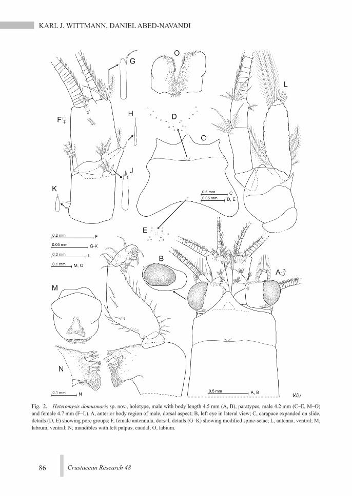
Fig. 2. Heteromysis domusmaris sp. nov., holotype, male with body length 4.5 mm (A, B), paratypes, male 4.2 mm (C–E, M–O) and female 4.7 mm (F–L). A, anterior body region of male, dorsal aspect; B, left eye in lateral view; C, carapace expanded on slide, details (D, E) showing pore groups; F, female antennula, dorsal, details (G–K) showing modified spine-setae; L, antenna, ventral; M, labrum, ventral; N, mandibles with left palpus, caudal; O, labium.
HETEROMYSIS DOMUSMARIS SP. NOV. FROM CORAL REEF AQUARIA
87Crustacean Research 48
dense set of stiff bristles on distal half of inner face (Fig. 2O). Distal segment of maxillula ter-minally with 10–13 smooth spines; no pores visible (Fig. 3A); subterminally with three se-tae barbed on distal half. Endite of maxillula with a plumose seta close to outer distal corner, all remaining setae barbed only on their distal half; endite (sub)apically with 10–12 such se-tae, plus an additional group of 4–5 such setae in median position on inner margin; no spines present. Maxilla (Fig. 3B) normal, densely se-tose, with various types of setae, but no spines or teeth. Its leaf-like exopod not extending be-yond basal segment of endopod; outer margin of exopod all along with plumose setae, the two apical setae larger than the remaining ones. Basal segment of endopod with three basally barbed setae. Terminal segment not expanded. This segment plus the sympod, and all three large endites of the sympod, with densely se-tose distal margins.
Foregut (Fig. 6J–N): Setae, but no spines, close to inlet from esophagus. Primary cardiac filter formed ventrally by dense combs of stiff setae behind inlet. Lateralia, infoldings, and superomedianum of the cardiac chamber dense-ly covered by smooth, slender setae and spines. Superomedianum in addition with a number of stronger, smooth spines. Lateralia anteriorly with dense series of slender, apically coronate spines of different length (Fig. 6K), more caudally with separate group of apically pronged spines (Fig. 6L). The latter spines with small teeth along their distal 30–60%. Posterior part of lateralia with 3–4 unilaterally serrated spines (Fig. 6N). Dorsolateral infolding with two stronger, api-cally pronged, serrated spines (Fig. 6M).
Thoracopods (general; Figs. 3E–O, 5A–H): a plumose seta plus a shorter barbed seta present at the intersegmental joint (Fig. 3F) connecting the sympod of thoracopod 2 with the corre-sponding thoracic sternite; no such setae in re-maining thoracopods. Total length of exopods as well as length of their basal plates increase from exopod 1 to 3, remain (sub)equal from 3
to 5 and then decrease caudad to exopod 8. Basal plates of exopods (Figs. 3E, 5A) weakly expanded, length 1.6–2.4 times maximum width in both sexes; exception: 2.6–3.0 in the (almost) not expanded plates of exopod 8 in fe-males. Outer margin of the plates ends in a well-rounded (Fig. 3E) to moderately acute edge (Fig. 5A). Flagellum 8-segmented (Fig. 3B) in exopod 1, 9-segmented (Fig. 5A) in exopods 2–8, not counting the large interseg-mental joint between basis and flagellum. The first thoracopods with large, leaf-like, smooth epipod (Fig. 3E). Length of endopods increases in the order of thoracopods 1, 2, 4, 3, (5–8). Endopod 5, when stretched, extends to basal or up to terminal segment of antennular trunk, en-dopod 8 to mandibles or at most to basal seg-ment of antennular trunk. Basis of endopod 4 (Fig. 5F) with a short, broad, lappet-like apophysis below endopod; this apophysis lon-ger and narrower in endopods 5–8 (Fig. 5G, H). Ischium becomes more slender and length of ischium increases in sequence of endopods 1–5; both these measurements remain (sub)equal among endopods 5–8. Ischium shorter than merus in endopods 1–4 (Figs. 3E, F; 5A, F), but longer than merus in endopods 5–8 (Fig. 5H). Meres 4–8 with smooth setae only, with the minor exception of a small barbed seta at the end of merus 4 (Fig. 5F) in 2 out of 6 specimens examined in this respect. Thoracic endopods 1–3 each with dactylus (Fig. 3G–J) larger than that of endopods 4–8 (Fig. 3K–O). Endopods 4–8 with “tarsus” in addition to claws bearing simple, smooth (in part curved) setae (Fig. 5F); endopods 5–8 in addition with 0–2 basally barbed setae. Length of claws in-creases in the order of endopods 2, 4, (5–8), 1, and 3 (Fig. 3G–O; note smaller scale in panels G–J compared with K–O). Claw 3 is the most powerful one (Fig. 3J), claw 4 the weakest (thinnest) (Fig. 3K). Claw 4 smooth all around, whereas claws 5–8 serrated along their median portions by acute, spine-like cilia (Figs. 3L–O, 4D), which become continuously longer with

KARL J. WITTMANN, DANIEL ABED-NAVANDI
88 Crustacean Research 48
Fig. 3. Heteromysis domusmaris sp. nov., paratypes, males with body length 4.8 mm (A, B), 4.2 mm (C, E–O), and female 4.7 mm (D). A, maxillula, caudal aspect; B, maxilla, rostral; C, male thoracic sternites 1–8 expanded on slide; D, female thoracic sternites 1–3; E, thoracopod 1, caudal; F, thoracic endopod 2, rostral; G–O, series of dactyli of endopods 1–8, lateral, setae omitted.

HETEROMYSIS DOMUSMARIS SP. NOV. FROM CORAL REEF AQUARIA
89Crustacean Research 48
more distal position.Maxillipeds (thoracic endopods 1, 2; Figs.
3E–H): coxa of first maxilliped (Fig. 3E) with small endite bearing one barbed seta at its tip. Basis with large prominent endite that is densely setose on inner margin. Ischium and merus each with one smaller but distinct, medi-ally setose endite. Large dactylus with strong smooth claw (Fig. 3G). Basis of second maxil-liped with large, distinctly medially projecting endite (Fig. 3F). In both sexes, merus slightly longer than combined praeischium plus ischi-um, but slightly shorter than combined carpop-ropodus plus dactylus. Dactylus very large, nonetheless bearing an only short, smooth claw (Fig. 3H). This claw not discernible in Fig. 3F because hidden among the dense brush of setae on dactylus. This brush formed by great num-bers of normal setae and 9–17 modified setae,
the latter apically bent, bearing two symmetri-cal series of denticles (stiff barbs) on either side in about median portions.
Gnathopods (thoracic endopods 3; Figs. 3J, 4E, 5A–E; Clip C in Suppl. 2): on average 20–40% larger in males than in females (note smaller scale in Fig. 5A versus Fig. 5D). Basis with much shorter endite (Fig. 5A) compared to that of endopod 2 (Fig. 3F). Ischium and merus strong, as normal in gnathopods. Merus with longitudinal series of 4–6 unilaterally barbed setae (Fig. 5E) on rostral face, remaining setae smooth. This series below drawing plane, its setae therefore rendered dotted in Fig. 5A. Car-pus 0.5–0.7 times merus length, 1.1–1.5 times ischium. The flagellate spines (Fig. 5B, C) on medial margin of carpus have smooth posterior margins (=proximal margins with respect to endopod). Most of these spines irregularly ser-
Fig. 4. Heteromysis domusmaris sp. nov., paratypes, adult females with 4.6 mm (A, B) and 4.3 mm (C–E) body length. A, B, ventral aspects of the same tail fan with focus on endopods of uropods (A) and telson (B); C, “tarsus” of thoracic endopod 5, detail (D) showing dactylus with claw; E, dactylus with claw in thoracic endopod 3. Laboratory photos by Helmuth Goldammer.

KARL J. WITTMANN, DANIEL ABED-NAVANDI
90 Crustacean Research 48
Fig. 5. Heteromysis domusmaris sp. nov., paratypes, male with body length 4.2 mm (A–C, F, H) and female 4.7 mm (D, E, G, J). A, male thoracopod 3, caudal aspect, details (B, C) showing flagellate spines of the carpopropodus; D, “tarsus” and part of merus of female thoracic endopod 3, rostral; E, detail of panel (D) showing barbed seta on the merus; F, thoracic endopod 4, rostral; G, sympod of thoracic endopod 6 with rudimentary oostegite, rostral; H, male thoracic endopod 8 with penis, rostral; J, telson; K, nauplioid larva at substage N4, lateral; L, tip of the nauplioid abdomen in another specimen.

HETEROMYSIS DOMUSMARIS SP. NOV. FROM CORAL REEF AQUARIA
91Crustacean Research 48
Fig. 6. Heteromysis domusmaris sp. nov., paratypes, males with body length 4.8 mm (A–F, J–N), 4.2 mm (H), female 4.7 mm (G). A–E, series of male pleopods 1–5, caudal face; F, detail of panel D showing flagellate spine; G, female pleopod 4, caudal; H, scutellum paracaudale, lateral; J, cardiac portion of the foregut, dorsal view, dorsal wall omitted, details (K–N) showing modified spines.

KARL J. WITTMANN, DANIEL ABED-NAVANDI
92 Crustacean Research 48
rate in median portions of their anterior (=dis-tal) margin; spines in more apical position (Fig. 5B) generally more strongly serrate than those in more basal position, the most basal ones may be weakly or not (Fig. 5C) serrate. Terminal claw 33–50% carpopropodus length.
Marsupium (Fig. 5G): oostegites 1, 2 (deri-vates of thoracopods 7, 8) without setae on upper (dorsal) margins. Lower margins of subbasal portions up to the rounded tip bearing series of setae, most of which are unilaterally barbed by fine cilia along their subbasal to median portions; bilaterally barbed ones also present. Oostegite 1 near basis with 6–11 setae, microserrated along their distal 40–70%. Oostegite 2 with only 1–2 such setae. Thoracopod 6 with rudimentary oo-stegite represented by a small, rounded lobe with 2–3 setae of that type (Fig. 5G).
Penes (Fig. 5H): both penes slender, length 79–100% that of the merus of the ultimate tho-racic endopod. Their shape roughly tube-like, pointing downwards-backwards. Each penis stiff, with smooth cuticle all around, including the lobes around the ejaculatory opening.
Pleopods (Fig. 6A–G): pleopods reduced to small setose, bilobate, or obscurely bilobate plates in both sexes, being generally larger in males than in females. Length without setae or spines increases from first to fifth pleopods in females. This series discontinuous in males, with size increasing in series of pleopods 1, 2, 5, (4, 3). For potential presence and numbers of flagellate spines on pleopods, see 'Definition' above. Pleopods 3, 4 knife-shaped in males only; each with a single seta at apex (Fig. 6C, D); their spines with a subapical flagellum (Fig. 6F). All setae of female pleopods and, not con-sidering spine-like setae, most setae of male pleopods are plumose or barbed.
Uropods (Fig. 4A, B): exopods slender, inner margin strongly convex; outer margin slightly sinusoid, almost straight. Endopods basally with large statocyst. Statoliths discoidal with shallow fundus and distinct tegmen. Mineral composition is fluorite (as also in all the 17
Heteromysis species listed by Wittmann & Ari-ani, 2019). Statolith diameter 78±14 µm (54–92 µm), statolith formula (1–2)+3+(0–1)+(6–11)+(5–8)=18–23 (n=8).
Telson (Figs. 4B, 5J): length 1.1–1.8 times that of sixth pleonite. Apical cleft distinctly deeper than wide. Proximal portions of cleft lined by acute laminae that are shorter than av-erage-sized spines along distal third of lateral margins of telson. For further details of the tel-son, see ‘Definition’ above.
Nauplioid larvae (Fig. 5K, L) with smooth cu-ticle all around except for the tip of the abdo-men, which bears a pair of cercopods and a few spines or setae. Each cercopod bears several small spines with apically increasing size. Size of cercopods measured without spines is 0.5–0.8% body length (n=6) thus much smaller than for example in Heteromysis (Heteromysis) cancelli Wittmann & Griffiths, 2017, whose cer-copods are about 9% body length (Wittmann & Griffiths, 2017: Fig. 3N). Remaining features in Fig. 5K are typical of the state of development.
■ Bionomic Account
Stomach content (studied in ten speci-mens).̶The foregut showed a prevalence of non-identifiable, finely macerated material. The identifiable parts were mostly arthropod re-mains, mainly from Artemia nauplii (used as fish food) and copepods. The consistent pres-ence of mineral particles in the cardiac and to a lesser extent in the pyloric part suggests that a significant portion of the food is taken from the sediment surface or close to it.
Color (Fig. 1, Suppl. 2).̶General appear-ance transparent, rendering visible, if present, the green eggs in ovaries and marsupium, and contents of foregut and alimentary canal. Small red chromatophore spots widely scattered over the body, with greatest density above the fore-gut and along the alimentary canal. While not proven here, we cannot exclude that expansion of the red chromatophore spots results in a red

HETEROMYSIS DOMUSMARIS SP. NOV. FROM CORAL REEF AQUARIA
93Crustacean Research 48
tinge of the entire body (Wittmann & Griffiths, 2017) as often observed in species of Heterom-ysis. The iridescence of bodies and appendages (Fig. 1, Suppl. 2) varies with the direction of incident light. Yolk mass of eggs and larvae bright green. During development the green dorsal yolk mass gradually shrinks in favor of the pale, ventral portions of the larval body. Advanced larvae and all free-living stages with deeply black cornea.
Transparent to opaque fat bodies (best visible in Fig. 1A and in Clip B of Suppl. 2) present around the foregut in living specimens. Fixa-tion in 80% ethanol turned the fat bodies red-orange within a few hours. Within several days the red-orange area expanded over the anterior half of the cephalothorax. The chromatophore pigment may have dissolved. Within subse-quent weeks these colors grew pale and finally the whole bodies became pale-white.
Eggs and larvae.̶In the ethanol-fixed mate-rial, twelve out of twenty adult females had 1–7 eggs or larvae in the brood pouch; eight lacked a brood. Totals of 13 eggs, 15 nauplioid larvae, and 11 postnauplioid larvae were avail-able for measurements. Three females measur-ing 4.8–5.3 mm body length carried 3–6 eggs each, with egg diameters of 0.37–0.47 mm. Five females with 3.6–4.8 mm carried 1–7 nauplioid larvae each: total of two nauplioids at substage N1 with 0.69–0.72 mm, eight at N2 with 0.69–0.85 mm, three at N3 with 1.00–1.08 mm, two at N4 with 0.93–1.03 mm. Four females with 4.2–4.8 mm carried 2–4 postnau-plioids each: total of two postnauplioids at sub-stage P1 with 1.11–1.14 mm, and nine P2 with 1.20–1.40 mm. A crude estimate suggests that the larvae attain one third of parent length shortly before the moult that leads to the free living juvenile stage.
In vitro observations.̶Artificially slowed movements of cold-treated specimens are doc-umented by video clips in Suppl. 2. “Normal” swimming involves only rotating movements of the thoracic exopods. This induces pos-
teromesiad water currents as indicated by small floating particles (Clips A, B). The currents also contribute to the water flow over the respi-ratory tissue in the carapace cavity. Neither thoracic endopods nor tail fan support “normal” swimming. Flipping the pleon including its tail fan is used only upon sudden escape behavior (not video registered). Small pumping move-ments of the oostegites (Clip C) clearly con-tribute to oxygenating the brood.
Aquarium observations.̶During daylight, the mysids hovered close to available surfaces: they cruised mainly above the bottom, but also directly below the water surface. Any type of substrate, including the water surface, was faced with the ventral side of the body. The dis-tance from the respective surface rarely exceed-ed about twice body length. The movement pat-terns were predominantly linear, parallel to the respective surface, and of a stop-and-go type. The mysids swam to and fro mostly along con-stant courses. Loose aggregations were formed by less than five specimens. Size-specific segre-gation was apparent, whereby smaller individu-als avoided encounters with larger ones. Abrupt changes in light intensity triggered quick direc-tional escape towards nearby crevices.
■ Discussion
Taxonomic differentiationHeteromysis (Olivemysis) domusmaris clear-
ly fits the diagnosis established by Price & Heard (2011) for the subgenus Olivemysis due to a laterally directed, large, smooth seta and an anteriorly directed, stout flagellate spine (modified seta) on the medio-apical edge of the terminal segment of the antennular trunk (be-sides plumose setae) in both sexes; third tho-racic endopod moderately robust, carpus en-larged; male pleopods 3, 4 modified; endopod of uropods shorter than exopod.
A firm basis for the establishment of H. do-musmaris at species level is provided by the unique set of five flagellate spines in four dif-

KARL J. WITTMANN, DANIEL ABED-NAVANDI
94 Crustacean Research 48
ferent modifications (Fig. 2G–K) on the anten-nular trunk in both sexes. At most three differ-ent species-specific modifications of such spines, if any, are known from the remaining 89 species currently attributed to Heteromysis, for example a total of three flagellate spines, each with different modifications in H. (O.) sa-belliphila Wittmann & Wirtz, 2017: Fig. 2B–D. We concede that numbers and structure of such spines are insufficiently known in the majority of species. Future researchers are encouraged to inspect and (re)describe the animals in sub-stantially greater detail.
The new species appears morphologically closely related with H. (O.) ningaloo Daneliya, 2012, from washings of overgrown limestone rocks at the Ningaloo Reef in Western Austra-lia, as concluded from the very similar struc-ture of eyes, endopod of uropods, and telson. The color patterns of the two species are also very similar (c.f. Daneliya, 2012: Fig. 1). Nonetheless, H. domusmaris differs from H. ningaloo by the presence of flagellate spines on the basal segment of the antennula, by a spini-form extension on the outer face of the anten-nal sympod, by a shorter antennal scale, by a more broadly rounded rostrum, and by fewer setae on the merus of thoracic endopods 5–8. Males are still unknown in H. ningaloo.
Heteromysis (O.) macrophthalma Băcescu, 1983, from the rocky littoral at Heron Island, Eastern Australia, shows strong similarity with H. domusmaris in the antennal scale, rostrum, gnathopod, endopod of uropods, and telson. The new species differs by smaller eyes, pres-ence of a flagellate spine on the median seg-ment of the antennula, by a spiniform exten-sion on the outer face of the antennal sympod, by greater numbers of flagellate spines on male pleopods 3, 4, and most conspicuously by the absence of such spines on male pleopod 2.
Heteromysis (O.) abrucei Băcescu, 1979, has been reported from sponges along the coast of Heron Island, Eastern Australia. A supplemen-tary description was given by Băcescu & Mül-
ler (1985) for material from sand between cor-als in northern Somalia. Heteromysis abrucei shares with H. domusmaris the structure of the eyes as well as the structure and relative size of the antennal scale, endopods of uropods, and of the telson cleft. Male pleopods 3, 4 are mod-ified in both species. The new species differs by flagellate spines on basal and median seg-ments of the antennular trunk, by a spiniform extension on the outer face of the antennal sympod, by an apically more rounded rostrum, by a greater number of flagellate spines on male pleopod 4, and by series of fewer spines interrupted by a spine-free stretch on the lateral margins of the telson.
Heteromysis (O.) sexspinosa Murano, 1988, from gorgonians off Port Essington, northern Australia, shares with H. domusmaris the structure and size of the eyes, the length of the antennal scale as well as the structure of tho-racic endopods 3, 4 and of uropods. The new species differs by flagellate spines on basal and median segments of the antennular trunk, by a two-segmented antennal scale, by a shorter, apically more rounded rostrum, and by fewer spines on the lateral margins of the telson. Only the basal part of the telson cleft (rather than the entire cleft) is armed with laminae. Males are still unknown in H. sexspinosa.
Heteromysis pacifica O. S. Tattersall, 1967, from corals along the coast of Nouméa, New Caledonia, is not classified at subgeneric level, mainly due to males being still unknown. It shares the structure of the eyes and the struc-ture and relative size of the antennal scale and uropods with H. domusmaris. The new species differs by a less massive terminal segment of the antennular trunk, by the presence of flagel-late spines on all segments of the antennular trunk, by smaller eyes and a more rounded ros-trum. The telson is very similar, but its cleft has longer unarmed margins in the new species.
Heteromysis (O.) zeylanica W. M. Tattersall, 1922, has been reported from pools on exposed reefs, from weeds and sponges along the coasts
HETEROMYSIS DOMUSMARIS SP. NOV. FROM CORAL REEF AQUARIA
95Crustacean Research 48
of Tanzania, India, and Ceylon. The first de-scription by W. M. Tattersall (1922) and sup-plementary descriptions by O. S. Tattersall (1962, 1967) are rather coarse, limiting com-parison with H. domusmaris. The two species share the structure of eyes, thoracic endopod 4, and telson. Male pleopods 3, 4 are modified in both species (details unknown in H. zeylanica). Murano (1988) may have addressed a different species upon reporting flagellate spines also for the second pleopod of two males from northern Australia. Heteromysis domusmaris differs from H. zeylanica in the present understanding by a spiniform extension on the outer face of the antennal sympod, by a less stout antennal trunk, by less dimorphic gnathopods, and by fewer spines on the endopods of uropods.
Heteromysis disrupta Brattegard, 1970, from corals at Isla Mujeres, Caribbean coast of Mex-ico, is not classified at subgeneric level, mainly due to the males being still unknown. It shares the structure of the eyes as well as the structure and relative size of the anterior margin of the antennula, antennal scale, rostrum and telson with H. domusmaris. The new species differs by the presence of flagellate spines on basal and median segments of the antennular trunk, by a spiniform extension on the outer face of the antennal sympod, by smaller eyes, by fewer spines implanted more proximally on endopods of uropods, and by the absence of a sickle-shaped, smooth seta on the exopods of uropods.
OccurrenceThe unexpected appearance of a Heteromysis
species in coral reef aquaria of the Haus des Meeres, Vienna, Austria, already had a prece-dence due to the appearance of two morpho-logically remote Heteromysis species in exhibi-tion tanks of the Kushimoto Marine Park Center in Wakayama, Japan (Murano & Fu-kuoka, 2003). Small crustaceans such as cope-pods, amphipods, and tanaids quite normally emerge in large, long-term maintained seawater aquaria. Nonetheless, it is surprising that all
these Heteromysis species were new to science upon detection. This may be best explained by great numbers of still undiscovered species, which so far have escaped detection due to their cryptic mode of life. In line with this, Heteromysis often appeared in field samples by extraction from gravel, foraminate stones, cor-als, sponges, weeds, dead mollusc shells, dis-carded bottles, etc. (f.i. Wittmann & Wirtz, 1998; Wittmann, 2001). Six species are so far known from marine caves, listed in Fukuoka (2005) and Wittmann & Chevaldonné (2017).
Five out of seven species discussed above as being similar with H. domusmaris are known from shallow tropical waters of the SW-Pacific. This fits well with the fact that the coral reef materials in the Haus des Meeres were import-ed during the last decade from the tropical In-do-Pacific (see ‘Materials and Methods’). Con-clusive data about the origin of the new mysid species await further field research.
■ Acknowledgements
The authors are greatly indebted to Helmuth Goldammer (Vienna) for providing photos and videos of the here described species. Sincere thanks to three anonymous reviewers whose valuable comments helped to improve the manuscript.
■ Literature Cited
Băcescu, M., 1968. Heteromysini nouveaux des eaux cubaines: Trois espèces nouvelles de Heteromysis et Heteromysoides spongicola n.g. n.sp. Revue Roumaine de Biologie – Zoologie, 13: 221–237.
Băcescu, M., 1976. Contribution à la connaissance des Mysidacés (Crustacés) de la côte Lybi-enne, avec la description de deux nouvelles espèces, Neoheteromysis mülleri n.sg. n.sp. et Heteromysis Iybiana n.sp. Revue Roumaine de Biologie – Biologie Animale, 21: 85–91.
Băcescu, M., 1979. A small contribution to the
KARL J. WITTMANN, DANIEL ABED-NAVANDI
96 Crustacean Research 48
knowledge of the mysids from the north-eastern Great Barrier Reef of Australia. Travaux du Muséum d’Histoire Naturelle «Grigore Antipa», 20: 143–147.
Băcescu, M., 1983. New Heteromysini from the coral area near Heron Island (SE Queensland) – Australia. Revue Roumaine de Biologie – Biologie Animale, 28: 3–11.
Băcescu, M., & Müller, G. I., 1985. Heteromysoi-des berberae n. sp. et autres Mysidacés dans les eaux littorales du NE de la Somalie. Re-vue Roumaine de Biologie – Biologie Ani-male, 30: 7–10.
Bonnier, J., & Pérez, C., 1902. Sur un Crustacé commensal des Pagures, Gnathomysis Ger-lachei, nov. sp., type d’une famille nouvelle des Schizopodes. Comptes Rendus Heb-domadaires des Séances de l’Académie des Sciences, 134: 117–119.
Brattegard, T., 1970. Mysidacea from shallow water in the Caribbean Sea. Sarsia, 43: 111–154.
Daneliya, M. E., 2012. Description of Heteromy-sis (Olivemysis) ningaloo new species and interesting records of H. (Gnathomysis) harpaxoides Băcescu and Bruce (Crustacea: Mysida: Mysidae) from Australian coral reefs. Records of the Western Australian Museum, 27: 135–147.
Fukuoka, K., 2005. A new species of Heteromysis (Mysida, Mysidae) associated with sponges, from the Uraga Channel, central Japan, with notes on distribution and habitat within the genus Heteromysis. Crustaceana, 77 (2004, 11): 1353–1373. DOI:10.1163/15685400431 65976
Haworth, A. H., 1825. XXIX. A new binary ar-rangement of the Macrurous Crustacea. The Philosophical Magazine and Journal, Lon-don, 65(323): 183–184.
ICZN, 1999. International Code of Zoological Nomenclature online. Accessed through https://www.nhm.ac.uk/hosted-sites/iczn/code/ on 2019-05-06
Kobusch, W., 1998. The foregut of the Mysida (Crustacea, Peracarida) and its phylogenetic relevance. Philosophical Transactions of the Royal Society. B. Biological Sciences, 353:
559–581.Mees, J., & Meland, K. (eds), 2019. World List of
Lophogastrida, Stygiomysida and Mysida. Heteromysis S.I. Smith, 1873. World Regis-ter of Marine Species accessed through http://www.marinespecies.org/aphia.php?p= taxdetails&id=119863 on 2019-07-22
Murano, M., 1988. Heteromysids (Crustacea; Mysidacea) from northern Australia with de-scription of six new species. The Beagle, Records of the Northern Territory Museum of Arts and Sciences, 5: 27–50.
Murano, M., & Fukuoka, K., 2003. Two new spe-cies of the genus Heteromysis (Crustacea: Mysida: Mysidae) occurred in the aquarium of the Kushimoto Marine Park Center, Ja-pan. Bulletin national Science Museum, To-kyo, Ser. A, Vol. 29: 185–196.
Norman, A. M., 1892. On British Mysidae, a family of Crustacea Schizopoda. Annals and magazine of natural history, Ser. 6, Vol. 10(56): 143–166.
Price, W. W., & Heard, R. W., 2011. Two new species of Heteromysis (Olivemysis) (Mysi-da, Mysidae, Heteromysinae) from the tropi-cal northwest Atlantic with diagnostics on the subgenus Olivemysis Băcescu, 1968. Zootaxa, 2823: 32–46.
Smith, S. I., 1873. Crustacea. In Verill, A. E. (ed.), Report upon the invertebrate animals of Vineyard Sound and the adjacent waters, with an account of the physical characters of the region. Report of U.S. Commissioner of Fish and Fisheries, 1871–2 (part 1, no. 18): 545–580, pls. I–IX.
Spalding, M. D., Fox, H. E., Allen, G. R., Davidson, N., Ferdana, Z. A., Finlayson, M., Halpern, B. S., Jorge, M. A., Lombana, A., Lourie, S. A., Martin, K. D., McManus, E., Molnar, J., Recchia, C. A. & Robertson, J. 2007. Marine ecoregions of the world: a bioregionalization of coastal and shelf areas. Bioscience, 57(7): 573–582.
Tattersall, O. S., 1962. Report on a collection of Mysidacea from South African off-shore and coastal waters (1957–59) and from Zanzibar (1961). Proceedings of the Zoological Soci-

HETEROMYSIS DOMUSMARIS SP. NOV. FROM CORAL REEF AQUARIA
97Crustacean Research 48
ety of London, 139: 221–247.Tattersall, O. S., 1967. A survey of the genus Het-
eromysis (Crustacea: Mysidacea) with de-scriptions of five new species from tropical coastal waters of the Pacific and Indian Ocean, with a key for the identification of the known species of the genus. Transac-tions of the Zoological Society of London, 31: 157–193.
Tattersall, W. M., 1922. Indian Mysidacea. Re-cords of the Indian Museum, 24: 445–504.
Verrill, A. E., 1923. Crustacea of Bermuda: Schizopoda, Cumacea, Stomatopoda, and Phyllocarida. Transactions of the Connecti-cut Academy of Arts and Sciences, 26: 181–211, 18 pls.
Wittmann, K. J., 1981. Comparative biology and morphology of marsupial development in Leptomysis and other Mediterranean Mysi-dacea (Crustacea). Journal of Experimental Marine Biology and Ecology, 52: 243–270.
Wittmann, K. J., 2001. Centennial changes in the near-shore mysid fauna of the Gulf of Naples (Mediterranean Sea), with description of Heteromysis riedli sp. n. (Crustacea, Mysida-cea). P.S.Z.N.: Marine Ecology, 22: 85–109.
Wittmann, K. J., 2008. Two new species of Het-eromysini (Mysida, Mysidae) from the Is-land of Madeira (N.E. Atlantic), with notes on sea anemone and hermit crab commen-salisms in the genus Heteromysis S. I. Smith, 1873. Crustaceana, 81: 351–374.
Wittmann, K. J., & Ariani, A. P., 2019. Amazonia versus Pontocaspis: a key to understanding the mineral composition of mysid statoliths (Crustacea: Mysida). Biogeographia̶The Journal of Integrative Biogeography, 34: 1–15, Suppl.: 1–34.
Wittmann, K. J., Ariani, A. P., & Lagardère, J. P., 2014. Orders Lophogastrida Boas, 1883, Stygiomysida Tchindonova, 1981, and My-sida Boas, 1883 (also known collectively as Mysidacea). In von Vaupel Klein, J. C., Charmantier-Daures, M., & Schram, F. R, (eds.), Treatise on Zoology – Anatomy, Tax-onomy, Biology. The Crustacea. Revised and updated, as well as extended from the
Traité de Zoologie, 4 Part B (54): 189–396, colour plates: 404–406. Brill, Leiden.
Wittmann, K. J., & P. Chevaldonné, P., 2017. De-scription of Heteromysis (Olivemysis) eka-mako sp. nov. (Mysida, Mysidae, Hetero-mysinae) from a marine cave at Nuku Hiva Island (Marquesas, French Polynesia, Pacific Ocean). Marine Biodiversity, 47(3): 879–886. DOI:10.1007/s12526-016-0522-1
Wittmann, K. J., & Griffiths, C. L., 2017. Three new species of Heteromysis (Mysida, Mysidae, Heteromysini) from the Cape Peninsula, South Africa, with first documentation of a mysid-cephalopod association. ZooKeys, 685: 15–47.
Wittmann, K. J., & Griffiths, C. L., 2018. A new species of Mysidopsis G. O. Sars, 1864 from the Atlantic coast of South Africa, with sup-plementary descriptions of two other species and notes on colour and feeding apparatus (Mysida: Mysidae: Leptomysinae: Mysidop-sini). Journal of Crustacean Biology, 38: 215–234.
Wittmann, K. J., & Wirtz, P., 1998. A first inven-tory of the mysid fauna (Crustacea: Mysida-cea) in coastal waters of the Madeira and Canary archipelagos. Boletim do Museu Municipal do Funchal, (Sup.) 5: 511–533.
Wittmann, K. J., & Wirtz, P., 2017. Heteromysis sabelliphila sp. nov. (Mysida: Mysidae: Het-eromysinae) in facultative association with sabellids from the Cape Verde Islands (sub-tropical N.E. Atlantic). Crustaceana, 90: 131–151.
Addresses(KJW) Medical University of Vienna, Department of Environmental Health, Kinderspitalgasse 15, A–1090 Vienna, Austria; (DAN) Haus des Meeres – Aqua Terra Zoo, Fritz Grünbaum Platz 1, A–1060 Vienna, Austria.
E-mail(KJW) [email protected], corresponding author; (DAN) [email protected]