radiobiology facoetti - cern indico
TRANSCRIPT
www.cnao.it
Radiobiology
� “Radiobiology is the study of the action of ionisin g radiation on living tissues».
� Radiobiology is of key importance for radiation therapy, diagnosticradiology and radioprotection.
www.cnao.it
Pag. 3 Data Titolo presentazione
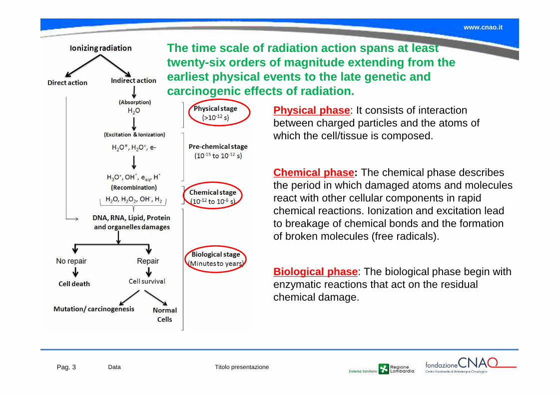
Physical phase : It consists of interaction between charged particles and the atoms of which the cell/tissue is composed.
Chemical phase : The chemical phase describes the period in which damaged atoms and molecules react with other cellular components in rapid chemical reactions. Ionization and excitation lead to breakage of chemical bonds and the formation of broken molecules (free radicals).
Biological phase : The biological phase begin with enzymatic reactions that act on the residual chemical damage.
The time scale of radiation action spans at least twenty-six orders of magnitude extending from the earliest physical events to the late genetic and carcinogenic effects of radiation.
www.cnao.it
� Molecular radiobiology
� Normal tissue radiobiology
� Cell radiobiology
� Clinical radiobiology
� DNA radiobiology
� Physics radiobiology
� Low-dose radiobiology
� Heavy-ion radiobiology
� Translational radiobiology
� Chemical radiobiology
� Applied radiobiology
� Space radiobiology
� Computational radiobiology
� …
Pag. 4 Data Titolo presentazione
www.cnao.it
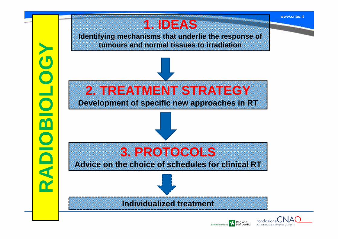
1. IDEASIdentifying mechanisms that underlie the response o f
tumours and normal tissues to irradiation
2. TREATMENT STRATEGYDevelopment of specific new approaches in RT
3. PROTOCOLSAdvice on the choice of schedules for clinical RT
Individualized treatment
RA
DIO
BIO
LOG
Y
www.cnao.it
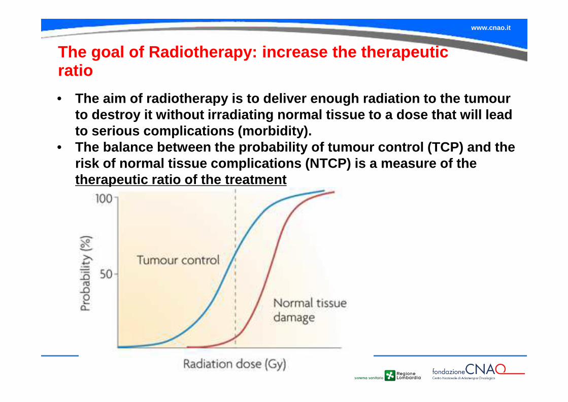
The goal of Radiotherapy: increase the therapeuticratio
• The aim of radiotherapy is to deliver enough radiat ion to the tumourto destroy it without irradiating normal tissue to a dose that will lead to serious complications (morbidity).
• The balance between the probability of tumour contro l (TCP) and the risk of normal tissue complications (NTCP) is a mea sure of the therapeutic ratio of the treatment
www.cnao.it
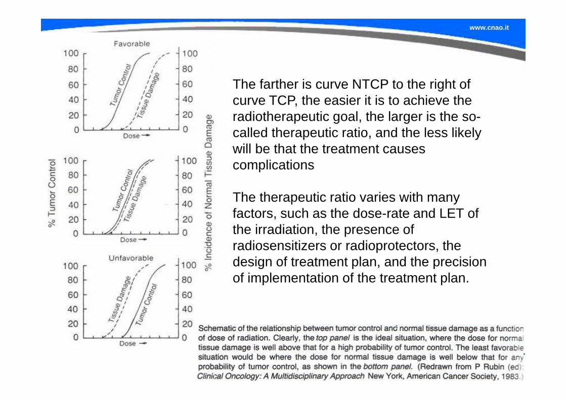
The farther is curve NTCP to the right of curve TCP, the easier it is to achieve the radiotherapeutic goal, the larger is the so-called therapeutic ratio, and the less likely will be that the treatment causes complications
The therapeutic ratio varies with many factors, such as the dose-rate and LET of the irradiation, the presence of radiosensitizers or radioprotectors, the design of treatment plan, and the precision of implementation of the treatment plan.
www.cnao.it
Radiation causes damage to all cellular molecules, but DNA damage
is most critical (most cellular and molecular components can be
replaced)
www.cnao.it
Effects of radiation damage on cells
� Cell cycle arrest
� DNA repair
� Cell death
Eukaryotic cells respond to DNA damage or blockage ofreplication by triggering "checkpoint" responses, which delaycell cycle progression, promote repair, and protect genomeintegrity.
www.cnao.it
Effects of radiation damage on cells
� Cell cycle arrest
� DNA repair
� Cell death
The subsequent action of DNA repair processes either removes the lesion(s) or misrepairs the induced damage such that all surviving progeny of an irradiated cell carry the burden of radiation exposure, e.g., a gene mutation and/or a chromosomal rearrangement
www.cnao.it
www.cnao.it
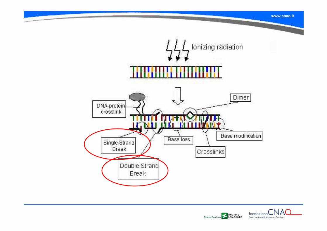
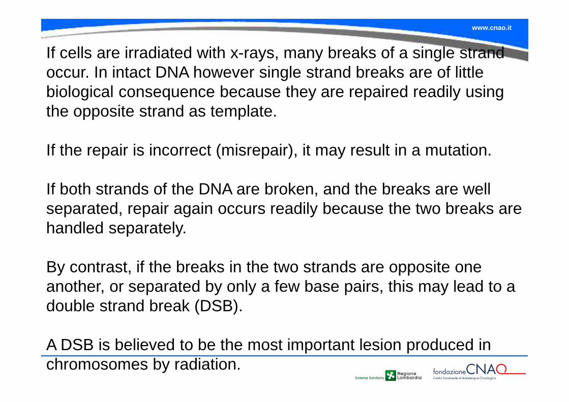
If cells are irradiated with x-rays, many breaks of a single strand occur. In intact DNA however single strand breaks are of little biological consequence because they are repaired readily using the opposite strand as template.
If the repair is incorrect (misrepair), it may result in a mutation.
If both strands of the DNA are broken, and the breaks are well separated, repair again occurs readily because the two breaks are handled separately.
By contrast, if the breaks in the two strands are opposite one another, or separated by only a few base pairs, this may lead to a double strand break (DSB).
A DSB is believed to be the most important lesion produced in chromosomes by radiation.
www.cnao.it
Effects of radiation damage on cells
� Cell cycle arrest
� DNA repair
� Cell death
www.cnao.it
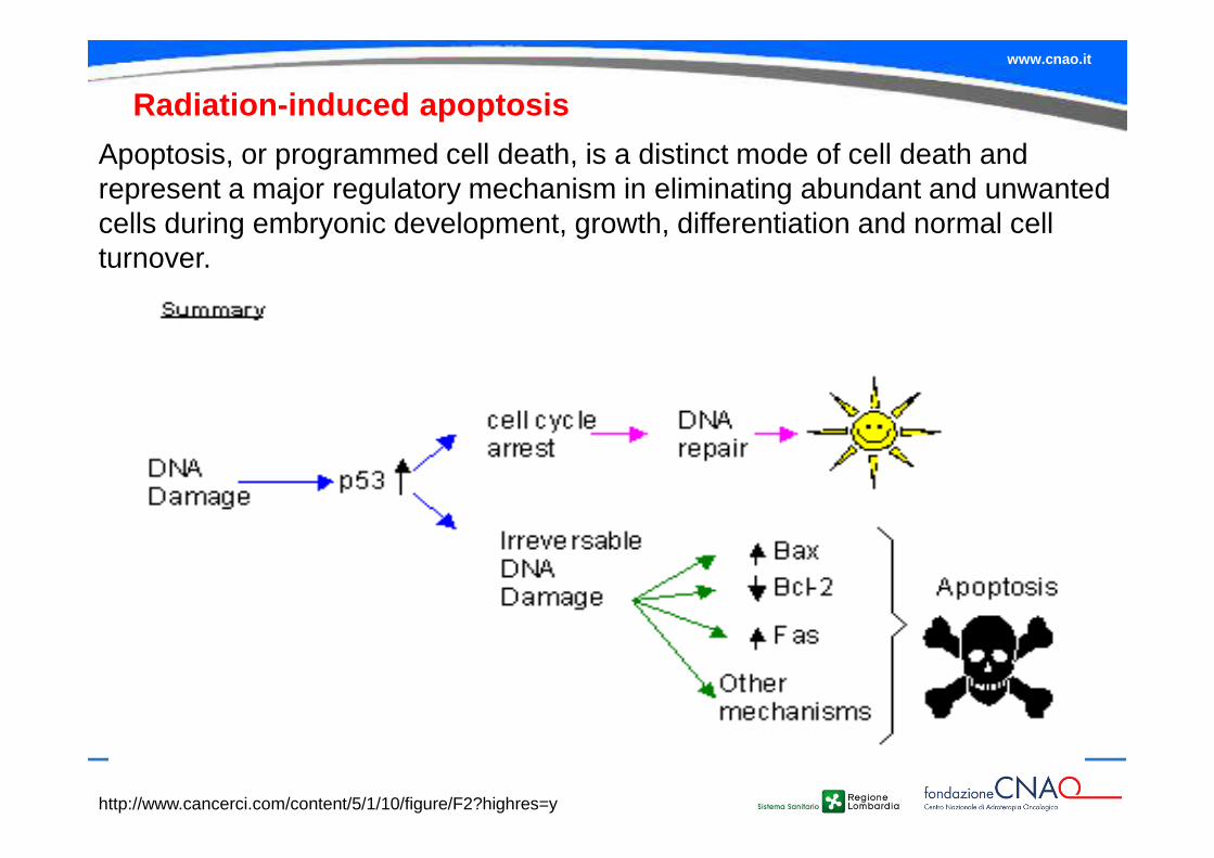
Radiation-induced apoptosis
Apoptosis, or programmed cell death, is a distinct mode of cell death and represent a major regulatory mechanism in eliminating abundant and unwanted cells during embryonic development, growth, differentiation and normal cell turnover.
http://www.cancerci.com/content/5/1/10/figure/F2?highres=y
www.cnao.it
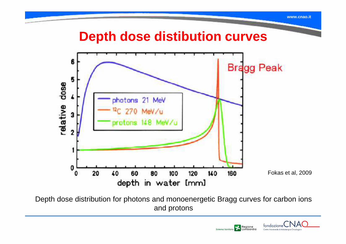
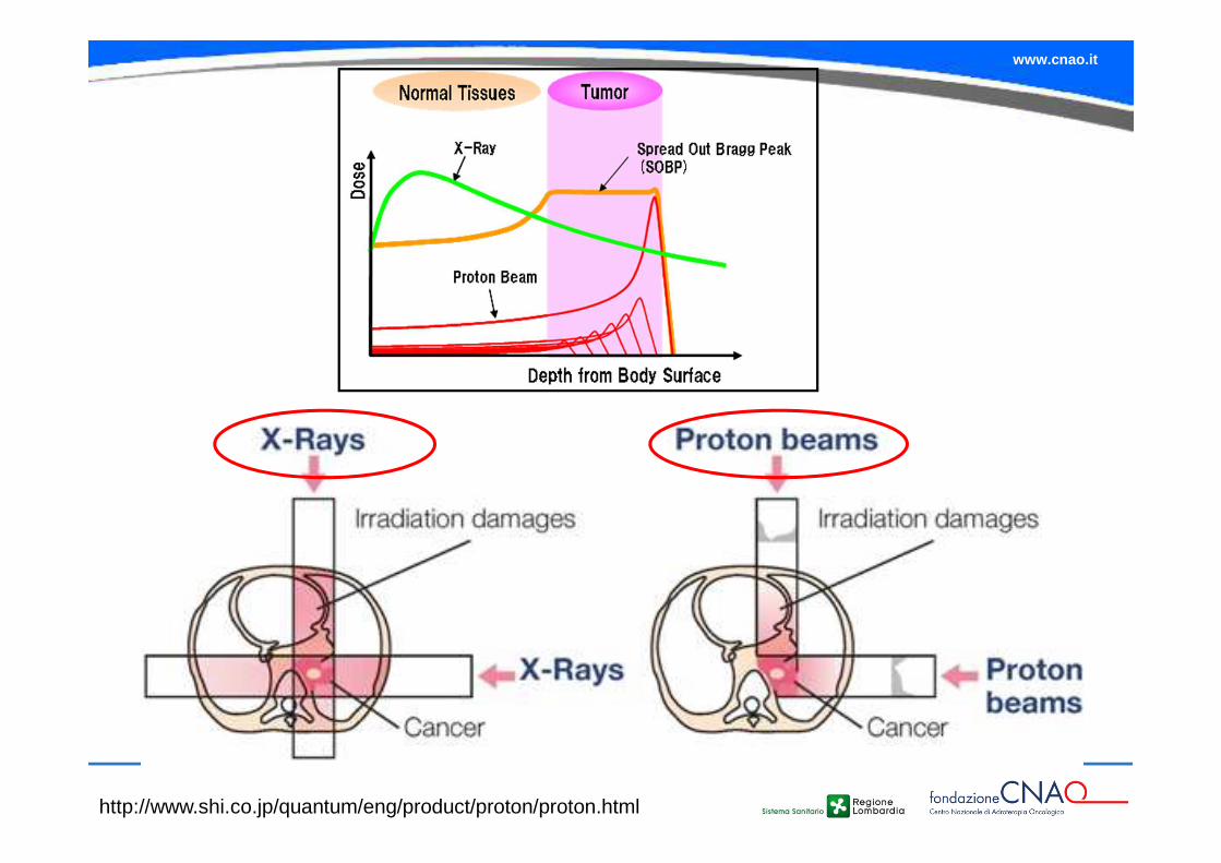
Depth dose distibution curves
Depth dose distribution for photons and monoenergetic Bragg curves for carbon ions and protons
Fokas et al, 2009
www.cnao.it
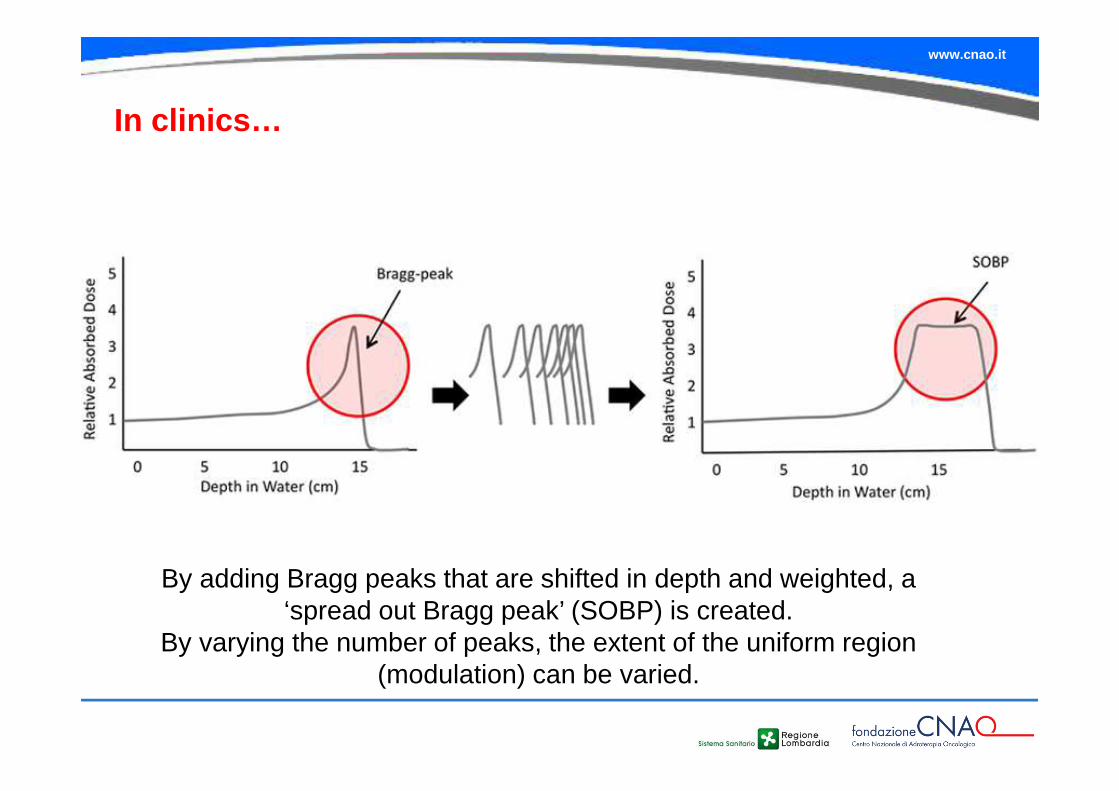
In clinics…
By adding Bragg peaks that are shifted in depth and weighted, a ‘spread out Bragg peak’ (SOBP) is created.
By varying the number of peaks, the extent of the uniform region (modulation) can be varied.
www.cnao.it
http://www.shi.co.jp/quantum/eng/product/proton/proton.html
www.cnao.it
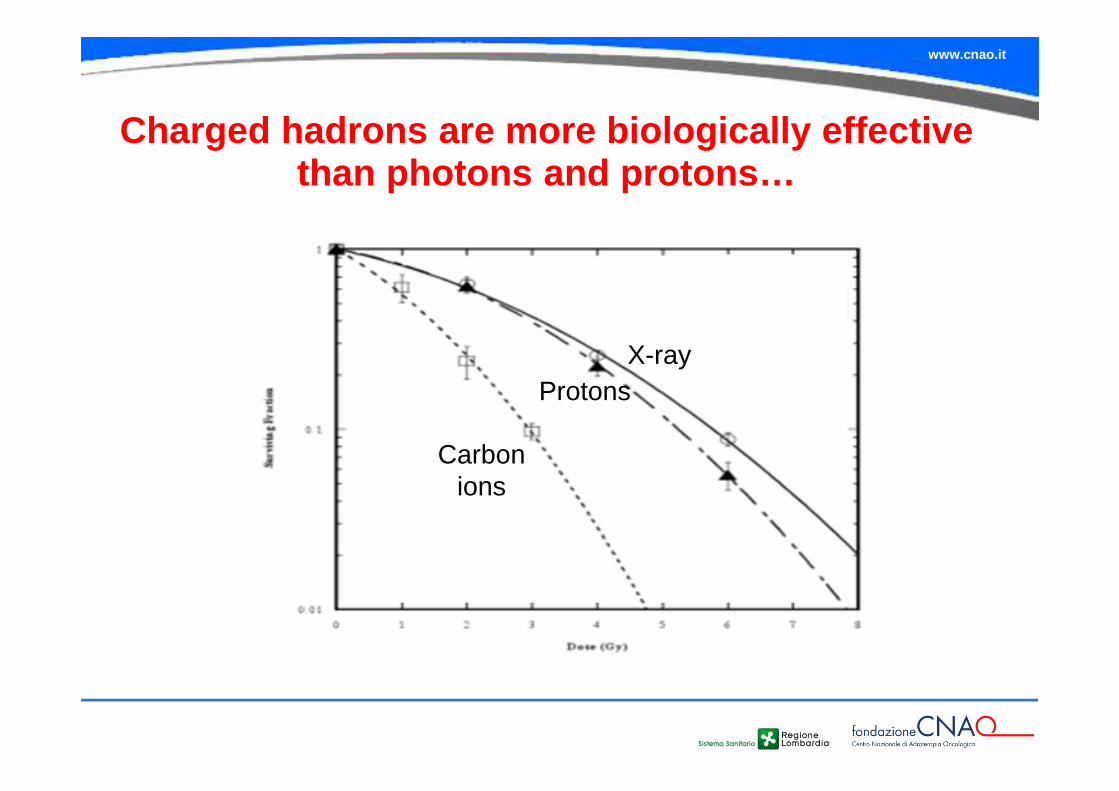
Charged hadrons are more biologically effective than photons and protons…
X-rayProtons
Carbon ions
www.cnao.it
Pag. 19 Data Titolo presentazione
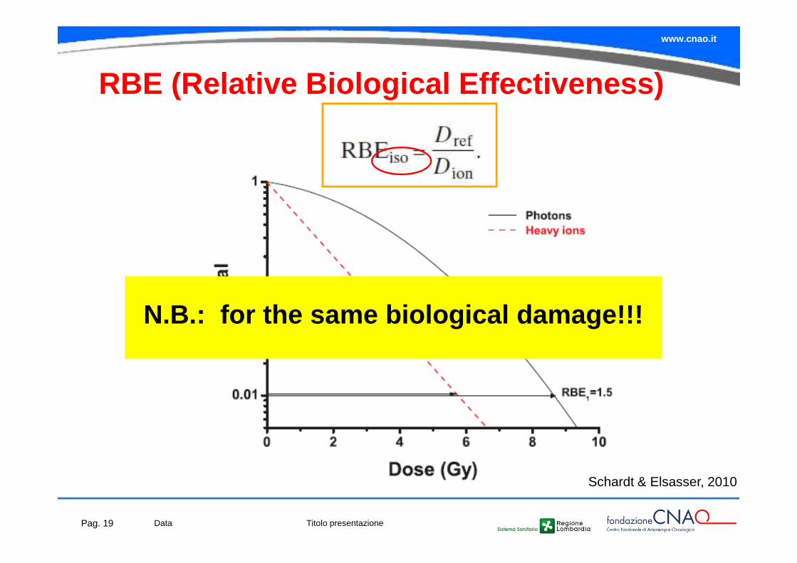
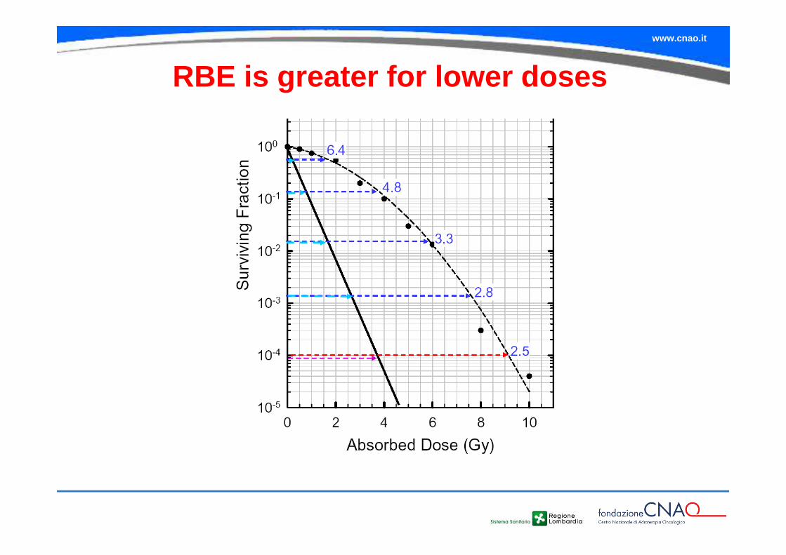
RBE (Relative Biological Effectiveness )
Schardt & Elsasser, 2010
N.B.: for the same biological damage!!!
www.cnao.it
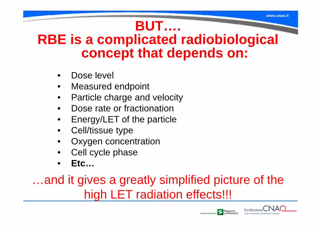
• Dose level• Measured endpoint• Particle charge and velocity• Dose rate or fractionation• Energy/LET of the particle• Cell/tissue type• Oxygen concentration• Cell cycle phase• Etc…
BUT…. RBE is a complicated radiobiological
concept that depends on:
…and it gives a greatly simplified picture of the high LET radiation effects!!!
www.cnao.it
RBE is greater for lower doses
www.cnao.it
Microscopic understanding of RBE
www.cnao.it
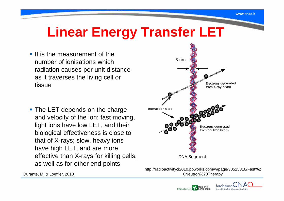
Linear Energy Transfer LET� It is the measurement of the
number of ionisations which radiation causes per unit distance as it traverses the living cell or tissue
� The LET depends on the charge and velocity of the ion: fast moving, light ions have low LET, and their biological effectiveness is close to that of X-rays; slow, heavy ions have high LET, and are more effective than X-rays for killing cells, as well as for other end points
http://radioactivityci2010.pbworks.com/w/page/30525316/Fast%20Neutron%20TherapyDurante, M. & Loeffler, 2010

www.cnao.it
Radiation causes damage to all cellular molecules, but DNA damage is most critical (most cellular and molecular components can
be replaced)
www.cnao.it
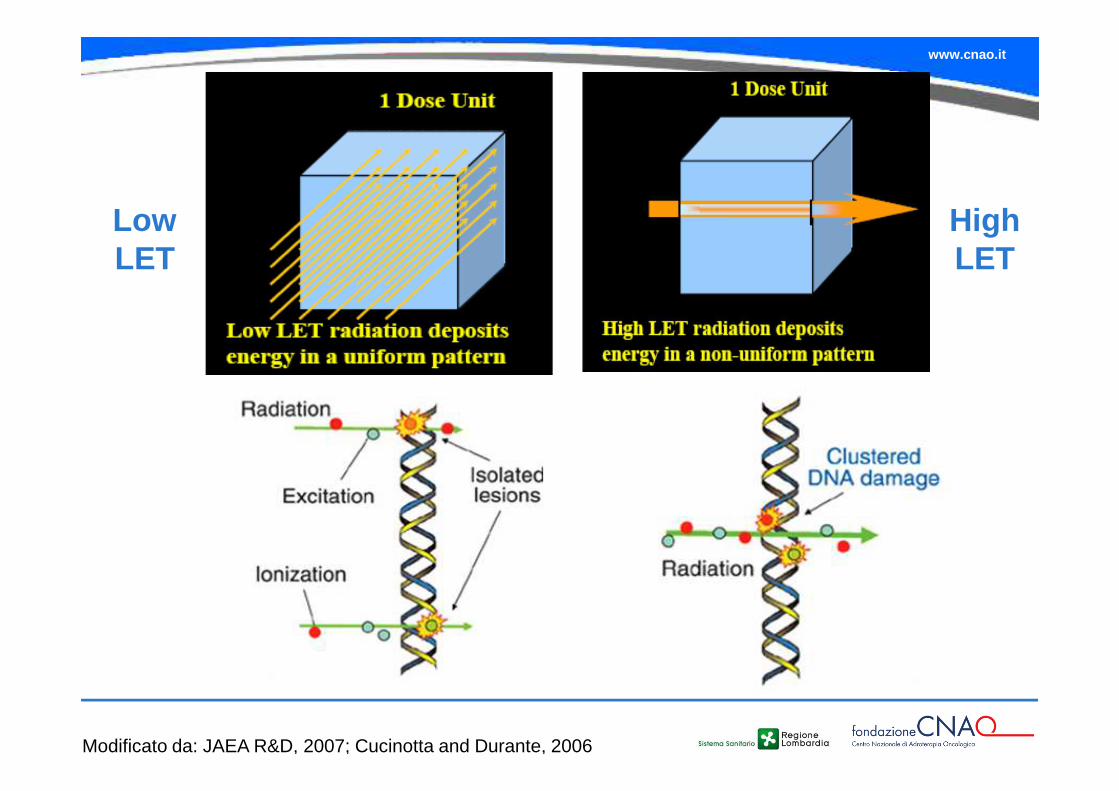
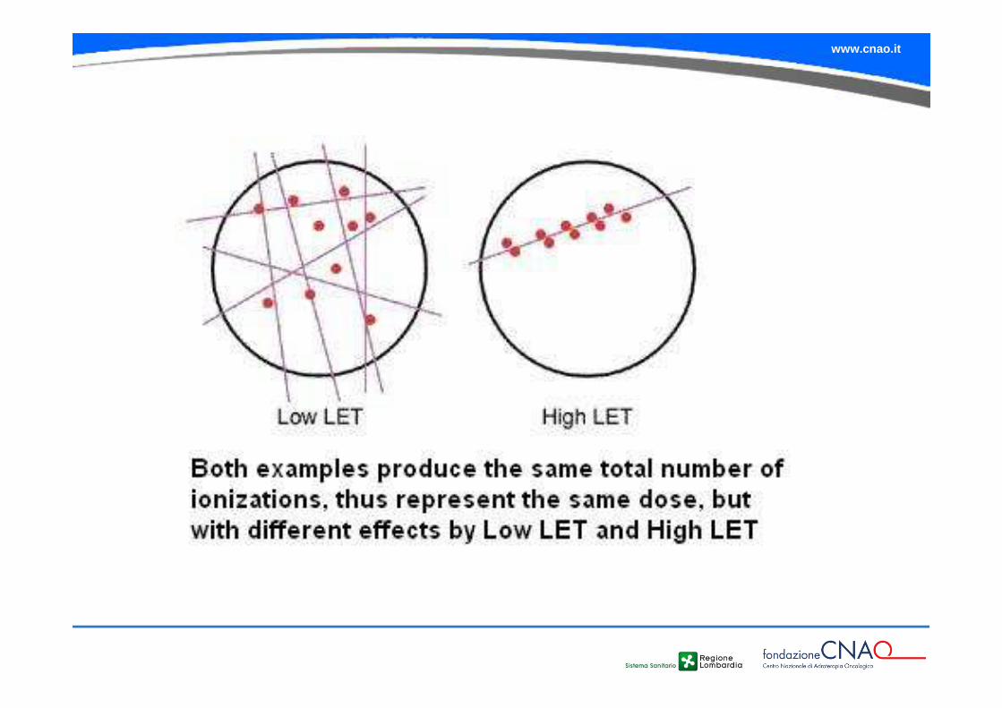
Low LET
High LET
Modificato da: JAEA R&D, 2007; Cucinotta and Durante, 2006
www.cnao.it
www.cnao.it
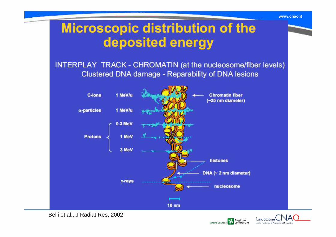
Belli et al., J Radiat Res, 2002
www.cnao.it
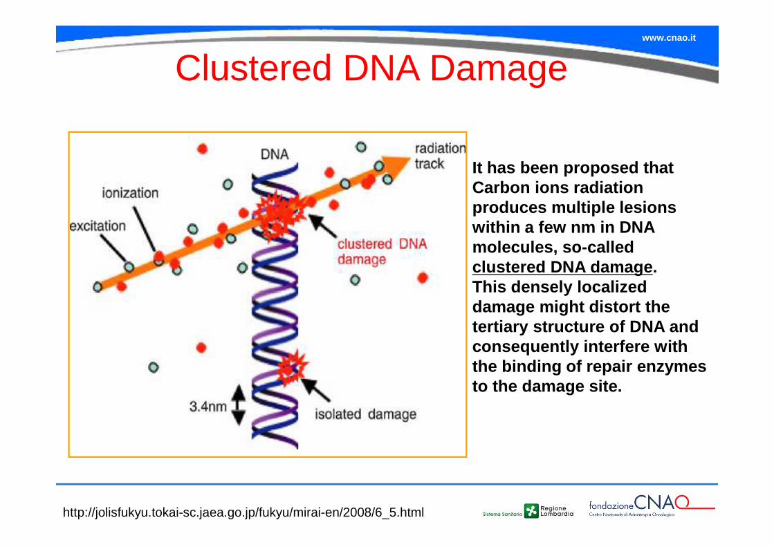
Clustered DNA Damage
http://jolisfukyu.tokai-sc.jaea.go.jp/fukyu/mirai-en/2008/6_5.html
It has been proposed that Carbon ions radiation produces multiple lesions within a few nm in DNA molecules, so-called clustered DNA damage. This densely localized damage might distort the tertiary structure of DNA and consequently interfere with the binding of repair enzymes to the damage site.
www.cnao.it
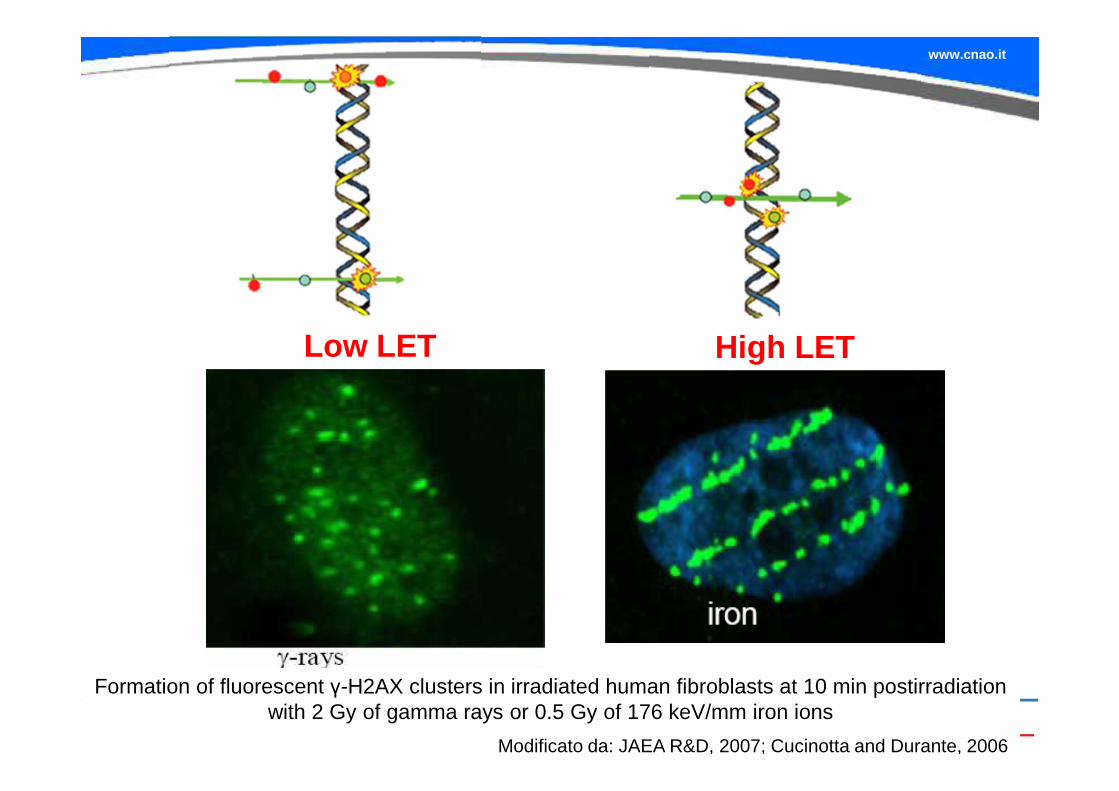
Formation of fluorescent γ-H2AX clusters in irradiated human fibroblasts at 10 min postirradiation with 2 Gy of gamma rays or 0.5 Gy of 176 keV/mm iron ions
Modificato da: JAEA R&D, 2007; Cucinotta and Durante, 2006
Low LET High LET
www.cnao.it
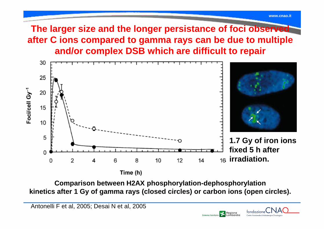
The larger size and the longer persistance of foci o bserved after C ions compared to gamma rays can be due to m ultiple
and/or complex DSB which are difficult to repair
Antonelli F et al, 2005; Desai N et al, 2005
Comparison between H2AX phosphorylation-dephosphory lationkinetics after 1 Gy of gamma rays (closed circles) or carbon ions (open circles).
1.7 Gy of iron ions fixed 5 h after irradiation.
www.cnao.it
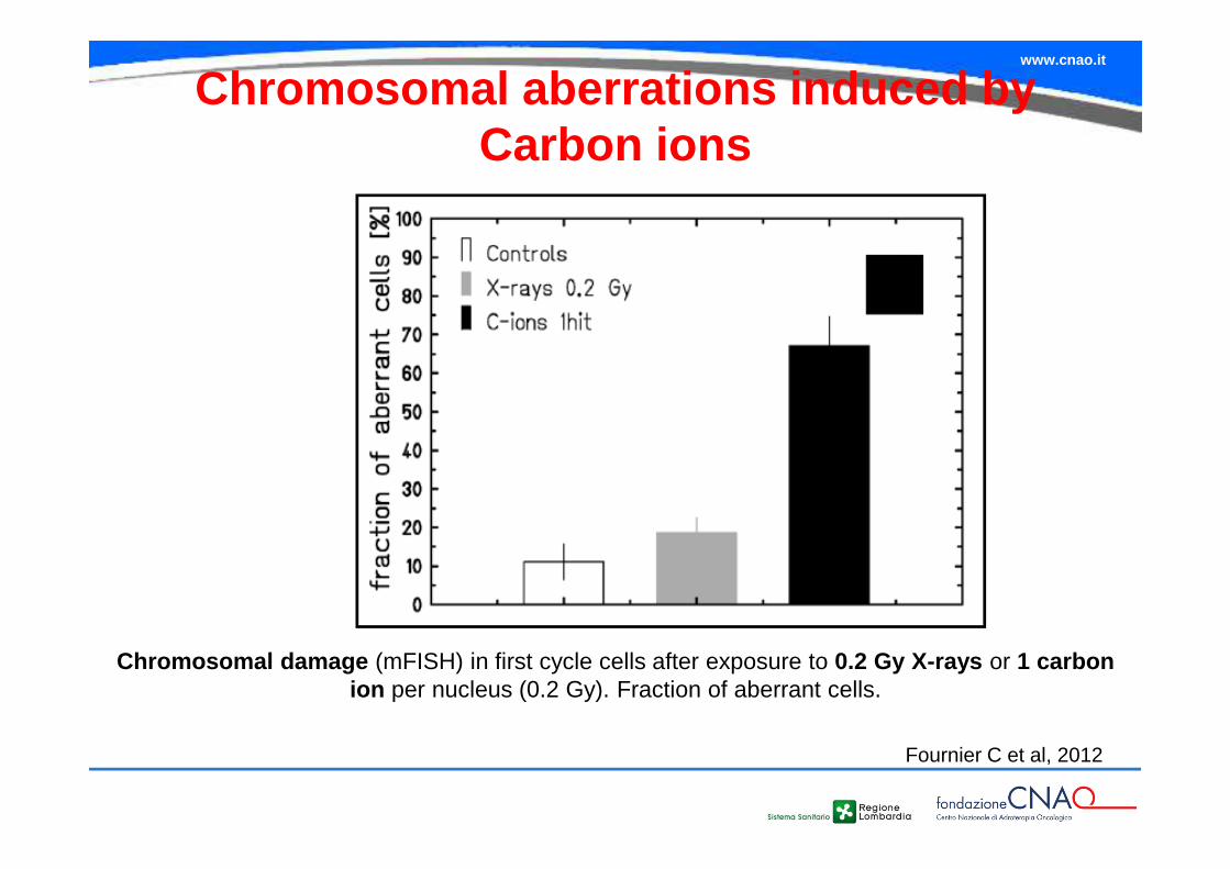
Chromosomal damage (mFISH) in first cycle cells after exposure to 0.2 Gy X-rays or 1 carbon ion per nucleus (0.2 Gy). Fraction of aberrant cells.
Fournier C et al, 2012
Chromosomal aberrations induced by Carbon ions
www.cnao.it
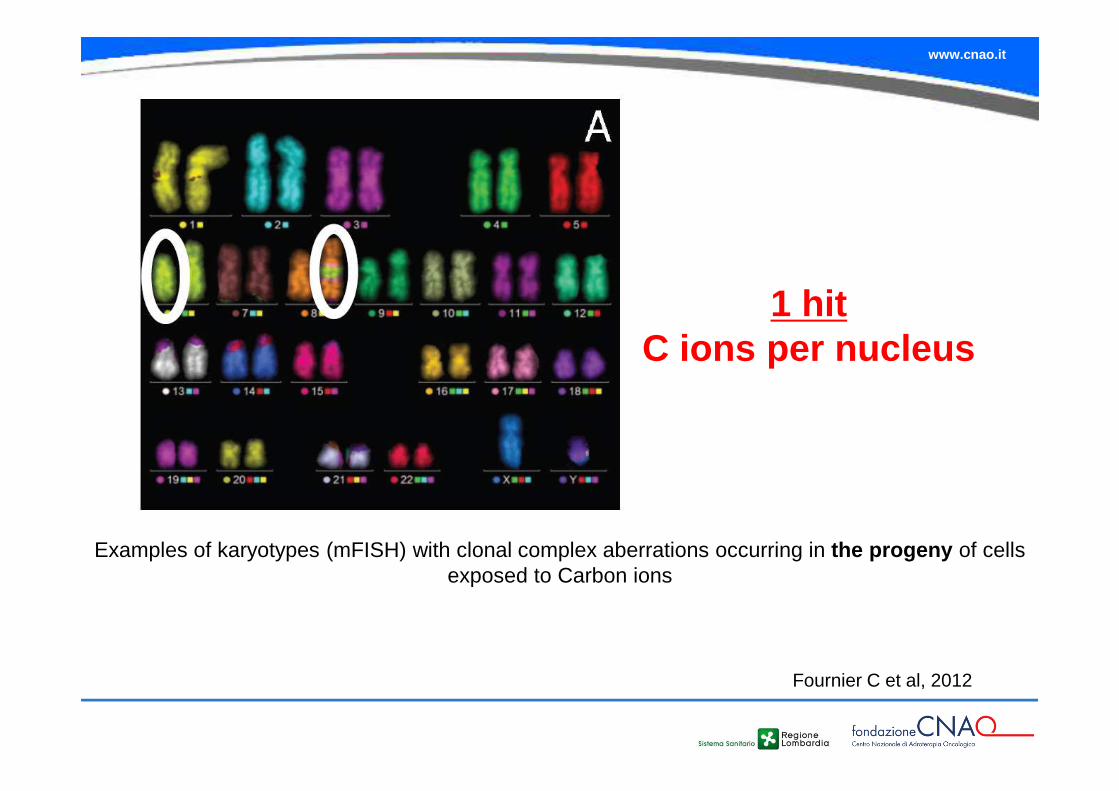
Examples of karyotypes (mFISH) with clonal complex aberrations occurring in the progeny of cells exposed to Carbon ions
1 hit C ions per nucleus
Fournier C et al, 2012
www.cnao.it
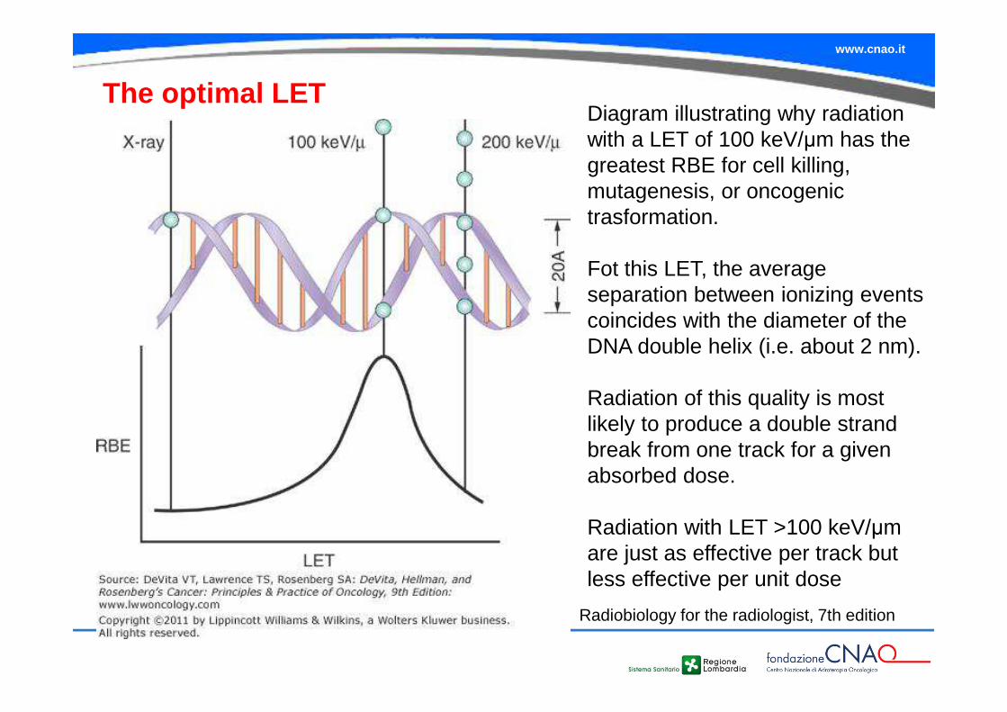
The optimal LET
Radiobiology for the radiologist, 7th edition
Diagram illustrating why radiation with a LET of 100 keV/µm has the greatest RBE for cell killing, mutagenesis, or oncogenic trasformation.
Fot this LET, the average separation between ionizing events coincides with the diameter of the DNA double helix (i.e. about 2 nm).
Radiation of this quality is most likely to produce a double strand break from one track for a given absorbed dose.
Radiation with LET >100 keV/µm are just as effective per track but less effective per unit dose
www.cnao.it
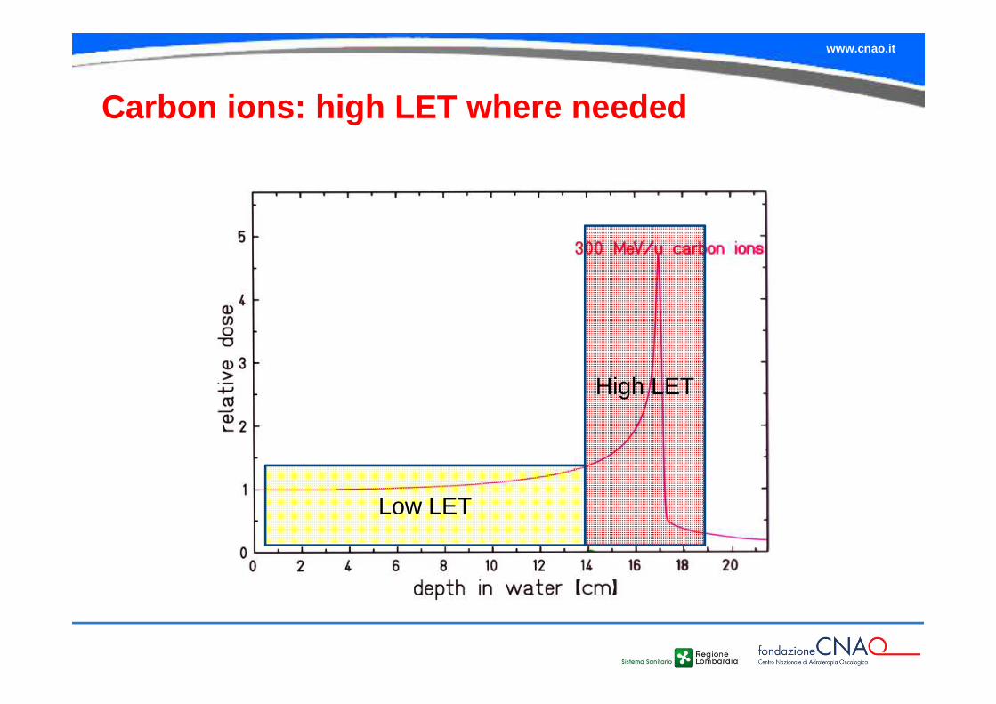
Carbon ions: high LET where needed
Low LET
High LET
www.cnao.it
Clonogenic Cell survival as a determinant of tumour response
www.cnao.it
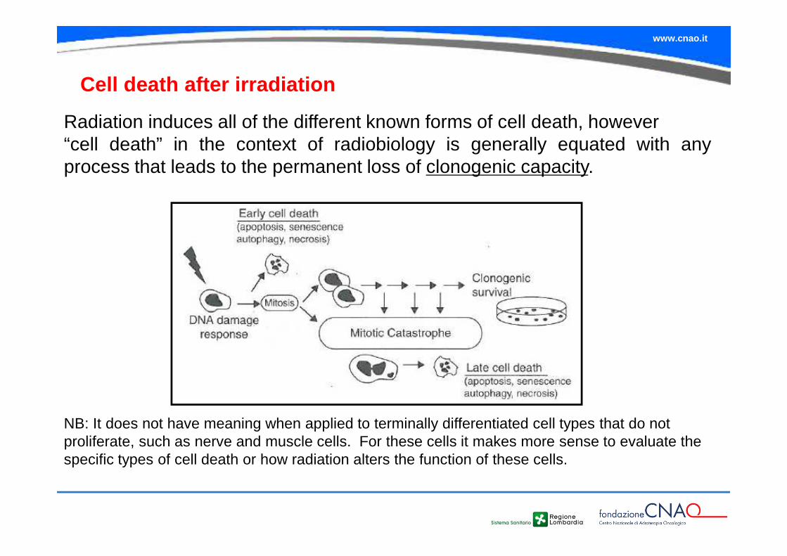
Cell death after irradiation
Radiation induces all of the different known forms of cell death, however“cell death” in the context of radiobiology is generally equated with anyprocess that leads to the permanent loss of clonogenic capacity.
NB: It does not have meaning when applied to terminally differentiated cell types that do not proliferate, such as nerve and muscle cells. For these cells it makes more sense to evaluate the specific types of cell death or how radiation alters the function of these cells.
www.cnao.it
A cell that is able to proliferate indefinitely and form a large colony from a single cell is said to be clonogenic .
Tumor cells can be grown indefinitely in cell culture; normal cells must be transformed to grow indefinitely in culture.
For cells growing in culture, the loss of the ability to continue growth is termed reproductive death .
Following irradiation, cells may still be physically present and apparently intact, may be able to produce proteins, synthesize new DNA and even go through one or two cell divisions. But if it has lost the capability to reproduce indefinitely , it is considered dead.
Reproductive dead
www.cnao.it
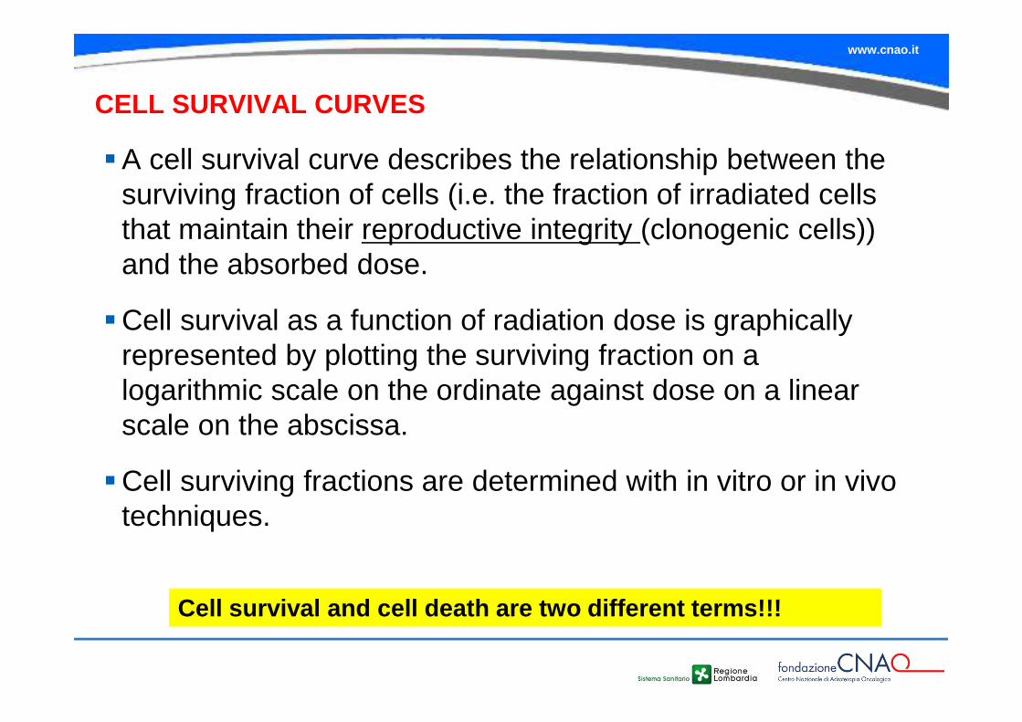
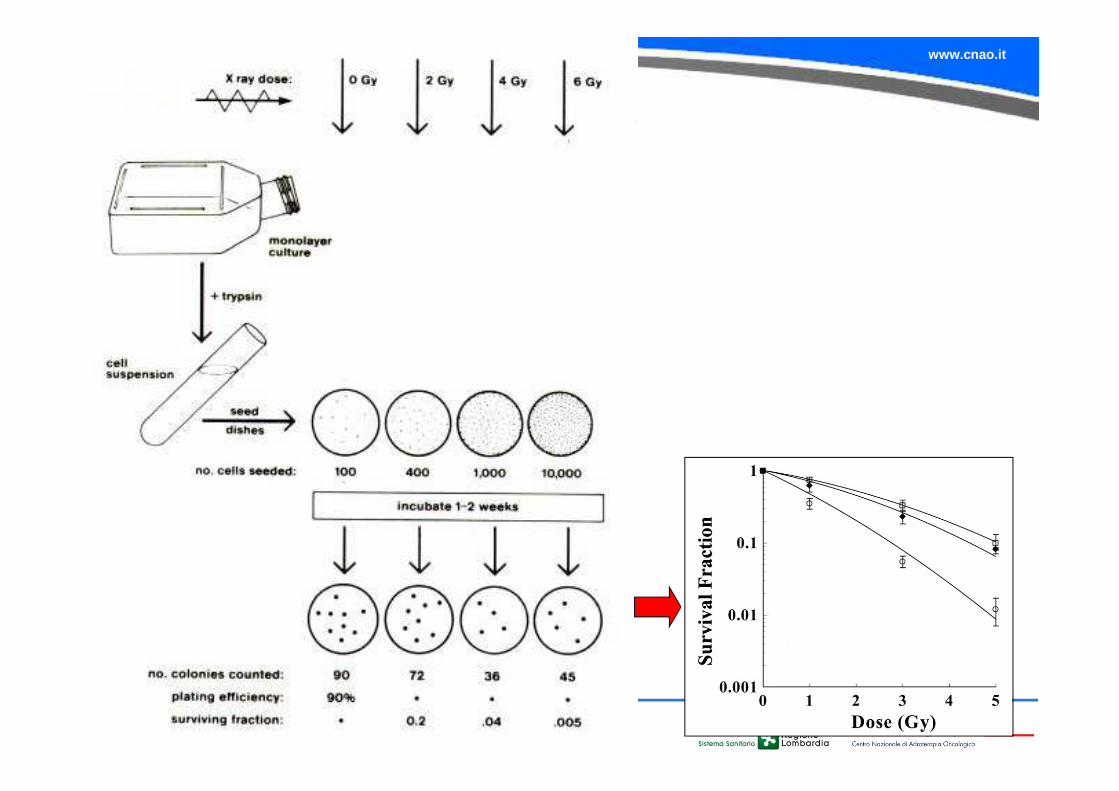
CELL SURVIVAL CURVES
�A cell survival curve describes the relationship between the surviving fraction of cells (i.e. the fraction of irradiated cells that maintain their reproductive integrity (clonogenic cells)) and the absorbed dose.
�Cell survival as a function of radiation dose is graphically represented by plotting the surviving fraction on a logarithmic scale on the ordinate against dose on a linear scale on the abscissa.
�Cell surviving fractions are determined with in vitro or in vivo techniques.
Cell survival and cell death are two different term s!!!
www.cnao.it
www.cnao.it
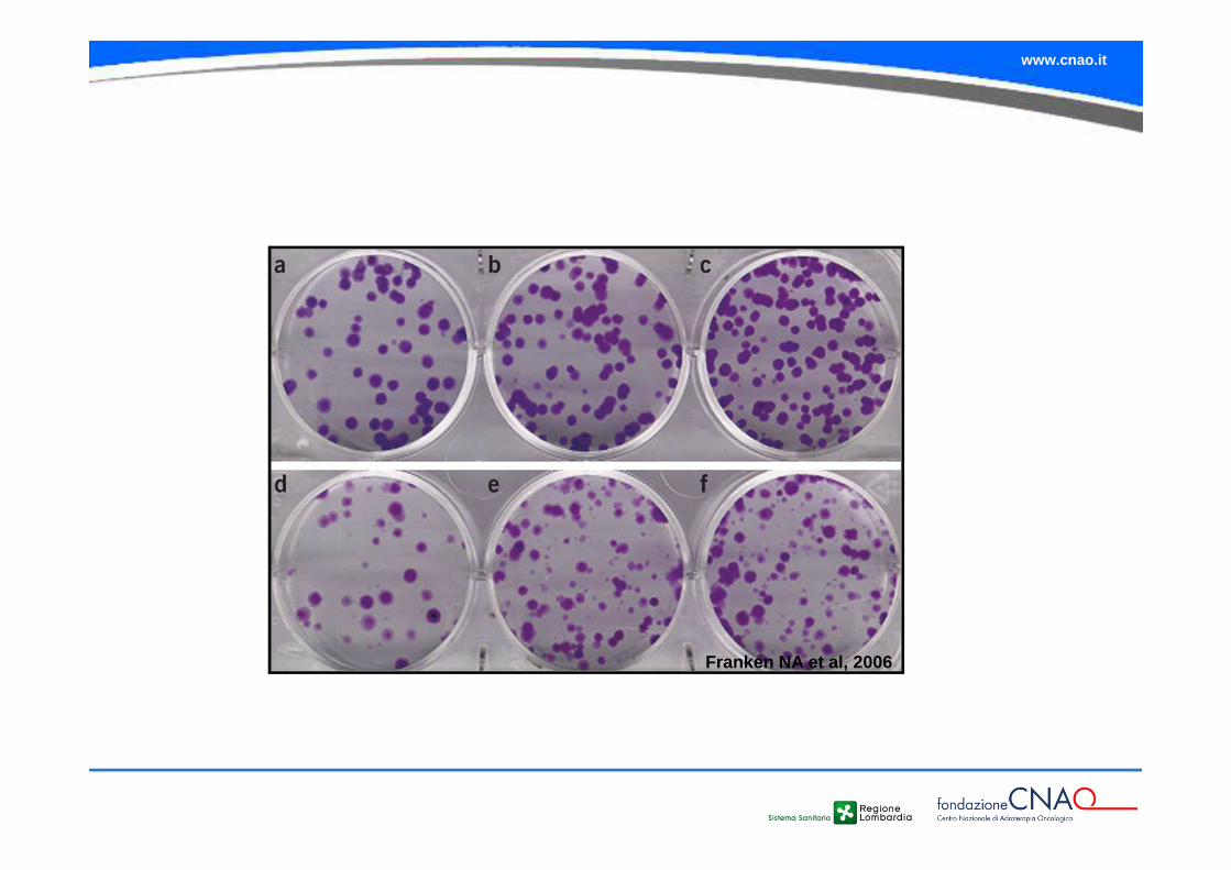
Franken NA et al, 2006
www.cnao.it
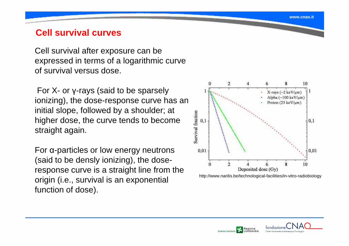
Cell survival curves
Cell survival after exposure can be expressed in terms of a logarithmic curve of survival versus dose.
For X- or γ-rays (said to be sparsely ionizing), the dose-response curve has an initial slope, followed by a shoulder; at higher dose, the curve tends to become straight again.
For α-particles or low energy neutrons (said to be densly ionizing), the dose-response curve is a straight line from the origin (i.e., survival is an exponential function of dose).
http://www.narilis.be/technological-facilities/in-vitro-radiobiology
www.cnao.it
Factors determining RBE(basic radiobiological properties of
charged hadrons )
www.cnao.it
The modifying effect of hypoxia is smaller for high LET radiations than for photons
www.cnao.it
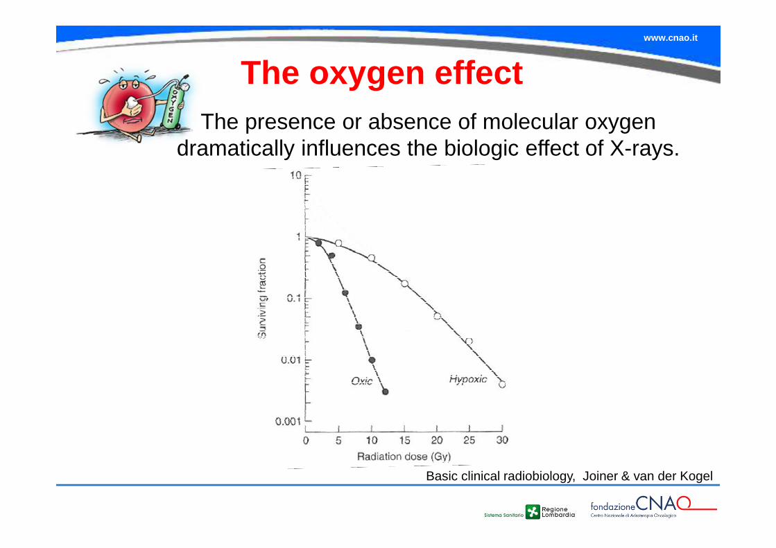
The oxygen effect
Basic clinical radiobiology, Joiner & van der Kogel
The presence or absence of molecular oxygen dramatically influences the biologic effect of X-rays.
www.cnao.it
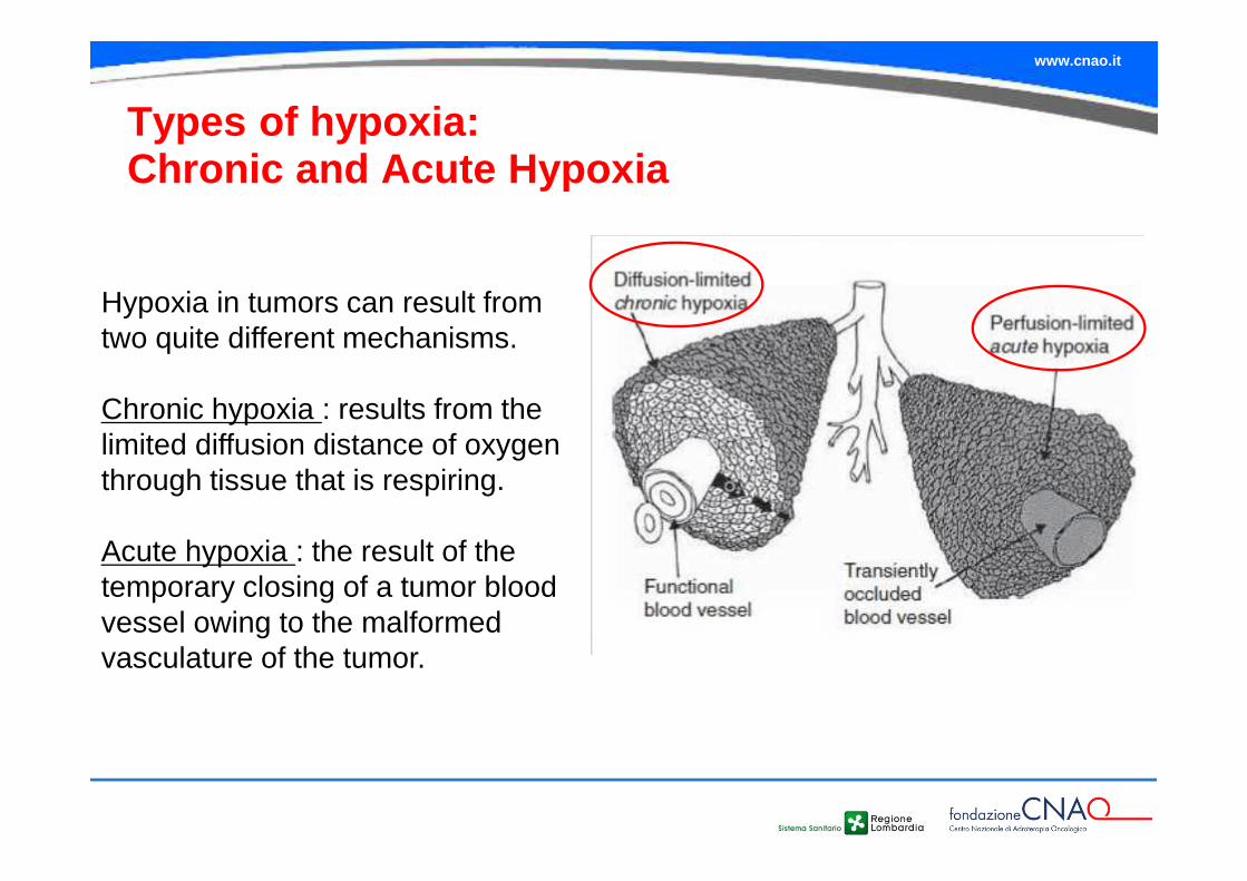
Types of hypoxia:Chronic and Acute Hypoxia
Hypoxia in tumors can result from two quite different mechanisms.
Chronic hypoxia : results from the limited diffusion distance of oxygen through tissue that is respiring.
Acute hypoxia : the result of the temporary closing of a tumor blood vessel owing to the malformed vasculature of the tumor.
www.cnao.it
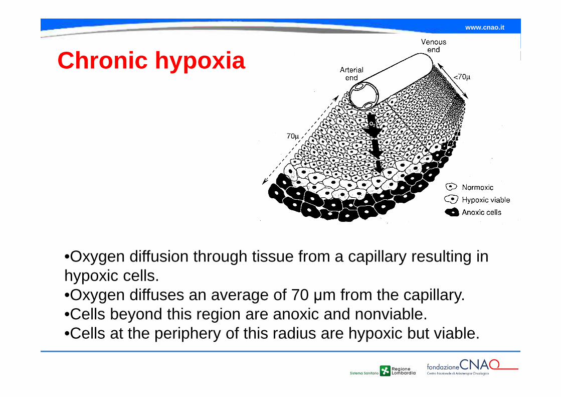
Chronic hypoxia
•Oxygen diffusion through tissue from a capillary resulting in hypoxic cells. •Oxygen diffuses an average of 70 µm from the capillary. •Cells beyond this region are anoxic and nonviable. •Cells at the periphery of this radius are hypoxic but viable.
www.cnao.it
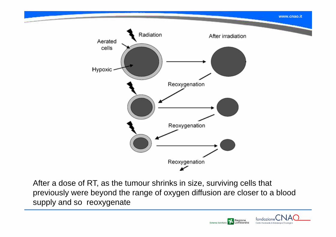
After a dose of RT, as the tumour shrinks in size, surviving cells that previously were beyond the range of oxygen diffusion are closer to a blood supply and so reoxygenate
www.cnao.it
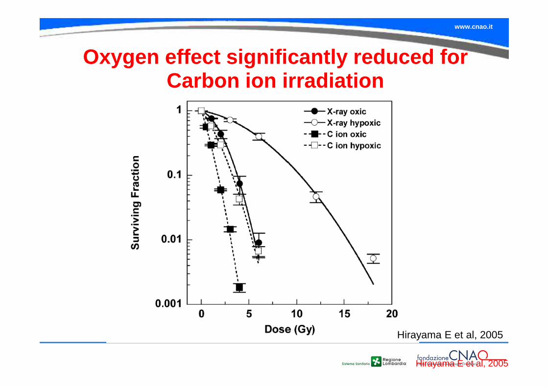
Hirayama E et al, 2005
Oxygen effect significantly reduced for Carbon ion irradiation
Hirayama E et al, 2005
www.cnao.it
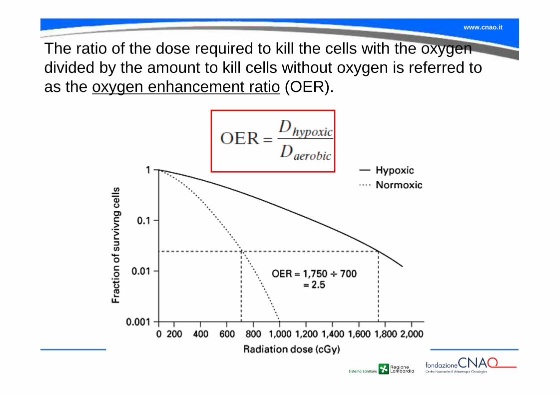
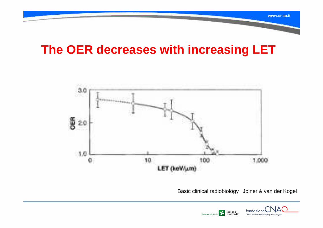
The ratio of the dose required to kill the cells with the oxygen divided by the amount to kill cells without oxygen is referred to as the oxygen enhancement ratio (OER).
www.cnao.it
The oxygen effect is quite dramatic for low LET (sparsely ionizing) radiations, while for high LET (densely ionizing) radiations it is much less pronounced.
•The OER for X rays and electrons is about three at high doses and falls to about two for doses of 1–2 Gy.
•The OER decreases as the LET increases andapproaches OER = 1 at about LET = 150 keV/mm
www.cnao.it
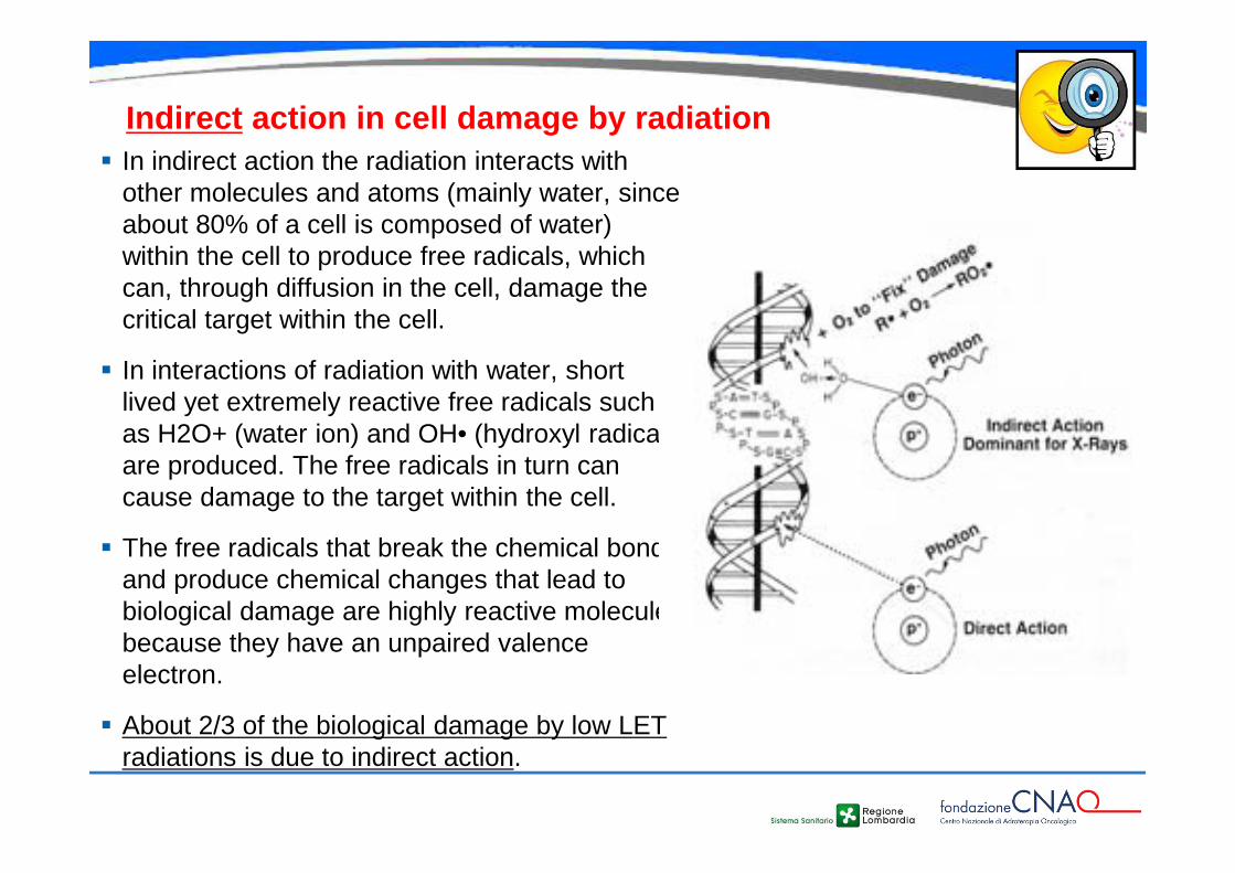
Indirect action in cell damage by radiation� In indirect action the radiation interacts with
other molecules and atoms (mainly water, since about 80% of a cell is composed of water) within the cell to produce free radicals, which can, through diffusion in the cell, damage the critical target within the cell.
� In interactions of radiation with water, short lived yet extremely reactive free radicals such as H2O+ (water ion) and OH• (hydroxyl radical) are produced. The free radicals in turn can cause damage to the target within the cell.
� The free radicals that break the chemical bonds and produce chemical changes that lead to biological damage are highly reactive molecules because they have an unpaired valence electron.
� About 2/3 of the biological damage by low LET radiations is due to indirect action.
www.cnao.it
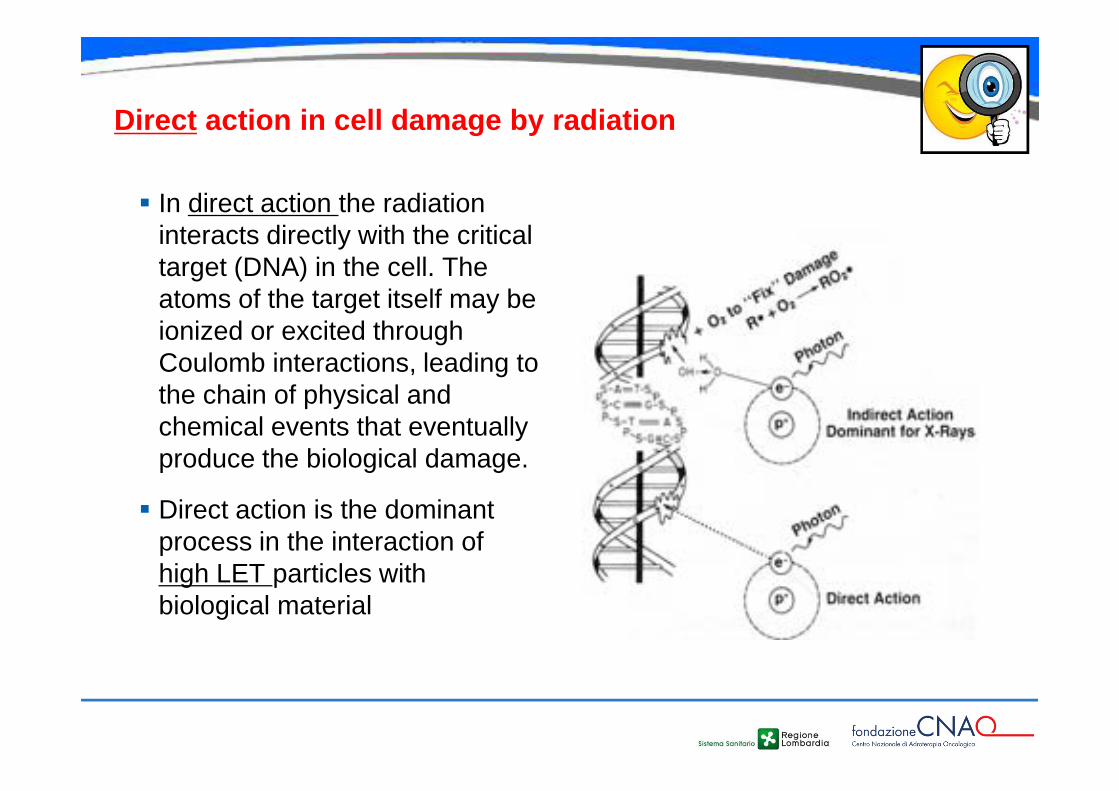
Direct action in cell damage by radiation
� In direct action the radiation interacts directly with the critical target (DNA) in the cell. The atoms of the target itself may be ionized or excited through Coulomb interactions, leading to the chain of physical and chemical events that eventually produce the biological damage.
� Direct action is the dominant process in the interaction of high LET particles with biological material
www.cnao.it
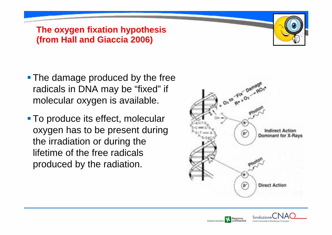
The oxygen fixation hypothesis(from Hall and Giaccia 2006)
�The damage produced by the free radicals in DNA may be “fixed” if molecular oxygen is available.
�To produce its effect, molecular oxygen has to be present during the irradiation or during the lifetime of the free radicals produced by the radiation.
www.cnao.it
Basic clinical radiobiology, Joiner & van der Kogel
The OER decreases with increasing LET
www.cnao.it
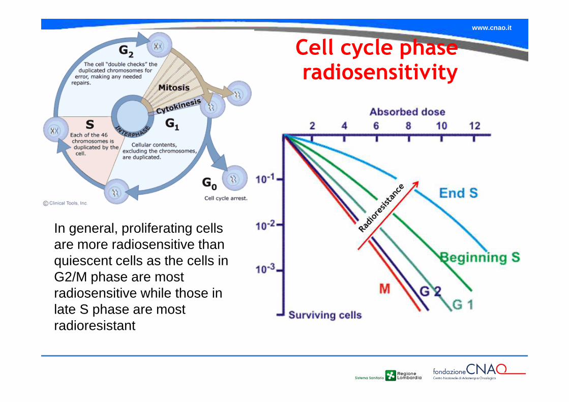
Cell cycle phaseradiosensitivity
In general, proliferating cells are more radiosensitive than quiescent cells as the cells in G2/M phase are most radiosensitive while those in late S phase are most radioresistant
www.cnao.it
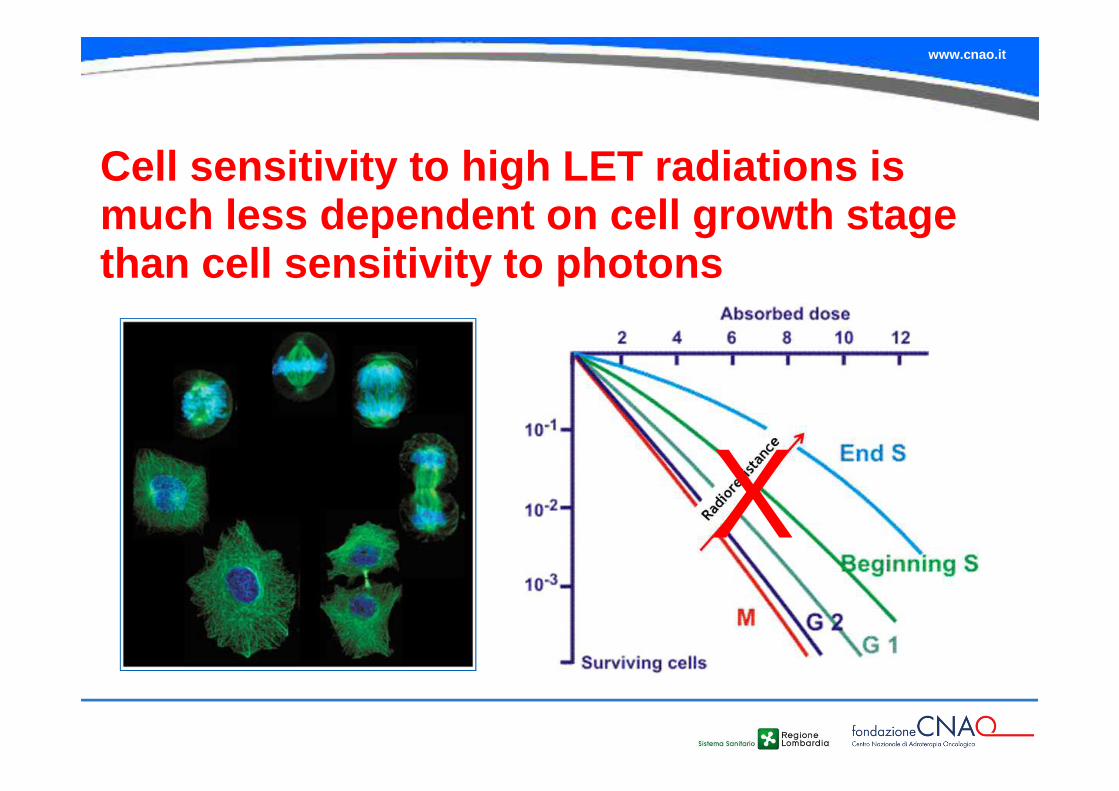
Cell sensitivity to high LET radiations is much less dependent on cell growth stage than cell sensitivity to photons
X
www.cnao.it
High LET radiations and TUMOUR CELLS genetic background
www.cnao.it
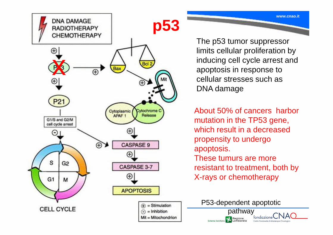
P53-dependent apoptoticpathway
p53The p53 tumor suppressor limits cellular proliferation by inducing cell cycle arrest and apoptosis in response to cellular stresses such as DNA damage
xAbout 50% of cancers harbor mutation in the TP53 gene, which result in a decreased propensity to undergo apoptosis. These tumurs are more resistant to treatment, both by X-rays or chemotherapy
www.cnao.it
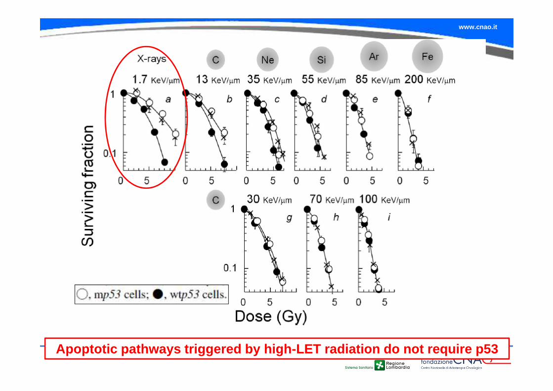
Apoptotic pathways triggered by high-LET radiation do not require p53
www.cnao.it
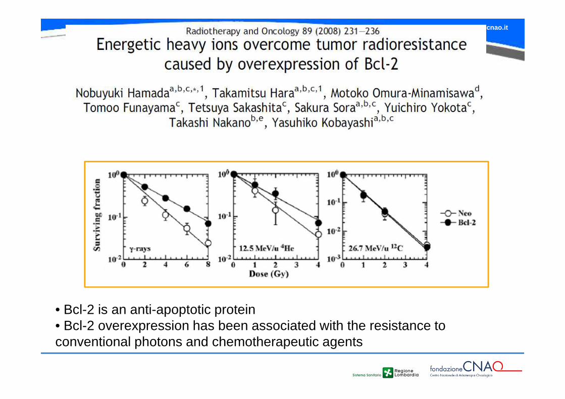
• Bcl-2 is an anti-apoptotic protein• Bcl-2 overexpression has been associated with the resistance to conventional photons and chemotherapeutic agents
www.cnao.it
Taking into consideration that Bcl-2overexpression and p53 mutations occur in morethan half of tumors, high-LET heavy ions appearto effectively kill a wide variety of radioresistanttumors.
!
www.cnao.it
Modulation of invasion and migration effects
www.cnao.it
www.cnao.it
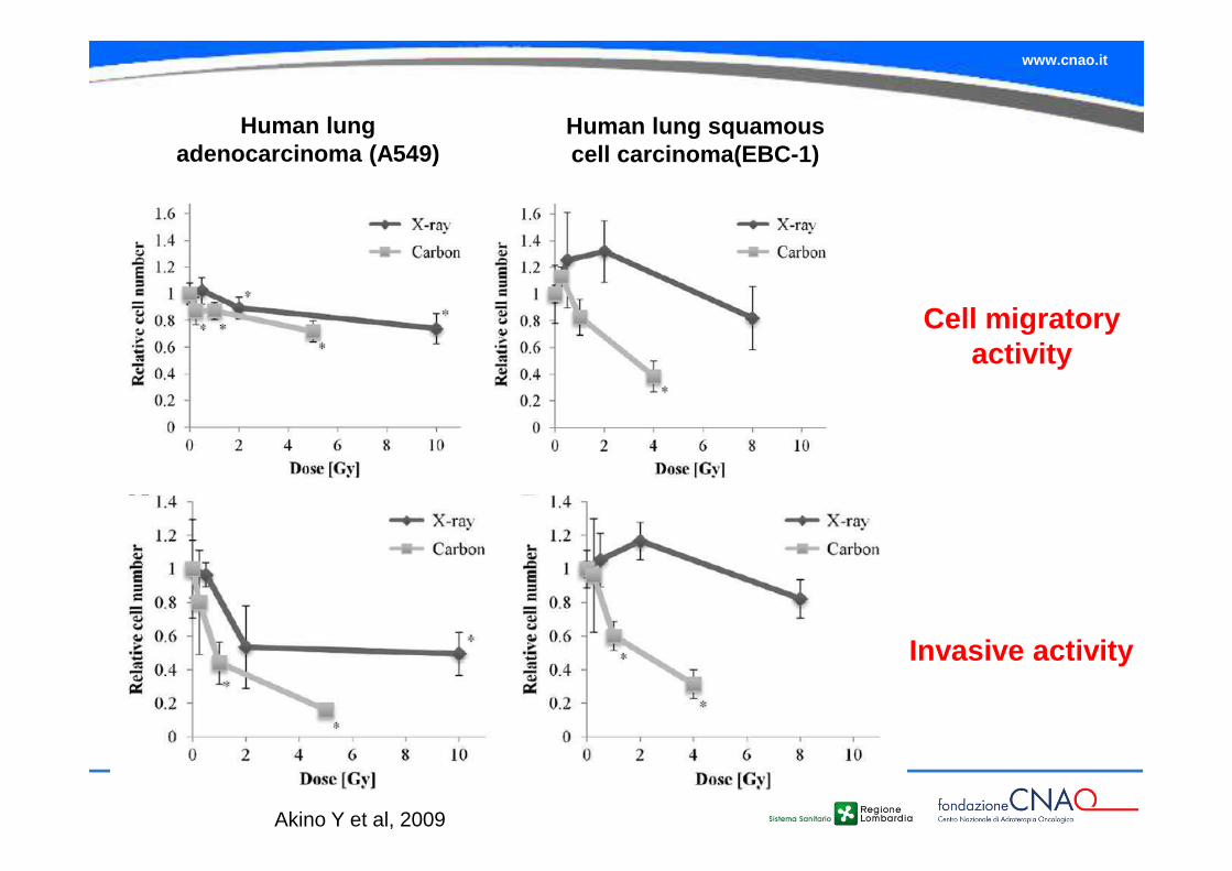
Human lung adenocarcinoma (A549)
Human lung squamous cell carcinoma(EBC-1)
Cell migratory activity
Invasive activity
Akino Y et al, 2009
www.cnao.it
www.cnao.it
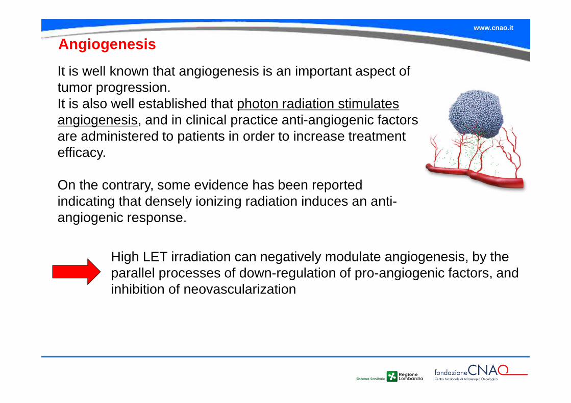
Angiogenesis
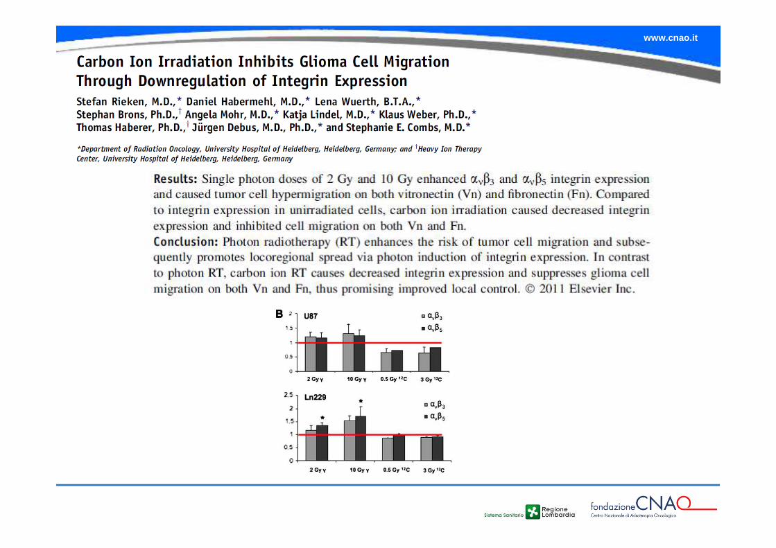
It is well known that angiogenesis is an important aspect of tumor progression. It is also well established that photon radiation stimulates angiogenesis, and in clinical practice anti-angiogenic factors are administered to patients in order to increase treatment efficacy.
On the contrary, some evidence has been reported indicating that densely ionizing radiation induces an anti-angiogenic response.
High LET irradiation can negatively modulate angiogenesis, by the parallel processes of down-regulation of pro-angiogenic factors, and inhibition of neovascularization
www.cnao.it
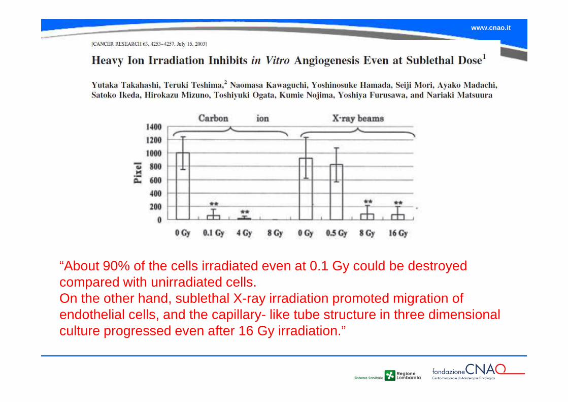
“About 90% of the cells irradiated even at 0.1 Gy could be destroyed compared with unirradiated cells. On the other hand, sublethal X-ray irradiation promoted migration of endothelial cells, and the capillary- like tube structure in three dimensional culture progressed even after 16 Gy irradiation.”
www.cnao.it
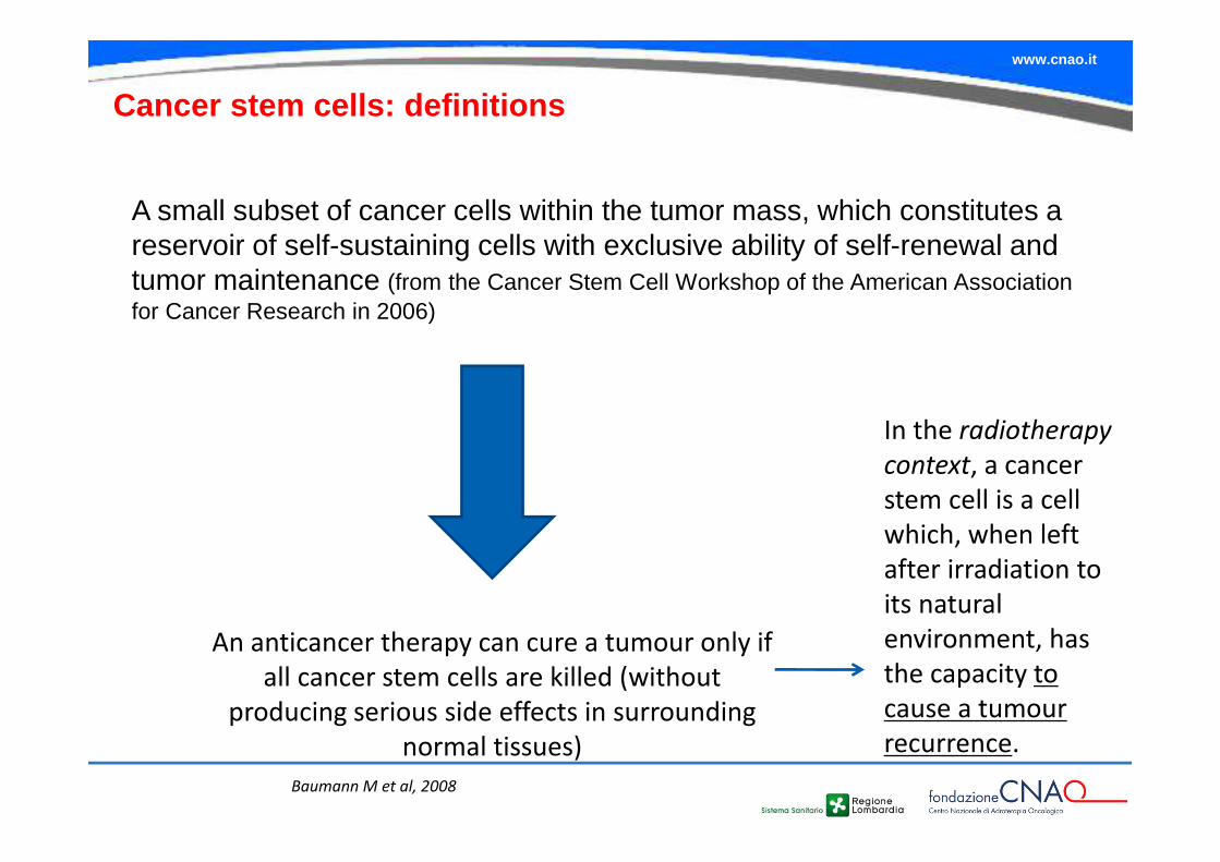
Cancer stem cells: definitions
A small subset of cancer cells within the tumor mass, which constitutes a reservoir of self-sustaining cells with exclusive ability of self-renewal and tumor maintenance (from the Cancer Stem Cell Workshop of the American Association for Cancer Research in 2006)
An anticancer therapy can cure a tumour only if
all cancer stem cells are killed (without
producing serious side effects in surrounding
normal tissues)
In the radiotherapy
context, a cancer
stem cell is a cell
which, when left
after irradiation to
its natural
environment, has
the capacity to
cause a tumour
recurrence.
Baumann M et al, 2008
www.cnao.it
The discovery of cancer stem cells (CSC) identifies a new cellular target that might
be amenable to novel or traditional treatments
www.cnao.it
CSCs & Ionizing Radiations
� Several studies have reported that cancer cells of the tumour bulk are more sensitive to sparsely ionising radiation (x-rays or γ-rays) than their stem-cell counterparts.
� Enrichment of tumour bulk with putative CSCs has been observed for many tumour types after irradiation, both in vitro and in vivo.
www.cnao.it
To date, the degree of CSC radioresistance is recognized to be related to both intrinsic properties
- DNA repair,
- cell cycle status (quiescence),
- survival pathways
and extrinsic properties (hypoxia) which include cues from the extracellular environment.
CSC - radioresistance
www.cnao.it
CSC may preferentially escape from radiation-induce d cell death by activating proteins associated with DNA damage chec kpoints andinstigating repair of radiation-induced DNA damage.
This increased activation of Chk1 and Chk2 kinases may enhance radioresistance by delaying the cell cycle and prov iding more time for DNA repair
•CD133+ cells exhibited increased activation of Chk1 and Chk2 kinases, indicative of up-regulated DNA damage checkpoint activation in GSC (Ropolo et al, 2009).
DNA repair
www.cnao.it
One possible explanation for why CSC escape therapyassociated cell death is that they exhibit a lower rate ofproliferation and therefore are not targeted by conventionaltherapeutic agents.
The stem cell population persists and could repopulate the entiretumour cell population, leading to tumour recurrence.
Cell cycle - quiescence
www.cnao.it
INCREASED POTENTIAL OF DEFENSE AGAINST ROS
CANCER STEM CELLS
Recent data suggest that genes involved in ROS scavenging are highly overexpressed in CSC-enriched cells compared with nontumorigenic cells and that the biochemical levels of ROS are lower
www.cnao.it
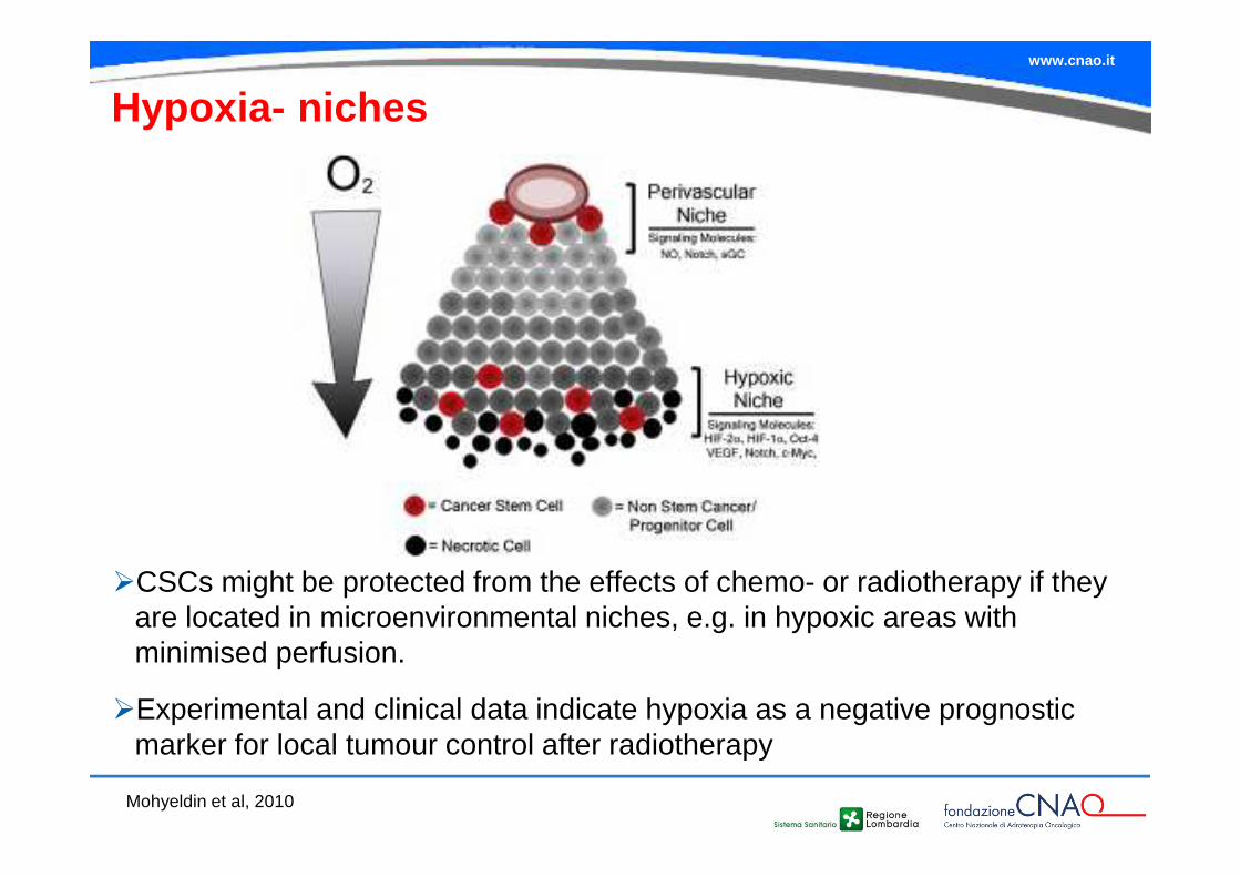
Mohyeldin et al, 2010
Hypoxia- niches
�CSCs might be protected from the effects of chemo- or radiotherapy if they are located in microenvironmental niches, e.g. in hypoxic areas with minimised perfusion.
�Experimental and clinical data indicate hypoxia as a negative prognostic marker for local tumour control after radiotherapy
www.cnao.it
Pag. 76 Data Titolo presentazione
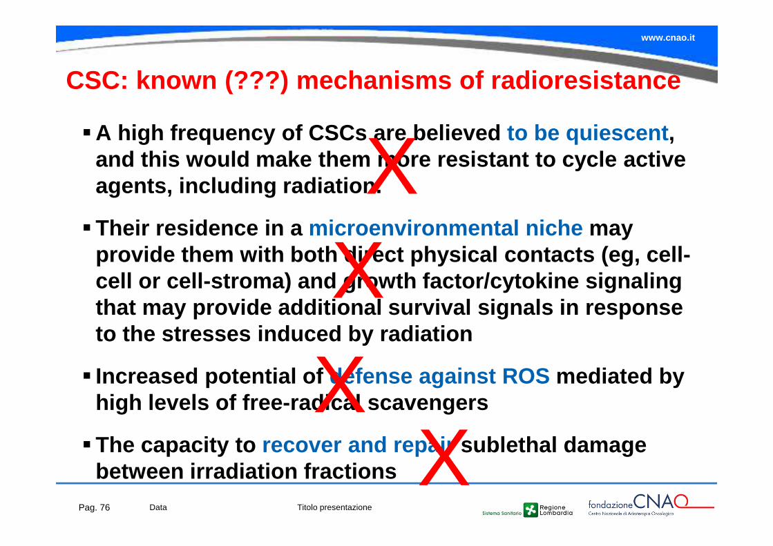
�A high frequency of CSCs are believed to be quiescent , and this would make them more resistant to cycle ac tive agents, including radiation.
�Their residence in a microenvironmental niche may provide them with both direct physical contacts (eg , cell-cell or cell-stroma) and growth factor/cytokine sig naling that may provide additional survival signals in res ponse to the stresses induced by radiation
� Increased potential of defense against ROS mediated by high levels of free-radical scavengers
�The capacity to recover and repair sublethal damage between irradiation fractions
X
X
XX
CSC: known (???) mechanisms of radioresistance
www.cnao.it
Until recently, many of the detrimental effects ascribed to cellular irradiation were considered the result of radiation depositing energy in the nucleus of the irradiated cell, and damaging a critical target in the nucleus, the DNA. The subsequent fate of the irradiated cell, tissue, organ or organism was though to reflect cellular responses to this induced DNA damage
Of late, there has been a rekindling of interest in nontargeted effects associated with exposure to ionizing radiation. These nontargeted effects describe a plethora of phenotypes associated with radiation exposure that seriously challenge the notion that radiation-induced deposition of energy in the nucleus of an irradiated cell leads to all those well-documented detrimental effects associated with exposure to radiation
Non-targeted effects
www.cnao.it
From target cells to orchestrated response
�The classical framework for discussing early and lateside effects was the target-cell hypothesis: that theseverity of side effects mainly reflected cell depletion asa result of the direct cell killing of a putative target cellleading to subsequent functional deficiency. This wasthe prevailing biological model until the mid 1990s.
�Recent research in radiobiology and molecularpathology has caused a change of paradigm,particularly in the understanding of late effects: radiationinduces concerted biological response at the cell andtissue level effected by the early activation of cytokinecascades.
www.cnao.it
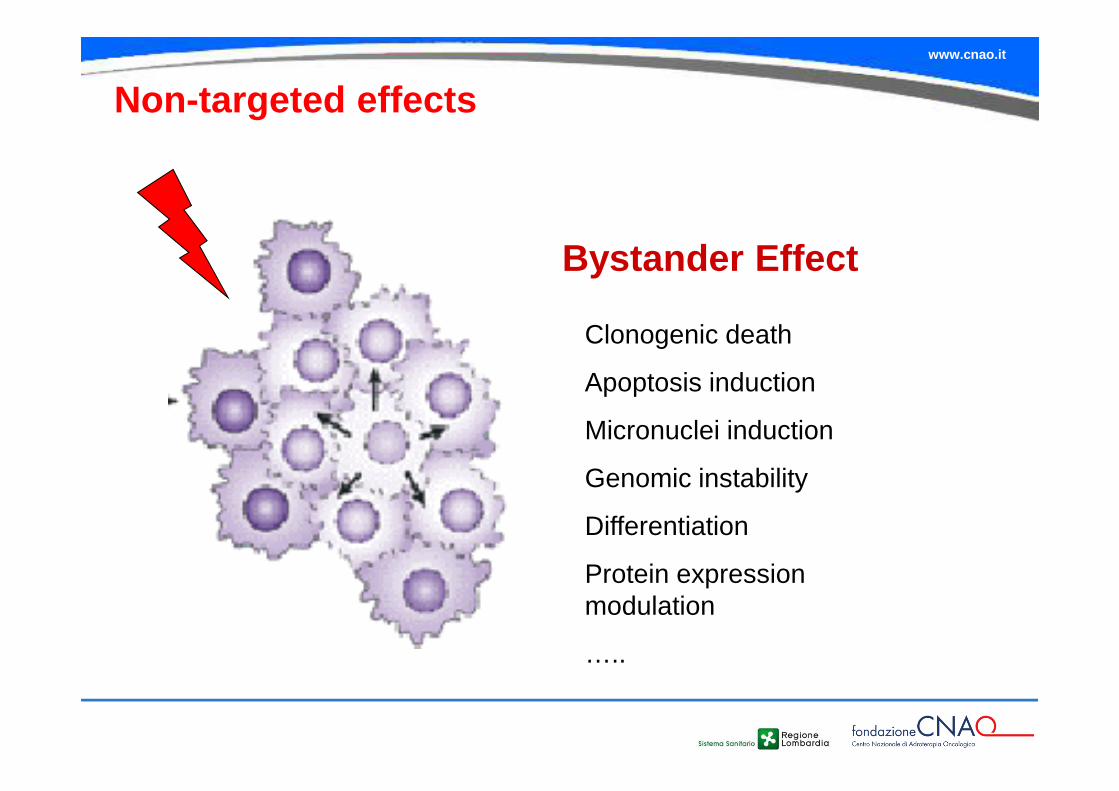
Non-targeted effects
Bystander Effect
Clonogenic death
Apoptosis induction
Micronuclei induction
Genomic instability
Differentiation
Protein expression modulation
…..
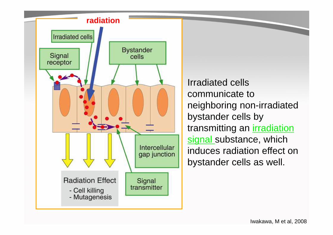
Iwakawa, M et al, 2008
Irradiated cells communicate to neighboring non-irradiated bystander cells by transmitting an irradiation signal substance, which induces radiation effect on bystander cells as well.
radiation
www.cnao.it
Radiation -induced genomic instability
Radiation-induced genomic instability defines effects
observed in the progeny of an irradiated cell, many cell
divisions after the initial insult.
Genomic instability is characterized by genetic changes
including chromosomal rearrangements, micronuclei,
transformation, gene amplifications, gene mutations
and reduced plating efficiency (lethal mutations or
delayed reproductive cell death) in cells derived and
clonally expanded from an irradiated cell
www.cnao.it
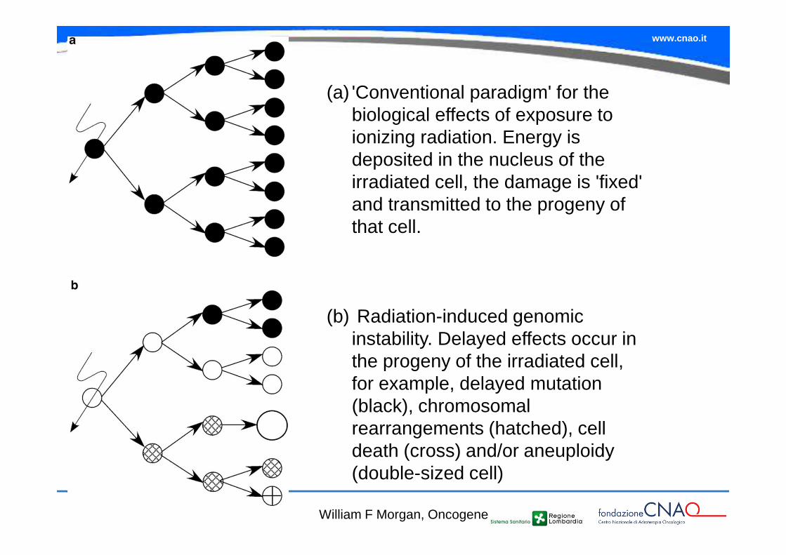
(a) 'Conventional paradigm' for the biological effects of exposure to ionizing radiation. Energy is deposited in the nucleus of the irradiated cell, the damage is 'fixed' and transmitted to the progeny of that cell.
(b) Radiation-induced genomic instability. Delayed effects occur in the progeny of the irradiated cell, for example, delayed mutation (black), chromosomal rearrangements (hatched), cell death (cross) and/or aneuploidy (double-sized cell)
William F Morgan, Oncogene
www.cnao.it
Bystander effectsRadiation-induced bystander effects occur when an irradiated cell communicates with nonirradiated cells via secreted factors and/or cell-to-cell gap junction communication pathways, eliciting responses in those cells that were not 'hit' by radiation .
These bystander effects include induced chromosomal rearrangements, micronuclei, transformation, gene mutations and reduced plating efficiency. Interestingly, bystander effects appear to predominate at low doses of radiation, after both low linear energy X or gamma rays and low doses of high linear energy alpha particles
Although the nature of the communication system that is involved in producing these responses is not yet known, there is strong evidence for a chemical signalling process that transmits information from the irradiated cell to neighboring cells.
The bystander effect has several important implications for radiation protection, radiotherapy and diagnostic radiology
www.cnao.it
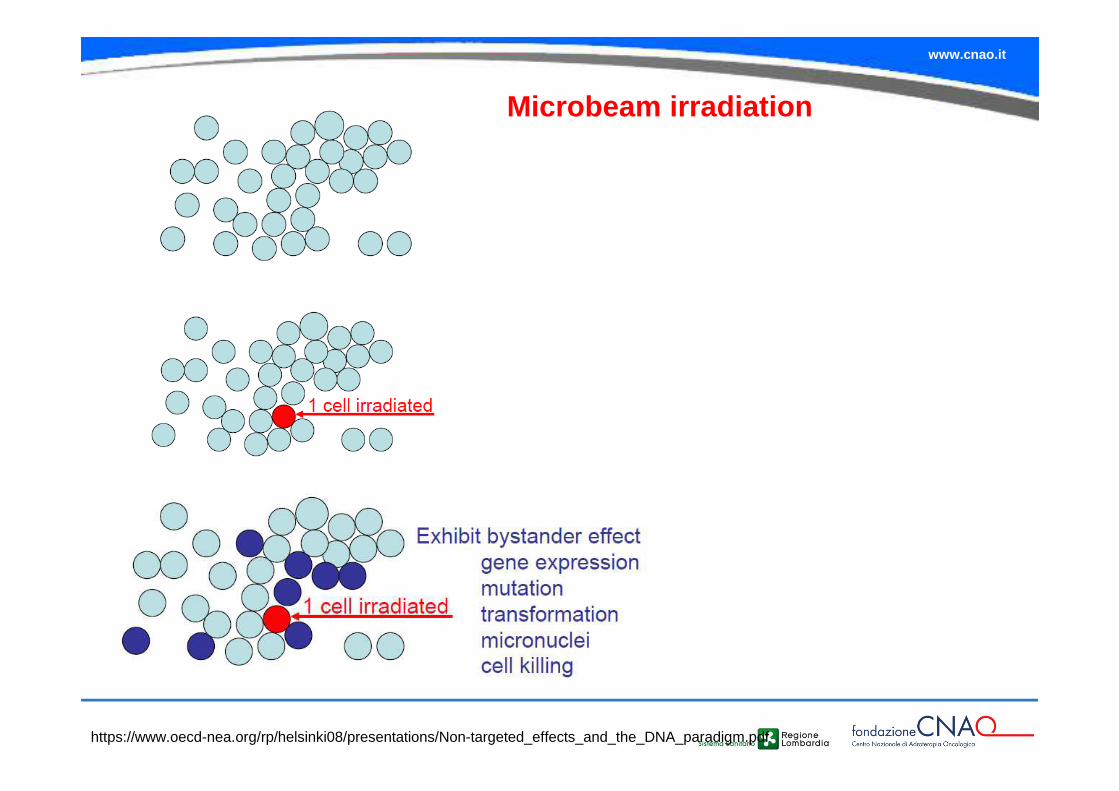
Microbeam irradiation
https://www.oecd-nea.org/rp/helsinki08/presentations/Non-targeted_effects_and_the_DNA_paradigm.pdf
www.cnao.it
www.cnao.it
Implications of non-targeted effects
These nontargeted effects indicate that the conventional paradigm ascribingthe biological effects to radiation-induced DNA damage does not accuratelyreflect all described radiation effects.
The deposition of energy in the nucleus certainly accounts for many of thedirect effects of radiation, for example, gene mutations, chromosomalrearrangements and cell death.However, nontargeted effects occurring in the progeny of irradiated cells, or innonirradiated cells indicate that alternative explanations to induced DNAdamage must be considered when fully evaluating the long-term effects ofradiation exposure.
Furthermore, these nontargeted effects indicate that the target for radiationeffects may be larger than the number of cells that were actually irradiated.
www.cnao.it
Fate of irradiated cells
Irradiation of a cell will result in one of the following possible outcomes:
�No effect.
�Division delay: The cell is delayed from going through division.
�Apoptosis: The cell dies before it can divide or afterwards byfragmentation into smaller bodies, which are taken up by neighbouringcells.
�Reproductive failure: The cell dies when attempting the first orsubsequent mitosis.
�Genomic instability: There is a delayed form of reproductive failure as aresult of induced genomic instability.
�Bystander effects: An irradiated cell can send signals to neighbouringunirradiated cells and induce genetic damage in them.
�Adaptive responses: The irradiated cell is stimulated to react andbecome more resistant to subsequent irradiation.
www.cnao.it
Good or bad ???
www.cnao.it
Abscopal effect
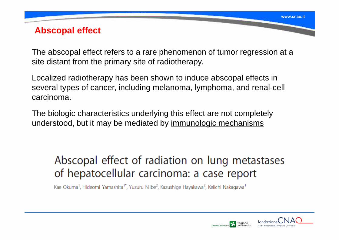
The abscopal effect refers to a rare phenomenon of tumor regression at a site distant from the primary site of radiotherapy.
Localized radiotherapy has been shown to induce abscopal effects in several types of cancer, including melanoma, lymphoma, and renal-cell carcinoma.
The biologic characteristics underlying this effect are not completely understood, but it may be mediated by immunologic mechanisms
www.cnao.it
Abscopal responses after particle therapy have been occasionally reported in patients treated in Japan.
A patient with colon carcinoma and distant lymph node metastasis was treated with local carbon ion therapy. Six months after treatment, both the primary tumor and the metastasis resolved (Durante M, Brenner DJ, Formenti SC. Does heavy ion therapy work through the immune system? Int J Radiat Oncol Biol Phys (2016) 96(5):934–6. ).
www.cnao.it
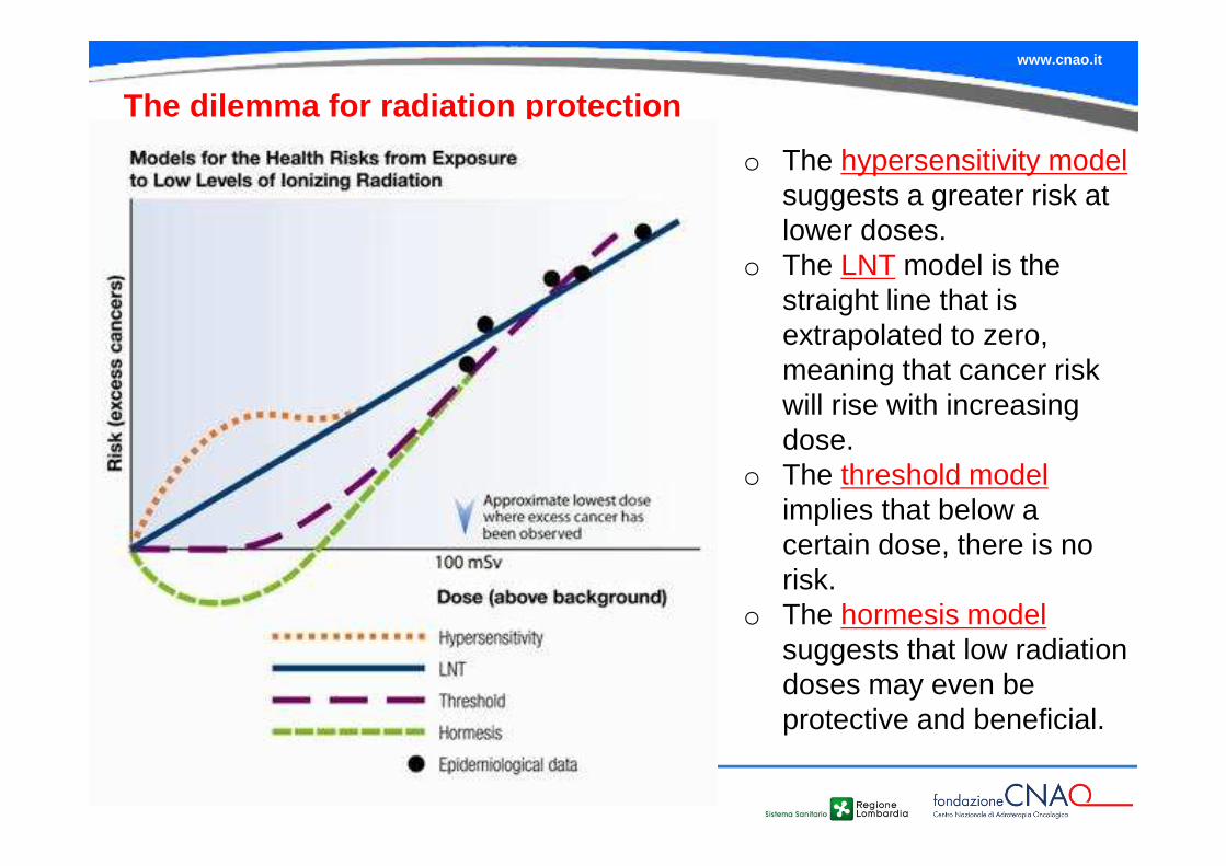
The dilemma for radiation protection
o The hypersensitivity model suggests a greater risk at lower doses.
o The LNT model is the straight line that is extrapolated to zero, meaning that cancer risk will rise with increasing dose.
o The threshold model implies that below a certain dose, there is no risk.
o The hormesis model suggests that low radiation doses may even be protective and beneficial.
Thanks for your attention!