membrane vesicles nucleate mineralo-organic nanoparticles and induce carbonate apatite precipitation...
TRANSCRIPT

Membrane Vesicles Nucleate Mineralo-organic Nanoparticlesand Induce Carbonate Apatite Precipitation in Human BodyFluids*
Received for publication, June 9, 2013, and in revised form, August 28, 2013 Published, JBC Papers in Press, August 29, 2013, DOI 10.1074/jbc.M113.492157
Cheng-Yeu Wu‡§¶1, Jan Martel‡§1, Wei-Yun Cheng‡§, Chao-Chih He‡§, David M. Ojcius§�, and John D. Young‡§**‡‡2
From the ‡Laboratory of Nanomaterials, the §Center for Molecular and Clinical Immunology, and the ¶Research Center of BacterialPathogenesis, Chang Gung University, Gueishan, Taoyuan 333, Taiwan, the �Molecular Cell Biology, Health Sciences ResearchInstitute, University of California, Merced, California 95343, the **Laboratory of Cellular Physiology and Immunology, RockefellerUniversity, New York, New York 10021, and the ‡‡Biochemical Engineering Research Center, Ming Chi University of Technology,Taishan, Taipei 24301, Taiwan
Background:Membrane vesicles (MVs) released from various cells are associated with human diseases.Results:MVs isolated from human serum induce the formation of mineralo-organic nanoparticles in culture.Conclusion: MVs represent a nucleating factor that promotes the formation of mineralo-organic nanoparticles and the pre-cipitation of mineral deposits in body fluids.Significance:The ectopic precipitation of carbonate apatite deposits in body fluids and tissues may be initiated in part byMVs.
Recent studies indicate that membrane vesicles (MVs)secreted by various cells are associated with human diseases,including arthritis, atherosclerosis, cancer, and chronic kidneydisease. The possibility that MVs may induce the formation ofmineralo-organic nanoparticles (NPs) and ectopic calcificationhas not been investigated so far. Here, we isolated MVs rangingin size between 20 and 400 nm from human serum and FBSusing ultracentrifugation and sucrose gradient centrifugation.TheMV preparations consisted of phospholipid-bound vesiclescontaining the serum proteins albumin, fetuin-A, and apolipo-protein A1; the mineralization-associated enzyme alkalinephosphatase; and the exosome proteins TNFR1 and CD63.Notably, we observed that MVs induced mineral precipitationfollowing inoculation and incubation in cell culture medium.The mineral precipitates consisted of round, mineralo-organicNPs containing carbonate hydroxyapatite, similar to previousdescriptions of the so-callednanobacteria.AnnexinV-immuno-gold staining revealed that the calcium-binding lipid phosphati-dylserine (PS) was exposed on the external surface of serumMVs. Treatment of MVs with an anti-PS antibody significantlydecreased theirmineral seeding activity, suggesting that PSmayprovide nucleating sites for calcium phosphate deposition onthe vesicles. These results indicate that MVs may representnucleating agents that induce the formation of mineral NPs inbody fluids. Given that mineralo-organic NPs represent precur-sors of calcification in vivo, our results suggest that MVs mayinitiate ectopic calcification in the human body.
Recent studies indicate that variousNPs3 present in the envi-ronment constantly interact with the human body and may beassociated with disease conditions. For instance, the inhalationof asbestos nanofibers derived from fireproof materials is asso-ciated with the development of lung cancer and fibrotic lungdamage known as asbestosis (1). Similarly, titanium dioxideNPs, which are widely used as whitening agents in commercialproducts, induce acute toxicity to various organs in laboratoryanimals (2). Given that NPs show unexpected size-dependentproperties and increased access to various body compartments,the potential toxicity of such nanomaterials represents animportant safety concern and is now a major research focus inthe fields of nanomedicine and nanotoxicology (3).Mineralized NPs called NB were initially described as the
smallest living microorganisms on earth (4) and as the possiblecausative agent of various humandiseases, includingAlzheimerdisease, atherosclerosis, cancer, chronic kidney disease, kidneystones, and prostatitis (5–7). However, recent studies per-formed by us (8–17) and others (18–22) have shown that theso-called NB actually represent non-living mineralo-organicNPs that possess biomimetic properties, including the ability toincrease in size andnumber in culture. Although the hypothesisthat NB represent living microorganisms has been discarded,several studies have indicated that mineral NPs similar to NBdo form in the growing bones and teeth of vertebrates (23–25).Mineralo-organic NPs have also been observed in undesired,ectopic calcification associated with various human and animalconditions (26–30). As such, these mineral NPs may representcalcification precursors found in both physiological and patho-logical mineralization processes.* This work was supported by the Primordia Institute of New Sciences and
Medicine and Chang Gung University Grant QZRPD88E; Ming Chi Univer-sity of Technology Grant 0XB0; Ministry of Education of Taiwan, Republic ofChina, Grant EMRPD1B0471; and National Science Council of Taiwan Grant101-2632-B-182-001-MY3.
1 Both authors contributed equally to this work.2 To whom correspondence should be addressed: Laboratory of Nanomate-
rials, Chang Gung University, Gueishan, Taoyuan 333, Taiwan, Republicof China. Tel.: 886-3211-8800 (ext. 3772); Fax: 886-3211-8534; E-mail:[email protected].
3 The abbreviations used are: NP, nanoparticle; ALP, alkaline phosphatase;apoA-I, apolipoprotein AI; CPP, calciprotein particle; DiR, 1,1�-dioctadecyl-3,3,3�,3�-tetramethylindotricarbocyanine iodide; EDX, energy-dispersivex-ray; HAP, hydroxyapatite; HS, human serum; MV, membrane vesicle; NB,nanobacteria; PS, phosphatidylserine; TEM, transmission electron micros-copy; VSMC, vascular smooth muscle cell.
THE JOURNAL OF BIOLOGICAL CHEMISTRY VOL. 288, NO. 42, pp. 30571–30584, October 18, 2013© 2013 by The American Society for Biochemistry and Molecular Biology, Inc. Published in the U.S.A.
OCTOBER 18, 2013 • VOLUME 288 • NUMBER 42 JOURNAL OF BIOLOGICAL CHEMISTRY 30571
by guest on July 17, 2015http://w
ww
.jbc.org/D
ownloaded from

Human body fluids have been shown to contain membranevesicles (MVs) in the form of small lipid-bound vesiclesreleased from various cells (31). These vesicles have beenbroadly categorized as exosomes, microvesicles, and apoptoticbodies (32). Exosomes represent 30–100-nm vesicles releasedfrom cellular endosomes and are thought to be involved in cell-to-cell signaling (32–34). Microvesicles are amore heterogene-ous group of 100–1,000 nm vesicles released from the plasmamembrane of various cells; they have been implicated in inter-cellular communication, homeostasis, and cellular waste dis-posal (32, 35). Apoptotic bodies represent particles rangingfrom 50 nm to 5 �m, which are released by apoptotic cells andremoved through phagocytosis bymacrophages (32, 34). Giventhat the level of MVs is elevated in the body fluids of humansafflicted with various ailments, some authors have proposedthat these vesicles may play a role in disease conditions thatinclude arthritis, atherosclerosis, cancer, and chronic kidneydisease (31, 36, 37). On the other hand, it remains unclearwhetherMVs represent a consequence of the disease process orwhether they play an active role in pathogenesis.Matrix vesicles represent a different type of extracellular
vesicles implicated in physiological and pathological calcifica-tion processes in vertebrates (38–40). Matrix vesicles are20–200-nm vesicles released from mineralizing cells, such asosteoblasts and odontoblasts, which induce mineralization inbones and teeth, respectively. During ectopic vascular calcifica-tion, these vesicles are also released by vascular smooth musclecells (VSMCs), which develop into osteoblast-like cells andinduce calcium phosphate precipitation when exposed toexcess phosphate, inflammation, or hyperlipidemia (38–40).Matrix vesicles are thought to induce mineralization by concen-trating calcium and phosphate ions in various ways, including viaspecific ion transporters; in addition, these vesicles containenzymes, such as alkaline phosphatase (ALP), which degrades cal-cification inhibitors (e.g. pyrophosphate) and releases phosphatefrom various organic molecules. Although MVs similar tomatrix vesicles and apoptotic bodies have been repeatedlydescribed in calcified tissues (30, 41), the possibility that suchvesicles may induce the formation of mineralo-organic NPs inbody fluids has not been investigated.Phosphatidylserine (PS), a phospholipid usually confined to
the inner lipid layer of the cell membrane (42), has been foundon the surface of various populations of MVs (32, 40). Whenpresent on platelet-derived microvesicles, PS induces bloodcoagulation, a phenomenon associatedwith an increased risk ofthrombosis (42, 43). On the surface of apoptotic bodies, PS isthought to represent a signal that induces phagocytosis andclearance of the vesicles by macrophages (42). Notably, PS pos-sesses calcium-binding properties and may provide a nucleat-ing site for calcium phosphate formation on both matrix vesi-cles (38–40) and apoptotic bodies (44). Whether PS found onthe surface of MVs may induce the mineralization of MVs inbody fluids remains to be examined.Several studies have been conducted to identify the factors
that induce formation of the so-called NB and mineralo-or-ganic NPs. Cisar et al. (18) observed that the cell membranelipid phosphatidylinositol produces mineral NPs similar to NBafter inoculation in DMEM and incubation in cell culture con-
ditions. Raoult et al. (22) speculated that fetuin-A, a systemiccalcification inhibitor (45, 46) associated with NB (9, 22), mayinitially inhibit NB formation but eventually act as a nucleatorof NB formation following a conformational change similar toprion conversion. Our own experiments have shown thatserum proteins like albumin and fetuin-A fail to induce NBformation under the conditions tested, although these proteinsmay form seeds for the formation of mineralo-organic NPsonce the concentrations of calcium and phosphate ions exceedsaturation (11). These results suggest that other molecules orstructures, possibly in the form of lipid membranes, may re-present factors that induce the formation of mineralo-organicNPs similar to the so-called NB in body fluids.In the present study, we examined the possibility that min-
eral NP formation may be induced by MVs present in bodyfluids. We isolated a population of MVs from human serum(HS) and FBS and characterized the morphological and bio-chemical composition of these particles. Our results show thatthe isolated serum MVs induce the formation of mineralo-organic NPs when inoculated and incubated in cell culturemedium, suggesting that MVs may serve as a nucleating agentof mineral NPs in culture and as a factor that induces ectopiccalcification in human body fluids.
MATERIALS AND METHODS
Isolation of Membrane Vesicles—Blood was collected fromhealthy human volunteers using a conventional venipuncturemethod. Written informed consents were obtained from thevolunteers, and the use of human samples was approved by theInstitutional Review Board of Chang Gung Memorial Hospital(Linkou, Taiwan). Whole blood was collected into Vacutainertubes without anticoagulant (BD Biosciences). After centrifu-gation at 1,500 � g for 15 min at room temperature, the super-natant corresponding toHSwas collected and stored at�20 °C.HS and commercial FBS (Biological Industries) were filteredthrough 0.2-�m pore membranes prior to use.MVs were isolated as before (47, 48), with minor modifica-
tions. Briefly, 10 ml of HS and FBS was centrifuged at 800 � gfor 15 min at 4 °C to spin down and remove large cell debris.The resulting supernatant was centrifuged for 30 min at10,000 � g. The supernatant obtained this way was centrifugedfor 60 min at 15,000 � g (SW27 rotor, Beckman Instruments).Material present in the supernatant was pelleted by ultracen-trifugation at 200,000� g for 2 h at 4 °C (SW41 rotor, BeckmanInstruments). The pellet was suspended in 1 ml of HEPESbuffer (20 mM HEPES, 140 mM NaCl, pH 7.4) and is referred inthe present study as “membrane vesicles” (MVs). In someexperiments, sodium azide was added at 0.02% to preventmicrobial contamination.Dynamic Light Scattering—Oneml of resuspendedMVs was
transferred to disposable plastic cuvettes (Kartell) andmixed bygentle inversion prior to reading using aDelsaNano SubmicronParticle Analyzer (BeckmanCoulter). Measurements were per-formed at room temperature at an incident angle of 165°.Although the relative particle unit used in the y axis of Fig. 1, AandB, represents arbitrary values, this unit correlates in a linearmanner with the observed particle amount under the condi-tions used.
Membrane Vesicles Nucleate Mineralo-organic Nanoparticles
30572 JOURNAL OF BIOLOGICAL CHEMISTRY VOLUME 288 • NUMBER 42 • OCTOBER 18, 2013
by guest on July 17, 2015http://w
ww
.jbc.org/D
ownloaded from

Optical and Electron Microscopies—Aliquots of MVs resus-pended in HEPES buffer were deposited on glass slides andobserved without fixation or staining with a BX-51 opticalmicroscope (Olympus) equipped with a �100 oil immersionobjective with iris (UPlanFLN, Olympus) and a dark field con-denser (Cerbe Distribution). Specimens were observed at amagnification of�1,000, and images were acquired with a SpotFlex color camera (Diagnostic Instruments).For negative stain TEM, MV preparations were deposited
onto Formvar carbon-coated grids and negatively stained with0.5% aqueous uranyl acetate, followed by drying overnight atroom temperature. In some experiments, MVs were treatedwith 0.1% (v/v) Triton X-100 prior to processing for negativestaining. Specimens were examined under an EMU-3C (RCA)or JEM-100B (JEOL) transmission electron microscope.Precipitates obtained following incubation of DMEM con-
taining MVs (with or without added serum) were pelleted bycentrifugation at 12,000 � g for 15 min and washed twice withHEPES buffer prior to resuspension in the same buffer. A smallaliquot was deposited onto carbon-coated grids before dryingovernight. In this case, the particles were observed without fix-ation or staining by TEM. Electron diffraction patterns wereobtained with the JEM-100B transmission electronmicroscopeoperated at 120 keV.For immunogold staining, MVs were deposited onto nickel
grids and blocked with PBS plus 1% gelatin. The grids wereplaced on liquid drops containing the diluted protein or anti-body for 1 h at room temperature. MVs were treated succes-sively with annexin V (BD Biosciences), rabbit polyclonal anti-annexin V antibody (sc-8300, Santa Cruz Biotechnology, Inc.),and goat anti-rabbit antibody conjugated with a 5-nm gold NP(R-14001, Agar Scientific). Between each treatment, MVs werewashed successively with PBST, 0.1% Tween 20 in PBS (5 min).Fluorescence Spectroscopy—MVs were quantified as before
(49, 50) by measuring the concentration of total proteins usinga commercial Bradford protein assay (Bio-Rad). To detect lipidmembranes, we mixed a fraction of MVs corresponding to 0.5�g of MV proteins with 0.1 mM 1,1�-dioctadecyl-3,3,3�,3�-tetramethylindotricarbocyanine iodide (DiR; Molecular Probes)in a final volume of 1 ml. The probe was initially dissolved in0.5% ethanol. Specimens were incubated for 1 h at room tem-perature with continuous agitation. The same amount of lipo-philic tracer was added to HEPES buffer and processed in thesame manner as a negative control. THP-1 cells (2 � 104 cells/ml) purchased from the American Type Culture Collection(ATCC) were used as a positive control. These cells weremain-tained in RPMI 1640 medium containing 10% FBS and 100units/ml of both penicillin and streptomycin. Fluorescence wasmeasured using a fluorescencemicroplate reader (SpectraMaxM5 Spectrophotometer, Molecular Devices) with excitation at748 nm and emission at 780 nm.To detect PS on the surface of MVs, we mixed 5 �l of FITC-
labeled annexin V (BD Biosciences) with a MV fraction corre-sponding to 30 �g of total MV proteins dissolved in 100 �l ofHEPES buffer (containing 2.5 mM CaCl2), with or without 0.5mM EDTA. Reaction mixtures were incubated for 30 min atroom temperature and subsequently washed with HEPESbuffer to remove unbound annexin V following ultracentrifu-
gation at 200,000 � g. Fluorescence emission was measured at535 nm following excitation at 485 nm.Sucrose Gradient Centrifugation—MV preparations obtained
byultracentrifugationwere diluted to 10% (v/v) in 1ml ofHEPESbuffer and layered on top of a centrifugation tube (Ultra-Clearcentrifugation tubes, 14� 89mm, Beckman Instruments) con-taining a linear sucrose gradient (0.2–20% sucrose, 20 mM
HEPES, pH 7.4) prepared using a gradient maker (GradientStation, BioComp Instruments). After centrifugation at100,000 � g for 15 h at 4 °C, 1-ml fractions were collected fromthe top of the tube. Collected fractions were dialyzed using acommercial microdialysis system (Invitrogen) using a mem-brane with a 12–14-kDa cut-off (Spectra).Lipid Analysis—Lipids were quantified in sucrose gradient
fractions by using commercial kits for phospholipids (BioAssaySystems), cholesterol, triglycerides, HDL, and LDL (BioVision)based on the manufacturer’s instructions.Western Blotting—SDS-PAGE and Western blot analysis
were performed as before (9). A fraction ofMVs correspondingto 60 �g of MV proteins, 60 �g of proteins from mineralo-organic NPs seeded by MVs, or 60 �g of proteins from eitherHeLa cells or whole serumwas dissolved in 5� “loading buffer”(0.313 M Tris-HCl, pH 6.8, 10% SDS, 0.05% bromophenol blue,50% glycerol, 12.5% �-mercaptoethanol) to a final concentra-tion of 1�, prior to heating at 95 °C for 5 min and separationunder denaturing and reducing conditions on 10% SDS-PAGEusing a minigel system (Hoefer). HeLa cells were purchasedfrom the ATCC and cultured in minimum essential mediumcontaining 10% FBS and 100 units/ml of both penicillin andstreptomycin. The primary antibodies used were goat poly-clonal anti-tissue nonspecific ALP (sc-15065, Santa CruzBiotechnology), mouse monoclonal anti-LAMP2 (lysosome-associated membrane protein 2) (sc-18822), goat polyclonalanti-human TNFR1 (tumor necrosis factor receptor 1) (sc-31349), rabbit polyclonal anti-annexin V (sc-8300), goat poly-clonal anti-CD63 (cluster of differentiation 63) (sc-31214), andrabbit polyclonal antibodies prepared in-house as describedbelow. The secondary antibodies used were horseradish perox-idase-conjugated anti-goat, anti-mouse, anti-sheep, or anti-rabbit antibodies (Santa Cruz Biotechnology). Primary and sec-ondary antibodies were diluted based on the instructionsprovided by the manufacturer. The polyclonal antibodies gen-erated in house were used at a dilution of 1:1,000. The blotswere revealed using enhanced chemiluminescence (AmershamBiosciences) and autoradiographic films (Molecular Technolo-gies). Membranes were stripped by using the ReBlot Westernblot recycling kit (Chemicon).Production of Polyclonal Antibodies—New Zealand White
rabbits were obtained at�12 weeks of age. Human serum albu-min, human serum fetuin-A, and human apolipoprotein AI(apoA-I) as well as bovine serum fetuin-A and bovine serumalbumin were purchased from Sigma. The purified proteins(500 �g) were dissolved in 1 ml of DMEM andmixed with 1 mlof Freund’s complete adjuvant (Sigma) using two syringes con-nected by a 3-way stopcock (Nipro). Each protein-adjuvantmixture was administered intradermally on the back of the ani-mals. Three weeks after the first immunization, one boosterdose (200 �g of protein in a 1:1 mixture of DMEM and incom-
Membrane Vesicles Nucleate Mineralo-organic Nanoparticles
OCTOBER 18, 2013 • VOLUME 288 • NUMBER 42 JOURNAL OF BIOLOGICAL CHEMISTRY 30573
by guest on July 17, 2015http://w
ww
.jbc.org/D
ownloaded from

plete Freund’s adjuvant; Sigma) was administered every monthfor a total of four times. Pre- and postimmunization blood wascollected from the ear vein. For apoA-I, 28–30 kDa gel bandscorresponding to�200�g of protein fromSDS-PAGEbands ofwhole serum were used for the immunizations as describedabove.Seeding of Mineralo-organic NPs by MVs—MVs obtained by
either ultracentrifugation or sucrose gradient centrifugationwere added into DMEMwith or without 10% serum to obtain afinal volume of 1 ml. The 24-well plates were incubated undercell culture conditions in a humidified cell culture incubator at37 °Cwith 5%CO2. Photography of the plates andA650 readingswere performed as before (9). Day 0 pictures and turbiditymeasurements were taken after reagent mixing. Mineralo-or-ganic NPs obtained after incubation were pelleted by centrifu-gation at 12,000 � g for 30 min and washed twice with HEPESbuffer prior to resuspension in the same buffer for furthermor-phological and spectroscopy analyses.CalciumDeposition Assay—The ability ofMVs to calcify was
assessed using a non-radioactive calcium phosphate depositionassay (49). Briefly, phospholipid-richMV fractions correspond-ing to 30 �g of total MV proteins were added into DMEM andincubated in cell culture conditions. The incubation was termi-nated at each time point by centrifugation at 12,000 � g for 30min to obtain a co-precipitate of MVs and mineralo-organicNPs. The mineral precipitate was solubilized in 0.6 M HCl for24 h. The calcium content of the HCl supernatant was thendetermined colorimetrically by using the O-cresolphthaleincomplexone method (BioVision). Briefly, 2 �l of acidifiedsupernatant was incubated for 1 min with 150 �l of calciumworking reagent (chromogenic � base reagent mix) prior tomeasurement with a microplate spectrophotometer. Absorb-ance was read against a blank at 575 nm within 10 min of rea-gent mixing.To evaluate the contribution of PS to mineral seeding, we
mixedMVs (2mg of total proteins) with 0.2�g of either anti-PSor anti-CD63 antibody in a final volumeof 1ml ofHEPES bufferand incubated the resulting solution for 1 h at room tempera-ture with gentle mixing.MVs were pelleted and washed follow-ing ultracentrifugation at 200,000 � g for 2 h at 4 °C. MVs wereresuspended and incubated in 1 ml of DMEM in cell cultureconditions. After 1 week of incubation, mineral pellets wereretrieved and washed using centrifugation at 12,000 � g for 15min. The calcium deposition assay was used to measure thelevel of mineral precipitate formed.MV Co-Precipitation Assay—A MV fraction corresponding
to 30 �g of MV proteins was added into DMEM prior to theaddition of 0.1–3 mM CaCl2 and NaH2PO4 each. After incuba-tion in cell culture conditions for 24 h, the precipitate was har-vested by centrifugation at 12,000� g for 30min. After washingin HEPES buffer, the precipitate was resuspended in 50 �l ofHEPES buffer and subjected to SDS-PAGE and Western blot-ting with the indicated antibodies. Samples that were not incu-bated were used in parallel for comparison.Spectroscopy Analyses—Energy-dispersive x-ray spectros-
copy (EDX)was performed as described previously (10). Briefly,material obtained after incubation of DMEM containing MVs(with or without serum) was pelleted by centrifugation at
12,000 � g for 30 min and washed twice with HEPES bufferprior to resuspension in water. Mineralo-organic NPs weredried overnight, and EDX spectra were acquired with an SEMS-3000N scanning electron microscope (Hitachi Science Sys-tems) equipped with an EMAX Energy EX-400 EDX device(Horiba).Fourier transform infrared spectroscopy (FTIR) was per-
formed as before (11). Briefly, the spectra were acquired using aNicolet 5700 FTIR spectrometer (Thermo Fisher Scientific)equipped with a deuterated triglycine sulfate detector. Miner-alo-organic NPs were prepared by adding 1 mM CaCl2 andNaH2PO4 each in 5% FBS, followed by incubation for 1 week incell culture conditions (Fig. 4D, FBS-NPs). The particles werecollected by centrifugation at 12,000 � g for 15min andwashedtwice with HEPES buffer prior to drying overnight at room tem-perature. Commercial CaCO3 (Mallinckrodt Baker), Ca3(PO4)2tribasic (KantoChemical) andHAP(bufferedaqueoussuspension,25% solid; Sigma) were used for comparison.Statistical Analysis—All experiments were performed in
triplicate. The values shown represent means � S.E. Compari-sons between control and experimental groupswere performedusing Student’s t test.
RESULTS
Isolation of Lipid-containing MVs from Serum—Mineralo-organic NPs initially described as NB have been shown to formwhen serum or other body fluids are inoculated into a cell cul-ture medium and incubated in culture conditions for severaldays (4, 5). In contrast, incubation of the cell culture mediumalone failed to producemineral NPs, suggesting that nucleatingagents exist in serum and body fluids. To verify whether MVsmay represent a nucleating agent that induces the formation ofmineralo-organic NPs in body fluids, we first isolated MVsfrom serum using an established protocol (see “Materials andMethods”). We hypothesized that MVs may induce mineralnucleation on their surface and thus become the core of miner-alo-organic NPs. Given that mineralo-organic NPs and the so-called NB have sizes ranging from 50 to 500 nm (5, 9), wefocused our attention on the smallest MVs present in body flu-ids and used a protocol involving ultracentrifugation to obtainvesicles with diameters below 100 nm.After ultracentrifugation of serum, we obtained a pellet con-
taining small, round particles of variable sizes when observedunder optical, dark field microscopy (Fig. 1, A and B, insets).Based on dynamic light scattering analysis, the particles derivedfrom HS showed a peak size of 37 � 14 nm, whereas the parti-cles obtained from FBS peaked at 28 � 3 nm (Fig. 1, A and B).The sizes of the particles obtained were relatively heterogene-ous; polydispersity index values of 0.29 and 0.22 were obtainedfor HS-MVs and FBS-MVs, respectively, indicating that thesizes of HS particles were more heterogeneous than for FBSparticles.Under negative stain TEM, MV preparations consisted of
round particles with sizes ranging from 20 to 400 nm (Fig. 1C,Control). In order to determine whether these particles weredelineated by a lipid membrane, we treated the MV prepara-tions with Triton X-100, a non-ionic detergent with lipid-dis-persing properties (48, 51). This treatment destroyed the
Membrane Vesicles Nucleate Mineralo-organic Nanoparticles
30574 JOURNAL OF BIOLOGICAL CHEMISTRY VOLUME 288 • NUMBER 42 • OCTOBER 18, 2013
by guest on July 17, 2015http://w
ww
.jbc.org/D
ownloaded from

integrity of serum-derived particles, leaving only membraneremnants (Fig. 1C, Triton X-100). To confirm these observa-tions, we used the lipophilic tracer DiR, which is weakly fluo-rescent in an aqueous environment but becomes highly fluores-cent when intercalated within a lipid membrane. Accordingly,DiR did not fluoresce in HEPES buffer used as a negative con-trol but produced fluorescence when incubated with humanacute monocytic leukemia THP-1 cells used as a positive con-trol (Fig. 1D). DiR treatment also produced fluorescence in thepresence of MVs obtained from either HS or FBS (Fig. 1D),confirming that the isolated MVs were lipid-bound vesicles.Biochemical Characterization of Serum-derivedMVs—Next,
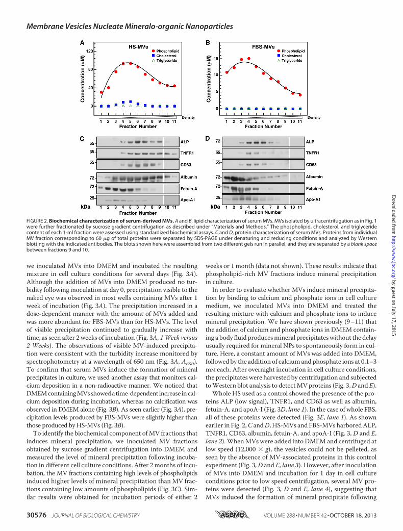
we fractionated the MV preparations by sucrose gradient cen-trifugation and determined the biochemical composition ofeach 1-ml fraction. The concentration of phospholipids, cho-lesterol, triglycerides, and lipoproteins (i.e. LDL and HDL) ineach fraction was determined using standardized biochemicalassays. Our results showed that MV fractions contained rela-tively high concentrations of phospholipids, averaging from 30to 93 �M for HS-derived vesicles (Fig. 2A) and from 4 to 15 �M
for FBS-derived vesicles (Fig. 2B). Cholesterol was detected inlow amounts in HS-derived MVs (Fig. 2A, fractions 4–6, peakat 2 �M), but none was found in FBS-MVs (Fig. 2B). Triglycer-ides, LDL, and HDL were not detected in the MV fractionstested (Fig. 2, A and B; data not shown), indicating that theisolated MV fractions did not contain lipoproteins.We then performed Western blot analysis to examine the
proteins associated with serum-derived MVs. We found thatALP, a glycosylated membrane-bound enzyme associated with
biomineralization processes (52, 53), was associated with thephospholipid-richMV fractions (Fig. 2, C andD). The molecu-lar weight of the ALP found in HS-MVs (55 kDa) was smallerthan the one found in FBS-MVs (72 kDa), possibly indicatingthe presence of different ALP isoenzymes in these samples.Wealso detected the presence of the exosome-associated proteinsTNFR1 and CD63 in phospholipid-rich fractions (Fig. 2, C andD), whereas other exosome proteins like LAMP2 were notdetected in our specimens (data not shown).Albumin, fetuin-A, and apoA-I were also found in associa-
tion with serum-derived MVs (Fig. 2, C and D). Althoughfetuin-A and apoA-I were associated with fractions of lighterdensities compared with fractions containing exosome pro-teins, the three serum proteins overlapped to some extent withexosome-associated proteins. These serum proteins have beenconsistently associated with mineralo-organic NPs derivedfrom body fluids, and they have been shown to act as bothinhibitors and seeders of mineral particles in culture (9–11).The structural and morphological data reported here along
with the co-distribution of phospholipids and exosome pro-teins confirmed that the particles isolated from serum con-sisted of MVs and included exosomes. Given that the sizes ofthe isolated MVs (20–400 nm) exceeded the sizes reportedpreviously for exosomes (30–100 nm) (32–34), we concludedthat other vesicles, possiblymicrovesicles and apoptotic bodies,may also be present in our MV preparations.Serum-derived MVs Induce Mineral Precipitation in Culture—
Todeterminewhether serumMVsmay induce the formation ofmineralo-organic NPs andmineral precipitation in body fluids,
FIGURE 1. Characterization of serum MVs isolated by ultracentrifugation. MVs were isolated from HS (A) and FBS (B) by ultracentrifugation as describedunder “Materials and Methods.” Round particles of heterogeneous sizes were observed under optical, dark field microscopy without fixation or staining(insets, �1,000). Particle sizes measured by dynamic light scattering peaked at 37 � 14 nm for HS and 28 � 3 nm for FBS. C, negative stain TEM revealed round,20 –500-nm structures (Control) susceptible to treatment with a lipid-dispersing detergent (Triton X-100; 0.1%, v/v). D, treatment with a fluorescent, lipophilicprobe (DiR; 0.1 mM) showed that both HS-MVs and FBS-MVs (corresponding to 0.5 �g of total MV proteins) produced fluorescence and thus harbored a lipidmembrane. As controls, DiR produced no fluorescence in HEPES buffer but fluoresced when incorporated in cell membranes (THP-1; 2 � 104 cells/ml). *, p �0.01 versus control HEPES. Error bars, S.E.
Membrane Vesicles Nucleate Mineralo-organic Nanoparticles
OCTOBER 18, 2013 • VOLUME 288 • NUMBER 42 JOURNAL OF BIOLOGICAL CHEMISTRY 30575
by guest on July 17, 2015http://w
ww
.jbc.org/D
ownloaded from
we inoculated MVs into DMEM and incubated the resultingmixture in cell culture conditions for several days (Fig. 3A).Although the addition of MVs into DMEM produced no tur-bidity following inoculation at day 0, precipitation visible to thenaked eye was observed in most wells containing MVs after 1week of incubation (Fig. 3A). The precipitation increased in adose-dependent manner with the amount of MVs added andwas more abundant for FBS-MVs than for HS-MVs. The levelof visible precipitation continued to gradually increase withtime, as seen after 2 weeks of incubation (Fig. 3A, 1Week versus2 Weeks). The observations of visible MV-induced precipita-tion were consistent with the turbidity increase monitored byspectrophotometry at a wavelength of 650 nm (Fig. 3A, A650).To confirm that serum MVs induce the formation of mineralprecipitates in culture, we used another assay that monitors cal-cium deposition in a non-radioactive manner. We noticed thatDMEMcontainingMVsshowedatime-dependent increase incal-cium deposition during incubation, whereas no calcification wasobserved in DMEM alone (Fig. 3B). As seen earlier (Fig. 3A), pre-cipitation levels produced by FBS-MVs were slightly higher thanthose produced by HS-MVs (Fig. 3B).To identify the biochemical component ofMV fractions that
induces mineral precipitation, we inoculated MV fractionsobtained by sucrose gradient centrifugation into DMEM andmeasured the level of mineral precipitation following incuba-tion in different cell culture conditions. After 2months of incu-bation, the MV fractions containing high levels of phospholipidsinduced higher levels of mineral precipitation than MV frac-tions containing low amounts of phospholipids (Fig. 3C). Sim-ilar results were obtained for incubation periods of either 2
weeks or 1 month (data not shown). These results indicate thatphospholipid-rich MV fractions induce mineral precipitationin culture.In order to evaluate whether MVs induce mineral precipita-
tion by binding to calcium and phosphate ions in cell culturemedium, we inoculated MVs into DMEM and treated theresulting mixture with calcium and phosphate ions to inducemineral precipitation. We have shown previously (9–11) thatthe addition of calcium and phosphate ions in DMEM contain-ing a body fluid producesmineral precipitateswithout the delayusually required for mineral NPs to spontaneously form in cul-ture. Here, a constant amount of MVs was added into DMEM,followed by the addition of calciumand phosphate ions at 0.1–3mM each. After overnight incubation in cell culture conditions,the precipitates were harvested by centrifugation and subjectedtoWestern blot analysis to detectMVproteins (Fig. 3,D and E).Whole HS used as a control showed the presence of the pro-
teins ALP (low signal), TNFR1, and CD63 as well as albumin,fetuin-A, and apoA-I (Fig. 3D, lane 1). In the case of whole FBS,all of these proteins were detected (Fig. 3E, lane 1). As shownearlier in Fig. 2,C andD, HS-MVs and FBS-MVs harboredALP,TNFR1, CD63, albumin, fetuin-A, and apoA-I (Fig. 3, D and E,lane 2).WhenMVs were added into DMEM and centrifuged atlow speed (12,000 � g), the vesicles could not be pelleted, asseen by the absence of MV-associated proteins in this controlexperiment (Fig. 3,D and E, lane 3). However, after inoculationof MVs into DMEM and incubation for 1 day in cell cultureconditions prior to low speed centrifugation, several MV pro-teins were detected (Fig. 3, D and E, lane 4), suggesting thatMVs induced the formation of mineral precipitate following
FIGURE 2. Biochemical characterization of serum-derived MVs. A and B, lipid characterization of serum MVs. MVs isolated by ultracentrifugation as in Fig. 1were further fractionated by sucrose gradient centrifugation as described under “Materials and Methods.” The phospholipid, cholesterol, and triglyceridecontent of each 1-ml fraction were assessed using standardized biochemical assays. C and D, protein characterization of serum MVs. Proteins from individualMV fraction corresponding to 60 �g of total proteins were separated by SDS-PAGE under denaturing and reducing conditions and analyzed by Westernblotting with the indicated antibodies. The blots shown here were assembled from two different gels run in parallel, and they are separated by a blank spacebetween fractions 9 and 10.
Membrane Vesicles Nucleate Mineralo-organic Nanoparticles
30576 JOURNAL OF BIOLOGICAL CHEMISTRY VOLUME 288 • NUMBER 42 • OCTOBER 18, 2013
by guest on July 17, 2015http://w
ww
.jbc.org/D
ownloaded from

incubation and that this precipitate was heavy enough to bepelleted by the low speed centrifugation used. When HS-MVswere incubated intoDMEMwith exogenous calcium and phos-
phate ions were added at 0.1–3 mM each, MV-associated pro-teins were also detected, and most proteins (ALP, TNFR1,CD63, and apoA-I) produced signals that increased in a dose-
FIGURE 3. Serum MVs induce mineral precipitation in cell culture conditions. A, MVs prepared by ultracentrifugation from HS and FBS were quantified based ontotal protein content prior to inoculation into DMEM at the amount indicated (Day 0, final volume of 1 ml). The solutions were incubated in cell culture conditions forthe time indicated. MVs produced visible precipitation that increased in a time-dependent manner. DMEM used as a negative control produced no precipitation. B, theability of MVs isolated by sucrose gradient centrifugation to undergo calcification. Phospholipid-rich MV fractions corresponding to 30 �g of total protein were addedinto DMEM and incubated in cell culture conditions. Precipitates were collected at the time indicated following centrifugation at 12,000�g for 30 min, and the calciumcontent was determined using the O-cresolphthalein complexone assay. Both HS-MVs and FBS-MVs induced the formation of calcified precipitate in a time-depen-dent manner during incubation. C, phospholipid-rich MV fractions induce mineral precipitation in culture. 100 �l of each sucrose gradient fraction was added intoDMEM in a final volume of 1 ml prior to incubation in cell culture conditions for 2 months. Mineral precipitation was assessed by A650 turbidity readings. Turbidityobserved before incubation was subtracted from the turbidity reading obtained after incubation (Differential A650). The content of phospholipids was determinedusing a standardized biochemical assay. Phospholipid-rich fractions produced slightly higher levels of precipitation under these conditions. D and E, Western blottinganalysis of co-precipitates containing MVs and mineralo-organic NPs. MVs were inoculated into DMEM, and CaCl2 and NaH2PO4 were added at the concentrationindicated in a final volume of 1 ml prior to incubation in cell culture conditions for 1 day. Mineral precipitates were pelleted by centrifugation at 12,000 � g for 30 minprior to washing steps in HEPES buffer. Equal amounts (60 �g) of proteins from whole serum, MVs, or MV-NP co-precipitates were separated under denaturing andreducing conditions by SDS-PAGE and probed with the indicated antibodies. Some of the lanes shown here originated from two different gels run in parallel; these aredelineated by the blank space seen in some of the blots. **, 0.1 � p � 0.5. Error bars, S.E.
Membrane Vesicles Nucleate Mineralo-organic Nanoparticles
OCTOBER 18, 2013 • VOLUME 288 • NUMBER 42 JOURNAL OF BIOLOGICAL CHEMISTRY 30577
by guest on July 17, 2015http://w
ww
.jbc.org/D
ownloaded from

dependent manner with the concentration of ions added (Fig.3D, lanes 5–7), indicating that MVs interacted with the exoge-nous ions to produce a mineral precipitate. In the case of FBS-MVs, protein signals were also detected for all MV proteins(Fig. 3E, lanes 5–7; note that ALP, TNFR1, CD63, and apoA-Iproduced signals only after the addition of ions at 3 mM).
These results suggest that the isolated serumMVs induce theformation of calcium-containing precipitates in culture. TheseMVsmay induce the formation ofmineral precipitates by bind-ing to exogenous calcium and phosphate ions in cell cultureconditions.Morphological and Mineral Characterization of Mineralo-
organic NPs Seeded by MVs—To verify the nature of the pre-cipitation induced by MVs, we submitted the precipitates(obtained in Fig. 3A) to TEM analysis without fixation or stain-ing. The precipitates produced following inoculation of MVsinto DMEM consisted of small, round, electron-dense particlesof sizes varying from 10 to 400 nm (Fig. 4A). The fact that theseparticles could be observed without staining suggested thattheymay be coated with an electron-densematerial like miner-als (compare the negatively stained, non-mineralized MVsshown in Fig. 1C with the unstained, mineralized MV-seededparticles in Fig. 4A). Mineralization of the particles was con-firmed by electron diffraction analysis, which showed severalconcentric rings corresponding to a polycrystalline mineral(Fig. 4A, insets). Particles of similar size, morphology, and elec-tron density were noted in the precipitates seeded by FBS-MVs(Fig. 4A). We confirmed the presence of MV proteins in theseeded mineral NPs by Western blots (Fig. 4A, right panels).Overall, the particles seeded by MVs were similar to the parti-cles described earlier as NB (6, 54). No mineral particles couldbe pelleted by centrifugation of incubated DMEM (data notshown), consistentwith the observation thatDMEMalone doesnot produce mineral NPs under these conditions (4, 5).We also inoculated MVs into DMEM containing either 5%
HS or FBS prior to incubation for 2 weeks to compare thenature of the particles obtained with and without serum. Themixtures of MVs, DMEM, and serum producedmineral NPs ofa crystalline nature following incubation (Fig. 4B). Accordingly,the electron diffraction patterns of these particles (Fig. 4B,insets) showed concentric rings that were more pronouncedthan that of the particles seededwithout serum (Fig. 4A, insets).The particles obtained this way appeared larger in size com-paredwith the particles preparedwithout added serum (Fig. 4B,20–500 nm). All of the proteins found earlier in MVs (Fig. 2, Cand D) were detected within the MV-seeded mineral NPs (Fig.4B, right panels).Notably, the mineral particles seeded by MVs and serum
were highly similar not only to NB (6, 54) but also to the sec-ondary calciprotein particles (CPPs) observed by Jahnen-Dech-ent and colleagues in specimens obtained from patients under-going dialysis (55) or suffering from calcifying peritonitis (27).These observations suggest that, in addition to MVs, serummay provide additional compounds that induce crystallizationof the mineral particles following prolonged incubation.Next, we used EDX and FTIR spectroscopies to identify the
mineral phase associated with MV-seeded particles. Previousstudies have shown that the so-called NB (5, 56) and the min-
eralo-organic NPs observed in calcified human tissues (30, 57)consist of carbonate HAP, a mineral similar to the one found inbones and teeth of vertebrates (58, 59). EDX spectra of mineralparticles seeded byMVs in DMEM showedmajor peaks of car-bon (C), calcium (Ca), oxygen (O), sodium (Na), phosphorus(P), and sulfur (S) (Fig. 4C, HS-MVs�DMEM and FBS-MVs�DMEM), consistent with the presence of carbonate-cal-ciumphosphate inMV-seededNPs. Similarly, theparticles seededby MVs in DMEM containing serum showed similar peaks con-sistent with the formation of carbonate-calcium phosphate (Fig.4C,HS-MVs�DMEM�HS and FBS-MVs�DMEM�FBS).FTIR analysis revealed major peaks of phosphate at 566
cm�1, 604 cm�1, 960 cm�1, and 1,033–1,100 cm�1 in the par-ticles seeded byMVs inDMEMwith or without serum (Fig. 4D;compare with the spectra of commercial Ca3(PO4)2 and HAPshown for reference; see also Refs. 60 and 61). In addition, peakscorresponding to carbonate at 875 and 1,430 cm�1 were foundin MV-seeded particles (Fig. 4D; compare with the signalsobtained for CaCO3; see also Refs. 62 and 63). For comparison,a sample of mineralo-organic NPs prepared by adding calciumand phosphate into DMEM containing 5% FBS showed similarphosphate and carbonate peaks (Fig. 4D, FBS-NPs). These FTIRspectra were consistent with the presence of carbonate HAPwithin the MV-seeded particles. Other peaks corresponding toH2O and the amide bonds of serum proteins were also detectedin the seeded particles (Fig. 4D; see also Refs. 58, 64, and 65).These spectroscopy analyses confirm that the MV-seeded par-ticles contain carbonate HAP similar to the particles describedearlier as NB (7) and to the mineralo-organic NPs found incalcified human tissues (30).PS on the Surface of MVs Contributes to Mineral NP Seeding—
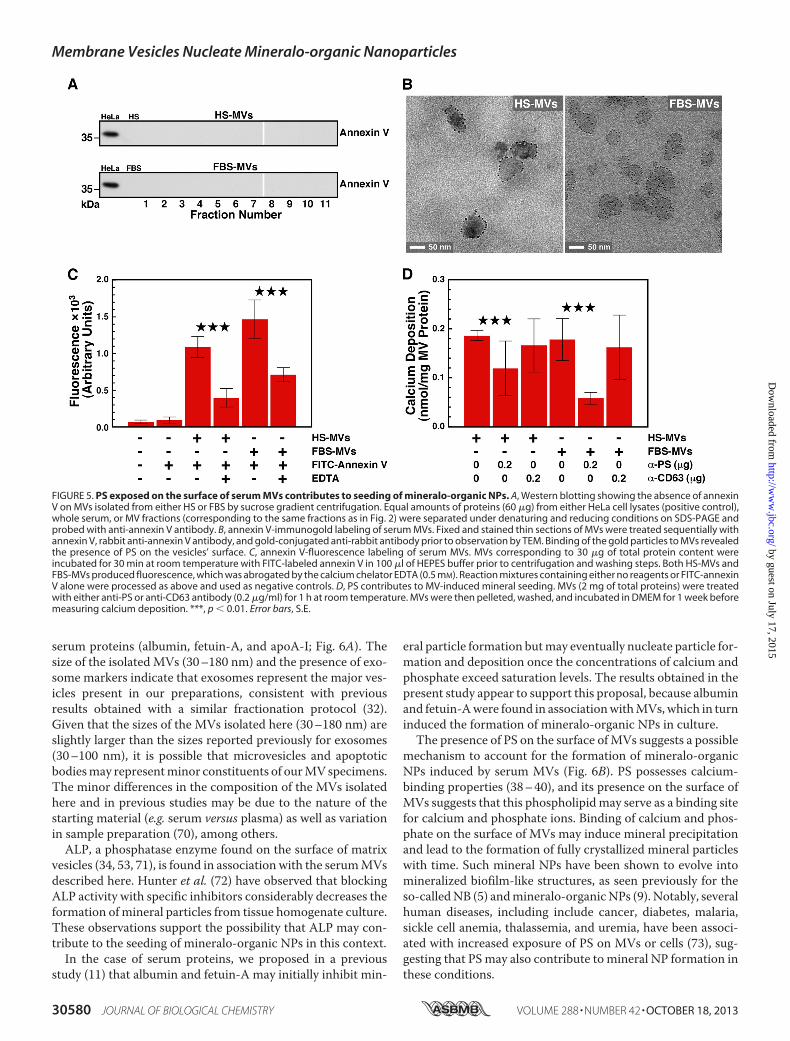
PS appears to represent a nucleator of calcium phosphate inmatrix vesicles (38, 66). Several MV populations, such as thosereleased by platelets and apoptotic cells, harbor PS on theirsurface (67, 68), but it is unclear whether PS acts as a nucleatorin this context. We first usedWestern blot analysis to examinewhether the PS-binding protein annexin V is present in serumMV fractions. AnnexinVwas not detected in eitherHS-MVs orFBS-MVs (Fig. 5A; a HeLa cell lysate was used as positive con-trol).Next, we examinedwhether PS is present onMVsbyusingimmunogold labeling and annexin V as a probe. TEM imagesshowed that annexin V binds the surface of MVs isolated fromHS or FBS (Fig. 5B). Similarly, using fluorescence spectroscopy,we observed that annexin V that was prelabeled with the fluo-rochrome FITC interacts with MVs of both human and bovineorigins. As a control, the interaction between PS and annexinV,which requires free calcium (69), was abrogated by the additionof the calcium chelator EDTA (Fig. 5C). These results confirmthat PS is present on the surface of MVs isolated from serum.We investigated whether PS found on the surface of MVs
contributes to the seeding of mineralo-organic NPs by the ves-icles. We incubated MVs with an anti-PS antibody and mea-sured the seeding potential of MVs following the washing steps.Blocking PS moieties with the antibody significantly decreasedseeding of mineral NPs by MVs, whereas an anti-CD63 anti-body used as a negative control showed no effect on seeding(Fig. 5D). These data indicate that PS contributes to the seedingactivity of MVs.
Membrane Vesicles Nucleate Mineralo-organic Nanoparticles
30578 JOURNAL OF BIOLOGICAL CHEMISTRY VOLUME 288 • NUMBER 42 • OCTOBER 18, 2013
by guest on July 17, 2015http://w
ww
.jbc.org/D
ownloaded from

DISCUSSIONIn the present study, we show that serum MVs isolated by
ultracentrifugation and sucrose gradient centrifugation inducethe formation of mineral NPs similar to the so-called NB fol-lowing incubation in cell culture conditions. These results indi-cate thatMVsmay represent the long sought nucleator of min-
eralo-organic NPs in body fluids. Moreover, these observationssupport our previous studies showing that NB-like structuresrepresent non-living mineral NPs that possess biomimeticproperties (9, 16).The MVs isolated from serum harbor various proteins,
including ALP, exosome markers (TNFR1 and CD63), and
FIGURE 4. MV-induced mineral precipitation represents calcium phosphate mineralo-organic NPs. A, mineral precipitates obtained from DMEM contain-ing MVs as in Fig. 3A were harvested by centrifugation and analyzed by TEM without fixation or staining. The precipitates displayed round NPs with a darkelectron-dense structure. The polycrystalline, mineralized nature of the particles was confirmed by the concentric rings produced on electron diffractionpatterns (insets). Western blotting analysis revealed the presence of MV-associated proteins within the seeded mineralo-organic NPs (right panels). B, mineralprecipitates obtained from DMEM containing both MVs and 5% serum showed ellipsoid NPs of a polycrystalline nature (insets). MV-associated proteins werealso detected in these particles (right panels). C, EDX spectra of mineral particles seeded by MVs with or without 5% serum showed peaks corresponding tocarbon, calcium, oxygen, sodium, phosphorus, and sulfur. D, FTIR analysis revealed the presence of both phosphate groups at 566, 604, and 960 cm�1 andbetween 1,033 and 1,100 cm�1 and carbonate at 875 and 1,430 cm�1, consistent with the presence of carbonate apatite. Peaks corresponding to water and theamide bonds of proteins were also observed in MV-seeded particles. Mineralo-organic NPs prepared by adding 1 mM CaCl2 and NaH2PO4 each in 1 ml of DMEMcontaining 5% FBS showed similar peaks (FBS-NPs). Commercial powders of Ca3(PO4)2, CaCO3, and HAP were included for comparison. WB, Western blot.
Membrane Vesicles Nucleate Mineralo-organic Nanoparticles
OCTOBER 18, 2013 • VOLUME 288 • NUMBER 42 JOURNAL OF BIOLOGICAL CHEMISTRY 30579
by guest on July 17, 2015http://w
ww
.jbc.org/D
ownloaded from
serum proteins (albumin, fetuin-A, and apoA-I; Fig. 6A). Thesize of the isolated MVs (30–180 nm) and the presence of exo-some markers indicate that exosomes represent the major ves-icles present in our preparations, consistent with previousresults obtained with a similar fractionation protocol (32).Given that the sizes of the MVs isolated here (30–180 nm) areslightly larger than the sizes reported previously for exosomes(30–100 nm), it is possible that microvesicles and apoptoticbodiesmay representminor constituents of ourMVspecimens.The minor differences in the composition of the MVs isolatedhere and in previous studies may be due to the nature of thestarting material (e.g. serum versus plasma) as well as variationin sample preparation (70), among others.ALP, a phosphatase enzyme found on the surface of matrix
vesicles (34, 53, 71), is found in association with the serumMVsdescribed here. Hunter et al. (72) have observed that blockingALP activity with specific inhibitors considerably decreases theformation ofmineral particles from tissue homogenate culture.These observations support the possibility that ALP may con-tribute to the seeding of mineralo-organic NPs in this context.In the case of serum proteins, we proposed in a previous
study (11) that albumin and fetuin-A may initially inhibit min-
eral particle formation butmay eventually nucleate particle for-mation and deposition once the concentrations of calcium andphosphate exceed saturation levels. The results obtained in thepresent study appear to support this proposal, because albuminand fetuin-Awere found in associationwithMVs,which in turninduced the formation of mineralo-organic NPs in culture.The presence of PS on the surface ofMVs suggests a possible
mechanism to account for the formation of mineralo-organicNPs induced by serum MVs (Fig. 6B). PS possesses calcium-binding properties (38–40), and its presence on the surface ofMVs suggests that this phospholipidmay serve as a binding sitefor calcium and phosphate ions. Binding of calcium and phos-phate on the surface of MVs may induce mineral precipitationand lead to the formation of fully crystallized mineral particleswith time. Such mineral NPs have been shown to evolve intomineralized biofilm-like structures, as seen previously for theso-calledNB (5) andmineralo-organicNPs (9). Notably, severalhuman diseases, including include cancer, diabetes, malaria,sickle cell anemia, thalassemia, and uremia, have been associ-ated with increased exposure of PS on MVs or cells (73), sug-gesting that PSmay also contribute to mineral NP formation inthese conditions.
FIGURE 5. PS exposed on the surface of serum MVs contributes to seeding of mineralo-organic NPs. A, Western blotting showing the absence of annexinV on MVs isolated from either HS or FBS by sucrose gradient centrifugation. Equal amounts of proteins (60 �g) from either HeLa cell lysates (positive control),whole serum, or MV fractions (corresponding to the same fractions as in Fig. 2) were separated under denaturing and reducing conditions on SDS-PAGE andprobed with anti-annexin V antibody. B, annexin V-immunogold labeling of serum MVs. Fixed and stained thin sections of MVs were treated sequentially withannexin V, rabbit anti-annexin V antibody, and gold-conjugated anti-rabbit antibody prior to observation by TEM. Binding of the gold particles to MVs revealedthe presence of PS on the vesicles’ surface. C, annexin V-fluorescence labeling of serum MVs. MVs corresponding to 30 �g of total protein content wereincubated for 30 min at room temperature with FITC-labeled annexin V in 100 �l of HEPES buffer prior to centrifugation and washing steps. Both HS-MVs andFBS-MVs produced fluorescence, which was abrogated by the calcium chelator EDTA (0.5 mM). Reaction mixtures containing either no reagents or FITC-annexinV alone were processed as above and used as negative controls. D, PS contributes to MV-induced mineral seeding. MVs (2 mg of total proteins) were treatedwith either anti-PS or anti-CD63 antibody (0.2 �g/ml) for 1 h at room temperature. MVs were then pelleted, washed, and incubated in DMEM for 1 week beforemeasuring calcium deposition. ***, p � 0.01. Error bars, S.E.
Membrane Vesicles Nucleate Mineralo-organic Nanoparticles
30580 JOURNAL OF BIOLOGICAL CHEMISTRY VOLUME 288 • NUMBER 42 • OCTOBER 18, 2013
by guest on July 17, 2015http://w
ww
.jbc.org/D
ownloaded from

Weobserved that FBS-MVs possess a highermineral seedingability compared with HS-MVs (Fig. 3, A and B). Fetal serum(i.e. FBS) may represent a better pro-calcifying milieu neededfor bone and teeth formation compared with adult serum (HS).If this is the case, FBS-MVs may show functional differencescompared with HS-MVs; accordingly, ALP appears to be pres-ent in slightly higher amounts in FBS-MVs compared with HS-MVs (Figs. 2 (C and D) and 4 (A and B)), an observation thatmay partially explain the difference in seeding ability of thesevesicles.Our results may have important implications for the under-
standing of ectopic calcification in humans. For one, MVs mayinduce the formation of mineral NPs in tissues and body fluidsunder certain conditions. Although the exact nature of thesecircumstances has not been examined in the present study, var-ious conditions could possibly induceMV-mediated seeding ofmineral NPs in vivo. For instance, a decrease in the level ofcalcification inhibition proteins, an increase of calcium andphosphate concentrations due to kidney failure, a disturbanceof vitamin D physiological activity, or an increase in the pro-duction of MVs due to inflammation or hyperlipidemia may
lead to MV-induced formation of mineral NPs. In addition,given thatMVs are present inmost body fluids, it appears plau-sible that these entities may induce the formation of mineralo-organic NPs in other areas of the body. Further studies arecurrently under way to assess the relevance of MV-inducedectopic calcification in vivo.A prominent factor that may induce the formation of MVs
and ectopic calcification in vivo is inflammation (40). Chronicinflammation can induce the differentiation of VSMCs intoosteoblast-like cells that produce matrix vesicles, which in turninduce ectopic calcification. A recent study byAikawa et al. (74)has shown that inflammation precedes ectopic calcification in amouse apolipoprotein E-deficient model of atherosclerosis,suggesting that inflammation may lead to ectopic calcification.These authors concluded that macrophages infiltrating thesites of lipid accumulation secrete cytokines that induce thephenotypic differentiation of VSMCs into functional osteo-blast-like cells. Given that calcium phosphate displays pro-in-flammatory properties (75–77), Shanahan (78) suggested thatthese processes may lead to a vicious cycle of inflammation andcalcification, which would eventually propitiate atherosclero-
FIGURE 6. Schematic model illustrating the composition of MVs and the seeding of mineralo-organic NPs by MVs. A, illustration representing an MVisolated from serum in the present study. Our results indicate that the isolated MVs consist of phospholipid-bound vesicles containing ALP, TNFR1, CD63,albumin, fetuin-A, and apoA-I. PS was found in the outer phospholipid layer of MVs. B, calcium ions present in cell culture medium and body fluids may bind toPS exposed on the surface of MVs and induce mineral precipitation. With time, mineralization of MVs may further increase and lead to the formation of fullymineralized mineralo-organic NPs similar to the so-called NB. Molecules present in serum and body fluids may promote the crystallization of the mineralparticles and produce mineral precipitates similar to those observed previously in human calcified tissues.
Membrane Vesicles Nucleate Mineralo-organic Nanoparticles
OCTOBER 18, 2013 • VOLUME 288 • NUMBER 42 JOURNAL OF BIOLOGICAL CHEMISTRY 30581
by guest on July 17, 2015http://w
ww
.jbc.org/D
ownloaded from

sis. We recently found that mineralo-organic NPs of calciumphosphate do not activate pro-inflammatory reactions inmacrophages but that large particle aggregates led to caspase-1activation and secretion of pro-inflammatory IL-1� (17). Theseresults suggest thatmineralNPs nucleated byMVsmay initiallybe inert in the body but may eventually induce inflammationandparticipate in disease processes after reaching a certain size.Apoptosis represents another factor that may control the
formation of mineral NPs and ectopic calcification in the body.A previous study by Proudfoot et al. (44) showed that VSMCscalcify after about 28 days in culture and that apoptosis appearsprior to mineral precipitation. Blocking apoptosis with a pan-caspase inhibitor reduced calcification, whereas induction ofapoptosis with anti-Fas IgM increased calcification in culture.In the end, calcification was attributed to the release of apopto-tic bodies (44) similar to the MVs studied here.Obviously, differences may exist between the in vitro system
studied here and what might occur in the human body. Forinstance, the observation that PS is exposed on the surface ofMVs suggests that these MVs could be subject to constantphagocytosis and clearance by macrophages of the reticuloen-dothelial system in vivo. It remains to be seen under what con-ditions procalcifying MVs similar to the ones characterizedhere may accumulate at amounts high enough in body fluids toinduce the formation of mineralo-organic NPs and larger cal-cified deposits.Jahnen-Dechent and colleagues (79, 80) have proposed that
mineral NPs in the form of CPPs may be part of a larger physi-ological cycle that controls the use and clearance of mineralions in the body. These authors have proposed that serum pro-teins like fetuin-Amay serve as systemic calcification inhibitorsand chaperone molecules that stabilize excess calcium andphosphate in the form of CPPs, leading to clearance of the par-ticles by the reticuloendothelial system (81). Recently, theseauthors have shown that the scavenger receptor-A present onmacrophages is implicated in the recognition and rapid clear-ance of CPPs from the blood of mice (82).From another perspective, we noticed that the MVs isolated
from serum (Fig. 1, A and B) share similarities with a group ofintriguing particles described previously in the blood undervarious names, including “microzymas,” “protits,” “somatids,”“cancer bacteria,” “pleomorphic bacteria,” and “filterable bod-ies,” among other terms (83–86). Vodyanoy and colleagues (87)have reported that metal nanoclusters may form structuresreferred to as “proteons” when they bind to denatured proteinsin various body fluids, such as blood. Based on these and similarobservations (8–17), we believe that the putative microzymas,protits, somatids, and related entities may represent a combi-nation of various particles that form spontaneously in the bloodduring incubation, which may include MVs, mineralo-organicNPs, proteons, and cellular remnants. Further studies areneeded to verify this intriguing possibility.In summary, our results indicate that human and bovine sera
containMVs that are able to induce the formation of mineralo-organic NPs when incubated in cell culture medium. Theseresults suggest that the formation of mineral NPs and ectopiccalcification occurring in the human body may be induced byMVs similar to the ones described here.
Acknowledgments—We thank Dr. Ching-Shiun Chen at Chang GungUniversity for help with FTIR; Dr. Po-Wen Gu for help with lipidanalysis; and Hwei-Chung Liu, Ji-Lung Peng, Hsi-Chien Su, Hui-Chun Kung, and Ya-Ling Chen for expertise in microscopy.
Note Added in Proof—We have recently extended the findings oncalcium apatite nanoparticles to other types of cations which alsoassemble nano- and micro-scale structures resembling biologicaltopologies, which we have termed collectively as bions (Wu, C. Y.,Young, L., Young, D.,Martel, J., Young, J. D. (2013)Bions: A family ofbiomimetic mineralo-organic complexes derived from biologicalfluids. PLoS One 8, e75501).
REFERENCES1. Donaldson, K., Murphy, F. A., Duffin, R., and Poland, C. A. (2010) Asbes-
tos, carbon nanotubes and the pleural mesothelium. A review of the hy-pothesis regarding the role of long fibre retention in the parietal pleura,inflammation and mesothelioma. Part. Fibre Toxicol. 7, 5
2. Chen, J., Dong, X., Zhao, J., and Tang, G. (2009) In vivo acute toxicity oftitanium dioxide nanoparticles to mice after intraperitioneal injection.J. Appl. Toxicol. 29, 330–337
3. Kim, B. Y., Rutka, J. T., and Chan, W. C. (2010) Nanomedicine. N. Engl.J. Med. 363, 2434–2443
4. Kajander, E. O., Kuronen, I., Akerman, K., Peltarri, A., and Cifcioglu, N.(1997) Nanobacteria from blood, the smallest culturable autonomouslyreplicating agent on Earth. Proc. Soc. Photo-Opt. Intrum. Eng. 3111,420–428
5. Kajander, E. O., and Ciftcioglu, N. (1998) Nanobacteria. An alternativemechanism for pathogenic intra- and extracellular calcification and stoneformation. Proc. Natl. Acad. Sci. U.S.A. 95, 8274–8279
6. Kajander, E. O., Ciftcioglu, N., Aho, K., and Garcia-Cuerpo, E. (2003)Characteristics of nanobacteria and their possible role in stone formation.Urol. Res. 31, 47–54
7. Ciftcioglu,N.,McKay,D. S.,Mathew,G., andKajander, E.O. (2006)Nano-bacteria. Fact or fiction? Characteristics, detection, and medical impor-tance of novel self-replicating, calcifying nanoparticles. J. Investig. Med.54, 385–394
8. Martel, J., and Young, J. D. (2008) Purported nanobacteria in human bloodas calcium carbonate nanoparticles. Proc. Natl. Acad. Sci. U.S.A. 105,5549–5554
9. Young, J. D., Martel, J., Young, L., Wu, C. Y., Young, A., and Young, D.(2009) Putative nanobacteria represent physiological remnants and cul-ture by-products of normal calcium homeostasis. PLoS One 4, e4417
10. Young, J. D., Martel, J., Young, D., Young, A., Hung, C. M., Young, L.,Chao, Y. J., Young, J., and Wu, C. Y. (2009) Characterization of granula-tions of calcium and apatite in serum as pleomorphic mineralo-proteincomplexes and as precursors of putative nanobacteria. PLoS One 4, e5421
11. Wu, C. Y., Martel, J., Young, D., and Young, J. D. (2009) Fetuin-A/albu-min-mineral complexes resembling serum calcium granules and putativenanobacteria. Demonstration of a dual inhibition-seeding concept. PLoSOne 4, e8058
12. Young, J. D., and Martel, J. (2010) The rise and fall of nanobacteria. Sci.Am. 302, 52–59
13. Martel, J., Wu, C. Y., and Young, J. D. (2010) Critical evaluation of �-irra-diated serum used as feeder in the culture and demonstration of putativenanobacteria and calcifying nanoparticles. PLoS One 5, e10343
14. Martel, J., Young, D., Young, A., Wu, C. Y., Chen, C. D., Yu, J. S., andYoung, J. D. (2011) Comprehensive proteomic analysis of mineral nano-particles derived from human body fluids and analyzed by liquid chroma-tography-tandem mass spectrometry. Anal. Biochem. 418, 111–125
15. Peng, H. H., Martel, J., Lee, Y. H., Ojcius, D. M., and Young, J. D. (2011)Serum-derived nanoparticles.De novo generation and growth in vitro, andinternalization by mammalian cells in culture.Nanomedicine 6, 643–658
16. Martel, J., Young, D., Peng, H. H., Wu, C. Y., and Young, J. D. (2012)Biomimetic properties of minerals and the search for life in the Martian
Membrane Vesicles Nucleate Mineralo-organic Nanoparticles
30582 JOURNAL OF BIOLOGICAL CHEMISTRY VOLUME 288 • NUMBER 42 • OCTOBER 18, 2013
by guest on July 17, 2015http://w
ww
.jbc.org/D
ownloaded from

meteorite ALH84001. Annu. Rev. Earth Planet. Sci. 40, 167–19317. Peng, H. H.,Wu, C. Y., Young, D., Martel, J., Young, A., Ojcius, D.M., Lee,
Y.H., andYoung, J. D. (2013) Physicochemical and biological properties ofbiomimetic mineralo-protein nanoparticles formed spontaneously in bi-ological fluids. Small 9, 2297–2307
18. Cisar, J. O., Xu, D. Q., Thompson, J., Swaim,W., Hu, L., and Kopecko, D. J.(2000) An alternative interpretation of nanobacteria-induced biomineral-ization. Proc. Natl. Acad. Sci. U.S.A. 97, 11511–11515
19. Vali, H., McKee, M. D., Ciftcioglu, N., Sears, S. K., Plows, F. L., Chevet, E.,Ghiabi, P., Plavsic, M., Kajander, E. O., and Zare, R. N. (2001) Nanoforms:A new type of protein-associated mineralization. Geochim. Cosmochim.Acta 65, 63–74
20. Drancourt, M., Jacomo, V., Lepidi, H., Lechevallier, E., Grisoni, V., Cou-lange, C., Ragni, E., Alasia, C., Dussol, B., Berland, Y., and Raoult, D. (2003)Attempted isolation of Nanobacterium sp. microorganisms from upperurinary tract stones. J. Clin. Microbiol. 41, 368–372
21. Barr, S. C., Linke, R. A., Janssen, D., Guard, C. L., Smith,M. C., Daugherty,C. S., and Scarlett, J. M. (2003) Detection of biofilm formation and nano-bacteria under long-term cell culture conditions in serum samples of cat-tle, goats, cats, and dogs. Am. J. Vet. Res. 64, 176–182
22. Raoult, D., Drancourt, M., Azza, S., Nappez, C., Guieu, R., Rolain, J. M.,Fourquet, P., Campagna, B., La Scola, B., Mege, J. L., Mansuelle, P., Lech-evalier, E., Berland, Y., Gorvel, J. P., and Renesto, P. (2008) Nanobacteriaare mineralo fetuin complexes. PLoS Pathog. 4, e41
23. Hohling, H. J., Arnold, S., Plate, U., Stratmann, U., and Wiesmann, H. P.(1997) Analysis of a general principle of crystal nucleation, formation inthe different hard tissues. Adv. Dent. Res. 11, 462–466
24. Robinson, C., Connell, S., Kirkham, J., Shore, R., and Smith, A. (2004)Dental enamel—A biological ceramic: Regular substructures in enamelhydroxyapatite crystals revealed by atomic force microscopy. J. Mater.Chem. 14, 2242–2248
25. Mahamid, J., Aichmayer, B., Shimoni, E., Ziblat, R., Li, C., Siegel, S., Paris,O., Fratzl, P., Weiner, S., and Addadi, L. (2010) Mapping amorphous cal-cium phosphate transformation into crystalline mineral from the cell tothe bone in zebrafish fin rays.Proc. Natl. Acad. Sci. U.S.A. 107, 6316–6321
26. Price, P. A., Thomas, G. R., Pardini, A. W., Figueira, W. F., Caputo, J. M.,andWilliamson, M. K. (2002) Discovery of a high molecular weight com-plex of calcium, phosphate, fetuin, and matrix �-carboxyglutamic acidprotein in the serum of etidronate-treated rats. J. Biol. Chem. 277,3926–3934
27. Heiss, A., Eckert, T., Aretz, A., Richtering, W., van Dorp, W., Schafer, C.,and Jahnen-Dechent, W. (2008) Hierarchical role of fetuin-A and acidicserum proteins in the formation and stabilization of calcium phosphateparticles. J. Biol. Chem. 283, 14815–14825
28. Matsui, I., Hamano, T.,Mikami, S., Fujii, N., Takabatake, Y., Nagasawa, Y.,Kawada, N., Ito, T., Rakugi, H., Imai, E., and Isaka, Y. (2009) Fully phos-phorylated fetuin-A forms a mineral complex in the serum of rats withadenine-induced renal failure. Kidney Int. 75, 915–928
29. Evan, A. P. (2010) Physiopathology and etiology of stone formation in thekidney and the urinary tract. Pediatr. Nephrol. 25, 831–841
30. Schlieper, G., Aretz, A., Verberckmoes, S. C., Kruger, T., Behets, G. J.,Ghadimi, R., Weirich, T. E., Rohrmann, D., Langer, S., Tordoir, J. H.,Amann, K., Westenfeld, R., Brandenburg, V. M., D’Haese, P. C., Mayer, J.,Ketteler, M., McKee, M. D., and Floege, J. (2010) Ultrastructural analysisof vascular calcifications in uremia. J. Am. Soc. Nephrol. 21, 689–696
31. Hugel, B., Martınez, M. C., Kunzelmann, C., and Freyssinet, J. M. (2005)Membrane microparticles. Two sides of the coin. Physiology 20, 22–27
32. György, B., Szabó, T.G., Pásztói,M., Pál, Z.,Misják, P., Aradi, B., László, V.,Pállinger, E., Pap, E., Kittel, A., Nagy, G., Falus, A., and Buzás, E. I. (2011)Membrane vesicles, current state-of-the-art. Emerging role of extracellu-lar vesicles. Cell. Mol. Life Sci. 68, 2667–2688
33. Keller, S., Sanderson, M. P., Stoeck, A., and Altevogt, P. (2006) Exosomes.From biogenesis and secretion to biological function. Immunol. Lett. 107,102–108
34. Théry, C., Ostrowski, M., and Segura, E. (2009) Membrane vesicles asconveyors of immune responses. Nat. Rev. Immunol. 9, 581–593
35. Nieuwland, R., and Sturk, A. (2010)Why do cells release vesicles?Thromb.Res. 125, S49–S51
36. Anderson,H. C.,Mulhall, D., andGarimella, R. (2010) Role of extracellularmembrane vesicles in the pathogenesis of various diseases, includingcancer, renal diseases, atherosclerosis, and arthritis. Lab. Invest. 90,1549–1557
37. Daniel, L., Dou, L., Berland, Y., Lesavre, P., Mecarelli-Halbwachs, L., andDignat-George, F. (2008) Circulating microparticles in renal diseases.Nephrol. Dial. Transplant. 23, 2129–2132
38. Anderson, H. C., Garimella, R., and Tague, S. E. (2005) The role of matrixvesicles in growth plate development and biomineralization. Front. Biosci.10, 822–837
39. Golub, E. E. (2009) Role of matrix vesicles in biomineralization. Biochim.Biophys. Acta 1790, 1592–1598
40. Golub, E. E. (2011) Biomineralization and matrix vesicles in biology andpathology. Semin. Immunopathol. 33, 409–417
41. Bonucci, E. (2002) Crystal ghosts and biological mineralization. Fancyspectres in an old castle, or neglected structures worthy of belief? J. BoneMiner. Metab. 20, 249–265
42. Leventis, P. A., and Grinstein, S. (2010) The distribution and function ofphosphatidylserine in cellular membranes. Annu. Rev. Biophys. 39,407–427
43. Nomura, S., Ozaki, Y., and Ikeda, Y. (2008) Function and role of micro-particles in various clinical settings. Thromb. Res. 123, 8–23
44. Proudfoot, D., Skepper, J. N., Hegyi, L., Bennett, M. R., Shanahan, C. M.,andWeissberg, P. L. (2000) Apoptosis regulates human vascular calcifica-tion in vitro. Evidence for initiation of vascular calcification by apoptoticbodies. Circ. Res. 87, 1055–1062
45. Schinke, T., Amendt, C., Trindl, A., Poschke, O., Muller-Esterl, W., andJahnen-Dechent, W. (1996) The serum protein �2-HS glycoprotein/fe-tuin inhibits apatite formation in vitro and inmineralizing calvaria cells. Apossible role in mineralization and calcium homeostasis. J. Biol. Chem.271, 20789–20796
46. Schafer, C., Heiss, A., Schwarz, A., Westenfeld, R., Ketteler, M., Floege, J.,Muller-Esterl,W., Schinke, T., and Jahnen-Dechent,W. (2003) The serumprotein � 2-Heremans-Schmid glycoprotein/fetuin-A is a systemicallyacting inhibitor of ectopic calcification. J. Clin. Invest. 112, 357–366
47. Caby, M. P., Lankar, D., Vincendeau-Scherrer, C., Raposo, G., and Bon-nerot, C. (2005) Exosomal-like vesicles are present in human bloodplasma. Int. Immunol. 17, 879–887
48. György, B., Módos, K., Pállinger, E., Pálóczi, K., Pásztói, M., Misják, P.,Deli, M. A., Sipos, A., Szalai, A., Voszka, I., Polgár, A., Tóth, K., Csete, M.,Nagy, G., Gay, S., Falus, A., Kittel, A., and Buzás, E. I. (2011) Detection andisolation of cell-derived microparticles are compromised by protein com-plexes resulting from shared biophysical parameters. Blood 117, e39–e48
49. Garimella, R., Sipe, J. B., and Anderson, H. C. (2004) A simple and non-radioactive technique to study the effect ofmonophosphoesters onmatrixvesicle-mediated calcification. Biol. Proced. Online 6, 263–267
50. Lässer, C., Eldh,M., and Lötvall, J. (2012) Isolation and characterization ofRNA-containing exosomes. J. Vis. Exp. 59, e3037
51. Girotti, A. W., and Thomas, J. P. (1984) Superoxide and hydrogen perox-ide-dependent lipid peroxidation in intact and triton-dispersed erythro-cyte membranes. Biochem. Biophys. Res. Commun. 118, 474–480
52. Whyte, M. P. (1994) Hypophosphatasia and the role of alkaline phospha-tase in skeletal mineralization. Endocr. Rev. 15, 439–461
53. Golub, E. E., and Boesze-Battaglia, K. (2007) The role of alkaline phospha-tase in mineralization. Curr. Opin. Orthop. 18, 444–448
54. Kajander, E. O., and Ciftcioglu, N. (1999) Nanobacteria as extremophiles.Proc. Soc. Photo-Opt. Intrum. Eng. 3755, 106–112
55. Heiss, A., Jahnen-Dechent, W., Endo, H., and Schwahn, D. (2007) Struc-tural dynamics of a colloidal protein-mineral complex bestowing on cal-cium phosphate a high solubility in biological fluids. Biointerphases 2,16–20
56. Kajander, E. O., Ciftcioglu, N., Miller-Hjelle, M. A., and Hjelle, J. T. (2001)Nanobacteria. Controversial pathogens in nephrolithiasis and polycystickidney disease. Curr. Opin. Nephrol. Hypertens. 10, 445–452
57. Evan, A. P., Coe, F. L., Rittling, S. R., Bledsoe, S. M., Shao, Y., Lingeman,J. E., andWorcester, E.M. (2005) Apatite plaque particles in innermedullaof kidneys of calcium oxalate stone formers. Osteopontin localization.Kidney Int. 68, 145–154
Membrane Vesicles Nucleate Mineralo-organic Nanoparticles
OCTOBER 18, 2013 • VOLUME 288 • NUMBER 42 JOURNAL OF BIOLOGICAL CHEMISTRY 30583
by guest on July 17, 2015http://w
ww
.jbc.org/D
ownloaded from

58. Carden, A., and Morris, M. D. (2000) Application of vibrational spectros-copy to the study of mineralized tissues (review). J. Biomed. Opt. 5,259–268
59. Barrere, F., van Blitterswijk, C. A., and de Groot, K. (2006) Bone regener-ation. Molecular and cellular interactions with calcium phosphateceramics. Int. J. Nanomedicine 1, 317–332
60. Barralet, J., Best, S., and Bonfield, W. (1998) Carbonate substitution inprecipitated hydroxyapatite. An investigation into the effects of reactiontemperature and bicarbonate ion concentration. J. Biomed.Mater. Res. 41,79–86
61. Vallet-Regi, M., and Ramila, A. (2000) New bioactive glass and changes inporosity during the growth of a carbonate hydroxyapatite layer on glasssurfaces. Chem. Mater. 12, 961–965
62. Aizenberg, J., Addadi, L., Weiner, S., and Lambert, G. (1996) Stabilizationof amorphous calcium carbonate by specialized macromolecules in bio-logical and synthetic precipitates. Adv. Mater. 8, 222–226
63. Raz, S., Testeniere,O., Hecker, A.,Weiner, S., and Luquet, G. (2002) Stableamorphous calcium carbonate is themain component of the calcium stor-age structures of the crustacean Orchestia cavimana. Biol. Bull. 203,269–274
64. Ayman, A., Talaat, M. S., Negm, S., and Talaat, H. (2008) Investigation ofbiophysical characteristics of diabetic living eye tissues using PA-FTIR-spectroscopy. Eur. Phys. J. Spec. Top. 153, 497–501
65. Chen, C. W., Oakes, C. S., Byrappa, K., Riman, R. E., Brown, K., TenHu-isen, K. S., and Janas, V. F. (2004) Synthesis, characterization, and disper-sion properties of hydroxyapatite prepared by mechanochemical-hydro-thermal methods. J. Mater. Chem. 14, 2425–2432
66. Merolli, A., and Santin, M. (2009) Role of phosphatidyl-serine in bonerepair and its technological exploitation.Molecules 14, 5367–5381
67. Fadok, V.A., Bratton,D. L., Frasch, S. C.,Warner,M. L., andHenson, P.M.(1998) The role of phosphatidylserine in recognition of apoptotic cells byphagocytes. Cell Death Differ. 5, 551–562
68. Lentz, B. R. (2003) Exposure of platelet membrane phosphatidylserineregulates blood coagulation. Prog. Lipid. Res. 42, 423–438
69. Huber, R., Schneider, M., Mayr, I., Römisch, J., and Paques, E. P. (1990)The calcium binding sites in human annexin V by crystal structure anal-ysis at 2.0 Å resolution. Implications for membrane binding and calciumchannel activity. FEBS Lett. 275, 15–21
70. Yuana, Y., Bertina, R. M., and Osanto, S. (2011) Pre-analytical and analyt-ical issues in the analysis of blood microparticles. Thromb. Haemost. 105,396–408
71. Thery, C., Zitvogel, L., and Amigorena, S. (2002) Exosomes. Composition,biogenesis and function. Nat. Rev. Immunol. 2, 569–579
72. Hunter, L.W., Shiekh, F. A., Pisimisis, G. T., Kim, S. H., Edeh, S. N.,Miller,V. M., and Lieske, J. C. (2011) Key role of alkaline phosphatase in thedevelopment of human-derived nanoparticles in vitro. Acta Biomater. 7,1339–1345
73. Zwaal, R. F., Comfurius, P., and Bevers, E. M. (2005) Surface exposure of
phosphatidylserine in pathological cells. Cell. Mol. Life Sci. 62, 971–98874. Aikawa, E., Nahrendorf, M., Figueiredo, J. L., Swirski, F. K., Shtatland, T.,
Kohler, R. H., Jaffer, F. A., Aikawa, M., and Weissleder, R. (2007) Osteo-genesis associates with inflammation in early-stage atherosclerosis evalu-ated by molecular imaging in vivo. Circulation 116, 2841–2850
75. Grandjean-Laquerriere, A., Laquerriere, P., Guenounou,M., Laurent-Ma-quin, D., and Phillips, T.M. (2005) Importance of the surface area ratio oncytokines production by human monocytes in vitro induced by varioushydroxyapatite particles. Biomaterials 26, 2361–2369
76. Nadra, I.,Mason, J. C., Philippidis, P., Florey, O., Smythe, C. D.,McCarthy,G. M., Landis, R. C., and Haskard, D. O. (2005) Proinflammatory activa-tion of macrophages by basic calcium phosphate crystals via protein ki-nase C and MAP kinase pathways. A vicious cycle of inflammation andarterial calcification? Circ. Res. 96, 1248–1256
77. Nadra, I., Boccaccini, A. R., Philippidis, P., Whelan, L. C., McCarthy,G. M., Haskard, D. O., and Landis, R. C. (2008) Effect of particle size onhydroxyapatite crystal-induced tumor necrosis factor alpha secretion bymacrophages. Atherosclerosis 196, 98–105
78. Shanahan, C. M. (2007) Inflammation ushers in calcification. A cycle ofdamage and protection? Circulation 116, 2782–2785
79. Jahnen-Dechent, W. (2005) Lot’s wife’s problem revisited. How we pre-vent pathological calcification. in Biomineralization (Bauerlein, E., ed) pp.243–267, Wiley, Weinheim, Germany
80. Jahnen-Dechent, W., Heiss, A., Schäfer, C., and Ketteler, M. (2011) Fe-tuin-A regulation of calcified matrix metabolism. Circ. Res. 108,1494–1509
81. Jahnen-Dechent, W., Schäfer, C., Ketteler, M., and McKee, M. D. (2008)Mineral chaperones. A role for fetuin-A and osteopontin in the inhibitionand regression of pathologic calcification. J. Mol. Med. 86, 379–389
82. Herrmann, M., Schäfer, C., Heiss, A., Gräber, S., Kinkeldey, A., Büscher,A., Schmitt,M.M., Bornemann, J., Nimmerjahn, F., Herrmann,M., Helm-ing, L., Gordon, S., and Jahnen-Dechent, W. (2012) Clearance of fetuin-A-containing calciprotein particles is mediated by scavenger receptor-A.Circ. Res. 111, 575–584
83. Hess, D. J. (1997)Can bacteria cause cancer? pp. 7–48, New York Univer-sity Press, New York
84. Domingue, G. J., Sr., and Woody, H. B. (1997) Bacterial persistence andexpression of disease. Clin. Microbiol. Rev. 10, 320–344
85. McLaughlin, R.W., Vali, H., Lau, P. C., Palfree, R. G., De Ciccio, A., Sirois,M., Ahmad, D., Villemur, R., Desrosiers, M., and Chan, E. C. (2002) Arethere naturally occurring pleomorphic bacteria in the blood of healthyhumans? J. Clin. Microbiol. 40, 4771–4775
86. Wainwright, M. (2010) The overlooked link between non-virus microbesand cancer. Sci. Prog. 93, 393–402
87. Samoylov, A. M., Samoylova, T. I., Pustovyy, O. M., Samoylov, A. A.,Toivio-Kinnucan, M. A., Morrison, N. E., Globa, L. P., Gale, W. F., andVodyanoy, V. (2005) Novel metal clusters isolated from blood are lethal tocancer cells. Cells Tissues Organs 179, 115–124
Membrane Vesicles Nucleate Mineralo-organic Nanoparticles
30584 JOURNAL OF BIOLOGICAL CHEMISTRY VOLUME 288 • NUMBER 42 • OCTOBER 18, 2013
by guest on July 17, 2015http://w
ww
.jbc.org/D
ownloaded from

YoungChao-Chih He, David M. Ojcius and John D. Cheng-Yeu Wu, Jan Martel, Wei-Yun Cheng, Body FluidsCarbonate Apatite Precipitation in Human
InduceMineralo-organic Nanoparticles and Membrane Vesicles NucleateMembrane Biology:
doi: 10.1074/jbc.M113.492157 originally published online August 29, 20132013, 288:30571-30584.J. Biol. Chem.
10.1074/jbc.M113.492157Access the most updated version of this article at doi:
.JBC Affinity SitesFind articles, minireviews, Reflections and Classics on similar topics on the
Alerts:
When a correction for this article is posted•
When this article is cited•
to choose from all of JBC's e-mail alertsClick here
http://www.jbc.org/content/288/42/30571.full.html#ref-list-1
This article cites 85 references, 23 of which can be accessed free at
by guest on July 17, 2015http://w
ww
.jbc.org/D
ownloaded from