production and characterization of niobate apatite
TRANSCRIPT

2238-7854/$ - see front matter © 2013 Brazilian Metallurgical, Materials and Mining Association. Published by Elsevier Editora Ltda. All rights reserved.
www.jmrt.com.br
J. MATER. RES. TECHNOL. 2013;2(1):24-29
1. Introduction
It has been observed, over the recent decades, an aging of population and a high demand to the practice of high-risk sports, resulting in an increase of number of people with bone defects. Bioceramics (i.e., ceramics, glasses and glass-ceramics) are generally used for repairing or replacing skeletal hard connective tissues in a variety of orthopedic and dental applications.
The clinical potential of calcium phosphate-based bio-ceramics is due mainly to its high degree of biocompatibility
Original Article
Production and characterization of niobate apatite
Daniel Navarro da Rochaa, Luciano de Andrade Gobbob, Marcelo Henrique Prado da Silvaa,*aInstituto Militar de Engenharia (IME), Rio de Janeiro, RJ, BrazilbPANalytical Spectris do Brasil Instr. Eletr. Ltda, São Paulo, SP, Brazil
ARTICLE INFO
Article history:
Received 29 August 2012
Accepted 16 October 2012
Keywords:
Hydroxyapatite
Niobium
Niobate
Apatite
* Corresponding author.E-mail address: [email protected] (M.H.P. da Silva).
A B S T R A C T
Bioceramics are widely used as grafts for orthopedic and dental applications, as well as
porous scaffolds for tissue engineering and bioactive coatings on metals. Because of its
similarity with bone mineral composition and surface reactivity, synthetic hydroxyapatite
(HAp) can be used as bone grafts and as drug delivery system. Substituted apatites have
been researched and designed by changig structure, crystallinity and surface charge
distribution of the material in order to optimize its performance in vivo. In the present
work, a novel nanostructured hydroxyapatite partially substituted with niobium was
obtained. The powders were synthesized by a patented aqueous precipitation method. The
green powders showed an anomalous fibrous morphology, with wires and aggregates of
nanoparticles, as observed by scanning electron microscopy (SEM). After heat treatment,
the particle size was estimated to lay between 20 nm and 40 nm. The characterization of
the material was performed by SEM, X-ray diffraction (XRD), Fourier transform infrared
(FTIR) spectroscopy and X-ray fluorescence (XRF) spectroscopy analyses. The element
niobium was identified by XRF analyses of the produced powder, which was previously
characterized by FTIR and XRD, as a single phase: HAp.
© 2013 Brazilian Metallurgical, Materials and Mining Association.
Published by Elsevier Editora Ltda. All rights reserved.
which is related to the chemical similarity of the materials normally found in bone tissues, a calcium phosphate in the form of carbonate apatite [1]. The great interest in hydroxyapatite (HAp) for use as bone grafts and drug delivery system is related to the fact that it is the principal mineral phase of teeth and bones, representing 30%-70% of their weight. Regardless of the fact that the HAp is found in enamel or bone, it is known that biological apatite is impure and non-stoichiometric [2].
Substituted apatites are researched and designed by investigating changes in structure, crystallinity and surface
http://dx.doi.org/10.1016/j.jmrt.2013.03.006http://dx.doi.org/10.1016/j.jmrt.2013.03.007

J. MATER. RES. TECHNOL. 2013;2(1):24-29 25
hydrated sodium niobate to the ultra-pure water. The salt was added up to saturation of the solution, to be incorporated in the synthesis of HAp partially substituted with Nb.
The introduction of the hydrated sodium niobate in the synthesis was performed in three different routes (Fig. 1). In the first route, a solution of hydrated sodium niobate was mixed with a solution of potassium hydroxide. In the second and third routes, the addition of the precursor of niobium was performed during the preparation of a transparent solution. In the second route, the hydrated sodium niobate was dripped simultaneously with ortho-phosphoric acid and in the third route, the addition of hydrated sodium niobate was also performed during the preparation of a transparent solution, being mixed to the suspension of calcium hydroxide.
The precursor solution was prepared by mixing, always under stirring by magnetic stirrer (Quimis, São Paulo, Brazil) a solution of hydrated sodium niobate [Na7(H3O)Nb19•14H2O], a suspension of 0.5 M calcium hydroxide (Merck, Darmstadt, Germany), a solution of 0.3 M ortho-phosphoric acid (Merck, Darmstadt, Germany) and a solution of 1.2 M lactic acid (Vetec, Rio de Janeiro, Brazil). The powder was obtained by lyophilisation (Labconco Freezone-1, USA) for 48 hours and thermally treated at 900 °C for 1 hour at a heating rate of 5 °C/min.
3. Results and discussion
Scanning electron microscopy analysis (SEM) of the green powders revelead the presence of wires and aggregates of nanoparticles, showing an anomalous fibrous morphology (Figs. 2a and 2b) when compared to pure HAp powders. After heat treatment, several nanoparticles aggregates (Figs. 2c and 2d) were observed in the calcined specimens, without the
charge distribution of the material in order to optimize the synthesis parameters and its performance in vivo [1,2]. The presence of CO3
2– substituted (in PO43– site), Cl– (in OH–) or Mg2+
and Sr2+ (in Ca2+) in the structure of apatites cause a decrease in crystallinity and an increase in solubility. Incorporation of fluoride ions (F–) in OH– site leads to an increase in crystallinity and turns the hexagonal structure to be more stable and less soluble than stoichiometric HAp [1].
Tissue response to the niobium metal is classified as bioreactive and can acquire bioactivity after activation of the surface treatment of its oxide [3]. Niobium phosphate glasses induced decomposition of the stoichiometric HAp with formation of a and b TCP phases after sintering. These glasses also showed no cytotoxicity [4]. Niobium, beta-tricalcium phosphate and HAp based composites, significantly increase the activity of alkaline phosphatase and calcification by human osteoblasts and the amount of calcified tissue directly proportional to concentration of niobium ion dissolved in the environment [5]. These authors have reported the incorporation of niobium to hydroxyapatite and tricalcium phosphate, but the results are shown to be inconclusive.
In the present study, strong evidences of niobate apatite formation are presented.
2. Materials and methods
The synthesis method chosen for obtaining the hydroxyapatite [6] followed the route of room temperature aqueous solution precipitation. For this study, the synthesis of hydrated sodium niobate was performed – niobium metal and niobium oxide pathways – according to the methodology developed by Azevedo [7], for use as niobium source in the synthesis of partially substituted HAp. The solutions were prepared by addition of
pH > 12
1st Route Aging for 24 hours
Washing and filtering
Thermal treatment
3rd RouteAqueous suspension Ca2+
Drip solutionH3PO4
KOH
“Transparent solution”in stirring
Lyophilization
2nd Route
Fig. 1 – Flow chart for the synthesis of hydroxyapatite wi th incorporation of sodium niobate to the solution in three different routes.

26 J. MATER. RES. TECHNOL. 2013;2(1):24-29
Pa 1 = 213.0 nmPb 1 = 69.0°
Pa 1
Pa R1
SUPRA 35-34-08 Mag = 14.60 KX EHT = 20.00 k V 24 Oct 2011Ext. Scan Control = off 200 nm WD = 6.2 mm 14:33:48 Signal A = InLens Aperture Size = 30.00 mm Gun Vacuum = 6.76e-010 mbar System Vacuum = 9.49e-007 mbar
presence of wires. The SEM analyses of the samples pointed to particle sizes on the order of a few tens of nanometers in the specimens calcined at 900 °C. These results highlight the presence of the nanoparticles, even after heat treatment at 900 °C for 1 hour.
The XRD analyses of the green powders showed a pattern characteristic of a low crystalline and/or nano-sized hydroxyapatite, similar to the standard human bone mineral [1]. The patterns indicated the presence of HAp phase in all routes, according to JCPDS 09-0432 (Fig. 3a). The results observed after calcination at 900 °C (Fig. 3b) indicated the presence of single phase HAp, since no peak corresponding to tricalcium phosphate or any other phase was detected. Only a small fraction of CaO (JCPDS 37-1497) was identified.
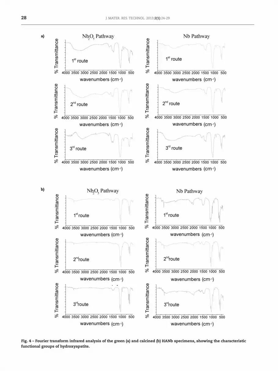
The FTIR analyses confirmed the XRD results in all specimens. The spectra of the green specimens (Fig. 4a) showed the characteristic bands of HAp, corresponding to the phosphate functional groups, hydroxyl and carbonate (CO3
2–) substitution in the structure of HAp. The presence of ~1,090, 1,040, 960, 604 and 575 cm–1 bands correspond to phosphates groups. Hydroxyl band at ~3,570 cm–1 is not well defined on these specimens. In addition, small peaks related to C-O vibration bands of carbonate groups at ~1,415-1,480 cm–1 suggest that some carbonate was incorporated during the low temperature HAp proces-sing [8].
It was found that the bands become more intense and well defined in calcined specimens (Fig. 4b) due to the
Fig. 2 – Morphology of nanometric and micrometric wires indicated by arrows in (a) and measured as indicated in (b). Aggregates of nanoparticles shown in powders after calcination at 900 °C (c) and (d). Particle diameter measurements ranging from 41.7 nm to 52.5 nm (d).
a)
b)
Pa 1
Pa R1 Pa 1 = 24.33 nmPb 1 = 153.4°
SUPRA 35-34-08 Mag = 61.57 KX EHT = 20.00 k V 24 Oct 2011Ext. Scan Control = off 100 nm WD = 9.6 mm 15:23:39 Signal A = InLens Aperture Size = 30.00 mm Gun Vacuum = 6.58e-010 mbar System Vacuum = 8.48e-007 mbar
H2 = 47.15 nm
SUPRA 35-34-08 Mag = 61.57 KX EHT = 20.00 k V 24 Oct 2011Ext. Scan Control = off 100 nm WD = 9.6 mm 15:23:39 Signal A = InLens Aperture Size = 30.00 mm Gun Vacuum = 6.58e-010 mbar System Vacuum = 8.48e-007 mbar
V2 = 52.59 nmV1 = 47.71 nm
H1 = 43.52 nm
c) d)

J. MATER. RES. TECHNOL. 2013;2(1):24-29 27
of intermolecular hydrogen bonding OH, at approximately 3,570 cm–1 [9,10]. Carbonate groups are still present, suggesting that substitution may occur both in hydroxyl
increase of structural order and crystallinity of the material, when compared green specimens. The calcination at 900 °C produced defined and intense bands of axial deformation
u
u
u u
u
u
u
u
u u
u
u
Fig. 3 – X-ray diffraction patterns of the green specimens (a ) and specimens calcined at 900 °C.
a)
b)
28 J. MATER. RES. TECHNOL. 2013;2(1):24-29
Fig. 4 – Fourier transform infrared analysis of the green (a) and calcined (b) HANb specimens, showing the characteristic functional groups of hydroxyapatite.
(cm–1)
(cm–1)
(cm–1)
(cm–1)
(cm–1)
(cm–1)
(cm–1)
(cm–1)
(cm–1)
(cm–1)
(cm–1) (cm–1)
a)
b)

J. MATER. RES. TECHNOL. 2013;2(1):24-29 29
(Type A carboapatite) and in phosphate (Type B carboapatite) sites.
The XRF results (Table 1) o f Nb2O5 pathway confirmed the presence of the chemical element niobium after heat treatment. The specimens synthesized by the second route presented the most marked change of the morphology of the powders and the lowest molar ratio Ca/P among all. In contrast, synthesized specimens by the first route presented less marked characteristic of wires and higher molar ratio Ca/P.
An increase in Ca/P molar ratio may be indicative of the presence of niobate group in PO4
3– site. However, carbonate in phosphate site may also account for high Ca/P ratio.
4. Conclusion
The results obtained in this study lead to the following conclusions:
• A partially substituted hydroxyapatite (HANb) was produced from the introduction of the hydrated sodium niobate salt (following two pathways: metal niobium and oxide niobium) by three different pathways. It produced a single phase, confirmed by X-ray diffraction analysis.
• Fourier transform infrared spectroscopy confirmed the XRD findings, showing the presence of functional groups typical of hydroxyapatite. The presence of carbonate groups was detected, confirming FTIR providing additional information on the structure of the produced powders.
• The results of X-ray fluorescence spectroscopy revealed the presence of niobium in the powder identified as HAp by XRD and FTIR. This technique was essential to characterize the powders as a niobate apatite (HANb).
• The material produced without heat treatment showed different morphological features produced in the synthesis of HAp, presenting a fibrous texture consisting of wires with aggregates of nano-sized particles. The particle size after heat treatment at 900 °C were estimated from 20 to 40 nm, indicating a route for production of nanoparticles.
Acknowledgements
The authors acknowledge the CAPES, CNPq and the institutions IME, CBPF and UFRJ for their cooperation and support for the paper.
R E F E R E N C E S
[1] Legeros RZ. Calcium phosphate -based osteoinductive mate-rials. Chem Rev. 2008;108:4742-53.
[2] Elliot JC. Structure and Chemistry of the apatites and other calcium orthophosphates. Amsterdam: Elsevier; 1994.
[3] Godley R, Strosvetsky D, Gotman T. Bonelike apatite formation on niobium metal treated in aqueous NaOH. J Mater Sci: Mater Med. 2004;15:1073-7.
[4] Prado da Silva MH, Moura Ramirez C, Granjeiro JM, Rossi AM. In vitro assessment of new niobium phosphate glasses and glass ceramics. Key Eng Mater. 2008;229:361-3.
[5] Tamai M, Isama K, Nakaoka R, Tsuchiya T. Synthesis of a novel b-tricalcium phosphate/hydroxyapatite biphasic calcium phosphate containing niobium ions and evaluation of its osteogenic properties. J Artif Organs. 2007;10:22-8.
[6] Prado da Silva MH, Navarro da Rocha D. Composição de hidro-xiapatita parcialmente substituída com nióbio e processo para sua obtenção. Patent BR n PI 020110137091. 2011.
[7] Azevedo LMS. Síntese do niobato de sódio a partir do óxido do nióbio e nióbio metálico [MD thesis]. Rio de Janeiro: Instituto Militar de Engenharia; 2010.
[8] Rehman I, Smith R, Hench LL, Bonfield W. Structural evalua-tion of human and sheep bone and comparison with synthe-tic hydroxyapatite by FTRaman spectroscopy. J Biomed Mater Res. 2005;29:1287-794.
[9] Santos ML, Florentino AO, Saeki MJ, Aparecida AH, Lia Fook MV, Guastaldi AC. Síntese e hidroxiapatita pelo método sol-gel utilizando precursores alternativos: nitrato de cálcio e ácido fosfórico. Ecl Quím. 2005;30:29-35.
[10] Silverstein RM, Webster FK, Kiemle DJ. Identificação espec-trométrica de compostos orgânicos. 7th ed. Rio de Janeiro: LTC Editora; 2007. p. 490.
Route % Molar content Molar ratio
Ca P Nb Ca P Nb Ca/P Ca/(P+Nb)
1st 28.92 18.05 1.11 11.57 5.60 1.03 2.07 1.742nd 26.57 19.68 0.66 10.63 6.10 0.61 1.74 1.583rd 30.06 20.95 0.56 12.02 6.50 0.52 1.85 1.71
Source: Author (2011).
Table 1 – X-ray fluorescence of HANb specimens by Nb2O5 pathway.