preparation and physicochemical characteristics of luminescent apatite-based colloids
TRANSCRIPT

Preparation and Physicochemical Characteristics of Luminescent Apatite-Based Colloids
Ahmed Al-Kattan,*,† Pascal Dufour,‡ Jeannette Dexpert-Ghys,§ and Christophe Drouet†
CIRIMAT Carnot Institute, UniVersity of Toulouse, CNRS/INPT/UPS, ENSIACET, 4 allee Emile Monso, BP44362, 31432 Toulouse cedex 4, France, CIRIMAT Carnot Institute, UniVersity of Toulouse, CNRS/INPT/UPS,LCMIE, UniVersite Paul Sabatier, 118 route de Narbonne, Bat. 2R1, 31062 Toulouse cedex 9, France, andCEMES 29, rue Jeanne MarVig, BP 94347, 31055 Toulouse cedex 4, France
ReceiVed: NoVember 17, 2009; ReVised Manuscript ReceiVed: January 13, 2010
Luminescent colloidal nanosystems based on europium-doped biomimetic apatite were prepared andinvestigated. The colloids were synthesized by soft chemistry in the presence of a phospholipid moiety,2-aminoethylphosphoric acid (AEP), with varying europium doping rates. Physicochemical features, includingcompositional, structural, morphological, and luminescence properties, were examined. Experimental evidenceshowed that suspensions prepared from an initial Eu/(Eu + Ca) molar ratio up to 2% consisted of single-phased biomimetic apatite nanocrystals covered with AEP molecules. The mean particle size was found todepend closely on the AEP content, enabling the production of apatite colloids with a controlled size downto ca. 30 nm. The colloids showed luminescence properties typical of europium-doped systems with narrowemission bands and long luminescence lifetimes of the order to the millisecond, and the data suggested thelocation of Eu3+ ions in a common crystallographic environment for all the colloids. These systems, stableover time and capable of being excited in close-to-visible or visible light domains, may raise interest in thefuture in the field of medical imaging.
1. Introduction
Lanthanide-based phosphors have been used for many yearsin the field of optical devices (lasers, phosphors in color TVtubes, etc.)1-3 and, more recently, for medical applications.4-7
In particular, the development of luminescent probes in biologyfaces the issue of particle size,8 especially when interactionswith cells are foreseen, either in vitro or in vivo.9-17
Organic dyes, such as fluoresceine, green fluorescent protein,or rhodamine, are fluorescent probes commonly used in biology.However, these molecules do not allow an extended analysisof biological tissues due to fast damage under irradiation, aswell as broad spectrum profiles and low photobleachingthresholds. To remedy this shortcoming, new classes of labelingagents, such as semiconductor quantum dots (e.g., CdSe-CdS,CdSe-ZnS, InAs), silica, or phosphate nanoparticles, wereintroduced more recently.18-21 In particular, semiconductorsystems show a higher photostability than usual organicfluorophores and a lower spectral width of emission, and theircolor can be adequately selected by modifying the particle size.These aspects have led many research groups to use quantumdots in biological assays.20,21 However, the luminescence of thesesystems is accompanied by a flickering effect, leading to randomappearance and disappearance of the luminescence, which limitsthe spatial resolution during bioluminescent analyses.22,23 Also,semiconductors appear as rather toxic systems24,25 due to thepresence in their composition of Cd or As elements, for example.
Alternative routes for the preparation of other types ofluminescent probes for a potential use in biology are still beingexplored. Lanthanide-doped inorganic nanoparticles have, forexample, several advantages compared to quantum dots: theirfluorescence is characterized by narrow emission bandwidths,and they show a high photochemical stability and a longfluorescence lifetime. Also, as for quantum dots, different colorsare available by varying the luminescent center used (e.g., Tb,Eu, etc.). Increasing efforts have been focused on the productionof luminescent nanoparticles doped with lanthanide(III) ions,such as lanthanum fluoride26 and lanthanum phosphate,27-30 oryttrium vanadate.31,32 Favorable biological properties of suchmaterials were reported in the last years.18,33
Nanocrystalline calcium phosphate apatites analogous to bonemineral, Ca10-x(PO4)6-x(HPO4)x(OH)2-x (0e xe 2), doped withlanthanide ions, such as Eu3+, or Tb3+ have also beenprepared.33-36 The physicochemical characteristics and surfacestate of biomimetic apatites have largely been investigated,especially in view of bone regeneration applications.37 Also,the ability of the apatite structure to accommodate many typesof ionic substituents has been widely reported.38,39 Someluminescence properties of Eu3+-doped apatites were examinedin several studies,40-44 unveiling the possibility to obtain a redfluorescent probe. Moreover, the possibility to excite this ionat low energy (in or near the visible domain) may prove to besuitable for studying biological tissues.
Recently,35 colloidal suspensions of apatite nanocrystals havebeen prepared in our laboratory for the setup of nanophosphors.However, the synthesis protocol used was based on precipitationfrom phosphoric acid and freshly calcined calcium oxide (underan inert atmosphere for avoiding the rehydration of CaO) andwas thus hardly transposable to an industrial scale. Thepreparation of nonluminescent apatite colloids was then recentlyrevisited in detail, revealing the possibility to obtain apatitecolloids with controlled particle size, starting from nontoxiceasily handled ionic salts.45
* To whom correspondence should be addressed. Phone: +33 (0)5 3432 34 50 (A.A.), +33 (0)5 34 32 34 11 (C.D.). Fax: +33 (0)5 34 32 33 99(A.A.), +33 (0)5 34 32 33 99 (C.D.). E-mail: [email protected](A.A.), [email protected] (C.D.).
† CIRIMAT Carnot Institute, University of Toulouse, CNRS/INPT/UPS,ENSIACET.
‡ CIRIMAT Carnot Institute, University of Toulouse, CNRS/INPT/UPS,LCMIE, Universite Paul Sabatier.
§ CEMES 29.
J. Phys. Chem. C 2010, 114, 2918–29242918
10.1021/jp910923g 2010 American Chemical SocietyPublished on Web 02/02/2010
In the present work, we succeeded in preparing stableluminescent apatite colloids doped with a variable Eu/(Eu + Ca)molar ratio, starting from easily handled innocuous starting salts.The physicochemical characteristics of these colloids, including,in particular, the size, morphology, composition, and lumines-cence properties, were explored based on several complementarytechniques.
2. Materials and Methods
2.1. Preparation of Apatite Colloids Doped with Eu-ropium. In this work, the preparation of colloidal apatitesuspensions was based on the rapid coprecipitation at roomtemperature of calcium, europium, and phosphate salts. Thesynthesis was run in deionized water in the presence of abiocompatible stabilizing agent: 2-aminoethylphosphoric acid,“AEP” (under the form NH3
+-CH2-CH2-O-P(O)(O-)2 at pH9.5). After precipitation, the samples were aged at 100 °C for16 h. AEP, a natural phospholipid moiety, was shown inprevious studies to interact strongly with calcium ions45-47 whileproviding an electrosteric repulsive effect, limiting apatite crystalagglomeration. Unless otherwise specified, the starting AEP/(Ca + Eu) molar ratio was set to 1. This value has been shown45
to lead to stable deagglomerated apatite particles with a meanparticle size around 30 nm. The effect of AEP content was alsofollowed. To this purpose, some suspensions were preparedstarting from a AEP/(Ca + Eu) ratio of 0.8, 0.6, 0.4, and 0.2.
For each preparation, three solutions were prepared. Solution(A) contained a total of 4.87 mmol of calcium nitrate(Ca(NO3)2 ·4H2O) and europium nitrate (Eu(NO3)3 ·6H2O) witha variable Eu/(Eu + Ca) molar ratio in the initial reactionmixture (0, 1.5, 2, 4, 7, 10, and 100%), dissolved in deionizedwater. Solution (B) contained 4.87 mmol of AEP in deionizedwater. Finally, solution (C) was prepared by dissolving 1.62mmol of di-ammonium hydrogen phosphate in deionized water,with an initial molar ratio (Ca + Eu)/P of 0.33. This ratio wasused previously for the preparation of apatite colloids in thepresence of AEP.45
In a typical procedure, solution (A) was mixed with solution(B) under constant stirring. The acidic pH of the resultingsolution, referred to as solution (D), as well as that of solution(C) was adjusted to 9.5 by addition of ammonia. Such analkaline pH value was selected in order to favor the precipitationof an apatite phase close to stoichiometry, which exhibits goodchemical stability.45 The pH of the colloids can be adjusted aftersynthesis to a physiological value prior to biological applications.The transparent colloids obtained after aging were purified bydialysis in water to remove unreacted salts (cellulose membrane,cutoff 6000-8000 Da). Ultracentrifugation at 50 000 rounds/min, and with 3 successive washing steps in deionized water,was also occasionally used as an alternative purification processfor cross-checking. No differences were evidenced betweenultracentrifuged and dialyzed samples.
For comparative purposes, “reference” noncolloidal apatitesuspensions were also synthesized using a similar protocol asabove, but without addition of AEP. In this case, solution (B)contained only deionized water.
In all cases, a part of the suspensions was freeze-dried inview of the physicochemical powder characterization.
2.2. Physicochemical Characterization. Calcium and eu-ropium contents were determined by induced coupled plasmaatomic emission spectroscopy, ICP-AES (relative uncertainty) 3%). The amount of mineral phosphate (from apatite) in thesamples was determined by colorimetry at λ ) 460 nm, usingthe yellow phospho-vanado-molybdenum complex48 (relative
uncertainty ) 0.5%). The AEP content in the stable colloidswas drawn from nitrogen microanalysis (relative uncertainty )0.4%).
The crystallographic structure of the powders obtained forvarying europium doping rates and after freeze-drying wasinvestigated by way of X-ray diffraction (XRD) using a CPS120 INEL diffractometer with the KR1 cobalt radiation (λ )1.78892 Å).
Fourier transform infrared (FTIR) analyses were performedon a Nicolet 5700 spectrometer, in the wavenumber range of400-4000 cm-1 with a resolution of 4 cm-1. Raman spectrawere recorded on a Jobin Yvon HR 800 spectrometer, with alaser excitation wavelength of 632.8 nm.
The size and morphology of the apatite nanocrystals werefollowed by transmission electron microscopy (TEM) on a JEOLJEM-1011 set at 100 keV. The experiments were conducted onthe purified colloidal suspensions. For these TEM observations,the suspensions were briefly sonicated (50 kHz, 30 s) and thecarbon-coated copper TEM grids were rapidly dipped into thesuspension and allowed to dry prior to analysis.
The particle size (hydrodynamic diameter, Dh) of the colloidsprepared was determined by dynamic light scattering (DLS) witha Nanosizer ZS apparatus from Malvern Instruments (λ ) 630nm). The dispersion of the data points is estimated to 0.5%.These experiments were carried out on nondialyzed suspensionsso as to provide high ionic strength, giving Dh values close togeometrical particle sizes and thus leading to fully exploitableDLS measurements. Punctual zeta potential measurements werealso made, as mentioned in the text, using this apparatus.
The luminescence properties of the colloids were investigatedusing a Horiba Jobin Yvon Fluorolog 3-11 spectrofluorometerequipped with a 450 W xenon lamp. Excitation and emissionwere measured at room temperature directly on the colloidalsuspensions, after dialysis.
Excitation spectra were recorded between 350 and 600 nm,monitoring the 5D0 f
7F2 emission of Eu3+ at λem ) 612 nm(spectral bandwidth ) 2 nm). Emission spectra were recordedin the 500-700 nm range, with a spectral bandwidth of 1 nm,under selected excitation in the 7F0 f
5L6 transition of Eu3+ atλex ) 392.8 nm or in the 7F0 f
5D2 transition at λex ) 464.2nm.
The transient characteristics of the emitting level 5D0 of Eu3+
were investigated with the phosphorimeter FL-1040, equippedwith a UV xenon flash tube. Emission decays were analyzed atchosen λex and λem on a time interval of 3.5 ms. The timeresolution imposed by the apparatus in the experimentalconditions employed is 30 µs.
3. Results and Discussion
3.1. Physicochemical Characterization. Several apatitesuspensions were prepared in the presence of AEP withincreasing europium contents corresponding to initial Eu/(Eu + Ca) molar ratios of 0, 1, 1.5, 2, 2.5, 4, 7, 10, and 100%.The suspensions prepared from initial ratios lower than 2.5%were translucent colloids, highly stable over time (greater than6 months). In contrast, beyond this initial europium content inthe solution, a noticeable increase of the suspension viscositywas observed, leading to a rapid sedimentation in the case of100%.
The XRD patterns obtained on the purified freeze-driedsuspensions are reported in Figure 1. The patterns correspondingto molar ratios in the initial reaction mixture up to 2.5% clearlyexhibit diffraction peaks characteristic of an apatite structure,and no additional crystallized phase could be detected. Ad-
Physicochemical Properties of Apatite Colloids J. Phys. Chem. C, Vol. 114, No. 7, 2010 2919

ditionally, the crystallinity state of these samples was found todecrease with increasing Eu contents in the initial mixture, whichis, for example, visible in the 2θ ranges of 35-41° and 53-63°.This point probably unveils some inhibitory effect of europiumon apatite crystal growth, as already evidenced for other ions.49
A similar inhibitory effect of AEP molecules can also be seenby comparing the 0% Eu colloidal suspension to its noncolloidalAEP-free counterpart (see Figure 1), which is in agreement witha previous report for AEP-containing apatite colloids.45
On the contrary, for initial Eu/(Eu + Ca) molar ratios greaterthan 2.5%, a drastic modification of the XRD pattern was seen,and apatite could no more be detected in these systems, whichwere found to approach the structure of europium phosphate(comparison to JCPDS card No. 20-1044 relative toEuPO4 ·H2O), despite a poor crystallinity state. Chemicalanalyses performed on the sample corresponding to 100% Eurevealed a Eu/PO4 molar ratio close to unity (experimental value) 0.96), therefore, corroborating the hypothesis of europiumphosphate formation in this case.
FTIR spectra were then recorded on the purified freeze-driedsamples corresponding to the varying europium doping rates(Figure 2). For initial Eu/(Eu + Ca) molar contents between 0and 2%, the characteristic absorption bands of apatite wereobserved, especially around 1095, 1033, 601, and 563 cm-1,assigned to the PO4
3- ν3 and ν4 vibration modes. The peakobserved around 875 cm-1 can be assigned to HPO4
2- ions.The broad bands observed are, however, only moderately well-defined, with a decreasing resolution as the europium dopingrate increases, which can be related to a rather low crystallinitystate, in accordance with the XRD findings. The apatitic natureof the phase in presence was also checked by Raman spectros-copy as the characteristic PO4
3- ν1 band was observed at 961cm-1.
In contrast, the FTIR spectra obtained for Eu/(Eu + Ca) ratiosgreater than 4% exhibit sharp alterations, revealing strongmodifications for elevated europium contents. In particular, thespectrum recorded for 100% Eu, with pronounced bands at 538,628, and 1082 cm-1, was found to be characteristic of hydratedeuropium phosphate; this was checked by comparison with areference EuPO4.H2O compound prepared by precipitation atroom temperature from europium chloride and ammoniumdihydrogen phosphate. The sample prepared with a Eu/(Eu + Ca) ratio of 2.5% was found to be in an intermediarysituation, with apatite obviously present beside europiumphosphate. This observation indicates that a finer analysis can
be drawn from FTIR data as compared to XRD results wherethe presence of europium phosphate could hardly be detectedfor the sample corresponding to the initial Eu/(Eu + Ca) ratioof 2.5% Eu.
The examination of the FTIR spectra for the single-phasedsystems corresponding to initial Eu/(Eu + Ca) ratios rangingfrom 0 to 2% also showed the presence of additional peaks, inparticular, at 754 cm-1. These bands are not assignable to apatitenor to europium phosphate. These additional features can beunambiguously linked to the presence of AEP molecules in thecolloids, in the form of Ca(AEP)2, as was shown earlier.45 Thesefindings thus unveil the existence of Ca2+(AEP-)2 complexeson the surface of the apatite nanocrystals, as was previouslyshown for europium-free apatite colloids.45
Chemical analyses were carried out on the purified samplescorresponding to Eu/(Eu + Ca) starting ratios from 0 to 2%.The final Eu/(Eu + Ca) ratios in the colloids were then foundin the range of 0-2.9 atomic %. The Ca, Eu, and mineralphosphate contents were quantified, enabling us to evaluate theoverall (Ca + Eu)/P ratio of the colloids. This ratio was foundin all cases between 1.35 and 1.50. These values are noticeablylower than the value of 1.67 corresponding to stoichiometrichydroxyapatite; they, therefore, point out the nonstoichiometricnature of the apatite phase, thus confirming its biomimeticnature.
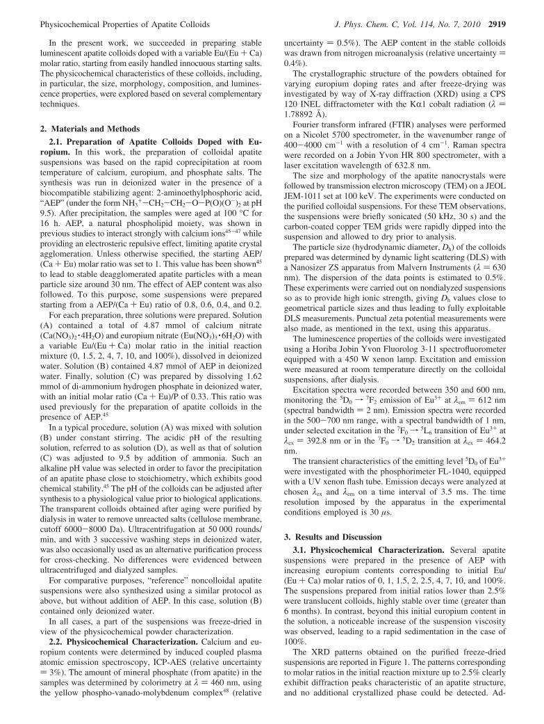
The possibility to incorporate europium in the apatite structurehas already been mentioned by several authors,42,44,45 althoughgenerally for apatite compounds prepared at higher temperature.Several substitution mechanisms can be considered43 for ex-plaining the incorporation of Eu3+ ions in Ca2+ sites. In theabsence of monovalent or tetravalent ions in the medium, fourdistinct mechanisms can be distinguished: the replacement ofthree Ca2+ by two Eu3+ (mechanism no.1), the deprotonationof some HPO4
2- ions into PO43- (mechanism no. 2), and the
additional incorporation of OH- (mechanism no. 3) or of oxygenO2- ions (mechanism no. 4). The existence of oxygen ions O2-
in Eu-doped apatites, as involved in mechanism no. 4, has indeedbeen reported and should thus not be a priori discarded.42-44 Itshould be noted that a combination of two or more of thesemechanisms may also be involved. To investigate some morethe Eu incorporation mechanism, a series of noncolloidal apatitesuspensions were prepared in similar conditions, but in the
Figure 1. XRD patterns for apatite colloids prepared with increasingEu/(Ca + Eu) initial molar ratios and for a noncolloidal reference (AEP-free).
Figure 2. FTIR spectra for apatite colloids prepared with increasing Eu/(Ca + Eu) initial molar ratios and for a noncolloidal reference (AEP-free).
2920 J. Phys. Chem. C, Vol. 114, No. 7, 2010 Al-Kattan et al.
absence of AEP in the medium. Again, increasing Eu contentsin the range of 0-2% were considered. After chemical analyseswere performed on several specimens of each sample, the(Ca + Eu)/P molar ratio of the apatite phase was evaluated forthese noncolloidal samples. The results obtained pointed out aslight decrease of this ratio upon Eu incorporation: typicallyfrom 1.66 down to 1.57 for Eu starting doping rates increasingfrom 0 to 2%. Based on the knowledge of Eu/(Ca + Eu) and(Ca + Eu)/P ratios for the 0, 1, 1.5, and 2% Eu samples, onecan thus estimate their chemical composition by consideringconsecutively each of the four possible incorporation mecha-nisms. Interestingly, these determinations indicated that all fourmechanisms remained possible for explaining the incorporationof Eu3+ ions in Ca2+ sites. This is, for instance, exemplified inFigure 3 in the case of the sample corresponding to a startingdoping rate of 1.5% Eu. A preferential mechanistic scheme thuscannot be derived at this point. It should also be noted that thepreferential mechanistic scheme for Eu incorporation maydepend, to some extent, on the doping rate. The observation ofa maximum limit of Eu incorporation does not seem to bedirectly explainable based only on mechanistic considerations:this limit probably originates from other factors, such asthermodynamically driven destabilization of the system, possiblylinked to differences between Eu-O and Ca-O bond lengths.Additional work specifically dedicated to examine these aspectswill be needed to investigate this point, which lies beyond thescope of the present paper. Although the presence of AEP inthe colloidal synthesis route leads to less-crystallized apatitephases, linked to some inhibitory effect of AEP pointed outfrom XRD analyses, it seems reasonable to expect a similarsituation in terms of Ca/Eu substitution.
The amount of AEP contained in the colloids was determinedfrom the amount of nitrogen titrated by way of elementalmicroanalyses. The AEP content was found to represent 8-11wt % of the colloidal particles, leading to an AEP/apatite molarratio in the range of 0.7-0.8. It is interesting to note that thisratio is lower than the one observed by Bouladjine et al.45 foreuropium-free apatite colloids prepared at 80 °C. These findingscan probably be explained by the differences existing in thesynthesis protocols used in the two studies, especially in termsof starting ionic concentrations and aging temperatures. Thelocation of the AEP molecules on the surface of the apatitecrystals was evidenced by Bouladjine et al.45 who showed thepositive surface charge of the colloids obtained in the presenceof AEP and the decrease in size upon increasing the AEPcontent. Interestingly, the zeta potential of the colloids preparedin the present work was also found to be clearly positive (e.g.,+12.4 mV for the sample corresponding to the initial dopingof 1.5% Eu), thus showing again the exposure of the ammoniumgroups -NH3
+ from AEP molecules toward the solution. Also,a decrease of the initial AEP concentration provoked a
sedimentation phenomenon for AEP/(Ca + Eu) ratios lower than0.2, whereas stable colloidal suspensions were obtained beyond0.4. This sedimentation can then most probably be explainedby an insufficient electrosteric repulsion between adjacentparticles when the surface content in AEP molecules decreasesbeyond a limit value.
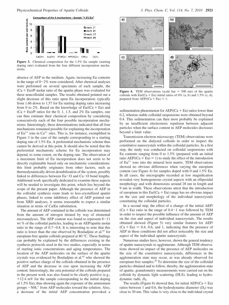
Transmission electron microscopy (TEM) observations wereperformed on the dialyzed colloids in order to inspect theconstitutive nanocrystals within the colloidal particles. In a firststep, the study was conducted on colloidal suspensions withEu contents ranging from 0 to 1.5% (prepared with an initialratio AEP/(Ca + Eu) ) 1) to study the effect of the introductionof Eu3+ ions into the mineral host matrix. TEM observationsshowed no obvious differences when varying the europiumcontent (see Figure 4) for samples doped with 0 and 1.5% Eu.In all cases, the micrographs recorded at low magnificationrevealed very homogeneous crystal aspects with an ellipsoidalmorphology and with dimensions around 28 nm in length and9 nm in width. These observations attest that the introductionof europium in this Eu/(Ca + Eu) range has no obvious impacton the size and morphology of the individual nanocrystalsconstituting the colloidal particles.
In a second step, the effect of a change of the initial AEP/(Ca + Eu) ratio in the range of 0.4-1 was followed by TEMin order to inspect the possible influence of the amount of AEPon the size and aspect of individual nanocrystals. The resultsobtained showed (Figure 5) very similar features for AEP/(Ca + Eu) ) 0.4, 0.6, and 1, indicating that the presence ofAEP in these conditions did not affect noticeably the size andaspect of the individual apatite nanocrystals.
Numerous studies have, however, shown the general tendencyof apatite nanocrystals to agglomerate. Although TEM observa-tions showed no impact of the presence of AEP molecules onthe size of the constitutive nanocrystals, differences in theagglomeration state may occur, as was already observed foreuropium-free samples.45 To determine the size of the colloidalparticles obtained and to follow, thereby, the agglomeration stateof apatite, granulometry measurements were carried out on thecolloids by dynamic light scattering (DLS), leading to hydro-dynamic radii, Dh.
The results (Figure 6) showed that, for initial AEP/(Ca + Eu)ratios between 1 and 0.6, the hydrodynamic diameter (Dh) wasclose to 30 nm. This value is very close to the individual crystal
Figure 3. Chemical composition for the 1.5% Eu sample (startingdoping rate) evaluated from the four different incorporation mecha-nisms.
Figure 4. TEM observations (scale bar ) 500 nm) of the apatitecolloids with Eu/(Ca + Eu) initial ratios of 0% (a, b) and 1.5% (c, d),prepared from AEP/(Ca + Eu) ) 1.
Physicochemical Properties of Apatite Colloids J. Phys. Chem. C, Vol. 114, No. 7, 2010 2921
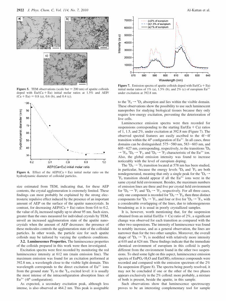
size estimated from TEM, indicating that, for these AEPcontents, the crystal agglomeration is extremely limited. Thesefindings can most probably be explained by the strong elec-trosteric repulsive effect induced by the presence of an importantamount of AEP on the surface of the apatite nanocrystals. Incontrast, for decreasing AEP/(Ca + Eu) ratios from 0.6 to 0.2,the value of Dh increased rapidly up to about 95 nm. Such sizes,greater than the ones measured for individual crystals by TEM,unveil an increased agglomeration state of the apatite nano-crystals when the amount of AEP decreases: the presence ofthese molecules controls the agglomeration state of the colloidalparticles. In other words, the particle size for such apatitecolloids may be tailored by varying the synthesis conditions.
3.2. Luminescence Properties. The luminescence propertiesof the colloids prepared in this work were then investigated.
Excitation spectra were first recorded by monitoring the redluminescence intensity at 612 nm (main emission line). Themaximum emission was found for an excitation performed at392.8 nm, a wavelength close to the visible light domain. Thiswavelength corresponds to the direct excitation of Eu3+ ionsfrom the ground state 7F0 to the 5L6 excited level: it is usuallythe most intense of the intraconfiguration absorption lines ofEu3+ (4f6 configuration).
As expected, a secondary excitation peak, although lessintense, is also observed at 464.2 nm. This peak is assignable
to the 7F0 f5D2 absorption and lies within the visible domain.
These observations show the possibility to use such luminescentnanoprobes for studying biological tissues because they onlyrequire low-energy excitation, preventing the deterioration oflive cells.
Luminescence emission spectra were then recorded forsuspensions corresponding to the starting Eu/(Eu + Ca) ratiosof 1, 1.5, and 2%, under excitation at 392.8 nm (Figure 7). Theobserved spectral features are easily ascribed to the 4f-4ftransition within the 4f6 configuration of Eu3+. In all cases, threedomains can be distinguished: 575-580 nm, 583-603 nm, and605-627 nm, corresponding, respectively, to the transitions 5D0
f 7F0, 5D0f7F1, and 5D0f
7F2 characteristic of the Eu3+ ion.Also, the global emission intensity was found to increasenoticeably with the level of europium doping.
The 5D0f7F0 transition located at 578 nm has been studied,
in particular, because the energy levels 5D0 and 7F0 are bothnondegenerated, meaning that only a single peak for the 5D0f7F0 transition should appear if all the Eu3+ ions were in thesame crystal field environment. Besides, the maximum numbersof emission lines are three and five per crystal field environmentfor 5D0 f
7F1 and 5D0 f7F2, respectively. For all three cases,
only one component is recorded for 5D0f7F0, then three distinct
components for 5D0f7F1, and four or five for 5D0f
7F2, witha considerable overlapping of the lines, due to inhomogeneousbroadening as it is usual in poorly crystallized samples.
It is, however, worth mentioning that, for the suspensionobtained from an initial Eu/(Eu + Ca) ratio of 2%, a significantchange was observed for each transition as compared with theother two suspensions. The intensity of luminescence was foundto notably increase, and as a general observation, the lines arenarrower than for the two other samples. Moreover, the overallshape of 5D0 f
7F2 is modified with relatively more intensityat 610 and at 624 nm. These findings indicate that the immediatechemical environment of europium in this colloid is partlydifferent from the environment found in the other two suspen-sions. To shed some light on this aspect, luminescence emissionspectra of EuPO4 ·H2O and Eu(OH)3 reference compounds wererecorded and compared with the emission spectrum of the 2%Eu suspension (Figure 8). The spectra being poorly resolved, itmay not be concluded if one or the other of the two phasesappears exclusively in the 2% colloid: more probably, a mixtureof both is present, beside the apatite, in this sample.
Such observations show that luminescence spectroscopyproves to be an interesting complementary tool for sample
Figure 5. TEM observations (scale bar ) 200 nm) of apatite colloidsdoped with Eu/(Ca + Eu) initial molar ratios at 1.5% and AEP/(Ca + Eu) ) 0.8 (a), 0.6 (b), and 0.4 (c).
Figure 6. Effect of the AEP/(Ca + Eu) initial molar ratio on thehydrodynamic diameter of colloidal particles.
Figure 7. Emission spectra of apatite colloids doped with Eu/(Ca + Eu)initial molar ratios of 1% (a), 1.5% (b), and 2% (c) of europium Eu3+
under excitation at 392.8 nm.
2922 J. Phys. Chem. C, Vol. 114, No. 7, 2010 Al-Kattan et al.

characterization, exploiting europium as an internal probe.Indeed, the presence of traces of such secondary phases hadonly been suggested from FTIR analyses beyond a starting Eu/(Ca + Eu) ratio of 2.5%.
The luminescence lifetime of the europium 5D0 level mea-sured on the same set of samples was also studied, and Figure9 displays the decays observed for suspensions correspondingto an initial Eu/(Eu + Ca) ratio of 1, 1.5, and 2%. As observedon the spectra, it may be noticed that the decays for 1% and1.5% Eu are essentially the same, whereas for 2% Eu, theemission decays more slowly. In the figure (Figure 9), the I(t)curves have been displayed on a logarithmic scale, showing thatthey are not purely monoexponential. We have, nevertheless,measured the average decay time τ as the time for which I(τ)) I0/exp(1), leading to 0.73 ( 0.03 ms for 1% or 1.5% Eu and1.00 ( 0.05 ms for 2% Eu. This observation may be linked tothe above results, suggesting the presence of secondary phasesbeside apatite for this sample, leading to modified luminescenceproperties.
It is interesting to note that, in all cases, the luminescenceexponential decay was found to be of the order to themillisecond. This order of magnitude is customary, in the caseof lanthanide-doped systems, due to the intrinsic properties ofLn3+ luminescent centers, and it enables one to envisionapplications as luminescent nanoprobes for biological applica-
tions because the autofluorescence of the biological mediumexhibits a luminescence decay time of the order of nanoseconds.In a second step, the luminescence properties, both in terms ofexcitation/emission and lifetime, of colloidal suspensions ob-tained at constant europium content (initial doping ) 1.5% Eu),but from varying initial amounts of AEP in the medium, werefollowed. AEP/(Ca + Eu) molar ratios of 0.4, 0.6, 0.8, and 1were tested. The results obtained indicated that no obviousdifferences could be noticed between the three suspensions,suggesting that the chemical environment of europium wasessentially unchanged.
4. Conclusions
Europium-doped calcium phosphate apatite-based colloidswere produced in the presence of a phospholipid moiety,2-aminoethylphosphoric acid (AEP), with varying europiumdoping rates, and their physicochemical characteristics wereinvestigated in detail. The experimental data obtained forsuspensions prepared from an initial Eu/(Ca + Eu) molar ratioup to 2% converge to indicate that these colloids consist ofbiomimetic apatite nanocrystals covered with AEP molecules.Moreover, the agglomeration state of apatite crystals was foundto be closely dependent on the amount of AEP in the system,and the mean particle size can be tailored between 30 and 100nm. The colloidal suspensions as prepared were found to bestable over time beyond at least 6 months.
The luminescence properties of these nanoprobes wereanalyzed in terms of excitation, emission, and lifetime. Thetypical luminescence features of europium-doped systems werewitnessed, with narrow emission peaks and long luminescencelifetimes. Our observations support the location of europium ina common crystallographic environment for all the colloidsprepared.
The potential of such systems in the field of optical imagingrelated to biology will then have to be investigated.
Supporting Information Available: Emission spectra ofapatite colloids doped with Eu/(Ca + Eu) initial molar ratiosof 1, 1.5, and 2% Eu under excitation at 392.8 nm; luminescencedecay profiles of the 5D0 (Eu3+) level for three apatite col-loidss1, 1.5, and 2% Eusunder excitation at 392.8 nm and atambient temperature; and TEM micrograph for apatite colloidsprepared from Eu/(Ca + Eu) ) 1.5%. This material is availablefree of charge via the Internet at http://pubs.acs.org.
References and Notes
(1) Gorller-Warlrand, C.; Binnemans, K. In Handbook on the Physicsand Chemistry of Rare Earths; Gschneidner, K. A., Eyring, L., Eds.; North-Holland: Amsterdam, 1998; Chapter 167.
(2) Franz, K. A.; Kethr, W. G.; Siggle, A.; Wiieczoreck, J. In Ullmann’sEncyclopedia of Industrial Chemistry; Elvers, B., Hawkins, S., Schulz, G.,Eds.; VCH: Weinheim, Germany, 1985; Chapter A15.
(3) Heyes, A. L.; Seefeldt, S.; Feist, J. P. Opt. Laser Technol. 2006,38, 257–265.
(4) Diamente, P. R.; Burk, R. D.; Van Veggel, F. C. Langmuir 2006,22, 1782–1788.
(5) Selvin, P. R. Annu. ReV. Biophys. Biomol. Struct. 2002, 31, 275–302.
(6) Huhtinen, P.; Vaarno, J.; Soukka, T.; Lovgren, T.; Haırmaı, H.Nanotechnology 2004, 15, 1708–1715.
(7) Gao, X.; Cui, Y.; Levenson, R. M.; Chung, L. W. K.; Nie, S. Nat.Biotechnol. 2004, 22, 969–976.
(8) Wang, C. W.; Moffitt, M. G. Langmuir 2005, 21, 2465–2473.(9) Stella, B.; Arpicco, S.; Peracchia, M. T.; Desmaele, D.; Hoebeke,
J.; Renoir, M.; D’Angelo, J.; Cattel, L.; Couvreur, P. J. Pharm. Sci. 2000,89, 1452–1464.
(10) Mitra, S.; Gaur, U.; Ghosh, P. C.; Maitra, A. N. J. ControlledRelease 2001, 74, 317–323.
Figure 8. Emission spectra of a Eu3+-doped apatite colloid with aEu/(Ca + Eu) initial molar ratio of 2% (a) and for reference compoundsEuPO4 ·H2O (b) and Eu(OH)3 (c), under excitation at 392.8 nm.
Figure 9. Luminescence decay profiles of the 5D0 (Eu3+) level forthree apatite colloids: 1% (a), 1.5% (b), and 2% (c), under excitationat 392.8 nm and at ambient temperature.
Physicochemical Properties of Apatite Colloids J. Phys. Chem. C, Vol. 114, No. 7, 2010 2923

(11) Allemann, E.; Gurny, R.; Doelker, E. Eur. J. Pharm. Biopharm.1993, 39, 91–173.
(12) Kohler, N.; Fryxell, G. E.; Zhang, M. J. Am. Chem. Soc. 2004,126, 7206–7211.
(13) Zuber, G.; Zammut-Italiano, L.; Dauty, E.; Beher, J. P. Angew.Chem. 2003, 42, 2666–2669.
(14) Drumond, D.; Meyer, C.; Hong, O.; Kirpotin, K.; Papahadjopoulos,D. Pharmacol. ReV. 1999, 51, 691–743.
(15) Dube, D.; Francis, M.; Leroux, J. C.; Winnik, F. M. BioconjugateChem. 2002, 13, 685–692.
(16) Mohapatra, S.; Mallick, S. K.; Maiti, T. K.; Ghosh, S. K.;Paramanik, P. Nanotechnology 2007, 18, 1–9.
(17) Battacharya, R.; Ranjan Patra, C.; Earl, A.; Wang, S.; Katraya, A.;Lu, L.; Kizhakketdarthu, J. N.; Yaszemski, M. J.; Greipp, P. R.; Mukhho-padhyay, D.; Mukherjee, P. Nanomedicine 2007, 3, 224–238.
(18) Meiser, F.; Cortez, C.; Caruso, F. Angew. Chem. 2004, 43, 5954–5957.
(19) Ow, H.; Larson, D. R.; Srivastava, M.; Baird, B. A.; Webb, W. W.;Weisner, U. Nano Lett. 2005, 5, 113–117.
(20) Parak, W. J.; Gerion, D.; Pellegrino, T.; Zanchet, D.; Micheel, C.;Williams, S. C.; Boudreau, R.; Le Gros, M. A.; Larabel, C. A.; Alivisatos,A. P. Nanotechnology 2003, 14, R15–R17.
(21) Bruchez, M.; Moronne, M.; Gin, P.; Weiss, S.; Alivisatos, A. P.Science 1998, 281, 2013–2016.
(22) Shimizu, K. T.; Neuhauser, R. G.; Leatherdale, C. A.; Empedocles,S. A.; Woo, W. K.; Bawendi, M. G. Phys. ReV. B 2001, 63, 205–316.
(23) Brokmann, X.; Hermier, J. P.; Messin, G.; Desbiolles, P.; Bouchaud,J. P.; Dahan, M. Phys. ReV. Lett. 2003, 90, 120601.
(24) Ballou, B.; Lagerholm, B. C.; Ernst, L. A.; Bruchez, M. P.;Waggoner, A. S. Bioconjugate Chem. 2004, 15, 79–86.
(25) Derfus, A. M.; Chan, W. C. W.; Bhatia, S. N. Nano Lett. 2004, 4,11–18.
(26) Wang, F.; Zhang, Y.; Fan, X.; Wang, M. J. Mater. Chem. 2006,16, 1031–1034.
(27) Meyssamy, H.; Riwotzki, K.; Kornowski, A.; Naused, S.; Haase,M. AdV. Mater. 1999, 11, 840–844.
(28) Riwotzki, K.; Meyssamy, H.; Kornowski, A.; Haase, M. J. Phys.Chem. B 2000, 104, 2824–2828.
(29) Riwotzki, K.; Meyssamy, H.; Schnablegger, H.; Kornowski, A.;Haase, M. Angew. Chem. 2001, 40, 573–576.
(30) Schuetz, P.; Caruso, F. Chem. Mater. 2002, 14, 4509–4516.(31) Huignard, A.; Buissette, V.; Franville, A. C.; Gacoin, T.; Boilot,
J. P. J. Phys. Chem. B 2003, 107, 6754–6759.(32) Huignard, A.; Gacoin, T.; Boilot, J. P. Chem. Mater. 2000, 12,
1090–1094.(33) Padilla Mondejar, S.; Kovtun, A.; Epple, M. J. Mater. Chem. 2007,
17, 4153–4159.(34) Doat, A.; Pelle, F.; Gardant, N.; Lebugle, A. J. Solid State Chem.
2004, 117, 1179–1187.(35) Chane-Ching, J. Y.; Lebugle, A.; Rousselot, I.; Pourpoint, A.; Pelle,
F. J. Mater. Chem. 2007, 17, 2904–2913.(36) Fanjul, M.; Doat, A.; Pelle, F.; Hollande, E.; Lebugle, A. In Vitro
Cell. DeV. Biol. 2004, 40, 81A.(37) Legeros, R. Z. Prog. Cryst. Growth Charact. 1981, 4, 1–45.(38) Cazalbou, S.; Eichert, D.; Ranz, X.; Drouet, C.; Combes, C.;
Harmand, M. F.; Rey, C. J. Mater. Sci.: Mater. Med. 2005, 16, 405–409.(39) Rey, C.; Combes, C.; Drouet, C.; Sfihi, H.; Barroug, A. Mater.
Sci. Eng., C 2007, 27, 198–205.(40) Doat, A.; Fanjul, M.; Pelle, F.; Holland, E.; Lebugle, A. Bioma-
terials 2003, 24, 3365–3371.(41) El Ouenzerfi, R.; Kbir-Ariguib, N.; Trabelsi-Ayedi, M.; Piriou, B.
J. Lumin. 1999, 85, 71–77.(42) Ternane, R.; Trabelsi-Ayedi, M.; Kbir-Ariguib, N.; Piriou, B. J.
Lumin. 1999, 81, 165–170.(43) Martin, P.; Carlot, G.; Chevarier, A.; Den-Auwer, C.; Panczer, G.
J. Nucl. Mater. 1999, 275, 268–276.(44) Eun Jin, K.; Sung-Woo, C.; Seong-Hyeon, H. J. Am. Chem. Soc.
2007, 90, 2795–2798.(45) Bouladjine, A.; Al-kattan, A.; Dufour, P.; Drouet, C. Langmuir
2009, 25, 12256–12265.(46) Rothfield, L.; Finkelstein, A. Annu. ReV. Biochem. 1968, 37, 463–
495.(47) Bissinger, P.; Kumberger, O.; Schier, A. Chem. Ber. 1991, 124,
509–513.(48) Gee, A.; Dietz, V. R. Anal. Chem. 1953, 25, 1320–1324.(49) Blumenthal, N. C. Clin. Orthop. Relat. Res. 1989, 247, 279–289.
JP910923G
2924 J. Phys. Chem. C, Vol. 114, No. 7, 2010 Al-Kattan et al.