proteomic characterization of extracellular vesicles produced
TRANSCRIPT
Proteomic characterization of extracellular vesiclesproduced by several wine yeast species
Ana Mencher,1 Pilar Morales,1 EvaValero,2 Jordi Tronchoni,1,† Kiran RaosahebPatil3,4 and Ramon Gonzalez1*1Instituto de Ciencias de la Vid y del Vino (CSIC,Gobierno de la Rioja, Universidad de La Rioja), Finca LaGrajera, Carretera de Burgos, km 6, Logro~no, La Rioja26071, Spain.2Universidad Pablo de Olavide, Sevilla, Spain.3European Molecular Biology Laboratory, Heidelberg,Germany.4The Medical Research Council Toxicology Unit,University of Cambridge, Cambridge, UK.
Summary
In winemaking, the use of alternative yeast startersis becoming increasingly popular. They contribute tothe diversity and complexity of wine sensory fea-tures and are typically used in combination with Sac-charomyces cerevisiae, to ensure completefermentation. This practice has drawn the interest oninteractions between different oenological yeasts,which are also relevant in spontaneous and conven-tional fermentations, or in the vineyard. Althoughseveral interactions have been described and somemechanisms have been suggested, the possibleinvolvement of extracellular vesicles (EVs) has notyet been considered. This work describes the pro-duction of EVs by six wine yeast species (S. cere-visiae, Torulaspora delbrueckii, Lachanceathermotolerans, Hanseniaspora uvarum, Candidasake and Metschnikowia pulcherrima) in syntheticgrape must. Proteomic analysis of EV-enriched frac-tions from S. cerevisiae and T. delbrueckii showedenrichment in glycolytic enzymes and cell-wall-
related proteins. The most abundant protein found inS. cerevisiae, T. delbrueckii and L. thermotoleransEV-enriched fractions was the enzyme exo-1,3-b-glu-canase. However, this protein was not involved inthe here-observed negative impact of T. delbrueckiiextracellular fractions on the growth of other yeastspecies. These findings suggest that EVs may play arole in fungal interactions during wine fermentationand other aspects of wine yeast biology.
Introduction
Extracellular vesicles (EVs) are particles naturallyreleased from living cells and delimited by a lipid bilayerthat cannot self-replicate (Th�ery et al., 2018). They areproduced by organisms belonging to all three domains ofthe tree of life and show a broad range of sizes, from 20to 500 nm, depending on biological species, cell typesand environmental conditions. They can be produced byvarious mechanisms and have historically received dif-ferent names, depending on the organism of origin orthe biogenesis pathway. However, in the absence ofclear evidence of the mechanism of release, the genericterm extracellular vesicle is recommended (Th�ery et al.,2018).Mammalian cells produce different types of EVs,
including secretory lysosomes, multi-vesicular body-derived exosomes or microvesicles (Nickel andRabouille, 2009; Rabouille et al., 2012). Mammalian EVshave been widely studied because they are involved inmultiple biological events such as antigen presentation,neuronal communication, viral transmission, immunemodulation, tumour angiogenesis or metastasis (Raposoet al., 1996; Marzesco et al., 2005; Bhatnagar et al.,2007; Park et al., 2010). EV research on other biologicalsystems (bacteria, plants, fungi) is a younger but grow-ing field (Regente et al., 2009; Brown et al., 2015). EVmorphology and content (including peptides, proteins,miRNAs or mRNAs) have been analysed for severalyeast species, mostly pathogenic (Rodrigues et al.,2007; Rodrigues et al., 2014; Gil-Bona et al., 2017;Rodrigues and Casadevall, 2018). Both non-conventionaland conventional secretory proteins have been identifiedon yeast EVs (Gil-Bona et al., 2015). Fungal EVs havebeen involved in cell-to-cell communication, response tonutrient availability, RNA export, morphological transition
Received 31 March, 2020; accepted 31 May, 2020.*For correspondence. E-mail [email protected]; Tel.+34 941 89 49 80; Fax +941 89 97 28.†Present address: Universidad Internacional de Valencia, Valencia,Spain.Microbial Biotechnology (2020) 0(0), 1–16doi:10.1111/1751-7915.13614Funding informationThis work was funded by the Spanish Government through grantsAGL2015-63629-R (co-financed by FEDER funds), PCI2018-092949(co-funded by ERA-CoBioTech) and BES-2016-077557 (trainingcontract for AM). JT is funded by FGCSIC by the COMFUTUROprogram.
ª 2020 The Authors. Microbial Biotechnology published by Society for Applied Microbiology and John Wiley & Sons LtdThis is an open access article under the terms of the Creative Commons Attribution-NonCommercial-NoDerivs License, which permitsuse and distribution in any medium, provided the original work is properly cited, the use is non-commercial and no modifications oradaptations are made.
bs_bs_banner
(e.g. biofilm formation), prion transmission, modulation ofhost immunity, cell wall remodelling, or survival to stressconditions (Oliveira et al., 2013; Peres da Silva et al.,2015; Rizzo et al., 2017; Zhao et al., 2019). However, inmost cases, the attribution of functions or biological rolesto fungal EVs is supported by few or single examples,and the mechanisms or cause–effect relationships arenot well established.The relevance of EVs for the biotechnological applica-
tion of yeasts remains unexplored. Food fermentationoften develops as mixed culture, including wine, kefir,sourdough and other foods (Farnworth, 2005; Man-zanares et al., 2011; Furukawa et al., 2013; De Vuystet al., 2014; Jouhten et al., 2016). In addition, there is acurrent trend in winemaking for the use of multiple (usu-ally two or three) yeast starter cultures, from differentyeast species in order to reach specific quality and com-positional outcomes (Gonzalez et al., 2013; Jolly et al.,2014).Many of the interactions observed between wine
microorganisms can be considered as indirect. Thiswould be the case for competition for the absorption ofnutrients (Fleet, 2003) or to the toxic effect of majormetabolites, such as ethanol (Kunkee, 1984). But director targeted mechanisms of interaction might be just asimportant in many instances. For example, killer factorshave been described in both Saccharomyces (Van Vuu-ren and Jacobs, 1992; P�erez et al., 2001; Rodr�ıguez-Cousi~no et al., 2011) and non-Saccharomyces speciessuch as Torulaspora delbrueckii (Vel�azquez et al., 2015).Interestingly, peptide fragments from GAPDH, a glycolyticenzyme, secreted by S. cerevisiae, show antimicrobialactivity against several wine microorganisms (Brancoet al., 2014). In addition, Rossouw et al., (2018) showedthe impact of physical contact on population dynamics,and several studies suggest a role of cell-to-cell contacton wine yeast interspecific interactions (Taillandier et al.,2014; Wang et al., 2015; P�erez-Torrado et al., 2017;Englezos et al., 2019; Shekhawat et al., 2019). Finally,quorum sensing mechanisms have been described forsome yeast species (Chen and Fink, 2006).Some insight on the mechanisms involved in interac-
tions between wine yeasts was provided by transcrip-tomic analysis (Tronchoni et al., 2017; Curiel et al.,2017). These authors found transcriptional reprogram-ming, suggesting activation of nutrient consumption path-ways by S. cerevisiae in response to several wine yeastspecies. Considering the short response time, transcrip-tional reprogramming was likely mediated by specificrecognition mechanisms. Recently, similar results havebeen reported by other authors (Shekhawat et al., 2019;Alonso del Real et al., 2019).As a follow-up of previous studies on interspecific
yeast interactions in winemaking, and considering the
multiple roles already attributed to fungal EVs, includingbiological communication (Stahl and Raposo, 2018;Raposo and Stahl, 2019), we posed the hypothesis thatEVs would be involved in, at least, some of theresponses to co-cultivation observed among wineyeasts. In this work, we show that EVs are indeed pro-duced by several wine yeast species under cultivationconditions relevant for winemaking and perform a pro-teomic analysis of the extracellular fractions of the twomore relevant wine yeast species.
Results
Production of extracellular vesicles by wine yeastspecies
Fractions from cell-free supernatants, enriched in eitherextracellular vesicles or free extracellular proteins, wereisolated as described under Experimental procedures.They were labelled as EV-enriched fractions (for extracel-lular vesicles) or VF-enriched fractions (for vesicle-free),and were obtained from two S. cerevisiae strains (EC1118and FX10) and five non-Saccharomyces yeasts (T. del-brueckii, Hanseniaspora uvarum, Candida sake, Metsch-nikowia pulcherrima and Lachancea thermotolerans),growing in synthetic grape must. Cell viability was quanti-fied by flow cytometry in order to rule out contamination bycytoplasmic proteins released by dead or lysed cells.According to this criterion, the 24-hour sampling point wasselected for the analyses. At this sampling point, the num-ber of dead cells was below 2% for all yeast strains.The EV-enriched fractions were prepared and visual-
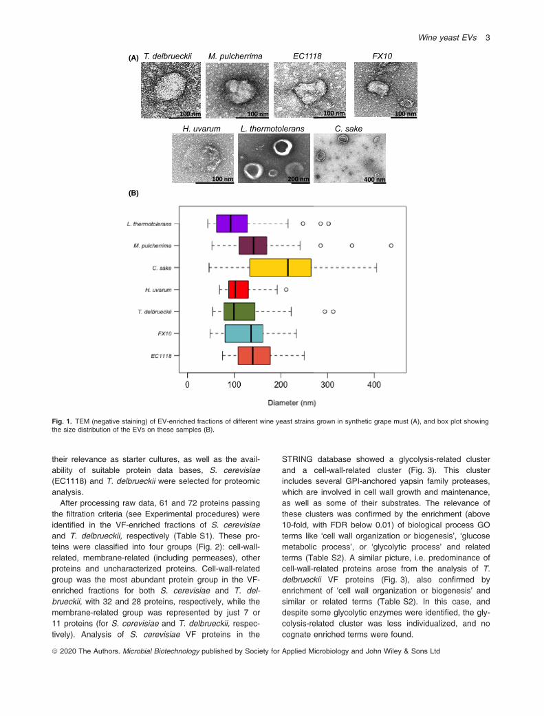
ized by TEM with negative staining (see Experimental pro-cedures). EVs were observed for all seven yeast strains.Intact EVs showed a white (stain free) perimeter, corre-sponding to the membrane of the vesicle, while the lumenwas dark due to stain absorption. Additionally, the foot-prints of other EVs were appreciated as clear haloes overthe darker background of the negative staining prepara-tions. They were spherical or ovoid in shape (Fig. 1) andshowed a size range around 100-200 nm in diameter.Size variability for each strain was roughly � 50%. Thesmallest average diameter was shown by L. thermotoler-ans (105 nm) and H. uvarum (111 nm), and the largerone by C. sake (204 nm). EVs from M. pulcherrimashowed the widest size distribution among all the speciestested. EVs produced by the two S. cerevisiae strainsshowed similar size distributions (Fig. 1).
Proteome composition of S. cerevisiae and T.delbrueckii EV- and VF-enriched fractions
To better understand the biological impact of EVs onwine yeast biology, the proteome composition of EV-and VF-enriched fractions was analysed. Considering
ª 2020 The Authors. Microbial Biotechnology published by Society for Applied Microbiology and John Wiley & Sons Ltd
2 A. Mencher et al.
their relevance as starter cultures, as well as the avail-ability of suitable protein data bases, S. cerevisiae(EC1118) and T. delbrueckii were selected for proteomicanalysis.After processing raw data, 61 and 72 proteins passing
the filtration criteria (see Experimental procedures) wereidentified in the VF-enriched fractions of S. cerevisiaeand T. delbrueckii, respectively (Table S1). These pro-teins were classified into four groups (Fig. 2): cell-wall-related, membrane-related (including permeases), otherproteins and uncharacterized proteins. Cell-wall-relatedgroup was the most abundant protein group in the VF-enriched fractions for both S. cerevisiae and T. del-brueckii, with 32 and 28 proteins, respectively, while themembrane-related group was represented by just 7 or11 proteins (for S. cerevisiae and T. delbrueckii, respec-tively). Analysis of S. cerevisiae VF proteins in the
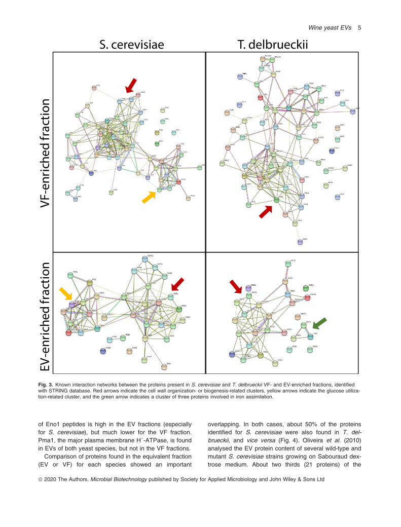
STRING database showed a glycolysis-related clusterand a cell-wall-related cluster (Fig. 3). This clusterincludes several GPI-anchored yapsin family proteases,which are involved in cell wall growth and maintenance,as well as some of their substrates. The relevance ofthese clusters was confirmed by the enrichment (above10-fold, with FDR below 0.01) of biological process GOterms like ‘cell wall organization or biogenesis’, ‘glucosemetabolic process’, or ‘glycolytic process’ and relatedterms (Table S2). A similar picture, i.e. predominance ofcell-wall-related proteins arose from the analysis of T.delbrueckii VF proteins (Fig. 3), also confirmed byenrichment of ‘cell wall organization or biogenesis’ andsimilar or related terms (Table S2). In this case, anddespite some glycolytic enzymes were identified, the gly-colysis-related cluster was less individualized, and nocognate enriched terms were found.
(A)
(B)
T. delbrueckii M. pulcherrima EC1118 FX10
C. sakeH. uvarum L. thermotolerans
100 nm 200 nm
100 nm100 nm 100 nm 100 nm
400 nm
Fig. 1. TEM (negative staining) of EV-enriched fractions of different wine yeast strains grown in synthetic grape must (A), and box plot showingthe size distribution of the EVs on these samples (B).
ª 2020 The Authors. Microbial Biotechnology published by Society for Applied Microbiology and John Wiley & Sons Ltd
Wine yeast EVs 3
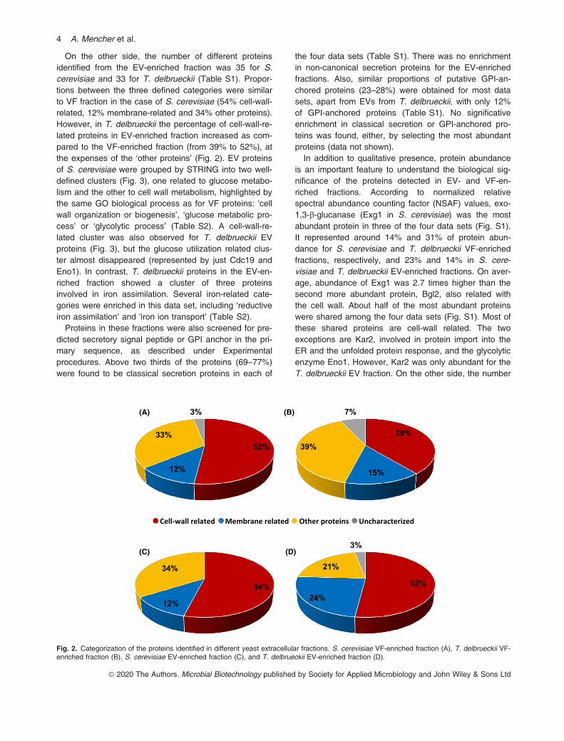
On the other side, the number of different proteinsidentified from the EV-enriched fraction was 35 for S.cerevisiae and 33 for T. delbrueckii (Table S1). Propor-tions between the three defined categories were similarto VF fraction in the case of S. cerevisiae (54% cell-wall-related, 12% membrane-related and 34% other proteins).However, in T. delbrueckii the percentage of cell-wall-re-lated proteins in EV-enriched fraction increased as com-pared to the VF-enriched fraction (from 39% to 52%), atthe expenses of the ‘other proteins’ (Fig. 2). EV proteinsof S. cerevisiae were grouped by STRING into two well-defined clusters (Fig. 3), one related to glucose metabo-lism and the other to cell wall metabolism, highlighted bythe same GO biological process as for VF proteins: ‘cellwall organization or biogenesis’, ‘glucose metabolic pro-cess’ or ‘glycolytic process’ (Table S2). A cell-wall-re-lated cluster was also observed for T. delbrueckii EVproteins (Fig. 3), but the glucose utilization related clus-ter almost disappeared (represented by just Cdc19 andEno1). In contrast, T. delbrueckii proteins in the EV-en-riched fraction showed a cluster of three proteinsinvolved in iron assimilation. Several iron-related cate-gories were enriched in this data set, including ‘reductiveiron assimilation’ and ‘iron ion transport’ (Table S2).Proteins in these fractions were also screened for pre-
dicted secretory signal peptide or GPI anchor in the pri-mary sequence, as described under Experimentalprocedures. Above two thirds of the proteins (69–77%)were found to be classical secretion proteins in each of
the four data sets (Table S1). There was no enrichmentin non-canonical secretion proteins for the EV-enrichedfractions. Also, similar proportions of putative GPI-an-chored proteins (23–28%) were obtained for most datasets, apart from EVs from T. delbrueckii, with only 12%of GPI-anchored proteins (Table S1). No significativeenrichment in classical secretion or GPI-anchored pro-teins was found, either, by selecting the most abundantproteins (data not shown).In addition to qualitative presence, protein abundance
is an important feature to understand the biological sig-nificance of the proteins detected in EV- and VF-en-riched fractions. According to normalized relativespectral abundance counting factor (NSAF) values, exo-1,3-b-glucanase (Exg1 in S. cerevisiae) was the mostabundant protein in three of the four data sets (Fig. S1).It represented around 14% and 31% of protein abun-dance for S. cerevisiae and T. delbrueckii VF-enrichedfractions, respectively, and 23% and 14% in S. cere-visiae and T. delbrueckii EV-enriched fractions. On aver-age, abundance of Exg1 was 2.7 times higher than thesecond more abundant protein, Bgl2, also related withthe cell wall. About half of the most abundant proteinswere shared among the four data sets (Fig. S1). Most ofthese shared proteins are cell-wall related. The twoexceptions are Kar2, involved in protein import into theER and the unfolded protein response, and the glycolyticenzyme Eno1. However, Kar2 was only abundant for theT. delbrueckii EV fraction. On the other side, the number
54%
12%
34%
52%
24%
21%
3%(D)
(A) (B)
(C)
39%
15%
39%
7%
52%
12%
33%
3%
Cell-wall related Membrane related Other proteins Uncharacterized
Fig. 2. Categorization of the proteins identified in different yeast extracellular fractions. S. cerevisiae VF-enriched fraction (A), T. delbrueckii VF-enriched fraction (B), S. cerevisiae EV-enriched fraction (C), and T. delbrueckii EV-enriched fraction (D).
ª 2020 The Authors. Microbial Biotechnology published by Society for Applied Microbiology and John Wiley & Sons Ltd
4 A. Mencher et al.
of Eno1 peptides is high in the EV fractions (especiallyfor S. cerevisiae), but much lower for the VF fraction.Pma1, the major plasma membrane H+-ATPase, is foundin EVs of both yeast species, but not in the VF fractions.Comparison of proteins found in the equivalent fraction
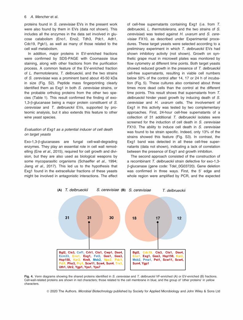
(EV or VF) for each species showed an important
overlapping. In both cases, about 50% of the proteinsidentified for S. cerevisiae were also found in T. del-brueckii, and vice versa (Fig. 4). Oliveira et al. (2010)analysed the EV protein content of several wild-type andmutant S. cerevisiae strains growing on Sabouraud dex-trose medium. About two thirds (21 proteins) of the
S. cerevisiae T. delbrueckiiVF
-noitcarf dehcirne
EV-
noitcarf dehcirne
Fig. 3. Known interaction networks between the proteins present in S. cerevisiae and T. delbrueckii VF- and EV-enriched fractions, identifiedwith STRING database. Red arrows indicate the cell wall organization- or biogenesis-related clusters, yellow arrows indicate the glucose utiliza-tion-related cluster, and the green arrow indicates a cluster of three proteins involved in iron assimilation.
ª 2020 The Authors. Microbial Biotechnology published by Society for Applied Microbiology and John Wiley & Sons Ltd
Wine yeast EVs 5
proteins found in S. cerevisiae EVs in the present workwere also found by them in EVs (data not shown). Thisincludes all the enzymes in the data set involved in glu-cose catabolism (Eno1, Eno2, Tdh3, Pdc1, Adh1,Cdc19, Pgk1), as well as many of those related to thecell wall metabolism.In addition, major proteins in EV-enriched fractions
were confirmed by SDS-PAGE with Coomassie bluestaining, along with other fractions from the purificationprocess. A common feature of the EV-enriched fractionof L. thermotolerans, T. delbrueckii, and the two strainsof S. cerevisiae was a prominent band about 45-50 kDain size (Fig. S2). Peptide mass fingerprinting clearlyidentified them as Exg1 in both S. cerevisiae strains, orthe probable ortholog proteins from the other two spe-cies (Table 1). This result confirmed the finding of exo-1,3-b-glucanase being a major protein constituent of S.cerevisiae and T. delbrueckii EVs, supported by pro-teomic analysis, but it also extends this feature to otherwine yeast species.
Evaluation of Exg1 as a potential inducer of cell deathon target yeasts
Exo-1,3-b-glucanases are fungal cell-wall-degradingenzymes. They play an essential role in cell wall remod-elling (Ene et al., 2015), required for cell growth and divi-sion, but they are also used as biological weapons bysome mycoparasitic organisms (Schaeffer et al., 1994;Jiang et al., 2017). This led us to the hypothesis thatExg1 found in the extracellular fractions of these yeastsmight be involved in antagonistic interactions. The effect
of cell-free supernatants containing Exg1 (i.e. from T.delbrueckii, L. thermotolerans, and the two strains of S.cerevisiae) was tested against H. uvarum and S. cere-visiae FX10, as described under Experimental proce-dures. These target yeasts were selected according to apreliminary experiment in which T. delbrueckii EVs hadshown inhibitory activity (not shown). Growth on syn-thetic grape must in microwell plates was monitored byflow cytometry at different time points. Both target yeastsshowed reduced growth in the presence of T. delbrueckiicell-free supernatants, resulting in viable cell numbersbelow 50% of the control after 14, 17 or 24 h of incuba-tion (Fig. 5). These cultures also contained about threetimes more dead cells than the control at the differenttime points. This result shows that supernatants from T.delbrueckii hinder yeast growth by inducing death of S.cerevisiae and H. uvarum cells. The involvement ofExg1 in this activity was tested by two complementaryapproaches. First, 24-hour cell-free supernatants of acollection of 31 additional T. delbrueckii isolates werescreened for the induction of cell death in S. cerevisiaeFX10. The ability to induce cell death in S. cerevisiaewas found to be strain specific. Indeed, only 13% of thestrains showed this feature (Fig. S3). In contrast, theExg1 band was detected in all these cell-free super-natants (data not shown), indicating a lack of correlationbetween the presence of Exg1 and growth inhibition.The second approach consisted of the construction of
a recombinant T. delbrueckii strain defective for exo-1,3-b-glucanase (gene code: Tdel_0G03720). Gene deletionwas confirmed in three ways. First, the 50 edge andwhole region were amplified by PCR, and the expected
Bgl2, Cis3, Cof1, Crh1, Cts1, Cwp1, Dse4,Ecm33, Eno1, Exg1, Fet3, Gas1, Gas3,Hsp150, Kar2, Kre9, Msb2, Npc2, Pdc1,Pdi1, Pho3, Pry1, Scw11, Scw4, Sun4, Trx1,Uth1, Utr2, Ygp1, Yps1, Yps7
Bgl2, Cdc19, Cis3, Cts1, Dse4,Eno1, Exg1, Gas3, Hsp150, Kar2,Msb2, Pma1, Pst1, Scw11, Scw4,Sun4, Ygp1
(A) (B)
Fig. 4. Venn diagrams showing the shared proteins identified in S. cerevisiae and T. delbrueckii VF-enriched (A) or EV-enriched (B) fractions.Cell-wall-related proteins are shown in red characters; those related to the cell membrane in blue; and the group of ‘other proteins’ in yellowcharacters.
ª 2020 The Authors. Microbial Biotechnology published by Society for Applied Microbiology and John Wiley & Sons Ltd
6 A. Mencher et al.
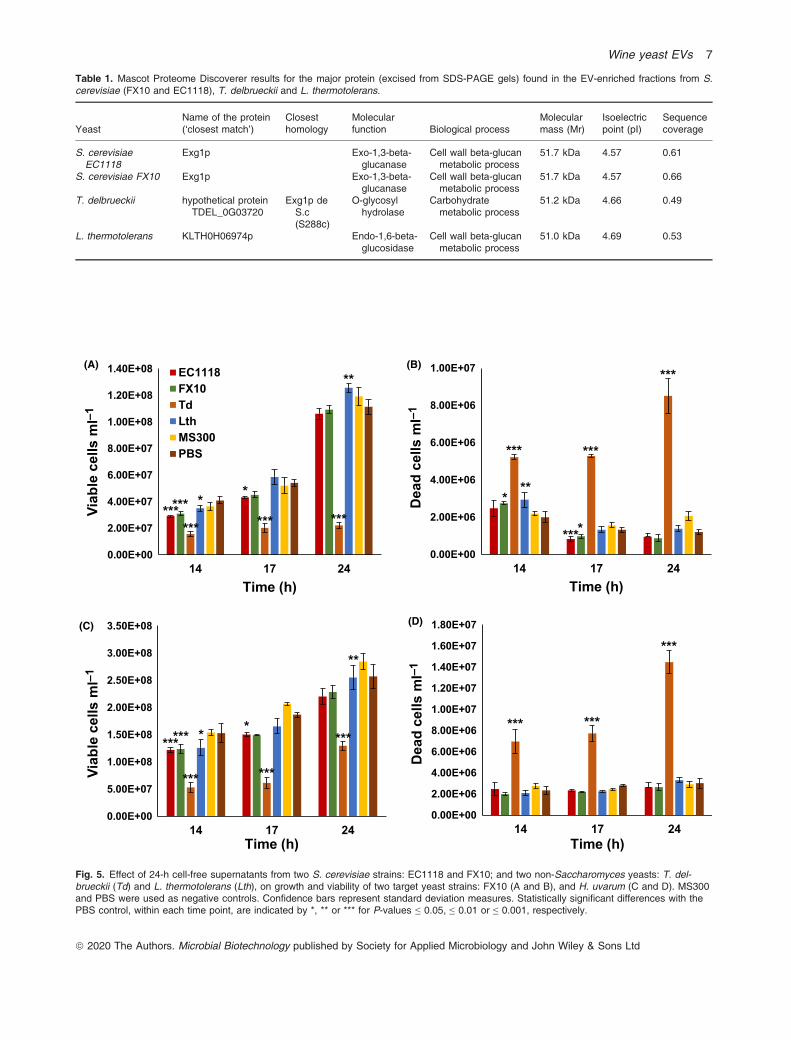
Table 1. Mascot Proteome Discoverer results for the major protein (excised from SDS-PAGE gels) found in the EV-enriched fractions from S.cerevisiae (FX10 and EC1118), T. delbrueckii and L. thermotolerans.
YeastName of the protein(‘closest match’)
Closesthomology
Molecularfunction Biological process
Molecularmass (Mr)
Isoelectricpoint (pI)
Sequencecoverage
S. cerevisiaeEC1118
Exg1p Exo-1,3-beta-glucanase
Cell wall beta-glucanmetabolic process
51.7 kDa 4.57 0.61
S. cerevisiae FX10 Exg1p Exo-1,3-beta-glucanase
Cell wall beta-glucanmetabolic process
51.7 kDa 4.57 0.66
T. delbrueckii hypothetical proteinTDEL_0G03720
Exg1p deS.c(S288c)
O-glycosylhydrolase
Carbohydratemetabolic process
51.2 kDa 4.66 0.49
L. thermotolerans KLTH0H06974p Endo-1,6-beta-glucosidase
Cell wall beta-glucanmetabolic process
51.0 kDa 4.69 0.53
0.00E+00
2.00E+06
4.00E+06
6.00E+06
8.00E+06
1.00E+07
14 17 24
Dea
dce
llsm
l–1
Time (h)
0.00E+00
2.00E+07
4.00E+07
6.00E+07
8.00E+07
1.00E+08
1.20E+08
1.40E+08
14 17 24
elbaiVce
llsm
l–1
Time (h)
EC1118FX10TdLthMS300PBS
0.00E+00
2.00E+06
4.00E+06
6.00E+06
8.00E+06
1.00E+07
1.20E+07
1.40E+07
1.60E+07
1.80E+07
14 17 24
Dea
dce
lls m
l–1
Time (h)
0.00E+00
5.00E+07
1.00E+08
1.50E+08
2.00E+08
2.50E+08
3.00E+08
3.50E+08
14 17 24
elbaiVce
lls m
l–1
Time (h)
(A)
(D)(C)
(B)
* ****
*** ***
**
***
***
*
***
***
****
***
*
**
******
******
***
***
***
***
***
Fig. 5. Effect of 24-h cell-free supernatants from two S. cerevisiae strains: EC1118 and FX10; and two non-Saccharomyces yeasts: T. del-brueckii (Td) and L. thermotolerans (Lth), on growth and viability of two target yeast strains: FX10 (A and B), and H. uvarum (C and D). MS300and PBS were used as negative controls. Confidence bars represent standard deviation measures. Statistically significant differences with thePBS control, within each time point, are indicated by *, ** or *** for P-values ≤ 0.05, ≤ 0.01 or ≤ 0.001, respectively.
ª 2020 The Authors. Microbial Biotechnology published by Society for Applied Microbiology and John Wiley & Sons Ltd
Wine yeast EVs 7
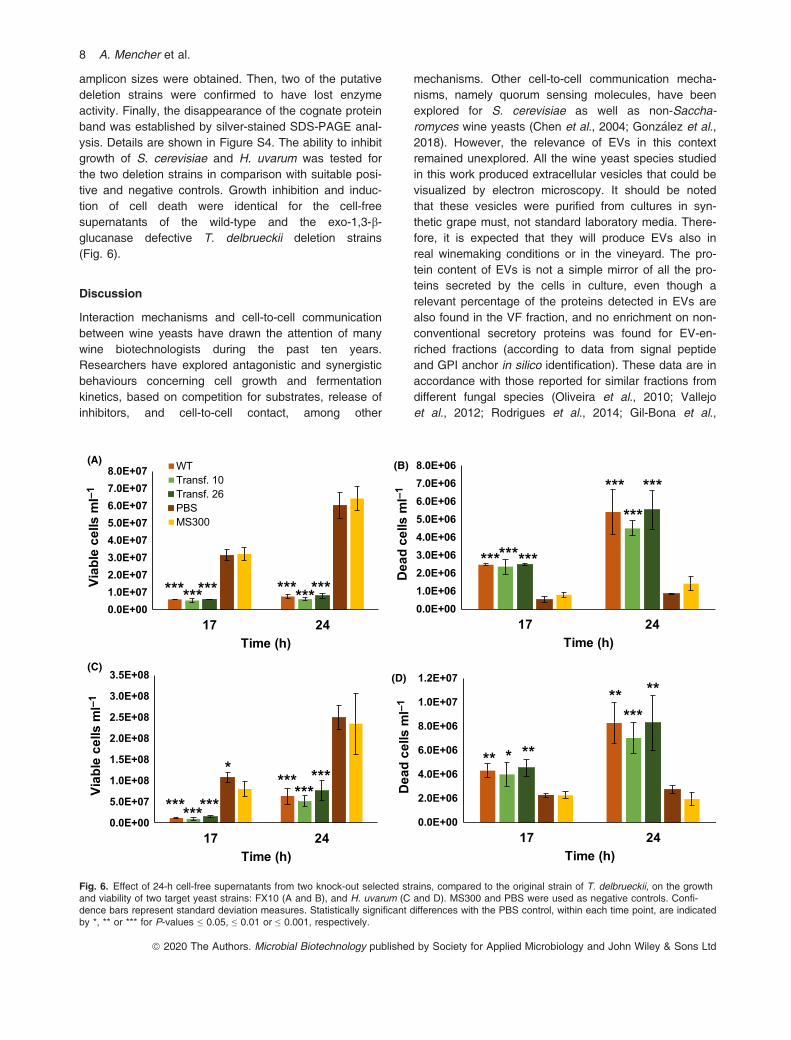
amplicon sizes were obtained. Then, two of the putativedeletion strains were confirmed to have lost enzymeactivity. Finally, the disappearance of the cognate proteinband was established by silver-stained SDS-PAGE anal-ysis. Details are shown in Figure S4. The ability to inhibitgrowth of S. cerevisiae and H. uvarum was tested forthe two deletion strains in comparison with suitable posi-tive and negative controls. Growth inhibition and induc-tion of cell death were identical for the cell-freesupernatants of the wild-type and the exo-1,3-b-glucanase defective T. delbrueckii deletion strains(Fig. 6).
Discussion
Interaction mechanisms and cell-to-cell communicationbetween wine yeasts have drawn the attention of manywine biotechnologists during the past ten years.Researchers have explored antagonistic and synergisticbehaviours concerning cell growth and fermentationkinetics, based on competition for substrates, release ofinhibitors, and cell-to-cell contact, among other
mechanisms. Other cell-to-cell communication mecha-nisms, namely quorum sensing molecules, have beenexplored for S. cerevisiae as well as non-Saccha-romyces wine yeasts (Chen et al., 2004; Gonz�alez et al.,2018). However, the relevance of EVs in this contextremained unexplored. All the wine yeast species studiedin this work produced extracellular vesicles that could bevisualized by electron microscopy. It should be notedthat these vesicles were purified from cultures in syn-thetic grape must, not standard laboratory media. There-fore, it is expected that they will produce EVs also inreal winemaking conditions or in the vineyard. The pro-tein content of EVs is not a simple mirror of all the pro-teins secreted by the cells in culture, even though arelevant percentage of the proteins detected in EVs arealso found in the VF fraction, and no enrichment on non-conventional secretory proteins was found for EV-en-riched fractions (according to data from signal peptideand GPI anchor in silico identification). These data are inaccordance with those reported for similar fractions fromdifferent fungal species (Oliveira et al., 2010; Vallejoet al., 2012; Rodrigues et al., 2014; Gil-Bona et al.,
(A) (B)
0.0E+001.0E+072.0E+073.0E+074.0E+075.0E+076.0E+077.0E+078.0E+07
17 24
elbaiVce
llsm
l–1
Time (h)
WTTransf. 10Transf. 26PBSMS300
0.0E+001.0E+062.0E+063.0E+064.0E+065.0E+066.0E+067.0E+068.0E+06
17 24
Dea
d ce
lls m
l–1
Time (h)
0.0E+00
5.0E+07
1.0E+08
1.5E+08
2.0E+08
2.5E+08
3.0E+08
3.5E+08
17 24
elbaiVce
llsm
l–1
Time (h)
0.0E+00
2.0E+06
4.0E+06
6.0E+06
8.0E+06
1.0E+07
1.2E+07
17 24
Dea
d ce
lls m
l–1
Time (h)
(C)(D)
*********
******
***
***
******
***
***
***
*********
****
******
** * **
** *****
Fig. 6. Effect of 24-h cell-free supernatants from two knock-out selected strains, compared to the original strain of T. delbrueckii, on the growthand viability of two target yeast strains: FX10 (A and B), and H. uvarum (C and D). MS300 and PBS were used as negative controls. Confi-dence bars represent standard deviation measures. Statistically significant differences with the PBS control, within each time point, are indicatedby *, ** or *** for P-values ≤ 0.05, ≤ 0.01 or ≤ 0.001, respectively.
ª 2020 The Authors. Microbial Biotechnology published by Society for Applied Microbiology and John Wiley & Sons Ltd
8 A. Mencher et al.
2015). It is currently assumed that many of these non-classical secretory proteins reach the extracellular spaceby several alternative pathways (Oliveira et al., 2010;Miura et al., 2012; Miura and Ueda, 2018; Winters et al.,2020). However, it cannot be excluded that some of theproteins found in the soluble fraction of the secretomereached the extracellular space as vesicle-associated,being released from those structures after crossing thecell wall, or during the purification process.Also noteworthy is the similarity in protein content
between different yeast species. Relevant similarities inEV protein content have been previously reportedamong pathogenic fungal species. Since S. cerevisiaeand T. delbrueckii, the two species analysed in moredepth in this work, are relatively close in the phyloge-netic tree, the extent of this similarities among wineyeast species cannot be predicted, but the fact that themajor protein found in L. thermotolerans was also anortholog of Exg1, the major protein found in the othertwo species, should be taken into account. Indeed, Exg1was also found by other authors in C. albicans and otherfungal EVs.The presence of glycolytic enzymes in the extracellular
fractions might be surprising at first sight. However, gly-colytic enzymes are frequently reported in EV prepara-tions from different yeast (Rodrigues et al., 2014; Gil-Bona et al., 2015) or bacterial (Hong et al., 2019) spe-cies, and some human exosomes have been shown tobe able to synthesize ATP by glycolysis, suggestingATP production might play a role in the uptake of extra-cellular vesicles by target cells (Fonseca et al., 2016).Indeed, enolase has been found in EVs from most yeastspecies analysed so far (Rodrigues et al., 2014), includ-ing S. cerevisiae and T. delbrueckii in the present work,which might be related to the moonlighting character ofthis protein (Decker and Wickner, 2006; Gancedo andFlores, 2008), involved for example in vacuolar mem-brane fusion. This might indicate a specific role of thisprotein in the biology of fungal EVs. The glycolyticenzyme GAPDH, identified as two different isoenzymes,Tdh2 and Tdh3, in the VF- and EV-enriched fractions(respectively) of S. cerevisiae has also a moonlightingcharacter; peptide fragments of this enzyme show inhibi-tory activity against other yeasts and bacteria (Brancoet al., 2014).Furthermore, enzymes related to cell wall architecture
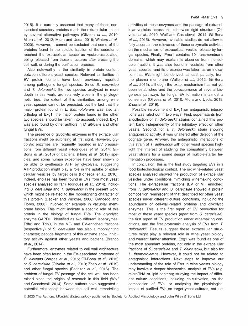
have been often found in the EV-associated proteome ofC. albicans (Vargas et al., 2015; Gil-Bona et al., 2015)or S. cerevisiae (Oliveira et al., 2010; Zhao et al., 2019)and other fungal species (Baltazar et al., 2016). Theproblem of fungal EV passage of the cell wall has beenraised since the origins of research in this field (Wolfand Casadevall, 2014). Some authors have suggested apotential relationship between the cell wall remodelling
activities of these enzymes and the passage of extracel-lular vesicles across this otherwise rigid structure (Oli-veira et al., 2010; Wolf and Casadevall, 2014; Gil-Bonaet al., 2015). However, available studies do not allow tofully ascertain the relevance of these enzymatic activitieson the mechanism of extracellular vesicle release by fun-gal species. Finally, Pma1 contains 10 transmembranedomains, which may explain its absence from the sol-uble fraction. It was also found in vesicles from otheryeast species, and its presence was taken as an indica-tion that EVs might be derived, at least partially, fromthe plasma membrane (Vallejo et al., 2012; Gil-Bonaet al., 2015), although the exact mechanism has not yetbeen established and the co-occurrence of several bio-genesis pathways for fungal EV formation is almost aconsensus (Oliveira et al., 2010; Miura and Ueda, 2018;Zhao et al., 2019).Possible involvement of Exg1 on antagonistic interac-
tions was ruled out in two ways. First, supernatants froma collection of T. delbrueckii strains contained this pro-tein band independently of the inhibitory effect on otheryeasts. Second, for a T. delbrueckii strain showingantagonistic activity, it was unaltered after deletion of thecognate gene. Anyway, the antagonistic interactions ofthis strain of T. delbrueckii with other yeast species high-light the interest of studying the compatibility betweenyeast strains for a sound design of multiple-starter fer-mentation processes.In conclusion, this is the first study targeting EVs in a
food biotechnological context. The six wine-related yeastspecies analysed showed the production of extracellularvesicles under conditions mimicking winemaking condi-tions. The extracellular fractions (EV or VF enriched)from T. delbrueckii and S. cerevisiae showed a proteincomposition reminiscent of that described for other yeastspecies under different culture conditions, including theabundance of cell-wall-related proteins and glycolyticenzymes. This is the first report of EV production formost of these yeast species (apart from S. cerevisiae),the first report of EV production under winemaking con-ditions, and the first proteomic analysis of EVs from T.delbrueckii. Results suggest these extracellular struc-tures might play a relevant role in wine yeast biologyand warrant further attention. Exg1 was found as one ofthe most abundant proteins, not only in the extracellularfractions of S. cerevisiae and T. delbrueckii, but also forL. thermotolerans. However, it could not be related toantagonistic interactions. Next steps to improve ourunderstanding of the role of EVs in wine yeasts biologymay involve a deeper biochemical analysis of EVs (e.g.microRNA or lipid content); studying the impact of differ-ent culture conditions, including co-cultivation, on thecomposition of EVs; or analysing the physiologicalimpact of purified EVs on target yeast cultures, not just
ª 2020 The Authors. Microbial Biotechnology published by Society for Applied Microbiology and John Wiley & Sons Ltd
Wine yeast EVs 9
considering growth kinetics and cell viability, but usingomics approaches (transcriptomics, metabolomics orproteomics).
Experimental procedures
Strains and growth conditions
The following wine S. cerevisiae strains were used inthis work EC1118 (Lallemand), FX10 (Laffort, SA). Non-Saccharomyces yeasts included T. delbrueckii CECT11199 (CBS 1146), H. uvarum CECT 10389, M. pulcher-rima CECT 11202, C. sake CECT 11909; as well asyeasts from the culture collection of the ICVV Microwinegroup (PRICVV collection) L. thermotoleransPRICVV905, and 31 T. delbrueckii wine strains isolatedfrom vineyard and winemaking environments (PRICVV7,PRICVV9, PRICVV29, PRICVV30, PRICVV34,PRICVV601, PRICVV814, PRICVV815, PRICVV820,PRICVV821, PRICVV846, PRICVV851, PRICVV858,PRICVV873, PRICVV885, PRICVV904, PRICVV925,PRICVV931, PRICVV1008, PRICVV1012, PRICVV1023,PRICVV1095, PRICVV1097, PRICVV1117,PRICVV1118, PRICVV119, PRICVV1120, PRICVV1121,PRICVV1122, PRICVV1123, PRICVV1124). Pre-cultureswere grown in YPD medium (1% yeast extract, 2% pep-tone, 2% glucose) for 48 h at 25°C in static tubes. Inorder to mimic wine fermentations, yeast cells were cul-tured in synthetic grape must MS300 (Bely et al., 1990)containing (per litre): 100 g glucose; 100 g fructose; 5 gmalic acid; 0.5465 g citric acid�H20; 3 g tartaric acid;minerals (0.75 g KH2PO4; 0.5 g K2SO4; 0.25 gMgSO4�7H20; 0.16 g CaCl2�2H2O; 0.2 g NaCl); 0.46 gtotal YAN NH4Cl (120 mg N l�1); 10 ml total YAN Aminoacids (288.3 mg N l�1) (Tyr 1.95 g; Trp 17.50 g; Ile3.25 g; Asp 4.42 g; Glu 11.95 g; Arg 44.5 g; Leu 4.80 g;Thr 7.54 g; Gly 1.82 g; Gln 49.92 g; Ala 14.56 g; Val4.42 g; Met 3.12 g; Phe 3.77 g; Ser 7.80 g; His 4.57 g;Lys 2.11 g; Cys 2.705 g; Pro 59.93 g, diluted in Na2CO3
2%); 1 ml trace elements from a 10009 stock solution(MnSO4�H2O 4 g; ZnSO4�7H2O 4 g; CuSO4�5H2O 1 g;KI 1 g; CoCl2�6H2O 0.4 g; H3BO3 1 g;(NH4)6Mo7O24�4H2O 1.0618 g); 10 ml vitamins from a1009 stock solution (Myo-inositol 2 g; pantothenate cal-cium 0.15 g; Tthiamine hydrochloride 0.025 g; Nicotinicacid 0.2 g; pyridoxal 50 phosphate�H2O 0.0365 g; biotin3 ml); 1 ml anaerobiosis factors from a 10009 stocksolution (Ergosterol 1.5 g; Na-Oleate 0.485 g; Tween 8050 ml, diluted in 100 ml ethanol); pH = 3.3 (adjustedwith 10 N NaOH). Flasks of 500 ml containing 200 ml ofMS300 were inoculated to an initial OD600 of 0.2and incubated at 25°C, during 24 h with gentle rotaryshaking (110 rpm). Depending on the strain, the OD600
of MS300 cultures at sampling time ranged from 5.5to 8.1.
Preparation of extracellular vesicle (EV)- and vesicle-free(VF)-enriched fractions
EV-enriched fractions were isolated from seven yeaststrains: S. cerevisiae EC1118, S. cerevisiae FX10, T.delbrueckii CBS1146, C. sake CECT 11909, H. uvarumCECT 10389, M. pulcherrima CECT 11202, and L. ther-motolerans PRICVV905, as described by Gil-Bona et al.(2015). A tablet of protease inhibitors (complete mini,EDTA-free, Roche) was used per litre of culture, and allthe steps were carried out at 4°C. Vesicles wereobtained from three fully independent cultures per yeaststrain. Briefly, yeast cells and debris were removed bytwo sequential centrifugation steps; first at 5200 g for15 min and then at 15 000 g for 30 min. The cell-freesupernatant was collected and filtrated by 0.22 µm usingthe Thermo Scientific Nalgene Disposable Filter Unit andthen concentrated using a 100-kDa Macrosep (Pall Cor-poration). The concentrated culture was centrifugedagain at 15 000 g for 30 min to remove smaller debris.The EV-enriched fraction was then recovered by ultra-centrifugation in 6.0 ml PC Thick-Walled Tubes(16 9 59 mm; Thermo Fisher Scientific) at 45 000 r.p.m.for 1 h at 4°C in Microultracentrifuge SorvallTM MTX150with S80-AT3 fixed angle rotor (Thermo Fisher Scien-tific). Depending on the use, pellets from ultracentrifuga-tion were washed once under the same conditions andresuspended in 0.5 M triethylammonium bicarbonate(TEAB) for proteomic analysis, or in phosphate-bufferedsaline (PBS) for TEM or functional studies. Final concen-tration factor of the EV-enriched fraction was 50 times.For TEM analysis, the final resuspension buffer con-tained 2% (w/v) paraformaldehyde. Flow-through of the100-kDa filter and the supernatant recovered from thefirst ultracentrifugation step were pooled and concen-trated by ultrafiltration through a 10-kDa cut-off filter toobtain the VF-enriched fraction. Finally, for the proteomicanalysis, the EV-enriched fraction was concentrated witha GenevacTM miVAC DNA Vacuum-Integrated CentrifugalConcentrator System, and the VF-enriched fraction wasfreeze-dried.
Transmission electron microscopy (TEM)
EV-enriched fractions resuspended in 2% paraformalde-hyde in PBS as described above were fixed for 15 minat room temperature and stored at 4°C until TEM analy-sis. The samples were adsorbed for 10 min to collodion-carbon-coated grids by floating the grids on 10 µl dropson parafilm. Grids with adhered vesicles were rinsedwith double-distilled water, stained with 2% uranyl acet-ate, and air dried. Finally, the samples were examined ina JEM1010 (Jeol) electron microscope operating at80 kV. Pictures were taken with a F416TemCam
ª 2020 The Authors. Microbial Biotechnology published by Society for Applied Microbiology and John Wiley & Sons Ltd
10 A. Mencher et al.
(TVIPS) CMOS camera. TEM images were analysedwith IMAGEJ Software.
Proteomic analysis of extracellular fractions
Vesicle samples (ranging from 35 to 56 lg ml�1 of pro-tein) in approximately 200 ll of TEAB 0.5 M pH 8 wereresuspended in 100 ll of urea 12M to solubilize betterthe proteins and proceed to the digestion with trypsin insolution. Samples of the VF fractions (ranging from 240–484 lg ml�1 of protein) in approximately 1 ml of syn-thetic must pH 3.3 were evaporated to about 300 ll in avacuum centrifuge (SpeedVac, Savant). Then, 200 ll ofurea 12M was added to completely resuspend the sam-ples, and 500 ll of each of the VF samples was loadedin a concentrator gel (stacking gel) for cleaning, beforedigestion with trypsin in gel. The stacking gel is a dis-continuous SDS-PAGE gel with a portion of 4% concen-trating gel followed by 10% separator gel. Theelectrophoresis stopped when the front was about 3 mmfrom the beginning of the separating gel. The sampleband corresponding to proteins without separating wasvisualized with colloidal Coomassie stain and trimmedfor later gel digestion.The proteins of the VF samples present in the band of
the concentrating gels were digested with trypsin. Forthis, the proteins were reduced with 10 mM DTT at 56°Cfor 30 min and then alkylated with 55 mM IA for 20 minin the dark. Finally, recombinant trypsin sequencinggrade in 25 mM ammonium bicarbonate (pH 8.5) wasadded at a 1/20 w/w ratio to each of the VF samplesand incubated overnight at 37°C. Peptide extraction wasperformed with 80% ACN, 0.1% TFA for 15 min for eachsample, and these samples were combined with thecrude obtained from each. The proteins in solution of theEV samples were reduced with 10 mM DTT for 1 h at37°C and then alkylated with 55 mM IA for 1 h in thedark at room temperature. They were diluted with 0.5 MTEAB to be below 2 M urea, the pH was checked (pH8), and recombinant trypsin was added to each of thesamples, in the same ratio as in gel digestion, 1/20 w/w.The samples were incubated overnight.The peptides obtained in each digestion were desalted
and concentrated by pointed C18 reverse-phase chro-matography (ZipTip Merck Millipore and OMIX C18 Agi-lent) following the manufacturer’s instructions. The elutedpeptides were dried by vacuum centrifugation, and thevesicle samples were reconstituted in 12 µl, while theVF-enriched fraction of S. cerevisiae and the VF-en-riched fraction of T. delbrueckii were reconstituted in15 µl and 30 µl of 2% ACN, 0.1% FA, respectively.The sample peptides were quantified in a Qubit 3.0 fluo-
rometer (Thermo Fischer Scientific) prior to analysis byLC-MS/MS, to inject approximately the same amount of all
samples (1µg), and were frozen at �20°C until analysis.In the case of EV-enriched fractions, the quantity of pep-tides was out of range and the entire sample was injected.The desalted digested proteins were analysed by RP-
LC-ESI-MS/MS in an EASY-nLC 1000 System coupledto the Q-Exactive HF mass spectrometer through theNano-Easy spray source (Thermo Fisher Scientific).Peptides were loaded onto an Acclaim PepMap 100Trapping column (Thermo Fisher Scientific,20 mm 9 75 lm ID, 3 lm C18 resin with 100 �A poresize) using buffer A (mobile phase A: 2% AN, 0.1% FA)and then were separated and eluted on a C18 resin ana-lytical column NTCC (Nikkyo Technos de150 mm 9 75 lm ID, 3 lm C18 resin with 100 �A poresize) with an integrated spray tip. A gradient of 5% to30% Buffer B (100% AN, 0.1% FA) in Buffer A in150 min at a constant flow rate of 250 nl min�1 wasused. Data acquisition was performed with a Q-ExactiveHF hybrid quadrupole-Orbitrap mass spectrometer(Thermo Fisher Scientific). Data were acquired using anion spray voltage 1.8 kV and ion transfer temperature of250°C. All data were acquired in a Full-MS data-depen-dent acquisition (DDA) in positive mode with XCALIBUR
4.1 software. DDA method selected top 10 most abun-dant precursors with charges of 2–4 in MS 1 scans forhigher energy collisional dissociation (HCD) fragmenta-tion with a dynamic exclusion of 20 s. The MS1 scanswere acquired at m/z range of 350–1700 Da with massresolution of 60000 and automatic gain control (AGC)target of 3E6 at a maximum Ion Time (ITmax) of 50 ms.The threshold to trigger MS2 scans was 1E3; the normal-ized collision energy (NCE) was 20%; and the resolvedfragments were scanned at mass resolution of 30000and AGC target value of 2E5 in an ITmax of 100 ms.The MS/MS data acquired in the Q-Exactive HF were
carried out using Proteome Discoverer software v.2.2(Thermo Fisher Scientific) with search engine MASCOT2.6 (Matrix Science, London, UK) to identify the peptidesagainst in home-made databases with the FASTAsequence of S. cerevisiae downloaded from Uniprot.org(6049 sequences) and T. delbrueckii from NCBI (10165sequences), a contaminant data Base (247 sequence)and Swiss-Prot (558 898 sequences). The searcheswere performed assuming trypsin digestion with up to 2missed cleavage allowed, a fragment ion mass toleranceof 0.02 Da and an ion precursor tolerance of 10 ppm.Carbamidomethyl cysteine was specified as fixed modifi-cation and acetyl N-terminal, methyl loss plus acetyl N-terminal and oxidation of methionine as variable modifi-cations. The acceptances criteria for proteins identifica-tion were an FDR < 1 %, and at least one uniquepeptide identified with high confidence (CI > 95%,P < 0.05). NSAF values were calculated, according toZybailov et al. (2007).
ª 2020 The Authors. Microbial Biotechnology published by Society for Applied Microbiology and John Wiley & Sons Ltd
Wine yeast EVs 11
Protein raw data were filtered, and only proteins identi-fied in at least two of the three replicates with more thantwo peptides in one of the them were further considered.Comparative analysis was done based on orthologuesbetween S. cerevisiae and T. delbrueckii. Although formost proteins there are well-identified orthologs betweenboth yeasts, in some cases more than one proteinshowed high similarity. This was especially true for cell-wall- or membrane-related proteins, both categoriesenriched in our data sets. This was considered whencomparing total number of proteins. GO enrichment andinteraction networks of protein data sets were analysedusing the STRING database (https://string-db.org/),respectively. Signal peptides for secretion and GPIanchor signals were predicted with SIGNALP4.1 (http://www.cbs.dtu.dk/services/SignalP) and PREDGPI (http://gpcr.biocomp.unibo.it/predgpi/pred.htm) algorithms,respectively. Venn diagrams were done by using Venny2.1.0 online tool software (http://bioinfogp.cnb.csic.es/tools/venny) (Oliveros, 2007–2015) and RStudio (RstudioTeam, 2015).
SDS-PAGE and peptide fingerprinting analysis of singleproteins
About 10–20 µl of each EV- and VF-enriched fractions(variable protein concentration) were denatured for 5 minat 100°C in a buffer containing 125 mM Tris-HCl pH 6.8,4% sodium dodecyl sulfate (SDS), 20% glycerol, 0.004%w/v bromophenol blue and 10% b-mercaptoethanol. Pro-tein samples were separated by 10% SDS–polyacry-lamide gel electrophoresis using the Mini-PROTEAN IIelectrophoresis system (Bio-Rad) according to Laemmli(1970). Gels were stained with Coomassie blue or silverstaining (Pierce Silver Stain Kit; Thermo Fisher Scien-tific). The unstained broad range SDS-PAGE standard(#161-0317, Bio-Rad) and the prestained broad rangeSDS-PAGE standard (#161-0318, Bio-Rad) were usedfor the Coomassie blue and for the silver staining gels,respectively.Protein bands were excised from Coomassie stained
gels to carry out in-gel trypsin digestion. Briefly, band ofproteins were in-gel reduced with 1,4-dithiothreitol (DTT),alkylated with iodoacetamide (IA) and digested with a 1/20 (w/w) ratio of trypsin sequencing grade (RocheMolecular Biochemicals) at 37�C, according to Sechiand Chait (1998). The peptides from proteins digestedwere desalted and concentrated with C18 reverse phasechromatography (OMIX C18, Agilent technologies) andthe peptides were eluted with 80% acetonitrile (ACN)/0.1% trifluoroacetic acid (TFA). Finally, the samples werefreeze-dried in SpeedVac, resuspended in 2% acetoni-trile (AN), 0.1% formic acid (FA), and stored at �20°Cuntil Nano LC-MS/MS analysis.
Protein identification was carried out using searchengine MASCOT 2.3.0 with Proteome Discoverer soft-ware version 1.4.1.14 (Thermo Fisher Scientific) used forwhole proteomic analysis. A database search was per-formed against T. delbruekii NCBI: ASM24337v1; S.cerevisiae NCBI: PRJNA128, PRJNA43747; and L. ther-motolerans NCBI: PRJNA39575, PRJNA12499. Searchparameters were oxidized methionine as variable modifi-cation, carbamidomethyl cysteine as fixed modification,peptide mass tolerance 9 ppm, 1 missed trypsin cleav-age site and MS/MS fragment mass tolerance of 0.8 Da.In all protein identification, the false discovery rate (FDR)was < 1%, using a Mascot Percolator, with a q-value of0.01.
EXG1 knock-out and activity assays
A disruption cassette was obtained by PCR amplificationof plasmid pYM39 (Euroscarf), in two steps, first with pri-mers 50-CAGCTCTAGTACGTCACAGAGGGATCCGC-TAGGGATAACAGG-30 and 50- GGCCTGGATATTGTCTTGCGGCATCGATGAATTCGAGCTCG -30, and thenwith primers 50-TTTTCATTTAGTAGTTTTTGAGATCTGTTTCAGCTCTAGTACGTCACAGAGG-30 and 50-CTA-TAAGGGCGATTTGAAATCAGTTACATTGGCCTGGA-TATTGTCTTGCGG-30. The final amplification product(1533 bp) contained the KanMX selection marker,flanked by Ashbya gossypii TEF promoter and terminatorsequences, and 50 bp overhangs to drive recombinationto replace the Tdel_0G03720 ORF (homologous to S.cerevisiae EXG1). T. delbrueckii was transformed withthis construction as described by Gietz and Woods(2002) but the thermal shock was at 42°C for 40 min.Transformants were selected at 25°C, 48–72 h in YPDplates supplemented with 200 µg ml�1 G418. Homolo-gous recombination was confirmed by PCR amplificationwith primers 50-GCTTCACTACGAGATACCGACG-30 and50-GTACGGGCGACAGTCACATCAT-30, targeting the 50
insertion edge; and 50-GTTTTTGCGTCTTAGCATC-TAGA-30 and 50-ATGGTCGGTACGCTCACAGCAT-30,for the whole region. Deletion was further confirmed by asilver-stained 10% SDS-PAGE gel, performed asdescribed above, containing 20 µl of the 0.22 µm filtered24-h cell-free supernatants of T. delbrueckii wild-typeand knock-out strains, and by the loss of hydrolytic activ-ity with 5 µl of the same cell-free supernatants. Thehydrolysis assay was performed in a final volume of50 µl, 4 mM p-Nitrophenyl b-D-glucopyranoside and0.1 M NaOAc pH 5.5. The samples were incubated at37�C for 6 h. The reaction was terminated with 100 µl1 M Na2CO3. The A405 of the samples was determinedin a microplate reader (SPECTROstar Nano, BMG LAB-TECH).
ª 2020 The Authors. Microbial Biotechnology published by Society for Applied Microbiology and John Wiley & Sons Ltd
12 A. Mencher et al.
Biological activity assay
Yeast growth in MS300 was performed at 25°C in fullyindependent triplicates in 96-well microplates, withoutstirring. Cultures were inoculated to 0.1 initial OD600, in afinal volume of 200 µl. Treatments with 0.22 µm filtered24-h cell-free supernatants from S. cerevisiae (FX10 andEC1118), T. delbrueckii (PR678 and PRICVV collection)and L. thermotolerans were performed with 50 µl. Sam-ples were taken at the indicated times (see Results). Liv-ing and dead cells were quantified by flow cytometryusing a CytoFLEX Flow Cytometer (Beckman Coulter)using the cell-impermeant dye SYTOXTM Green DeadCell (Thermo Fisher Scientific). Dye was diluted (1/200)in 0.1 M pH 8 Tris-HCl buffer from a 40 µM frozen stock,and 200 µl of it was combined with 50 µl of sample (di-rect or 1:10 diluted). Samples were analysed with a sam-ple flow rate of 10 µl ml�1, and 60 s recording time, withautomatic threshold in FSC channel. The detection chan-nels were FITC-A for living cells, and PC5.5-A for deadcells.
Statistical analysis
Average values from flow cytometry analyses were com-pared by one-way analysis of variance (ANOVA), withDunnett’s (bilateral) test, with a level of significancea = 0.05. All analyses were performed using SPSS Statis-tics v. 25 software (IBM, Armonk, NY, USA).
Acknowledgements
This work was funded by the Spanish Governmentthrough grants AGL2015-63629-R (co-financed byFEDER funds), PCI2018-092949 (co-funded by ERA-CoBioTech) and BES-2016-077557 (training contract forAM). JT is funded by FGCSIC by the COMFUTURO pro-gram. The authors would like to thank Cristina Juez andLaura L�opez for technical assistance, Fernanda Ruiz forhelp with ultracentrifugation, Mar�ıa Teresa Rejas for hersupport with the TEM analysis, and Concha Gil and LolaGuti�errez for advice on sample processing forproteomics.
Conflict of interest
None declared.
Author contribution
RG and PM conceptualized and designed the project.AM, EV, PM and JT performed the experimental work.AM and JT performed data analysis. RG, PM, JT, AMand KRP interpreted results. RG and AM wrote the
article. All the authors reviewed and approved the manu-script.
References
Alonso del Real, J., P�erez-Torrado, R., Querol, A., and Bar-rio, E. (2019) Dominance of wine Saccharomyces cere-visiae strains over S. kudriavzevii in industrialfermentation competitions is related to an acceleration ofnutrient uptake and utilization. Environ Microbiol 21:1627–1644.
Baltazar, M.L., Nakayasu, E.S., Sobreira, T.J., Choi, H.,Casadevall, A., Nimrichter, L., et al. (2016) Antibody bind-ing alters the characteristics and contents of extracellularvesicles released by Histoplasma capsulatum. mSphere1: e00085-15.
Bely, M., Sablayrolles, J., and Barr�e, P. (1990) Automaticdetection of assimilable nitrogen deficiencies during alco-holic fermentation in oenological conditions. J FermentBioengineer 70: 246–252.
Bhatnagar, S., Shinagawa, K., Castellino, F.J., and Schorey,J.S. (2007) Exosomes released from macrophagesinfected with intracellular pathogens stimulate a proinflam-matory response in vitro and in vivo. Blood 110: 3234–3244.
Branco, P., Francisco, D., Chambon, C., H�ebraud, M., Arne-borg, N., Almeida, M.G., et al. (2014) Identification ofnovel GAPDH-derived antimicrobial peptides secreted bySaccharomyces cerevisiae and involved in wine microbialinteractions. Appl Microbiol Biotechnol 98: 843–853.
Brown, L., Wolf, J.M., Prados-Rosales, R., and Casadevall,A. (2015) Through the wall: extracellular vesicles inGram-positive bacteria, mycobacteria and fungi. Nat RevMicrobiol 13: 620–630.
Chen, H., and Fink, G.R. (2006) Feedback control of mor-phogenesis in fungi by aromatic alcohols. Genes Dev 20:1150–1161.
Chen, H., Fujita, M., Feng, Q., Clardy, J., and Fink, G.R.(2004) Tyrosol is a quorum-sensing molecule in Candidaalbicans. Proc Natl Acad Sci 101: 5048–5052.
Curiel, J.A., Morales, P., Gonzalez, R., and Tronchoni, J.(2017) Different non-Saccharomyces yeast species stimu-late nutrient consumption in S. cerevisiae mixed cultures.Front Microbiol 8: 2121.
De Vuyst, L., Van Kerrebroeck, S., Harth, H., Huys, G.,Daniel, H.M., and Weckx, S. (2014) Microbial ecology ofsourdough fermentations: diverse or uniform? Food Micro-biol 37: 11–29.
Decker, B.L., and Wickner, W.T. (2006) Enolase activateshomotypic vacuole fusion and protein transport to the vac-uole in yeast. J Biol Chem 281: 14523–14528.
Ene, I.V., Walker, L.A., Schiavone, M., Lee, K.K., Martin-Yken, H., Dague, E., et al. (2015) Cell wall remodelingenzymes modulate fungal cell wall elasticity and osmoticstress resistance. MBio 6: e00986-15.
Englezos, V., Rantsiou, K., Giacosa, S., R�ıo Segade, S.,Rolle, L., and Cocolin, L. (2019) Cell-to-cell contact mech-anism modulates Starmerella bacillaris death in mixedculture fermentations with Saccharomyces cerevisiae. IntJ Food Microbiol 289: 106–114.
ª 2020 The Authors. Microbial Biotechnology published by Society for Applied Microbiology and John Wiley & Sons Ltd
Wine yeast EVs 13
Farnworth, E.R. (2005) Kefir—a complex probiotic. Food SciTechnol Bull 2: 1–17.
Fleet, G.H. (2003) Yeast interactions and wine flavour. Int JFood Microbiol 86: 11–22.
Fonseca, P., Vardaki, I., Occhionero, A., and Panaretakis,T. (2016) Metabolic and signaling functions of cancer cell-derived extracellular vesicles. Int Rev Cell Mol Biol 326:175–199.
Furukawa, S., Watanabe, T., Toyama, H., and Morinaga, Y.(2013) Significance of microbial symbiotic coexistence intraditional fermentation. J Biosci Bioeng 116: 533–539.
Gancedo, C., and Flores, C.L. (2008) Moonlighting proteinsin yeasts. Microbiol Mol Biol Rev 72: 197–210.
Gietz, R.D., and Woods, R.A. (2002) Transformation ofyeast by lithium acetate/single-stranded carrier DNA/poly-ethylene glycol method. Methods Enzymol 350: 87–96.
Gil-Bona, A., Llama-Palacios, A., Parra, C.M., Vivanco, F.,Nombela, C., Monteoliva, L., et al. (2015) Proteomicsunravels extracellular vesicles as carriers of classicalcytoplasmic proteins in Candida albicans. J ProteomeRes 14: 142–153.
Gil-Bona, A., Amador-Garc�ıa, A., Gil, C., and Monteoliva, L.(2017) The external face of Candida albicans: a pro-teomic view of the cell surface and the extracellular envi-ronment. J Proteomics 180: 70–79.
Gonzalez, R., Quir�os, M., and Morales, P. (2013) Yeast res-piration of sugars by non-Saccharomyces yeast species:a promising and barely explored approach to loweringalcohol content of wines. Trends Food Sci Technol 29:55–61.
Gonz�alez, B., V�azquez, J., Cullen, P.J., Mas, A., Beltran,G., and Torija, M.-J. (2018) Aromatic amino acid-derivedcompounds induce morphological changes and modulatethe cell growth of wine yeast species. Front Microbiol 9:670.
Hong, J., Dauros-Singorenko, P., Whitcombe, A., Payne, L.,Blenkiron, C., Phillips, A., et al. (2019) Analysis of theEscherichia coli extracellular vesicle proteome identifiesmarkers of purity and culture conditions. J Extracell Vesi-cles 8: 1632099.
Jiang, C., Song, J., Cong, H., Zhang, J., and Yang, Q.(2017) Expression and characterization of a novel antifun-gal exo-b-1,3-glucanase from Chaetomium cupreum. ApplBiochem Biotechnol 182: 261–275.
Jolly, N.P., Varela, C., and Pretorius, I.S. (2014) Not yourordinary yeast: non-Saccharomyces yeasts in wine pro-duction uncovered. FEMS Yeast Res 14: 215–237.
Jouhten, P., Ponomarova, O., Gonzalez, R., and Patil, K.R.(2016) Saccharomyces cerevisiae metabolism in ecologi-cal context. FEMS Yeast Res 16: fow080.
Kunkee, R.E. (1984) Selection and modification of yeastsand lactic acid bacteria for wine fermentation. Food Micro-biol 1: 315–332.
Laemmli, U.K. (1970) Cleavage of structural proteins duringthe assembly of the head of bacteriophage T4. Nature227: 680–685.
Manzanares, P., Valles, S., and Viana, F. (2011) Non-Sac-charomyces yeasts in the winemaking process. In Molec-ular Wine Microbiology. Carrascosa, A., Mu~noz, R., andGonzalez, R. (eds). Ansterdam: Academic Press, pp. 85–110.
Marzesco, A.M., Janich, P., Wilsch-Br€auninger, M., Dubreuil,V., Langenfeld, K., Corbeil, D., and Hutter, W.B. (2005)Release of extracellular membrane particles carrying thestem cell marker prominin-1 (CD133) from neural progeni-tors and other epithelial cells. J Cell Sci 118: 2849–2858.
Miura, N., and Ueda, M. (2018) Evaluation of unconven-tional protein secretion by Saccharomyces cerevisiae andother fungi. Cells 7: 128.
Miura, N., Kirino, A., Endo, S., Morisaka, H., Kuroda, K.,Takagi, M., et al. (2012) Tracing putative trafficking of theglycolytic enzyme enolase via SNARE-driven unconven-tional secretion. Eukaryot Cell 11: 1075–1082.
Nickel, W., and Rabouille, C. (2009) Mechanisms of regu-lated unconventional protein secretion. Nat Rev Mol CellBiol 10: 148–155.
Oliveira, D.L., Nakayasu, E.S., Joffe, L.S., Guimar~aes, A.J.,Sobreira, T.J.P., Nosanchuk, J.D., et al. (2010) Character-ization of yeast extracellular vesicles: evidence for theparticipation of different pathways of cellular traffic in vesi-cle biogenesis. PLoS One 5: e11113.
Oliveira, D.L., Rizzo, J., Joffe, L.S., Godinho, R.M.C., andRodrigues, M.L. (2013) Where do they come from andwhere do they go: Candidates for regulating extracellularvesicle formation in fungi. Int J Mol Sci 14: 9581–9603.
Oliveros, J.C. (2007–2015) Venny. An interactive tool forcomparing lists with Venn’s diagrams. URL http://bioinfogp.cnb.csic.es/tools/venny/index.html.
Park, J.E., Tan, H.S., Datta, A., Lai, R.C., Zhang, H., Meng,W., et al. (2010) Hypoxic tumor cell modulates itsmicroenvironment to enhance angiogenic and metastaticpotential by secretion of proteins and exosomes. Mol CellProteomics 9: 1085–1099.
Peres da Silva, R., Puccia, R., Rodrigues, M.L., Oliveira,D.L., Joffe, L.S., C�esar, G.V., et al. (2015) Extracellularvesicle-mediated export of fungal RNA. Sci Rep 5: 7763.
P�erez, F., Ram�ırez, M., and Regod�on, J.A. (2001) Influenceof killer strains of Saccharomyces cerevisiae on wine fer-mentation. Antonie van Leeuwenhoek, Int J Gen MolMicrobiol 79: 393–399.
P�erez-Torrado, R., Rantsiou, K., Perrone, B., Navarro-Tapia,E., Querol, A., and Cocolin, L. (2017) Ecological interac-tions among Saccharomyces cerevisiae strains: insightinto the dominance phenomenon. Sci Rep 7: 43603.
Rabouille, C., Malhotra, V., and Nickel, W. (2012) Diversityin unconventional protein secretion. J Cell Sci 125: 5251–5255.
Raposo, G., and Stahl, P.D. (2019) Extracellular vesicles: anew communication paradigm? Nat Rev Mol Cell Biol 20:509–510.
Raposo, G., Nijman, H.W., Stoorvogel, W., Liejendekker, R.,Harding, C.V., Melief, C.J., and Geuze, H.J. (1996) B lym-phocytes secrete antigen-presenting vesicles. J Exp Med183: 1161–1172.
Regente, M., Corti-Monz�on, G., Maldonado, A.M., Pinedo,M., Jorr�ın, L., and de la Canal, J. (2009) Vesicular frac-tions of sunflower apoplastic fluids are associated withpotential exosome marker proteins. FEBS Lett 583:3363–3366.
Rizzo, J., Nimrichter, L., and Rodrigues, M.L. (2017) Whatis new? Recent knowledge on fungal extracellular vesi-cles. Curr Fungal Infect Rep 11: 141–147.
ª 2020 The Authors. Microbial Biotechnology published by Society for Applied Microbiology and John Wiley & Sons Ltd
14 A. Mencher et al.
Rodrigues, M.L., and Casadevall, A. (2018) A two-way road:novel roles for fungal extracellular vesicles. Mol Microbiol110: 11–15.
Rodrigues, M.L., Nimrichter, L., Oliveira, D.L., Frases, S.,Miranda, K., Zaragoza, O., et al. (2007) Vesicularpolysaccharide export in Cryptococcus neoformans is aeukaryotic solution to the problem of fungal trans-cell walltransport. Eukaryot Cell 6: 48–59.
Rodrigues, M.L., Nakayasu, E.S., Almeida, I.C., and Nim-richter, L. (2014) The impact of proteomics on the under-standing of functions and biogenesis of fungalextracellular vesicles. J Proteomics 31: 177–186.
Rodr�ıguez-Cousi~no, N., Maqueda, M., Ambrona, J., Zamora,E., Esteban, R., and Ram�ırez, M. (2011) A new wine Sac-charomyces cerevisiae killer toxin (Klus), encoded by adouble-stranded RNA virus, with broad antifungal activityis evolutionarily related to a chromosomal host gene. ApplEnviron Microbiol 77: 1822–1832.
Rossouw, D., Meiring, S.P., and Bauer, F.F. (2018) Modify-ing Saccharomyces cerevisiae adhesion properties regu-lates yeast ecosystem dynamics. mSphere 3: e00383-18.
RStudio Team (2015) RStudio: Integrated Development forR. Boston, MA: RStudio. URL http://www.rstudio.com/.
Schaeffer, H.J., Leykam, J., and Walton, J.D. (1994) Clon-ing and targeted gene disruption of EXG1, encoding exo-beta 1,3-glucanase, in the phytopathogenic fungusCochliobolus carbonum. Appl Environ Microbiol 60: 594–598.
Sechi, S., and Chait, B.T. (1998) Modification of cysteineresidues by alkylation. A tool in peptide mapping and pro-tein identification. Anal Chem 70: 5150–5158.
Shekhawat, K., Patterton, H., Bauer, F.F., and Setati, M.E.(2019) RNA-seq based transcriptional analysis of Saccha-romyces cerevisiae and Lachancea thermotolerans inmixed-culture fermentations under anaerobic conditions.BMC Genom 20: 145.
Stahl, P.D., and Raposo, G. (2018) Exosomes and extracel-lular vesicles: the path forward. Essays Biochem 62:119–124.
Taillandier, P., Lai, Q.P., Julien-Ortiz, A., and Brandam, C.(2014) Interactions between Torulaspora delbrueckii andSaccharomyces cerevisiae in wine fermentation: influenceof inoculation and nitrogen content. World J MicrobiolBiotechnol 30: 1959–1967.
Th�ery, C., Witwer, K.W., Aikawa, E., Alcaraz, M.J., Ander-son, J.D., Andriantsitohaina, R., et al. (2018) Minimalinformation for studies of extracellular vesicles 2018(MISEV2018): a position statement of the InternationalSociety for Extracellular Vesicles and update of theMISEV2014 guidelines. J Extracell Vesicles 7: 1535750.
Tronchoni, J., Curiel, J.A., Morales, P., Torres-P�erez, R.,and Gonzalez, R. (2017) Early transcriptional response tobiotic stress in mixed starter fermentations involving Sac-charomyces cerevisiae and Torulaspora delbrueckii. Int JFood Microbiol 241: 60–68.
Vallejo, M.C., Nakayasu, E.S., Matsuo, A.L., Sobreira, T.J.,Longo, L.V., Ganiko, L., et al. (2012) Vesicle and vesicle-free extracellular proteome of Paracoccidioides brasilien-sis: comparative analysis with other pathogenic fungi. JProteome Res 11: 1676–1685.
Van Vuuren, H.J.J., and Jacobs, C.J. (1992) Killer yeasts inthe wine industry: a review. Am J Enol Vitic 43: 119–128.
Vargas, G., Rocha, J.D., Oliveira, D.L., Albuquerque, P.C.,Frases, S., Santos, S.S., et al. (2015) Compositional andimmunobiological analyses of extracellular vesiclesreleased by Candida albicans. Cell Microbiol 17: 389–407.
Vel�azquez, R., Zamora, E., �Alvarez, M.L., Hern�andez, L.M.,and Ram�ırez, M. (2015) Effects of new Torulaspora del-brueckii killer yeasts on the must fermentation kineticsand aroma compounds of white table wine. Front Micro-biol 6: 1222.
Wang, C., Mas, A., and Esteve-Zarzoso, B. (2015) Interac-tion between Hanseniaspora uvarum and Saccharomycescerevisiae during alcoholic fermentation. Int J Food Micro-biol 206: 67–74.
Winters, C.M., Hong-Brown, L.Q., and Hui-Ling, C. (2020)Intracellular vesicle clusters are organelles that synthesizeextracellular vesicle-associated cargo proteins in yeast. JBiol Chem 295: 2650–2663.
Wolf, J.M., and Casadevall, A. (2014) Challenges posed byextracellular vesicles from eukaryotic microbes. Curr OpinMicrobiol 22: 73–78.
Zhao, K., Bleackley, M., Chisanga, D., Gangoda, L., Fon-seka, P., Liem, M., et al. (2019) Extracellular vesiclessecreted by Saccharomyces cerevisiae are involved incell wall remodelling. Commun Biol 2: 305.
Zybailov, B.L., Florens, L., and Washburn, M.P. (2007)Quantitative shotgun proteomics using a protease withbroad specificity and normalized spectral abundance fac-tors. Mol Biosyst 3: 354–360.
Supporting information
Additional supporting information may be found online inthe Supporting Information section at the end of the arti-cle.Fig. S1. Box plot showing normalized relative spectral abun-dance factor (NSAF) of the 22 most abundant proteins in S.cerevisiae and T. delbrueckii VF and EV-enriched fractions.Data from three biological replicates are presented.Fig. S2. Coomassie Blue stained SDS-PAGE gels of pro-teins from extracellular fractions from different wine yeaststrains. EV: EV-enriched fraction; FT: Flow-through; RT:Retentate (both FT and RT contribute to the VF-enrichedfraction; see Experimental procedures). Size of bands fromthe molecular weight marker, in KDa, are shown to the leftof each gel.Fig. S3. Box plot showing mortality of S. cerevisiae FX10induced by different cell-free supernatants of T. delbrueckiistrains. Confidence bars represent standard deviation mea-sures. Data from three biological replicates are presented.Statistically significant differences with the PBS control,within each time point, are indicated by * or *** for P-val-ues ≤ 0.05 or ≤ 0.001, respectively.Fig. S4. Silver-stained SDS-PAGE confirming the absenceof Exg1 (50 KDa approx. band) in the cell-free supernatantsof two knock-out selected strains compared to the originalstrain of T. delbrueckii (A), and b-glucosidase activity assay
ª 2020 The Authors. Microbial Biotechnology published by Society for Applied Microbiology and John Wiley & Sons Ltd
Wine yeast EVs 15
confirming lack of b-glucanase enzymatic activity on thesame samples (B).Table S1. NSAF values for proteins identified in at least 2replicates with more than 2 peptides in one of them in eachof four data sets (EV-enriched fraction, and VF-enrichedfraction for both T. delbrueckii and S. cerevisiae). Additionalinformation includes cellular categorization as described in
the manuscript, presence of signal peptide processing sig-nals, and presence of GPI-anchor signals.Table S2. GO enrichment analysis of the proteins presentin VF- and EV-enriched fraction of S. cerevisiae (Sc), T. del-brueckii (Td) separately and in common between the frac-tions of the different yeasts. Categories mentioned verbatimin the manuscript are highlighted in read.
ª 2020 The Authors. Microbial Biotechnology published by Society for Applied Microbiology and John Wiley & Sons Ltd
16 A. Mencher et al.