extracellular calcium sensing and extracellular ... - citeseerx
TRANSCRIPT
Extracellular Calcium Sensing and ExtracellularCalcium Signaling
EDWARD M. BROWN AND R. JOHN MACLEOD
Endocrine-Hypertension Division, Department of Medicine, Brigham and Women’s Hospital
and Harvard Medical School, Boston, Massachusetts
I. Introduction 240II. Molecular Cloning of Parathyroid, Renal, and Other Extracellular Calcium-Sensing Receptors 241
III. Molecular Similarity of the Extracellular Calcium-Sensing Receptor to OtherG Protein-Coupled Receptors 242
IV. Are There Additional Extracellular Calcium-Sensing Receptors or Sensors? 244A. mGluRs and GABAB receptors 244B. Other putative Cao
21 sensors 244V. The Extracellular Calcium-Sensing Receptor Gene and Regulation of Extracellular Calcium-Sensing
Receptor Expression 247A. The CaR gene 247B. Genetic polymorphisms of the CaR 247C. Splice variants of the CaR 248D. Regulation of CaR expression 248
VI. Structure-Function Relationships of the Extracellular Calcium-Sensing Receptor 250A. Biochemical evaluation of the CaR expressed endogenously and in CaR-transfected cells 250B. Role of N-linked glycosylation in the CaR’s cell surface expression 251C. Role of the CaR’s ECD in binding Cao
21 251D. Role of cysteines in receptor dimerization 252E. Functional importance of receptor dimerization 252F. Regulation of the CaR by PKC 252G. Functional significance of the CaR’s COOH terminus 253
VII. G Proteins and Signal Transduction Pathways to Which the Extracellular Calcium-SensingReceptor Couples 254
A. G proteins coupled to the CaR 254B. Intracellular signaling systems regulated by the CaR 255C. Role of caveolae in CaR-mediated signal transduction 257
VIII. Cellular Processes Regulated by the Extracellular Calcium-Sensing Receptor 257A. Overview of cellular processes regulated by the CaR 257B. Secretion 257C. Proliferation, differentiation, and apoptosis 258D. Gene expression 259
IX. Other Potential Extracellular Calcium-Sensing Receptor Agonists and Modulators and the Role ofthe Extracellular Calcium-Sensing Receptor as an Integrator of Diverse Physiological Signals 259
A. Mgo21 259
B. Spermine 260C. Amyloid b-peptides 261D. Ionic strength 261E. Amino acids 261F. The CaR as an integrator of diverse physiological signals 262
X. The Extracellular Calcium-Sensing Receptor’s Cellular Distribution and Functions in TissuesInvolved in Mineral Ion Homeostasis 262
A. Parathyroid 262B. C cells 263C. Kidney 264D. Osteoclasts, osteoblasts, and osteocytes 267E. Chondrocytes 268F. Intestine 269G. Placenta 269
PHYSIOLOGICAL REVIEWS
Vol. 81, No. 1, January 2001Printed in U.S.A.
http://physrev.physiology.org 2390031-9333/01 $15.00 Copyright © 2001 the American Physiological Society
XI. The Extracellular Calcium-Sensing Receptor and the Integrated Control of Systemic ExtracellularCalcium Homeostasis 270
XII. Extracellular Calcium-Sensing Receptor-Based Therapeutics 273XIII. Tissue Distribution and Functions of the Extracellular Calcium-Sensing Receptor in Tissues
Uninvolved in Systemic Ionic Homeostasis 273A. Brain cells, including neurons and glia 273B. Lens epithelial cells 275C. Pituitary gland 275D. Bone marrow and peripheral blood 275E. Breast ductal cells 276F. Keratinocytes 277G. Gastrointestinal system 277H. Pancreas 280
XIV. Physiological Basis for Local Extracellular Calcium Signaling 280A. Role of local levels of Cao
21 in systemic Cao21 homeostasis 280
B. Other microenvironments with levels of Cao21 that differ from its systemic level 281
C. Physiological relevance of local Cao21 sensing and Cao
21 signaling 285XV. Summary 285
Brown, Edward M., and R. John MacLeod. Extracellular Calcium Sensing and Extracellular Calcium Signaling.Physiol Rev 81: 239–297, 2001.—The cloning of a G protein-coupled extracellular Ca21 (Cao
21)-sensing receptor(CaR) has elucidated the molecular basis for many of the previously recognized effects of Cao
21 on tissues thatmaintain systemic Cao
21 homeostasis, especially parathyroid chief cells and several cells in the kidney. Theavailability of the cloned CaR enabled the development of DNA and antibody probes for identifying the CaR’s mRNAand protein, respectively, within these and other tissues. It also permitted the identification of human diseasesresulting from inactivating or activating mutations of the CaR gene and the subsequent generation of mice withtargeted disruption of the CaR gene. The characteristic alterations in parathyroid and renal function in these patientsand in the mice with “knockout” of the CaR gene have provided valuable information on the CaR’s physiologicalroles in these tissues participating in mineral ion homeostasis. Nevertheless, relatively little is known about how theCaR regulates other tissues involved in systemic Cao
21 homeostasis, particularly bone and intestine. Moreover, thereis evidence that additional Cao
21 sensors may exist in bone cells that mediate some or even all of the known effectsof Cao
21 on these cells. Even more remains to be learned about the CaR’s function in the rapidly growing list of cellsthat express it but are uninvolved in systemic Cao
21 metabolism. Available data suggest that the receptor servesnumerous roles outside of systemic mineral ion homeostasis, ranging from the regulation of hormonal secretion andthe activities of various ion channels to the longer term control of gene expression, programmed cell death(apoptosis), and cellular proliferation. In some cases, the CaR on these “nonhomeostatic” cells responds to localchanges in Cao
21 taking place within compartments of the extracellular fluid (ECF) that communicate with theoutside environment (e.g., the gastrointestinal tract). In others, localized changes in Cao
21 within the ECF canoriginate from several mechanisms, including fluxes of calcium ions into or out of cellular or extracellular stores oracross epithelium that absorb or secrete Ca21. In any event, the CaR and other receptors/sensors for Cao
21 andprobably for other extracellular ions represent versatile regulators of numerous cellular functions and may serve asimportant therapeutic targets.
I. INTRODUCTION
A great deal has occurred in the field of extracellularCa21 (Cao
21) sensing since 1991 when an earlier article inPhysiological Reviews addressed this subject (52). At thattime it was apparent that certain cells, such as the chiefcells of the parathyroid gland, were capable of sensing(i.e., recognizing and responding to) small changes in theextracellular ionized calcium concentration. Moreover,indirect evidence suggested that Cao
21 sensing by parathy-roid cells involved a process sharing certain propertieswith the mechanism through which G protein-coupled,cell surface receptors for a variety of extracellular mes-sengers (e.g., peptides, catecholamines, prostaglandins)
responded to their respective agonists (48, 95, 220, 315,409). The cloning of a G protein-coupled Cao
21-sensingreceptor (CaR) from bovine parathyroid gland in 1993(58) proved that the calcium ion can, in fact, serve as anextracellular first messenger.1
This review addresses the following areas in whichprogress has been particularly rapid over the past 5–10years in elucidating the mechanisms underlying Cao
21
1 In this review we refer to the Cao21-sensing receptor originally
cloned from bovine parathyroid by the abbreviation CaR; in some cases,the alternative designation CaSR (214) has been employed, while theabbreviation CaS, for calcium sensor, has been used to describe anotherputative Cao
21-sensing protein with an entirely distinct structure (273)(see sect. IVB).
240 EDWARD M. BROWN AND R. JOHN MACLEOD Volume 81
sensing. First, we briefly review the cloning of the CaRfrom various cells expressing it and discuss what isknown about its homology with other members of thesuperfamily of G protein-coupled receptors (GPCRs), in-cluding some that also sense Cao
21. Next we describe whatis known about the structure of the CaR gene and thefactors regulating its expression and review the results ofrecent studies on the receptor’s structure-function rela-tionships and the various intracellular signaling pathwaysto which it couples. The discussion then covers the rap-idly expanding range of cellular functions regulated bythe CaR, including its physiological roles in the cellsexpressing it that are involved in as well as those that areuninvolved in systemic mineral ion homeostasis. Finally,we address the related area of Cao
21 signaling, that is, themechanisms that underlie local and/or systemic changesin Cao
21, thereby producing signals that can modulate thereceptor’s activity both in tissues involved in systemicCao
21 homeostasis as well as those that are not. We do notaddress disorders of Cao
21 sensing resulting from abnor-malities in the CaR’s structure and/or function (for re-view, see Ref. 54), except insofar as they elucidate theCaR’s role of normal physiology.
II. MOLECULAR CLONING OF PARATHYROID,
RENAL, AND OTHER EXTRACELLULAR
CALCIUM-SENSING RECEPTORS
Expression cloning in Xenopus laevis oocytes en-abled isolation of a single 5.3-kb clone (BoPCaR 5 bovineparathyroid Cao
21-sensing receptor) that exhibited phar-macological properties very similar to those of the Cao
21-sensing mechanism expressed endogenously in bovineparathyroid cells (58, 96, 368). Nucleic acid hybridization-based techniques then led to the cloning of full-lengthCaRs from several different tissues in various mammalianspecies, including human parathyroid (157); rat (381),human (3), and rabbit kidney (71); rat C cells (146, 158);and striatum of rat brain (392). All are highly homologous(.90% identical in their amino acid sequences to BoP-CaR) and represent species and tissue homologs of thesame ancestral gene.
A full-length CaR has also been cloned and charac-terized from chicken parathyroid (126), and a smallersegment of the CaR has been amplified and sequenced byRT-PCR from gastric mucosa of the mudpuppy, an am-phibian (104). The chicken CaR and the portion of themudpuppy CaR available for analysis exhibit slightlylower but still very substantial levels of homology tomammalian CaRs (84% identity at the amino acid level forthe chicken CaR and 84% identity at the nucleotide levelfor the mudpuppy CaR), stressing the high degree ofconservation of this gene among members of the mam-mals, birds, and amphibians examined to date.
Mammals, birds, amphibians, and reptiles, the so-called tetrapods, e.g., organisms having four extremities,all possess parathyroid glands and utilize Cao
21 homeo-static mechanisms similar in their overall design (335).Not surprisingly perhaps, given its wide tissue distribu-tion, particularly in tissues apparently uninvolved in sys-temic mineral ion metabolism (see sect. XIII), the CaR didnot originate when the parathyroid gland first appearedduring evolution. A highly homologous CaR gene has beenidentified in fishes and in the dogfish shark (26). Thesespecies have levels of Cao
21 in their blood and extracellu-lar fluids that are not dissimilar from those in humans andother mammals. They utilize hormones distinct from para-thyroid hormone (PTH), such as stanniocalcin in fishes,for example (446, 447), to maintain Cao
21 homeostasis.Little work on the CaR’s role in mineral ion homeostasisis available in these species. Further work is needed todetermine whether, similar to its role in tetrapods, theCaR in these aquatic species controls Cao
21 both by regu-lating the secretion of calciotropic hormones, which thenmodulate the functions of target tissues (e.g., kidney,intestine, and gill), and by exerting direct, CaR-mediatedactions on mineral ion transport by the latter. Given thecrucial roles that both extra- and intracellular calciumplay in essentially all organisms, it will be of great interestin future studies to understand the ontogeny and phylog-eny of the CaR over a much broader evolutionary scale.
Surprisingly, given the diversity of structurally re-lated GPCRs (see sect. III), an extensive search has yet touncover definitive evidence for additional CaR isoformsarising from distinct genes, although there are severalsplice variants of the receptor that are expressed in var-ious tissues (see sect. VC). The latter are currently ofuncertain physiological relevance. Furthermore, theremay be additional, physiologically relevant Cao
21 sensors,which are described in section IV.
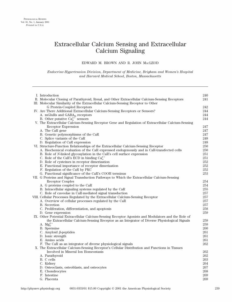
The amino acid sequences of BoPCaR and the otherCaRs cloned to date that are predicted from their nucle-otide sequences reveal a common overall topology, whichincludes a very large (;600 amino acids), NH2-terminalextracellular domain (ECD), a central core of some 250amino acids with seven predicted transmembrane do-mains (TMDs) that are characteristic of the superfamily ofGPCRs and a large intracellular COOH-terminal tail of;200 amino acids (58) (Fig. 1). As described in moredetail in section VID, data from studies on the CaR’sstructure-function relationships indicate that Cao
21 bindsto its ECD. The receptor’s ECD also contains multipleN-linked glycosylation sites (58, 134), whereas its intra-cellular domains [three intracellular loops (ICLs) andCOOH tail] harbor several predicted consensus proteinkinase C (PKC) and protein kinase A (PKA) phosphory-lation sites (the PKA sites are present in all species stud-ied to date except the bovine CaR). The PKC phosphory-lation sites are known to modulate the receptor’s activity,
January 2001 Cao21 SENSING AND Cao
21 SIGNALING 241
whereas the physiological relevance of the PKA sites, ifany, is currently unknown (18, 58, 89) (see sect. VIF).
III. MOLECULAR SIMILARITY OF THE
EXTRACELLULAR CALCIUM-SENSING
RECEPTOR TO OTHER G PROTEIN-
COUPLED RECEPTORS
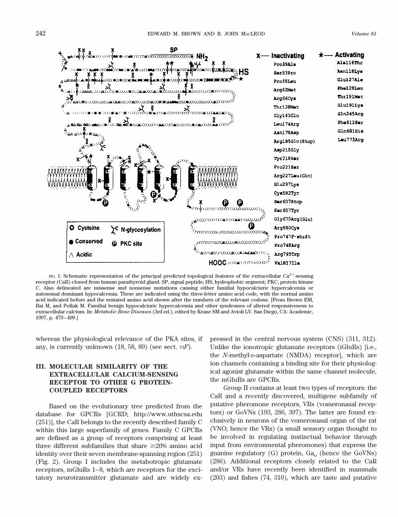
Based on the evolutionary tree predicted from thedatabase for GPCRs [GCRD; http://www.uthscsa.edu(251)], the CaR belongs to the recently described family Cwithin this large superfamily of genes. Family C GPCRsare defined as a group of receptors comprising at leastthree different subfamilies that share $20% amino acididentity over their seven membrane-spanning region (251)(Fig. 2). Group I includes the metabotropic glutamatereceptors, mGluRs 1–8, which are receptors for the exci-tatory neurotransmitter glutamate and are widely ex-
pressed in the central nervous system (CNS) (311, 312).Unlike the ionotropic glutamate receptors (iGluRs) [i.e.,the N-methyl-D-aspartate (NMDA) receptor], which areion channels containing a binding site for their physiolog-ical agonist glutamate within the same channel molecule,the mGluRs are GPCRs.
Group II contains at least two types of receptors: theCaR and a recently discovered, multigene subfamily ofputative pheromone receptors, VRs (vomeronasal recep-tors) or GoVNs (193, 286, 397). The latter are found ex-clusively in neurons of the vomeronasal organ of the rat(VNO; hence the VRs) (a small sensory organ thought tobe involved in regulating instinctual behavior throughinput from environmental pheromones) that express theguanine regulatory (G) protein, Gao (hence the GoVNs)(286). Additional receptors closely related to the CaRand/or VRs have recently been identified in mammals(203) and fishes (74, 310), which are taste and putative
FIG. 1. Schematic representation of the principal predicted topological features of the extracellular Ca21-sensingreceptor (CaR) cloned from human parathyroid gland. SP, signal peptide; HS, hydrophobic segment; PKC, protein kinaseC. Also delineated are missense and nonsense mutations causing either familial hypocalciuric hypercalcemia orautosomal dominant hypocalcemia. These are indicated using the three-letter amino acid code, with the normal aminoacid indicated before and the mutated amino acid shown after the numbers of the relevant codons. [From Brown EM,Bai M, and Pollak M. Familial benign hypocalciuric hypercalcemia and other syndromes of altered responsiveness toextracellular calcium. In: Metabolic Bone Diseases (3rd ed.), edited by Krane SM and Avioli LV. San Diego, CA: Academic,1997, p. 479–499.]
242 EDWARD M. BROWN AND R. JOHN MACLEOD Volume 81
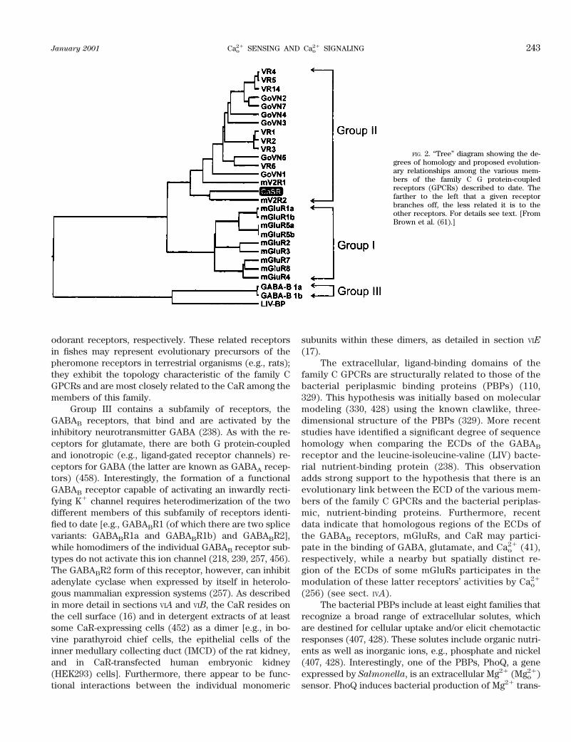
odorant receptors, respectively. These related receptorsin fishes may represent evolutionary precursors of thepheromone receptors in terrestrial organisms (e.g., rats);they exhibit the topology characteristic of the family CGPCRs and are most closely related to the CaR among themembers of this family.
Group III contains a subfamily of receptors, theGABAB receptors, that bind and are activated by theinhibitory neurotransmitter GABA (238). As with the re-ceptors for glutamate, there are both G protein-coupledand ionotropic (e.g., ligand-gated receptor channels) re-ceptors for GABA (the latter are known as GABAA recep-tors) (458). Interestingly, the formation of a functionalGABAB receptor capable of activating an inwardly recti-fying K1 channel requires heterodimerization of the twodifferent members of this subfamily of receptors identi-fied to date [e.g., GABABR1 (of which there are two splicevariants: GABABR1a and GABABR1b) and GABABR2],while homodimers of the individual GABAB receptor sub-types do not activate this ion channel (218, 239, 257, 456).The GABABR2 form of this receptor, however, can inhibitadenylate cyclase when expressed by itself in heterolo-gous mammalian expression systems (257). As describedin more detail in sections VIA and VIB, the CaR resides onthe cell surface (16) and in detergent extracts of at leastsome CaR-expressing cells (452) as a dimer [e.g., in bo-vine parathyroid chief cells, the epithelial cells of theinner medullary collecting duct (IMCD) of the rat kidney,and in CaR-transfected human embryonic kidney(HEK293) cells]. Furthermore, there appear to be func-tional interactions between the individual monomeric
subunits within these dimers, as detailed in section VIE
(17).The extracellular, ligand-binding domains of the
family C GPCRs are structurally related to those of thebacterial periplasmic binding proteins (PBPs) (110,329). This hypothesis was initially based on molecularmodeling (330, 428) using the known clawlike, three-dimensional structure of the PBPs (329). More recentstudies have identified a significant degree of sequencehomology when comparing the ECDs of the GABAB
receptor and the leucine-isoleucine-valine (LIV) bacte-rial nutrient-binding protein (238). This observationadds strong support to the hypothesis that there is anevolutionary link between the ECD of the various mem-bers of the family C GPCRs and the bacterial periplas-mic, nutrient-binding proteins. Furthermore, recentdata indicate that homologous regions of the ECDs ofthe GABAB receptors, mGluRs, and CaR may partici-pate in the binding of GABA, glutamate, and Cao
21 (41),respectively, while a nearby but spatially distinct re-gion of the ECDs of some mGluRs participates in themodulation of these latter receptors’ activities by Cao
21
(256) (see sect. IVA).The bacterial PBPs include at least eight families that
recognize a broad range of extracellular solutes, whichare destined for cellular uptake and/or elicit chemotacticresponses (407, 428). These solutes include organic nutri-ents as well as inorganic ions, e.g., phosphate and nickel(407, 428). Interestingly, one of the PBPs, PhoQ, a geneexpressed by Salmonella, is an extracellular Mg21 (Mgo
21)sensor. PhoQ induces bacterial production of Mg21 trans-
FIG. 2. “Tree” diagram showing the de-grees of homology and proposed evolution-ary relationships among the various mem-bers of the family C G protein-coupledreceptors (GPCRs) described to date. Thefarther to the left that a given receptorbranches off, the less related it is to theother receptors. For details see text. [FromBrown et al. (61).]
January 2001 Cao21 SENSING AND Cao
21 SIGNALING 243
port proteins in response to environmental Mg21 depriva-tion (299).
In their capacities to act as cell surface receptorsparticipating in chemoreception and sensory transductionor membrane transport, the PBPs interact with integralmembrane proteins within the bacterial cell membraneafter they bind specific chemosensory substances or nu-trients to transmit the signal or transport the nutrient intothe cell. Therefore, it seems likely that the family CGPCRs, including the CaR, evolved as “fusion proteins”comprising an NH2-terminal ECD derived from an ancientfamily of solute-binding proteins and the seven mem-brane-spanning, “serpentine” motif that evolved sepa-rately to transmit extracellular signals to the interior ofeukaryotic cells via the GPCRs. Interestingly, the CaR canalso participate in the stimulation of chemotaxis by Cao
21
in monocytes (464) and, perhaps, osteoblasts and theirprecursors (462, 463). Thus there may be conservation ofboth the functional as well as the structural attributes ofthis domain across a very broad evolutionary time scale.
IV. ARE THERE ADDITIONAL EXTRACELLULAR
CALCIUM-SENSING RECEPTORS
OR SENSORS?
A. mGluRs and GABAB Receptors
Recent work has revealed that some mGluRs cansense Cao
21 in addition to responding to glutamate as theirprincipal agonist in vivo, although the physiological rele-vance of this Cao
21-sensing remains uncertain. Kubo et al.(256) demonstrated that mGluRs 1, 3, and 5 sense Cao
21
over a range of ;0.1–10 mM, while mGluR2 is consider-ably less responsive to changes in Cao
21. Construction ofchimeric receptors, in which the ECDs of mGluR2 and -3were fused to the TMDs and COOH tail of mGluR1a,proved that the capacities of the respective receptors tobe activated by Cao
21 (or lack thereof) was conferred bytheir ECDs. All three of the mGluRs that sense Cao
21 haveidentical serines and threonines, respectively, at aminoacid positions homologous to residues 165 and 188 inmGluR1a (41). These two residues are thought to play keyroles in the binding of glutamate to the ECDs of themGluRs (330). In contrast, while mGluRs 1a, 3, and 5 havea serine at a position equivalent to residue 166 inmGluR1a, mGluR2 has an aspartate in this position (256).Furthermore, changing the serines in mGluRs 1a, 3, and 5to an aspartate considerably reduces their capacity tosense Cao
21, while replacing the aspartate in mGluR2 witha serine increases its apparent affinity for Cao
21, to a levelsimilar to those of mGluRs 1a, 3, and 5 (256). Therefore,the serines at amino acid position 166 in mGluR1a and atthe equivalent positions in mGluRs 3 and 5 apparentlyplay key roles in their capacities to sense Cao
21, although
the molecular mechanism underlying this action is notclear from these studies. It should be pointed out, how-ever, that the Hill coefficients for the modulation of theactivities of these mGluRs by Cao
21 (as well as by gluta-mate) are close to one (256), considerably lower than thatfor the CaR, which is ;3 (15, 52, 58, 152, 393). Thus thereare apparently additional aspects of the binding of Cao
21
by the CaR (e.g., the presence of several binding sites)and/or subsequent steps in its activation that confer pos-itive cooperativity on this overall process (see sect. VI).The latter is a key element contributing to the narrowrange within which Cao
21 is maintained by the mineral ionhomeostatic system.
Of interest, a recent study has documented thatchanges in Cao
21 also modulate the GABAB receptors,although Cao
21 by itself has no effect on this class ofreceptors (459). Cao
21 potentiated the stimulatory actionof GABA on GTP binding to this receptor and enhancedthe coupling of the GABAB receptor to activation of a K1
channel and inhibition of forskolin-stimulated cAMP ac-cumulation. The actions of Cao
21 were not mimicked byother polyvalent cations. Therefore, given that not onlyCao
21, but also amino acids (109; see also sect. IXE), mod-ulate the function of the CaR, when taken in the contextof the actions of amino acids (e.g., glutamate) or theirderivatives (i.e., GABA) on the mGluRs and GABAB re-ceptors, respectively, further emphasizes the structuraland functional relationships among these three types ofreceptors.
B. Other Putative Cao21 Sensors
It is likely that there are Cao21 receptors or sensors in
addition to the CaR and mGluRs, which mediate some ofthe very substantial number of actions of Cao
21 on diversecell types. The availability of the cloned CaR has made itfeasible to begin to catalog the cell types that express thisreceptor, as described in more detail in sections X, XI, andXIII. For example, the inhibitory action of Cao
21 on PTHsecretion, the stimulatory effect of Cao
21 on calcitonin(CT) release (146, 158, 288) and many of the actions ofCao
21 on the kidney (for review, see Ref. 180) are mostlikely CaR mediated. The presence of the CaR in a cellwhose function is modulated by Cao
21 does not prove,however, that it mediates that particular action of Cao
21.The availability of mice with targeted disruption of theCaR gene (196) and the discovery of human diseasescaused by CaR mutations (for review, see Ref. 53) haveprovided very useful tools for assessing this receptor’srole in Cao
21-induced changes in various cellular functionsin vivo and/or in vitro. The recent development of selec-tive activators (320) and antagonists of the CaR (318) aswell as the use of dominant negative CaR constructs (14,15, 291) will likewise be of utility in determining whether
244 EDWARD M. BROWN AND R. JOHN MACLEOD Volume 81
the CaR mediates known actions of Cao21 on specific,
CaR-expressing cell types.There are, however, cells whose functions are mod-
ulated by Cao21 that do not express the CaR or have not
yet been examined for its expression. In the former case,the actions of Cao
21 could potentially be mediated by oneor more of the mGluRs that sense Cao
21, an hypothesisthat has not yet been tested. Alternatively, this Cao
21-sensing capability may be conferred by one or more of theadditional putative Cao
21 receptors/sensors discussed be-low. There may well be other Cao
21-receptors/sensingmechanisms as well, although a discussion of the evi-dence supporting the existence of these is beyond thescope of this review.
1. Megalin/gp330
Monoclonal antibodies that are directed at a largeprotein, called megalin or gp330, that is present at highlevels in parathyroid, proximal tubular, and placentalcells (220) as well as in a variety of other cell types canmodulate the Cao
21-sensing functions of these cells (223).For instance, such antibodies can interfere with the ca-pacity of high Cao
21 to inhibit PTH secretion from humanparathyroid cells (221). The level of expression of thisprotein is reduced substantially in pathological parathy-roid glands from patients having various forms of hyper-parathyroidism (HPT) (222). In these hyperparathyroidstates, the abnormal cells are generally less sensitive thannormal parathyroid cells to the suppressive effect of highCao
21 on PTH release (51, 170, 326). Therefore, the re-duced expression of the protein recognized by these an-tibodies could conceivably contribute to the defectiveCao
21 sensing in HPT. Furthermore, the same proteincould potentially participate in Cao
21 sensing by normalparathyroid cells. The level of expression of the CaR inpathological parathyroid cells, however, has also beenfound to be reduced in HPT in most (136, 162, 246) but notall studies (155), raising the possibility that the changes inmegalin expression in hyperparathyroidism could be theconsequence rather than the cause of the disease.
cDNAs coding for megalin/gp330 have been isolatedfrom human (195, 273) and rat cDNA libraries (398).These cDNAs encode very large, ;500-kDa proteins thatbelong to the low-density lipoprotein receptor superfam-ily. Recent studies have provided strong evidence thatmegalin’s principal role is to serve as an endocytic recep-tor (116) that binds to and mediates uptake of albumin(115), insulin (333), the transcobalamin-B12 complex(101), retinol and its binding protein (103), and thyroglob-ulin (279), as well as other proteins (298) and even drugs(137). Indeed, megalin “knockout” mice show defectiveproximal tubular uptake of the serum vitamin D andretinol binding proteins and their associated vitamin Dmetabolites and retinol, respectively, providing strong
support for the role of this protein as an endocytic recep-tor (103). Although megalin does bind extracellular cal-cium ions (102), this Cao
21 binding probably does notparticipate directly in systemic mineral ion metabolism. Itwill be of interest to determine whether megalin interactswith the CaR in cells that coexpress both proteins and/orregulates the CaR’s internalization or other aspects of itsfunction, thereby participating indirectly in Cao
21 sensing.
2. Cao21 sensing by osteoblasts
Raising Cao21 has several actions on cells of the os-
teoblastic lineage. Elevated levels of Cao21 stimulate bone
formation in explants of rodent bone (372). In addition,Cao
21 and other polycations [e.g., strontium (73) and alu-minum (Al31) (364)] stimulate the proliferation (161, 363,421) and/or chemotaxis (161) of osteoblasts and theirprecursors, an effect that could be mediated, in part, byan associated increase in the release of insulin-likegrowth factor II (IGF-II) (201). High Cao
21 also modulatesintracellular second messengers in the murine osteoblas-tic cell line, MC3T3-E1. Elevated levels of Cao
21 raisediacylglycerol (174) and cAMP levels (175) in these cellsbut do not promote the formation of inositol phosphatesthat would occur with activation of phosphoinositide(PI)-specific phospholipase (PL) C. Quarles et al. (362)were unable to detect CaR transcripts by RT-PCR andNorthern analysis in MC3T3-E1 cells and suggested on thebasis of this result as well as pharmacological differencesfrom the CaR [including the latter’s low affinity for Alo
31
(417)] that a distinct Cao21-sensing receptor mediated the
actions of Cao21 on this cell line. The same group has
identified genomic clones for several CaR-related genes(194) that are ;60% similar and 40% identical to the CaRoriginally cloned from parathyroid (58) and kidney (381)within regions corresponding to their predicted TMDs.Transcripts for these putative receptors, however, are notexpressed in bone cells at levels that can be detected byNorthern analysis or RNase protection (194). It is possi-ble, therefore, that they encode either pseudogenes orrelated receptors, viz., homologs of the putative phero-mone receptors in the VNO of the rat (193, 286, 397) butare not involved in sensing Cao
21 and other polyvalentcations in osteoblasts. Moreover, as discussed in moredetail in section XD, we (463) and others (230) haverecently found that MC3T3-E1 cells express both CaRtranscripts as assessed by RT-PCR and Northern analysisand receptor protein as detected by Western analysisand/or immunocytochemistry. Further studies of theseand other osteoblastic cell lines are needed in which theCaR has been “knocked out” through the use of selectiveCaR activators (320) or antagonists (318) and/or domi-nant negative constructs of the CaR (291), to prove which,if any, of the effects of Cao
21 on osteoblastic cell lines aremediated by the CaR versus some other Cao
21-sensing
January 2001 Cao21 SENSING AND Cao
21 SIGNALING 245
mechanism(s). Furthermore, while MC3T3-E1 cells andother osteoblast-like cell lines represent useful models forinvestigating the control of osteoblastic function, theymay or may not faithfully reproduce the phenotype ofosteoblasts in vivo. It will be important, therefore, todetermine whether bona fide osteoblasts and/or their pre-cursor cells in intact bone express the CaR and/or otherCao
21-sensing mechanisms. Of interest in this regard, thestudies of Pi et al. (349) have recently shown that primaryosteoblasts derived from mice with targeted disruption ofthe CaR gene retain certain responses to Cao
21, consistentwith the presence of another Cao
21-sensing mechanism(349). The presence of the latter and/or the CaR in osteo-blasts could enable these cells to respond in physiologi-cally relevant ways to local changes in Cao
21 within thebone/bone marrow microenvironment (see also sects. XD
and XIV for further discussions of local Cao21 sensing and
Cao21 signaling in bone, respectively).
3. Cao21 sensing by osteoclasts
Another example of a cell that appears to possess aCao
21-sensing mechanism distinct from the CaR is theosteoclast, based largely on indirect, pharmacological ev-idence. Several groups first reported in 1989 that elevatingCao
21 had direct actions on isolated osteoclasts in vitro,inhibiting bone resorption and producing elevations in thecytosolic calcium concentration (Cai
21), which were rem-iniscent of those elicited in parathyroid cells by raisingCao
21 (277, 476). Although it remains to be determinedwhether this mechanism functions in a physiologicallyrelevant manner in vivo (e.g., by creating mice with tar-geted disruption of the relevant gene), it could representa Cao
21-sensing system through which the osteoclast reg-ulates its own resorptive activity; that is, when Cao
21 risesabove a certain level, owing to osteoclast-mediated boneresorption, activation of the putative Cao
21-sensing recep-tor in this cell type would feed back to inhibit furtherbone breakdown. Subsequent studies, principally by Zaidiet al. (475), have elucidated several features of the pro-cess of Cao
21 sensing by osteoclasts (see below), althoughcharacterization of the sensor/receptor at a molecularlevel has not yet been accomplished.
Elevating Cao21 in vitro produces marked retraction
of osteoclasts, decreased expression of podosomes (thestructures that anchor resorbing osteoclasts to the under-lying bone), inhibition of the release of hydrolytic en-zymes, and a reduction in bone resorption (277, 476). Theobserved Cao
21-induced increases in Cai21 are probably an
important mediator of the associated alterations in cellu-lar function, because the calcium ionophore ionomycincauses similar effects. Not all osteoclasts possess thisCao
21-sensing mechanism. Those freshly isolated frommedullary bone of the Japanese quail, for example, do notexhibit these responses to elevated levels of Cao
21 (25).
After being cultured for 5–8 days, however, these cellsdevelop the capacity to sense Cao
21 in a manner similar tothat of osteoclasts freshly isolated from chick or rat bone(25). These cultured quail osteoclasts could, therefore,represent an appropriate source of mRNA encoding theputative sensor that could be utilized to isolate the rele-vant gene using an expression cloning strategy.
A variety of polyvalent cations mimic the actions ofCao
21 on the osteoclast, but they generally exhibit a phar-macological profile that differs distinctly from that exhib-ited by parathyroid cells and other cells expressing theCaR (405) [although more recent studies have providedexamples of pharmacological profile more similar to thatof the CaR, including effects of extracellular Gd31 andneomycin resembling those of high Cao
21 (474); see alsosect. XD]. In general, activation of the CaR in parathyroidcells by Cao
21, Mgo21, or extracellular Ba21 takes place at
concentrations of these divalent cations that are several-fold lower than those modulating the function of oste-oclasts (57, 409, 477). The lower affinity of the Cao
21-sensing mechanism in osteoclasts for Cao
21 may bephysiologically appropriate, because Cao
21 measured di-rectly beneath osteoclasts that are actively resorbingbone can be as high as 8–40 mM (412). Other polyvalentcations that activate the osteoclast’s Cao
21-sensing mech-anism include extracellular Ni21, extracellular Cd21
(which do not stimulate the CaR) (405), and extracellularLa31 (which does activate the CaR) (404).
The putative Cao21-sensing receptor in the osteoclast
may be related to the ryanodine receptor (479). Agents[e.g., ryanodine (478) or caffeine (406)] that interact withand modulate the activity of the ryanodine receptor(which mediates high Cai
21-induced release of Ca21 fromintracellular stores in skeletal muscle and other celltypes) modify osteoclastic Cao
21 sensing. Moreover, osteo-clasts bind [3H]ryanodine, and this binding is displaced byCao
21 and by the ryanodine receptor antagonist rutheniumred (479). Finally, an antibody that recognizes an epitopewithin the ryanodine receptor’s channel-forming domainpotentiates the effects of extracellular Ni21 on osteoclastsand labels the plasma membrane of nonpermeabilizedosteoclasts. Conversely, an antibody that interacts withan intracellular epitope does not exert either of theseactions. Taken together, these results suggest the pres-ence of a ryanodine receptor-like molecule on the oste-oclast plasma membrane (479) (in contrast to other celltypes in which the ryanodine receptor is located intracel-lularly) that functions as a Cao
21 sensor or in close asso-ciation with some other Cao
21-sensing mechanism. Itshould be pointed out, however (as described in moredetail in sect. XD), that recent studies have suggested thatthe CaR is also expressed in osteoclasts and/or theirprecursors. It remains to be determined whether there areactually two distinct Cao
21-sensing mechanisms in this celltype.
246 EDWARD M. BROWN AND R. JOHN MACLEOD Volume 81
4. Genetic evidence for the existence of additional
receptors/sensors
The identification of inherited diseases of Cao21 sens-
ing has not only provided strong genetic evidence for thecentral role of the CaR in systemic Cao
21 homeostasis buthas also raised the possibility that there may be additionalCao
21 sensors/receptors. Familial hypocalciuric hypercal-cemia (FHH) is a generally benign, inherited condition(indeed it is sometimes called FBH, familial benign hyper-calcemia) in which there is autosomal dominant inheri-tance of hypercalcemia accompanied in most cases byrelative hypocalciuria (i.e., lower rates of urinary calciumexcretion than would have been expected in the setting ofhypercalcemia) (for review, see Ref. 49). The great ma-jority of families with this condition (at least 90%) showgenetic linkage to the long arm of chromosome 3 in theregion where the CaR gene is known to reside (100, 179,353, 354, 434). Of the families exhibiting this linkage tochromosome 3, about two-thirds have heterozygous inac-tivating mutations within the coding region of the CaRgene (49). Most of these mutations are point mutationsthat reduce the receptor’s activity by decreasing its cellsurface expression and/or reducing its intrinsic biologicalactivity. Some mutations exert an additional dominantnegative action on the wild-type CaR (15, 178, 342, 353).
In a few families, consanguineous marriages of indi-viduals with FHH (yielding infants homozygous for CaRinactivation) (213, 353, 354) or union of persons with FHHharboring different CaR mutations (producing a com-pound heterozygous infant) (249) produces a much moresevere form of hypercalcemia, termed neonatal severehyperparathyroidism (NSHPT) (for review, see Ref. 49).The discovery that FHH and NSHPT can represent, re-spectively, the equivalent of the heterozygous and ho-mozygous forms of complete or partial knockout of theCaR gene has 1) established the central, nonredundantrole of the CaR in mineral ion metabolism, 2) proved thatexpression of the CaR is required for normal regulation ofPTH secretion and probably parathyroid cellular prolifer-ation by Cao
21 (see also sect. XA), and 3) documented thatthe CaR plays a key role in regulating the renal tubularhandling of divalent cations (see sect. XC for more de-tails).
Of great interest, clinical conditions similar in manyof their features to FHH can be caused by genetic defectsat chromosomal loci other than that harboring the CaRgene. The first such condition was assigned to a locus onchromosome 19p13.3 by Heath et al. (179) in a family withclinical characteristics indistinguishable from thosepresent in the form of FHH caused by mutations in theCaR. Subsequently, Trump et al. (434) have shown thatanother family with FHH exhibiting certain atypical fea-tures (e.g., osteomalacia and progressive elevations inserum PTH with increasing age in certain family mem-
bers) exhibits linkage to a different locus on chromosome19 (19q13) (269), further documenting the genetic heter-ogeneity of this clinical syndrome. It is possible, there-fore, that these two genetic loci contain genes encodingCao
21 sensors other than the CaR. Alternatively, thesegenes might represent additional, presumably down-stream elements along the Cao
21-sensing pathway(s) reg-ulated by the CaR (or some other Cao
21 sensor) that, whenmutated, interfere with the ability of parathyroid andkidney to respond normally to the Cao
21 signal. It is alsoconceivable that these genes encode transcription factorsor other proteins necessary for expression of the CaRgene in parathyroid and kidney. In the latter case, loss ofthe relevant transcription factor might reduce the expres-sion of the CaR, analogous to certain forms of diabetesthat result from mutations in transcription factors partic-ipating in expression of the insulin gene (165).
V. THE EXTRACELLULAR CALCIUM-SENSING
RECEPTOR GENE AND REGULATION
OF EXTRACELLULAR CALCIUM-SENSING
RECEPTOR EXPRESSION
A. The CaR Gene
Very little work has been carried out directed atcharacterizing the CaR gene. The human gene is locatedon the long arm of chromosome 3 (3q21-q24) as assessedby linkage analysis (100) and at band 3q13.3–21 as deter-mined by fluorescent in situ hybridization (214). In the ratand mouse, the gene resides on chromosomes 11 and 16(214), respectively, while in the bovine species it ispresent on chromosome 1 (314). The human CaR genecontains at least seven exons (343). Six encode the recep-tor’s large ECD and/or its upstream untranslated regions,while a single exon codes for the receptor’s TMDs andCOOH terminus (343, 353). The regulatory regions of thegene have not yet been characterized but will be of sub-stantial interest, since expression of the CaR can changeunder several circumstances in vivo and/or in vitro asdescribed below.
B. Genetic Polymorphisms of the CaR
Three apparently benign polymorphisms have beenidentified in the predicted COOH tail of the CaR: A986S,G990R, Q1011E, which were present in 30, 15, and 10% ofmore than 100 persons in the United States who wereapparently unaffected with any disturbance in calciumhomeostasis (178). In a subsequent study (106), the A986Spolymorphism was found to be present in 16% of 163Canadian individuals and was associated with a slightincrease in serum total calcium concentration when cor-
January 2001 Cao21 SENSING AND Cao
21 SIGNALING 247
rected for albumin and in fasting calcium concentration(106). It is possible that this polymorphism could contrib-ute to a genetic predisposition to certain bone and/ormineral disorders.
C. Splice Variants of the CaR
Several splice variants of the CaR gene have beendescribed. One cDNA clone of the human parathyroidCaR contained a 30-nucleotide insertion within the regionof the gene encoding the receptor’s predicted ECD (157).This 10-amino acid insertion had no apparent effect on thefunction of the CaR as assessed by expression in Xenopus
laevis oocytes (157). The same splice variant has subse-quently been identified in human breast cancer tissue(97). Its functional significance, if any, when studied inmammalian expression systems requires further investi-gation. Another alternatively spliced CaR transcript that isexpressed in human cytotrophoblasts and parathyroidlacks exon 3 and encodes a truncated, presumably inac-tive receptor (40). Whether this receptor could interferein some way with the normal CaR’s function or exhibitsany other functional attribute(s) remains to be deter-mined. Oda et al. (327) have recently described an alter-natively spliced form of the CaR in keratinocytes thatlacks exon 5, producing an in-frame deletion of 77 aminoacids and resulting in an expressed protein that is smallerand exhibits an altered glycosylation pattern comparedwith the full-length CaR. The truncated CaR was inactivewhen transfected into HEK293 cells or keratinocytes asassessed by high Cao
21-evoked increases in inositol phos-phates, and it interfered with the function of the coex-pressed full-length CaR (327). This latter observation mayexplain the reduced responsiveness of differentiated ker-atinocytes to Cao
21-induced elevations of Cai21, as the
alternatively spliced form of the receptor is expressed atgreater levels in the differentiated cells and could exert,therefore, a dominant negative action on the full-lengthCaR expressed within the same cells (327).
Finally, there can be alternative splicing within the59-untranslated region (UTR) of the CaR gene. For in-stance, transcripts in human parathyroid vary within their59-UTRs, consistent with alternative splicing of noncodingexons within the 59-upstream region of the gene, without,however, altering the coding region (157). Such alterna-tive splicing within the gene’s putative upstream regula-tory regions could clearly participate in tissue-specificexpression and/or regulation of the CaR gene, but furtherstudies are needed to define further its importance in thisregard. Chikatsu et al. (99) have recently cloned a portionof the upstream region of the human CaR gene and iden-tified two promoters (present within, respectively, exons1A and 1B). The more upstream of the two promoters hasTATA and CAAT boxes, while the downstream promoteris GC rich.
D. Regulation of CaR Expression
1. CaR expression in cultured parathyroid cells
Recent studies have documented that the expressionof the CaR mRNA and/or protein can change in a varietyof circumstances, although the mechanisms underlyingthese alterations in gene expression are, as yet, poorlyunderstood. Calf parathyroid cells show rapid (withinhours and 1–2 days, respectively) and marked (up to80–85%) reductions in CaR mRNA and protein after theyare put in culture (47, 297). This reduction in CaR expres-sion probably contributes to a major extent to the accom-panying reduction in high Cao
21-elicited inhibition of PTHrelease (47, 259, 260, 297). Of interest, the expression ofthe receptor and the associated suppression of PTH se-cretion by high Cao
21 are maintained to a substantiallygreater extent in long-term cultures of human parathyroidcells, for unclear reasons (391).
2. CaR expression in renal insufficiency
The level of expression of the CaR also decreases inthe kidney in chronic renal insufficiency induced in ratsby subtotal nephrectomy (283, 285). This reduction in CaRexpression may contribute to the associated reduction inurinary Ca21 excretion occurring in this setting, based onthe inverse relationship between CaR activity and/or ex-pression levels and concomitant renal excretion of Ca21
that is present in persons with inactivating mutations inthe CaR (see sect. XC) (181). Since, as described later,1,25-dihydroxyvitamin D [1,25(OH)2D] can increase therenal expression of the CaR (46), the decrease in CaRexpression in the kidney with impaired renal functioncould result, at least in part, from the associated reduc-tion in the level of 1,25(OH)2D that occurs during thedevelopment of renal insufficiency (419). Alternatively,the rise in circulating PTH levels in the setting of chronicrenal failure (419) may also contribute to the reduction inCaR gene expression in the kidney (285). Further studiesare needed to distinguish between these possibilities.
3. Reduced CaR expression in hyperparathyroidism
The level of expression of the CaR in parathyroidcells is diminished in pathological parathyroid glands re-sected from patients with primary hyperparathyroidismor with the severe hyperparathyroidism that can developduring chronic hemodialysis in patients with renal failuredue to end-stage renal disease (162, 246). This reductionin CaR mRNA and protein expression has been observedin most (45, 135, 136, 232) but not all studies (155). Thestudy that did not show a decrease in receptor expressionemployed semi-quantitative RT-PCR to compare the lev-els of expression of CaR transcripts in normal and patho-logical parathyroid glands. One potential problem in the
248 EDWARD M. BROWN AND R. JOHN MACLEOD Volume 81

interpretation of the results of this study (155) is thatnormal but not hyperparathyroid parathyroid glands con-tain substantial numbers of fat cells that can account for;50% of the volume of the normal parathyroid gland.Studies using immunocytochemistry or in situ hybridiza-tion have not detected expression of the CaR in the fatcells of normal parathyroid glands (162, 246). Extractionof RNA from normal parathyroid glands (155), therefore,may have in effect “diluted” CaR transcripts from para-thyroid chief cells with RNA not containing these tran-scripts from fats cells, thereby reducing apparent CaRmRNA expression in normal parathyroid glands to levelsthat are comparable to those in pathological parathyroidcells. A recent study has shown a selective reduction inparathyroid adenomas of the CaR transcript arising fromthe further upstream of the two promoters regulating thisgene’s expression (99). The mechanism underlying thischange in the pattern of expression of the CaR gene,however, remains to be determined.
In view of the reduced sensitivity to Cao21 of parathy-
roid glands from patients with inactivating mutations ofthe CaR gene, the observed reduction in CaR expressionin pathological parathyroid tissue could contribute to theelevated set point for Cao
21-regulated PTH secretion thatis often observed not only in primary but also in severe,uremic secondary HPT (51, 62). The relationship of thereduced CaR expression to the associated excessive cel-lular proliferation in these pathological parathyroidglands remains to be determined. It should be noted,however, that the parathyroid cellular proliferation ob-served in mice with knockout of the CaR gene (196) aswell as in patients homozygous for inactivating mutationsof the human CaR gene (for review, see Ref. 49) stronglysupport the CaR’s involvement in tonically suppressingparathyroid cellular proliferation. There is further discus-sion of the CaR’s roles in controlling various aspects ofparathyroid function in section XA.
4. Effects of vitamin D and high Cao21
on CaR expression
There are interactions between the CaR (or at leastthe effects of high Cao
21) and vitamin D receptor (VDR)that are likely physiologically relevant in that the tworeceptors regulate their own levels of expression and/orthe expression of the other receptor. Vitamin D, specifi-cally its active form 1,25(OH)2D, upregulates its own recep-tor at the transcriptional level (313). Vitamin D also in-creases the expression of the CaR in parathyroid and kidneyin vivo in the rat (46), although another study, carried outusing a slightly different experimental approach, failed toobserve any vitamin D-induced changes in the CaR’s expres-sion in these two tissues (387). If confirmed in future stud-ies, vitamin D-elicited upregulation of the CaR in the para-thyroid could be physiologically appropriate, since it would
tend to facilitate inhibition of parathyroid function by highCao
21, e.g., suppression of PTH secretion and parathyroidcellular proliferation (see sect. XA).
High Cao21 raises the levels of expression of the CaR
in the pituitary-derived, ACTH-secreting murine AtT-20cell line (131) and of the VDR in rat parathyroid glands invivo (394). Although these actions of Cao
21 have not yetbeen proven to be CaR mediated, taken together with theknown effects of vitamin D on the expression of the CaRand VDR, they would afford the opportunity for synergis-tic interactions between Cao
21 and vitamin D in regulatingtheir target tissues. Such interactions could contribute,for instance, to the known synergistic actions of vitaminD and Cao
21 in promoting the differentiation of the humancolon cancer cell line Caco-2 (113) and in enhancing theexpression of calbindin D28K in the kidney (105). Further-more, the combination of high Cao
21 and vitamin D couldproduce a synergistic inhibition of the expression of thepreproPTH gene (413, 467), at least in part, by upregulat-ing the receptors involved in mediating this action as wellas through the direct effects of each agent on its ownreceptor. Further studies are needed, however, aimed atunderstanding the mechanism(s) (e.g., transcriptional orposttranscriptional) by which high Cao
21 and vitamin D reg-ulate the expression of the CaR gene. Additional discussionof the regulation of the expression of various genes by highCao
21 and the CaR can be found in section VIIID.Suzuki et al. (425) have recently identified thyroid
transcription factor 1 (TTF-1) as a potentially importantelement in the mechanism(s) through which Cao
21 mayinduce changes in gene expression in CaR-expressingcells. TTF-1 is a transcription factor that is a key mediatorof thyroid-specific gene expression. It is also expressed inthyroid C cells and in parathyroid chief cells and interactswith elements within the 59-flanking regions of the CaR,calmodulin, and calcitonin genes (425). Increases or de-creases in Cai
21 enhance or reduce, respectively, the ac-tivity of this promoter, its RNA levels, and the binding ofTTF-1 to these genes. In CaR-expressing cells that alsoexpress TTF-1, in which activation of the receptor islinked to elevations in Cai
21, therefore, TTF-1 may medi-ate the regulation of Cao
21-dependent genes (425).
5. High phosphorus intake and CaR expression
Whether phosphorus intake modulates CaR expres-sion is controversial. Brown et al. (45) showed that highphosphorus intake was associated with reduced CaRmRNA and protein expression in the parathyroid glandsof rats with secondary hyperparathyroidism owing to sub-total nephrectomy. In another study using a similarmodel, in contrast, there was no change in CaR mRNAexpression during intake of a high phosphorus diet (192).In the first of these studies, the reduced CaR immuno-staining was limited to regions of active chief cell prolif-
January 2001 Cao21 SENSING AND Cao
21 SIGNALING 249
eration, suggesting that the reduction in CaR expressionmight be secondary to enhanced proliferation rather thanthe change in phosphorus intake per se (45). It is possiblethat if high dietary phosphorus induces only focal changesin CaR expression, the latter might be missed by tech-niques that measure total (i.e., integrated) tissue levels ofthe receptor (e.g., Northern analysis) (192) as opposed tothose that assess localized differences in expression (i.e.,in situ hybridization or immunocytochemistry).
6. Developmental changes in CaR expression
There are substantial developmental increases in theexpression of the CaR in both kidney (84, 88) and hip-pocampus of the rat (87). The upregulation of the CaR inthe kidney occurs in the immediate peri- and postnatalperiod, and the ensuing higher level of expression of thereceptor persists through adulthood (84, 88). The increasein CaR expression in brain, in contrast, occurs about aweek postnatally. Furthermore, it is transient, decreasingseveralfold ;2 wk later to a lower level that remainsstable into adulthood (87). The mechanisms underlyingthese alterations in expression of the CaR gene, includingthe relative importance of alterations in gene transcrip-tion versus posttranscriptional mechanisms, require fur-ther investigation.
7. Other factors affecting CaR expression
Interleukin (IL)-1b modestly raises the level of CaRmRNA in bovine parathyroid gland fragments in associa-tion with a reduction in PTH secretion (324). In prelimi-nary studies, we have also demonstrated that sheep thathave undergone experimental burn injury show increasedCaR mRNA and protein expression in parathyroid but notin kidney (E. D. Murphey, N. Chattopadhyay, M. Bai, O.Kifor, D. Harper, D. L. Traber, E. M. Brown, and G. L.Klein, unpublished data). Because the levels of inflamma-tory cytokines are elevated in patients who have suffereda burn, including IL-1 levels, the latter could potentiallyaccount, at least in part, for the accompanying rise in CaRexpression in the sheep model of burn injury. Further-more, the increase in CaR protein expression presumablycontributes to the associated reduction in PTH secretionand to the failure of PTH levels to rise normally despitehypocalcemia that can occur after burn injury (248).
VI. STRUCTURE-FUNCTION RELATIONSHIPS
OF THE EXTRACELLULAR CALCIUM-
SENSING RECEPTOR
A. Biochemical Evaluation of the CaR Expressed
Endogenously and in CaR-Transfected Cells
CaR proteins extracted from HEK293 cells tran-siently transfected with the human CaR (15, 134) have
similar expression patterns to those isolated from bovineparathyroid cells (15). Western blot analysis using anti-CaR antisera reveals a doublet of protein bands at molec-ular masses corresponding to ;130–140 and 150–160 kDathat are present in considerably greater amounts thananother band at ;115–120 kDa (15, 134). The latter isclose to, or slightly smaller than, the expected size of thefull-length, nonglycosylated receptor protein predictedfrom the CaR’s cDNA. Direct sequence analysis of thereceptor’s ECD, in fact, has revealed that the putativesignal peptide at the CaR’s NH2 terminus predicted fromits nucleotide sequence has been cleaved off (163). Thusthe first residue encountered is the tyrosine predicted atamino acid position 20 (163) of the human CaR cDNA(157).
Biochemical analysis of CaR-transfected HEK293cells using appropriate endoglycosidases has shown thatthe immunoreactive band at 130–140 kDa corresponds toan immature form(s) of the CaR glycosylated with carbo-hydrate(s) high in mannose content, while the band at150–160 kDa is the mature form of the receptor glycosy-lated with complex carbohydrates (15, 134). The receptorpresent on the cell surface of these cells is the matureform of the receptor, although the latter probably onlyrepresents a relatively small fraction of the total cellularimmunoreactivity of the CaR as assessed by Western blotanalysis (16). Therefore, much of the CaR is located in-tracellularly. Indeed, immunocytochemistry using anti-CaR antisera performed on a variety of CaR-expressingcells, when performed following detergent permeabiliza-tion (e.g., Triton X-100), demonstrates substantial CaRimmunoreactivity over the cytoplasm, often with a prom-inent perinuclear component (for review, see Ref. 88). Itis not currently known whether intracellular forms of theCaR simply represent nascent receptor protein in passagethrough the biosynthetic pathway or whether these intra-cellular receptors have distinct biological functions. Forinstance, the concentration of Cao
21 within its intracellularstores in the endoplasmic reticulum (ER) (300) ap-proaches the millimolar range and could potentially besensed by the CaR’s NH2-terminal “extracellular” domain,which would face the lumen of the ER.
In addition to these monomeric forms of the CaR,there are variable amounts of a doublet of immunoreac-tivity on Western analysis performed using reducingagents at the expected molecular weights of high man-nose-containing and fully glycosylated CaR dimers and, toa lesser extent, higher oligomers of the receptor (15, 452).These are not simply artifacts resulting from aggregationof the receptor occurring during its extraction from cellsand subsequent PAGE in a denaturing buffer, since thesedimers are also present when the receptor is extractedusing nondenaturing buffers, and its size is estimatedusing gel permeation chromatography (452). Further-more, Bai et al. (16) have shown using a nonpermeant
250 EDWARD M. BROWN AND R. JOHN MACLEOD Volume 81
cross-linking reagent combined with cell surface biotiny-lation that most of the CaR on the cell surface of tran-siently transfected HEK293 cells is present as a dimer.These CaR dimers appear to be disulfide linked, as theinclusion of reducing agents, such as dithiothreitol ormercaptoethanol, is needed for their conversion to mono-mers (16, 452). Even following reduction, however, asubstantial fraction of the receptor protein can still run ondenaturing PAGE as a dimer (16). Therefore, there may beadditional intermolecular interactions contributing todimerization. Interestingly, the CaR has within its fifthTMD a putative hydrophobic dimerization motif presentin one of the b2-adrenergic receptor’s TMDs (185). More-over, the CaR is far from unique among the GPCRs informing dimers. Recent studies have emphasized that anumber of GPCRs (184, 219, 480), including the mGluRs(389), may exist as dimers and that dimerization maypotentially play important roles in the function of thesereceptors.
B. Role of N-Linked Glycosylation in the CaR’s
Cell Surface Expression
As noted above, the CaR’s mature, cell surface formhas a carbohydrate content of ;35–40 kDa/receptormonomer as assessed on Western blots (15, 134). Thesecarbohydrate residues could potentially contribute to thebinding of Cao
21 or participate in other aspects of thereceptor’s structure and/or function. Indeed, treatment ofCaR-transfected HEK293 cells with tunicamycin, whichblocks N-linked glycosylation, markedly reduced the re-sponse of the cells to raised levels of Cao
21 in associationwith reduced cell surface expression of the receptor(134). The use of site-directed mutagenesis subsequentlyrevealed that of the nine predicted N-liked glycosylationsites within the human CaR’s ECD, eight are efficientlyglycosylated (375). Removal of any four or five of theseproduced substantial (50–90%) reductions in cell surfaceexpression and biological activity, and at least three intactglycosylation sites were required for efficient cell surfaceexpression. Glycosylation per se, however, did not appearto be critical for the CaR’s biological activity as assessedby high Cao
21-induced increases in inositol phosphate ac-cumulation (375).
C. Role of the CaR’s ECD in Binding Cao21
The first direct evidence that Cao21 binds to the CaR’s
ECD was provided by studies utilizing chimeric receptorsin which the ECD of either the CaR or an mGluR wasfused to the TMDs and COOH tail of the other receptor(317). A chimeric receptor containing the CaR’s ECD andthe TMDs and COOH tail of mGluR1a was activated byhigh Cao
21 but not by mGluR agonists. Conversely, a chi-
meric receptor comprising the mGluR’s ECD and theCaR’s TMDs and COOH tail was activated by glutamatebut not by high Cao
21. [In these studies, in which thereceptors were expressed in X. laevis oocytes, activationof the mGluR by high Cao
21 was not observed (317);subsequent studies have shown, however, that certainmGluRs can sense Cao
21, as noted above (256)]. Thus theECD confers ligand specificity upon the CaR, the mGluRs,and presumably the other members of the family CGPCRs.
Subsequent studies (41) have confirmed that Cao21
acts on the CaR by binding to its ECD, taking a similarapproach that utilized chimeric receptors and showingthat a chimeric receptor comprising the CaR’s ECD andmGluR1a’s TMDs and COOH tail was activated by Cao
21,Mgo
21 and Bao21 with EC50 values very similar to those of
the wild-type CaR. These workers also extended this anal-ysis to define specific residues (e.g., Ser-147 and Ser-170)within the CaR’s ECD that may be involved in determiningthe receptor’s apparent affinity for Cao
21 (41). As de-scribed above, these residues are in positions homolo-gous to those of Ser-165 and Thr-188 in mGluR1a. Thesetwo serine residues in the CaR and the equivalent residuesin mGluR1a and the other mGluRs are thought to play keyroles in the binding of these receptors’ respective ligandsto their ECDs. Moreover, amino acid residues in similarpositions within the GABAB receptors have recently beensuggested to play important roles in the binding of GABA.Brauner-Osborne et al. (41) found that mutating Ser-147to alanine produced a fourfold reduction in the EC50 ofthe CaR for Cao
21, whereas a CaR in which Ser-170 waschanged to alanine showed no activation by 50 mM Cao
21.Therefore, it is possible that the binding of Cao
21 by theCaR involves residues within the ECD that are equivalentto those within the mGluRs and GABAB receptors thatbind glutamate and GABA, respectively.
It should be noted that there are currently no assaysof the binding to the CaR of its physiological ligands.Therefore, studies on the determinants within the ECDthat are potentially involved in binding these ligands (41,317) have so far relied on indirect measures of binding,namely, high Cao
21- and other agonist-evoked increases inCai
21 or PLC activity (e.g., as assessed by accumulation ofinositol phosphates). Clearly mutating a residue within aGPCR need not modify that receptor’s function solelythrough a direct action (e.g., by interfering with the bind-ing of a ligand to a specific amino acid residue). Byaltering the receptor’s conformation, mutating such a res-idue could also modulate the protein’s function indirectlyby secondarily perturbing agonist binding and/or subse-quent steps involved in activating intracellular signaltransduction [e.g., coupling of the receptor to its respec-tive G protein(s)]. Thus further direct structural studies(e.g., using X-ray crystallography) will be necessary toestablish definitively whether the serines at amino acid
January 2001 Cao21 SENSING AND Cao
21 SIGNALING 251
positions 147 and 170 within the CaR’s ECD participatedirectly or indirectly in Cao
21 sensing. Moreover, the CaRexhibits a Hill coefficient for its activation by high Cao
21 of;3 (15), while the Hill coefficients for activation of themGluRs by glutamate or by Cao
21 are ;1 (256). Therefore,it is likely that there are several (probably at least 3 asestimated from the Hill coefficient) binding sites for Cao
21
within the CaR’s ECD and/or elsewhere on the receptor.As discussed in section VID, the fact that the CaR exists onthe cell surface principally as a disulfide-linked dimer maycontribute to this receptor’s apparent positive cooperat-ivity in its binding of Cao
21.It is noteworthy that the agonists of the family C
receptors, namely calcium ions, glutamate, and GABA,are small molecules (or ions) that bind predominantly, ifnot entirely, to these receptors’ large ECDs. The othermajor class of GPCRs that bind their agonists to a sub-stantial extent within their ECDs are the receptors for theglycoprotein hormones: thyroid-stimulating hormone, lu-teinizing hormone, follicle-stimulating hormone, and hu-man chorionic gonadotropin, all of which, in contrast tothe agonists for the family C GPCRs, are relatively largeheterodimeric glycoproteins (440). In the great majorityof the other GPCRs, even those whose agonists are smallmolecules, such as the biogenic amines, the receptors’agonists have binding sites that likely involve amino acidresidues near the extracellular ends of the TMDs, deeperwithin the TMDs, or in the extracellular loops (454).
D. Role of Cysteines in Receptor Dimerization
The CaR shares with the mGluRs the same relativepositions of a total of 20 cysteines: 17 within the ECD, 1each in the first and second predicted extracellular loops,and 1 in the fifth TMD (58, 157). Clearly these cysteinescould be involved in intra- and/or intermolecular disulfidebonds that are important in stabilizing the CaR’s tertiaryand quaternary structures, e.g., by participating in recep-tor dimerization. It is of interest in this regard thatmGluR5 is a dimer held together by intermolecular disul-fide bond(s) present within the first 17 kDa of its NH2
terminus (389). In this region, the CaR harbors six cys-teines, although two of them are within the predictedsignal peptide and are likely removed during biosynthesis.Recent studies using site-directed mutagenesis have doc-umented that Cys-129 and Cys-131 are necessary fordimer formation (377), since the CaR migrates on PAGEprincipally as a monomer when these two cysteines arereplaced by serines. Interestingly, the resultant CaR issubstantially more sensitive to Cao
21 than the wild-typereceptor, suggesting that these intermolecular disulfidebonds could participate in constraining the receptor in itsinactive conformation(s). Although Ray et al. (377) inter-preted these findings as showing that the CaR lackingCys-129 and Cys-131 resides on the cell surface as mono-
mers, we have recently found using immunoprecipitationand cell surface cross-linking that noncovalently bounddimers represent the major form of the cell surface CaR inHEK293 cells transiently transfected HEK293 with a re-ceptor lacking Cys-129 and Cys-131 (M. Bai and E. M.Brown, unpublished data).
E. Functional Importance
of Receptor Dimerization
As noted above, the cell surface form of the CaR, atleast in transiently transfected HEK293 cells, is princi-pally a disulfide-linked dimer. Recent studies have ad-dressed the question of whether dimerization of the CaRis of functional significance; that is, do the two mono-meric partners within these dimers bind calcium and/orother CaR ligands and subsequently activate G proteinsand intracellular signaling pathways in a largely indepen-dent manner or do the monomers interact functionally insome way? Bai et al. (17) showed that individually inac-tive CaRs containing either inactivating point mutationswithin their ECDs or sufficient truncation of their COOHtails to abrogate biological activity showed substantialreconstitution of biological activity when cotransfected inHEK293 cells (Fig. 3) (17). Much less reconstitution ofactivity, in contrast, occurred when two inactive or par-tially active CaRs were cotransfected that both harboredECD mutations, or both had defects (e.g., either trunca-tion of the COOH tail or an inactivating point mutationwithin the third cytoplasmic loop) within domains of thereceptor likely involved in intracellular signaling (17).This result suggested that the CaR has at least two func-tional domains, i.e., the ECD and the remainder of thereceptor. These two domains can complement one an-other in cotransfections involving mutant receptors hav-ing defects in different domains. It is also likely thatintermolecular interactions between CaR monomerswithin the dimeric receptor may contribute to the domi-nant negative action of some CaR mutations identified inFHH, particularly those exhibiting relatively robust levelsof cell surface expression and forming substantial quan-tities of heterodimers of the mutant and wild-type recep-tors (14, 15). Such dominant negative CaRs have provideda useful tool for determining the CaR’s mediatory role inCao
21-regulated cellular processes (see sect. VIII) (291).
F. Regulation of the CaR by PKC
Activators of PKC, such as phorbol 12-myristate 13-acetate (PMA), substantially reduce Cao
21-evoked in-creases in inositol phosphates and Cai
21 in bovine para-thyroid cells (243, 293, 326, 369, 408). The presence ofpredicted PKC phosphorylation sites in the CaR’s intra-cellular domains suggests that PKC could participate inmodulating the receptor’s function by phosphorylating
252 EDWARD M. BROWN AND R. JOHN MACLEOD Volume 81
one or more of these sites (88). Bai et al. (18) recentlyexamined the functional importance of the predicted PKCphosphorylation sites within the human CaR’s intracellu-lar domains. The human receptor contains a total of fivepredicted PKC sites, one each within the second and thirdICLs and three within the COOH tail (18, 157). Deletingthe two PKC sites within the ICLs had little or no effect onPMA-induced modulation of high Cao
21-elicited increasesin Cai
21 in HEK cells transiently transfected with themutant CaRs. Deletion of the PKC site at residue 888within the CaR’s COOH tail, in contrast, substantiallyreduced the effect of PMA. Individually removing the twoother PKC sites within the tail had relatively little impacton the effect of PMA on the CaR’s function, but when allthree PKC sites within the COOH tail were deleted, therewas a modest further reduction in the effect of PKCactivators on high Cao
21-evoked Cai21 responses (18).
Thus the phosphorylation of the CaR’s PKC sites, partic-ularly that present at residue 888 in its COOH tail, canaccount for much of the inhibitory effect of PKC activa-tors on CaR-mediated signaling through the PLC-inositoltrisphosphate pathway. The small (;30%) residual effectof PMA that remains after deletion of all of the CaR’s PKCsites suggests that PKC can phosphorylate other sites onthe CaR and/or regulate other components within thispathway [e.g., G protein(s) and/or PLC].
G. Functional Significance of the CaR’s
COOH Terminus
Like several other members of the family C GPCRs,the CaR has a long COOH tail, 222 amino acid residues in
the case of the bovine CaR (58) and 216 for the humanreceptor (157). Most of the COOH tail is not needed forthe receptor’s biological activity. Ray et al. (376) foundthat CaRs with truncations at amino acid residues 888 and903 in the COOH tail (the wild-type receptor has 1,078residues) exhibited biological activities equivalent to thatof the wild-type CaR, while those truncated at positions865 and 874 were inactive, despite exhibiting only ;25%reductions in cell-surface expression (376). Furthermore,mutant receptors with a full-length COOH tail, but withindividual residues between positions 874 and 888 re-placed with alanines, had relatively normal levels of cellsurface expression but markedly reduced biological ac-tivities (376), further implicating this region as containingcrucial structural determinants required for normal bio-logical activity.
Bai et al. (16) found similar results in their studies ofseveral additional tail-truncated CaRs, albeit with at leastone significant difference. Similar to the results of Ray etal. (376), they (16) found that truncation of the CaR’sCOOH tail beyond residue 870 (e.g., Lys863stop) pro-duced an inactive receptor (16). A mutant CaR truncatedat position 877, Ala877stop, was also inactive (16), notunlike the findings of Ray et al. (376) that a CaR truncatedat position 874 had no activity. Thus the minimum numberof residues in the human CaR required for biologicalactivity is somewhere between 877 and 888, correspond-ing to a predicted COOH tail of 15–26 residues in length.Furthermore, Bai et al. (16) showed that the mutant re-ceptor, Ser892stop, had an increased level of cell surfaceexpression compared with the wild-type CaR and had asignificantly left-shifted EC50 for its activation by Cao
21
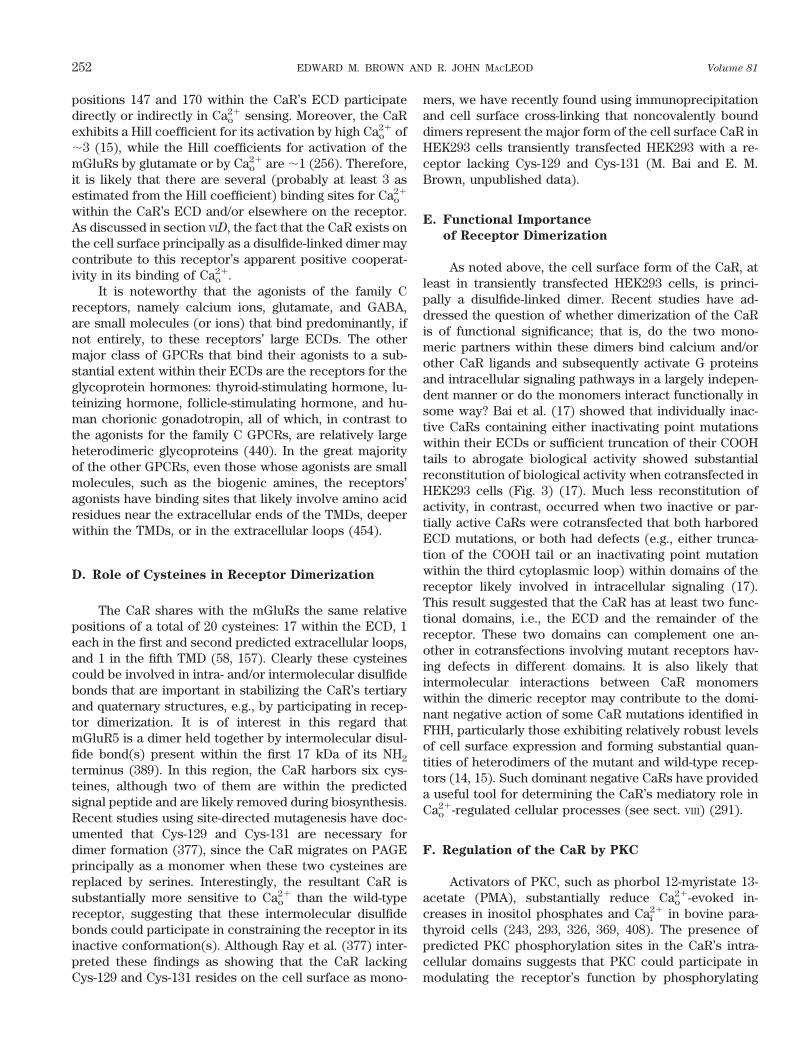
FIG. 3. Cotransfection of inactivemutant CaRs reconstitutes CaR-medi-ated, extracellular Ca21 (Cao
21)-elicitedcytosolic Ca21 (Cai
21) signaling in HEKcells. Responses are normalized to themaximal cumulative Cai
21 responses ob-served with cells transfected with nor-mal (wt) receptor alone for both A and B.A: HEK cells were transfected with eitherwt or one of the two mutant CaRs, G143Eor E297K, either of which had very littleactivity by itself. B: cells were trans-fected with the truncation mutantA877Stop or were cotransfected withA877Stop and the full-length wt (wt/A877Stop) or a mutant CaR, eitherG143E (G143E/A877Stop) or E297K(E297K/A877Stop). Points are the meanvalues 6 SE (n 5 3–9). ECD, extracellu-lar domain of a GPCR; TMs, transmem-brane domains. [From Bai et al. (17).Copyright 1999 National Academy of Sci-ences, USA.]
January 2001 Cao21 SENSING AND Cao
21 SIGNALING 253
relative to the latter (16). The tail-truncated CaR,Ala877stop, likewise exhibited an increased level of cellsurface expression (16, 17), despite its lack of biologicalactivity. These results suggested that there are structuralelements within regions of the receptor’s COOH tail distalto residue 892 that reduce its cell surface expression insome manner. Ray et al. (376), in contrast, observed nei-ther increased levels of expression nor left-shifted EC50
values of their tail-truncated CaRs. We have recently iden-tified, however, a family with autosomal dominant hy-pocalcemia caused by a large internal deletion within theCaR’s COOH tail, producing a loss of most of the normaltail, beginning at amino acid position 895 and ending withthe three residues normally present at the receptor’s ex-treme COOH terminus (265). This mutant receptor isexpressed at increased levels when transiently trans-fected in HEK293 cells and shows a left-shifted EC50. Thusthis experiment-in-nature provides strong additional sup-port for the hypothesis that truncation within a criticalregion of the CaR’s COOH tail between residues 895 and1075 can produce an “activated” receptor in vivo.
A recent study has also implicated the CaR’s COOHtail in contributing to the positive cooperativity that ischaracteristic of this receptor (152) as well as in influenc-ing the rate at which the CaR desensitizes after repeatedexposures to its agonists. Desensitization refers to a pro-gressive reduction in agonist-mediated activation of areceptor following multiple exposures to that agonist.Given its crucial role as the body’s “thermostat” or “cal-ciostat” for Cao
21, it is probably not surprising that thewild-type CaR desensitizes little, if at all, when it is ex-posed to its agonists several times in succession or forextended periods of time (58, 157) (for review, see Ref.52). This property of the receptor may be crucial in en-suring that the parathyroid gland, for instance, is capableof responding to increases or decreases in Cao
21 from itsnormal level in the blood with immediate, CaR-mediatedinhibition or stimulation of PTH secretion, respectively.This persistent responsiveness of the CaR in the parathy-roid to changes in Cao
21 occurs despite the fact thatambient levels of Cao
21 probably produce some degree ofreceptor activation at all times. In contrast, most otherGPCRs show prominent and, in some cases, nearly com-plete loss of activation following several exposures totheir respective agonists (67).
Gama and Breitwieser (152) produced a series oftruncated CaRs that had enhanced green fluorescent pro-tein (EGFP) fused to the receptor’s COOH terminus. CaRswith truncation and fusion at residues 1024, 908, or 886were essentially indistinguishable from the wild-type re-ceptor fused to EGFP in their apparent affinities for Cao
21,degrees of cooperativity and rates of desensitization(152). A CaR construct truncated and fused at position868, in contrast, had a reduction in apparent affinity forCao
21 and exhibited both reduced cooperativity and accel-
erated desensitization. The decreased affinity and coop-erativity of the latter mutant CaR were mimicked by apoint mutation within the COOH tail of the full-length CaR(Thr876Asp), although the latter did not show accelerateddesensitization (152). It is not clear whether the presenceof the EGFP at the COOH terminus of the CaR truncatedat residue 868 conferred some biological activity on thismutant receptor, despite the lack of activity of CaRstruncated to residues 877 (16), 874, or beyond in studiescarried out previously by other workers (376). Neverthe-less, it seems apparent that the CaR’s COOH tail pos-sesses determinants that impact on several importantproperties of this receptor, namely, its level of cell surfaceexpression, capacity to activate intracellular signaling,degree of positive cooperativity, and/or rate of desensiti-zation.
VII. G PROTEINS AND SIGNAL TRANSDUCTION
PATHWAYS TO WHICH THE
EXTRACELLULAR CALCIUM-SENSING
RECEPTOR COUPLES
A. G Proteins Coupled to the CaR
Most cells express a variety of G proteins that couplediverse GPCRs to their respective downstream effectors.The parathyroid chief cell is no exception, expressing thepertussis toxin-sensitive G proteins, G i-1, Gi-2 and Gi-3, andGo, as well as the pertussis toxin-insensitive G proteins,Gs, Gq, G11, G12, and Gz (439). The presence of a G proteindoes not, of course, indicate that it couples to a particularGPCR, and there is relatively little information availabledefining the G proteins that are directly activated by theCaR. In one of the few studies examining putatively CaR-mediated changes in the activities of various G proteins,Raymond and co-workers (10) showed that the Madin-Darby canine kidney (MDCK) cell line expresses the CaRas assessed by RT-PCR and sequencing using nested CaRprimers. Addition of the CaR agonist extracellular Gd31
increased the binding of the photoaffinity-labeled GTPanalog [a-32P]GTP azidoanalide (AA-GTP), to Gq,11, Gi-2,and Gi-3 but not to Gi-1 in cell membranes prepared fromMDCK cells, suggesting that the CaR might couple to andactivate these three G proteins. We have observed, how-ever, very low levels of CaR protein expression in MDCKcells as assessed by immunocytochemistry and Westernanalysis (N. Chattopadhyay, O. Kifor, and E. M. Brown,unpublished data). Thus additional studies using similarapproaches will be important in other cell types express-ing the CaR at higher levels in which it has been defini-tively established to modulate cell function in a G protein-dependent manner.
Only indirect studies are available addressing theroles of various G proteins in cells with high levels of CaR
254 EDWARD M. BROWN AND R. JOHN MACLEOD Volume 81
expression. In bovine parathyroid cells, CaR-mediatedinhibition of agonist-stimulated cAMP accumulation isabrogated by pretreatment with pertussis toxin (95), sug-gesting that the CaR inhibits adenylate cyclase via one ofthe isoforms of Gi expressed in this cell type (see above).One earlier study (141), but not another (56), showed thatpertussis toxin blocks the inhibition of PTH secretionmediated by high Cao
21 or other CaR agonists. Chang et al.(81) have recently shown that CaR agonists also inhibitcAMP accumulation in HEK293 cells stably transfectedwith the human CaR. The effect of pertussis toxin on thisparameter, however, was not examined in this study.Therefore, it is not currently known whether the inhibi-tion of adenylate cyclase in CaR-transfected HEK293 cellsoccurs through a mechanism involving Gi as opposed to,for example, one entailing release of Gbg subunits from apertussis toxin-insensitive, CaR-activated G protein (e.g.,Gq) or through an associated, PLC-mediated increase inCao
21, which then inhibits, respectively, Gbg- or Ca21-inhibited isoforms of adenylate cyclase (for review, seeRef. 423).
The activation of PI-specific PLC by the CaR in bo-vine parathyroid cells (177) and in CaR-transfectedHEK293 cells is pertussis toxin insensitive and most likelyoccurs through a mechanism involving Gq/11. In contrast,CaR-mediated stimulation of PI-PLC in ACTH-secretingAtT-20 cells (131) as well as in X. laevis oocytes injectedwith mRNA encoding the CaR (58) is substantially inhib-ited by pretreatment with pertussis toxin. Therefore, theCaR can apparently utilize distinct signaling pathways,involving either pertussis toxin-sensitive or -insensitive Gproteins, to activate PI-PLC depending on the cellularcontext in which it is expressed. A similar phenomenonhas been documented with other GPCRs (302).
B. Intracellular Signaling Systems Regulated
by the CaR
1. Phospholipases
The CaR stably expressed in HEK293 cells activatesphospholipases C, A2, and D, whereas CaR agonists haveno effects on these phospholipases in nontransfectedHEK cells (244). Moreover, CaR agonists stimulate thesame three phospholipases in bovine parathyroid cells,presumably acting via the CaR, since high Cao
21 no longerexerts these effects in parathyroid cells in primary cul-ture, in which CaR expression decreases dramaticallyafter 3–4 days (244). CaR-mediated activation of PI-PLCin parathyroid and CaR-transfected HEK cells appears toentail a direct, G protein-mediated process, probably in-volving Gq/11, since this effect is not blocked by pertussistoxin, as noted above. CaR agonist-evoked activation ofPI-PLC and attendant mobilization of intracellular cal-cium have also been documented in several other cell
types (19, 40, 63, 79, 151, 290, 291) and appear to be animportant mechanism(s) through which the CaR exerts itsbiological actions.
Activation of PLA2 and PLD by high Cao21 and other
CaR agonists are probably indirect, utilizing CaR-medi-ated, PLC-dependent stimulation of PKC (244), becausedownregulation or inhibition of PKC largely abolishesthese effects. We recently showed that the isoform ofPLA2 activated by the CaR is the cytosolic form of theenzyme and that the mechanism underlying its activationinvolves phosphorylation of the enzyme by mitogen-acti-vated protein kinase (MAPK) (245a).
In addition to activating PI-PLC, PLA2, and PLD, re-cent studies have suggested that the CaR may stimulatethe activity of phosphatidylcholine (PC)-specific PLC incalcitonin- and serotonin-secreting sheep parafollicularcells. McGehee et al. (288) showed that inhibitors ofPC-PLC partially (e.g., by 50%) blocked the high Cao
21-evoked, presumably CaR-mediated stimulation of seroto-nin secretion in this experimental model (288). Additionalstudies are needed to document directly activation of thisenzyme by the CaR in this and other cells and to elucidatethe mechanism through which this effect takes place.
2. MAPKs and tyrosine kinases
Activation of the CaR by its agonists or the calcimi-metic CaR activator R-568 [which binds to the TMDs ofthe receptor and activates it through an allosteric mech-anism (320); see sect. XII], stimulates the proliferation ofseveral cell types (276, 290, 291, 463) (see sect. VIIIC).Many GPCRs are known to stimulate the proliferation ofnumerous cell types through activation of MAPK signalingcascades (169, 438). In mammalian cells, MAPK have beenclassified into at least five subfamilies: the extracellularsignal-regulated kinases (ERK) MAPK group, the c-JunNH2-terminal kinase/stress-activated protein kinases(JNK/SAPK), p38 MAPK and BMK1/ERK5, on the basis ofsequence homology and functional properties (159). Ofthese, the ERK/MAPK subfamily is commonly linked tothe control of cellular proliferation.
Tyrosine kinases play important roles in GPCR-me-diated stimulation of the ERK/MAPK pathway that occursthrough activation of the low-molecular-weight GTP-bind-ing protein, p21 Ras (112, 250). Recent studies have im-plicated three distinct classes of tyrosine kinases as keymediators of GPCR-induced Ras-activation. These are 1)transactivated receptor tyrosine kinases, such as the epi-dermal growth factor (EGF) or platelet-derived growthfactor (PDGF) receptors; 2) integrin-based focal adhesionkinases (FAK), which are activated following dimeriza-tion of integrins induced by binding of their extracellulardomains to their respective ligands in the extracellularmatrix; and 3) Src family kinases (274), which are nonre-ceptor tyrosine kinases resident within the cytoplasm.
January 2001 Cao21 SENSING AND Cao
21 SIGNALING 255
There is a subsequent recruitment upon the “scaffolds”provided by transactivated receptor tyrosine kinasesand/or focal adhesion complexes of the adapter proteins[e.g., Shc and Gab1 (78, 437)], signaling proteins (likeGrb2) and guanine nucleotide exchange factors (i.e.,SOS1) required for activating Ras (121, 169, 438). In somecases, GPCRs can utilize a Ras-independent mechanismfor activation of MAPK that is thought to be mediated byPKC (176, 262).
Biochemical pathways involving MAPK that aresimilar to those defined for various other GPCRs mayunderlie the CaR-mediated stimulation of cellular pro-liferation in some cell types. McNeil et al. (291) em-ployed wild-type rat-1 fibroblasts as well as those stablytransfected with a naturally occurring, dominant nega-tive mutant of the CaR [e.g., Arg795Trp (14, 15)] toshow that CaR activation substantially increases c-Srctyrosine kinase activity in wild-type rat-1 fibroblasts butnot in those stably transfected with the dominant-neg-ative CaR construct. Activation of the receptor-inducedtyrosine phosphorylation of p125 FAK, a focal adhesionkinase, as well as unidentified proteins of 63– 65 kDa.CaR agonists likewise produced associated 10- to 25-fold increases in Erk-1 kinase activity, one of the MAPKfamily members, that was completely blocked by thedominant negative CaR construct. Herbimycin, a ty-rosine kinase inhibitor with selectivity for Src, blockedthe increases in both Erk-1 kinase activity and thymi-dine incorporation, indicating that CaR-mediated acti-vation of Src tyrosine kinase activity took place up-stream of these latter two biological responses.Furthermore, PD98069, a specific inhibitor of MAPKkinase (MEK1), a protein kinase immediately upstreamof MAPK that activates the latter, also inhibited CaRagonist-stimulated thymidine incorporation. These in-vestigators also used EGF as a positive control for areceptor-mediated stimulation of cell proliferation oc-curring via the MAPK cascade. Interestingly, in cellstransfected with the dominant negative CaR construct,stimulation of Erk-1 by EGF was substantially reducedcompared with that observed in wild-type rat-1 fibro-blasts (291). Thus there may be some type of cross-talkbetween the CaR and the EGF receptor, similar to thetransactivation of receptor tyrosine kinases describedfor other GPCRs (274).
Further studies are required to understand how theCaR activates the MAPK pathway. Our current under-standing of how other GPCRs that couple to signalingpathways similar to those utilized by the CaR to activateMAPK makes it reasonable to address the following issuesin future studies: to what extent in different cell typesdoes CaR-stimulated, Ras-dependent MAPK activation in-volve transactivation of receptor tyrosine kinases versusstimulation of focal adhesion kinases? What are the rela-
tive contributions of Gq/11- and Gi-mediated activation ofRas in different CaR-expressing cells? Is there an addi-tional Ras-independent pathway for activation of MAPKinvolving PKC? Furthermore, additional studies areneeded to determine whether CaR activation results in thestimulation of other MAPK cascades, such as JNK/SAPKor p38.
3. Adenylate cyclase
As noted above, the CaR confers high Cao21-induced
inhibition of cAMP accumulation upon parathyroid cellsand HEK293 cells stably transfected with the CaR (81, 95,386). In the case of parathyroid cells, there is likely adirect, Gi-mediated inhibition of adenylate cyclase. A sim-ilar, pertussis toxin-sensitive, high Cao
21-evoked inhibitionof cAMP accumulation that is likely CaR-mediated takesplace in tubules isolated from the medullary thick limb(MTAL) of the mouse kidney (426). Recent studies carriedout using tubules isolated from the MTAL of the ratkidney, however, have suggested that high Cao
21-inducedinhibition of agonist-stimulated cAMP accumulation (426)occurs through an indirect, albeit pertussis toxin-sensi-tive, mechanism involving arachidonic acid (140); that is,addition of arachidonic acid to suspensions of tubulesproduced a pertussis toxin-sensitive reduction in cAMPaccumulation (140). Since the cloned CaR activates PLA2
in CaR-transfected HEK cells (244), and high Cao21, likely
acting through the CaR, stimulates PLA2 activity in para-thyroid cells (36) and cells of the cortical thick ascendinglimb (CTAL) of the rat kidney (449, 450), pertussis toxin-sensitive, CaR-induced inhibition of cAMP accumulationin some tissues could potentially utilize this indirectmechanism. In bovine parathyroid cells, however, wehave found no effect of adding exogenous arachidonicacid on agonist-stimulated cAMP accumulation underconditions where high Cao
21 inhibits the latter by 80% ormore (R. Butters and E. M. Brown, unpublished data).
In some cells expressing the CaR, high Cao21 can
inhibit cAMP accumulation through a pertussis-toxin-in-sensitive mechanism involving a CaR-evoked increase inCai
21, which then inhibits a Ca21-sensitive form of ade-nylate cyclase (119). Thus it is likely that the CaR caninhibit adenylate cyclase activity and, in turn, cAMP ac-cumulation through several different mechanisms involv-ing not only direct Gi-mediated inhibition of this enzymebut also indirect mechanisms. Interestingly, in AtT-20cells (131), as well as in cells isolated from pituitaryadenomas (390), high Cao
21 raises cAMP levels. Whetherthis action involves stimulation of adenylate cyclase [via aCa21-stimulated isoform of the enzyme, for example, asopposed to a direct Gs-mediated activation (423)] requiresfurther study.
256 EDWARD M. BROWN AND R. JOHN MACLEOD Volume 81
C. Role of Caveolae in CaR-Mediated
Signal Transduction
Kifor et al. (245) have recently shown that the CaR inbovine parathyroid cells resides principally within caveo-lae-like structures (245). Caveolae are small (;50 nm),flasklike structures within the plasma membrane that areknown to participate in potocytosis and transcytosis, pro-cesses by which bulk substances within the extracellularfluid compartment are taken up by cells for internal uti-lization (i.e., by potocytosis 5 “cell drinking”) or taken up,transferred across the cell, and released at the oppositecell surface (e.g., by transcytosis) (8). They are specificmicrodomains within the plasma membrane that differfrom the rest of the cell membrane in their lipid compo-sition, being rich in cholesterol and sphingolipids. Theygenerally comprise 10% or less of the total surface area ofmost cells. Recently, accumulating evidence suggests thatcaveolae can serve as key cellular “signaling centers,”where GPCRs, G proteins and other important elementsin signal transduction, such as various isoforms of PKCand Src family tyrosine kinases, are localized and orga-nized into signaling complexes (8, 258, 268, 403).
Bovine parathyroid cells are relatively rich in caveo-lin-1 (245), one of a family of integral membrane proteins[e.g., caveolins-1, -2, and -3 (258)] that are thought tocontribute importantly to the structure and function ofcaveolae by participating in their structural organizationand through binding to and modulation of important sig-naling molecules resident within them, such as G proteins(264). About 80% of the CaR within parathyroid cellsresides within caveolae-like structures, independent ofthe state of receptor activation, as assessed by biochem-ical isolation of these structures using either detergent-based or detergent-free methods that have been widelyused for this purpose (245). The bulk of the cellular CaRin bovine parathyroid cells can be immunoprecipitatedwith anti-caveolin-1 antibodies, further supporting caveo-lae as a key site of localization of the receptor (245). Thephysical association of the CaR and caveolin-1 suggeststhat the receptor either interacts directly with the latter ordoes so indirectly by interacting with other proteins thatbind to both CaR and caveolin-1. Such intermolecularinteractions could potentially modulate the receptor’sstructure and function.
Bovine parathyroid caveolae contain several addi-tional proteins of potential relevance to signaling via theCaR, including Gq/11 and both PKC-a and PKC-z (245).Interestingly, activation of the CaR promotes tyrosinephosphorylation of caveolin-1 in parathyroid cells, al-though the functional significance of this effect is cur-rently unknown (245). It will be of interest to determinewhether the CaR expressed in other cell types is alsopresent in caveolae and to elucidate the importance ofthis cellular localization in the mechanisms through
which this receptor regulates the diverse cellular func-tions described in section VIII.
VIII. CELLULAR PROCESSES REGULATED
BY THE EXTRACELLULAR
CALCIUM-SENSING RECEPTOR
A. Overview of Cellular Processes Regulated
by the CaR
The CaR regulates numerous cellular processes (Ta-ble 1), likely acting via the intracellular signaling path-ways just delineated. Most of these are covered later indiscussions of the CaR’s roles in the various cells express-ing it that are either involved (sect. X) or uninvolved (sect.XIII) in systemic Cao
21 homeostasis. In a few cases, how-ever, where the relevant processes and their control bythe CaR are not addressed in other parts of this review,these are described briefly in the present section.
B. Secretion
The roles of the CaR in regulating PTH and CT se-cretion (288, 402, 481) are well-established, and the feed-back control of these hormones by Cao
21 is crucial ele-ments in the homeostatic mechanisms governingsystemic Cao
21 homeostasis (see sect. X). The CaR alsolikely represents the cellular mechanism underlying theknown stimulatory actions of high Cao
21 on the secretionof several additional hormones by cells that do not par-ticipate in systemic Cao
21 homeostasis [e.g., of gastrin(374), ACTH (131, 139), growth hormone (390), and insu-lin (130, 261)]. High Cao
21 likewise stimulates PTH-relatedpeptide (PTHrP) secretion from several types of cells,including normal keratinocytes (189) and cervical cells(255), squamous cancer cells (295), JEG-3 choriocarci-noma cells (187), and H-500 Leydig cell cancer cells (399).The presence of the CaR in these cells has only beenexamined in the case of H-500 cells, however, whichexpress readily detectable amounts of CaR mRNA andprotein (399).
Parathyroid cells are not the only cell type in whichhigh Cao
21 inhibits hormonal secretion. Raising Cao21 also
inhibits the release of glucagon from the pancreatic isletsunder some circumstances (130, 261). Moreover, highlevels of Cao
21 inhibit the secretion of renin by the juxta-glomerular apparatus (JGA) of the kidney (145). Althoughthe CaR is expressed in multiple regions of the kidney(379, 380, 469), it is not currently known if the CaR isexpressed in the JGA and if it mediates the inhibitoryaction of high Cao
21 on renin secretion.
January 2001 Cao21 SENSING AND Cao
21 SIGNALING 257
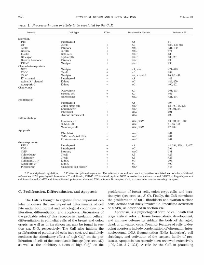
C. Proliferation, Differentiation, and Apoptosis
The CaR is thought to regulate three important cel-lular processes that are important determinants of cellfate under both normal and pathological conditions: pro-liferation, differentiation, and apoptosis. Discussions ofthe probable roles of this receptor in regulating cellulardifferentiation in epithelial cells of the breast and coloncrypts, as well as in keratinocytes, may be found in sec-tion XIII, E–G, respectively. The CaR also inhibits theproliferation of parathyroid cells (see sect. XA) and likelymediates the stimulatory effect of high Cao
21 on the pro-liferation of cells of the osteoblastic lineage (see sect. XD)as well as the inhibitory actions of high Cao
21 on the
proliferation of breast cells, colon crypt cells, and kera-tinocytes (see sect. XIII, E–G). Finally, the CaR stimulatesthe proliferation of rat-1 fibroblasts and ovarian surfacecells, actions that likely involve CaR-mediated activationof MAPK, as described in section VIIB.
Apoptosis is a physiological form of cell death thatplays critical roles in tissue homeostasis, development,and immune defense by ridding the body of damaged,dead, or unwanted cells. Common features of cells under-going apoptosis include condensation of chromatin, inter-nucleosomal DNA fragmentation (DNA laddering), cellshrinkage, and activation of the caspase family of pro-teases. Apoptosis has recently been reviewed extensively(190, 210, 217, 321). A role for the CaR in protecting
TABLE 1. Processes known or likely to be regulated by the CaR
Process Cell Type Effect Discussed in Section Reference No.
SecretionPTH Parathyroid 2 XA 61CT C cell 1 XB 288, 402, 481ACTH Pituitary 1 XIIIC 131, 139Gastrin G cells 1 XIIIG 374Insulin Beta cells 1 XIIIH 236Glucagon Alpha cells 2 XIIIH 130, 261Growth hormone Pituitary 1 XIIIC 390PTHrP Multiple 1 XIIIE 399
Channels/transportersNCC Multiple 1 XA, XIIIA 471–473VDCC C cell 1 XB 288CAKC Multiple 1 XIII, A and B 90, 92, 441K1 channel Parathyroid 2 XA 442Apical K1 channel Kidney 2 XC 449, 450Aquaporin-2 Kidney 2 XC 400, 401
ChemotaxisOsteoblasts 1 XD 161, 463Stromal cell 1 XD 462Macrophage 1 XIIID 421, 464
ProliferationParathyroid 2 XA 196Colon crypt cell 2 XIIIF 68, 70, 114, 225Keratinocyte 2 XIIIF 30, 191, 351Fibroblast 1 VIIIB 291Ovarian surface cell 1 VIIIB 290
DifferentiationKeratinocyte 1 VIIIC, XIIIF 30, 191, 351, 435Goblet cell 1 VIIIC 31, 85, 151Mammary cell 1 VIIIC, XIIIE 97, 289
ApoptosisFibroblast 2 VIIID 267CaR-transfected HEK 2 VIIID 267Prostate cancer cell 2 VIIID 267
Gene expressionPTH*† Parathyroid 2 XA 44, 394, 395, 413, 467VDR* Parathyroid 1 XC 394CaR* Pituitary 1 XIIIC 131Calmodulin* C cell 1 XB 425Calcitonin* C cell 1 XB 425CalbindinD28K* Kidney 1 XC 105Aquaporin-2† Kidney 1 XC 400P-cadherin† Squamous cell cancer 1 XIIIF 448
* Transcriptional regulation. † Posttranscriptional regulation. The reference no. column is not exhaustive; see listed sections for additionalreferences. PTH, parathyroid hormone; CT, calcitonin; PTHrP, PTH-related peptide; NCC, nonselective cation channel; VDCC, voltage-dependentcalcium channel; CAKC, calcium-activated potassium channel; VDR, vitamin D receptor; CaR, extracellular calcium-sensing receptor.
258 EDWARD M. BROWN AND R. JOHN MACLEOD Volume 81
against apoptosis was found using AT-3 rat prostate car-cinoma cells, c-myc-overexpressing rat 1A fibroblasts,and CaR-transfected HEK cells (267). Both AT-3 cells andrat 1A fibroblasts express the CaR as assessed by immu-nocytochemistry and Northern analysis (267). Further-more, raising the level of Cao
21 to 5–10 mM reduced by50% or more the extent of apoptosis in AT-3 cells or rat 1Afibroblasts that was induced by infection with Sindbisvirus or c-myc overexpression/serum deprivation, respec-tively, as assessed by determining DNA laddering andquantitating cell viability (267). As additional evidencethat this action was mediated by the CaR, raising the levelof Cao
21 substantially reduced the extent of apoptosisinduced by Sindbis virus in CaR-transfected HEK293 cellsbut not in nontransfected HEK293 cells (267). Further-more, elevating Cao
21 had no effect on viral replication ineither cell type. Taken together, these findings suggestedthat the activated CaR protects these three cell types fromapoptosis induced by various agents.
Of interest, the structurally and functionally relatedmGluRs can protect neurons against apoptosis inducedby either oxygen-glucose deprivation (65) or addition ofthe neurotoxic b-amyloid peptides (111). In addition,5-oxo-eicosatetraenoic acid, which interacts with a spe-cific GPCR on its target cells (328, 355), blocks apoptosistriggered by inhibition of 5-lipoxygenase in human pros-tate cancer cells (164a). Thus a protective effect againstapoptosis may be shared by many GPCRs, potentially bystimulating Ras, which then exerts an antiapoptotic effecteither by stimulating PKB/Akt via PI 3-kinase or throughactivation of NFkB (128). However, it is currently un-known whether the antiapoptotic effects of activating theCaR result from activation of these pathways.
D. Gene Expression
High Cao21 modulates the expression of several genes
inhibiting the PTH gene in the parathyroid gland (44, 394,395, 413, 467) and increasing the expression of calbindinD28K in rat kidney (105) and VDR gene in the chick para-thyroid (394). More recently, Suzuki et al. (425) haveshown that high Cao
21 increases the promoter activitiesand mRNA levels of the CaR, calmodulin, and calcitoningenes in C cells, while Emanuel et al. (131) have demon-strated a high Cao
21-induced increase in the level of themRNA for the CaR in AtT-20 cells. In some cases, vitaminD and Cao
21 exert synergistic effects in inhibiting [e.g., thePTH gene in the parathyroid gland (394, 467)] or increas-ing gene expression [viz., of calbindin D28K in rat kidney(105) or the VDR in chick parathyroid (394)]. Because theCaR is expressed in all of the cell types noted above, it isa strong candidate for mediating these various actions ofCao
21 on gene expression, although its mediatory role hasnot been formally proven to date.
There are at least two putative molecular mecha-nisms that underlie these high Cao
21-elicited, presumablyCaR-mediated actions on gene expression. 1) In C cellsand probably in several other cell types, the TTF-1 tran-scription factor is activated by the CaR through a mech-anism that likely involves an increase in Cai
21 (425). 2) Inparathyroid cells and other cells in which gene transcrip-tion is regulated by Cao
21, it has been suggested that theredox factor, ref-1, serves as a transcription factor thatnegatively regulates the PTH gene (331, 332). Furtherstudies are needed, however, utilizing the cloned CaR todocument directly that the effect of this protein on PTHgene transcription is mediated by the CaR and to investi-gate the underlying mechanism(s) of action.
In addition to regulating the levels of the mRNAs forvarious genes, high Cao
21 and the CaR can also likelymodulate the activities of proteins in a posttranscriptionalor posttranslational manner. For instance, induction ofhypercalcemia in rats reduces the level of the aquaporin-2water channel protein, without altering the expression ofits RNA (400). In addition, in a CaR-expressing squamouscancer cell line, elevating Cao
21 stimulates the transloca-tion of the adhesion molecule P-cadherin to the plasmamembrane and increases the total amount of cellularP-cadherin protein without affecting its mRNA level (448).The latter effect was blocked by an inhibitor of tyrosinekinase. Therefore, although only limited data are availablerelated to the possible actions of the CaR on the expres-sion of the mRNAs and/or proteins for various genes,including the CaR gene itself, available data suggest that itexerts several such actions, in addition to its well-knowneffects on short-term parameters of cellular function,such as modulating secretion, ion channel activity, andion transport.
IX. OTHER POTENTIAL EXTRACELLULAR
CALCIUM-SENSING RECEPTOR AGONISTS
AND MODULATORS AND THE ROLE OF
THE EXTRACELLULAR CALCIUM-SENSING
RECEPTOR AS AN INTEGRATOR
OF DIVERSE PHYSIOLOGICAL SIGNALS
A. Mgo21
The effects of elevated levels of Mgo21 have long been
known to mimic those of Cao21 in certain cell types. For
instance, raising Mgo21 inhibits PTH secretion from para-
thyroid cells (60, 171), reduces agonist-stimulated cAMPaccumulation in the parathyroid (50) and kidney (284),and diminishes renal tubular reabsorption of Mg21 (aswell as Ca21) (358, 359, 361). The cloning of the CaR madeit possible to demonstrate directly that not only Cao
21 butalso Mgo
21 can serve as a CaR agonist (58, 71, 81, 126, 393).Generally, as observed in bovine parathyroid cells, Mgo
21
January 2001 Cao21 SENSING AND Cao
21 SIGNALING 259
is about two- to threefold less potent than Cao21 as a CaR
agonist in activating PLC (81), producing transient in-creases in Cai
21 (71), stimulating the activity of PLA2
(393), and inhibiting cAMP accumulation (81). In addition,Mgo
21 acts as a partial agonist for many of these actions,producing maximal effects that are less than those elic-ited by Cao
21 (393).It is not currently clear whether the differences be-
tween the actions of Cao21 and Mgo
21 on the CaR areentirely accounted for by intrinsic differences in theircapacities to interact with the receptor and/or other con-tributory factors. For instance, in the absence of a reliablebinding assay for assessing how various agonists interactwith the CaR, it has been necessary to infer the interac-tion of these agents with the receptor indirectly, throughchanges in downstream biological responses (e.g., variousphospholipases and/or adenylate cyclase). Moreover,Mgo
21 could exert additional actions on CaR-expressingcells that interfere with its CaR-mediated actions, such asblocking Ca21 influx pathways, thereby reducing the sus-tained phase of PLC activation that is dependent on up-take of Cao
21. The latter actions could potentially account,at least in part, for some currently unexplained differ-ences in the potencies of Cao
21 and Mgo21 in eliciting
CaR-mediated biological responses in various cells. Forinstance, raising Mgo
21 clearly exerts Cao21-like actions on
the parathyroid in vitro (60, 171, 430) and in vivo (432),but it has little, if any, effect on calcitonin secretion invitro in some systems (288), perhaps because Mgo
21
blocks key Ca21-permeable channels.Because serum levels of Mgo
21 are, if anything,slightly lower than those for Cao
21, can Mgo21 serve as a
physiologically relevant CaR agonist in vivo? Genetic dis-eases of the CaR suggest that it does, in fact, function asa Mgo
21 sensor in vivo (49, 420); that is, individuals withdisorders with inactivating mutations of the CaR tend tohave high-normal or mildly elevated levels of Mgo
21 (281),whereas those with activating mutations can have mildhypomagnesemia (344). Indeed, there is a positive corre-lation between serum Mg21 and Ca21 in persons withFHH, such that those families with more severe hypercal-cemia tend to have higher levels of serum Mgo
21 (281). Incontrast, there is an inverse correlation between serumMg21 and Ca21 in primary hyperparathyroidism (281),perhaps because hypercalcemia tends to promote urinarymagnesium excretion (358). A particularly striking exam-ple of an individual with “resetting” of both Cao
21 andMgo
21 was the homozygous offspring of related parentswith an inactivating mutation of the CaR who was onlyidentified serendipitously at the age of 35 years (2). Bothserum Mgo
21 and Cao21 were elevated to levels ;50% above
normal, in association with a concentration of serum PTHthat was at the upper limit of normal and normal renalfunction (2).
There may be several ways in which Mgo21 acts via
the CaR to control its own homeostasis in vivo. All of thepolycationic agonists (e.g., di- and trivalent cations andpolycations such as neomycin) potentiate one another’sactions on the CaR (60, 393). Therefore, an increase inMgo
21 may activate the CaR by, in effect, sensitizing it toCao
21. Furthermore, there may be specific microenviron-ments where Mgo
21 differs from its level in blood. A goodexample is the level of Mgo
21 within the renal tubule.Because proximal tubular reabsorption of Mg21 is lessthan that for Na1, Cl2, Ca21, and water, the level of Mgo
21
rises progressively along the nephron and is 1.6- to 1.8-fold higher in the thick ascending limb than in the initialglomerular filtrate (123). This increase in Mgo
21 within thetubular fluid is probably sufficient to activate CaRs in theCTAL that are thought to regulate the reabsorption ofboth Ca21 and Mg21 (181).
In addition to the inhibitory effect of raising Mgo21 on
PTH secretion, lowering Mgo21 also reduces PTH secretion
(6, 7) and, to a lesser extent, the actions of PTH on itstarget tissues in kidney and bone (216). The effects of lowlevels of Mgo
21 on the mineral ion homeostatic system,however, do not appear to involve the CaR, and themolecular mechanism(s) underlying them is poorly un-derstood.
B. Spermine
Spermine is an effective CaR agonist at ;100 mM to1 mM (367). Like di- and trivalent cations, it activates PLC,thereby promoting transient increases in Cai
21 owing tomobilization of intracellular calcium stores in CaR-trans-fected HEK293 cells and inhibits PTH release from bovineparathyroid cells (367). As with other polycations, such asthe aminoglycoside antibiotics (237), the number ofcharges on the polyamines is an important determinant oftheir potency as CaR agonists. Their order of potency(spermine . spermidine . putrescine) reflects the factthat these three polyamines contain 4, 3, and 2 free aminogroups, respectively (367). Although the concentrationsof spermine needed to activate the CaR are relatively high(;millimolar) in the presence of low levels of Cao
21 (e.g.,0.5 mM), when Cao
21 is raised to levels approaching thosefound in vivo (;1.5 mM), concentrations of spermine inthe range of 100 mM modulate the CaR’s activity, becauseof the self-potentiating actions of polycationic CaR ago-nists (see above). The levels of spermine present in vivoare on the order of 100 mM, and in specific microenviron-ments, such as in the gastrointestinal tract (where bacte-ria produce spermine) and in the brain (where sperminecan be cosecreted with neurotransmitters), concentra-tions of spermine in extracellular fluids can approach 1mM (367). Therefore, it is entirely possible that the CaRserves as a physiologically relevant “receptor” for sperm-ine in vivo under certain circumstances, although this
260 EDWARD M. BROWN AND R. JOHN MACLEOD Volume 81
possibility has not been formally tested. There are a num-ber of other highly cationic substances present in vivo,such as protamine, which could conceivably also act onthe CaR in a similar manner (59).
C. Amyloid b-Peptides
Vassilev et al. (470) have shown that amyloid b-pep-tides (Ab) can serve as CaR agonists in vitro (470). Mi-cromolar concentrations of Ab-(1O40) potently activatenonselective cation channels (NCC) in neurons from wild-type mice but not in those from mice with knockout of theCaR, as well as in CaR-transfected but not in nontrans-fected HEK293 cells (470). These actions could poten-tially be mediated by fibrillar forms of Ab, since fibrillardeposits of these peptides in the brains of persons suffer-ing from Alzheimer’s disease are stained by anionic dyes,such as Congo red. These dyes have a spacing of negativecharges approximating that present in spermine (e.g., aknown polycationic CaR agonist) for its positive charges.Moreover, it is possible that Ab-fibrils deposited in brainregions involved in the pathology of Alzheimer’s disease,where the CaR is expressed at substantial levels, such ashippocampus, could modulate the activity of these CaRs,contributing to the pathophysiology of this disorder inways that are currently not understood.
D. Ionic Strength
Quinn et al. (366) have shown that alterations in ionicstrength produce parallel changes in the CaR’s EC50 forhigh Cao
21 and other CaR agonist-evoked increases inCai
21 (366); that is, an increase in ionic strength reducesthe receptor’s sensitivity to elevations in Cao
21 and viceversa. Similar observations were made independent of thenature of the monovalent cation (e.g., sodium or choline)or anion (chloride or iodide) employed to modify ionicstrength. Moreover, changes in osmolality per se (i.e.,obtained by substituting sodium chloride with sucrose)had no effect on Cao
21 sensing by the CaR, indicating thationic strength was the responsible factor (366). Similareffects were noted with the use of Mgo
21 or spermine asthe CaR agonist, indicating that the observed effect ofionic strength was not agonist specific.
Changes in ionic strength sufficient to modify theCaR’s EC50 to the point where the level of Cao
21 would beexpected to become overtly abnormal are seldom encoun-tered in vivo except in severe illness. Nevertheless, sub-stantial changes in ionic strength can take place in spe-cific microenvironment, even in normal physiologicalstates. For instance, the concentration of sodium chloride(and, therefore, ionic strength) in the urine can vary from;50 to 300 mM. Therefore, the CaR on the apical mem-brane of the IMCD could experience alterations in ionic
strength more than sufficient to modify substantially itsEC50 for Cao
21 and other CaR agonists. Comparablechanges in ionic strength might be encountered by CaR-expressing cells exposed to the contents of the gastroin-testinal tract, particularly in its more proximal portions(e.g., stomach and upper small intestine). Finally, epithe-lia that transport electrolytes and water at different ratescan likely generate sufficiently large changes in ionicstrength to modify the CaR’s function. In the thick ascend-ing limb (427) of the loop of Henle of the kidney, forinstance, sodium chloride is reabsorbed in excess of wa-ter, because the epithelial cells of this portion of thenephron exhibit a very low permeability to water. There-fore, CaRs at the basolateral aspect of these epithelialcells could encounter elevated levels of ionic strengthrelative to those in the initial filtered urine that might altertheir Cao
21-sensing capacity. To date, however, there havebeen no studies that have directly documented the ex-pected alteration of Cao
21 sensing in vivo in a cell typeendogenously expressing the CaR that has been exposedto a physiologically relevant change in ionic strength.
The effects of ionic strength on the sensing by theCaR of its various polycationic agonists raise the possi-bility of an “electromotive” mechanism of activation ofthe receptor by these agents (366); that is, polycationsmight screen charges on the receptor, which then changesthe conformation of the ECD or other regions of thereceptor from its inactive to its active state (366). Such amechanism could perhaps explain why such a wide vari-ety of polycations, ranging from divalent and trivalentcations to small organic polycations, e.g., neomycin (55,383), and even substantially larger polycationic proteins,including protamine (59), can all activate the CaR. More-over, in general, the greater the total number of its posi-tive charges, the greater the potency of a given moleculeas a CaR agonist (preparations of polyarginine with achain length averaging 100 residues act at nM levels).Nevertheless, there must be some steric and/or otherfactors in addition to just the total number of charges thatcontribute to activation of the receptor by polycationicagonists. For example, while the trivalent lanthanidesexhibit micromolar potencies as CaR agonists (57), gal-lium (Ga31) is only a weak activator of the receptor inparathyroid cells (382) and in CaR-transfected HEK293 (S.Quinn and E. M. Brown, unpublished data). In addition,there are differences in potency among the aminoglyco-sides that are not explained by differences in the numberof charges alone (237).
E. Amino Acids
Conigrave et al. (109) have recently shown that avariety of amino acids can serve as positive modulators ofthe CaR, potentiating the actions of the receptor’s poly-
January 2001 Cao21 SENSING AND Cao
21 SIGNALING 261
cationic activators in their presence (e.g., of $1 mMCao
21) but not in their absence. This action is stereoselec-tive for most amino acids, with L-amino acids being sev-eralfold more potent than D-amino acids. Although indi-vidual amino acids exhibit relatively low potencies foractivating the CaR, doing so in the millimolar range, amixture of amino acids emulating that present in fastingpersons has a substantial impact on the receptor’s sensi-tivity to its polycationic agonists, decreasing the EC50 forCao
21 by 20–40% (109). Furthermore, the order of potencyfor the effects of amino acids on the CaR, with aromaticamino acids being most potent, is highly reminiscent ofthat for the known effects of various amino acids instimulating the secretion of gastrin or gastric acid (109). Itis possible, therefore, that the CaR mediates these latteractions and functions in vivo more generally as a “nutrientreceptor” than as a sensor responding solely to mineralions (and other polycations).
Indeed, it is quite possible that the CaR mediatesknown effects of changes in protein intake on mineral ionmetabolism. For instance, high protein intake substan-tially increases urinary calcium excretion (69), whereaslow protein intake has recently been shown to producenearly a doubling of serum PTH levels in normal youngwomen (241). Thus the recognition that the CaR respondsto amino acids as well as to mineral ions may provideevidence for a fundamental role of this receptor in inte-grating protein and mineral ion metabolism (109).
F. The CaR as an Integrator of Diverse
Physiological Signals
The foregoing discussion emphasizes that the CaR iscapable of responding to several different agonists ormodulators that are present in vivo and whose concen-trations are or could potentially be within a range capableof modulating the receptor’s activity, including Cao
21,Mgo
21, spermine, Ab, changes in ionic strength, and aminoacids. It is an oversimplification, therefore, to think of theCaR as simply a Cao
21 sensor. Depending on the particularmicroenvironment within which it resides, the CaR maybe exposed to sufficiently high levels of CaR agonistsand/or modulators other than Cao
21 to substantially mod-ify its activity. Because polycationic CaR agonists poten-tiate one another’s actions on the receptor, a relativelysmall change in the concentration of one agonist canmarkedly change the apparent affinity of the receptor foranother (60, 367, 393). Much additional work is required,however, to characterize various microenvironments interms of their content(s) of CaR agonists or modulatorsother than Cao
21 to assess more fully their contributions toCaR-mediated biological responses. Nevertheless, it ispossible that the CaR integrates multiple physiologicalsignals, thereby enabling it to respond in an appropriate
manner to the requirements of a specific tissue or micro-environment (see also sects. X, XI, and XIII).
X. THE EXTRACELLULAR CALCIUM-SENSING
RECEPTOR’S CELLULAR DISTRIBUTION AND
FUNCTIONS IN TISSUES INVOLVED IN
MINERAL ION HOMEOSTASIS
A. Parathyroid
The parathyroid glands of humans (162, 246), rats(13), mice (196), rabbits (71), and chickens (126) expressabundant CaR mRNA and protein, as assessed usingNorthern analysis and immunohistochemistry and West-ern analysis, respectively. Recent studies of inherited dis-eases of Cao
21 homeostasis caused by inactivating muta-tions in the CaR gene and of mice with targeted disruptionof this gene strongly support the receptor’s central role inmediating Cao
21-regulated PTH secretion. Humans het-erozygous for inactivating mutations as well as mice het-erozygous for knockout of the CaR show modest (10–20%) rightward shifts in the “set point” of the parathyroidgland for Cao
21 (the level of Cao21 half-maximally inhibiting
PTH secretion). In humans homozygous for inactivatingCaR mutations, who suffer from NSHPT, as well as inmice homozygous for knockout of the receptor, there ismuch more severe “resistance” of the parathyroid glandsto Cao
21. In both instances, there is severe hypercalcemia(with elevations in Cao
21 that are 50% or more above theupper limit of normal) as well as markedly elevated levelsof serum PTH. Furthermore, persons harboring activatingmutations in the CaR, which render the parathyroid celloverly sensitive to the suppressive action of elevated Cao
21
on PTH secretion, exhibit hypocalcemia accompanied byinappropriately normal or even low levels of serum PTH(that is, a stimulus which normally increases PTH secre-tion fails to do so). Thus these experiments-in-natureafford compelling evidence for the central, nonredundantrole of the CaR as a mediator of high Cao
21-inhibited PTHrelease.
The intracellular mechanism(s) through which theCaR inhibits PTH secretion, however, remains an impor-tant unresolved issue. As outlined above, the CaR modu-lates diverse intracellular signaling pathways, activatingPLC, PLA2, and PLD as well as the MAPK pathway andinhibiting adenylate cyclase (see sect. VII). Products of thestimulation of PLC (i.e., inositol trisphosphate, whichcauses a transient increase in Cai
21, and/or diacylglycer-ol), PLA2 (e.g., yielding arachidonic acid and/or the prod-ucts of its further metabolism), or PLD (i.e., generatingphosphatidic acid), the high Cao
21-elicited, sustained in-crease in Cai
21 or decrease in cellular cAMP and/or otherfactors have all been suggested as central mediators ofhigh Cao
21-inhibited PTH secretion (for review, see Ref.
262 EDWARD M. BROWN AND R. JOHN MACLEOD Volume 81
52). In virtually all instances, however, high Cao21-evoked
alterations in the relevant mediators can be dissociatedfrom concomitant changes in PTH secretion under appro-priate conditions. Indeed, even the crucial step(s) alongthe pathway for Cao
21-regulated PTH secretion that iscontrolled by the CaR remain largely unknown (e.g., fromthe budding of secretory granules at the Golgi apparatusto their final exocytosis at the cell surface). Thus, despiterapid recent advances in the elucidation of a number ofthe molecular components of the exocytotic apparatus inother cell types and the cloning of the CaR, much remainsto be learned about how the parathyroid cell responds viathe CaR to Cao
21 in a fashion directly opposite to thatexhibited by most other secretory cells.
The CaR likely also exerts a tonic suppressive actionon parathyroid cellular proliferation, since humans withNSHPT who are homozygous for inactivating mutationsof the CaR (49) and mice homozygous for knockout ofthis receptor (196) exhibit marked parathyroid cellularhyperplasia (in contrast, no such mutations in the CaRgene have been found in pathological parathyroid glandsfrom patients with primary or secondary hyperparathy-roidism). Furthermore, treating rats with renal impair-ment owing to subtotal nephrectomy with the calcimi-metic CaR activator R-568 prevents the parathyroidhyperplasia that otherwise develops in the setting of renalinsufficiency (445). Although the receptor may inhibitparathyroid cellular proliferation directly, indirect effectsare also possible. For example, severe hypercalcemia inthe setting of humans with NSHPT or mice homozygousfor knockout of the CaR might indirectly stimulate para-thyroid cellular proliferation by reducing circulating lev-els of 1,25(OH)2D (455), since the latter inhibits the pro-liferation of parathyroid cells, at least in someexperimental systems (254). The intracellular mecha-nism(s) through which the CaR inhibits parathyroid cel-lular proliferation remains to be identified.
Another parameter of parathyroid function that isprobably controlled by the CaR is the level of expressionof the PTH gene. Several studies have established thathigh Cao
21 reduces the level of the mRNA for preproPTH(44, 394, 395, 413, 467). Garrett et al. (156) showed inpreliminary studies that NPS R-568 decreases the level ofPTH mRNA in bovine parathyroid cells, thereby suggest-ing that the CaR mediates this action of high Cao
21 on thisparameter (156). High Cao
21 exerts numerous additionaleffects on parathyroid cells, such as modulating K1 chan-nels (108, 231, 270), stimulating the activity of the hexosemonophosphate shunt (303), and increasing cellular res-piration (173; for review, see Ref. 52). Additional studiesare needed to determine which of these various actions ofCao
21 are mediated by the CaR and to identify the signaltransduction pathways that are involved.
B. C Cells
Studies on the regulation of calcitonin secretion byhigh Cao
21 initially stressed that this process differed in afundamental way from that through which Cao
21 regulatesPTH release (132, 133, 147, 402). CT secretion, in contrastto PTH release, is stimulated by raising Cao
21 above itsnormal levels in the blood in association with increases inCai
21. The relationship between increases in Cai21 and
activation of CT secretion is similar to the more classical,positive relationship between Ca21 and exocytosis that isobserved in most other secretory cells (52, 127). Further-more, influx of Cao
21 via voltage-dependent calcium chan-nels is the predominant contributor to high Cao
21-evokedincreases in Cai
21 in C cells (133, 147, 305, 402). In para-thyroid cells, in contrast, mobilization of Cai
21 is a majorfactor contributing to high Cao
21-evoked increases inCai
21, particularly immediately after raising Cao21 (315,
319). The patterns of the high Cao21-elicited increases in
Cai21 are also different in these two cell types. Single C
cells often show oscillations in Cai21 (132, 133), which are
either much less frequent (296) or are not observed insingle parathyroid cells (133). Finally, most of the highCao
21-induced influx of Cao21 into C cells occurs through
voltage-sensitive Ca21 channels, as noted above, whileconsiderable uncertainty remains concerning the chan-nels through which uptake of Cao
21 takes place in para-thyroid cells. The latter are most likely NCCs permeableto Ca21 (52, 80, 305, 352). Thus it was generally assumedthat the mechanisms mediating Cao
21 sensing in parathy-roid and C cells are distinctly different, with the latterlikely involving some form of voltage-sensitive Ca21 chan-nel.
Studies utilizing Northern analysis, in situ hybridiza-tion, RT-PCR with sequencing, and/or immunohistochem-istry with anti-CaR antisera have convincingly demon-strated, however, that C cells contain the same CaR thatis present in parathyroid and kidney cells (146, 158).Nevertheless, not all CaR agonists that modulate PTHsecretion and other parameters of parathyroid functionelicit CT secretion from C cells. For instance, while rais-ing Mgo
21 inhibits PTH secretion, Mgo21 has little or no
effect on CT secretion from sheep parafollicular cells(288). Recent studies using sheep C cells have suggestedthe following model for how the CaR activates voltage-sensitive Ca21 channels and CT secretion: high Cao
21-evoked, CaR-mediated stimulation of PC-PLC provides asource of diacylglycerol for the PKC-induced activation ofa NCC. The latter permits entry of Na1 and Ca21 into thecells, which produces cellular depolarization and subse-quent activation of voltage-gated, principally L-type Ca21
channels (288). The resultant rise in Cai21 stimulates se-
cretion of CT. The CaR likely also regulates other aspectsof C-cell function, including activating a pertussis toxin-sensitive, PKC-mediated acidification of serotonin-con-
January 2001 Cao21 SENSING AND Cao
21 SIGNALING 263
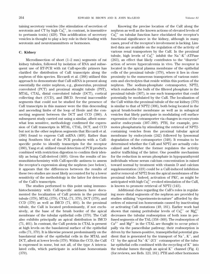
taining secretory vesicles (the stimulation of secretion ofserotonin and CT by high Cao
21, in contrast, is insensitiveto pertussis toxin) (429). This acidification of secretoryvesicles is thought to play a key role in their loading withserotonin and neurotransmitters or hormones.
C. Kidney
Microdissection of short (1–2 mm) segments of ratkidney tubules, followed by isolation of RNA and subse-quent use of RT-PCR with rat CaR-specific primers hasclarified the distribution of CaR transcripts along thenephron of this species. Riccardi et al. (388) utilized thisapproach to demonstrate that CaR mRNA is present alongessentially the entire nephron, e.g., glomerulus, proximalconvoluted (PCT) and proximal straight tubule (PST),MTAL, CTAL, distal convoluted tubule (DCT), corticalcollecting duct (CCD), and IMCD. Several inaccessiblesegments that could not be studied for the presence ofCaR transcripts in this manner were the thin descendingand ascending limbs of the loop of Henle and the con-necting segment between the DCT and CCD (380). Asubsequent study carried out using a similar, albeit some-what less sensitive, methodology confirmed the expres-sion of CaR transcripts in MTAL, CTAL, DCT, and CCDbut not in the other nephron segments that Riccardi et al.(380) found to express CaR mRNA (469). Rather thanusing Southern blot of the PCR products with a CaR-specific probe to identify transcripts for the receptor(380), Yang et al. utilized visual detection of PCR productscombined with restriction digestion to confirm their iden-tify as being CaR-derived (469). Given the results of im-munohistochemistry with CaR-specific antisera to assessthe receptor’s expression along the nephron (see below),it appears that the differences between the results ofthese two studies are most likely accounted for by a lowersensitivity of the methodology in the latter for detectionof the CaR’s transcripts.
The studies performed to this point using immuno-histochemistry with CaR-specific antisera have docu-mented the localization of CaR protein in the proximaltubule (379), MTAL (379), CTAL (71, 379), DCT (379), andCCD (379) as well as IMCD (71, 401). In the proximaltubule, the CaR is located predominantly, if not exclu-sively, at the base of the brush border of the apicalmembrane of the tubular epithelial cells (379). The CaRalso exhibits principally an apical distribution in IMCD(71, 401). In contrast, the CaR protein in CTAL is presentat high levels on the basolateral surface of the epithelialcells (71, 379). It is likewise present predominantly on thebasolateral side of the epithelial cells in the MTAL andDCT, albeit at lower levels (379). Within the CCD, the CaRis expressed in some, but not all, of the type A interca-lated cells, which participate in acid-base homeostasis(379).
Knowing the precise location of the CaR along thenephron as well as the known actions of elevated levels ofCao
21 on tubular function have elucidated the receptor’sfunctional significance in the kidney, although in manycases, proof of the receptor’s involvement is lacking. Lim-ited data are available on the regulation of the activity ofvarious renal transporters by the CaR. In the proximaltubule, high levels of Cao
21 inhibit the Na1-K1-ATPase(263), an effect that likely contributes to the “diuretic”action of severe hypercalcemia in vivo. The receptor islocated in the apical plasma membrane of the epithelialcells of the proximal tubule (379), where it lies in closeproximity to the numerous transporters of various nutri-ents and electrolytes that reside within this portion of thenephron. The sodium-phosphate cotransporter, NPT2,which reabsorbs the bulk of the filtered phosphate in theproximal tubule (307), is one such transporter that couldpotentially be modulated by the CaR. The localization ofthe CaR within the proximal tubule of the rat kidney (379)is similar to that of NPT2 (308), both being located in theapical brush-border membrane as well as in subapicalvesicles that likely participate in modulating cell surfaceexpression of the cotransporter via changes in exocytosisand/or endocytosis (307–309). PTH, for example, pro-motes phosphaturia by stimulating the retrieval of NPT2-containing vesicles from the proximal tubular apicalmembrane by endocytosis (242) followed by lysosomaldegradation of the cotransporter (348). It remains to bedetermined whether the CaR and NPT2 are actually colo-calized and whether the former regulates the activityand/or trafficking of the latter. One possible mechanismfor the reduction in serum phosphate in hypoparathyroidindividuals whose serum calcium concentration is raisedtoward normal by treatment with vitamin D and calciumsupplementation (419) could be a CaR-induced inhibitionand/or removal of NPT2 from the apical membranes of theproximal tubule. Indeed, activation of PKC, as might beanticipated with high Cao
21-evoked stimulation of the CaR,is known to promote retrieval of NPT2 (143).
Additional clues regarding the CaR’s roles in regulat-ing more distal segments of the nephron are provided bystudies utilizing “experiments-in-nature” afforded by dis-orders of mineral ion homeostasis caused by inactivatingor activating CaR mutations (49, 181). Earlier work hadshown that raising peritubular levels of Cao
21 or Mgo21
decreases the tubular reabsorption of both ions in per-fused segments of the TAL (358–360). The reabsorption ofCa21 and Mg21 in the CTAL are thought to occur princi-pally via the paracellular pathway; their reabsorption isdriven by the lumen-positive, transepithelial potential gra-dient that is generated by the transport of Na1, K1, andCl2 by the apical Na1-K1-2Cl2 cotransporter of the tubu-lar epithelial cells combined with the recycling of K1 intothe tubular lumen through an apical K1 channel (Fig. 4)(for reviews, see Refs. 123, 181). PTH and other hormones
264 EDWARD M. BROWN AND R. JOHN MACLEOD Volume 81
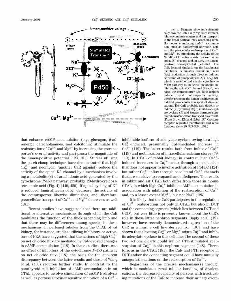
that enhance cAMP accumulation (e.g., glucagon, b-ad-renergic catecholamines, and calcitonin) stimulate thereabsorption of Ca21 and Mg21 by increasing the cotrans-porter’s overall activity and pari passu the magnitude ofthe lumen-positive potential (123, 181). Studies utilizingthe patch-clamp technique have demonstrated that highCao
21 and neomycin (another CaR agonist) reduce theactivity of the apical K1 channel by a mechanism involv-ing a metabolite(s) of arachidonic acid generated by thecytochrome P-450 pathway, probably 20-hydroxyeicosa-tetraenoic acid (Fig. 4) (449, 450). If apical cycling of K1
is reduced, luminal levels of K1 decrease, the activity ofthe cotransporter likewise diminishes, and, therefore,paracellular transport of Ca21 and Mg21 decreases as well(181).
Recent studies have suggested that there are addi-tional or alternative mechanisms through which the CaRmodulates the function of the thick ascending limb andthat there may be differences among species in thesemechanisms. In perfused tubules from the CTAL of ratkidney, for instance, studies utilizing inhibitors or activa-tors of PKA have suggested that the actions of high Cao
21
on net chloride flux are mediated by CaR-evoked changesin cAMP accumulation (118). In these studies, there wasno effect of inhibitors of the cytochrome P-450 pathwayon net chloride flux (118); the basis for the apparentdiscrepancy between the latter results and those of Wanget al. (450) requires further study. In contrast to theparathyroid cell, inhibition of cAMP accumulation in ratCTAL appears to involve stimulation of cAMP hydrolysisas well as pertussis toxin-insensitive inhibition of a Ca21-
inhibitable isoform of adenylate cyclase owing to a highCao
21-induced, presumably CaR-mediated increase inCai
21 (119). The latter results both from influx of Cao21
(119) and mobilization of intracellular calcium stores (79,119). In CTAL of rabbit kidney, in contrast, high Cao
21-induced increases in Cai
21 occur through a mechanismthat does not appear to involve activation of PI-PLC (124)but rather Cao
21 influx through basolateral Ca21 channelsthat are sensitive to verapamil and nifedipene. The resultsin rabbit and rat CTAL both differ from those in mouseCTAL, in which high Cao
21 inhibits cAMP accumulation inassociation with inhibition of the reabsorption of Ca21
and, to a lesser extent Mg21, but not NaCl (125).It is likely that the CaR participates in the regulation
of Ca21 reabsorption not only in CTAL but also in DCTand the connecting segment (which lies between DCT andCCD), but very little is presently known about the CaR’srole in these latter nephron segments. Bapty et al. (19),however, have recently demonstrated expression of theCaR in a murine cell line derived from DCT and haveshown that elevating Cao
21 or Mgo21 raises Cai
21 and inhib-its adenylate cyclase in this cell line. The second of thesetwo actions clearly could inhibit PTH-stimulated reab-sorption of Cao
21 in this nephron segment (148). There-fore, as in the CTAL (181), the CaR and PTH receptor inDCT and/or the connecting segment could have mutuallyantagonistic actions on the reabsorption of Ca21.
Regardless of the precise mechanism(s) throughwhich it modulates renal tubular handling of divalentcations, the decreased capacity of persons with inactivat-ing mutations of the CaR to increase their urinary excre-
FIG. 4. Diagram showing schemati-cally how the CaR likely regulates intracel-lular second messengers and ion transportin the renal cortical thick ascending limb.Hormones stimulating cAMP accumula-tion, such as parathyroid hormone, acti-vate the paracellular reabsorption of Ca21
and Mg21 by stimulating the activity of theNa1-K1-2Cl2 cotransporter as well as anapical K1 channel and, in turn, the lumen-positive, transepithelial potential. TheCaR, located similarly on the basolateralmembrane, stimulates arachidonic acid(AA) production through direct or indirectactivation of phospholipase A2 (PLA2) (2),which is metabolized via the cytochromeP-450 pathway to an active metabolite in-hibiting the apical K1 channel (4) and, per-haps, the cotransporter (3). Both actionsreduce overall cotransporter activity,thereby reducing the lumen-positive poten-tial and paracellular transport of divalentcations. The CaR probably also directly orindirectly (by raising Cai
21) inhibits adenyl-ate cyclase (1) and causes hormone-stim-ulated divalent cation transport as a result.(From Brown EM and Hebert SC. Calcium-receptor regulated parathyroid and renalfunction. Bone 20: 303–309, 1997.)
January 2001 Cao21 SENSING AND Cao
21 SIGNALING 265
tion of Ca21 in response to hypercalcemia provides indi-rect support for the CaR’s role in this process (11, 49, 117,181). In contrast, individuals with activating mutations ofthe CaR exhibit excessively high levels of urinary Ca21
excretion at any given level of serum Ca21 in the un-treated state, presumably owing to the presence of acti-vated CaRs along in the kidney, especially in CTAL (20,344).
Hypercalcemic patients, particularly those with se-vere hypercalcemia (greater than ;14 mg/dl), not infre-quently have abnormally decreased urinary concentratingcapacity and, occasionally, frank nephrogenic diabetesinsipidus (160, 422). The CaR’s presence in segments ofthe nephron that participate in urinary concentration(379, 401) has afforded novel insights into the likely mech-anism(s) underlying the long-recognized but poorly un-derstood effects of high Cao
21 on this parameter of renalfunction. As noted previously, perfusing isolated ratIMCD tubules with high Cao
21 or neomycin, probably byactivating CaRs present in their apical membrane, revers-ibly inhibits vasopressin-elicited, transepithelial waterflow by ;35–40% (401). The CaR is also present withinthe same apical endosomes that contain the vasopressin-regulated water channel, aquaporin-2. This observationraises the possibility that the CaR reduces vasopressin-stimulated water flow in this nephron segment by stimu-lating the endocytosis and/or inhibiting the exocytosis ofthese endosomes out of or into the apical plasma mem-brane, respectively (401). Furthermore, chronic hypercal-cemia in rats induced by treatment with vitamin D causesreduced expression of the aquaporin-2 water channel
(400), which would further reduce vasopressin-stimulatedwater flow in the terminal collecting duct. High Cao
21-induced, CaR-mediated inhibition of NaCl reabsorption inthe MTAL (449, 450) would also diminish the magnitudeof the medullary countercurrent gradient, which would beexpected to reduce further the maximal urinary concen-trating power of hypercalcemic persons (Fig. 5). Interest-ingly, individuals with inactivating mutations of the CaRare able to concentrate their urine normally despite theirhypercalcemia (282), probably because they are resistantto the usual inhibitory effects of Cao
21 on the urinaryconcentrating mechanism. Conversely, persons with acti-vating mutations of the CaR can develop symptoms ofdiminished urinary concentrating capacity at normal oreven low levels of Cao
21 when treated with vitamin D andcalcium supplementation, presumably because their renalCaRs are overly sensitive to the usual actions of raisingCao
21 on the urinary concentrating system (344). The CaRlikely has additional functional roles in the collecting ductgiven its expression in some type A intercalated cells(379), but additional studies are needed to determinewhether, for example, it directly modulates renal acid-base handling, one of the key functions of this cell type.
What are the physiological implications of the defec-tive renal handling of water in hypercalcemic patients?We previously suggested that this action of high Cao
21
affords a mechanism that integrates the renal handling ofdivalent cations, particularly Ca21 and water, therebypermitting appropriate “trade offs” in how these parame-ters of renal function are regulated under specific physi-ological conditions (181). For example, when a systemic
FIG. 5. Hypothetical mechanismsthat could coordinate systemic calciumand water homeostasis in humans. Seetext for details. The top panel illustratesrenal mechanisms through which theCaR may inhibit maximal urinary con-centrating capacity. TAL, thick ascend-ing limb. (From Brown EM and HebertSC. Novel insights into the physiologyand pathophysiology of Ca21 homeosta-sis from the cloning of an extracellularCa21-sensing receptor. Regulatory Pept
Lett 7: 43–47, 1997.)
266 EDWARD M. BROWN AND R. JOHN MACLEOD Volume 81
calcium load must be disposed of, there is a CaR-medi-ated increase in urinary Ca21 content owing to reducedreabsorption tubular of Ca21 in the CTAL, and perhapsthe DCT. The resultant rise in luminal levels of Cao
21 inIMCD, particularly in a dehydrated individual, might pre-dispose to the formation of Ca21-containing renal stoneswere it not for the concomitant inhibition of maximalurinary concentrating capacity in MTAL and IMCD.
Furthermore, abundant CaRs are present in the sub-fornical organ (SFO) (388), which is an important hypo-thalamic thirst center (415). These CaRs may provide anadditional layer of integration of Cao
21 and water ho-meostasis, as follows: a high Cao
21-induced, CaR-mediatedincrease in thirst with a resultant increase in drinkingcould prevent dehydration that might otherwise be theconsequence of a fixed renal loss of free water because ofconcomitant resistance of the kidney to vasopressin (Fig.5). Finally, prior studies have documented the existenceof a specific “calcium appetite” in rats (433) that couldprovide a mechanism for a physiologically appropriatemodulation of the intake of calcium-containing food dur-ing hypo- and hypercalcemia. We postulate, therefore,that multiple layers of CaR-mediated integration and co-ordination participate in the regulation of water and Ca21
metabolism, serving to optimize the capacity of terrestrialorganisms to adapt to their intermittent access to dietaryCao
21 and water (181). Thus in addition to integratingmultiple physiological signals, as described in section IX,the CaR likely participates in the coordination of severalof the body’s homeostatic systems (e.g., for Na1, Ca21,Mg21, and water).
D. Osteoclasts, Osteoblasts, and Osteocytes
1. Osteoclasts and their precursors
Recent studies have provided increasing evidencethat the CaR is expressed in and could have importantfunctional roles in bone cells and/or their precursors. Forexample, among cells of the osteoclast lineage, monocyte/macrophage-like cells express the CaR (464, 466). TheseCaR-expressing cells may serve as osteoclast precursors,since cells of this lineage are known to form maturemultinucleated osteoclasts through a process of differen-tiation and fusion (278, 419). Indeed, a recent study iden-tified the CaR in putative preosteoclasts in spleen-derivedcultures that form multinucleated, osteoclast-like cells invitro (230). These workers showed that high levels ofCao
21 as well as the calcimimetic CaR activator R-467(albeit at very high concentrations) (320) inhibited theformation of osteoclast-like cells in vitro, suggesting arole for the CaR in regulating osteoclastogenesis. Anotherstudy suggesting a functional role for the CaR in matureosteoclasts demonstrated that polycationic CaR agonists,including extracellular Gd31 and neomycin, activated an a
NCC in rat osteoclasts and mobilized intracellular cal-cium stores (474). Several other studies, as noted previ-ously, have suggested that the pharmacology of the oste-oclast Cao
21-sensing mechanism differs significantly fromthat of the CaR. Further studies are needed to resolve thispoint. A recent study, however, has shown that maturerabbit osteoclasts, in fact, express the CaR as assessed byNorthern blot analysis and RT-PCR (226). Furthermore,raising the level of Cao
21 in the medium or addition of thepolycationic CaR agonists extracellular Gd31 or neomy-cin inhibited bone resorption by these rabbit osteoclastsin vitro (226), providing additional indirect evidence for afunctional role of the CaR. It is important in a study suchas this, however, with a potentially heterogeneous cellpopulation, to utilize approaches that identify CaR tran-scripts or protein in individual cells. Thus the use of insitu hybridization and/or immunohistochemistry in futurestudies would rule out the possibility that CaR transcriptswere actually present in some contaminating cell type.Therefore, while increasing evidence supports the CaR’sexpression and functional relevance in both osteoclastprecursors and mature osteoclasts, additional confirma-tory studies are needed. Moreover, further work is neededto determine whether the CaR can explain the knowneffects of high Cao
21 and other polyvalent cations on os-teoclast function or whether there is, in fact, anotherryanodine receptor-like Cao
21-sensing mechanism ex-pressed in these cells as well (469). Finally, in addition tostudying the putative role of the CaR in regulating pro-cesses such as bone resorption and osteoclastogenesis inosteoclasts and/or their precursors, it will be important todetermine whether it has additional important roles inthese cells, such as modulating apoptosis and/or cytokinesecretion (475).
2. Osteoblasts and their precursors
House et al. (204) showed that alkaline phosphatase-expressing cells derived from bone marrow, potentiallyrepresenting osteoblasts or preosteoblasts, expressed theCaR, raising the possibility that this receptor is expressedin and plays functional roles in cells of the osteoblastlineage. In fact, elevated levels of Cao
21 have several phys-iologically relevant actions on osteoblast-like cells, in-cluding stimulating their proliferation and chemotaxis aswell as modulating several intracellular second messen-ger systems (for more detailed discussions, see sect. IVB
and Ref. 363). What is the evidence that osteoblastsand/or their precursors express the CaR? One group hasconsistently failed to find evidence for expression of theCaR in the osteoblast lineage and, instead, has suggestedthat osteoblasts express a distinct cation-sensing receptorthat responds to extracellular Al31 as one of its agonists[extracellular Al31 is only a weak agonist of the CaR(417)] (362, 364, 365).
January 2001 Cao21 SENSING AND Cao
21 SIGNALING 267
Two other groups, however, have provided evidencethat the CaR is expressed in several osteoblast-like celllines and is a candidate for mediating some or even all ofthe known actions of high Cao
21 on osteoblast function.Yamaguchi and co-workers have shown that CaR proteinand mRNA are expressed in several osteoblast-like cellslines, including murine MC3T3-E1 cells (463) as well asthe human SAOS-2 and the rat UMR-106 cell lines (465).These workers found that high levels of Cao
21 and otherpolycationic CaR agonists stimulate the proliferation andchemotaxis of MC3T3-E1 cells, indicating that the CaRcould potentially mediate these actions of high Cao
21 onthis cell line (463). Kanatani et al. (230) have recentlyconfirmed the expression of CaR mRNA and protein inMC3T3-E1 cells (230). During their proliferative phase inculture, MC3T3-E1 cells resemble preosteoblasts; theysubsequently cease dividing, express alkaline phospha-tase and other markers of mature osteoblasts, and actu-ally form mineralized bone nodules in vitro under appro-priate conditions (463). Yamaguchi et al. (463) found thatthe CaR is expressed throughout the culture period asassessed by Western analysis, suggesting that the CaRmay function in both preosteoblasts and mature osteo-blasts. Finally, Chang et al. (83) have recently shown thatboth CaR mRNA and protein are present in slices ofbovine, murine, and rat bone using in situ hybridizationand immunohistochemistry, respectively.
The murine ST-2 stromal cell line also expresses CaRmRNA and protein, and high Cao
21 stimulates their prolif-eration and chemotaxis (462). Stromal cells can serve asprecursors of osteoblasts and also produce mediators thatimpact on bone turnover by modulating the functions ofosteoblasts and osteoclasts (278). Therefore, the CaR instromal cells could participate in bone turnover eitherdirectly, if these cell differentiate to osteoblasts, or indi-rectly, by influencing this process via one or more medi-ators. In future studies it will be very important for addi-tional laboratories to confirm these findings and to extendthe use of similar approaches to identifying the CaR inbona fide osteoblasts (and, for that matter, osteoclasts) inintact bone. Furthermore, it will be essential to utilizegenetic approaches [e.g., isolating bone cells from micewith knockout of the CaR (196) or utilizing dominantnegative CaR constructs (15, 291)] or pharmacologicalmethods [i.e., specific CaR agonists (320) and/or CaRantagonists (318)] to document that the CaR is not onlyexpressed in bone cells but also mediates some or all ofthe known actions of Cao
21 on these cells.
3. Osteocytes
Not only osteoclasts and osteoblasts, but also osteo-cytes are responsive to changes in the level of Cao
21 (227,228). The latter represent osteoblasts that have completedtheir role in bone formation and have become encased
within the bone substance, where they extend processesinto narrow canaliculi within the bone (212, 227, 370).They still, however, may participate in mineral ion ho-meostasis, perhaps sensing mechanical forces on bone(419). Kamioka et al. (228) showed that raising the level ofCao
21 increases Cai21 in isolated chick osteocytes through
a mechanism that involves mobilization of Ca21 from itsintracellular stores, presumably by some type of Cao
21-sensing mechanism. The pharmacology of the effects ofvarious divalent cations on Cai
21 in osteocytes, especiallythe fact that extracellular Ni21 and extracellular Cd21
evoke similar responses to those of Cao21 (228), suggests
that the Cao21-sensing mechanism in this cell type is more
similar to that of the osteoclast than to the CaR (477, 479).
E. Chondrocytes
Although cartilage-forming cells (chondrocytes) arenot directly involved in systemic mineral ion homeostasis,they play key roles in the formation and growth of theskeleton by providing a cartilaginous model of the futureskeleton that is gradually transformed into bone. Thegrowth plate represents a site where the process of bonyreplacement of the cartilaginous model of the future longbone enables longitudinal growth until the skeleton isfully mature at the end of puberty. The availability of Ca21
is known to be important for ensuring the proper growthand differentiation of cartilage cells and attendant skele-tal growth (208, 209, 378). Furthermore, alterations inCao
21 modulate the differentiation and/or other functionsof chondrocytes (33, 460), which arise from the samemesenchymal stem cell giving to osteoblasts, adipocytes,smooth muscle cells, and fibroblasts (37, 122). For thesereasons, Chang et al. (82) utilized a rat cartilage cell line,RCJ3.1C5.18, to determine whether cells of this lineageexpress the CaR and whether the latter mediates variousactions of Cao
21 on the function of these cells.Raising the level of Cao
21 exerts several actions onthis cell line, including producing dose-dependent reduc-tions in the mRNAs encoding a major proteoglycan incartilage, aggrecan, as well as the a1-chains of types II andX collagen and alkaline phosphatase (82). RCJ3.1C5.18cells expressed CaR transcripts as assessed by in situhybridization and CaR protein as determined by immuno-cytochemistry and Western blot analysis. Furthermore,treatment of these cells for 48–72 h with an antisenseoligonucleotide construct specific for the CaR loweredthe level of the CaR protein substantially and promoted anassociated increase in expression of aggrecan mRNA (82),consistent with a mediatory role of the CaR in regulatingthis gene. These results indicated, therefore, that 1) Cao
21
modulates the expression of several biologically impor-tant genes expressed by this chondrocytic cell line, 2)cells of the cartilage-forming lineage express the CaR, and
268 EDWARD M. BROWN AND R. JOHN MACLEOD Volume 81
3) the receptor mediates some or all of these actions ofCao
21 in this cartilage cell model. Further studies areneeded to assess the CaR’s expression and biologicalrole(s) in cartilage cells in vivo.
Therefore, the CaR is expressed not only in the cellsthat form and resorb bone as part of skeletal remodelingand systemic Cao
21 homeostasis but also in those that areinvolved in the processes of skeletal development andgrowth. Moreover, it regulates the functions of these cellsin ways that appear to be biologically relevant. Additionalstudies are needed, however, using model systems suchas the CaR knockout mice (196) or transgenic mice withknockout or overexpression of the CaR in osteoblasts,osteoclasts, osteocytes, or chondrocytes and/or their pre-cursors to document further the importance of Cao
21 sens-ing by these various cells in vivo. Presumably, it is criticalthat not only the formation and resorption of bone inadult life but also the development and growth of theskeleton earlier in life be precisely coordinated so as tomatch the availability of mineral ions to their dispositionin either the growing or mature but constantly remodelingskeleton. It will be of interest in future studies to deter-mine whether similar mechanisms also exist for sensingand regulating the disposition of skeletal phosphate, theother major component of the mineral phase of bone.
F. Intestine
The major site of Ca21 absorption is the duodenum,although substantial absorption of calcium also takesplace in the proximal colon. The roles of 1,25(OH)2D, theintracellular calcium binding protein calbindin, and thebasolateral calcium pump and sodium/calcium exchangerin intestinal Ca21 absorption have been extensively inves-tigated, although many details of this process remain tobe fully understood (43, 138, 233, 336–338, 453). Calciumions taken up across the apical (e.g., luminal) plasmamembrane of the absorptive cell diffuse down their intra-cellular concentration gradient from the apical to thebasolateral side of the cell using calbindin as a “shuttle,”where they are then pumped out of the cell via the pumpand exchanger. Until recently, a precise molecular char-acterization of the mechanism underlying apical uptake ofcalcium ions by the absorptive cells of the small and largeintestine was lacking. We (345) and others (197, 198) haverecently cloned and characterized calcium channel-liketransporters that likely represent the major mediator(s) ofthe apical uptake of Ca21 in the intestine as well as in theDCT and connecting segment of the kidney (197, 198).
Several cell types within both the small and largeintestines, including the villus cells of the proximal smallintestine (duodenum and ileum) and the surface epithelialcells of the proximal colon, both of which are involved inabsorption of calcium, also express CaR mRNA and pro-
tein (85, 151). Thus the CaR could potentially directly orindirectly participate in mineral ion homeostasis via itsactions on intestinal function, such as modulation of theabsorption and/or secretion of calcium, but only limitedinformation is available in this regard, and no such ac-tions have been described to date. A more detailed dis-cussion of the CaR’s possible roles in regulating functionsof the gastrointestinal tract unrelated to mineral ion ho-meostasis is provided in section XIII.
G. Placenta
During pregnancy, the placenta plays a key role inmineral ion metabolism of the fetus owing to the fact thatall Ca21 must be transported from the maternal to thefetal circulation through the placenta. Most of the fetalskeleton forms in the third trimester, and there is depo-sition of ;30 g of skeletal calcium during this time (385).Cao
21-sensing cells have been shown to be present in theplacenta and could potentially play some role(s) in regu-lating transport of Ca21 between mother and fetus, per-haps by regulating the secretion of PTHrP by placentalcells (188). As with parathyroid chief cells, high levels ofCao
21 raise the level of Cai21 in human placental cytotro-
phoblasts (39, 223). Moreover, there is an inverse relation-ship between Cao
21 and PTHrP secretion in these cells,suggesting similarities in the mechanism(s) underlyingCao
21 sensing by parathyroid cells and cytotrophoblasts(188). The fetal parathyroid gland also secretes both PTHand PTHrP in utero, however, and it has been suggestedthat it may also contribute to regulating Ca21 transferacross the placenta (385).
Recently, Bradbury et al. (40) have demonstratedthat transcripts for the CaR are expressed in cytotropho-blast cells from human term placenta. In addition to tran-scripts that encode the full-length receptor protein, anadditional, alternatively spliced, transcript is expressednot only in cytotrophoblasts but also in human parathy-roid (40). This alternatively spliced RNA lacks exon 3 andencodes a truncated and very likely inactive receptorprotein, since removal of this exon introduces a frameshift, thereby producing a premature stop codon withinthe receptor’s ECD. The full-length CaR is also present incytotrophoblasts, however, which makes it a candidatefor mediating some or all of the known actions of Cao
21 onthis cell type.
A careful study by Kovacs et al. (253), utilizing micewith knockout of the CaR and/or PTHrP and its receptor,has recently provided additional insights into the role ofthe CaR in maternal-fetal calcium metabolism. This workdemonstrated that disruption of the CaR leads to an in-crease in the fetal calcium concentration of both CaR(1/2) (e.g., those heterozygous for CaR knockout) and(2/2) (i.e., homozygous) fetuses, although the level of
January 2001 Cao21 SENSING AND Cao
21 SIGNALING 269
Cao21 in the latter is not significantly higher than that in
the former. Additional biochemical abnormalities in-cluded modest and marked increases in serum PTH andserum 1,25(OH)2D levels in the (1/2) and (2/2) fetuses(indicative of hyperparathyroidism), respectively, re-duced skeletal Ca21 in the (2/2) fetuses accompanied byincreased markers of bone resorption in amniotic fluid(documenting increased bone resorption) and elevatedamniotic fluid Ca21 in both (suggesting increased urinaryCa21 excretion) (253). The increased levels of 1,25(OH)2Din the (1/2) and (2/2) fetuses were thought to be sec-ondary to the hyperparathyroidism, which, in turn, wascaused by the reduced levels of CaR expression. Thehyperparathyroidism also presumably caused the in-creased bone resorption, although it was not possible torule out a contribution of a lack of the CaR in bone cellsper se. The increased urinary Ca21 excretion despite theloss of one or two CaR alleles presumably reflected thefact that CaR expression in the kidney is normally low inutero and only rises peri- and postnatally to its adult levels(84). Thus the loss of the CaR had no impact on renalcalcium handling in utero, and the increased filtered loadof Ca21 caused by the attendant hypercalcemia was ap-parently largely excreted into the amniotic fluid by the“immature” fetal kidneys. Thus, in terms of fetal Ca21
homeostasis, the presence of the CaR is essential fornormal Cao
21-regulated PTH release and, indirectly, fornormal bone turnover and renal Ca21 excretion (by en-suring normal fetal levels of PTH and serum Ca21) (253).
What was the impact of loss of the CaR in the fetuson placental Ca21 transport in this model? Placentaltransport of Ca21 was modestly reduced in (1/2) fetusesand substantially decreased in those that were (2/2). Thegreater reduction in this parameter in the (2/2) fetusespresumably contributed to the similarity in their levels ofblood Cao
21 to those in the (1/2) fetuses despite theirmarkedly higher levels of PTH. This difference in placen-tal calcium transport was abolished by concomitantknockout of the PTHrP gene, suggesting that reducedplacental transfer of Cao
21 owing to loss of the CaR mightbe mediated in part by reduced PTHrP-mediated calciumtransfer. Thus the fetal CaR apparently contributes to theregulation of placental Cao
21 transfer in a fashion that isdependent on PTHrP, although the mechanism(s) bywhich it does so requires further investigation (253).
Juhlin and co-workers (195, 223, 272, 273) have pro-vided evidence that gp330, the large Cao
21-binding proteinthat binds Cao
21 but likely serves principally as an endo-cytic receptor, is expressed in cytotrophoblastic cells ofthe placenta. It remains to be determined whether thisprotein participates in maternal-fetal mineral ion ho-meostasis directly or perhaps indirectly by interacting insome manner with the CaR, since the two proteins arecoexpressed in several tissues [e.g., parathyroid, placenta,and proximal tubule of the kidney (220)], and monoclonal
antibodies directed against gp330 interfere with Cao21
sensing by the parathyroid cell (221).
XI. THE EXTRACELLULAR CALCIUM-SENSING
RECEPTOR AND THE INTEGRATED
CONTROL OF SYSTEMIC EXTRACELLULAR
CALCIUM HOMEOSTASIS
In free-living terrestrial organisms, there is only in-termittent dietary intake of Ca21 from the environment(419). Therefore, tetrapods have evolved a complex ho-meostatic system that ensures a nearly constant level ofCao
21 in bodily fluids, which varies by only a few percentover the course of a day or even a lifetime (Fig. 6) (52,419). This system affords great flexibility in its capacity toadjust the fluxes of Ca21 between the extracellular fluid(ECF) and the environment via the kidneys and intestinesas well as between the ECF and bone.
The egg-laying cycle of birds provides a remarkableexample of how this system adjusts to large changes inthe organism’s needs for Ca21. A laying hen deposits anamount of Ca21 in each egg that is on the order of 10% ofwhat is present in the hen’s entire skeleton. This Ca21
must be acquired from dietary sources and from the skel-eton over a few hours. Subsequently, however, the Ca21
lost from the skeleton must be rapidly repleted, as oftenas on a daily basis, to avoid progressive depletion ofskeletal Ca21 over many egg-laying cycles. These largeCa21 fluxes necessitate that all of the ionic Ca21 in theplasma of the hen be turned over with a time constant onthe order of minutes (206). Nonetheless, the hen’s mineralion homeostatic mechanism is capable of maintaining anearly invariant serum ionized Ca21 concentration (126,205). Such precision in the control of Cao
21 is, of course,crucial to ensure constant availability of Cao
21 for vitalprocesses such as hormonal secretion, cardiac contractil-ity, and so forth.
To maintain near constancy of Cao21 in tetrapods
requires that specialized cells sense even minute fluctua-tions in blood calcium concentration (for review, see Ref.52). In normal human beings, the coefficient of variationof the serum ionized calcium concentration about itsmean value is 2% or less (339, 340), a reflection of theexquisite capacity of these cells to sense small changes inCao
21. Classical examples of Cao21-sensing cells involved in
systemic Cao21 homeostasis are the PTH-secreting para-
thyroid chief cells and the CT-secreting C cells, whichsecrete less and more, respectively, of these calciotropichormones when Cao
21 rises (52). Figure 7A shows thesteep sigmoidal, inverse relationship between blood lev-els of the intact, secreted form of parathyroid hormone,PTH-(1O84), and Cao
21 in normal persons (42). This curveis described quantitatively by the following four parame-ters (Fig. 7B) (51): the maximal secretory rate at low Cao
21
270 EDWARD M. BROWN AND R. JOHN MACLEOD Volume 81
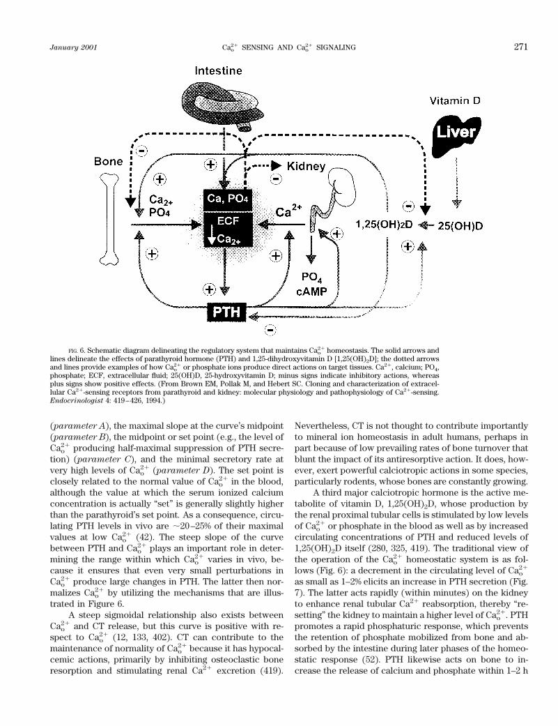
(parameter A), the maximal slope at the curve’s midpoint(parameter B), the midpoint or set point (e.g., the level ofCao
21 producing half-maximal suppression of PTH secre-tion) (parameter C), and the minimal secretory rate atvery high levels of Cao
21 (parameter D). The set point isclosely related to the normal value of Cao
21 in the blood,although the value at which the serum ionized calciumconcentration is actually “set” is generally slightly higherthan the parathyroid’s set point. As a consequence, circu-lating PTH levels in vivo are ;20–25% of their maximalvalues at low Cao
21 (42). The steep slope of the curvebetween PTH and Cao
21 plays an important role in deter-mining the range within which Cao
21 varies in vivo, be-cause it ensures that even very small perturbations inCao
21 produce large changes in PTH. The latter then nor-malizes Cao
21 by utilizing the mechanisms that are illus-trated in Figure 6.
A steep sigmoidal relationship also exists betweenCao
21 and CT release, but this curve is positive with re-spect to Cao
21 (12, 133, 402). CT can contribute to themaintenance of normality of Cao
21 because it has hypocal-cemic actions, primarily by inhibiting osteoclastic boneresorption and stimulating renal Ca21 excretion (419).
Nevertheless, CT is not thought to contribute importantlyto mineral ion homeostasis in adult humans, perhaps inpart because of low prevailing rates of bone turnover thatblunt the impact of its antiresorptive action. It does, how-ever, exert powerful calciotropic actions in some species,particularly rodents, whose bones are constantly growing.
A third major calciotropic hormone is the active me-tabolite of vitamin D, 1,25(OH)2D, whose production bythe renal proximal tubular cells is stimulated by low levelsof Cao
21 or phosphate in the blood as well as by increasedcirculating concentrations of PTH and reduced levels of1,25(OH)2D itself (280, 325, 419). The traditional view ofthe operation of the Cao
21 homeostatic system is as fol-lows (Fig. 6): a decrement in the circulating level of Cao
21
as small as 1–2% elicits an increase in PTH secretion (Fig.7). The latter acts rapidly (within minutes) on the kidneyto enhance renal tubular Ca21 reabsorption, thereby “re-setting” the kidney to maintain a higher level of Cao
21. PTHpromotes a rapid phosphaturic response, which preventsthe retention of phosphate mobilized from bone and ab-sorbed by the intestine during later phases of the homeo-static response (52). PTH likewise acts on bone to in-crease the release of calcium and phosphate within 1–2 h
FIG. 6. Schematic diagram delineating the regulatory system that maintains Cao21 homeostasis. The solid arrows and
lines delineate the effects of parathyroid hormone (PTH) and 1,25-dihydroxyvitamin D [1,25(OH)2D]; the dotted arrowsand lines provide examples of how Cao
21 or phosphate ions produce direct actions on target tissues. Ca21, calcium; PO4,phosphate; ECF, extracellular fluid; 25(OH)D, 25-hydroxyvitamin D; minus signs indicate inhibitory actions, whereasplus signs show positive effects. (From Brown EM, Pollak M, and Hebert SC. Cloning and characterization of extracel-lular Ca21-sensing receptors from parathyroid and kidney: molecular physiology and pathophysiology of Ca21-sensing.Endocrinologist 4: 419–426, 1994.)
January 2001 Cao21 SENSING AND Cao
21 SIGNALING 271
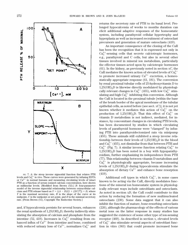
and, if hypocalcemia persists for several hours, enhancesthe renal synthesis of 1,25(OH)2D, thereby indirectly stim-ulating the absorption of calcium and phosphate from theintestine (52, 419). Increases in Cao
21 resulting from en-hanced influx of Ca21 from intestine and bone, combinedwith reduced urinary loss of Ca21, normalizes Cao
21 and
returns the secretory rate of PTH to its basal level. Pro-longed hypocalcemia of weeks to months duration canelicit additional adaptive responses of the homeostaticsystem, including parathyroid cellular hypertrophy andhyperplasia as well as increased recruitment of osteoclastprecursors and generation of mature osteoclasts (419).
An important consequence of the cloning of the CaRhas been the recognition that it is expressed not only inCao
21-sensing cells that secrete calciotropic hormones,e.g., parathyroid and C cells, but also in several othertissues involved in mineral ion metabolism, particularlythe effector tissues acted upon by calciotropic hormones(61). In the kidney, as previously noted in section XC, theCaR mediates the known action of elevated levels of Cao
21
to promote increased urinary Ca21 excretion, a homeo-statically appropriate response (61, 181). The conversionby renal proximal tubular cells of 25-hydroxyvitamin D to1,25(OH)2D is likewise directly modulated by physiologi-cally relevant changes in Cao
21 (455), with low Cao21 stim-
ulating and high Cao21 inhibiting this conversion. Although
the CaR is located in the proximal tubule [within the baseof the brush border of the apical membrane of the tubularepithelial cells, as noted before (see sect. XC)], it is not yetknown whether it mediates this action of Cao
21 on theproduction of 1,25(OH)2D. That this effect of Cao
21 onvitamin D metabolism is not indirect, mediated, for in-stance, by concomitant changes in circulating PTH levels,has been documented by studies in which circulatinglevels of parathyroid hormone were “clamped” by infus-ing PTH into parathyroidectomized rats via minipump(455). These animals still exhibited a steep inverse rela-tionship between their levels of 1,25(OH)2D in the bloodand Cao
21 (455), not dissimilar from that between PTH andCao
21 (Fig. 7). A similar inverse function relating Cao21 to
1,25(OH)2D has been noted in a boy with hypoparathy-roidism, further emphasizing its independence from PTH(77). This relationship between vitamin D metabolism andCao
21 is physiologically appropriate, because increasinglevels of 1,25(OH)2D during hypocalcemia stimulate theabsorption of dietary Ca21 and enhance bone resorption(419).
Additional cell types in which Cao21, in some cases
known to be acting via the CaR, directly modulates func-tions of the mineral ion homeostatic system in physiolog-ically relevant ways include osteoblasts and osteoclasts.As noted in section XD, the CaR could contribute to thisaction by reducing the formation of new multinucleatedosteoclasts (230). Some data suggest that it can alsoinhibit the function of mature, bone-resorbing osteoclasts(226), although the pharmacology of the effects of variousmetal ions on the latter response in many studies hassuggested the existence of some other type of ion-sensingreceptor (469). As described in section X, elevated levelsof Cao
21 also stimulate several aspects of osteoblast func-tion in vitro (363) that could promote increased bone
FIG. 7. A: the steep inverse sigmoidal function that relates PTHlevels and Cao
21 in vivo. These curves were generated by infusing EDTAor Ca21 in normal humans and measuring circulating levels of intactPTH as a function of serum ionized calcium concentration, here shownas millimolar levels. [Modified from Brown (52).] B: four-parametermodel of the inverse sigmoidal relationship between extracellular cal-cium and PTH release based on Y 5 {(A 2 D)/[1 1 (X/C)B]} 1 D, whereY is the maximal secretory rate, B is the slope of the curve at itsmidpoint, C is the midpoint or set point, and D is the minimal secretoryrate. [From Brown (51), Copyright The Endocrine Society.]
272 EDWARD M. BROWN AND R. JOHN MACLEOD Volume 81
formation in vivo and, therefore, reductions in Cao21 (161,
201, 229, 363, 372, 421, 463).This body of data strongly suggests that, in addition
to participating in mineral ion homeostasis by regulatingthe secretion of the classical calciotropic hormones, PTH,CT, and 1,25(OH)2D, Cao
21 itself can act in effect as a localor systemic Cao
21-regulating “hormone” (Fig. 6). In func-tions in this manner as the body’s principal Cao
21-loweringhormone, acting in part via stimulation of the secretion ofCT but primarily through actions mediated by its own cellsurface, Cao
21-sensing receptor. This hormonelike role ofCao
21 modulates the function(s) of a variety of cells andtissues participating in mineral ion metabolism throughchanges in the local or systemic levels of Cao
21 that arisefrom the Ca21-translocating actions of these tissues. Al-though the CaR may be an important mediator of theseactions of Cao
21, there may well be additional Cao21 sen-
sors/receptors that contribute as well. The dashed lines inFigure 6 illustrate some of these direct actions of Cao
21 ontissues involved in mineral ion metabolism. Figure 6 alsopoints out that extracellular levels of phosphate likewiseexert direct actions on cellular function. It is likely thatthere is some type of phosphate-sensing mechanism inthese cells (52), but the nature of the putative phosphatesensor remains obscure.
XII. EXTRACELLULAR CALCIUM-SENSING
RECEPTOR-BASED THERAPEUTICS
The CaR represents an important target for the de-velopment of CaR-based therapeutics for the treatment ofdisorders in which the CaR is over- or underactive (316,317). Although the development of such therapeutics hasso far been directed at parathyroid disorders, it appearslikely that it could extend to a wider variety of diseases ofCaR-expressing tissues in which the CaR malfunctions ormodulating the receptor’s activity would have desirabletherapeutic consequences (see sect. XIII for a discussion ofthe CaR’s roles in “nonhomeostatic” tissues). Clinicalstudies are currently underway that are testing so-called“calcimimetic” CaR activators in the treatment of primaryand secondary HPT (414). These agents are hydrophobic,low-molecular-weight drugs that allosterically activatethe CaR (316, 320). They are ineffective in the absence ofCao
21, but in the presence of Cao21 or other polycationic
CaR agonists, they enhance the receptor’s apparent affin-ity for these agonists.
In primary HPT, calcimimetics produce rapid (withinminutes) and substantial (.50%) reductions in circulatingPTH levels that are followed several hours later by de-creases in the serum calcium concentration, particularlyat higher doses (414), because the drug resets the ele-vated set point of pathological parathyroid tissue towardnormal. There is an initial increase in urinary Ca21 excre-
tion in patients with primary HPT during treatment with acalcimimetic that would have been anticipated, in part, asa result of the attendant rapid reduction in serum PTH. Itis also possible that additional, direct actions of calcimi-metics on CaRs in the CTAL and, perhaps, the DCT willlead to sustained hypercalciuria, but this effect has notbeen prominent to date. CaR agonists will also be veryuseful for treating uremic hyperparathyroidism, for whichthere are currently no fully effective forms of therapy.Conversely, a kidney-specific CaR antagonist might rep-resent an effective mode of treatment for individuals withCa21-containing renal stones. In the latter clinical setting,reducing the activity of CaRs in the CTAL would likelylower urinary Ca21 excretion markedly (e.g., similar tothat resulting from the renal Cao
21 resistance in FHH).
XIII. TISSUE DISTRIBUTION AND FUNCTIONS
OF THE EXTRACELLULAR CALCIUM-
SENSING RECEPTOR IN TISSUES
UNINVOLVED IN SYSTEMIC
IONIC HOMEOSTASIS
A. Brain Cells, Including Neurons and Glia
In the adult rat brain, the CaR has been localized bothby immunocytochemistry as well as by in situ hybridiza-tion with CaR specific antibodies and probes, respectively(86, 382, 386). The receptor is present at widely varyinglevels in numerous regions of the brain. The highest levelsare found within the SFO and the olfactory bulbs. Sub-stantial expression levels are also evident within the hip-pocampus, striatum, cingulate cortex, cerebellum, theependymal zones of the cerebral ventricles, and perivas-cular nerves around cerebral arteries (87, 388, 392). Abun-dant levels of CaR expression within the SFO, which is animportant hypothalamic thirst center (415), suggest that itmay participate in the central control of systemic fluidand electrolyte balance, as noted in section XC. Thus,although mineral ion homeostasis is not often thought ofas having central regulatory elements (i.e., in the brain),there are perhaps more complex relationships among thesystems regulating mineral ion homeostasis and otherhomeostatic systems that are known to exhibit prominentneuroendocrine elements (i.e., water homeostasis) thanrecognized previously.
1. Neurons
The CaR is present within synaptic areas of all re-gions of the hippocampus. It is not presently known withcertainty, however, whether its distribution is predomi-nantly pre- and/or postsynaptic (87, 392). Its overall dis-tribution is similar to those of the mGluRs and iGluRs(e.g., the NMDA receptor), which both have important
January 2001 Cao21 SENSING AND Cao
21 SIGNALING 273
roles in certain types of long-term potentiation (LTP).Thus it is of interest that there are substantial increases inthe expression of the CaR in the developing rat hippocam-pus at the time (during the first several weeks of life)when the development of the brain is progressing rapidlyand LTP can first be induced (87). The function(s) that thereceptor plays during this period of time, however, isessentially unknown (87, 89). The expression of the CaRin the granule and Purkinje cells of the cerebellum alsosuggests that it could have some type of role(s) in mod-ulating cerebellar function, but no studies on the CaR’sfunctions in these two cell types have been presented todate. The availability of brain tissue from mice that havetargeted disruption of the CaR gene should eventuallyenable studies of whether the CaR participates in anysuch postulated roles; unfortunately, the limited viabilityof the homozygous CaR knockout mice complicates suchinvestigations (196).
How could the CaR modulate the functions of cere-bellar, hippocampal, and other types of neurons? Re-cently, we have shown that the CaR stimulates the activityof Ca21-permeable NCCs in several cell types, includinghippocampal pyramidal neurons from rats and mice (471,472). The relationship of this NCC to iGluRs, such as theNMDA channel, is not yet understood. CaR-mediatedstimulation of this Ca21-permeable cation channel, how-ever, could contribute to the increases in Cai
21 known tobe necessary for induction of synaptic plasticity andwould also be expected to increase neuronal excitability.It is possible that the depolarization induced by CaR-mediated activation of NCCs, owing to the influx of Ca21
and Na1, could secondarily activate voltage-sensitiveCa21 channels, as is thought to occur in sheep parafol-licular cells (284). Such an effect of CaR activation onneurons, however, has not been reported to date. In ad-dition to activating neuronal NCCs, the CaR also en-hances the activity of a Ca21-activated K1 channelpresent in hippocampal pyramidal neurons from wild typebut not in those from CaR knockout mice (441). In con-trast to the effect of activating NCC, CaR-induced activa-tion of calcium-activated potassium channel (CAKC)would be expected to reduce neuronal excitability. Thusthe CaR modulates the activities of ion channels thatcontribute to the overall state of neuronal excitability. Asdiscussed in more detail in section XIV, neuronal activationis often associated with reductions in Cao
21 as a result ofactivation of Ca21-permeable influx pathways. The ensu-ing reduction in Cao
21 could, therefore, reduce the degreeof activation of NCCs mediated by the CaR and perhapsprovide a negative-feedback mechanism for avoiding ex-cessive elevations in neuronal Cai
21. In contrast, in neu-rons in which the CaR couples strongly to CAKC, it couldparticipate in a positive feedback mode; that is, decreasedactivity of the receptor during neuronal activity-depen-dent lowering of Cao
21 could reduce the CaR-mediated
activation of CAKC and resultant cellular hyper-polarization, thereby enhancing neuronal excitability.
We showed recently that the CaR is present at robustlevels in the enteric nervous system along essentially theentire gastrointestinal tract, being expressed both in Au-rbach’s plexus (e.g., between the circular and longitudinalsmooth muscle layers in the intestinal wall) and Meiss-ner’s plexus (within the submucosa) (85, 98). Both plexiparticipate in the regulation of key intestinal functions,including motility, secretory, and absorptive activities.Interestingly, changes in Cao
21 affect many of these pro-cesses, especially motility. For instance, hypercalcemicpatients not infrequently complain of constipation, whilethose with hypocalcemia may have symptoms of in-creased gastrointestinal motility (419). Additional effectsof raising Cao
21 include increased gastrin release (374) andenhanced gastric acid secretion (23, 27, 142). All of theseactions of Cao
21 could be CaR mediated (see below).Finally, Bukoski et al. (66) have demonstrated that
the CaR is expressed in perivascular sensory nerve end-ings in rat mesenteric artery. Subsequent studies of sen-sory nerve endings in other vascular beds revealed thefollowing distribution of the CaR in terms of its apparentdensity in various beds: mesenteric branch artery . basi-lar artery 5 renal interlobar artery . main renal trunkartery . left anterior descending coronary artery (451).Additional work has documented that stimulation of theCaR in these nerve endings releases a vasodilatory sub-stance, which is most likely an endogenous cannabinoid,such as N-arachidoylethanolamine (anandamide), thatacts on a cannabinoid receptor within the vascular wall(207). As described in section XIV, there could potentiallybe sufficiently large changes in the level of Cao
21 in theinterstitial fluid within a contracting muscle to modulatethe activity of CaRs in the immediate vicinity (4).
2. Oligodendroglia
Not only neurons, but also oligodendrocytes expressthe CaR (92). In the latter cell type, the receptor stimu-lates cellular proliferation and the activity of a CAKC (92).Very little is known about how the CaR regulates thefunction of oligodendrocytes in vivo. It is of interest,however, that expression of the receptor increases sever-alfold during the first several weeks of postnatal life in therat, a time when myelin formation is proceeding rapidly inthe developing brain. It is conceivable, therefore, that theincrease in CaR expression in oligodendroglia during thistime could contribute indirectly (e.g., by increasing cellu-lar proliferation) or directly (by currently unknown mech-anisms) to formation of myelin during brain development.
The expression level of the CaR subsequently falls toa lower and stable level in adult rats. Subsequent to theirrole in myelin formation, oligodendroglia are thought toparticipate importantly in local ionic homeostasis within
274 EDWARD M. BROWN AND R. JOHN MACLEOD Volume 81
the brain ECF. The CaR present in these cells couldpotentially contribute to such local homeostatic control.For example, because neuronal activity-dependent de-creases in Cao
21 are accompanied by elevations in extra-cellular K1, reduced activity of oligodendroglial CaRscould lower the activity of CAKC in these cells, therebymitigating further increases in extracellular K1.
3. Astrocytes
We have recently shown that the CaR is expressed inthe astrocytoma cell line U87, where it stimulates theactivity of a CAKC (93). Although an earlier study failed todetect expression of the CaR in primary astrocytes iso-lated from rat brain (392), we have recently demonstratedthe presence of CaR mRNA and protein in primary humanastrocytes (86). The function of the CaR in normal astro-cytes is currently unknown.
4. Microglia
Microglia are the brain’s macrophages. Because theCaR is expressed at robust levels in most circulatingmonocytes (466), it is not surprising that the CaR is alsoreadily detectable in rat primary microglia (91), where, asin most other brain-derived cells studied to date, it acti-vates a CAKC. Characterization of the function of thereceptor in microglia requires further study. It is of inter-est, however, that Ab peptides are known to modulateCai
21 in microglia (252) and to be chemotactic for thesecells (275), effects that could be CaR mediated (421, 466).Thus it is possible that CaR-mediated chemotaxis of mi-croglia could participate in the inflammatory reaction thatcan be present in the vicinity of amyloid plaques in Alz-heimer’s disease.
B. Lens Epithelial Cells
High Cao21 has a number of actions on cultured lens
epithelial cells, including disrupting plasma membraneintegrity, promoting loss of the cytoskeletal protein, vi-mentin, and stimulating Ca21-ATPase, effects that couldcontribute to cataract formation (For review, see Ref.120). Furthermore, clinical findings, viz., the developmentof cataracts in individuals with hypoparathyroidism andthe high Ca21 content noted in their cataracts, suggestthat Cao
21 (and potentially the CaR) could participate inthe physiology and pathophysiology of lens epithelialcells (129). We recently demonstrated that cultured hu-man lens epithelial cells express CaR mRNA and protein(90). The receptor stimulates the activity of a CAKC witha unitary conductance of ;82 pS. In view of the key roleof Cao
21 in maintaining lens integrity, it is conceivable thatthe CaR contributes to local ionic homeostasis within thelens. Examining the properties of the lens and of lens
epithelial cells from mice with knockout of the CaR gene(196) might provide additional useful information in thisregard.
C. Pituitary Gland
RNA isolated from mouse and rat pituitary expressestranscripts for the CaR as assessed by RT-PCR (131), andmurine pituitary-derived, ACTH-secreting AtT-20 cells ex-press a functional CaR that mediates stimulation of ACTHsecretion (131, 139). Elevating Cao
21 within a physiologi-cally relevant range (e.g., 10–15% above the upper limit ofnormal) also stimulates ACTH release in vivo in normalvolunteers (149, 444) as well as in persons being treatedfor psychiatric disorders with lithium (172). Furthermore,modulating the level of Cao
21 in vivo in humans has beenfound to alter the circulating levels of several other pitu-itary hormones, e.g., increasing gonadotropin hormone-releasing hormone-stimulated follicle-stimulating hor-mone and luteinizing hormone levels and inhibitingthyrotropin-releasing hormone-stimulated thyrotropin-stimulating hormone and prolactin secretion (444).
Functional evidence for CaR expression in the nor-mal bovine pituitary gland has been provided by Shorteand Schofield (410), who showed that polycationic CaRagonists mobilized intracellular stores of calcium in themajority of dispersed pituitary cells. The expression ofthe CaR has recently been directly shown in growth hor-mone (GH)-secreting pituitary adenomas, in which thereceptor increased Cai
21 and cAMP accumulation (390),as it does in AtT-20 cells (131). Although agonists of theCaR did not stimulate the release of growth hormonefrom these tumors in vitro in the absence of additionalsecretagogues, they did enhance growth hormone-releas-ing hormone-stimulated secretion of GH (390). Thus theCaR acts as a coagonist in this system. Although it is notknown why the CaR is expressed in normal pituitary cells,let alone in these tumors, it would be of interest to deter-mine whether the portal blood that supplies the releasinghormones to the pituitary gland from the hypothalamusundergoes changes in its level of Cao
21 as a function ofalterations in the secretion of hypothalamic releasing fac-tors. Secretory vesicles are known to contain high levelsof Cao
21, in some cases as high as 200 mM (322). There-fore, it is conceivable that Cao
21 could be secreted alongwith releasing factors from the hypothalamus and serveas a coagonist, as it does in vitro for the secretion of GHfrom GH-secreting adenomas (390). Moreover, furtherstudies are needed to define which cell type(s) within thenormal pituitary express the CaR.
D. Bone Marrow and Peripheral Blood
We recently showed that several cell types within thebone marrow express the CaR, including megakaryo-
January 2001 Cao21 SENSING AND Cao
21 SIGNALING 275
cytes, erythroid progenitors, myeloid precursors, andcells with the morphological appearance of monocyte/macrophages (204). Of the cells of the various hemato-poietic lineages that express the receptor in marrow, theonly ones whose mature cells in peripheral blood containthe CaR are platelets and monocytes (204). We recentlyutilized CaR-specific antisera as well as RT-PCR to iden-tify CaR protein and transcripts, respectively, in the ma-jority (;85%) of peripheral blood monocytes (466),whereas Bornefalk et al. (35), who demonstrated that highCao
21 stimulates secretion of IL-6 both in vivo and in vitrofrom peripheral blood monocytes, were unable to identifyCaR transcripts in these cells using RT-PCR. The rea-son(s) for the failure to detect the CaR in the latter studyis unclear.
Other studies that have examined the effects of phys-iologically relevant changes in Cao
21 on the functions ofmarrow-derived cells are scarce. Raising Cao
21 enhancesthe fusion of rat alveolar macrophages induced by1,25(OH)2D (215). Elevating Cao
21 also stimulates colonyformation and increases Cai
21 in erythroid precursorsobtained from uremic patients, an effect that was poten-tiated by 1,25(OH)2D (76), perhaps because the latterupregulates the expression of the CaR (46). In the mar-row, the CaR is expressed in hematopoietic precursorsthat probably experience significant alterations in thelevels of Cao
21 to which they are exposed related to theprevailing state of bone turnover within the local bone/bone marrow microenvironment. In addition to the ac-tions of Cao
21 on hematopoietic cells that were describedabove, it is also conceivable that the CaR could controlother aspects of these cells’ functions. For instance, be-cause the CaR is expressed on erythroid and some my-eloid precursors in the bone marrow but not on maturecells of those lineages in the peripheral blood (with theexception of monocytes) (204), perhaps it could controltrafficking of these cells between marrow and peripheralblood. Finally, Cao
21 is known to modulate a variety ofprocesses in platelets, including stimulating arachidonicacid release (287) and inhibiting cAMP accumulation(411); these effects could potentially be CaR mediated.Interestingly, Cao
21 has been quantified directly in plateletclumps formed as a result of platelet activation in vivo,and it decreases substantially (334). Thus changes in Cao
21
in the microenvironment to which platelets are exposedduring clumping of these blood elements could perhapsboth modulate their function(s) and also local levels ofCao
21 by regulating the fluxes of calcium ions betweenplatelets and their immediate microenvironment.
High levels of Cao21 promote the chemotaxis of sev-
eral cell types, including monocytes (421), the murinemonocytic cell line J774 (464), a bone marrow-derived,murine stromal cell line (ST-2) (462), and the murineosteoblastic cell line MC3T3-E1 (161, 463). Both J-774cells and peripheral blood monocytes express robust lev-
els of the CaR (466), making the receptor a good candi-date for mediating this action of Cao
21. CaR transcriptsand protein are also expressed in ST-2 cells (462).
What is the physiological relevance of CaR-activatedchemotaxis in these various cell types? In the case ofosteoblasts and/or their precursors, the chemotactic re-sponse to high Cao
21 may provide a signal directing themto sites of ongoing bone resorption requiring replacementof the missing bone during the osteoblastic phase of boneturnover (363, 461). Furthermore, mononuclear cells withthe appearance of monocytes/macrophages migrate tosites of bone resorption during the reversal phase of boneturnover interposed between the resorptive and formativephases (21). Sugimoto et al. (421) have shown that treat-ment of monocytes with elevated Cao
21 leads to the re-lease of factor(s) that stimulate the expression of alkalinephosphatase (a marker of osteoblast differentiation) inMC3T3-E1 osteoblastic bone cells, while monocyte-con-ditioned medium inhibits the formation of multinucleated,putative osteoclasts in vitro. Therefore, high Cao
21, bystimulating the release of cytokines or other factors [highCao
21 is known to stimulate the release of IL-6 from pe-ripheral blood monocytes (35)], could potentially serve toinhibit the formation of new osteoclasts and, at the sametime, stimulate the differentiation of osteoblasts at sitesof recent bone resorption.
High Cao21 could also conceivably serve as a more
generalized inflammatory signal for monocytes, macro-phages, and their various tissue-specific forms (e.g., mi-croglia and pulmonary alveolar macrophages). With theexception of red blood cells, which lack intracellularorganelles, all other types of cells have levels of totalintracellular calcium that are substantially higher thanthat in the blood owing to the presence of high concen-trations of calcium within intracellular stores, e.g., the ERand secretory vesicles (384). In the case of smooth mus-cle, total cellular calcium is 8.3 mmol/kg wet weight,which is nearly half that of mineralized bone (384). There-fore, death of cells and release of cellular calcium in asoluble form could lead to substantial local increases inCao
21. In addition, high Cao21 induces the fusion of pulmo-
nary alveolar macrophages to form multinucleated giantcells (215), which could also potentially represent part ofan integrated, CaR-mediated inflammatory reaction initi-ated by local increases in Cao
21 owing to cellular death orother mechanisms.
E. Breast Ductal Cells
The CaR is expressed at robust levels in normalbreast duct cells, in the duct cells of fibrocystic breasttissue, and in ductal carcinomas of the breast (97). Cao
21
plays key roles in the breast, not only in its normal phys-iological function(s) but also in various pathological
276 EDWARD M. BROWN AND R. JOHN MACLEOD Volume 81
states. For instance, Cao21 is clearly an important constit-
uent of milk, which has a content of ;200 mg Ca21/liter(357). A diagnostically important characteristic of breastcancers that can be very useful in their radiological de-tection during mammographic screening is their tendencyto form microcalcifications within ducts (150). Further-more, in vitro studies have shown that elevating Cao
21
induces terminal differentiation of normal human breastepithelial cells maintained in culture (289).
There is only limited information available related tothe regulation of calcium transport into milk, but theCaR’s presence in ductal cells raises the possibility that itcould participate in controlling such transport processes.Moreover, metastatic breast cancer has a marked ten-dency to spread to bone (346). Given the recent demon-stration that numerous cells within the bone marrowexpress the CaR under normal circumstances (204), per-haps the CaR’s presence on malignant breast cells con-tributes to the propensity of these and other CaR-express-ing cancer cells to metastasize to bone, in which locallyhigh levels of Cao
21 are present during bone resorption(412). In addition, we have recently found (J. L. Sandersand E. M. Brown, unpublished data) that high Cao
21 stim-ulates the secretion of PTHrP from some breast cancercell lines in vitro. This high Cao
21-stimulated, presumablyCaR-mediated, secretion of PTHrP could contribute to theexcessive osteolysis caused by breast cancers (107, 166,167) metastatic to bone by promoting a feedforwardmechanism in which release of calcium from the bonestimulated by PTHrP stimulates further PTHrP produc-tion, more bone resorption, and so forth. Interrupting thiscycle by blocking the activity of the CaR (318) couldpotentially offer substantial therapeutic benefit in thissituation. Therefore, given the importance of Cao
21 in boththe physiology and pathophysiology of the breast, the CaRcould play diverse and important roles in these processes.
F. Keratinocytes
Among the tissues uninvolved in systemic Cao21 ho-
meostasis that express the CaR are keratinocytes (29).Increases in Cao
21 are well known to trigger the differen-tiation of human and mouse keratinocytes in vitro (191,351). Cao
21-evoked differentiation of keratinocytes is ac-companied by changes in several intracellular signalingpathways, including accumulation of inositol phosphates(304) and elevations in Cai
21 resulting from both releaseof Ca21 from intracellular stores and Ca21 influx throughNCC (29). Bikle et al. (29) have recently identified tran-scripts for the CaR in human keratinocytes, and the dif-ferentiating stimulus of a rise in Cao
21 leads to an increasein the level of CaR mRNA. Therefore, the CaR couldmediate the known effects of Cao
21 on keratinocyte dif-ferentiation (29), at least in part, by activating PLC (89,
244, 393) and NCC (471–473), as it does in a number ofother cell types.
There are several interesting features of the induc-tion of differentiation of keratinocytes by increases inCao
21. First, only very small increases in Cao21, e.g., from
levels less than ;0.05 mM to those greater than 0.1 mM,are needed to induce differentiation. These levels are10-fold or more lower than those that activate the clonedCaR. Nevertheless, recent studies have shown that thecalcimimetic CaR activator, NPS R-467, but not its lessactive stereoisomer, NPS S-467, potentiates the actions ofCao
21 on several aspects of the function of keratinocytes,including increases in Cai
21 and inositol phosphates andalso upregulates the expression of the involucrin andtransglutaminase genes (435). Increases in Cao
21 withinthe same range also promote the differentiation of otherepithelial cells known to express the CaR, including hu-man mammary epithelial cells (289) and chick intestinalgoblet cells (31). It will be of interest to determinewhether the CaR also mediates those effects.
Oda et al. (327) have recently identified a splicevariant of the CaR in keratinocytes whose expressionincreases as differentiation progresses. This alternativelyspliced CaR lacks exon 5 and has an in-frame deletion of77 amino acids within its extracellular domain. This trun-cated form of the CaR does not elicit high Cao
21-evokedincreases in inositol phosphates when transfected inHEK293 cells or keratinocytes. Furthermore, it exerts adominant negative action on the function of the coex-pressed full-length CaR (327). This latter observation pro-vides a plausible explanation for the reduced responsive-ness of differentiated keratinocytes to Cao
21-inducedelevations in Cai
21 (327). It is at present unknown whetherthis alternatively spliced version of the CaR is expressedin and serves biological roles in tissues other than theskin.
F. Gastrointestinal System
1. Esophagus
The only study to date of the CaR in the esophagusdemonstrated the receptor’s presence in the basal cells ofthe stratified squamous epithelium of the rat esophagus(98). It will be of interest to determine whether the CaR inthese cells participates in promoting the differentiation ofthe esophageal epithelium as it does in the skin or servesother functional roles (29, 327).
2. Stomach
Increases in Cao21 stimulate acid secretion by gastric
glands and bicarbonate secretion by the gastric surfaceepithelium (142, 154). These observations suggest thatgastric parietal and surface epithelial cells are capable of
January 2001 Cao21 SENSING AND Cao
21 SIGNALING 277
sensing changes in Cao21. Cheng et al. (98) have employed
RT-PCR to amplify products from RNA isolated from ratforestomach and glandular stomach that were 99% homol-ogous to CaR transcripts expressed in rat kidney. In ad-dition, Northern analysis revealed the presence of CaRtranscripts in both the mucosa and muscularis of ratstomach. Immunohistochemistry showed that CaR pro-tein was expressed on mucous-producing surface cellsand the acid-secreting parietal cells of the body of the ratstomach (98), although it was not established whether theCaR had predominantly an apical and/or basolateral dis-tribution on these two cell types. A recent study hasextended this work to show that the CaR is expressed atthe highest level on the basolateral aspect of culturedhuman gastric surface cells and at lower levels on theapical membrane (396). Activation of the receptor stimu-lates the proliferation of these cells in vitro. The CaRmight participate, therefore, in the known role of thesecells in responding to injuries to the epithelial covering ofthe stomach in vivo. The CaR is also present on gastricepithelial cells of the amphibian, Necturus (the so-calledmudpuppy) (104). RT-PCR amplified a DNA fragment thatexhibited 84% nucleotide identity with the cDNA encod-ing the rat kidney CaR. Immunohistochemical localizationrevealed that the CaR was present on the basal surface ofmudpuppy gastric surface cells, similar to its localizationin human surface cells (104). Electrophysiological studiesdemonstrated that activating the amphibian CaR with itspolycationic agonists or a calcimimetic CaR activator (R-467) (320) decreased basolateral membrane resistance inthese cells. These effects were blocked by inhibitors ofprostaglandin synthesis. It will be of interest to determinewhether in Necturus there is a different isoform of CaRand if its signal transduction cascade is predominantlythrough PLA2. The degree of homology between the mud-puppy (104) and mammalian CaRs (89), however, is sim-ilar to that between the avian (126) and mammalian re-ceptors; therefore, it is likely that all represent the variousspecies homologs of the same ancestral gene.
In addition to its likely role in gastric surface cells,CaR agonists stimulate increases in Cai
21 in parietal cellswithin isolated gastric glands and also potentiate the in-creases in Cai
21 elicited by histamine (98), indicating thatthe receptor could mediate the known action of high Cao
21
to stimulate gastric acid production (142, 154). As in othersegments of the gastrointestinal tract, the CaR was alsoheavily expressed in the enteric nervous system, both inAuerbach’s as well as Meissner’s plexi (see also sect.XIIIA), where it could potentially contribute to the controlof gastric secretion and motility (98).
In addition to being present in the surface epithelialcells of the stomach and those within the gastric pits,recent studies have localized the CaR in the gastrin-se-creting cells of the gastric antrum (374). CaR transcriptsand protein were identified using RT-PCR as well as im-
munocytochemistry and Western blot analysis, respec-tively, of primary cultures enriched in human gastrin-secreting cells. The CaR expressed in these cells maymediate the long-recognized but poorly understood stim-ulatory effect of elevated levels of Cao
21 on gastrin secre-tion in vitro and in vivo (22, 23, 27). The receptor couldalso contribute to (e.g., via increases in gastrin secretion)the stimulatory effect of high Cao
21 on gastric acid secre-tion (22, 27, 142). Further work is required to prove that itis indeed the CaR that mediates these actions of Cao
21 andto elucidate the signal transduction pathways throughwhich it acts in this tissue. It is of interest, as discussed indetail in section XIVB2, that stimulation of gastric acidsecretion produces levels of Cao
21 within the gastric lu-men that are substantially lower (60–75%) than those inthe blood. This gradient in Cao
21 or the absolute level ofCao
21 on the blood side of the cells, which probably in-creases owing to the accompanying “extraction” of Ca21
from the gastric juice being elaborated, could conceivablyactivate CaRs present on their basolateral surfaces andthereby modulate gastric acid secretion (98, 396).
3. Small intestine
Increasing Cao21 decreases the proliferation and stim-
ulates the differentiation of intestinal epithelial cells (i.e.,goblet cells) in culture (31). How Cao
21 produces theseeffects is not known (38, 68). The CaR is expressedthroughout the rat small intestine and represents, there-fore, a candidate for the known actions of Cao
21 on vari-ous aspects of intestinal function, such as those just noted(85). The use of RT-PCR on RNA isolated from rat duo-denal mucosa, duodenal muscularis, jejunum, and ileumamplified a fragment with .99% nucleotide identity to aportion of the rat kidney CaR cDNA corresponding to aregion within the receptor’s NH2 terminus (85). In addi-tion, Northern analysis demonstrated 4.1- and 7.5-kb tran-scripts in each of these tissues. Immunohistochemistrywith CaR-specific antisera showed clear basal staining onvillus and crypt epithelial cells of the small intestine (withonly faint apical staining of the villus cells) (85). Finally,the use of in situ hybridization and immunohistochemis-try also demonstrated CaR expression in Auerbach’s my-enteric plexus of the small intestine, within the submu-cosa of the duodenum, and in Meissner’s plexus andBrunner’s glands (85).
What are the functional implications of the CaRwithin various cell types within the small intestine? Hy-percalcemia reduces gastrointestinal motility, whereashypocalcemia is associated with increased motility (419).In view of the CaR’s presence within the enteric nervoussystem throughout the small and large intestines [as wellas in the stomach (98)], it may well be that alterations insystemic levels of Cao
21 affect gastrointestinal motility ina CaR-mediated fashion. Direct investigation of the ef-
278 EDWARD M. BROWN AND R. JOHN MACLEOD Volume 81
fects of specific CaR activators (320) and antagonists(318) on intestinal motility, coupled with the use of intes-tinal tissues from homozygous knockout mice for suchstudies, will be required to address this issue definitively.
Expression of the CaR has also been reported inthree adenocarcinoma-derived, intestinal cell lines,which might potentially provide useful model systemsfor examining the receptor’s functional roles in theintestine at the cellular level, viz., T84, HT-29, andCaco-2 (151). The evidence for CaR expression wasobtained by RT-PCR using intron-spanning primers lo-cated within a portion of the receptor’s cDNA encodingits extracellular domain (thereby precluding amplifica-tion of similarly sized products from contaminatinggenomic DNA). Northern analysis using a biotinylatedriboprobe generated from the human CaR showed vary-ing levels of CaR expression in the three cell lines(HT-29 . T84 . Caco-2) (151). CaR agonists evokedtransient increases of Cai
21 in HT-29 cells, which wereprevented by pretreatment of these cells with a PI-PLCinhibitor or thapsigargin, strongly suggesting that cal-cium was being mobilized from its intracellular storesvia a CaR-mediated, PLC-dependent mechanism (151).Further evidence of the CaR’s role in mediating theseactions in the three cell lines will, however, require theuse of specific CaR activators or antagonists, or trans-fection of the cells with dominant-negative mutants orantisense constructs.
Given the known actions of other GPCRs on thefunction of small intestinal epithelial cells, it will also beof interest to address the following issues related to theCaR’s potential actions in the small intestine. Does Cao
21,acting through the CaR, modulate the secretion of Cl2
stimulated by agents raising cAMP as in T84 cells (24,224)? Does activating the CaR mimic the action of carba-chol to inhibit Cl2 secretion through a mechanismthought to involve stimulation of the MAPK cascade(240)? It will also be of interest to determine whether thereceptor affects other secretory processes, such as mucinproduction (144, 202), or modulates the absorption ofCa21 and/or other nutrients in this portion of the gastro-intestinal tract.
4. Colon
In the epithelial cells of the rat colon, there is clearexpression of the CaR on both their apical and basalmembranes as well as in the enteric nervous system alongthe entire large intestine (85). The growth and differenti-ation of colonocytes are exquisitely sensitive to changesin Cao
21 (31, 68, 168, 457). In cell culture, low levels ofCao
21 stimulate proliferation, while elevating Cao21 inhibits
growth and promotes differentiation, as in keratinocytes(191, 351). There is a growing body of circumstantialevidence from epidemiological studies that the incidence
of colorectal cancer is inversely correlated with dietarycalcium intake (153, 200, 416). It is presently unclearwhether this action of Cao
21 in vivo is indirect, involving,for instance, formation of insoluble salts of calcium withpotentially tumorigenic fatty acids and bile salts, or direct(e.g., by inhibiting cellular proliferation of colonocytes)(38, 457).
Kallay et al. (225) recently employed Caco-2 cellsas a model system to examine the direct actions ofCao
21 in vitro on this colon cancer-derived cell line.These cells express the CaR as assessed by RT-PCR andimmunohistochemistry performed with a CaR-specificmonoclonal antibody. At levels of Cao
21 of 0.025– 0.25mM, thymidine incorporation into DNA was increased.These low concentrations of Cao
21 also caused a rapid,PKC-dependent increase in c-myc protooncogene ex-pression. When grown on semipermeable supports, toallow addition of medium to either the apical or baso-lateral surface of the cells, elevating Cao
21 to 1.8 mM onthe apical but not on the basal cell surface blocked theincrease in c-myc expression (225). This result sug-gested that activation of the CaR on the apical mem-brane prevented the increase in c-myc expression thatwas required to induce cellular proliferation. Becausethe EC50 for activation of the CaR in HEK cells (;3.9mM) (15) and parathyroid cells (;1.0 –1.25 mM) (50) byCao
21 is so much greater than the level of Cao21 required
to inhibit growth of Caco-2 cells, it is currently notcertain that CaR mediates the inhibitory effect of rais-ing Cao
21 on colonocyte growth. It will be important toutilize antisense or dominant-negative constructs of theCaR as well as specific CaR activators and antagoniststo address this point further.
5. Does the CaR modulate chloride secretion in
intestinal crypt cells and other secretory epithelia?
There is now broad recognition that chloride secre-tion from intestinal crypt cells and other epithelial cellsinvolves a mechanism requiring apical chloride channels,such as the cystic fibrosis transmembrane regulator(CFTR) or a Ca21-activated chloride channel, togetherwith a basolateral Na1-K1-2Cl2 cotransporter, K1 chan-nels, and the Na1-K1-ATPase pump. The presence of theCaR on the basal membrane of intestinal crypt epithelialcells (which may be viewed as an archetypal Cl2-secret-ing cell) raises the possibility that it could also be presenton other Cl2 secretory cells that use the same mecha-nisms for secretion. To date, however, there are nodata available on the CaR’s role in regulating Cl2 secre-tion in crypt or other epithelial cells, although the recep-tor has recently been shown to stimulate bicarbonatesecretion in the acinar cells of the exocrine pancreas andin pancreatic ductal cells (see sect. XIIIH) (63). Furtherstudies are needed to determine whether the CaR in cells
January 2001 Cao21 SENSING AND Cao
21 SIGNALING 279
such as the ductal cells of the pancreatic, biliary, orsubmandibular glandular cells regulates the secretion ofchloride as well.
H. Pancreas
Bruce et al. (63) have recently identified the CaR inthe rat pancreas and characterized some of its func-tional properties. Using a combination of RT-PCR withCaR-specific primers and immunohistochemistry withan anti-CaR antiserum, these workers demonstratedthat a CaR-like molecule is expressed in pancreaticacinar cells, epithelial cells of the pancreatic ducts, andin the islets of Langerhans, although the specific celltypes in the latter containing the receptor were notidentified with certainty. The function of the CaR wasassessed in isolated acinar cells and interlobular ductsby measuring the effects of polycationic CaR agonistson Cai
21 in fura 2-loaded cell preparations. Both celltypes responded to addition of extracellular Gd31 orraising Cao
21 with modest increases in Cai21 (63). The
effect of activation of the CaR on bicarbonate secretionby isolated ducts was then determined by measuringchanges in intracellular pH. Luminal extracellular Gd31
was a potent stimulator of bicarbonate secretion andwas equal in efficacy to elevation in intracellular cAMPcaused by addition of forskolin. The results of thisstudy suggest that the CaR within the exocrine pan-creas senses the level of Cao
21 in pancreatic juice andcould participate in regulating luminal Cao
21 underbasal as well as stimulated conditions so as to mitigatethe risk of the formation of calcium carbonate stonesowing to excessively high levels of Cao
21 (63).The putative functional role(s) of the CaR in pancre-
atic islets requires further studies. Recent investigationshave shown the receptor to be present in the insulin-secreting b-cells, where the CaR could potentially medi-ate the stimulatory effects of Cao
21 on insulin secretionthat have been shown in normal b-cells (373) and in tumorcells derived from the b-cells (236). Interestingly, how-ever, a recent study showing that normal pancreaticb-cells express the CaR has demonstrated that Cao
21 caninhibit insulin secretion from these cells under certainconditions (418). Further studies are needed to under-stand the CaR’s normal physiological role, if any, in pan-creatic b-cells. The same study also showed that theglucagon-secreting d-cells of the human islet expressedthe CaR (418). It will be of interest in future studies todetermine whether the receptor mediates the inhibitoryaction of Cao
21 on glucagon secretion that can be demon-strated under specific circumstances (130, 261), andwhether it is also present in and regulates the function ofother cell types within the islet.
XIV. PHYSIOLOGICAL BASIS FOR LOCAL
EXTRACELLULAR CALCIUM SIGNALING
A. Role of Local Levels of Cao21 in Systemic
Cao21 Homeostasis
The homeostatic mechanism illustrated in Figure 6 isremarkable for the precision with which it maintains nearconstancy of Cao
21. From the discussion to this point, it isclear that CaR-expressing, Cao
21-sensing cells are key el-ements within this system, acting as calciostats that cansense and correct small changes in Cao
21. Cells participat-ing in systemic mineral ion homeostasis that detect Cao
21
in some cases presumably respond primarily to systemiclevels of Cao
21, similar to those measured in the peripheralblood (e.g., parathyroid or C cells). In other cases, how-ever, the levels of Cao
21 within tissues involved in mineralion metabolism must differ from that present in blood.For instance, an extreme example is the level of Cao
21
beneath a resorbing osteoclast, which has been measuredto be as high as 8–40 mM (412). Conversely, Cao
21 wouldbe expected to be lower than its systemic level near sitesof active bone formation owing to depletion of calciumions as a result of their removal from a soluble into aninsoluble phase (371, 372). Indeed, blood aspirated fromthe bone marrow of patients with widespread osteoblas-tic metastases of prostate cancer to bone can exhibit avalue for Cao
21 that is 20% lower than that present insystemic blood (292). Presumably, exuberant formationof new bone stimulated by the cancer cells in this settingoccurs more rapidly than Cao
21 can be delivered from thecirculation.
Local levels of Cao21 also likely differ from the “aver-
age” level of calcium in blood near sites where calciumions are being translocated across epithelial interfaces. Agood example is the CTAL of the kidney. The level of Cao
21
within the tubular fluid is similar to or slightly lower thanthat of the initial glomerular filtrate, which, in turn, issimilar to that of ultrafiltrable Ca21 in blood (123, 424).When stimulated by hormones raising cellular cAMP lev-els in CTAL, the magnitude of the lumen-positive trans-epithelial potential difference increases, driving passivereabsorption of Na1, Ca21, and Mg21 via the paracellularroute (123, 183). Furthermore, there is little reabsorptionof water in this so-called “diluting segment” of thenephron so that the levels of Cao
21 to which the basolat-eral surface of the tubular epithelial cells of the CTAL areexposed may be substantially higher than those in thetubular fluid. Similarly, during the active absorption ofcalcium ions from the gastrointestinal tract occurringunder the influence of vitamin D and after ingesting aCa21-containing meal, the level of Cao
21 within the inter-stitial fluid at the basolateral side of the intestinal epithe-lial cells would likely be well above that in the blood. In
280 EDWARD M. BROWN AND R. JOHN MACLEOD Volume 81
fact, interstitial Cao21 within the duodenal submucosa has
recently been shown to increase nearly twofold when thelumen is perfused with a fluid containing 10 mM Cao
21, asnoted in section XIIIG (306). Thus, unlike the Cao
21-sensingcells that secrete CT and PTH, Cao
21-sensing cells withinthe effector tissues involved in controlling the movementsof calcium ions into or out of the ECF in bone, intestine,and kidney may encounter concentrations of Cao
21 sub-stantially different from those in blood. Ultimately, theselevels of Cao
21 must be quantified during the responses ofthe mineral ion homeostatic system to various stresses tounderstand fully how Cao
21-sensing cells participate insystemic Cao
21 metabolism at both systemic and locallevels.
B. Other Microenvironments With Levels of Cao21
That Differ From Its Systemic Level
Most tissues that do not participate in systemic Cao21
homeostasis presumably do not gain or lose net quantitiesof calcium in response to changes in the circulating levelsof calciotropic hormones that are designed to normalizethe systemic level of Cao
21. Nevertheless, as is elaboratedon in the remainder of this review, it is apparent, on theone hand, that there are diverse microenvironments inwhich Cao
21 either differs from its systemic level orchanges largely independently of the latter. On the otherhand, it is equally clear that numerous cell types expressthe CaR that are seemingly uninvolved in systemic Cao
21
homeostasis (e.g., sect. XIII). In some instances, these cellsmay participate in the regulation of “local” Cao
21 ho-meostasis, sensing changes in Cao
21 within their immedi-ate microenvironments and adjusting the translocation ofeither ions (e.g., divalent cations) or water so as to adjustthe local ionic composition in a physiologically relevantmanner. The tubular fluid of the IMCD may represent agood example of such a local homeostatic mechanism.CaRs on the apical membrane of the tubular epithelialcells of the IMCD appear to modulate vasopressin-stimu-lated water flow so as to set an upper limit to the level towhich Cao
21 is allowed to rise (400, 401), thereby perhapsminimizing the risk of precipitation of calcium-containingsalts in the tubular lumen (181).
In other cases, cells may utilize extracellular ionicclues that are characteristic of specific microenviron-ments to control cellular functions having nothing to dowith either systemic or local Cao
21 homeostasis. In thediscussion that follows, we first review circumstances inwhich local levels of Cao
21 differ from that in systemicblood and then give several examples of how CaR-medi-ated Cao
21 sensing may contribute to the regulation of thelocal ionic milieu (i.e., control local Cao
21 homeostasis) orprovide information utilized by cells for other, nonhomeo-static purposes. There are a number of instances where
local levels of Cao21 have been unequivocally shown to
differ from corresponding systemic levels, which are cat-egorized in terms of their underlying mechanisms.
1. Locations where Ca21 from the environment
contributes to variations in Cao21
As noted before, there is only intermittent availabilityof dietary Ca21 to free-living terrestrial organisms. As aconsequence, the levels of Cao
21 and other ions in thelumen of the gastrointestinal tract can vary substantially.For instance, the level of Cao
21 directly measured in re-constituted, dried milk is ;7 mM (306). Because cells inboth the stomach (374) and small intestine express theCaR on their luminal surfaces, these cells will likely ex-perience variations in Cao
21 that could modulate theirfunctions in physiologically relevant ways. Studies cur-rently ongoing should clarify considerably the importanceof this Cao
21 sensing in the regulation of various aspects ofthese cells’ functions (85, 98, 374).
2. Changes in local Cao21 as a consequence
of epithelial ionic transport
Translocation of calcium ions across epithelial inter-faces in the kidney and elsewhere takes place via trans-cellular [viz., in DCT (148)] and/or paracellular [i.e., inCTAL (123, 181)] pathways. In some cases, as in theproximal tubule of the kidney, ionic transport occurs insuch a fashion that the composition of the reabsorbedfluid generally reflects that present in the tubular fluiditself (e.g., with regard to the levels of Na1, K1, Ca21, andCl2) (123, 424). In other instances, however, certain ionsare selectively reabsorbed, sometimes without accompa-nying water, thereby modifying substantially the concen-trations of Cao
21 and/or the other ions within the fluidbeing reabsorbed (and, by extension, those remainingwithin the tubular lumen). For example, in proximal seg-ments of the nephron, there is less reabsorption of Mg21
than of monovalent ions and Ca21 as noted earlier (123).Consequently, the level of Mgo
21 within the tubular fluidincreases progressively until it is ;1.8-fold higher in thethick ascending limb than in the initial glomerular filtrate.In contrast, the level of Cao
21 rises only modestly in thetubular fluid of the nephron segments proximal to thethick ascending limb. Na1, K1, Ca21 , Mg21, and Cl2 arereabsorbed in the latter segment via both transcellularand paracellular routes without accompanying water(247). Thus the concentrations of both Cao
21 and Mgo21
that the CaRs on the basolateral surface of the tubularepithelial cells will experience should be significantlyhigher than those in either the tubular fluid or systemicECF. These local changes in Cao
21 taking place as a resultof ionic transport by epithelial cells could provide signalsto CaRs that are substantially independent of systemiclevels of Cao
21. For instance, a reduction in the systemic
January 2001 Cao21 SENSING AND Cao
21 SIGNALING 281
level of Cao21 will enhance tubular reabsorption of Cao
21 inthe CTAL and presumably be associated with actual in-crease in the level of Cao
21 within interstitial fluid on thebasolateral side of CTAL cells. An interesting issue thatcould be relevant to these local changes in Cao
21 and toCao
21-sensing mechanisms within the immediate microen-vironment is the impact of the so-called “unstirred layers”of fluid close to biological membranes, such as at theouter face of the plasma membrane.
The specific ion-transporting properties of a giventissue could generate levels of Cao
21 differing markedlyfrom those in the systemic ECF. For instance, lactatingmothers produce on the order of ;200 mg of calciumdaily in each liter of milk, which would result in a con-centration of Cao
21 in milk that is approximately twice thatin blood (357). An even more extreme example is affordedby the prostatic fluid, where Cao
21 reaches 30 mM (436).Ionized levels of Cao
21 in these fluids are no doubt lowerdue to the binding of Ca21 to proteins and/or other ions[e.g., to citrate in prostate fluid, thereby lowering the levelof ionized calcium below 1.0 mM (384)], but they couldstill differ substantially from those in systemic ECF andmodulate CaRs known to be present, for instance, in theductal epithelial cells of the breast (97).
Another example of the impact of epithelial solutetransport on the level of Cao
21 within a specific microen-vironment occurs within the stomach. The concentrationof Cao
21 within gastric juice is known to vary inverselywith the rate of production of gastric fluid during stimu-lation with gastrin or histamine and is only 30–40% of thatpresent in blood during maximal stimulation (301). There-fore, calcium is, in effect, being extracted from the gastricjuice and presumably accumulates in the interstitial fluidat the basolateral surface of the epithelial cells producingit. Direct measurement of Cao
21 in the interstitital fluidimmediately beneath the gastric mucosa will be of inter-est to determine whether it increases during treatmentwith agents stimulating acid production sufficiently toprovide a CaR-mediated “feed-forward” mechanism thatcould further enhance the production of gastrin (374) andgastric acid (22, 27, 142) (see also sect. XIIIG). Such localchanges in Cao
21 could also potentially modulate the ac-tivity of CaRs on cells within the enteric nervous system.
3. Alterations in local Cao21 resulting from movement
of water without ions
In contrast to the situation in the thick ascendinglimb, where Ca21 and Mg21 are transported without ac-companying water, the reverse is true in the IMCD (1);that is, reabsorption of water takes place largely withoutaccompanying ions at rates that vary depending on theactivity of vasopressin-stimulated aquaporin-2 waterchannels in the apical membrane. This mechanism en-ables the kidney to adjust the amount of “free” water that
is retained or excreted. As noted earlier, these alterationsin the reabsorption of water can be accompanied bychanges in Cao
21 within the tubular fluid in the IMCD thatare sufficiently great to promote renal stone formation ifthere were excessive water reabsorption at the time aCa21 load is being excreted (400, 401, 436). As notedpreviously, the CaR’s presence on the apical (i.e., luminal)surface of the IMCD tubular epithelial cells affords anexample of how this receptor may participate in localCao
21 homeostasis. Because elevations in Cao21 within the
tubular fluid in the IMCD reduce vasopressin-stimulatedreabsorption of water, probably through a CaR-mediatedmechanism(s), a rise in Cao
21 in the tubular fluid to anexcessively high level can feed back to lower Cao
21 withinthe fluid in a homeostatically appropriate manner (183).Interestingly, in contrast to the homeostatic mechanismgoverning systemic Cao
21 homeostasis, which adjust thelevel of Cao
21 largely by modifying the movements of Ca21
into and out of the ECF (i.e., via intestine, bone, andkidney), in the IMCD the CaR appears to regulate Cao
21
principally through controlling the movement of waterbut not Ca21.
4. Alterations in local Cao21 owing to fluxes of Ca21
between the intra- and extracellular spaces
A substantial body of evidence documents that Cao21
changes appreciably in specific microenvironments owingto alterations in the fluxes of Ca21 between the intra- andextracellular compartments. Substantial changes in Cao
21
take place in the narrow intercellular spaces of the brainowing to changes in the activity of neurons that areaccompanied by cellular uptake of Ca21 through variouscalcium-permeable channels (e.g., NMDA channels) (9,186, 271). These alterations in Cao
21 can be accompaniedby changes in the extracellular levels of extracellular Na1
and extracellular K1 as a result of influx of Na1 throughvoltage-sensitive Na1 channels and efflux of K1 thoughCa21-activated and other K1 channels. A particularlystriking example of such changes in the extracellularionic composition in the ECF of the brain is provided bythose occurring during strong electrical stimulation of thecerebellum of the anesthetized rat (323). During electricalstimulation lasting for even a few seconds in this model,Cao
21 can decrease by up to 90%, whereas extracellular K1
rises by severalfold. These alterations are rapidly revers-ible (within a matter of seconds) after termination of thestimulation. Even much milder, more physiologically rel-evant activation of neurons can be accompanied by easilydetectable changes in Cao
21 that could be sensed by CaRswithin the immediate vicinity. For instance, stroking ananesthetized cat’s paw with a camel hair brush decreasesCao
21 by several percent in the ECF within the contralat-eral primary somatosensory cortex innervating thestroked paw (186). While the magnitude of this alteration
282 EDWARD M. BROWN AND R. JOHN MACLEOD Volume 81
in Cao21 may appear small, the CaR on parathyroid cells
readily detects reductions in Cao21 of this magnitude (e.g.,
Fig. 7).We have used modeling to examine the special case
of activity-dependent reductions in Cao21 within the syn-
aptic cleft (443). A rise in Cai21 in the postsynaptic den-
dritic spines of the hippocampus owing to influx of Ca21
via NMDA channels participates importantly in the induc-tion of LTP (211). Ca21 influx within or close to synapticclefts also plays key roles in all steps of synaptic trans-mission, both in the pre- and postsynaptic compartments(32). The synaptic cleft can be visualized as a thin disk ofECF. During stimulation of a synapse at high frequencies,the interval between pulses might potentially be insuffi-cient for Cao
21 to diffuse in from the periphery of the cleftto replace that lost at the cleft’s center owing to activationof postsynaptic, Ca21-permeable channels. We elaborateda computer model to estimate the alterations in Cao
21 thatmight be occurring in the synaptic cleft as a result of theinflux of Ca21 into the postsynaptic compartment viaiGluRs, combined with subsequent efflux via the calciumpump and Na1-Ca21 exchanger (443). The levels of Cao
21
within the cleft were approximated utilizing a compart-mental model incorporating fluxes across the postsynap-tic membrane combined with radial diffusion in from theedge of the cleft.
Resultant simulations using this model suggestedthat substantial reductions in Cao
21 can take place insynaptic clefts attendant on activation of iGluRs, espe-cially at the high stimulation frequencies required to in-duce LTP. Only minimal, transitory alterations in Cao
21, incontrast, were predicted by the model at low frequenciesof stimulation. These frequency-dependent changes inCao
21 reflect the activity of iGluRs and could potentiallymodulate presynaptic function through a mechanism in-volving changes in Cao
21, which then serves as a retro-grade messenger, if Cao
21 sensors were resident on thepresynaptic membranes. The CaR is known to be ex-pressed on nerve terminals in hippocampus and otherareas of the brain (although additional work is needed todefine whether it is located pre- and/or postsynaptically),and it could potentially play such a role (87, 388, 392).
In contrast to the reductions in Cao21 predicted to
take place during the initial phase of electrical stimulationof a synapse by this model, eventual return of the systemto its steady state must involve transient increases in Cao
21
as Ca21 is pumped out of the postsynaptic spine (443).Depending on the “set” of CaRs in the immediate vicinity(e.g., whether they are poised to respond more sensitivelyto increases or to decreases in Cao
21), activity-dependentchanges in the function of the CaR, if they exist, mightoccur primarily during the initial stimulation of the syn-apse and accompanying Cao
21 depletion or during therecovery phase, when Cao
21 rises above its resting level.Furthermore, the overall (e.g., phasic or oscillatory) pat-
tern of changes in Cao21 could potentially encode impor-
tant physiological signals that are decoded by CaRs orother Cao
21 sensors in the vicinity, as has been suggestedto occur intracellularly (5, 34, 94).
Another tissue in which increases in cellular activityproduce transient reductions in Cao
21 is the beating heart(28). Pacing of a frog heart in vitro leads to substantialdecreases in Cao
21 within the interstitial fluid within theheart muscle in this model system (28). Even greaterchanges in Cao
21 might occur in this setting within thet-tubular system, where there are narrow infoldings of theplasma membrane into the muscle fibers that ensure closeproximity between the ECF within the t tubules and theintracellular sites where Ca21 promotes stimulus-contrac-tion coupling (4).
Two additional examples where Cao21 depletion oc-
curs owing to cellular influx of calcium ions from theextracellular fluid are the pancreatic islet and the platelet.During the spontaneous electrical activity of isolated is-lets investigated in vitro, the periodic initiation of anaction potential, which is accompanied by activation ofvoltage-sensitive Ca21 channels and severalfold increasesin Cai
21, causes substantial (;0.5 mM) reciprocal decre-ments in Cao
21 (347). With this electrical activity ceases,both Cai
21 and Cao21 returned to their basal levels. In
addition, when platelets aggregate in vivo, there are sub-stantial (;80%) reductions in Cao
21, presumably owing toinflux of Cao
21 into the platelets, which is rapidly revers-ible when the platelets disperse after addition of an agentinhibiting their aggregation (334).
In contrast to the situations just described, in whichreductions in Cao
21 are initiated by cellular uptake ofCa21, activation of cells by Ca21-mobilizing hormonesbinding to their respective receptors (e.g., those coupledto activation of PI-PLC), can produce an initial rise ratherthan fall in Cao
21 following cellular activation. Intracellu-lar Ca21 stores can comprise total cellular calcium con-centrations of several millimoles per kilogram (356)(smooth muscle cells can have ;8 mmol/kg, half thecalcium content per wet weight of mineralized bone, Ref.384). Thus extrusion of Ca21 mobilized from these storesby inositol trisphosphate via the plasma membrane Ca21
pump could potentially produce substantial increases inthe level of Cao
21 within narrow intercellular spaces invivo (72, 75, 199, 431, 468). Subsequent refilling of thesecellular stores would then presumably result in transientlowering of Cao
21 after removal of the Ca21-mobilizinghormone. Again, either the absolute change in Cao
21 or thepattern of the change might provide important informa-tion that could be decoded by CaRs in the immediateenvironment. Thus the initiation of Cai
21 signaling may beaccompanied by an obligate activation of Cao
21 signalinggiven the impact of changes in Cai
21 dynamics on Cao21 in
many cell types and the CaR’s wide distribution.The level of Cao
21 is ;10,000-fold greater than that of
January 2001 Cao21 SENSING AND Cao
21 SIGNALING 283
Cai21. It may be surprising, therefore, that changes in
cellular activity, such as those just described, which usu-ally increase the Cai
21 by ;10-fold or less (300, 350), areable to significantly decrease or increase Cao
21 when thereis cellular Ca21 influx or efflux, respectively. The answerto this apparent paradox lies in two factors: 1) the re-stricted spaces in the ECF of intact tissues, which, there-fore, contain only limited amounts of Cao
21 and 2) themagnitudes of the influx or efflux of Ca21 during cellularactivation or recovery, which are much greater than theassociated changes in Cai
21 owing to the presence ofintracellular Ca21 buffers. It is also probable that there issubstantial heterogeneity in terms of the magnitudes andlocations of the changes in Cao
21 that may occur over theoutside of the plasma membranes of individual cells, de-pending on the locations of the sites of Ca21 influx andefflux as well as of the CaR itself in cells expressing thisreceptor.
One specific microenvironment of potential interestin this regard is that within caveolae (see sect. VIIC) (8,268, 341). Recent studies suggest that caveolae can serveas “message centers” for the cell, since they contain im-portant components involved in signal transduction, suchas GPCRs and tyrosine-coupled receptors, G proteins,PKC isoforms, the plasma membrane calcium pump(Ca21-ATPase), and inositol trisphosphate-regulatedchannels (8, 268, 341). On the order of 80% of the CaRprotein expressed in bovine parathyroid cells is locatedwithin caveolae (245). Thus, depending on the relativedensities of influx versus efflux pathways for calcium ionswithin the caveolae, there could potentially be large localfluxes of Ca21. For instance, consider the hypotheticalcase in which the CaR in parathyroid cells resides incaveolae containing the Ca21-ATPase, but calcium influxpathways stimulated by the CaR are elsewhere in theplasma membrane. Activation of the receptor by in-creases in Cao
21 would elevate Cai21 through both cellular
mobilization and influx of calcium ions. The latter wouldthen be pumped out of the cell in the immediate vicinityof the receptor within caveolae and could potentiallygenerate a feed-forward activation of the receptor byelevating the local level of Cao
21 above that present in thegeneral ECF.
The distribution of Ca21 buffers within the cytosolwill likely also alter the patterns of changes in both Cai
21
and Cao21 attendant on cellular activation, since a larger
influx of Cao21 would be required to bring about a given
change in Cai21 if influx takes place where there is a
locally high concentration of intracellular calcium buff-er(s). Thus the presence of Cai
21 buffers (i.e., calbindins)in CaR-expressing cells, such as parathyroid cells (58, 64),intestinal cells (85, 453), and cells of the DCT (105, 379)could modify local alterations in Cao
21 occurring in asso-ciation with changes in Cai
21, including those produced byactivating the CaR.
The large amounts of Ca21 within intracellular storescan also serve as a reservoir for Ca21 that can raise locallevels of Cao
21 in the setting of inflammation and celldeath. Menkin (294) showed over 40 years ago that thefluid within exudative abscesses (e.g., containing manyinflammatory cells) induced in dogs by subcutaneous in-jection of irritants could contain levels of Cao
21 threefoldhigher than those measured simultaneously in blood. Incontrast, transudative fluid accumulations (e.g., thosethat are devoid of inflammatory cells) have levels of Cao
21
similar to those in the blood. It is likely that calciumreleased by dying inflammatory cells contributed to thehigh levels of Cao
21 within the fluid in the exudative ab-scesses, since the calcium content of most cells is sever-alfold higher than that in the systemic ECF (384).
Additional examples of fluids in which increases inCao
21 have been documented related to inflammation in-clude the fluid from inflamed gingiva (gums) (234, 235)and peritoneal fluid isolated from patients being sub-jected to peritoneal dialysis for renal insufficiency whodevelop peritonitis (266). Because elevated levels of Cao
21
promote the chemotaxis of monocytes (421) and stimu-late the formation of multinucleated giant cells from pul-monary alveolar macrophages (215), the elevated levels ofCao
21 within inflammatory fluids may serve as a signalpromoting the influx and activation of macrophages (seealso sect. XIIID).
5. Local changes in Cao21 resulting from movements of
Ca21 into and out of extracellular reservoirs
Translocation of calcium ions into or out of boneprovides an instructive example of how Ca21 fluxes intoor out of extracellular reservoirs that contain large quan-tities of this ion can perturb local (and also systemic)levels of Cao
21. As noted before, Cao21 underneath a
resorbing osteoclast can reach levels as high as 40 mM(412). Although the impact of releasing this resorbed Ca21
into the osteoclast’s immediate microenvironment on thelocal level of Cao
21 is unknown, it is probable that thelatter would rise substantially. Indeed, when there is un-controlled release of skeletal Cao
21 from bone, as withskeletal metastases of breast cancer and certain othermalignancies, even systemic levels of Cao
21 can increaseto supranormal levels (419). Cao
21 within the bony micro-environment would presumably be even higher in suchcircumstances.
In addition to serving as a source of calcium ions(e.g., during resorption of bone), the skeleton can alsoserve as a “sink” for Cao
21. For instance, extensive osteo-blastic metastases of prostate cancer to bone can causesystemic hypocalcemia, presumably because exuberantbone formation caused by tumor-derived products (164)outstrips the Cao
21 homeostatic system’s capacity to pro-
284 EDWARD M. BROWN AND R. JOHN MACLEOD Volume 81
vide additional Ca21 via renal conservation, intestinal ab-sorption, and bone resorption (419).
C. Physiological Relevance of Local Cao21 Sensing
and Cao21 Signaling
Therefore, the substantial number of cell types ex-pressing the CaR that are seemingly uninvolved in sys-temic Cao
21 metabolism may engage in Cao21 sensing in
response to Cao21 signals that arise largely independent of
the homeostatic processes that maintain near constancyof Cao
21 in the blood (52); that is, local changes in Cao21
owing to alterations in cellular activity, ion transport, orthe other processes just described may elicit cellular re-sponses by CaR-expressing cells via modes of communi-cation analogous to paracrine or autocrine signaling bymore classical hormones or cytokines. In some cases,Cao
21-induced activation of the receptor may serve torestore local ionic homeostasis, as in the tubular fluid ofthe IMCD of the kidney. In other cases, there are tanta-lizing hints of previously unappreciated roles for Cao
21
sensing and Cao21 signaling at the local level. The chemo-
tactic responses of osteoblasts and their precursors tosites of high Cao
21 may serve as an important componentof the mechanism through which the osteoclastic “injury”to bone is “healed” by new bone formation. As an exten-sion of this concept, the chemotactic response of mono-cytes to Cao
21 and the presence of high local levels of Cao21
within inflammatory fluids may suggest a broader role forCao
21 as a “chemokine” and as a more general inflamma-tory signal. Furthermore, it is likely that not only Cao
21 butother endogenous CaR activators and modulators willcontribute to regulating the activity of the CaR in localmicroenvironments, including not only other polycationicagonists, such as Mgo
21, spermine, Ab peptides, and per-haps other endogenous polycations, but also ionicstrength. Therefore, the CaR will likely integrate informa-tion encoded by several different local environmentalfactors that will ultimately determine the functional im-pact of the receptor on a given cell.
XV. SUMMARY
The cloning of the G protein-coupled CaR has pro-vided an actual or potential molecular mechanism medi-ating many of the known effects of Cao
21 on the cells andtissues that maintain systemic Cao
21 homeostasis, partic-ularly parathyroid and kidney. In addition to affordinguseful molecular tools for documenting the presence ofCaR mRNA and protein in these tissues, the cloned CaRhas permitted the identification of human diseases thatare the consequences of inactivating or activating muta-tions of this receptor as well as to generate mice withknockout of the CaR gene. The characteristic abnormali-
ties in the functions of the parathyroids and kidneys ofthese patients and in CaR knockout mice have providedthe means to dissect the physiological roles of the CaR inmineral ion metabolism. Much remains to be learned,however, about how the CaR regulates other tissues in-volved in systemic homeostasis, such as bone and intes-tine, including the role(s), if any, of additional Cao
21-sensing receptors/sensors that have yet to becharacterized at a molecular level. Moreover, althoughthese human diseases and mouse models will afford use-ful tools for further investigating the receptor’s roles inthese latter tissues, the development of potent and spe-cific CaR antagonists, tools that are only just beginning tobe developed (318), would be extremely useful in thisregard.
In addition, much remains to be learned about theCaR’s functions in tissues that are not directly involved insystemic mineral ion homeostasis, where the receptorprobably serves numerous additional roles, some relatedto local intra- and extracellular regulation of ions andothers unrelated to either systemic or local ionic ho-meostasis. In any event, the CaR and perhaps other re-ceptors/sensors for calcium or other extracellular ionswill likely be versatile regulators of a wide variety ofcellular functions (52, 457) and represent potentially im-portant therapeutic targets.
We gratefully acknowledge generous grant support fromnumerous sources, including the National Institutes of Health(Grants DK-48330, DK-41415, DK-46422, and DK-52005), the St.Giles Foundation, NPS Pharmaceuticals, the National DairyCouncil, the National Space and Bioscience Research Institute,and the Stanley Foundation (all to E. M. Brown) R. J. MacLeodis a recipient of a postdoctoral fellowship from the MedicalResearch Council of Canada.
Address for reprint requests and other correspondence:E. M. Brown, Endocrine-Hypertension Division, Brigham andWomen’s Hospital, 221 Longwood Ave., Boston, MA 02115 (E-mail: [email protected]).
REFERENCES
1. AGRE P. Molecular physiology of water transport: aquaporin no-menclature workshop. Mammalian aquaporins. Biol Cell 89: 255–257, 1997.
2. AIDA K, KOISHI S, INOUE M, NAKAZATO M, TAWATA M, AND ONAYA T.Familial hypocalciuric hypercalcemia associated with mutation inthe human Ca21-sensing receptor gene. J Clin Endocrinol Metab
80: 2594–2598, 1995.3. AIDA K, KOISHI S, TAWATA M, AND ONAYA T. Molecular cloning of a
putative Ca21-sensing receptor cDNA from human kidney. Bio-
chem Biophys Res Commun 214: 524–529, 1995.4. ALMERS W, FINK R, AND PALADE PT. Calcium depletion in frog muscle
tubules: the decline of calcium current under maintained depolar-ization. J Physiol (Lond) 312: 177–207, 1981.
5. AMUNDSON J AND CLAPHAM D. Calcium waves. Curr Opin Neurobiol
3: 375–382, 1993.6. ANAST CS, MOHS JM, KAPLAN SL, AND BURNS TW. Evidence for
parathyroid failure in magnesium deficiency. Science 177: 606–608,1972.
January 2001 Cao21 SENSING AND Cao
21 SIGNALING 285
7. ANAST CS, WINNACKER JL, FORTE LR, AND BURNS TW. Impaired re-lease of parathyroid hormone in magnesium deficiency. J Clin
Endocrinol Metab 42: 707–717, 1976.8. ANDERSON RG. Plasmalemmal caveolae and GPI-anchored mem-
brane proteins. Curr Opin Cell Biol 5: 647–652, 1993.9. ARENS J, STABEL J, AND HEINEMANN U. Pharmacological properties of
excitatory amino acid induced changes in extracellular calciumconcentration in rat hippocampal slices. Can J Physiol Pharmacol
70 Suppl: S194–S205, 1992.10. ARTHUR JM, COLLINSWORTH GP, GETTYS TW, QUARLES LD, AND RAY-
MOND JR. Specific coupling of a cation-sensing receptor to G proteinalpha-subunits in MDCK cells. Am J Physiol Renal Physiol 273:F129–F135, 1997.
11. ATTIE MF, GILL J JR, STOCK JL, SPIEGEL AM, DOWNS RW JR, LEVINE
MA, AND MARX SJ. Urinary calcium excretion in familial hypocalci-uric hypercalcemia. Persistence of relative hypocalciuria after in-duction of hypoparathyroidism. J Clin Invest 72: 667–676, 1983.
12. AUSTIN LA AND HEATH H III. Calcitonin: physiology and pathophys-iology. N Engl J Med 304: 269–278, 1981.
13. AUTRY CP, BROWN EM, FULLER FH, ROGERS KV, AND HALLORAN BP.Ca21 receptor mRNA and protein increase in the rat parathyroidgland with age. J Endocrinol 153: 437–444, 1997.
14. BAI M, PEARCE SH, KIFOR O, TRIVEDI S, STAUFFER UG, THAKKER RV,BROWN EM, AND STEINMANN B. In vivo and in vitro characterizationof neonatal hyperparathyroidism resulting from a de novo, het-erozygous mutation in the Ca21-sensing receptor gene: normalmaternal calcium homeostasis as a cause of secondary hyperpara-thyroidism in familial benign hypocalciuric hypercalcemia. J Clin
Invest 99: 88–96, 1997.15. BAI M, QUINN S, TRIVEDI S, KIFOR O, PEARCE SHS, POLLAK MR,
KRAPCHO K, HEBERT SC, AND BROWN EM. Expression and character-ization of inactivating and activating mutations in the human Cao
21-sensing receptor. J Biol Chem 271: 19537–19545, 1996.
16. BAI M, TRIVEDI S, AND BROWN EM. Dimerization of the extracellularcalcium-sensing receptor (CaR) on the cell surface of CaR-trans-fected HEK293 cells. J Biol Chem 273: 23605–23610, 1998.
17. BAI M, TRIVEDI S, KIFOR O, QUINN SJ, AND BROWN EM. Intermolecularinteractions between dimeric calcium-sensing receptor monomersare important for its normal function. Proc Natl Acad Sci USA 96:2834–2839, 1999.
18. BAI M, TRIVEDI S, LANE CR, YANG Y, QUINN SJ, AND BROWN EM.Protein kinase C phosphorylation of threonine at position 888 inCao
21-sensing receptor (CaR) inhibits coupling to Ca21 store re-lease. J Biol Chem 273: 21267–21275, 1998.
19. BAPTY BW, DAI LJ, RITCHIE G, JIRIK F, CANAFF L, HENDY GN, AND
QUAMME GA. Extracellular Mg21-and Ca21-sensing in mouse distalconvoluted tubule cells. Kidney Int 53: 583–592, 1998.
20. BARON J, WINER KK, YANOVSKI JA, CUNNINGHAM AW, LAUE L, ZIMMER-MAN D, AND CUTLER GB JR. Mutations in the Ca21-sensing receptorgene cause autosomal dominant and sporadic hypoparathyroidism.Hum Mol Genet 5: 601–606, 1996.
21. BARON R. Anatomy and ultrastructure of bone. In: Primer on the
Metabolic Bone Diseases and Disorders of Mineral Metabolism
(3rd ed.), edited by Favus MJ. Philadelphia, PA: Lippincott-Raven,Philadelphia, 1996, p. 3–10.
22. BARRERAS RF AND DONALDSON RM JR. Effects of induced hypercal-cemia on human gastric secretion. Gastroenterology 52: 670–675,1967.
23. BARRERAS RF AND DONALDSON RM JR. Role of calcium in gastrichypersecretion, parathyroid adenoma and peptic ulcer. N Engl
J Med 276: 1122–1124, 1967.24. BARRETT KE, COHN JA, HUOTT PA, WASSERMAN SI, AND DHARMSATHA-
PHORN K. Immune-related intestinal chloride secretion. II. Effect ofadenosine on T84 cell line. Am J Physiol Cell Physiol 258: C902–C912, 1990.
25. BASCAL Z, ALAM A, ZAIDI M, AND DACKE C. Effect of raised extracel-lular calcium on cell spread area in quail medullary bone oste-oclasts. Exp Physiol 79: 15–24, 1994.
26. BAUM M, HENTSCHEL H, ELGER M, BROWN E, HEBERT S, AND HARRIS H.Homologs of an extracellular calcium/polyvalent cation-sensingreceptor (CaR) are localized to the apical surface of specific epi-thelial cells in organs critical for ionic homeostasis in the elasmo-branchs, Squalus acanthias and Raja erinacea, as well as teleosts,
Pseudopleuronectes americanus and Onchorynchus and Fundu-
lus heteroclitus. Bull MDIBL 35: 32–33, 1996.27. BEHAR J, HITCHINGS M, AND SMYTH RD. Calcium stimulation of gastrin
and gastric acid secretion: effect of small doses of calcium carbon-ate. Gut 18: 442–448, 1977.
28. BERS DM. Early transient depletion of extracellular Ca during indi-vidual cardiac muscle contractions. Am J Physiol Heart Circ
Physiol 244: H462–H468, 1983.29. BIKLE D, RATNAM A, MAURO T, HARRIS J, AND PILLAI S. Changes in
calcium responsiveness and handling during keratinocyte differen-tiation. Potential role of the calcium receptor. J Clin Invest 97:1085–1093, 1996.
30. BIKLE DD AND PILLAI S. Vitamin D, calcium, and epidermal differen-tiation. Endocr Rev 14: 3–19, 1993.
31. BLACK BL AND SMITH JE. Regulation of goblet cell differentiation bycalcium in embryonic chick intestine. FASEB J 3: 2653–2659, 1989.
32. BLISS TVP AND COLLINGRIDGE GL. A synaptic model of memory:long-term potentiation in the hippocampus. Nature 361: 31–39,1993.
33. BONEN DK AND SCHMID TM. Elevated extracellular calcium concen-trations induce type X collagen synthesis in chondrocyte cultures.J Cell Biol 115: 1171–1178, 1991.
34. BOOTMAN MD AND BERRIDGE MJ. The elemental principles of calciumsignaling. Cell 83: 675–678, 1995.
35. BORNEFALK E, LJUNGHALL S, LINDH E, BENGSTEN O, JOHANSSON A, AND
LJUNGGREN O. Regulation of interleukin-6 secretion from mononu-clear blood cells by extracellular calcium. J Bone Miner Res 12:228–233, 1997.
36. BOURDEAU A, SOUBERBIELLE JC, BONNET P, HERVIAUX P, SACHS C, AND
LIEBERHERR M. Phospholipase-A2 action and arachidonic acid me-tabolism in calcium-mediated parathyroid hormone secretion. En-
docrinology 130: 1339–1344, 1992.37. BOYAN BD, CAPLAN AI, HECKMAN JD, LENNON DP, EHLER W, AND
SCHWARTZ Z. Osteochondral progenitor cells in acute and chroniccanine nonunions. J Orthop Res 17: 246–255, 1999.
38. BOYNTON AL. Calcium and epithelial cell proliferation. Miner Elec-
trolyte Metab 14: 86–94, 1988.39. BRADBURY RA, MCCALL MN, BROWN MJ, AND CONIGRAVE AD. Func-
tional heterogeneity of human term cytotrophoblasts revealed bydifferential sensitivity to extracellular Ca21 and nucleotides. J
Endocrinol 149: 135–144, 1996.40. BRADBURY RA, SUNN KL, CROSSLEY M, BAI M, BROWN EM, DELBRIDGE
L, AND CONIGRAVE AD. Expression of the parathyroid Ca21-sensingreceptor in cytotrophoblasts from human term placenta. J Endo-
crinol 156: 425–430, 1998.41. BRAUNER-OSBORNE H, JENSEN AA, SHEPPARD PO, O’HARA P, AND
KROGSGAARD-LARSEN P. The agonist-binding domain of the calcium-sensing receptor is located at the amino-terminal domain. J Biol
Chem 274: 18382–18386, 1999.42. BRENT GA, LEBOFF MS, SEELY EW, CONLIN PR, AND BROWN EM.
Relationship between the concentration and rate of change ofcalcium and serum intact parathyroid hormone levels in normalhumans. J Clin Endocrinol Metab 67: 944–950, 1988.
43. BRONNER F, PANSU D, BOSSHARD A, AND LIPTON JH. Calcium uptake byisolated rat intestinal cells. J Cell Physiol 116: 322–328, 1983.
44. BROOKMAN J, FARROW S, NICHOLSON L, O’RIORDAN J, AND HENDY G.Regulation by calcium of parathyroid hormone mRNA in culturedparathyroid tissue. J Bone Miner Res 1: 529–537, 1986.
45. BROWN AJ, RITTER CS, FINCH JL, AND SLATOPOLSKY EA. Decreasedcalcium-sensing receptor expression in hyperplastic parathyroidglands of uremic rats: role of dietary phosphate. Kidney Int 55:1284–1292, 1999.
46. BROWN AJ, ZHONG M, FINCH J, RITTER C, MCCRACKEN R, MORRISSEY J,AND SLATOPOLSKY E. Rat calcium-sensing receptor is regulated byvitamin D but not by calcium. Am J Physiol Renal Fluid Electrolyte
Physiol 270: F454–F460, 1996.47. BROWN AJ, ZHONG M, RITTER C, BROWN EM, AND SLATOPOLSKY E. Loss
of calcium responsiveness in cultured bovine parathyroid cells isassociated with decreased calcium receptor expression. Biochem
Biophys Res Commun 212: 861–867, 1995.48. BROWN E, ENYEDI P, LEBOFF M, ROTBERG J, PRESTON J, AND CHEN C.
High extracellular Ca21 and Mg21 stimulate accumulation of ino-
286 EDWARD M. BROWN AND R. JOHN MACLEOD Volume 81
sitol phosphates in bovine parathyroid cells. FEBS Lett 218: 113–118, 1987.
49. BROWN E, KIFOR O, AND BAI M. Decreased responsiveness to extra-cellular Ca21 due to abnormalities in the Cao
21-sensing receptor. In:
Contemporary Endocrinology: Hormone Resistance Syndromes,edited by Jameson L. Totowa, NJ: Humana, 1999, p. 87–110.
50. BROWN EM. Relationship of 39,59-adenosine monophosphate accu-mulation to parathyroid hormone release in dispersed cells frompathological human parathyroid tissue. J Clin Endocrinol Metab
52: 961–968, 1981.51. BROWN EM. Four parameter model of the sigmoidal relationship
between parathyroid hormone release and extracellular calciumconcentration in normal and abnormal parathyroid tissue. J Clin
Endocrinol Metab 56: 572–581, 1983.52. BROWN EM. Extracellular Ca21 sensing, regulation of parathyroid
cell function, and role of Ca21 and other ions as extracellular (first)messengers. Physiol Rev 71: 371–411, 1991.
53. BROWN EM. Mutations in the calcium-sensing receptor and theirclinical implications. Horm Res 48: 199–208, 1997.
54. BROWN EM. Physiology and pathophysiology of the extracellularcalcium-sensing receptor. Am J Med 106: 238–253, 1999.
55. BROWN EM, BUTTERS R, KATZ C, AND KIFOR O. Neomycin mimics theeffects of high extracellular calcium concentrations on parathyroidfunction in dispersed bovine parathyroid cells. Endocrinology 128:3047–3054, 1991.
56. BROWN EM, BUTTERS R, KATZ C, KIFOR O, AND FULEIHAN GEH. Acomparison of the effects of concanavalin A and TPA on themodulation of parathyroid function by extracellular calcium andneomycin in dispersed bovine parathyroid cells. Endocrinology
130: 3143–3151, 1992.57. BROWN EM, FULEIHAN GEH, CHEN CJ, AND KIFOR O. A comparison of
the effects of divalent and trivalent cations on parathyroid hor-mone release, 39,59-cyclic-adenosine monophosphate accumula-tion, and the levels of inositol phosphates in bovine parathyroidcells. Endocrinology 127: 1064–1071, 1990.
58. BROWN EM, GAMBA G, RICCARDI D, LOMBARDI M, BUTTERS R, KIFOR O,SUN A, HEDIGER MA, LYTTON J, AND HEBERT SC. Cloning and charac-terization of an extracellular Ca21-sensing receptor from bovineparathyroid. Nature 366: 575–580, 1993.
59. BROWN EM, KATZ C, BUTTERS R, AND KIFOR O. Polyarginine, polyly-sine, and protamine mimic the effects of high extracellular calciumconcentrations on dispersed bovine parathyroid cells. J Bone
Miner Res 6: 1217–1225, 1991.60. BROWN EM, THATCHER JG, WATSON EJ, AND LEOMBRUNO R. Extracel-
lular calcium potentiates the inhibitory effects of magnesium onparathyroid function in dispersed bovine parathyroid cells. Metab-
olism 33: 171–176, 1984.61. BROWN EM, VASSILEV PM, QUINN S, AND HEBERT SC. G-protein-cou-
pled, extracellular Ca21-sensing receptor: a versatile regulator ofdiverse cellular functions. Vitam Horm 55: 1–71, 1999.
62. BROWN EM, WILSON RE, EASTMAN RC, PALLOTTA J, AND MARYNICK SP.Abnormal regulation of parathyroid hormone release by calcium insecondary hyperparathyroidism due to chronic renal failure. J Clin
Endocrinol Metab 54: 172–179, 1982.63. BRUCE JI, YANG X, FERGUSON CJ, ELLIOTT AC, STEWARD MC, CASE RM,
AND RICCARDI D. Molecular and functional identification of a Ca21
(polyvalent cation)-sensing receptor in rat pancreas. J Biol Chem
274: 20561–20568, 1999.64. BUFFA R, MARE P, SALVADORE M, SOLCIA E, FURNESS JB, AND LAWSON
DE. Calbindin 28 kDa in endocrine cells of known or putativecalcium-regulating function. Thyro-parathyroid C cells, gastric ECLcells, intestinal secretin and enteroglucagon cells, pancreatic glu-cagon, insulin and PP cells, adrenal medullary NA cells and somepituitary (TSH?) cells. Histochemistry 91: 107–113, 1989.
65. BUISSON A AND CHOI DW. The inhibitory mGluR agonist, S-4-car-boxy-3-hydroxy-phenylglycine selectively attenuates NMDA neuro-toxicity and oxygen-glucose deprivation-induced neuronal death.Neuropharmacology 34: 1081–1087, 1995.
66. BUKOSKI RD, BIAN K, WANG Y, AND MUPANOMUNDA M. Perivascularsensory nerve Ca21 receptor and Ca21-induced relaxation of iso-lated arteries. Hypertension 30: 1431–1439, 1997.
67. BUNEMANN M, LEE KB, PALS-RYLAARSDAM R, ROSEBERRY AG, AND
HOSEY MM. Desensitization of G-protein-coupled receptors in thecardiovascular system. Annu Rev Physiol 61: 169–192, 1999.
68. BURAS RR, SHABAHANG M, DAVOODI F, SCHUMAKER LM, CULLEN KJ,BYERS S, NAUTA RJ, AND EVANS SR. The effect of extracellularcalcium on colonocytes: evidence for differential responsivenessbased upon degree of cell differentiation. Cell Prolif 28: 245–262,1995.
69. BURTIS WJ, GAY L, INSOGNA KL, ELLISON A, AND BROADUS AE. Dietaryhypercalciuria in patients with calcium oxalate kidney stones.Am J Clin Nutr 60: 424–429, 1994.
70. BUSET M, LIPKIN M, WINAWER S, SWAROOP S, AND FRIEDMAN E. Inhibi-tion of human colonic epithelial cell proliferation in vivo and invitro by calcium. Cancer Res 46: 5426–5430, 1986.
71. BUTTERS RR JR, CHATTOPADHYAY N, NIELSEN P, SMITH CP, MITHAL A,KIFOR O, BAI M, QUINN S, GOLDSMITH P, HURWITZ S, KRAPCHO K, BUSBY
J, AND BROWN EM. Cloning and characterization of a calcium-sensing receptor from the hypercalcemic New Zealand White rab-bit reveals unaltered responsiveness to extracellular calcium.J Bone Miner Res 12: 568–579, 1997.
72. CAMELLO P, GARDNER J, PETERSEN OH, AND TEPIKIN AV. Calciumdependence of calcium extrusion and calcium uptake in mousepancreatic acinar cells. J Physiol (Lond) 490: 585–593, 1996.
73. CANALIS E, HOTT M, DELOFFRE P, TSOUDEROS Y, AND MARIE PJ. Thedivalent strontium salt S12911 enhances bone cell replication andbone formation in vitro. Bone 18: 517–523, 1996.
74. CAO Y, OH BC, AND STRYER L. Cloning and localization of twomultigene receptor families in goldfish olfactory epithelium. Proc
Natl Acad Sci USA 95: 11987–11992, 1998.75. CARAFOLI E, VERMA AK, JAMES P, STREHLER E, AND PENNISTON JT. The
calcium pump of the plasma membrane: structure-function rela-tionships. Adv Exp Med Biol 255: 61–70, 1989.
76. CAROZZI S, RAMELLO A, NASINI MG, SCHELOTTO C, CAVIGLIA PM, CAN-TALUPPI A, SALIT M, AND LAMPERI S. Bone marrow erythroid precur-sor Ca21 regulates the response to human recombinant erythropoi-etin (rHuEPO) in hemodialysis patients. Int J Artific Organs 13:747–750, 1990.
77. CARPENTER TO, INSOGNA KL, BOULWARE SD, AND MITNICK MA. VitaminD metabolism in chronic childhood hypoparathyroidism: evidencefor a direct regulatory effect of calcium. J Pediatr 116: 252–257,1990.
78. CAZAUBON S, RAMOS-MORALES F, SCHWEIGHOFFER F, STROSBERG A, AND
COURAUD P. Endothelin induces tyrosine phosphorylation andGRB2 association of Shc in astyrocytes. J Biol Chem 269: 24805–24809, 1994.
79. CHAMPIGNEULLE A, SIGA E, VASSENT G, AND IMBERT-TEBOUL M. Rela-tionship between extra- and intracellular calcium in distal seg-ments of the renal tubule Role of the Ca21 receptor RaKCaR. J
Membr Biol 156: 117–129, 1997.80. CHANG W, CHEN TH, GARDNER P, AND SHOBACK D. Regulation of
Ca21-conducting currents in parathyroid cells by extracellularCa21 and channel blockers. Am J Physiol Endocrinol Metab 269:E864–E877, 1995.
81. CHANG W, PRATT S, CHEN TH, NEMETH E, HUANG Z, AND SHOBACK D.Coupling of calcium receptors to inositol phosphate and cyclicAMP generation in mammalian cells and Xenopus laevis oocytesand immunodetection of receptor protein by region-specific anti-peptide antisera. J Bone Miner Res 13: 570–580, 1998.
82. CHANG W, TU C, BAJRA R, KOMUVES L, MILLER S, STREWLER G, AND
SHOBACK D. Calcium sensing in cultured chondrogenic RCJ3.1C518cells. Endocrinology 140: 1911–1919, 1999.
83. CHANG W, TU C, CHEN TH, KOMUVES L, ODA Y, PRATT S, MILLER S, AND
SHOBACK D. Expression and signal transduction of calcium-sensingreceptors in cartilage and bone. Endocrinology 140: 5883–5893,1999.
84. CHATTOPADHYAY N, BAUM M, BAI M, RICCARDI D, HEBERT SC, HARRIS
HW, AND BROWN EM. Ontogeny of the extracellular calcium-sensingreceptor in rat kidney. Am J Physiol Renal Physiol 271: F736–F743, 1996.
85. CHATTOPADHYAY N, CHENG I, ROGERS K, RICCARDI D, HALL A, DIAZ R,HEBERT SC, SOYBEL DI, AND BROWN EM. Identification and localiza-tion of extracellular Ca21-sensing receptor in rat intestine. Am J
Physiol Gastrointest Liver Physiol 274: G122–G130, 1998.86. CHATTOPADHYAY N, EVLIYAOGLU C, HEESE O, CARROLL R, SANDERS JPB,
January 2001 Cao21 SENSING AND Cao
21 SIGNALING 287
AND BROWN E. Expression and secretion of parathyroid hormone-related proteins by human primary astrocytes, astrocytomas andmeningiomas: evidence for regulation of secretion by extracellularcalcium-sensing receptor (CaR). Am J Physiol. In press.
87. CHATTOPADHYAY N, LEGRADI G, BAI M, KIFOR O, YE C, VASSILEV PM,BROWN EM, AND LECHAN RM. Calcium-sensing receptor in the rathippocampus: a developmental study. Brain Res Dev Brain Res
100: 13–21, 1997.88. CHATTOPADHYAY N, VASSILEV PM, AND BROWN EM. Calcium-sensing
receptor: roles in and beyond systemic calcium homeostasis. Biol
Chem 378: 759–768, 1997.89. CHATTOPADHYAY N, YAMAGUCHI T, AND BROWN E. Calcium-sensing
receptor from brain to gut: universal stimulus, diverse effects.Trends Endocrinol Metab 9: 354–359, 1998.
90. CHATTOPADHYAY N, YE C, SINGH DP, KIFOR O, VASSILEV PM, SHINOHARA
T, CHYLACK, LT JR, AND BROWN EM. Expression of extracellularcalcium-sensing receptor by human lens epithelial cells. Biochem
Biophys Res Commun 233: 801–805, 1997.91. CHATTOPADHYAY N, YE C, YAMAGUCHI T, NAKAI M, KIFOR O, VASSILEV
PM, NISHIMURA RN, AND BROWN EM. The extracellular calcium-sensing receptor is expressed in rat microglia and modulates anoutward K1 channel. J Neurochem 72: 1915–1922, 1999.
92. CHATTOPADHYAY N, YE CP, YAMAGUCHI T, KIFOR O, VASSILEV PM,NISHIMURA R, AND BROWN EM. Extracellular calcium-sensing recep-tor in rat oligodendrocytes: expression and potential role in regu-lation of cellular proliferation and an outward K1 channel. Glia 24:449–458, 1998.
93. CHATTOPADHYAY N, YE CP, YAMAGUCHI T, VASSILEV PM, AND BROWN
EM. Evidence for extracellular calcium-sensing receptor mediatedopening of an outward K1 channel in a human astrocytoma cell line(U87). Glia 26: 64–72, 1999.
94. CHEEK T, O’SULLIVAN A, MORETON R, BERRIDGE M, AND BURGOYNE R.Spatial localization of the stimulus-induced rise in cytosolic Ca21 inbovine adrenal chromaffin cells. Distinct nicotinic and muscarinicpatterns. FEBS Lett 247: 429–434, 1989.
95. CHEN C, BARNETT J, CONGO D, AND BROWN E. Divalent cations sup-press 39,59-adenosine monophosphate accumulation by stimulatinga pertussis toxin-sensitive guanine nucleotide-binding protein incultured bovine parathyroid cells. Endocrinology 124: 233–239,1989.
96. CHEN TH, PRATT SA, AND SHOBACK DM. Injection of bovine parathy-roid poly(A)1 RNA into Xenopus oocytes confers sensitivity to highextracellular calcium. J Bone Miner Res 9: 293–300, 1994.
97. CHENG I, KLINGENSMITH ME, CHATTOPADHYAY N, KIFOR O, BUTTERS RR,SOYBEL DI, AND BROWN EM. Identification and localization of theextracellular calcium-sensing receptor in human breast. J Clin
Endocrinol Metab 83: 703–707, 1998.98. CHENG I, QURESHI I, CHATTOPADHYAY N, QURESHI A, BUTTERS RR, HALL
AE, CIMA RR, ROGERS KV, HEBERT SC, GEIBEL JP, BROWN EM, AND
SOYBEL DI. Expression of an extracellular calcium-sensing receptorin rat stomach. Gastroenterology 116: 118–126, 1999.
99. CHIKATSU N, FUKUMOTO S, TAKEUCHI Y, SUZAWA M, OBARA T, MATSU-MOTO T, AND FUJITA T. Cloning and characterization of two promot-ers for the human calcium-sensing receptor (CaSR) and changes ofCaSR expression in parathyroid adenomas. J Biol Chem 275: 7553–7557, 2000.
100. CHOU YH, BROWN EM, LEVI T, CROWE G, ATKINSON AB, ARNQVIST HJ,TOSS G, FULEIHAN GE, SEIDMAN JG, AND SEIDMAN CE. The generesponsible for familial hypocalciuric hypercalcemia maps to chro-mosome 3q in four unrelated families. Nat Genet 1: 295–300, 1992.
101. CHRISTENSEN EI AND BIRN H. Hormone, growth factor, and vitaminhandling by proximal tubule cells. Curr Opin Nephrol Hypertens 6:20–27, 1997.
102. CHRISTENSEN EI, GLIEMANN J, AND MOESTRUP SK. Renal tubule gp330is a calcium binding receptor for endocytic uptake of protein.J Histochem Cytochem 40: 1481–1490, 1992.
103. CHRISTENSEN EI, MOSKAUG JO, VORUM H, JACOBSEN C, GUNDERSEN TE,NYKJAER A, BLOMHOFF R, WILLNOW TE, AND MOESTRUP SK. Evidencefor an essential role of megalin in transepithelial transport ofretinol. J Am Soc Nephrol 10: 685–695, 1999.
104. CIMA RR, CHENG I, KLINGENSMITH ME, CHATTOPADHYAY N, KIFOR O,HEBERT SC, BROWN EM, AND SOYBEL DI. Identification and functionalassay of an extracellular calcium-sensing receptor in Necturus
gastric mucosa. Am J Physiol Gastrointest Liver Physiol 273:G1051–G1060, 1997.
105. CLEMENS T, MCGLADE S, GARRETT K, CRAVISO G, AND HENDY G. Ex-tracellular calcium modulates vitamin D-dependent calbindin-D28K
gene expression in chick kidney cells. Endocrinology 124: 1582–1584, 1989.
106. COLE DE, PELTEKOVA VD, RUBIN LA, HAWKER GA, VIETH R, LIEW CC,HWANG DM, EVROVSKI J, AND HENDY GN. A986S polymorphism of thecalcium-sensing receptor and circulating calcium concentrations.Lancet 353: 112–115, 1999.
107. COLEMAN RE. Skeletal complications of malignancy. Cancer 80:1588–1594, 1997.
108. CONIGRAVE AD, PORONNIK P, DELBRIDGE L, YOUNG JA, AND COOK DI. Aninwardly rectifying K1 channel in human adenomatous parathyroidcells. Cell Calcium 14: 517–523, 1993.
109. CONIGRAVE AD, QUINN SJ, AND BROWN EM. Calcium-dependent acti-vation of the Ca21 sensing receptor by aromatic amino acids. Proc
Natl Acad Sci USA. In press.110. CONKLIN BR AND BOURNE HR. Homeostatic signals. Marriage of the
flytrap and the serpent. Nature 367: 22, 1994.111. COPANI A, BRUNO V, BATTAGLIA G, LEANZA G, PELLITTERI R, RUSSO A,
STANZANI S, AND NICOLETTI F. Activation of metabotropic glutamatereceptors protects cultured neurons against apoptosis induced bybeta-amyloid peptide. Mol Pharmacol 47: 890–897, 1995.
112. CRESPO P, XU N, SIMONDS W, AND GUTKIND J. Ras-dependent activa-tion of MAP kinase pathway mediated by G-protein beta gammasubunits. Nature 369: 418–420, 1994.
113. CROSS HS, HUBER C, AND PETERLIK M. Antiproliferative effect of1,25-dihydroxyvitamin D3 and its analogs on human colon adeno-carcinoma cells (CaCo-2): influence of extracellular calcium. Bio-
chem Biophys Res Commun 179: 57–62, 1991.114. CROSS NA, HILLMAN LS, AND FORTE LR. The effects of calcium
supplementation, duration of lactation, and time of day on concen-trations of parathyroid hormone-related protein in human milk: apilot study. J Hum Lact 14: 111–117, 1998.
115. CUI S, VERROUST PJ, MOESTRUP SK, AND CHRISTENSEN EI. Megalin/gp330 mediates uptake of albumin in renal proximal tubule. Am J
Physiol Renal Fluid Electrolyte Physiol 271: F900–F907, 1996.116. CZEKAY RP, ORLANDO RA, WOODWARD L, LUNDSTROM M, AND FARQUHAR
MG. Endocytic trafficking of megalin/RAP complexes: dissociationof the complexes in late endosomes. Mol Biol Cell 8: 517–532, 1997.
117. DAVIES M, ADAMS PH, LUMB GA, BERRY JL, AND LOVERIDGE N. Familialhypocalciuric hypercalcaemia: evidence for continued enhancedrenal tubular reabsorption of calcium following total parathyroid-ectomy. Acta Endocrinol 106: 499–504, 1984.
118. DE JESUS FERREIRA MC AND BAILLY C. Extracellular Ca21 decreaseschloride reabsorption in rat CTAL by inhibiting cAMP pathway.Am J Physiol Renal Physiol 275: F198–F203, 1998.
119. DE JESUS FERREIRA MC, HELIES-TOUSSAINT C, IMBERT-TEBOUL M,BAILLY C, VERBAVATZ JM, BELLANGER AC, AND CHABARDES D. Co-expression of a Ca21-inhibitable adenylyl cyclase and of a Ca21-sensing receptor in the cortical thick ascending limb cell of the ratkidney. Inhibition of hormone-dependent cAMP accumulation byextracellular Ca21. J Biol Chem 273: 15192–15202, 1998.
120. DELAMERE NA AND PATERSON CA. Hypocalcaemic cataract. In: Mech-
anisms of Cataract Formation in the Human Lens, edited byDuncan G. New York: Academic, 1981, p. 219–236.
121. DELLA ROCCA GJ, MAUDSLEY S, DAAKA Y, LEFKOWITZ RJ, AND LUTTRELL
LM. Pleiotropic coupling of G protein-coupled receptors to themitogen-activated protein kinase cascade. Role of focal adhesionsand receptor tyrosine kinases. J Biol Chem 274: 13978–13984, 1999.
122. DENNIS JE, MERRIAM A, AWADALLAH A, YOO JU, JOHNSTONE B, AND
CAPLAN AI. A quadripotential mesenchymal progenitor cell isolatedfrom the marrow of an adult mouse. J Bone Miner Res 14: 700–709,1999.
123. DE ROUFFIGNAC C AND QUAMME GA. Renal magnesium handling andits hormonal control. Physiol Rev 74: 305–322, 1994.
124. DESFLEURS E, WITTNER M, PAJAUD S, NITSCHKE R, RAJERISON RM, AND
DI STEFANO A. The Ca21-sensing receptor in the rabbit cortical thickascending limb (CTAL) is functionally not coupled to phospho-lipase C. Pflugers Arch 437: 716–723, 1999.
125. DESFLEURS E, WITTNER M, SIMEONE S, PAJAUD S, MOINE G, RAJERISON
R, AND DI STEFANO A. Calcium-sensing receptor: regulation of elec-
288 EDWARD M. BROWN AND R. JOHN MACLEOD Volume 81

trolyte transport in the thick ascending limb of Henle’s loop. Kid-
ney Blood Press Res 21: 401–412, 1998.126. DIAZ R, HURWITZ S, CHATTOPADHYAY N, PINES M, YANG Y, KIFOR O,
EINAT MS, BUTTERS R, HEBERT SC, AND BROWN EM. Cloning, expres-sion, and tissue localization of the calcium-sensing receptor inchicken (Gallus domesticus). Am J Physiol Regulatory Integrative
Comp Physiol 273: R1008–R1016, 1997.127. DOUGLAS W. Stimulus-secretion coupling: variations on the theme of
calcium activated exocytosis involving cellular and extracellularsources of calcium. CIBA Found Symp 54: 61–90, 1978.
128. DOWNWARD J. Ras signalling and apoptosis. Curr Opin Genet Dev 8:49–54, 1998.
129. DUNCAN G AND BUSHELL AR. Ion analyses of human cataractouslenses. Exp Eye Res 20: 223–230, 1975.
130. EFENDIC S, GRILL V, NYLEN A, AND OSTENSSON CG. Difference incalcium dependency of insulin, glucagon and somatostatin secre-tion in response to glibenclamide in perfused rat pancreas. Diabe-
tologia 22: 475–479, 1982.131. EMANUEL RL, ADLER GK, KIFOR O, QUINN SJ, FULLER F, KRAPCHO K,
AND BROWN EM. Calcium-sensing receptor expression and regula-tion by extracellular calcium in the AtT-20 pituitary cell line. Mol
Endocrinol 10: 555–565, 1996.132. ESKERT R, SCHERUBL H, PETZELT C, FRIEDHELM R, AND ZIEGLER R.
Rhythmic oscillations of cytosolic calcium in rat C-cells. Mol Cell
Endocrinol 64: 267–270, 1989.133. FAJTOVA V, QUINN S, AND BROWN E. Cytosolic calcium responses of
single rMTC 44–2 cells to stimulation with external calcium andpotassium. Am J Physiol Endocrinol Metab 261: E151–E158, 1991.
134. FAN G, GOLDSMITH PK, COLLINS R, DUNN CK, KRAPCHO KJ, ROGERS KV,AND SPIEGEL AM. N-linked glycosylation of the human Ca21 receptoris essential for its expression at the cell surface. Endocrinology
138: 1916–1922, 1997.135. FARNEBO F, ENBERG U, GRIMELIUS L, BACKDAHL M, SCHALLING M,
LARSSON C, AND FARNEBO LO. Tumor-specific decreased expressionof calcium sensing receptor messenger ribonucleic acid in sporadicprimary hyperparathyroidism. J Clin Endocrinol Metab 82: 3481–3486, 1997.
136. FARNEBO F, HOOG A, SANDELIN K, LARSSON C, AND FARNEBO LO.Decreased expression of calcium-sensing receptor messenger ribo-nucleic acids in parathyroid adenomas. Surgery 124: 1094–1099,1998.
137. FARQUHAR MG. The unfolding story of megalin (gp330): now recog-nized as a drug receptor. J Clin Invest 96: 1184, 1995.
138. FAVUS MJ, KATHPALIA SC, AND COE FL. Kinetic characteristics ofcalcium absorption and secretion by rat colon. Am J Physiol
Gastrointest Liver Physiol 240: G350–G354, 1981.139. FERRY S, CHATEL B, DODD RH, LAIR C, GULLY D, MAFFRAND JP, AND
RUAT M. Effects of divalent cations and of a calcimimetic onadrenocorticotropic hormone release in pituitary tumor cells. Bio-
chem Biophys Res Commun 238: 866–873, 1997.140. FIRSOV D, AARAB L, MANDON B, SIAUME-PEREZ S, ROUFFIGNAC SD, AND
CHABARDES D. Arachidonic acid inhibits hormone-stimulated cAMPaccumulation in the medullary thick ascending limb of the ratkidney by a mechanism sensitive to pertussis toxin. Pflugers Arch
429: 636–646, 1995.141. FITZPATRICK L AND AURBACH G. Calcium inhibition of parathyroid
hormone secretion is mediated via a guanine nucleotide regulatoryprotein. Endocrinology 119: 2700–2703, 1986.
142. FLEMSTROM G AND GARNER A. Stimulation of gastric acid and bicar-bonate secretions by calcium in guinea pig stomach and amphibianisolated mucosa. Acta Physiol Scand 110: 419–426, 1980.
143. FORSTER IC, TRAEBERT M, JANKOWSKI M, STANGE G, BIBER J, AND
MURER H. Protein kinase C activators induce membrane retrieval oftype II Na1-phosphate cotransporters expressed in Xenopus oo-cytes. J Physiol (Lond) 517: 327–340, 1999.
144. FORSTNER G. Signal transduction, packaging and secretion of mu-cins. Annu Rev Physiol 57: 585–605, 1995.
145. FRAY J, PARK C, AND VALENTINE N. Calcium and the control of reninsecretion. Endocr Rev 8: 53–93, 1987.
146. FREICHEL M, ZINK-LORENZ A, HOLLOSCHI A, HAFNER M, FLOCKERZI V,AND RAUE F. Expression of a calcium-sensing receptor in a humanmedullary thyroid carcinoma cell line and its contribution to cal-citonin secretion. Endocrinology 137: 3842–3848, 1996.
147. FRIED R AND TASHJIAN AJ. Unusual sensitivity of cytosolic free Ca21
to changes in extracellular Ca21 in rat C-cells. J Biol Chem 261:7669–7674, 1986.
148. FRIEDMAN PA AND GESEK FA. Cellular calcium transport in renalepithelia: measurement, mechanisms, and regulation. Physiol Rev
75: 429–471, 1995.149. FULEIHAN GE, BROWN EM, GLEASON R, SCOTT J, AND ADLER GK.
Calcium modulation of adrenocorticotropin levels in women—aclinical research center study. J Clin Endocrinol Metab 81: 932–936, 1996.
150. GALKIN BM, FEIG SA, PATCHEFSKY AS, RUE JW, GAMBLIN WJ JR, GOMEZ
DG, AND MARCHANT LM. Ultrastructure and microanalysis of “be-nign” and “malignant” breast calcifications. Radiology 124: 245–249, 1977.
151. GAMA L, BAXENDALE-COX LM, AND BREITWIESER GE. Ca21-sensingreceptors in intestinal epithelium. Am J Physiol Cell Physiol 273:C1168–C1175, 1997.
152. GAMA L AND BREITWIESER GE. A carboxyl-terminal domain controlsthe cooperativity for extracellular Ca21 activation of the humancalcium sensing receptor. A study with receptor-green fluorescentprotein fusions. J Biol Chem 273: 29712–29718, 1998.
153. GARLAND C, SHEKELLE RB, BARRETT-CONNOR E, CRIQUI MH, ROSSOF
AH, AND PAUL O. Dietary vitamin D and calcium and risk of colo-rectal cancer: a 19-year prospective study in men. Lancet 1: 307–309, 1985.
154. GARNER A AND FLEMSTROM G. Gastric HCO3 secretion in the guineapig. Am J Physiol Endocrinol Metab Gastrointest Physiol 234:E535–E541, 1978.
155. GARNER SC, HINSON TK, MCCARTY KS, LEIGHT M, LEIGHT GS JR, AND
QUARLES LD. Quantitative analysis of the calcium-sensing receptormessenger RNA in parathyroid adenomas. Surgery 122: 1166–1175,1997.
156. GARRETT J, STEFFEY M, AND NEMETH E. The calcium receptor agonistR-568 suppresses PTH mRNA levels in cultured bovine parathyroidcells (Abstract). J Bone Miner Res 10 Suppl 1: S387, 1995.
157. GARRETT JE, CAPUANO IV, HAMMERLAND LG, HUNG BC, BROWN EM,HEBERT SC, NEMETH EF, AND FULLER F. Molecular cloning andfunctional expression of human parathyroid calcium receptorcDNAs. J Biol Chem 270: 12919–12925, 1995.
158. GARRETT JE, TAMIR H, KIFOR O, SIMIN RT, ROGERS KV, MITHAL A,GAGEL RF, AND BROWN EM. Calcitonin-secreting cells of the thyroidexpress an extracellular calcium receptor gene. Endocrinology
136: 5202–5211, 1995.159. GARRINGTON T AND JOHNSON G. Organization and regulation of mito-
gen-activated protein kinase signaling pathways. Curr Opin Cell
Biol 11: 211–218, 1999.160. GILL JJ AND BARTTER F. On the impairment of renal concentrating
ability in prolonged hypercalcemia and hypercalciuria in man.J Clin Invest 40: 716–722, 1961.
161. GODWIN SL AND SOLTOFF SP. Extracellular calcium and platelet-derived growth factor promote receptor-mediated chemotaxis inosteoblasts through different signaling pathways. J Biol Chem 272:11307–11312, 1997.
162. GOGUSEV J, DUCHAMBON P, HORY B, GIOVANNINI M, GOUREAU Y, SARFATI
E, AND DRUEKE TB. Depressed expression of calcium receptor inparathyroid gland tissue of patients with hyperparathyroidism.Kidney Int 51: 328–336, 1997.
163. GOLDSMITH PK, FAN GF, RAY K, SHILOACH J, MCPHIE P, ROGERS KV,AND SPIEGEL AM. Expression, purification, and biochemical charac-terization of the amino-terminal extracellular domain of the humancalcium receptor. J Biol Chem 274: 11303–11309, 1999.
164. GOLTZMAN D. Mechanisms of the development of osteoblastic me-tastases. Cancer 80: 1581–1587, 1997.
164a.GHOSH J AND MYERS CE. Inhibition of arachidonate 5-lipoxygenasetriggers massive apoptosis in human prostate cancer cells. Proc
Natl Acad Sci USA 95: 13182–13187, 1998.165. GUAZZAROTTI L, BARTOLOTTA E, AND CHIARELLI F. Maturity-onset dia-
betes of the young (MODY): a new challenge for pediatric diabe-tologists. J Pediatr Endocrinol Metab 12: 487–497, 1999.
166. GUISE TA AND MUNDY GR. Cancer and bone. Endocr Rev 19: 18–54,1998.
167. GUISE TA, YIN JJ, TAYLOR SD, KUMAGAI Y, DALLAS M, BOYCE BF,YONEDA T, AND MUNDY GR. Evidence for a causal role of parathyroid
January 2001 Cao21 SENSING AND Cao
21 SIGNALING 289
hormone-related protein in the pathogenesis of human breast can-cer-mediated osteolysis. J Clin Invest 98: 1544–1549, 1996.
168. GUO YS, DRAVIAM E, TOWNSEND, CM JR, AND SINGH P. Differentialeffects of Ca21 on proliferation of stomach, colonic, and pancreaticcancer cell lines in vitro. Nutr Cancer 14: 149–157, 1990.
169. GUTKIND J. The pathways connecting G protein-coupled receptorsto the nucleus through divergent mitogen-activated protein kinasecascades. J Biol Chem 273: 1839–1842, 1998.
170. HABENER JF. Responsiveness of neoplastic and hyperplastic para-thyroid tissue to calcium in vitro. J Clin Invest 62: 436–450, 1978.
171. HABENER JF AND POTTS JT JR. Relative effectiveness of calcium andmagnesium in the secretion and biosynthesis of parathyroid hor-mone in vitro. Endocrinology 98: 197–202, 1976.
172. HADEN ST, BROWN EM, STOLL AL, SCOTT J, AND FULEIHAN GE. Theeffect of lithium on calcium-induced changes in adrenocorticotro-phin levels. J Clin Endocrinol Metab 84: 198–200, 1999.
173. HANSSON C AND HAMBERGER L. Influence of calcium and magnesiumon respiration of isolated parathyroid cells from the rat. Endocri-
nology 92: 1582–1587, 1973.174. HARTLE JE, ARTHUR JM, RAYMOND JP, AND QUARLES LD. Evidence for
a cation sensing receptor in osteoblasts coupled to G-protein acti-vation and DNA synthesis (Abstract). J Bone Miner Res 9 Suppl 1:S159, 1994.
175. HARTLE JE II, PRPIC V, SIDDHANTI SR, SPURNEY RF, AND QUARLES LD.Differential regulation of receptor-stimulated cyclic AMP produc-tion by polyvalent cations in MC3T3–E1 osteoblasts. J Bone Miner
Res 11: 789–799, 1996.176. HAWES BE, VAN BIESEN T, KOCH WJ, LUTTRELL LM, AND LEFKOWITZ RJ.
Distinct pathways of Gi- and Gq-mediated mitogen-activated pro-tein kinase activation. J Biol Chem 270: 17148–17153, 1995.
177. HAWKINS D, ENYEDI P, AND BROWN EM. The effects of high extracel-lular Ca21 and Mg21 concentrations on the levels of inositol 1,3,4,5-tetrakisphosphate in bovine parathyroid cells. Endocrinology 124:838–844, 1989.
178. HEATH H III, ODELBERG S, JACKSON CE, TEH BT, HAYWARD N, LARSSON
C, BUIST NR, KRAPCHO KJ, HUNG BC, CAPUANO IV, GARRETT JE, AND
LEPPERT MF. Clustered inactivating mutations and benign polymor-phisms of the calcium receptor gene in familial benign hypocalci-uric hypercalcemia suggest receptor functional domains. J Clin
Endocrinol Metab 81: 1312–1317, 1996.179. HEATH H III., JACKSON CE, OTTERUD B, AND LEPPERT MF. Genetic
linkage analysis in familial benign (hypocalciuric) hypercalcemia:evidence for locus heterogeneity. Am J Hum Genet 53: 193–200,1993.
180. HEBERT SC. Extracellular calcium-sensing receptor: implicationsfor calcium and magnesium handling in the kidney. Kidney Int 50:2129–2139, 1996.
181. HEBERT SC, BROWN EM, AND HARRIS HW. Role of the Ca21 sensingreceptor in divalent mineral ion homeostasis. J Exp Biol 200:295–302, 1997.
183. HEBERT SC, POLLAK M, RICCARDI D, AND BROWN EM. A Ca21-sensingreceptor: from physiology to inherited disorders of calcium ho-meostasis. Adv Nephrol Necker Hosp 25: 245–255, 1996.
184. HEBERT TE AND BOUVIER M. Structural and functional aspects of Gprotein-coupled receptor oligomerization. Biochem Cell Biol 76:1–11, 1998.
185. HEBERT TE, MOFFETT S, MORELLO JP, LOISEL TP, BICHET DG, BARRET
C, AND BOUVIER M. A peptide derived from a beta2-adrenergic re-ceptor transmembrane domain inhibits both receptor dimerizationand activation. J Biol Chem 271: 16384–16392, 1996.
186. HEINEMANN U, LUX HD, AND GUTNICK MJ. Extracellular free calciumand potassium during paroxysmal activity in cerebral cortex of therat. Exp Brain Res 27: 237–243, 1977.
187. HELLMAN P, HELLMAN B, JUHLIN C, JUPPNER H, RASTAD J, RIDEFELT P,AND AKERSTROM G. Regulation of proliferation in JEG-3 cells by a500-kDa Ca21 sensor and parathyroid hormone-related protein.Arch Biochem Biophys 307: 379–385, 1993.
188. HELLMAN PPR, JUHLIN C, AKERSTROM G, RASTAD J, AND GYLFE E.Parathyroid-like regulation of parathyroid-hormone-related proteinrelease and cytoplasmic calcium in cytotrophoblast cells of humanplacenta. Arch Biochem Biophys 293: 174–180, 1992.
189. HENDERSON J, SEBAG M, RHIM J, GOLTZMAN D, AND KREMER R. Dys-regulation of parathyroid hormone-like peptide expression and
secretion in a keratinocyte model of tumor progression. Cancer
Res 51: 6521–6528, 1991.190. HENGARTNER MO AND HORVITZ HR. The ins and outs of programmed
cell death during C. elegans development. Philos Trans R Soc Lond
B Biol Sci 345: 243–246, 1994.191. HENNINGS H, MICHAEL D, CHENG C, STEINERT P, HOLBROOK K, AND
YUSPA SH. Calcium regulation of growth and differentiation ofmouse epidermal cells in culture. Cell 19: 245–254, 1980.
192. HERNANDEZ A, TORRES A, CONCEPCION MT, AND SALIDO E. Parathyroidgland calcium receptor gene expression is not regulated by in-creased dietary phosphorus in normal and renal failure rats. Neph-
rol Dial Transplant 11: 11–14, 1996.193. HERRADA G AND DULAC C. A novel family of putative pheromone
receptors in mammals with a topographically organized and sexu-ally dimorphic distribution. Cell 90: 763–773, 1997.
194. HINSON TK, DAMODARAN TV, CHEN J, ZHANG X, QUMSIYEH MB, SELDIN
MF, AND QUARLES LD. Identification of putative transmembranereceptor sequences homologous to the calcium-sensing G-protein-coupled receptor. Genomics 45: 279–289, 1997.
195. HJALM G, MURRAY E, CRUMLEY G, HARAZIM W, LUNDGREN S, ONYANGO
I, EK B, LARSSON M, JUHLIN C, HELLMAN P, DAVIS H, AKERSTROM G,RASK L, AND MORSE B. Cloning and sequencing of human gp330, aCa21-binding receptor with potential intracellular signaling prop-erties. Eur J Biochem 239: 132–137, 1996.
196. HO C, CONNER DA, POLLAK MR, LADD DJ, KIFOR O, WARREN HB,BROWN EM, SEIDMAN JG, AND SEIDMAN CE. A mouse model of humanfamilial hypocalciuric hypercalcemia and neonatal severe hyper-parathyroidism. Nat Genet 11: 389–394, 1995.
197. HOENDEROP JG, VAN DER KEMP AW, HARTOG A, VAN DE GRAAF SF, VAN
OS CH, WILLEMS PH, AND BINDELS RJ. Molecular identification of theapical Ca21 channel in 1,25-dihydroxyvitamin D3-responsive epithe-lia. J Biol Chem 274: 8375–8378, 1999.
198. HOENDEROP JG, VAN DER KEMP AW, HARTOG A, VAN OS CH, WILLEMS
PH, AND BINDELS RJ. The epithelial calcium channel, ECaC, isactivated by hyperpolarization and regulated by cytosolic calcium.Biochem Biophys Res Commun 261: 488–492, 1999.
199. HOFER AM, LANDOLFI B, DEBELLIS L, POZZAN T, AND CURCI S. Free[Ca21] dynamics measured in agonist-sensitive stores of singleliving intact cells: a new look at the refilling process. EMBO J 17:1986–1995, 1998.
200. HOLT PR, LIPKIN M, AND NEWMARK H. Calcium intake and coloncancer biomarkers. JAMA 281: 1172–1173, 1999.
201. HONDA Y, FITZSIMMONS RJ, BAYLINK DJ, AND MOHAN S. Effects ofextracellular calcium on insulin-like growth factor II in humanbone cells. J Bone Miner Res 10: 1660–1665, 1995.
202. HONG DH, FORSTNER JF, AND FORSTNER GG. Protein kinase C-epsilonis the likely mediator of mucin exocytosis in human colonic celllines. Am J Physiol Gastrointest Liver Physiol 272: G31–G37, 1997.
203. HOON MA, ADLER E, LINDEMEIER J, BATTEY JF, RYBA NJ, AND ZUKER
CS. Putative mammalian taste receptors: a class of taste-specificGPCRs with distinct topographic selectivity. Cell 96: 541–551, 1999.
204. HOUSE MG, KOHLMEIER L, CHATTOPADHYAY N, KIFOR O, YAMAGUCHI T,LEBOFF MS, GLOWACKI J, AND BROWN EM. Expression of an extracel-lular calcium-sensing receptor in human and mouse bone marrowcells. J Bone Miner Res 12: 1959–1970, 1997.
205. HURWITZ S. Homeostatic control of plasma calcium concentration.Crit Rev Biochem Mol Biol 31: 41–100, 1996.
206. HURWITZ S, BAR A, AND COHEN I. Regulation of calcium absorption infowl intestine. Am J Physiol 225: 140–154, 1973.
207. ISHIOKA N AND BUKOSKI RD. A role for N-arachidonylethanolamine(anandamide) as the mediator of sensory nerve-dependent Ca21
induced relaxation. J Pharmacol Exp Ther 289: 245–250, 1999.208. JACENKO O, SAN ANTONIO JD, AND TUAN RS. Chondrogenic potential
of chick embryonic calvaria. II. Matrix calcium may repress carti-lage differentiation. Dev Dyn 202: 27–41, 1995.
209. JACENKO O AND TUAN RS. Chondrogenic potential of chick embry-onic calvaria. I. Low calcium permits cartilage differentiation. Dev
Dyn 202: 13–26, 1995.210. JACOBSON M, WEIL M, AND RAFF M. Programmed cell death in animal
development. Cell 88: 347–354, 1997.211. JAHR CE AND STEVENS CF. Calcium permeability of the N-methyl-D-
aspartate receptor channel in hippocampal neurons in culture. Proc
Natl Acad Sci USA 90: 11573–11577, 1993.
290 EDWARD M. BROWN AND R. JOHN MACLEOD Volume 81
212. JANDE SS AND BELANGER LF. Microscopy of osteocytes and thepericellular matrix in rat trabecular bone. Calcif Tissue Res 6:280–289, 1971.
213. JANICIC N, PAUSOVA Z, COLE DE, AND HENDY GN. Insertion of an Alusequence in the Ca21-sensing receptor gene in familial hypocalci-uric hypercalcemia and neonatal severe hyperparathyroidism.Am J Hum Genet 56: 880–886, 1995.
214. JANICIC N, SOLIMAN E, PAUSOVA Z, SELDIN MF, RIVIERE M, SZPIRER J,SZPIRER C, AND HENDY GN. Mapping of the calcium-sensing receptorgene (CASR) to human chromosome 3q13.3–21 by fluorescence insitu hybridization, and localization to rat chromosome 11 andmouse chromosome 16. Mamm Genome 6: 798–801, 1995.
215. JIN HJ, MIYAURA C, TANAKA H, TAKITO J, ABE E, AND SUDA T. Fusionof mouse alveloar macrophages induced by 1-alpha,25-dihydroxyvi-tamin D3 involves extracellular, but not intracellular, calcium.J Cell Physiol 142: 434–439, 1990.
216. JOHANNESSON AJ AND RAISZ LG. Effects of low medium magnesiumconcentration on bone resorption in response to parathyroid hor-mone and 1,25-dihydroxyvitamin D in organ culture. Endocrinol-
ogy 113: 2294–2298, 1983.217. JONES B AND GORES G. Physiology and pathophysiology of apoptosis
in epithelial cells of the liver, pancreas, and intestine. Am J Physiol
Gastrointest Liver Physiol 273: G1174–G1188, 1997.218. JONES KA, BOROWSKY B, TAMM JA, CRAIG DA, DURKIN MM, DAI M, YAO
WJ, JOHNSON M, GUNWALDSEN C, HUANG LY, TANG C, SHEN Q, SALON
JA, MORSE K, LAZ T, SMITH KE, NAGARATHNAM D, AND NOBLE SA.GABA(B) receptors function as a heteromeric assembly of thesubunits GABA(B)R1 and GABA(B)R2. Nature 396: 674–679, 1998.
219. JORDAN BA AND DEVI LA. G-protein-coupled receptor heterodimer-ization modulates receptor function. Nature 399: 697–700, 1999.
220. JUHLIN C, HOLMDAHL R, JOHANSSON H, RASTAD J, AKERSTROM G, AND
KLARESKOG L. Monoclonal antibodies with exclusive reactivityagainst parathyroid cells and tubule cells of the kidney. Proc Natl
Acad Sci USA 84: 2990–2994, 1987.221. JUHLIN C, JOHANSSON H, HOLMDAHL R, GYLFE E, LARSSON R, RASTAD J,
AKERSTROM G, AND KLARESKOG L. Monoclonal anti-parathyroid anti-bodies interfering with a Ca21 sensor of human parathyroid cells.Biochem Biophys Res Commun 143: 570–574, 1987.
222. JUHLIN C, KLARESKOG L, NYGREN P, LJUNGHALL S, GYLFE E, RASTAD J,AND AKERSTROM G. Hyperparathyroidism is associated with reducedexpression of a parathyroid calcium receptor mechanism definedby monoclonal antiparathyroid antibodies. Endocrinology 122:2999–3001, 1988.
223. JUHLIN C, LUNDGREN S, JOHANSSON H, RASTAD J, AKERSTROM G, AND
KLARESKOG L. 500 Kilodalton calcium sensor regulating cytoplasmicCa21 in cytotrophoblast cells of human placenta. J Biol Chem 265:8275–8280, 1990.
224. KACHINTORN U, VAJANAPHANICH M, TRAYNOR-KAPLAN AE, DHARMSATHA-PHORN K, AND BARRETT KE. Activation by calcium alone of chloridesecretion in T84 epithelial cells. Br J Pharmacol 109: 510–517,1993.
225. KALLAY E, KIFOR O, CHATTOPADHYAY N, BROWN EM, BISCHOF MG,PETERLIK M, AND CROSS HS. Calcium-dependent c-myc proto-onco-gene expression and proliferation of Caco-2 cells: a role for aluminal extracellular calcium-sensing receptor. Biochem Biophys
Res Commun 232: 80–83, 1997.226. KAMEDA T, MANO H, YAMADA Y, TAKAI H, AMIZUKA N, KOBORI M, IZUMI
N, KAWASHIMA H, OZAWA H, IKEDA K, KAMEDA A, HAKEDA Y, AND
KUMEGAWA M. Calcium-sensing receptor in mature osteoclasts,which are bone resorbing cells. Biochem Biophys Res Commun
245: 419–422, 1998.227. KAMIOKA H, MIKI Y, SUMITANI K, TAGAMI K, TERAI K, HOSOI K, AND
KAWATA T. Extracellular calcium causes the release of calcium fromintracellular stores in chick osteocytes. Biochem Biophys Res
Commun 212: 692–696, 1995.228. KAMIOKA H, SUMITANI K, TAGAMI K, MIKI Y, TERAI K, HAKEDA Y,
KUMEGAWA M, AND KAWATA T. Divalent cations elevate cytosoliccalcium of chick osteocytes. Biochem Biophys Res Commun 204:519–524, 1994.
229. KANATANI M, SUGIMOTO T, FUKASE M, AND FUJITA T. Effect of elevatedextracellular calcium on the proliferation of osteoblasticMC3T3–E1 cells: its direct and indirect effects via monocytes.Biochem Biophys Res Commun 181: 1425–1430, 1991.
230. KANATANI M, SUGIMOTO T, KANZAWA M, YANO S, AND CHIHARA K. Highextracellular calcium inhibits osteoclast-like cell formation by di-rectly acting on the calcium-sensing receptor existing in osteoclastprecursor cells. Biochem Biophys Res Commun 261: 144–148,1999.
231. KANAZIRSKA MP, VASSILEV PM, YE CP, FRANCIS JE, AND BROWN EM.Intracellular Ca21-activated K1 channels modulated by variationsin extracellular Ca21 in dispersed bovine parathyroid cells. Endo-
crinology 136: 2238–2243, 1995.232. KANEKO C, MIZUNASHI K, TANAKA M, UZUKI M, KIKUCHI M, SAWAI T, AND
GOTO MM. Relationship between Ca-dependent change of serumPTH and extracellular Ca21-sensing receptor expression in para-thyroid adenoma. Calcif Tissue Int 64: 271–272, 1999.
233. KARBACH U. Segmental heterogeneity of cellular and paracellularcalcium transport across the rat duodenum and jejunum. Gastro-
enterology 100: 47–58, 1991.234. KASLICK RS, CHASENS AI, MANDEL ID, WEINSTEIN D, WALDMAN R,
PLUHAR T, AND LAZZARA R. Quantitative analysis of sodium, potas-sium and calcium in gingival fluid from gingiva in varying degreesof inflammation. J Periodontol 41: 93–97, 1970.
235. KASLICK RS, CHASENS AI, MANDEL ID, WEINSTEIN D, WALDMAN R,PLUHAR T, AND LAZZARA R. Sodium, potassium and calcium in gingi-val fluid: a study of the relationship of the ions to one another, tocircadian rhythms, gingival bleeding, purulence, and to conserva-tive periodontal therapy. J Periodontol 41: 442–448, 1970.
236. KATO M, DOI R, IMAMURA M, FURUTANI M, HOSOTANI R, AND SHIMADA Y.Calcium-evoked insulin release from insulinoma cells is mediatedvia calcium-sensing receptor. Surgery 122: 1203–1211, 1997.
237. KATZ C, BUTTERS R, CHEN C, AND BROWN E. Structure-function rela-tionships for the effects of various aminoglycoside antibiotics ondispersed bovine parathyroid cells. Endocrinology 131: 903–910,1992.
238. KAUPMANN K, HUGGEL K, HEID J, FLOR PJ, BISCHOFF S, KICKEL SJ,MCMASTER C, ANGST C, BITTIGER H, FROESTL W, AND BETTLER B.Expression cloning of GABAB receptors uncovers similarity tometabotropic glutamate receptor. Nature 386: 239–246, 1997.
239. KAUPMANN K, MALITSCHEK B, SCHULER V, HEID J, FROESTL W, BECK P,MOSBACHER J, BISCHOFF S, KULIK A, SHIGEMOTO R, KARSCHIN A, AND
BETTLER B. GABA(B)-receptor subtypes assemble into functionalheteromeric complexes. Nature 396: 683–687, 1998.
240. KEELY SJ, URIBE JM, AND BARRETT KE. Carbachol stimulates trans-activation of epidermal growth factor receptor and mitogen-acti-vated protein kinase in T84 cells. Implications for carbachol-stim-ulated chloride secretion. J Biol Chem 273: 27111–27117, 1998.
241. KERSTETTER JE, O’BRIEN KO, AND INSOGNA KL. Dietary protein affectsintestinal calcium absorption. Am J Clin Nutr 68: 859–865, 1998.
242. KEUSCH I, TRAEBERT M, LOTSCHER M, KAISSLING B, MURER H, AND BIBER
J. Parathyroid hormone and dietary phosphate provoke a lysoso-mal routing of the proximal tubular Na/Pi-cotransporter type II.Kidney Int 54: 1224–1232, 1998.
243. KIFOR O, CONGO D, AND BROWN EM. Phorbol esters modulate thehigh Ca21 stimulated accumulation of inositol phosphates in bo-vine parathyroid cells. J Bone Miner Res 5: 1003–1011, 1990.
244. KIFOR O, DIAZ R, BUTTERS R, AND BROWN EM. The Ca21-sensingreceptor (CaR) activates phospholipases C, A2, and D in bovineparathyroid and CaR-transfected, human embryonic kidney(HEK293) cells. J Bone Miner Res 12: 715–725, 1997.
245. KIFOR O, DIAZ R, BUTTERS R, KIFOR I, AND BROWN EM. The calcium-sensing receptor is localized in caveolin-rich plasma membranedomains of bovine parathyroid cells. J Biol Chem 273: 21708–21713, 1998.
245a.KIFOR O, MACLEOD RJ, DIAZ R, BAI M, YAMAGUCHI T, YAO T, KIFOR I,AND BROWN EM. Regulation of MAP kinase by calcium-sensingreceptor in bovine parathyroid and CaR-transfected HEK293 cells.Am J Physiol Renal Physiol. In press.
246. KIFOR O, MOORE FD JR, WANG P, GOLDSTEIN M, VASSILEV P, KIFOR I,HEBERT SC, AND BROWN EM. Reduced immunostaining for the ex-tracellular Ca21 sensing receptor in primary and uremic secondaryhyperparathyroidism. J Clin Endocrinol Metab 81: 1598–1606,1996.
247. KIKERI D, SUN A, ZEIDEL ML, AND HEBERT SC. Cell membranesimpermeable to NH3. Nature 339: 478–480, 1989.
248. KLEIN GL, NICOLAI M, LANGMAN CB, CUNEO BF, SAILER DE, AND
January 2001 Cao21 SENSING AND Cao
21 SIGNALING 291
HERNDON DN. Dysregulation of calcium homeostasis after severeburn injury in children: possible role of magnesium depletion.J Pediatr 131: 246–251, 1997.
249. KOBAYASHI M, TANAKA H, TSUZUKI K, TSUYUKI M, IGAKI H, ICHINOSE Y,AYA K, NISHIOKA N, AND SEINO Y. Two novel missense mutations incalcium-sensing receptor gene associated with neonatal severehyperparathyroidism. J Clin Endocrinol Metab 82: 2716–2719,1997.
250. KOCH W, HAWES B, ALLEN L, AND LEFKOWITZ R. Direct evidence thatGi-coupled receptor stimulation of mitogen-activated protein ki-nase is mediated by G-beta and -gamma activation of p21Ras. Proc
Natl Acad Sci USA 91: 12706–12710, 1994.251. KOLAKOWSKI LF. GCRDb: a G-protein-coupled receptor database.
Receptors Channels 2: 1–7, 1994.252. KOROTZER AR, WHITTEMORE ER, AND COTMAN CW. Differential regu-
lation by beta-amyloid peptides of intracellular free Ca21 concen-tration in cultured rat microglia. Eur J Pharmacol 288: 125–130,1995.
253. KOVACS CS, HO-PAO CL, HUNZELMAN JL, LANSKE B, FOX J, SEIDMAN JG,SEIDMAN CE, AND KRONENBERG HM. Regulation of murine fetal-placental calcium metabolism by the calcium-sensing receptor.J Clin Invest 101: 2812–2820, 1998.
254. KREMER R, BOLIVAR I, GOLTZMAN D, AND HENDY GN. Influence ofcalcium and 1,25-dihydroxycholecalciferol on proliferation andproto-oncogene expression in primary cultures of bovine parathy-roid cells. Endocrinology 125: 935–941, 1989.
255. KREMER R, WOODWORTH CD, AND GOLTZMAN D. Expression and actionof parathyroid hormone-related peptide in human cervical epithe-lial cells. Am J Physiol Cell Physiol 271: C164–C171, 1996.
256. KUBO Y, MIYASHITA T, AND MURATA Y. Structural basis for a Ca21-sensing function of the metabotropic glutamate receptors. Science
279: 1722–1725, 1998.257. KUNER R, KOHR G, GRUNEWALD S, EISENHARDT G, BACH A, AND KORNAU
HC. Role of heteromer formation in GABAB receptor function.Science 283: 74–77, 1999.
258. KURZCHALIA TV AND PARTON RG. Membrane microdomains andcaveolae. Curr Opin Cell Biol 11: 424–431, 1999.
259. LEBOFF MS, RENNKE HG, AND BROWN EM. Abnormal regulation ofparathyroid cell secretion and proliferation in primary cultures ofbovine parathyroid cells. Endocrinology 113: 277–284, 1983.
260. LEBOFF MS, SHOBACK D, BROWN EM, THATCHER J, LEOMBRUNO R,BEAUDOIN D, HENRY M, WILSON R, PALLOTTA J, AND MARYNICK S.Regulation of parathyroid hormone release and cytosolic calciumby extracellular calcium in dispersed and cultured bovine andpathological human parathyroid cells. J Clin Invest 75: 49–57,1985.
261. LECLERCQ-MEYER V, MARCHAND J, LECLERCQ R, AND MALAISSE WJ.Calcium deprivation enhances glucagon release in the presence of2-ketoisocaproate. Endocrinology 108: 2093–2097, 1981.
262. LEV S, MORENO H, MARTINEZ R, CANOLL P, PELES E, MUSACCHIO J,PLOWMAN G, RUDY B, AND SCHLESSINGER J. Protein tyrosine kinasePYK2 involved in Ca21-induced regulation of ion cahannel andMAP kinase functions. Nature 376: 737–745, 1995.
263. LEVI M, MOLITORIS BA, BURKE TJ, SCHRIER RW, AND SIMON FR. Effectsof vitamin D-induced chronic hypercalcemia on rat renal corticalplasma membranes and mitochondria. Am J Physiol Renal Fluid
Electrolyte Physiol 252: F267–F275, 1987.264. LI S, COUET J, AND LISANTI MP. Src tyrosine kinases, Galpha sub-
units, and H-Ras share a common membrane-anchored scaffoldingprotein, caveolin. Caveolin binding negatively regulates the auto-activation of Src tyrosine kinases. J Biol Chem 271: 29182–29190,1996.
265. LIENHARDT A, BAI M, GARABEDIAN M, SINDING C, BOULESTEIX J, RIGAUD
IM, BROWN EM, AND KOTTLER ML. A large homozygous or heterozy-gous in-frame deletion within the calcium-sensing receptor’s car-boxyl-terminal cytoplasmic tail that causes autosomal dominanthypocalcemia. J Clin Endocrinol Metab. In press.
266. LIN CY AND HUANG TP. Enhancement of ionized calcium and 1,25-dihydroxycholecalciferol loss from peritoneal fluid during perito-nitis in patients treated with continuous ambulatory peritonealdialysis. Nephron 59: 90–95, 1991.
267. LIN KI, CHATTOPADHYAY N, BAI M, ALVAREZ R, DANG CV, BARABAN JM,BROWN EM, AND RATAN RR. Elevated extracellular calcium can
prevent apoptosis via the calcium-sensing receptor. Biochem Bio-
phys Res Commun 249: 325–331, 1998.268. LISANTI MP, TANG Z, SCHERER PE, KUBLER E, KOLESKE AJ, AND SAR-
GIACOMO M. Caveolae, transmembrane signalling and cellular trans-formation. Mol Membr Biol 12: 121–124, 1995.
269. LLOYD SE, PANNETT AA, DIXON PH, WHYTE MP, AND THAKKER RV.Localization of familial benign hypercalcemia, Oklahoma variant(FBHOk), to chromosome 19q13. Am J Hum Genet 64: 189–195,1999.
270. LOPEZ-BARNEO J AND ARMSTRONG CM. Depolarizing response of ratparathyroid cells to divalent cations. J Gen Physiol 82: 269–294,1983.
271. LUCKE A, KOHLING R, STRAUB H, MOSKOPP D, WASSMAN H, AND SPECK-MANN EJ. Changes of extracellular calcium concentration inducedby application of excitatory amino acids in the human neocortex invitro. Brain Res 671: 222–226, 1995.
272. LUNDGREN S, CARLING T, HJALM G, JUHLIN C, RASTAD J, PIHLGREN U,RASK L, AKERSTROM G, AND HELLMAN P. Tissue distribution of humangp330/megalin, a putative Ca21-sensing protein. J Histochem Cy-
tochem 45: 383–392, 1997.273. LUNDGREN S, HJALM G, HELLMAN P, EK B, JUHLIN C, RASTAD J, KLARES-
KOG L, AKERSTROM G, AND RASK L. A protein involved in calciumsensing of the human parathyroid and placental cytotrophoblastcells belongs to the LDL-receptor protein superfamily. Exp Cell Res
212: 344–350, 1994.274. LUTTRELL L, DAAKA Y, AND LEFKOWITZ R. Regulation of tyrosine
kinase cascades by G-protein-coupled receptors. Curr Opin Cell
Biol 11: 177–183, 1999.275. MAEDA K, NAKAI M, MAEDA S, KAWAMATA T, YAMAGUCHI T, AND TANAKA
C. Possible different mechanism between amyloid-beta (25–35)-and substance P-induced chemotaxis of murine microglia. Geron-
tology 43: 11–15, 1997.276. MAILLAND M, WAELCHLI R, RUAT M, BODDEKE HG, AND SEUWEN K.
Stimulation of cell proliferation by calcium and a calcimimeticcompound. Endocrinology 138: 3601–3605, 1997.
277. MALGAROLI A, MELDOLESI J, ZAMBONE-ZALLONE A, AND TETI A. Controlof cytosolic free calcium in rat and chicken osteoclasts. The role ofextracellular calcium and calcitonin. J Biol Chem 264: 14342–14349, 1989.
278. MANOLAGAS SC AND JILKA RL. Bone marrow, cytokines, and boneremodeling. Emerging insights into the pathophysiology of osteo-porosis. N Engl J Med 332: 305–311, 1995.
279. MARINO M, ZHENG G, AND MCCLUSKEY RT. Megalin (gp330) is anendocytic receptor for thyroglobulin on cultured Fisher rat thyroidcells. J Biol Chem 274: 12898–12904, 1999.
280. MARX S, LIBERMAN U, AND EIL C. Calciferols; actions and deficienciesin actions. Vitam Horm 40: 235–308, 1983.
281. MARX SJ, ATTIE MF, LEVINE MA, SPIEGEL AM, DOWNS RW JR, AND
LASKER RD. The hypocalciuric or benign variant of familial hyper-calcemia: clinical and biochemical features in fifteen kindreds.Medicine 60: 397–412, 1981.
282. MARX SJ, ATTIE MF, STOCK JL, SPIEGEL AM, AND LEVINE MA. Maximalurine-concentrating ability: familial hypocalciuric hypercalcemiaversus typical primary hyperparathyroidism. J Clin Endocrinol
Metab 52: 736–740, 1981.283. MATHIAS R, NGUYEN H, ZHANG M, AND PORTALE A. Expression of the
renal calcium-sensing receptor is reduced in rats with experimentalchronic renal insufficiency (Abstract). J Bone Miner Res 12: S326,1997.
284. MATHIAS RS AND BROWN EM. Divalent cations modulate PTH-depen-dent 39,59-cyclic adenosine monophosphate production in renalproximal tubular cells. Endocrinology 128: 3005–3012, 1991.
285. MATHIAS RS, NGUYEN HT, ZHANG MY, AND PORTALE AA. Reducedexpression of the renal calcium-sensing receptor in rats with ex-perimental chronic renal insufficiency. J Am Soc Nephrol 9: 2067–2074, 1998.
286. MATSUNAMI H AND BUCK LB. A multigene family encoding a diversearray of putative pheromone receptors in mammals. Cell 90: 775–784, 1997.
287. MATSUOKA I, NAKAHATA N, AND NAKANISHI H. Inhibitory effect of8-bromo cyclic AMP on an extracellular Ca21-dependent arachi-donic acid liberation in collagen-stimulated rabbit platelets. Bio-
chem Pharmacol 38: 1841–1847, 1989.
292 EDWARD M. BROWN AND R. JOHN MACLEOD Volume 81
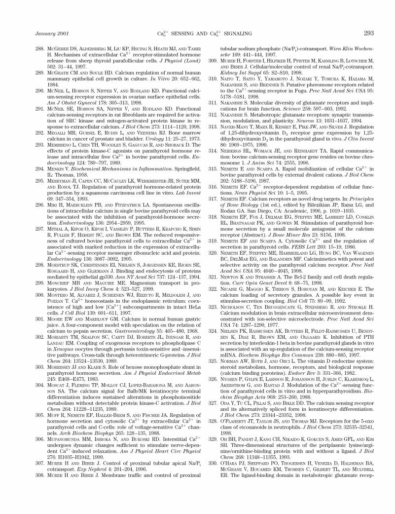
288. MCGEHEE DS, ALDERSBERG M, LIU KP, HSUING S, HEATH MJ, AND TAMIR
H. Mechanism of extracellular Ca21 receptor-stimulated hormonerelease from sheep thyroid parafollicular cells. J Physiol (Lond)
502: 31–44, 1997.289. MCGRATH CM AND SOULE HD. Calcium regulation of normal human
mammary epithelial cell growth in culture. In Vitro 20: 652–662,1984.
290. MCNEIL L, HOBSON S, NIPPER V, AND RODLAND KD. Functional calci-um-sensing receptor expression in ovarian surface epithelial cells.Am J Obstet Gynecol 178: 305–313, 1998.
291. MCNEIL SE, HOBSON SA, NIPPER V, AND RODLAND KD. Functionalcalcium-sensing receptors in rat fibroblasts are required for activa-tion of SRC kinase and mitogen-activated protein kinase in re-sponse to extracellular calcium. J Biol Chem 273: 1114–1120, 1998.
292. MEGALLI MR, GURSEL E, RUDIN L, AND VEENEMA RJ. Bone marrowcalcium in cancer of prostate and bladder. Urology 11: 25–27, 1973.
293. MEMBRENO L, CHEN TH, WOODLEY S, GAGUCAS R, AND SHOBACK D. Theeffects of protein kinase-C agonists on parathyroid hormone re-lease and intracellular free Ca21 in bovine parathyroid cells. En-
docrinology 124: 789–797, 1989.294. MENKIN V. Biochemical Mechanisms in Inflammation. Springfield,
IL: Thomas, 1958.295. MERRYMAN JI, CAPEN CC, MCCAULEY LK, WERKMEISTER JR, SUTER MM,
AND ROSOL TJ. Regulation of parathyroid hormone-related proteinproduction by a squamous carcinoma cell line in vitro. Lab Invest
69: 347–354, 1993.296. MIKI H, MAERCKLEIN PB, AND FITZPATRICK LA. Spontaneous oscilla-
tions of intracellular calcium in single bovine parathyroid cells maybe associated with the inhibition of parathyroid-hormone secre-tion. Endocrinology 136: 2954–2959, 1995.
297. MITHAL A, KIFOR O, KIFOR I, VASSILEV P, BUTTERS R, KRAPCHO K, SIMIN
R, FULLER F, HEBERT SC, AND BROWN EM. The reduced responsive-ness of cultured bovine parathyroid cells to extracellular Ca21 isassociated with marked reduction in the expression of extracellu-lar Ca21-sensing receptor messenger ribonucleic acid and protein.Endocrinology 136: 3087–3092, 1995.
298. MOESTRUP SK, CHRISTENSEN EI, NIELSEN S, JORGENSEN KE, BJORN SE,ROIGAARD H, AND GLIEMANN J. Binding and endocytosis of proteinsmediated by epithelial gp330. Ann NY Acad Sci 737: 124–137, 1994.
299. MONCRIEF MB AND MAGUIRE ME. Magnesium transport in pro-karyotes. J Biol Inorg Chem 4: 523–527, 1999.
300. MONTERO M, ALVAREZ J, SCHEENEN WJ, RIZZUTO R, MELDOLESI J, AND
POZZAN T. Ca21 homeostasis in the endoplasmic reticulum: coex-istence of high and low [Ca21] subcompartments in intact HeLacells. J Cell Biol 139: 601–611, 1997.
301. MOORE EW AND MAKHLOUF GM. Calcium in normal human gastricjuice. A four-component model with speculation on the relation ofcalcium to pepsin secretion. Gastroenterology 55: 465–480, 1968.
302. MORIARTY TM, SEALFON SC, CARTY DJ, ROBERTS JL, IYENGAR R, AND
LANDAU EM. Coupling of exogenous receptors to phospholipase Cin Xenopus oocytes through pertussis toxin-sensitive and -insensi-tive pathways. Cross-talk through heterotrimeric G-proteins. J Biol
Chem 264: 13524–13530, 1989.303. MORRISSEY JJ AND KLAHR S. Role of hexose monophosphate shunt in
parathyroid hormone secretion. Am J Physiol Endocrinol Metab
245: E468–E475, 1983.304. MOSCAT J, FLEMING TP, MOLLOY CJ, LOPEZ-BARAHONA M, AND AARON-
SON SA. The calcium signal for Balb/MK keratinocyte terminaldifferentiation induces sustained alterations in phosphoinositidemetabolism without detectable protein kinase-C activation. J Biol
Chem 264: 11228–11235, 1989.305. MUFF R, NEMETH EF, HALLER-BREM S, AND FISCHER JA. Regulation of
hormone secretion and cytosolic Ca21 by extracellular Ca21 inparathyroid cells and C-cells: role of voltage-sensitive Ca21 chan-nels. Arch Biochem Biophys 265: 128–135, 1988.
306. MUPANOMUNDA MM, ISHIOKA N, AND BUKOSKI RD. Interstitial Ca21
undergoes dynamic changes sufficient to stimulate nerve-depen-dent Ca21-induced relaxation. Am J Physiol Heart Circ Physiol
276: H1035–H1042, 1999.307. MURER H AND BIBER J. Control of proximal tubular apical Na/Pi
cotransport. Exp Nephrol 4: 201–204, 1996.308. MURER H AND BIBER J. Membrane traffic and control of proximal
tubular sodium phosphate (Na/Pi)-cotransport. Wien Klin Wochen-
schr 109: 441–444, 1997.309. MURER H, FORSTER I, HILFIKER H, PFISTER M, KAISSLING B, LOTSCHER M,
AND BIBER J. Cellular/molecular control of renal Na/Pi-cotransport.Kidney Int Suppl 65: S2–S10, 1998.
310. NAITO T, SAITO Y, YAMAMOTO J, NOZAKI Y, TOMURA K, HAZAMA M,NAKANISHI S, AND BRENNER S. Putative pheromone receptors relatedto the Ca21-sensing receptor in Fugu. Proc Natl Acad Sci USA 95:5178–5181, 1998.
311. NAKANISHI S. Molecular diversity of glutamate receptors and impli-cations for brain function. Science 258: 597–603, 1992.
312. NAKANISHI S. Metabotropic glutamate receptors: synaptic transmis-sion, modulation, and plasticity. Neuron 13: 1031–1037, 1994.
313. NAVEH-MANY T, MARX R, KESHET E, PIKE JW, AND SILVER J. Regulationof 1,25-dihydroxyvitamin D3 receptor gene expression by 1,25-dihydroxyvitamin D3 in the parathyroid gland in vivo. J Clin Invest
86: 1969–1975, 1990.314. NEIBERGS HL, WOMACK JE, AND REINHARDT TA. Rapid communica-
tion: bovine calcium-sensing receptor gene resides on bovine chro-mosome 1. J Anim Sci 74: 2555, 1996.
315. NEMETH E AND SCARPA A. Rapid mobilization of cellular Ca21 inbovine parathyroid cells by external divalent cations. J Biol Chem
202: 5188–5196, 1987.316. NEMETH EF. Ca21 receptor-dependent regulation of cellular func-
tions. News Physiol Sci 10: 1–5, 1995.317. NEMETH EF. Calcium receptors as novel drug targets. In: Principles
of Bone Biology (1st ed.), edited by Bilezikian JP, Raisz LG, andRodan GA. San Diego, CA: Academic, 1996, p. 1019–1035.
318. NEMETH EF, FOX J, DELMAR EG, STEFFEY ME, LAMBERT LD, CONKLIN
RL, BHATNAGAR PK, AND GOWEN M. Stimulation of parathyroid hor-mone secretion by a small molecule antagonist of the calciumreceptor (Abstract). J Bone Miner Res 23: S156, 1998.
319. NEMETH EF AND SCARPA A. Cytosolic Ca21 and the regulation ofsecretion in parathyroid cells. FEBS Lett 203: 15–19, 1986.
320. NEMETH EF, STEFFEY ME, HAMMERLAND LG, HUNG BC, VAN WAGENEN
BC, DELMAR EG, AND BALANDRIN MF. Calcimimetics with potent andselective activity on the parathyroid calcium receptor. Proc Natl
Acad Sci USA 95: 4040–4045, 1998.321. NEWTON K AND STRASSER A. The Bcl-2 family and cell death regula-
tion. Curr Opin Genet Devel 8: 68–75, 1998.322. NICAISE G, MAGGIO K, THIRION S, HOROYAN M, AND KEICHER E. The
calcium loading of secretory granules. A possible key event instimulus-secretion coupling. Biol Cell 75: 89–99, 1992.
323. NICHOLSON C, TEN BRUGGENCATE G, STEINBERG R, AND STROKLE H.Calcium modulation in brain extracellular microenvironment dem-onstrated with ion-selective microelectrode. Proc Natl Acad Sci
USA 74: 1287–1290, 1977.324. NIELSEN PK, RASMUSSEN AK, BUTTERS R, FELDT-RASMUSSEN U, BENDT-
ZEN K, DIAZ R, BROWN EM, AND OLGAARD K. Inhibition of PTHsecretion by interleukin-1 beta in bovine parathyroid glands in vitrois associated with an up-regulation of the calcium-sensing receptormRNA. Biochem Biophys Res Commun 238: 880–885, 1997.
325. NORMAN AW, ROTH J, AND ORCI L. The vitamin D endocrine system:steroid metabolism, hormone, receptors, and biological response(calcium binding poroteins). Endocr Rev 3: 331–366, 1982.
326. NYGREN P, GYLFE E, LARSSON R, JOHANSSON H, JUHLIN C, KLARESKOQ L,AKERSTROM G, AND RASTAD J. Modulation of the Ca21-sensing func-tion of parathyroid cells in vitro and in hyperparathyroidism. Bio-
chim Biophys Acta 968: 253–260, 1988.327. ODA Y, TU CL, PILLAI S, AND BIKLE DD. The calcium sensing receptor
and its alternatively spliced form in keratinocyte differentiation.J Biol Chem 273: 23344–23352, 1998.
328. O’FLAHERTY JT, TAYLOR JS, AND THOMAS MJ. Receptors for the 5-oxoclass of eicosanoids in neutrophils. J Biol Chem 273: 32535–32541,1998.
329. OH BH, PANDIT J, KANG CH, NIKAIDO K, GOKCEN S, AMES GFL, AND KIM
SH. Three-dimensional structures of the periplasmic lysine/argi-nine/ornithine-binding protein with and without a ligand. J Biol
Chem 268: 11348–11355, 1993.330. O’HARA PJ, SHEPPARD PO, THOGERSEN H, VENEZIA D, HALDEMAN BA,
MCGRANE V, HOUAMED KM, THOMSEN C, GILBERT TL, AND MULVIHILL
ER. The ligand-binding domain in metabotropic glutamate recep-
January 2001 Cao21 SENSING AND Cao
21 SIGNALING 293
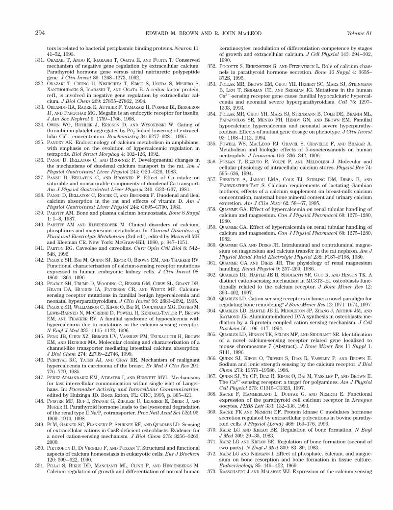
tors is related to bacterial periplasmic binding proteins. Neuron 11:41–52, 1993.
331. OKAZAKI T, ANDO K, IGARASHI T, OGATA E, AND FUJITA T. Conservedmechanism of negative gene regulation by extracellular calcium.Parathyroid hormone gene versus atrial natriuretic polypeptidegene. J Clin Invest 89: 1268–1273, 1992.
332. OKAZAKI T, CHUNG U, NISHISHITA T, EBISU S, USUDA S, MISHIRO S,XANTHOUDAKIS S, IGARASHI T, AND OGATA E. A redox factor protein,ref1, is involved in negative gene regulation by extracellular cal-cium. J Biol Chem 269: 27855–27862, 1994.
333. ORLANDO RA, RADER K, AUTHIER F, YAMAZAKI H, POSNER BI, BERGERON
JJ, AND FARQUHAR MG. Megalin is an endocytic receptor for insulin.J Am Soc Nephrol 9: 1759–1766, 1998.
334. OWEN WG, BICHLER J, ERICSON D, AND WYSOKINSKI W. Gating ofthrombin in platelet aggregates by PO2-linked lowering of extracel-lular Ca21 concentration. Biochemistry 34: 9277–9281, 1995.
335. PANDEY AK. Endocrinology of calcium metabolism in amphibians,with emphasis on the evolution of hypercalcemic regulation intetrapods. Biol Struct Morphog 4: 102–126, 1992.
336. PANSU D, BELLATON C, AND BRONNER F. Developmental changes inthe mechanisms of duodenal calcium transport in the rat. Am J
Physiol Gastrointest Liver Physiol 244: G20–G26, 1983.337. PANSU D, BELLATON C, AND BRONNER F. Effect of Ca intake on
saturable and nonsaturable components of duodenal Ca transport.Am J Physiol Gastrointest Liver Physiol 240: G32–G37, 1981.
338. PANSU D, BELLATON C, ROCHE C, AND BRONNER F. Duodenal and ilealcalcium absorption in the rat and effects of vitamin D. Am J
Physiol Gastrointest Liver Physiol 244: G695–G700, 1983.339. PARFITT AM. Bone and plasma calcium homeostasis. Bone 8 Suppl
1: 1–8, 1987.340. PARFITT AM AND KLEEREKOPER M. Clinical disorders of calcium,
phosphorus and magnesium metabolism. In: Clinical Disorders of
Fluid and Electrolyte Metabolism (3rd ed.), edited by Maxwell MHand Kleeman CR. New York: McGraw-Hill, 1980, p. 947–1151.
341. PARTON RG. Caveolae and caveolins. Curr Opin Cell Biol 8: 542–548, 1996.
342. PEARCE SH, BAI M, QUINN SJ, KIFOR O, BROWN EM, AND THAKKER RV.Functional characterization of calcium-sensing receptor mutationsexpressed in human embryonic kidney cells. J Clin Invest 98:1860–1866, 1996.
343. PEARCE SH, TRUMP D, WOODING C, BESSER GM, CHEW SL, GRANT DB,HEATH DA, HUGHES IA, PATERSON CR, AND WHYTE MP. Calcium-sensing receptor mutations in familial benign hypercalcemia andneonatal hyperparathyroidism. J Clin Invest 96: 2683–2692, 1995.
344. PEARCE SH, WILLIAMSON C, KIFOR O, BAI M, COULTHARD MG, DAVIES M,LEWIS-BARNED N, MCCREDIE D, POWELL H, KENDALL-TAYLOR P, BROWN
EM, AND THAKKER RV. A familial syndrome of hypocalcemia withhypercalciuria due to mutations in the calcium-sensing receptor.N Engl J Med 335: 1115–1122, 1996.
345. PENG JB, CHEN XZ, BERGER UV, VASSILEV PM, TSUKAGUCHI H, BROWN
EM, AND HEDIGER MA. Molecular cloning and characterization of achannel-like transporter mediating intestinal calcium absorption.J Biol Chem 274: 22739–22746, 1999.
346. PERCIVAL RC, YATES AJ, AND GRAY RE. Mechanism of malignanthypercalcemia in carcinoma of the breast. Br Med J Clin Res 291:776–779, 1985.
347. PEREZ-ARMANDARIZ EM, ATWATER I, AND BENNETT MVL. Mechanismsfor fast intercellular communication within single islet of Langer-hans. In: Pacemaker Activity and Intercellular Communication,edited by Huizinga JD. Boca Raton, FL: CRC, 1995, p. 305–321.
348. PFISTER MF, RUF I, STANGE G, ZIEGLER U, LEDERER E, BIBER J, AND
MURER H. Parathyroid hormone leads to the lysosomal degradationof the renal type II Na/Pi cotransporter. Proc Natl Acad Sci USA 95:1909–1914, 1998.
349. PI M, GARNER SC, FLANNERY P, SPURNEY RF, AND QUARLES LD. Sensingof extracellular cations in CasR-deficient osteoblasts. Evidence fora novel cation-sensing mechanism. J Biol Chem 275: 3256–3263,2000.
350. PIETROBON D, DI VIRGILIO F, AND POZZAN T. Structural and functionalaspects of calcium homeostasis in eukaryotic cells. Eur J Biochem
120: 599–622, 1990.351. PILLAI S, BIKLE DD, MANCIANTI ML, CLINE P, AND HINCENBERGS M.
Calcium regulation of growth and differentiation of normal human
keratinocytes: modulation of differentiation competence by stagesof growth and extracellular calcium. J Cell Physiol 143: 294–302,1990.
352. POCOTTE S, EHRENSTEIN G, AND FITZPATRICK L. Role of calcium chan-nels in parathyroid hormone secretion. Bone 16 Suppl 4: 365S–372S, 1995.
353. POLLAK MR, BROWN EM, CHOU YH, HEBERT SC, MARX SJ, STEINMANN
B, LEVI T, SEIDMAN CE, AND SEIDMAN JG. Mutations in the humanCa21-sensing receptor gene cause familial hypocalciuric hypercal-cemia and neonatal severe hyperparathyroidism. Cell 75: 1297–1303, 1993.
354. POLLAK MR, CHOU YH, MARX SJ, STEINMANN B, COLE DE, BRANDI ML,PAPAPOULOS SE, MENKO FH, HENDY GN, AND BROWN EM. Familialhypocalciuric hypercalcemia and neonatal severe hyperparathy-roidism. Effects of mutant gene dosage on phenotype. J Clin Invest
93: 1108–1112, 1994.355. POWELL WS, MACLEOD RJ, GRAVEL S, GRAVELLE F, AND BHAKAR A.
Metabolism and biologic effects of 5-oxoeicosanoids on humanneutrophils. J Immunol 156: 336–342, 1996.
356. POZZAN T, RIZZUTO R, VOLPE P, AND MELDOLESI J. Molecular andcellular physiology of intracellular calcium stores. Physiol Rev 74:595–636, 1994.
357. PRENTICE A, JARJOU LMA, COLE TJ, STIRLING DM, DIBBA B, AND
FAIRWEATHER-TAIT S. Calcium requirements of lactating Gambianmothers, effects of a calcium supplement on breast-milk calciumconcentration, maternal bone mineral content and urinary calciumexcretion. Am J Clin Nutr 62: 58–67, 1995.
358. QUAMME GA. Effect of hypercalcemia on renal tubular handling ofcalcium and magnesium. Can J Physiol Pharmacol 60: 1275–1280,1980.
359. QUAMME GA. Effect of hypercalcemia on renal tubular handling ofcalcium and magnesium. Can J Physiol Pharmacol 60: 1275–1280,1982.
360. QUAMME GA AND DIRKS JH. Intraluminal and contraluminal magne-sium on magnesium and calcium transfer in the rat nephron. Am J
Physiol Renal Fluid Electrolyte Physiol 238: F187–F198, 1980.361. QUAMME GA AND DIRKS JH. The physiology of renal magnesium
handling. Renal Physiol 9: 257–269, 1986.362. QUARLES DL, HARTLE JE II, SIDDHANTI SR, GUO R, AND HINSON TK. A
distinct cation-sensing mechanism in MC3T3–E1 osteoblasts func-tionally related to the calcium receptor. J Bone Miner Res 12:393–402, 1997.
363. QUARLES LD. Cation-sensing receptors in bone: a novel paradigm forregulating bone remodeling? J Bone Miner Res 12: 1971–1974, 1997.
364. QUARLES LD, HARTLE JE II, MIDDLETON JP, ZHANG J, ARTHUR JM, AND
RAYMOND JR. Aluminum-induced DNA synthesis in osteoblasts: me-diation by a G-protein coupled cation sensing mechanism. J Cell
Biochem 56: 106–117, 1994.365. QUARLES LD, HINSON TK, SELDIN MF, AND SIDDHANTI SR. Identification
of a novel calcium-sensing receptor related gene localized tomouse chromosome 7 (Abstract). J Bone Miner Res 11 Suppl 1:S141, 1996.
366. QUINN SJ, KIFOR O, TRIVEDI S, DIAZ R, VASSILEV P, AND BROWN E.Sodium and ionic strength sensing by the calcium receptor. J Biol
Chem 273: 19579–19586, 1998.367. QUINN SJ, YE CP, DIAZ R, KIFOR O, BAI M, VASSILEV P, AND BROWN E.
The Ca21-sensing receptor: a target for polyamines. Am J Physiol
Cell Physiol 273: C1315–C1323, 1997.368. RACKE F, HAMMERLAND L, DUBYAK G, AND NEMETH E. Functional
expression of the parathyroid cell calcium receptor in Xenopus
oocytes. FEBS Lett 333: 132–136, 1993.369. RACKE FK AND NEMETH EF. Protein kinase C modulates hormone
secretion regulated by extracellular polycations in bovine parathy-roid cells. J Physiol (Lond) 468: 163–176, 1993.
370. RAISZ LG AND KREAM BE. Regulation of bone formation. N Engl
J Med 309: 29–35, 1983.371. RAISZ LG AND KREAM BE. Regulation of bone formation (second of
two parts). N Engl J Med 309: 83–89, 1983.372. RAISZ LG AND NIEMANN I. Effect of phosphate, calcium, and magne-
sium on bone resorption and bone formation in tissue culture.Endocrinology 85: 446–452, 1969.
373. RASSCHAERT J AND MALAISSE WJ. Expression of the calcium-sensing
294 EDWARD M. BROWN AND R. JOHN MACLEOD Volume 81
receptor in pancreatic islet B-cells. Biochem Biophys Res Commun
264: 615–618, 1999.374. RAY JM, SQUIRES PE, CURTIS SB, MELOCHE MR, AND BUCHAN AM.
Expression of the calcium-sensing receptor on human antral gas-trin cells in culture. J Clin Invest 99: 2328–2333, 1997.
375. RAY K, CLAPP P, GOLDSMITH PK, AND SPIEGEL AM. Identification of thesites of N-linked glycosylation on the human calcium receptor andassessment of their role in cell surface expression and signaltransduction. J Biol Chem 273: 34558–34567, 1998.
376. RAY K, FAN GF, GOLDSMITH PK, AND SPIEGEL AM. The carboxylterminus of the human calcium receptor. Requirements for cell-surface expression and signal transduction. J Biol Chem 272:31355–31361, 1997.
377. RAY K, HAUSCHILD BC, STEINBACH PJ, GOLDSMITH PK, HAUACHE O, AND
SPIEGEL AM. Identification of the cysteine residues in the amino-terminal extracellular domain of the human Ca21 receptor criticalfor dimerization. Implications for function of monomeric Ca21
receptor. J Biol Chem 274: 27642–27650, 1999.378. REGINATO AM, TUAN RS, ONO T, JIMENEZ SA, AND JACENKO O. Effects
of calcium deficiency on chondrocyte hypertrophy and type Xcollagen expression in chick embryonic sternum. Dev Dyn 198:284–295, 1993.
379. RICCARDI D, HALL AE, CHATTOPADHYAY N, XU JZ, BROWN EM, AND
HEBERT SC. Localization of the extracellular Ca21/polyvalent cat-ion-sensing protein in rat kidney. Am J Physiol Renal Physiol 274:F611–F622, 1998.
380. RICCARDI D, LEE WS, LEE K, SEGRE GV, BROWN EM, AND HEBERT SC.Localization of the extracellular Ca21-sensing receptor and PTH/PTHrP receptor in rat kidney. Am J Physiol Renal Fluid Electrolyte
Physiol 271: F951–F956, 1996.381. RICCARDI D, PARK J, LEE WS, GAMBA G, BROWN EM, AND HEBERT SC.
Cloning and functional expression of a rat kidney extracellularcalcium/polyvalent cation-sensing receptor. Proc Natl Acad Sci
USA 92: 131–135, 1995.382. RIDEFELT P, HELLMAN P, LUNDGGREN O, LLUNGHALL S, AKERSTROM G,
RASTAD J, AND GYLFE E. Ga31 inhibits parathyroid hormone releasewithout interacting with the Ca21 receptor of the parathyroid cell.Biochem Biophys Res Commun 186: 1556–1561, 1992.
383. RIDEFELT P, HELLMAN P, WALLFELT C, AKERSTROM G, RASTAD J, AND
GYLFE E. Neomycin interacts with Ca21 sensing of normal andadenomatous parathyroid cells. Mol Cell Endocrinol 83: 211–218,1992.
384. ROBERTSON WG AND MARSHALL RW. Ionized calcium in body fluids.Crit Rev Clin Lab Sci 15: 85–125, 1981.
385. RODDA CP, CAPLE IW, AND MARTIN TJ. Role of PTHrP in fetal andneonatal physiology. In: Parathyroid Hormone Related Protein:
Normal Physiology and Its Role in Cancer, edited by Halloran BPand Nissenson RA. Boca Raton, FL: CRC, 1992, p. 169–196.
386. ROGERS K, DUNN C, HEBERT S, BROWN E, AND NEMETH E. Pharmaco-logical comparison of bovine parathyroid, human parathyroid, andrat kidney calcium receptors expressed in HEK 293 cells (Ab-stract). J Bone Miner Res 10 Suppl 1: S483, 1995.
387. ROGERS KV, DUNN CK, CONKLIN RL, HADFIELD S, PETTY BA, BROWN
EM, HEBERT SC, NEMETH EF, AND FOX J. Calcium receptor messen-ger ribonucleic acid levels in the parathyroid glands and kidney ofvitamin D-deficient rats are not regulated by plasma calcium or1,25-dihydroxyvitamin D3. Endocrinology 136: 499–504, 1995.
388. ROGERS KV, DUNN CK, HEBERT SC, AND BROWN EM. Localization ofcalcium receptor mRNA in the adult rat central nervous system byin situ hybridization. Brain Res 744: 47–56, 1997.
389. ROMANO C, YANG WL, AND O’MALLEY KL. Metabotropic glutamatereceptor 5 is a disulfide-linked dimer. J Biol Chem 271: 28612–28616, 1996.
390. ROMOLI R, LANIA A, MANTOVANI G, CORBETTA S, PERSANI L, AND SPADA
A. Expression of calcium-sensing receptor and characterization ofintracellular signaling in human pituitary adenomas. J Clin Endo-
crinol Metab 84: 2848–2853, 1999.391. ROUSSANNE MC, GOGUSEV J, HORY B, DUCHAMBON P, SOUBERBIELLE JC,
NABARRA B, PIERRAT D, SARFATI E, DRUEKE T, AND BOURDEAU A.Persistence of Ca21-sensing receptor expression in functionallyactive, long-term human parathyroid cell cultures. J Bone Miner
Res 13: 354–362, 1998.392. RUAT M, MOLLIVER ME, SNOWMAN AM, AND SNYDER SH. Calcium
sensing receptor: molecular cloning in rat and localization to nerveterminals. Proc Natl Acad Sci USA 92: 3161–3165, 1995.
393. RUAT M, SNOWMAN AM, HESTER LD, AND SNYDER SH. Cloned andexpressed rat Ca21-sensing receptor. J Biol Chem 271: 5972–5975,1996.
394. RUSSELL J, BAR A, SHERWOOD LM, AND HURWITZ S. Interaction be-tween calcium and 1,25-dihydroxyvitamin D3 in the regulation ofpreproparathyroid hormone and vitamin D receptor messengerribonucleic acid in avian parathyroids. Endocrinology 132: 2639–2644, 1993.
395. RUSSELL J, LETTIERI DF, AND SHERWOOD LM. Direct regulation bycalcium of cytoplasmic messenger ribonucleic acid coding for pre-proparathyroid hormone in isolated bovine parathyroid cells.J Clin Invest 72: 1851–1855, 1983.
396. RUTTEN MJ, BACON KD, MARLINK KL, STONEY M, MEICHSNER CL, LEE
FP, HOBSON SA, RODLAND KD, SHEPPARD BC, TRUNKEY DD, DEVENEY
KE, AND DEVENEY CW. Identification of a functional Ca21-sensingreceptor in normal human gastric mucous epithelial cells. Am J
Physiol Gastrointest Liver Physiol 277: G662–G670, 1999.397. RYBA NJ AND TIRINDELLI R. A new multigene family of putative
pheromone receptors. Neuron 19: 371–379, 1997.398. SAITO A, PIETROMONACO S, LOO AK, AND FARQUHAR MG. Complete
cloning and sequencing of rat gp330/“megalin,” a distinctive mem-ber of the low density lipoprotein receptor gene family. Proc Natl
Acad Sci USA 91: 9725–9729, 1994.399. SANDERS JL, CHATTOPADHYAY N, KIFOR O, YAMAGUCHI T, AND BROWN
EM. Extracellular calcium-sensing receptor (CaR) expression andits potential role in parathyroid hormone-related peptide (PTHrP)secretion in the H-500 rat Leydig cell model of humoral hypercal-cemia of malignancy. Biochem Biophys Res Commun 269: 427–432, 2000.
400. SANDS JM, FLORES FX, KATO A, BAUM MA, BROWN EM, WARD DT,HEBERT SC, AND HARRIS HW. Vasopressin-elicited water and ureapermeabilities are altered in IMCD in hypercalcemic rats. Am J
Physiol Renal Physiol 274: F978–F985, 1998.401. SANDS JM, NARUSE M, BAUM M, JO I, HEBERT SC, BROWN EM, AND
HARRIS HW. Apical extracellular calcium/polyvalent cation-sensingreceptor regulates vasopressin-elicited water permeability in ratkidney inner medullary collecting duct. J Clin Invest 99: 1399–1405, 1997.
402. SCHERUBL H, KLEPPISCH T, ZINK A, RAUE F, KRAUTWURST D, AND
HESCHELER J. Major role of dihydropyridine-sensitive Ca21 channelsin Ca21-induced calcitonin secretion. Am J Physiol Endocrinol
Metab 264: E354–E360, 1993.403. SCHLEGEL A, VOLONTE D, ENGELMAN JA, GALBIATI F, MEHTA P, ZHANG
XL, SCHERER PE, AND LISANTI MP. Crowded little caves: structureand function of caveolae. Cell Signal 10: 457–463, 1998.
404. SHANKAR VA, ALAM AS, BAX CM, PAZIANIS M, HUANG CL, AND ZAIDI M.Activation and inactivation of the osteoclast Ca21-receptor by thetrivalent cation, La31. Biochem Biophys Res Commun 187: 907–912, 1992.
405. SHANKAR VS, BAX CM, ALAM AS, BAX BE, HUANG CL, AND ZAIDI M. Theosteoclast Ca21 receptor is highly sensitive to activation by transi-tion metal cations. Biochem Biophys Res Commun 187: 913–918,1992.
406. SHANKAR VS, PAZIANIS M, HUANG CL, SIMON B, ADEBANJO CA, AND ZAIDI
M. Caffeine modulates Ca21 receptor activation in isolated ratosteclasts and induces intracellular Ca21 release. Am J Physiol
Renal Fluid Electrolyte Physiol 268: F447–F454, 1995.407. SHARFF AJ, RODSETH LE, SPURLINO JC, AND QUIOCHO FA. Crystallo-
graphic evidence for a large ligand-induced hinge-twist motionbetween the two domains of the maltodextrin binding proteininvolved in active transport and chemotaxis. Biochemistry 31:10657–10663, 1992.
408. SHOBACK DM AND CHEN TH. Effects of protein kinase C activation oninositol phosphate generation and intracellular Ca21 mobilizationin bovine parathyroid cells. Endocrinology 127: 141–148, 1990.
409. SHOBACK DM, MEMBRENO LA, AND MCGHEE JG. High calcium andother divalent cations increase inositol trisphosphate in bovineparathyroid cells. Endocrinology 123: 382–389, 1988.
410. SHORTE SL AND SCHOFIELD JG. The effect of extracellular polyvalentcations on bovine anterior pituitary cells. Evidence for a Ca21-
January 2001 Cao21 SENSING AND Cao
21 SIGNALING 295
sensing receptor coupled to release of intracellular calcium stores.
Cell Calcium 19: 43–57, 1996.411. SIEGEL AM AND DALY JW. Receptor (norepinephrine), P-site (29,59-
dideoxyadenosine), and calcium-mediated inhibition of prostaglan-din and forskolin-activated cAMP generating systems in humanplatelets. J Cyclic Nucleotide Protein Phosphorylation Res 10:229–246, 1985.
412. SILVER IA, MURRILS RJ, AND ETHERINGTON DJ. Microlectrode studieson the acid microenvironment beneath adherent macrophages andosteoclasts. Exp Cell Res 175: 266–276, 1988.
413. SILVER J, MOALLEM E, KILAV R, EPSTEIN E, SELA A, AND NAVEH-MANY T.New insights into the regulation of parathyroid hormone synthesisand secretion in chronic renal failure. Nephrol Dial Transplant 11:2–5, 1996.
414. SILVERBERG SJ, BONE HG III, MARRIOTT TB, LOCKER FG, THYS-JACOBS
S, DZIEM G, KAATZ S, SANGUINETTI EL, AND BILEZIKIAN JP. Short-terminhibition of parathyroid hormone secretion by a calcium-receptoragonist in patients with primary hyperparathyroidism. N Engl
J Med 337: 1506–1510, 1997.415. SIMPSON JB AND ROUTENBERG A. Subfornical organ lesions reduce
intravenous angiotensin-induced drinking. Brain Res 88: 154–161,1975.
416. SLATTERY ML, SORENSON AW, AND FORD MH. Dietary calcium intakeas a mitigating factor in colon cancer. Am J Epidemiol 128: 504–514, 1988.
417. SPURNEY RF, PI M, FLANNERY P, AND QUARLES LD. Aluminum is aweak agonist for the calcium-sensing receptor. Kidney Int 55:1750–1758, 1999.
418. SQUIRES PE, HARRIS TE, PERSAUD SJ, CURTIS SB, BUCHAN AMJ, AND
JONES PM. The extracellular calcium-sensing receptor on humanbeta-cells negatively modulates insulin secretion. Diabetes 49: 409–417, 2000.
419. STEWART AF AND BROADUS AE. Mineral metabolism. In: Endocrinol-
ogy and Metabolism (2nd ed.), edited by Felig P, Baxter JD, Broa-dus AE, and Frohman LA. New York: McGraw-Hill, 1987, p. 1317–1453.
420. STREWLER GJ. Familial benign hypocalciuric hypercalcemia—fromthe clinic to the calcium sensor. West J Med 160: 579–580, 1994.
421. SUGIMOTO T, KANATANI M, KANO J, KAJI H, TSUKAMOTO T, YAMAGUCHI T,FUKASE M, AND CHIHARA K. Effects of high calcium concentration onthe functions and interactions of osteoblastic cells and monocytesand on the formation of osteoclast-like cells. J Bone Miner Res 8:1445–1452, 1993.
422. SUKI WN, EKNOYAN G, RECTOR FC JR, AND SELDIN DW. The renaldiluting and concentrating mechanism in hypercalcemia. Nephron
6: 50–61, 1969.423. SUNAHARA RK, DESSAUER CW, AND GILMAN AG. Complexity and di-
versity of mammalian adenylyl cyclases. Annu Rev Pharmacol
Toxicol 36: 461–480, 1996.424. SUTTON RAL AND DIRKS JH. Calcium and magnesium: renal handling
and disorders of metabolism. In: The Kidney (3rd ed.), edited byBrenner B and Rector F Jr. Philadelphia, PA: Saunders, 1986, p.551–618.
425. SUZUKI K, LAVARONI S, MORI A, OKAJIMA F, KIMURA S, KATOH R, KAWAOI
A, AND KOHN LD. Thyroid transcription factor 1 is calcium modu-lated and coordinately regulates genes involved in calcium ho-meostasis in C cells. Mol Cell Biol 18: 7410–7422, 1998.
426. TAKAICHI K AND KUROKAWA K. Inhibitory guanosine triphosphate-binding protein-mediated regulation of vasopressin action in iso-lated single medullary tubules of mouse kidney. J Clin Invest 82:1437–1444, 1988.
427. TAL M, AMMAR DA, KARPUJ M, KRIZHANOVSKY V, NAIM M, AND THOMP-SON DA. A novel putative neuropeptide receptor expressed in neu-ral tissue, including sensory epithelia. Biochem Biophys Res Com-
mun 209: 752–759, 1995.428. TAM R AND SAIER MH JR. Structural, functional, and evolutionary
relationships among extracellular solute-binding receptors of bac-teria. Microbiol Rev 57: 320–346, 1993.
429. TAMIR H, LIU KP, ALDERSBERG M, HSUIUNG SC, AND GERSHON MD.Acidification of serotonin-containing secretory vesicles induced bya plasma membrane calcium receptor. J Biol Chem 271: 6441–6450,1996.
430. TARGOVNIK JH, RODMAN JS, AND SHERWOOD LM. Regulation of para-
thyroid hormone secretion in vitro: quantitative aspects of calciumand magnesium ion control. Endocrinology 88: 1477–1482, 1971.
431. TEPIKIN AV, VORONINA SG, GALLACHER DV, AND PETERSEN OH. Pulsa-tile Ca21 extrusion from single pancreatic acinar cells during re-ceptor-activated cytosolic Ca21 spiking. J Biol Chem 267: 14073–14076, 1992.
432. TOFFALETTI J, COOPER DL, AND LOBAUGH B. The response of parathy-roid hormone to specific changes in either ionized calcium, ionizedmagnesium, or protein-bound calcium in humans. Metabolism 40:814–818, 1991.
433. TORDOFF MG. Voluntary intake of calcium and other minerals byrats. Am J Physiol Regulatory Integrative Comp Physiol 267:R470–R475, 1994.
434. TRUMP D, WHYTE MP, WOODING C, PANG JT, PEARCE SH, KOCHER DB,AND THAKKER RV. Linkage studies in a kindred from Oklahoma, withfamilial benign (hypocalciuric) hypercalcaemia (FBH) and devel-opmental elevations in serum parathyroid hormone levels, indicatea third locus for FBH. Hum Genet 96: 183–187, 1995.
435. TU CL, ODA Y, AND BIKLE DD. Effects of a calcium receptor activatoron the cellular response to calcium in human keratinocytes. J In-
vest Dermatol 113: 340–345, 1999.436. VALTIN H. Renal Function. Mechanisms Preserving Fluid and
Electrolyte Balance in Health. Boston, MA: Little, Brown, 1983.437. VAN BIESEN T, HAWES B, LUTTRELL D, KRUEGER K, TOUHARA K, PORFIRI
E, SAKAUE M, LUTTRELL L, AND LEFKOWITZ R. Receptor-tyrosine-ki-nase and Gbeta and gamma-mediated MAP kinase activation by acommon signalling pathway. Nature 376: 781–784, 1995.
438. VAN BIESEN T, LUTTRELL LM, HAWES BE, AND LEFKOWITZ RJ. Mitogenicsignaling via G protein-coupled receptors. Endocr Rev 17: 698–714,1996.
439. VARRAULT A, PENA MS, GOLDSMITH PK, MITHAL A, BROWN EM, AND
SPIEGEL AM. Expression of G protein alpha-subunits in bovineparathyroid. Endocrinology 136: 4390–4396, 1995.
440. VASSART G. New pathophysiological mechanisms for hyperthyroid-ism. Horm Res 48: 47–50, 1997.
441. VASSILEV PM, HO-PAO CL, KANAZIRSKA MP, YE C, HONG K, SEIDMAN CE,SEIDMAN JG, AND BROWN EM. Cao-sensing receptor (CaR)-mediatedactivation of K1 channels is blunted in CaR gene-deficient mouseneurons. Neuroreport 8: 1411–1416, 1997.
442. VASSILEV PM, KANAZIRSKA MP, YE C, FRANCIS J, HONG K, AND BROWN
EM. A flickery block of a K1 channel mediated by extracellularCa21 and other agonists of the Ca21-sensing receptors in dispersedbovine parathyroid cells. Biochem Biophys Res Commun 230:616–623, 1997.
443. VASSILEV PM, MITCHEL J, VASSILEV M, KANAZIRSKA M, AND BROWN EM.Assessment of frequency-dependent alterations in the level of ex-tracellular Ca21 in the synaptic cleft. Biophys J 72: 1–14, 1997.
444. VELDHUIS JD, BORGES JL, DRAKE CR, AND ROGOL AD. Divergent influ-ences of calcium ions on releasing factor-stimulated anterior pitu-itary hormone secretion in normal man. J Clin Endocrinol Metab
59: 56–61, 1984.445. WADA M, FURUYA Y, SAKIYAMA J, KOBAYASHI N, MIYATA S, ISHII H, AND
HAGANO N. The calcimimetic compound NPS R-568 suppressesparathyroid cell proliferation in rats with renal insufficiency. J Clin
Invest 100: 2977–2983, 1997.446. WAGNER GF, DIMATTIA GE, DAVIE JR, COPP DH, AND FRIESEN HG.
Molecular cloning and cDNA sequence analysis of coho salmonstanniocalcin. Mol Cell Endocrinol 90: 7–15, 1992.
447. WAGNER GF, MILLIKEN C, FRIESEN HG, AND COPP DH. Studies on theregulation and characterization of plasma stanniocalcin in rainbowtrout. Mol Cell Endocrinol 79: 129–138, 1991.
448. WAKITA H, FURUKAWA F, BABA S, AND TAKIGAWA M. Human squamous-cell-carcinoma cell line (DJM-1) cells synthesize P-cadherin mole-cules via an elevation of extracellular calcium: calcium regulatesP-cadherin-gene expression at the translational level via proteintyrosine phosphorylation. Int J Cancer 73: 432–439, 1997.
449. WANG W, LU M, BALAZY M, AND HEBERT SC. Phospholipase A2 isinvolved in mediating the effect of extracellular Ca21 on apical K1
channels in rat TAL. Am J Physiol Renal Physiol 273: F421–F429,1997.
450. WANG WH, LU M, AND HEBERT SC. Cytochrome P-450 metabolitesmediate extracellular Ca21-induced inhibition of apical K1 chan-nels in the TAL. Am J Physiol Cell Physiol 271: C103–C111, 1996.
296 EDWARD M. BROWN AND R. JOHN MACLEOD Volume 81
451. WANG Y AND BUKOSKI RD. Distribution of the perivascular nerveCa21 receptor in rat arteries. Br J Pharmacol 125: 1397–1404, 1998.
452. WARD DT, BROWN EM, AND HARRIS HW. Disulfide bonds in theextracellular calcium-polyvalent cation-sensing receptor correlatewith dimer formation and its response to divalent cations in vitro.J Biol Chem 273: 14476–14483, 1998.
453. WASSERMAN RH AND FULLMER CS. Vitamin D and intestinal calciumtransport: facts, speculations and hypotheses. J Nutr 125: 1971S–1979S, 1995.
454. WATSON S AND ARKINSTALL. The G-Protein Linked Receptor Facts
Book. New York: Academic, 1994.455. WEISINGER JR, FAVUS MJ, LANGMAN CB, AND BUSHINSKY D. Regulation
of 1,25-dihydroxyvitamin D3 by calcium in the parathyroidecto-mized, parathyroid hormone-replete rat. J Bone Miner Res 4: 929–935, 1989.
456. WHITE JH, WISE A, MAIN MJ, GREEN A, FRASER NJ, DISNEY GH, BARNES
AA, EMSON P, FOORD SM, AND MARSHALL FH. Heterodimerization isrequired for the formation of a functional GABA(B) receptor. Na-
ture 396: 679–682, 1998.457. WHITFIELD J. Calcium as differentiator and killer-colon cells. In:
Calcium in Cell Cycles and Cancer, edited by Whitfield JF. BocaRaton, FL: CRC, 1995, p. 153–177.
458. WHITING PJ. The GABA-A receptor gene family: new targets fortherapeutic intervention. Neurochem Int 34: 387–390, 1999.
459. WISE A, GREEN A, MAIN MJ, WILSON R, FRASER N, AND MARSHALL FH.Calcium sensing properties of the GABA(B) receptor. Neurophar-
macology 38: 1647–1656, 1999.460. WONG M AND TUAN RS. Interactive cellular modulation of chondro-
genic differentiation in vitro by subpopulations of chick embryoniccalvarial cells. Dev Biol 167: 130–147, 1995.
461. YAMAGUCHI T, CHATTOPADHYAY N, AND BROWN EM. G protein-coupledextracellular Ca21 (Cao
21)-sensing receptor (CaR): roles in cellsignaling and control of diverse cellular functions. Adv Pharmacol
47: 209–253, 1999.462. YAMAGUCHI T, CHATTOPADHYAY N, KIFOR O, AND BROWN EM. Extracel-
lular calcium (Cao21)-sensing receptor in a murine bone marrow-
derived stromal cell line (ST2): potential mediator of the actions ofCao
21 on the function of ST2 cells. Endocrinology 139: 3561–3568,1998.
463. YAMAGUCHI T, CHATTOPADHYAY N, KIFOR O, BUTTERS RR JR, SUGIMOTO
T, AND BROWN EM. Mouse osteoblastic cell line (MC3T3–E1) ex-presses extracellular calcium (Cao
21)-sensing receptor and its ago-nists stimulate chemotaxis and proliferation of MC3T3–E1 cells.J Bone Miner Res 13: 1530–1538, 1998.
464. YAMAGUCHI T, KIFOR O, CHATTOPADHYAY N, BAI M, AND BROWN EM.Extracellular calcium (Cao
21)-sensing receptor in a mouse mono-cyte-macrophage cell line (J774): potential mediator of the actionsof Cao
21 on the function of J774 cells. J Bone Miner Res 13:1390–1397, 1998.
465. YAMAGUCHI T, KIFOR O, CHATTOPADHYAY N, AND BROWN EM. Expres-sion of extracellular calcium (Cao
21)-sensing receptor in the clonalosteoblast-like cell lines, UMR-106 and SAOS-2. Biochem Biophys
Res Commun 243: 753–757, 1998.466. YAMAGUCHI T, OLOZAK I, CHATTOPADHYAY N, BUTTERS RR, KIFOR O,
SCADDEN DT, AND BROWN EM. Expression of extracellular calcium(Cao
21)-sensing receptor in human peripheral blood monocytes.Biochem Biophys Res Commun 246: 501–506, 1998.
467. YAMAMOTO M, IGARISHI T, MURAMATSU M, FUKAGAWA M, MOTOKURA T,
AND OGATA E. Hypocalcemia increases and hypercalcemia de-creases the steady state level of parathyroid hormone messengerRNA in the rat. J Clin Invest 83: 1053–1056, 1989.
468. YAMOAH EN, LUMPKIN EA, DUMONT RA, SMITH PJ, HUDSPETH AJ, AND
GILLESPIE PG. Plasma membrane Ca21-ATPase extrudes Ca21 fromhair cell stereocilia. J Neurosci 18: 610–624, 1998.
469. YANG T, HASSAN S, HUANG YG, SMART AM, BRIGGS JP, AND SCHNER-MANN JB. Expression of PTHrP, PTH/PTHrP receptor, and Ca21-sensing receptor mRNAs along the rat nephron. Am J Physiol
Renal Physiol 272: F751–F758, 1997.470. YE C, HO-PAO CL, KANAZIRSKA M, QUINN S, ROGERS K, SEIDMAN CE,
SEIDMAN JG, BROWN EM, AND VASSILEV PM. Amyloid-beta proteinsactivate Ca21-permeable channels through calcium-sensing recep-tors. J Neurosci Res 47: 547–554, 1997.
471. YE C, HO-PAO CL, KANAZIRSKA M, QUINN S, SEIDMAN CE, SEIDMAN JG,BROWN EM, AND VASSILEV PM. Deficient cation channel regulation inneurons from mice with targeted disruption of the extracellularCa21-sensing receptor gene. Brain Res Bull 44: 75–84, 1997.
472. YE C, KANAZIRSKA M, QUINN S, BROWN EM, AND VASSILEV PM. Modu-lation by polycationic Ca21-sensing receptor agonists of nonselec-tive cation channels in rat hippocampal neurons. Biochem Biophys
Res Commun 224: 271–280, 1996.473. YE C, ROGERS K, BAI M, QUINN SJ, BROWN EM, AND VASSILEV PM.
Agonists of the Ca21-sensing receptor (CaR) activate nonselectivecation channels in HEK293 cells stably transfected with the humanCaR. Biochem Biophys Res Commun 226: 572–579, 1996.
474. YOSHIDA N, SATO T, KOBAYASHI K, AND OKADA Y. High extracellularCa21 and Ca21-sensing receptor agonists activate nonselective cat-ion conductance in freshly isolated rat osteoclasts. Bone 22: 495–501, 1998.
475. ZAIDI M, ADEBANJO OA, MOONGA BS, SUN L, AND HUANG CL. Emerginginsights into the role of calcium ions in osteoclast regulation.J Bone Miner Res 14: 669–674, 1999.
476. ZAIDI M, DATTA HK, PATCHELL A, MOONGA B, AND MACINTYRE I. “Cal-cium-activated” intracellular calcium elevation: a novel mechanismof osteoclast regulation. Biochem Biophys Res Commun 183:1461–1465, 1989.
477. ZAIDI M, KERBY J, HUANG CL, ALAM T, RATHOD H, CHAMBERS TJ, AND
MOONGA B. Divalent cations mimic the inhibitory effect of extracel-lular ionised calcium on bone resorption by isolated rat oste-oclasts: further evidence for a “calcium receptor.” J Cell Physiol
149: 422–427, 1991.478. ZAIDI M, SHANKAR VS, ALAM AST, MOONGA BS, PAZIANIS M, AND HUANG
CL. Evidence that a ryanodine receptor triggers signal transductionin the osteoclast. Biochem Biophys Res Commun 188: 1332–1336,1992.
479. ZAIDI M, SHANKAR VS, TUNWELL R, ADEBANJO OA, MACKRILL J, PAZIANAS
M, O’CONNELL D, SIMON BJ, RIFKIN BR, AND VENKITARAMAN AR. Aryanodine receptor-like molecule expressed in the osteoclastplasma membrane functions in extracellular Ca21 sensing. J Clin
Invest 96: 1582–1590, 1995.480. ZENG FY AND WESS J. Identification and molecular characterization
of m3 muscarinic receptor dimers. J Biol Chem 274: 19487–19497,1999.
481. ZINK-LORENZ A, FLOCKERZ V, FREICHEL M, GRAUER A, HAFNER M, AND
RAUE F. Evidence for a “calcium-sensing” receptor in C-cells (Ab-stract). J Bone Miner Res 10: S284, 1995.
January 2001 Cao21 SENSING AND Cao
21 SIGNALING 297