calcium-sensing receptor stimulation induces nonselective cation channel activation in breast cancer...
TRANSCRIPT

Calcium-Sensing Receptor Stimulation Induces Nonselective Cation Channel Activation in
Breast Cancer Cells
Yassine El Hiani1, Ahmed Ahidouch1,2, Morad Roudbaraki3, Stephanie Guenin4, Gerard Brule1,
Halima Ouadid-Ahidouch1
1Laboratoire de Physiologie Cellulaire et Moleculaire, EA 2086, Faculte des Sciences, Universite de Picardie Jules Verne, 33 rue Saint-leu,
80039 Amiens, France2Laboratoire de Physiologie Animale, Faculte des Sciences, Universite Ibn-Zohr, BP 28/S, 80000 Agadir, Morocco3INSERM EMI 0228, Universite de Lille, 59655 Villeneuve d�Ascq, France4Centre de Ressources Regionales en Biologie Moleculaire, Universite de Picardie Jules Verne, 10 rue Baudeloque, 80039 Amiens , Cedex 1,
France
Received: 24 March 2006/Revised: 30 June 2006
Abstract. The calcium-sensing receptor (CaR) isexpressed in epithelial ducts of both normal humanbreast and breast cancer tissue, as well as in the MCF-7 cell line as assessed by immunohistochemistry andWestern blot analysis. However, to date, there are nodata regarding the transduction pathways of CaR inbreast cancer cells. In this study, we show that a CaRagonist, spermine, and increased extracellular Ca2+
([Ca2+]o) sequentially activate two inward currents at–80 mV. The first was highly permeable to Ca2+ andinhibited by 2-aminophenyl borate (2-APB). In con-trast, the second was more sensitive to Na+ and Li+
than to Ca2+ and insensitive to 2-APB. Furthermore,intracellular dialysis with high Mg2+, flufenamic acidor amiloride perfusion was without any effect on thesecond current. Both currents were inhibited by La3+.Calcium imaging recordings showed that both[Ca2+]o and spermine induced an increase in intra-cellular calcium ([Ca2+]i) and that removal of extra-cellular Ca2+ or perfusion of 2-APB caused a declinein [Ca2+]i. It is well known that stimulation of CaRby an increase in [Ca2+]o or with spermine is associ-ated with activation of phospholipase C (PLC).Inhibition of PLC reduced the [Ca2+]o-stimulated[Ca2+]i increase. Lastly, reverse-transcriptase poly-merase chain reaction showed that MCF-7 cells ex-pressed canonical transient receptor potential(TRPCs) channels. Our results suggest that, in MCF-
7 cells, CaR is functionally coupled to Ca2+-perme-able cationic TRPCs, for which TRPC1 and TRPC6are the most likely candidates for the highly selectiveCa2+ current. Moreover, the pharmacology of thesecond Na+ current excludes the involvement of themore selective Na+ transient receptor potential mel-astatin (TRPM4 and TRPM5) and the classical epi-thelial Na+ channels.
Key words: Calcium-sensing receptor — Cationiccurrent — Transient receptor potential channel —Breast cancer cell
Introduction
Breast cancer is the most common cancer and aleading cause of cancer-associated death in women(Boring et al., 1994). It most commonly metastasizesto the bone, with 70% of patients dying of breastcarcinoma having bone metastases (Mundy, 1997).Consequently, tumors induce bone resorption andlead to the release of large quantities of Ca2+ into thebone microenvironment. The local Ca2+ level atresorption sites has been reported to rise as high as40 mM (Silver, Murrills & Etherington, 1988). It hasbeen reported that high Ca2+ concentrations stimu-late the calcium-sensing receptor (CaR), therebyinducing parathormone-related peptide (PTHrP)production by MCF-7 and MDA-MB-231 breastcancer cells (Sanders et al., 2000). High levels ofPTHrP contribute to increased bone resorption andto the osteolysis observed in association with metas-tasis of epithelium-derived tumors to bone (Rodland,
This work concretizes the scientific cooperation between Universite
de Picardie Jules Verne and Universite Ibn-Zohr.
Correspondence to:Halima Ouadid-Ahidouch; email: ha-sciences@
u-picardie.fr
J. Membrane Biol. 211, 127–137 (2006)
DOI: 10.1007/s00232-006-0017-2

2004). The potential role of CaR in modulatingPTHrP secretion has been investigated in breastcancer (Sanders et al., 2000), prostate cancer (Sanderset al., 2001), glial tumors (Chattopadyay et al., 2000)and Leydig cell tumors (Buchs et al., 2000). Recently,Li, Huang & Peng (2005) reported overexpression ofCaR in some cancer tissues and suggested that CaRmay play a role in cancer progression.
CaR is a G protein-coupled receptor, originallycloned from bovine parathyroid cells (Brown et al.,1993), which plays an important role in the mainte-nance of systemic Ca2+ homeostasis. It has been re-ported that CaR stimulation by an increase inextracellular Ca2+ concentration ([Ca2+]o) or apolyamine (spermine) is associated with activation ofphospholipase C (PLC) (Huang, Handlogten & Mill-er, 2002) and increases the levels of intracellular ino-sitol 1,4,5-trisphosphate (IP3) (Guise et al., 1996). It iswell known that PLC cleaves phosphatidylinositol4,5-biphosphate into IP3 and diacylglycerol (DAG).IP3 releases Ca2+ from intracellular stores, and theconcomitant store depletion activates store-operatedCa2+ channels (SOCs), which in many cases have beententatively identified as transient receptor potential(TRP) family members (Zitt, Halaszovich & Luckhoff,2002; Pedersen, Owsianik & Nilius, 2005; Parekh &Putney, 2005). Moreover, DAG is also capable ofactivating some other TRPs directly, without deplet-ing intracellular Ca2+ stores (Pedersen et al., 2005).
In breast cells, CaR stimulation induces an in-crease in intracellular Ca2+ concentration ([Ca2+]i)(Parkash, Chaudhry & Rhoten, 2004) as well asPTHrP secretion (Sanders et al., 2000). Furthermore, acationic nonselective current has been recorded both inresponse to CaR stimulation in hippocampal neurons(Ye et al., 1996a, 1997) and in HEK293 cells stablytransfected with human CaR (Ye et al., 1996b). Thegreater incidence of this cationic current has beenassociated with U373 human astrocytoma cell prolif-eration (Chattopadhyay et al., 2000) and with differ-entiation of human promyelocytic leukemia cells(Yamaguchi et al., 2000). However, nothing is cur-rently known about the pharmacological and electro-physiological properties of this cationic current andthe link between the CaR and TRP channels (TRPCs).
We combined electrophysiological and molecularmethods to demonstrate, for the first time, that bothCaR agonists (spermine and Ca2+) sequentiallyactivate two cationic currents: a primary highlyCa2+-sensitive current (I1), which is always followedby a secondary highly Na+-sensitive stronger one(I2). Furthermore, the pharmacological and electro-physiological properties of the primary current, onthe one hand, and the transcripts of canonical TRPCsthat we have found in MCF-7 cells, on the otherhand, lead us to suggest that TRPC1 and/or TRPC6might be candidates for the first current induced bythe activation of CaR.
Materials and Methods
CELL CULTURE
MCF-7 cells between passages 149 and 210 were cultured in Eagle�sminimum essential medium supplemented with 5% fetal calf serum,
2 mM L-glutamine and 0.06% 4-(2-hydroxyethyl)-1-piperazinee-
thanesulfonic acid (HEPES) buffer and maintained at 37�C in a
humid atmosphere of 5% CO2 in air.
ELECTROPHYSIOLOGY
For electrophysiological analysis, cells were cultured in 35-mm Petri
dishes at a density of 5 · 104 for 2 days before patch-clamp experi-
ments. Currents were recorded in voltage-clamp mode, using an
Axopatch 200B patch-clamp amplifier and aDigidata 1200 interface
(both from Axon Instruments, Burlingame, CA). PClamp software
(v. 6.03, Axon Instruments) was used to control voltage as well as to
acquire and analyze data. The whole-cell mode of the patch-clamp
techniquewas usedwith 3–5MX resistance borosilicate fire-polished
pipettes (Hirschmann� Laborgerate, Eberstadt, Germany). Seal
resistance was typically in the 10–20 GW range. The maximum
uncompensated series resistance was <10 MW during whole-cell
recordings, so the voltage error was <5 mV for a current amplitude
of 500 pA. Recordings where series resistance resulted in errors >5
mV in voltage commands were discarded. Whole-cell currents were
allowed to stabilize for 5 min before being measured. Membrane
capacitancewasmeasuredby voltage clampwith a voltage pulse after
completion of a whole-cell patch-clamp procedure, and compensa-
tionof the electrode capacitancewith electronic circuitswasbuilt into
the patch-clamp amplifier. Results were expressed using current
densities instead of current amplitude. The MCF-7 cell surface was
thus estimated by measuring membrane capacitance (30 ± 1.7 pF,
n = 45). Currents were recorded using the whole-cell patch-clamp
technique during ramps from )80 to +80 mV, applied from a
holding potential of )40 mV for 250 ms. The current value was
measured at the end of the ramp protocol at )80 mV.
Cells were allowed to settle in Petri dishes placed at the
opening of a 250-lm inner diameter capillary for extracellular
perfusions. The cell under investigation was continuously super-
fused with control or test solutions. All electrophysiological
experiments were performed at room temperature.
SOLUTIONS
External and internal solutions had the following compositions
(in mM): external, NaCl 100, KCl 5, MgCl2 1 and HEPES 10 at pH
7.4 (TEAOH); internal, CsCl 150, HEPES 10, ethyleneglycoltet-
raacetic acid (EGTA) 0.1 and MgCl2 2 at pH 7.2 (CsOH). Extra-
cellular and intracellular osmolarity values measured with a
freezing-point depression were 300 and 292 mOs, respectively. In
order to completely block K+ channels, we added tetraethylam-
monium (TEA) at 20 mM to the extracellular medium. 2-Amin-
ophenyl borate (2-APB), U73122 and U73343 (Sigma, Saint
Quentin Fallavier, France) was made in dimethyl sulfoxide
(DMSO). Final concentrations were obtained by appropriate
dilution in an external control solution. The final concentration of
DMSO was <0.1%. For the Na+-free solution, Na+ was replaced
by choline and the pH was adjusted to 7.4 by TEAOH.
STATISTICAL ANALYSIS
Results were expressed as mean ± standard error (SE). Experi-
ments were repeated at least three times. Student�s t-test was
used to compare treatment means with electrophysiological
128 Y. El Hiani et al.: CaR Stimulation in Breast Cancer Cells

control means (paired t-test). P < 0.05 was considered signifi-
cant.
CALCIUM MEASUREMENTS
MCF-7 cells were grown on glass coverslips for calcium imaging
experiments. The cytosolic calcium concentration was measured
using Fura-2-loaded cells. MCF-7 cells were loaded for 1 h at room
temperature with 3.3 lM Fura-2/AM prepared in saline solution
and subsequently washed three times with the same dye-free solu-
tion. The coverslip was then transferred into a perfusion chamber
of a Zeiss (Thornwood, NY) microscope equipped for fluorescence.
Fluorescence was excited at 350 and 380 nm alternately, using a
monochromator (Polychrome IV; TILL Photonics, Planegg, Ger-
many), and captured by a Cool SNAP HQ camera (Princeton
Instruments, Evry, France) after filtration through a long-pass filter
(510 nm). Metafluor software (v. 6.2r6; Universal Imaging, West
Chester, PA) was used for acquisition and analysis. All recordings
were carried out at room temperature. The cells were continuously
perfused with the saline solution, and chemicals were added via the
perfusion system. The flow rate of the whole-chamber perfusion
system was set at 1 ml/min, and the chamber volume was 500 ll.
REVERSE-TRANSCRIPTASE POLYMERASE CHAIN REACTION
ANALYSIS
Total RNA from MCF-7 cells was extracted by the guanidinium
thiocyanate-phenol-chloroform procedure. After deoxyribonucle-
ase I treatment (0.1 U/ll, 1 h at 25�C; Invitrogen, La Jolla, CA) to
eliminate genomic DNA, total RNA was reverse-transcribed into
cDNA. For polymerase chain reactions (PCRs), specific sense and
antisense primers were designed, based on GenBank hTRP
sequences, using Genejockey II software (Biosoft, Cambridge, UK)
as listed in Table 1. Primers were synthesized by Invitrogen. TRPC
cDNA was amplified using Ampli-Taq Gold DNA Polymerase
(Perkin-Elmer, Oak Brook, IL) in an automated thermal cycler
(GenAmp 2400, Perkin-Elmer). DNA amplification conditions in-
cluded an initial 7-min denaturation step at 95�C (which also acti-
vated the Taq Gold) and 40 cycles of 30 s at 95�C, 30 s at 58�C, 40 s-1 min (depending on the fragments amplified) at 72�C and final
elongation of 7 min at 72�C. PCR products were electrophoresed on
a 1.5–2% agarose gel and stained with ethidium bromide (0.5 lg/ml), then visualized under ultraviolet light. In order to identify
them, each PCR band was extracted from the gel and then subjected
either to restriction analysis using the specific enzymes for each
fragment amplified or subcloned in Taq-amplified-cloning
(TA-cloning) vector (Invitrogen), followed by the sequencing
analysis.
Results
CAR STIMULATION INDUCESTWOCATIONICCURRENTS IN
MCF-7 CELLS
Because the local Ca2+ concentration near resorbingosteoclasts may rise as high as 40 mM (Silver et al.,1988), metastatic tumor cells in bone could beexposed to very high Ca2+ levels. In this study, wetested the effects of 10 and 20 mM [Ca2+]o on MCF-7breast cancer cells, focusing on the regulation ofcalcium and sodium entry.
The two broad classes of CaR agonists, Ca2+
and spermine, were used. Experiments were per-formed on MCF-7 cells under conditions thatensured effective suppression of the K+ currentscharacteristic of these cells (Ouadid-Ahidouch et al.,2004). Voltage ramps from -80 to +80 mV wereapplied every 30 s. Under our experimental condi-tions, these pulses elicited only a small backgroundcurrent. Exposing the cell to 20 mM [Ca2+]o sequen-tially activated, at )80 mV, two inward currents(Fig. 1A). The primary current (I1) reached a maxi-mal steady state after about 5.9 ± 0.7 min (n = 10,Fig. 1A). The current density increased by 20 mM
[Ca2+]o from )3.8 ± 0.2 to )32.8 ± 2.6 pA/pF(n = 12) at )80 mV. The reversal potential (Erev) was5 ± 1 mV (n = 10, Fig. 1B), which is consistent withnonselective cation currents. A second current (I2)was subsequently activated and increased rapidly inmagnitude (Fig. 1A). The I2 current density increasedby 20 mM [Ca2+]o from )3.8 ± 0.2 to )118 ± 8.2pA/pF (n = 13) at )80 mV. Again, the reversalpotential close to 0 mV ()4.8 ± 0.9 mV, n = 8;Fig. 1C) indicated that I2 is nonselective withincations. The two inward currents were also developedin the presence of 1 mM spermine (Fig. 1D-F): thefirst increased from )3.3 ± 0.6 to )12 ± 1.2 pA/pF(n = 20, Fig. 1D), and the reversal potential was3.2 ± 2 mV (n = 10, Fig. 1E); the second had adensity and Erev of )110.7 ± 10 pA/pF (Fig. 1D)and -7 ± 1 mV (n = 12, Fig. 1F), respectively.
Table 1. Sequences of selected oligonucleotides used as RT-PCR primers
Targets Oligonucleotides sequences
Position in GenBank sequence
(accession number) Expected fragment size (bp)
hTRPC1 Forward: 5¢- TTCCTCTCCATCCTCTTCCTCG-3¢Reverse: 5¢- CATAGTTGTTACGATGAGCAGC-3¢
457–478
(Z73903)
458 for TRPC1,356 for
TRPC1A,298 for TRPC1B
hTRPC3 Forward: 5¢-TACTCAACATGCTAATTGCTATGAT-3¢ 2147–2170 (U47050) 383 for TRPC3
Reverse: 5¢-CACAGTTGCTTGGCTCTTGTCTTCC-3¢ 26192643 (U47050)
hTRPC4 Forward: 5¢-CTCTGGTTGTTCTACTCAACATG-3¢ 2058–2082 (AF063822) 781 for TRPC4
Reverse: 5¢-CCTGTTGACGAGCAACTTCTTCT-3¢ 2861–2839 (AF063822)
hTRPC6 Forward: 5¢-GAACTTAGCAATGAACTGGCAGT-3¢ 1322–1345 (AJ006276) 625 for TRPC6,
Reverse: 5¢-CATATCATGCCTATTACCCAGGA-3¢ 1947–1925 (AJ006276) 277 for TRPC6chTRPC7 Forward: 5¢-GTCCGAATGCAAGGAAATCT-3¢ 1356–1375 (AJ272034) 477 for TRPC7
Reverse: 5¢-TGGGTTGTATTTGGCACCTC-3¢ 1814–1834 (AJ272034)
Y. El Hiani et al.: CaR Stimulation in Breast Cancer Cells 129

Moreover, both 20 mM [Ca2+]o and 1 mM spermineactivated an outward current (Fig. 1B, C, E, F).These data show that both [Ca2+]o and spermineactivate CaR. This would therefore justify the inter-changeable usage of the two agonists.
THE PRIMARYCURRENTMEASURED AT – 80 MV IS 2-APB-
SENSITIVE AND CARRIED BY CA2+
Stimulation of CaR by both [Ca2+]o and spermineinduced activation of PLC (Huang et al., 2002).This pathway eventually results in the activationof IP3 store-dependent and store-independentmembrane channels, both of which are thought tobelong to the TRPC family (Pedersen et al., 2005;Beech, 2005).
To investigate the involvement of TRPCs in theCaR-evoked activation of the primary cationic cur-rent in MCF-7 cells, we performed a series of exper-iments using a number of organic and inorganiccompounds, which have been shown in numerousstudies to act either on endogenous cationic currentsin different cell types or on the cationic currents
induced by the heterologous expression of variousTRPs. The organic compounds used were 2-APB, anSOC inhibitor (Broad et al., 2001); ruthenium red(RR), a blocker of the vanilloid subfamily of TRPCs;and the most widely used of inorganic blockers,La3+. Figure 2 shows that 2-APB at 50 lM rapidlyinhibited I1 induced by both 20 mM [Ca2+]o and 1 mM
spermine. The average percentage blockade values ofthe [Ca2+]o- and spermine-evoked current at -80 mVwere 87.6 ± 2% (n = 9) and 83.6 ± 4% (n = 9),respectively. Moreover, I1 was completely inhibitedby 500 lM La3+ (n = 20, data not shown), while 5 lMRR perfusion failed to inhibit it (n = 6, datanot shown). Furthermore, we investigated whetherCaR-activated cationic channels were permeable todivalent cations, particularly Ca2+. Figure 2 showsthat removal of Ca2+ from the extracellular solutionabolished the I1 induced by both 20 mM [Ca2+]o(n = 12, Fig. 2C) and spermine (n = 6, Fig. 2D),thereby suggesting that Ca2+ is an essential compo-nent of this current. Moreover, extracellular perfu-sion of Na+-free solution failed to reduce I1amplitude (data not shown, n = 10).
Fig. 1. CaR activation sequentially
induces two cationic currents in MCF-7
cells. Time course development of [Ca2+]oand spermine inward currents (measured at
)80 mV) in a representative MCF-7 cell
bathed in external control medium. (A)
Primary current, I1, and secondary current,
I2, were activated by 20 mM [Ca2+]o. (B)
Typical current traces recorded before
(Ctrl) and after (I1) perfusion of 20 mM
[Ca2+]o. The holding potential was -40
mV, and 250 ms voltage ramp was applied
from -80 mV to +80 mV. (C) Typical
current trace recorded during I2 in the
same experimental conditions. (D) I1 and I2were activated by 1 mM spermine. (E)
Typical current traces recorded before
(Ctrl) and after (I1) the extracellular
perfusion of 1 mM spermine. The holding
potential was -40 mV, and 250 ms voltage
ramp was applied from -80 to +80 mV. (F)
Typical current trace recorded during I2 in
1 mM spermine.
130 Y. El Hiani et al.: CaR Stimulation in Breast Cancer Cells

To further validate the involvement of TRPCs inthe CaR stimulation of MCF-7 cells, we tested theability of 2-APB to interfere with the effect of [Ca2+]oand spermine on Ca2+ homeostasis. An increase in[Ca2+]o from 2 to 20 mM elicited an increase in[Ca2+]i (n = 41, Fig. 2E). Perfusions of 2-APB at50 lM reversibly blocked CaR-induced Ca2+ entry(n = 41, Fig. 2E). Moreover, removal of extracellu-lar Ca2+ caused a sharp decline in [Ca2+]i (n = 41,Fig. 2E), suggesting that virtually most of the Ca2+
entered the cell from the extracellular space. Perfu-sion of 50 lM 2-APB caused a sharp and reversibledecline of the 1 mM spermine-induced [Ca2+]i rise inMCF-7 cells (n = 54, Fig. 2F). The removal ofextracellular Ca2+ induced a rapid decline in [Ca2+]i(n = 54, Fig. 2F).
INVOLVEMENT OF THE PLC PATHWAY IN [CA2+]O-INDUCED
INTRACELLULAR CALCIUM INCREASE
In order to prove the dependence of CaR agonist-evoked Ca2+ entry on the signaling cascade triggeredby PLC-catalyzed phospholipid turnover, we used thePLC inhibitor U73122 and its inactive analogueU73343. Figure 3A shows that pretreatment ofMCF-7 cells with 10 lM U73122 (n = 12) for 30 minprevented any [Ca2+]o-induced [Ca2+]i increase atall, whereas U73343 exerted no inhibitory effect un-der equivalent conditions (n = 19). Similar resultswere found in the presence of spermine (data notshown, n = 6). To further examine whether DAG-activated TRPCs make an insignificant or nocontribution to the Ca2+ entry stimulated by CaRactivation, we first treated MCF-7 cells with thapsi-gargin, which is known to deplete intracellular Ca2+
stores and thus activates SOCs, in turn promotingCa2+ entry. After the thapsigargin effect, we perfuseda solution containing spermine. The ability ofthapsigargin to release Ca2+, resulting in increased[Ca2+]i, is shown in Figure 3B. As store emptyinginduced by thapsigargin activates SOCs in the pres-ence of 2 mM extracellular Ca2+, we recorded a smallCa2+ entry, which is characteristic of MCF-7 cells.Spermine treatment resulted in an insignificantincrease in [Ca2+]i. The thapsigargin effect was re-ported to be irreversible; thus, the SOCs remainedopen. In order to better visualize the Ca2+ entrythrough SOCs, we increased [Ca2+]o from 2 to 20 mM
and observed a large increase in [Ca2+]i (Fig. 3B).Perfusion of a Ca2+-free solution reduced [Ca2+]i tobaseline. Taken together, the PLC inhibition andthapsigargin depletion data suggest that, in contrastto SOCs, DAG-activated TRPCs make an insignifi-cant or no contribution to the CaR response.
DOSE DEPENDENCE OF [CA2+]I AND I1 AND I2 CURRENT
DENSITIES ON [CA2+]O-INDUCED CAR STIMULATION
The dose-response relationship for [Ca2+]o-elicitedincreases in [Ca2+]i is shown in Figure 4A. Extra-cellular Ca2+ increased [Ca2+]i by 13 ± 4%,192 ± 25% and 691 ± 60% when used at 5, 10 and20 mM, respectively. The current density of I1 variedwith the dose response of [Ca2+]o (-20.5 ± 2.1,n = 8, and )32.8 ± 2.6, n = 10, pA/pF for 10 and20 mM [Ca2+]o, respectively). The I1 current densityinduced by spermine was very small (-8.7 ± 0.7 pA/pF, n = 10, Fig. 4B). However, the density of thesecond current was unvaried: )110.7 ± 10 (n = 10),)110.6 ± 9.4 (n = 12) and )118.1 ± 8.2 (n = 13)pA/pF for spermine, 10 mM and 20 mM [Ca2+]o,respectively (Fig. 4C). Taken together, these resultssuggest that Ca2+ is an essential component of theoverall I1 current, thus contributing to the fluoro-metrically measured [Ca2+]o or spermine-induced
Fig. 2. 2-APB and Ca2+ sensitivity of [Ca2+]o- and spermine-
evoked primary current in MCF-7 cells. (A, B) Time courses of 20
mM [Ca2+]o- and spermine-evoked inward currents (at -80 mV) in
representative MCF-7 cells and responses to application of 50 lM2-APB. (C, D) Effect of removal of extracellular Ca2+ on the time
course of the primary inward current evoked by 20 mM [Ca2+]o or
1 mM spermine. (E, F) Coapplication of cationic channel blocker
2-APB (50 lM) suppresses the 20 mM [Ca2+]o- (E, n = 41) and 1 mM
spermine- (F, n = 54) evoked [Ca2+]i rise in MCF-7 cells. Both 20
mM [Ca2+]o- and 1 mM spermine-evoked [Ca2+]i increases in MCF-
7 cells disappeared in nominally Ca2+-free medium (0 Ca2+).
Y. El Hiani et al.: CaR Stimulation in Breast Cancer Cells 131

increase in [Ca2+]i. However, the subsequent in-creases in the driving force of Ca2+ may contribute tothe I1 amplitude. Moreover, the I2 amplitude wasunvaried in the presence of either spermine or 10 or20 mM [Ca2+]o, thereby suggesting that I2 is notaffected by [Ca2+]o.
THE SECOND CURRENT IS NA+- AND LI+-SENSITIVE AND
DEPENDENT ON THE PRIMARY ONE
As the density of the second current was so large, wesuggest that this current is carried to a great extent byNa+ ions. Replacing external Na+ by cholinereduced 77 ± 2.7% of the I2 activated by 20 mM
[Ca2+]o (n = 8, Fig. 5A). Similar results wereobtained by spermine (n = 6, data not shown).However, I2 was insensitive to both 2-APB up to 75lM (n = 6, data not shown) and to 5 lM RR (n = 4,data not shown). In contrast, I2 induced by both 20mM [Ca2+]o (Fig. 5B) and spermine (Fig. 5C) washighly and reversibly inhibited by La3+ 500 lM. In
order to quantify the relative permeability of Li+
through I2, we replaced all Na+ with equimolaramounts of Li+. Figure 5D shows representative I–Vcoordinates derived from a ramp recording of[Ca2+]o-evoked I2 current. Li
+ is less permeable andable to carry less current than Na+ (n = 12).Moreover, I2 carried by Li+ is completely abolishedby La3+ 500 lM (n = 12, Fig. 5D).
In order to clearly separate the two currents, weused two different protocols. Figure 6A shows thesequential time course development of 20 mM
[Ca2+]o, which induced two inward currents at -80mV. First, we recorded only I1, in Na+-free solution,which was completely and slowly inhibited by La3+
500 lM (Fig. 6A). The effect of La3+ was reversible.After the steady state of the first current was reached,
Fig. 3. Involvement of the PLC pathway in [Ca2+]o-induced
intracellular calcium increase. (A) Effect of extracellular Ca2+ is
abolished in the presence of the PLC inhibitor U73122 (10 lM,
n = 12) and maintained in the presence of the inactive analogue
U73343 (10 lM, n = 19). These results are representative of the
mean value. (B) The Ca2+ response to spermine (1 mM) is abol-
ished when the reticulum Ca2+ content is previously depleted by
thapsigargin (1 lM, n = 20). These results are representative of the
mean.Fig. 4. Quantification of the maximal densities for [Ca2+]o- and
spermine-induced currents. (A) Changes in [Ca2+]i upon stepwise
addition of increasing [Ca2+]o to MCF-7 cells. (B) Variation of I1amplitude according to the nature and/or concentration of the
agonist used. (C) The amplitude of I2 is stable despite the changes
in the nature and/or concentrations of the agonist used.
Mean ± SE. *P < 0.05, ***P < 0.001.
132 Y. El Hiani et al.: CaR Stimulation in Breast Cancer Cells

we perfused a solution containing Na+, which acti-vated I2 (Fig. 6A). The perfusion of La3+ induced aquick and total inhibition of I2 (Fig. 6A). No secondcurrent was recorded in the Na+-free solution(n = 40). Second, in the control solution, both I1 andI2 were reversibly inhibited by 500 lM La3+ (Fig. 6B).Moreover, I2 was also quickly and reversibly inhib-ited by a Na+-free medium and La3+ (Fig. 6B).
In all our experiments, I2 always appeared afterI1 activation, thus suggesting a dependent relation-ship between these two currents. To find out if thiswere true, we stimulated CaR in the presence of50 lM 2-APB. Figure 6C shows that in the presenceof 2-APB no currents were activated but, as soon aswe washed the 2-APB out, we induced sequentialactivation of I1 followed by I2. Extracellular perfu-sion of 500 lM La3+ completely inhibited I2(Fig. 6C). These results show that I2 is probablyactivated by [Ca2+]i. Similar results were obtainedwith La3+ (data not shown, n = 10).
PHARMACOLOGY OF I2
As I2 was highly permeable to Na+ and activatedafter I1, we tested its sensitivity to flufenamic acidand Mg2+, which were shown to inhibit humanTRPM4, TRPM5 (Ullrich et al., 2005; Kraft &Harteneck, 2005) and transient receptor potentialmelastatin (TRPM) 7 (Kraft & Harteneck, 2005).Neither 100 lM internal flufenamic acid (n = 5,Fig. 7A) nor dialysis with high Mg2+ content (6mM) (n = 6, Fig. 7B) inhibited I2. Extracellularperfusion of Na+-free solution or La3+ 500 lMinhibited this current as usual. Furthermore, internaldialysis with 100 lM spermine failed to inhibit I2(n = 5, data not shown).
These results demonstrate that I2 could not beone of the TRPMs mentioned above. Moreover, asepithelial Na+ channels are mostly expressed in epi-thelial-type cells, we tested the effect of amiloride.
Fig. 5. Na+ sensitivity of the inward current (I2). (A) Removal of
Na+ dramatically reduced the 20 mM [Ca2+]o, which evoked the I2inward current. Extracellular perfusion of 500 lM La3+ completely
inhibited I2 induced by 20 mM [Ca2+]o (B) or by 1 mM spermine
(C). (D) I-V relationship of [Ca2+]o-evoked I2 in a representative
cell sequentially exposed to extracellular solution containing Na+,
Li+ or both Li+ and La3+.
Fig. 6. Sequential activation of both currents (I1 and I2). (A) In the
control conditions, Na+-free medium induced the disappearance of
I2 but not I1. (B) Both 500 lM La3+ and Na+-free medium
inhibited I2 current recorded at -80 mV. Moreover, I1 current was
inhibited by 500 lM La3+. (C) Sequential time course development
of the two 20 mM [Ca2+]o-induced inward currents at -40 mV.
Neither I1 nor I2 was activated when perfused with 2-APB (50 lM)before the activation of I1. However, after washout of 2-APB, we
activated I1 followed by I2.
Y. El Hiani et al.: CaR Stimulation in Breast Cancer Cells 133
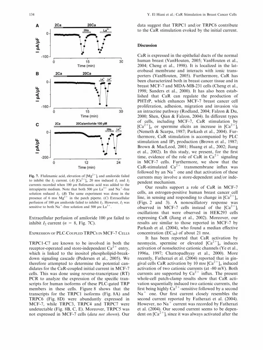
Extracellular perfusion of amiloride 100 lM failed toinhibit I2 current (n = 8, Fig. 7C).
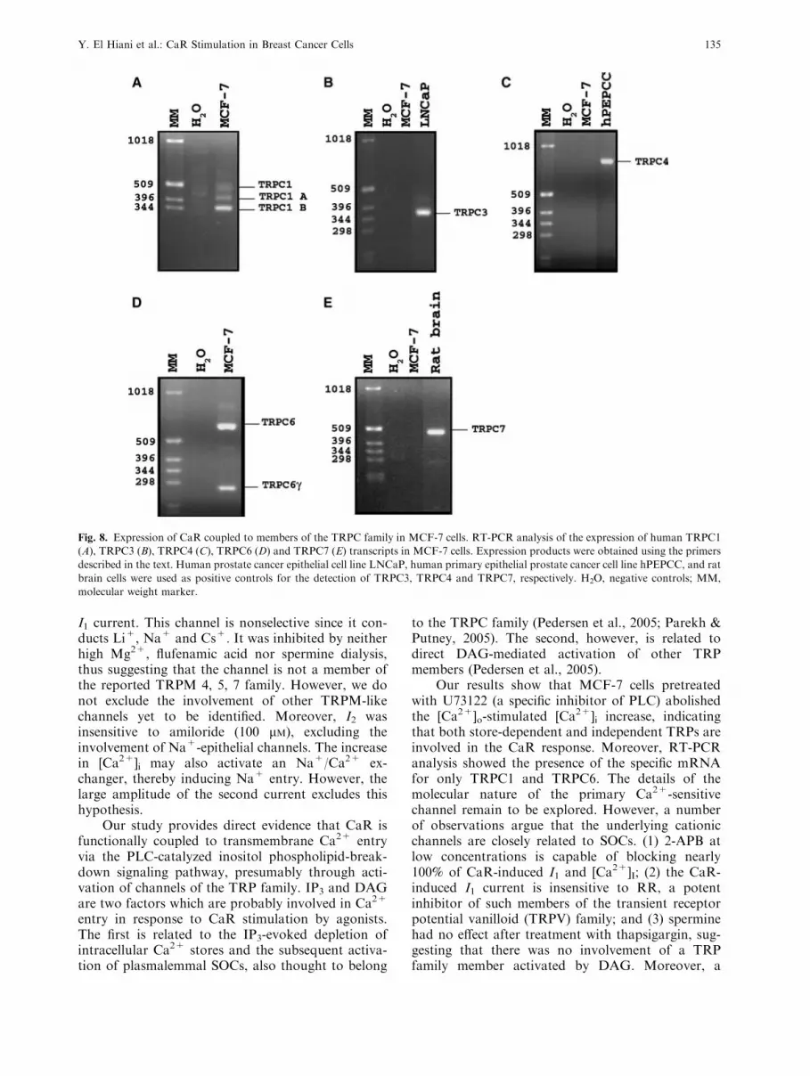
EXPRESSION OF PLC-COUPLED TRPCS INMCF-7 CELLS
TRPC1-C7 are known to be involved in both thereceptor-operated and store-independent Ca2+ entry,which is linked to the inositol phospholipid-break-down signaling cascade (Pedersen et al., 2005). Wetherefore attempted to determine the potential can-didates for the CaR-coupled initial current in MCF-7cells. This was done using reverse-trancriptase (RT)PCR to analyze the expression of the specific tran-scripts for human isoforms of these PLC-gated TRPmembers in these cells. Figure 8 shows that thetranscripts for the TRPC1 isoforms (Fig. 8A) andTRPC6 (Fig. 8D) were abundantly expressed inMCF-7, while TRPC3, TRPC4 and TRPC7 wereundetectable (Fig. 8B, C, E). Moreover, TRPC5 wasnot expressed in MCF-7 cells (data not shown). Our
data suggest that TRPC1 and/or TRPC6 contributeto the CaR stimulation evoked by the initial current.
Discussion
CaR is expressed in the epithelial ducts of the normalhuman breast (VanHouten, 2005; VanHouten et al.,2004; Cheng et al., 1998). It is localized in the lat-erobasal membrane and interacts with ionic trans-porters (VanHouten, 2005). Furthermore, CaR hasbeen characterized both in breast cancer tissue and inbreast MCF-7 and MDA-MB-231 cells (Cheng et al.,1998; Sanders et al., 2000). It has also been estab-lished that CaR can regulate the production ofPHTrP, which enhances MCF-7 breast cancer cellproliferation, adhesion, migration and invasion viaan intracrine pathway (Rodland, 2004; Falzon & Du,2000; Shen, Qian & Falzon, 2004). In different typesof cells, including MCF-7, CaR stimulation by[Ca2+]o or spermine elicits an increase in [Ca2+]i(Nemeth & Scarpa, 1987; Parkash et al., 2004). Fur-thermore, CaR stimulation is accompanied by PLCstimulation and IP3 production (Brown et al., 1987;Brown & MacLeod, 2001; Huang et al., 2002; Jianget al., 2002). In this study, we present, for the firsttime, evidence of the role of CaR in Ca2+ signalingin MCF-7 cells. Furthermore, we show that theCaR-stimulated Ca2+ transmembrane influx wasfollowed by an Na+ one and that activation of thesecurrents may involve a store-dependent and/or inde-pendent mechanism.
Our results support a role of CaR in MCF-7cells, an estrogen-positive human breast cancer cellline, in sensing and responding to change in [Ca2+]o(Figs. 2 and 3). A nonoscillatory response wasobserved in MCF-7 cells instead of the [Ca2+]ioscillations that were observed in HEK293 cellsexpressing CaR (Jiang et al., 2002). Moreover, ourresults are similar to those reported in MCF-7 byParkash et al. (2004), who found a median effectiveconcentration (EC50) of about 21 mM.
It has been reported that CaR activation byneomycin, spermine or elevated [Ca2+]o inducesactivation of nonselective cationic channels (Ye et al.,1996a, 1997; Chattopadhyay et al., 2000). Morerecently, Fatherazi et al. (2004) reported that in gin-gival cells CaR activation by 10 mM [Ca2+]o inducedactivation of two cationic currents (at -80 mV). Bothcurrents are supported by Ca2+ influx. The presentwhole-cell patch-clamp results show that CaR acti-vation sequentially induced two cationic currents, thefirst being highly Ca2+-sensitive followed by a secondNa+ one. Our first current closely resembles thesecond current reported by Fatherazi et al. (2004).However, no Na+ current was recorded by Fatheraziet al. (2004). Our second current seems to be depen-dent on [Ca2+]i since it was always activated after the
Fig. 7. Flufenamic acid, elevation of [Mg2+]i and amiloride failed
to inhibit the I2 current. (A) [Ca2+]o 20 mM induced I1 and I2currents recorded when 100 lM flufenamic acid was added to the
intrapipette medium. Note that both 500 lM La3+ and Na+-free
solution reduced I2. (B) The same experiment was done in the
presence of 6 mM Mg2+ in the patch pipette. (C) Extracellular
perfusion of 100 lM amiloride failed to inhibit I2. However, I2 was
sensitive to both Na+-free solution and 500 lM La3+.
134 Y. El Hiani et al.: CaR Stimulation in Breast Cancer Cells
I1 current. This channel is nonselective since it con-ducts Li+, Na+ and Cs+. It was inhibited by neitherhigh Mg2+, flufenamic acid nor spermine dialysis,thus suggesting that the channel is not a member ofthe reported TRPM 4, 5, 7 family. However, we donot exclude the involvement of other TRPM-likechannels yet to be identified. Moreover, I2 wasinsensitive to amiloride (100 lM), excluding theinvolvement of Na+-epithelial channels. The increasein [Ca2+]i may also activate an Na+/Ca2+ ex-changer, thereby inducing Na+ entry. However, thelarge amplitude of the second current excludes thishypothesis.
Our study provides direct evidence that CaR isfunctionally coupled to transmembrane Ca2+ entryvia the PLC-catalyzed inositol phospholipid-break-down signaling pathway, presumably through acti-vation of channels of the TRP family. IP3 and DAGare two factors which are probably involved in Ca2+
entry in response to CaR stimulation by agonists.The first is related to the IP3-evoked depletion ofintracellular Ca2+ stores and the subsequent activa-tion of plasmalemmal SOCs, also thought to belong
to the TRPC family (Pedersen et al., 2005; Parekh &Putney, 2005). The second, however, is related todirect DAG-mediated activation of other TRPmembers (Pedersen et al., 2005).
Our results show that MCF-7 cells pretreatedwith U73122 (a specific inhibitor of PLC) abolishedthe [Ca2+]o-stimulated [Ca2+]i increase, indicatingthat both store-dependent and independent TRPs areinvolved in the CaR response. Moreover, RT-PCRanalysis showed the presence of the specific mRNAfor only TRPC1 and TRPC6. The details of themolecular nature of the primary Ca2+-sensitivechannel remain to be explored. However, a numberof observations argue that the underlying cationicchannels are closely related to SOCs. (1) 2-APB atlow concentrations is capable of blocking nearly100% of CaR-induced I1 and [Ca2+]I; (2) the CaR-induced I1 current is insensitive to RR, a potentinhibitor of such members of the transient receptorpotential vanilloid (TRPV) family; and (3) sperminehad no effect after treatment with thapsigargin, sug-gesting that there was no involvement of a TRPfamily member activated by DAG. Moreover, a
Fig. 8. Expression of CaR coupled to members of the TRPC family in MCF-7 cells. RT-PCR analysis of the expression of human TRPC1
(A), TRPC3 (B), TRPC4 (C), TRPC6 (D) and TRPC7 (E) transcripts in MCF-7 cells. Expression products were obtained using the primers
described in the text. Human prostate cancer epithelial cell line LNCaP, human primary epithelial prostate cancer cell line hPEPCC, and rat
brain cells were used as positive controls for the detection of TRPC3, TRPC4 and TRPC7, respectively. H2O, negative controls; MM,
molecular weight marker.
Y. El Hiani et al.: CaR Stimulation in Breast Cancer Cells 135

correlation with the permeation and pharmacologicalprofiles of the first cationic current, I1, in MCF-7 cellswith those described for TRPC6 shows a number ofnotable differences: (1) I1 was highly permeable toCa2+, in contrast to TRPC6 which is permeable toCa2+ and Na+; (2) the increase in [Ca2+]o increasedI1 amplitude, while it reduced TRPC6 activity; and(3) dialysis with 100 lM flufenamic acid, a TRPC6activator, was without effect on I1. Taken together,these finding suggest that the CaR-activated cationicchannels in MCF-7 cells are probably heterotetra-multimers that necessarily include both TRPC6 andTRPC1 together with some other TRP members,which confer the property of SOC gating and, incombination, determine the resultant permeation andpharmacological characteristics of the whole channel.
Changes in [Ca2+]i are known to play animportant role in the regulation of PTHrP secretionby MCF-7 cells (Brown et al., 1993). The MCF-7 cellline expresses functional PTH/PTHrP receptors, andPTHrP affects its growth in an autocrine/paracrinemanner (Liapis et al., 1993). In this way, the CaR-induced TRP activation (store-dependent and/orindependent) permits Ca2+ influx and an increase in[Ca2+]i. Moreover, the Na+ entry by a nonselectivecation channel, which could be a member of theTRPM family, may induce an inverse function of theNa+/Ca2+ exchanger, thereby causing an additionalCa2+ entry, which allows a sustained increase in[Ca2+]i and the secretion of PTHrP and/or changes ingene expression, such as estrogen receptor downre-gulation in MCF-7 cells (Journe et al., 2004).
In conclusion, our findings provide new infor-mation about nonselective cationic channels sup-porting CaR-activated Ca2+ influx pathways inMCF-7 cells. These findings therefore contribute tothe understanding of the functional significance of thepresence of CaR in breast cancer cells.
We thank Jean Francois Lefebvre and Philippe Delcourt for their
excellent technical assistance. This work was supported by the
Ministere de l�Education Nationale, the Ligue Nationale Contre le
Cancer, the Association pour la Recherche Contre le Cancer and
the Region Picardie, France and by grants from Morocco.
References
Beech, D.J 2005. TRPC1: Store-operated channel and more.
Pfluegers Arch. 451:53–60
Boring, C.C., Squire, T.S., Tong, T., Montgomery, S 1994. Cancer
statistics. CA Cancer J. Clin. 44:7–26
Broad,L.M., Braun,F.J., Lievremont, J.P., Bird,G.S.,Kurosaki, T.,
Putney, J.W., Jr 2001. Role of the phospholipase C-inositol
1,4,5-trisphosphatepathway in calciumrelease-activated calcium
current and capacitative calciumentry. J. Biol. Chem. 276:15945–
15952
Brown, E.M., Enyedi, P., LeBoff, M., Rotberg, J., Preston, J.,
Chen, C 1987. High extracellular Ca2+ and Mg2+ stimulate
accumulation of inositol phosphates in bovine parathyroid
cells. FEBS Lett. 218:113–138
Brown, E.M., Gamba, G., Riccardi, D., Lombardi, M., Butters, R.,
Kifor, O., Sun, A., Hediger, M.A., Lytton, J., Hebert, S.C 1993.
Cloning and characterization of an extracellular Ca2+-sensing
receptor from bovine parathyroid. Nature 366:575–580
Brown, E.M., MacLeod, R.J 2001. Extracellular calcium sensing
and extracellular calcium signalling. Physiol. Rev. 81:239–297
Buchs, N., Manen, D., Bonjour, J.P., Rizzoli, R 2000. Calcium
stimulates parathyroid hormone-related protein production in
Leydig tumor cells through a putative cation-sensing mecha-
nism. Eur. J. Endocrinol. 142:500–505
Chattopadyay, N., Evliyaoglu, C., Heese, O., Carroll, R.,
Sanders, J., Black, P., Brown, E.M 2000. Regulation of
secretion of PTHrP by Ca2+-sensing receptor in human as-
trocytes, astrocytomas, and meningiomas. Am. J. Physiol.
279:C691–C699
Cheng, I., Klingensmith, M.E., Chattopadhyay, N., Kifor, O.,
Butters, R.R., Soybel, D.I., Brown, E.M 1998. Identification
and localization of the extracellular calcium-sensing receptor in
human breast. J. Clin. Endocrinol. Metab. 83:703–707
Falzon, M., Du, P 2000. Enhanced growth of MCF-7 breast cancer
cells overexpressing parathyroid hormone-related peptide.
Endocrinology 141:1882–1892
Fatherazi, S., Belton, C.M., Cai, S., Zarif, S., Goodwin, P.C.,
Lamont, R.J., Izutsu, K.T 2004. Calcium receptor message,
expression and function decrease in differentiating keratino-
cytes. Pfluegers Arch. 448:93–104
Guise, T.A., Yin, T.A., Taylor, S.D., Kumagai, Y., Dallas, M.,
Boyce, B.F., Yoneda, T., Mundy, G.R 1996. Evidence for a
causal role of parathyroid hormone-related protein in the
pathogenesis of human breast cancer-mediated osteolysis. J.
Clin. Invest. 98:1544–1549
Huang, C., Handlogten, M.E., Miller, R.T 2002. Parallel activation
of phosphotidylinositol 4-kinase and phospholipase C by the
extracellular calcium-sensing receptor. J. Biol. Chem.
277:20293–20300
Jiang, Y.F., Zhang, Z., Kifor, O., Lane, C.R., Suinn, S.J., Bai, M
2002. Protein kinase C (PKC) phosphorylation of the Ca2+-
sensing receptor (CaR) modulates functional interaction of G
proteins with the CaR cytoplasmic tail. J. Biol. Chem.
277:50543–50549
Journe, F., Dumon, J.C., Kheddoumi, N., Fox, J., Laios, I.,
Leclercq, G., Body, J.J 2004. Extracellular calcium downregu-
lates estrogen receptor alpha and increases its transcriptional
activity through calcium-sensing receptor in breast cancer cells.
Bone 35:479–488
Kraft, R., Harteneck, C 2005. The mammalian melastatin-related
transient receptor potential cation channels: An overview.
Pfluegers Arch. 451:204–211
Li, S., Huang, S., Peng, S.B 2005. Overexpression of G protein-
coupled receptors in cancer cells: Involvement in tumor pro-
gression. Int. J. Oncol. 27:1329–1338
Liapis, H., Crouch, E.C., Grosso, L.E., Kitazawa, S., Wick, M.R
1993. Expression of parathyroidlike protein in normal, prolif-
erative, and neoplastic human breast tissues. Am. J. Pathol.
174:1169–1178
Mundy, G.R 1997. Mechanisms of bone metastasis. Cancer
80:1546–1556
Nemeth, E.F., Scarpa, A 1987. Rapid mobilization of cellular Ca2+
in bovine parathyroid cells evoked by extracellular divalent
cations. Evidence for a cell surface calcium receptor. J. Biol.
Chem. 262:5188–5196
Ouadid-Ahidouch,H.,Roudbaraki,M.,Delcourt, P., Ahidouch,A.,
Joury, N., Prevarskaya, N 2004. Functional and molecular
identification of intermediate-conductance Ca2+-activated K+
channels in breast cancer cells: Association with cell cycle pro-
gression. Am. J. Physiol. 287:C125–C134
136 Y. El Hiani et al.: CaR Stimulation in Breast Cancer Cells

Parekh, A.B., Putney, J.W., , Jr 2005. Store-operated calcium
channels. Physiol. Rev. 85:757–810
Parkash, J., Chaudhry, M.A., Rhoten, W.B 2004. Calbindin-D28k
and calcium sensing receptor cooperate in MCF-7 human
breast cancer cells. Int. J. Oncol. 24:1111–1119
Pedersen, S.F., Owsianik, G., Nilius, B 2005. TRP channels: An
overview. Cell Calcium 38:233–252
Rodland, K.D 2004. The role of the calcium-sensing receptor in
cancer. Cell Calcium 35:291–295
Sanders, J.L., Chattopadhyay, N., Kifor, O., Yamaguchi, T.,
Butter, R.R., Brown, E.M 2000. Extracellular calcium-sensing
receptor expression and its potential role in regulating para-
thyroid hormone-related peptide secretion in human breast
cancer cell lines. Endocrinology 141:4357–4364
Sanders, J.L., Chattopadhyay, N., Kifor, O., Yamaguchi, T.,
Butter, R.R., Brown, E.M 2001. Ca2+-sensing receptor
expression and PTHrP secretion in PC-3 human prostate cancer
cells. Am. J. Physiol. 281:E1267–E1274
Shen, X., Qian, L., Falzon, M 2004. PTH-related protein enhances
MCF-7 breast cancer cell adhesion, migration, and invasion via
an intracrine pathway. Exp. Cell. Res. 294:420–433
Silver, I.A., Murrills, R.J., Etherington, D.J 1988. Microelectrode
studies on the acid microenvironment beneath adherent mac-
rophages and osteoclast. Exp. Cell. Res. 175:266–276
Ullrich, N.D., Voets, T., Prenen, J., Vennekens, R., Talavera, K.,
Droogmans, G., Nilius, B 2005. Comparison of functional
properties of the Ca2+-activated cation channels TRPM4 and
TRPM5 from mice. Cell Calcium 37:267–278
VanHouten, J.N 2005. Calcium sensing by the mammary gland.
Neoplasia 10:129–139
VanHouten, J.N., Danna, P., McGeoch, G., Brown, E.M., Krap-
cho, K., Neville, M., Wysolmerski, J.J 2004. The calcium-
sensing receptor regulates mammary gland parathyroid
hormone-related protein production and calcium transport.
J. Clin. Invest. 113:598–608
Yamaguchi, T., Ye, C., Chattopadhyay, N., Sanders, J.L., Vassilev,
P.M., Brown, E.M 2000. Enhanced expression of extracellular
calcium sensing receptor in monocyte-differentiated versus
undifferentiated HL-60 cells: Potential role in regulation of a
nonselective cation channel. Calcif. Tissue Int. 66:375–382
Ye, C.P., Kanazirskia, M., Quinn, S., Brown, E.M., Vassilev, P.M
1996a. Modulation by polycationic Ca2+-sensing receptor
agonists of nonselective cation channels in rat hippocampal
neurons. Biochem. Biophys. Res. Commun. 224:271–280
Ye, C.P., Rogers, K., Bai, M., Quinn, S., Seidman, C.E., Seidman,
J.G., Brown, E.M., Vassilev, P.M 1996b. Agonists of the Ca2+-
sensing receptor (CaR) activate nonselective cation channels in
HEK293 cells stably transfected with the human CaR. Biochem.
Biophys. Res. Commun. 226:272–279
Ye, C.P., Ho-Pao, C.L., Kanazirskia, M., Quinn, S., Rogers, K.,
Seidman, C.E., Seidman, J.G., Brown, E.M., Vassilev, P.M
1997. Amyloid-beta proteins activate Ca2+-permeable channels
through calcium-sensing receptors. J. Neurosci. Res. 47:547–554
Zitt, C., Halaszovich, C.R., Luckhoff, A 2002. The TRP family of
cation channels: Probing and advancing the concepts on
receptor-activated calcium entry. Prog. Neurobiol. 66:243–264
Y. El Hiani et al.: CaR Stimulation in Breast Cancer Cells 137