extracellular vesicles in osteosarcoma - mdpi
TRANSCRIPT
International Journal of
Molecular Sciences
Review
Extracellular Vesicles in Osteosarcoma: Antagonists orTherapeutic Agents?
Viviana De Martino 1 , Michela Rossi 2 , Giulia Battafarano 2 , Jessica Pepe 1, Salvatore Minisola 1
and Andrea Del Fattore 2,*
�����������������
Citation: De Martino, V.; Rossi, M.;
Battafarano, G.; Pepe, J.; Minisola, S.;
Del Fattore, A. Extracellular Vesicles
in Osteosarcoma: Antagonists or
Therapeutic Agents? Int. J. Mol. Sci.
2021, 22, 12586. https://doi.org/
10.3390/ijms222212586
Academic Editor: Julia Costa
Received: 19 October 2021
Accepted: 20 November 2021
Published: 22 November 2021
Publisher’s Note: MDPI stays neutral
with regard to jurisdictional claims in
published maps and institutional affil-
iations.
Copyright: © 2021 by the authors.
Licensee MDPI, Basel, Switzerland.
This article is an open access article
distributed under the terms and
conditions of the Creative Commons
Attribution (CC BY) license (https://
creativecommons.org/licenses/by/
4.0/).
1 Department of Clinical, Internal, Anaesthesiology and Cardiovascular Sciences, Sapienza University,00185 Rome, Italy; [email protected] (V.D.M.); [email protected] (J.P.);[email protected] (S.M.)
2 Bone Physiopathology Research Unit, Genetics and Rare Diseases Research Division,Bambino Gesù Children’s Hospital, IRCCS, 00165 Rome, Italy; [email protected] (M.R.);[email protected] (G.B.)
* Correspondence: [email protected]
Abstract: Osteosarcoma (OS) is a skeletal tumor affecting mainly children and adolescents. Thepresence of distance metastasis is frequent and it is localized preferentially to the lung, representingthe main reason for death among patients. The therapeutic approaches are based on surgery andchemotherapeutics. However, the drug resistance and the side effects associated with the chemother-apy require the identification of new therapeutic approaches. The understanding of the complexbiological scenario of the osteosarcoma will open the way for the identification of new targets for itstreatment. Recently, a great interest of scientific community is for extracellular vesicles (EVs), that arereleased in the tumor microenvironment and are important regulators of tumor proliferation and themetastatic process. At the same time, circulating extracellular vesicles can be exploited as diagnosticand prognostic biomarkers, and they can be loaded with drugs as a new therapeutic approach forosteosarcoma patients. Thus, the characterization of OS-related EVs could represent a way to convertthese vesicles from antagonists for human health into therapeutic and/or diagnostic agents.
Keywords: osteosarcoma; extracellular vesicles; therapy
1. Introduction
Osteosarcoma (OS) is a highly malignant skeletal tumor characterized by the presenceof neoplastic cells of mesenchymal origin that deposit an immature osteoid matrix. Despiteits rarity, osteosarcoma is the third most frequent primary malignancy affecting mainlychildren, adolescents and young adults [1]. Osteosarcoma is more common in male individ-uals, with an overall ratio between males and females of 1.43:1. The incidence is 2–3 casesper 1,000,000 inhabitants/year [1]. The peak incidence occurs in the group from 10 to 19years old and seems to be related to the period in which maximum bone growth occurs,suggesting a connection between tumor formation and growth factors expressed duringbone growth. A second peak of OS in adults over 65 years of age has been reported [2,3].
The most affected areas are the metaphyses of long bones and bone segments suchas the proximal tibia, distal femur, proximal humerus, and all areas characterized by amassive bone rearrangement; it rarely occurs in flat bones and spine [4]. The most commonsymptom in patients is a relatively non-specific pain in the affected area, often wronglyattributed to bone growth, accompanied by swelling of soft tissues. The manifestation ofpain can result from the weakening of the bone with the development of microfractures;severe pain occurs in case of more serious pathological fractures, found in more than 15%of pediatric patients. Symptoms of general malaise, including weight loss, pallor, feverand/or anorexia are very rare [4].
Several subtypes of osteosarcoma can be identified: classical intramedullary or central(osteoblastic, chondroblastic and fibroblastic); telangiectasic; small cell; high-grade surface;
Int. J. Mol. Sci. 2021, 22, 12586. https://doi.org/10.3390/ijms222212586 https://www.mdpi.com/journal/ijms
Int. J. Mol. Sci. 2021, 22, 12586 2 of 16
secondary osteosarcoma; parosteal; periostal; and central with a low degree of malignancy.The first type of OS (classical intramedullary or central) is the most common amongteenagers and includes about 85% of all OS cases [5].
Osteosarcoma is characterized by highly invasive ability. The presence of distantmetastases is very frequent and represents the main reason of death among osteosarcomapatients; the preferential site of metastasis is the lung [6,7]. The 5-year survival rate of OSpatients with metastasis is 20% compared to 65% of patients with localized disease [4,8].Consequently, due to its aggressiveness and the lowest overall survival rates, metastaticosteosarcoma is considered one of the main causes of death.
Numerous studies have been carried out on the cytogenetic and molecular aspectsof osteosarcoma with often conflicting results; therefore, their diagnostic and prognosticvalue still appears limited. The rarity and the heterogeneity of the pathology also do nothelp to clarify its etiological meaning. Osteosarcoma is counted among complex karyotypesarcomas [9]. Seventy percent of osteosarcoma cases show numerical, structural alterationsand genomic amplifications. Cytogenetic analysis revealed numerous breaking points andtranslocations, underlining the complexity and instability of the genetic background inthis tumor [10]. At the molecular level, the most compromised signaling pathways arelinked to the altered activity of oncogenes, such as Myc (avian myelocytomatosis viraloncogene homolog) and tumor suppressors genes (Rb (retinoblastoma protein) and p53),which are functionally inactivated in most cases of osteosarcoma [11]. Recently, genomicalterations in genes involved in the mechanisms of DNA repair were reported in a subsetof patients exhibiting a specific combination of single-base substitutions, LOH (loss ofheterozygosity), or large-scale genome instability signatures characteristic of BRCA1/2-deficient tumors [12].
Although osteosarcoma is sporadic in 95% of cases, several inherited syndromessuch as Li–Fraumeni, Rothmund–Thomson and Retinoblastoma familial cancers havebeen associated with a predisposition to develop osteosarcoma [13–15]. Paget’s disease,generally a benign condition characterized an increase in bone turnover, could represent arisk condition for osteosarcoma [16]. Chronic osteomyelitis, osteochondroma, encondromaand fibrous dysplasia are also associated with osteosarcoma [2,11].
For a diagnosis, a set of clinical analyses, radiological investigations and the evaluationof the pathological tissue by performing biopsy is required [17].
Currently, the therapeutic strategies for osteosarcoma include three treatments: thesurgical approach, and neoadjuvant and adjuvant chemotherapy [18,19]. Indeed, about85% of cases of high-grade OS can be successfully resected and reconstructed, preservingthe affected limb and its function [20]. A meta-analysis performed by Xiaojuan Li et al.reported that patients subjected to limb salvage surgery (LSS) had a similar local recur-rence compared to patients treated with amputation [21]. In addition, they found thatthe 5-year overall survival rate of patients treated with LSS was higher than those treatedwith amputation [22]. Amputation is generally reserved only for those tumors in whicha complete resection of tumor and the preservation of limb function is not feasible [23].Neoadjuvant chemotherapy is administered about 8–10 weeks before surgery; the use ofpreoperative chemotherapy offers time for surgical planning, decreases tumor size and po-tentially facilitates its removal, reduces the risk of distant metastases and allows assessmentof response to therapy [20]. The intensification of neoadjuvant chemotherapy increased thenumber of good respondents but did not alter overall survival [21,24]. Today, cooperativegroup studies in North America and Europe provided a standard protocol neoadjuvantchemotherapy, known as MAP, characterized by the use of multi-drugs such as methotrex-ate in high doses (HDMTX), doxorubicin (adriamycin, ADM) and cisplatin (CDP) [25].Numerous clinical trials have tested various combinations of the five chemotherapeuticagents known to be active in this disease (methotrexate, doxorubicin, cisplatin, ifosfamideand etoposide) [26,27]. Although the chemotherapy has improved the life of osteosarcomapatients, the onset of drug resistance, toxicity and related side effects limits the use of thesechemotherapy agents in clinical practice [28,29].
Int. J. Mol. Sci. 2021, 22, 12586 3 of 16
The identification of new therapeutic targets is therefore necessary above all in pa-tients who have chemoresistance or who experience local relapses (35% of patients) or lungmetastases (60% of patients) [4]. The development of chemoresistance induces complica-tions, linked above all to the therapeutic need to increase the dose of drug for treatment,which is not always well tolerated by the patient due to its high toxicity, and often to stoptreatment [30,31]. In the past few years, there is increased attention on understanding thecomplex biological scenario in osteosarcoma. Due to the inter- and intratumoral heterogene-ity, a unique targeted pattern does not exist, and this has made attempts unsuccessful overthe past three decades [32]. The development of novel therapeutic strategies remains animportant clinical need. In this review, we summarized the new advances in osteosarcomabiology, particularly the involvement of extracellular vesicles as potential diagnostic andprognostic biomarkers and as a new therapeutic approach for osteosarcoma.
2. Molecular Mechanisms of Osteosarcoma Progression
Osteosarcoma develops in the bone microenvironment, a very specialized environ-ment in which bone cells (Mesenchymal stem cells (MSC), osteoblasts, osteocytes, osteo-clasts precursors and osteoclasts), and immune and vascular cells communicate with eachother to maintain the integrity of the skeleton [2,33]. This is a soil rich in growth factors,cytokines, chemokines and extracellular vesicles that create a fertile microenvironment forosteosarcoma growth [34].
Alterations of the bone remodeling are the first steps in the osteosarcoma onset. Inthe past 15 years, osteosarcoma was commonly described as a disease related to the alter-ations of MSC; recently, it was demonstrated that osteosarcoma can also occur followingdysregulation of multiple points in bone development [35].
Regarding the role of MSC in the osteosarcoma progression, two different MSCpopulations exist in the osteosarcoma microenvironment. Naïve MSC derive from normaltissue and can exert pro- and antitumoral activity [36,37]; the tight crosstalk between MSCand osteosarcoma cells leads to the reprogramming of MSC into MSC stimulating tumorprogression (tumor-tissue educated MSC) [38].
Indeed, osteosarcoma cells can modulate the microenvironment; the high-rate ener-getic glycolytic metabolism of cancer cells causes high lactic acid production and a highproton efflux; short-term acidosis activates downstream signaling of the NF-kB (Nuclearfactor-kappa B) pathway in MSC but not in osteosarcoma cells [39]. Low extracellularpH in these tumors induces an increased invasive behavior and promotes the secretionof high levels of Interleukin 6 (IL-6) and IL-8 by mesenchymal stem cells, stimulatingosteosarcoma growth and metastasis [39]. IL-8 can activate the chemokine receptor CXCR1(C-X-C Motif Chemokine Receptor 1) and can lead to anoikis resistance of osteosarcomacells and progression of pulmonary metastasis. Furthermore, MSC also secretes CCL5 (C-Cmotif ligand 5), SDF-1 (Stromal derived factor 1) and VEGF (Vascular Endothelial GrowthFactor), promoting osteosarcoma progression, angiogenesis and metastasis [39,40].
In vivo experiments revealed that OSDC (Osteosarcoma associated stromal cells,also named osteosarcoma-derived cells) and MSC co-injections with tumor cells led toincreased tumor growth and eventually to metastases in nude and/or severe combinedimmunodeficiency (SCID) mice [41].
Acidosis, hypoxia and inflammation induce neovascularization that allows the de-livery of nutrients and oxygen to the tumor cells. In the tumor microenvironment, tumorcells and endothelial cells express pro-angiogenic factors as VEGF, PDGF (Platelet DerivedGrowth Factor), FGF (Fibroblast Growth Factor) and TGF-β (Transforming Growth Factorbeta) [42].
Osteosarcoma is a highly vascularized bone tumor and mainly occurs in the region ofbone growth close to metaphysis, where type-H endothelial cells promoting angiogenesisare located, suggesting their role in osteosarcoma neo-angiogenesis [43,44]. The neo-angiogenesis in osteosarcoma could derive from pre-existing vessels or be dependentfrom endothelial progenitor cells that can differentiate into mature endothelial cells [45].
Int. J. Mol. Sci. 2021, 22, 12586 4 of 16
However, vascular mimicry is also observed in osteosarcoma and is characterized by theformation of vasculogenic-like microchannels generated by tumor cells; this alternativeprocess of angiogenesis and vasculogenesis occurs in about 20% of patients and is associatedwith poor prognosis [46,47]. The mechanisms of vascular mimicry still remain largelyunknown; however, the autocrine VEGF/VEGFR1 (VEGF Receptor 1) signaling has beenproposed as key pathway for the vasculogenic features of osteosarcoma cells, and a clearcorrelation between VEGF levels and tumor progression was demonstrated [48,49].
Factors secreted in the bone microenvironment also contribute to the abnormal os-teoclast activity. At the same time, osteoclast bone resorption can lead to the release ofpro-tumor factors as IGF-1 (Insulin Growth Factor 1) and TGF-β from the bone matrixthat stimulate tumor cells [50,51]. The role of osteoclasts in the onset and progressionof osteosarcoma is still controversial [52,53]. There are clinical and experimental datashowing that the presence of osteoclasts in osteosarcoma-adjacent tissue is associated withpoor outcomes [52], but on the other hand, published studies suggest that the presence ofosteoclasts at the primary site of OS lesions prevents metastasis [52,54]. Endo-Munoz et al.found that expression of osteoclast-specific tartrate-resistant acid phosphatase 5 (TRAcP5)is significantly downregulated in biopsies isolated from osteosarcoma patients comparedwith nonmalignant bone tissue. However, the lesions of patients with lung metastasis hadincreased levels of TRAcP5 expression compared to lesions of non-metastatic disease [53].
Understanding the role of osteoclasts in osteosarcoma onset, progression and metasta-sis is relevant for therapeutic approaches. If osteoclasts are essential for the lung metasta-sis [53], the administration of antiresorptive drugs including bisphosphonates and Deno-sumab would be effective therapeutic strategy; if the bone resorption suppresses themetastasis development, this approach would be contraindicated. A phase III clinical trialstudy demonstrated a worse therapeutic outcome following a combined treatment withchemotherapy/surgery and antiresorptive zoledronic acid [55].
Tumor cells also express MMP-9 (Matrix Metallopeptidase-9) that allows the dissem-ination of tumor cells and, at the same time, is essential for the activation of angiogenicfactors [7].
Regarding the role of immune cells in the progression of osteosarcoma, both myeloidand lymphoid cells have been detected in osteosarcoma [56]. Particularly, tumor-associatedmacrophages (TAM) were detected in osteosarcoma biopsies, and they were associated withreduced metastasis and improved survival by still-unclear mechanisms [2,57,58]. More-over, osteosarcoma tumors are characterized by poor infiltration of CD8+ lymphocytes,suggesting the poor immunogenic feature of this tumor; the scarce infiltrate of cytotoxiclymphocytes allowed the osteosarcoma to be defined as cold tumor [59]. Moreover, theratio between CD8+ cells and regulatory T cells (CD4+FoxP3+) in bone biopsies from os-teosarcoma patients is important for discriminating between patients with an expectedprolonged survival from those with a poor prognosis [60]. The presence of Antigen Present-ing Cells (APC), including dendritic cells and CD68+ macrophages, has been associatedwith a poorer prognosis [61].
From this general overview, it is quite evident that in the osteosarcoma microenvi-ronment there is a tight crosstalk among bone, endothelial and immune cells, mediatedby cell-cell contact, soluble factors and extracellular vesicles. Indeed, it was demonstratedthat EVs are spontaneously released by osteosarcoma cells in the microenvironment andthey can exert several functions: they can mediate the immune escape of tumor cells, andpromote angiogenesis, proliferation and metastatic activity of osteosarcoma cells [62].
3. Extracellular Vesicles
EVs are lipid-bound vesicles secreted by cells into the extracellular space [63,64]. Extra-cellular vesicles can be vehicles for nucleic acids (DNA, RNA and microRNAs (miRNAs)),proteins, lipids (eicosanoids, fatty acids and cholesterol), and also intact organelles [63].It was reported that EVs can contain mitochondria that can be transferred from the par-ent/donor to recipient cells [65].
Int. J. Mol. Sci. 2021, 22, 12586 5 of 16
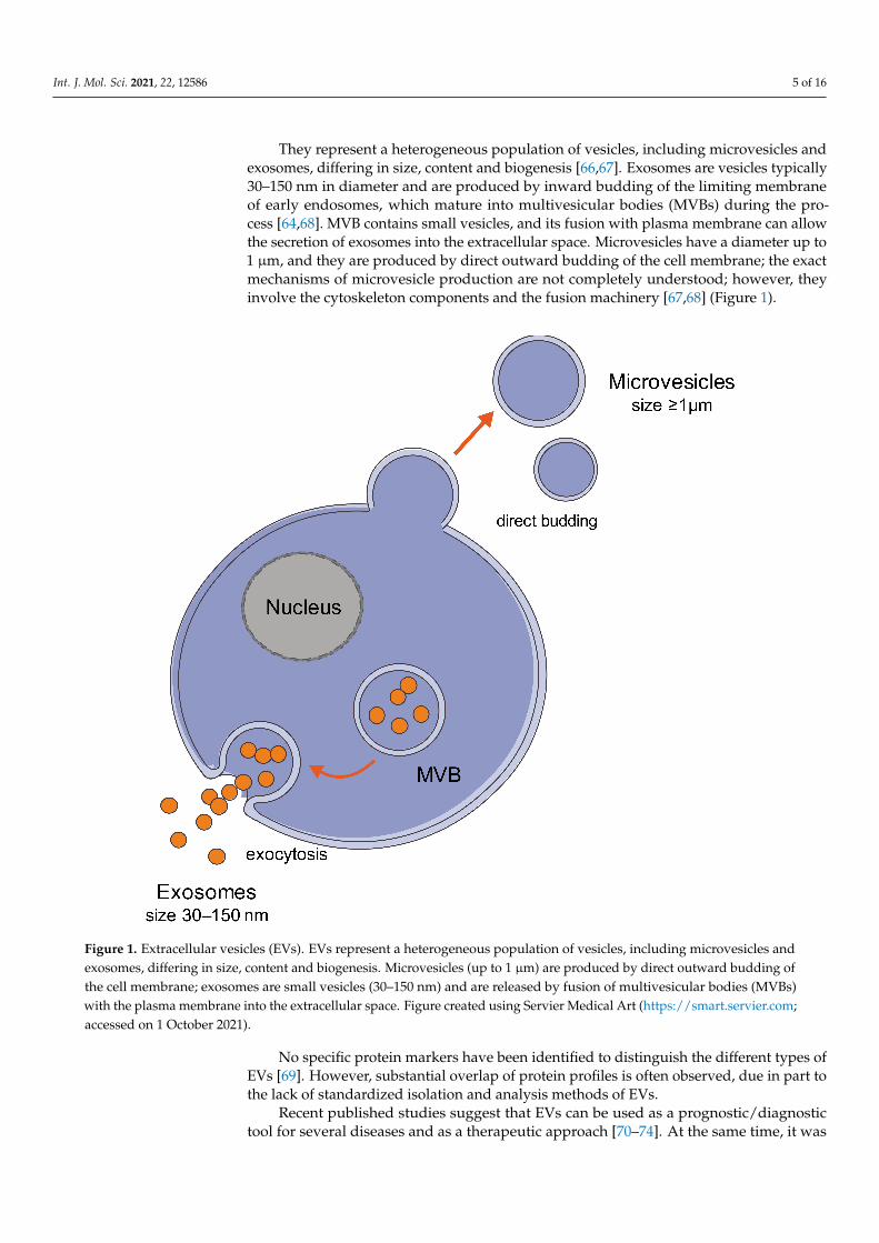
They represent a heterogeneous population of vesicles, including microvesicles andexosomes, differing in size, content and biogenesis [66,67]. Exosomes are vesicles typically30–150 nm in diameter and are produced by inward budding of the limiting membraneof early endosomes, which mature into multivesicular bodies (MVBs) during the pro-cess [64,68]. MVB contains small vesicles, and its fusion with plasma membrane can allowthe secretion of exosomes into the extracellular space. Microvesicles have a diameter up to1 µm, and they are produced by direct outward budding of the cell membrane; the exactmechanisms of microvesicle production are not completely understood; however, theyinvolve the cytoskeleton components and the fusion machinery [67,68] (Figure 1).
Figure 1. Extracellular vesicles (EVs). EVs represent a heterogeneous population of vesicles, including microvesicles andexosomes, differing in size, content and biogenesis. Microvesicles (up to 1 µm) are produced by direct outward budding ofthe cell membrane; exosomes are small vesicles (30–150 nm) and are released by fusion of multivesicular bodies (MVBs)with the plasma membrane into the extracellular space. Figure created using Servier Medical Art (https://smart.servier.com;accessed on 1 October 2021).
No specific protein markers have been identified to distinguish the different types ofEVs [69]. However, substantial overlap of protein profiles is often observed, due in part tothe lack of standardized isolation and analysis methods of EVs.
Recent published studies suggest that EVs can be used as a prognostic/diagnostictool for several diseases and as a therapeutic approach [70–74]. At the same time, it was
Int. J. Mol. Sci. 2021, 22, 12586 6 of 16
demonstrated that cancer cells can use EVs as a mechanism to expulse chemotherapy drugs,contributing to drug resistance [75,76].
3.1. Role of EVs in Osteosarcoma Microenvironment and Tumoral Growth
In 2013, Garimella et al. reported the presence of extracellular vesicles in the osteosar-coma microenvironment of an OS orthotopic mouse (BOOM) model using a human OS cellline 143B [77]. Electron microscopic examination revealed the presence of EVs of 50–200 nmin diameter that derive from bone and tumor cells. MSC-derived exosomes can promote cellproliferation, migration and invasion in osteosarcoma in vitro and in vivo [78,79]. More-over, MSC-EVs can also promote autophagy contributing to MSC-EV-induced promotionof malignant tumorigenesis [78].
Moreover, multivesicular bodies were detected in the tumor tissue. A great interestof the scientific community regards the identification of the proteins delivered by theseextracellular vesicles [77]. Mannerström et al. demonstrated that OS-EVs modulate theepigenetic status of MSC through hypomethylation of long interspersed nuclear element1 [80]. It was demonstrated that EVs secreted by the osteosarcoma 143B cell line containa pro-osteoclastogenic cargo, which includes MMPs (MMP-1 and MMP-13), RANK-L(Receptor Activator of Nuclear Factor κ B Ligand), CD-9 and TGF-β [81], involved in thebone remodeling activity.
Indeed MMP-1 (Matrix metalloproteinase-1) and MMP-13 (Matrix metalloproteinase-3) are the key collagenases responsible for degradation of type I collagen [82]. They areimportant in regulating the osteoblastic differentiation and also bone erosion [82]. Thesignificantly higher expression of MMPs and downregulation of the MMP13-targetingmiRNA143 (miR-143) are related to poor prognostic outcomes in patients with OS [83].
RANK-L is the main osteoclastogenic cytokine that binds to the RANK receptor ex-pressed on osteoclast precursors and osteoclasts. The RANK-L/RANK binding stimulatesosteoclast differentiation and bone erosion; at the same time, it was demonstrated that extra-cellular vesicles can also carry RANK, and its binding to RANK-L expressed on osteoblastmembrane stimulates the bone formation [2]. The presence of RANK on extracellularvesicles isolated from osteosarcoma cells still needs to be evaluated.
CD9 belongs to the Tetraspan transmembrane (TM4)-superfamily proteins and can beassociated with integrins, heparin-binding EGF-like growth factor, small G proteins, andother TM4-superfamily proteins, CD63, CD81 and CD82 [84–86]. Its expression on stromalcell is essential for osteoclastogenesis [87]. CD9 is also important for the bone metastasissince it was reported that CD9 was significantly overexpressed in bone metastases versusprimary tumors and visceral metastatic lesions, and that its inhibition delays homing oftumor cells in bone marrow, slowing down bone destruction [88].
The delivery of TGF-β by EV is important for the regulation of bone remodeling; in-deed, this growth factor is mainly released from the bone matrix during the bone resorptionactivity, and it is able to stimulate osteoblast progenitors and osteosarcoma cells, and toregulate osteoclastogenesis [81,89].
Moreover, Raimondi et al. showed that OS-derived EVs stimulated endothelial cellsto express and secrete elevated levels of the proangiogenic factor VEGF and interleukins(IL-6 and IL-8) [90].
3.2. Role of EVs in Osteosarcoma Metastasis
The establishment of cancer metastasis involves several steps, including intravasation,arrest at a distant organ, extravasation and growth in secondary sites [91]. The mechanismsinvolved in the migration arrest of metastatic cells are still controversial [92]. Over 80% ofall metastases in OS occur in the lung [93]; the mechanism by which osteosarcoma cellsprefer lung to metastasize is still under investigation. Several papers suggested that theC-X-C-motif chemokine receptor 4 (CXCR4) and the interaction with CXCL12 ligand playa relevant role in this process. Indeed, CXCL12 is expressed at high levels in the lung,and CXCR4 expression was high in osteosarcoma-patient samples [7,94]. Another possible
Int. J. Mol. Sci. 2021, 22, 12586 7 of 16
mechanism is mediated by CXCR3 expressed in osteosarcoma and CXCL9-10-11 expressedin the lung [95]; the interaction led to the increase of proliferation and invasion of tumorcells in the metastatic organ [7,96].
The genesis of a metastasis requires the adhesion of cancer cells to the new environ-ment; a crucial role in this step is played by ezrin that is linked to Rho and PI3K/Aktpathways [97,98].
Several reports suggest that EVs released by tumor cells regulate the metastatic pro-cess [99]. Indeed, it was demonstrated that tumor-derived exosomes can directly stimulatethe metastasis and can regulate the microenvironment to support tumor growth [100].Mazumdar et al. reported how osteosarcoma-derived EVs could influence the differen-tiation of lung fibroblast into a cancer-associated fibroblast supporting metastatic pro-gression [101]. OS-derived EVs may furthermore contribute to the metastatic process byprompting MSC to acquire a pro-tumorigenic and pro-metastatic phenotype [102,103].Moreover, exosomes released by osteosarcoma contain urokinase plasminogen activator(uPA) [104]; interestingly, the autocrine and paracrine activation of the uPA/uPAR axis hasbeen related to the conversion of OS cells to a metastatic phenotype [104].
Macklin et al. showed that EVs secreted by cells derived from a highly metastaticclonal variant of osteosarcoma can be internalized by a poorly metastatic clonal variant andinduced a migratory and invasive phenotype [105]. In addition, it was demonstrated thatEVs released by highly metastatic clones selectively concentrate within lung tissue wherethey may set up a chemotactic gradient to recruit osteosarcoma cells to the pre-metastaticniche within the lung [105].
It was suggested that miRNAs contained in the exosomes can have a crucial role inthe metastasis [106,107]. Jerez et al. isolated miRNAs from extracellular vesicles releasedfrom six different human osteosarcoma or osteoblastic cell lines with different degreesof metastatic potential (i.e., SAOS2, MG63, HOS, 143B, U2OS and hFOB1.19). About 300miRNAs are contained in EVs of each cell line, and about 70 are expressed at high level.MiR-21-5p (microRNA-21-5p), miR-143-3p, miR-148a-3p and miR-181a-5p are relativelyabundant in vesicles from metastatic cells compared to the non-metastatic MG63 [108].MiR-21 is already well described as oncomir [47]. Regarding the role of miR-143-3p,some studies suggested that it may counteract metastatic properties of squamous cellcarcinoma [109]. MiR-148a-3p and miR-181a-5p have been detected in serum samples fromgastrointestinal cancer patients [110,111]. However, bioinformatics analysis revealed thatthese miRNAs can regulate apoptosis, angiogenesis, cell adhesion and migration [108].
There is abundant evidence that long ncRNA (lncRNA) also plays a key role in thedevelopment and progression of OS [112]. In particular, the study by Li et al. disclosedthat highly enriched lncRNA OIP5-AS1 in exosomes secreted by OS cells regulates angio-genesis and ATG5-mediated autophagy in OS through miR-153, thereby participating inthe formation and development of malignant tumors [113].
TGF-β can regulate tumor invasion, metastasis, angiogenesis and cell apoptosis [114–116].TGF-β has been detected in exosomes released by osteosarcoma cells, and this can influencethe metastasis process [117]. Indeed, TGF-β can regulate the secretion of CXCL16 by osteo-clasts that stimulate osteoblastic and osteosarcoma cell migration, regulating the metastaticprocess [118]. Moreover, vesicular TGF-β induces proinflammatory IL-6 production by MSCs,allowing the tumor EV-educated mesenchymal stem cells to promote osteosarcoma progres-sion together with intratumor STAT3 activation and lung metastasis formation [119]. Indeed,the IL-6/STAT3 signaling pathway stimulates cell proliferation and migration/invasion, andprotects tumor cells from drug-induced apoptosis [120].
3.3. Role of EVs in Chemioresistance
Exosomes can be used by tumor cells to develop chemoresistance [75]. In vitroand in vivo studies linked EVs to drug resistance in many tumors, including osteosar-coma [121–123]. Indeed, EVs can represent a way to eliminate apoptotic stimuli from thecells or can acquire antitumor drug substance to achieve resistance, for example, to pacli-
Int. J. Mol. Sci. 2021, 22, 12586 8 of 16
taxel [124,125]. Torreggiani et al. showed that exosomes derived from doxorubicin-resistantosteosarcoma cells could be taken up into secondary cells, thus inducing a doxorubicin-resistant phenotype. Moreover, they suggested that the mechanism by which exosomestransfer drug resistance among osteosarcoma cells is mediated by multidrug resistance-associated protein 1 (MDR-1) mRNA and P-glycoprotein [122].
Accordingly, Pan et al. demonstrated that exosomes derived from cisplatin-resistantosteosarcoma cells reduce the sensitivity of MG63 and U2OS cells to cisplatin, inhibitapoptosis, and increase the expression of MDR-1 and P-glycoprotein [126]. Moreover theyrevealed a relationship between the levels of circular_103801 miRNA carried by exosomesin patients’ sera and their survival, suggesting circulating exosomes and miRNA as aprognostic tool [126].
Xu et al. demonstrated that a substantial profile of exosomal miRNAs, includingmiR-124, miR-133a, miR-135b, miR-148a, miR-199a-3p, miR-27a, miR-385, and miR-9, wasdysregulated in poor chemotherapeutic response [127].
These results would assist with potential clinical chemotherapeutic treatment ofOS and help in monitoring or predicting disease progression during chemotherapy inosteosarcoma.
3.4. Role of EVs in Therapy
Due to the rarity of osteosarcoma, several anatomical-clinical variants, genome com-plexity, different presentation modalities of disease and age of the affected population, thetreatment of this disease still remains an unsolved problem [11]. Indeed, in the past 40years, few changes have been reported for clinical care of patients.
A recent study of Kyung-Mi and coauthors revealed that EVs exert an anti-tumoraleffect on osteosarcoma cells. EVs from canine macrophages activate apoptotic pathways incanine OS cells and can be an effective anticancer therapeutic approach [128]. Moreover,MSC-derived EVs carrying miR-150 reduce proliferation and migration of osteosarcomacells by targeting IGF2BP1 (Insulin-like Growth Factor-2 mRNA-Binding Protein 1) [129].
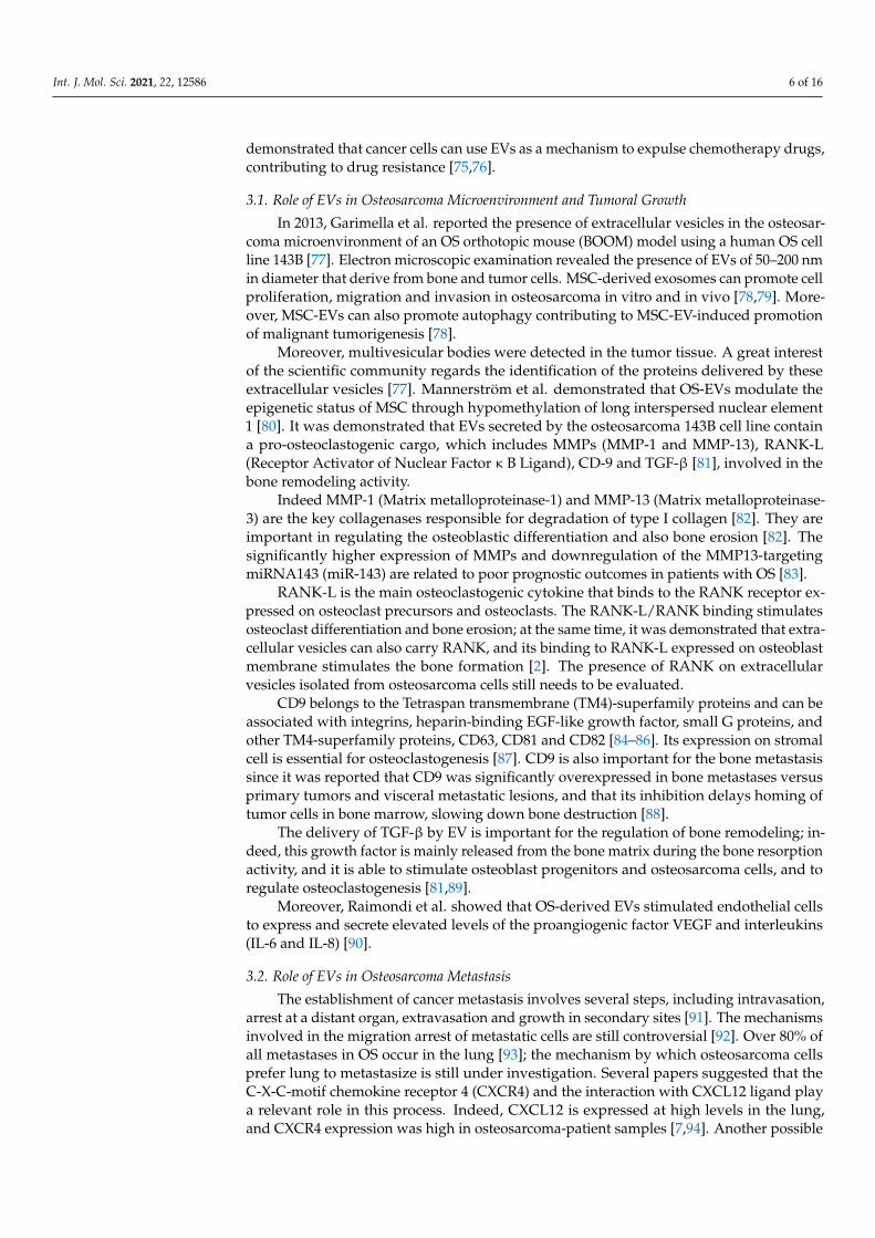
Several studies suggest that exosomes can be used as a vehicle to deliver chemothera-peutic drugs to osteosarcoma cells (Figure 2) [122,130,131]. Indeed, it was demonstratedthat exosomes can be directly charged with drugs [132,133]; another approach is to loadthe cells with drugs that will be removed from the cells via extracellular vesicles.
Figure 2. Extracellular vesicles as a vehicle for delivering chemotherapeutic drugs. Figure createdusing Servier Medical Art (https://smart.servier.com; accessed on 1 October 2021).
Several papers suggested that mesenchymal stem/stromal cells isolated from thebone marrow can be loaded with chemotherapic drugs, and exosomes isolated from theconditioned medium exert a pro-apoptotic effect on tumor cells [134,135]. Furthermore,
Int. J. Mol. Sci. 2021, 22, 12586 9 of 16
MSC can be loaded with synthetic miRNAs that can be transferred into recipient cells andsuppress migration of OS cells [136,137].
In some cases, the apoptotic effect was increased following the treatment with drugEVs compared to that observed with a free drug at the same concentration contained in theEVs [37], confirming that EV itself can exert an apoptotic stimulus [128].
3.5. EVs for Diagnosis
Extracellular vesicles can be detected in body fluids, including blood, urine andcerebrospinal fluid, and great interest is for their use as a diagnostic and/or prognostictool [69,138–140]. Indeed the presence of membrane structure provides stability and allowsprolonged periods of storage of EVs before analysis, making their clinical use feasible [141].EVs are highly produced by tumoral cells compared to healthy cells, and are usually presentat increased levels at tumor diagnosis and/or can increase during tumor progression [142].EV cargo reflects metastatic progression and treatment response [143,144]. Xu et al. deci-phered alteration of specific miRNAs in patients with a poor chemotherapeutic responsewhen compared with good responders [127]. A recent and promising study by Cambierand colleagues reported the possibility to use OS-associated EVs as possible liquid biopsiesfor early detection of cancer. The authors identified in OS patients’ EV-specific repetitiveelement DNA sequences compared to a control serum EV preparation [145].
The major concern regarding the study on EVs is the lack of standardization pro-tocols for their isolation and analysis [146,147]. Indeed, many methods are reported toisolate EVs, including ultracentrifugation, filtration, sucrose gradient and mixed proto-cols [148–150]. Another problem is related to their quantification [151]. Many studieswere conducted performing a quantification by FACS (fluorescence activated cell sorting)analysis [152]. However, the limit of instrument resolution is usually about 0.5 µm, makingthe quantification not appropriate with this method; the other approach is based on NTA(Nanoparticle Tracking Analysis) technology that allows visualization and measurement ofnanoparticles in suspension in the range of 10–1000 nm based on the analysis of Brownianmotion [153]. However, a great standardization protocol is required [69], and in this respectthe International Society of Extracellular Vesicles supported several initiatives, such as theEV Transparent Reporting and Centralizing Knowledge [154], the Minimal Information forStudies of EVs [147] and the Clinical Wrap-Up session at ISEV2018 [155].
4. Conclusions
There is a need for therapeutic approaches to improve the survival of patients with apoor response. Indeed, there are many challenges to face. First of all, osteosarcoma is arare disease with approximately 400 patients diagnosed every year in the United States,making it difficult to complete an accurate clinical trial. Moreover, some of the molecularmechanisms, including TP53 or Rb, altered in osteosarcoma are difficult to target.
The leading cause of mortality in osteosarcoma patients continues to be the develop-ment of metastasis; understanding the biology of osteosarcoma and the role of extracellularvesicles will open the way for developing or identifying novel therapeutics to prevent orarrest metastasis. In the future we believe that new diagnostic and therapeutic approachescould exploit the same mechanisms, including extracellular vesicles that cancer cells useto grow and disseminate, for the establishment of more specific treatments for patients.The identification of molecules carried by OS-derived EVs that help tumor growth, sur-vival and metastasis could represent a way to convert these antagonists into a therapeuticand/or diagnostic tool. However, a major limitation in this field is the lack of a well-standardized method for EV isolation and quantification, and a rigorous methodology andcharacterization will open the avenue for to better employ EVs as therapeutic carriers anddiagnostics tools.
Author Contributions: V.D.M., M.R., G.B., J.P., S.M. and A.D.F. wrote and revised the paper. Allauthors have read and agreed to the published version of the manuscript.
Int. J. Mol. Sci. 2021, 22, 12586 10 of 16
Funding: M.R. is supported by Fondazione Veronesi Post-Doctoral Fellowship.
Conflicts of Interest: The authors declare no conflict of interest.
References1. Mirabello, L.; Troisi, R.J.; Savage, S.A. International osteosarcoma incidence patterns in children and adolescents, middle ages
and elderly persons. Int. J. Cancer 2009, 125, 229–234. [CrossRef] [PubMed]2. Corre, I.; Verrecchia, F.; Crenn, V.; Redini, F.; Trichet, V. The osteosarcoma microenvironment: A complex but targetable ecosystem.
Cells 2020, 9, 976. [CrossRef] [PubMed]3. Ottaviani, G.; Jaffe, N. The epidemiology of osteosarcoma. Cancer Treat Res. 2009, 152, 3–13. [CrossRef]4. Kansara, M.; Teng, M.W.; Smyth, M.J.; Thomas, D.M. Translational biology of osteosarcoma. Nat. Rev. Cancer 2014, 14, 722–735.
[CrossRef] [PubMed]5. Rickel, K.; Fang, F.; Tao, J. Molecular genetics of osteosarcoma. Bone 2017, 102, 69–79. [CrossRef]6. Eccles, S.A.; Welch, D.R. Metastasis: Recent discoveries and novel treatment strategies. Lancet 2007, 369, 1742–1757. [CrossRef]7. PosthumaDeBoer, J.; Witlox, M.A.; Kaspers, G.J.; van Royen, B.J. Molecular alterations as target for therapy in metastatic
osteosarcoma: A review of literature. Clin. Exp. Metastasis 2011, 28, 493–503. [CrossRef] [PubMed]8. Daw, N.C.; Chou, A.J.; Jaffe, N.; Rao, B.N.; Billups, C.A.; Rodriguez-Galindo, C.; Meyers, P.A.; Huh, W.W. Recurrent osteosarcoma
with a single pulmonary metastasis: A multi-institutional review. Br. J. Cancer 2015, 112, 278–282. [CrossRef] [PubMed]9. Lau, C.C.; Harris, C.P.; Lu, X.Y.; Perlaky, L.; Gogineni, S.; Chintagumpala, M.; Hicks, J.; Johnson, M.E.; Davino, N.A.; Huvos, A.G.;
et al. Frequent amplification and rearrangement of chromosomal bands 6p12-p21 and 17p11.2 in osteosarcoma. Genes Chromosom.Cancer 2004, 39, 11–21. [CrossRef] [PubMed]
10. Chen, X.; Bahrami, A.; Pappo, A.; Easton, J.; Dalton, J.; Hedlund, E.; Ellison, D.; Shurtleff, S.; Wu, G.; Wei, L.; et al. Recurrentsomatic structural variations contribute to tumorigenesis in pediatric osteosarcoma. Cell Rep. 2014, 7, 104–112. [CrossRef][PubMed]
11. Gianferante, D.M.; Mirabello, L.; Savage, S.A. Germline and somatic genetics of osteosarcoma—Connecting aetiology, biologyand therapy. Nat. Rev. Endocrinol. 2017, 13, 480–491. [CrossRef] [PubMed]
12. Kovac, M.; Blattmann, C.; Ribi, S.; Smida, J.; Mueller, N.S.; Engert, F.; Castro-Giner, F.; Weischenfeldt, J.; Kovacova, M.; Krieg, A.;et al. Exome sequencing of osteosarcoma reveals mutation signatures reminiscent of BRCA deficiency. Nat. Commun. 2015, 6,8940. [CrossRef]
13. Gokgoz, N.; Wunder, J.S.; Mousses, S.; Eskandarian, S.; Bell, R.S.; Andrulis, I.L. Comparison of p53 mutations in patients withlocalized osteosarcoma and metastatic osteosarcoma. Cancer 2001, 92, 2181–2189. [CrossRef]
14. Wang, L.L.; Gannavarapu, A.; Kozinetz, C.A.; Levy, M.L.; Lewis, R.A.; Chintagumpala, M.M.; Ruiz-Maldanado, R.; Contreras-Ruiz, J.; Cunniff, C.; Erickson, R.P.; et al. Association between osteosarcoma and deleterious mutations in the RECQL4 gene inRothmund-Thomson syndrome. J. Natl. Cancer Inst. 2003, 95, 669–674. [CrossRef] [PubMed]
15. Hansen, M.F.; Koufos, A.; Gallie, B.L.; Phillips, R.A.; Fodstad, O.; Brogger, A.; Gedde-Dahl, T.; Cavenee, W.K. Osteosarcomaand retinoblastoma: A shared chromosomal mechanism revealing recessive predisposition. Proc. Natl. Acad. Sci. USA 1985, 82,6216–6220. [CrossRef] [PubMed]
16. Smith, J.; Botet, J.F.; Yeh, S.D. Bone sarcomas in Paget disease: A study of 85 patients. Radiology 1984, 152, 583–590. [CrossRef][PubMed]
17. Misaghi, A.; Goldin, A.; Awad, M.; Kulidjian, A.A. Osteosarcoma: A comprehensive review. SICOT J. 2018, 4, 12. [CrossRef]18. Eilber, F.; Giuliano, A.; Eckardt, J.; Patterson, K.; Moseley, S.; Goodnight, J. Adjuvant chemotherapy for osteosarcoma: A
randomized prospective trial. J. Clin. Oncol. 1987, 5, 21–26. [CrossRef] [PubMed]19. Hong, A.M.; Millington, S.; Ahern, V.; McCowage, G.; Boyle, R.; Tattersall, M.; Haydu, L.; Stalley, P.D. Limb preservation surgery
with extracorporeal irradiation in the management of malignant bone tumor: The oncological outcomes of 101 patients. Ann.Oncol. 2013, 24, 2676–2680. [CrossRef] [PubMed]
20. Isakoff, M.S.; Bielack, S.S.; Meltzer, P.; Gorlick, R. Osteosarcoma: Current treatment and a collaborative pathway to success. J.Clin. Oncol. 2015, 33, 3029–3035. [CrossRef] [PubMed]
21. Li, X.; Zhang, Y.; Wan, S.; Li, H.; Li, D.; Xia, J.; Yuan, Z.; Ren, M.; Yu, S.; Li, S.; et al. A comparative study between limb-salvageand amputation for treating osteosarcoma. J. Bone Oncol. 2016, 5, 15–21. [CrossRef] [PubMed]
22. Yin, K.; Liao, Q.; Zhong, D.; Ding, J.; Niu, B.; Long, Q.; Ding, D. Meta-analysis of limb salvage versus amputation for treatinghigh-grade and localized osteosarcoma in patients with pathological fracture. Exp. Ther. Med. 2012, 4, 889–894. [CrossRef]
23. Grimer, R.J. Surgical options for children with osteosarcoma. Lancet Oncol. 2005, 6, 85–92. [CrossRef]24. Tiwari, A. Current concepts in surgical treatment of osteosarcoma. J. Clin. Orthop. Trauma 2012, 3, 4–9. [CrossRef] [PubMed]25. Link, M.P.; Goorin, A.M.; Horowitz, M.; Meyer, W.H.; Belasco, J.; Baker, A.; Ayala, A.; Shuster, J. Adjuvant chemotherapy of
high-grade osteosarcoma of the extremity. Updated results of the Multi-Institutional Osteosarcoma Study. Clin. Orthop. Relat. Res.1991, 270, 8–14.
26. Anninga, J.K.; Gelderblom, H.; Fiocco, M.; Kroep, J.R.; Taminiau, A.H.; Hogendoorn, P.C.; Egeler, R.M. Chemotherapeuticadjuvant treatment for osteosarcoma: Where do we stand? Eur. J. Cancer 2011, 47, 2431–2445. [CrossRef]
27. Zhang, Y.; Yang, J.; Zhao, N.; Wang, C.; Kamar, S.; Zhou, Y.; He, Z.; Yang, J.; Sun, B.; Shi, X.; et al. Progress in the chemotherapeutictreatment of osteosarcoma. Oncol. Lett. 2018, 16, 6228–6237. [CrossRef] [PubMed]
Int. J. Mol. Sci. 2021, 22, 12586 11 of 16
28. Wang, B.; Xu, M.; Zheng, K.; Yu, X. Effect of unplanned therapy on the prognosis of patients with extremity osteosarcoma. Sci.Rep. 2016, 6, 38783. [CrossRef] [PubMed]
29. Holmboe, L.; Andersen, A.M.; Morkrid, L.; Slordal, L.; Hall, K.S. High dose methotrexate chemotherapy: Pharmacokinetics,folate and toxicity in osteosarcoma patients. Br. J. Clin. Pharmacol. 2012, 73, 106–114. [CrossRef]
30. Patino-Garcia, A.; Zalacain, M.; Marrodan, L.; San-Julian, M.; Sierrasesumaga, L. Methotrexate in pediatric osteosarcoma:Response and toxicity in relation to genetic polymorphisms and dihydrofolate reductase and reduced folate carrier 1 expression.J. Pediatr. 2009, 154, 688–693. [CrossRef]
31. Jaffe, N.; Gorlick, R. High-dose methotrexate in osteosarcoma: Let the questions surcease—Time for final acceptance. J. Clin.Oncol. 2008, 26, 4365–4366. [CrossRef] [PubMed]
32. Picci, P. Osteosarcoma (osteogenic sarcoma). Orphanet J. Rare Dis. 2007, 2, 6. [CrossRef]33. Del Fattore, A.; Capannolo, M.; Rucci, N. Bone and bone marrow: The same organ. Arch. Biochem. Biophys. 2010, 503, 28–34.
[CrossRef] [PubMed]34. Yang, C.; Tian, Y.; Zhao, F.; Chen, Z.; Su, P.; Li, Y.; Qian, A. Bone microenvironment and osteosarcoma metastasis. Int. J. Mol. Sci.
2020, 21, 6985. [CrossRef] [PubMed]35. Tolar, J.; Nauta, A.J.; Osborn, M.J.; Panoskaltsis Mortari, A.; McElmurry, R.T.; Bell, S.; Xia, L.; Zhou, N.; Riddle, M.; Schroeder,
T.M.; et al. Sarcoma derived from cultured mesenchymal stem cells. Stem Cells 2007, 25, 371–379. [CrossRef]36. Klopp, A.H.; Gupta, A.; Spaeth, E.; Andreeff, M.; Marini, F., 3rd. Concise review: Dissecting a discrepancy in the literature: Do
mesenchymal stem cells support or suppress tumor growth? Stem Cells 2011, 29, 11–19. [CrossRef]37. Del Fattore, A.; Luciano, R.; Saracino, R.; Battafarano, G.; Rizzo, C.; Pascucci, L.; Alessandri, G.; Pessina, A.; Perrotta, A.;
Fierabracci, A.; et al. Differential effects of extracellular vesicles secreted by mesenchymal stem cells from different sources onglioblastoma cells. Expert Opin. Biol. Ther. 2015, 15, 495–504. [CrossRef] [PubMed]
38. Cortini, M.; Massa, A.; Avnet, S.; Bonuccelli, G.; Baldini, N. Tumor-activated mesenchymal stromal cells promote osteosarcomastemness and migratory potential via IL-6 secretion. PLoS ONE 2016, 11, e0166500. [CrossRef] [PubMed]
39. Avnet, S.; Di Pompo, G.; Chano, T.; Errani, C.; Ibrahim-Hashim, A.; Gillies, R.J.; Donati, D.M.; Baldini, N. Cancer-associatedmesenchymal stroma fosters the stemness of osteosarcoma cells in response to intratumoral acidosis via NF-kappaB activation.Int. J. Cancer 2017, 140, 1331–1345. [CrossRef]
40. Pietrovito, L.; Leo, A.; Gori, V.; Lulli, M.; Parri, M.; Becherucci, V.; Piccini, L.; Bambi, F.; Taddei, M.L.; Chiarugi, P. Bone marrow-derived mesenchymal stem cells promote invasiveness and transendothelial migration of osteosarcoma cells via a mesenchymalto amoeboid transition. Mol. Oncol. 2018, 12, 659–676. [CrossRef] [PubMed]
41. Le Nail, L.R.; Brennan, M.; Rosset, P.; Deschaseaux, F.; Piloquet, P.; Pichon, O.; Le Caignec, C.; Crenn, V.; Layrolle, P.; Herault,O.; et al. Comparison of tumor- and bone marrow-derived mesenchymal stromal/stem cells from patients with high-gradeosteosarcoma. Int. J. Mol. Sci. 2018, 19, 707. [CrossRef] [PubMed]
42. Katayama, Y.; Uchino, J.; Chihara, Y.; Tamiya, N.; Kaneko, Y.; Yamada, T.; Takayama, K. Tumor neovascularization anddevelopments in therapeutics. Cancers 2019, 11, 316. [CrossRef] [PubMed]
43. Kusumbe, A.P.; Ramasamy, S.K.; Adams, R.H. Coupling of angiogenesis and osteogenesis by a specific vessel subtype in bone.Nature 2014, 507, 323–328. [CrossRef] [PubMed]
44. Ramasamy, S.K.; Kusumbe, A.P.; Wang, L.; Adams, R.H. Endothelial Notch activity promotes angiogenesis and osteogenesis inbone. Nature 2014, 507, 376–380. [CrossRef]
45. Marcola, M.; Rodrigues, C.E. Endothelial progenitor cells in tumor angiogenesis: Another brick in the wall. Stem Cells Int. 2015,2015, 832649. [CrossRef] [PubMed]
46. Ren, K.; Zhang, J.; Gu, X.; Wu, S.; Shi, X.; Ni, Y.; Chen, Y.; Lu, J.; Gao, Z.; Wang, C.; et al. Migration-inducing gene-7 independentlypredicts poor prognosis of human osteosarcoma and is associated with vasculogenic mimicry. Exp. Cell Res. 2018, 369, 80–89.[CrossRef] [PubMed]
47. Ren, X.; Shen, Y.; Zheng, S.; Liu, J.; Jiang, X. miR-21 predicts poor prognosis in patients with osteosarcoma. Br. J. Biomed. Sci. 2016,73, 158–162. [CrossRef] [PubMed]
48. Mei, J.; Gao, Y.; Zhang, L.; Cai, X.; Qian, Z.; Huang, H.; Huang, W. VEGF-siRNA silencing induces apoptosis, inhibits proliferationand suppresses vasculogenic mimicry in osteosarcoma in vitro. Exp. Oncol. 2008, 30, 29–34. [PubMed]
49. Liu, Y.; Huang, N.; Liao, S.; Rothzerg, E.; Yao, F.; Li, Y.; Wood, D.; Xu, J. Current research progress in targeted anti-angiogenesistherapy for osteosarcoma. Cell Prolif. 2021, 54, e13102. [CrossRef]
50. Lamora, A.; Talbot, J.; Mullard, M.; Brounais-Le Royer, B.; Redini, F.; Verrecchia, F. TGF-beta signaling in bone remodeling andosteosarcoma progression. J. Clin. Med. 2016, 5, 96. [CrossRef] [PubMed]
51. Alfranca, A.; Martinez-Cruzado, L.; Tornin, J.; Abarrategi, A.; Amaral, T.; de Alava, E.; Menendez, P.; Garcia-Castro, J.; Rodriguez,R. Bone microenvironment signals in osteosarcoma development. Cell. Mol. Life Sci. 2015, 72, 3097–3113. [CrossRef]
52. Norregaard, K.S.; Jurgensen, H.J.; Gardsvoll, H.; Engelholm, L.H.; Behrendt, N.; Soe, K. Osteosarcoma and metastasis associatedbone degradation—A tale of osteoclast and malignant cell cooperativity. Int. J. Mol. Sci. 2021, 22, 6865. [CrossRef] [PubMed]
53. Endo-Munoz, L.; Cumming, A.; Rickwood, D.; Wilson, D.; Cueva, C.; Ng, C.; Strutton, G.; Cassady, A.I.; Evdokiou, A.;Sommerville, S.; et al. Loss of osteoclasts contributes to development of osteosarcoma pulmonary metastases. Cancer Res. 2010,70, 7063–7072. [CrossRef]
Int. J. Mol. Sci. 2021, 22, 12586 12 of 16
54. Endo-Munoz, L.; Evdokiou, A.; Saunders, N.A. The role of osteoclasts and tumour-associated macrophages in osteosarcomametastasis. Biochim. Biophys. Acta 2012, 1826, 434–442. [CrossRef] [PubMed]
55. Piperno-Neumann, S.; Le Deley, M.C.; Redini, F.; Pacquement, H.; Marec-Berard, P.; Petit, P.; Brisse, H.; Lervat, C.; Gentet, J.C.;Entz-Werle, N.; et al. Zoledronate in combination with chemotherapy and surgery to treat osteosarcoma (OS2006): A randomised,multicentre, open-label, phase 3 trial. Lancet Oncol. 2016, 17, 1070–1080. [CrossRef]
56. Liu, T.; Fang, X.C.; Ding, Z.; Sun, Z.G.; Sun, L.M.; Wang, Y.L. Pre-operative lymphocyte-to-monocyte ratio as a predictor of overallsurvival in patients suffering from osteosarcoma. FEBS Open Bio 2015, 5, 682–687. [CrossRef] [PubMed]
57. Buddingh, E.P.; Kuijjer, M.L.; Duim, R.A.; Burger, H.; Agelopoulos, K.; Myklebost, O.; Serra, M.; Mertens, F.; Hogendoorn, P.C.;Lankester, A.C.; et al. Tumor-infiltrating macrophages are associated with metastasis suppression in high-grade osteosarcoma: Arationale for treatment with macrophage activating agents. Clin. Cancer Res. 2011, 17, 2110–2119. [CrossRef] [PubMed]
58. Dumars, C.; Ngyuen, J.M.; Gaultier, A.; Lanel, R.; Corradini, N.; Gouin, F.; Heymann, D.; Heymann, M.F. Dysregulation ofmacrophage polarization is associated with the metastatic process in osteosarcoma. Oncotarget 2016, 7, 78343–78354. [CrossRef]
59. Casey, D.L.; Cheung, N.V. Immunotherapy of pediatric solid tumors: Treatments at a crossroads, with an emphasis on antibodies.Cancer Immunol. Res. 2020, 8, 161–166. [CrossRef] [PubMed]
60. Fritzsching, B.; Fellenberg, J.; Moskovszky, L.; Sapi, Z.; Krenacs, T.; Machado, I.; Poeschl, J.; Lehner, B.; Szendroi, M.; Bosch,A.L.; et al. CD8(+)/FOXP3(+)-ratio in osteosarcoma microenvironment separates survivors from non-survivors: A multicentervalidated retrospective study. Oncoimmunology 2015, 4, e990800. [CrossRef] [PubMed]
61. Koirala, P.; Roth, M.E.; Gill, J.; Piperdi, S.; Chinai, J.M.; Geller, D.S.; Hoang, B.H.; Park, A.; Fremed, M.A.; Zang, X.; et al. Immuneinfiltration and PD-L1 expression in the tumor microenvironment are prognostic in osteosarcoma. Sci. Rep. 2016, 6, 30093.[CrossRef] [PubMed]
62. Mazumdar, A.; Urdinez, J.; Boro, A.; Arlt, M.J.E.; Egli, F.E.; Niederost, B.; Jaeger, P.K.; Moschini, G.; Muff, R.; Fuchs, B.; et al.Exploring the role of osteosarcoma-derived extracellular vesicles in pre-metastatic niche formation and metastasis in the 143-Bxenograft mouse osteosarcoma model. Cancers 2020, 12, 3457. [CrossRef] [PubMed]
63. Zaborowski, M.P.; Balaj, L.; Breakefield, X.O.; Lai, C.P. Extracellular vesicles: Composition, biological relevance, and methods ofstudy. Bioscience 2015, 65, 783–797. [CrossRef]
64. Yanez-Mo, M.; Siljander, P.R.; Andreu, Z.; Zavec, A.B.; Borras, F.E.; Buzas, E.I.; Buzas, K.; Casal, E.; Cappello, F.; Carvalho, J.; et al.Biological properties of extracellular vesicles and their physiological functions. J. Extracell. Vesicles 2015, 4, 27066. [CrossRef][PubMed]
65. Phinney, D.G.; Di Giuseppe, M.; Njah, J.; Sala, E.; Shiva, S.; St Croix, C.M.; Stolz, D.B.; Watkins, S.C.; Di, Y.P.; Leikauf, G.D.; et al.Mesenchymal stem cells use extracellular vesicles to outsource mitophagy and shuttle microRNAs. Nat. Commun. 2015, 6, 8472.[CrossRef] [PubMed]
66. Dragovic, R.A.; Gardiner, C.; Brooks, A.S.; Tannetta, D.S.; Ferguson, D.J.; Hole, P.; Carr, B.; Redman, C.W.; Harris, A.L.; Dobson,P.J.; et al. Sizing and phenotyping of cellular vesicles using Nanoparticle Tracking Analysis. Nanomedicine 2011, 7, 780–788.[CrossRef]
67. Raposo, G.; Stoorvogel, W. Extracellular vesicles: Exosomes, microvesicles, and friends. J. Cell Biol. 2013, 200, 373–383. [CrossRef]68. Bebelman, M.P.; Smit, M.J.; Pegtel, D.M.; Baglio, S.R. Biogenesis and function of extracellular vesicles in cancer. Pharmacol. Ther.
2018, 188, 1–11. [CrossRef]69. Doyle, L.M.; Wang, M.Z. Overview of extracellular vesicles, their origin, composition, purpose, and methods for exosome
isolation and analysis. Cells 2019, 8, 727. [CrossRef]70. Revenfeld, A.L.; Baek, R.; Nielsen, M.H.; Stensballe, A.; Varming, K.; Jorgensen, M. Diagnostic and prognostic potential of
extracellular vesicles in peripheral blood. Clin. Ther. 2014, 36, 830–846. [CrossRef] [PubMed]71. Ciferri, M.C.; Quarto, R.; Tasso, R. Extracellular vesicles as biomarkers and therapeutic tools: From pre-clinical to clinical
applications. Biology 2021, 10, 359. [CrossRef]72. Trino, S.; Lamorte, D.; Caivano, A.; De Luca, L.; Sgambato, A.; Laurenzana, I. Clinical relevance of extracellular vesicles in
hematological neoplasms: From liquid biopsy to cell biopsy. Leukemia 2021, 35, 661–678. [CrossRef]73. Fierabracci, A.; Del Fattore, A.; Muraca, M. The immunoregulatory activity of mesenchymal stem cells: ‘State of Art’ and ‘Future
Avenues’. Curr. Med. Chem. 2016, 23, 3014–3024. [CrossRef] [PubMed]74. Luciano, R.; Battafarano, G.; Saracino, R.; Rossi, M.; Perrotta, A.; Manco, M.; Muraca, M.; Del Fattore, A. New perspectives in
glioblastoma: Nanoparticles-based approaches. Curr. Cancer Drug Targets 2017, 17, 203–220. [CrossRef] [PubMed]75. Shedden, K.; Xie, X.T.; Chandaroy, P.; Chang, Y.T.; Rosania, G.R. Expulsion of small molecules in vesicles shed by cancer cells:
Association with gene expression and chemosensitivity profiles. Cancer Res. 2003, 63, 4331–4337. [PubMed]76. Tao, S.C.; Guo, S.C. Role of extracellular vesicles in tumour microenvironment. Cell Commun. Signal. 2020, 18, 163. [CrossRef]77. Garimella, R.; Eskew, J.; Bhamidi, P.; Vielhauer, G.; Hong, Y.; Anderson, H.C.; Tawfik, O.; Rowe, P. Biological characterization of
preclinical Bioluminescent Osteosarcoma Orthotopic Mouse (BOOM) model: A multi-modality approach. J. Bone Oncol. 2013, 2,11–21. [CrossRef] [PubMed]
78. Huang, Y.; Liu, W.; He, B.; Wang, L.; Zhang, F.; Shu, H.; Sun, L. Exosomes derived from bone marrow mesenchymal stem cellspromote osteosarcoma development by activating oncogenic autophagy. J. Bone Oncol. 2020, 21, 100280. [CrossRef]
Int. J. Mol. Sci. 2021, 22, 12586 13 of 16
79. Li, F.; Chen, X.; Shang, C.; Ying, Q.; Zhou, X.; Zhu, R.; Lu, H.; Hao, X.; Dong, Q.; Jiang, Z. Bone marrow mesenchymalstem cells-derived extracellular vesicles promote proliferation, invasion and migration of osteosarcoma cells via the lncRNAMALAT1/miR-143/NRSN2/Wnt/beta-Catenin axis. OncoTargets Ther. 2021, 14, 737–749. [CrossRef] [PubMed]
80. Mannerstrom, B.; Kornilov, R.; Abu-Shahba, A.G.; Chowdhury, I.M.; Sinha, S.; Seppanen-Kaijansinkko, R.; Kaur, S. Epigeneticalterations in mesenchymal stem cells by osteosarcoma-derived extracellular vesicles. Epigenetics 2019, 14, 352–364. [CrossRef][PubMed]
81. Garimella, R.; Washington, L.; Isaacson, J.; Vallejo, J.; Spence, M.; Tawfik, O.; Rowe, P.; Brotto, M.; Perez, R. Extracellularmembrane vesicles derived from 143B osteosarcoma cells contain pro-osteoclastogenic cargo: A novel communication mechanismin osteosarcoma bone microenvironment. Transl. Oncol. 2014, 7, 331–340. [CrossRef]
82. Hayami, T.; Kapila, Y.L.; Kapila, S. MMP-1 (collagenase-1) and MMP-13 (collagenase-3) differentially regulate markers ofosteoblastic differentiation in osteogenic cells. Matrix Biol. 2008, 27, 682–692. [CrossRef] [PubMed]
83. Osaki, M.; Takeshita, F.; Sugimoto, Y.; Kosaka, N.; Yamamoto, Y.; Yoshioka, Y.; Kobayashi, E.; Yamada, T.; Kawai, A.; Inoue, T.;et al. MicroRNA-143 regulates human osteosarcoma metastasis by regulating matrix metalloprotease-13 expression. Mol. Ther.2011, 19, 1123–1130. [CrossRef] [PubMed]
84. Masellis-Smith, A.; Shaw, A.R. CD9-regulated adhesion. Anti-CD9 monoclonal antibody induce pre-B cell adhesion to bonemarrow fibroblasts through de novo recognition of fibronectin. J. Immunol. 1994, 152, 2768–2777. [PubMed]
85. Nakamura, K.; Iwamoto, R.; Mekada, E. Membrane-anchored heparin-binding EGF-like growth factor (HB-EGF) and diphtheriatoxin receptor-associated protein (DRAP27)/CD9 form a complex with integrin alpha 3 beta 1 at cell-cell contact sites. J. Cell Biol.1995, 129, 1691–1705. [CrossRef] [PubMed]
86. Rubinstein, E.; Le Naour, F.; Lagaudriere-Gesbert, C.; Billard, M.; Conjeaud, H.; Boucheix, C. CD9, CD63, CD81, and CD82 arecomponents of a surface tetraspan network connected to HLA-DR and VLA integrins. Eur. J. Immunol. 1996, 26, 2657–2665.[CrossRef] [PubMed]
87. Tanio, Y.; Yamazaki, H.; Kunisada, T.; Miyake, K.; Hayashi, S.I. CD9 molecule expressed on stromal cells is involved inosteoclastogenesis. Exp. Hematol. 1999, 27, 853–859. [CrossRef]
88. Kischel, P.; Bellahcene, A.; Deux, B.; Lamour, V.; Dobson, R.; De Pauw, E.; Clezardin, P.; Castronovo, V. Overexpression of CD9 inhuman breast cancer cells promotes the development of bone metastases. Anticancer. Res. 2012, 32, 5211–5220. [PubMed]
89. Celotti, F.; Colciago, A.; Negri-Cesi, P.; Pravettoni, A.; Zaninetti, R.; Sacchi, M.C. Effect of platelet-rich plasma on migration andproliferation of SaOS-2 osteoblasts: Role of platelet-derived growth factor and transforming growth factor-beta. Wound RepairRegen. 2006, 14, 195–202. [CrossRef]
90. Raimondi, L.; De Luca, A.; Gallo, A.; Costa, V.; Russelli, G.; Cuscino, N.; Manno, M.; Raccosta, S.; Carina, V.; Bellavia, D.; et al.Osteosarcoma cell-derived exosomes affect tumor microenvironment by specific packaging of microRNAs. Carcinogenesis 2020,41, 666–677. [CrossRef]
91. Valastyan, S.; Weinberg, R.A. Tumor metastasis: Molecular insights and evolving paradigms. Cell 2011, 147, 275–292. [CrossRef][PubMed]
92. Thiery, J.P. Epithelial-mesenchymal transitions in tumour progression. Nat. Rev. Cancer 2002, 2, 442–454. [CrossRef]93. Bacci, G.; Mercuri, M.; Briccoli, A.; Ferrari, S.; Bertoni, F.; Donati, D.; Monti, C.; Zanoni, A.; Forni, C.; Manfrini, M. Osteogenic
sarcoma of the extremity with detectable lung metastases at presentation. Results of treatment of 23 patients with chemotherapyfollowed by simultaneous resection of primary and metastatic lesions. Cancer 1997, 79, 245–254. [CrossRef]
94. Laverdiere, C.; Hoang, B.H.; Yang, R.; Sowers, R.; Qin, J.; Meyers, P.A.; Huvos, A.G.; Healey, J.H.; Gorlick, R. Messenger RNAexpression levels of CXCR4 correlate with metastatic behavior and outcome in patients with osteosarcoma. Clin. Cancer Res. 2005,11, 2561–2567. [CrossRef] [PubMed]
95. Pradelli, E.; Karimdjee-Soilihi, B.; Michiels, J.F.; Ricci, J.E.; Millet, M.A.; Vandenbos, F.; Sullivan, T.J.; Collins, T.L.; Johnson, M.G.;Medina, J.C.; et al. Antagonism of chemokine receptor CXCR3 inhibits osteosarcoma metastasis to lungs. Int. J. Cancer 2009, 125,2586–2594. [CrossRef] [PubMed]
96. Tokunaga, R.; Zhang, W.; Naseem, M.; Puccini, A.; Berger, M.D.; Soni, S.; McSkane, M.; Baba, H.; Lenz, H.J. CXCL9, CXCL10,CXCL11/CXCR3 axis for immune activation—A target for novel cancer therapy. Cancer Treat. Rev. 2018, 63, 40–47. [CrossRef][PubMed]
97. Khanna, C.; Wan, X.; Bose, S.; Cassaday, R.; Olomu, O.; Mendoza, A.; Yeung, C.; Gorlick, R.; Hewitt, S.M.; Helman, L.J. Themembrane-cytoskeleton linker ezrin is necessary for osteosarcoma metastasis. Nat. Med. 2004, 10, 182–186. [CrossRef] [PubMed]
98. Ren, L.; Hong, S.H.; Cassavaugh, J.; Osborne, T.; Chou, A.J.; Kim, S.Y.; Gorlick, R.; Hewitt, S.M.; Khanna, C. The actin-cytoskeletonlinker protein ezrin is regulated during osteosarcoma metastasis by PKC. Oncogene 2009, 28, 792–802. [CrossRef] [PubMed]
99. Kikuchi, S.; Yoshioka, Y.; Prieto-Vila, M.; Ochiya, T. Involvement of extracellular vesicles in vascular-related functions in cancerprogression and metastasis. Int. J. Mol. Sci. 2019, 20, 2584. [CrossRef]
100. Rossi, M.; Battafarano, G.; D′Agostini, M.; Del Fattore, A. The role of extracellular vesicles in bone metastasis. Int. J. Mol. Sci.2018, 19, 1136. [CrossRef] [PubMed]
101. Mazumdar, A.; Urdinez, J.; Boro, A.; Migliavacca, J.; Arlt, M.J.E.; Muff, R.; Fuchs, B.; Snedeker, J.G.; Gvozdenovic, A. Osteosarcoma-derived extracellular vesicles induce lung fibroblast reprogramming. Int. J. Mol. Sci. 2020, 21, 5451. [CrossRef] [PubMed]
102. Perut, F.; Roncuzzi, L.; Baldini, N. The emerging roles of extracellular vesicles in osteosarcoma. Front. Oncol. 2019, 9, 1342.[CrossRef] [PubMed]
Int. J. Mol. Sci. 2021, 22, 12586 14 of 16
103. Perut, F.; Roncuzzi, L.; Zini, N.; Massa, A.; Baldini, N. Extracellular nanovesicles secreted by human osteosarcoma cells promoteangiogenesis. Cancers 2019, 11, 779. [CrossRef]
104. Endo-Munoz, L.; Cai, N.; Cumming, A.; Macklin, R.; Merida de Long, L.; Topkas, E.; Mukhopadhyay, P.; Hill, M.; Saunders,N.A. Progression of osteosarcoma from a non-metastatic to a metastatic phenotype is causally associated with activation of anautocrine and paracrine uPA axis. PLoS ONE 2015, 10, e0133592. [CrossRef] [PubMed]
105. Macklin, R.; Wang, H.; Loo, D.; Martin, S.; Cumming, A.; Cai, N.; Lane, R.; Ponce, N.S.; Topkas, E.; Inder, K.; et al. Extracellularvesicles secreted by highly metastatic clonal variants of osteosarcoma preferentially localize to the lungs and induce metastaticbehaviour in poorly metastatic clones. Oncotarget 2016, 7, 43570–43587. [CrossRef] [PubMed]
106. Zhou, W.; Fong, M.Y.; Min, Y.; Somlo, G.; Liu, L.; Palomares, M.R.; Yu, Y.; Chow, A.; O′Connor, S.T.; Chin, A.R.; et al. Cancer-secreted miR-105 destroys vascular endothelial barriers to promote metastasis. Cancer Cell 2014, 25, 501–515. [CrossRef] [PubMed]
107. Wortzel, I.; Dror, S.; Kenific, C.M.; Lyden, D. Exosome-mediated metastasis: Communication from a distance. Dev. Cell 2019, 49,347–360. [CrossRef]
108. Jerez, S.; Araya, H.; Hevia, D.; Irarrazaval, C.E.; Thaler, R.; van Wijnen, A.J.; Galindo, M. Extracellular vesicles from osteosarcomacell lines contain miRNAs associated with cell adhesion and apoptosis. Gene 2019, 710, 246–257. [CrossRef] [PubMed]
109. Han, L.; Tang, M.; Xu, X.; Jiang, B.; Wei, Y.; Qian, H.; Lu, X. MiR-143-3p suppresses cell proliferation, migration, and invasionby targeting Melanoma-Associated Antigen A9 in laryngeal squamous cell carcinoma. J. Cell. Biochem. 2019, 120, 1245–1257.[CrossRef] [PubMed]
110. Ghaedi, H.; Mozaffari, M.A.N.; Salehi, Z.; Ghasemi, H.; Zadian, S.S.; Alipoor, S.; Hadianpour, S.; Alipoor, B. Co-expressionprofiling of plasma miRNAs and long noncoding RNAs in gastric cancer patients. Gene 2019, 687, 135–142. [CrossRef]
111. Jacob, H.; Stanisavljevic, L.; Storli, K.E.; Hestetun, K.E.; Dahl, O.; Myklebust, M.P. Identification of a sixteen-microRNA signatureas prognostic biomarker for stage II and III colon cancer. Oncotarget 2017, 8, 87837–87847. [CrossRef]
112. Wang, J.Y.; Yang, Y.; Ma, Y.; Wang, F.; Xue, A.; Zhu, J.; Yang, H.; Chen, Q.; Chen, M.; Ye, L.; et al. Potential regulatory role oflncRNA-miRNA-mRNA axis in osteosarcoma. Biomed. Pharmacother. 2020, 121, 109627. [CrossRef] [PubMed]
113. Li, Y.; Lin, S.; Xie, X.; Zhu, H.; Fan, T.; Wang, S. Highly enriched exosomal lncRNA OIP5-AS1 regulates osteosarcoma tumorangiogenesis and autophagy through miR-153 and ATG5. Am. J. Transl. Res. 2021, 13, 4211–4223.
114. Vervoort, S.J.; Lourenco, A.R.; van Boxtel, R.; Coffer, P.J. SOX4 mediates TGF-beta-induced expression of mesenchymal markersduring mammary cell epithelial to mesenchymal transition. PLoS ONE 2013, 8, e53238. [CrossRef] [PubMed]
115. Edlund, S.; Landstrom, M.; Heldin, C.H.; Aspenstrom, P. Transforming growth factor-beta-induced mobilization of actincytoskeleton requires signaling by small GTPases Cdc42 and RhoA. Mol. Biol. Cell 2002, 13, 902–914. [CrossRef] [PubMed]
116. Zu, L.; Xue, Y.; Wang, J.; Fu, Y.; Wang, X.; Xiao, G.; Hao, M.; Sun, X.; Wang, Y.; Fu, G.; et al. The feedback loop betweenmiR-124 and TGF-beta pathway plays a significant role in non-small cell lung cancer metastasis. Carcinogenesis 2016, 37, 333–343.[CrossRef]
117. Kloen, P.; Gebhardt, M.C.; Perez-Atayde, A.; Rosenberg, A.E.; Springfield, D.S.; Gold, L.I.; Mankin, H.J. Expression of transforminggrowth factor-beta (TGF-beta) isoforms in osteosarcomas: TGF-beta3 is related to disease progression. Cancer 1997, 80, 2230–2239.[CrossRef]
118. Ota, K.; Quint, P.; Weivoda, M.M.; Ruan, M.; Pederson, L.; Westendorf, J.J.; Khosla, S.; Oursler, M.J. Transforming growth factorbeta 1 induces CXCL16 and leukemia inhibitory factor expression in osteoclasts to modulate migration of osteoblast progenitors.Bone 2013, 57, 68–75. [CrossRef] [PubMed]
119. Baglio, S.R.; Lagerweij, T.; Perez-Lanzon, M.; Ho, X.D.; Leveille, N.; Melo, S.A.; Cleton-Jansen, A.M.; Jordanova, E.S.; Roncuzzi, L.;Greco, M.; et al. Blocking tumor-educated MSC paracrine activity halts osteosarcoma progression. Clin. Cancer Res. 2017, 23,3721–3733. [CrossRef]
120. Tu, B.; Du, L.; Fan, Q.M.; Tang, Z.; Tang, T.T. STAT3 activation by IL-6 from mesenchymal stem cells promotes the proliferationand metastasis of osteosarcoma. Cancer Lett. 2012, 325, 80–88. [CrossRef]
121. Corcoran, C.; Rani, S.; O′Brien, K.; O′Neill, A.; Prencipe, M.; Sheikh, R.; Webb, G.; McDermott, R.; Watson, W.; Crown, J.; et al.Docetaxel-resistance in prostate cancer: Evaluating associated phenotypic changes and potential for resistance transfer viaexosomes. PLoS ONE 2012, 7, e50999. [CrossRef]
122. Torreggiani, E.; Roncuzzi, L.; Perut, F.; Zini, N.; Baldini, N. Multimodal transfer of MDR by exosomes in human osteosarcoma.Int. J. Oncol. 2016, 49, 189–196. [CrossRef] [PubMed]
123. Namee, N.M.; O′Driscoll, L. Extracellular vesicles and anti-cancer drug resistance. Biochim. Biophys. Acta Rev. Cancer 2018, 1870,123–136. [CrossRef] [PubMed]
124. Wang, J.Q.; DeChalus, A.; Chatterjee, D.N.; Keller, E.T.; Mizokami, A.; Camussi, G.; Mendelsohn, A.R.; Renzulli Ii, J.F.; Quesen-berry, P.J.; Chatterjee, D. Extracellular vesicle-mediated reversal of paclitaxel resistance in prostate cancer. Crit. Rev. Oncog. 2015,20, 407–417. [CrossRef]
125. Zeng, A.L.; Yan, W.; Liu, Y.W.; Wang, Z.; Hu, Q.; Nie, E.; Zhou, X.; Li, R.; Wang, X.F.; Jiang, T.; et al. Tumour exosomes fromcells harbouring PTPRZ1-MET fusion contribute to a malignant phenotype and temozolomide chemoresistance in glioblastoma.Oncogene 2017, 36, 5369–5381. [CrossRef]
126. Pan, Y.; Lin, Y.; Mi, C. Cisplatin-resistant osteosarcoma cell-derived exosomes confer cisplatin resistance to recipient cells in anexosomal circ_103801-dependent manner. Cell Biol. Int. 2021, 45, 858–868. [CrossRef]
Int. J. Mol. Sci. 2021, 22, 12586 15 of 16
127. Xu, J.F.; Wang, Y.P.; Zhang, S.J.; Chen, Y.; Gu, H.F.; Dou, X.F.; Xia, B.; Bi, Q.; Fan, S.W. Exosomes containing differential expressionof microRNA and mRNA in osteosarcoma that can predict response to chemotherapy. Oncotarget 2017, 8, 75968–75978. [CrossRef][PubMed]
128. Lee, K.M.; An, J.H.; Yang, S.J.; Park, S.M.; Lee, J.H.; Chae, H.K.; Song, W.J.; Youn, H.Y. Influence of canine macrophage-derivedextracellular vesicles on apoptosis in canine melanoma and osteosarcoma cell lines. Anticancer. Res. 2021, 41, 719–730. [CrossRef][PubMed]
129. Xu, Z.; Zhou, X.; Wu, J.; Cui, X.; Wang, M.; Wang, X.; Gao, Z. Mesenchymal stem cell-derived exosomes carrying microRNA-150suppresses the proliferation and migration of osteosarcoma cells via targeting IGF2BP1. Transl. Cancer Res. 2020, 9, 5323–5335.[CrossRef]
130. Samal, S.; Dash, P.; Dash, M. Drug delivery to the bone microenvironment mediated by exosomes: An axiom or enigma. Int. J.Nanomed. 2021, 16, 3509–3540. [CrossRef]
131. Wei, H.; Chen, J.; Wang, S.; Fu, F.; Zhu, X.; Wu, C.; Liu, Z.; Zhong, G.; Lin, J. A nanodrug consisting of doxorubicin and exosomederived from mesenchymal stem cells for osteosarcoma treatment in vitro. Int. J. Nanomed. 2019, 14, 8603–8610. [CrossRef]
132. Gomari, H.; Forouzandeh Moghadam, M.; Soleimani, M. Targeted cancer therapy using engineered exosome as a natural drugdelivery vehicle. Onco Targets Ther. 2018, 11, 5753–5762. [CrossRef] [PubMed]
133. Pullan, J.E.; Confeld, M.I.; Osborn, J.K.; Kim, J.; Sarkar, K.; Mallik, S. Exosomes as drug carriers for cancer therapy. Mol. Pharm.2019, 16, 1789–1798. [CrossRef]
134. Kalimuthu, S.; Gangadaran, P.; Rajendran, R.L.; Zhu, L.; Oh, J.M.; Lee, H.W.; Gopal, A.; Baek, S.H.; Jeong, S.Y.; Lee, S.W.; et al. Anew approach for loading anticancer drugs into mesenchymal stem cell-derived exosome mimetics for cancer therapy. Front.Pharmacol. 2018, 9, 1116. [CrossRef] [PubMed]
135. Babajani, A.; Soltani, P.; Jamshidi, E.; Farjoo, M.H.; Niknejad, H. Recent advances on drug-loaded mesenchymal stem cells withanti-neoplastic agents for targeted treatment of cancer. Front. Bioeng. Biotechnol. 2020, 8, 748. [CrossRef] [PubMed]
136. Shimbo, K.; Miyaki, S.; Ishitobi, H.; Kato, Y.; Kubo, T.; Shimose, S.; Ochi, M. Exosome-formed synthetic microRNA-143 istransferred to osteosarcoma cells and inhibits their migration. Biochem. Biophys. Res. Commun. 2014, 445, 381–387. [CrossRef]
137. Zhang, K.; Dong, C.; Chen, M.; Yang, T.; Wang, X.; Gao, Y.; Wang, L.; Wen, Y.; Chen, G.; Wang, X.; et al. Extracellular vesicle-mediated delivery of miR-101 inhibits lung metastasis in osteosarcoma. Theranostics 2020, 10, 411–425. [CrossRef] [PubMed]
138. Huang, X.; Yuan, T.; Tschannen, M.; Sun, Z.; Jacob, H.; Du, M.; Liang, M.; Dittmar, R.L.; Liu, Y.; Liang, M.; et al. Characterizationof human plasma-derived exosomal RNAs by deep sequencing. BMC Genom. 2013, 14, 319. [CrossRef]
139. Cheng, L.; Sun, X.; Scicluna, B.J.; Coleman, B.M.; Hill, A.F. Characterization and deep sequencing analysis of exosomal andnon-exosomal miRNA in human urine. Kidney Int. 2014, 86, 433–444. [CrossRef] [PubMed]
140. Michael, A.; Bajracharya, S.D.; Yuen, P.S.; Zhou, H.; Star, R.A.; Illei, G.G.; Alevizos, I. Exosomes from human saliva as a source ofmicroRNA biomarkers. Oral Dis. 2010, 16, 34–38. [CrossRef] [PubMed]
141. Jeyaram, A.; Jay, S.M. Preservation and storage stability of extracellular vesicles for therapeutic applications. AAPS J. 2017, 20, 1.[CrossRef] [PubMed]
142. Kalluri, R. The biology and function of exosomes in cancer. J. Clin. Investig. 2016, 126, 1208–1215. [CrossRef] [PubMed]143. Corrado, C.; Raimondo, S.; Chiesi, A.; Ciccia, F.; De Leo, G.; Alessandro, R. Exosomes as intercellular signaling organelles
involved in health and disease: Basic science and clinical applications. Int. J. Mol. Sci. 2013, 14, 5338–5366. [CrossRef]144. Whiteside, T.L. Tumor-derived exosomes and their role in cancer progression. Adv. Clin. Chem. 2016, 74, 103–141. [CrossRef]
[PubMed]145. Cambier, L.; Stachelek, K.; Triska, M.; Jubran, R.; Huang, M.; Li, W.; Zhang, J.; Li, J.; Cobrinik, D. Extracellular vesicle-associated
repetitive element DNAs as candidate osteosarcoma biomarkers. Sci. Rep. 2021, 11, 94. [CrossRef] [PubMed]146. Witwer, K.W.; Buzas, E.I.; Bemis, L.T.; Bora, A.; Lasser, C.; Lotvall, J.; Nolte-′t Hoen, E.N.; Piper, M.G.; Sivaraman, S.; Skog, J.; et al.
Standardization of sample collection, isolation and analysis methods in extracellular vesicle research. J. Extracell. Vesicles 2013, 2,20360. [CrossRef] [PubMed]
147. Thery, C.; Witwer, K.W.; Aikawa, E.; Alcaraz, M.J.; Anderson, J.D.; Andriantsitohaina, R.; Antoniou, A.; Arab, T.; Archer, F.;Atkin-Smith, G.K.; et al. Minimal information for studies of extracellular vesicles 2018 (MISEV2018): A position statement ofthe international society for extracellular vesicles and update of the MISEV2014 guidelines. J. Extracell. Vesicles 2018, 7, 1535750.[CrossRef] [PubMed]
148. Thery, C.; Amigorena, S.; Raposo, G.; Clayton, A. Isolation and characterization of exosomes from cell culture supernatants andbiological fluids. Curr. Protoc. Cell Biol. 2006, 30, 3–22. [CrossRef]
149. Taylor, D.D.; Shah, S. Methods of isolating extracellular vesicles impact down-stream analyses of their cargoes. Methods 2015, 87,3–10. [CrossRef]
150. Konoshenko, M.Y.; Lekchnov, E.A.; Vlassov, A.V.; Laktionov, P.P. Isolation of extracellular vesicles: General methodologies andlatest trends. Biomed Res. Int. 2018, 2018, 8545347. [CrossRef]
151. Gandham, S.; Su, X.; Wood, J.; Nocera, A.L.; Alli, S.C.; Milane, L.; Zimmerman, A.; Amiji, M.; Ivanov, A.R. Technologies andstandardization in research on extracellular vesicles. Trends Biotechnol. 2020, 38, 1066–1098. [CrossRef] [PubMed]
152. Marchisio, M.; Simeone, P.; Bologna, G.; Ercolino, E.; Pierdomenico, L.; Pieragostino, D.; Ventrella, A.; Antonini, F.; Del Zotto, G.;Vergara, D.; et al. Flow cytometry analysis of circulating extracellular vesicle subtypes from fresh peripheral blood samples. Int. J.Mol. Sci. 2020, 22, 48. [CrossRef] [PubMed]

Int. J. Mol. Sci. 2021, 22, 12586 16 of 16
153. Szatanek, R.; Baj-Krzyworzeka, M.; Zimoch, J.; Lekka, M.; Siedlar, M.; Baran, J. The methods of choice for extracellular vesicles(EVs) characterization. Int. J. Mol. Sci. 2017, 18, 1153. [CrossRef] [PubMed]
154. Consortium, E.-T.; Van Deun, J.; Mestdagh, P.; Agostinis, P.; Akay, O.; Anand, S.; Anckaert, J.; Martinez, Z.A.; Baetens, T.; Beghein,E.; et al. EV-TRACK: Transparent reporting and centralizing knowledge in extracellular vesicle research. Nat. Methods 2017, 14,228–232. [CrossRef]
155. Rayyan, M.; Zheutlin, A.; Byrd, J.B. Clinical research using extracellular vesicles: Insights from the international society forextracellular vesicles 2018 annual meeting. J. Extracell. Vesicles 2018, 7, 1535744. [CrossRef] [PubMed]