gabarapl1 (gec1) associates with autophagic vesicles
TRANSCRIPT
www.landesbioscience.com Autophagy 495
Autophagy 6:4, 495-505; May 16, 2010; © 2010 Landes Bioscience
BAsic ReseARch PAPeR BAsic ReseARch PAPeR
*Correspondence to: Michaël Boyer-Guittaut; Email: [email protected]: 09/26/09; Revised: 03/17/10; Accepted: 03/22/10Previously published online: www.landesbioscience.com/journals/autophagy/article/11819
Introduction
The search for new estrogen-regulated genes in guinea pig cells performed within our laboratory resulted in the discovery of a new gene called gec1 (glandular epithelial cell 1).1 Later work showed that this new gene is also expressed in human cells and shares a high homology with a gene named gabarap (GABA
A
receptor-associated protein).2 The two proteins present an almost identical primary sequence (86% identity) and share common features such as their small size (117 amino-acids) and their abil-ity to bind tubulin and GABA
A receptors.3,4 Accordingly, GEC1
was classified as a GABARAP-like 1 protein (GABARAPL1) and was described to play a role in intracellular GABA
A receptor traf-
ficking. The GABARAP family is composed of several members which share a high identity and an almost identical three-dimen-sional structure.5 The five identified members of this family are: GABARAP, GABARAPL1, MAP-LC3 (light chain 3 of micro-tubule associated protein), GATE-16 (Golgi-associated ATPase
GABARAPL1 (GEC1) associates with autophagic vesicles
Fatima Zahra chakrama, stéphanie seguin-Py, Jaclyn Nicole Le Grand, Annick Fraichard, Régis Delage-Mourroux, Gilles Despouy, Valérie Perez, Michèle Jouvenot and Michaël Boyer-Guittaut*
Université de Franche-comté; Laboratoire de Biochimie; eA3922 “estrogènes, expression Génique et Pathologies du système Nerveux central”; iFR133; U.F.R. sciences et Techniques; Besançon, France
Key words: GEC1, GABARAPL1, GABARAP, LC3, autophagy, lysosome, HEK293, MCF-7
Abbreviations: Atg, autophagy-related; DMSO, dimethylsulfoxyde; EBSS, Earle’s balanced salt solution; FBS, fetal bovine serum; GABA
A, γ-aminobutyric acid, type A; GABARAP, GABA
A receptor-associated protein; GABARAPL1, GABARAPL2, GABA
A
receptor-protein-like 1 and 2; GATE-16, Golgi-associated ATPase enhancer of 16 kDa; GEC1, glandular epithelial cell protein 1; GFP, green fluorescent protein; HEK293, human embryonic kidney 293; MAP-LC3, microtubule-associated protein light chain 3; MCF-7, Michigan Cancer Foundation-7; MEFs, mouse embryonic fibroblasts; NH
4Cl, ammonium chloride; PBS, phosphate
buffer saline; PFA, paraformaldehyde; PMSF, phenylmethylsulfonidefluoride; PVDF, polyvinylidene difluoride; SDS, sodium dodecylsulfate; SDSLB, SDS loading buffer; SDS-PAGE, SDS polyacrylamide gel electrophoresis; TEMED, N’, N’, N’, N’-
tetramethyl ethylenediamine
enhancer of 16 kDa or GABARAPL2) and the yeast protein Atg8 (Autophagy-related protein 8).
Members of the GABARAP family have been studied mainly for their implication in protein and vesicle intracellular transport due to their reported interaction with the cytoskeleton. It has also been established, however, that GABARAP is involved in cell proliferation, programmed cell death and tumorigenesis6-8 and that Atg8, GATE-16, GABARAP and MAP-LC3 (LC3) are all involved in the formation and elongation of autophagic vesicles called autophagosomes.9
Macroautophagy, hereafter referred to simply as autophagy, is a protein and organelle degradation pathway, first discovered in yeast, allowing the recycling of intracellular molecules during stress conditions such as nutrient or oxygen deprivation.10 This cellular phenomenon has been described to play a major role dur-ing physiological processes such as neurodegenerative diseases, cardiomyopathies, cancer, programmed cell death and bacte-rial and viral infections.11-13 During this process, a cup-shaped
Gabarapl1 (gec1) was first described as an estrogen-regulated gene which shares a high sequence homology with the gabarap gene. We previously demonstrated that GABARAPL1, like GABARAP, interacts with the GABAA receptor and tubulin and promotes tubulin polymerization. Previous work has demonstrated that the GABARAP family members (GABARAP, Lc3, GATe-16 and Atg8) are not only involved in the transport of proteins or vesicles but are also implicated in various mechanisms such as autophagy, cell death, cell proliferation and tumor progression. We therefore asked whether GABARAPL1 might also play a role in autophagy. First, we showed that GABARAPL1 is cleaved at glycine 116, a residue which is conserved in other members of the family. We also demonstrated that GABARAPL1 is linked to phospholipids, delipidated by Atg4B, associated with intracellular membranes and accumulated in intracellular vesicles after inhibition of lysosomal activity. Finally, we showed that GABARAPL1 partially colocalizes with Lc3 or Lysotracker green in intracellular vesicles. Taken together, our results demonstrate that GABARAPL1 associates with autophagic vesicles.
496 Autophagy Volume 6 issue 4
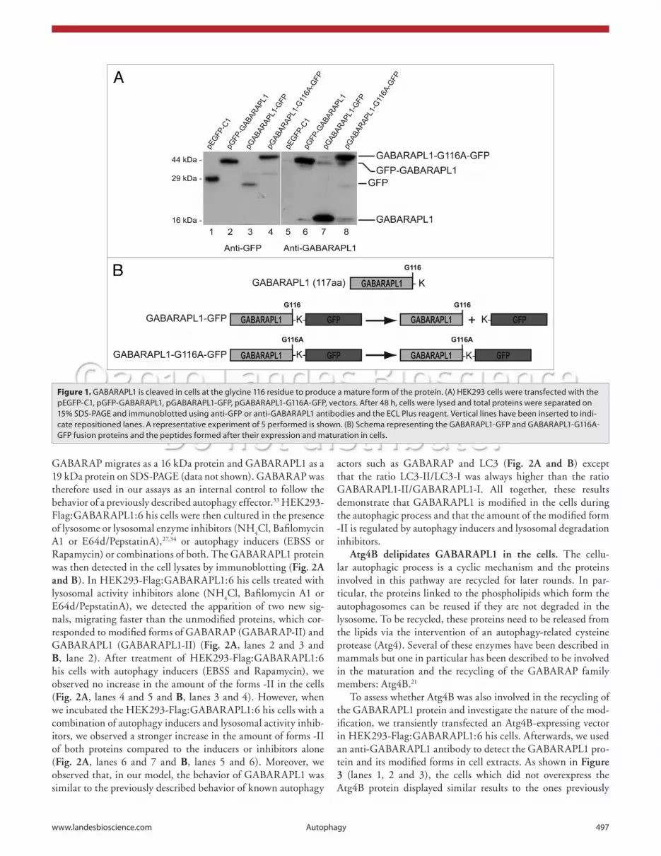
in the cells and the corresponding lysate probed with the anti-GFP antibody, we observed only one signal, corresponding to the truncated GFP protein, below the 29 kDa marker (Fig. 1A, lane 3). The difference in size between the native GFP, 29 kDa in size (lane 1), and the cleaved GFP (lane 3) was due to the pres-ence of a supplementary linker sequence present in the pEGFP-C1 commercial vector. We confirmed these results by probing with the anti-GABARAPL1 antibody which produced only one signal at 16 kDa (Fig. 1A, lane 7). These data demonstrate that the GABARAPL1-GFP protein is cleaved in the cells into two peptides, GFP and GABARAPL1. To investigate whether the glycine 116 was the site of cleavage in GABARAPL1, we cre-ated a vector encoding the GABARAPL1-G116A-GFP protein, a mutant protein in which the Gly116 is replaced by an alanine (Fig. 1B). This mutation has previously been shown to inhibit the cleavage of autophagic effectors of the GABARAP family including GABARAP and LC3.17,29 When the GABARAPL1-G116A-GFP protein was expressed in HEK293 cells, we detected a signal at the expected size of 44 kDa, corresponding to the fusion protein (Fig. 1A, lanes 4 and 8). These results demonstrate that a single point mutation (G116A) inhibits the cleavage of the GABARAPL1 protein in the cells.
GABARAPL1 is modified during the autophagic process. Previous work reported that several autophagic effectors (LC3, GABARAP, GATE-16) are linked to phospholipids during the course of autophagy to lead to a modified form of these factors, denoted by the suffix “-II”.18 This modification status is easily detected by western blotting since the modified phospholipid-linked protein migrates faster than the mature form on SDS-PAGE. To assess if GABARAPL1 is linked to phospholipids in the cells and exists as the modified protein GABARAPL1-II, we used a HEK293 stable cell line expressing the GABARAPL1 pro-tein fused to the Flag epitope at its N-terminus and six histidine residues at its C-terminus, Flag:GABARAPL1:6 his.
To enrich and detect the modified form of Flag:GABARAPL1:6 his (Flag:GABARAPL1-II), we induced and/or blocked the autophagic flux by using different chemical compounds: either inhibitors of lysosomal activity [Bafilomycin A1, a specific inhib-itor of the vacuolar type H+-ATPase (V-ATPase) or NH
4Cl, a
V-ATPase-independent neutralyzer of lysosomal pH],30 inhibi-tors of lysosomal enzyme activity (E64d and PepstatinA, cal-pain and cathepsins B, D and E inhibitors)26 or inducers of autophagy (Rapamycin, an inhibitor of the mTOR kinase and EBSS, a starvation medium).31 By inducing and/or blocking the autophagic pathway, we expected to observe an accumulation of autophagosomes and therefore an increase in the levels of the Flag:GABARAPL1-II form of the protein.
When untreated HEK293-Flag:GABARAPL1:6his cell lysates were probed with an antibody against GABARAPL1, we detected two signals at 16 and 19 kDa (Fig. 2A and B, lanes 1), corresponding to the mature GABARAP-I and GABARAPL1-I proteins. This can be explained by the fact that no commercial or homemade antibody which we have tested so far has been able to differentiate between the two homologous proteins.3,32 Moreover, previous transient transfection experiments using GABARAP and GABARAPL1-expressing vectors showed that
membrane sac (pre-autophagosome) expands to engulf cytoso-lic components and finally closes to form an autophagosome. The autophagosome then fuses with the lysosome leading to the proteolytic degradation of its content. These two structures are characterized by different molecular markers: Atg5 modifica-tion by Atg12 is linked to the formation of pre-autophagosomes and the cleavage and covalent lipidation of LC3 is a marker of autophagosomes.14
During autophagy, the covalent link between phospholipids and effector proteins occurs through a process similar to that described for Ubiquitin and SUMO-1 (Small Ubiquitin-like MOdifier 1).15 This mechanism and the enzymes involved were first described in yeast but homologous proteins have since been identified in mammals.16 First, one or several amino acids at the C-terminal end of the precursor protein are cleaved, thanks to a maturation enzyme (Atg4 family), to free a glycine residue (Gly120 in LC3,17 or Gly116 in GATE-16 and GABARAP18). This first step leads to the mature form of the protein, denoted by the suffix “-I”. The mature protein is then transferred onto phos-pholipids via the intervention of an activating enzyme (E1-like, Atg7)19 and a conjugating enzyme (E2-like, Atg3)20 to yield the phospholipid-linked protein, denoted by the suffix “-II”. This process is cyclic since the effectors can be released thanks to a delipidating enzyme (Atg4 family) which breaks the covalent bond between the protein and the lipid.21 Since GABARAPL1 shares a high homology with the GABARAP family members and the glycine 116 residue, essential for the autophagic process, is conserved in this protein, we asked whether GABARAPL1 might be implicated in the autophagic process as well.
Here we report data demonstrating that GABARAPL1 is cleaved at glycine 116 in the cell types tested and is only detect-able as the processed mature form of the protein (-I). We also show that GABARAPL1 is linked onto phospholipids to gener-ate a modified form of the protein (-II) and demonstrate that it associates with autophagosomes or lysosomes in the cells. Therefore, our work describes, for the first time, the implication of GABARAPL1 in autophagy.
Results
GABARAPL1 is cleaved in cells at the glycine 116 residue to produce a mature form of the protein. Since it has been pre-viously reported that some effectors of autophagy such as LC3, GATE-16 and GABARAP undergo a necessary maturation step before their involvement in the autophagic process and that GABARAPL1 shares a high identity with these proteins, we hypothesized that GABARAPL1 may also undergo cleavage to free the glycine 116 (Gly116) and produce the mature form of the protein, GABARAPL1-I.
To do so, we designed two different vectors encoding GFP fused to the N-terminus of GABARAPL1 (GFP-GABARAPL1) or the C-terminus of GABARAPL1 (GABARAPL1-GFP). When these proteins were expressed in HEK293 cells, we detected the GFP-GABARAPL1 protein at the expected size of 44 kDa using either the GFP or GABARAPL1 antibody (Fig. 1A, lanes 2 and 6). However when the GABARAPL1-GFP protein was expressed
www.landesbioscience.com Autophagy 497
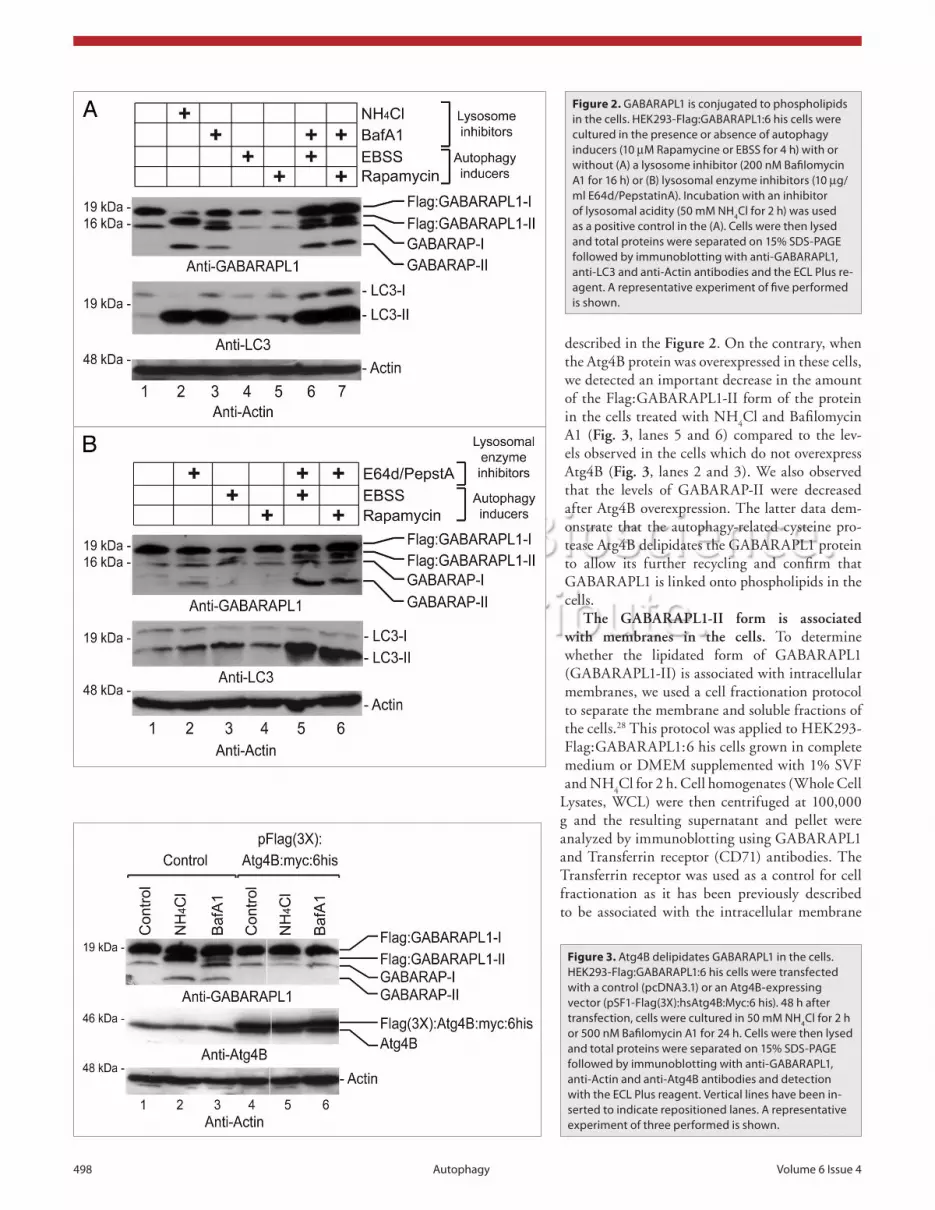
actors such as GABARAP and LC3 (Fig. 2A and B) except that the ratio LC3-II/LC3-I was always higher than the ratio GABARAPL1-II/GABARAPL1-I. All together, these results demonstrate that GABARAPL1 is modified in the cells during the autophagic process and that the amount of the modified form -II is regulated by autophagy inducers and lysosomal degradation inhibitors.
Atg4B delipidates GABARAPL1 in the cells. The cellu-lar autophagic process is a cyclic mechanism and the proteins involved in this pathway are recycled for later rounds. In par-ticular, the proteins linked to the phospholipids which form the autophagosomes can be reused if they are not degraded in the lysosome. To be recycled, these proteins need to be released from the lipids via the intervention of an autophagy-related cysteine protease (Atg4). Several of these enzymes have been described in mammals but one in particular has been described to be involved in the maturation and the recycling of the GABARAP family members: Atg4B.21
To assess whether Atg4B was also involved in the recycling of the GABARAPL1 protein and investigate the nature of the mod-ification, we transiently transfected an Atg4B-expressing vector in HEK293-Flag:GABARAPL1:6 his cells. Afterwards, we used an anti-GABARAPL1 antibody to detect the GABARAPL1 pro-tein and its modified forms in cell extracts. As shown in Figure 3 (lanes 1, 2 and 3), the cells which did not overexpress the Atg4B protein displayed similar results to the ones previously
GABARAP migrates as a 16 kDa protein and GABARAPL1 as a 19 kDa protein on SDS-PAGE (data not shown). GABARAP was therefore used in our assays as an internal control to follow the behavior of a previously described autophagy effector.33 HEK293-Flag:GABARAPL1:6 his cells were then cultured in the presence of lysosome or lysosomal enzyme inhibitors (NH
4Cl, Bafilomycin
A1 or E64d/PepstatinA),27,34 or autophagy inducers (EBSS or Rapamycin) or combinations of both. The GABARAPL1 protein was then detected in the cell lysates by immunoblotting (Fig. 2A and B). In HEK293-Flag:GABARAPL1:6 his cells treated with lysosomal activity inhibitors alone (NH
4Cl, Bafilomycin A1 or
E64d/PepstatinA), we detected the apparition of two new sig-nals, migrating faster than the unmodified proteins, which cor-responded to modified forms of GABARAP (GABARAP-II) and GABARAPL1 (GABARAPL1-II) (Fig. 2A, lanes 2 and 3 and B, lane 2). After treatment of HEK293-Flag:GABARAPL1:6 his cells with autophagy inducers (EBSS and Rapamycin), we observed no increase in the amount of the forms -II in the cells (Fig. 2A, lanes 4 and 5 and B, lanes 3 and 4). However, when we incubated the HEK293-Flag:GABARAPL1:6 his cells with a combination of autophagy inducers and lysosomal activity inhib-itors, we observed a stronger increase in the amount of forms -II of both proteins compared to the inducers or inhibitors alone (Fig. 2A, lanes 6 and 7 and B, lanes 5 and 6). Moreover, we observed that, in our model, the behavior of GABARAPL1 was similar to the previously described behavior of known autophagy
Figure 1. GABARAPL1 is cleaved in cells at the glycine 116 residue to produce a mature form of the protein. (A) heK293 cells were transfected with the peGFP-c1, pGFP-GABARAPL1, pGABARAPL1-GFP, pGABARAPL1-G116A-GFP, vectors. After 48 h, cells were lysed and total proteins were separated on 15% sDs-PAGe and immunoblotted using anti-GFP or anti-GABARAPL1 antibodies and the ecL Plus reagent. Vertical lines have been inserted to indi-cate repositioned lanes. A representative experiment of 5 performed is shown. (B) schema representing the GABARAPL1-GFP and GABARAPL1-G116A-GFP fusion proteins and the peptides formed after their expression and maturation in cells.
498 Autophagy Volume 6 issue 4
described in the Figure 2. On the contrary, when the Atg4B protein was overexpressed in these cells, we detected an important decrease in the amount of the Flag:GABARAPL1-II form of the protein in the cells treated with NH
4Cl and Bafilomycin
A1 (Fig. 3, lanes 5 and 6) compared to the lev-els observed in the cells which do not overexpress Atg4B (Fig. 3, lanes 2 and 3). We also observed that the levels of GABARAP-II were decreased after Atg4B overexpression. The latter data dem-onstrate that the autophagy-related cysteine pro-tease Atg4B delipidates the GABARAPL1 protein to allow its further recycling and confirm that GABARAPL1 is linked onto phospholipids in the cells.
The GABARAPL1-II form is associated with membranes in the cells. To determine whether the lipidated form of GABARAPL1 (GABARAPL1-II) is associated with intracellular membranes, we used a cell fractionation protocol to separate the membrane and soluble fractions of the cells.28 This protocol was applied to HEK293-Flag:GABARAPL1:6 his cells grown in complete medium or DMEM supplemented with 1% SVF and NH
4Cl for 2 h. Cell homogenates (Whole Cell
Lysates, WCL) were then centrifuged at 100,000 g and the resulting supernatant and pellet were analyzed by immunoblotting using GABARAPL1 and Transferrin receptor (CD71) antibodies. The Transferrin receptor was used as a control for cell fractionation as it has been previously described to be associated with the intracellular membrane
Figure 2. GABARAPL1 is conjugated to phospholipids in the cells. heK293-Flag:GABARAPL1:6 his cells were cultured in the presence or absence of autophagy inducers (10 µM Rapamycine or eBss for 4 h) with or without (A) a lysosome inhibitor (200 nM Bafilomycin A1 for 16 h) or (B) lysosomal enzyme inhibitors (10 µg/ml e64d/PepstatinA). incubation with an inhibitor of lysosomal acidity (50 mM Nh4cl for 2 h) was used as a positive control in the (A). cells were then lysed and total proteins were separated on 15% sDs-PAGe followed by immunoblotting with anti-GABARAPL1, anti-Lc3 and anti-Actin antibodies and the ecL Plus re-agent. A representative experiment of five performed is shown.
Figure 3. Atg4B delipidates GABARAPL1 in the cells. heK293-Flag:GABARAPL1:6 his cells were transfected with a control (pcDNA3.1) or an Atg4B-expressing vector (psF1-Flag(3X):hsAtg4B:Myc:6 his). 48 h after transfection, cells were cultured in 50 mM Nh4cl for 2 h or 500 nM Bafilomycin A1 for 24 h. cells were then lysed and total proteins were separated on 15% sDs-PAGe followed by immunoblotting with anti-GABARAPL1, anti-Actin and anti-Atg4B antibodies and detection with the ecL Plus reagent. Vertical lines have been in-serted to indicate repositioned lanes. A representative experiment of three performed is shown.

www.landesbioscience.com Autophagy 499
fraction.28 Our results showed that NH4Cl induced the
apparition of the GABARAP-II and GABARAPL1-II forms in the cells (Fig. 4, lane 4, WCL) as described above (Fig. 2). Following cell fractionation, the mature proteins (-I) were predominantly detected in the supernatants of the untreated and treated cells (Fig. 4, lanes 2 and 5). Conversely, the lipidated forms (-II) were enriched in the pellet (membraneous frac-tion) of the induced cells (Fig. 4, lane 6). No form -II was detected in the pellet obtained from the untreated cells (Fig. 4, lane 3). Moreover, the Transferrin recep-tor was detected predominantly in the pellets dem-onstrating the specific enrichment of membranes in these fractions. All together, our results show that the GABARAPL1-II form is associated with intracellular membranes in the cells.
Inhibition of lysosomal activity results in the accu-mulation of GABARAPL1-positive intracytoplasmic vesicles. Thus far, we have shown that GABARAPL1 is modified in the cells, is delipidated by Atg4B, and that the modified form is enriched in intracellular membrane fractions. To confirm that GABARAPL1 is in fact associated with autophagosomes, we inves-tigated the intracellular distribution of this protein after the inhibition of the autophagic flux. Indeed, we already know that proteins linked to autophagic vesi-cles display a punctate cytoplasmic distribution, corre-sponding to their localization into autophagosomes.23 As such, we first designed a MCF-7 stable cell line expressing the DsRed-GABARAPL1 protein, then we inhibited lysosomal activity in these cells. In the untreated DsRed-GABARAPL1 cell lines, the fusion protein was detected in punctate locations throughout the cytosol (Fig. 5A and D). When the MCF7-DsRed-GABARAPL1 cells were treated with NH
4Cl (Fig. 5B and E), we
detected a slight increase in the number of intracellular DsRed-GABARAPL1-positive vesicles. When the DsRed-GABARAPL1 cells were treated with Bafilomycin A1, we detected an increase in the number of GABARAPL1-positive vesicles but more strik-ingly, we observed a total relocalization of these vesicles in a clus-ter on one side of the nucleus (Fig. 5C and F). Such a pattern has been previously described for the localization of the lysosomes in the cell.30 In conclusion, GABARAPL1 is localized in intracyto-plasmic vesicles, a localization which is increased after treatment with lysosomal activity inhibitors.
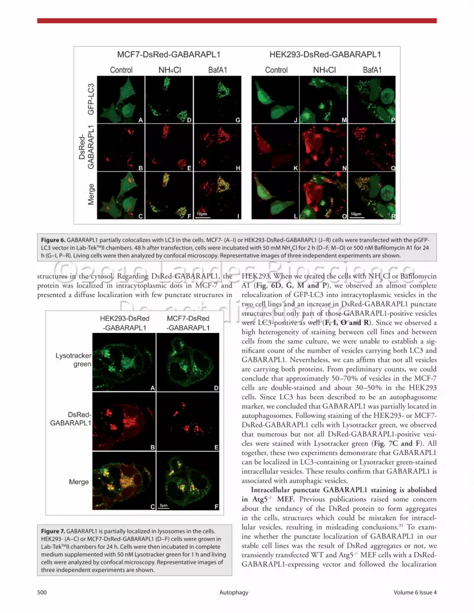
GABARAPL1 partially colocalizes with GFP-LC3 or Lysotracker green in intracytoplasmic vesicles. To determine the nature of the DsRed-GABARAPL1-containing vesicles, we used two different approaches: (1) we transiently overexpressed the GFP-LC3 protein in HEK293- and MCF-7-DsRed-GABARAPL1 cells and followed the intracellular localization of the two fusion proteins after treatment of the cells with lysosome inhibitors; (2) we stained the HEK293- and MCF7-Dsred-GABARAPL1 cells with a specific lysosomal marker called Lysotracker green. When we expressed the GFP-LC3 in the DsRed-GABARAPL1-expressing cell lines, we observed a diffuse localization of GFP-LC3 throughout the cytoplasm and the nucleus and some punctate
Figure 4. The GABARAPL1-ii form is associated with membranes in the cells. heK293-Flag:GABARAPL1:6 his cells were cultured in complete medium or DMeM supplemented with 1% FBs and 50 mM Nh4cl for 2 h. cells were then collected in PBs supplemented with 1 mM PMsF, washed, homogenized (WcL, Whole cell Lysates) and centrifuged for 10 min at 800 g. Following two ultracentrifuga-tion rounds (30 min at 100,000 g), vesicles were resuspended in PBs (Pellet). The supernatant sample corresponds to the supernatant obtained after the first round of ultracentrifugation. One aliquot (20 µl) of each of the different fractions (WcL, supernatant and pellet) was separated on 15% sDs-PAGe followed by immunob-lotting with anti-GABARAPL1 and anti-Transferrin receptor (Tfr, cD71) antibodies and detection with the ecL Plus reagent. A representative experiment of three performed is shown.
Figure 5. inhibition of lysosomal activity results in the accumulation of GABARAPL1-positive intracytoplasmic vesicles. McF-7 cells overexpress-ing the DsRed-GABARAPL1 protein were incubated in complete me-dium (control) (A and D), DMeM supplemented with 1% FBs and 50 mM Nh4cl for 2 h (Nh4cl) (B and e) or complete medium supplemented with 500 nM of Bafilomycin A1 for 24 h (BafA1) (c and F). cells were then fixed and analyzed by confocal microscopy. The images on the right (D–F) correspond to a 3-fold magnification of the images on the left (A–c). Representative images of three independent experiments are shown.
500 Autophagy Volume 6 issue 4
HEK293. When we treated the cells with NH4Cl or Bafilomycin
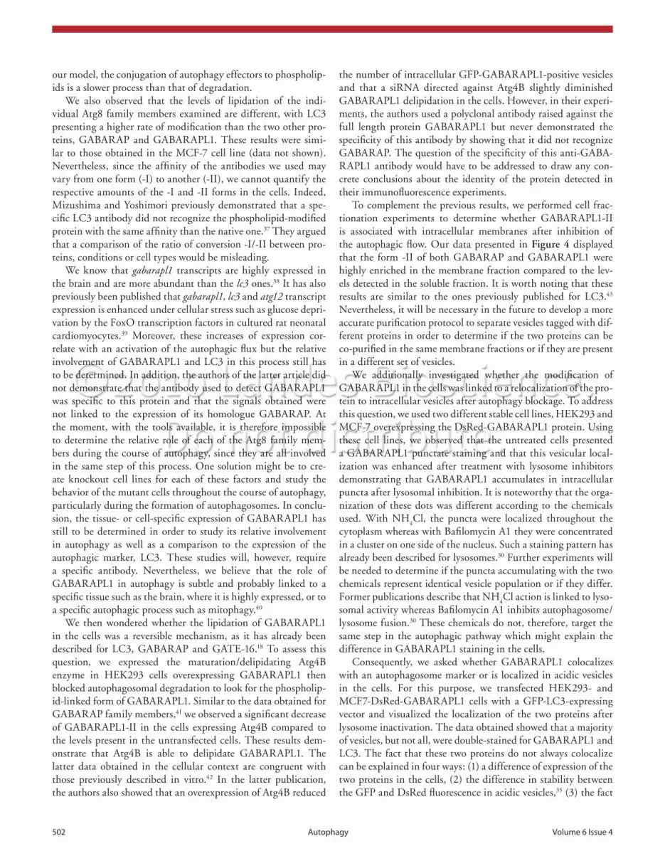
A1 (Fig. 6D, G, M and P), we observed an almost complete relocalization of GFP-LC3 into intracytoplasmic vesicles in the two cell lines and an increase in DsRed-GABARAPL1 punctate structures but only part of those GABARAPL1-positive vesicles were LC3-positive as well (F, I, O and R). Since we observed a high heterogeneity of staining between cell lines and between cells from the same culture, we were unable to establish a sig-nificant count of the number of vesicles carrying both LC3 and GABARAPL1. Nevertheless, we can affirm that not all vesicles are carrying both proteins. From preliminary counts, we could conclude that approximately 50–70% of vesicles in the MCF-7 cells are double-stained and about 30–50% in the HEK293 cells. Since LC3 has been described to be an autophagosome marker, we concluded that GABARAPL1 was partially located in autophagosomes. Following staining of the HEK293- or MCF7-DsRed-GABARAPL1 cells with Lysotracker green, we observed that numerous but not all DsRed-GABARAPL1-positive vesi-cles were stained with Lysotracker green (Fig. 7C and F). All together, these two experiments demonstrate that GABARAPL1 can be localized in LC3-containing or Lysotracker green-stained intracellular vesicles. These results confirm that GABARAPL1 is associated with autophagic vesicles.
Intracellular punctate GABARAPL1 staining is abolished in Atg5-/- MEF. Previous publications raised some concern about the tendancy of the DsRed protein to form aggregates in the cells, structures which could be mistaken for intracel-lular vesicles, resulting in misleading conclusions.35 To exam-ine whether the punctate localization of GABARAPL1 in our stable cell lines was the result of DsRed aggregates or not, we transiently transfected WT and Atg5-/- MEF cells with a DsRed-GABARAPL1-expressing vector and followed the localization
structures in the cytosol. Regarding DsRed-GABARAPL1, the protein was localized in intracytoplasmic dots in MCF-7 and presented a diffuse localization with few punctate structures in
Figure 6. GABARAPL1 partially colocalizes with Lc3 in the cells. McF7- (A–i) or heK293-DsRed-GABARAPL1 (J–R) cells were transfected with the pGFP-Lc3 vector in Lab-TekTMii chambers. 48 h after transfection, cells were incubated with 50 mM Nh4cl for 2 h (D–F, M–O) or 500 nM Bafilomycin A1 for 24 h (G–i, P–R). Living cells were then analyzed by confocal microscopy. Representative images of three independent experiments are shown.
Figure 7. GABARAPL1 is partially localized in lysosomes in the cells. heK293- (A–c) or McF7-DsRed-GABARAPL1 (D–F) cells were grown in Lab-TekTMii chambers for 24 h. cells were then incubated in complete medium supplemented with 50 nM Lysotracker green for 1 h and living cells were analyzed by confocal microscopy. Representative images of three independent experiments are shown.

www.landesbioscience.com Autophagy 501
the autophagic process or inhibiting the lysosomal degradation. We incubated HEK293 cells overexpressing the GABARAPL1 protein with: (1) lysosome inhibitors (NH
4Cl, Bafilomycin A1 or
E64d/PepstatinA), (2) autophagy inducers (rapamycin or EBSS), or (3) a combination of both. Subsequently, we searched for the modified form of the protein (GABARAPL1-II) in these cells. All of these treatments have been previously described to result in a significant increase in the amount of the phospholipid-linked forms of autophagy effectors such as LC3 and GABARAP. In our experiments, we effectively detected GABARAPL1-II but we observed variations in the amount of this form according to the protocol used. The highest levels were detected with NH
4Cl. EBSS
and rapamycin did not allow the detection of the form -II without the addition of lysosomal degradation inhibitors (Bafilomycin A1 or E64d/PepstatinA). It is noteworthy that we obtained the same results with the stable MCF7-Flag:GABARAPL1:6 his cell line (data not shown). In conclusion, we show that GABARAPL1 is modified in the cells and that this modification is induced by autophagy inducers. These results are in agreement with the data obtained for the other members of the GABARAP family includ-ing GABARAP, LC3 and GATE-16.36 The fact that this modi-fication is only detected when the degradation of the forms -II are inhibited by lysosomal activity inhibitors is congruent with previously published data on LC3.26 Moreover, we clearly see in our experiment that the lipidation of LC3 is weak in the presence of the autophagy inducers alone compared to the levels detected after addition of lysosome inhibitors. Therefore, it seems that, in
of the fusion protein before and after inhibition of lysosomal activity by NH
4Cl or Bafilomycin A1 (Fig. 8). Former publica-
tions showed that the deletion of Atg5 abrogated the autophagic pathway in MEF cells.24 Our results demonstrated that, before treatment, the WT cells presented a high number of red vesicles whereas the Atg5-/- cells contained a very low number of these DsRed-GABARAPL1-stained vesicles. After incubation with lysosomal inhibitors, we detected an increase in the number of punctate structures in the WT cells (Fig. 8A–C), and a signifi-cantly smaller increase in the Atg5-/- cell line (Fig. 8D–F). Taken together, our results demonstrate that the deletion of Atg5, and therefore the inhibition of the autophagic process, alters the for-mation of DsRed-GABARAPL1-stained vesicles showing that these structures are not protein aggregates but rather related to the process of autophagy.
Discussion
Previous work has described the involvement of different GABARAP family members in the autophagic process. Atg8, LC3, GABARAP and GATE-16 have all been shown to be linked to phospholipids and recruited to autophagosomes after induction of autophagy.23 These effectors play an important role during the elongation of double-membraned structures leading to the sequestration of cytosolic constituents and to the formation of autophagosomes. After their completion, these vesicles fuse with lysosomes to induce the degradation and recycling of their contents.18 To our knowledge, all the data published to date show that these proteins play a role as effectors of autophagy but not as activators. Since GABARAPL1 (GEC1) shares a high iden-tity with GABARAP as well as other members of the family, we asked whether GABARAPL1 might be implicated in this process as well. To examine this possibility, we addressed different ques-tions: (1) Is GABARAPL1 cleaved in the cells to free the glycine 116 residue at its C-terminal end?, (2) Is GABARAPL1 linked to phospholipids in the cells?, (3) Is GABARAPL1 delipidated by the autophagy-related cysteine protease Atg4B?, (4) Is GABARAPL1 associated with intracellular vesicles?, (5) Does GABARAPL1 relocalize to autophagosomes during the autophagic process?
First, we showed that a GABARAPL1-GFP fusion pro-tein was cleaved to give two products, GABARAPL1 and GFP but that this cleavage was abolished when the glycine 116 resi-due was replaced by an alanine. These results demonstrate that GABARAPL1 is processed in the cells and that the maturation occurs at glycine 116. Moreover, it is worth noting that we were unable to detect the GABARAPL1-GFP fusion protein in our cell lysates, suggesting that this protein was cleaved following its synthesis and that this maturation step was not a consequence of autophagy induction. In addition, similar data were obtained using GABARAP wild-type and mutant-expressing vectors in HEK293 (data not shown) and these results were confirmed, for the two proteins (GABARAPL1 and GABARAP), in different cell types (COS7, MCF-7) showing that this process is not cell-type specific (data not shown).
We then asked whether GABARAPL1 was modified in the cells. To do so, we used a combination of different chemicals inducing
Figure 8. intracellular punctate GABARAPL1 staining is abolished in Atg5-/- MeFs. WT (A–c) and Atg5-/- (D–F) MeF cells were transfected with a DsRed-GABARAPL1-expressing vector. 48 h after transfection, cells were incubated in complete medium (A and D), with 50 mM Nh4cl for 2 h (B and e) or 500 nM Bafilomycin A1 for 24 h (c and F). cells were then analyzed by confocal microscopy. Representative images of three independent experiments are shown.
502 Autophagy Volume 6 issue 4
the number of intracellular GFP-GABARAPL1-positive vesicles and that a siRNA directed against Atg4B slightly diminished GABARAPL1 delipidation in the cells. However, in their experi-ments, the authors used a polyclonal antibody raised against the full length protein GABARAPL1 but never demonstrated the specificity of this antibody by showing that it did not recognize GABARAP. The question of the specificity of this anti-GABA-RAPL1 antibody would have to be addressed to draw any con-crete conclusions about the identity of the protein detected in their immunofluorescence experiments.
To complement the previous results, we performed cell frac-tionation experiments to determine whether GABARAPL1-II is associated with intracellular membranes after inhibition of the autophagic flow. Our data presented in Figure 4 displayed that the form -II of both GABARAP and GABARAPL1 were highly enriched in the membrane fraction compared to the lev-els detected in the soluble fraction. It is worth noting that these results are similar to the ones previously published for LC3.43 Nevertheless, it will be necessary in the future to develop a more accurate purification protocol to separate vesicles tagged with dif-ferent proteins in order to determine if the two proteins can be co-purified in the same membrane fractions or if they are present in a different set of vesicles.
We additionally investigated whether the modification of GABARAPL1 in the cells was linked to a relocalization of the pro-tein to intracellular vesicles after autophagy blockage. To address this question, we used two different stable cell lines, HEK293 and MCF-7 overexpressing the DsRed-GABARAPL1 protein. Using these cell lines, we observed that the untreated cells presented a GABARAPL1 punctate staining and that this vesicular local-ization was enhanced after treatment with lysosome inhibitors demonstrating that GABARAPL1 accumulates in intracellular puncta after lysosomal inhibition. It is noteworthy that the orga-nization of these dots was different according to the chemicals used. With NH
4Cl, the puncta were localized throughout the
cytoplasm whereas with Bafilomycin A1 they were concentrated in a cluster on one side of the nucleus. Such a staining pattern has already been described for lysosomes.30 Further experiments will be needed to determine if the puncta accumulating with the two chemicals represent identical vesicle population or if they differ. Former publications describe that NH
4Cl action is linked to lyso-
somal activity whereas Bafilomycin A1 inhibits autophagosome/lysosome fusion.30 These chemicals do not, therefore, target the same step in the autophagic pathway which might explain the difference in GABARAPL1 staining in the cells.
Consequently, we asked whether GABARAPL1 colocalizes with an autophagosome marker or is localized in acidic vesicles in the cells. For this purpose, we transfected HEK293- and MCF7-DsRed-GABARAPL1 cells with a GFP-LC3-expressing vector and visualized the localization of the two proteins after lysosome inactivation. The data obtained showed that a majority of vesicles, but not all, were double-stained for GABARAPL1 and LC3. The fact that these two proteins do not always colocalize can be explained in four ways: (1) a difference of expression of the two proteins in the cells, (2) the difference in stability between the GFP and DsRed fluorescence in acidic vesicles,35 (3) the fact
our model, the conjugation of autophagy effectors to phospholip-ids is a slower process than that of degradation.
We also observed that the levels of lipidation of the indi-vidual Atg8 family members examined are different, with LC3 presenting a higher rate of modification than the two other pro-teins, GABARAP and GABARAPL1. These results were simi-lar to those obtained in the MCF-7 cell line (data not shown). Nevertheless, since the affinity of the antibodies we used may vary from one form (-I) to another (-II), we cannot quantify the respective amounts of the -I and -II forms in the cells. Indeed, Mizushima and Yoshimori previously demonstrated that a spe-cific LC3 antibody did not recognize the phospholipid-modified protein with the same affinity than the native one.37 They argued that a comparison of the ratio of conversion -I/-II between pro-teins, conditions or cell types would be misleading.
We know that gabarapl1 transcripts are highly expressed in the brain and are more abundant than the lc3 ones.38 It has also previously been published that gabarapl1, lc3 and atg12 transcript expression is enhanced under cellular stress such as glucose depri-vation by the FoxO transcription factors in cultured rat neonatal cardiomyocytes.39 Moreover, these increases of expression cor-relate with an activation of the autophagic flux but the relative involvement of GABARAPL1 and LC3 in this process still has to be determined. In addition, the authors of the latter article did not demonstrate that the antibody used to detect GABARAPL1 was specific to this protein and that the signals obtained were not linked to the expression of its homologue GABARAP. At the moment, with the tools available, it is therefore impossible to determine the relative role of each of the Atg8 family mem-bers during the course of autophagy, since they are all involved in the same step of this process. One solution might be to cre-ate knockout cell lines for each of these factors and study the behavior of the mutant cells throughout the course of autophagy, particularly during the formation of autophagosomes. In conclu-sion, the tissue- or cell-specific expression of GABARAPL1 has still to be determined in order to study its relative involvement in autophagy as well as a comparison to the expression of the autophagic marker, LC3. These studies will, however, require a specific antibody. Nevertheless, we believe that the role of GABARAPL1 in autophagy is subtle and probably linked to a specific tissue such as the brain, where it is highly expressed, or to a specific autophagic process such as mitophagy.40
We then wondered whether the lipidation of GABARAPL1 in the cells was a reversible mechanism, as it has already been described for LC3, GABARAP and GATE-16.18 To assess this question, we expressed the maturation/delipidating Atg4B enzyme in HEK293 cells overexpressing GABARAPL1 then blocked autophagosomal degradation to look for the phospholip-id-linked form of GABARAPL1. Similar to the data obtained for GABARAP family members,41 we observed a significant decrease of GABARAPL1-II in the cells expressing Atg4B compared to the levels present in the untransfected cells. These results dem-onstrate that Atg4B is able to delipidate GABARAPL1. The latter data obtained in the cellular context are congruent with those previously described in vitro.42 In the latter publication, the authors also showed that an overexpression of Atg4B reduced
www.landesbioscience.com Autophagy 503
86% identity at the amino-acid level. Recently published papers have used polyclonal anti-GABARAPL1 antibodies to detect the protein in immunofluorescence experiments. However, the question of specificity of these antibodies was not addressed and therefore the identity of the protein studied in these publications remains unclear.39,42 Consequently, apart from western blotting experiments in which we were able to differentiate between the two homologous proteins, we opted out of using our anti-GABA-RAPL1 antibody for immunofluorescence experiments. The pro-duction of a specific antibody directed against the GABARAPL1 protein will be one of the next challenges before extending the study of this protein.
Using different experimental assays, we demonstrated in a multitude of ways that GABARAPL1 is implicated in the process of autophagy. Nevertheless, further experiments will be needed to explain the link between autophagy, GABARAPL1 and its function in vivo. In particular, it would be of great interest to study the relative involvement of the different members of the GABARAP family in the autophagic process. Are these proteins redundant in the cells or are they tissue specific? We know that numerous tissues co-express the GABARAP and GABARAPL1 proteins but also that some tissues express one protein rather than the other.32,38 For example, gabarapl1 transcripts are much more abundant in the brain compared to lc3 and gabarap.38 Given the importance of autophagy in brain damage44 and neurode-generative diseases,45 it will be essential to study the function of GABARAPL1 in the brain with respect to its implication in autophagy.
Materials and Methods
Reagents and antibodies. Cell culture reagents were purchased from Invitrogen (Carlsbad, CA). The following antibodies were used: polyclonal anti-GABARAPL1 (Chemicon Millipore, AB15278), polyclonal anti-LC3 (Novus Biologicals, NB100-2220), polyclonal anti-GFP (Chemicon Millipore, AB3080), polyclonal anti-Transferrin receptor (CD71) (Santa Cruz, sc65882), monoclonal anti-Atg4B (Sigma, A2981) and polyclonal anti-Actin (Sigma, A5060). Bafilomycin A1 (Sigma, B1793) was prepared as a 1 mM stock in DMSO. Ammonium chloride (NH
4Cl, Sigma, A0171) was prepared as a 0.5 M stock in H
2O.
Rapamycin (Sigma, R8781) was prepared as a 2.5 mM stock in DMSO. E64d and Pepstatin A (Sigma, E8640 and P5318) were prepared as 10 mg/ml stocks in DMSO. All other reagents were purchased from Sigma-Aldrich.
Plasmids and site-directed mutagenesis. The pcDNA3.1 vector (Invitrogen, V795-20) was used for control transfec-tions. The pcDNA5-FRT vector was purchased from Invitrogen (V6010-20). The pEGFP-C1 and pEGFP-N3 plasmids were obtained from Clontech (6084-1 and 6080-1). The pGFP-LC3, and pSF1-Flag(3X):hsAtg4B:Myc:6 his plasmids were kindly provided by Dr. Elazar (Weizmann Institute, Israël),22 and Dr. Yoshimori (Osaka University, Japan),23 respectively. The pGABARAPL1-GFP and pGFP-GABARAPL1 plasmids encod-ing the Enhanced Green Fluorescent Protein (EGFP) fused to the N-terminus or C-terminus of the GABARAPL1 protein were
that all autophagosomes might not be carrying both proteins, or (4) that GABARAPL1 may also be associated with a different set of vesicles not involved in the autophagic pathway. To answer this question, we stained HEK293- and MCF7-DsRed-GABARAPL1 cells with a specific lysosomal marker, Lysotracker green. In this experiment, we did not use lysosomal inhibitors since Lysotracker green chemical is sensitive to pH changes. This double-staining experiment showed that the majority of GABARAPL1 colocal-izes with the lysosomes but that GABARAPL1 can be also found outside of the lysosomes. All together, the latter data demonstrate that GABARAPL1 is predominantly localized in autophagosomes or lysosomes. These results are not surprising since autopha-gosomes fuse with lysosomes to form autophagolysosomes and induce the degradation of their contents. Since GFP fluorescence is rapidly inhibited in acidic compartments, GFP-LC3 might allow the detection of early nonacidic autophagosomes whereas Lysotracker green stains late acidic vesicles.
These data are really interesting and will lead to some fol-low-up experiments. Indeed, a recent publication described the interaction of LC3/GABARAP family members with the protein Nix involved in the clearance of damaged mitochondria by the autophagic pathway.40 The authors showed that GABARAPL1 binds more strongly than other members of the family to Nix and that GABARAPL1 colocalizes with Nix in cells treated with a mitochondria poison. These results suggest that the clearance of damaged mitochondria may be mediated by the specific asso-ciation between GABARAPL1 and Nix and therefore confer a specificity to the nonspecific autophagic process. In the near future, it will be of interest to determine whether the autophagic proteins of the LC3/GABARAP family have specific and differ-ent cellular patterns, are linked to specific autophagic vesicles or if these proteins are involved in distinct autophagic degradation processes in the cells.
One main concern regarding the use of overexpression sys-tems or exogenous protein expression is the fact that some pro-teins can aggregate and therefore create intracellular punctate structures which might be mistaken for vesicles. To demonstrate that the GABARAPL1-containing dots observed in our experi-ments were autophagy-related, we used the MEF Atg5-/- cell line defective for the autophagic pathway.24 When we expressed the DsRed-Protein in this cell line, we observed a very low number of intracellular red vesicles before or after treatment with NH
4Cl
or Bafilomycin A1 compared to the wild-type control cells. These results confirmed that the vesicles we observed are autophagy-related or at least that their formation requires Atg5. The low number of vesicles still detectable in the Atg5-/- cells might be due to the role of GABARAPL1 as a transport protein38 but we also cannot completely rule out the idea that some aggregates are formed when using the DsRed protein as previously described.35 Further experiments will be needed to assess this question.
In our study, we used transient transfection assays or designed stable cell lines overexpressing fusion proteins because we did not possess a specific anti-GABARAPL1 antibody. Indeed, all the homemade or commercial antibodies we have tested to date are not specific for GABARAPL1, as they detect the GABARAP pro-tein as well.3,32 This is not surprising since the two proteins share
504 Autophagy Volume 6 issue 4
ammonium chloride,25 4 h in complete medium supplemented with 10 µg/ml E64d/PepstatinA26 or 24 h in complete medium supplemented with 500 nM Bafilomycin A1.27 For the induction of autophagy, cells were incubated for either 4 h in EBSS medium (Sigma, E3024) or 4 h in complete medium supplemented with 10 µM Rapamycin. Treated or untreated DsRed- or GFP-expressing cells were then fixed with 2% paraformaldehyde (PFA, Sigma, P6148) in PBS for 15 min, washed thrice with PBS and analyzed using a fluorescence laser scanning confocal microscope Fluoview FV1000 (Olympus). Each picture corresponds to one example of a typical cell staining observed in six fields chosen at random. For the Lysotracker green staining, cells were grown in Lab-TekTMII chambers (Nunc, 155380) for 24 h and subsequently incubated in complete medium supplemented with 50 nM Lysotracker green (Invitrogen, L7526) for 1 h. Living cells were then analyzed by confocal microscopy as described above.
Cell fractionation. Cell fractionation was carried out essentially as described previously.28 In brief, HEK293-Flag:GABARAPL1:6 his cells were cultured in complete medium or DMEM supplemented with 1% FBS and 50 mM NH
4Cl for
2 h to inhibit lysosomal activity. Cells were washed, collected in 1 ml PBS containing 1 mM phenylmethylsulfonylfluoride (PMSF, Sigma, 93482), homogenized by sonication (Sonics and Materials) and the large vesicles and nuclei were pelleted at 800 g for 10 min. Supernatants were centrifuged at 100,000 g for 30 min to harvest the membranes and intracellular vesicles and organelles. The pellets were resuspended in homogenization buf-fer, sonicated once more and subjected to a second round of ultra-centrifugation. Pellets were then resuspended in 1 ml PBS and one aliquot (20 µl) of the different fractions (Whole Cell Lysate, Supernatant of the first centrifugation and Pellet) were reserved for western blot analysis.
Western blot analysis. Cells were rinsed with ice-cold PBS (0.137 M NaCl, 3.3 mM KCl, 10 mM Na
2HPO
4, 1.8 mM
KH2PO
4) and lysed in a SDS-PAGE loading buffer (78 mM Tris-
HCl, pH 6.8, 2.5% SDS, 12.5%, 6.25% β-mercaptoethanol, 0.025% bromophenol blue). Samples were sonicated for 30 s before loading (Sonics and Materials) and separated by SDS-PAGE before transfer to Immobilon-P PVDF membrane (Dutscher, 44088). The membranes were blocked with 5% skim milk in 0.1% Tween 20/TBS (199 mM Tris-HCl, pH 7.4, 1.36 mM NaCl, 0.1% Tween 20) and incubated with primary anti-bodies in antibody block buffer (0.5% skim milk in 0.1% Tween 20/TBS). The polyclonal anti-GFP, anti-GABARAPL1, anti-LC3, anti-Actin and anti-Transferrin receptor (CD71) antibodies were diluted at 1:5,000, 1:2,000, 1:2,000, 1:2,000 and 1:1,000, respectively in the antibody block buffer. Immunoreactive bands were detected using goat horseradish peroxydase (HRP)-coupled secondary anti-mouse or anti-rabbit antibodies (1:20,000 in anti-body block buffer) (PARIS, P1291) and the ECL Plus reagent (GE Healthcare Life Sciences, RPN2132), according to the man-ufacturer’s protocol.
Acknowledgements
This work was supported by a grant from Ligue Contre le Cancer, Comité du Doubs. Fatima Zahra Chakrama and Jaclyn
generated by polymerase chain reaction (PCR). The reactions were performed in a 25 µl sample volume containing 0.2 µM of each primer, 1.5 mM MgCl
2, 200 µM dNTPs and 1 U of
Taq polymerase (Promega, M8305). The PCR fragments were digested with the XhoI/SacII or BglII/EcoRI restriction enzymes (Fermentas, ER0691, ER0201, ER0281 and ER0271) and sub-cloned into the pEGFP-N3 or the pEGFP-C1 vectors to produce the pGABARAPL1-GFP and the pGFP-GABARAPL1 plasmids, respectively. The point mutation (G116A) was introduced using the QuickChange Site-Directed mutagenesis system (Stratagene, 600385) to produce the pGABARAPL1-G116A-GFP vector. The Flag:GABARAPL1:6 his cDNA was digested with the NheI/NotI restriction enzymes (Fermentas, ER0971 and ER0591) and sub-cloned into the pcDNA5-FRT vector (Invitrogen) to produce the pcDNA5-Flag:GABARAPL1:6 his vector. Plasmid sequences and mutations were confirmed by DNA sequencing (Genetic Analyzer 3130, Applied Biosystems). The nucleotide sequences of the different primers are available upon request.
Cell culture and transfection. The HEK293 and MCF-7 cells (ATCC, CRL-1573 and HTB-22), WT and Atg5-/- MEF cells (kindly provided by Dr. Mizushima)24 were cultured in DMEM (Dulbecco’s Minimum Essential Medium, Invitrogen, 11880) supplemented with 2 mM L-Glutamine (Invitrogen, 25030-032), 100 µg/ml penicillin, 100 µg/ml streptomy-cin (Invitrogen, 15140) and 10% foetal bovine serum (FBS, Invitrogen, 10270-106) in a 5% CO
2 incubator at 37°C. The
HEK293- and MCF7-Flag:GABARAPL1:6 his cell lines were maintained in the complete medium supplemented with 100 µg/ml Hygromycin B (PAA, P02-015). The HEK293- and MCF7-DsRed-GABARAPL1 cell lines were maintained in the complete medium supplemented with 100 µg/ml G418 (PAA, P02-012). Transient transfections were carried out using HEK293, MCF-7, WT and Atg5-/- MEF cells plated in 6-well plates (3 x 10⁵ per well). 500 ng of pGFP-GABARAPL1, pGABARAPL1-GFP, pGABARAPL1-G116A-GFP, pGFP-LC3 or pDsRed-GABA-RAPL1 and 1 µl TransFast reagent (Promega, E2431) were used per reaction according to the manufacturer’s protocol. To create the stable cell lines HEK293- or MCF7-Flag:GABARAPL1:6 his, HEK293 or MCF-7 cells were plated in 10 cm-diameter culture dishes (106 per plate) and were transfected using 20 µg pcDNA5-Flag:GABARAPL1:6 his and 40 µl TransFast reagent per reaction, according to the manufacturer’s protocol. After 48 h incubation, cells were split in two culture dishes and 400 µg/ml Hygromycin B were added to the complete medium for the 14 following days until the appearance of antibiotic-resistant single clones. The clones were then tested for the expression of the ectopic protein by western blotting using a polyclonal anti-GABARAPL1 antibody. To create the stable HEK293- or MCF7-DsRed-GABARAPL1 cell lines, HEK293 or MCF-7 cells were transfected using 20 µg pDsRed-GABARAPL1 and 40 µl TransFast reagent per reaction, according to the manufacturer’s protocol. 400 µg/ml G418 were added to the complete medium to select for single clones.
Autophagy and confocal microscopy. For the inhibition of lys-osomal activity, cells cultured on glass coverslips were incubated for either 2 h in DMEM supplemented with 1% FBS and 50 mM
www.landesbioscience.com Autophagy 505
pSF1-Flag(3X):hsAtg4B:Myc:6 his vectors, respectively. We thank Dr. Mizushima for kindly providing the WT and Atg5-/- MEF cells. Authors are grateful to Sophie Launay and Fabrice Poncet for their technical help (Plateformes microscopie et séquençage, IFR133, Besançon). Special thanks to Dr. Petra Gross for editorial advice.
Nicole Le Grand are supported by fellowships from Ministère de l’Enseignement Supérieur et de la Recherche (MESR) and Stéphanie Seguin is supported by a fellowship from Région de Franche-Comté/Cancéropôle Grand-Est. We thank Dr. Elazar, and Dr. Yoshimori for kindly providing the pGFP-LC3, and the
References1. Pellerin S, Lafeuillade B, Wade RH, Savona C,
Chambaz EM, Feige JJ. The molecular structure of corticotropin-induced secreted protein, a novel mem-ber of the thrombospondin family. J Biol Chem 1993; 268:18810-7.
2. Vernier-Magnin S, Muller S, Sallot M, Radom J, Musard JF, Adami P, et al. A novel early estrogen-regu-lated gene gec1 encodes a protein related to GABARAP. Biochem Biophys Res Commun 2001; 284:118-25.
3. Mansuy V, Boireau W, Fraichard A, Schlick JL, Jouvenot M, Delage-Mourroux R. GEC1, a protein related to GABARAP, interacts with tubulin and GABA(A) receptor. Biochem Biophys Res Commun 2004; 325:639-48.
4. Wang H, Bedford FK, Brandon NJ, Moss SJ, Olsen RW. GABA(A)-receptor-associated protein links GABA(A) receptors and the cytoskeleton. Nature 1999; 397:69-72.
5. Mohrluder J, Schwarten M, Willbold D. Structure and potential function of gamma-aminobutyrate type A receptor-associated protein. FEBS J 2009; 276:4989-5005.
6. Klebig C, Seitz S, Arnold W, Deutschmann N, Pacyna-Gengelbach M, Scherneck S, Petersen I. Characterization of {gamma}-aminobutyric acid type A receptor-associated protein, a novel tumor suppressor, showing reduced expression in breast cancer. Cancer Res 2005; 65:394-400.
7. Comes F, Matrone A, Lastella P, Nico B, Susca FC, Bagnulo R, et al. A novel cell type-specific role of p38alpha in the control of autophagy and cell death in colorectal cancer cells. Cell Death Differ 2007; 14:693-702.
8. Schwarten M, Mohrluder J, Ma P, Stoldt M, Thielmann Y, Stangler T, et al. Nix directly binds to GABARAP: a possible crosstalk between apoptosis and autophagy. Autophagy 2009; 5:690-8.
9. Longatti A, Tooze SA. Vesicular trafficking and autophagosome formation. Cell Death Differ 2009; 16:956-65.
10. Yang Z, Klionsky DJ. An overview of the molecu-lar mechanism of autophagy. Curr Top Microbiol Immunol 2009; 335:1-32.
11. Brech A, Ahlquist T, Lothe RA, Stenmark H. Autophagy in tumour suppression and promotion. Mol Oncol 2009; 3:366-75.
12. Rami A. Review: Autophagy in neurodegeneration: firefighter and/or incendiarist? Neuropathol Appl Neurobiol 2009; 35:449-61.
13. Orvedahl A, Levine B. Eating the enemy within: autophagy in infectious diseases. Cell Death Differ 2009; 16:57-69.
14. Mizushima N, Noda T, Yoshimori T, Tanaka Y, Ishii T, George MD, et al. A protein conjugation system essential for autophagy. Nature 1998; 395:395-8.
15. Denuc A, Marfany G. SUMO and ubiquitin paths converge. Biochem Soc Trans 38:34-9.
16. Reggiori F, Klionsky DJ. Autophagy in the eukaryotic cell. Eukaryot Cell 2002; 1:11-21.
17. Tanida I, Ueno T, Kominami E. Human light chain 3/MAP1LC3B is cleaved at its carboxyl-terminal Met121 to expose Gly120 for lipidation and targeting to autophagosomal membranes. J Biol Chem 2004; 279:47704-10.
18. Tanida I, Ueno T, Kominami E. LC3 conjugation sys-tem in mammalian autophagy. Int J Biochem Cell Biol 2004; 36:2503-18.
19. Tanida I, Tanida-Miyake E, Ueno T, Kominami E. The human homolog of Saccharomyces cerevisiae Apg7p is a Protein-activating enzyme for multiple substrates including human Apg12p, GATE-16, GABARAP and MAP-LC3. J Biol Chem 2001; 276:1701-6.
20. Tanida I, Tanida-Miyake E, Komatsu M, Ueno T, Kominami E. Human Apg3p/Aut1p homologue is an authentic E2 enzyme for multiple substrates, GATE-16, GABARAP and MAP-LC3, and facilitates the con-jugation of hApg12p to hApg5p. J Biol Chem 2002; 277:13739-44.
21. Hemelaar J, Lelyveld VS, Kessler BM, Ploegh HL. A single protease, Apg4B, is specific for the autophagy-related ubiquitin-like proteins GATE-16, MAP1-LC3, GABARAP and Apg8L. J Biol Chem 2003; 278:51841-50.
22. Shvets E, Elazar Z. Autophagy-independent incorpora-tion of GFP-LC3 into protein aggregates is dependent on its interaction with p62/SQSTM1. Autophagy 2008; 4:1054-6.
23. Kabeya Y, Mizushima N, Yamamoto A, Oshitani-Okamoto S, Ohsumi Y, Yoshimori T. LC3, GABARAP and GATE16 localize to autophagosomal membrane depending on form-II formation. J Cell Sci 2004; 117:2805-12.
24. Kuma A, Hatano M, Matsui M, Yamamoto A, Nakaya H, Yoshimori T, Ohsumi Y, Tokuhisa T, Mizushima N. The role of autophagy during the early neonatal starva-tion period. Nature 2004; 432:1032-6.
25. Klionsky DJ, Cuervo AM, Seglen PO. Methods for monitoring autophagy from yeast to human. Autophagy 2007; 3:181-206.
26. Tanida I, Minematsu-Ikeguchi N, Ueno T, Kominami E. Lysosomal turnover, but not a cellular level, of endogenous LC3 is a marker for autophagy. Autophagy 2005; 1:84-91.
27. Marazziti D, Di Pietro C, Golini E, Mandillo S, Matteoni R, Tocchini-Valentini GP. Macroautophagy of the GPR37 orphan receptor and Parkinson dis-ease-associated neurodegeneration. Autophagy 2009; 5:741-2.
28. Yoshimori T, Yamagata F, Yamamoto A, Mizushima N, Kabeya Y, Nara A, et al. The mouse SKD1, a homo-logue of yeast Vps4p, is required for normal endosomal trafficking and morphology in mammalian cells. Mol Biol Cell 2000; 11:747-63.
29. Chen ZW, Chang CS, Leil TA, Olsen RW. C-terminal modification is required for GABARAP-mediated GABA(A) receptor trafficking. J Neurosci 2007; 27:6655-63.
30. Rubinsztein DC, Cuervo AM, Ravikumar B, Sarkar S, Korolchuk V, Kaushik S, Klionsky DJ. In search of an “autophagomometer”. Autophagy 2009; 5:585-9.
31. Mizushima N. Methods for monitoring autophagy. Int J Biochem Cell Biol 2004; 36:2491-502.
32. Tolle F, Risold PY, Mansuy-Schlick V, Rossi E, Boyer-Guittaut M, Fraichard A, Jouvenot M. Specific regional distribution of gec1 mRNAs in adult rat central ner-vous system. Brain Res 2008; 1210:103-15.
33. Sou YS, Tanida I, Komatsu M, Ueno T, Kominami E. Phosphatidylserine in addition to phosphatidyletha-nolamine is an in vitro target of the mammalian Atg8 modifiers, LC3, GABARAP and GATE-16. J Biol Chem 2006; 281:3017-24.
34. Klionsky DJ, Abeliovich H, Agostinis P, Agrawal DK, Aliev G, Askew DS, et al. Guidelines for the use and interpretation of assays for monitoring autophagy in higher eukaryotes. Autophagy 2008; 4:151-75.
35. Katayama H, Yamamoto A, Mizushima N, Yoshimori T, Miyawaki A. GFP-like proteins stably accumulate in lysosomes. Cell Struct Funct 2008; 33:1-12.
36. Tanida I, Komatsu M, Ueno T, Kominami E. GATE-16 and GABARAP are authentic modifiers mediated by Apg7 and Apg3. Biochem Biophys Res Commun 2003; 300:637-44.
37. Mizushima N, Yoshimori T. How to interpret LC3 immunoblotting. Autophagy 2007; 3:542-5.
38. Mansuy-Schlick V, Tolle F, Delage-Mourroux R, Fraichard A, Risold PY, Jouvenot M. Specific distribu-tion of gabarap, gec1/gabarap Like 1, gate16/gabarap Like 2, lc3 messenger RNAs in rat brain areas by quan-titative real-time PCR. Brain Res 2006; 1074:83-7.
39. Sengupta A, Molkentin JD, Yutzey KE. FoxO transcrip-tion factors promote autophagy in cardiomyocytes. The Journal of biological chemistry 2009; 284:28319-31.
40. Novak I, Kirkin V, McEwan DG, Zhang J, Wild P, Rozenknop A, et al. Nix is a selective autophagy recep-tor for mitochondrial clearance. EMBO Rep 11:45-51.
41. Tanida I, Sou YS, Ezaki J, Minematsu-Ikeguchi N, Ueno T, Kominami E. HsAtg4B/HsApg4B/autophagin-1 cleaves the carboxyl termini of three human Atg8 homologues and delipidates microtubule-associated protein light chain 3- and GABA
A receptor-associated
protein-phospholipid conjugates. J Biol Chem 2004; 279:36268-76.
42. Betin VM, Lane JD. Caspase cleavage of Atg4D stimulates GABARAP-L1 processing and triggers mito-chondrial targeting and apoptosis. J Cell Sci 2009; 122:2554-66.
43. Kabeya Y, Mizushima N, Ueno T, Yamamoto A, Kirisako T, Noda T, et al. LC3, a mammalian homo-logue of yeast Apg8p, is localized in autophago-some membranes after processing. EMBO J 2000; 19:5720-8.
44. Zheng YQ, Liu JX, Li XZ, Xu L, Xu YG. RNA inter-ference-mediated downregulation of Beclin1 attenuates cerebral ischemic injury in rats. Acta Pharmacol Sin 2009; 30:919-27.
45. Lee JA. Autophagy in neurodegeneration: two sides of the same coin. BMB Rep 2009; 42:324-30.