extracellular vesicles derived-lat1 mrna as a ... - mdpi
TRANSCRIPT
�����������������
Citation: Almeida, C.; Teixeira, A.L.;
Dias, F.; Machado, V.; Morais, M.;
Martins, G.; Palmeira, C.; Sousa, M.E.;
Godinho, I.; Batista, S.; et al.
Extracellular Vesicles Derived-LAT1
mRNA as a Powerful Inducer of
Colorectal Cancer Aggressive
Phenotype. Biology 2022, 11, 145.
https://doi.org/10.3390/
biology11010145
Academic Editor: Pilar Roca
Received: 9 December 2021
Accepted: 7 January 2022
Published: 15 January 2022
Publisher’s Note: MDPI stays neutral
with regard to jurisdictional claims in
published maps and institutional affil-
iations.
Copyright: © 2022 by the authors.
Licensee MDPI, Basel, Switzerland.
This article is an open access article
distributed under the terms and
conditions of the Creative Commons
Attribution (CC BY) license (https://
creativecommons.org/licenses/by/
4.0/).
biology
Article
Extracellular Vesicles Derived-LAT1 mRNA as a PowerfulInducer of Colorectal Cancer Aggressive PhenotypeCristina Almeida 1,2,†, Ana Luísa Teixeira 1,3,*,† , Francisca Dias 1,† , Vera Machado 1, Mariana Morais 1 ,Gabriela Martins 4, Carlos Palmeira 4,5,6, Maria Emília Sousa 4, Inês Godinho 4, Sílvia Batista 7,Bruno Costa-Silva 7 and Rui Medeiros 1,2,3,6,8
1 Molecular Oncology and Viral Pathology Group, Research Center of IPO Porto (CI-IPOP)/ RISE@CI-IPOP(Health Research Network), Portuguese Oncology Institute of Porto (IPO Porto)/Porto ComprehensiveCancer Center (Porto.CCC), Rua Dr. António Bernardino de Almeida, 4200-072 Porto, Portugal;[email protected] (C.A.); [email protected] (F.D.);[email protected] (V.M.); [email protected] (M.M.);[email protected] (R.M.)
2 Research Department of the Portuguese League against Cancer Regional Nucleus of the North (LPCC-NRN),Estrada da Circunvalação 6657, 4200-177 Porto, Portugal
3 ICBAS School of Medicine and Biomedical Sciences, University of Porto (UP), Rua Jorge Viterbo Ferreira 228,4050-513 Porto, Portugal
4 Immunology Department, Portuguese Oncology Institute of Porto (IPO Porto), Rua Dr. António Bernardinode Almeida, 4200-072 Porto, Portugal; [email protected] (G.M.);[email protected] (C.P.); [email protected] (M.E.S.);[email protected] (I.G.)
5 Pathology and Experimental Therapeutic Group, Research Center of IPO Porto (CI-IPOP)/RISE@CI-IPOP(Health Research Network), Portuguese Oncology Institute of Porto (IPO Porto)/Porto ComprehensiveCancer Center (Porto.CCC), Rua Dr. António Bernardino de Almeida, 4200-072 Porto, Portugal
6 Fernando Pessoa Research, Innovation and Development Institute (I3ID FFP),Fernando Pessoa University (UFP), Praça 9 de Abril 349, 4249-004 Porto, Portugal
7 Systems Oncology Group, Champalimaud Research, Champalimaud Centre for the Unknown, Av. Brasília,1400-038 Lisbon, Portugal; [email protected] (S.B.);[email protected] (B.C.-S.)
8 Faculty of Medicine, University of Porto (FMUP), Alameda Prof. Hernâni Monteiro, 4200-319 Porto, Portugal* Correspondence: [email protected]; Tel.: +351-225-084-000 (ext. 5410)† These authors contributed equally to this work.
Simple Summary: The metastatic spread of tumor cells to the liver is one of the most commoncauses of mortality in CRC. Extracellular vesicles are currently considered vehicles of metastization,playing a role in the modification of the recipient cell’s phenotype. LAT1 and ASCT2 are aminoacids transporters associated with increased proliferation in CRC. This study analyzed the effect ofLAT1 and ASCT2 mRNAs derived from CRC-EVs in the phenotype modulation of recipient cells. Inthis study we demonstrate LAT1-EVs mRNA involvement in recipient cells’ phenotype modulation,conferring advantages in cell migration and proliferation.
Abstract: Colorectal cancer (CRC) is the third most common cancer in the world and represents thethird most deadly tumor worldwide. About 15–25% of patients present metastasis in the momentof diagnosis, the liver being the most common site of metastization. Therefore, the development ofnew therapeutic agents is needed, to improve the patients’ prognosis. Amino acids transporters,LAT1 and ASCT2, are described as upregulated in CRC, being associated with a poor prognosis.Extracellular vesicles have emerged as key players in cell-to-cell communication due to their abilityto transfer biomolecules between cells, with a phenotypic impact on the recipient cells. Thus, thisstudy analyzes the presence of LAT1 and ASCT2 mRNAs in CRC-EVs and evaluates their role inphenotype modulation in a panel of four recipient cell lines (HCA-7, HEPG-2, SK-HEP-1, HKC-8).We found that HCT 116-EVs carry LAT1, ASCT2 and other oncogenic mRNAs being taken up byrecipient cells. Moreover, the HCT 116-EVs’ internalization was associated with the increase of LAT1mRNA in SK-HEP-1 cells. We also observed that HCT 116-EVs induce a higher cell migration capacity
Biology 2022, 11, 145. https://doi.org/10.3390/biology11010145 https://www.mdpi.com/journal/biology
Biology 2022, 11, 145 2 of 18
and proliferation of SK-HEP-1 and HKC-8 cells. The present study supports the LAT1-EVs’ mRNAinvolvement in cell phenotype modulation, conferring advantages in cell migration and proliferation.
Keywords: colorectal cancer (CRC); mRNAs; LAT1; ASCT2; extracellular vesicles (EV’s)
1. Introduction
Colorectal cancer (CRC) represents 10% of all diagnosed cancers, being the third mostfrequent cancer worldwide [1]. According to World Health Organization (WHO), CRCis also the third most deadly cancer [2]. In fact, patients that are diagnosed at advancedstages of CRC present 5-year survival rates of 12%, compared to the 90% observed inpatients diagnosed at early stages of the disease [3]. However, approximately 15–25% ofCRC patients present a metastatic disease at diagnosis, with an increase to 50% during thecourse of the disease. The liver is the most frequent initial site of metastases establishment,and although extremally rare, kidney metastasis formation can also occur [4–7]. Therefore,the definitions of new prognosis, follow-up molecular biomarkers and therapeutic agentsare imperative to improve CRC patients’ care and promote a more individualized andprecise follow-up.
In 2011, metabolic reprogramming was defined as a cancer “hallmark”, being essen-tial to support tumor cells’ growth and progression in response to altered microenviron-ments [8]. In fact, cancer cells present plasticity and adaptative capacity in response tomicroenvironment alterations to continue to proliferate and adapt to dynamic changes, theamino acid (AA) bioavailability being an important necessity to answer the high demandof nutrients during the cancer progression [9]. Thus, solute carriers’ transporters (SLCs)present a key role in CRC progression, since these transmembrane carriers are involvedin the transport of several solutes such as AA, lipids and inorganic solutes, participatingin key carcinogenesis processes, such as proliferation, apoptosis, invasion and metastasesformation [10,11].
Recently, the overexpression of some AA transporters was associated with the in-creased AA demand of cancer cells [12]. LAT1 and ASCT2 are two of the SLCs deregu-lated during CRC progression [10,13,14]. LAT1 is a sodium (Na+) and pH independenttransporter, involved in the essential amino acids’ (EAAs) transport [14–16]. It forms aheterodimer complex with CD98 to import large and neutral AAs, in exchange for theefflux of intracellular substrates, including EAAs and glutamine [12,15–17]. On the otherhand, ASCT2 is a Na+ and pH dependent transporter, responsible for the transport ofglutamine inside of cells [18–21]. The overexpression of LAT1 and ASCT2 is related withpatients’ poor prognosis [18,22–24]. Furthermore, the upregulation of LAT1 is frequentlyobserved in CRC, liver and kidney cancers [15,16,22,24–26], and its knockdown has beenassociated with the reduction of leucine uptake and cell proliferation [16]. ASCT2 over-expression has also been reported in several cancers, including breast, lung cancer andhepatocellular carcinoma, and its expression levels were also associated with tumor depthand the vascular invasion in KRAS-mutant CRC [27–31]. Interestingly, Toda and co-workersobserved that ASCT2-knockdown presents a more suppressive effect on cell growth thanglutamine depletion [27]. Namikawa and co-workers showed that the overexpression ofboth transporters in hepatocellular carcinoma was associated with metastases developmentand disease aggressiveness [32].
Extracellular vesicles (EV’s) have been recognized as key mediators of cell communi-cation and microenvironment modulation. They can transfer molecular information (suchas DNA, RNA and protein) between cells and modify their phenotype [33]. EVs are smallnanovesicles surrounded by a membrane composed by a lipid bilayer and hydrophilicproteins, being released by numerous cell types, both in physiologic and pathologic con-ditions [34–38]. EVs play an important role in the pre-metastatic niche establishment,promoting the upregulation of inflammatory molecules, immune suppression, increasing
Biology 2022, 11, 145 3 of 18
angiogenesis and vascular permeability, and determining organotropism metastases [39].In fact, Chiba and co-workers have reported that the EVs secreted from human CRC cellscan transfer RNAs to liver cells, and modify their phenotype features, inducing invasion,intravasation and metastatic capacity [40]. Thus, considering the EVs’ ability to modifythe phenotype of recipient cells, our aim was to analyze the effect of LAT1 and ASCT2mRNA derived CRC-cells’ EVs on the phenotype modulation of recipient cells to clarifytheir potential tropism.
2. Materials and Methods2.1. Cell Culture
A panel of five cell lines was used: HCT 116 and HCA-7 (CRC cell lines), SK-HEP-1and HEPG-2 (hepatocellular carcinoma cell lines) and HKC-8 (human-derived normalrenal proximal epithelial tubular cell line). HCT 116 and SK-HEP-1 cells were kindlyprovided by the Biomedicine Department of Faculty of Medicine of Porto Universityand HKC-8 was obtained from the Department of Genetics of University Medical Cen-ter of Groningen. HEPG-2 cell line was provided by the Basic and Clinical Research onIron Biology Group from I3S. HCA-7 was obtained from American Type Culture Collec-tion (ATCC® RRID: CVCL_0289). Briefly, HCT 116 cells were maintained in McCoy’s 5amedium (Sigma-Aldrich®, St. Louis, MO, USA), supplemented with 2.2 g/L sodium bicar-bonate (Merck®, Darmstadt, Germany), 6 g/L Hepes (Sigma-Aldrich®), 10% fetal bovineserum (FBS) (Gibco-Thermo Fisher Scientific®, Waltham, MA, USA) and 1% of pen-strep(penicillium-streptomycin mixture) (Gibco®, Thermo Fisher Scientific®). The SK-HEP-1cells were maintained in RPMI medium (Sigma-Aldrich®), with 2.2. g/L sodium bicarbon-ate (Merck®), 6 g/L Hepes (Sigma-Aldrich®), 10% FBS (Gibco®, Thermo Fisher Scientific®)and 1% of pen-strep (Gibco®, Thermo Fisher Scientific®). The HCA-7 and HEPG-2 cellswere maintained in DMEM medium (Gibco®, Thermo Fisher Scientific®), supplementedwith 10% of FBS (Gibco®, Thermo Fisher Scientific®) and 1% of pen-strep (Gibco®, ThermoFisher Scientific®). Additionally, HKC-8 cells were maintained in DMEM/F12 mediumsupplemented with ITS (Insulin-Transferrine-Selenum) (Sigma-Aldrich®), EGF (Epider-mal Growth Factor) (Sigma-Aldrich®), Hepes buffer (Sigma-Aldrich®), pen-strep (Gibco®,Thermo Fisher Scientific®) and hydrocortisone (Sigma-Aldrich®). The cell lines were keptin an incubator at 37 ◦C with 5% of CO2. All cells were routinely tested for mycoplasmapresence every two weeks, being free from contamination.
2.2. EVs’ Isolation
The HCT 116 cell line was used as EVs’ producer. Briefly, the HCT 116 cell line wascultured in the normal medium until it reached 80–90% of confluence. The medium wasthen replaced by McCoy’s 5A supplemented with 10% of exosome-depleted (exo-free) FBS(Thermo Fisher Scientific®) (McCoy’s Exo-free) for 48 h. Then, the cell culture medium washarvested and centrifuged for 30 min at 3500 rpm at 4 ◦C. Subsequently, the supernatantwas filtered with a 0.22 µm filter (GE Healthcare WhatmanTM, Chicago, IL, USA). Thepurified supernatant and the EV’s isolation reagent (Total Exosome Isolation Reagent)(Thermo Fisher Scientific®) were mixed in a 2:1 proportion and incubated overnight at 4 ◦C.The mixture was then centrifuged at 10,000× g, at 4 ◦C, for 1 h and the pellet (EVs’ fraction)was resuspended in filtered PBS (Gibco®, Thermo Fisher Scientific®).
2.3. EVs’ Characterization
The HCT 116-EVs were analyzed for size distribution by the NS300 Nanoparticle Track-ing Analysis (NTA) system (NanoSight—Malvern Panalytical, Malvern, UK). Samples werepre-diluted in filtered PBS 1X (Gibco®, Thermo Fisher Scientific®) to achieve a concentrationwithin the range for optimal NTA analysis. Video acquisitions were made using a cameralevel of 16 and a threshold between 5 and 7. Five to nine videos of 30 s were capturedper sample. Analysis of particle size distribution was performed with NTA software v3.4(Figure 2A). EVs’ quantification in our isolates was made using a CFSE (Carboxyfluorescein
Biology 2022, 11, 145 4 of 18
succinimidyl ester) (ab113853—Abcam®, Cambridge, UK) staining protocol, optimizedfor a conventional flow cytometer, using a BD FACS Canto II flow cytometer (BD Bio-sciences, Franklin Lakes, NJ, USA). Before acquisition, an intensive distilled water cleaningof the fluidic system of the cytometer was performed. We defined the cytometer settingsfor EVs’ analysis using Megamix-Plus SSC beads (BioCytex, Marseille, France), a mix offluorescent beads of different diameters that cover a variety of microparticle size ranges(0.16 to 0.40 µm) (Figure 2B) and use side scatter (SSC) as a size-related parameter. TheEVs’ suspension was stained with CFSE at a final concentration of 2 µM and incubatedin the dark at 37 ◦C for 45 min. Finally, EVs’ morphology and shape were analyzed byTransmission Electron Microscopy (TEM) (Figure 2E).
2.4. Quantitative Real-Time Polymerase Chain Reaction (qRT-PCR)
The mRNA from all the cell lines and HCT 116-EVs was isolated using the GRSTotal RNA Kit- Blood & Cultured Cells (Gibco®, Thermo Fisher Scientific®), accordingto the manufacturer’s protocol. When the cells reached a confluence of 80–90%, theculture medium was collected for EVs’ isolation and, subsequently, 3 × 10⁶ cells wereused for mRNA extraction. For each condition used in this study, the procedure wasreplicated three times. The mRNA samples were used as templates for complementarycDNA synthesis, using the High-Capacity cDNA Reverse Transcription Kit (AppliedBiosystems®, Thermo Fisher Scientific®, Waltham, MA, USA), according to the man-ufacturer’s protocol. The mRNA expression levels were analyzed by Real Time PCRand the reactions were carried out on a StepOneTMqPCR Real-Time PCR machine, con-taining 1X TaqmanTM Fast advanced master mix (Applied Biosystems®, Thermo FisherScientific®), with 1X probes (TaqMan®mRNA Expression Assays: LAT1 (Hs01001186_m1),ASCT2 (Hs01056542_m1), HIF1-A (Hs00153153_m1), EGFR (Hs01076078_m1), VEGFA(Hs00900055_m1), CXCR4 (Hs00607978_s1) and cDNA sample (≈50 ng). B2M (Beta-2-Macroglobulin (Hs99999907_m1)) was used as endogenous control for mRNA data normal-ization and data analysis was made using StepOneTM Sofware v2.2 (Applied Biosystems®,Thermo Fisher Scientific®). All quantification reactions were performed in duplicate andnegative controls were included in each run.
2.5. EVs’ Uptake Studies
The internalization of HCT 116-EVs by the recipient cells (HCA-7, SK-HEP-1, HEPG-2and HKC-8) was analyzed through fluorescence analysis. Additionally, their impact in therecipient cells was analyzed in terms of cell proliferation, migration capacity, transcription,and protein levels.
Thus, 2 × 10⁵ cells of recipient cells were seeded in a 6 multi-well plate and main-tained, according to the conditions previously described. When the cells reached 60–70%of confluence, the medium was changed to exo-free medium and the HCT 116-EVs wereadministrated in two different concentrations: (1) 5.3 × 10⁸ EVs/mL (HCT 116-EVs condi-tion 1) and 15.9 × 10⁸ EVs/mL (HCT 116-EVs condition 2).
2.5.1. EVs’ Uptake by Recipient Cells
Firstly, the HCT 116-EVs were labeled with 0.5 µL of CFSE membrane-permeablefluorescent dye (ab113853—Abcam®), for 45 min, in the dark, at 37 ºC. Then, the labeledHCT 116-EVs’ conditions 1 and 2 were resuspended in fresh culture medium and added tothe HCA-7, SK-HEP-1, HEPG-2 and HKC-8 cell lines which were cultured in cover slips in6 multi-well plates, for 24 h. Subsequently, each well was washed with PBS 1X (Gibco®,Thermo Fisher Scientific®), three times, and the cells were incubated with 4% paraformalde-hyde for 15 min, followed by three washes with PBS 1X (Gibco®, Thermo Fisher Scientific®).Then, 1 mL of DAPI (4′,6′-diamino-2-fenil-indol) (Thermo Fisher Scientific®) was added toeach well for 5 min to stain the cell’s nucleus. Finally, Kaiser’s glycerol gelatin (Merck®)was added to the cover slip and then preparations were analyzed using an Olympus® IX51microscope.
Biology 2022, 11, 145 5 of 18
2.5.2. Cell Phenotypic Assays
To evaluate the cell migration capacity, we performed a wound healing assay. Briefly,2 × 106 cells of recipient cells were cultured, in a 6-multi well plate and, after 24 h, ascratch was performed in the confluent cell monolayer and the HCT 116-EVs’ conditions1 and 2 were added with 1.5 mL of exo-free medium. The scratch distances and woundclosure was evaluated using the beWound software (beWound-Cell Migration tool (Version1.5)) [41]. The relative migration distances were calculated according to the followingformula: % of wound closure = 100 × (d0 − dt)/mean of d0, where d0 is the width ofcell wounds at time point 0 h, and dt is the width of cell wounds at different time points.Additionally, in the moment of scratch closure, a proliferation assay was made. To analyzecell proliferation, 2 × 10⁵ recipient cells were plated per well in a 96 multi-well and, after24 h, the HCT 116-EVs were administrated in the two concentrations (5.3 × 10⁸ EVs/mLand 15.9 × 10⁸ EVs/mL), as described above. After 24 h of incubation, the WST-1 reagent(Abcam®, Cambridge, United King) was added and incubated for 1 h and the absorbancewas read at 450 nm.
2.5.3. EVs’ Impact on Transcriptional Profile of Recipient Cells
To analyze the effect of EVs’ mRNA cargo in the recipient cells, 2 × 10⁶ cells (of eachof the recipient cells analyzed) were seeded in a 6 multi-well plate and, after 24 h, themedium was replaced by exo-free medium and the HCT 116-EVs’ conditions 1 and 2 wereadministrated. Following 24 h, the cells were trypsinized and the total of 7 × 10⁶ cells ofeach cell line were recovered for mRNA extraction. The protocol used for mRNA extraction,quantification, and analysis was as previously described in Section 2.4.
2.5.4. LAT1 and ASCT2 Protein Levels’ Analysis
To quantify the protein levels of LAT1 and ASCT2, we performed a Western blot assay.For protein extraction, 2 × 10⁶ cells were cultured in the respective culture medium. Whencells reached a confluence of 80–90%, they were trypsinized and centrifuged for 10 min at2500× g, at 4 ◦C. The pellet was then lysed with 150 µL of RIPA buffer (Radioimmunopre-cipitation Assay Buffer) (Santa Cruz Biotechnology®, Dallas, TX, USA) and supplementedwith 1.5 µL of phosphatase inhibitor cocktail (Thermo Fisher Scientific®). Subsequently,the cell lysates were centrifugated for 15 min at 14,000× g, at 4 ◦C, and the supernatantwas recovered for protein quantification using a DC Protein Assay (BioRad Laboratories®,Hercules, CA, USA), measuring the solution’s absorbance at 750 nm. The electrophoreticseparation of proteins (20 µg) was performed in Mini-Protean TGX Gels (4–20%) (BioRadLaboratories®). The separated proteins were electrotransferred to polyvinylidene difluoride(PVDF) membranes (BioRad Laboratories®) and blocked using 5% BSA (Albumin BovineFraction V) (Enzytech®, Lisbon, Portugal) in Tris-buffered saline with Tween20 (TBS-T). Fol-lowing on, the membranes were incubated with primary antibodies (LAT1 (5347S) (1:250)(Cell Signaling Technology®, Danvers, MA, USA), ASCT2 (8057S) (1:800) (Cell SignalingTechnology®), GAPDH (1:800) (sc-365062) (Santa Cruz Biotechnology®)), overnight at4 ◦C. Subsequently, the membranes were incubated with conjugated secondary antibod-ies (anti-mouse (sc-2005) (1:10,000) (Santa Cruz Biotechnology®) and anti-rabbit (7074S)(1:3000) (Cell Signaling Technology®)) for 1 h at room temperature. The chemiluminescencewas evaluated using ECLTM Prime Western Blotting System (CytivaTM, Amersham, UK),according to manufacturer’s instructions. The experiment was replicated 2 times.
2.6. Statistical Analysis
Statistical analysis was made using IBM®SPSS®Statisticals for Windows v23. Toevaluate the statistic differences in the normalized expression levels of the mRNAs, the2−∆∆Cq method, along with the Student’s t-test were used [42]. Data are expressed asthe mean ± standard error for each group. One-way analysis of variance was used toanalyze the difference between groups and the least significance difference test was usedfor comparisons between two groups. p < 0.01 and p < 0.05 were considered to indicate a
Biology 2022, 11, 145 6 of 18
statistically significant difference. Additionally, GraphPad Prism 8 was used for graphicalpresentation of the data.
3. Results3.1. Cell Lines Transcriptional Profile Characterization
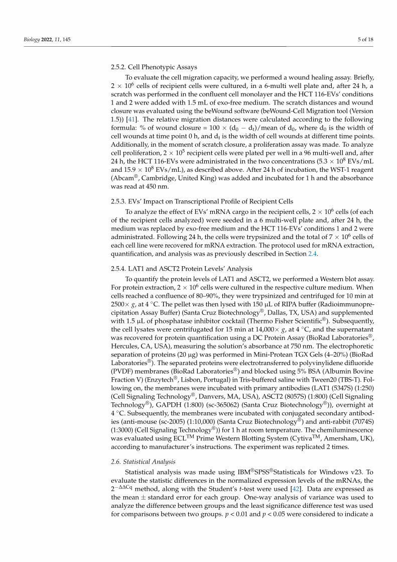
As previously mentioned, the transcriptional levels of LAT1 and ASCT2 mRNA werequantified by real-time qPCR in the HCT 116, SK-HEP-1, HEPG-2, HCA-7 and HKC-8 celllines. Moreover, we also analyzed the levels of EGFR, VEGFA, CXCR4 and HIF1-A mRNAs,considering their oncogenic potential role. We found higher expression levels of LAT1mRNA in the HCT 116 cell line compared to SK-HEP-1 (p < 0.001) and HKC-8 (p = 0.001)cell lines (Figure 1A). The HCT 116 cell line also presented higher levels of ASCT2 mRNAcompared to HCA-7 (p < 0.001), SK-HEP-1 (p < 0.001), HEPG-2 (p = 0.035) and HKC-8(p < 0.001) (Figure 1A).
Biology 2022, 11, x FOR PEER REVIEW 6 of 18
(CytivaTM, Amersham, UK), according to manufacturer’s instructions. The experiment was replicated 2 times.
2.6. Statistical Analysis Statistical analysis was made using IBM®SPSS®Statisticals for Windows v23. To
evaluate the statistic differences in the normalized expression levels of the mRNAs, the 2−ΔΔCq method, along with the Student’s t-test were used [42]. Data are expressed as the mean ± standard error for each group. One-way analysis of variance was used to analyze the difference between groups and the least significance difference test was used for comparisons between two groups. p < 0.01 and p < 0.05 were considered to indicate a statistically significant difference. Additionally, GraphPad Prism 8 was used for graphical presentation of the data.
3. Results 3.1. Cell Lines Transcriptional Profile Characterization
As previously mentioned, the transcriptional levels of LAT1 and ASCT2 mRNA were quantified by real-time qPCR in the HCT 116, SK-HEP-1, HEPG-2, HCA-7 and HKC-8 cell lines. Moreover, we also analyzed the levels of EGFR, VEGFA, CXCR4 and HIF1-A mRNAs, considering their oncogenic potential role. We found higher expression levels of LAT1 mRNA in the HCT 116 cell line compared to SK-HEP-1 (p < 0.001) and HKC-8 (p = 0.001) cell lines (Figure 1A). The HCT 116 cell line also presented higher levels of ASCT2 mRNA compared to HCA-7 (p < 0.001), SK-HEP-1 (p < 0.001), HEPG-2 (p = 0.035) and HKC-8 (p < 0.001) (Figure 1A).
Regarding the EGFR mRNA levels, we also found an increase in the expression levels in the HCT 116 cell line compared to the HEPG-2 (p < 0.001) and HKC-8 (p = 0.001) cell lines. We also observed higher mRNA levels of VEGFA, CXCR4 and HIF1-A mRNAs in the HCT 116 cell line compared to the SK-HEP-1 and HKC-8 cell lines. Concerning the VEGF (p = 0.001) and HIF1-A (p = 0.020) mRNAs, higher levels were also found in the HCT 116 compared to the other CRC cell line, HCA-7. The HEPG-2 cell line presented lower levels of CXCR4 (p = 0.002) mRNA compared to the HCT 116 (Figure 1B).
Figure 1. The bars represent the fold change of the mRNAs’ expression, normalized to B2M. Expression levels shown are means of three technical replicates for each sample. (A) The graphs show the increase of LAT1 and ASCT2 mRNA and (B) EGFR, VEGFA, CXCR4 and HIF1–A mRNAs expression levels in HCT 116 cells, compared to the others cell lines in the analyzed panel. (Mean ± Std. Error, ** p < 0.001, * p < 0.05).
3.2. HCT 116-EVs’ Characterization The EVs were characterized according to size, shape, and quantity. The NTA analysis
indicated that the vast majority of isolated EVs presented a mean size of 128 nm, which is consistent with the size of small EVs (Figure 2A). These results are consistent with the flow cytometry analysis, where were found the presence of EVs smaller than 160 nm
Figure 1. The bars represent the fold change of the mRNAs’ expression, normalized to B2M. Expres-sion levels shown are means of three technical replicates for each sample. (A) The graphs show theincrease of LAT1 and ASCT2 mRNA and (B) EGFR, VEGFA, CXCR4 and HIF1–A mRNAs expressionlevels in HCT 116 cells, compared to the others cell lines in the analyzed panel. (Mean ± Std. Error,** p < 0.001, * p < 0.05).
Regarding the EGFR mRNA levels, we also found an increase in the expression levelsin the HCT 116 cell line compared to the HEPG-2 (p < 0.001) and HKC-8 (p = 0.001) celllines. We also observed higher mRNA levels of VEGFA, CXCR4 and HIF1-A mRNAs inthe HCT 116 cell line compared to the SK-HEP-1 and HKC-8 cell lines. Concerning theVEGF (p = 0.001) and HIF1-A (p = 0.020) mRNAs, higher levels were also found in the HCT116 compared to the other CRC cell line, HCA-7. The HEPG-2 cell line presented lowerlevels of CXCR4 (p = 0.002) mRNA compared to the HCT 116 (Figure 1B).
3.2. HCT 116-EVs’ Characterization
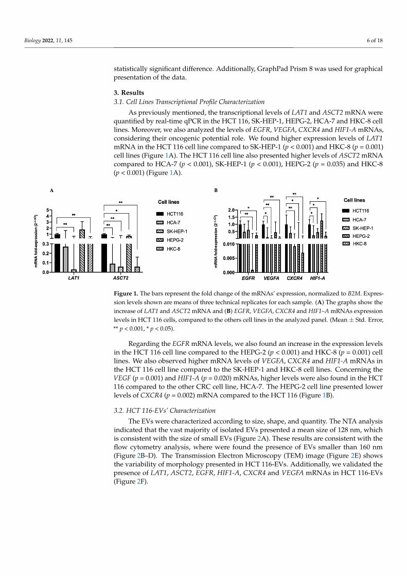
The EVs were characterized according to size, shape, and quantity. The NTA analysisindicated that the vast majority of isolated EVs presented a mean size of 128 nm, whichis consistent with the size of small EVs (Figure 2A). These results are consistent with theflow cytometry analysis, where were found the presence of EVs smaller than 160 nm(Figure 2B–D). The Transmission Electron Microscopy (TEM) image (Figure 2E) showsthe variability of morphology presented in HCT 116-EVs. Additionally, we validated thepresence of LAT1, ASCT2, EGFR, HIF1-A, CXCR4 and VEGFA mRNAs in HCT 116-EVs(Figure 2F).
Biology 2022, 11, 145 7 of 18
1
Figure 2. (A) NTA analysis of HCT 116-EVs showing the size distribution of EVs. (B) Megamix-PlusSSC beads used to define the cytometer settings for EVs’ acquisition, with the following diameters:0.5 µm (gate P1), 0.24 µm (gate P2), 0.20 µm (gate P3) and 0.16 µm (gate P4); (C) Fluorescent EVs(approximately 2.6× 10⁵/200 µL) as observed by flow cytometry from a sample of EVs isolate derivedfrom HCT 116 cell line stained with CFSE (gate P7); (D) Negative control of EVs isolate derived fromHCT 116 cell line without previous staining (gate P7). (E) Transmission electron microscopy (TEM) ofEVs derived from HCT 116 cell line (scale 200 nm). The TEM image was acquired in the Histologyand Electron Microscopy platform from I3S Porto using a Transmission Electron Microscope Jeol JEM1400. (F) The bars represent the –∆Cq of the mRNAs’ expression, normalized to B2M. The graphshows the presence of intracellular and EVs related mRNAs levels (LAT1, ASCT2, EGFR, HIF1-A,CXCR4 and VEGFA) derived from HCT 116 cell line. (Mean ± Std. Error).
3.3. HCT 116-EVs’ Uptake Effect on HCA-7, SK-HEP-1, HEPG-2 and HKC-8 Recipient Cells
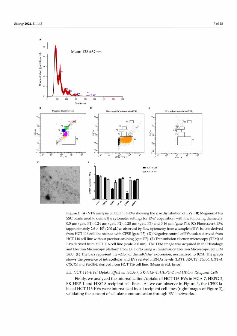
Firstly, we analyzed the internalization/uptake of HCT 116-EVs in HCA-7, HEPG-2,SK-HEP-1 and HKC-8 recipient cell lines. As we can observe in Figure 3, the CFSE la-beled HCT 116-EVs were internalized by all recipient cell lines (right images of Figure 3),validating the concept of cellular communication through EVs’ networks.
Biology 2022, 11, 145 8 of 18
Biology 2022, 11, x FOR PEER REVIEW 10 of 20
Figure 3. Representative immunofluorescence image shows the internalization of HCT 116-EVs la-beled with CFSE (green dye) by HCA-7, SK-HEP-1, HEPG-2 and HKC-8 cells. The cell’s nucleus is stained with DAPI (blue dye). (10X Olympus® IX51 microscope).
We then evaluated the HCT 116-EV’s effect (condition 1: 5.3 × 10⁸ EV’s/mL and con-dition 2: 15.9 × 10⁸ EV’s/mL) on cell proliferation and migration (Figure 4A–D) and prolif-eration capacity (Figure 4G–J) of the recipient cells: HCA-7 (Figure 4A,G); SK-HEP-1 (Fig-ure 4B,H); HEPG-2 (Figure 4C,I) and HKC-8 (Figure 4D,J).
The figures of the wound healing assays of all recipient cells can be observed in the Supplementary Material (Figure S1). All the represented statistical analyses were per-formed between the control condition (recipient cells) and the uptake internalization
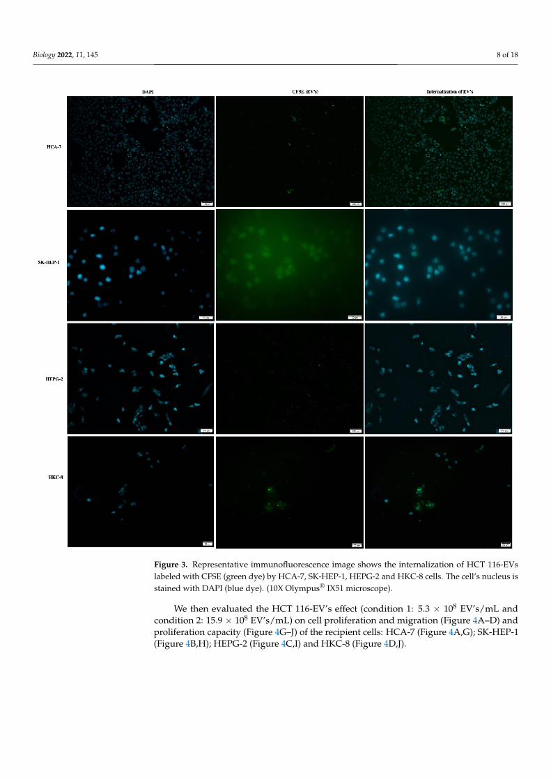
Figure 3. Representative immunofluorescence image shows the internalization of HCT 116-EVslabeled with CFSE (green dye) by HCA-7, SK-HEP-1, HEPG-2 and HKC-8 cells. The cell’s nucleus isstained with DAPI (blue dye). (10X Olympus® IX51 microscope).
We then evaluated the HCT 116-EV’s effect (condition 1: 5.3 × 10⁸ EV’s/mL andcondition 2: 15.9 × 10⁸ EV’s/mL) on cell proliferation and migration (Figure 4A–D) andproliferation capacity (Figure 4G–J) of the recipient cells: HCA-7 (Figure 4A,G); SK-HEP-1(Figure 4B,H); HEPG-2 (Figure 4C,I) and HKC-8 (Figure 4D,J).
Biology 2022, 11, 145 9 of 18Biology 2022, 11, x FOR PEER REVIEW 9 of 20
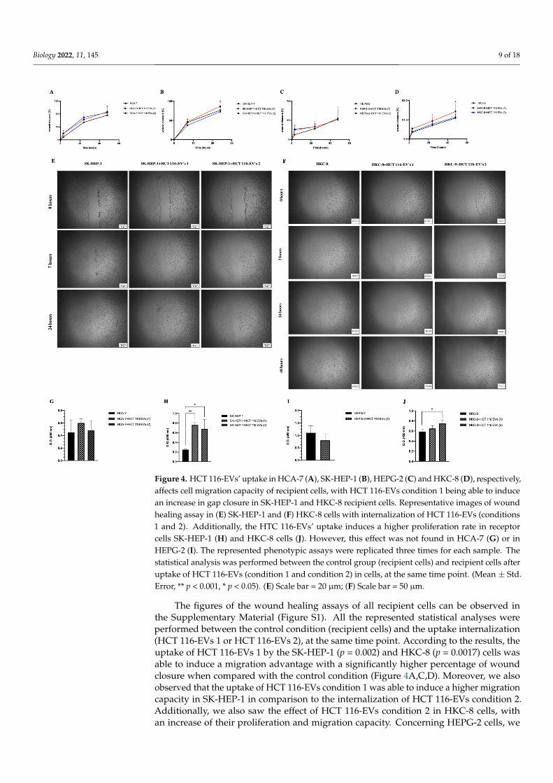
Figure 4. HCT 116-EVs’ uptake in HCA-7 (A), SK-HEP-1 (B), HEPG-2 (C) and HKC-8 (D), respectively,affects cell migration capacity of recipient cells, with HCT 116-EVs condition 1 being able to inducean increase in gap closure in SK-HEP-1 and HKC-8 recipient cells. Representative images of woundhealing assay in (E) SK-HEP-1 and (F) HKC-8 cells with internalization of HCT 116-EVs (conditions1 and 2). Additionally, the HTC 116-EVs’ uptake induces a higher proliferation rate in receptorcells SK-HEP-1 (H) and HKC-8 cells (J). However, this effect was not found in HCA-7 (G) or inHEPG-2 (I). The represented phenotypic assays were replicated three times for each sample. Thestatistical analysis was performed between the control group (recipient cells) and recipient cells afteruptake of HCT 116-EVs (condition 1 and condition 2) in cells, at the same time point. (Mean ± Std.Error, ** p < 0.001, * p < 0.05). (E) Scale bar = 20 µm; (F) Scale bar = 50 µm.
The figures of the wound healing assays of all recipient cells can be observed inthe Supplementary Material (Figure S1). All the represented statistical analyses wereperformed between the control condition (recipient cells) and the uptake internalization(HCT 116-EVs 1 or HCT 116-EVs 2), at the same time point. According to the results, theuptake of HCT 116-EVs 1 by the SK-HEP-1 (p = 0.002) and HKC-8 (p = 0.0017) cells wasable to induce a migration advantage with a significantly higher percentage of woundclosure when compared with the control condition (Figure 4A,C,D). Moreover, we alsoobserved that the uptake of HCT 116-EVs condition 1 was able to induce a higher migrationcapacity in SK-HEP-1 in comparison to the internalization of HCT 116-EVs condition 2.Additionally, we also saw the effect of HCT 116-EVs condition 2 in HKC-8 cells, withan increase of their proliferation and migration capacity. Concerning HEPG-2 cells, we
Biology 2022, 11, 145 10 of 18
observed cell detachment in HCT 116-EVs condition 2, which did not allow the analysisof this experimental condition. Regarding the HCA-7 cells, after 48 h of the HCT 116-EVs’incubation, an increase of migration capacity was observed, when compared to the controlcondition.
The proliferation results are in agreement with the results observed for the migrationcapacity since we observed a higher proliferation rate in SK-HEP-1 (p < 0.001) after HCT116-EVs condition 1’s uptake (Figure 4B). Additionally, there was also a significant increaseof proliferation capacity of SK-HEP-1 (p = 0.003) and HKC-8 (p = 0.002) after stimulus withHCT 116-EVs 2. Thus, these results demonstrated that HCT 116-EVs promote cell migrationcapacity and proliferation of specific recipient cells, namely of SK-HEP-1 and HKC-8.
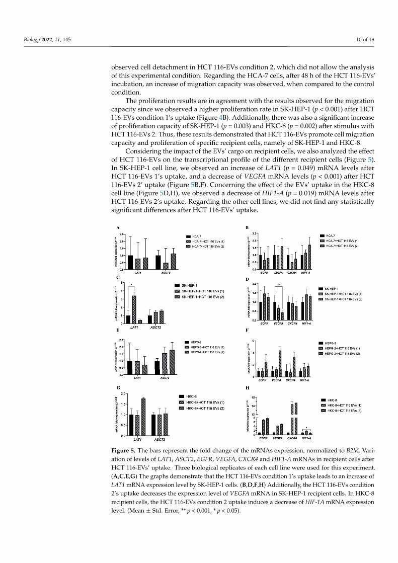
Considering the impact of the EVs’ cargo on recipient cells, we also analyzed the effectof HCT 116-EVs on the transcriptional profile of the different recipient cells (Figure 5).In SK-HEP-1 cell line, we observed an increase of LAT1 (p = 0.049) mRNA levels afterHCT 116-EVs 1’s uptake, and a decrease of VEGFA mRNA levels (p < 0.001) after HCT116-EVs 2’ uptake (Figure 5B,F). Concerning the effect of the EVs’ uptake in the HKC-8cell line (Figure 5D,H), we observed a decrease of HIF1-A (p = 0.019) mRNA levels afterHCT 116-EVs 2’s uptake. Regarding the other cell lines, we did not find any statisticallysignificant differences after HCT 116-EVs’ uptake.
Biology 2022, 11, x FOR PEER REVIEW 10 of 18
Considering the impact of the EVs’ cargo on recipient cells, we also analyzed the effect of HCT 116-EVs on the transcriptional profile of the different recipient cells (Figure 5). In SK-HEP-1 cell line, we observed an increase of LAT1 (p = 0.049) mRNA levels after HCT 116-EVs 1’s uptake, and a decrease of VEGFA mRNA levels (p < 0.001) after HCT 116-EVs 2’ uptake (Figure 5B,F). Concerning the effect of the EVs’ uptake in the HKC-8 cell line (Figure 5D,H), we observed a decrease of HIF1-A (p = 0.019) mRNA levels after HCT 116-EVs 2’s uptake. Regarding the other cell lines, we did not find any statistically significant differences after HCT 116-EVs’ uptake.
Figure 5. The bars represent the fold change of the mRNAs expression, normalized to B2M. Variation of levels of LAT1, ASCT2, EGFR, VEGFA, CXCR4 and HIF1-A mRNAs in recipient cells after HCT 116-EVs’ uptake. Three biological replicates of each cell line were used for this experiment. (A,C,E,G) The graphs demonstrate that the HCT 116-EVs condition 1’s uptake leads to an increase of LAT1 mRNA expression level by SK-HEP-1 cells. (B,D,F,H) Additionally, the HCT 116-EVs condition 2’s uptake decreases the expression level of VEGFA mRNA in SK-HEP-1 recipient cells. In HKC-8 recipient cells, the HCT 116-EVs condition 2 uptake induces a decrease of HIF-1A mRNA expression level. (Mean ± Std. Error, ** p < 0.001, * p < 0.05).
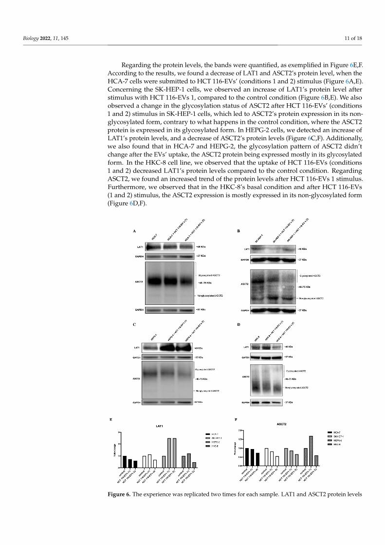
Regarding the protein levels, the bands were quantified, as exemplified in Figure 6E,F. According to the results, we found a decrease of LAT1 and ASCT2’s protein level, when the HCA-7 cells were submitted to HCT 116-EVs’ (conditions 1 and 2) stimulus (Figure 6A,E). Concerning the SK-HEP-1 cells, we observed an increase of LAT1’s protein level after stimulus with HCT 116-EVs 1, compared to the control condition (Figure 6B,E). We also observed a change in the glycosylation status of ASCT2 after HCT 116-EVs’ (conditions 1 and 2) stimulus in SK-HEP-1 cells, which led to ASCT2’s protein expression in its non-glycosylated form, contrary to what happens in the control condition, where the ASCT2 protein is expressed in its glycosylated form. In HEPG-2 cells, we detected an increase of LAT1’s protein levels, and a decrease of ASCT2’s protein levels (Figure 6C,F).
Figure 5. The bars represent the fold change of the mRNAs expression, normalized to B2M. Vari-ation of levels of LAT1, ASCT2, EGFR, VEGFA, CXCR4 and HIF1-A mRNAs in recipient cells afterHCT 116-EVs’ uptake. Three biological replicates of each cell line were used for this experiment.(A,C,E,G) The graphs demonstrate that the HCT 116-EVs condition 1’s uptake leads to an increase ofLAT1 mRNA expression level by SK-HEP-1 cells. (B,D,F,H) Additionally, the HCT 116-EVs condition2’s uptake decreases the expression level of VEGFA mRNA in SK-HEP-1 recipient cells. In HKC-8recipient cells, the HCT 116-EVs condition 2 uptake induces a decrease of HIF-1A mRNA expressionlevel. (Mean ± Std. Error, ** p < 0.001, * p < 0.05).
Biology 2022, 11, 145 11 of 18
Regarding the protein levels, the bands were quantified, as exemplified in Figure 6E,F.According to the results, we found a decrease of LAT1 and ASCT2’s protein level, when theHCA-7 cells were submitted to HCT 116-EVs’ (conditions 1 and 2) stimulus (Figure 6A,E).Concerning the SK-HEP-1 cells, we observed an increase of LAT1’s protein level afterstimulus with HCT 116-EVs 1, compared to the control condition (Figure 6B,E). We alsoobserved a change in the glycosylation status of ASCT2 after HCT 116-EVs’ (conditions1 and 2) stimulus in SK-HEP-1 cells, which led to ASCT2’s protein expression in its non-glycosylated form, contrary to what happens in the control condition, where the ASCT2protein is expressed in its glycosylated form. In HEPG-2 cells, we detected an increase ofLAT1’s protein levels, and a decrease of ASCT2’s protein levels (Figure 6C,F). Additionally,we also found that in HCA-7 and HEPG-2, the glycosylation pattern of ASCT2 didn’tchange after the EVs’ uptake, the ASCT2 protein being expressed mostly in its glycosylatedform. In the HKC-8 cell line, we observed that the uptake of HCT 116-EVs (conditions1 and 2) decreased LAT1’s protein levels compared to the control condition. RegardingASCT2, we found an increased trend of the protein levels after HCT 116-EVs 1 stimulus.Furthermore, we observed that in the HKC-8’s basal condition and after HCT 116-EVs(1 and 2) stimulus, the ASCT2 expression is mostly expressed in its non-glycosylated form(Figure 6D,F).
Biology 2022, 11, x FOR PEER REVIEW 11 of 18
Additionally, we also found that in HCA-7 and HEPG-2, the glycosylation pattern of ASCT2 didn’t change after the EVs’ uptake, the ASCT2 protein being expressed mostly in its glycosylated form. In the HKC-8 cell line, we observed that the uptake of HCT 116-EVs (conditions 1 and 2) decreased LAT1’s protein levels compared to the control condition. Regarding ASCT2, we found an increased trend of the protein levels after HCT 116-EVs 1 stimulus. Furthermore, we observed that in the HKC-8’s basal condition and after HCT 116-EVs (1 and 2) stimulus, the ASCT2 expression is mostly expressed in its non-glycosylated form (Figure 6D,F).
Figure 6. The experience was replicated two times for each sample. LAT1 and ASCT2 protein levels and HCT 116-EVs’ uptake effects in (A) HCA-7, (B) SK-HEP-1, (C) HEPG-2 and in (D) HKC-8 cell
Figure 6. The experience was replicated two times for each sample. LAT1 and ASCT2 protein levels
Biology 2022, 11, 145 12 of 18
and HCT 116-EVs’ uptake effects in (A) HCA-7, (B) SK-HEP-1, (C) HEPG-2 and in (D) HKC-8 celllines. Fold-change of the pixels’ volume adjusted intensity of (E) LAT1 and (F) ASCT2 proteinlevels, in recipient cells after HCT 116-EVs’ uptake. The bands show that HCT 116-EVs condition1 internalized by SK-HEP-1 induce an increase of LAT1 protein level. Similarly, in HEPG-2 recipientcells, after HCT 116-EVs’ (condition 1 and 2) uptake, there is an increase of LAT1 protein levels. Onthe other hand, the internalization of HCT 116-EVs 1 by HKC-8 cells leads to an increase of ASCT2protein levels.
4. Discussion
One of the major concerns of CRC management is the fact that a high number ofpatients are diagnosed in advanced stages of disease, and that, in 50% of patients diagnosedwith local diseases, metastasis eventually develops. In 30–50% of CRC patients, the liveris the predominant site of metastatic disease, in consequence of its drainage from thegastrointestinal tract [4,6,43,44]. In these cases, the only curative approach is surgery.However, a limited number of patients are considered eligible [44].
It is well established that the tumor microenvironment represents a complex network,in which tumor cells communicate with others cell types, including fibroblasts, endothelialcells and immune cells [45,46]. In recent years, the scientific community have focused onEVs’ intercellular communication role, and their potential as vehicles and mediators ofcell communication and cellular microenvironment modulation has already been demon-strated [47,48]. Studies demonstrated that cancer derived EVs participate in critical stepsof pre-metastatic niche formation in the primary tumor by delivering cargo to recipientcells in target organs [39,49]. The pivotal role of EVs in regulating several immune-relatedpathways leading to activation, differentiation and expression of immune cells and mod-ulation of the tumor microenvironment has already been demonstrated, as well as itssignificant role in CRC progression and metastasis [50]. Increasing evidence shows thatmRNAs can be transferred to the surrounding microenvironment, via EVs’ pathways, andinfluence the metabolism of recipient cells to favor cancer progression [49,51]. In fact,according to Chiba and colleagues, EVs derived from three CRC cells (HCT-15, SW480 andWiDr) showed the capacity to transfer mRNAs into 2D A549 cells (lung cancer cells) andHEPG-2 cell lines, validating that EVs-derived RNAs can be shuttled between cells, andcan be involved in the regulation of gene expression in recipient cells [40]. Furthermore,Shao and co-workers described that CRC derived EVs present a pivotal role in promotingliver metastasis, by inducing a premetastatic niche through miR-21-TLR7-IL-6 axis [43].These authors described that CRC-EVs can specifically target liver tissue and induce livermacrophages toward an IL-6 proinflammatory phenotype [43]. Additionally, the studymentions miR-21 as highly enriched in CRC-EVs, this miRNA being essential for creatinga proinflammatory phenotype in the liver and creating liver metastasis in CRC [43]. Fi-nally, the authors also demonstrate that silencing either miR-21 in CRC-EVs, or TLR-7 inmacrophages, abolished the CRC-EVs’ induction of proinflammatory macrophages [43].Costa-Silva and co-workers described, for the first time, the sequential steps responsiblefor the formation of liver pre-metastatic niches (LPMN) supportive of PC metastasis, whichinvolved binding of Macrophage Migration inhibitory factor (MIF)+ PC-derived EVs toliver Kupffer cells, followed by TGF-β production by these cells. TGF-β, in turn, promotedfibronectin production by hepatic stellate cells, that supported the accumulation of bonemarrow-derived macrophages, completing the LPMN formation [52]. More recently, Xuanand colleagues reported that EVs derived from breast cancer contribute to pre-metastaticniche formation and promote bone metastasis of tumor cells [53]. This process is mediatedby EVs derived from breast cancer cells (SCP28 and MDA-MB-231 cells), which promoteosteoclast differentiation and enhance bone metastasis [53]. Therefore, these studies suggestthe role of EVs in metastasis establishment, through the transfer of biomolecular cargo,presenting a great potential to be used as predictive targets.
Nevertheless, there is still no evidence in the literature about the influence of LAT1 andASCT2 mRNAs derived from CRC-EVs in CRC progression and metastasis, even though
Biology 2022, 11, 145 13 of 18
they play an important role in the growth and survival of CRC cancer cell lines, since theyensure the rapid exchange of AA and the maintenance of an AA pool in the cytosol [54]. Inthis study, we detected for the first time the presence of LAT1, ASCT2 and other oncogenicmRNAs on CRC-EVs, as well as their capacity to modify the transcriptional profile andphenotypic characteristics of recipient cell lines. Moreover, we showed the presence ofEGFR, VEGFA, CXCR4 and HIF1-A mRNAs in CRC-EVs, which is in agreement withseveral evidences that support the involvement of these molecules in CRC developmentand metastases formation [26,55–64]. The overexpression of EGFR, HIF-1α and VEGFA hasbeen described in CRC, being associated with poor prognosis, aggressiveness, and a higherpotential of metastases formation [59,62,65]. Moreover, CXCR4 is also correlated with poorhistological differentiation, distant metastasis and lymph node metastasis, being its higherlevels associated with poor prognosis in CRC patients [66].
Thus, we hypothesize that the incorporation of these molecules on EVs are essential totrigger the establishment of a microenvironment that supports the metastases’ formation.In fact, we observed an increase of LAT1 and a decrease of VEGFA mRNAs in SK-HEP-1,and an increase of HIF1-A mRNA levels in HKC-8 cells after stimulus with HCT 116-EVs. These molecular changes may be associated with the release of pro-inflammatorycytokines such as IL-6, leading to an inflammatory process which is influenced by cellularmetabolism and hypoxia [67]. In fact, Quan and colleagues, showed that LAT1 wasrequired for angiogenic processes, since VEGFA’s stimulus induced LAT1 overexpressionthat consequently triggered angiogenesis [68]. On the other hand, Shi and co-workersdescribe that, in non-small cell lung cancer, the expression of LAT1 is correlated withHIF1-A levels [69,70]. Therefore, according to our results, we can assume that an increase inHIF-1A and hypoxia leads to an increase of LAT1 expression [70]. Interestingly, the increaseof LAT1 mRNA in the SK-HEP-1 cell line, after HCT 116-EVs’ uptake, was able to supportthe high demand of amino acids by these cells. In fact, the increase of LAT1 mRNA inSK-HEP-1 translated into the increase of LAT1 protein expression, cell proliferation, aswell as in the invasion capacity of these cells. Wang and co-workers had already reportedhigher protein levels of CXCR4 in the liver of the HT-29-derived exosome-treated Caco-2-implanted mice [71].
Interestingly, we also observed that the internalization of different concentrationsof HCT 116-EVs cause different effects in recipient cells. In fact, we observed that SK-HEP-1 cells reach a saturation peak after HCT 116-EVs condition 1’s stimulus for around24 h. Additionally, we also saw that there is a dose dependency of EVs administratedto SK-HEP-1 recipient cells, since the migration and proliferation capacity displayed astrong dose dependence with a minimal effective dose of 5.3 × 10⁸ EVs/mL (HCT 116-EVscondition 1). Similarly, Franzen and colleagues also demonstrated that the EVs’ uptakeby recipient cells is time and dose dependent, a peak of EVs’ internalization occurringbefore the 24 h of incubation [72]. These authors described that the recipient cells reacheda saturation point of exosomes internalization after 14 h, however, after 24 h of stimulusthe authors demonstrated that exosomes continued to be taken up by cells [72]. Similarly,Jurgielewicz and co-workers also describe that HEK293T-EVs’ uptake is time and dosedependent, the peak of uptake being around 12 h [73]. Moreover, the authors also reportthat after a dose of 6000 EVs/cell are taken up by HEK293T recipient cells, these reach a dosesaturation limit [73]. Jurgielewicz and co-workers also report that since EVs have shownto be internalized then released after 24 h, longer incubations may generate inaccurateinternalizations readouts [73]. In fact, after 48 h of incubation with HCT 116-EVs condition2, the results of migration and proliferation assays in HKC-8 cells seem to be inconsistent,which could be consequence of the long EVs’ incubation period, and could be associated inan inaccurate internalization readout.
Considering the key role of LAT1 during CRC progression, and specially the capacityof tumor cells to encapsulate mRNA molecules inside EVs to modulate the surroundingmicroenvironment, the development of pharmacological strategies based on the inhibitionof LAT1 could be promising for CRC patients’ management. In fact, Okano and co-workers
Biology 2022, 11, 145 14 of 18
have already reported that JPH203, a LAT1 inhibitor, demonstrated potential to be used forCRC patients’ treatment [23]. In a phase I study, JPH203 treatment was well tolerated bypatients and led to disease control in two of the six CRC patients and in three of the fivepatients with biliary tract cancer [23].
The upregulation of LAT1 mRNA could also be associated with changes in the ASCT2glycosylation pattern. In fact, a study performed by Polet and colleagues reports thatglucose availability regulates the glycosylation of ASCT2 [74]. The authors describe thatinhibition of glucose metabolism prevents ASCT2 glycosylation and promotes LAT1 up-regulation as a countertrading mechanism of glycosylation’s inhibition [74]. We observeda change in the ASCT2 glycosylation pattern after HCT116-EVs stimulus. The changespreviously described in SK-HEP-1 were also followed by changes in ASCT2 protein’sconformation, that changed to a non-glycosylated form. This may be due to the fact thatHCT 116-EVs stimulate metabolic deregulation of SK-HEP-1 cells, which could lead to anincrease of glucose consumption by these cells, and consequently, lead to a change of theglycosylation status of the ASCT2 protein.
In conclusion, the present study supports the role of CRC-EVs as key mediators oftumoral progression, supporting a proangiogenic and proliferative microenvironmentestablishment.
5. Conclusions
Our results uncovered an additional role of EVs in aggressive phenotypes of CRC,through the transference of LAT1 mRNA, with a phenotypic impact on cell proliferation andinvasion capacity. Moreover, future studies should consider the replication of this in vitrostudy in a three-dimensional (3D) cell culture model to validate the cellular response to HCT116-EVs’ stimulus. These models provide more physiologically information and predictivedata for in vivo tests since they mimic the biological conditions. In 3D cell culture models,cancer cells can maintain the shape, polarity and the heterogeneity observed in vivo. Onthe other hand, after the cell culture validation it will be crucial to check the influence ofthe HCT 116-EVs in animal models, to elucidate the role of this structure in the metastasisformation and clarify the metastatic routes of CRC. One possible approach to validate thehypothesis raised in this study may be to study if the inoculation of HCT 116-EVs onlypresents tropism for liver and kidney cells, or if they are able to affect the proliferation andmigration capacity of cells from other organs. Additionally, it would also be interestingto validate our findings in CRC patients’ plasma EVs to evaluate the biomarker potentialof CRC EVs-derived mRNA, especially LAT1, in patients’ prognoses and follow-ups. Thevalidation of the biomarker potential of CRC EVs-derived mRNAs would be useful forliquid biopsies’ implementation and the development of new therapeutic approaches forCRC.
Supplementary Materials: The following are available online at https://www.mdpi.com/article/10.3390/biology11010145/s1, Figure S1: Representative images of wound healing assay in (A) HCA-7,(B) SK-HEP-1, (C) HEPG-2 and (D) HKC-8 recipient cells, with respective uptakes of HCT 116-EVs (1and 2), in the different time points. Figure S2: Representative original images of Western Blot assay in(A) HCA-7, HEPG-2, (B) SK-HEP-1 and (C) HKC-8 recipient cells, with respective uptakes of HCT116-EVs (1 and 2).
Author Contributions: Conceptualization, A.L.T., F.D. and R.M.; methodology, C.A., F.D., V.M., M.M.and A.L.T.; validation, A.L.T., F.D. and R.M.; formal analysis, A.L.T., F.D., G.M., C.P., B.C.-S. andR.M.; investigation, C.A., A.L.T., F.D., V.M., M.E.S., I.G., S.B.; resources, A.L.T., G.M., C.P., B.C.-S.and R.M.; data curation, A.L.T., B.C.-S. and R.M.; writing—original draft preparation, C.A., V.M.;writing—review and editing, A.L.T., F.D., G.M., C.P., B.C.-S. and R.M.; supervision, F.D., A.L.T. andR.M.; project administration, A.L.T. and R.M.; funding acquisition, A.L.T. and R.M. All authors haveread and agreed to the published version of the manuscript.
Funding: This research was supported by the Norte Portugal Region Operational Programme(NORTE 2020), under the PORTUGAL 2020 Partnership Agreement—Project SIRNAC—Novel siRNA
Biology 2022, 11, 145 15 of 18
therapies against metastatic colorectal cancer [NORTE-01-0247-FEDER-033399], and by the IPO-PortoResearch Center [CI-IPO-21-2015].
Institutional Review Board Statement: Not applicable.
Informed Consent Statement: Not applicable.
Data Availability Statement: Not applicable.
Acknowledgments: The authors would like to thank the Research Department of the PortugueseLeague against Cancer Regional Nucleus of the North (LPCC-NRN). C.A. is a recipient of a researchgrant from LPCC-NRN.
Conflicts of Interest: The authors declare no conflict of interest. The funders had no role in the designof the study; in the collection, analyses, or interpretation of data; in the writing of the manuscript, orin the decision to publish the results.
References1. Mármol, I.; Sánchez-De-Diego, C.; Pradilla Dieste, A.; Cerrada, E.; Rodriguez Yoldi, M. Colorectal Carcinoma: A General
Overview and Future Perspectives in Colorectal Cancer. Int. J. Mol. Sci. 2017, 18, 197. [CrossRef] [PubMed]2. Sung, H.; Ferlay, J.; Siegel, R.L.; Laversanne, M.; Soerjomataram, I.; Jemal, A.; Bray, F. Global Cancer Statistics 2020: GLOBOCAN
Estimates of Incidence and Mortality Worldwide for 36 Cancers in 185 Countries. CA Cancer J. Clin. 2021, 71, 209–249. [CrossRef][PubMed]
3. Yin, W.; Xu, J.; Li, C.; Dai, X.; Wu, T.; Wen, J. Circular RNA circ_0007142 Facilitates Colorectal Cancer Progression by ModulatingCDC25A Expression via miR-122-5p. OncoTargets Ther. 2020, 13, 3689–3701. [CrossRef] [PubMed]
4. Al Bandar, M.H.; Kim, N.K. Current status and future perspectives on treatment of liver metastasis in colorectal cancer (Review).Oncol. Rep. 2017, 37, 2553–2564. [CrossRef] [PubMed]
5. Hur, K.; Toiyama, Y.; Okugawa, Y.; Ide, S.; Imaoka, H.; Boland, C.R.; Goel, A. Circulating microRNA-203 predicts prognosis andmetastasis in human colorectal cancer. Gut 2017, 66, 654–665. [CrossRef]
6. Zarour, L.R.; Anand, S.; Billingsley, K.G.; Bisson, W.H.; Cercek, A.; Clarke, M.F.; Coussens, L.M.; Gast, C.E.; Geltzeiler, C.B.;Hansen, L.; et al. Colorectal Cancer Liver Metastasis: Evolving Paradigms and Future Directions. Cell. Mol. Gastroenterol. Hepatol.2017, 3, 163–173. [CrossRef]
7. Fodde, R. The APC gene in colorectal cancer. Eur. J. Cancer 2002, 38, 867–871. [CrossRef]8. Ward, P.; Thompson, C.B. Metabolic Reprogramming: A Cancer Hallmark Even Warburg Did Not Anticipate. Cancer Cell 2012, 21,
297–308. [CrossRef]9. Pranzini, E.; Pardella, E.; Paoli, P.; Fendt, S.-M.; Taddei, M.L. Metabolic Reprogramming in Anticancer Drug Resistance: A Focus
on Amino Acids. Trends Cancer 2021, 7, 682–699. [CrossRef]10. Xie, J.; Zhu, X.Y.; Liu, L.M.; Meng, Z.Q. Solute carrier transporters: Potential targets for digestive system neoplasms. Cancer
Manag. Res. 2018, 10, 153–166. [CrossRef]11. Wu, Z.; Xu, J.; Liang, C.; Meng, Q.; Hua, J.; Wang, W.; Zhang, B.; Liu, J.; Yu, X.; Shi, S. Emerging roles of the solute carrier family
in pancreatic cancer. Clin. Transl. Med. 2021, 11, e356. [CrossRef]12. Fuchs, B.C.; Bode, B.P. Amino acid transporters ASCT2 and LAT1 in cancer: Partners in crime? Semin. Cancer Biol. 2005, 15,
254–266. [CrossRef]13. Nyquist, M.D.; Prasad, B.; Mostaghel, E.A. Harnessing Solute Carrier Transporters for Precision Oncology. Molecules 2017, 22, 539.
[CrossRef]14. Wang, Q.; Holst, J. L-type amino acid transport and cancer: Targeting the mTORC1 pathway to inhibit neoplasia. Am. J. Cancer
Res. 2015, 5, 1281–1294.15. Hayase, S.; Kumamoto, K.; Saito, K.; Kofunato, Y.; Sato, Y.; Okayama, H.; Miyamoto, K.; Ohki, S.; Takenoshita, S. L-type amino
acid transporter 1 expression is upregulated and associated with cellular proliferation in colorectal cancer. Oncol. Lett. 2017, 14,7410–7416. [CrossRef] [PubMed]
16. Napolitano, L.; Scalise, M.; Galluccio, M.; Pochini, L.; Albanese, L.M.; Indiveri, C. LAT1 is the transport competent unit of theLAT1/CD98 heterodimeric amino acid transporter. Int. J. Biochem. Cell Biol. 2015, 67, 25–33. [CrossRef] [PubMed]
17. Scalise, M.; Pochini, L.; Console, L.; Losso, M.A.; Indiveri, C. The Human SLC1A5 (ASCT2) Amino Acid Transporter: FromFunction to Structure and Role in Cell Biology. Front. Cell Dev. Biol. 2018, 6, 96. [CrossRef]
18. Song, W.; Li, D.; Tao, L.; Luo, Q.; Chen, L. Solute carrier transporters: The metabolic gatekeepers of immune cells. Acta Pharm.Sin. B 2020, 10, 61–78. [CrossRef] [PubMed]
19. Bai, X.; Moraes, T.F.; Reithmeier, R.A.F. Structural biology of solute carrier (SLC) membrane transport proteins. Mol. Membr. Biol.2017, 34, 1–32. [CrossRef] [PubMed]
20. Jiang, H.; Zhang, N.; Tang, T.; Feng, F.; Sun, H.; Qu, W. Target the human Alanine/Serine/Cysteine Transporter 2(ASCT2):Achievement and Future for Novel Cancer Therapy. Pharmacol. Res. 2020, 158, 104844. [CrossRef]
Biology 2022, 11, 145 16 of 18
21. Scalise, M.; Pochini, L.; Galluccio, M.; Console, L.; Indiveri, C. Glutamine Transport and Mitochondrial Metabolism in CancerCell Growth. Front. Oncol. 2017, 7, 306. [CrossRef]
22. Ogawa, H.; Kaira, K.; Motegi, Y.; Yokobori, T.; Takada, T.; Katoh, R.; Osone, K.; Takahashi, R.; Katayama, C.; Oyama, T.; et al. Roleof Amino Acid Transporter Expression as a Prognostic Marker in Patients With Surgically Resected Colorectal Cancer. Anticancer.Res. 2019, 39, 2535–2543. [CrossRef] [PubMed]
23. Okano, N.; Naruge, D.; Kawai, K.; Kobayashi, T.; Nagashima, F.; Endou, H.; Furuse, J. First-in-human phase I study of JPH203,an L-type amino acid transporter 1 inhibitor, in patients with advanced solid tumors. Investig. New Drugs 2020, 38, 1495–1506.[CrossRef] [PubMed]
24. Scalise, M.; Galluccio, M.; Console, L.; Pochini, L.; Indiveri, C. The Human SLC7A5 (LAT1): The Intriguing Histidine/LargeNeutral Amino Acid Transporter and Its Relevance to Human Health. Front. Chem. 2018, 6, 243. [CrossRef] [PubMed]
25. Verrey, F. System L: Heteromeric exchangers of large, neutral amino acids involved in directional transport. Pflug. Arch. 2003, 445,529–533. [CrossRef]
26. Häfliger, P.; Charles, R.-P. The L-Type Amino Acid Transporter LAT1—An Emerging Target in Cancer. Int. J. Mol. Sci. 2019,20, 2428. [CrossRef]
27. Toda, K.; Nishikawa, G.; Iwamoto, M.; Itatani, Y.; Takahashi, R.; Sakai, Y.; Kawada, K. Clinical Role of ASCT2 (SLC1A5) inKRAS-Mutated Colorectal Cancer. Int. J. Mol. Sci. 2017, 18, 1632. [CrossRef]
28. Toda, K.; Kawada, K.; Iwamoto, M.; Inamoto, S.; Sasazuki, T.; Shirasawa, S.; Hasegawa, S.; Sakai, Y. Metabolic Alterations Causedby KRAS Mutations in Colorectal Cancer Contribute to Cell Adaptation to Glutamine Depletion by Upregulation of AsparagineSynthetase. Neoplasia 2016, 18, 654–665. [CrossRef] [PubMed]
29. Miyo, M.; Konno, M.; Nishida, N.; Sueda, T.; Noguchi, K.; Matsui, H.; Colvin, H.; Kawamoto, K.; Koseki, J.; Haraguchi, N.; et al.Metabolic Adaptation to Nutritional Stress in Human Colorectal Cancer. Sci. Rep. 2016, 6, 38415. [CrossRef]
30. Wong, C.C.; Qian, Y.; Li, X.; Xu, J.; Kang, W.; Tong, J.H.; To, K.-F.; Jin, Y.; Li, W.; Chen, H.; et al. SLC25A22 Promotes Proliferationand Survival of Colorectal Cancer Cells With KRAS Mutations and Xenograft Tumor Progression in Mice via IntracellularSynthesis of Aspartate. Gastroenterology 2016, 151, 945–960.e6. [CrossRef]
31. Yun, J.; Rago, C.; Cheong, I.; Pagliarini, R.; Angenendt, P.; Rajagopalan, H.; Schmidt, K.; Willson, J.K.V.; Markowitz, S.; Zhou,S.; et al. Glucose Deprivation Contributes to the Development of KRAS Pathway Mutations in Tumor Cells. Science 2009, 325,1555–1559. [CrossRef]
32. Namikawa, M.; Kakizaki, S.; Kaira, K.; Tojima, H.; Yamazaki, Y.; Horiguchi, N.; Satoru, K.; Oriuchi, N.; Tominaga, H.; Sunose, Y.;et al. Expression of amino acid transporters (LAT1, ASCT2 and xCT) as clinical significance in hepatocellular carcinoma. Hepatol.Res. 2014, 45, 1014–1022. [CrossRef]
33. Raposo, G.; Stoorvogel, W. Extracellular vesicles: Exosomes, microvesicles, and friends. J. Cell Biol. 2013, 200, 373–383. [CrossRef][PubMed]
34. Wang, M.; Yu, F.; Ding, H.; Wang, Y.; Li, P.; Wang, K. Emerging Function and Clinical Values of Exosomal MicroRNAs in Cancer.Mol. Ther.-Nucleic Acids 2019, 16, 791–804. [CrossRef]
35. Zhou, J.; Li, X.-L.; Chen, Z.-R.; Chng, W.-J. Tumor-derived exosomes in colorectal cancer progression and their clinical applications.Oncotarget 2017, 8, 100781–100790. [CrossRef] [PubMed]
36. Kalluri, R. The biology and function of exosomes in cancer. J. Clin. Investig. 2016, 126, 1208–1215. [CrossRef]37. Jella, K.K.; Nasti, T.H.; Li, Z.; Malla, S.R.; Buchwald, Z.S.; Khan, M.K. Exosomes, Their Biogenesis and Role in Inter-Cellular
Communication, Tumor Microenvironment and Cancer Immunotherapy. Vaccines 2018, 6, 69. [CrossRef] [PubMed]38. Akbar, N.; Azzimato, V.; Choudhury, R.P.; Aouadi, M. Extracellular vesicles in metabolic disease. Diabetologia 2019, 62, 2179–2187.
[CrossRef]39. Guo, Y.; Ji, X.; Liu, J.; Fan, D.; Zhou, Q.; Chen, C.; Wang, W.; Wang, G.; Wang, H.; Yuan, W.; et al. Effects of exosomes on
pre-metastatic niche formation in tumors. Mol. Cancer 2019, 18, 39. [CrossRef]40. Chiba, M.; Kimura, M.; Asari, S. Exosomes secreted from human colorectal cancer cell lines contain mRNAs, microRNAs and
natural antisense RNAs, that can transfer into the human hepatoma HepG2 and lung cancer A549 cell lines. Oncol. Rep. 2012, 28,1551–1558. [CrossRef]
41. Moreira, A.H.J.Q.S.; Vilaça, J.L. Biomedical Engineering Solutions Research Group LaHSRI, University of Minho. Availableonline: http://www.besurg.com/sites/default/files/beWoundApp.zip (accessed on 21 April 2020).
42. Livak, K.J.; Schmittgen, T.D. Analysis of relative gene expression data using real-time quantitative PCR and the 2(−∆∆CT)Method. Methods 2001, 25, 402–408. [CrossRef]
43. Shao, Y.; Chen, T.; Zheng, X.; Yang, S.; Xu, K.; Chen, X.; Xu, F.; Wang, L.; Shen, Y.; Wang, T.; et al. Colorectal cancer-derivedsmall extracellular vesicles establish an inflammatory premetastatic niche in liver metastasis. Carcinogenesis 2018, 39, 1368–1379.[CrossRef] [PubMed]
44. Birrer, D.L.; Tschuor, C.; Reiner, C.; Fritsch, R.; Pfammatter, T.; Schüler, H.G.; Pavic, M.; De Oliveira, M.; Petrowsky, H.; Dutkowski,P.; et al. Multimodal treatment strategies for colorectal liver metastases. Swiss Med. Wkly. 2021, 151, w20390. [CrossRef]
45. Maacha, S.; Bhat, A.A.; Jimenez, L.; Raza, A.; Haris, M.; Uddin, S.; Grivel, J.-C. Extracellular vesicles-mediated intercellularcommunication: Roles in the tumor microenvironment and anti-cancer drug resistance. Mol. Cancer 2019, 18, 55. [CrossRef]
46. Couto, N.; Caja, S.; Maia, J.; Moraes, M.C.S.; Costa-Silva, B. Exosomes as emerging players in cancer biology. Biochimie 2018, 155,2–10. [CrossRef] [PubMed]
Biology 2022, 11, 145 17 of 18
47. Jurj, A.; Zanoaga, O.; Braicu, C.; Lazar, V.; Tomuleasa, C.; Irimie, A.; Berindan-Neagoe, I. A Comprehensive Picture of ExtracellularVesicles and Their Contents. Molecular Transfer to Cancer Cells. Cancers 2020, 12, 298. [CrossRef]
48. Console, L.; Scalise, M.; Indiveri, C. Exosomes in inflammation and role as biomarkers. Clin. Chim. Acta 2019, 488, 165–171.[CrossRef] [PubMed]
49. Yang, E.; Wang, X.; Gong, Z.; Yu, M.; Wu, H.; Zhang, D. Exosome-mediated metabolic reprogramming: The emerging rolein tumor microenvironment remodeling and its influence on cancer progression. Signal Transduct. Target. Ther. 2020, 5, 242.[CrossRef]
50. Siveen, K.S.; Raza, A.; Ahmed, E.I.; Khan, A.Q.; Prabhu, K.S.; Kuttikrishnan, S.; Mateo, J.M.; Zayed, H.; Rasul, K.; Azizi, F.; et al.The Role of Extracellular Vesicles as Modulators of the Tumor Microenvironment, Metastasis and Drug Resistance in ColorectalCancer. Cancers 2019, 11, 746. [CrossRef]
51. Lucchetti, D.; Tenore, C.R.; Colella, F.; Sgambato, A. Extracellular Vesicles and Cancer: A Focus on Metabolism, Cytokines, andImmunity. Cancers 2020, 12, 171. [CrossRef]
52. Costa-Silva, B.; Aiello, N.M.; Ocean, A.J.; Singh, S.; Zhang, H.; Thakur, B.K.; Becker, A.; Hoshino, A.; Mark, M.T.; Molina, H.;et al. Pancreatic cancer exosomes initiate pre-metastatic niche formation in the liver. Nat. Cell Biol. 2015, 17, 816–826. [CrossRef][PubMed]
53. Yuan, X.; Qian, N.; Ling, S.; Li, Y.; Sun, W.; Li, J.; Du, R.; Zhong, G.; Liu, C.; Yu, G.; et al. Breast cancer exosomes contribute topre-metastatic niche formation and promote bone metastasis of tumor cells. Theranostics 2021, 11, 1429–1445. [CrossRef] [PubMed]
54. Lopes, C.; Pereira, C.; Medeiros, R. ASCT2 and LAT1 Contribution to the Hallmarks of Cancer: From a Molecular Perspective toClinical Translation. Cancers 2021, 13, 203. [CrossRef] [PubMed]
55. Khan, K.; Valeri, N.; Dearman, C.; Rao, S.; Watkins, D.; Starling, N.; Chau, I.; Cunningham, D. Targeting EGFR pathway inmetastatic colorectal cancer- tumour heterogeniety and convergent evolution. Crit. Rev. Oncol. 2019, 143, 153–163. [CrossRef]
56. Wee, P.; Wang, Z. Epidermal Growth Factor Receptor Cell Proliferation Signaling Pathways. Cancers 2017, 9, 52. [CrossRef][PubMed]
57. Mizukami, T.; Izawa, N.; Nakajima, T.E.; Sunakawa, Y. Targeting EGFR and RAS/RAF Signaling in the Treatment of MetastaticColorectal Cancer: From Current Treatment Strategies to Future Perspectives. Drugs 2019, 79, 633–645. [CrossRef]
58. Lech, G.; Slotwinski, R.; Słodkowski, M.; Krasnodebski, I.W. Colorectal cancer tumour markers and biomarkers: Recent therapeuticadvances. World J. Gastroenterol. 2016, 22, 1745–1755. [CrossRef]
59. Spano, J.-P.; Lagorce, C.; Atlan, D.; Milano, G.; Domont, J.; Benamouzig, R.; Attar, A.; Benichou, J.; Martin, A.; Morere, J.-F.;et al. Impact of EGFR expression on colorectal cancer patient prognosis and survival. Ann. Oncol. 2005, 16, 102–108. [CrossRef][PubMed]
60. D’Ignazio, L.; Batie, M.; Rocha, S. Hypoxia and Inflammation in Cancer, Focus on HIF and NF-κB. Biomedicines 2017, 5, 21.[CrossRef]
61. Krock, B.L.; Skuli, N.; Simon, M.C. Hypoxia-Induced Angiogenesis: Good and Evil. Genes Cancer 2011, 2, 1117–1133. [CrossRef]62. Mohamed, S.Y.; Mohammed, H.L.; Ibrahim, H.M.; Mohamed, E.M.; Salah, M. Role of VEGF, CD105, and CD31 in the Prognosis of
Colorectal Cancer Cases. J. Gastrointest. Cancer 2019, 50, 23–34. [CrossRef] [PubMed]63. George, M.L.; Tutton, M.G.; Janssen, F.; Arnaout, A.; Abulafi, A.M.; Eccles, S.A.; Swift, R.I. VEGF-A, VEGF-C, and VEGF-D in
Colorectal Cancer Progression. Neoplasia 2001, 3, 420–427. [CrossRef] [PubMed]64. Pozzobon, T.; Goldoni, G.; Viola, A.; Molon, B. CXCR4 signaling in health and disease. Immunol. Lett. 2016, 177, 6–15. [CrossRef]
[PubMed]65. Ioannou, M.; Paraskeva, E.; Baxevanidou, K.; Simos, G.; Papamichali, R.; Papacharalambous, C.; Samara, M.; Koukoulis, G.
HIF-1α in colorectal carcinoma: Review of the literature. J. Buon 2015, 20, 680–689.66. Xu, C.; Zheng, L.; Li, D.; Chen, G.; Gu, J.; Chen, J.; Yao, Q. CXCR4 overexpression is correlated with poor prognosis in colorectal
cancer. Life Sci. 2018, 208, 333–340. [CrossRef]67. Shi, J.; Fan, J.; Su, Q.; Yang, Z. Cytokines and Abnormal Glucose and Lipid Metabolism. Front. Endocrinol. 2019, 10. [CrossRef]68. Quan, L.; Ohgaki, R.; Hara, S.; Okuda, S.; Wei, L.; Okanishi, H.; Nagamori, S.; Endou, H.; Kanai, Y. Amino acid transporter
LAT1 in tumor-associated vascular endothelium promotes angiogenesis by regulating cell proliferation and VEGF-A-dependentmTORC1 activation. J. Exp. Clin. Cancer Res. 2020, 39, 266. [CrossRef]
69. Shi, L.; Luo, W.; Huang, W.; Huang, S.; Huang, G. Downregulation of L-type amino acid transporter 1 expression inhibits thegrowth, migration and invasion of gastric cancer cells. Oncol. Lett. 2013, 6, 106–112. [CrossRef]
70. Kaira, K.; Oriuchi, N.; Takahashi, T.; Nakagawa, K.; Ohde, Y.; Okumura, T.; Murakami, H.; Shukuya, T.; Kenmotsu, H.; Naito, T.;et al. LAT1 expression is closely associated with hypoxic markers and mTOR in resected non-small cell lung cancer. Am. J. Transl.Res. 2011, 3, 468–478.
71. Wang, X.; Ding, X.; Nan, L.; Wang, Y.; Wang, J.; Yan, Z.; Zhang, W.; Sun, J.; Zhu, W.; Ni, B.; et al. Investigation of the roles ofexosomes in colorectal cancer liver metastasis. Oncol. Rep. 2015, 33, 2445–2453. [CrossRef]
72. Franzen, C.; Simms, P.E.; Van Huis, A.F.; Foreman, K.E.; Kuo, P.C.; Gupta, G.N. Characterization of Uptake and Internalization ofExosomes by Bladder Cancer Cells. BioMed Res. Int. 2014, 2014, 619829. [CrossRef] [PubMed]

Biology 2022, 11, 145 18 of 18
73. Jurgielewicz, B.; Yao, Y.; Stice, S.L. Kinetics and Specificity of HEK293T Extracellular Vesicle Uptake using Imaging FlowCytometry. Nanoscale Res. Lett. 2020, 15, 170. [CrossRef] [PubMed]
74. Polet, F.M.R.; Corbet, C.; Pinto, A.; Feron, O. Inhibition of glucose metabolism prevents glycosylation of the glutamine transporterASCT2 and promotes compensatory LAT1 upregulation in leukemia cells. Oncotarget 2016, 7, 46371. [CrossRef] [PubMed]