structural elucidation of light activated vesicles
TRANSCRIPT
Published on Web Date: February 26, 2010
r 2010 American Chemical Society 962 DOI: 10.1021/jz100226v |J. Phys. Chem. Lett. 2010, 1, 962–966
pubs.acs.org/JPCL
Structural Elucidation of Light Activated VesiclesAnan Yaghmur,*,† Lauri Paasonen,‡,§ Marjo Yliperttula,§ Arto Urtti,‡ and Michael Rappolt*, )
†Faculty of Pharmaceutical Sciences, Department of Pharmaceutics and Analytical Chemistry, University of Copenhagen,Copenhagen, Denmark, ‡Centre for Drug Research, University of Helsinki, Helsinki, Finland, §Division of Biopharmacy andPharmacokinetics, Faculty of Pharmacy, University of Helsinki, Helsinki, Finland, and )Institute of Biophysics andNanosystemsResearch (IBN), Austrian Academy of Sciences, Graz, Austria
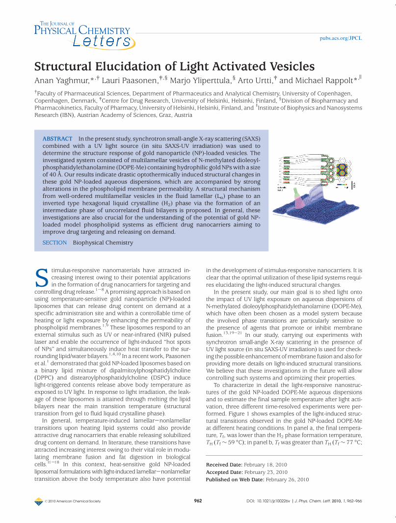
ABSTRACT In the present study, synchrotron small-angle X-ray scattering (SAXS)combined with a UV light source (in situ SAXS-UV irradiation) was used todetermine the structure response of gold nanoparticle (NP)-loaded vesicles. Theinvestigated system consisted of multilamellar vesicles of N-methylated dioleoyl-phosphatidylethanolamine (DOPE-Me) containing hydrophilic gold NPswith a sizeof 40 Å. Our results indicate drastic optothermically induced structural changes inthese gold NP-loaded aqueous dispersions, which are accompanied by strongalterations in the phospholipid membrane permeability. A structural mechanismfrom well-ordered multilamellar vesicles in the fluid lamellar (LR) phase to aninverted type hexagonal liquid crystalline (H2) phase via the formation of anintermediate phase of uncorrelated fluid bilayers is proposed. In general, theseinvestigations are also crucial for the understanding of the potential of gold NP-loaded model phospholipid systems as efficient drug nanocarriers aiming toimprove drug targeting and releasing on demand.
SECTION Biophysical Chemistry
S timulus-responsive nanomaterials have attracted in-creasing interest owing to their potential applicationsin the formation of drug nanocarriers for targeting and
controlling drug release.1-8 A promising approach is based onusing temperature-sensitive gold nanoparticle (NP)-loadedliposomes that can release drug content on demand at aspecific administration site and within a controllable time ofheating or light exposure by enhancing the permeability ofphospholipid membranes.1,9 These liposomes respond to anexternal stimulus such as UV or near-infrared (NIR) pulsedlaser and enable the occurrence of light-induced “hot spotsof NPs” and simultaneously induce heat transfer to the sur-rounding lipid/water bilayers.1,4,10 In a recentwork, Paasonenet al.1 demonstrated that gold NP-loaded liposomes based ona binary lipid mixture of dipalmitoylphosphatidylcholine(DPPC) and distearoylphosphatidylcholine (DSPC) inducelight-triggered contents release above body temperature asexposed to UV light. In response to light irradiation, the leak-age of these liposomes is attained through melting the lipidbilayers near the main transition temperature (structuraltransition from gel to fluid liquid crystalline phase).
In general, temperature-induced lamellar-nonlamellartransitions upon heating lipid systems could also provideattractive drug nanocarriers that enable releasing solubilizeddrug content on demand. In literature, these transitions haveattracted increasing interest owing to their vital role in modu-lating membrane fusion and fat digestion in biologicalcells.11-18 In this context, heat-sensitive gold NP-loadedliposomal formulations with light-induced lamellar-nonlamellartransition above the body temperature also have potential
in the development of stimulus-responsive nanocarriers. It isclear that the optimal utilization of these lipid systems requi-res elucidating the light-induced structural changes.
In the present study, our main goal is to shed light ontothe impact of UV light exposure on aqueous dispersions ofN-methylated dioleoylphosphatidylethanolamine (DOPE-Me),which have often been chosen as a model system becausethe involved phase transitions are particularly sensitive tothe presence of agents that promote or inhibit membranefusion.13,19-21 In our study, carrying out experiments withsynchrotron small-angle X-ray scattering in the presence ofUV light source (in situ SAXS-UV irradiation) is used for check-ing thepossible enhancementofmembrane fusionandalso forproviding more details on light-induced structural transitions.We believe that these investigations in the future will allowcontrolling such systems and optimizing their properties.
To characterize in detail the light-responsive nanostruc-tures of the gold NP-loaded DOPE-Me aqueous dispersionsand to estimate the final sample temperature after light acti-vation, three different time-resolved experiments were per-formed. Figure 1 shows examples of the light-induced struc-tural transitions observed in the gold NP-loaded DOPE-Meat different heating conditions. In panel a, the final tempera-ture, Tf, was lower than the H2 phase formation temperature,TH (Tf∼ 59 �C); in panel b, Tf was greater than TH (Tf∼ 77 �C;
Received Date: February 18, 2010Accepted Date: February 23, 2010

r 2010 American Chemical Society 963 DOI: 10.1021/jz100226v |J. Phys. Chem. Lett. 2010, 1, 962–966
pubs.acs.org/JPCL
TH=71 �C); and in panel c, the sample was superheated(Tf ∼106 �C). Before theUV-lightwas switchedon, the sampletemperature was 25 �C, and the diffraction patterns displaythe first- and second-order Bragg-peaks of well orderedmulti-lamellar vesicles in the fluid LRphase. Apart froman increasedscattering contribution in the low angle regime, no further dif-ferences are observed with respect to the nonloaded vesicles.The position and the Bragg-peak intensities remain the same,i.e., the presence of the gold NPs do not alter the compositionof the multilamellar vesicles (see also the Supporting Infor-mation). Therefore, the gold NPs are likely to absorb, in part,at the outer surface of thevesicles, and someamount is expec-ted to be in the surrounding excess water.
After switching on the UV light, the first induced phase isstill composed of fluid bilayers, but without positional correla-tion. Here, the SAXS patterns mainly display diffuse scatter-ing, i.e., to a great extent the form factor contribution of thebilayer. However, global analysis of this data as described inthe Supporting Information reveal that not all lamellae con-verted to an unbound state, but a few percent of the oligola-mellar membrane stacks remain (cf. to inset of Figure 1d andthe Supporting Information).
The time for formation of this intermediate phase dependson the final temperature and takes about 90 s for Tf ∼ 59 �C(Figure 1a), 35 s for Tf ∼ 77 �C (Figure 1b), and only 5 s forTf ∼ 106 �C (Figure 1c). Above the transition temperature, TH,
finally theH2phase forms (panelsb andc). This ismost clearlyseen in panel c (highlighted in white): the real-time evolutionof the SAXS patterns demonstrates the drastic impact of UVlight on the curvature of DOPE-Me monolayers. Illuminationwith the UV light source quickly leads to a significant effecton the lamellar (LR) phase: it induces within a few seconds adisordered bilayer phase, and shortly thereafter the H2 phase.One minute after switching off the light, the nanostructureretransforms to a vesicular dispersion from the H2 phase witha SAXS pattern that ismainlydominatedbydiffuse scattering.Interestingly, the heating process is not reversible in the caseof superheating. The sample does not reorganize into well-ordered multilamellar vesicles as seen during passive coolingin Figure 1a,b, but remains in the disordered state. The mostprobable explanation would be that, under these extremeconditions, unilamellar vesicles are formed. Further details onthe temporal evolution of different lattice spacings can be stu-died in the Supporting Information.
The LR-H2 structural transition has been explained by avariety of models; especially different mechanisms wereproposed for phosphatidylethanolamine (PE)/water modelsystems (see ref 22 and the references therein). Unfortu-nately, several of these models suffer from incorrect geome-trical dimensions, such as unrealistically bent monolayers orwater layers that are too thick. The most common reason forthis lies in the lack of detailed structural information. Part of
Figure 1. Time-resolved SAXS experiments on hydrophilic gold NP-loaded liposomes. (a) During UV-light heating (marked by a red bar),disorder is induced in the liposomes. (b)Heating above TH, the H2 phase forms via a disordered LR phase. (c) After superheating the sample,only uncorrelated bilayers remain. In panels a-c, the X-ray intensities are color coded in the insets, and the transition toH2 phase in panel cis highlighted in white. (d) Typical diffraction patterns just before, during, and after the LR-H2 transition are shown. The blue curve showsthe disordered phase consisting ofmainly uncorrelated bilayers, and the inset displays the global fitting (red line) of this intermediate phaseconsidering both the form and the structure factor contributions (for details, see the Supporting Information).

r 2010 American Chemical Society 964 DOI: 10.1021/jz100226v |J. Phys. Chem. Lett. 2010, 1, 962–966
pubs.acs.org/JPCL
the motivation for this work was to use SAXS at a high brilli-ance synchrotron source to address this deficiency for DOPE-Me aqueous dispersion. Recently, this strategy has led to thefirst detailed interface reconstruction involving the LR and H2
phases in palmitoyl-oleoyl-phosphatidylethanolamine (POPE)water systems22 (Figure 2a). In contrast to N-methylatedPEs, long-chain PE/water systems display a long-range co-existence region of both involved phases.22
In this respect, it is important to note that no disorderedphase is spotted during the transition process. ThereforeLaggner and Kriechbaum23 adopted the model of a diffu-sion-free martensitic process, in which the two neighboringlattices are mutually inclined. In this way, the path length ofrearranging lipid molecules in the interface region is mini-mized. This view is depicted in a refinedmanner in Figure 2a.It should be noted that the bulb-like form of bent back mono-layers is a consequence of the stringent constraint to mini-mize the interstitial regions of the interface (*). Free energysimulations in the transition regime of POPE have confirmedthat this bulb-like form of monolayers is the energeticallymost favorable.24
Figure 2b illustrates the same transition model for thegeometryof theDOPE-Mesystem investigatedhere. Althoughthe resulting snapshot of the interface looks quite appealing, itdoes not account for the disordered phase observed in theSAXS study. According to this model, the arrangement iscoplanar, i.e., the repeat distance of the LR phase and thed10-spacing of the H2 phase have an epitaxial relation. On thecontrary, our SAXS results show that the transition mecha-nismproceeds via an intermediate disordered phase. Such an
order-disorder-order transitionmodel is shown in Figure 2c.On the left-hand side, the LR phase is reconstructed on thebasis of the determined electron density profile at 71 �C, andon the right-hand side theH2 phase is illustrated according tothe electron density map determined just above 71 �C (forstructural details see the Supporting Information). Impor-tantly, when the initial and the final lattices are not orientedin a coplanar manner, the lattice repeat distance of the LRphase can not be retained. It is worth noting that, even if twoneighboringbilayers remain in thebound state (LRmembrane-membrane distance is conserved), simultaneously the next-nearest lamellae is disturbed to adhere at the preferreddistance. This is exactly what we observe (see Figure 1d,inset): the disordered phase consists of mainly uncorrelatedbilayers coexisting with very few bound membranes (globalfitting reveals 5 ( 3 correlated bilayers; see SupportingInformation). The reason why the transition favors to pro-ceed via a disordered intermediate lamellar phase is twofold.First, the DOPE-Me bilayers are not as strongly bound asin the pure PE/water systems (the interlamellar distance,dW(DOPE-Me)=19 Å at 71 �C and dW(POPE)=5 Å (ref 22) at74 �C, respectively), and second, the concentration of waterin the LR phase is too high to allow an ordered transitionwithout massive diffusion of the surplus water molecules.The number of water molecules per lipid, nW, reduces byabout 50% during the LR-H2 transition (nWlam = 29 andnWhex=19), which means that the surrounding disorderedlamellar phase (and excess of water regions) have to take up10 water molecules for every lipid molecule that rearrangesin the H2 phase (see Supporting Information). An alternative
Figure 2. Interface reconstruction involving the LR and the H2 phases. For clarity, lipid molecules are superimposed to the outlinedlocations of the polar interfaces (dashed lines) and themethyl trough regions (full lines). (a) Interfacemodel deduced from electron densitymaps of POPE (TH=74 �C). (b)Coplanar interfacemodel reconstructed on the basis of electrondensitymaps ofDOPE-Medata (TH=71 �C).(c) Order-disorder-order model depicted for the DOPE-Me system. The electron density profile of the LR phase is shown at the far left ofpanel c, and the electron density map of the H2 phase is shown on the far right of c. For the H2 phase, the electron density values are colorcoded.

r 2010 American Chemical Society 965 DOI: 10.1021/jz100226v |J. Phys. Chem. Lett. 2010, 1, 962–966
pubs.acs.org/JPCL
transformation pathway from flat bilayers into hexagonallypacked monolayer cylinders is believed to involve complexstructural rearrangements including the aggregation of trans-monolayer contacts into a body-centered cubic or primitivetetragonal phase, which in turn serves as a precursor for theformation of the H2 phase.
11,12 However, such three-dimen-sional (3D) intermediate phase was not observed in ourstudy.
In summary, the absorption of UV light by the gold NPstriggers fast LR-H2 structural transition in the DOPE-Meaqueous dispersions. This significant UV-activated impact onthe membrane permeability can be utilized for the formationof stimulus-responsive systems and drug nanocarriers withcontrolled drug release.
EXPERIMENTAL METHODS
Sample Preparation. DOPE-Me was purchased from AvantiPolar Lipids (Alabasta, LA) and dissolved in chloroform. Thissolvent was then evaporated using a gentle stream of nitro-gen, followed by drying under vacuum for at least 12 h inorder to completely remove the residual organic solvent. Thedry lipid film was hydrated by adding an aqueous stocksolution1 of hydrophilic gold NPs containing 1 mg NPs/mLand having an average NP size of 40 Å, carrying out at leastfive freeze-thaw cycles between liquid nitrogen and roomtemperature, and then homogenizing several times duringthe thawing steps by vigorous vortexing. The used gold NPswere synthesized as previously described,1 and the liposomalsamples were formed with a fixed total lipid concentration of10 wt %.
Combined Synchrotron SAXS and UV-Light Set-Up. Synchro-tron SAXS patterns were recorded at the Austrian SAXSbeamline25 (camera length 75 cm) at the synchrotron lightsource ELETTRA (Trieste, Italy) using a one-dimensional posi-tion sensitive detector (Gabriel type), which covered thes-range (s= 2 sin θ/λ, where λ is the wavelength and 2θ isthe scattering angle) of interest from about 1/640 to 1/13 Å-1
at an X-ray energy of 8 keV. Silver behenate (CH3-(CH2)20-COOAg with a d-spacing value of 58.38 Å) was used as astandard to calibrate the angular scale of the measuredintensity.26 In the absence of gold NPs, the control samplewas sealed in a thin-walled quartz capillary (Anton-Paar, Graz,Austria) and thermostatted with a programmable water bath(stability ( 0.1 �C, Unistat CC, Huber, Offenburg, Germany).Before each time-resolved experiment, fresh sample wasfilled into the capillary, and static measurements were doneat 25 �C with an exposure time of 30 s. The exposure timeduring the UV-light experiments was 2 s for each frame. Thetemperature scan on pure DOPE-Mewas performed at a scanrate of 1 �C/min. Two X-ray patterns were recorded perminute with a constant exposure time of 10 s. Between eachexposure, a small solenoid driven shutter blocked the directX-ray beam in order tominimize the total radiation dosage onthe sample. The SAXS analysis and electron density calcula-tions are given in the Supporting Information.
UV irradiation of DOPE-Me aqueous dispersions was doneat a wavelength of 365 nm with an EXFO Omnicure S1000light source (EXFO Life Sciences and Industrial Division,Ontario, Canada). The light source was operated with an
external switch, a 365 nm filter, a 5 mm optical guide, andanadjustable collimator. Thedistance fromthe collimator lensto the sample capillary was 40, 50, and 62 mm, respectively.
SUPPORTING INFORMATION AVAILABLE SAXS analysis,electron density calculations, and supporting figures. This materialis available free of charge via Internet at http://pubs.acs.org.
AUTHOR INFORMATION
Corresponding Author:*Towhomcorrespondence should be addressed. Tel.:þ45 35 33 6541. Fax: þ45 35336030. E-mail: [email protected] (A.Y.); [email protected] (M.R.).
REFERENCES
(1) Paasonen, L.; Laaksonen, T.; Johans, C.; Yliperttula, M.;Kontturi, K.; Urtti, A. Gold Nanoparticles Enable SelectiveLight-Induced Contents Release from Liposomes. J. Con-trolled Release 2007, 122, 86–93.
(2) Yliperttula, M.; Chung, B. G.; Navaladi, A.; Manbachi, A.; Urtti,A. High-Throughput Screening of Cell Responses to Bio-materials. Eur. J. Pharm. Sci. 2008, 35, 151–160.
(3) Li, J.; Wang, B. C.; Wang, Y. Z.; Liu, P.; Qiao, W. L. Preparationand Characterization of Thermosensitive Nanoparticles forTargeted Drug Delivery. J. Macromol. Sci., Part A: Pure Appl.Chem. 2008, 45, 833–838.
(4) Wu, G. H.; Milkhailovsky, A.; Khant, H. A.; Fu, C.; Chiu, W.;Zasadzinski, J. A. Remotely Triggered Liposome Release byNear-Infrared Light Absorption via Hollow Gold Nanoshells.J. Am. Chem. Soc. 2008, 130, 8175–8177.
(5) del Amo, E. M.; Urtti, A. Current and Future Ophthalmic DrugDelivery Systems. A Shift to the Posterior Segment. DrugDiscovery Today 2008, 13, 135–143.
(6) Lu, J.; Choi, E.; Tamanoi, F.; Zink, J. I. Light-Activated Nano-impeller-Controlled Drug Release in Cancer Cells. Small2008, 4, 421–426.
(7) Fong, W. K.; Hanley, T.; Boyd, B. J. Stimuli Responsive LiquidCrystals Provide 'On-Demand' Drug Delivery In Vitro and InVivo. J. Controlled Release 2009, 135, 218–226.
(8) Robbins, G. P.; Jimbo, M.; Swift, J.; Therien, M. J.; Hammer,D. A.; Dmochowski, I. J. Photoinitiated Destruction of Com-posite Porphyrin-Protein Polymersomes. J. Am. Chem. Soc.2009, 131, 3872–3874.
(9) Kojima, C.; Hirano, Y.; Yuba, E.; Harada, A.; Kono, K. Pre-paration and Characterization of Complexes of Liposomeswith Gold Nanoparticles. Colloids Surf. B 2008, 66, 246–252.
(10) Richardson, H. H.; Carlson, M. T.; Tandler, P. J.; Hernandez, P.;Govorov, A. O. Experimental and Theoretical Studies of Light-to-Heat Conversion and Collective Heating Effects in MetalNanoparticle Solutions. Nano Lett. 2009, 9, 1139–1146.
(11) Siegel, D. P. The Modified Stalk Mechanism of Lamellar/Inverted Phase Transitions and Its Implications for Mem-brane Fusion. Biophys. J. 1999, 76, 291–313.
(12) Siegel, D. P.; Epand, R. M. The Mechanism of Lamellar-to-Inverted Hexagonal Phase Transitions in Phosphatidyletha-nolamine: Implications for Membrane Fusion Mechanisms.Biophys. J. 1997, 73, 3089–3111.
(13) Harroun, T. A.; Balali-Mood, K.; Gourlay, I.; Bradshaw, J. P. TheFusion Peptide of Simian Immunodeficiency Virus and thePhase Behaviour of N-Methylated Dioleoylphosphatidyletha-nolamine. Biochim. Biophys. Acta 2003, 1617, 62–68.

r 2010 American Chemical Society 966 DOI: 10.1021/jz100226v |J. Phys. Chem. Lett. 2010, 1, 962–966
pubs.acs.org/JPCL
(14) Yaghmur, A.; Laggner, P.; Sartori, B.; Rappolt, M. CalciumTriggered LR-H2 Phase Transition Monitored by CombinedRapid Mixing and Time-Resolved Synchrotron SAXS. PLoSONE 2008, 3, e2072.
(15) Gruner, S. M.; Tate, M. W.; Kirk, G. L.; So, P. T.; Turner, D. C.;Keane, D. T.; Tilcock, C. P.; Cullis, P. R. X-RayDiffraction Studyof the Polymorphic Behavior of N-Methylated Dioleoylphos-phatidylethanolamine. Biochemistry 1988, 27, 2853–2866.
(16) Patton, J. S.; Carey, M. C. Watching Fat Digestion. Science1979, 204, 145–148.
(17) Hui, S. W.; Stewart, T. P.; Boni, L. T.; Yeagle, P. L. MembraneFusion Through Point Defects in Bilayers. Science 1981, 212,921–923.
(18) Yaghmur, A.; Laggner, P.; Almgren, M.; Rappolt, M. Self-Assembly in Monoelaidin Aqueous Dispersions: Direct Vesi-cles to Cubosomes Transition. PLoS ONE 2008, 3, e3747.
(19) Siegel, D. P.; Banschbach, J.; Alford, D.; Ellens, H.; Lis, L. J.;Quinn, P. J.; Yeagle, P. L.; Bentz, J. Physiological Levels ofDiacylglycerols in Phospholipid Membranes Induce Mem-brane Fusion and Stabilize Inverted Phases. Biochemistry1989, 28, 3703–3709.
(20) Siegel, D. P.; Cherezov, V.; Greathouse, D. V.; Koeppe, R. E.;Killian, J. A.; Caffrey, M. Transmembrane Peptides StabilizeInverted Cubic Phases in A Biphasic Length-DependentManner: Implications for Protein-InducedMembrane Fusion.Biophys. J. 2006, 90, 200–211.
(21) Cherezov, V.; Siegel, D. P.; Shaw,W.; Burgess, S.W.; Caffrey,M.The Kinetics of Non-lamellar Phase Formation in DOPE-Me:Relevance to Biomembrane Fusion. J. Membr. Biol. 2003, 195,165–182.
(22) Rappolt, M.; Hickel, A.; Bringezu, F.; Lohner, K. Mechanism ofthe Lamellar/Inverse Hexagonal Phase Transition Examinedby High Resolution X-Ray Diffraction. Biophys. J. 2003, 84,3111–3122.
(23) Laggner, P.; Kriechbaum, M. Phospholipid Phase Transitions:Kinetics and Structural Mechanisms. Chem. Phys. Lipids1991, 57, 121–145.
(24) Mares, T.; Daniel, M.; Perutkova, S.; Perne, A.; Dolinar, G.;Iglic, A.; Rappolt, M.; Kralj-Iglic, V. Role of Phospholipid Asym-metry in the Stability of Inverted Hexagonal MesoscopicPhases. J. Phys. Chem. B 2008, 112, 16575–16584.
(25) Amenitsch, H.; Rappolt, M.; Kriechbaum, M.; Mio, H. L. P.;et al. First Performance Assessment of the Small-AngleX-Ray Scattering Beamline at ELETTRA. J. Synchrotron Rad.1998, 5, 506–508.
(26) Huang, T. C.; Toraya, H.; Blanton, T. N.; Wu, Y. X-Ray-PowderDiffraction Analysis of Silver Behenate, A Possible Low-AngleDiffraction Standard. J. Appl. Crystallogr. 1993, 26, 180–184.
1
Supporting Information
Structural Elucidation of Light Activated Vesicles
Anan Yaghmur*,‡
, Lauri Paasonen†, Marjo Yliperttula
⊥, Arto Urtti
†, Michael Rappolt
*,§
‡Faculty of Pharmaceutical Sciences, Department of Pharmaceutics and Analytical Chemistry,
University of Copenhagen, Copenhagen, Denmark
†Centre for Drug Research, University of Helsinki, Helsinki, Finland
⊥Division of Biopharmacy and Pharmacokinetics, Faculty of Pharmacy,
University of Helsinki, Helsinki, Finland
§Istitute of Biophysics and Nanosystems Research (IBN), Austrian Academy of Sciences, Graz, Austria
Received date E-mail: [email protected]; [email protected]
2
SAXS Analysis and Electron Density Calculations
The two dimensional (2D) electron density map of the H2-phase of DOPE-Me/water at 74.5 °C
was derived from the SAXS diffraction pattern by standard procedures (for details see Rappolt et al.1).
After the raw data had been corrected for detector efficiency, and the background scattering both from
water and the sample cell had been subtracted, all Bragg peaks were fitted by Lorentzian distributions.
The fittings were carried out with the software package Origin 5.0 (Microcal Software). Next the
intensities were also corrected for their multiplicity, i.e. the intensity of the (2,1) peaks was divided by 2.
Thereafter, a Lorentz correction was applied to all powder diffraction images by multiplying each peak
intensity (peak area) by its corresponding wave vector s2 (for discussion of the Lorentz correction on
powder samples, the interested reader is directed to ref2). Finally, the square root of the corrected peak
intensity determined the form factor, F, of each respective reflection. The electron density contrast was
calculated by the Fourier synthesis. The phase combination (+1;-1;-1;+1;+1) for the reflections (10),
(11), (20), (21) and (30) was taken from literature3. Table S1 shows the structural parameters of the H2
phase of DOPE-Me based aqueous dispersion at 74.5 °C. One static scattering pattern of the
uncorrelated bilayers recorded at 71 °C was analyzed by the global analysis program (GAP). This model
uses one Gaussian for the headgroups and another for the hydrophobic core. A more detailed description
of this model is given elsewhere4,5. The fit parameters derived from the GAP evaluation for these
uncorrelated bilayers (Figure 1d, inset) are presented in Table S2. In Table S3, the calculated properties
of the fully hydrated DOPE-Me phases at a temperature, which is very close to the Lα/H2 transition
temperature (TH = 71 °C), are shown.
Supporting Experimental Details
Figure S1 compares the raw diffraction data of DOPE-ME dispersions with and without loaded
hydrophilic gold nanoparticles (NPs). The d-spacings of the fluid Lα phase and the intensities are within
3
experimental errors the same. At 25 °C, both aqueous dispersions have d value of 61. 4 Å. This means
that the NPs do not significantly alter the composition and the structure of the multilamellar vesicles.
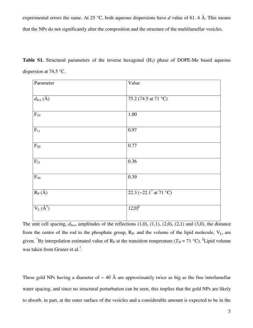
Table S1. Structural parameters of the inverse hexagonal (H2) phase of DOPE-Me based aqueous
dispersion at 74.5 °C.
Parameter Value
dhex (Å) 75.2 (74.5 at 71 °C)
F10 1.00
F11 0.97
F20 0.77
F21 0.36
F30 0.39
RP (Å) 22.3 (~22.1* at 71 °C)
VL (Å3) 1220§
The unit cell spacing, dhex, amplitudes of the reflections (1,0), (1,1), (2,0), (2,1) and (3,0), the distance
from the centre of the rod to the phosphate group, RP, and the volume of the lipid molecule, VL, are
given. *By interpolation estimated value of RP at the transition temperature (TH = 71 °C). §Lipid volume
was taken from Gruner et al.7.
These gold NPs having a diameter of ~ 40 Å are approximately twice as big as the free interlamellar
water spacing, and since no structural perturbation can be seen, this implies that the gold NPs are likely
to absorb, in part, at the outer surface of the vesicles and a considerable amount is expected to be in the
4
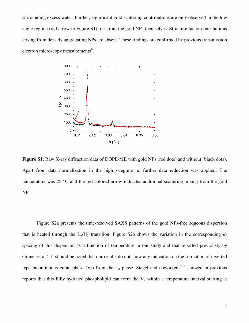
surrounding excess water. Further, significant gold scattering contributions are only observed in the low
angle regime (red arrow in Figure S1), i.e. from the gold NPs themselves. Structure factor contributions
arising from densely aggregating NPs are absent. These findings are confirmed by previous transmission
electron microscopy measurements6.
Figure S1. Raw X-ray diffraction data of DOPE-ME with gold NPs (red dots) and without (black dots).
Apart from data normalization in the high s-regime no further data reduction was applied. The
temperature was 25 °C and the red colored arrow indicates additional scattering arising from the gold
NPs.
Figure S2a presents the time-resolved SAXS patterns of the gold NPs-free aqueous dispersion
that is heated through the Lα/H2 transition. Figure S2b shows the variation in the corresponding d-
spacing of this dispersion as a function of temperature in our study and that reported previously by
Gruner et al.7. It should be noted that our results do not show any indication on the formation of inverted
type bicontinuous cubic phase (V2) from the Lα phase. Siegel and coworkers8-11 showed in previous
reports that this fully hydrated phospholipid can form the V2 within a temperature interval starting at

5
62 °C and thus can induce indirect Lα-H2 transition as heating is done at slow scan rates or during
prolonged incubations of months at temperatures below the Lα-H2 transition temperature.
Table S2. Fit parameters derived from the GAP evaluation of the SAXS pattern (Figure 1d, inset) for
the DOPE-Me based liposomes at 71 °C.
Parameter Value
d (Å) 64.5 ± 0.8
Nmean 5 ± 3
η 1.4 ± 0.1
zH (Å) 18.6 ± 0.5 (19.5§ at 2 °C)
σH (Å) 3.0 ± 0.6
σC (Å) 6.6 ± 1.0
ρr -0.99 ± 0.07
Ndiff 0.93 ± 0.14
The parameters given are the d-spacing, the membrane correlation number, Nmean, the Caillé parameter,
η, the headgroup position, zH, the headgroup width, σH, the width of the hydrophobic core, σC, the
relative electron density of the bilayer trough set in relation to the headgroup density, ρr, and the fraction
of the diffuse scattering, Ndiff. §Value published by Gruner et al.7.

6
Figure S2. Time-resolved temperature scan on gold NPs-free DOPE-Me aqueous dispersion. Panel (a)
displays the recorded raw SAXS data and panel (b) displays the corresponding d-spacings. Experimental
data from Gruner et al.7 is shown by open circles. In the temperature range of 50-71 °C, the lattice
parameter of the formed multi-lamellar vesicles (MLVs) increases during heating from 61.0 to 64.5 Å.
At 71 °C, the Lα-H2 transition is observed. The lattice parameter of the newly formed H2 phase decreases
linearly with increasing temperature (dhex (Å) = -0.1835 * T + 87.8, where dhex is the lattice constant of
the H2 phase, and T is the investigated temperature).
7
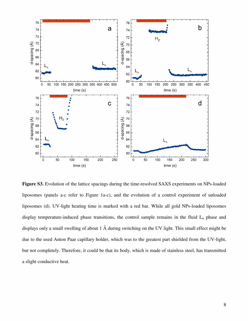
Figure S3 illustrates the evolution of the lattice spacings during the time-resolved SAXS
experiments on the gold NPs-loaded DOPE-ME liposomes (Figure S3a-c refers to Figure 1a-c). For
these aqueous dispersions, the final sample temperature, Tf, was estimated from the lattice parameter of
the H2 phase at a lamp to sample distance of 50 mm (this experiment is illustrated in Figure 1b). The
minimum lattice spacing reached was 73.6 Å (Figure S3b), which corresponds to a temperature of 77 °C
(compare Figure S1b). From this value, it follows that the Tf at a distance of 62 mm (lamp distance in
experiment of Figure 1a) is about 59 °C, and at sample to lamp distance of 40 mm (lamp distance in
experiment of Figure 1c) the Tf equals about 106 °C. Since no signs of air bubbles were detected by
SAXS in the latter case, the sample was most probably superheated under these extreme experimental
conditions. In Figure S3c (referring to Figure 1c), the heating rate, ∆T/∆t, can be estimated by taking a
closer look to the variation of the lattice spacing of the H2 phase with time in the interval of 30-50 s. In
this case, the calculated ∆T/∆t value is approximately 60 °C/min. For the other two experiments, we
accordingly estimated ∆T/∆t to be approximately 42 °C/min (lamp distance 50 mm, referring to Figure
1b) and 27 °C/min (lamp distance 62 mm, referring to Figure 1a)), respectively.
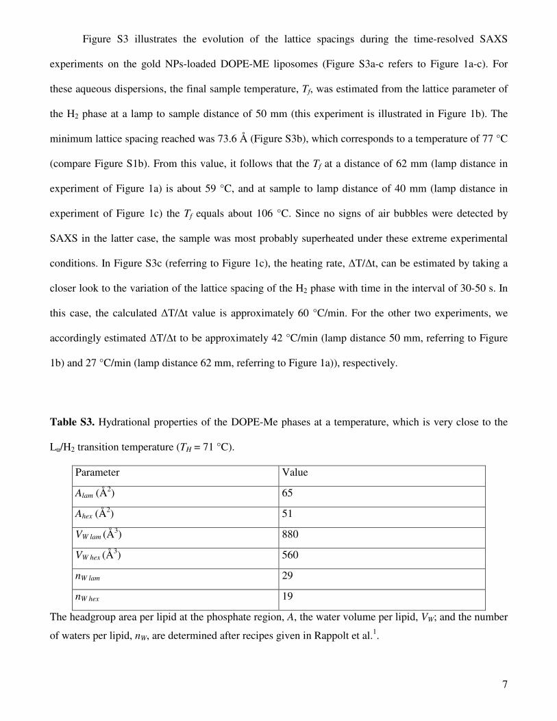
Table S3. Hydrational properties of the DOPE-Me phases at a temperature, which is very close to the
Lα/H2 transition temperature (TH = 71 °C).
Parameter Value
Alam (Å2) 65
Ahex (Å2) 51
VW lam (Å3) 880
VW hex (Å3) 560
nW lam 29
nW hex 19
The headgroup area per lipid at the phosphate region, A, the water volume per lipid, VW; and the number
of waters per lipid, nW, are determined after recipes given in Rappolt et al.1.
8
Figure S3. Evolution of the lattice spacings during the time-resolved SAXS experiments on NPs-loaded
liposomes (panels a-c refer to Figure 1a-c), and the evolution of a control experiment of unloaded
liposomes (d). UV-light heating time is marked with a red bar. While all gold NPs-loaded liposomes
display temperature-induced phase transitions, the control sample remains in the fluid Lα phase and
displays only a small swelling of about 1 Å during switching on the UV light. This small effect might be
due to the used Anton Paar capillary holder, which was to the greatest part shielded from the UV-light,
but not completely. Therefore, it could be that its body, which is made of stainless steel, has transmitted
a slight conductive heat.

9
References
(1) Rappolt, M., Hickel, A., Bringezu, F., Lohner, K. Biophys. J. 2003, 84, 3111–3122.
(2) Warren, B. E. 1969. X-ray Diffraction. Addison-Wesley, Reading.
(3) Harper, P. E., Mannock, D. A., Lewis, R. N., McElhaney, R. N., Gruner, S. M. Biophys. J. 2001, 81,
2693–2706.
(4) Pabst, G., Rappolt, M., Amenitsch, H., Laggner, P. Phys. Rev. E 2000, 62, 4000-4009.
(5) Rappolt, M. In Advances in Planar Lipid Bilayers and Liposomes. A. Leitmannova-Liu, Ed.,
Elsevier: Amsterdam, 2006; Vol. 5, pp. 253-283.
(6) Paasonen, L.; Laaksonen, T.; Johans, C.; Yliperttula, M.; Konturri, K.; Urtti, A. J. Controlled
Release 2007, 122, 86-93.
(7) Gruner, S. M.; Tate, M. W.; Kirk, G. L.; So, P. T. C.; Tunrer, D. C.; Keane, D. T. Biochemistry
1988, 27, 2853-2866.
(8) Siegel, D. P.; Banschbach, J. L. Biochemistry 1990, 29, 5975-5981.
(9) Cherezov, V.; Siegel, D. P.; Shaw, W.; Burgess, S. W.; Caffrey, M. J. Membrane Biol. 2003, 195,
165-182.
(10) Siegel, D. P.; Tenchov, B. G. Biophys. J. 2008, 94, 3987–3995.
(11) Siegel, D. P.; Cherezov, V.; Greathouse, D. V.; Koeppe, R. E.; Killian, J. A.; Caffrey, M. Biophys.
J. 2006, 90, 200–211.