tetanus toxin blocks the exocytosis of synaptic vesicles clustered at synapses but not of synaptic...
TRANSCRIPT

Tetanus Toxin Blocks the Exocytosis of Synaptic Vesicles Clusteredat Synapses But Not of Synaptic Vesicles in Isolated Axons
Claudia Verderio,1 Silvia Coco,1 Alberto Bacci,1 Ornella Rossetto,2 Pietro De Camilli,3 Cesare Montecucco,2and Michela Matteoli1
1Consiglio Nazionale delle Ricerche Cellular and Molecular Pharmacology and B. Ceccarelli Centers, Department ofMedical Pharmacology, 20129 Milano, Italy; 2Dipartimento di Scienze Biomediche, Consiglio Nazionale delle Ricerche-Centro Biomembrane, University of Padova, 35122 Padova, Italy, and 3Howard Hughes Medical Institute and Departmentof Cell Biology, Yale University School of Medicine, New Haven, Connecticut 06510
Recycling synaptic vesicles are already present in isolatedaxons of developing neurons (Matteoli et al., 1992; Zakharenkoet al., 1999). This vesicle recycling is distinct from the vesiculartraffic implicated in axon outgrowth. Formation of synapticcontacts coincides with a clustering of synaptic vesicles at thecontact site and with a downregulation of their basal rateof exo-endocytosis (Kraszewski et al., 1995; Coco et al., 1998)We report here that tetanus toxin-mediated cleavage ofsynaptobrevin/vesicle-associated membrane protein (VAMP2),previously shown not to affect axon outgrowth, also does notinhibit synaptic vesicle exocytosis in isolated axons, despite itspotent blocking effect on their exocytosis at synapses. Thisdifferential effect of tetanus toxin could be seen even on differ-ent branches of a same neuron. In contrast, botulinum toxins Aand E [which cleave synaptosome-associated protein of 25 kDa.
(SNAP-25)] and F (which cleaves synaptobrevin/VAMP1 and 2)blocked synaptic vesicle exocytosis both in isolated axons andat synapses, strongly suggesting that this process is dependenton “classical” synaptic SNAP receptor (SNARE) complexesboth before and after synaptogenesis. A tetanus toxin-resistantform of synaptic vesicle recycling, which proceeds in the ab-sence of external stimuli and is sensitive to botulinum toxin F, E,and A, persists at mature synapses. These data suggest theinvolvement of a tetanus toxin-resistant, but botulinumF-sensitive, isoform of synaptobrevin/VAMP in synaptic vesicleexocytosis before synapse formation and the partial persis-tence of this form of exocytosis at mature synaptic contacts.
Key words: exocytosis; synaptic vesicles; tetanus toxin; syn-aptogenesis; hippocampal neurons; synaptobrevin
Major characteristics of synaptic transmission are high spatialprecision, speed, and great fidelity. These features are dependenton exocytosis taking place at restricted and well defined areas ofthe neuronal membrane. They rely on the presence of an ex-tremely specialized machinery, allowing very rapid triggering andswitching off of synaptic vesicle (SV) exocytosis in response todepolarization-evoked calcium influx (Barrett and Stevens, 1972).Regulated SV exocytosis at mature synaptic sites has been widelyinvestigated, and several proteins that participate in this processhave now been identified (Sollner et al., 1993; Bennett andScheller, 1994; Ferro-Novick and Jahn, 1994; Sudhof, 1995). It iswell established that SV exocytosis involves the interaction ofthe synaptic vesicle membrane proteins synaptobrevin/vesicle-associated membrane protein (VAMP) 1 and 2 [v-SNAREs (sol-uble N-ethylmaleimide factor-attached protein (SNAP) receptors(SNARE)] with the plasma membrane proteins syntaxin andSNAP-25 (t-SNAREs). Synaptic SNARE proteins are targets forthe proteolytic action of clostridial tetanus and botulinum neuro-
toxins (TeNT and BoNTs), which potently block exocytosis innerve terminals (Blasi et al., 1993a,b; Schiavo et al., 1992, 1993a,b,1995).
The molecular mechanisms involved in the development ofCNS synapses are still poorly understood. A powerful experi-mental system to investigate these mechanisms is represented byprimary cultures of hippocampal neurons. In these neurons, SVs,which release neurotransmitter and undergo high basal exo-endocytotic recycling, are already present at very early develop-mental stages when axons grow in isolation. Formation of synap-tic contacts coincides with a clustering of synaptic vesicles, with adownregulation of their basal recycling rate (Kraszewski et al.,1995; Coco et al., 1998), with a change in the calcium sensitivity ofthe exocytotic process (Coco et al., 1998), and with a switch in thepopulation of calcium channels controlling neurotransmitter (glu-tamate) release (Scholz and Miller, 1995; Verderio et al., 1995).
Synaptic vesicle exocytosis, which occurs in developing axons,is clearly distinct from the constitutive exocytosis of vesicles thatmediate axon elongation. Whereas the latter occurs primarily atthe axon ending (Pfenninger and Maylie-Pfenninger 1981; Futer-man et al., 1993; Craig et al., 1995; Zakharenko and Popov 1998),the former occurs along the entire distal axonal arbor (Matteoli etal., 1992; Kraszewski et al., 1995; Zakharenko et al., 1999). It wasshown previously that axonal outgrowth and synaptogenesis incultured CNS neurons is not inhibited by tetanus toxin (Ahnert-Hilger et al., 1996; Osen Sand et al., 1996), strongly suggestingthat the v-SNARE(s) implicated in this process are distinct fromthe classical synaptic v-SNARE(s). We report here that, surpris-
Received March 22, 1999; revised May 24, 1999; accepted May 24, 1999.This work has been supported by Telethon Grants 1042 (to M.M.) and 1068 (to
C.M.), by Human Frontier Science Program (to M.M. and P.D.C.), by the EuropeanCommunity Grants Biomed 2 BMH4 CT97 2410 (to C.M.) and BIO4–98-0408 (toM.M.), and by National Institutes of Health Grant NS36251 (to P.D.C.). Weacknowledge Dr. R. Jahn (Gottingen, Germany) for gift of antibodies againstsynaptophysin, synaptobrevin/VAMP2, rab3a, and synaptotagmin.
Correspondence should be addressed to Michela Matteoli, Consiglio Nazionaledelle Ricerche Cellular and Molecular Pharmacology, and B. Ceccarelli Centers,Department of Medical Pharmacology, University of Milano, via Vanvitelli 32,20129 Milano, Italy.Copyright © 1999 Society for Neuroscience 0270-6474/99/196723-10$05.00/0
The Journal of Neuroscience, August 15, 1999, 19(16):6723–6732
ingly, even synaptic vesicle exocytosis is insensitive to the actionof tetanus toxin in developing axons. This tetanus toxin-resistantform of SV recycling partially persists at mature synapses asspontaneous SV exocytosis. These findings suggest that the dif-ferent properties of nonsynaptic and synaptic exocytosis of SVscorrelate with a switch in v-SNARE isoforms underlying SNAREcomplex formation.
MATERIALS AND METHODSHippocampal cell cultures. Primary neuronal cultures were prepared fromthe hippocampi of 18-d-old fetal rats as described by Banker and Cowan(1977) and Bartlett and Banker (1984). Briefly, hippocampi were disso-ciated by treatment with trypsin (0.25% for 15 min at 37°C), followed bytrituration with a fire-polished Pasteur pipette. Dissociated cells wereplated on poly-L-lysine-treated (Sigma, Milano, Italy) glass coverslips inMEM with 10% horse serum at densities ranging from 10,000 to 20,000cells/cm 2. After few hours, coverslips were transferred to dishes contain-ing a monolayer of cortical glial cells (Booher and Sensenbrenner, 1972),so that they were suspended over the glial cells but not in direct contactwith them (Bartlett and Banker, 1984). Cells were maintained in MEM(Life Technologies, S. Giuliano, Italy) without sera, supplemented with1% N2 (Life Technologies), 2 mM glutamine, and 1 mg/ml BSA (neuro-nal medium). A modification of the method used by Furshpan et al.(1976) was used to grow single neurons on small islands of substrate,consisting in a fine mist of poly-L-lysine sprayed on glass coverslips.
Experimental treatments. Neuronal cultures were exposed to 10 nMTeNT for 5 min in the presence or absence of 55 mM KCl in the externalmedium, thoroughly washed, maintained in regular medium at 37°C for2–18 hr, fixed, and double stained for synaptobrevin/VAMP2 and forsynaptophysin. In some experiments, neurons were also exposed to 20 nMBoNT/A, 80 nM BoNT/E, and 60–100 nM BoNT/F. An immunocyto-chemical assay based on the use of antibodies directed against theintravesicular domain of rat synaptotagmin I (Syt-ecto Abs) was used totest the efficacy of the toxins in blocking synaptic vesicle recycling. Inparticular, cultures were incubated with Syt-ecto Abs for 5 min or 1 hr at37°C in the presence or absence of 55 mM KCl. Cells were then fixed with4% paraformaldehyde in 0.12 M phosphate buffer containing 0.12 Msucrose for 25 min at 37°C. Fixed cells were detergent-permeabilized andlabeled with rhodamine-conjugated anti-rabbit antibodies as describedpreviously (Matteoli et al., 1992). Counterstaining of neurons with anti-bodies directed against total synaptotagmin I (Syt mono), followed byfluorescein-conjugated anti-mouse antibodies, was performed as de-
scribed previously (Matteoli et al., 1992). Coverslips were mounted in70% glycerol in phosphate buffer containing 1 mg/ml phenylendiamine.Preparations were examined with a Zeiss (Oberkochen, Germany) micro-scope equipped with epifluorescence and photographed with T-MAX 400(Kodak, Milano, Italy). Quantitative analysis was performed as describedpreviously (Matteoli et al., 1996; Coco et al., 1998; Verderio et al., 1999a).For each experiment, 60–80 neurons were examined. Values obtained indifferent experiments were averaged and plotted.
Immunoblotting. Cultured hippocampal neurons were solubilized in1% SDS, 5% 2-mercaptoethanol, 65 mM Tris-HCl, pH 6.8, and 10%sucrose as described previously (Coco et al., 1997). SDS-PAGE electro-phoresis and Western blotting were performed as described previously(Laemmli, 1970; Towbin et al., 1979). Briefly, cell extracts (30 mg) weresubjected to SDS-PAGE (10% polyacrylamide gels) and transferred byelectroblotting to nitrocellulose (Sartorius, Gottingen, Germany). Blotswere blocked in 5% nonfat dry milk, 0.1% Tween 20, 20 mM Tris, and150 mM NaCl, pH 7.5, at room temperature. Blots were incubated withantibodies for 2 hr at room temperature in blocking buffer. Blots werethen thoroughly washed, incubated (1 hr) with horseradish peroxidase-conjugated anti-rabbit IgG or horseradish peroxidase-conjugated anti-mouse IgG (1:5000 in blocking buffer; Sigma), and finally washed withTris-NaCl. The immunoreactive proteins were visualized with enhancedchemiluminescence (Amersham, Milano, Italy).
Antibodies. Rabbit polyclonal antibodies directed against the intravesicu-lar domain of rat synaptotagmin I (Syt-ecto Abs) were generated as de-scribed previously (Matteoli et al., 1992) using a synthetic peptide corre-sponding to the residue 1–19 of the protein. Antibodies againstsynaptobrevin/VAMP1 and 2 were generated in rabbit as described previ-ously (Rossetto et al., 1996). Polyclonal antibodies against synaptophysinand monoclonal antibodies against synaptobrevin/VAMP2, rab3a, andsynaptotagmin I were a kind gift from Dr. R. Jahn (Gottingen, Germany).Polyclonal antibodies against syntaxin I and SNAP-25 were raised and usedas described previously (Chilcote et al., 1995; Papini et al., 1995). Anti-rabbit rhodamine-conjugated antibodies were purchased from BoehringerMannheim (Milano, Italy). Anti-mouse fluorescein-conjugated antibodieswere from Jackson ImmunoResearch (West Grove, PA).
RESULTSSynaptic v- and t-SNAREs are already expressed byneurons at early developmental stagesWhen maintained in primary cultures, embryonic hippocampalneurons develop through a series of well characterized develop-
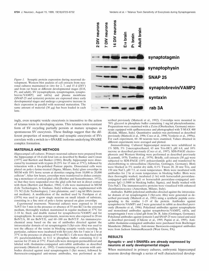
Figure 1. Synaptic protein expression during neuronal de-velopment. Western blot analysis of cell extracts from neu-ronal cultures maintained in vitro for 2, 7, and 15 d (DIV )and from rat brain at different developmental stages (E18,P1, and adult). SV (synaptophysin, synaptotagmin, synapto-brevin/VAMP2, and rab3a) and plasma membrane(SNAP-25 and syntaxin) proteins are expressed since earlydevelopmental stages and undergo a progressive increase intheir expression in parallel with neuronal maturation. Thesame amount of material (30 mg) has been loaded in eachlane.
6724 J. Neurosci., August 15, 1999, 19(16):6723–6732 Verderio et al. • Tetanus Toxin Sensitivity of Exocytosis during Synaptogenesis

mental stages (Dotti et al., 1988). After few days in culture, theyestablish a clear axo-dendritic polarity and, eventually, form anetwork of functional synaptic contacts characterized by presyn-aptic clusters of SVs and by the postsynaptic accumulation ofglutamate receptors (Craig and Banker, 1994; Verderio et al.,1999b). Figure 1 shows that the SV proteins synaptotagmin I,
synaptophysin, and rab3a, together with the three SNARE pro-teins synaptobrevin/VAMP2, SNAP-25, and syntaxin I, are al-ready expressed by cultured hippocampal neurons at early devel-opmental stages. For all of them, an increase in the levels ofexpression takes place in parallel with the time of differentiationin culture. A similar increase in synaptic protein expression was
Figure 2. Tetanus toxin treatment inhibits SV exocytosis atmature synapses but not in developing neurons. A, B, Fifteen-day-old neurons were incubated for 5 min in the presence ofSyt-ecto Abs in 55 mM external KCl before (A, B) or after (C,D) treatment with 10 nM TeNT. After this incubation, neu-rons were washed, fixed, detergent-permeabilized, reactedwith rhodamine-conjugated goat anti-rabbit IgGs (B, D), andcounterstained with antibodies against total synaptotagmin(syt), followed by FITC-conjugated goat anti-mouse IgGs (A,C). Puncta of immunoreactivity represent presynaptic nerveterminals, which outline perikarya and dendrites. Syt-ectoAbs are internalized at synaptic contacts when applied incontrol conditions (B) but not after treatment with TeNT(D). E–H, Exocytosis-dependent uptake of Syt-ecto Abs (ap-plied for 5 min in the presence of 55 mM KCl in the externalmedium) in living neurons before synaptogenesis, in controlconditions ( F), or after treatment with 10 nM TeNT (H ). E,G, Double immunofluorescence of total synaptotagmin (syt)of the same neurons as in F and H. Note that an efficientinternalization of Syt-ecto Abs takes place in axons, evenafter treatment with TeNT (H ). Scale bar: A–D, 20 mm; E–H,28 mm. I, Quantitative analysis of Syt-ecto internalization inneurons before and after synaptogenesis, both in controlconditions or after treatment with 10 nM TeNT.
Verderio et al. • Tetanus Toxin Sensitivity of Exocytosis during Synaptogenesis J. Neurosci., August 15, 1999, 19(16):6723–6732 6725

found to take place also during brain development, as indicatedby Western blot analysis of total homogenates from embryonicday 18 (E18), postnatal day (P1), and adult rat brains. In contrast,we could not detect any synaptobrevin/VAMP1 immunoreactiv-ity in cultured hippocampal neurons before synaptogenesis (datanot shown). Immunofluorescence experiments revealed that, be-fore synaptogenesis, synaptobrevin/VAMP2 immunoreactivitywas localized on vesicular structures dispersed along the axon(see Fig. 3A). These vesicular structures, which are immunoreac-tive for synaptotagmin I (see Fig. 3B), synaptophysin, and rab3a(data not shown), have been already identified as bona fide SVs(Fletcher et al., 1991; Matteoli et al., 1991, 1992, 1996; Kraszewskiet al., 1995; Coco et al., 1998). Syntaxin I and SNAP-25 werefound to be present throughout all neuronal compartments (datanot shown) (Galli et al., 1995).
TeNT blocks recycling of SVs clustered at synapsesbut does not block SV recycling before synaptogenesisTo monitor SV recycling, we used an assay based on antibodiesdirected against the intravesicular domain of the SV proteinsynaptotagmin I (Syt-ecto Abs), which become internalized in thelumen of SVs when they undergo exocytosis and compensatoryendocytosis (Matteoli et al., 1992). As already shown, Syt-ectoAbs become internalized by an activity-dependent mechanism at
synaptic contacts of cultured hippocampal neurons (Fig. 2B) andare also actively taken up by recycling vesicles in the axon ofdeveloping neurons (Fig. 2F). Mature cultures were incubatedwith 10 nM TeNT for 5 min under depolarizing conditions andassayed for SV recycling 2 hr later, when the TeNT substratesynaptobrevin/VAMP2 is completely cleaved (Matteoli et al.,1996). Virtually no internalization of Syt-ecto Abs at synapticsites was detected (n 5 15 experiments) (Fig. 2D, I, quantitativeanalysis), consistent with a block of exocytosis produced by TeNTtreatment. In contrast, when applied to neurons before synapto-genesis, TeNT was actively internalized (data not shown) (Mat-teoli et al., 1996) but did not produce any relevant inhibitory effecton SV recycling (n 5 7) (Fig. 2H, I, quantitative analysis).
The lack of effect of TeNT on SV recycling in neurons beforesynaptogenesis could result from inaccessibility of synaptobrevin/VAMP2 to the proteolytic action of the toxin. To test thispossibility, hippocampal neurons were stained for synaptobrevin/VAMP2 after TeNT treatment. In control neurons, synaptobre-vin/VAMP2 was localized on vesicles dispersed throughout thedistal axonal arbor (Fig. 3A). These vesicles colocalized withinternalized Syt-ecto Abs (Fig. 3B), thus supporting their identi-fication as recycling SVs. After 6 hr of TeNT treatment, a sub-stantial reduction (up to 77%) of synaptobrevin/VAMP 2 stain-
Figure 3. TeNT treatment cleaves syn-aptobrevin/VAMP2 without blocking SVrecycling in neurons before synaptogen-esis. A, B, Immature control neuron ex-posed to Syt-ecto Abs (for 5 min in thepresence of 55 mM KCl in the externalmedium) (B) and double labeled for syn-aptobrevin/VAMP2 (A). C, D, Imma-ture neuron exposed to Syt-ecto Abs (D)and double labeled for synaptobrevin/VAMP2 (C) after treatment with 10 nMTeNT. Note that TeNT treatment cleavessynaptobrevin/VAMP2 without impair-ing SV recycling. Scale bar, 11.25 mm. E,Quantitative analysis of Syt-ecto Ab in-ternalization (F) and synaptobrevin/VAMP2 cleavage (M) in developing neu-rons at different times after cultureintoxication. Note that TeNT treatmentdramatically reduces synaptobrevin /VAMP2 immunoreactivity over timewithout significantly reducing SV recy-cling. F, Quantitative analysis of Syt-ectoAb internalization (F) and synaptobre-vin/VAMP2 cleavage (M) in developingneurons exposed to increasing doses ofBoNT/F. Note the existence of a strictcorrelation between synaptobrevin /VAMP2 cleavage and inhibition of SVrecycling. Values were expressed as a ra-tio between the signals produced by Syt-ecto Abs or by synaptobrevin/VAMP2antibodies and those produced by anti-bodies directed against total synaptotag-min I or synaptophysin.
6726 J. Neurosci., August 15, 1999, 19(16):6723–6732 Verderio et al. • Tetanus Toxin Sensitivity of Exocytosis during Synaptogenesis
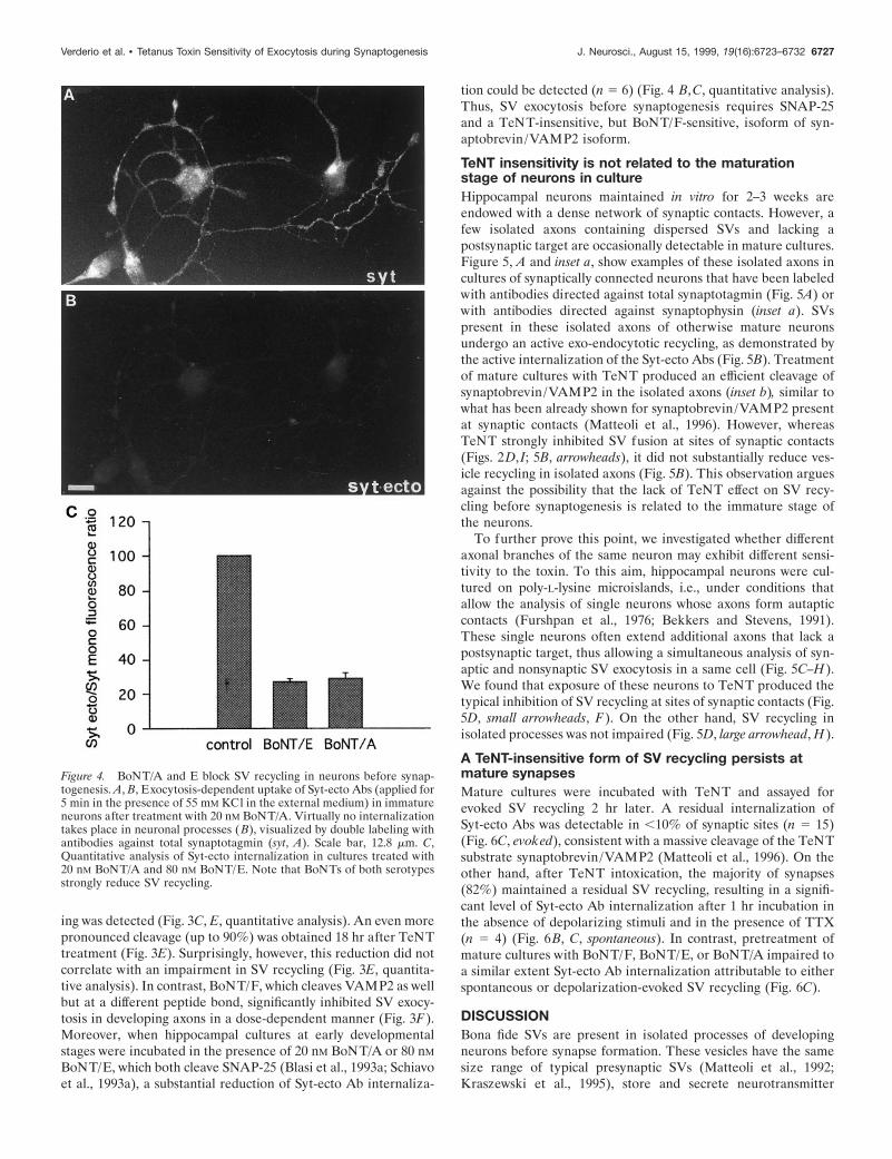
ing was detected (Fig. 3C, E, quantitative analysis). An even morepronounced cleavage (up to 90%) was obtained 18 hr after TeNTtreatment (Fig. 3E). Surprisingly, however, this reduction did notcorrelate with an impairment in SV recycling (Fig. 3E, quantita-tive analysis). In contrast, BoNT/F, which cleaves VAMP2 as wellbut at a different peptide bond, significantly inhibited SV exocy-tosis in developing axons in a dose-dependent manner (Fig. 3F).Moreover, when hippocampal cultures at early developmentalstages were incubated in the presence of 20 nM BoNT/A or 80 nM
BoNT/E, which both cleave SNAP-25 (Blasi et al., 1993a; Schiavoet al., 1993a), a substantial reduction of Syt-ecto Ab internaliza-
tion could be detected (n 5 6) (Fig. 4 B,C, quantitative analysis).Thus, SV exocytosis before synaptogenesis requires SNAP-25and a TeNT-insensitive, but BoNT/F-sensitive, isoform of syn-aptobrevin/VAMP2 isoform.
TeNT insensitivity is not related to the maturationstage of neurons in cultureHippocampal neurons maintained in vitro for 2–3 weeks areendowed with a dense network of synaptic contacts. However, afew isolated axons containing dispersed SVs and lacking apostsynaptic target are occasionally detectable in mature cultures.Figure 5, A and inset a, show examples of these isolated axons incultures of synaptically connected neurons that have been labeledwith antibodies directed against total synaptotagmin (Fig. 5A) orwith antibodies directed against synaptophysin (inset a). SVspresent in these isolated axons of otherwise mature neuronsundergo an active exo-endocytotic recycling, as demonstrated bythe active internalization of the Syt-ecto Abs (Fig. 5B). Treatmentof mature cultures with TeNT produced an efficient cleavage ofsynaptobrevin/VAMP2 in the isolated axons (inset b), similar towhat has been already shown for synaptobrevin/VAMP2 presentat synaptic contacts (Matteoli et al., 1996). However, whereasTeNT strongly inhibited SV fusion at sites of synaptic contacts(Figs. 2D, I; 5B, arrowheads), it did not substantially reduce ves-icle recycling in isolated axons (Fig. 5B). This observation arguesagainst the possibility that the lack of TeNT effect on SV recy-cling before synaptogenesis is related to the immature stage ofthe neurons.
To further prove this point, we investigated whether differentaxonal branches of the same neuron may exhibit different sensi-tivity to the toxin. To this aim, hippocampal neurons were cul-tured on poly-L-lysine microislands, i.e., under conditions thatallow the analysis of single neurons whose axons form autapticcontacts (Furshpan et al., 1976; Bekkers and Stevens, 1991).These single neurons often extend additional axons that lack apostsynaptic target, thus allowing a simultaneous analysis of syn-aptic and nonsynaptic SV exocytosis in a same cell (Fig. 5C–H).We found that exposure of these neurons to TeNT produced thetypical inhibition of SV recycling at sites of synaptic contacts (Fig.5D, small arrowheads, F). On the other hand, SV recycling inisolated processes was not impaired (Fig. 5D, large arrowhead, H).
A TeNT-insensitive form of SV recycling persists atmature synapsesMature cultures were incubated with TeNT and assayed forevoked SV recycling 2 hr later. A residual internalization ofSyt-ecto Abs was detectable in ,10% of synaptic sites (n 5 15)(Fig. 6C, evoked), consistent with a massive cleavage of the TeNTsubstrate synaptobrevin/VAMP2 (Matteoli et al., 1996). On theother hand, after TeNT intoxication, the majority of synapses(82%) maintained a residual SV recycling, resulting in a signifi-cant level of Syt-ecto Ab internalization after 1 hr incubation inthe absence of depolarizing stimuli and in the presence of TTX(n 5 4) (Fig. 6B, C, spontaneous). In contrast, pretreatment ofmature cultures with BoNT/F, BoNT/E, or BoNT/A impaired toa similar extent Syt-ecto Ab internalization attributable to eitherspontaneous or depolarization-evoked SV recycling (Fig. 6C).
DISCUSSIONBona fide SVs are present in isolated processes of developingneurons before synapse formation. These vesicles have the samesize range of typical presynaptic SVs (Matteoli et al., 1992;Kraszewski et al., 1995), store and secrete neurotransmitter
Figure 4. BoNT/A and E block SV recycling in neurons before synap-togenesis. A, B, Exocytosis-dependent uptake of Syt-ecto Abs (applied for5 min in the presence of 55 mM KCl in the external medium) in immatureneurons after treatment with 20 nM BoNT/A. Virtually no internalizationtakes place in neuronal processes ( B), visualized by double labeling withantibodies against total synaptotagmin (syt, A). Scale bar, 12.8 mm. C,Quantitative analysis of Syt-ecto internalization in cultures treated with20 nM BoNT/A and 80 nM BoNT/E. Note that BoNTs of both serotypesstrongly reduce SV recycling.
Verderio et al. • Tetanus Toxin Sensitivity of Exocytosis during Synaptogenesis J. Neurosci., August 15, 1999, 19(16):6723–6732 6727

Figure 5. SV recycling is TeNT-insensitive in isolated axons of mature cultures. An efficient internalization of Syt-ecto Abs, applied for 5 min in 55 mMKCl, takes place in the isolated axons present in fully differentiated cultures after exposure to 10 nM TeNT (B). No labeling is detectable at synapticcontacts (arrowheads). A, Double labeling of the same culture as in B with antibodies against total synaptotagmin (syt). Insets a, b, After TeNTintoxication, synaptobrevin/VAMP2 immunoreactivity is no longer visible in an isolated axon (b) double labeled with the (Figure legend continues)
6728 J. Neurosci., August 15, 1999, 19(16):6723–6732 Verderio et al. • Tetanus Toxin Sensitivity of Exocytosis during Synaptogenesis

(Young and Poo 1983; Sun and Poo, 1987; Zakharenko et al.,1999), and, based on immunocytochemical results, have an SVprotein composition (Fletcher et al., 1991; Matteoli et al., 1991,1992). In developing processes, these vesicles are organized insmall clusters that, similar to SVs of the presynapse, are disruptedby okadaic acid (Kraszewski et al., 1995). Moreover, they areexcluded from the leading edge of the growth cone (Kraszewski etal., 1995), indicating that they do not participate to the elongationof the axon. Finally, they undergo active exo-endocytotic recy-cling (Matteoli et al., 1992; Kraszewski et al., 1995; Coco et al.,1998), and their fusion with the plasma membrane is partiallystimulated by depolarization (Kraszewski et al., 1995; Coco et al.,1998; Zakharenko et al., 1999).
We used a morphological assay (Matteoli et al., 1992) to followSV dynamics after clostridial toxin treatment, independent frompossible effects of these toxins on postsynaptic exocytosis andresponsiveness (Lledo et al., 1998, Maletic-Savatic et al., 1998;Maletic-Savatic and Malinow, 1998; Nishimune et al., 1998; Ostenet al., 1998; Song et al., 1998). Our results demonstrate that,different from SV exocytosis at mature synapses, SV recyclingbefore synaptogenesis is not significantly inhibited by treatmentwith TeNT. The lack of TeNT effect on SV recycling beforesynaptogenesis is not because of a poor penetration of the toxinin immature neurons, because an efficient internalization ofTeNT has been shown to occur in neurons already before synap-togenesis (Matteoli et al., 1996). On the other hand, the resis-tance of SV recycling to treatment with TeNT does not resultfrom inaccessibility of the substrate synaptobrevin/VAMP2 tothe toxin itself. Indeed, a substantial cleavage of synaptobrevin/VAMP2 is achieved by TeNT treatment in neurons before syn-aptogenesis, although a more prolonged exposure to the toxinwith respect to neurons after formation of synaptic contacts isrequired. Because TeNT enters SVs before synaptogenesis with ahigh rate (Matteoli et al., 1996), the reduced efficacy of TeNT incleaving synaptobrevin/VAMP2 in immature neurons may beattributable to a less efficient translocation of the toxin in thecytosol and/or to the higher turnover of the protein in neurons atearly developmental stages (Daly and Ziff, 1997).
The persistence of SV exo-endocytotic recycling in hippocam-pal neurons before synaptogenesis after TeNT treatment raises atleast three possibilities. The first is that SV recycling is supportedby a fraction of synaptobrevin/VAMP2, which, being shielded bya tight complex with other proteins because of post-translationalmodifications, cannot be cleaved by the toxin. However, becausethe percentage of cleaved synaptobrevin/VAMP2 and the inhi-bition of SV recycling are unrelated, this possibility is unlikely.The second is that SV fusion may proceed in the absence ofsynaptobrevin/VAMP2. It has been shown in yeast that or-ganelles endowed with syntaxin homologs, but not synaptobre-vin/VAMP2 homologs, may fuse each other (Nichols et al.,1997). Although it has been demonstrated that pools of syntaxinI and SNAP-25 may be present on SV membranes (Walch-Solimena et al., 1995), the relatively low rate of fusion in theabsence of synaptobrevin/VAMP2 (Nichols et al., 1997) does notfit with the high efficiency of SV recycling before synaptogenesis
(Kraszewski et al., 1995; Coco et al., 1998). The third is that aTeNT-resistant isoform of synaptobrevin/VAMP2 may mediateSV exocytosis at early developmental stages. The finding thatBoNT/F-induced cleavage of synaptobrevin/VAMP2 correlateswith inhibition of SV recycling strongly supports this third hy-pothesis, suggesting the existence of a synaptobrevin/VAMPisoform, specifically lacking the cleavage or recognition site forTeNT and mostly supporting exocytosis before synaptogenesis.The involvement of the SNARE fusion complex proteins in SVexocytosis before synaptogenesis is further supported by thefinding that SV recycling, although being not reduced by TeNT,is strongly inhibited by BoNT/A and BoNT/E, which cleaveSNAP-25, the other protein component that, together with syn-aptobrevin/VAMP2 and syntaxin, mediates fusion of SVs atmature synapses.
When synapses form, SVs enter a regulated, synaptobrevin/VAMP2-dependent exocytotic pathway. However, the acquisitionof a TeNT-sensitive exocytosis does not correlate with a completeelimination of the TeNT-insensitive mechanism of SV recycling.Indeed, a residual TeNT-resistent and action potential-independent SV recycling is maintained in the majority (85%) ofthe synapses. The existence of a TeNT-insensitive form of exo-cytosis at mature synapses has been already reported (Duchenand Tonge 1973; Dreyer and Schmitt 1981; Dreyer 1989; Herreroset al., 1995; Sweeney et al., 1995; Capogna et al., 1997). In someexperimental models, TeNT-resistant exocytosis appears to en-tirely account for the spontaneous neurotransmitter release(Bevan and Wendon, 1984; Hua et al., 1998). In other systems,application of TeNT produces a relevant decrease but never acomplete block of spontaneous events (with a single exceptionreported by Mellanby and Thompson, 1972). In our experimentalmodel, although strongly reduced, miniature EPSC frequency wasnot completely blocked, whereas BoNT/E completely inhibitedminiature spontaneous activity (our unpublished observations).This observation is in agreement with the results obtained withthe Syt-ecto Ab presynaptic assay, which demonstrate that themajority of synaptic contacts that are able to recycle SVs afterTeNT treatment do not recycle SVs after treatment withBoNT/A, E, and, F, suggesting the involvement of the samemachinery operating before synaptogenesis. It is noteworthy that,in Drosophila synaptobrevin mutants, the evoked release is en-tirely abolished, whereas only a fraction of spontaneous release isinhibited (Deitcher et al., 1998). On the other hand, both evokedand spontaneous release are disrupted by cleavage of syntaxin I(Schulze et al., 1995).
The mechanism responsible for altering the balance betweenthe TeNT-insensitive and the TeNT-sensitive pathway duringsynaptogenesis is not clear yet. It is possible to hypothesize thatthe presence of a postsynaptic target plays a role in favoringexocytosis being predominantly controlled by high rises in thelevels of calcium (Coco et al., 1998) and mostly relying on syn-aptobrevin/VAMP2. This possibility is supported by results ob-tained in single neurons growing in microislands. In this experi-mental model, the same neuron may be endowed with two distinctSV-containing compartments: the typical presynaptic terminals
4
SV marker synaptophysin (a). C–H, SV exocytosis is differentially affected by TeNT in distinct compartments of a same neuron grown in microislandand forming autaptic contacts. Exposure to 10 nM TeNT completely prevents Syt-ecto Ab internalization at autapses, the sites where the axon gets intouch with dendrites (small arrowheads), whereas an efficient internalization of Syt-ecto antibodies takes place in the isolated axon (large arrowhead) (D).F, H, High magnification details of Syt-ecto Ab internalization at autaptic contacts (F) and in the isolated axon (H ). C, E, G, Double labeling of thesame neuron with antibodies against total synaptotagmin. Scale bar: A, B, 11 mm; C, D, 25 mm; G, H, 10 mm.
Verderio et al. • Tetanus Toxin Sensitivity of Exocytosis during Synaptogenesis J. Neurosci., August 15, 1999, 19(16):6723–6732 6729

containing clustered vesicles, and the isolated axon characterizedby diffusely distributed SVs. Strikingly, whereas SV recycling issensitive to TeNT at sites of synaptic contacts, it is not inhibitedin the isolated axon of the same cell, indicating that the sameneuron may differently regulate exocytosis occurring in two dif-ferent compartments, possibly in relation with the presence of apostsynaptic target. Several data, recently obtained in differentexperimental systems, suggest that indeed maturation of presyn-aptic structure and function is affected by signals from thepostsynaptic cell (Fletcher et al., 1994; Haydon and Zoran, 1994;Campagna et al., 1995; Dechiara et al., 1996; Petersen et al., 1997;Davis et al., 1998; Fitzsimonds and Poo, 1998). Our data supportthe possibility that the postsynaptic cell may actively affect thepresynaptic machinery controlling SV fusion during synaptogen-esis between neurons in the mammalian CNS.
REFERENCESAhnert-Hilger G, Kutay U, Chahoud I, Rapoport T, Wiedenmann B
(1996) Synaptobrevin is essential for secretion but not for the devel-opment of synaptic processes. Eur J Cell Biol 70:1–11.
Banker GA, Cowan WM (1977) Rat hippocampal neurons in dis-persed cell culture. Brain Res 126:379 – 425.
Barrett EF, Stevens CF (1972) The kinetics of transmitter release atthe frog neuromuscular junction. J Physiol (Lond) 227:691–708.
Bartlett WP, Banker GA (1984) An electron microscopic study of thedevelopment of axon and dendrites by hippocampal neurons inculture. II. Synaptic relationships. J Neurosci 4:1954 –1965.
Bekkers JM, Stevens CF (1991) Excitatory and inhibitory autapticcurrents in isolated hippocampal neurons maintained in cell cul-tures. Proc Natl Acad Sci USA 88:7834 –7838.
Bennet MK, Scheller RH (1994) Molecular correlates of synapticvesicles docking and fusion. Curr Opin Neurobiol 4:324 –329.
Bevan S, Wendon LMB (1984) A study of the action of tetanus toxinat rat soleous neuromuscular junction. J Physiol (Lond) 348:1–17.
Blasi J, Chapman ER, Yamasaki S, Binz T, Niemann H, Jahn R(1993a) Botulinum neurotoxin C1 blocks neurotransmitter releaseby means of cleaving HPC-1/syntaxin. EMBO J 12:4821– 4828.
Blasi J, Chapman ER, Link E, Binz T, Yamasaki S, De Camilli P,Sudhof TC, Niemann H, Jahn R (1993b) Botulinum neurotoxin Aselectively cleaves the synaptic protein SNAP-25. Nature365:160 –163.
Booher J, Sensenbrenner M (1972) Growth and cultivation of disso-ciated neurons and glial cells from embryonic chick, rat, and humanbrain in flask cultures. Neurobiology 2:97–105.
Campagna JA, Ruegg MA, Bixby JL (1995) Agrin is adifferentiation-inducing “stop signal” for motoneurons in vitro. Neu-ron 15:1365–1374.
Capogna M, McKinney RA, O’Connor V, Gahwiler BH, ThompsonSM (1997) Ca 21 or Sr 21 partially rescues synaptic transmission inhippocampal cultures treated with botulinum toxin A and C, but nottetanus toxin. J Neurosci 17:7190 –7202.
Chilcote TJ, Galli T, Mundigl O, Edelmann L, McPherson PS, TakeiK, De Camilli P (1995) Cellubrevin and synaptobrevins: similarsubcellular localization and biochemical properties in PC12 cells.J Cell Biol 129:219 –231.
Coco S, Verderio C, Trotti D, Rothstein JD, Volterra A, Matteoli M(1997) Non-synaptic localization of the glutamate transporterEAAC1 in cultured hippocampal neurons. Eur J Neurosci9:1902–1910.
Coco S, Verderio C, De Camilli P, Matteoli M (1998) Calcium de-pendence of synaptic vesicle recycling before and after synaptogen-esis. J Neurochem 71:1987–1992.
Craig AM, Banker G (1994) Neuronal polarity. Annu Rev Neurosci17:267–310.
Craig AM, Wyborski RJ, Banker J (1995) Preferential addition ofnewly synthesized membrane protein at axonal growth cones. Na-ture 375:592–594.
Daly C, Ziff EB (1997) Post-trascriptional regulation of synaptic ves-icle protein expression and the developmental control of synapticvesicle formation. J Neurosci 17:2365–2375.
Davis GW, DiAntonio A, Petersen SA, Goodman CS (1998) Postsyn-aptic PKA controls quantal size and reveals a retrograde signal that
Figure 6. Persistence of spontaneous release at the synapse after TeNTbut not BoNT treatment. A, B, Spontaneous Syt-ecto internalization atsynaptic sites in TeNT-poisoned neurons (B) double labeled with anti-bodies against total synaptotagmin (syt, A). C, Quantitative analysis ofSyt-ecto Ab internalization in mature neurons intoxicated with TeNT orwith BoNTs (20 nM BoNT/A, 80 nM BoNT/E, and 80 nM BoNT/F). Notethat BoNTs of all serotypes prevent similar spontaneous and evokedSyt-ecto Ab internalization at the majority of synaptic contacts, whereasTeNT inhibits evoked Syt-ecto internalization without substantially im-pairing spontaneous uptake. Incubation with Syt-ecto antibodies is per-formed for 5 min in the presence of 55 mM KCl in the external mediumor for 1 hr in low KCl (5 mM) and 1 mM TTX.
6730 J. Neurosci., August 15, 1999, 19(16):6723–6732 Verderio et al. • Tetanus Toxin Sensitivity of Exocytosis during Synaptogenesis

regulates presynaptic transmitter release in Drosophila. Neuron20:305–315.
Dechiara TM, Bowen DC, Valenzuela DM, Simmons MV, Pou-eymirou WT, Thomas S, Kinetz E, Compton DL, Rojas E, Park JS(1996) The receptor tyrosine kinase MuSK is required for neuro-muscular formation in vivo. Cell 85:501–512.
Deitcher DL, Ueda A, Stewart BA, Burgess RW, Kikodoro Y,Schwartz TL (1998) Distinct requirements for evoked and sponta-neous release of neurotransmitter are revealed by mutations in theDrosophila gene neuronal-synaptobrevin. J Neurosci 18:2028 –2039.
Dotti CG, Sullivan CA, Banker GA (1988) The establishment ofpolarity by hippocampal neurons in culture. J Neurosci 8:1454 –1468.
Dreyer F (1989) Peripheral actions of tetanus toxin. In: Botulinumneurotoxin and tetanus neurotoxin (Simpson LL, ed), pp 179 –202.San Diego: Academic.
Dreyer F, Schmitt A (1981) Different effects of botulinum A toxin andtetanus toxin on the transmitter releasing process at the mammalianneuromuscular junction. Neurosci Lett 26:307–311.
Duchen LW, Tonge DA (1973) The effects of tetanus toxin on neu-romuscular transmission and on the morphology of motor end–plates in slow and fast skeletal muscle of the mouse. J Physiol (Lond)228:157–172.
Ferro-Novick S, Jahn R (1994) Vesicle fusion from yeast to man.Nature 370:191–193.
Fitzsimonds RM, Poo MM (1998) Retrograde signaling in the devel-opment and modification of synapses. Physiol Rev 78:143–170.
Fletcher TL, Cameron PL, De Camilli P, Banker GA (1991) Thedistribution of synapsin I and synaptophysin in hippocampal neu-rons developing in culture. J Neurosci 11:1617–1626.
Fletcher TL, De Camilli P, Banker GA (1994) Synaptogenesis inhippocampal cultures: evidence indicating that axons and dendritesbecome competent to form synapses at different stages of neuronaldevelopment. J Neurosci 14:6695– 6706.
Furshpan EJ, Mac Leish PR, O’Lague PH, Potter DD (1976) Chem-ical transmission between rat sympathetic neurons and cardiac myo-cytes developing in microcultures: evidence for cholinergic, adren-ergic, and dual-function neurons. Proc Natl Acad Sci USA73:4225– 4229.
Futerman AH, Khanin R, Segel LA (1993) Lipid diffusion in neu-rons. Nature 362:119.
Galli T, Garcia EP, Mundigl O, Chilcote TJ, De Camilli P (1995) v-and t-SNAREs in neuronal exocytosis: a need for additional com-ponents to define sites of release. Neuropharmacology34:1351–1360.
Haydon PG, Zoran MJ (1994) Retrograde regulation of presynapticdevelopment during synaptogenesis. J Neurobiol 25:694 –706.
Herreros J, Miralles FX, Solsona C, Bizzini B, Blasi J, Marsal J (1995)Tetanus toxin inhibits spontaneous quantal release and cleavesVAMP/synaptobrevin. Brain Res 699:165–170.
Hua S-Y, Raciborska DA, Trimble WS, Charlton MP (1998) Differ-ent VAMP/synaptobrevin complexes for spontaneous and evokedtransmitter release at the crayfish neuromuscular junction. J Neuro-physiol 6:3233–3246.
Kraszewski K, Mundigl O, Daniell L, Verderio C, Matteoli M, DeCamilli P (1995) Synaptic vesicle dynamics in living cultured hip-pocampal neurons visualized with CY3-conjugated antibodies di-rected against the lumenal domain of synaptotagmin. J Neurosci15:4328 – 4342.
Laemmli UK (1970) Cleavage of structural proteins during the as-sembly of the head of bacteriophage T4. Nature 227:680 – 685.
L ledo P-M, Zhang X, Sudhof T, Malenka CR, Nicoll RA (1998)Postsynaptic membrane fusion and long-term potentiation. Science279:399 – 403.
Maletic-Savatic M, Malinow R (1998) Calcium-evoked dendritic exo-cytosis in cultured hippocampal neurons. I. Trans-golgi network-derived organells undergo regulated exocytosis. J Neurosci18:6803– 6813.
Maletic-Savatic M, KoothanT, Malinow R (1998) Calcium-evockeddendritic exocytosis in cultured hippocampal neurons. II. Mediationby calcium/calmodulin-dependent protein kinase II. J Neurosci18:6814 – 6821.
Matteoli M, Takei K, Cameron P, Johnston PA, Hurlbut P, Jahn R,Sudhof TC, De Camilli P (1991) Association of rab3 with synapticvesicles at late stages of the secretory pathway. J Cell Biol115:625– 633.
Matteoli M, Takei K, Perin MS, Sudhof TC, De Camilli P (1992)Exo-endocytotic recycling of synaptic vesicles in developing pro-cesses of cultured hippocampal neurons. J Cell Biol 117:849 – 861.
Matteoli M, Verderio C, Rossetto O, Iezzi N, Coco S, Schiavo G,Montecucco C (1996) Synaptic vesicle endocytosis mediates theentry of tetanus neurotoxin into hippocampal neurons. Proc NatlAcad Sci USA 93:13310 –13315.
Mellanby J, Thompson PA (1972) The effect of tetanus toxin at theneuromuscular junction in the goldfish. J Physiol (Lond) 224:407–419.
Nichols BJ, Ungermann C, Pelham HR, Wickner WT, Haas A (1997)Homotypic vacuolar fusion mediated by t- and v-SNAREs. Nature387:199 –202.
Nishimune A, Isaac JTR, Molnar E, Noel J, Nash SR, Tagaya N,Collingridge GL, Nakanishi S, Henley JM (1998) NSF binding toGluR2 regulates synaptic transmission. Neuron 21:87–97.
Osen Sand A, Staple JK, Naldi E, Schiavo G, Rossetto O, PetitpierreS, Malgaroli A, Montecucco C, Catsicas S (1996) Common anddistinct fusion proteins in axonal growth and transmitter release.J Comp Neurol 367:222–234.
Osten P, Srivastava S, Inman GJ, Vilim FS, Khatri LM, Lee LM,States BA, Einheber S, Milner TA, Hanson PI, Ziff EB (1998) TheAMPA receptor GluR2 C terminus can mediate a reversible, ATP-dependent interaction with NSF and a- and b-SNAPs. Neuron21:99 –110.
Papini E, Rossetto O, Cutler DF (1995) Vesicle associated membraneprotein (VAMP)/synaptobrevin 2 is associated with dense coresecretory granules in PC12 neuroendocrine cells. J Biol Chem270:1332–1336.
Petersen SA, Fetter RD, Noordermeer JN, Goodman CS, DiAntonioA (1997) Genetic analysis of glutamate receptors in Drosophilareveals a retrograde signal regulating presynaptic transmitter re-lease. Neuron 19:1237–1248.
Pfenninger KH, Maylie-Pfenninger MF (1981) Lectin labeling ofsprouting neurons. II. Relative movement appearance of glycocon-jugates during plasmalemmal expansion. J Cell Biol 89:547–559.
Rossetto O, Gorza L, Schiavo G, Schiavo N, Scheller RH, MontecuccoC (1996) VAMP/synaptobrevin isoforms 1 and 2 are widely anddifferentially expressed in nonneuronal tissues. J Cell Biol132:167–179.
Schiavo G, Benfenati F, Poulain B, Rossetto O, Polverino de LauretoP, DasGupta BR, Montecucco C (1992) Tetanus and botulinum-Bneurotoxins block neurotransmitter release by proteolytic cleavageof synaptobrevin. Nature 359:832– 835.
Schiavo G, Rossetto O, Catsicas S, Polverino de Laureto P, DasGuptaBR, Benfenati F, Montecucco C (1993a) Identification of the nerveterminal targets of botulinum neurotoxin serotypes A, D, and E.J Biol Chem 268:23784 –23787.
Schiavo G, Shone CC, Rossetto O, Alexander FC, Montecucco C(1993b) Botulinum neurotoxin serotype F is a zinc endopeptidasespecific for VAMP/synaptobrevin. J Biol Chem 268:11516 –11519.
Schiavo G, Shone CC, Bennett MK, Scheller RH, Montecucco C(1995) Botulinum neurotoxin type C cleaves a single Lys–Ala bondwithin the carboxyl-terminal region of syntaxins. J Biol Chem270:10566 –10570.
Scholz KP, Miller RJ (1995) Developmental changes in presynapticcalcium channels coupled to glutamate release in cultured rat hip-pocampal neurons. J Neurosci 15:4612– 4617.
Schulze KL, Broadie K, Perin MS, Bellen HJ (1995) Genetic andelectrophysiological studies of syntaxin 1A demonstrate its role innonneuronal secretion and neurotransmission. Cell 80:311–320.
Sollner T, Whiteheart SW, Brunner M, Erdjument-Bromage H, Gero-manos S, Tempst P, Rothman JE (1993) SNAP receptors impli-cated in vesicle targeting and fusion. Nature 362:318 –324.
Song I, Kamboj S, Xia J, Dong H, Liao D, Huganir RL (1998)Interaction of the N-ethylmaleimide-sensitive factor with AMPAreceptors. Neuron 21:393– 400.
Sudhof TC (1995) The synaptic vesicle cycle: a cascade of protein-protein interactions. Nature 375:645– 653.
Sun YA, Poo MM (1987) Evoked release of acetylcoline from thegrowing embryonic neuron. Proc Natl Acad Sci USA 84:2540 –2544.
Sweeney ST, Broadie K, Keane J, Niemann H, O’Kane CJ (1995)Targeted expression of tetanus toxin light chain in Drosophila spe-cifically eliminates synaptic transmission and causes behavioral de-fects. Neuron 14:341–351.
Verderio et al. • Tetanus Toxin Sensitivity of Exocytosis during Synaptogenesis J. Neurosci., August 15, 1999, 19(16):6723–6732 6731

Towbin H, Staehelin T, Gordon J (1979) Electrophoretic transfer ofproteins from polyacrylamide gels to nitrocellulose sheets: proce-dures and some applications. Proc Natl Acad Sci USA 76:4350 – 4354.
Verderio C, Coco S, Fumagalli G, Matteoli M (1995) Calcium-dependent glutamate release during neuronal development and syn-aptogenesis: different involvement of v-Aga-IVA and v-Ctx-GVIAsensitive channels. Proc Natl Acad Sci USA 92:6449 – 6453.
Verderio C, Coco S, Rossetto O, Montecucco C, Matteoli M (1999a)Internalization and proteolytic action of Botulinum toxins in CNSneurons and astrocytes. J Neurochem, in press.
Verderio C, Coco S, Pravettoni E, Bacci A, Matteoli M (1999b)Synaptogenesis in hippocampal cultures. Cell Mol Life Sci, in press.
Walch-Solimena C, Blasi J, Edelmann L, Chapman ER, von MollardGF, Jahn R (1995) The t-SNARE syntaxin 1 and SNAP-25 arepresent on organelles that participate in synaptic vesicle recycling.J Cell Biol 128:637– 645.
Young SH, Poo MM (1983) Spontaneous release of transmitter fromgrowth cones of embryonic neurones. Nature 368:140 –144.
Zakharenko S, Popov S (1998) Dynamics of axonal microtubulesregulate the topology of new membrane insertion into the growingneurites. J Cell Biol 143:1077–1086.
Zakharenko S, Chang S, O’Donoghue M, Popov SV (1999) Neuro-transmitter secretion along growing nerve processes: comparisonwith synaptic vesicle exocytosis. J Cell Biol 114:507–518.
6732 J. Neurosci., August 15, 1999, 19(16):6723–6732 Verderio et al. • Tetanus Toxin Sensitivity of Exocytosis during Synaptogenesis