expanding the crustacean neuropeptidome using a multifaceted mass spectrometric approach
TRANSCRIPT
Expanding the Crustacean Neuropeptidome using a Multi-FacetedMass Spectrometric Approach
Mingming Ma, Junhua Wang, Ruibing Chen, and Lingjun Li*School of Pharmacy and Department of Chemistry, University of Wisconsin-Madison, 777 HighlandAvenue, Madison, WI 53705-2222, USA
AbstractJonah crab Cancer borealis is an excellent model organism long served for many areas of physiology,including the study of endocrinology and neurobiology. Characterizing the neuropeptides present inits nervous system provides the first critical step toward understanding the physiological roles ofthese complex molecules. Multiple mass spectral techniques were used to comprehensivelycharacterize the neuropeptidome in C. borealis, including matrix assisted laser desorption/ionizationFourier transform mass spectrometry (MALDI FTMS), MALDI time of flight (TOF)/TOF MS andnanoflow liquid chromatography coupled to electrospray ionization quadrupole time of flight tandemmass spectrometry (nanoLC ESI Q TOF MS/MS). In order to enhance the detection signals andexpand the dynamic range, direct tissue analysis, tissue extraction, capillary electrophoresis (CE)and off-line HPLC separation have also been employed. In total, 142 peptides were identified,including 85 previously known C. borealis peptides, 22 peptides characterized previously from otherdecapods, but new to this species, and 35 new peptides de novo sequenced for the first time in thisstudy. Seventeen neuropeptide families were revealed including RFamide, allatostatin (A and Btype), RYamide, orcokinin, orcomyotropin, proctolin, crustacean cardioactive peptide (CCAP),crustacean hyperglycemic hormone precursor-related peptide (CPRP), crustacean hyperglycemichormone (CHH), corazonin, pigment-dispersing hormone (PDH), tachykinin, pyrokinin, SIFamide,red pigment concentrating hormone (RPCH) and HISGLYRamide. Collectively, our results greatlyincrease the number and expand the coverage of known C. borealis neuropeptides, and thus providea stronger framework for future studies on the physiological roles played by these molecules in thisimportant model organism.
KeywordsCancer borealis; matrix assisted laser desorption/ionization Fourier transform mass spectrometry(MALDI FTMS); electrospray ionization quadrupole time of flight mass spectrometry (ESI-Q-TOFMS); neuropeptides; peptide sequencing; peptidomics; thoracic ganglia; stomatogastric ganglia;commissural ganglia; sinus gland; pericardial organ
IntroductionAs the most diverse and complex group of signaling molecules, neuropeptides are presentthroughout the central nervous system (CNS) as well as in peripheral organs, which induceand regulate many important physiological processes.1-3 Comprehensive profiling andcharacterization of the neuropeptides represents an important first step towards a betterunderstanding of the structure and function relationship of these complex signaling molecules.
*To whom correspondence should be addressed. E mail: E-mail: [email protected]. Phone: (608)265 8491, Fax: (608)262 5345..
NIH Public AccessAuthor ManuscriptJ Proteome Res. Author manuscript; available in PMC 2010 May 1.
Published in final edited form as:J Proteome Res. 2009 May ; 8(5): 2426–2437. doi:10.1021/pr801047v.
NIH
-PA Author Manuscript
NIH
-PA Author Manuscript
NIH
-PA Author Manuscript
However, such neuropeptidomic characterization is often challenging due to the large diversityof endogenous neuropeptides, wide dynamic range and difficulty of inferring its final productsfrom neuropeptide genes or a complete lack of sequenced genomes for many organisms.Techniques such as Edman degradation and immunocytochemistry have been used for theanalysis of neuropeptides in the nervous system.4-6 However, limitations exist in thesetraditional techniques because of the requirement of extensive purifications from a largeamount of starting materials and/or the need for specific antibodies and difficulties tosimultaneously investigate multiple structurally related peptides. Recent advances in massspectrometry have made it possible to identify and discover the neuropeptides in nervoussystems, even in species without genomic sequence information.7-11
Jonah crab Cancer borealis is a model organism long served for many areas of physiology,12, 13 including the study of endocrinology and neurobiology.14-17 Over the past severaldecades, C. borealis stomatogastric nervous system (STNS) has become a premier system forthe study of generation, maintenance and modulation of rhythmic behavior at the cellular andnetwork levels.17-22 Numerous studies have demonstrated that the functional output of thesewell defined neural circuits is extensively modulated by various neuropeptides.2, 23, 24Therefore, it is important and highly desirable to obtain a complete profile of the neuropeptidesin this species.
Numerous studies have reported on the identification of specific neuropeptide families suchas tachykinin, orcokinin and allatostatins in C. borealis.23, 25, 26 In addition, severalpeptidomic analyses of neuroendocrine organs such as pericardial organ (PO) or sinus gland(SG) have been published.9, 10, 25, 27 A neuropeptidomic study of C. borealis brain andthoracic ganglia was also reported.28 Expanding upon these previous studies, here we describea more comprehensive neuropeptidomic study in the C. borealis nervous system includingbrain, thoracic ganglia, PO, SG, commissural ganglia (CoG) and stomatogastric ganglion(STG) with the latter two located within the STNS. In order to obtain a more completecharacterization of the full complement of neuropeptides expressed in C. borealis nervoussystem, different sample preparation methods and multiple complementary mass spectraltechniques have been employed in this study (Figure 1). To begin determining the complementof peptides present in C. borealis nervous system, direct tissue analysis of fragments fromvarious nervous tissues was performed using high resolution high mass accuracy matrixassisted laser desorption/ionization (MALDI) Fourier transform mass spectrometry (FTMS)analysis. Tissues were further extracted with acidified methanol and analyzed with nanoflowliquid chromatography electrospray ionization quadrupole time-of-flight (nano-LC-ESI-Q-TOF) for de novo sequencing. Various chemical derivatization techniques such asformaldehyde labeling and methyl esterification were employed to improve peptidefragmentation and resolve ambiguities in sequence assignments. Furthermore, to enhance thedynamic range and improve peptidome coverage, pooled tissue extracts were fractionated byreverse-phase HPLC followed by MALDI-TOF/TOF, MALDI-FTMS and nano-LC ESIQTOF MS/MS analyses. For several tissue extracts with lower concentrations of neuropeptidessuch as CoG and STG extracts or HPLC fractions, MALDI sample plate with parafilm coatingwas used for FTMS detection to enhance sensitivity. Capillary electrophoresis (CE) separationwas also employed for micro-scale separation followed by MALDI-FTMS detections. Overall,the combined use of multiple complementary methodologies enabled the identification of mostof the previously known peptides as well as the discovery of numerous novel peptides,including new members from the well-characterized peptide families, in this important modelorganism.
Ma et al. Page 2
J Proteome Res. Author manuscript; available in PMC 2010 May 1.
NIH
-PA Author Manuscript
NIH
-PA Author Manuscript
NIH
-PA Author Manuscript

Materials and methodsMaterials
Methanol, acetonitrile, formic acid and glacial acetic acid were purchased from FisherScientific (Pittsburgh, PA), NaBH3CN, ammonia, acetic chloride and formaldehyde-H2(FH2, 37% in H2O) were purchased from Sigma-Aldrich (St. Louis, MO). 2, 5-dihydroxybenzoic acid (DHB) was obtained from ICN Biomedicals Inc.
Animal and Tissue CollectionJonah crabs, C. borealis, were shipped from Marine Biological Laboratories (Woods Hole,MA) and maintained without food in an artificial seawater tank at 10-12°C. Animals were cold-anesthetized by packing in ice for 15-30 min prior to dissection. They were dissected byremoving the stomach section, eyestalks, thoracic ganglia, and pericardial ridges located oneither side of the heart. The pericardial organs (POs) were removed from the pericardial ridgesand the sinus glands (SGs) were removed from the eyestalks. The brain, commissural ganglion(CoG) and the stomatogastric ganglion (STG) were removed from the stomach portion of thecrab. All dissections were carried out in chilled physiological saline (composition in mM: NaCl,440; KCl, 11; MgCl2, 26; CaCl2, 13; Trizma base, 11; maleic acid, 5; pH 7.45).
Tissue Extraction, Off-line HPLC Fractionation and CE SeparationTissues were separately pooled, homogenized, and extracted with acidified methanol: 90%methanol (Fisher Scientific, Pittsburgh, PA), 9% glacial acetic acid (Fisher Scientific), and 1%deionized water. Extracts were dried in a speedvac concentrator (Thermo Electron) and re-suspended with minimum amount of 0.1% formic acid. The re-suspended extracts were thenvortexed and briefly centrifuged. The resulting supernatants were subsequently fractionatedvia high performance liquid chromatography (HPLC).
HPLC separations were performed using a Rainin Dynamax HPLC system equipped with aDynamax UV-D II absorbance detector (Rainin Instrument Inc., Woburn, MA). The mobilephases included: (Solution A) deionized water containing 0.1% formic acid, and (Solution B)acetonitrile (HPLC grade, Fisher Scientific) containing 0.1% formic acid. About 20-50 μL ofextract was injected onto a Macrosphere C18 column (2.1 mm i.d. × 250 mm length, 5 μmparticle size; Alltech Assoc. Inc., Deerfield, IL). The separations consisted of a 120 minutegradient of 5%-95% Solution B. Fractions were automatically collected every two minute usinga Rainin Dynamax FC-4 fraction collector.
Off-line CE separation was performed on a home-built CE apparatus equipped with a capillaryof 75 cm in length (50 μm i.d. × 360 μm o.d.) as described elsewhere.29 The CE runs under-18kV using ammonium formate buffer (50 mM, 10% ACN, pH 3.5) at room temperature, 25°C. The CE fractions were deposited, in every 30 s (or 60 s), onto the tiny matrix spots pre-deposited onto the hydrophobic Parafilm as described previously.30
Reductive Methylation, Acetylation and Methyl Esterification of Tissue Extracts or HPLCFractions
For some experiments, peptides in extracts or HPLC fractions were derivatized withformaldehyde prior to mass spectral analysis. Ten microliters of crude extract was mixed with10 μL of acetate buffer (1 M, pH 4.8), followed by the addition and mixing of 5 μL offormaldehyde (37% in H2O vol/vol, Sigma Aldrich, St. Louis, MO), and subsequent additionof 2 μL of 2M NaBH3CN (Sigma Aldrich). The labeling reaction was allowed to take placefor 1 hr at room temperature. Excess formaldehyde was quenched via the addition of 4 μL ofammonia (37% in H2O vol/vol, Sigma-Aldrich). The resulting solution was stored at -20°Cbefore LC MS/MS analysis.
Ma et al. Page 3
J Proteome Res. Author manuscript; available in PMC 2010 May 1.
NIH
-PA Author Manuscript
NIH
-PA Author Manuscript
NIH
-PA Author Manuscript
For acetylation reaction, 0.3 μL of tissue extract or HPLC fraction was spotted on the MALDIplate, followed by the addition of 0.3 μL of 3:1 methanol-acetic anhydride. The solution wasleft at room temperature for 3 minutes, and then mixed with 0.3 μL of 50mM ammoniabicarbonate solution.
Esterification was performed at room temperature or 37 °C for 2 h by adding methanolic HClto the tube with pre-dried sample. The reaction solution was then concentrated to dryness in aSpeedvac. Esterified peptides were resuspended in 10 μL of 0.1% formic acid in 30% methanol.
MALDI-FTMS and Direct Tissue AnalysesMatrix assisted laser desorption/ionization Fourier transform mass spectrometry (MALDI-FTMS) experiments were performed on a Varian/IonSpec ProMALDI Fourier transform massspectrometer (Lake Forest, CA) equipped with a 7.0 Tesla actively-shielded superconductingmagnet. The FTMS instrument contains a high pressure MALDI source where the ions frommultiple laser shots can be accumulated in the external hexapole storage trap before the ionsare transferred to the ICR cell via a quadrupole ion guide. A 355 nm Nd: YAG laser (LaserScience, Inc., Franklin, MA) was used to create ions in an external source. The ions wereexcited prior to detection with an rf sweep beginning at 7050 ms with a width of 4 ms andamplitude of 150 V base to peak. The filament and quadrupole trapping plates were initializedto 15 V, and both were ramped to 1V from 6500 to 7000 ms to reduce baseline distortion ofpeaks. Detection was performed in broadband mode from m/z 108.00 to 4500.00.
Peptide fragmentation was accomplished by sustained off resonance irradiation-collisioninduced dissociation (SORI-CID). An arbitrary waveform from 2000 ms to 2131 ms with a±10 Da isolation window was introduced to isolate the ion of interest. Ions were excited withSORI Burst excitation (2.648V, 2500-3000 ms). A pulse of nitrogen gas was introducedthrough a pulse valve from 2500 to 2750 ms to introduce collision activation.
Off-line analysis of HPLC fractions was performed by spotting 0.3 μl of HPLC fraction ofinterest on the MALDI sample plate and adding 0.3 μl of the saturated DHB. The resultingmixture was allowed to crystallize at room temperature. The MALDI-FTMS analysis was thenperformed as described above.
For direct tissue analysis, tissue fragments were desalted by briefly rinsing in a solution ofDHB prepared in deionized water (10 mg/ml). The tissue was then placed onto the MALDIsample plate followed by adding 0.3 μl of saturated DHB matrix on top of it and crystallizingat room temperature.
To further increase the sensitivity of analysis for HPLC fractions and tissue extracts, a Parafilm-coated MALDI sample plate was used with predeposited nanoliter volume DHB matrix spotsfor LC fraction collection followed by MALDI MS analysis. Detailed procedures weredescribed in previous publications.29, 30 Briefly, a narrow piece of Parafilm M was cut to thesize of 2.5 cm (l) × 0.4 cm (w), stretched to 8.0 cm and directly placed onto the MALDI sampletarget. About 50 nL DHB matrix (150 mg/mL in 50:50/ methanol: water) was deposited bycapillary onto the film. Upon drying small spots at about 400 μm were formed due to the solventrepellent property of the film.
MALDI-TOF/TOFA model 4800 MALDI-TOF/TOF analyzer (Applied Biosystems, Framingham, MA) equippedwith a 200 Hz, 355 nm Nd:YAG laser was used for direct peptide profiling in brain sampleand HPLC fraction screening. Acquisitions were performed in positive ion reflectron mode.Instrument parameters were set using the 4000 Series Explorer software (Applied Biosystems).Mass spectra were obtained by averaging 1000 laser shots covering mass range m/z 500-4000.
Ma et al. Page 4
J Proteome Res. Author manuscript; available in PMC 2010 May 1.
NIH
-PA Author Manuscript
NIH
-PA Author Manuscript
NIH
-PA Author Manuscript

MS/MS was achieved by 1 kV collision induced dissociation (CID) using air. A saturatedsolution of α-cyano-4-hydroxycinnamic acid (CHCA) in 70% acetonitrile was used as matrix.For sample spotting, 0.5 μl of sample was spotted on MALDI plate first and allowed to dryfollowed by the addition of 0.5 μl matrix.
Capillary LC-ESI-QTOF-MS/MSNanoscale LC-ESI-Q-TOF MS/MS was performed using a Waters capillary LC systemcoupled to a Q-TOF Micro mass spectrometer (Waters Corp., Milford, MA). Chromatographicseparations were performed on a C18 reverse phase capillary column (75 μm internal diameter×150 mm length, 3 μm particle size; Micro-Tech Scientific Inc., Vista, CA). The mobile phasesused were: deionized water with 5% acetonitrile and 0.1% formic acid (A); acetonitrile with5% deionized water and 0.1% formic acid (B); deionized water with 0.1% formic acid (C). Analiquot of 6.0 μl of an HPLC fraction was injected and loaded onto the trap column (PepMap™C18; 300 μm column internal diameter × 1 mm, 5 μm particle size; LC Packings, Sunnyvale,CA, USA) using mobile phase C at a flow rate of 30 μl/min for 3 minutes. Following this, thestream select module was switched to a position at which the trap column became in line withthe analytical capillary column, and a linear gradient of mobile phases A and B was initiated.A splitter was added between the mobile phase mixer and the stream select module to reducethe flow rate from 15 μl/min to 200 nl/min.
The nanoflow ESI source conditions were set as follows: capillary voltage 3200 V, samplecone voltage 35 V, extraction cone voltage 1 V, source temperature 120°C, cone gas (N2) 10l/hr. A data dependent acquisition was employed for the MS survey scan and the selection ofprecursor ions and subsequent MS/MS of the selected parent ions. The MS scan range wasfrom m/z 300-2000 and the MS/MS scan was from m/z 50 1800. The MS/MS de novosequencing was performed with a combination of manual sequencing and automaticsequencing by PepSeq software (Waters Corp.).
Figure ProductionMALDI-FTMS figures were produced by converting the initial spectra obtained using IonSpecversion 7.0 software (IonSpec Corp.) to a bitmap image using Boston University Data Analysis(BUDA) software (version 1.4; Boston University, Boston, MA). The BUDA files were thenpasted into Fireworks MX 2004 (Macromedia, Inc., San Francisco, CA) and resampled toimprove the resolution. All MS/MS figures were produced using a combination of FireworksMX 2004 and Microsoft Windows Paint tool (Microsoft Corporation, Redmond, WA).
Results and DiscussionEnhancing Neuropeptidome Coverage in C. borealis using a Combination of MicroscaleSeparation Methods and Complementary Mass Spectral Techniques
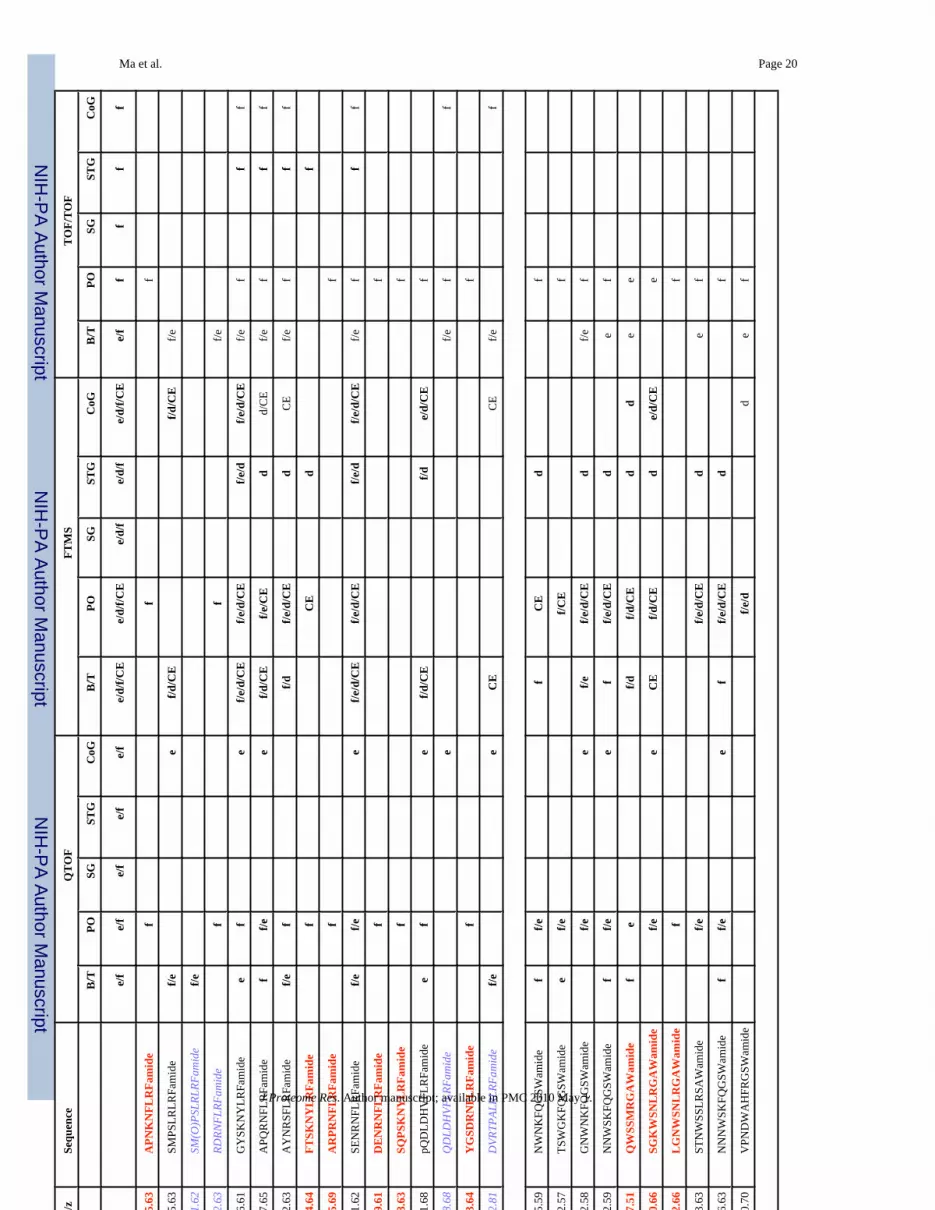
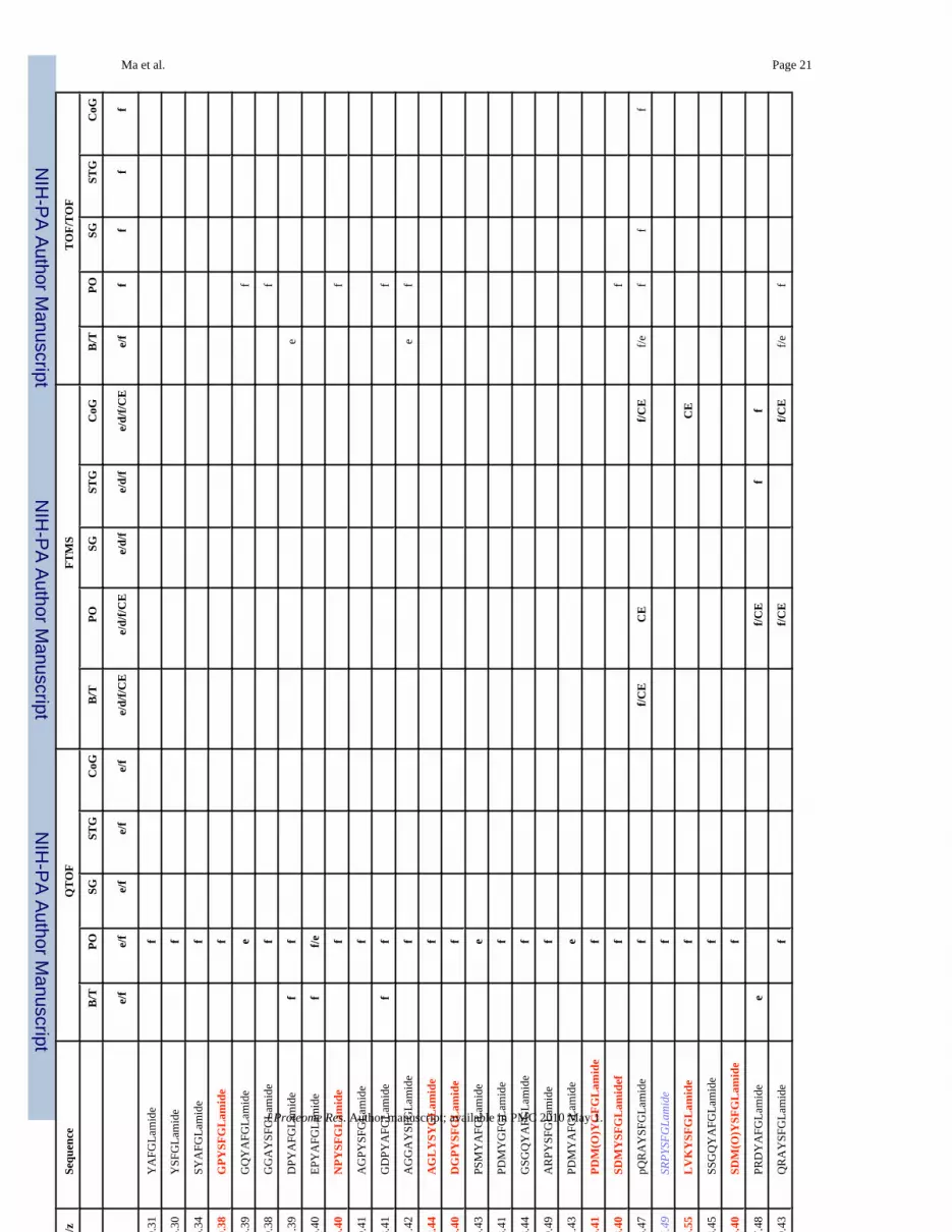
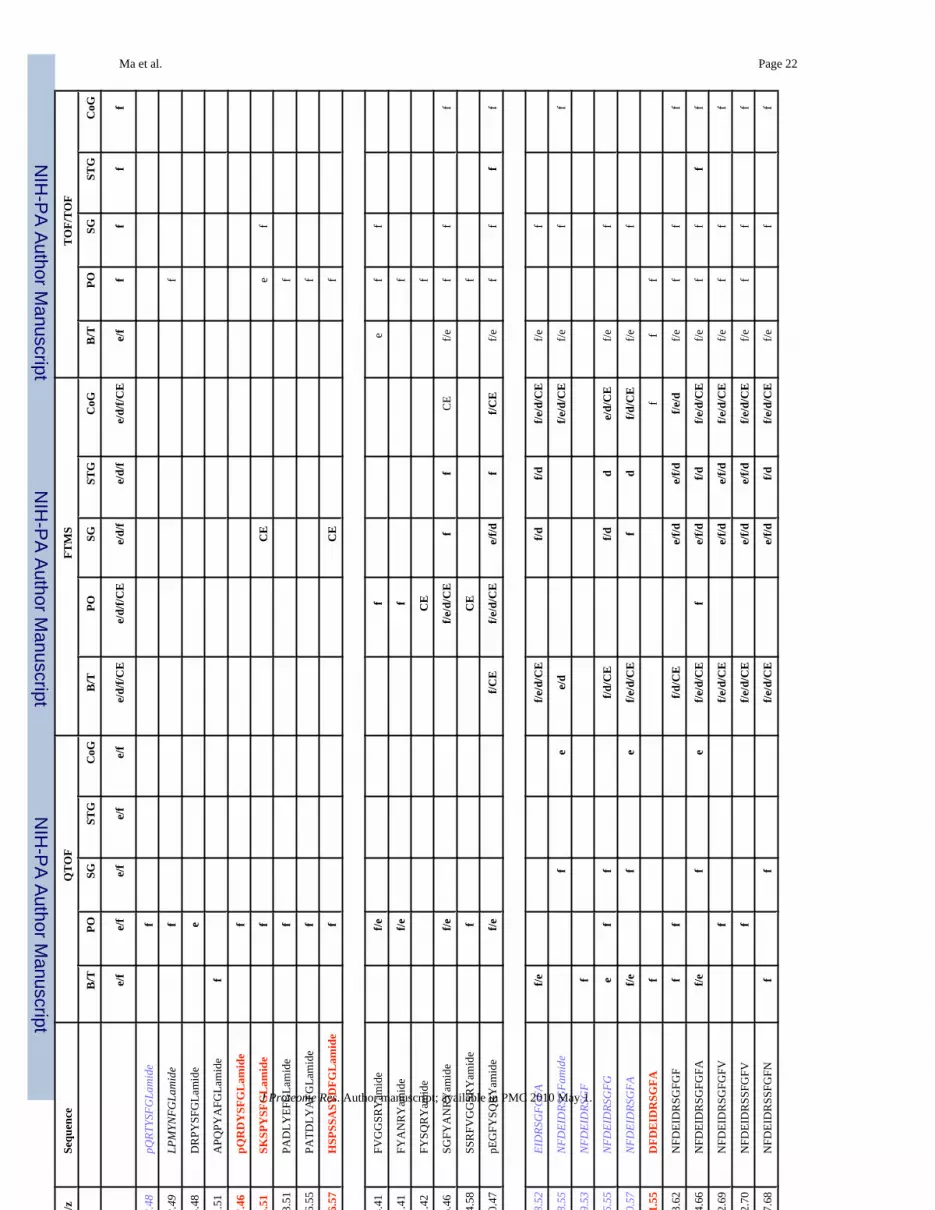
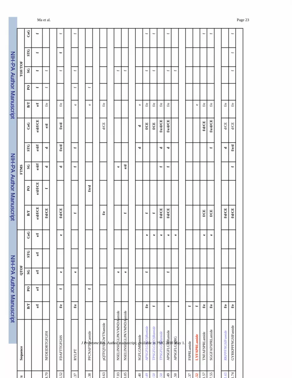
The combined mass spectrometric approach involving nanoLC-ESI-Q-TOF, MALDI-FTMSand MALDI-TOF/TOF is used for comprehensive characterization of the neuropeptidecomplements expressed in C. borealis nervous system. With high sensitivity of MALDI-TOF/TOF, high resolution and high mass measurement accuracy of MALDI-FTMS and the denovo sequencing capability of ESI-Q-TOF, 142 neuropeptides were identified from this speciesincluding 85 previously known C. borealis peptides, 24 peptides identified in other species,but new to C. borealis, and 35 novel peptides de novo sequenced for the first time in this study.Herein we sorted the identified peptides and their tissue distribution along with techniques usedfor their identification in Table 1. As shown in Table 1, seventeen neuropeptide families wererevealed in our peptidomic analysis with some of these peptides being uniquely expressed inone type of tissue. For example, neuropeptide PFCNAFTGCamide (CCAP) was only seen inthe pericardial organ (PO), whereas others are commonly present in several tissues.
Ma et al. Page 5
J Proteome Res. Author manuscript; available in PMC 2010 May 1.
NIH
-PA Author Manuscript
NIH
-PA Author Manuscript
NIH
-PA Author Manuscript
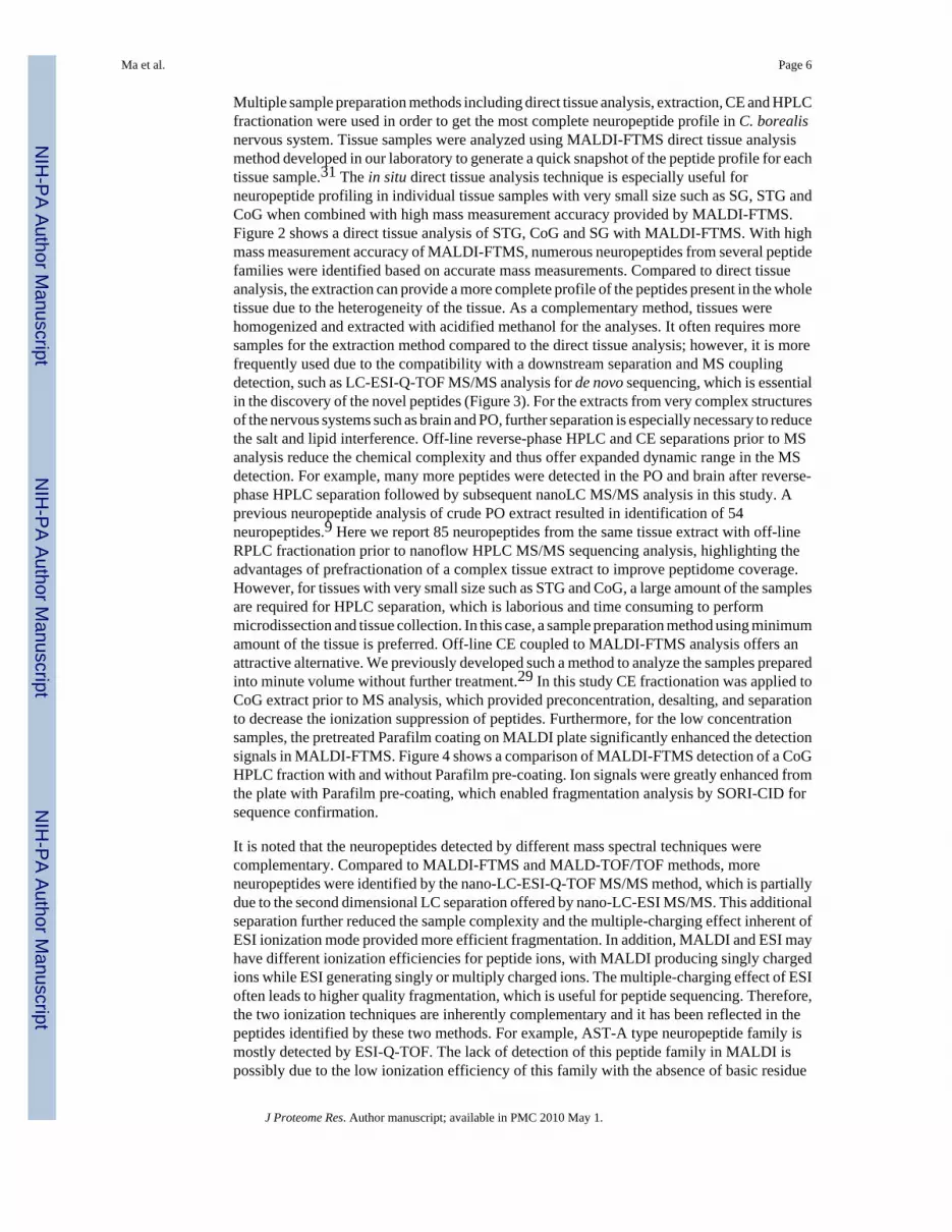
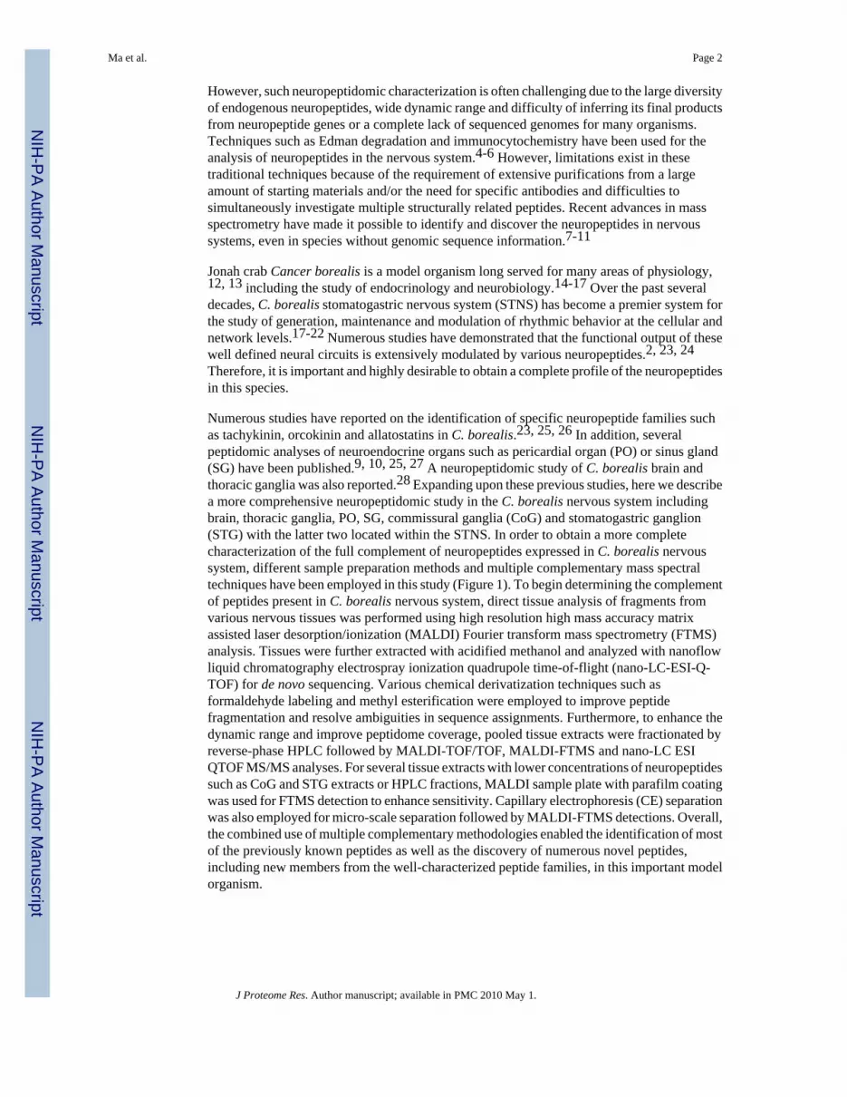
Multiple sample preparation methods including direct tissue analysis, extraction, CE and HPLCfractionation were used in order to get the most complete neuropeptide profile in C. borealisnervous system. Tissue samples were analyzed using MALDI-FTMS direct tissue analysismethod developed in our laboratory to generate a quick snapshot of the peptide profile for eachtissue sample.31 The in situ direct tissue analysis technique is especially useful forneuropeptide profiling in individual tissue samples with very small size such as SG, STG andCoG when combined with high mass measurement accuracy provided by MALDI-FTMS.Figure 2 shows a direct tissue analysis of STG, CoG and SG with MALDI-FTMS. With highmass measurement accuracy of MALDI-FTMS, numerous neuropeptides from several peptidefamilies were identified based on accurate mass measurements. Compared to direct tissueanalysis, the extraction can provide a more complete profile of the peptides present in the wholetissue due to the heterogeneity of the tissue. As a complementary method, tissues werehomogenized and extracted with acidified methanol for the analyses. It often requires moresamples for the extraction method compared to the direct tissue analysis; however, it is morefrequently used due to the compatibility with a downstream separation and MS couplingdetection, such as LC-ESI-Q-TOF MS/MS analysis for de novo sequencing, which is essentialin the discovery of the novel peptides (Figure 3). For the extracts from very complex structuresof the nervous systems such as brain and PO, further separation is especially necessary to reducethe salt and lipid interference. Off-line reverse-phase HPLC and CE separations prior to MSanalysis reduce the chemical complexity and thus offer expanded dynamic range in the MSdetection. For example, many more peptides were detected in the PO and brain after reverse-phase HPLC separation followed by subsequent nanoLC MS/MS analysis in this study. Aprevious neuropeptide analysis of crude PO extract resulted in identification of 54neuropeptides.9 Here we report 85 neuropeptides from the same tissue extract with off-lineRPLC fractionation prior to nanoflow HPLC MS/MS sequencing analysis, highlighting theadvantages of prefractionation of a complex tissue extract to improve peptidome coverage.However, for tissues with very small size such as STG and CoG, a large amount of the samplesare required for HPLC separation, which is laborious and time consuming to performmicrodissection and tissue collection. In this case, a sample preparation method using minimumamount of the tissue is preferred. Off-line CE coupled to MALDI-FTMS analysis offers anattractive alternative. We previously developed such a method to analyze the samples preparedinto minute volume without further treatment.29 In this study CE fractionation was applied toCoG extract prior to MS analysis, which provided preconcentration, desalting, and separationto decrease the ionization suppression of peptides. Furthermore, for the low concentrationsamples, the pretreated Parafilm coating on MALDI plate significantly enhanced the detectionsignals in MALDI-FTMS. Figure 4 shows a comparison of MALDI-FTMS detection of a CoGHPLC fraction with and without Parafilm pre-coating. Ion signals were greatly enhanced fromthe plate with Parafilm pre-coating, which enabled fragmentation analysis by SORI-CID forsequence confirmation.
It is noted that the neuropeptides detected by different mass spectral techniques werecomplementary. Compared to MALDI-FTMS and MALD-TOF/TOF methods, moreneuropeptides were identified by the nano-LC-ESI-Q-TOF MS/MS method, which is partiallydue to the second dimensional LC separation offered by nano-LC-ESI MS/MS. This additionalseparation further reduced the sample complexity and the multiple-charging effect inherent ofESI ionization mode provided more efficient fragmentation. In addition, MALDI and ESI mayhave different ionization efficiencies for peptide ions, with MALDI producing singly chargedions while ESI generating singly or multiply charged ions. The multiple-charging effect of ESIoften leads to higher quality fragmentation, which is useful for peptide sequencing. Therefore,the two ionization techniques are inherently complementary and it has been reflected in thepeptides identified by these two methods. For example, AST-A type neuropeptide family ismostly detected by ESI-Q-TOF. The lack of detection of this peptide family in MALDI ispossibly due to the low ionization efficiency of this family with the absence of basic residue
Ma et al. Page 6
J Proteome Res. Author manuscript; available in PMC 2010 May 1.
NIH
-PA Author Manuscript
NIH
-PA Author Manuscript
NIH
-PA Author Manuscript
in their sequence, leading to potential ion suppression in the complex extracts and thus cannotbe detected. Because nanoLC interfaced with ESI-Q-TOF offers second dimensional LCseparation, this peptide family is further separated from other ions that enabled detection. Onthe other hand, MALDI-FTMS and MALDI TOF/TOF analyses enabled the identification ofseveral neuropeptides that were otherwise missed detection using ESI-Q-TOF MS/MSanalysis. For example, the corazonin was detected in PO and CoG by MALDI-MS but notdetected by ESI-Q-TOF MS approach. This observation might be due to the sample lossespecially for low-abundance neuropeptides during LC separation coupled to ESI-Q-TOF MSanalysis.
In the previous peptidomic study of C. borealis brain and thoracic ganglia by Huybrechts etal.,28 28 peptides from 4 families were identified, among which most of the FaRPs andorcokinins were identified in this study. However, for the AST-A type peptides, none of thepeptides overlap with our study. In the previous study, all of the A-type AST peptides weredetected by MALDI-TOF and the identifications were made by mass matching to those fromCarcinus maenas and Penaeus monodon. In our study, most of the AST-A type peptides wereunambiguously de novo sequenced by ESI-Q-TOF tandem MS. It is noted that most of theAST-A type peptides detected were singly charged in ESI-Q-TOF MS analysis. However, wenormally exclude singly charged ions for the MS/MS fragmentation in the Q-TOF runs. Inorder to improve the detection of this particular peptide family, gas phase fractionationapproach was used. Basically, one of the run was set to selectively fragment the singly chargedprecursors while the other set to fragment the doubly, triply and quadruply charged ions. Inthis way, it is not only possible to detect the singly charged ions but also simplify the spectrumto improve the detection. Overall, in our study, we identified 68 peptides from 11 differentpeptide families in C. borealis brain and thoracic ganglia, doubling the peptides identified inthe C. borealis CNS.
In a previous peptidomic study of C. borealis POs, 54 peptides were identified by analyzingthe crude extract of POs with ESI-Q-TOF.9 Here, in combination of multiple samplepreparation and multi-faceted mass spectrometric approach, the list of peptides identified inPOs has been greatly expanded with 96 peptides being identified, including 28 novel peptides.Most of the novel peptides were detected in HPLC fractions, highlighting the advantages ofprefractionation of a complex tissue extract to improve peptidome coverage.
It is also noted that only one CHH isoform was identified in this study. CHH is a big familyincludes CHHs, moult-inhibiting hormones (MIHs), gonad inhibiting hormones (GIHs),vitellogenesis-inhibiting hormones (VIHs) and mandibular organ-inhibiting hormones(MOIHs). Furthermore, CHH may exist in several isoforms in one or more neurosecretoryorgans such as the sinus glands and the pericardial organs. However, due to the big size andmultiple posttranslational modifications such as disulfide-bond linkages and N-terminalpyroglutamylation, it is extremely challenge to de novo sequence the CHH family. The differentdynamic range and abundances of these peptides in various tissues could further complicatedetection. Our group recently developed a novel hybrid strategy that combines “top-down”and “bottom-up” approaches for large neuropeptide de novo sequencing.32 In the future study,we will combine the separation techniques developed in this study with the hybrid de novosequencing strategy, to search and identify additional CHH isoforms and other peptides in theCHH superfamily.
Derivatization Reaction Facilitated De Novo SequencingChallenges exist in the de novo sequencing due to the possible incompleteness of MS/MSfragmentation and the ambiguity occurring in a complex MS/MS spectrum. Therefore, a varietyof derivatization reactions have been developed to overcome such difficulties and ambiguitiesin mass spectrometric sequencing. Derivatization techniques label the N- or C-terminus of a
Ma et al. Page 7
J Proteome Res. Author manuscript; available in PMC 2010 May 1.
NIH
-PA Author Manuscript
NIH
-PA Author Manuscript
NIH
-PA Author Manuscript
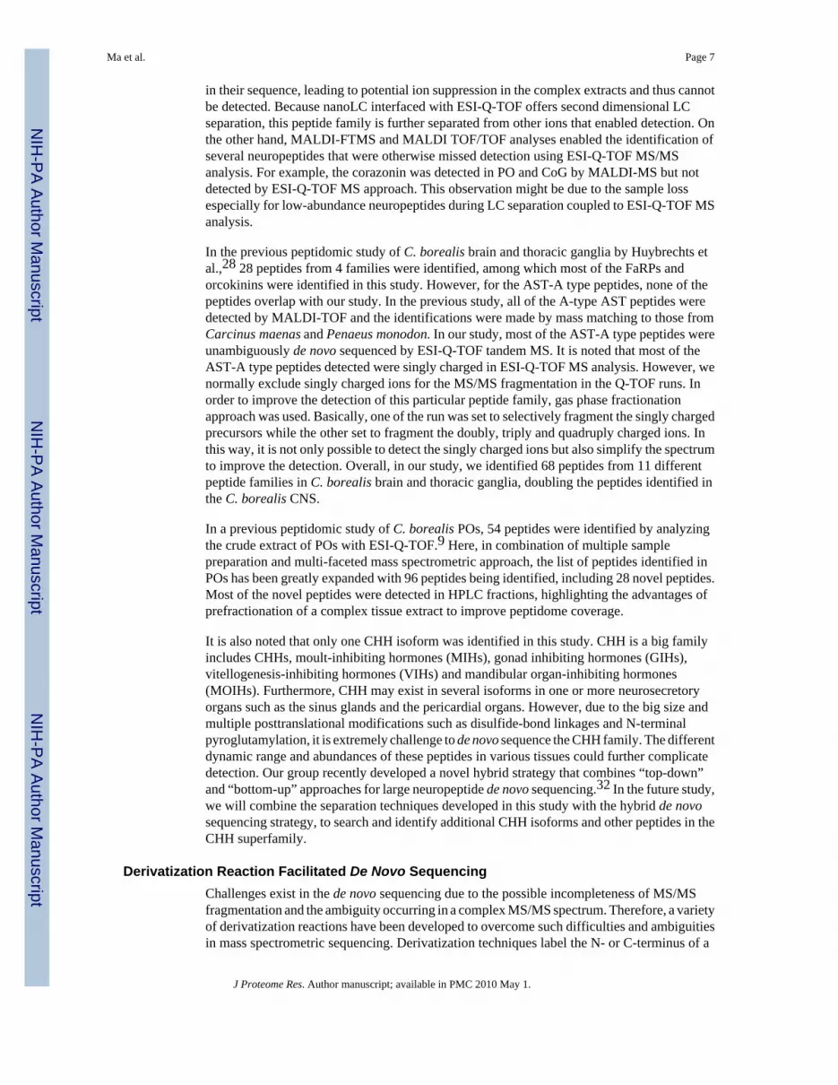
peptide and thus increase the information content of a peptide fragmentation spectrum. In thisstudy, various derivatization reactions including methyl esterification, reductive methylation,and acetylation were utilized to facilitate the de novo sequencing and resolve sequenceambiguities. Methyl esterification converts carboxylic acids on the side chains of aspartic andglutamic acids as well as the carboxyl terminus to methyl esters with a 14-Da mass increment.It also reacts with the amide group of asparagine, glutamine and amidated C-terminus with a15-Da mass increment.33 Here, the methyl esterification was utilized to confirm the numberof free carboxylic acid and the amide group in a peptide. Reductive methylation and acetylationare designed to label the N-terminus and the ε-amino groups of lysine residue. These labelingmethods not only allow the differentiation between b- and y-type fragment ions but also canbe used to confirm the number of lysine residues in a peptide. We also used these derivatizationmethods to differentiate isobaric amino acid residues K and Q in this study. Furthermore, thereductive methylation is reported to be useful in the de novo sequencing of singly chargedneuropeptides such as A-type AST by enhancing the a1 ion and simplifying the MS/MSfragmentation pattern.9 Figure 5 shows an example of using reductive methylation to facilitatede novo sequencing of an AST-A type peptide AGLYSYGLamide. As shown in Figure 5(A),MS/MS spectrum of native peptide is very complex due to extensive internal fragmentations.Furthermore, it is difficult to resolve the ambiguity of AG/GA/Q/K due to their similarmolecular mass at the N-terminus. In contrast, after the reductive methylation, a and b ionseries are enhanced while internal fragmentations are suppressed, which yielded a much cleanerMS/MS spectrum (Figure 5(B)). In addition, enhanced a1 ion helps resolve the ambiguities ofthe N-terminal residue, suggesting that the N-terminal sequence is AG. Figure 5(C) showsanother example of reductive methylation facilitated de novo sequencing. Upon the reaction,no mass shift is observed for the peptide m/z 967.511+, which indicates the pyroglutamylationat the N-terminus. So the peptide sequence is unambiguously resolved as pQRDYSFGLamide.
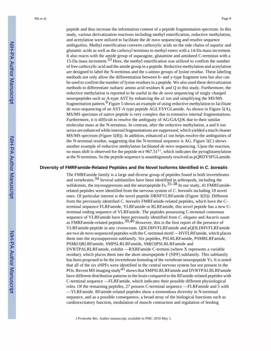
Diversity of FMRFamide-Related Peptides and the Novel Isoforms Identified in C. borealisThe FMRFamide family is a large and diverse group of peptides found in both invertebratesand vertebrates.34 Several subfamilies have been identified in arthropods, including thesulfakinins, the myosuppressins and the neuropeptide Fs.35-38 In our study, 41 FMRFamide-related peptides were identified from the nervous system of C. borealis including 18 novelones. Of particular interest is the novel peptide DRNFVLRFamide (Figure 3(D)). Differentfrom the previously identified C. borealis FMRFamide-related peptides, which have the C-terminal sequence FLRFamide, YLRFamide or RLRFamide, this novel peptide has a new C-terminal ending sequence of VLRFamide. The peptides possessing C-terminal consensussequence of VLRFamide have been previously identified from C. elegans and Ascaris suumas FMRFamide-related peptides.39,40 However, this is the first report of the presence ofVLRFamide-peptide in any crustaceans. QDLDHVFLRFamide and pQDLDHVFLRFamideare two de novo sequenced peptides with the C-terminal motif —HVFLRFamide, which placesthem into the myosuppressin subfamily. Six peptides, PSLRLRFamide, PSMRLRFamide,PSM(O)RLRFamide, SMPSLRLRFamide, SM(O)PSLRLRFamide andDVRTPALRLRFamide, exhibit —RXRFamide C-termini (where X represents a variableresidue), which places them into the short neuropeptide F (NPF) subfamily. This subfamilyhas been proposed to be the invertebrate homolog of the vertebrate neuropeptide Ys. It is notedthat all of the six sNPFs were identified in the central nervous system but not present in thePOs. Recent MS imaging study41 shows that SMPSLRLRFamide and DVRTPALRLRFamidehave different distribution patterns in the brain compared to the RFamide-related peptides withC-terminal sequence —FLRFamide, which indicates their possible different physiologicalroles. Of the remaining peptides, 27 possess C-terminal sequence —FLRFamide and 5 with—YLRFamide. RFamide-related peptides show a tremendous diversity in N-terminalsequence, and as a possible consequence, a broad array of the biological functions such ascardioexcitatory function, modulation of muscle contraction and regulation of feeding
Ma et al. Page 8
J Proteome Res. Author manuscript; available in PMC 2010 May 1.
NIH
-PA Author Manuscript
NIH
-PA Author Manuscript
NIH
-PA Author Manuscript
behavior.42-44 Several RFamide physiological studies even demonstrated that differentisoforms might have opposite biological effects.45 A recent study on several RFamide-relatedpeptides in C. borealis showed differential degradation of several isoforms in the presence ofextracellular peptidases.46 Overall, the identification of this large array of closely relatedFaRPs in conjunction with the well-characterized STNS provides an excellent opportunity tofurther investigate the functional consequence of peptide diversity.
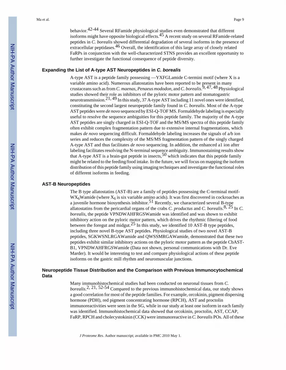
Expanding the List of A-type AST Neuropeptides in C. borealisA-type AST is a peptide family possessing —YXFGLamide C-termini motif (where X is avariable amino acid). Numerous allatostatins have been reported to be present in manycrustaceans such as from C. maenas, Penaeus mododon, and C. borealis.9, 47, 48 Physiologicalstudies showed their role as inhibitors of the pyloric motor pattern and stomatogastricneurotransmission.21, 49 In this study, 37 A-type AST including 11 novel ones were identified,constituting the second largest neuropeptide family found in C. borealis. Most of the A-typeAST peptides were de novo sequenced by ESI-Q-TOF MS. Formaldehyde labeling is especiallyuseful to resolve the sequence ambiguities for this peptide family. The majority of the A-typeAST peptides are singly charged in ESI-Q-TOF and the MS/MS spectra of this peptide familyoften exhibit complex fragmentation pattern due to extensive internal fragmentations, whichmakes de novo sequencing difficult. Formaldehyde labeling increases the signals of a/b ionseries and reduces the complexity of the MS/MS fragmentation pattern of the singly chargedA-type AST and thus facilitates de novo sequencing. In addition, the enhanced a1 ion afterlabeling facilitates resolving the N-terminal sequence ambiguity. Immunostaining results showthat A-type AST is a brain-gut peptide in insects,50 which indicates that this peptide familymight be related to the feeding/food intake. In the future, we will focus on mapping the isoformdistribution of this peptide family using imaging techniques and investigate the functional rolesof different isoforms in feeding.
AST-B NeuropeptidesThe B type allatostatins (AST-B) are a family of peptides possessing the C-terminal motif-WX6Wamide (where X6 is six variable amino acids). It was first discovered in cockroaches asa juvenile hormone biosynthesis inhibitor.51 Recently, we characterized several B-typeallatostatins from the pericardial organs of the crabs C. productus and C. borealis.8, 25 In C.borealis, the peptide VPNDWAHFRGSWamide was identified and was shown to exhibitinhibitory action on the pyloric motor pattern, which drives the rhythmic filtering of foodbetween the foregut and midgut.25 In this study, we identified 10 AST-B type peptides,including three novel B-type AST peptides. Physiological studies of two novel AST-Bpeptides, SGKWSNLRGAWamide and QWSSMRGAWamide, demonstrated that these twopeptides exhibit similar inhibitory actions on the pyloric motor pattern as the peptide CbAST-B1, VPNDWAHFRGSWamide (Data not shown, personal communications with Dr. EveMarder). It would be interesting to test and compare physiological actions of these peptideisoforms on the gastric mill rhythm and neuromuscular junctions.
Neuropeptide Tissue Distribution and the Comparison with Previous ImmunocytochemicalData
Many immunohistochemical studies had been conducted on neuronal tissues from C.borealis.2, 21, 52-54 Compared to the previous immunohistochemical data, our study showsa good correlation for most of the peptide families. For example, orcokinin, pigment dispersinghormone (PDH), red pigment concentrating hormone (RPCH), AST and proctolinimmunoreactivities were seen in the SG, while in our study at least one isoform in each familywas identified. Immunohistochemical data showed that orcokinin, proctolin, AST, CCAP,FaRP, RPCH and cholecystokinin (CCK) were immunoreactive in C. borealis POs. All of these
Ma et al. Page 9
J Proteome Res. Author manuscript; available in PMC 2010 May 1.
NIH
-PA Author Manuscript
NIH
-PA Author Manuscript
NIH
-PA Author Manuscript
peptide families have been detected in this study except RPCH and CCK. This discrepancy islikely due to the poor ionization efficiency of the RPCH and CCK. For STG, CCK, corazoninand RPCH immunoreactivities were seen, however, these peptides were not detected in ourstudy. This difference in observation may be due to the very low concentration or poorionization efficiency of these peptide families. In addition, MALDI was the only ionizationtechnique used in the study of the STG, which could limit the peptides being detected. Table1 shows the identified peptides via MS and their tissue distribution. Some peptides aredistributed in all of tissues we investigated such as some orcokinins, orcomyotropinFDAFTTGFGHS, two RYamides: SGFYANRYamide and pEGFYSQRYamide, and thepeptide HIGSLYRamide. However, some of the peptides are unique to a specific tissue. Forexample, PDHs are only present in the SG and brain while CCAP is only present in the PO,which is also consistent with the previously published immunocytochemical data.52 Someisoforms of orcokinins, FaRPs, AST and proctolin identified in the PO or SG were also foundin the STG, indicating their roles as circulating hormone as well as neuromodulators. Overall,the MS-based neuropeptide identification data agree with immunocytochemical data.However, MS-based approach enabled simultaneous mapping and characterizing multipleisoforms which is not available for immunocytochemical approaches.
ConclusionIn this study we combined multiple sample preparation methods and multifaceted mass spectraltechniques to comprehensively characterize the neuropeptides present in the nervous systemof the Jonah crab C. borealis. In total, 142 peptides were identified with 59 that are new to thisspecies. These data greatly increase the number of known peptides present in this species andprovide a strong foundation for future studies on the physiological roles played by thesesignaling molecules in a well defined neural network.
AcknowledgementsThe authors thank Dr. Joshua J. Schmidt and Dr. Kimberly K. Kutz Naber from the Li laboratory for providing someof the C. borealis PO and brain HPLC fractions. Xin Wei is thanked for helpful discussions. We are also grateful tothe University of Wisconsin (UW) School of Pharmacy Analytical Instrumentation Center for access to the MALDI-FTMS instrument and UW Biotechnology Center for access to the MALDI-TOF/TOF instrument. This work wassupported in part by the School of Pharmacy and the Wisconsin Alumni Research Foundation at the University ofWisconsin-Madison, a National Science Foundation CAREER Award (CHE 0449991), and the National Institutes ofHealth through grant 1R01DK071801 (to LL). L.L. acknowledges an Alfred P. Sloan Research Fellowship.
References1. Schwartz MW, Woods SC, Porte D, Seeley RJ, Baskin DG. Central nervous system control of food
intake. Nature 2000;404(6778):661–671. [PubMed: 10766253]2. Skiebe P. Neuropeptides are ubiquitous chemical mediators: Using the stomatogastric nervous system
as a model system. J Exp Biol 2001;204(12):2035–2048. [PubMed: 11441046]3. Hokfelt T, Broberger C, Xu ZQD, Sergeyev V, Ubink R, Diez M. Neuropeptides--an overview.
Neuropharmacology 2000;39(8):1337–1356. [PubMed: 10818251]4. Marshak DW, Reeve JR, Shively JE, Hawke D, Takami MS, Yamada T. Structure of somatostatin
isolated from bovine retina. J Neurochem 1983;41(3):601–606. [PubMed: 6135755]5. Wainwright G, Webster SG, Wilkinson MC, Chung JS, Rees HH. Structure and significance of
mandibular organ-inhibiting hormone in the crab, Cancer pagurus. Involvement in multihormonalregulation of growth and reproduction. J. Biol. Chem 1996;271:12749–12754. [PubMed: 8662685]
6. Panchan N, Bendena WG, Bowser P, Lungchukiet P, Tobe SS, Sithigorngul W, Chaivisuthangkura P,Rangsiruji A, Petsom A, Pewnim T, Sithigorngul P. Immunolocalization of allatostatin-likeneuropeptides and their putative receptor in eyestalks of the tiger prawn, Penaeus monodon. Peptides2003;24(10):1563–1570. [PubMed: 14706535]
Ma et al. Page 10
J Proteome Res. Author manuscript; available in PMC 2010 May 1.
NIH
-PA Author Manuscript
NIH
-PA Author Manuscript
NIH
-PA Author Manuscript
7. Hill SR, Orchard I. Isolation and sequencing of two FMRFamide-related peptides from the gut ofLocusta migratoria L. Peptides 2007;28(8):1490–1497. [PubMed: 17707763]
8. Fu Q, Kutz KK, Schmidt JJ, Hsu Y-WA, Messinger DI, Cain SD, Lglesia HODL, Christie AE, Li L.Hormone complement of the Cancer productus sinus gland and pericardial organ: An anatomical andmass spectrometric investigation. J. Comp. Neurol 2005;493:607–626. [PubMed: 16304631]
9. Fu Q, Li L. De novo sequencing of neuropeptides using reductive isotopic methylation and investigationof ESI QTOF MS/MS fragmentation pattern of neuropeptides with N-terminal dimethylation. Anal.Chem 2005;77:7783–7795. [PubMed: 16316189]
10. Li L, Kelley WP, Billimoria CP, Christie AE, Pulver SR, Sweedler JV, Marder E. Mass spectrometricinvestigation of the neuropeptide complement and release in the pericardial organs of the crab, Cancerborealis. J. Neurochem 2003;87:642–656. [PubMed: 14535947]
11. Li L, Sweedler JV. Peptides in the brain mass spectrometry-based measurement approaches andchallenges. Annu Rev Anal Chem 2008;1:451–483.
12. Marder E. Motor pattern generation. Current Opin Neurobiol 2000;10(6):691–698.13. Nusbaum MP, Beenhakker MP. A small-systems approach to motor pattern generation. Nature
2002;417(6886):343–350. [PubMed: 12015615]14. Terwilliger RC, Terwilliger NB, Clay GA, Belamarich FA. The subcellular localization of a
cardioexcitatory peptide in the pericardial organs of the crab, Cancer borealis. General andComparative Endocrinology 1970;15(1):70–79. [PubMed: 5457019]
15. Marder E, Hooper SL, Siwicki KK. Modulatory action and distribution of the neuropeptide proctolinin the crustacean stomatogastric nervous system. J Comp Neurol 1986;243(4):454–467. [PubMed:2869069]
16. Cruz-Bermudez ND, Marder E. Multiple modulators act on the cardiac ganglion of the crab, Cancerborealis. J Exp Biol 2007;210(16):2873–2884. [PubMed: 17690236]
17. Weimann JM, Meyrand P, Marder E. Neurons that form multiple pattern generators: identificationand multiple activity patterns of gastric/pyloric neurons in the crab stomatogastric system. JNeurophysiol 1991;65(1):111–122. [PubMed: 1999725]
18. Buchholtz F, Golowasch J, Epstein IR, Marder E. Mathematical model of an identified stomatogastricganglion neuron. J Neurophysiol 1992;67(2):332–340. [PubMed: 1373763]
19. Coleman MJ, Nusbaum MP, Cournil I, Claiborne BJ. Distribution of modulatory inputs to thestomatogastric ganglion of the crab, Cancer borealis. J Comp Neurol 1992;325(4):581–594.[PubMed: 1361498]
20. Weimann JM, Marder E, Evans B, Calabrese RL. The effects of SDRNFLRFamide andTNRNFLRFamide on the motor patterns of the stomatogastric ganglion of the crab Cancerborealis. J Exp Biol 1993;181(1):1–26. [PubMed: 8409825]
21. Skiebe P, Schneider H. Allatostatin peptides in the crab stomatogastric nervous system: inhibition ofthe pyloric motor pattern and distribution of allatostatin-like immunoreactivity. J Exp Biol 1994;194(1):195–208. [PubMed: 7964402]
22. Saideman SR, Ma M, Kutz-Naber KK, Cook A, Torfs P, Schoofs L, Li L, Nusbaum MP. Modulationof rhythmic motor activity by pyrokinin peptides. J Neurophysiol 2007;97(1):579–595. [PubMed:17065249]
23. Christie AE, Lundquist CT, Nassel DR, Nusbaum MP. Two novel tachykinin-related peptides fromthe nervous system of the crab Cancer borealis. J Exp Biol 1997;200(17):2279–2294. [PubMed:9316266]
24. Nusbaum MP. Regulating peptidergic modulation of rhythmically active neural circuits. Brain BehavEvol 2002;60(6):378–387. [PubMed: 12563170]
25. Fu Q, Tang LS, Marder E, Li L. Mass spectrometric characterization and physiological actions ofVPNDWAHFRGSWamide, a novel B type allatostatin in the crab, Cancer borealis. J. Neurochem2007;101:1099–1107. [PubMed: 17394556]
26. Li L, Pulver SR, Kelley WP, Thirumalai V, Sweedler JV, Marder E. Orcokinin peptides in developingand adult crustacean stomatogastric nervous systems and pericardial organs. J Comp Neurol 2002;444(3):227–244. [PubMed: 11840477]
27. Stemmler EA, Peguero B, Bruns EA, Dickinson PS, Christie AE. Identification, physiological actions,and distribution of TPSGFLGMRamide: a novel tachykinin-related peptide from the midgut and
Ma et al. Page 11
J Proteome Res. Author manuscript; available in PMC 2010 May 1.
NIH
-PA Author Manuscript
NIH
-PA Author Manuscript
NIH
-PA Author Manuscript
stomatogastric nervous system of Cancer crabs. J Neurochem 2007;101(5):1351–1366. [PubMed:17437551]
28. Huybrechts J, Nusbaum MP, Bosch LV, Baggerman G, Loof AD, Schoofs L. Neuropeptidomicanalysis of the brain and thoracic ganglion from the Jonah crab, Cancer borealis. Biochem BiophysRes Comm 2003;308(3):535–544. [PubMed: 12914784]
29. Wang J, Ma M, Chen R, Li L. Enhanced neuropeptide profiling via capillary electrophoresis off-linecoupled with MALDI FTMS. Anal. Chem 2008;80:6168–6177. [PubMed: 18642879]
30. Wang J, Chen R, Ma M, Li L. MALDI MS sample preparation by using paraffin wax film: systematicstudy and application for peptide analysis. Anal. Chem 2008;80(2):491–500. [PubMed: 18189446]
31. Kutz KK, Schmidt JJ, Li L. In situ tissue analysis of neuropeptides by MALDI FTMS in-cellaccumulation. Anal. Chem 2004;76:5630–5640. [PubMed: 15456280]
32. Ma M, Chen R, Ge Y, He H, Marshall AG, Li L. Combining bottom-up and top-down massspectrometric strategies for de novo sequencing of the crustacean hyperglycemic hormone fromCancer borealis. Anal Chem 2009;81(1):240–247. [PubMed: 19046072]
33. Ma M, Kutz-Naber KK, Li L. Methyl Esterification assisted MALDI FTMS characterization of theorcokinin neuropeptide family. Anal Chem 2007;79:673–681. [PubMed: 17222036]
34. Zajac J-M, Mollereau C. Introduction. Peptides 2006;27(5):941–942. [PubMed: 16621148]35. Ma M, Chen R, Sousa GL, Bors EK, Kwiatkowski MA, Goiney CC, Goy MF, Christie AE, Li L.
Mass spectral characterization of peptide transmitters/hormones in the nervous system andneuroendocrine organs of the American lobster Homarus americanus. Gen Comp Endocrinol2008;156(2):395–409. [PubMed: 18304551]
36. Dickinson PS, Stevens JS, Rus S, Brennan HR, Goiney CC, Smith CM, Li L, Towle DW, ChristieAE. Identification and cardiotropic actions of sulfakinin peptides in the American lobster Homarusamericanus. J Exp Biol 2007;210(13):2278–2289. [PubMed: 17575033]
37. Nichols R, McCormick J, Lim I. Dromyosuppressin and drosulfakinin, two structurally relatedDrosophila neuropeptides, are uniquely expressed in the adult central nervous system. Ann N Y AcadSci 1997;814:315–318. [PubMed: 9160985]
38. Garczynski SF, Brown MR, Crim JW. Structural studies of Drosophila short neuropeptide F:Occurrence and receptor binding activity. Peptides 2006;27(3):575–582. [PubMed: 16330127]
39. Husson SJ, Clynen E, Baggerman G, De Loof A, Schoofs L. Discovering neuropeptides inCaenorhabditis elegans by two dimensional liquid chromatography and mass spectrometry. BiochemBiophys Res Comm 2005;335(1):76–86. [PubMed: 16061202]
40. Cowden C, Stretton AOW. Eight novel FMRFamide-like neuropeptides isolated from the nematodeAscaris suum. Peptides 1995;16(3):491–500. [PubMed: 7651904]
41. DeKeyser SS, Kutz-Naber KK, Schmidt JJ, Barrett-Wilt GA, Li L. Imaging mass spectrometry ofneuropeptides in decapod crustacean neuronal tissues. J. Proteome Res 2007;6:1782–1791. [PubMed:17381149]
42. Price DA, Greenberg MJ. Structure of a molluscan cardioexcitatory neuropeptide. Science1977;197:670–671. [PubMed: 877582]
43. Moulis A, Huddart H. RFamide neuropeptide actions on molluscan proboscis smooth muscle:interactions with primary neurotransmitters. J Comp Physiol B-Biochem Systemic and EnvironPhysiol 2004;174(5):363–370.
44. Bechtold DA, Luckman SM. The role of RFamide peptides in feeding. J Endocrinol 2007;192(1):3–15. [PubMed: 17210738]
45. Yew JY, Davis R, Dikler S, Nanda J, Reinders B, Stretton AO. Peptide products of the afp-6 gene ofthe nematode Ascaris suum have different biological actions. J Comp Neurol 2007;502(5):872–882.[PubMed: 17436302]
46. Cruz-Bermudez ND, Fu Q, Kutz-Naber KK, Christie AE, Li L, Marder E. Mass spectrometriccharacterization and physiological actions of GAHKNYLRFamide, a novel FMRFamide-like peptidefrom crabs of the genus Cancer. J Neurochem 2006;97(3):784–799. [PubMed: 16515542]
47. Duve H, Johnsen AH, Maestro J-L, Scott AG, Jaros PP, Thorpe A. Isolation and identification ofmultiple neuropeptides of the allatostatin superfamily in the shore crab Carcinus maenas. Eur JBiochem 1997;250(3):727–734. [PubMed: 9461295]
Ma et al. Page 12
J Proteome Res. Author manuscript; available in PMC 2010 May 1.
NIH
-PA Author Manuscript
NIH
-PA Author Manuscript
NIH
-PA Author Manuscript
48. Duve H, Johnsen AH, Scott AG, Thorpe A. Allatostatins of the tiger prawn, Penaeus monodon(Crustacea: Penaeidea). Peptides 2002;23(6):1039–1051. [PubMed: 12126730]
49. Jorge-Rivera J, MarderY E. Allatostatin decreases stomatogastric neuromuscular transmission in thecrab Cancer borealis. J Exp Biol 1997;200(23):2937–2946. [PubMed: 9359878]
50. Abdel-Latief M, Meyering-Vos M, Hoffmann MH. Type-A allatostatins from the fall armyworm,Spodoptera frugiperda: Molecular cloning, expression and tissue-specific localization. Arch InsectBiochem Physiol 2004;56(3):120–132. [PubMed: 15211550]
51. Woodhead AP, Stay B, Seidel SL, Khan MA, Tobe SS. Primary structure of four allatostatins:neuropeptide inhibitors of juvenile hormone synthesis. Proc Natl Acad Sci USA 1989;86(15):5997–6001. [PubMed: 2762309]
52. Christie AE, Skiebe P, Marder E. Matrix of neuromodulators in neurosecretory structures of the crabCancer borealis. J Exp Biol 1995;198(12):2431–2439. [PubMed: 8576680]
53. Marder E, Calabrese RL, Nusbaum MP, Trimmer B. Distribution and partial characterization ofFMRFamide-like peptides in the stomatogastric nervous systems of the rock crab, Cancer borealis,and the spiny lobster, Panulirus interruptus. J Comp Neurol 1987;259:150–163. [PubMed: 3584554]
54. Christie AE, Baldwin D, Turrigiano G, Graubard K, Marder E. Immunocytochemical localization ofmultiple cholecystokinin-like peptides in the stomatogastric nervous system of the crab Cancerborealis. J Exp Biol 1995;198:263–271. [PubMed: 7891039]
Ma et al. Page 13
J Proteome Res. Author manuscript; available in PMC 2010 May 1.
NIH
-PA Author Manuscript
NIH
-PA Author Manuscript
NIH
-PA Author Manuscript
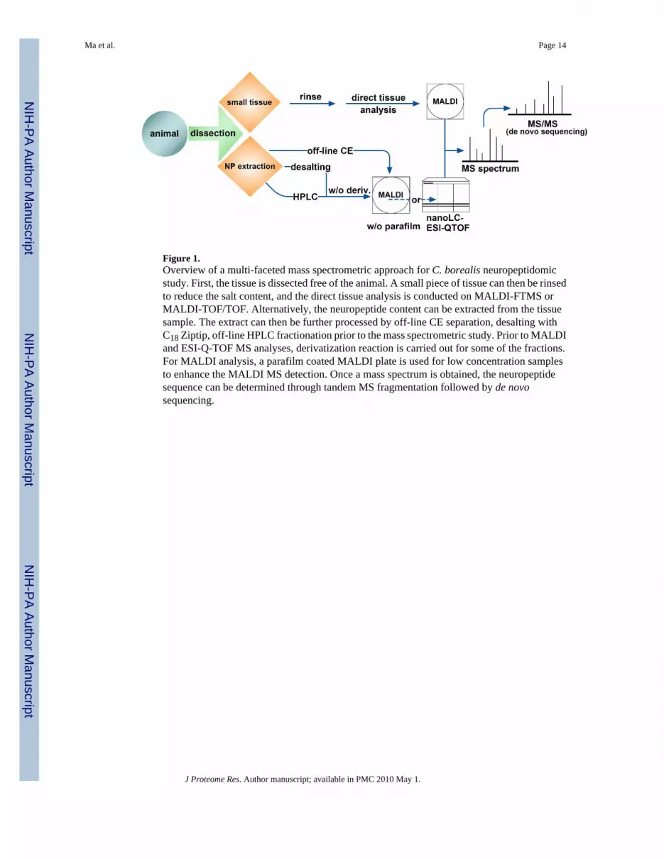
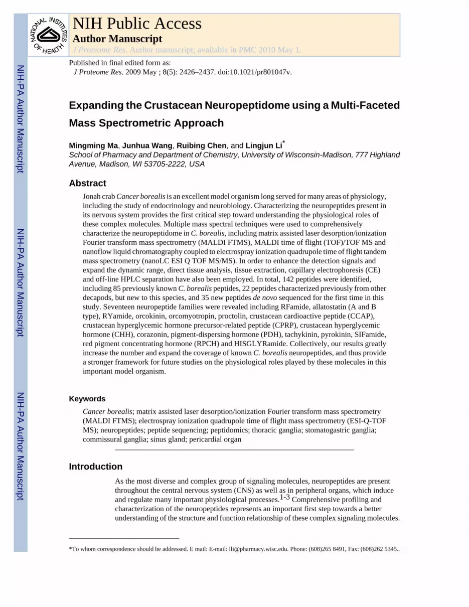
Figure 1.Overview of a multi-faceted mass spectrometric approach for C. borealis neuropeptidomicstudy. First, the tissue is dissected free of the animal. A small piece of tissue can then be rinsedto reduce the salt content, and the direct tissue analysis is conducted on MALDI-FTMS orMALDI-TOF/TOF. Alternatively, the neuropeptide content can be extracted from the tissuesample. The extract can then be further processed by off-line CE separation, desalting withC18 Ziptip, off-line HPLC fractionation prior to the mass spectrometric study. Prior to MALDIand ESI-Q-TOF MS analyses, derivatization reaction is carried out for some of the fractions.For MALDI analysis, a parafilm coated MALDI plate is used for low concentration samplesto enhance the MALDI MS detection. Once a mass spectrum is obtained, the neuropeptidesequence can be determined through tandem MS fragmentation followed by de novosequencing.
Ma et al. Page 14
J Proteome Res. Author manuscript; available in PMC 2010 May 1.
NIH
-PA Author Manuscript
NIH
-PA Author Manuscript
NIH
-PA Author Manuscript

Figure 2.Direct tissue peptide profiling of C. borealis STG (A), CoG (B), and SG (C) by MALDI-FTMS.Signals correspond to the protonated molecular ions, [M+H]+, where M is the molecular weightof each peptide. The identified peptides are marked with the symbols indicating specificfamilies to which they belong.
Ma et al. Page 15
J Proteome Res. Author manuscript; available in PMC 2010 May 1.
NIH
-PA Author Manuscript
NIH
-PA Author Manuscript
NIH
-PA Author Manuscript

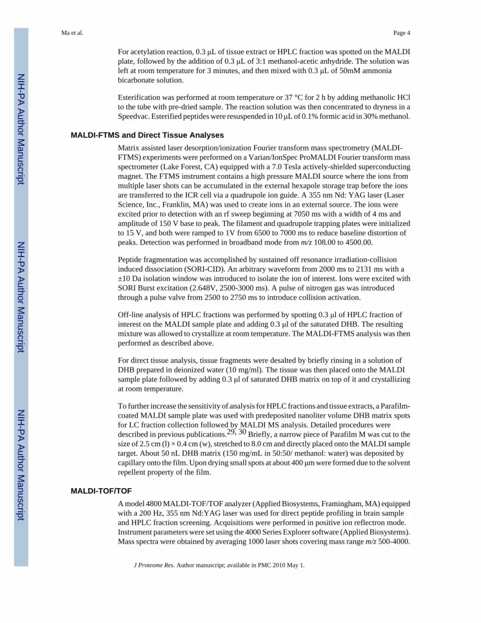
Figure 3.Collision-induced dissociation spectra of four de novo sequenced peptides. ESI-Q-TOF MS/MS sequencing of four FaRPs: DENRNFLRFamide (605.432+; A), YGSDRNFLRFamide(637.262+; B), PSMRLRFamide (453.252+; C), and RDNFVLRFamide (533.302+; D).
Ma et al. Page 16
J Proteome Res. Author manuscript; available in PMC 2010 May 1.
NIH
-PA Author Manuscript
NIH
-PA Author Manuscript
NIH
-PA Author Manuscript
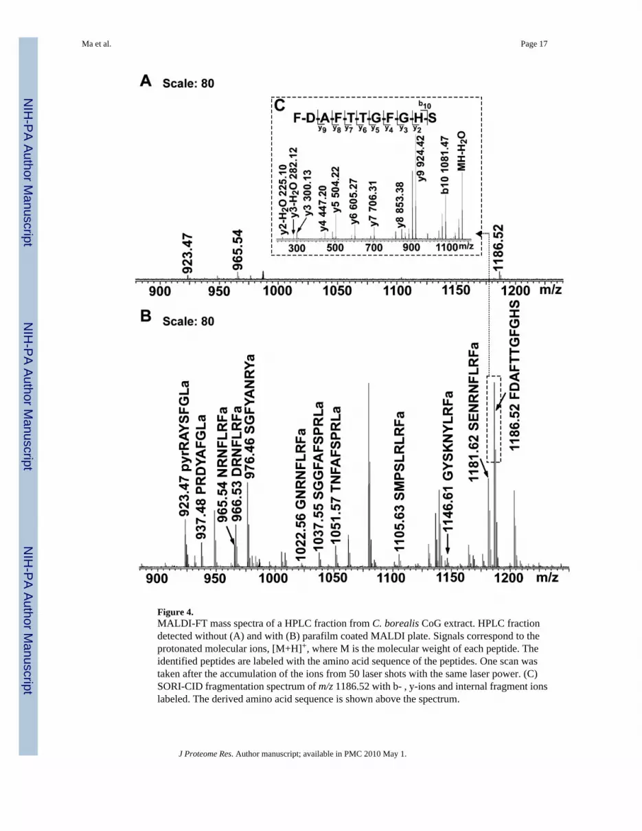
Figure 4.MALDI-FT mass spectra of a HPLC fraction from C. borealis CoG extract. HPLC fractiondetected without (A) and with (B) parafilm coated MALDI plate. Signals correspond to theprotonated molecular ions, [M+H]+, where M is the molecular weight of each peptide. Theidentified peptides are labeled with the amino acid sequence of the peptides. One scan wastaken after the accumulation of the ions from 50 laser shots with the same laser power. (C)SORI-CID fragmentation spectrum of m/z 1186.52 with b- , y-ions and internal fragment ionslabeled. The derived amino acid sequence is shown above the spectrum.
Ma et al. Page 17
J Proteome Res. Author manuscript; available in PMC 2010 May 1.
NIH
-PA Author Manuscript
NIH
-PA Author Manuscript
NIH
-PA Author Manuscript
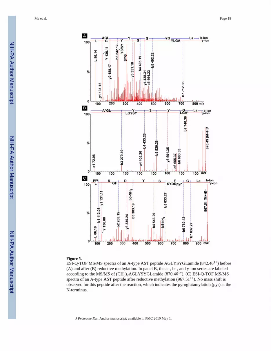
Figure 5.ESI-Q-TOF MS/MS spectra of an A-type AST peptide AGLYSYGLamide (842.461+) before(A) and after (B) reductive methylation. In panel B, the a- , b- , and y-ion series are labeledaccording to the MS/MS of (CH3)2AGLYSYGLamide (870.461+). (C) ESI-Q-TOF MS/MSspectra of an A-type AST peptide after reductive methylation (967.511+). No mass shift isobserved for this peptide after the reaction, which indicates the pyroglutamylation (pyr) at theN-terminus.
Ma et al. Page 18
J Proteome Res. Author manuscript; available in PMC 2010 May 1.
NIH
-PA Author Manuscript
NIH
-PA Author Manuscript
NIH
-PA Author Manuscript

NIH
-PA Author Manuscript
NIH
-PA Author Manuscript
NIH
-PA Author Manuscript
Ma et al. Page 19Ta
ble
1N
euro
pept
ides
det
ecte
d in
mul
tiple
neu
rona
l tis
sues
from
C. b
orea
lis b
y na
no-L
C-E
SI-Q
-TO
F, M
ALD
I-FT
MS
and
MA
LDI-
TOF/
TOF.
m/z
Sequ
ence
QT
OF
FTM
ST
OF/
TO
F
Loc
atio
nB
/TPO
SGST
GC
oGB
/TPO
SGST
GC
oGB
/TPO
SGST
GC
oG
Sam
ple
prep
e/f
e/f
e/f
e/f
e/f
e/d/
f/CE
e/d/
f/CE
e/d/
fe/
d/f
e/d/
f/CE
e/f
ff
ff
FaR
Ps
695.
40N
FLRF
amid
ef
f
735.
43G
PFL
RFa
mid
ef
851.
50R
NFL
RFa
mid
ef/e
fe/
f/CE
887.
56PS
LRLR
Fam
ide
f/e
908.
52G
RN
FLR
Fam
ide
ef/e
905.
51SM
RL
RFa
mid
ee
ef
ff
921.
51PS
M(O
)RL
RFa
mid
ef
926.
52SK
NY
LRFa
mid
ef/e
ef
938.
53N
RSF
LRFa
mid
ee
ff
962.
53pQ
RN
FLR
Fam
ide
ff/C
Ef
965.
54N
RN
FLR
Fam
ide
f/ef/e
ef/e
/df/e
/d/C
Ee/
df/e
/d/C
Ef/e
ff
f
966.
53D
RN
FLR
Fam
ide
f/ef/e
f/df/C
Ef
CE
f
993.
53N
PSN
FLR
Fam
ide
ff
994.
51N
PSD
FLR
Fam
ide
f
1007
.58
PKSN
FLR
Fam
ide
f
1019
.59
APR
NFL
RFa
mid
ed/
CE
ff
1022
.56
GN
RN
FLR
Fam
ide
f/ef/e
ef/e
/d/C
Ef/e
/d/C
Ed
d/C
Ef/e
ff
f
1023
.55
GD
R N
FLR
Fam
ide
CE
1029
.56
DH
VPF
LR
Fam
ide
f
1031
.59
AH
KN
FLR
Fam
ide
f
1045
.58
GH
RN
FLR
Fam
ide
fC
Ef
1059
.59
AH
RN
FLR
Fam
ide
ff
1065
.60
RD
NFV
LR
Fam
ide
e
1094
.58
EN
RN
FLR
Fam
ide
f
1104
.61
GA
HK
NY
LRFa
mid
ef/e
f/e/d
/CE
f/e/d
/CE
f/df/e
/d/C
Ef/e
ff
f
J Proteome Res. Author manuscript; available in PMC 2010 May 1.
NIH
-PA Author Manuscript
NIH
-PA Author Manuscript
NIH
-PA Author Manuscript
Ma et al. Page 20m
/zSe
quen
ceQ
TO
FFT
MS
TO
F/T
OF
Loc
atio
nB
/TPO
SGST
GC
oGB
/TPO
SGST
GC
oGB
/TPO
SGST
GC
oG
Sam
ple
prep
e/f
e/f
e/f
e/f
e/f
e/d/
f/CE
e/d/
f/CE
e/d/
fe/
d/f
e/d/
f/CE
e/f
ff
ff
1105
.63
APN
KN
FLR
Fam
ide
ff
f
1105
.63
SMPS
LRLR
Fam
ide
f/ee
f/d/C
Ef/d
/CE
f/e
1121
.62
SM(O
)PSL
RLRF
amid
ef/e
1122
.63
RDRN
FLRF
amid
ef
ff/e
1146
.61
GY
SKN
YLR
Fam
ide
ef
ef/e
/d/C
Ef/e
/d/C
Ef/e
/df/e
/d/C
Ef/e
ff
f
1147
.65
APQ
RN
FLR
Fam
ide
ff/e
ef/d
/CE
f/e/C
Ed
d/C
Ef/e
ff
f
1172
.63
AY
NR
SFLR
Fam
ide
f/ef
f/df/e
/d/C
Ed
CE
f/ef
ff
1174
.64
FTSK
NY
LR
Fam
ide
fC
Ed
f
1175
.69
AR
PRN
FLR
Fam
ide
ff
1181
.62
SEN
RN
FLR
Fam
ide
f/ef/e
ef/e
/d/C
Ef/e
/d/C
Ef/e
/df/e
/d/C
Ef/e
ff
f
1209
.61
DE
NR
NFL
RFa
mid
ef
f
1238
.63
SQPS
KN
YL
RFa
mid
ef
f
1271
.68
pQD
LDH
VFL
RFa
mid
ee
fe
f/d/C
Ef/d
e/d/
CE
f
1288
.68
QD
LDH
VFLR
Fam
ide
ef/e
ff
1273
.64
YG
SDR
NFL
RFa
mid
ef
f
1342
.81
DVR
TPAL
RLRF
amid
ef/e
eC
EC
Ef/e
f
AST
-B
1165
.59
NW
NK
FQG
SWam
ide
ff/e
fC
Ed
f
1182
.57
TSW
GK
FQG
SWam
ide
ef/e
f/CE
f
1222
.58
GN
WN
KFQ
GSW
amid
ef/e
ef/e
f/e/d
/CE
df/e
f
1252
.59
NN
WSK
FQG
SWam
ide
ff/e
ef
f/e/d
/CE
de
f
1107
.51
QW
SSM
RG
AW
amid
ef
ef/d
f/d/C
Ed
de
e
1260
.66
SGK
WSN
LR
GA
Wam
ide
f/ee
CE
f/d/C
Ed
e/d/
CE
e
1272
.66
LG
NW
SNL
RG
AW
amid
ef
f
1293
.63
STN
WSS
LRSA
Wam
ide
f/ef/e
/d/C
Ed
ef
1366
.63
NN
NW
SKFQ
GSW
amid
ef
f/ee
ff/e
/d/C
Ed
f
1470
.70
VPN
DW
AH
FRG
SWam
ide
f/e/d
de
f
AST
-A
J Proteome Res. Author manuscript; available in PMC 2010 May 1.
NIH
-PA Author Manuscript
NIH
-PA Author Manuscript
NIH
-PA Author Manuscript
Ma et al. Page 21m
/zSe
quen
ceQ
TO
FFT
MS
TO
F/T
OF
Loc
atio
nB
/TPO
SGST
GC
oGB
/TPO
SGST
GC
oGB
/TPO
SGST
GC
oG
Sam
ple
prep
e/f
e/f
e/f
e/f
e/f
e/d/
f/CE
e/d/
f/CE
e/d/
fe/
d/f
e/d/
f/CE
e/f
ff
ff
569.
31Y
AFG
Lam
ide
f
585.
30Y
SFG
Lam
ide
f
656.
34SY
AFG
Lam
ide
f
739.
38G
PYSF
GL
amid
ef
754.
39G
QY
AFG
Lam
ide
ef
770.
38G
GA
YSF
GLa
mid
ef
f
781.
39D
PYA
FGLa
mid
ef
fe
795.
40EP
YA
FGLa
mid
ef
f/e
796.
40N
PYSF
GL
amid
ef
f
810.
41A
GPY
SFG
Lam
ide
f
838.
41G
DPY
AFG
Lam
ide
ff
f
841.
42A
GG
AY
SFG
Lam
ide
fe
f
842.
44A
GL
YSY
GL
amid
ef
854.
40D
GPY
SFG
Lam
ide
f
884.
43PS
MY
AFG
Lam
ide
e
898.
41PD
MY
GFG
Lam
ide
f
898.
44G
SGQ
YA
FGLa
mid
ef
909.
49A
RPY
SFG
Lam
ide
f
912.
43PD
MY
AFG
Lam
ide
e
914.
41PD
M(O
)YG
FGL
amid
ef
918.
40SD
MY
SFG
Lam
idef
ff
923.
47pQ
RA
YSF
GLa
mid
ef
f/CE
CE
f/CE
f/ef
ff
925.
49SR
PYSF
GLa
mid
ef
925.
55L
VK
YSF
GL
amid
ef
CE
928.
45SS
GQ
YA
FGLa
mid
ef
934.
40SD
M(O
)YSF
GL
amid
ef
937.
48PR
DY
AFG
Lam
ide
ef/C
Ef
f
940.
43Q
RA
YSF
GLa
mid
ef
f/CE
f/CE
f/ef
J Proteome Res. Author manuscript; available in PMC 2010 May 1.
NIH
-PA Author Manuscript
NIH
-PA Author Manuscript
NIH
-PA Author Manuscript
Ma et al. Page 22m
/zSe
quen
ceQ
TO
FFT
MS
TO
F/T
OF
Loc
atio
nB
/TPO
SGST
GC
oGB
/TPO
SGST
GC
oGB
/TPO
SGST
GC
oG
Sam
ple
prep
e/f
e/f
e/f
e/f
e/f
e/d/
f/CE
e/d/
f/CE
e/d/
fe/
d/f
e/d/
f/CE
e/f
ff
ff
953.
48pQ
RTYS
FGLa
mid
ef
953.
49LP
MYN
FGLa
mid
ef
f
953.
48D
RPY
SFG
Lam
ide
e
962.
51A
PQPY
AFG
Lam
ide
f
967.
46pQ
RD
YSF
GL
amid
ef
984.
51SK
SPY
SFG
Lam
ide
fC
Ee
f
1023
.51
PAD
LYEF
GLa
mid
ef
f
1066
.55
PATD
LYA
FGLa
mid
ef
f
1266
.57
HSP
SSA
SYD
FGL
amid
ef
CE
f
RY
amid
e
784.
41FV
GG
SRY
amid
ef/e
fe
ff
832.
41FY
AN
RY
amid
ef/e
ff
862.
42FY
SQR
Yam
ide
CE
f
976.
46SG
FYA
NR
Yam
ide
f/ef/e
/d/C
Ef
fC
Ef/e
ff
f
1114
.58
SSR
FVG
GSR
Yam
ide
fC
Ef
1030
.47
pEG
FYSQ
RY
amid
ef/e
f/CE
f/e/d
/CE
e/f/d
ff/C
Ef/e
ff
ff
Orc
okin
in
1098
.52
EID
RSG
FGFA
f/ef/e
/d/C
Ef/d
f/df/e
/d/C
Ef/e
f
1198
.55
NFD
EID
RSG
Fam
ide
fe
e/d
f/e/d
/CE
f/ef
f
1199
.53
NFD
EID
RSG
Ff
1256
.55
NFD
EID
RSG
FGe
ff
f/d/C
Ef/d
de/
d/C
Ef/e
f
1270
.57
NFD
EID
RSG
FAf/e
fe
f/e/d
/CE
fd
f/d/C
Ef/e
f
1271
.55
DFD
EID
RSG
FAf
ff
f
1403
.62
NFD
EID
RSG
FGF
ff
f/d/C
Ee/
f/de/
f/df/e
/df/e
ff
f
1474
.66
NFD
EID
RSG
FGFA
f/ef
ef/e
/d/C
Ef
e/f/d
f/df/e
/d/C
Ef/e
ff
ff
1502
.69
NFD
EID
RSG
FGFV
ff/e
/d/C
Ee/
f/de/
f/df/e
/d/C
Ef/e
ff
f
1532
.70
NFD
EID
RSS
FGFV
ff/e
/d/C
Ee/
f/de/
f/df/e
/d/C
Ef/e
ff
f
1547
.68
NFD
EID
RSS
FGFN
ff
f/e/d
/CE
e/f/d
f/df/e
/d/C
Ef/e
ff
J Proteome Res. Author manuscript; available in PMC 2010 May 1.
NIH
-PA Author Manuscript
NIH
-PA Author Manuscript
NIH
-PA Author Manuscript
Ma et al. Page 23m
/zSe
quen
ceQ
TO
FFT
MS
TO
F/T
OF
Loc
atio
nB
/TPO
SGST
GC
oGB
/TPO
SGST
GC
oGB
/TPO
SGST
GC
oG
Sam
ple
prep
e/f
e/f
e/f
e/f
e/f
e/d/
f/CE
e/d/
f/CE
e/d/
fe/
d/f
e/d/
f/CE
e/f
ff
ff
1554
.70
NFD
EID
RTG
FGFH
f/d/C
Ef
dd
e/d
f/ef
f
Orc
omyo
tropi
n
1186
.52
FDA
FTTG
FGH
Sf/e
fe
ef/d
/CE
df/e
/df/e
/df/e
ff
f
Proc
tolin
649.
37R
YLP
Tf/e
ef
ff
ef
ff
CC
AP
956.
38PF
CN
AFT
GC
amid
ef
f/e/d
ef
Cor
azon
in
1369
.63
pQTF
QY
SRG
WTN
amid
ef/e
d/C
Ef/e
PDH
s
1927
.03
NSE
LIN
SILG
LPK
VM
ND
Aam
ide
ee
f
1973
.05
NSE
LIN
SILG
LPK
VM
ND
Aam
ide
ef
e/d
f
Cab
TRP
766.
40SG
FLG
MR
amid
ed
de
950.
49AP
SGFL
GM
(O)R
amid
ef/e
fe
ff/C
Ef/e
ff
980.
50TP
SGFL
GM
(O)R
amid
ef
ef
f/CE
f/ef
f
964.
50TP
SGFL
GM
Ram
ide
ef/d
/CE
fd
f/e/d
/CE
f/e
934.
49A
PSG
FLG
MR
amid
ee
fe
f/d/C
Ef
df/e
/d/C
Ef/e
ff
992.
50A
PSG
FLG
MR
Ge
f
Pyro
kini
n
618.
37FS
PRLa
mid
ef
878.
52L
YFA
PRL
amid
ef
e
1051
.57
TNFA
FSPR
Lam
ide
f/ee
f/CE
f/d/C
Ef/e
f
1037
.55
SGG
FAFS
PRLa
mid
ef/e
ef/C
Ef
f/e/d
/CE
f/ef
SIFa
mid
e
1161
.65
RKPP
FNG
SIFa
mid
ef/e
f/d/C
Ed
d/C
Ef/e
1381
.74
GY
RK
PPFN
GSI
Fam
ide
f/ef/d
/CE
ff/e
/dd/
CE
f/ef
ff
CPR
P
J Proteome Res. Author manuscript; available in PMC 2010 May 1.

NIH
-PA Author Manuscript
NIH
-PA Author Manuscript
NIH
-PA Author Manuscript
Ma et al. Page 24m
/zSe
quen
ceQ
TO
FFT
MS
TO
F/T
OF
Loc
atio
nB
/TPO
SGST
GC
oGB
/TPO
SGST
GC
oGB
/TPO
SGST
GC
oG
Sam
ple
prep
e/f
e/f
e/f
e/f
e/f
e/d/
f/CE
e/d/
f/CE
e/d/
fe/
d/f
e/d/
f/CE
e/f
ff
ff
1361
.73
RSAQ
GLG
KM
(O)E
RLf
f
1632
.89
RSAQ
GLG
KM
(O)E
RLLA
Sf
ef/e
1232
.69
RSA
QG
LG
KM
ER
e
1458
.86
RSAQ
GLG
KM
ERLL
e
1478
.75
TPLG
DLS
GSL
GH
PVE
e
1529
.89
RSA
QG
LGK
MER
LLA
e
1616
.93
RSAQ
GLG
KM
ERLL
ASe
f
1760
.95
RSAQ
GLG
KM
EHLL
ASY
ef
1796
.05
RSA
QG
LG
KY
LR
LL
ASY
ef
2060
.03
GA
LEPN
TPLG
DLS
GSL
GH
PVE
e
2517
.33
RSAQ
GLG
KM
ERLL
ASYR
GAL
EPN
e
3963
.05
CPR
P I
f/ef/e
/d
3977
.07
CPR
PII
f/ef/e
/d
3958
.02
CPR
PIII
f/e
3991
.08
CPR
PIV
f/e
CH
H
8545
.80
f/e/d
RPC
H
930.
45pQ
LNFS
PGW
amid
ee
Oth
ers
844.
48H
IGSL
YR
amid
ef/e
f/ee
f/d/C
Ef/e
/df/e
/df/d
/CE
f/ef
ff
fTh
e sa
mpl
e pr
epar
atio
n m
etho
ds in
clud
e of
f-lin
e C
E se
para
tion
met
hod
(CE)
, off
-line
HPL
C se
para
tion
met
hod
(f),
dire
ct ti
ssue
ana
lyse
s (d)
and
cru
de e
xtra
ctio
n (e
). Th
e tis
sues
ana
lyze
d in
clud
ebr
ain
and
thor
acic
gan
glia
(B/T
), pe
ricar
dial
org
an (P
O),
sinu
s gla
nd (S
G),
stom
atog
astri
c ga
nglio
n (S
TG) a
nd c
omm
issu
ral g
angl
ion
(CoG
). Pe
ptid
e fa
mili
es in
clud
e Fa
RP
(FM
RFa
mid
e-re
late
dpe
ptid
e), A
ST (a
lloto
stat
in),
CC
AP
(cru
stac
ean
card
ioac
tive
pept
ide)
, CH
H (c
rust
acea
n hy
perg
lyce
mic
hor
mon
e), C
PRP
(cru
stac
ean
hype
rgly
cem
ic h
orm
one
prec
urso
r rel
ated
pep
tide)
, PD
H (p
igm
ent
disp
ersi
ng h
orm
one)
, RPC
H (r
ed p
igm
ent c
once
ntra
ting
horm
one)
, and
TR
P (ta
chyk
inin
-rel
ated
pep
tide)
. pQ
stan
ds fo
r N-te
rmin
al p
yrog
luta
myl
atio
n m
odifi
catio
n. P
revi
ousl
y kn
own
C. b
orea
lispe
ptid
es a
re sh
own
in b
lack
; pep
tides
pre
viou
sly
desc
ribed
from
oth
er d
ecap
ods,
but n
ew to
C. b
orea
lis a
re sh
own
in b
lue;
nov
el p
eptid
es a
re sh
own
in re
d.
J Proteome Res. Author manuscript; available in PMC 2010 May 1.