egr1 transcription factor is a multifaceted regulator of
TRANSCRIPT
HAL Id: hal-03011223https://hal.archives-ouvertes.fr/hal-03011223
Submitted on 18 Nov 2020
HAL is a multi-disciplinary open accessarchive for the deposit and dissemination of sci-entific research documents, whether they are pub-lished or not. The documents may come fromteaching and research institutions in France orabroad, or from public or private research centers.
L’archive ouverte pluridisciplinaire HAL, estdestinée au dépôt et à la diffusion de documentsscientifiques de niveau recherche, publiés ou non,émanant des établissements d’enseignement et derecherche français ou étrangers, des laboratoirespublics ou privés.
Distributed under a Creative Commons Attribution| 4.0 International License
EGR1 Transcription Factor is a Multifaceted Regulatorof Matrix Production in Tendons and Other Connective
TissuesEmmanuelle Havis, Delphine Duprez
To cite this version:Emmanuelle Havis, Delphine Duprez. EGR1 Transcription Factor is a Multifaceted Regulator ofMatrix Production in Tendons and Other Connective Tissues. International Journal of MolecularSciences, MDPI, 2020, 21, �10.3390/ijms21051664�. �hal-03011223�
Int. J. Mol. Sci. 2020, 21, 1664; doi:10.3390/ijms21051664 www.mdpi.com/journal/ijms
Review
EGR1 Transcription Factor is a Multifaceted
Regulator of Matrix Production in Tendons and
Other Connective Tissues
Emmanuelle Havis and Delphine Duprez *
Laboratoire de Biologie du Développement, UMR7622, Institut Biologie Paris Seine, Sorbonne Université,
CNRS, Inserm U1156, F75005 Paris, France
* Correspondence: [email protected]
Received: 5 February 2020; Accepted: 25 February 2020; Published: 28 February 2020
Abstract: Although the transcription factor EGR1 is known as NGF1‐A, TIS8, Krox24, zif/268, and
ZENK, it still has many fewer names than biological functions. A broad range of signals induce Egr1
gene expression via numerous regulatory elements identified in the Egr1 promoter. EGR1 is also the
target of multiple post‐translational modifications, which modulate EGR1 transcriptional activity.
Despite the myriad regulators of Egr1 transcription and translation, and the numerous biological
functions identified for EGR1, the literature reveals a recurring theme of EGR1 transcriptional
activity in connective tissues, regulating genes related to the extracellular matrix. Egr1 is expressed
in different connective tissues, such as tendon (a dense connective tissue), cartilage and bone
(supportive connective tissues), and adipose tissue (a loose connective tissue). Egr1 is involved in
the development, homeostasis, and healing processes of these tissues, mainly via the regulation of
extracellular matrix. In addition, Egr1 is often involved in the abnormal production of extracellular
matrix in fibrotic conditions, and Egr1 deletion is seen as a target for therapeutic strategies to fight
fibrotic conditions. This generic EGR1 function in matrix regulation has little‐explored implications
but is potentially important for tendon repair.
Keywords: connective tissues; tendon; adipose tissue; extracellular matrix; collagen; EGR1
1. Tendon is a Proper Dense Regular Connective Tissue
Connective tissues support and link organs, and are composed of specialized fibroblasts derived
from mesenchymal stem cells. A feature of connective tissues is the presence of extracellular matrix
conferring specific biomechanical properties and functions, reviewed in [1]. Connective tissues
include proper and supportive connective tissues, such as cartilage and bone. Proper connective
tissues are further divided into two types: dense and soft/loose, reviewed in [2]. The dense connective
tissues can also be divided into two subtypes: (1) the regular connective tissue, which refers to
tendons/ligaments built with regular collagen fibers and (2) the irregular connective tissues
embedding organs, composed of irregular collagen fibers, such as skeletal muscle connective tissue,
pericardium, or peritoneum. Adipose tissue is a proper loose connective tissue mainly composed of
adipocytes, included in a disorganized network of collagen fibers. Connective tissues contain
fibroblasts described as interstitial cells producing extracellular matrix. However, fibroblasts display
remarkable heterogeneity of molecular signatures and phenotypes across connective tissues,
reviewed in [3] and fibroblast populations are still not well characterized. Fibroblast deregulation
leads to fibrosis, assessed by excessive deposition and anarchic organization of extracellular matrix
(ECM).
Int. J. Mol. Sci. 2020, 21, 1664 2 of 26
Tendon is a proper dense regular connective tissue that links muscle to bone and is involved in
the transmission of forces generated by muscle contraction to bone. Tendon is a key component of
the musculo‐skeletal system, which allows body movement. Although tendon stem cells have been
identified [4], the molecular identity of tendon cells is not well defined and the understanding of
tendon biology lags behind that of other organs. Researchers in the tendon field eagerly await the
analysis of tendon transcriptomic single‐cell data to characterize tendon cell propulations. The main
structural and functional component of the tendon is type I collagen. However, the presence of type
I collagen is not specific to tendon since it is expressed in many other connective tissues such as bone
and adipose tissue. Tendon specificity is given by the spatial organization of type I collagen fibrils
paralel to the tendon axis. A multitude of matrix molecules are involved in collagen fibrillogenesis
leading to the specific spatial organization of type I collagen in tendons, reviewed in [5,6]. To date,
the bHLH transcription factor Scleraxis (SCX) and the transmembrane protein tenomodulin (TNMD)
are recognized as matrix regulators in tendons during development, homeostasis, and repair, as
reviewed in [7] for SCX and in [8] for TNMD. SCX is recognized to regulate Tnmd expression [9,10].
However, these two genes are not fully specific to tendons, since they are expressed in other
connective tissues. Scx is also expressed in heart valves [11], muscle connective tissue [12], and
fibroblasts of kidney, testis, and lung [13]. In addition to being expressed in tendon, Tnmd is also
expressed in dermis, brain, and adipose tissue, reviewed in [8]. In addition to SCX, two other
transcription factors have been shown to positively regulate the expression of Col1a and Col1a2 genes
and type I fibril organization in tendons: the homeobox Mohawk (MKX) and the zinc finger
transcription factor Early growth response gene 1 (EGR1). However, Mkx and Egr1 are also not
specific to tendon, since they display numerous expression sites and have been shown to be involved
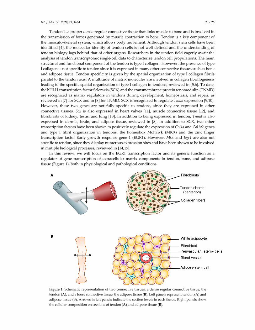
in mutiple biological processes, reviewed in [14,15]. In this review, we will focus on the EGR1 transcription factor and its generic function as a
regulator of gene transcription of extracellular matrix components in tendon, bone, and adipose
tissue (Figure 1), both in physiological and pathological conditions.
Figure 1. Schematic representation of two connective tissues: a dense regular connective tissue, the
tendon (A), and a loose connective tissue, the adipose tissue (B). Left panels represent tendon (A) and
adipose tissue (B). Arrows in left panels indicate the section levels in each tissue. Right panels show
the cellular composition on sections of tendon (A) and adipose tissue (B).
Int. J. Mol. Sci. 2020, 21, 1664 3 of 26
2. Egr1 “Identity Card”
2.1. Multiple Names for a Single Gene
The Egr1 (Early growth response 1) gene was first identified as NGFI‐A (Nerve Growth Factor
Induced‐A) because of its activation by NGF (nerve growth factor) in the neuronal rat cell line PC12
[16]. It was subsequently described in four different laboratories as a rapidly and transiently activated
gene in various fibroblast cell lines. Serum addition or mitogen treatment with the tumor promoter
TPA (tetradecanoyl phorbol acetate) on mouse fibroblasts led to a rapid and strong induction of an
early growth response gene then named Egr1 [17,18]. Egr1 was also named TIS8 (TPA Inducible
Sequence 8), because of its activation following TPA treatment in a murine 3T3 cell line [19]. After the
identification of a zinc‐finger‐region similar to the Drosophila Krüppel segmentation gene in the Egr1
genomic sequence, the murine gene was called Krox24, for “Krüppel box 24” [20] or zif/268 in
reference to three tandem zinc finger sequences [21]. Presentation of recorded bird songs to songbirds
such as canaries and zebra finches induces Egr1 expression in their forebrains. In this case, Egr1 was
referred to by the acronym ZENK [22]. The Egr1/EGR1 gene spans about 3.8 Kb and is located on
chromosome 18 in mice and on chromosome 5 in humans. It is composed of two exons and one 700
bp intron. The first exon includes the first 99 amino acids of the protein and the second exon includes
the three tandem zinc finger motifs [23]. The Egr1/EGR1 gene is highly conserved between mouse,
rat, chicken, zebrafish, chimpanzee, dog, cow, and human.
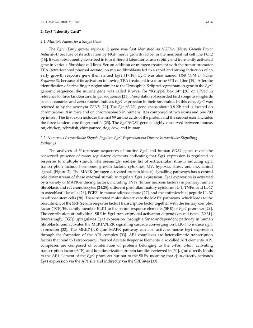
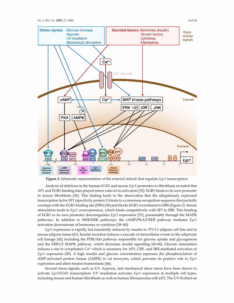
2.2. Numerous Extracellular Signals Regulate Egr1 Expression via Diverse Intracellular Signalling
Pathways
The analyses of 5’‐upstream sequences of murine Egr1 and human EGR1 genes reveal the
conserved presence of many regulatory elements, indicating that Egr1 expression is regulated in
response to multiple stimuli. The seemingly endless list of extracellular stimuli inducing Egr1
transcription include hormones, growth factors, cytokines, UV, hypoxia, stress, and mechanical
signals (Figure 2). The MAPK (mitogen‐activated protein kinase) signalling pathways has a central
role downstream of these external stimuli to regulate Egr1 expression. Egr1 expression is activated
by a variety of MAPK‐inducing factors, including TNFs (tumor necrosis factors) in primary human
fibroblasts and rat chondrocytes [24,25], different pro‐inflammatory cytokines IL‐1, TNFα, and IL‐17
in osteoblast‐like cells [26], FGF21 in mouse adipose tissue [27], and the antimicrobial peptide LL‐37
in adipose stem cells [28]. These secreted molecules activate the MAPK pathways, which leads to the
recruitment of the SRF (serum response factor) transcription factor together with the ternary complex
factor (TCF)/Ets family member ELK1 to the serum response elements (SRE) of Egr1 promoter [29].
The contribution of individual SRE in Egr1 transcriptional activation depends on cell types [30,31].
Interestingly, TGFβupregulates Egr1 expression through a Smad‐independent pathway in human
fibroblasts, and activates the MEK1/2/ERK signalling cascade converging on ELK‐1 to induce Egr1
expression [32]. The MKK7‐JNK‐cJun MAPK pathway can also activate mouse Egr1 expression
through the formation of the AP1 complex [33]. AP1 complexes are heterodimeric transcription
factors that bind to Tetracecanoyl Phorbol Acetate Response Elements, also called AP1 elements. AP1
complexes are composed of combination of proteins belonging to the c‐Fos, c‐Jun, activating
transcription factor (ATF), and Jun dimerization protein families reviewed in [34]. cJun directly binds
to the AP1 element of the Egr1 promoter but not to the SREs, meaning that cJun directly activates
Egr1 expression via the AP1 site and indirectly via the SRE sites [33].
Int. J. Mol. Sci. 2020, 21, 1664 4 of 26
Figure 2. Schematic representation of the external stimuli that regulate Egr1 transcription.
Analysis of deletions in the human EGR1 and mouse Egr1 promoters in fibroblasts revealed that
AP1 and EGR1 binding sites played minor roles in its activation [35]. EGR1 binds to its own promoter
in mouse fibroblasts [36]. This finding leads to the observation that the ubiquitously expressed
transcription factor SP1 (specificity protein 1) binds to a consensus recognition sequence that partially
overlaps with the EGR1 binding site (EBS) [36] and blocks EGR1 recruitment to EBS (Figure 2). Serum
stimulation leads to Egr1 overexpression, which binds competitively with SP1 to EBS. This binding
of EGR1 to its own promoter downregulates Egr1 expression [37], presumably through the MAPK
pathways. In addition to MEK/ERK pathways, the cAMP/PKA/CREB pathway mediates Egr1
activation downstream of hormones or cytokines [38–40].
Egr1 expression is rapidly but transiently induced by insulin in 3T3‐L1 adipose cell line and in
mouse adipose tissue [41]. Insulin secretion induces a cascade of intracellular events in the adipocyte
cell lineage [42] including the PI3K/Akt pathway responsible for glucose uptake and glycogenesis
and the ERK1/2 MAPK pathway, which decreases insulin signalling [43,44]. Glucose stimulation
induces a rise in cytoplasmic Ca2+ which is necessary for AP1, CRE, and SRE‐mediated activation of
Egr1 expression [45]. A high insulin and glucose concentration represses the phosphorylation of
AMP‐activated protein kinase (AMPK) in rat tenocytes, which prevents its positive role in Egr1
expression and alters tendon homeostasis [46].
Several stress signals, such as UV, hypoxia, and mechanical shear stress have been shown to
activate Egr1/EGR1 transcription. UV irradiation activates Egr1 expression in multiple cell types,
including mouse and human fibroblasts as well as human fibrosarcoma cells [47]. The UV‐B effect on
Int. J. Mol. Sci. 2020, 21, 1664 5 of 26
Egr1 expression is mediated through the activation and recruitment of the NFB family member p65
to a NFB binding site located on the Egr1 promoter [48]. Under hypoxic conditions, the hypoxia‐
inducible factor‐1α (HIF1α) binds to the EGR1 promoter in adipose stem cells isolated from obese
patients, but not not in hASCs from healthy patients [49]. Hypoxia induces EGR1 upregulation in
hASCs of diabetic patients either through the MAPK/ERK pathway or via the direct recruitment of
HIF1α to the EGR1 promoter [49]. Fluid shear stress activation activates EGR1 transcription in human
endothelial cells and epithelial cells [50]. In summary, Egr1 expression is regulated by a myriad of secreted molecules and stress factors
through numerous regulatory elements located upstream of the Egr1 coding sequence.
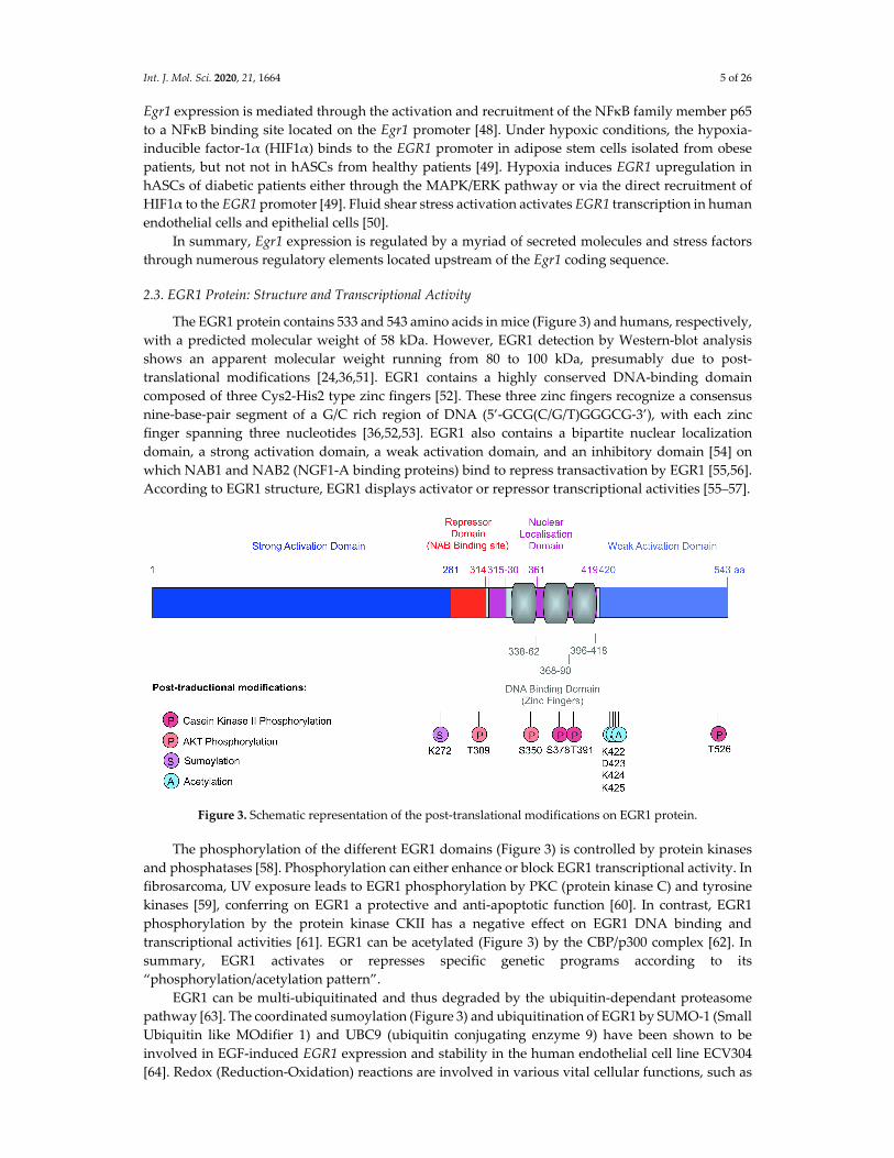
2.3. EGR1 Protein: Structure and Transcriptional Activity
The EGR1 protein contains 533 and 543 amino acids in mice (Figure 3) and humans, respectively,
with a predicted molecular weight of 58 kDa. However, EGR1 detection by Western‐blot analysis
shows an apparent molecular weight running from 80 to 100 kDa, presumably due to post‐
translational modifications [24,36,51]. EGR1 contains a highly conserved DNA‐binding domain
composed of three Cys2‐His2 type zinc fingers [52]. These three zinc fingers recognize a consensus
nine‐base‐pair segment of a G/C rich region of DNA (5’‐GCG(C/G/T)GGGCG‐3’), with each zinc
finger spanning three nucleotides [36,52,53]. EGR1 also contains a bipartite nuclear localization
domain, a strong activation domain, a weak activation domain, and an inhibitory domain [54] on
which NAB1 and NAB2 (NGF1‐A binding proteins) bind to repress transactivation by EGR1 [55,56].
According to EGR1 structure, EGR1 displays activator or repressor transcriptional activities [55–57].
Figure 3. Schematic representation of the post‐translational modifications on EGR1 protein.
The phosphorylation of the different EGR1 domains (Figure 3) is controlled by protein kinases
and phosphatases [58]. Phosphorylation can either enhance or block EGR1 transcriptional activity. In
fibrosarcoma, UV exposure leads to EGR1 phosphorylation by PKC (protein kinase C) and tyrosine
kinases [59], conferring on EGR1 a protective and anti‐apoptotic function [60]. In contrast, EGR1
phosphorylation by the protein kinase CKII has a negative effect on EGR1 DNA binding and
transcriptional activities [61]. EGR1 can be acetylated (Figure 3) by the CBP/p300 complex [62]. In
summary, EGR1 activates or represses specific genetic programs according to its
“phosphorylation/acetylation pattern”.
EGR1 can be multi‐ubiquitinated and thus degraded by the ubiquitin‐dependant proteasome
pathway [63]. The coordinated sumoylation (Figure 3) and ubiquitination of EGR1 by SUMO‐1 (Small
Ubiquitin like MOdifier 1) and UBC9 (ubiquitin conjugating enzyme 9) have been shown to be
involved in EGF‐induced EGR1 expression and stability in the human endothelial cell line ECV304
[64]. Redox (Reduction‐Oxidation) reactions are involved in various vital cellular functions, such as
Int. J. Mol. Sci. 2020, 21, 1664 6 of 26
aerobic cellular respiration, nucleic acid synthesis, and also for the production and elimination of
reactive oxygen species (ROS), which includes superoxide, nitric oxide, hydroxyl radical, hydrogen
peroxide, and hypochlorus acid. At high doses, ROS are toxic for the cell and the damage they cause
was termed oxidative stress. Oxidative stress induces bone loss by stimulating osteoclastic bone
resorption and inhibiting osteoblastic differentiation [65–68]. However, hydrogen peroxide at non‐
toxic doses increases the expression and DNA‐binding activity of EGR1 in mouse and human
osteoblastic cells without affecting their differentiation [69,70]. The DNA‐binding properties of EGR1
are modulated by the redox state: EGR1 binding to the DNA depends on the presence of reducing
agents, which are necessary for the correct conformation of the EGR1 zinc‐finger region. Oxidized or
metal‐free EGR1 does not bind to DNA [71]. In the human osteoblastic HOBIT cell line, under non‐
toxic ROS doses, a DNA repair enzyme, the APE1 (apurinic/apyrimidic endonuclease 1) increases
EGR1 binding to DNA with nuclear redox activity. EGR1 also upregulates APE1 gene expression,
showing the existence of a positive‐autoregulatory loop between APE1 and EGR1 proteins [70].
In summary, EGR1 displays diverse transcriptional activation or repression functions
depending on its post‐translational modification statues.
2.4. Egr1 Expression Profile In Vivo
Egr1 is expressed in numerous organs and cell types during development and adult life.
However, Egr1 expression is not ubiquitous. In situ hybridization experiments performed on chicken
and mouse embryos indicate a punctiform location of Egr1 transcripts in various tissues such as
tendon, cartilage, bone, skeletal muscle, innervation, vessels, and dermis [72,73]. In developing
tendons, Egr1 transcripts are not observed in all tendon cells, but in subregions, such as the
myotendinous junction and around long tendons in mouse and chicken embryos [73,74]. In the adult,
the mouse ENCODE transcriptome data set indicates that Egr1 is expressed in many if not all adult
tissues, with high expression in cortex, mammary gland, ovary, and thymus [75]. In situ hybridization
and immunohistochemistry experiments performed on adult mouse tissues show Egr1 expression in
Achilles tendons [76], subcutaneous adipose tissue [77], hypertrophic cartilage [78,79], and bone
[80,81].
3. EGR1 Roles in Connective Tissue Formation, Homeostasis and Healing
Consistent with the broad range of Egr1/EGR1 expression sites, EGR1 is involved in the
formation and homeostasis of many organs. One powerful tool for addressing gene function is the
use of knock‐out mice. The Egr1 gene was inactivated in mice by homologous recombination with
the insertion of the neomycin resistance cassette upstream of the EGR1 DNA‐binding domain [82]
and with the insertion of the LacZ coding sequence within the Egr1 5’ untranslated region added with
a frameshift mutation upstream of the DNA‐binding domain of Egr1 [83]. Both Egr1 mutant mouse
lines were initially described with no overt phenotype during development or postnatal life [82,83];
with the exception of subtil pituitary and ovarian defects observed in the LacZ insertion mutant mice
[83]. However, connective tissue defects were subsequently described in these Egr1 mutant mice,
affecting tendon, cartilage, bone and adipose tissue formation and homeostasis. The analysis of Egr1
loss‐of‐function in mice has identified numerous EGR1 target genes in connective tissues (reported
in Table 1). A striking point is that the target genes positively regulated by EGR1 are mainly
components of the extracellular matrix (ECM) or linked to ECM regulation, while those negatively
regulated by EGR1 are cartilage, bone, or adipose tissue differentiation markers (Table 1).
Int. J. Mol. Sci. 2020, 21, 1664 7 of 26
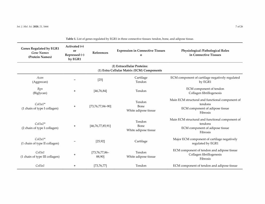
Table 1. List of genes regulated by EGR1 in three connective tissues: tendon, bone, and adipose tissue.
Genes Regulated by EGR1
Gene Names
(Protein Names)
Activated (+)
or
Repressed (−)
by EGR1
References Expression in Connective Tissues
o
Physiological /Pathological Roles
in Connective Tissues
(I) Extracellular Proteins:
(1) Extra Cellular Matrix (ECM) Components
Acan
(Aggrecan) − [25]
Cartilage
Tendon
ECM component of cartilage negatively regulated
by EGR1
Bgn
(Biglycan) + [46,76,84] Tendon
ECM component of tendon
Collagen fibrillogenesis
Col1a1*
(1 chain of type I collagen) + [73,76,77,84–90]
Tendon
Bone
White adipose tissue
Main ECM structural and functional component of
tendons
ECM component of adipose tissue
Fibrosis
Col1a2*
(2 chain of type I collagen) + [46,76,77,85,91]
Tendon
Bone
White adipose tissue
Main ECM structural and functional component of
tendons
ECM component of adipose tissue
Fibrosis
Col2a1*
(1 chain of type II collagen) − [25,92] Cartilage
Major ECM component of cartilage negatively
regulated by EGR1
Col3a1
(1 chain of type III collagen) +
[73,76,77,86–
88,90]
Tendon
White adipose tissue
ECM component of tendon and adipose tissue
Collagen fibrillogenesis
Fibrosis
Col5a1 + [73,76,77] Tendon ECM component of tendon and adipose tissue
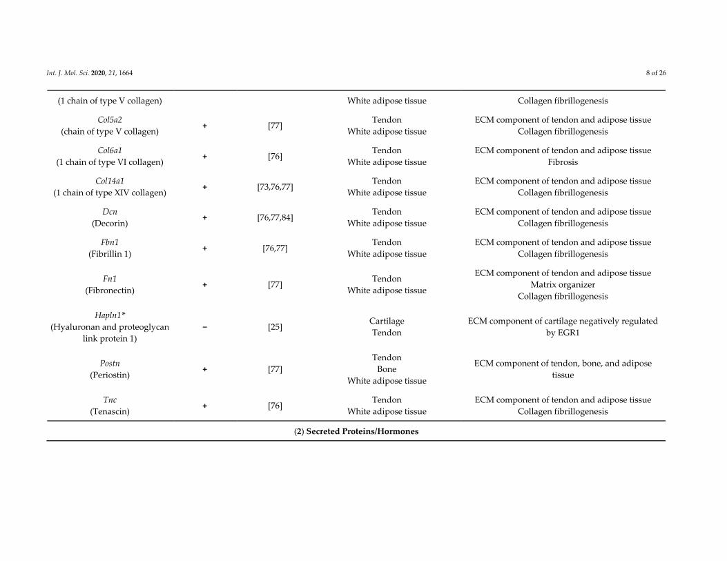
Int. J. Mol. Sci. 2020, 21, 1664 8 of 26
(1 chain of type V collagen) White adipose tissue Collagen fibrillogenesis
Col5a2
(chain of type V collagen) + [77]
Tendon
White adipose tissue
ECM component of tendon and adipose tissue
Collagen fibrillogenesis
Col6a1
(1 chain of type VI collagen) + [76]
Tendon
White adipose tissue
ECM component of tendon and adipose tissue
Fibrosis
Col14a1
(1 chain of type XIV collagen) + [73,76,77]
Tendon
White adipose tissue
ECM component of tendon and adipose tissue
Collagen fibrillogenesis
Dcn
(Decorin) + [76,77,84]
Tendon
White adipose tissue
ECM component of tendon and adipose tissue
Collagen fibrillogenesis
Fbn1
(Fibrillin 1) + [76,77]
Tendon
White adipose tissue
ECM component of tendon and adipose tissue
Collagen fibrillogenesis
Fn1
(Fibronectin) + [77]
Tendon
White adipose tissue
ECM component of tendon and adipose tissue
Matrix organizer
Collagen fibrillogenesis
Hapln1*
(Hyaluronan and proteoglycan
link protein 1)
− [25] Cartilage
Tendon
ECM component of cartilage negatively regulated
by EGR1
Postn
(Periostin) + [77]
Tendon
Bone
White adipose tissue
ECM component of tendon, bone, and adipose
tissue
Tnc
(Tenascin) + [76]
Tendon
White adipose tissue
ECM component of tendon and adipose tissue
Collagen fibrillogenesis
(2) Secreted Proteins/Hormones
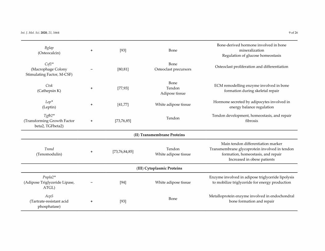
Int. J. Mol. Sci. 2020, 21, 1664 9 of 26
Bglap
(Osteocalcin) + [93] Bone
Bone‐derived hormone involved in bone
mineralization
Regulation of glucose homeostasis
Csf1*
(Macrophage Colony
Stimulating Factor, M‐CSF)
− [80,81]
Bone
Osteoclast precursors
Osteoclast proliferation and differentiation
Ctsk
(Cathepsin K) + [77,93]
Bone
Tendon
Adipose tissue
ECM remodelling enzyme involved in bone
formation during skeletal repair
Lep*
(Leptin) + [41,77] White adipose tissue
Hormone secreted by adipocytes involved in
energy balance regulation
Tgfb2*
(Transforming Growth Factor
beta2, TGFbeta2)
+ [73,76,85] Tendon
Tendon development, homeostasis, and repair
fibrosis
(II) Transmembrane Proteins
Tnmd
(Tenomodulin) + [73,76,84,85]
Tendon
White adipose tissue
Main tendon differentiation marker
Transmembrane glycoprotein involved in tendon
formation, homeostasis, and repair
Increased in obese patients
(III) Cytoplasmic Proteins
Pnpla2*
(Adipose Triglyceride Lipase,
ATGL)
− [94] White adipose tissue
Enzyme involved in adipose triglyceride lipolysis
to mobilize triglyceride for energy production
Acp5
(Tartrate‐resistant acid
phosphatase)
+ [93] Bone
Metalloprotein enzyme involved in endochondral
bone formation and repair
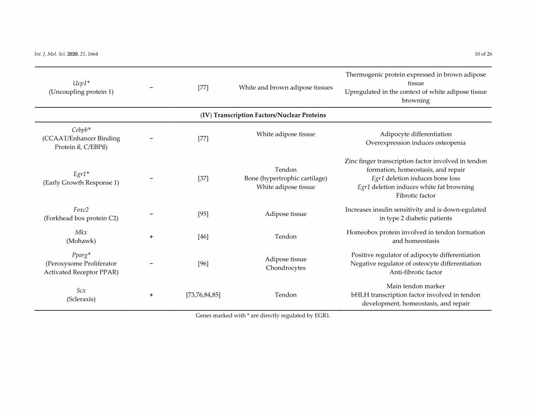
Int. J. Mol. Sci. 2020, 21, 1664 10 of 26
Ucp1*
(Uncoupling protein 1) − [77] White and brown adipose tissues
Thermogenic protein expressed in brown adipose
tissue
Upregulated in the context of white adipose tissue
browning
(IV) Transcription Factors/Nuclear Proteins
Cebpb*
(CCAAT/Enhancer Binding
Protein ß, C/EBPß)
− [77] White adipose tissue
Adipocyte differentiation
Overexpression induces osteopenia
Egr1*
(Early Growth Response 1) − [37]
Tendon
Bone (hypertrophic cartilage)
White adipose tissue
Zinc finger transcription factor involved in tendon
formation, homeostasis, and repair
Egr1 deletion induces bone loss
Egr1 deletion induces white fat browning
Fibrotic factor
Foxc2
(Forkhead box protein C2) − [95] Adipose tissue
Increases insulin sensitivity and is down‐egulated
in type 2 diabetic patients
Mkx
(Mohawk) + [46] Tendon
Homeobox protein involved in tendon formation
and homeostasis
Pparg*
(Peroxysome Proliferator
Activated Receptor PPAR)
− [96] Adipose tissue
Chondrocytes
Positive regulator of adipocyte differentiation
Negative regulator of osteocyte differentiation
Anti‐fibrotic factor
Scx
(Scleraxis) + [73,76,84,85] Tendon
Main tendon marker
bHLH transcription factor involved in tendon
development, homeostasis, and repair
Genes marked with * are directly regulated by EGR1.
Int. J. Mol. Sci. 2020, 21, 1664 11 of 26
3.1. EGR1 is a Potent Inducer of Extracellular Matrix Production in Tendons
3.1.1. EGR1 Function in Tendon Formation, Homeostasis, and Ageing
Several studies have addressed the role of EGR1 in tendon biology. In developing limb tendons,
Egr1 is expressed close to the myotendinous junction and delineates the long tendons in mouse and
chicken embryos [73,74]. The Egr1 mutant mice do not display a strong overt tendon phenotype;
however, Egr1−/− mice show a significant decrease in the expression of the key tendon markers, Scx,
Tnmd, and Col1a1 in addition to that of tendon‐associated collagens in developing E18.5 limbs [73]
and adult tail tendons and Achilles tendons [76]. Comparison of adult tail tendons in Egr1−/− versus
Egr1+/+ mice shows a reduced number of collagen fibers in mutant mice. Tail and Achilles tendons
have collagen fibrils with smaller diameter and impaired biomechanical properties in Egr1−/−
compared to Egr1+/+ mice [76]. Conversely, Egr1 is sufficient to induce de novo expression of a large
variety of tendon genes (including Scx and tendon‐associated collagen genes) in ectopic contexts in
chicken embryos [73]. The Egr1 gene is sufficient to induce ectopic Scx, Col1a1, Col3a1, Col5a1, and
Col14a1 expression in the neural tube, an unrelated embryonic tissue derived form the ectoderm [73].
Consistently with the in vivo situation, EGR1 is sufficient to induce the expression of a large panel of
tendon genes including, Scx and Tnmd, collagen associated‐tendon genes (Col1a1, Col1a2 Col3a1,
Col5a1, Col6a1, Col14a1), and tendon matrix‐associated molecules (Tnc, Bgn, Dcn, and Fbn1) in mouse
C3H10T1/2 mesenchymal stem cells [76]. EGR1 also promotes the formation of 3D‐engineered tendon
constructs made of C3H10T1/2 cells by increasing the expression of Scx, Tnmd, and Col1a genes
[76,85]. Moreover, EGR1 induces tenogenic differentiation in rabbit tendon stem cells [97]. EGR1
mediates the promoting effect of the anti‐miR124 on collagen production in human tendon‐derived
stem cells [98] and the promoting effect of ferulic acid on self‐renewal ability of human tendon‐
derived stem cells [99]. Consistent with the positive regulation of Col1a gene transcription by EGR1
observed both in vivo and in vitro, chromatin immunoprecipitation (ChIP) experiments show the
recruitment of EGR1 to the tendon regulatory regions of the Col1a1 promoter in E18.5 limbs [73] and
to Col1a1 and Col1a2 regulatory regions in adult Achilles tendons [76]. Lastly, Egr1 downregulation
has been associated with a loss of the tenogenic differentiation potential in ageing human tendon
progenitor cells, while Egr1 gain‐of‐function has the ability to rescue their tendon differentiation
potential as assessed by the upregulation of Scx, Tnmd, Bgn, Dcn, and Col1a1 gene expression [84].
3.1.2. EGR1 Function in Tendon Healing
EGR1 has been shown to be required for the expression of the key tendon markers and tendon‐
related ECM genes during healing after tendon injury [76]. Tendon injury models where the tension
is maintained (partial rupture) induce a massive increase of tendon gene expression [76,100], while
tendon injury models where the tension is lost (total rupture) induce a loss of tendon gene expression
[101,102]. Different models of tendon injury, where tension is maintained, lead to a massive increase
of Egr1 expression after injury, in mouse or rat Achilles tendons [76,100] and rabbit flexor tendons
[103]. In addition, needle‐induced microlesions in healing rat Achilles tendons increase Egr1
expression [104]. The transcriptional response i.e., the increase of Scx, Tnmd, Col1a1, Col1a2, Col5a1,
Col6a1, Col14a1, Tnc, and Dcn gene expression in response to longitudinal lesion along the tendon
axis is drastically decreased in Egr1−/− Achilles tendons, showing that Egr1 is required for the injury‐
induced expression of tendon‐related ECM genes and key tendon markers [76]. Moreover, EGR1‐
producing cells promote tendon repair in a rat model of Achilles tendon injury [76] and in a rabbit
model of rotator cuff injury [97]. In summary, Egr1 appears to be required for the correct expression of matrix genes during
tendon formation, homeostasis, and ageing in vitro and in vivo, but also during tendon healing. A
recent discovery is that tendons are peripheral circadian clock tissues, reviewed in [105], in which
collagen synthesis and homeostasis is under a circadian control [106]. Interestingly, the circadian
clock is disturbed in Egr1−/− mice, associated with impaired locomotor activity and body temperature
Int. J. Mol. Sci. 2020, 21, 1664 12 of 26
[107], suggesting that Egr1 could be involved in the circadian synthesis of tendon‐associated
collagens.
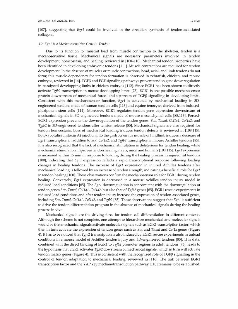
3.2. Egr1 is a Mechanosensitive Gene in Tendon
Due to its function to transmit load from muscle contraction to the skeleton, tendon is a
mecanosensitive tissue. Mechanical signals are necessary parameters involved in tendon
development, homeostasis, and healing, reviewed in [108–110]. Mechanical tendon properties have
been identified in developing embryonic tendons [111]. Muscle contractions are required for tendon
development. In the absence of muscles or muscle contractions, head, axial, and limb tendons do not
form; this muscle‐dependency for tendon formation is observed in zebrafish, chicken, and mouse
embryos, reviewed in [14]. TGFβand FGF signalling pathways prevent tendon gene downregulation
in paralyzed developping limbs in chicken embryos [112]. Since EGR1 has been shown to directly
activate Tgfb2 transcription in mouse developping limbs [73], EGR1 is one possible mechanosensor
protein downstream of mechanical forces and upstream of TGFβ signalling in developing limbs.
Consistent with this mechanosensor function, Egr1 is activated by mechanical loading in 3D‐
engineered tendons made of human tendon cells [113] and equine tenocytes derived from induced‐
pluripotent stem cells [114]. Moreover, EGR1 regulates tendon gene expression downstream of
mechanical signals in 3D‐engineered tendons made of mouse mesenchymal cells [85,113]. Forced‐
EGR1 expression prevents the downregulation of the tendon genes, Scx, Tnmd, Col1a1, Col1a2, and
Tgfb2 in 3D‐engineered tendons after tension release [85]. Mechanical signals are also required for
tendon homeostasis. Loss of mechanical loading induces tendon defects is reviewed in [108,115].
Botox (botuliniumtoxin A) injection into the gastrocnemius muscle of hindlimb induces a decrease of
Egr1 transcription in addition to Scx, Col1a2, and Tgfb2 transcription in mouse Achilles tendons [85].
It is also recognized that the lack of mechanical stimulation is deleterious for tendon healing, while
mechanical stimulation improves tendon healing in rats, mice, and humans [108,115]. Egr1 expression
is increased within 15 min in response to loading during the healing process in injured rat tendons
[100], indicating that Egr1 expression reflects a rapid transcriptional response following loading
changes in healing tendons. The increase of Egr1 expression in injured Achilles tendons after
mechanical loading is followed by an increase of tendon strength, indicating a beneficial role for Egr1
in tendon healing [100]. These observations confirm the mechanosensor role for EGR1 during tendon
healing. Conversely, Egr1 expression is decreased in a mouse Achilles tendon injury model in
reduced load conditions [85]. The Egr1 downregulation is concomitent with the downregulation of
tendon genes Scx, Tnmd, Col1a1, Col1a2, but also that of Tgfb2 genes [85]. EGR1 rescue experiments in
reduced load conditions and after tendon injury increase the expression of tendon‐associated genes,
including Scx, Tnmd, Col1a1, Col1a2, and Tgfb2 [85]. These observations suggest that Egr1 is sufficient
to drive the tendon differentiation program in the absence of mechanical signals during the healing
process in vivo.
Mechanical signals are the driving force for tendon cell differentiation in different contexts.
Although the scheme is not complete, one attempt to hierarchize mechanical and molecular signals
would be that mechanical signals activate molecular signals such as EGR1 transcription factor, which
then in turn activate the expression of tendon genes such as Scx and Tnmd and Col1a genes (Figure
4). It has to be noticed that Tgfb2 transcription is also induced by EGR1 rescue experiments in unload
conditions in a mouse model of Achilles tendon injury and 3D‐engineered tendons [85]. This data,
combined with the direct binding of EGR1 to Tgfb2 promoter regions in adult tendons [76], leads to
the hypothesis that EGR1 activates Tgfb2 dowstream of mechanical signals, which in turn will activate
tendon matrix genes (Figure 4). This is consistent with the recognized role of TGFβ signalling in the
control of tendon adaptation to mechanical loading, reviewed in [116]. The link between EGR1
transcription factor and the YAP key mechanotransduction pathway [110] remains to be established.
Int. J. Mol. Sci. 2020, 21, 1664 13 of 26
Figure 4. Schematic representation of the position of Egr1/EGR1 in the molecular cascade
downstream of mechanical signals involved in tendon gene expression.
3.3. EGR1 and Endochondral Bone Formation and Healing
Bone is a supportive connective tissue composed of cells, fibers, and a mineralized solid ground
substance [117]. Egr1 expression is detected in several areas undergoing endochondral bone
formation, such as hypertrophic cartilage [78,79] and periostal regions of the developping long bones
[72,73]. Egr1 is a negative regulator of cartilage markers [92]. Egr1−/− mice display bone loss
[80,118,119]. The bone loss upon Egr1 deletion is a consequence of an increased bone resorption, via
the increased production of the colony stimulating factor‐1 CSF‐1/M‐CSF known to positively
regulate osteoclast differentiation [80]. Bone development and homeostasis is controlled by the
interplay between bone‐forming osteoblasts and bone‐resorbing osteoclasts [120], and this
equilibrium is lost in Egr1−/− mice. EGR1 is a negative regulator of the osteoclastogenic cytokine CSF‐
1 production by stromal cells. Phosphorylated‐EGR1 (upon estrogen) blocks Csf1 gene transcription
by preventing the binding of the transcriptional activator SP1 to the Csf1 promoter [80]. The failure
of estrogen to rescue the CSF‐1 production and consequent osteoclast formation in Egr1−/− in
ovariectomized mice positiones Egr1 as a pivotal actor to mediate the anti‐osteoclastogenic effect of
estrogen [80,81]. Transcription factors that play a role in bone formation are expected to participate in bone
healing after fracture since endochondral bone formation that occurs in embryos is recapitulated
during bone healing. In a bone fracture mouse model, Egr1 deficiency leads to several bone defects
including persistant fibrin accumulation in the fracture gap, abnormal callus ossification with
enlarged areas of cartilaginous tissue, decreased expression levels of Bglap (Osteocalcin), and bone
resorbtion markers that regulate extracellular matrix, including Acp5 (Tartrate‐resistant acid
phosphatase) and Ctsk (Cathepsin K) genes [93,118]. This data confirms that EGR1 controls the
balance between bone tissue formation and resorption during skeletal repair.
3.4. EGR1 Regulates Extracellular Matrix Production in Adipose Tissue
Int. J. Mol. Sci. 2020, 21, 1664 14 of 26
Adipose tissue is a loose connective tissue mainly composed of specialized white adipocytes,
held in a framework of collagen fibers, which plays a fundamental role in fat storage, metabolic
control, and thermoregulation [121]. Adipocytes are surrounded by an extracellular matrix, which
serves as mechanical support and is mainly composed of collagens, fibronectin, and elastin [122].
Cells producing ECM in adipose tissue are not clearly indentified; however, it is recognized that
collagens are mostly produced by adipocytes, but also by endothelial cells and adipose stem cells in
normal conditions [123]. The molecular signature of subcutaneous white adipose tissue with Egr1
deletion identifies a downregulation of ECM genes, including Col1a1, Col1a2, Col3a1, Col5a1, Col5a2,
Col14a1, Fn1, Dcn, and Post (Periostin) [77]. Egr1 deletion is also associated with a spontaneous
browning of subcutaneous white adipose tissue in Egr1 mutant mice compared to wild‐types [77].
The browning phenotype corresponds to the appearence of beige adipocytes within the white
adipose tissue mainly by de novo differentiation of progenitors [124]. In contrast to white adipocytes,
beige adipocytes dissipate excess energy through heat production by a large number of mitochondria,
which exhibit uncoupling activity via the thermogenic protein UCP1 (uncoupling protein‐1) [125].
Consistent with the browning phenotype, transcriptomic analysis in Egr1‐deleted adipose tissue
shows a concomitant downregulation of the white adipocyte marker, Lep (leptin) and upregulation
of the key beige adipocyte marker Ucp1 [77]. Both positive (Lep) and negative (Ucp1) transcriptional
regulations have been shown to occur via direct EGR1 recruitment to regulatory regions of these
genes [41,77]. The browning process reduces the deleterious consequences of fat accumulation and is
seen as possible mechanism to fight against obesity and to improve metabolic health [125].
Consistently, Egr1−/− mice are protected from high fat diet‐induced obesity via an increase of energy
expenditure [95]. Conversely to Egr1 loss‐of‐function in mice, Egr1 overexpression in C3H10T1/2 cells
increases the transcription of matrix genes and prevents the beige adipocyte differentiation [77].
Overall, Egr1 deletion leads to a drastic loss of ECM genes associated with a browning phenotype in
subcutaneous white adipose tissue in mice.
To summarize, Egr1 loss‐of‐function is associated with reduced ECM production in tendon (a
dense connective tissue), bone (a supportive connective tissue), and adipose tissue (a loose connective
tissue). Although the altered matrix genes upon Egr1 deletion appear to be similar in the different
connective tissues, the reduced ECM production is deleterious for tendon and bone formation and
homeostasis, while being beneficial for white adipose tissue to increase energy expenditure.
4. EGR1 is a Fibrotic Factor
Fibroblast deregulation leads to fibrosis, a process attributed to anarchic deposition of
extracellular matrix, in response to injury or in pathological conditions. Fibrosis is observed in almost
any tissue in cases of organ dysfunction, but is also a key process in cancer, inflammation, and ageing.
Myofibroblasts are the main cellular component of fibrosis. Myofibroblasts are non‐muscle
contractile cells responsible for the excessive synthesis, deposition, and remodelling of ECM proteins
in fibrosis; for a recent review see [126]. A recognized molecular driver of fibrosis is the TGFβ
signalling pathway. TGFβ drives the conversion of fibroblasts to myofibroblasts to induce the
excessive deposition of collagen and inappropriate ECM during fibrosis, recently reviewed in [127].
Generic molecular markers for myofibroblasts are similar to markers for smooth muscle cells, such
as Acta2 (smooth muscle actin), whose expression is regulated by TGFβ signalling [127]. Although
myofibroblasts and TGFβ are recognized to be the respective cellular and molecular hallmarks of
fibrosis, there is no comprehensive understanding of the cellular and molecular mechanisms
underlying fibrosis. As EGR1 is a potent regulator of matrix components in different contexts, EGR1
is seen as a fibrotic factor.
4.1. EGR1 and Fibrosis in Metabolic Diseases Linked to Adipose Tissue (Obesity and Diabetes)
Fibrosis in adipose tissue is considered a hallmark of metabolically dysfunctional adipose tissue
and is associated with obesity and insulin resistance [86,122,128]. In pathological conditions such as
obesity, the rapid expansion of adipose tissue causes hypoxia since neovascularization cannot keep
up with rapid adipose tissue growth. Hypoxia is followed by the necrosis of adipocytes accompanied
Int. J. Mol. Sci. 2020, 21, 1664 15 of 26
with the infiltration of inflammatory leucocytes and macrophages to remove the dead cells. In these
pathological conditions, myofibroblasts associated with the inflammatory response, accumulate
within the adipose tissue and cause ECM thickening, characteristic of fibrosis [121,129]. Adipose
tissue fibrosis results from the imbalance between excess synthesis and impaired degradation of type
I, III, and VI collagens [130]. Consistently, type I, III, and VI collagens are particularly abundant in
adipose tissue of obese patients [131] and collagen content affects tensile strengh of adipose tissue
[132]. Moreover, Col6a1 loss‐of‐function significantly reduces adipose tissue fibrosis in obese mice
[122]. Egr1/EGR1 expression is increased in adipose tissue of obese Lep/Lep mice and patients [95],
and of diabetic db/db mice and type 2 diabetes mellitus (T2DM) patients [133]. T2DM is associated
with insulin resistance and the majority of T2DM patients are overweight or obese [129]. Egr1 gain‐
of‐function in epididymal fat induces insulin resistance [133], while Egr1 loss‐of‐function improves
the whole‐body insulin sensitivity in diabetic mice [133]. One possible mechanism would be that
insulin‐induced EGR1 [41,133] directly inhibits Pnpla2 (ATGL, Adipose TriGlyceride Lipase) expression
in adipocytes that leads to lipolysis inhibition and promote fat accumulation [94].
Egr1 overexpression is associated with metabolically dysfunctional adipose tissues. As Egr1
regulates matrix production in subcutaneous white adipose tissue and tendon, Egr1 is a credible
regulator of inappropriate ECM production in metabolically dysfunctional adipose tissues.
Consistent with this idea, Egr1 transcription is directly activated by insulin and F1α [49] via direct recruitment to Egr1 promoter. In addition, the insulin and F1α expression is increased in obese and T2DM mice and HIF1α regulates the expression of ECM genes such as Col1a1, Col3a1, and Col14a1
[86,87]. Egr1 is thus a good therapeutic target to counteract obesity and associated fibrosis since its
loss‐of‐function reduces ECM production and stimulates the white fat browning process.
Interestingly, the tendon differentiation gene, Tmnd, involved in ECM regulation in tendons [8],
is also involved in adipose tissue function. TNMD mRNA expression levels are strongly correlated
with the body mass index. TNMD gene expression is significantly higher in obese subjects compared
to lean subjects [134–136], while TNMD gene expression is downregulated in visceral adipose tissue
during diet‐induced weight loss [134]. Although EGR1 and TNMD genes share the same expression
profile in white adipose tissues of obese patients, in contrast to EGR1, TNMD acts as a protective
factor in visceral adipose tissue to alleviate insulin resistance in obesity [136].
4.2. Tendon Defects in Type 2 Diabetes Mellitus
Type II diabetes mellitus (T2DM) is associated with high risk of tendinopathy or tendon tears,
reviewed recently in [137,138]. Various T2DM rodent models, which cannot be dissociated from obese
mouse models, display tendons with decreased collagen content, ECM disorganization, and impaired
mechanical properties, reviewed in [138]. T2DM also impairs the healing process following tendon
injury and amplifies the fibrotic process during healing, leading to scarred tendons. There is an
increase of Col1a1 and Col3a1 expression in FDL tendons after injury associated with decreased
mechanical properties in diet‐induced obesity mice [88]. High glucose also affects tendon gene
expression and cell behavior in tendon cell cultures, which induces changes in extracellular matrix
[139]. Interestingly, Egr1 expression was also modified (associated with Mkx, Tgfb1, Col1a2, and Bgn
expression alteration) in rat tendon cells cultured in high glucose for 14 days [46].
4.3. EGR1 Controls Fibrosis in Systemic Sclerosis
A classical fibrotic disease is systemic sclerosis, also known as scleroderma, which is a rare
disease characterized by excessive collagen deposition resulting in fibrosis in different organs such
as skin and lungs but also the diggestive track (esophagus, stomach, and intestine) and myocard
[140,141]. Systemic sclerosis pathology also displays inflammation and vasculopathy components.
Myofibroblasts are key cells of the physiopathology of systemic sclerosis [140]. TGFβ and Wnt
signalling are recognized to play a fundamental role in the pathogenesis of fibrosis in systemic
sclerosis, in particular in the differentiation process of activated myofibroblasts [141]. EGR1 transcription factor has been shown to be at the crossroad of the molecular processes
leading to the TGFβ‐dependent fibrosic process in systemic sclerosis, reviewed in [142,143]. EGR1
Int. J. Mol. Sci. 2020, 21, 1664 16 of 26
expression is increased in biopsies of fibrotic skin and lung from patients with systemic sclerosis [32].
In the mouse model of bleomycin‐induced scleroderma, fibrosis is reduced in skin and lung of Egr1−/−
mice, with decreased Col1a1 expression and αSMA+ fibroblasts in both tissues [89]. In human
fibroblasts, EGR1 upregulates COL1A2 transcription downstream of TGFβ [91]. The high throughput
analysis of EGR1‐responsive genes in human primary fibroblasts identifies over 600 genes involved
in extracellular matrix synthesis, wound healing, and TGFbeta signalling, but also in cell proliferation
and vascular development [144]. This EGR1‐responsive genes signature is enriched in skin biopsies
from patients with systemic sclerosis compared to healthy controls [144]. The demonstrated
involvement of EGR1 in this fibrotic disease identifies EGR1 as a pertinent target to control fibrosis
in systemic sclerosis [142,143].
4.4. EGR1 is at the Crossroad of the Molecular Pathways Involved in the Fibrotic Process in Animal Models
for Organ Fibrosis
Classical animal models for fibrosis target idiopathic pulmonary fibrosis, renal fibrosis, liver
fibrosis, and heart fibrosis. EGR1 is frequently mentioned as being associated with the progression of
fibrotic process in animal models and in the transcription of fibrotic genes in cellular models. Egr1
deletion is often described as being beneficial to fight fibrosis progression in animal models of organ
fibrosis.
Lung fibrosis. EGR1 is involved in lung fibrosis downstream of IGFBP‐5 (insulin‐like growth
factor (IGF) binding protein‐5) to promote fibrotic gene transcription [145] and downstream of TGFβ1
to activate the transcription of the hyaluronan receptor CD44V6 (CD44 containing variable exon 6
(v6)) expression in lung fibroblasts in the context of idiopathic pulmonary fibrosis [146].
Renal fibrosis. In a mouse model of adenine‐enriched diet induced tubulointerstitial nephritis
leading to renal fibrosis, Egr1 is increased in kidney. Egr1−/− mice display reduced TGFbeta activity
and reduced renal fibrotic zones and were protected from renal failure [147]. The miR181 was
identified as an inhibitor of renal fibrosis via Egr1 inhibition, which suppressed the expression levels
of alphaSMA (ACTA2), connective tissue growth factor (CTGF), collagen type I (COL1A1), and type
III collagen (COL3A1) in NRK49F cells [90].
Liver fibrosis. Egr1 has been shown to contribute to liver fibrosis progression downstream of Elk‐
3 in CCl₄‐induced mouse liver fibrotic tissues and human liver cirrhotic tissues [148]. However, the
beneficial effect of Egr1 deletion in the context of liver fibrosis is contradictory. In an acute
acetaminophen‐induced liver injury mouse model, the inhibition on ERK1/2‐mediated Egr1
transcriptional activity attenuates hepatotoxicity, suggesting that inhibiting Egr1 is beneficial to
protect against liver fibrosis observed in long‐term application of acetaminophen [149]. By contrast,
another study shows that livers of Egr1−/− mutant mice exhibit a more severe fibrotic response
compared to those of wild‐type mice under acetaminophen overdose [150]. The Egr1 function
remains elusive in liver fibrosis. Heart fibrosis. EGR1 has been shown to be involved in cardiovascular homeostasis and diseases.
Notably, Egr1 transcription is activated in hypoxic and ischemic conditions in heart and in calcified
heart valves [151,152]. The miR‐150‐5p retards the progression of myocardial fibrosis by targeting
EGR1 [153].
As EGR1 is systematically mentioned as being involved in fibrosis progession in organ fibrosis
animal models, Egr1 is seen as a putative target to fight fibrosis. A recent antifibrotic chemical
component has been identified with the PPARγ agonist (pioglitazone) that inhibits TGF‐β‐driven
fibrosis in animal models for pulmonary, renal, and cardiac fibrosis, reviewed in [154]. Interestingly,
pioglitazone has been shown to repress Egr1 transcription and traduction in kidneys of TGF‐β‐driven
renal fibrosis in mice [155] and in pancreas of a cerulein‐induced acute pancreatitis mouse model
[156].
4.5. EGR1 and Matrix Production in Rheumatoid Arthritis and Osteoarthritis
Consistent with Egr1 expression in cartilage and bone, the Egr1 gene is reiteratively cited to be
involved in chronic diseases that lead to articular cartilage degeneration, such as osteoarthritis and
Int. J. Mol. Sci. 2020, 21, 1664 17 of 26
rheumatoid arthritis [157]. The molecular cascade underlying the pathogenesis of these two joint
diseases are not well understood. Osteoarthritis leads to cartilage degeneration, reviewed in [158],
while rheumatoid arthritis is an autoimmune and inflamatory disease associated with an increase of
synovial fibroblasts leading to joint degeneration [159]. TNFα levels are increased in the synovial
fluid of patients with osteoarthritis and rheumatoid arthritis [25] and reduce the expression of Col2a1,
Acan (Aggrecan), and Hapln1 through EGR1 recruitment to their promoters [25]. Classical and global
transcriptomic analysis identified high EGR1 expression in articular cartilage of patients with
osteoarthritis [96,160,161] and in synovial tissues of rheumatoid arthritis patients [162–164].
Chondrocytes stimulation with interleukin‐1β (IL‐1) leads to the recruitment of EGR1 to Pparg
promoter and downregulates its expression, preventing the protective effect of PPAR in osteoarthritis [96]. Ectopic expression of EGR1 in articular cartilage aggravated the degradation of
the cartilage matrix in mice [78]. The excess of EGR1 induced an increase of transcripts and protein
of type I collagen in synovial fibroblasts from rheumatoid arthritis patients [165]. Egr1 represents a
potential target for drug intervention in osteoarthritis or rheumatoid arthritis.
4.6. EGR1 and Scarred Tendon
Abnormal tendon healing is frequent following tendon injury reviewed in [166,167]. Following
accute rupture, tendons undergo a healing process involving the sequential and overlapping phases
of inflamation, cell migration, cell proliferation, ECM production, and remodelling. These successive
phases ultimately result in the production and spatial organization of type I collagen. However, the
healing process is often incomplete in tendons, which leads to scar tendons that do not regain the
mechanical properties of native tendons. The cellular basis of tendon fibrosis is not well understood
and involves the contribution of intrinsic (tendon sheeths) and extrinsic (circulating cells) cell
populations, recently rewiewed by [168]. The molecular basis underlying tendon fibrosis involves the
main fibrotic signalling pathway, TGFβ [102], the transmenbrane protein TNMD [169], and the SCX
transcription factor [170], which are also the main actors involved in tendon development [9,171,172].
Interestingly, SCX directly regulates the transcription of the Acta2 gene (a fibrotic marker) in cardiac
fibrosis [173]. Although Egr1 is required for the correct transcriptional response in healing tendons
[76], EGR1 function in the fibrotic response in tendon has not been established. However, given the
EGR1 involvement in the fibrotic response in organs, EGR1 is very likely to be involved in tendon
scarring.
5. Concluding Remarks
In addition to being involved in matrix production in normal conditions and fibrotic processes,
EGR1 transcription factor has been associated with numerous cancers and has been shown to act as
a tumor suppressor or a tumor promoter depending on cancer types, for reviews see [174,175]. The
reason for this paradoxal/antagonistic EGR1 function depending on cancer types is not clear.
Interestingly, EGR1 expression is correlated with prostate cancer progression and promotes prostate
cancer metastases [176], which are associated with a massive increase of matrix [177]. Egr1 has been
already targeted to prevent the progression of prostate cancer carcinoma [178]. Interestingly, there is
no associated cancer in tendons. Giant cell tumor of the tendon sheath (GCTTS) very rarely impacts
tendon proper [179]. One attractive hypothesis is that the tendon matrix environement regulated by
EGR1 is protective against cancer. In summary, the EGR1 transcription factor is a key checkpoint in the transcriptional response to
external stimuli. Despite multiple regulatory elements in the Egr1 promoter, Egr1 has been repeatedly
associated with matrix production in connective tissues in homeostatic and pathological conditions.
Egr1 deletion is a good therapeutic option for reducing fibrosis in many tissues. One attractive
hypothesis is that EGR1 has a generic function in the transcription of matrix genes. Based on a recent
report of EGR1 function in the brain, in the epigenetic control of the methylome during development
and upon neuronal activity [180], EGR1 could act on the methylome of matrix genes in tendons and
other connective tissues.
Int. J. Mol. Sci. 2020, 21, 1664 18 of 26
Funding: This work was funded by Centre National de la Recherche Scientifique and Sorbonne Université.
Conflicts of Interest: The authors declare no conflict of interest. The funders had no role in the design of the
study; in the collection, analyses, or interpretation of data; in the writing of the manuscript, or in the decision to
publish the results
References
1. Santos, A.; Lagares, D. Matrix Stiffness: The Conductor of Organ Fibrosis. Curr. Rheumatol. Rep. 2018, 20, 2.
2. Nassari, S.; Duprez, D.; Fournier‐Thibault, C. Non‐myogenic Contribution to Muscle Development and
Homeostasis: The Role of Connective Tissues. Front. Cell Dev. Biol. 2017, 5, 1–17.
3. Hannan, R.T.; Peirce, S.M.; Barker, T.H. Fibroblasts: Diverse Cells Critical to Biomaterials Integration. ACS
Biomater. Sci. Eng. 2018, 4, 1223–1232.
4. Bi, Y.; Ehirchiou, D.; Kilts, T.M.; Inkson, C.A.; Embree, M.C.; Sonoyama, W.; Li, L.; Leet, A.I.; Seo, B.‐M.;
Zhang, L.; et al. Identification of tendon stem/progenitor cells and the role of the extracellular matrix in
their niche. Nat. Med. 2007, 13, 1219–1227.
5. Mienaltowski, M.J.; Birk, D.E. Structure, Physiology, and Biochemistry of Collagens. Adv. Exp. Med. Biol.
2014, 802, 5–29.
6. Kadler, K.E.; Holmes, D.F.; Trotter, J.A.; Chapman, J.A. Collagen fibril formation. Biochem. J. 1996, 316, 1–
11.
7. Huang, A.H.; Lu, H.H.; Schweitzer, R. Molecular regulation of tendon cell fate during development. J.
Orthop. Res. 2015, 33, 800–812.
8. Dex, S.; Lin, D.; Shukunami, C.; Docheva, D. Tenogenic modulating insider factor: Systematic assessment
on the functions of tenomodulin gene. Gene 2016, 587, 1–17.
9. Murchison, N.D.; Price, B.A.; Conner, D.A.; Keene, D.R.; Olson, E.N.; Tabin, C.J.; Schweitzer, R. Regulation
of tendon differentiation by scleraxis distinguishes force‐transmitting tendons from muscle‐anchoring
tendons. Development 2007, 134, 2697–2708.
10. Shukunami, C.; Takimoto, A.; Nishizaki, Y.; Yoshimoto, Y.; Tanaka, S.; Miura, S.; Watanabe, H.; Sakuma,
T.; Yamamoto, T.; Kondoh, G.; et al. Scleraxis is a transcriptional activator that regulates the expression of
Tenomodulin, a marker of mature tenocytes and ligamentocytes. Sci. Rep. 2018, 8, 3155.
11. Levay, A.K.; Peacock, J.D.; Lu, Y.; Koch, M.; Hinton, R.B.; Kadler, K.E.; Lincoln, J. Scleraxis is required for
cell lineage differentiation and extracellular matrix remodeling during murine heart valve formation in
vivo. Circ. Res. 2008, 103, 948–956.
12. Mendias, C.L.; Gumucio, J.P.; Davis, M.E.; Bromley, C.W.; Davis, C.S.; Brooks, S.V. Transforming growth
factor‐beta induces skeletal muscle atrophy and fibrosis through the induction of atrogin‐1 and scleraxis.
Muscle Nerve 2012, 45, 55–59.
13. Pryce, B.A.; Brent, A.E.; Murchison, N.D.; Tabin, C.J.; Schweitzer, R. Generation of transgenic tendon
reporters, ScxGFP and ScxAP, using regulatory elements of the scleraxis gene. Dev. Dyn. Off. Publ. Am.
Assoc. Anat. 2007, 236, 1677–1682.
14. Gaut, L.; Duprez, D. Tendon development and diseases. Dev. Biol. 2016, 5, 5–23.
15. Milet, C.; Duprez, D. The Mkx homeoprotein promotes tenogenesis in stem cells and improves tendon
repair. Ann. Transl. Med. 2015, 3, S33.
16. Milbrandt, J. A nerve growth factor‐induced gene encodes a possible transcriptional regulatory factor.
Science. 1987, 238, 797–799.
17. Sukhatme, V.P.; Kartha, S.; Toback, F.G.; Taub, R.; Hoover, R.G.; Tsai‐Morris, C.H. A novel early growth
response gene rapidly induced by fibroblast, epithelial cell and lymphocyte mitogens. Oncogene Res. 1987,
1, 343–355.
18. Sukhatme, V.P.; Cao, X.; Chang, L.C.; Tsai‐Morris, C.H.; Stamenkovich, D.; Ferreira, P.C.P.; Cohen, D.R.;
Edwards, S.A.; Shows, T.B.; Curran, T.; et al. A zinc finger‐encoding gene coregulated with c‐fos during
growth and differentiation, and after cellular depolarization. Cell 1988, 53, 37–43.
19. Lim, R.W.; Varnum, B.C.; Herschman, H.R. Cloning of tetradecanoyl phorbol ester‐induced “primary
response” sequences and their expression in density‐arrested Swiss 3T3 cells and a TPA non‐proliferative
variant. Oncogene 1987, 1, 263–270.
20. Lemaire, P.; Relevant, O.; Bravo, R.; Charnay, P. Two mouse genes encoding potential transcription factors
with identical DNA‐binding domains are activated by growth factors in cultured cells. Proc. Natl. Acad. Sci.
U. S. A. 1988, 85, 4691–4695.
Int. J. Mol. Sci. 2020, 21, 1664 19 of 26
21. Christy, B.A.; Lau, L.F.; Nathans, D. A gene activated in mouse 3T3 cells by serum growth factors encodes
a protein with “zinc finger” sequences. Proc. Natl. Acad. Sci. U. S. A. 1988, 85, 7857–7861.
22. Mello, C.V.; Vicario, D.S.; Clayton, D.F. Song presentation induces gene expression in the songbird
forebrain. Proc. Natl. Acad. Sci. U. S. A. 1992, 89, 6818–6822.
23. Tsai‐Morris, C.H.; Cao, X.M.; Sukhatme, V.P. 5′ flanking sequence and genomic structure of Egr‐1, a murine
mitogen inducible zinc finger encoding gene. Nucleic Acids Res. 1988, 16, 8835–8846.
24. Cao, X.; Guy, G.R.; Sukhatme, V.P.; Tan, Y.H. Regulation of the Egr‐1 gene by tumor necrosis factor and
interferons in primary human fibroblasts. J. Biol. Chem. 1992, 267, 1345–1349.
25. Rockel, J.S.; Bernier, S.M.; Leask, A. Egr‐1 inhibits the expression of extracellular matrix genes in
chondrocytes by TNFα‐induced MEK/ERK signalling. Arthritis Res. Ther. 2009, 11, R8.
26. Granet, C.; Miossec, P. Combination of the pro‐inflammatory cytokines IL‐1, TNF‐α and IL‐17 leads to
enhanced expression and additional recruitment of AP‐1 family members, Egr‐1 and NF‐κB in osteoblast‐
like cells. Cytokine 2004, 26, 169–177.
27. Geng, L.; Liao, B.; Jin, L.; Huang, Z.; Triggle, C.R.; Ding, H.; Zhang, J.; Huang, Y.; Lin, Z.; Xu, A. Exercise
Alleviates Obesity‐Induced Metabolic Dysfunction via Enhancing FGF21 Sensitivity in Adipose Tissues.
Cell Rep. 2019, 26, 2738–2752.
28. Yang, Y.; Choi, H.; Seon, M.; Cho, D.; Bang, S.I. LL‐37 stimulates the functions of adipose‐derived
stromal/stem cells via early growth response 1 and the MAPK pathway. Stem Cell Res. Ther. 2016, 7, 1–12.
29. Dalton, S.; Treisman, R. Characterization of SAP‐1, a protein recruited by serum response factor to the c‐
fos serum response element. Cell 1992, 68, 597–612.
30. Christy, B.; Nathans, D. Functional serum response elements upstream of the growth factor‐inducible gene
zif268. Mol. Cell. Biol. 1989, 9, 4889–4895.
31. McMahon, S.B.; Monroe, J.G. A ternary complex factor‐dependent mechanism mediates induction of egr‐1
through selective serum response elements following antigen receptor cross‐linking in B lymphocytes. Mol.
Cell. Biol. 1995, 15,1086–1093.
32. Bhattacharyya, S.; Chen, S.J.; Wu, M.; Warner‐Blankenship, M.; Ning, H.; Lakos, G.; Mori, Y.; Chang, E.;
Nihijima, C.; Takehara, K.; et al. Smad‐independent transforming growth factor‐β regulation of early
growth response‐1 and sustained expression in fibrosis: Implications for scleroderma. Am. J. Pathol. 2008,
173, 1085–1099.
33. Hoffmann, E.; Ashouri, J.; Wolter, S.; Doerrie, A.; Dittrich‐Breiholz, O.; Schneider, H.; Wagner, E.F.;
Troppmair, J.; Mackman, N.; Kracht, M. Transcriptional regulation of EGR‐1 by the interleukin‐1‐JNK‐
MKK7‐c‐Jun pathway. J. Biol. Chem. 2008, 283, 12120–12128.
34. Hess, J.; Angel, P.; Schorpp‐Kistner, M. AP‐1 subunits: Quarrel and harmony among siblings. J. Cell Sci.
2004, 117, 5965–5973.
35. Aicher, W.K.; Sakamoto, K.M.; Hack, A.; Eibel, H. Analysis of functional elements in the human Egr‐1 gene
promoter. Rheumatol. Int. 1999, 18, 207–214.
36. Cao, X.M.; Koski, R.A.; Gashler, A.; McKiernan, M.; Morris, C.F.; Gaffney, R.; Hay, R. V.; Sukhatme, V.P.
Identification and characterization of the Egr‐1 gene product, a DNA‐binding zinc finger protein induced
by differentiation and growth signals. Mol. Cell. Biol. 1990, 10, 1931–1939.
37. Cao, X.; Mahendran, R.; Guy, G.R.; Tan, Y.H. Detection and characterization of cellular EGR‐1 binding to
its recognition site. J. Biol. Chem. 1993, 268, 16949–16957.
38. Sheng, M.; McFadden, G.; Greenberg, M.E. Membrane depolarization and calcium induce c‐fos
transcription via phosphorylation of transcription factor CREB. Neuron 1990, 4, 571–582.
39. Vaccarino, F.M.; Hayward, M.D.; Le, H.N.; Hartigan, D.J.; Duman, R.S.; Nestler, E.J. Induction of immediate
early genes by cyclic AMP in primary cultures of neurons from rat cerebral cortex. Mol. Brain Res. 1993, 19,
76–82.
40. Kang, J.H.; Kim, M.J.; Jang, H.I.; Koh, K.H.; Yum, K.S.; Rhie, D.J.; Shin, H.Y.; Sang, J.H.; Kim, M.S.; Jo, Y.H.
Proximal cyclic AMP response element is essential for exendin‐4 induction of rat EGR‐1 gene. Am. J. Physiol.
Endocrinol. Metab. 2007, 292, 215–222.
41. Mohtar, O.; Ozdemir, C.; Roy, D.; Shantaram, D.; Emili, A.; Kandror, K.V. Egr1 mediates the effect of insulin
on leptin transcription in adipocytes. J. Biol. Chem. 2019, 294, 5784–5789.
42. Saltiel, A.R.; Kahn, C.R. Insulin signalling and the regulation of glucose and lipid metabolism. Nature 2001,
414, 799–806.
Int. J. Mol. Sci. 2020, 21, 1664 20 of 26
43. Biddinger, S.B.; Kahn, C.R. FROM MICE TO MEN: Insights into the Insulin Resistance Syndromes. Annu.
Rev. Physiol. 2006, 68, 123–158.
44. Franke, T.F.; Yang, S. Il; Chan, T.O.; Datta, K.; Kazlauskas, A.; Morrison, D.K.; Kaplan, D.R.; Tsichlis, P.N.
The protein kinase encoded by the Akt proto‐oncogene is a target of the PDGF‐activated
phosphatidylinositol 3‐kinase. Cell 1995, 81, 727–736.
45. Müller, I.; Lipp, P.; Thiel, G. Ca2+ signaling and gene transcription in glucose‐stimulated insulinoma cells.
Cell Calcium 2012, 52, 137–151.
46. Wu, Y.F.; Wang, H.K.; Chang, H.W.; Sun, J.; Sun, J.S.; Chao, Y.H. High glucose alters tendon homeostasis
through downregulation of the AMPK/Egr1 pathway. Sci. Rep. 2017, 7, 1–12.
47. Huang, R.P.; Yan, F.; Boynton, A.L. UV irradiation upregulates Egr‐1 expression at transcription level. J.
Cell. Biochem. 1999, 73, 227–236.
48. Thyss, R.; Virolle, V.; Imbert, V.; Peyron, J.F.; Aberdam, D.; Virolle, T. NF‐κB/Egr‐1/Gadd45 are sequentially
activated upon UVB irradiation to mediate epidermal cell death. EMBO J. 2005, 24, 128–137.
49. Trinh, N.T.; Yamashita, T.; Ohneda, K.; Kimura, K.; Salazar, G.T.A.; Sato, F.; Ohneda, O. Increased
Expression of EGR‐1 in Diabetic Human Adipose Tissue‐Derived Mesenchymal Stem Cells Reduces Their
Wound Healing Capacity. Stem Cells Dev. 2016, 25, 760–773.
50. Schwachtgen, J.L.; Houston, P.; Campbell, C.; Sukhatme, V.; Braddock, M. Fluid shear stress activation of
egr‐1 transcription in cultured human endothelial and epithelial cells is mediated via the extracellular
signal‐related kinase 1/2 mitogen‐activated protein kinase pathway. J. Clin. Invest. 1998, 101, 2540–2549.
51. Yu, J.; Zhang, S.S.; Saito, K.; Williams, S.; Arimura, Y.; Ma, Y.; Ke, Y.; Baron, V.; Mercola, D.; Feng, G.S.; et
al. PTEN regulation by Akt‐EGR1‐ARF‐PTEN axis. EMBO J. 2009, 28, 21–33.
52. Christy, B.; Nathans, D. DNA binding site of the growth factor‐inducible protein Zif268. Proc. Natl. Acad.
Sci. U. S. A. 1989, 86, 8737–8741.
53. Lemaire, P.; Vesque, C.; Schmitt, J.; Stunnenberg, H.; Frank, R.; Charnay, P. The serum‐inducible mouse
gene Krox‐24 encodes a sequence‐specific transcriptional activator. Mol. Cell. Biol. 1990, 10, 3456–3467.
54. Gashler, A.L.; Swaminathan, S.; Sukhatme, V.P. A novel repression module, an extensive activation
domain, and a bipartite nuclear localization signal defined in the immediate‐early transcription factor Egr‐
1. Mol. Cell. Biol. 1993, 13, 4556–4571.
55. Russo, M.W.; Sevetson, B.R.; Milbrandt, J. Identification of NAB1, a repressor of NGFI‐A‐ and Krox20‐
mediated transcription. Proc. Natl. Acad. Sci. U. S. A. 1995, 92, 6873–6877.
56. Svaren, J.; Sevetson, B.R.; Apel, E.D.; Zimonjic, D.B.; Popescu, N.C.; Milbrandt, J. NAB2, a corepressor of
NGFI‐A (Egr‐1) and Krox20, is induced by proliferative and differentiative stimuli. Mol. Cell. Biol. 1996, 16,
3545–3553.
57. Sevetson, B.R.; Svaren, J.; Milbrandt, J. A novel activation function for NAB proteins in EGR‐dependent
transcription of the luteinizing hormone β gene. J. Biol. Chem. 2000, 275, 9749–9757.
58. Cao, X.; Mahendran, R.; Guy, G.R.; Tan, Y.H. Protein phosphatase inhibitors induce the sustained
expression of the Egr‐ 1 gene and the hyperphosphorylation of its gene product. J. Biol. Chem. 1992, 267,
12991–12997.
59. Huang, R.P.; Fan, Y.; DeBelle, I.; Ni, Z.; Matheny, W.; Adamson, E.D. Egr‐1 inhibits apoptosis during the
UV response: Correlation of cell survival with Egr‐1 phosphorylation. Cell Death Differ. 1998, 5, 96–106.
60. Huang, R.P.; Fan, Y.; Peng, A.; Zeng, Z.L.; Reed, J.C.; Adamson, E.D.; Boynton, A.L. Suppression of human
fibrosarcoma cell growth by transcription factor, Egr‐1, involves down‐regulation of Bcl‐2. Int. J. Cancer
1998, 77, 880–886.
61. Jain, N.; Mahendran, R.; Philp, R.; Guy, G.R.; Tan, Y.H.; Cao, X. Casein kinase II associates with Egr‐1 and
acts as a negative modulator of its DNA binding and transcription activities in NIH 3T3 cells. J. Biol. Chem.
1996, 271, 13530–13536.
62. Yu, J.; De Belle, I.; Liang, H.; Adamson, E.D. Coactivating factors p300 and CBP are transcriptionally
crossregulated by Egr1 in prostate cells, leading to divergent responses. Mol. Cell 2004, 15, 83–94.
63. Bae, M.‐H.; Jeong, C.‐H.; Kim, S.‐H.; Bae, M.‐K.; Jeong, J.‐W.; Ahn, M.‐Y.; Bae, S.‐K.; Kim, N.D.; Kim, C.W.;
Kim, K.‐R.; et al. Regulation of Egr‐1 by association with the proteasome component C8. Biochim. Biophys.
Acta 2002, 1592, 163–167.
64. Manente, A.G.; Pinton, G.; Tavian, D.; Lopez‐Rodas, G.; Brunelli, E.; Moro, L. Coordinated sumoylation
and ubiquitination modulate EGF induced EGR1 expression and stability. PLoS ONE 2011, 6, 25676.
Int. J. Mol. Sci. 2020, 21, 1664 21 of 26
65. Bai, X.C.; Lu, D.; Bai, J.; Zheng, H.; Ke, Z.Y.; Li, X.M.; Luo, S.Q. Oxidative stress inhibits osteoblastic
differentiation of bone cells by ERK and NF‐κB. Biochem. Biophys. Res. Commun. 2004, 314, 197–207.
66. Garrett, I.R.; Boyce, B.F.; Oreffo, R.O.C.; Bonewald, L.; Poser, J.; Mundy, G.R. Oxygen‐derived free radicals
stimulate osteoclastic bone resorption in rodent bone in vitro and in vivo. J. Clin. Invest. 1990, 85, 632–639.
67. Key, L.L.; Wolf, W.C.; Gundberg, C.M.; Ries, W.L. Superoxide and bone resorption. Bone 1994, 15, 431–436.
68. Mody, N.; Parhami, F.; Sarafian, T.A.; Demer, L.L. Oxidative stress modulates osteoblastic differentiation
of vascular and bone cells. Free Radic. Biol. Med. 2001, 31, 509–519.
69. Nose, K.; Shibanuma, M.; Kikuchi, K.; Kageyama, H.; Sakiyama, S.; Kuroki, T. Transcriptional activation of
early‐response genes by hydrogen peroxide in a mouse osteoblastic cell line. Eur. J. Biochem. 1991, 201, 99–
106.
70. Pines, A.; Bivi, N.; Romanello, M.; Damante, G.; Kelley, M.R.; Adamson, E.D.; D’Andrea, P.; Quadrifoglio,
F.; Moro, L.; Tell, G. Cross‐regulation between Egr‐1 and APE/Ref‐1 during early response to oxidative
stress in the human osteoblastic HOBIT cell line: Evidence for an autoregulatory loop. Free Radic. Res. 2005,
39, 269–281.
71. Huang, R.P.; Adamson, E.D. Characterization of the DNA‐Binding Properties of the Early Growth
Response‐1 (Egr‐1) Transcription Factor: Evidence for Modulation by a Redox Mechanism. DNA Cell Biol.
1993, 12, 265–273.
72. McMahon, A.P.; Champion, J.E.; McMahon, J.A.; Sukhatme, V.P. Developmental expression of the putative
transcription factor Egr‐1 suggests that Egr‐1 and c‐fos are coregulated in some tissues. Development 1990,
108, 281–287.
73. Lejard, V.; Blais, F.; Guerquin, M.‐J.; Bonnet, A.; Bonnin, M.‐A.; Havis, E.; Malbouyres, M.; Bidaud, C.B.;
Maro, G.; Gilardi‐Hebenstreit, P.; et al. EGR1 and EGR2 involvement in vertebrate tendon differentiation.
J. Biol. Chem. 2011, 286, 5855–5867.
74. Orgeur, M.; Martens, M.; Leonte, G.; Nassari, S.; Bonnin, M.A.; Börno, S.T.; Timmermann, B.; Hecht, J.;
Duprez, D.; Stricker, S. Genome‐wide strategies identify downstream target genes of chick connective
tissue‐associated transcription factors. Development 2018, 145, doi:10.1242/dev.161208.
75. Yue, F.; Cheng, Y.; Breschi, A.; Vierstra, J.; Wu, W.; Ryba, T.; Sandstrom, R.; Ma, Z.; Davis, C.; Pope, B.D.;
et al. A comparative encyclopedia of DNA elements in the mouse genome. Nature 2014, 515, 355–364.
76. Guerquin, M.‐J.; Charvet, B.; Nourissat, G.; Havis, E.; Ronsin, O.; Bonnin, M.‐A.; Ruggiu, M.; Olivera‐
Martinez, I.; Robert, N.; Lu, Y.; et al. Transcription factor EGR1 directs tendon differentiation and promotes
tendon repair. J. Clin. Invest. 2013, 123, 3564–3576.
77. Milet, C.; Bléher, M.; Allbright, K.; Orgeur, M.; Coulpier, F.; Duprez, D.; Havis, E. Egr1 deficiency induces
browning of inguinal subcutaneous white adipose tissue in mice. Sci. Rep. 2017, 7, 16153.
78. Sun, X.; Huang, H.; Pan, X.; Li, S.; Xie, Z.; Ma, Y.; Hu, B.; Wang, J.; Chen, Z.; Shi, P. EGR1 promotes the
cartilage degeneration and hypertrophy by activating the Krüppel‐like factor 5 and β‐catenin signaling.
Biochim. Biophys. Acta. Mol. basis Dis. 2019, 1865, 2490–2503.
79. Chen, Z.; Yue, S.X.; Zhou, G.; Greenfield, E.M.; Murakami, S. ERK1 and ERK2 regulate chondrocyte
terminal differentiation during endochondral bone formation. J. Bone Miner. Res. 2015, 30, 765–774.
80. Srivastava, S.; Weitzmann, M.N.; Kimble, R.B.; Rizzo, M.; Zahner, M.; Milbrandt, J.; Ross, F.P.; Pacifici, R.
Estrogen blocks M‐CSF gene expression and osteoclast formation by regulating phosphorylation of Egr‐1
and its interaction with Sp‐1. J. Clin. Invest. 1998, 102, 1850–1859.
81. Cenci, S.; Weitzmann, M.N.; Gentile, M.A.; Aisa, M.C.; Pacifici, R. M‐CSF neutralization and Egr‐1
deficiency prevent ovariectomy‐induced bone loss. J. Clin. Invest. 2000, 105, 1279–1287.
82. Lee, S.L.; Tourtellotte, L.C.; Wesselschmidt, R.L.; Milbrandt, J. Growth and differentiation proceeds
normally in cells deficient in the immediate early gene NGFI‐A. J. Biol. Chem. 1995, 270, 9971–9977.
83. Topilko, P.; Schneider‐Maunoury, S.; Levi, G.; Trembleau, a; Gourdji, D.; Driancourt, M. a; Rao, C. V.;
Charnay, P. Multiple pituitary and ovarian defects in Krox‐24 (NGFI‐A, Egr‐1)‐targeted mice. Mol.
Endocrinol. 1998, 12, 107–122.
84. Han, W.; Wang, B.; Liu, J.; Chen, L. The p16/miR‐217/EGR1 pathway modulates age‐related tenogenic
differentiation in tendon stem/progenitor cells. Acta Biochim. Biophys. Sin. (Shanghai) 2017, 49, 1015–1021.
85. Gaut, L.; Robert, N.; Delalande, A.; Bonnin, M.A.; Pichon, C.; Duprez, D. EGR1 regulates transcription
downstream of mechanical signals during tendon formation and healing. PLoS ONE 2016, 11, 1–16.
Int. J. Mol. Sci. 2020, 21, 1664 22 of 26
86. Halberg, N.; Khan, T.; Trujillo, M.E.; Wernstedt‐Asterholm, I.; Attie, A.D.; Sherwani, S.; Wang, Z.V.;
Landskroner‐Eiger, S.; Dineen, S.; Magalang, U.J.; et al. Hypoxia‐Inducible Factor 1 Induces Fibrosis and
Insulin Resistance in White Adipose Tissue. Mol. Cell. Biol. 2009, 29, 4467–4483.
87. Sun, K.; Halberg, N.; Khan, M.; Magalang, U.J.; Scherer, P.E. Selective Inhibition of Hypoxia‐Inducible
Factor 1 Ameliorates Adipose Tissue Dysfunction. Mol. Cell. Biol. 2013, 33, 904–917.
88. Ackerman, J.E.; Geary, M.B.; Orner, C.A.; Bawany, F.; Loiselle, A.E. Obesity/Type II diabetes alters
macrophage polarization resulting in a fibrotic tendon healing response. PLoS ONE 2017, 12,
doi:10.1371/journal.pone.0181127.
89. Wu, M.; Melichian, D.S.; De La Garza, M.; Gruner, K.; Bhattacharyya, S.; Barr, L.; Nair, A.; Shahrara, S.;
Sporn, P.H.S.; Mustoe, T.A.; et al. Essential roles for early growth response transcription factor Egr‐1 in
tissue fibrosis and wound healing. Am. J. Pathol. 2009, 175, 1041–1055.
90. Zhang, X.; Yang, Z.; Heng, Y.; Miao, C. MicroRNA‐181 exerts an inhibitory role during renal fibrosis by
targeting early growth response factor‐1 and attenuating the expression of profibrotic markers. Mol. Med.
Rep. 2019, 19, 3305–3313.
91. Chen, S.J.; Ning, H.; Ishida, W.; Sodin‐Semrl, S.; Takagawa, S.; Mori, Y.; Varga, J. The early‐immediate gene
EGR‐1 is induced by transforming growth factor‐β and mediates stimulation of collagen gene expression.
J. Biol. Chem. 2006, 281, 21183–21197.
92. Tan, L.; Peng, H.; Osaki, M.; Choy, B.K.; Auron, P.E.; Sandell, L.J.; Goldring, M.B. Egr‐1 mediates
transcriptional repression of COL2A1 promoter activity by interleukin‐1β. J. Biol. Chem. 2003, 278, 17688–
17700.
93. Reumann, M.K.; Strachna, O.; Yagerman, S.; Torrecilla, D.; Kim, J.; Doty, S.B.; Lukashova, L.; Boskey, A.L.;
Mayer‐Kuckuk, P. Loss of transcription factor early growth response gene 1 results in impaired
endochondral bone repair. Bone 2011, 49, 743–752.
94. Chakrabarti, P.; Kim, J.Y.; Singh, M.; Shin, Y.‐K.; Kim, J.; Kumbrink, J.; Wu, Y.; Lee, M.‐J.; Kirsch, K.H.;
Fried, S.K.; et al. Insulin inhibits lipolysis in adipocytes via the evolutionarily conserved mTORC1‐Egr1‐
ATGL‐mediated pathway. Mol. Cell. Biol. 2013, 33, 3659–3666.
95. Zhang, J.; Zhang, Y.; Sun, T.; Guo, F.; Huang, S.; Chandalia, M.; Abate, N.; Fan, D.; Xin, H.‐B.; Chen, Y.E.;
et al. Dietary obesity‐induced Egr‐1 in adipocytes facilitates energy storage via suppression of FOXC2. Sci.
Rep. 2013, 3, 1476.
96. Nebbaki, S.‐S.; El Mansouri, F.E.; Afif, H.; Kapoor, M.; Benderdour, M.; Duval, N.; Pelletier, J.‐P.; Martel‐
Pelletier, J.; Fahmi, H. Egr‐1 contributes to IL‐1‐mediated down‐regulation of peroxisome proliferator‐
activated receptor γ expression in human osteoarthritic chondrocytes. Arthritis Res. Ther. 2012, 14, R69–R69.
97. Tao, X.; Liu, J.; Chen, L.; Zhou, Y.; Tang, K. EGR1 Induces Tenogenic Differentiation of Tendon Stem Cells
and Promotes Rabbit Rotator Cuff Repair. Cell. Physiol. Biochem. 2015, 35, 699–709.
98. Wang, B.; Guo, J.; Feng, L.; Suen, C.‐W.; Fu, W.‐M.; Zhang, J.‐F.; Li, G. MiR124 suppresses collagen
formation of human tendon derived stem cells through targeting egr1. Exp. Cell Res. 2016, 347, 360–366.
99. Qiu, S.; Sun, Y.; Xu, J.; Wen, G.; Yu, Y.; Wu, T.; Chai, Y. Ferulic acid improves self‐renewal and
differentiation of human tendon‐derived stem cells by upregulating early growth response 1 through
hypoxia. Genesis 2019, 57, doi:10.1002/dvg.23291.
100. Eliasson, P.; Andersson, T.; Hammerman, M.; Aspenberg, P. Primary gene response to mechanical loading
in healing rat Achilles tendons. J. Appl. Physiol. 2013, 114, 1519–1526.
101. Eliasson, P.; Andersson, T.; Aspenberg, P. Rat Achilles tendon healing: Mechanical loading and gene
expression. J. Appl. Physiol. 2009, 107, 399–407.
102. Maeda, T.; Sakabe, T.; Sunaga, A.; Sakai, K.; Rivera, A.L.; Keene, D.R.; Sasaki, T.; Stavnezer, E.; Iannotti, J.;
Schweitzer, R.; et al. Conversion of mechanical force into TGF‐β‐mediated biochemical signals. Curr. Biol.
2011, 21, 933–941.
103. Derby, B.M.; Reichensperger, J.; Chambers, C.; Bueno, R.A.; Suchy, H.; Neumeister, M.W. Early growth
response factor‐1: Expression in a rabbit flexor tendon scar model. Plast. Reconstr. Surg. 2012, 129, 435–442.
104. Hammerman, M.; Aspenberg, P.; Eliasson, P. Microtrauma stimulates rat Achilles tendon healing via an
early gene expression pattern similar to mechanical loading. J. Appl. Physiol. 2014, 116, 54–60.
105. Yeung, C.‐Y.C.; Kadler, K.E. Chapter Eleven ‐ Importance of the circadian clock in tendon development.
Curr. Top. Dev. Biol. 2019, 133, 309–342.
Int. J. Mol. Sci. 2020, 21, 1664 23 of 26
106. Chang, J.; Garva, R.; Pickard, A.; Yeung, C.‐Y.C.; Mallikarjun, V.; Swift, J.; Holmes, D.F.; Calverley, B.; Lu,
Y.; Adamson, A.; et al. Circadian control of the secretory pathway maintains collagen homeostasis. Nat. Cell
Biol. 2020, 22, 74–86.
107. Riedel, C.S.; Georg, B.; Jørgensen, H.L.; Hannibal, J.; Fahrenkrug, J. Mice Lacking EGR1 Have Impaired
Clock Gene (BMAL1) Oscillation, Locomotor Activity, and Body Temperature. J. Mol. Neurosci. 2018, 64, 9–
19.
108. Magnusson, S.P.; Kjaer, M. The impact of loading, unloading, ageing and injury on the human tendon. J.
Physiol. 2019, 597, 1283–1298.
109. Schiele, N.R.; Marturano, J.E.; Kuo, C.K. Mechanical factors in embryonic tendon development: Potential
cues for stem cell tenogenesis. Curr. Opin. Biotechnol. 2013, 24, 834–840.
110. Arvind, V.; Huang, A.H. Mechanobiology of limb musculoskeletal development. Ann. N. Y. Acad. Sci. 2017,
1409, 18–32.
111. Marturano, J.E.; Arena, J.D.; Schiller, Z.A.; Georgakoudi, I.; Kuo, C.K. Characterization of mechanical and
biochemical properties of developing embryonic tendon. Proc. Natl. Acad. Sci. 2013, 110, 6370–6375.
112. Havis, E.; Bonnin, M.‐A.; De Lima, J.E.; Charvet, B.; Milet, C.; Duprez, D. TGFβ and FGF promote tendon
progenitor fate and act downstream of muscle contraction to regulate tendon differentiation during chick
limb development. Development 2016, 143, 3839–3851.
113. Herchenhan, A.; Dietrich‐Zagonel, F.; Schjerling, P.; Kjær, M.; Eliasson, P. Early Growth Response Genes
Increases Rapidly After Mechanical Overloading and Unloading in Tendon Constructs. J. Orthop. Res. 2019,
1–9.
114. Yang, F.; Zhang, A.; Richardson, D.W. Regulation of the tenogenic gene expression in equine tenocyte‐
derived induced pluripotent stem cells by mechanical loading and Mohawk. Stem Cell Res. 2019, 39, 101489.
115. Aspenberg, P. Stimulation of tendon repair: Mechanical loading, GDFs and platelets. A mini‐review. Int.
Orthop. 2007, 31, 783–789.
116. Gumucio, J.P.; Sugg, K.B.; Mendias, C.L. TGF‐β Superfamily Signaling in Muscle and Tendon Adaptation
to Resistance Exercise. Exerc. Sport Sci. Rev. 2015, 43, 93–99.
117. Weatherholt, A.M.; Fuchs, R.K.; Warden, S.J. Specialized connective tissue: Bone, the structural framework
of the upper extremity. J. Hand Ther. 2012, 25, 123–132.
118. Reumann, M.K.; Strachna, O.; Lukashova, L.; Verdelis, K.; Donnelly, E.; Boskey, A.L.; Mayer‐Kuckuk, P.
Early growth response gene 1 regulates bone properties in mice. Calcif. Tissue Int. 2011, 89, 1–9.
119. Lu, K.; Shi, T.‐S.; Shen, S.‐Y.; Lu, W.‐L.; Wu, J.; Zhang, K.‐J.; Zhu, X.‐B.; Shi, Y.; Liu, X.‐L.; Yu, F.; et al. Egr1
deficiency disrupts dynamic equilibrium of chondrocyte extracellular matrix through PPARγ/RUNX2
signaling pathways. Am. J. Transl. Res. 2018, 10, 1620—1632.
120. Karsenty, G.; Wagner, E.F. Reaching a genetic and molecular understanding of skeletal development. Dev.
Cell 2002, 2, 389–406.
121. Marcelin, G.; Clément, K. Adipose tissue fibrosis: An aggravating factor in obesity. Medecine/Sciences 2018,
34, 424–431.
122. Khan, T.; Muise, E.S.; Iyengar, P.; Wang, Z.V.; Chandalia, M.; Abate, N.; Zhang, B.B.; Bonaldo, P.; Chua, S.;
Scherer, P.E. Metabolic Dysregulation and Adipose Tissue Fibrosis: Role of Collagen VI. Mol. Cell. Biol.
2009, 29, 1575–1591.
123. Ruiz‐Ojeda, F.J.; Méndez‐Gutiérrez, A.; Aguilera, C.M.; Plaza‐Díaz, J. Extracellular matrix remodeling of
adipose tissue in obesity and metabolic diseases. Int. J. Mol. Sci. 2019, 20, doi:10.3390/ijms20194888.
124. Wang, Q.A.; Tao, C.; Gupta, R.K.; Scherer, P.E. Tracking adipogenesis during white adipose tissue
development, expansion and regeneration. Nat. Med. 2013, 19, 1338–1344.
125. Bartelt, A.; Heeren, J. Adipose tissue browning and metabolic health. Nat. Rev. Endocrinol. 2014, 10, 24–36.
126. Hinz, B.; Lagares, D. Evasion of apoptosis by myofibroblasts: A hallmark of fibrotic diseases. Nat. Rev.
Rheumatol. 2020, 16, 11–31.
127. Vallée, A.; Lecarpentier, Y. TGF‐β in fibrosis by acting as a conductor for contractile properties of
myofibroblasts. Cell Biosci. 2019, 9, 98.
128. Buechler, C.; Krautbauer, S.; Eisinger, K. Adipose tissue fibrosis. World J. Diabetes 2015, 6, 548–553.
129. Sun, K.; Tordjman, J.; Clément, K.; Scherer, P.E. Fibrosis and adipose tissue dysfunction. Cell Metab. 2013,
18, 470–477.
Int. J. Mol. Sci. 2020, 21, 1664 24 of 26
130. Sun, K.; Kusminski, C.M.; Scherer, P.E.; Sun, K.; Kusminski, C.M.; Scherer, P.E. Adipose tissue remodeling
and obesity Find the latest version: Review series Adipose tissue remodeling and obesity. J. Clin. Invest.
2011, 121, 2094–2101.
131. Divoux, A.; Clement, K. Architecture and the extracellular matrix: The still unappreciated components of
the adipose tissue. Obes. Rev. 2011, 12, 494–503.
132. Lackey, D.E.; Burk, D.H.; Ali, M.R.; Mostaedi, R.; Smith, W.H.; Park, J.; Scherer, P.E.; Seay, S.A.; McCoin,
C.S.; Bonaldo, P.; et al. Contributions of adipose tissue architectural and tensile properties toward defining
healthy and unhealthy obesity. Am. J. Physiol. Endocrinol. Metab. 2014, 306, 233–246.
133. Yu, X.; Shen, N.; Zhang, M.‐L.; Pan, F.‐Y.; Wang, C.; Jia, W.‐P.; Liu, C.; Gao, Q.; Gao, X.; Xue, B.; et al. Egr‐
1 decreases adipocyte insulin sensitivity by tilting PI3K/Akt and MAPK signal balance in mice. EMBO J.
2011, 30, 3754–3765.
134. Saiki, A.; Olsson, M.; Jerna s, M.; Gummesson, A.; McTernan, P.G.; Andersson, J.; Jacobson, P.; Sjoholm, K.;
Olsson, B.; Yamamura, S.; et al. Tenomodulin Is Highly Expressed in Adipose Tissue, Increased in Obesity,
and Down‐Regulated during Diet‐Induced Weight Loss. J. Clin. Endocrinol. Metab. 2009, 94, 3987–3994.
135. Aguilera, C.M.; Gomez‐Llorente, C.; Tofe, I.; Gil‐Campos, M.; Cañete, R.; Gil, Á. Genome‐wide expression
in visceral adipose tissue from obese prepubertal children. Int. J. Mol. Sci. 2015, 16, 7723–7737.
136. Senol‐Cosar, O.; Flach, R.J.R.; DiStefano, M.; Chawla, A.; Nicoloro, S.; Straubhaar, J.; Hardy, O.T.; Noh,
H.L.; Kim, J.K.; Wabitsch, M.; et al. Tenomodulin promotes human adipocyte differentiation and beneficial
visceral adipose tissue expansion. Nat. Commun. 2016, 7, doi:10.1038/ncomms10686.
137. Ranger, T.A.; Wong, A.M.Y.; Cook, J.L.; Gaida, J.E. Is there an association between tendinopathy and
diabetes mellitus? A systematic review with meta‐analysis. Br. J. Sports Med. 2016, 50, 982–989.
138. Nichols, A.E.C.; Oh, I.; Loiselle, A.E. Effects of Type II Diabetes Mellitus on Tendon Homeostasis and
Healing. J. Orthop. Res. 2019, 38, 13–22.
139. Lin, Y.‐C.; Li, Y.‐J.; Rui, Y.‐F.; Dai, G.‐C.; Shi, L.; Xu, H.‐L.; Ni, M.; Zhao, S.; Chen, H.; Wang, C.; et al. The
effects of high glucose on tendon‐derived stem cells: Implications of the pathogenesis of diabetic tendon
disorders. Oncotarget 2017, 8, 17518–17528.
140. van Caam, A.; Vonk, M.; van den Hoogen, F.; van Lent, P.; van der Kraan, P. Unraveling SSc
Pathophysiology; The Myofibroblast. Front. Immunol. 2018, 9, 2452.
141. Korman, B. Evolving insights into the cellular and molecular pathogenesis of fibrosis in systemic sclerosis.
Transl. Res. 2019, 209, 77–89.
142. Bhattacharyya, S.; Wu, M.; Fang, F.; Tourtellotte, W.; Feghali‐Bostwick, C.; Varga, J. Early growth response
transcription factors: Key mediators of fibrosis and novel targets for anti‐fibrotic therapy. Matrix Biol. 2011,
30, 235–242.
143. Bhattacharyya, S.; Fang, F.; Tourtellotte, W.; Varga, J. Egr‐1: New conductor for the tissue repair orchestra
directs harmony (regeneration) or cacophony (fibrosis). J. Pathol. 2013, 229, 286–297.
144. Bhattacharyya, S.; Sargent, J.L.; Du, P.; Lin, S.; Tourtellotte, W.G.; Takehara, K.; Whitfield, M.L.; Varga, J.
Egr‐1 induces a profibrotic injury/repair gene program associated with systemic sclerosis. PLoS ONE 2011,
6, 1–7.
145. Yasuoka, H.; Hsu, E.; Ruiz, X.D.; Steinman, R.A.; Choi, A.M.K.; Feghali‐Bostwick, C.A. The fibrotic
phenotype induced by IGFBP‐5 is regulated by MAPK activation and egr‐1‐dependent and ‐independent
mechanisms. Am. J. Pathol. 2009, 175, 605–615.
146. Ghatak, S.; Markwald, R.R.; Hascall, V.C.; Dowling, W.; Lottes, R.G.; Baatz, J.E.; Beeson, G.; Beeson, C.C.;
Perrella, M.A.; Thannickal, V.J.; et al. Transforming growth factor β1 (TGF β1) regulates CD44V6 expression
and activity through extracellular signal‐regulated kinase (ERK)‐induced EGR1 in pulmonary fibrogenic
fibroblasts. J. Biol. Chem. 2017, 292, 10465–10489.
147. Ho, L.C.; Sung, J.M.; Shen, Y.T.; Jheng, H.F.; Chen, S.H.; Tsai, P.J.; Tsai, Y.S. Egr‐1 deficiency protects from
renal inflammation and fibrosis. J. Mol. Med. 2016, 94, 933–942.
148. Li, T.Z.; Kim, S.M.; Hur, W.; Choi, J.E.; Kim, J.‐H.; Hong, S.W.; Lee, E.B.; Lee, J.H.; Yoon, S.K. Elk‐3
Contributes to the Progression of Liver Fibrosis by Regulating the Epithelial‐Mesenchymal Transition. Gut
Liver 2017, 11, 102–111.
149. Peng, W.‐X.; Xiong, E.‐M.; Ge, L.; Wan, Y.‐Y.; Zhang, C.‐L.; Du, F.‐Y.; Xu, M.; Bhat, R.A.; Jin, J.; Gong, A.‐
H. Egr‐1 promotes hypoxia‐induced autophagy to enhance chemo‐resistance of hepatocellular carcinoma
cells. Exp. Cell Res. 2016, 340, 62–70.
Int. J. Mol. Sci. 2020, 21, 1664 25 of 26
150. Bai, Q.; Yan, H.; Sheng, Y.; Jin, Y.; Shi, L.; Ji, L.; Wang, Z. Long‐term acetaminophen treatment induced liver
fibrosis in mice and the involvement of Egr‐1. Toxicology 2017, 382, 47–58.
151. Gess, B.; Wolf, K.; Pfeifer, M.; Riegger, G.A.; Kurtz, A. In vivo carbon monoxide exposure and hypoxic
hypoxia stimulate immediate early gene expression. Pflugers Arch. 1997, 434, 568–574.
152. Ghazvini‐Boroujerdi, M.; Clark, J.; Narula, N.; Palmatory, E.; Connolly, J.M.; DeFelice, S.; Xu, J.; Jian, B.;
Hazelwood, S.; Levy, R.J. Transcription factor Egr‐1 in calcific aortic valve disease. J. Heart Valve Dis. 2004,
13, 894–903.
153. Shen, J.; Xing, W.; Gong, F.; Wang, W.; Yan, Y.; Zhang, Y.; Xie, C.; Fu, S. MiR‐150‐5p retards the progression
of myocardial fibrosis by targeting EGR1. Cell Cycle 2019, 18, 1335–1348.
154. Kökény, G.; Calvier, L.; Legchenko, E.; Chouvarine, P.; Mózes, M.M.; Hansmann, G. PPARγ is a gatekeeper
for extracellular matrix and vascular cell homeostasis: Beneficial role in pulmonary hypertension and
renal/cardiac/pulmonary fibrosis. Curr. Opin. Nephrol. Hypertens. 2020, 29, 171–179.
155. Németh, Á.; Mózes, M.M.; Calvier, L.; Hansmann, G.; Kökény, G. The PPARγ agonist pioglitazone prevents
TGF‐β induced renal fibrosis by repressing EGR‐1 and STAT3. BMC Nephrol. 2019, 20, 245.
156. Wan, H.; Yuan, Y.; Liu, J.; Chen, G. Pioglitazone, a PPAR‐γ activator, attenuates the severity of cerulein‐
induced acute pancreatitis by modulating early growth response‐1 transcription factor. Transl. Res. 2012,
160, 153–161.
157. Li, G.; Han, N.; Li, Z.; Lu, Q. Identification of transcription regulatory relationships in rheumatoid arthritis
and osteoarthritis. Clin. Rheumatol. 2013, 32, 609–615.
158. Kloppenburg, M.; Berenbaum, F. Osteoarthritis year in review 2019: Epidemiology and therapy. Osteoarthr.
Cartil. 2020, doi:10.1016/j.joca.2020.01.002.
159. Huber, L.C.; Distler, O.; Tarner, I.; Gay, R.E.; Gay, S.; Pap, T. Synovial fibroblasts: Key players in rheumatoid
arthritis. Rheumatology 2006, 45, 669–675.
160. Zhang, X.; Yuan, Z.; Cui, S. Identifying candidate genes involved in osteoarthritis through bioinformatics
analysis. Clin. Exp. Rheumatol. 2016, 34, 282–290.
161. Feng, Z.; Lian, K.‐J. Identification of genes and pathways associated with osteoarthritis by bioinformatics
analyses. Eur. Rev. Med. Pharmacol. Sci. 2015, 19, 736–744.
162. Grimbacher, B.; Aicher, W.K.; Peter, H.H.; Eibel, H. Measurement of transcription factor c‐fos and EGR‐1
mRNA transcription levels in synovial tissue by quantitative RT‐PCR. Rheumatol. Int. 1997, 17, 109–112.
163. Trabandt, A.; Aicher, W.K.; Gay, R.E.; Sukhatme, V.P.; Fassbender, H.G.; Gay, S. Spontaneous expression
of immediately‐early response genes c‐fos and egr‐1 in collagenase‐producing rheumatoid synovial
fibroblasts. Rheumatol. Int. 1992, 12, 53–59.
164. Aicher, W.K.; Heer, A.H.; Trabandt, A.; Bridges Jr, S.L.; Schroeder Jr, H.W.; Stransky, G.; Gay, R.E.; Eibel,
H.; Peter, H.H.; Siebenlist, U. Overexpression of zinc‐finger transcription factor Z‐225/Egr‐1 in synoviocytes
from rheumatoid arthritis patients. J. Immunol. 1994, 152, 5940–5948.
165. Alexander, D.; Judex, M.; Meyringer, H.; Weis‐Klemm, M.; Gay, B.; Müller‐Ladner, U.; Aicher, W.K.
Transcription factor Egr‐1 activates collagen expression in immortalized fibroblasts or fibrosarcoma cells.
Biol. Chem. 2002, 383, 1845–1853.
166. Nourissat, G.; Berenbaum, F.; Duprez, D. Tendon injury: From biology to tendon repair. Nat. Rev.
Rheumatol. 2015, 11, 223–233.
167. Docheva, D.; Müller, S.A.; Majewski, M.; Evans, C.H. Biologics for tendon repair. Adv. Drug Deliv. Rev.
2015, 84, 222–239.
168. Nichols, A.E.C.; Best, K.T.; Loiselle, A.E. The cellular basis of fibrotic tendon healing: Challenges and
opportunities. Transl. Res. 2019, 209, 156–168.
169. Lin, D.; Alberton, P.; Caceres, M.D.; Volkmer, E.; Schieker, M.; Docheva, D. Tenomodulin is essential for
prevention of adipocyte accumulation and fibrovascular scar formation during early tendon healing. Cell
Death Dis. 2017, 8, e3116–e3116.
170. Sakabe, T.; Sakai, K.; Maeda, T.; Sunaga, A.; Furuta, N.; Schweitzer, R.; Sasaki, T.; Sakai, T. Transcription
factor scleraxis vitally contributes to progenitor lineage direction in wound healing of adult tendon in mice.
J. Biol. Chem. 2018, 293, 5766–5780.
171. Pryce, B.A.; Watson, S.S.; Murchison, N.D.; Staverosky, J.A.; Dünker, N.; Schweitzer, R. Recruitment and
maintenance of tendon progenitors by TGFbeta signaling are essential for tendon formation. Development
2009, 136, 1351–1361.
Int. J. Mol. Sci. 2020, 21, 1664 26 of 26
172. Docheva, D.; Hunziker, E.B.; Fässler, R.; Brandau, O. Tenomodulin is necessary for tenocyte proliferation
and tendon maturation. Mol. Cell. Biol. 2005, 25, 699–705.
173. Bagchi, R.A.; Roche, P.; Aroutiounova, N.; Espira, L.; Abrenica, B.; Schweitzer, R.; Czubryt, M.P. The
transcription factor scleraxis is a critical regulator of cardiac fibroblast phenotype. BMC Biol. 2016, 14, 21.
174. Pagel, J.‐I.; Deindl, E. Early growth response 1—a transcription factor in the crossfire of signal transduction
cascades. Indian J. Biochem. Biophys. 2011, 48, 226–235.
175. Magee, N.; Zhang, Y. Role of early growth response 1 in liver metabolism and liver cancer. Hepatoma Res.
2017, 3, 268–277.
176. Li, L.; Ameri, A.H.; Wang, S.; Jansson, K.H.; Casey, O.M.; Yang, Q.; Beshiri, M.L.; Fang, L.; Lake, R.G.;
Agarwal, S.; et al. EGR1 regulates angiogenic and osteoclastogenic factors in prostate cancer and promotes
metastasis. Oncogene 2019, 38, 6241–6255.
177. Penet, M.‐F.; Kakkad, S.; Pathak, A.P.; Krishnamachary, B.; Mironchik, Y.; Raman, V.; Solaiyappan, M.;
Bhujwalla, Z.M. Structure and Function of a Prostate Cancer Dissemination‐Permissive Extracellular
Matrix. Clin. Cancer Res. 2017, 23, 2245–2254.
178. Gitenay, D.; Baron, V.T. Is EGR1 a potential target for prostate cancer therapy? Future Oncol. 2009, 5, 993–
1003.
179. Zhao, Q.; Lu, H. Giant cell tumor of tendon sheath in the wrist that damaged the extensor indicis proprius
tendon: A case report and literature review. BMC Cancer 2019, 19, 1057.
180. Sun, Z.; Xu, X.; He, J.; Murray, A.; Sun, M.‐A.; Wei, X.; Wang, X.; McCoig, E.; Xie, E.; Jiang, X.; et al. EGR1
recruits TET1 to shape the brain methylome during development and upon neuronal activity. Nat. Commun.
2019, 10, 3892.
© 2020 by the authors. Licensee MDPI, Basel, Switzerland. This article is an open access
article distributed under the terms and conditions of the Creative Commons Attribution
(CC BY) license (http://creativecommons.org/licenses/by/4.0/).