multifaceted activities of transcription factor eb in cancer onset
TRANSCRIPT
REVIEW ARTICLE
Multifaceted activities of transcription factor EB in canceronset and progressionElena Astanina1,2, Federico Bussolino1,2 and Gabriella Doronzo1,2
1 Department of Oncology, University of Torino, Candiolo, Italy
2 Candiolo Cancer Institute-IRCCS-FPO, Candiolo, Italy
Keywords
angiogenesis; autophagy; cell-cycle;
lysosome; metabolism; tumor
microenvironment
*Correspondence
F. Bussolino, Department of Oncology,
University of Torino, sp 142, Km 3.95,
Candiolo 10060, Italy
Tel: +390119933347
E-mail: [email protected]
Federico Bussolino and Gabriella Doronzo
contributed equally to this work.
(Received 18 September 2020, revised 11
November 2020, accepted 27 November
2020, available online 23 December 2020)
doi:10.1002/1878-0261.12867
Transcription factor EB (TFEB) represents an emerging player in cancer
biology. Together with microphthalmia-associated transcription factor,
transcription factor E3 and transcription factor EC, TFEB belongs to the
microphthalmia family of bHLH-leucine zipper transcription factors that
may be implicated in human melanomas, renal and pancreatic cancers.
TFEB was originally described as being translocated in a juvenile subset of
pediatric renal cell carcinoma; however, whole-genome sequencing reported
that somatic mutations were sporadically found in many different cancers.
Besides its oncogenic activity, TFEB controls the autophagy-lysosomal
pathway by recognizing a recurrent motif present in the promoter regions
of a set of genes that participate in lysosome biogenesis; furthermore, its
dysregulation was found to have a crucial pathogenic role in different
tumors by modulating the autophagy process. Other than regulating cancer
cell-autonomous responses, recent findings indicate that TFEB participates
in the regulation of cellular functions of the tumor microenvironment.
Here, we review the emerging role of TFEB in regulating cancer cell behav-
ior and choreographing tumor–microenvironment interaction. Recognizing
TFEB as a hub of network of signals exchanged within the tumor between
cancer and stroma cells provides a fresh perspective on the molecular prin-
ciples of tumor self-organization, promising to reveal numerous new and
potentially druggable vulnerabilities.
1. Introduction
In the last 15 years, transcription factors (TF) and
enhancers are becoming emergent players in oncogene-
sis and cancer progression, with a bloom of new infor-
mation focusing on molecular aberrations and altered
regulatory functions resulting in pro-tumoral genetic
landscapes. TF functions are modified in many cancers
through direct mechanisms including point mutations,
translocations, amplifications, deletions and altered
expression, or indirectly by mechanisms altering the
binding to promoters [1–3]. For many years, TF were
considered undruggable. However, the recent progress
in understanding the mechanisms of DNA–protein and
protein–protein interactions, the degradation process
and the post-translational modifications of TF as well
as the epigenetic control of their expression have
enabled the generation of specific inhibitors, and many
clinical trials are underway both in solid cancers and
in onco-hematologic diseases [3].
Abbreviations
AMPK, AMP-activated kinase; CDK, cyclin-dependent kinase; CLEAR, coordinated lysosomal expression and regulation; EMT, epithelial-
mesenchymal transition; ERK, extracellular-signal-regulated kinase; GSK, glycogen synthase kinase; HLH, helix-loop-helix; MALAT,
metastasis-associated lung adenocarcinoma transcript 1; MAP3K3, mitogen-activated protein kinase kinase kinase 3; MiT, microphthalmia;
PK, protein kinase; Ppar, peroxisome proliferator-activated receptor; Ppargc, Ppar gamma coactivator; Rheb, Ras homolog enriched in brain;
TF, transcription factor; TFEB, transcription factor EB; TGF-b, transforming growth factor b; TME, tumor microenvironment.
327Molecular Oncology 15 (2021) 327–346 ª 2020 The Authors. Published by FEBS Press and John Wiley & Sons Ltd.
This is an open access article under the terms of the Creative Commons Attribution License, which permits use,
distribution and reproduction in any medium, provided the original work is properly cited.
Here, we illustrate the role of TFEB in tumor biol-
ogy, envisaging that the control of its de-regulated
activities observed in some cancers could be of thera-
peutic interest. TFEB belongs to the microphthalmia
(MiT) family of basic helix-loop-helix (bHLH)-leucine
zipper TF, which includes microphtalmia-associated
TF (MITF), TFE3 and TFEC. It is considered a mas-
ter regulator of lysosomal and autophagosomal bio-
genesis and represents a molecular tool to adapt cells
to stress, including starvation and energy depletion.
However, recent findings clearly demonstrate wider
regulatory activities encompassing metabolism, immu-
nity, angiogenesis and inflammation, which are not
necessarily connected with autophagy.
2. The molecular features of TFEB
Transcription factor EB was cloned in 1990 and identi-
fied as a protein characterized by an HLH and a leu-
cine zipper domain flanked by an upstream basic
region, able to recognize an E-box sequence
(CAYGTG) in the heavy-chain Ig enhancer and in
major late promoter of adenovirus [4,5] as well as in
other targeted genes [6,7]. TFEB structure also con-
tains an acidic and a proline-rich region (Fig. 1)
A palindromic consensus sequence overlapping that
of the E-Box, called the coordinated lysosomal expres-
sion and regulation motif (GTCACGTGAC;
CLEAR), was recently described as a common deter-
minant of lysosomal gene promoters regulated by
TFEB [6,8,9] and has stimulated a large number of
studies supporting the concept that TFEB orchestrates
autophagy, lysosome functions and is a potential ther-
apeutic target in lysosome storage diseases [10,11].
Human TFEB is located on chromosome 6 (6p21.1)
and encodes a 2364-bp messenger (m) RNA transcript,
consisting of two non-coding and eight coding exons,
with a 302-bp 50 UTR followed by a start codon in
exon 3 and a stop codon in exon 10, followed by a
621-bp 30 UTR. A least seven different mRNA that
contain alternative 50 exons have been described with
differential and restricted tissue distributions [12].
TFEB is highly conserved during the evolution and
present in worms (Caenorhabidis elegans), flies
(Drosophila melanogaster), fishes (Danio rerio),
amphibian (Xenopus tropicalis), avians (Gallus gallus)
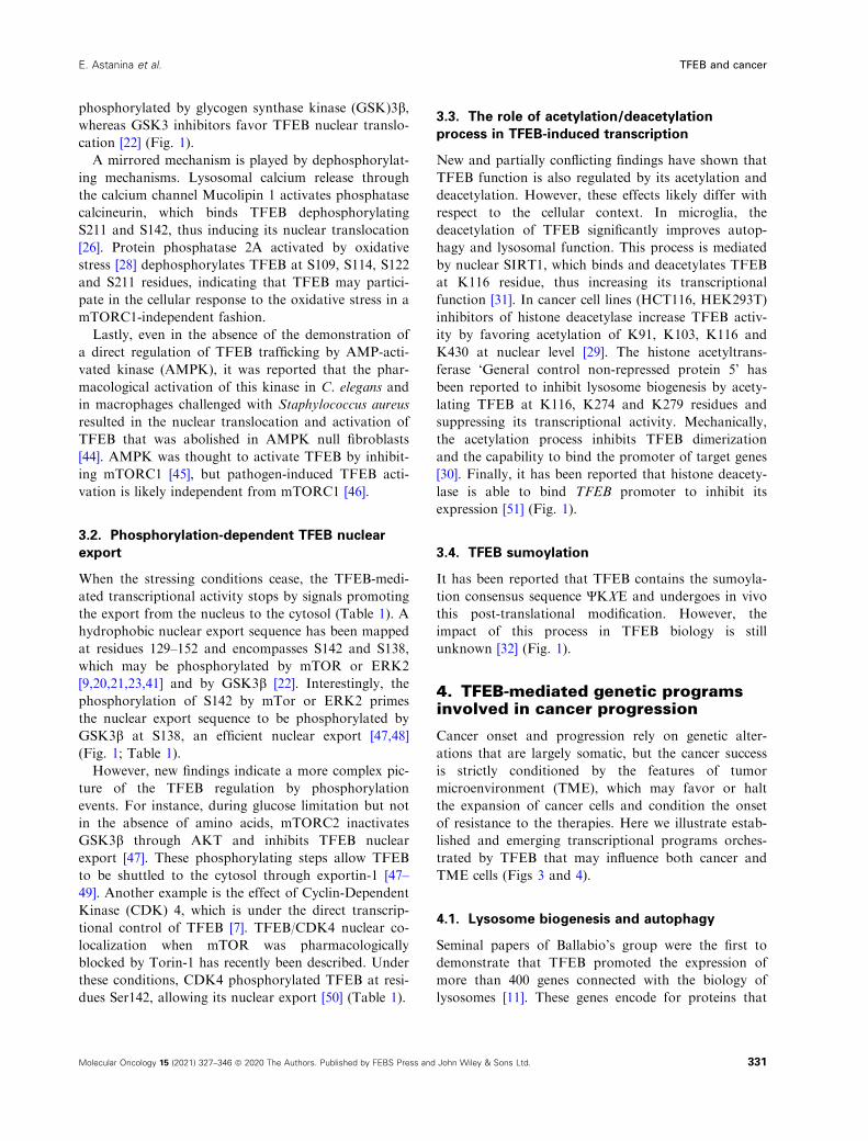
Fig. 1. Schematic representation of TFEB gene (A) and protein with the relevant domains, regions and amino acid residues undergoing post-
translational modifications (B). (A) Numbers indicate exons; ATG and TGA stop codon at exon 10 and the breakpoint cluster region are
shown. (B) AD, activation domain; Zip, leucine zipper domain.
328 Molecular Oncology 15 (2021) 327–346 ª 2020 The Authors. Published by FEBS Press and John Wiley & Sons Ltd.
TFEB and cancer E. Astanina et al.
and mammals [13–16] (Fig. 1). Efficient DNA binding
requires its homodimerization or the formation of a
heterodimer with TFE3 and MITF [5,17,18]. However,
the biological meaning of homo- and heterodimers is
still unknown. The evident homology sequence of
TFEB with MITF/TFE and TFE3 predicts a con-
served activation domain that is important for the
transcriptional activation and is able to bind p300 [19].
3. The regulatory mechanisms ofTFEB nuclear-cytosolic shuttling
Transcription factor EB is a cytosolic protein which
translocates to the nucleus to trigger specific genetic
programs. TFEB nuclear-cytosolic-shuttling as well as
its nuclear activity are regulated by post-translational
modifications including phosphorylation/dephosphory-
lation [9,20–28], acetylation/deacetylation [29–31] and
sumoylation [32] events.
3.1. Phosphorylation-dependent TFEB cytosolic
retention
The most defined system phosphorylating TFEB and
thus halting its nuclear translocation is represented by
mTORC1 [20,21,23,25] and Rag GTPases, which
determine the localization of mTORC1 and TFEB
itself on the cytosolic surface of lysosomes [21,33–35].The first 30 amino acid residues of TFEB structure
represent the Rag binding site, and its deletion or
S3A/R4A point mutations force the localization of
TFEB in the nucleus [35].
The mTORC1 localizes on lysosomes through a het-
ero-complex constituted by Ragulator, a pentameric
complex constituted of Lamtor 1–5 with guanidine
nucleotide exchange factor activity, and the Rag
GTPases, which function as heterodimers [36,37]. Rag
heterodimers consist of two functionally equivalent
pairs, RagA or RagB in complex with RagC or RagD.
Nutrients trigger the transition from the ‘inactive’
combination of GDP-bound Rag A and GTP-bound
Rag C (RagAGDP:RagCGTP) to the ‘active’ RagAGTP:
RagCGDP state. The ‘active’ Rag heterodimer binds
directly to mTORC1 and recruits it to the lysosome,
enabling its subsequent activation, which is strongly
enhanced by the binding of mTORC1 to the GTP-
bound form of ‘Ras homolog enriched in brain’
(Rheb) GTPase. Two GTPase-activating protein com-
plexes mediate, in part, the conversion between ‘active’
and ‘inactive’ Rag GTPase states. When nutrients are
low, GATOR1 promotes GTP hydrolysis of RagA or
B [38]. Conversely, nucleotide hydrolysis on RagC or
D is stimulated by folliculin (FLCN), in complex with
FLCN-interacting protein 1 or 2 [33,39]. In the
absence of amino acids, the Rags become inactive
(GDP-bound RagA/B and GTP-bound RagC/D) and
mTORC1 is released again in the cytosol (Fig. 2).
It is likely that a similar scenario occurs with TFEB,
as recently detailed by the demonstration that the N-
terminal region is involved in the binding to Rags and
relies on the nucleotide active binding configuration,
as reported for mTORC1 [35,40]. In particular, a com-
plex constituted of Raptor, TFEB, Rag and mTOR is
required for the TFEB phosphorylation and depends
on a direct interaction between TFEB and the Rags in
active form [33,35,40] (Fig. 2).
Active mTOR on lysosomal surface phosphorylates
TFEB at residues S122, S142 and S211. Once phos-
phorylation of S211 occurs, TFEB is released from the
lysosomal surface and is bound by 14–3–3 scaffold
protein, which renders TFEB inactive in the cytosol
[9,20,21,23,41]. Phosphorylated S142 and S211 induce
the degradation of TFEB through ubiquitin-protea-
some pathway [42], whereas S122 does not modify
TFEB subcellular localization but enhances the effect
of phosphorylated S211 [23]. Of note and differently
from other substrates, the mTOR-mediated TFEB
phosphorylation is independent of Rheb GTPase but
just requires Rags [40] (Fig. 1; Table 1).
However, the regulatory role of mTOR on TFEB
nuclear localization is likely more complex. In fact, the
substitution of S462, S463, S466, S467 and 469 with
phosphomimetic aspartate residues forces TFEB
nuclear translocation [43] (Fig. 1).
Besides mTOR, other serine/threonine kinases recog-
nize TFEB as substrate and modulate its localization
but the biological meaning of these modifications are
still poorly understood. Phosphorylation of TFEB
S142 by extracellular-signal-regulated kinase (ERK) 2
inhibits the nuclear entry of TFEB, and ERK inhibi-
tors induce its nuclear translocation [9,21]. In osteo-
clasts, the phosphorylation of S461, S462, S466 and
S468 residues by protein kinase (PK) Cb activated by
Receptor activator of nuclear factor kappa-Β ligand
stabilizes TFEB without affecting its subcellular local-
ization, thus suggesting a cooperative mechanism with
mTOR [24]. Mitogen-activated PK kinase kinase 3
(MAP3K3) physically associates with TFEB and phos-
phorylates S3 residue. This post-translational modifica-
tion induced by an abundance of amino acids appears
necessary to inhibit the TFEB phosphorylation at resi-
due S211 by mTORC1 [27]. AKT phosphorylates
TFEB on S467, contributing to its cytosolic retention
as confirmed by the nuclear localization of TFEB
S467A mutant [25]. Finally, TFEB S134 and S138 resi-
dues can have inhibitory functions when
329Molecular Oncology 15 (2021) 327–346 ª 2020 The Authors. Published by FEBS Press and John Wiley & Sons Ltd.
E. Astanina et al. TFEB and cancer
Fig. 2. Regulation of the nuclear translocation of TFEB by mTORC1. (Left) With few nutrients, TFEB is not phosphorylated by mTORC1 and
is free to undergo nuclear translocation. (Right) In the presence of nutrients and, in particular, amino acids, TFEB is recruited on lysosomal
surfaces and phosphorylated by active mTORC1. Phosphorylation of Ser211 allows the binding to 14-3-3 protein, which blocks its nuclear
entry. TFEB is then degraded by proteasome. For details see text (section 3).
Table 1. Role of phosphorylated serine residues in TFEB cellular localization.
Residue PK Cytosolic retention Nuclear translocation Nuclear export Other
3 MAP3K3 No effect No effect No effect Inhibits S211 phosphorylation by mTOR
122 mTOR No effect No effect No effect Enhancement of phosphorylated S211 effect
134 GSK3b Yes No effect No effect
138 GSK3b Yes No effect Yes
142 mTOR Yes No effect Yes
142 ERK 2 Yes No effect Yes
142 CDK4 No effect No effect Yes
211 mTOR Yes No effect No effect 14-3-3 binding
461 PKCb No effect No effect No effect TFEB stabilization
462 mTOR No effect Yes No effect
462 PKCb No effect No effect No effect TFEB stabilization
463 mTOR No effect Yes No effect
466 mTOR No effect Yes No effect
466 PKCb No effect No effect No effect TFEB stabilization
467 AKT Yes No effect No effect
467 mTOR No effect Yes No effect
468 PKCb No effect No effect No effect TFEB stabilization
469 mTOR No effect Yes No effect
330 Molecular Oncology 15 (2021) 327–346 ª 2020 The Authors. Published by FEBS Press and John Wiley & Sons Ltd.
TFEB and cancer E. Astanina et al.
phosphorylated by glycogen synthase kinase (GSK)3b,whereas GSK3 inhibitors favor TFEB nuclear translo-
cation [22] (Fig. 1).
A mirrored mechanism is played by dephosphorylat-
ing mechanisms. Lysosomal calcium release through
the calcium channel Mucolipin 1 activates phosphatase
calcineurin, which binds TFEB dephosphorylating
S211 and S142, thus inducing its nuclear translocation
[26]. Protein phosphatase 2A activated by oxidative
stress [28] dephosphorylates TFEB at S109, S114, S122
and S211 residues, indicating that TFEB may partici-
pate in the cellular response to the oxidative stress in a
mTORC1-independent fashion.
Lastly, even in the absence of the demonstration of
a direct regulation of TFEB trafficking by AMP-acti-
vated kinase (AMPK), it was reported that the phar-
macological activation of this kinase in C. elegans and
in macrophages challenged with Staphylococcus aureus
resulted in the nuclear translocation and activation of
TFEB that was abolished in AMPK null fibroblasts
[44]. AMPK was thought to activate TFEB by inhibit-
ing mTORC1 [45], but pathogen-induced TFEB acti-
vation is likely independent from mTORC1 [46].
3.2. Phosphorylation-dependent TFEB nuclear
export
When the stressing conditions cease, the TFEB-medi-
ated transcriptional activity stops by signals promoting
the export from the nucleus to the cytosol (Table 1). A
hydrophobic nuclear export sequence has been mapped
at residues 129–152 and encompasses S142 and S138,
which may be phosphorylated by mTOR or ERK2
[9,20,21,23,41] and by GSK3b [22]. Interestingly, the
phosphorylation of S142 by mTor or ERK2 primes
the nuclear export sequence to be phosphorylated by
GSK3b at S138, an efficient nuclear export [47,48]
(Fig. 1; Table 1).
However, new findings indicate a more complex pic-
ture of the TFEB regulation by phosphorylation
events. For instance, during glucose limitation but not
in the absence of amino acids, mTORC2 inactivates
GSK3b through AKT and inhibits TFEB nuclear
export [47]. These phosphorylating steps allow TFEB
to be shuttled to the cytosol through exportin-1 [47–49]. Another example is the effect of Cyclin-Dependent
Kinase (CDK) 4, which is under the direct transcrip-
tional control of TFEB [7]. TFEB/CDK4 nuclear co-
localization when mTOR was pharmacologically
blocked by Torin-1 has recently been described. Under
these conditions, CDK4 phosphorylated TFEB at resi-
dues Ser142, allowing its nuclear export [50] (Table 1).
3.3. The role of acetylation/deacetylation
process in TFEB-induced transcription
New and partially conflicting findings have shown that
TFEB function is also regulated by its acetylation and
deacetylation. However, these effects likely differ with
respect to the cellular context. In microglia, the
deacetylation of TFEB significantly improves autop-
hagy and lysosomal function. This process is mediated
by nuclear SIRT1, which binds and deacetylates TFEB
at K116 residue, thus increasing its transcriptional
function [31]. In cancer cell lines (HCT116, HEK293T)
inhibitors of histone deacetylase increase TFEB activ-
ity by favoring acetylation of K91, K103, K116 and
K430 at nuclear level [29]. The histone acetyltrans-
ferase ‘General control non-repressed protein 5’ has
been reported to inhibit lysosome biogenesis by acety-
lating TFEB at K116, K274 and K279 residues and
suppressing its transcriptional activity. Mechanically,
the acetylation process inhibits TFEB dimerization
and the capability to bind the promoter of target genes
[30]. Finally, it has been reported that histone deacety-
lase is able to bind TFEB promoter to inhibit its
expression [51] (Fig. 1).
3.4. TFEB sumoylation
It has been reported that TFEB contains the sumoyla-
tion consensus sequence ΨKXE and undergoes in vivo
this post-translational modification. However, the
impact of this process in TFEB biology is still
unknown [32] (Fig. 1).
4. TFEB-mediated genetic programsinvolved in cancer progression
Cancer onset and progression rely on genetic alter-
ations that are largely somatic, but the cancer success
is strictly conditioned by the features of tumor
microenvironment (TME), which may favor or halt
the expansion of cancer cells and condition the onset
of resistance to the therapies. Here we illustrate estab-
lished and emerging transcriptional programs orches-
trated by TFEB that may influence both cancer and
TME cells (Figs 3 and 4).
4.1. Lysosome biogenesis and autophagy
Seminal papers of Ballabio’s group were the first to
demonstrate that TFEB promoted the expression of
more than 400 genes connected with the biology of
lysosomes [11]. These genes encode for proteins that
331Molecular Oncology 15 (2021) 327–346 ª 2020 The Authors. Published by FEBS Press and John Wiley & Sons Ltd.
E. Astanina et al. TFEB and cancer
orchestrate the expression, localization, entrance,
influx and performance of lysosomal and non-lysoso-
mal enzymes participating in the destruction of cellular
macromolecules such as proteins, glycosaminoglycans,
lipids, glycogen and hemoglobin [6,8,9]. In addition,
TFEB can promote lysosomal exocytosis, allowing
their cargo secretion through the fusion to cell mem-
brane [26]. In the CLEAR network there are some
genes that are known to play a direct role in the differ-
ent steps of autophagy (ATG9, UVRAG, VPS11,
VPS18, WIPI) [9].
These findings have been immediately connected
with the instrumental role of lysosomes in the ‘lysoso-
mal storage diseases’ and in the establishment of
autophagic flux [52]. At present, TFEB may be consid-
ered one of the most important principle regulators of
autophagy which has opposing, context-dependent
roles in cancer. The role of autophagy in cancer has
been extensively reviewed elsewhere, which we refer to.
Two opposite scenarios may be envisaged. Failure of
autophagy interferes with oncogene-induced senescence
and thereby might favor uncontrolled proliferation of
cancer cells. Conversely, in established cancers, autop-
hagy is required for cell survival by providing nutrients
[53–56]. Accordingly, specific correlations between the
level of TFEB and the autophagic flux have been
described in several cancers (breast, pancreas, lung and
in part discussed in section 5 [57–60].
4.2. Cell cycle
A first suggestion that TFEB regulates the cell cycle
was provided in cancer cell lines lacking TFEB and
TFE3 and treated with the genotoxic agent etoposide.
In this model, the knock-out of these TF resulted in
reduced expression of multiple genes implicated in cell
cycle progression as well as genes involved in chromo-
some segregation and cytokinesis [61]. These data were
confirmed in a model of triple negative breast cancer,
in which TFEB silencing combined with doxorubicin
resulted in a down-modulation of cell-cycle genes.
These observations were refined in TFEB-deleted
endothelial cells [7] and hepatoblasts [62], which
showed a block of the G1–S cycle transition. In
endothelial cells, TFEB bound (CDK4) promoter and
in the absence of TFEB, its transcriptional rate was
impaired. As a consequence the phosphorylation of
Retinoblastoma protein is reduced, blocking the
nuclear translocation of E2F to transcript genes neces-
sary for S-phase [7]. Similarly, in HeLa cells, TFEB
deletion resulted in reduced Rb phosphorylation and
the TFEBS142A active mutant increased the expres-
sion of CDK4 and CDK7 [61]. The direct transcrip-
tional control of CDK4 by TFEB, together with the
recent observation that TFEB Ser142 residue is a sub-
strate of CDK4 itself [50], opens new perspectives in
the co-regulation of lysosome biogenesis and cell cycle
(Fig. 5).
Besides inhibiting CDK4, TFEB depletion reduces
the expression level of p21 cyclin kinase inhibitor,
while its overexpression has an opposite effect through
a direct binding to its promoter in a p53-dependent
manner [63]. Doxorubicin-mediated DNA damage pro-
moted the activation of CDKN1A (CDK Inhibitor 1A;
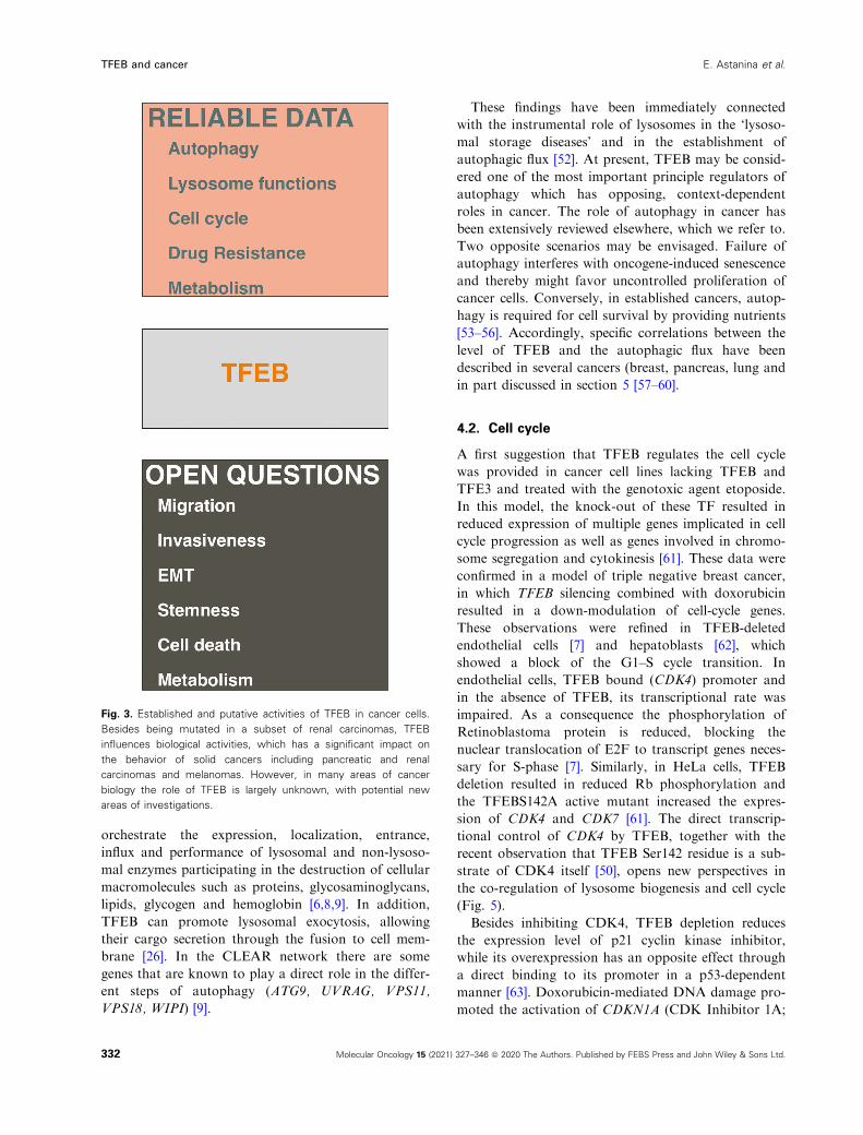
Fig. 3. Established and putative activities of TFEB in cancer cells.
Besides being mutated in a subset of renal carcinomas, TFEB
influences biological activities, which has a significant impact on
the behavior of solid cancers including pancreatic and renal
carcinomas and melanomas. However, in many areas of cancer
biology the role of TFEB is largely unknown, with potential new
areas of investigations.
332 Molecular Oncology 15 (2021) 327–346 ª 2020 The Authors. Published by FEBS Press and John Wiley & Sons Ltd.
TFEB and cancer E. Astanina et al.
p21) through TFEB, leading to cell cycle arrest [63].
Of note, a positive regulatory loop between p53 and
TFEB has been described. In HeLa cell p53 activated
TFEB via the up-regulation of Sesn1 and Sesn2, which
inhibits mTORC1 signaling. In parallel, TFEB further
enhanced p53 stabilization [61] (Fig. 5).
A further link between cell cycle, TFEB activity and
autophagy has been recently demonstrated in a cellular
model, in which CDK inhibitor 1B (p27) was deleted.
This study demonstrated that p27 was recruited to
lysosomes to interact with LAMTOR1, a component
of the Ragulator complex required for mTORC1 acti-
vation. Binding of p27 to LAMTOR1 prevented Ragu-
lator assembly and mTORC1 activation, promoting
autophagy in a TFEB-dependent manner [64].
Another control of cell proliferation by TFEB relies
on apoptosis. TFEB knockdown induced cell death
and the decrease of cell viability via the up-regulation
of pro-apoptotic molecules belonging to interferon and
tumor necrosis factor pathways and the down-modula-
tion of apoptosis inhibitors [65].
4.3. Metabolism
In cancer, malignant cells show a high metabolic flexi-
bility to adapt themselves in response to cell-extrinsic
and cell-intrinsic cues. According to the role of TFEB
in regulating autophagy and the diverse substrates that
can be degraded through this process, it is not surpris-
ing that TFEB has the potential to participate indi-
rectly with nearly all aspects of carbon metabolism,
including mitochondrial activities [66]. However, direct
and specific effects of TFEB on lipid metabolism have
been described. TFEB recognizes binding sites on the
promoters of Peroxisome proliferator-activated recep-
tor (Ppar) gamma coactivator 1a (PPARGC) and Ppar
c2 (PPARG2), which are regulators of lipid anabolic
and catabolic pathways [67,68]. When overexpressed in
the liver, TFEB increases the expression of
PPARGC1a, which fosters the b-oxidation of fatty
acids and the ketogenesis. Conversely, TFEB reduces
the expression of enzymes involved in some lipid
biosynthetic pathways [69]. However, in mice fed with
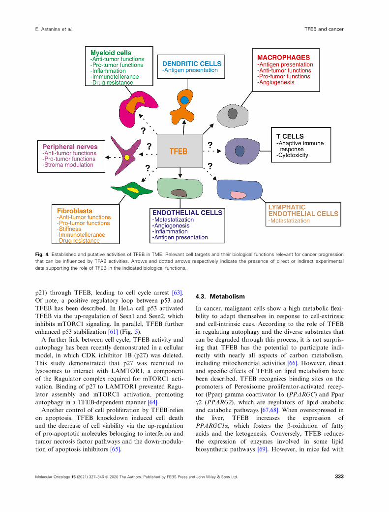
Fig. 4. Established and putative activities of TFEB in TME. Relevant cell targets and their biological functions relevant for cancer progression
that can be influenced by TFAB activities. Arrows and dotted arrows respectively indicate the presence of direct or indirect experimental
data supporting the role of TFEB in the indicated biological functions.
333Molecular Oncology 15 (2021) 327–346 ª 2020 The Authors. Published by FEBS Press and John Wiley & Sons Ltd.
E. Astanina et al. TFEB and cancer
western diet, TFEB overexpression has been reported
to upregulate enzymes involved in cholesterol and bile
acid synthesis [70]. In adipose tissue, TFEB expression
stimulated by glucose and insulin is necessary to sus-
tain adipocyte differentiation by a mechanism medi-
ated by Pparc2 [71], which promotes lipogenesis [72].
In cardiomyocytes, TFEB deletion reduces their ability
to oxidize fatty acids and shifts the energetic metabo-
lism towards the use of glucose [73]. Finally, emerging
findings demonstrate that TFEB is involved in cAMP-
induced lipolysis regulated by calcium influx mediated
by the store-operated calcium entry [74].
Besides controlling lipid metabolism, in active skele-
tal muscle, TFEB controls energy expenditure and
mitochondrial activity in an autophagy- and Ppargc1-
independent manner. Overexpression of TFEB induces
mitochondrial biogenesis and improves oxidative
phosphorylation. These effects are associated to direct
control of TFEB of glucose homeostasis through
GLUT1/4 expression and insulin sensitivity via nitric
oxide synthase [75].
Therefore, it is intriguing to speculate that the effect
of TFEB on metabolism might depend on specific tis-
sue commitments and defined metabolic conditions.
This hypothesis is further sustained by the control of
TFEB expression by the cAMP response element-bind-
ing protein [76], which regulates lipid and glucose
metabolism.
4.4. Epithelial-mesenchymal transition
Epithelial-mesenchymal transition (EMT) has a key
role in neoplastic transformation and in the metastatic
process [77]. The role of TFEB in establishing the
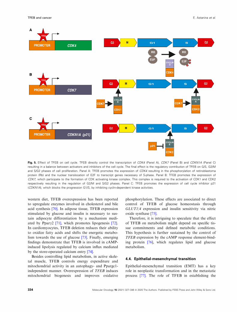
Fig. 5. Effect of TFEB on cell cycle. TFEB directly control the transcription of CDK4 (Panel A), CDK7 (Panel B) and CDKN1A (Panel C)
resulting in a balance between activators and inhibitors of the cell cycle. The final effect is the regulatory contribution of TFEB on G/S, G2/M
and S/G2 phases of cell proliferation. Panel A: TFEB promotes the expression of CDK4 resulting in the phosphorylation of retinoblastoma
protein (Rb) and the nuclear translocation of E2F to transcript genes necessary of S-phase. Panel B: TFEB promotes the expression of
CDK7, which participate to the formation of CDK activating kinase complex. This complex is required to the activation of CDK1 and CDK2
respectively resulting in the regulation of G2/M and S/G2 phases. Panel C: TFEB promotes the expression of cell cycle inhibitor p21
(CDKN1A), which blocks the progression G1/S, by inhibiting cyclin-dependent kinase activities.
334 Molecular Oncology 15 (2021) 327–346 ª 2020 The Authors. Published by FEBS Press and John Wiley & Sons Ltd.
TFEB and cancer E. Astanina et al.
equilibrium between epithelial and mesenchymal phe-
notypes was discovered in 2005 [78] and has not been
studied in depth since. TFEB overexpressed in 3T3
and mouse embryonic fibroblasts directly activated E-
cadherin promoter, and TFEB was shown to be
required for E-cadherin endogenous expression in
these cells. TFEB also upregulated WT1 [78], a TF
able to regulate EMT in both directions depending on
context [79] and decreased the expression of EMT reg-
ulator Snail. Conversely, E-cadherin expression in
epithelial cell lines did not depend on TFEB. More-
over, TFEB overexpression led to E-cadherin downreg-
ulation. Taking into consideration EMT as a milestone
of cancer development, it would be worth investigating
whether TFEB is involved.
5. TFEB and cancer subtypes
Genetic alterations of TFEB are mainly involved in
the pathogenesis of tumors developing in kidney, exo-
crine pancreas and melanomas. Besides these cancers,
TFEB alterations have been described in colorectal
cancer [80,81], gastric carcinoma [82], non-small cell
lung cancer [59] and breast cancer [83]
5.1. Renal carcinomas
Gene fusions involving two members of the MiT fam-
ily, TFEB and TFE3, characterize a subset of sporadic
clear cell renal carcinomas often characterized by
expression of cathepsin k and melanocytic markers
and defined as MiT family translocation renal cell car-
cinoma [84]. The most common variant results from
the fusion between TFEB and the non-protein encod-
ing Metastasis associated lung adenocarcinoma tran-
script 1 gene (MALAT1) on chromosome 11q13.
Fusion occurs within a breakpoint cluster region
upstream of TFEB exon 3 or 4 and within a 1205-bp
breakpoint region of MALAT1 [85–87]. This transloca-tion results in the substitution of TFEB promoter and
a consistent increase of TFEB protein level able to
translocate into the nucleus [85,86]. When overex-
pressed in renal carcinoma cell lines, MALAT1-TFEB
increases cell proliferation, invasiveness and in vivo
tumorigenicity [88]. Single cases with different fusion
patterns have been described and include KHDRBS2,
COL21A1, CADM2, CLTC and EWSR1 [87].
The pathogenetic role of the overexpression of
TFEB in this subset of clear cell renal carcinomas has
been recently supported by the analysis of transgenic
mice in which TFEB was specifically expressed in kid-
ney under the control of cadherin 16 promoter. These
mice developed tubular cysts morphologically similar
to those observed in MALAT1-TFEB clear cell renal
carcinomas, and then showed metastatic cancer. This
process was autophagy-independent and characterized
by activation of Wnt pathway. Interestingly, a recent
report in gastric carcinomas extends this observation,
showing that TFEB activated Wnt/b-catenin signaling
to initiate a pro-invasive program [82]. The crosstalk
between TFEB and Wnt/b-catenin pathways is emerg-
ing as a relevant and probably more general mecha-
nism of oncogenesis [90]. The molecular candidate to
bridge these pathways is GSKb, a regulator of TFEB
nuclear trafficking [22,91] which belongs to the Wnt
degradation complex [92]. It has been reported that
TFEB inhibition in AMPK null mice resulted in
impairment of endodermal differentiation. The com-
promised endolysosomal system resulting from TFEB
inactivation blunted Wnt pathway [45]. It is intriguing
to speculate that upon Wnt activation, components of
Wnt degradation complex, including GSKb, are
sequestered into multivesicular bodies [92] enabling
TFEB stabilization and nuclear activity.
Transcription factor EB is also a determinant effec-
tor of Birt–Hogg–Dub�e syndrome, a genetic disease
caused by germ line mutations in the RagC and RagD
activator FLCN and characterized by benign skin
tumors, lung and kidney cysts and renal cell carcinoma
[93]. Mice with kidney-specific knockout of Flcn devel-
oped polycystic disease and pre-neoplastic foci with
increased nuclear localization of TFEB. This pheno-
type reverted when Flcn null mice were backcrossed
with mice lacking renal Tfeb [40].
The oncogenic role of overexpressed TFEB may also
result from its amplification and in the last decade
many cases of renal carcinomas with amplified TFEB
have been reported [94]. Of note, in some cases TFEB
was co-amplified with VEGFA [95], known to be a piv-
otal player in tumor angiogenesis and to be amplified
in some aggressive types of colorectal carcinoma.
TFEB has been also implicated in the mechanisms
of acquired resistance to mTOR inhibitors, which are
exploited in the treatment of metastatic renal carcino-
mas. The inhibition of mTOR led to enhanced TFEB
nuclear translocation and CD274 (Programmed
Death-Ligand 1; PD-L1) expression by a direct activa-
tion of its promoter, resulting in an immune evasion
via suppression of the cytotoxic function of CD8+
cells [96]
5.2. Pancreatic carcinomas
Pancreatic adenocarcinomas show unique and specific
features dependent on an extensive and prominent
stromal reaction, resulting in a hypovascular and
335Molecular Oncology 15 (2021) 327–346 ª 2020 The Authors. Published by FEBS Press and John Wiley & Sons Ltd.
E. Astanina et al. TFEB and cancer
hypoxic TME, reprogramming of cellular metabolism
and evasion of tumor immunity [97].
Recently, increased mRNA and protein expression
of TFEB, MITF and TFE3 was detected in pancreatic
ductal adenocarcinoma (PDAC) cell lines and patient
cancers resulting in increased autophagic flux [57,98],
which increases amino acid availability exploited for
tumor growth. Interestingly, this subset of tumors is
characterized by the failure to traffic these TF because
of an aberrant expression of importin 8 [57], which
regulates direct nuclear import of specific cargos. Fur-
thermore, reduction of TFEB expression impairs
PANC1 anchorage-independent growth, indicating its
relevant role in pancreatic oncogenesis [91].
The role of TFEB in sustaining PDAC progression
is further supported by studies on micro RNA (miR)
29a, which downregulates TFEB through direct inter-
actions with the 30UTR binding site and reduces
autophagy and invasive properties of PDAC cell lines
[99]. Interestingly, transforming growth factor b(TGF-b), which is one of the signaling systems pivotal
in PDAC behavior [97], has been demonstrated to acti-
vate a TFEB-mediated autophagic process that favors
PDAC cell migration and metastasis [100].
TFEB also seems to be involved in mechanisms of
acquired resistance to targeted therapies in PDAC. In
in vitro and in vivo PDAC models, MEK inhibitors
enhanced lysosome biogenesis in a TFEB-dependent
manner, which resulted in sequestration and inactiva-
tion of the inhibitor in the lysosomal compartment. It
is likely that genetic depletion of TFEB leads to a
decreased lysosomal biogenesis of MEK inhibitors and
potentiates tumor sensitivity to MEK inhibition [101].
5.3. Melanomas
The MiT gene family includes MITF, which is recog-
nized as a master regulator of melanogenesis and a
melanoma oncogene [102]. Increasing evidence indi-
cates a potential role of TFEB in melanoma oncogene-
sis and a specific regulatory circuit has been described
between MITF and TFEB itself. In fact, MITF posi-
tively regulates the expression of TFEB by a direct
control of promoter activity through the binding to
intron 1 [103].
Human melanomas are characterized in 40–60% of
the cases by the presence of mutated BRAF [104]. In
BRAF-mutated melanomas, autophagy has been
reported to exert both pro- and anti-tumor activity
[105,106]. Furthermore, BRAF inhibitors, which are
currently the standard therapeutic regimen in
BRAFV600E melanomas, induce autophagy [107].
BRAFV600E has been reported to phosphorylate TFEB
by an Erk-dependent mechanism and inhibit TFEB
transcriptional programs. The use of a BRAF inhibitor
has been demonstrated to reverse this condition, acti-
vate autophagy and reduce in vivo tumor growth.
Accordingly, the expression of the dominant active
TFEBS142A in A375 melanoma cells increased their
in vivo tumorigenic activity. The inhibition of the tran-
scriptional program triggered by TFEB, promoted
tumor progression and chemoresistance to BRAF inhi-
bitor, which were associated with TGF-b-mediated
epithelial–mesenchymal transition [108].
Melanomas are characterized by a high mutational
burden, which represents a molecular advantage in
the response to immunotherapy strategies. Interest-
ingly, in metastatic melanomas, TFEB is positively
correlated with the expression of genes required for
the immune response, posing important therapeutic
questions about the role of TFEB in immunotherapy
[109].
6. TFEB and tumor microenvironment
The tumor microenvironment encompasses a heteroge-
neous population of differentiated and progenitor cells
that play a critical role in regulating tumor fate. For
example, endothelial cells, their precursors and peri-
cytes are essential for tumor angiogenesis, a process
that allows oxygen and nutrient supply, and the new
vessels formed contribute to the recruitment of leuko-
cytes and the escape of tumor cells from the primary
tumor [110]. Fibroblasts and activated fibroblasts (can-
cer-associated fibroblasts) dictate the features of extra-
cellular matrix and its stiffness, support or contrast
tumor growth as well as the immune response [111].
The cellular arms of innate and adaptive immunity
participate to a complex network of signals, which
drive an efficient or inadequate immune response to
cancer cells and their invasiveness properties [112,113].
Peripheral nerves (sympathetic, parasympathetic and
sensory) interact with tumor and stromal cells to pro-
mote the initiation and progression of a variety of
malignancies [114]. Finally, TME strongly influences
the response to therapies and can contribute to the
acquisition of resistance [115].
6.1. Immune system
Transcription factor EB was discovered as a binding
protein of the µE3 enhancer of the human
immunoglobulin heavy chain locus in human B cells
[4]. This finding prefigured a role of TFEB in regulat-
ing activities of the immune system and many studies
have indicated so far that TFEB has a principal role
336 Molecular Oncology 15 (2021) 327–346 ª 2020 The Authors. Published by FEBS Press and John Wiley & Sons Ltd.
TFEB and cancer E. Astanina et al.
in regulating aspects of innate immunity conserved
during the evolution from worm [44,116] to human.
In this scenario, the most relevant cellular target of
TFEB are phagocytic cells, in particular macrophages,
involved in removing pathogens. The effect of TFEB
in protecting cells against infective agents is mainly
linked to the biogenesis of lysosomes and their role in
phagocytosis [44,46,117].
TFEB also regulates other macrophage functions
more strictly connected with cancer biology indepen-
dently from the canonical regulation of autophagy and
lysosome activities.
Macrophages undergo specific differentiation
depending on the local tissue environment and assume
distinct functional phenotypes. In TME, macrophages
can differ in M1 or M2 phenotypes, which respectively
have anti- and pro-tumor activities [118].
Transcription factor EB is probably able to activate
both M1- or M2-like macrophages according to the
features of inflammatory stimulus [119–121].In alveolar- and bone marrow-derived macrophages,
the deletion of LAMTOR1, a scaffold protein required
to activate mTORC1, results in TFEB activation, and
increases autophagy and acquisition of the M2 pheno-
type. This process is connected with the increased pro-
duction of interleukin-6 and tumor necrosis factor-aby a direct effect of TFEB on their promoters [119]. In
a model in which mesenchymal stem cells are co-cul-
tured with macrophages, the activation of TFEB
increases the polarization of M2-like macrophages by
a lysosomal-dependent mechanism [121].
Tumor-infiltrating macrophages show the opposite
response. In macrophages, TFEB upregulated suppres-
sor of cytokine signaling 3, halting Stat3 activation
and thereby blocking M2-like polarization indepen-
dently of the stimulated autophagy. Furthermore,
TFEB activated the transcription of PPARG, which
blunts NFjB activation, resulting in downregulation
of the inflammatory response [120,122]. When macro-
phages are stimulated with conditioned media of dif-
ferent tumor cell types, TFEB is retained in the
cytosol with the appearance of markers of M2 polar-
ization. These macrophages show a reduced ability to
present the antigen by down-modulation of major his-
tocompatibility complex (MHC)-II and the co-stimula-
tory molecule CD80. In vivo, macrophages lacking
TFEB co-injected with tumor cells enhance tumor
growth with increased infiltration of M2-like macro-
phages, reduced infiltration of CD8+ cells, and
enhanced angiogenesis [120,122]. Additional insights
for the M1-polarizing effect of TFEB in a tumor con-
text are provided by the anti-tumor effect of chloro-
quine, which switches macrophages from the M2 to
the M1 phenotype by a TFEB-dependent mechanism
[123].
The TFEB-regulating effect of lysosome functions is
further exploited by antigen-presenting cells, in partic-
ular by dendritic cells. MHC I and II molecules pre-
sent protein fragments to CD8+ and CD4+ T cells,
respectively, with the final aim to eliminate host anti-
gens. Endogenously synthesized antigens in the cytosol
are presented as peptides bound to MHC I molecules,
whereas exogenous antigens ingested into endocytic
compartments of antigen-presenting cells are presented
as peptides bound to MHC II molecules. In addition
to these pathways, exogenous antigens can be pro-
cessed to MCH class I by cross-presentation [124]. In
dendritic cells, activated TFEB downregulates MHC
class I-restricted antigen cross-presentation and upreg-
ulates the processing and presentation of antigen by
MHC class II. Mechanistically, this effect results from
the increased expression of lysosomal proteases cou-
pled with increased lysosome acidification [125]. Fur-
thermore, TFEB participates in the recruitment of
dendritic cells to the lymph node where antigen presen-
tation occurs. Upon lipopolysaccharide sensing, lyso-
somal calcium efflux by transient receptor potential
cation channel, mucolipin subfamily-1 (TRPML1) pro-
motes sustained myosin IIA activity at the rear of the
dendritic cells and thus stabilizes F-actin and increased
cell migration. Concomitantly, TFEB translocates to
the nucleus to maintain a high level of trpml1 gene
expression [126].
The regulatory role of TFEB in antigen presentation
to T cells by antigen-presenting cells could influence
the cancer treatment based on immunotherapies. In
particular, emerging data indicate a good correlation
between the amount of tumor neoantigens and the suc-
cess of clinical tumor immunotherapies [127].
A role of TFEB in humoral immunity has been
envisaged due to its ability to bind CD40LG promoter
and positively regulate its expression in activated
CD4+ cells. In this way, TFEB controls the T-cell-de-
pendent immunoglobulin response [128].
6.2. Vascular system
Although the regulatory activities of TFEB in tumor
angiogenesis are still unknown, numerous studies in
embryo development and in other pathological condi-
tions support a framework suggesting a role of TFEB
in the formation of the tumor vascular bed.
Mouse mutants have highlighted the role of TFEB
as a pro-angiogenic factor both in embryonic [14] and
in adult life [7,129]. Mice carrying a null mutation at
the Tfeb locus in a homozygous state are characterized
337Molecular Oncology 15 (2021) 327–346 ª 2020 The Authors. Published by FEBS Press and John Wiley & Sons Ltd.
E. Astanina et al. TFEB and cancer
by defective vascularization of the placenta, leading to
death of the embryos between E9.5 and E11.5. In
wild-type mice, Tfeb was expressed at very low levels
at E8.5- to E10.5-day-old embryos but highly
expressed in labyrinthine trophoblasts from 8.5-day-
old placentas. In Tfeb null mice, the vascular invasion
of labyrinthine trophoblast layer is blocked and capil-
laries stop in the chorion. Mechanistically, Tfeb null
mice expressed a lower level of Vascular Endothelial
Growth Factor (VEGFA) in the trophoblast than did
wild-type mice. The placenta hypovascularity results in
severe hypoxia, which determines embryo death. This
observation has been further refined in endothelial-
specific Tfeb null mouse mutants and in human
TFEB-silenced endothelial cells [7]. In this model,
endothelial cells start to express Tfeb at E8.5, which is
maintained after birth. Embryonic lethality of mutants
is observed at E10.5. The defects of vascular structures
rely mainly on an impairment of the vascular remodel-
ing of the primitive vascular plexus. After birth, Tfeb
deletion inhibits the retinal vascularization and the
maturation of renal glomeruli, with loss of endothelial
fenestration and podocyte foot processes. Study of the
molecular mechanisms sustaining this phenotype indi-
cates that in endothelial TFEB null mice, the cell cycle
is blocked for the reduced expression of CDK4 and
other mitotic genes. Furthermore, these mice carry a
defect of VEGFR2 functions based on the combined
effect of Tfeb deletion on Vascular endothelial growth
factor receptor 2 (VEGFR2) trafficking and the post-
transcriptional regulation of its gene expression. TFEB
inhibits the expression of Myosin 1C, which delivers
VEGFR2 to plasma membrane [130] and inhibits the
expression of miRNA-15a/16-1 cluster, which specifi-
cally targets VEGFR2 30UTR [131].
During the tumor angiogenic process, autophagy in
the blood vessels is emerging as a critical mechanism
enabling endothelial cells dynamically to accommodate
their higher energy demands to the extracellular envi-
ronment and connect with other components of the
tumor stroma through paracrine signaling [132]. Inter-
estingly, extracellular proteoglycan decorin has been
demonstrated to activate endothelial autophagy
through TFEB and its nuclear translocation required
the catalytic activity of VEGFR2 [133]. This study
parallels the observation that VEGF stimulates the
expression and activation of TFEB [129].
Transcription factor EB has also been reported to
play a role in the angiogenic response after ischemia.
Endothelial overexpression of TFEB improves blood
perfusion and increases capillary density after hindlimb
ischemia in mouse. The TFEB pro-angiogenic effect is
mediated by the significantly increased expression of
autophagy genes and by the activation of AMPK sig-
naling [129].
The role of capillaries in tumor progression is not
just limited to providing nutrients and oxygen.
In the metastatic cascade, the interactions of the
metastatic cancer cell with the vascular wall in the pri-
mary tumors and in distant organs is largely intuitive.
In the primary tumor, the capillaries are poorly struc-
tured and leaky, thus facilitating the intravasation of
cancer cells to the bloodstream. In contrast, the
extravasation in a secondary tissue is highly selective
and is considered a limiting step of the metastatic pro-
cess [134]. Many studies indicate that metastatic cancer
cells actively adhere to the endothelial surface and then
pass through the cell homotypic junction, similar to
that observed during leukocyte diapedesis in inflamma-
tory processes [135]. Recently, some studies have drawn
attention to the endothelial anti-inflammatory role of
TFEB [136,137]. TFEB activation in endothelial cells
results in the reduction of the synthesis of inflammatory
cytokines and adhesive molecules and the adhesion of
circulating monocytes. These effects are independent of
the autophagic flux but rely on the suppressor activity
on the Nuclear factor kappa-light-chain-enhancer of
activated B cells (NF-jB) pathway. TFEB suppressed
IjB kinase activity to protect IjB-a from degradation,
leading to reduced p65 nuclear translocation [136]. Fur-
thermore, the overexpression of TFEB in endothelial
cells activates the production of anti-oxidant species by
direct binding on the promoter of heme oxygenase-1
and superoxide dismutase 2 [137].
6.3. Peripheral nervous system
In the same way that increased blood vessel formation
is necessary for tumor growth, nerve density nearly
doubles in tumors compared with non-neoplastic tissue
controls and with the increase in nerve density corre-
lated with aggressiveness of many solid tumors, includ-
ing prostate, colon, head and neck, pancreas stomach
and lung [138–140]. The effect of peripheral nervous
system stimulation on cancer is largely unknown but
the data available indicate that adrenergic and sensory
stimuli exert pro-tumoral activity, whereas cholinergic
signals exhibit tissue-dependent effects. The overall
effect of peripheral nervous stimulation on cancer pro-
gression depends on a direct modulation of both can-
cer and stroma cells [114]. The role of TFEB in the
modulation of cancer peripheral nervous system is
mostly speculative. Of note, TFEB has been demon-
strated to regulate the formation of myelin [141,142],
which is instrumental in perineural invasion and tumor
spread [114,143].
338 Molecular Oncology 15 (2021) 327–346 ª 2020 The Authors. Published by FEBS Press and John Wiley & Sons Ltd.
TFEB and cancer E. Astanina et al.
7. Conclusions
The emerging results about the molecular and biologi-
cal activities of TFEB envisage relevant functions in a
wide array of cancers. However, TFEB therapeutic tar-
geting is dependent on a detailed understanding of the
mechanistic complexities that govern TFEB activities
and several issues have to be faced and solved. First,
understanding the impact of deregulated kinase cas-
cades described in many cancers on the phosphorylat-
ing events involved in TFEB regulation may help
pinpoint possible interactions between small kinase
inhibitors with drugs that modulate this TF [144].
Secondly, TFEB represents a node of an undefined
TF network, which drives specific and context-depen-
dent transcriptional programs. So far, little informa-
tion (Table 2) is available on TF-regulating TFEB
expression, as well as on those TF genes regulated by
TFEB. These interactions pose the question of the
genetic programs orchestrated or modulated by TFEB,
besides the so far well-established canonical pathway
sustaining autophagy. Furthermore, it might be rele-
vant to describe an association between the level of
TFEB activation and the type of transcriptional
response. A paradigmatic example is provided by the
hypoxia-inducible factor, which determines the reper-
toire of gene expression according to the severity of
hypoxic conditions [145]. Attempts to put TFEB in a
defined transcriptional cellular landscape are manda-
tory to understand how the different and in part unre-
lated biological activities described in this review (e.g.
the role of TFEB in immune system) participate in a
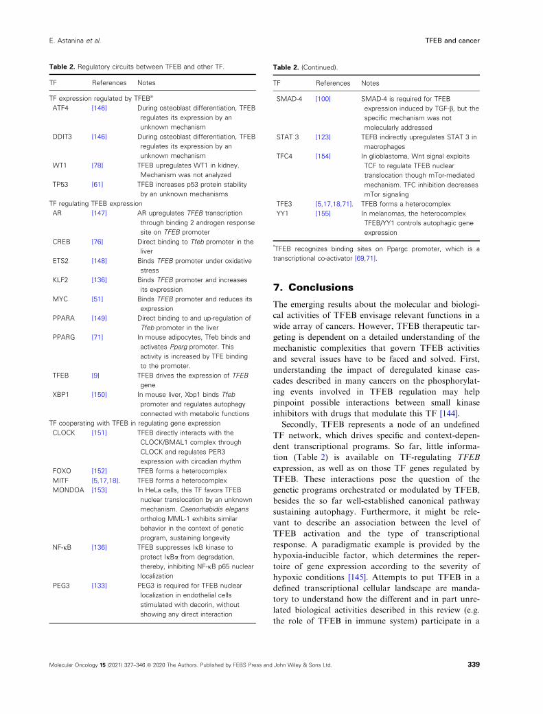
Table 2. Regulatory circuits between TFEB and other TF.
TF References Notes
TF expression regulated by TFEBa
ATF4 [146] During osteoblast differentiation, TFEB
regulates its expression by an
unknown mechanism
DDIT3 [146] During osteoblast differentiation, TFEB
regulates its expression by an
unknown mechanism
WT1 [78] TFEB upregulates WT1 in kidney.
Mechanism was not analyzed
TP53 [61] TFEB increases p53 protein stability
by an unknown mechanisms
TF regulating TFEB expression
AR [147] AR upregulates TFEB transcription
through binding 2 androgen response
site on TFEB promoter
CREB [76] Direct binding to Tfeb promoter in the
liver
ETS2 [148] Binds TFEB promoter under oxidative
stress
KLF2 [136] Binds TFEB promoter and increases
its expression
MYC [51] Binds TFEB promoter and reduces its
expression
PPARA [149] Direct binding to and up-regulation of
Tfeb promoter in the liver
PPARG [71] In mouse adipocytes, Tfeb binds and
activates Pparg promoter. This
activity is increased by TFE binding
to the promoter.
TFEB [9] TFEB drives the expression of TFEB
gene
XBP1 [150] In mouse liver, Xbp1 binds Tfeb
promoter and regulates autophagy
connected with metabolic functions
TF cooperating with TFEB in regulating gene expression
CLOCK [151] TFEB directly interacts with the
CLOCK/BMAL1 complex through
CLOCK and regulates PER3
expression with circadian rhythm
FOXO [152] TFEB forms a heterocomplex
MITF [5,17,18]. TFEB forms a heterocomplex
MONDOA [153] In HeLa cells, this TF favors TFEB
nuclear translocation by an unknown
mechanism. Caenorhabidis elegans
ortholog MML-1 exhibits similar
behavior in the context of genetic
program, sustaining longevity
NF-jB [136] TFEB suppresses IjB kinase to
protect IjBa from degradation,
thereby, inhibiting NF-jB p65 nuclear
localization
PEG3 [133] PEG3 is required for TFEB nuclear
localization in endothelial cells
stimulated with decorin, without
showing any direct interaction
Table 2. (Continued).
TF References Notes
SMAD-4 [100] SMAD-4 is required for TFEB
expression induced by TGF-b, but the
specific mechanism was not
molecularly addressed
STAT 3 [123] TEFB indirectly upregulates STAT 3 in
macrophages
TFC4 [154] In glioblastoma, Wnt signal exploits
TCF to regulate TFEB nuclear
translocation though mTor-mediated
mechanism. TFC inhibition decreases
mTor signaling
TFE3 [5,17,18,71]. TFEB forms a heterocomplex
YY1 [155] In melanomas, the heterocomplex
TFEB/YY1 controls autophagic gene
expression
a
TFEB recognizes binding sites on Ppargc promoter, which is a
transcriptional co-activator [69,71].
339Molecular Oncology 15 (2021) 327–346 ª 2020 The Authors. Published by FEBS Press and John Wiley & Sons Ltd.
E. Astanina et al. TFEB and cancer
distinctive and final cell outcome. Furthermore, a bet-
ter description of TFEB-TF loops is required to pre-
dict limits and chances of TFEB targeting.
Thirdly, continued characterization of the TFEB
effect on stroma compartment of cancer might inform
the generation of new approaches to improve the cur-
rent stromal therapies based on immunotherapy and
anti-angiogenic regimens. The current knowledge of
TFEB in regulating TME is in its infancy (Fig. 4) and
new efforts are needed to investigate its role in other
stroma components such as fibroblasts and nerves.
These studies will reveal crucial feedforward and feed-
back circuits within cells belonging to TME that can
influence cancer cell behavior that could benefit from
specific TFEB regulation.
Fourthly, new findings of TFEB effects on key
mechanisms of cancer progression including EMT, cell
cycle, invasive properties and cancer stemness, may
open up new translational opportunities.
Acknowledgements
This work was supported by AIRC – Associazione
Italiana Per la Ricerca sul Cancro (grant 22910),
Regione Piemonte (grant A1907A, Deflect), Fon-
dazione CRT, Ministero dell’Universita‘ e della
Ricerca (PRIN 2017, grant 2017237P5X), FPRC
5xmille 2016 MIUR (Biofilm) and ERA-Net Transcan-
2 (grant TRS-2018–00000689) to FB.
Conflict of interest
The authors declare no conflict of interest.
Author contributions
EA wrote the sections on the general aspects of TFEB
biology. FB inspired the review, wrote the sections on
TFEB and cancer, and supervised the final version.
GD wrote the sections on TME.
References
1 Lee TI & Young RA (2013) Transcriptional regulation
and its misregulation in disease. Cell 152, 1237–1251.2 Sur I & Taipale J (2016) The role of enhancers in
cancer. Nat Rev Cancer 16, 483–493.3 Bushweller JH (2019) Targeting transcription factors in
cancer – from undruggable to reality. Nat Rev Cancer
19, 611–624.4 Carr CS & Sharp PA (1990) A helix-loop-helix protein
related to the immunoglobulin E box-binding proteins.
Mol Cell Biol 10, 4384–4388.
5 Fisher DE, Carr CS, Parent LA & Sharp PA (1991)
TFEB has DNA-binding and oligomerization
properties of a unique helix-loop-helix/leucine-zipper
family. Genes Dev 5, 2342–2352.6 Palmieri M, Impey S, Kang H, di Ronza A, Pelz C,
Sardiello M & Ballabio A (2011) Characterization of
the CLEAR network reveals an integrated control of
cellular clearance pathways. Hum Mol Genet 20, 3852–3866.
7 Doronzo G, Astanina E, Coe�a D, Chiabotto G,
Comunanza V, Noghero A, Neri F, Puliafito A, Primo
L, Spampanato C et al. (2019) TFEB controls vascular
development by regulating the proliferation of
endothelial cells. EMBO J 38, e98250.
8 Sardiello M, Palmieri MA, Medina DL, Valenza M,
Gennarino VDMC, Donaudy F, Embrione V,
Polishchuk RS, Banfi S, Parenti G et al. (2009) A gene
network regulating lysosomal biogenesis and function.
Science 325, 473–477.9 Settembre C, Di Malta C, Polito VA, Garcia Arencibia
M, Vetrini F, Erdin S, Erdin SU, Huynh T, Medina D,
Colella P et al. (2011) TFEB links autophagy to
lysosomal biogenesis. Science 332, 1429–1433.10 Parenti G, Andria G & Ballabio A (2015) Lysosomal
storage diseases: from pathophysiology to therapy.
Annu Rev Med 66, 471–486.11 Napolitano G & Ballabio A (2016) TFEB at a glance.
J Cell Sci 129, 2475–2481.12 Kuiper RP, Schepens M, Thijssen J, Schoenmakers EF
& van Kessel AG (2004) Regulation of the MiTF/TFE
bHLH-LZ transcription factors through restricted
spatial expression and alternative splicing of functional
domains. Nucleic Acids Res 32, 2315–2322.13 Lister JA, Lane BM, Nguyen A & Lunney K (2011)
Embryonic expression of zebrafish MiT family genes
tfe3b, tfeb, and tfec. Dev Dyn 240, 2529–2538.14 Steingr�ımsson E, Tessarollo L, Reid SW, Jenkins NA
& Copeland NG (1998) The bHLH-Zip transcription
factor Tfeb is essential for placental vascularization.
Development 125, 4607–4616.15 Hallsson JH, Haflidad�ottir BS, Stivers C, Odenwald
W, Arnheiter H, Pignoni F & Steingr�ımsson E (2004)
The basic helix-loop-helix leucine zipper transcription
factor Mitf is conserved in Drosophila and functions in
eye development. Genetics 167, 233–241.16 Lapierre LR, Magalhaes De, Filho CD, McQuary PR,
Chu CC, Visvikis O, ChangJS OB, Davis AE,
IrazoquiJE DA & Hansen M (2013) The TFEB
orthologue HLH-30 regulates autophagy and
modulates longevity in Caenorhabditis elegans. Nat
Commun 4, 2267.
17 Muhle-Goll C, Gibson T, Schuck P, Schubert D, Nalis
D, Nilges M & Pastore A (1994) The dimerization
stability of the HLH-LZ transcription protein family is
modulated by the leucine zippers: a CD and NMR
340 Molecular Oncology 15 (2021) 327–346 ª 2020 The Authors. Published by FEBS Press and John Wiley & Sons Ltd.
TFEB and cancer E. Astanina et al.
study of TFEB and c-Myc. Biochemistry 33, 11296–11306.
18 Hemesath TJ, Steingr�ımsson E, McGill G, Hansen MJ,
Vaught J, Hodgkinson CA, Arnheiter H, Copeland
NG, Jenkins NA & Fisher DE (1994) Microphthalmia,
a critical factor in melanocyte development, defines a
discrete transcription factor family. Genes Dev 8, 2770–2780.
19 Steingr�ımsson E, Copeland NG & Jenkins NA (2004)
Melanocytes and the microphthalmia transcription
factor network. Annu Rev Genet 38, 365–411.20 Roczniak-Ferguson A, Petit CS, Froehlich F, Qian S,
Ky J, Angarola B, Walther TC & Ferguson SM (2012)
The transcription factor TFEB links mTORC1
signaling to transcriptional control of lysosome
homeostasis. Sci Signal 5, ra42.
21 Settembre C, Zoncu R, Medina DL, Vetrini F, Erdin
S, Erdin S, Huynh T, Ferron M, Karsenty G, Vellard
MC et al. (2012) A lysosome-to-nucleus signalling
mechanism senses and regulates the lysosome via
mTOR and TFEB. EMBO J 31, 1095–1108.22 Li Y, Xu M, Ding X, Yan C, Song Z, Chen L, Huang
X, Wang X, Jian Y, Tang G et al. (2016) Protein
kinase C controls lysosome biogenesis independently of
mTORC1. Nat Cell Biol 18, 1065–1077.23 Vega-Rubin-de-Celis S, Pe~na-Llopis S, Konda M &
Brugarolas J (2017) Multistep regulation of TFEB by
MTORC1. Autophagy 13, 464–472.24 Ferron M, Settembre C, Shimazu J, Lacombe J, Kato
S, Rawlings DJ, Ballabio A & Karsenty G (2013) A
RANKL-PKCb-TFEB signaling cascade is necessary
for lysosomal biogenesis in osteoclasts. Genes Dev 27,
955–969.25 Palmieri M, Pal R, Nelvagal HR, Lotfi P, Stinnett
GR, Seymour ML, Chaudhury A, Bajaj L, Bondar
VV, Bremner L et al. (2017) mTORC1-independent
TFEB activation via Akt inhibition promotes cellular
clearance in neurodegenerative storage diseases. Nat
Commun 8, 14338.
26 Medina DL, Di Paola S, Peluso I, Armani A, De
Stefani D, Venditti R, Montefusco S, Scotto-Rosato
A, Prezioso C, Forrester A et al. (2015) Lysosomal
calcium signalling regulates autophagy through
calcineurin and TFEB. Nat Cell Biol 17, 288–299.27 Hsu CL, Lee EX, Gordon KL, Paz EA, Shen WC,
Ohnishi K, Meisenhelder J, Hunter T & La Spada AR
(2018) MAP4K3 mediates amino acid-dependent
regulation of autophagy via phosphorylation of TFEB.
Nat Commun 9, 942.
28 Martina JA & Puertollano R (2018) Protein
phosphatase 2A stimulates activation of TFEB and
TFE3 transcription factors in response to oxidative
stress. J Biol Chem 293, 12525–12534.29 Zhang J, Wang J, Zhou Z, Park JE, Wang L, Wu S,
Sun X, Lu L, Wang T, Lin Q et al. (2018) Importance
of TFEB acetylation in control of its transcriptional
activity and lysosomal function in response to histone
deacetylase inhibitors. Autophagy 14, 1043–1059.30 Wang Y, Huang Y, Liu J, Zhang J, Xu M, You Z,
Peng C, Gong Z & Liu W (2020) Acetyltransferase
GCN5 regulates autophagy and lysosome biogenesis
by targeting TFEB. EMBO Rep 21, e48335.
31 Bao J, Zheng L, Zhang Q, Li X, Zhang X, Li Z, Bai
X, Zhang Z, Huo W, Zhao X et al. (2016)
Deacetylation of TFEB promotes fibrillar Abdegradation by upregulating lysosomal biogenesis in
microglia. Protein Cell 7, 417–433.32 Miller AJ, Levy C, Davis IJ, Razin E & Fisher DE
(2005) Sumoylation of MITF and its related family
members TFE3 and TFEB. J Biol Chem 280, 146–155.33 Petit CS, Roczniak-Ferguson A & Ferguson SM
(2013) Recruitment of folliculin to lysosomes supports
the amino acid-dependent activation of Rag GTPases.
J Cell Biol 202, 1107–1122.34 Di Malta C, Siciliano D, Calcagni A, Monfregola J,
Punzi S, Pastore N, Eastes AN, Davis O, De Cegli R,
Zampelli A et al. (2017) Transcriptional activation of
RagD GTPase controls mTORC1 and promotes
cancer growth. Science 356, 1188–1192.35 Martina JA & Puertollano R (2013) Rag GTPases
mediate amino acid-dependent recruitment of TFEB
and MITF to lysosomes. J Cell Biol 200, 475–491.36 Sancak Y, Bar-Peled L, Zoncu R, Markhard AL, Nada
S & Sabatini DM (2010) Ragulator-Rag complex targets
mTORC1 to the lysosomal surface and is necessary for
its activation by amino acids. Cell 141, 290–303.37 Bar-Peled L, Schweitzer LD, Zoncu R & Sabatini DM
(2012) Ragulator is a GEF for the rag GTPases that
signal amino acid levels to mTORC1. Cell 150, 1196–1208.
38 Bar-Peled L, Chantranupong L, Cherniack AD, Chen
WW, Ottina KA, Grabiner BC, Spear ED, Carter SL,
Meyerson M & Sabatini DM (2013) A Tumor
suppressor complex with GAP activity for the Rag
GTPases that signal amino acid sufficiency to
mTORC1. Science 340, 1100–1106.39 Tsun ZY, Bar-Peled L, Chantranupong L, Zoncu R,
Wang T, Kim C, Spooner E & Sabatini DM (2013)
The folliculin tumor suppressor is a GAP for the
RagC/D GTPases that signal amino acid levels to
mTORC1. Mol Cell 52, 495–505.40 Napolitano G, Di Malta C, Esposito A, de Araujo
MEG, Pece S, Bertalot G, Matarese M, Benedetti V,
Zampelli A, Stasyk T et al. (2020) A substrate-specific
mTORC1 pathway underlies Birt-Hogg-Dub�e
syndrome. Nature 585, 597–602.41 Martina JA, Chen Y, Gucek M & Puertollano R
(2012) MTORC1 functions as a transcriptional
regulator of autophagy by preventing nuclear transport
of TFEB. Autophagy 8, 903–914.
341Molecular Oncology 15 (2021) 327–346 ª 2020 The Authors. Published by FEBS Press and John Wiley & Sons Ltd.
E. Astanina et al. TFEB and cancer
42 Sha Y, Rao L, Settembre C, Ballabio A & Eissa NT
(2017) STUB1 regulates TFEB-induced autophagy-
lysosome pathway. EMBO J 36, 2544–2552.43 Pe~na-Llopis S, Vega-Rubin-de-Celis S, Schwartz JC,
Wolff NC, Tran TA, Zou L, Xie XJ, Corey DR &
Brugarolas J (2011) Regulation of TFEB and V-
ATPases by mTORC1. EMBO J 30, 3242–3258.44 El-Houjeiri L, Possik E, Vijayaraghavan T, Paquette
M, Martina JA, Kazan JM, Ma EH, Jones R,
Blanchette P, Puertollano R et al. (2019) The
transcription factors TFEB and TFE3 Link the
FLCN-AMPK signaling axis to innate immune
response and pathogen resistance. Cell Rep 26, 3613–3628.e6.
45 Young NP, Kamireddy A, Van Nostrand JL, Eichner
LJ, Shokhirev MN, Dayn Y & Shaw RJ (2016)
AMPK governs lineage specification through Tfeb-
dependent regulation of lysosomes. Genes Dev 30, 535–552.
46 Pastore N, Brady OA, Diab HI, Martina JA, Sun L,
Huynh T, Lim JA, Zare H, Raben N, Ballabio A et al.
(2016) TFEB and TFE3 cooperate in the regulation of
the innate immune response in activated macrophages.
Autophagy 12, 1240–1258.47 Li L, Friedrichsen HJ, Andrews S, Picaud S, Volpon
L, Ngeow K, Berridge G, Fischer R, Borden KLB,
Filippakopoulos P et al. (2018) A TFEB nuclear
export signal integrates amino acid supply and glucose
availability. Nat Commun 9, 2685.
48 Napolitano G, Esposito A, Choi H, Matarese M,
Benedetti V, Di Malta C, Monfregola J, Medina DL,
Lippincott-Schwartz J & Ballabio A (2018) mTOR-
dependent phosphorylation controls TFEB nuclear
export. Nat Commun 9, 3312.
49 Silvestrini MJ, Johnson JR, Kumar AV, Thakurta TG,
Blais K, Neill ZA, Mariont SW, St Amand V, Reenan
RA & Lapierre LR (2018) Nuclear export inhibition
enhances HLH-30/TFEB activity, autophagy, and
lifespan. Cell Rep 23, 1915–1921.50 Yin Q, Jian Y, Xu M, Huang X, Wang N, Liu Z, Li
Q, Li J, Zhou H, Xu L et al. (2020) CDK4/6 regulate
lysosome biogenesis through TFEB/TFE3. J Cell Biol
219, e201911036.
51 Annunziata I, van de Vlekkert D, Wolf E, Finkelstein
D, Neale G, Machado E, Mosca R, Campos Y,
Tillman H, Roussel MF et al. (2019) MYC competes
with MiT/TFE in regulating lysosomal biogenesis and
autophagy through an epigenetic rheostat. Nat
Commun 10, 3623.
52 Xu H & Ren D (2015) Lysosomal physiology. Annu
Rev Physiol 77, 57–80.53 Levy JMM, Towers CG & Thorburn A (2017)
Targeting autophagy in cancer. Nat Rev Cancer 17,
528–542.
54 Levine B & Kroemer G (2019) Biological functions of
autophagy genes: a disease perspective. Cell 176, 11–42.
55 White E (2015) The role for autophagy in cancer. J
Clin Invest 125, 42–46.56 Zhong Z, Sanchez-Lopez E & Karin M (2016)
Autophagy, inflammation, and immunity: a troika
governing cancer and its treatment. Cell 166, 288–298.57 Perera RM, Stoykova S, Nicolay BN, Ross KN,
Fitamant J, Boukhali M, Lengrand J, Deshpande V,
Selig MK, Ferrone CR et al. (2015) Transcriptional
control of autophagy-lysosome function drives
pancreatic cancer metabolism. Nature 524, 361–365.58 Tan S, Bajalovic N, Wong E & Lin V (2019) Ligand-
activated progesterone receptor B activates
transcription factor EB to promote autophagy in
human breast cancer cells. Exp Cell Res 382, 11433.
59 Giatromanolaki A, Kalamida D, Sivridis E,
Karagounis IV, Gatter KC, Harris AL & Koukourakis
MI (2015) Increased expression of transcription factor
EB (TFEB) is associated with autophagy, migratory
phenotype and poor prognosis in non-small cell lung
cancer. Lung Cancer 90, 98–105.60 Blessing AM, Rajapakshe K, Reddy Bollu L, Shi Y,
White MA, Pham AH, Lin C, Jonsson P, Cortes CJ,
Cheung E et al. (2017) Transcriptional regulation of
core autophagy and lysosomal genes by the androgen
receptor promotes prostate cancer progression.
Autophagy 13, 506–521.61 Brady OA, Jeong E, Martina JA, Pirooznia M, Tunc I
& Puertollano R (2018) The transcription factors
TFE3 and TFEB amplify p53 dependent
transcriptional programs in response to DNA damage.
Elife 7, e40856.
62 Pastore N, Huynh T, Herz NJ, Calcagni’ A, Klisch TJ,
Brunetti L, Kim KH, De Giorgi M, Hurley A,
Carissimo A et al. (2020) TFEB regulates murine liver
cell fate during development and regeneration. Nat
Commun 11, 2461.
63 Pisonero-Vaquero S, Soldati C, Cesana M, Ballabio A
& Medina DL (2020) TFEB Modulates p21/WAF1/
CIP1 during the DNA Damage Response. Cells 9,
1186.
64 Nowosad A, Jeannot P, Callot C, Creff J, Perchey RT,
Joffre C, Codogno P, Manenti S & Besson A (2020)
p27 controls Ragulator and mTOR activity in amino
acid-deprived cells to regulate the autophagy-lysosomal
pathway and coordinate cell cycle and cell growth. Nat
Cell Biol 22, 1076–1090.65 Slade L, Biswas D, Ihionu F, El Hiani Y,
Kienesberger PC & Pulinilkunnil T (2020) A lysosome
independent role for TFEB in activating DNA repair
and inhibiting apoptosis in breast cancer cells. Biochem
J 477, 137–160.
342 Molecular Oncology 15 (2021) 327–346 ª 2020 The Authors. Published by FEBS Press and John Wiley & Sons Ltd.
TFEB and cancer E. Astanina et al.
66 Yu S, Wang Z, Ding L & Yang L (2020) The
regulation of TFEB in lipid homeostasis of non-
alcoholic fatty liver disease: molecular mechanism and
promising therapeutic targets. Life Sci 246, 117418.
67 Villena JA (2015) New insights into PGC-1
coactivators: redefining their role in the regulation of
mitochondrial function and beyond. FEBS J 282, 647–672.
68 Tontonoz P & Spiegelman BM (2008) Fat and beyond:
the diverse biology of PPARgamma. Annu Rev
Biochem 77, 289–312.69 Settembre C, De Cegli R, Mansueto G, Saha PK,
Vetrini F, Visvikis O, Huynh T, Carissimo A, Palmer
D, Klisch TJ et al. (2013) TFEB controls cellular lipid
metabolism through a starvation-induced
autoregulatory loop. Nat Cell Biol 15, 647–658.70 Wang Y, Gunewardena S, Li F, Matye DJ, Chen C,
Chao X, Jung T, Zhang Y, Czerwi�nski M, Ni HM
et al. (2020) An FGF15/19-TFEB regulatory loop
controls hepatic cholesterol and bile acid homeostasis.
Nat Commun 11, 3612.
71 Salma N, Song JS, Kawakami A, Devi SP, Khaled M,
Cacicedo JM & Fisher DE (2017) Tfe3 and Tfeb
transcriptionally regulate peroxisome proliferator-
activated receptor c2 expression in adipocytes and
mediate adiponectin and glucose levels in mice. Mol
Cell Biol 37, e00608–e00616.72 Gross B, Pawlak M, Lefebvre P & Staels B (2017)
PPARs in obesity-induced T2DM, dyslipidaemia and
NAFLD. Nat Rev Endocrinol 13, 36–49.73 Trivedi PC, Bartlett JJ, Mercer A, Slade L, Surette M,
Ballabio A, Flibotte S, Hussein B, Rodrigues B,
Kienesberger PC et al. (2020) Loss of function of
transcription factor EB remodels lipid metabolism and
cell death pathways in the cardiomyocyte. Biochim
Biophys Acta Mol Basis Dis 1866, 165832.
74 Maus M, Cuk M, Patel B, Lian J, Ouimet M,
Kaufmann U, Yang J, Horvath R, Hornig-Do HT,
Chrzanowska-Lightowlers ZM et al. (2017) Store-
operated Ca 2+ entry controls induction of lipolysis
and the transcriptional reprogramming of lipid
metabolism. Cell Metab 25, 698–712.75 Mansueto G, Armani A, Viscomi C, D’Orsi L, De
Cegli R, Polishchuk EV, Lamperti C, Di Meo I,
Romanello V, Marchet S, Saha PK et al. (2017)
Transcription factor EB controls metabolic flexibility
during exercise. Cell Metab 25, 182–196.76 Seok S, Fu T, Choi SE, Li Y, Zhu R, Kumar S, Sun
X, Yoon G, Kang Y, Zhong W et al. (2014)
Transcriptional regulation of autophagy by an FXR-
CREB axis. Nature 516, 108–111.77 Nieto MA, Huang RY, Jackson RA & Thiery JP
(2016) EMT: 2016. Cell 166, 21–45.78 Huan C, Sashital D, Hailemariam T, Kelly ML &
Roman CA (2005) Renal carcinoma-associated
transcription factors TFE3 and TFEB are leukemia
inhibitory factor-responsive transcription activators of
E-cadherin. J Biol Chem 280, 30225–30235.79 Miller-Hodges E & Hohenstein P (2012) WT1 in
disease: shifting the epithelial-mesenchymal balance. J
Pathol 226, 229–240.80 Liang J, Jia X, Wang K & Zhao N (2018) High
expression of TFEB is associated with aggressive
clinical features in colorectal cancer. Onco Targets
Ther 11, 8089–8098.81 Zeng C, Matsuda K, Jia WH, Chang J, Kweon SS,
Xiang YB, Shin A, Jee SH, Kim DH, Zhang B et al.
(2016) Identification of susceptibility loci and genes for
colorectal cancer risk. Gastroenterology 150, 1633–1645.
82 Li S, Liu F, Xu L, Li C, Yang X, Guo B, Gu J &
Wang L (2020) Wnt/b-catenin signaling axis is
required for TFEB-mediated gastric cancer metastasis
and epithelial-mesenchymal transition. Mol Cancer Res
18, 1650–1659.83 Giatromanolaki A, Sivridis E, Kalamida D &
Koukourakis MI (2017) Transcription factor EB
expression in early breast cancer relates to lysosomal/
autophagosomal markers and prognosis. Clin Breast
Cancer 17, e119–e125.84 Argani P (2015) MiT family translocation renal cell
carcinoma. Semin Diagn Pathol 32, 103–113.85 Davis IJ, His BL, Arroyo JD, Vargas SO, Yeh YA,
Motyckova G, Valencia P, Perez-Atayde AR, Argani
P, Ladanyi M et al. (2003) Cloning of an Alpha-TFEB
fusion in renal tumors harboring the t(6;11)(p21;q13)
chromosome translocation. Proc Natl Acad Sci USA
100, 6051–6056.86 Kuiper RP, Schepens M, Thijssen J, van Asseldonk M,
van den Berg E, Bridge J, Schuuring E, Schoenmakers
EF & van Kessel AG (2003) Upregulation of the
transcription factor TFEB in t(6;11)(p21;q13)-positive
renal cell carcinomas due to promoter substitution.
Hum Mol Genet 12, 1661–1669.87 Xia QY, Wang XT, Fang R, Wang Z, Zhao M, Chen
H, Chen N, Teng XD, Wang X, Wei X et al. (2020)
Clinicopathologic and molecular analysis of the TFEB
fusion variant reveals new members of TFEB
translocation renal cell carcinomas (RCCS): expanding
the genomic spectrum. Am J Surg Pathol 44, 477–489.88 Zhan HQ, Li ST, Shu Y, Liu MM, Qin R, Li YL &
Gan L (2018) Alpha gene upregulates TFEB
expression in renal cell carcinoma with t(6;11)
translocation, which promotes cell canceration. Int J
Oncol 52, 933–944.89 Calcagn�ı A, Kors L, Verschuren E, De Cegli R,
Zampelli N, Nusco N, Confalonieri S, Bertalot G,
Pece S, Settembre C et al. (2016) Modelling TFE renal
cell carcinoma in mice reveals a critical role of WNT
signaling. Elife 5, e17047.
343Molecular Oncology 15 (2021) 327–346 ª 2020 The Authors. Published by FEBS Press and John Wiley & Sons Ltd.
E. Astanina et al. TFEB and cancer
90 Slade L & Pulinilkunnil T (2017) The MiTF/TFE
family of transcription factors: master regulators of
organelle signaling, metabolism, and stress adaptation.
Mol Cancer Res 15, 1637–1643.91 Marchand B, Arsenault D, Raymond-Fleury A,
Boisvert FM & Boucher MJ (2015) Glycogen synthase
kinase-3 (GSK3) inhibition induces prosurvival
autophagic signals in human pancreatic cancer cells. J
Biol Chem 290, 5592–5605.92 Nusse R & Clevers H (2017) Wnt/b-catenin signaling,
disease, and emerging therapeutic modalities. Cell 167,
985–999.93 Hasumi H, Baba M, Hasumi Y, Furuya M & Yao M
(2016) Birt-Hogg-Dub�e syndrome: clinical and
molecular aspects of recently identified kidney cancer
syndrome. Int J Urol 23, 204–210.94 Mendel L, Ambrosetti D, Bodokh Y, Ngo-Mai M,
Durand M, Simbsler-Michel C, Delhorbe M, Amiel J
& Pedeutour F (2018) Comprehensive study of three
novel cases of TFEB-amplified renal cell carcinoma
and review of the literature: evidence for a specific
entity with poor outcome. Genes Chromosomes Cancer
57, 99–113.95 Gupta S, Johnson SH, Vasmatzis G, Porath B, Rustin
JG, Rao P, Costello BA, Leibovich BC, Thompson
RH, Cheville JC et al. (2017) TFEB-VEGFA (6p21 1)
co-amplified renal cell carcinoma: a distinct entity with
potential implications for clinical management. Mod
Pathol 30, 998–1012.96 Zhang C, Duan Y, Xia M, Dong Y, Chen Y, Zheng
L, Chai S, Zhang Q, Wei Z, Liu N et al. (2019) TFEB
mediates immune evasion and resistance to mTOR
inhibition of renal cell carcinoma via induction of PD-
L1. Clin Canc Res 25, 6827–6838.97 Ryan DP, Hong TS & Bardeesy N (2014) Pancreatic
adenocarcinoma. N Engl J Med 371, 1039–1049.98 Klein K, Werner K, Teske C, Schenk M, Giese T,
Weitz J & Welsch T (2016) Role of TFEB-driven
autophagy regulation in pancreatic cancer treatment.
Int J Oncol 49, 164–172.99 Kwon JJ, Willy JA, Quirin KA, Wek RC, Korc M,
Yin XM & Kota J (2016) Novel role of miR-29a in
pancreatic cancer autophagy and its therapeutic
potential. Oncotarget 7, 71635–71650.100 He R, Wang M, Zhao C, Shen M, Yu Y, He L, Zhao
Y, Chen H, Shi X, Zhou M et al. (2019) TFEB-driven
autophagy potentiates TGF-b induced migration in
pancreatic cancer cells. J Exp Clin Cancer Res 38, 340.
101 Zhao B, Dierichs L, Gu JN, Trajkovic-Arsic M, Axel
Hilger R, Savvatakis K, Vega-Rubin-de-Celis S,
Liffers T, Pe~na-Llopis S, Behrens D et al. (2020)
TFEB-mediated lysosomal biogenesis and lysosomal
drug sequestration confer resistance to MEK
inhibition in pancreatic cancer. Cell Death Discov 6,
12.
102 Goding CR & Arnheiter H (2019) MITF – the first 25
years. Genes Dev 33, 983–1007.103 Ballesteros-�Alvarez J, Dilshat R, Fock V, M€oller K,
Karl L, Larue L, €Ogmundsd�ottir MH & Steingr�ımsson
E (2020) MITF and TFEB cross-regulation in
melanoma cells. PLoS One 15, e0238546.
104 Davies H, Bignell GR, Cox C, Stephens P, Edkins S,
Clegg S, Teague J, Woffendin H, Garnett MJ,
Bottomley W et al. (2002) Mutations of the BRAF
gene in human cancer. Nature 417, 949–954.105 Liu H, He Z & Simon HU (2014) Autophagy
suppresses melanoma tumorigenesis by inducing
senescence. Autophagy 10, 372–373.106 Xie X, Koh JY, Price S, White E & Mehnert JM
(2015) Atg7 overcomes senescence and promotes
growth of BrafV600E-driven melanoma. Cancer Discov
5, 410–423.107 Hartman ML (2020) Non-apoptotic cell death
signaling pathways in melanoma. Int J Mol Sci 21,
2980.
108 Li S, Song Y, Quach C, Guo H, Jang GB, Maazi H,
Zhao S, Sands NA, Liu Q, In GK et al. (2019)
Transcriptional regulation of autophagy-lysosomal
function in BRAF-driven melanoma progression and
chemoresistance. Nat Commun 10, 1693.
109 M€oller K, Sigurbjornsdottir S, Arnthorsson AP,
Pogenberg V, Dilshat R, Fock V, Brynjolfsdottir SH,
Bindesboll C, Bessadottir M, Ogmundsdottir HM
et al. (2019) MITF has a central role in regulating
starvation-induced autophagy in melanoma. Sci Rep 9,
1055.
110 Carmeliet P & Jain RK (2011) Principles and
mechanisms of vessel normalization for cancer and
other angiogenic diseases. Nature Rev Drug Discov 10,
417–427.111 Kalluri R (2016) The biology and function of
fibroblasts in cancer. Nat Rev Cancer 16, 582–598.112 Mantovani A & Allavena P (2015) The interaction of
anticancer therapies with tumor-associated
macrophages. J Exp Med 212, 435–445.113 Chen DS & Mellman I (2017) Elements of cancer
immunity and the cancer-immune set point. Nature
541, 321–330.114 Zahalka AH & Frenette PS (2020) Nerves in cancer.
Nat Rev Cancer 20, 143–157.115 Hanahan D & Weinberg RA (2011) Hallmarks of
cancer: the next generation. Cell 144, 646–674.116 Visvikis O, Ihuegbu N, Labed SA, Luhachack LG,
Alves AF, Wollenberg AC, Stuart LM, Stormo GD &
Irazoqui JE (2014) Innate host defense requires TFEB-
mediated transcription of cytoprotective and
antimicrobial genes. Immunity 40, 896–909.117 Gray MA, Choy CH, Dayam RM, Ospina-Escobar E,
Somerville A, Xiao X, Ferguson SM & Botelho RJ
(2016) Phagocytosis enhances lysosomal and
344 Molecular Oncology 15 (2021) 327–346 ª 2020 The Authors. Published by FEBS Press and John Wiley & Sons Ltd.
TFEB and cancer E. Astanina et al.
bactericidal properties by activating the transcription
factor TFEB. Curr Biol 26, 1955–1964.118 Mantovani A, Marchesi F, Malesci A, Laghi L &
Allavena P (2017) Tumour-associated macrophages as
treatment targets in oncology. Nat Rev Clin Oncol 14,
399–416.119 Hayama Y, Kimura T, Takeda Y, Nada S, Koyama
S, Takamatsu H, Kang S, Ito D, Maeda Y, Nishide M
et al. (2018) Lysosomal protein Lamtor1 controls
innate immune responses via nuclear translocation of
transcription factor EB. J Immunol 200, 3790–3800.120 Fang L, Hodge J, Saaoud F, Wang J, Iwanowycz S,
Wang Y, Hui Y, Evans TD, Razani B & Fan D
(2017) Transcriptional factor EB regulates macrophage
polarization in the tumor microenvironment.
Oncoimmunology 6, e1312042.
121 Yuan Y, Li L, Zhu L, Liu F, Tang X, Liao G, Liu J,
Cheng J, Chen Y & Lu Y (2020) Mesenchymal stem
cells elicit macrophages into M2 phenotype via
improving transcription factor EB-mediated autophagy
to alleviate diabetic nephropathy. Stem Cells 38, 639–652.
122 Li Y, Hodge J, Liu Q, Wang J, Wang Y, Evans TD,
Altomare D, Yao Y, Murphy EA, Razani B et al.
(2020) TFEB is a master regulator of tumor-associated
macrophages in breast cancer. J Immunother Cancer 8,
e000543.
123 Chen D, Xie J, Fiskesund R, Dong W, Liang X, Lv J,
Jin X, Liu J, Mo S, Zhang T et al. (2018) Chloroquine
modulates antitumor immune response by resetting
tumor-associated macrophages toward M1 phenotype.
Nat Commun 9, 873.
124 Rock K, Reits E & Neefjes J (2016) Present Yourself!By MHC Class I and MHC Class II molecules. Trends
Immunol 37, 724–737.125 Samie M & Cresswell P (2015) The transcription
factor TFEB acts as a molecular switch that regulates
exogenous antigen-presentation pathways. Nat
Immunol 16, 729–736.126 Bretou M, Saez PJ, Sanseau D, Maurin M, Lankar D,
Chabaud M, Spampanato C, Malbec O, Barbier L,
Muallem S et al. (2017) Lysosome signaling controls
the migration of dendritic cells. Sci Immunol 2,
eaak9573.
127 Schumacher T & Schreiber R (2015) Neoantigens in
cancer immunotherapy. Science 348, 69–74.128 Huan C, Kelly M, Steele R, Shapira J, Gottesman S &
Roman C (2006) Transcription factors TFE3 and
TFEB are critical for CD40 ligand expression and
thymus-dependent humoral immunity. Nat Immunol 7,
1082–1091.129 Fan Y, Lu H, Liang W, Garcia-Barrio MT, Guo Y,
Zhang J, Zhu T, Hao Y & Chen Y (2018) Endothelial
TFEB (Transcription Factor EB) positively regulates
postischemic angiogenesis. Circ Res 122, 945–957.
130 Tiwari A, Jung JJ, Inamdar SM, Nihalani D &
Choudhury A (2013) The myosin motor Myo1c is
required for VEGFR2 delivery to the cell surface and
for angiogenic signaling. Am J Physiol Heart Circ
Physiol 304, H687–H696.
131 Chamorro-Jorganes A, Araldi E, Penalva LO, Sandhu
D, Fern�andez-Hernando C & Su�arez Y (2011)
MicroRNA-16 and microRNA-424 regulate cell-
autonomous angiogenic functions in endothelial cells
via targeting vascular endothelial growth factor
receptor-2 and fibroblast growth factor receptor-1.
Arterioscler Thromb Vasc Biol 31, 2595–2606.132 Schaaf MB, Houbaert D, Mec�e O & Agostinis P
(2019) Autophagy in endothelial cells and tumor
angiogenesis. Cell Death Differ 26, 665–679.133 Neill T, Sharpe C, Owens RT & Iozzo RV (2017)
Decorin-evoked paternally expressed gene 3 (PEG3) is
an upstream regulator of the transcription factor EB
(TFEB) in endothelial cell autophagy. J Biol Chem
292, 16211–16220.134 Labelle M & Hynes RO (2012) The initial hours of
metastasis: the importance of cooperative host-tumor
cell interactions during hematogenous dissemination.
Cancer Discov 2, 1091–1099.135 Lambert AW, Pattabiraman DR & Weinberg RA
(2017) Emerging biological principles of metastasis.
Cell 168, 670–691.136 Song W, Zhang CL, Gou L, He L, Gong YY, Qu D,
Zhao L, Jin N, Chan TF, Wang L et al. (2019)
Endothelial TFEB (Transcription Factor EB) restrains
IKK (IjB kinase)-p65 pathway to attenuate vascular
inflammation in diabetic db/db Mice. Arterioscler
Thromb Vasc Biol 39, 719–730.137 Lu H, Fan Y, Qiao C, Liang W, Hu W, Zhu T,
Zhang J & Chen YE (2017) TFEB inhibits endothelial
cell inflammation and reduces atherosclerosis. Sci
Signal 10, eaah4214.
138 Zahalka AH, Arnal-Estap�e A, Maryanovich M,
Nakahara F, Cruz CD, Finley LWS & Frenette PS
(2017) Adrenergic nerves activate an angio-metabolic
switch in prostate cancer. Science 358, 321–326.139 Albo D, Akay CL, Marshall CL, Wilks JA,
Verstovsek G, Liu H, Agarwal N, Berger DH & Ayala
GE (2011) Neurogenesis in colorectal cancer is a
marker of aggressive tumor behavior and poor
outcomes. Cancer 117, 4834–4845.140 Partecke LI, Speerforck S, K€ading A, Seubert F,
K€uhn S, Lorenz E, Schwandke S, Sendler M, Keßler
W, Trung DN et al. (2016) Chronic stress increases
experimental pancreatic cancer growth, reduces
survival and can be antagonised by beta-adrenergic
receptor blockade. Pancreatology 16, 423–433.141 Meireles AM, Shen K, Zoupi L, Iyer H, Bouchard
EL, Williams A & Talbot WS (2018) The lysosomal
transcription factor TFEB represses myelination
345Molecular Oncology 15 (2021) 327–346 ª 2020 The Authors. Published by FEBS Press and John Wiley & Sons Ltd.
E. Astanina et al. TFEB and cancer
downstream of the rag-ragulator complex. Dev Cell 47,
319–330.e5.142 Reed CB, Frick LR, Weaver A, Sidoli M, Schlant E,
Feltri ML & Wrabetz L (2020) Deletion of calcineurin
in Schwann cells does not affect developmental
myelination, but reduces autophagy and delays myelin
clearance after peripheral nerve injury. J Neurosci 40,
6165–6176.143 Swanson BJ, McDermott KM, Singh PK, Eggers JP,
Crocker PR & Hollingsworth MA (2007) MUC1 is a
counter-receptor for myelin-associated glycoprotein
(Siglec-4a) and their interaction contributes to
adhesion in pancreatic cancer perineural invasion.
Cancer Res 67, 10222–10229.144 Chauhan S, Ahmed Z, Bradfute SB, Arko-Mensah J,
Mandell MA, Won Choi S, Kimura T, Blanchet F,
Waller A, Mudd MH et al. (2015) Pharmaceutical
screen identifies novel target processes for activation of
autophagy with a broad translational potential. Nat
Commun 6, 8620.
145 Pouyssegur J, Dayan F & Mazure NM (2006)
Hypoxia signalling in cancer and approaches to
enforce tumour regression. Nature 441, 437–443.146 Yoneshima E, Okamoto K, Sakai E, Nishishita K,
Yoshida N & Tsukuba T (2016) The transcription
factor EB (TFEB) regulates osteoblast differentiation
through ATF4/CHOP-dependent pathway. J Cell
Physiol 231, 1321–1333.147 Shang D, Wang L, Klionsky DJ, Cheng H & Zhou R
(2020) Sex differences in autophagy-mediated diseases:
toward precision medicine. Autophagy 17, 1–12.148 Ma S, Fang Z, Luo W, Yang Y, Wang C, Zhang Q,
Wang H, Chen H, Chan CB & Liu Z (2016) The C-
ETS2-TFEB Axis promotes neuron survival under
oxidative stress by regulating lysosome activity. Oxid
Med Cell Longev 2016, 4693703.
149 Byun S, Seok S, Kim YC, Zhang Y, Yau P, Iwamori
N, Xu HE, Ma J, Kemper B & Kemper JK (2020)
Fasting-induced FGF21 signaling activates hepatic
autophagy and lipid degradation via JMJD3 histone
demethylase. Nat Commun 11, 807.
150 Zhang Z, Qian Q, Li M, Shao F, Ding WX, Lira VA,
Chen SX, Sebag SC, Hotamisligil GS, Cao H et al.
(2020) The unfolded protein response regulates hepatic
autophagy by sXBP1-mediated activation of TFEB.
Autophagy 1–15.151 Luo W, Ma S, Yang Y, Wang C, Zhang D, Zhang Q,
Liu Y & Liu Z (2016) TFEB regulates PER3
expression via glucose-dependent effects on CLOCK/
BMAL1. Int J Biochem Cell Biol 78, 31–42.152 Lin XX, Sen I, Janssens GE, Zhou X, Fonslow BR,
Edgar D, Stroustrup N, Swoboda P, Yates JR,
Ruvkun G et al. (2018) DAF-16/FOXO and HLH-30/
TFEB function as combinatorial transcription factors
to promote stress resistance and longevity. Nat
Commun 9, 4400.
153 Nakamura S, Karalay O, J€ager PS, Horikawa M,
Klein C, Nakamura K, Latza C, Templer SE,
Dieterich C & Antebi A (2016) Mondo complexes
regulate TFEB via TOR inhibition to promote
longevity in response to gonadal signals. Nat Commun
7, 10944.
154 N�ager M, Sall�an MC, Visa A, Pushparaj C, Santacana
M, Maci�a A, Yeramian A, Cant�ı C & Herreros J
(2018) Inhibition of WNT-CTNNB1 signaling
upregulates SQSTM1 and sensitizes glioblastoma cells
to autophagy blockers. Autophagy 14, 619–636.155 Du J, Ren W, Yao F, Wang H, Zhang K, Luo M,
Shang Y, O’Connell D, Bei Z, Xiong R et al. (2019)
YY1 cooperates with TFEB to regulate autophagy and
lysosomal biogenesis in melanoma. Mol Carcinog 58,
2149–2160.
346 Molecular Oncology 15 (2021) 327–346 ª 2020 The Authors. Published by FEBS Press and John Wiley & Sons Ltd.
TFEB and cancer E. Astanina et al.