effect of the t-domain on intracellular transport of diphtheria toxin
TRANSCRIPT

ISSN 0201 — 8470. Ukr. Biochem. J., 2014, Vol. 86, N 3 77
UDC 577(112+25+354.9)+57(085.23+086.2)+616.931
EffEct of thE t-domain on intracEllulartransport of diphthEria toxin
А. J. Labyntsev, D. v. KoLybo, e. s. yurchenKo, a. a. KaberniuK,n. v. KorotKevych, s. v. KomisarenKo
Palladin institute of biochemistry, national academy of sciences of ukraine, Kyiv; e-mail: [email protected]
subunit b of diphtheria toxin (Dt), which consists of two domains: r (receptor-binding) and t (trans-membrane), plays an important role in toxin-receptor binding on the cell-targets and in transportation of catalytic subunit a to the cell cytosol. recombinant analogues of the subunit b are promising representatives in the unique class of transporting proteins, able to deliver different types of biologically active molecules to cell cytosol. in the development of these protein constructs understanding of the role of each of the Dt frag-ments in determination of transporting pathways of endocytosed complex toxin-receptor is urgently required.
We have studied in this work the t-domain effect on intracellular transport of recombinant fragments of Dt. We have compared intracellular transport of the r-domain and the subunit b, the last one consisted of both r-domain and t-domain. recombinant fragments of Dt used in this work were labeled with fluorescent proteins, which allowed applying colocalization technique for our study. application of confocal microscopy technique revealed differences in transportation of recombinant derivates of Dt in vero cells: r-domain moved faster than subunit b to tubular compartments. analysis of r-domain and subunit b transportation confirmed almost linear increase of their colocalization with the time regarding to Pearsons correlation co-efficient (Pcc). however, amount of colocalized with r-domain subunit b were not linearly increased with time according to manders coefficient (m1 ), this could indicate the ability of subunit b to transport to such compartments that r-domain do not reach. Possible role of the t-domain in intracellular transportation and compartmentalization of the toxin may be associated with the ability of the t-domain to form a proton chan-nels and its ability to interact with coPi complex.
K e y w o r d s: diphtheria toxin, t-domain of diphtheria toxin, endocytosis, fluorescent proteins, con-focal microscopy, intracellular transport.
D iphtheria toxin (DТ) is a major virulence factor of the diphtheria causative agent bac-teria corynebacterium diphtheriae. DT be-
longs to the family of bacterial AB toxins, featured by existence in their structure of two functionally distinct subunits of subunit A (Enzymatically Ac-tive) and subunit В (Receptor Binding). Subunit В (SubB) is responsible for binding with membrane-anchored receptor and participates in a translocation of subunit A (SubA) into cell cytosol. Subunit A is ADP-ribosyltransferase, which modifies intracellu-lar target – eukaryotic translation elongation factor 2 (eEF-2). Accumulation of the large amount of modi-fied eEF-2 leads to termination of protein biosynthe-sis and further cell death. Detailed scheme of toxin action was covered in reviews [1, 2].
DT is made by one polypeptide chain, but to reach cytotoxic activity proteolytic cleavage of the
toxin into separate fragments (subunits) is necessary. DT subunits are connected by polypeptide linker, which cleavages by membrane-anchored proteina-se from the serine endoproteases family furin. It is known that other proteinases [3], like proteinase РАСЕ4 [4, 5], may activate DT. Furin make DT ac-tive by cleavage of polypeptide bond between resi-dues of Arg193 and Ser194 in a loop region consist-ing of 14 amino acid residues, after DT subunits are linked only by disulfide bond between cysteine 186 and 201 [6, 7]. Its reduction takes place in cytosol with assistance of thioredoxin reductase [8].
Tertiary structure of DT is formed by three domains, each of which has specific structure and function. N-terminal catalytic C-domain made by amino acid residues 1-193, corresponds to subunit А (21.1 kDа). Subunit B (37.2 kDa) consists of trans-membrane T-domain (Td) formed by amino acid

ISSN 0201 — 8470. Ukr. Biochem. J., 2014, Vol. 86, N 378
residues 205-378 and of С-terminal receptor-binding R-domain (Rd), formed by residues 386-535 [9]. T-domain possesses hydrophobic properties and have high content of spiral structures, it is responsible for DT-endosomal membrane interaction and for trans-location of subunit A into the cell cytosol [9]. R-do-main of subunit B possesses hydrophilic properties and have high affinity for trans-membrane form of proHB-EGF (progenitor of Heparin-Binding Epider-mal Growth Factor-like Growth Factor) [10–13].
DT-sensitive cells have on their surface from 4 000 to 200 000 proHB-EGF molecules per cell [14]. DT transports into the cell through endosomes, which formed via clathrin-dependent receptor-media ted endocytosis [15]. DT changes its con-formation after acidification of endosome lumen, hydrophobic domains become exponated outside the molecule and, correspondently, binding of the toxin with the membrane lipids increases, that may cause SubA translocation into the cell cytosol. Re-duction of disulfide bound between subunits takes place in the cytosol and it causes release of SubA and develop ment of its catalytic activity.
SubA translocation throughout endosome lipid bilayer is mediated by the T-domain [16, 17]. But DT receptor proHB-EGF and the R-domain, possibly, participates in this process as well [18–20]. Few re-gions of the R-domain (380-421, 422-441 і 442-483) plunge deeply into the membrane, this process is ac-companied by formation of α-spiral transmembrane structure [20]. One of those three regions (442-483) possesses phosphate-binding Р-site (456-458-460-472-474) [21], which, possibly, plays some role in the process of translocation.
Transmembrane domain consists of 9 helixes, gathered in 3 layers. The first layer is created by 2 hydrophobic C-terminal helixes TH8 and TH9, second layer was created by 3 hydrophobic helixes ТН5-ТН7, and the third one is created by 4 helixes ТН1-ТН4, which possess strong hydrophilic fea-tures. Helixes ТН8-ТН9, which are bound by loop TL5, create the nucleus of the domain and partici-pate in subunit A translocation into cytosol [9]. It is known that Td is able to interact with proteins which have intrinsically disordered structure and are in the condition of “molten globule” [22]. One of the con-ditions of successful recognition of protein by Td is its mild hydrophobicity. Thus, Td in the process of translocation shows properties similar to chaperon proteins [23].
Despite detailed mechanism of SubA translo-cation is not clearly known, the major requirement
for successful translocation is acidification of endo-some lumen what causes conformation changes in the toxin molecule. It is known that both DT subu-nits plunge into the membrane, though SubA plunges less deep and its groups less exponated to lipids due to contact with SubB, which partially covers SubA and prohibits its contact with membrane lipids [24].
That is why further detailed study of mecha-nisms of SubA translocation into cytosol and mecha-nisms of resistance to DT needs new experimental models and new instruments for research of these processes. One of such instruments may be fluores-cent truncated DT analogues, in particular, its SubB and R-domain, fused with different f luorescent proteins, for example, with red fluorescent protein mCherry and with green fluorescent protein EGFP [25]. Thus, the aim of this work is to investigate features of transportation of subunit B of diphtheria toxin and its R-domain on cell line Vero with recom-binant fluorescent truncated DT forms mCh-SubВ and EGFP-Rd.
materials and methods
recombinant proteins expression. Recom-binant proteins EGFP, mCherry, mCh-SubВ and EGFP-Rd have been obtained from escherichia coli BL 21 (DE3) Rosetta (Novagen, USA) host strain, transformed by genetic constructions based on plas-mid vector pET-24a(+) (Novagen, USA).
Bacterial culture has been grown at 37 °С un-der aeration conditions (250 rpm) up to A600 – 0.5-0.7 in the 2хYT medium with 50 mg/l of kanamycin and 170 mg/l of chloramphenicol. Expression of the proteins has been inducted by the addition of 1 mM isopropyl β-D-1-thiogalactopyranoside (IPTG). Tar-get proteins expression has been performed during 3 hours at 30 ºС under strong aeration conditions (250 rpm), after what cells have been precipitated by centrifugation at 3300 g during 10 min.
Purification of recombinant proteins with met-al-affinity chromatography on ni2+-nta-agarose. Recombinant proteins has been purified with metal-affinity chromatography on the column with Ni2+-NTA-agarose. Column containing affine sorbent has been equilibrated with buffer Е (50 mM Na2HPO4, 0.5 M NaCl, 10 mM imidazole, рН 8.0) with 8 M of urea. Centrifuged bacterial cells have been re-suspended in buffer Е containing 8 M urea (1 ml of buffer solution per 10 ml of cell culture). Next, sam-ples have been sonicated by ultrasonic homogenizer LabsonicM (Sartorius, Germany). Cell residues have
ЕкСпЕРимЕНТАльНі РОБОТи
ISSN 0201 — 8470. Ukr. Biochem. J., 2014, Vol. 86, N 3 79
been precipitated by centrifugation under 10 000 g 15 min, and а column has been filled by supernatant.
Renaturation by washing the column with gra-dational decrease of urea concentration (8 M → 6 M → 4 M → 2 M → 0 M) in buffer E has been per-formed for obtaining soluble recombinant proteins. Protein was eluted by buffer E containing 400 mM imidazole. Protein for further procedures was dia-lyzed against PBS (0.14 M NaCl, 0.03 M KCl, 0.011 M Na2HPO4, 0.002 M KH2PO4, рН 7.2).
electrophoretic separation of proteins. Electro-phoresis in polyacrylamide gel with SDS has been performed in compliance with modified methodolo-gy of Schagger H. [26].
cultivation of vero cell line. Vero cell line, originating from green monkey kidney epithelium (cercopithecus aethiops) [27], was obtained from the cell lines bank of the R.E. Kavetskiy Experimen-tal Pathology, Oncology and Radiobiology Institute of NAS of Ukraine. Vero cell line has been culti-vated on the nutrient medium RPMI-1640 containing L-glutamine, with addition of 5% FCS (fetal calf se-rum), streptomycin (100 mg/l), penicillin (10 000 U) and amphotericin В (250 μg/l) under conditions of increased concentration of СО2 in atmosphere.
Flow cytometry. Cells have been detached from flask by addition of 20 mM ЕDTA in PBS. Optimal quantity of cells for staining was 0.3-0.5×10⁶ per probe. Cells have been stained by incubation with 725 nM of proteins in 200 μl of BSA/PBS solution (1% BSA in PBS) for 15 min at 4 °С.
For washing of non-bound proteins, 1 ml of BSA/PBS was added, and then cell pellet carefully resuspended and centrifuged (200-300 g, 10min). Next, cell pellet was resuspended in 1 ml of BSA/PBS. Further solution of stained cells were trans-ferred into the test-tubes for cytometer.
Determination of cells fluorescence intensity were performed on flow cytometer Coulter Epics XL (Beckman Coulter, USA). There are parame-ters beneath, used in the protocol: forward light scatter (FS), side light scatter (SS) and logarithm of the fluorescence level on channel FL1 (515-535 nm) for probes stained with EGFP and channel FL3 (610–630 nm) for probes stained with mCherry. Two graphs have been calculated with these parameters: dot plot of cells morphology (FS vs SS) and histo-gram of fluorescence intensity according to chan-nel FL1 or FL3. Quantity of events per graph was 10 000.
Preparation of cell specimens for confocal microscopy. Vero cells grew up on cover glass to
semiconfluent state and then washed up by solution RPMI-1640 (pH 7.3). Fluorescent proteins in concen-tration of 145 nM (EGFP-Rd) and 390 nM (mCh-SubВ) and 10 μM of cell nuclei stain Hoechst 33342 in RPMI-1640 incubated at 37 °С from 5 to 75 min. Non bound proteins have been washed up by a solu-tion RPMI-1640 after 15 min of incubation and only solution 10 μM Hoechst 33342 in RPMI-1640 have been used for further incubation. After incubation cells were fixed by solution 4% of paraformaldehyde in 0.1 M phosphate buffer during 40 min at 4 °С . Cover glass with cells has been mounted on a slide in mounting medium based on polyvinyl alcohol
Cell specimens were analyzed on confocal microscope Zeiss LSM 510 Meta (Germany). Oil immersion objective Plan-Apochromat 63x/1.4 Oil DIC was used. EGFP chromophore was excited with 488 nm laser and chromophore mCherry chromo-phore was excited with 543 nm laser, information about fluorescence was collected on channels 505–530 nm for EGFP and 560–615 nm for mCherry. Nu-clei stain Hoechst 33342 was excited with 405 nm laser and detected on 420–480 nm channel.
results and discussions
obtaining of recombinant fluorescent proteins. Genes, which encode fused proteins mCherry – sub-unit В DТ and R-domain DT - EGFP (mCh-SubВ and EGFP-Rd) was created by merging of cor-responding nucleotide sequences into one readin frame [25]. Protein expression was performed in the culture e. coli BL 21 (DE3) Rosetta (Novagen, USA), transformed by pET-24a(+)-based (Novagen, USA) genetic constructs. Purification of recombinant proteins EGFP, mCherry, mCh-SubВ and EGFP-Rd performed with metal affinity chromatography on Ni2+-NTI column. Taking into the account that products of interest were insoluble, procedure of re-folding was performed. Analysis of protein fractions after the refolding was performed on 12% PAAGE. (Fig. 1).
Flow cytometry. Determination of ability of fluorescent DT fragments mCh-SubВ and EGFP-Rd to the specific interaction with cell DT receptor performed on Vero cells, on the surface of which proHB-EGF represented in considerable quantity (around 1–2×10⁵ per cell).
Results of binding analysis presented in a his-togram of fluorescence intensity distribution of Vero cells on channel FL1 (515-535 nm), which repre-sents fluorescence EGFP, and on channel FL3 (610-
A. J. LABYNTSEV, D. V. KOLYBO, E. S. YURCHENKO et al.

ISSN 0201 — 8470. Ukr. Biochem. J., 2014, Vol. 86, N 380
630 nm), which represents fluorescence mCherry (Fig. 2). As that appears from the presented data, obtained fluorescent proteins mCh-SubВ and EGFP-Rd effectively bind to Vero cells in comparison with control proteins mCherry and EGFP, what indicates on specificity of fluorescent DT derivates interaction of with those sells.
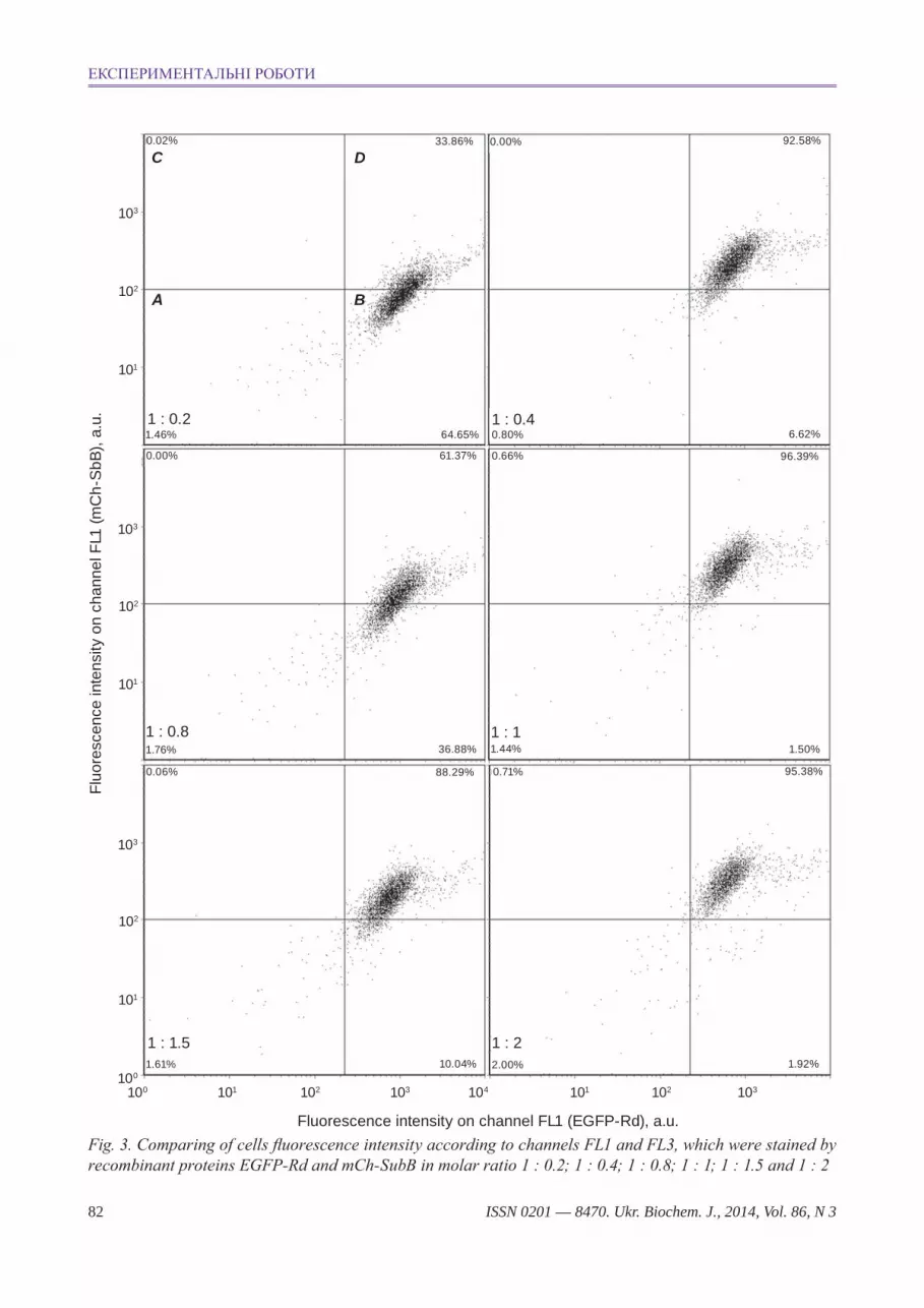
To compare traffic of different DT derivates un-der condition of their simultaneous adding to Vero cells, molar ratio of these chimera proteins for their binding in equivalent quantity with cells needed to be determined first. To determine such ratio, Vero cells were stained with two proteins under condition that concentration of mCh-SubВ has been changed from 100 to 857.14 nM, but concentration of EGFP-Rd was constant (500 nM). That is molar concentra-tions ratio of mCh-SubВ and EGFP-Rd changed in probes from 0.2 : 1 to 2 : 1.
EGFP-Rd and mCh-SubВ binding with cells has been determined according to channels FL1 and FL3, correspondingly. Obtained results of fluores-cence intensity of these cells is shown on dot plot (Fig. 3), where axis x demonstrates intensity of fluo-rescence on channel FL1 (EGFP-Rd fluorescence), and axis у is intensity of fluorescence on channel FL3 (mCh-SubВ fluorescence). Four regions are gated in the diagram with usage of corresponding control probes, which were stained with fluorescent proteins EGFP, mCherry, EGFP-Rd and mCh-SubВ. Gate A on the Fig. 3 (upper left dot plot). Gate А includes the unstained cells, according to channels FL1 and FL3 (double-negative staining), gate B in-cludes the cells stained according to channel FL1, but not stained according to channel FL3, gate C gates cells, stained according to channel FL3, but not stained according to channel FL1, and gate D includes cells, stained according to both channels (double-positive staining).
Fluorescent signal from mCh-SubB increased according to increase of concentration of this protein in the probe (Fig. 3). As mCh-SubB concentration in-creased, under stable EGFP-Rd concentration, fluo-rescence on channel FL1 slowly decreases, but stays in the gates B and C, but intensity of fluorescence on channel FL3 gradually increases and transfers from negative gate B to a positive gate C, what proves steady binding with cell receptors of both fluores-cent DT derivates. In compliance with the experi-mental data, molar ratio of EGFP-Rd to mCh-SubВ 1:2 was used in the next studies on confocal micro-scope. Best staining of cells by both DT fluorescent
derivates was observed under this ratio (Fig. 3, low right dot plot).
confocal microscopy. Details of fluorescent DT derivates transport inside the cells were studied on Vero cell line. Cells were stained with proteins EGFP-Rd and mCh-SubВ in molar ratio 1 : 2. Colo-calization between those proteins on the confocal images were detected with RG2B_Colocalization plugin for FIJI software [28].
As it appeared from Fig. 4, these proteins had different localization on the 15th minute of observa-tion: mCh-SubB located closer to the cell surface at the time when EGFP-Rd is localized closer to the cell nucleus; colocalization between these proteins was low. Similar state also observed on the 30th and 45th minutes of observation, however colo-calization between them gradually increased. Both proteins started significantly colocalize only on the 60th min of observation, however EGFP-Rd has been situated already in tubular structures, at the time when mCh-SubB, predominantly, was situated in vesicular structures. mCh-SubB and EGFP-Rd had a high level of colocalization in tubular struc-
Fig. 1. electroforegram of recombinant proteins 1 – mch-subb; 2 – mcherry; 3 – molecular weight ladder; 4 – eGFP- rd; 5 – eGFP
1 2 3 4 5kDa
20015012010085706050
40
30
25
20
15
10
ЕкСпЕРимЕНТАльНі РОБОТи

ISSN 0201 — 8470. Ukr. Biochem. J., 2014, Vol. 86, N 3 81
tures on the 75th min of observation, however a part of mCh-SubB stayed not colocalized with EGFP-Rd. Thus, the obtained results point to possible DT Т-domain influen ce on the intracellular toxin transport.
FIJI software with JACoP plugin was used for quantification analysis of colocalization between those proteins [28, 29]. Obtained pictures were ana-lyzed to calculate two parameters: Pearson correla-
Fig. 2. comparative histograms of fluorescence intensity of vero cells, which were stained with recombinant fluorescent proteins or unstained (control). А – Fluorescence intensity distribution of cells according to chan-nel FL1 for control probe and probes, stained with eGFP and eGFP-rd. B – Fluorescence intensity distribu-tion of cells according to channel FL3 for control probe and probes, stained with mcherry and mch-subВ
tion coefficient and Manders correlation coefficient (M coefficients) for mCh-SubB and EGFP-Rd. Pear-son correlation coefficient (PCC) in this case shows probability that proteins mCh-SubB and EGFP-Rd are colocalized, where meaning «0» corresponds with absence of colocalization, meaning «1» corre-sponds with full colocalization, and meaning «-1» means reverse dependency.
Even
ts c
ount
40
30
20
10
0100 101 102 103 104
Fluorescence intensity on channel FL1, a.u.
А Control EGFP
Rd-EGFP
B
Even
ts c
ount
30
23
15
8
0100 101 102 103 104
Fluorescence intensity on channel FL3, a.u.
Control mCherry
mCh-SubB
A. J. LABYNTSEV, D. V. KOLYBO, E. S. YURCHENKO et al.
ISSN 0201 — 8470. Ukr. Biochem. J., 2014, Vol. 86, N 382
Fig. 3. comparing of cells fluorescence intensity according to channels FL1 and FL3, which were stained by recombinant proteins eGFP-rd and mch-subВ in molar ratio 1 : 0.2; 1 : 0.4; 1 : 0.8; 1 : 1; 1 : 1.5 and 1 : 2
Fluo
resc
ence
inte
nsity
on
chan
nel F
L1 (m
Ch-
SbB
), a.
u.
B
D
А
100 101 102 103 104 101 102 103100
101
102
103
101
102
103
101
102
103
Fluorescence intensity on channel FL1 (EGFP-Rd), a.u.
1 : 0.21.46% 64.65%
61.37%0.00%
36.88%
88.29% 0.71%0.06%
10.04%
95.38%
1.50%
1.92%
96.39%
6.62%
0.66%
92.58%0.00%33.86%
1 : 0.81.76%
1 : 1.51.61%
1 : 22.00%
1 : 11.44%
1 : 0.40.80%
0.02%
C
ЕкСпЕРимЕНТАльНі РОБОТи

ISSN 0201 — 8470. Ukr. Biochem. J., 2014, Vol. 86, N 3 83
PCC formula looks like this:
,
where ri and Gi mean intensity of separate pixels i on the picture according to channels of fluores-cence mCh-SubB and EGFP-Rd correspondently; and are average values of fluorescence intensity mCh-SubB and EGFP-Rd on the whole picture.
Manders coefficients M1 and M2 show the per-centage of protein of every color, colocalized with the studied partner of other color, corresponding with all quantity of protein of this color on the pic-ture. In our case, coefficient M1 shows percentage SubB, colocalized with protein Rd, and coefficient M2 shows percentage Rd, colocalized with SubB correspondently. For example, meaning «1» of coef-ficient M1 shows on this picture 100% of protein SubB colocalized with protein Rd, but meaning «1» for M2 shows that 100% of Rd protein colocalized with protein SubB.
It is worth to note, that M1 points only to the por-tion of pixels of red colour (SubB), colocoli zed with pixels of green colour (Rd), and M2 point only to the portion of pixels of green colour (Rd), colocalzed
with pixels of red colour (SubB):
, where ri,coloc = ri if signal from Gi > 0, а ri,coloc
= 0 if signal from Gi = 0; , where
Gi,coloc = Gi, if signal from ri > 0, and Gi,coloc = 0, if
signal from ri = 0. Meaning «0» means absence of colocalization between proteins. More detailed cal-culation and interpretation of the above-mentioned correlation coefficients we may learn from the pub-lication Dunn et al. [30].
The calculated results of SubB and Rd colocali-zation are presented on the Fig. 5. As that appears from the graphs, relatively weak colocalization be-tween SubB and Rd (PCC = 0.3–0.4 for 5–45 min of study) was observed on the initial stages of those proteins internalization. Then colocalization be-tween those two proteins increased and reached its maximum on the 75th min. (PCC = 0.67), what correlates with the results of visual colocalization (Fig. 4). However, according to calculated Man-ders coefficients, SubB has a distinct dependency of colocalization on the time with Rd (coefficient M1), in comparison to dependency on colocalization
of Rd with SubB (coefficient M2). Thus, coefficient M2 linearly increases within the entire observation time (5–75 min) from 10 to 65%. However, the coef-ficient M1 shows linear increase only from 5th to 30th min from 27 to 55%, in the observation time 30–60 min it reaches plateau, and in an observation time from 60th to 75th min it shows a tendency to decrease from 55 to 45%. According to Manders coefficients, such colocalization results may prove the ability of SubB from 30th to 60th min of inves-tigation to transport through some compartment, in which the Rd goes as well. Correspondingly, the proteins may colocalize in this compartment, which was demonstrated with M2 and PCC increase and the results of visual colocalization (Fig. 4). However, different character of curves of M2 and M1 change in a time from 45th till 75th min of observation may point to the ability of SubB to be transferred to other compartments, which Rd does not reach within the studied period, and thus, the coefficient M1 from 45th to 75th min has a tendency to decrease. Moreover, not colocalized with Rd part of SubB can be seen on Fig. 4 (images of 60th min and 75th min of obser-vation). Thus, the results of quantitative colocaliza-tion analysis of proteins SubB and Rd have proved similar compartmentalization of these proteins from 30th to 60th min of observation; however, transfer of SubB to another non-reachable to Rd compartment is observed possibly from 60th to 75th min.
The ability of T-domain was recently shown to interact with COPI complex (Coatomer protein і) [31], which is responsible for retrograde vesicular transport, predominantly between Golgi apparatus and endoplasmic reticulum. According to the exis-ting data, sites of high COPI concentration were found in compartments of early endosomes [32], role of which in early endosomes functioning is not fully understood. It has been known, however, that elimination of COPI leads to disorder in formation of early endosomes [33] and their sorting [34]. Possi-bly, DT Т-domain throughout interaction with COPI is able to influence on toxin-contained endosomes maturation or even change their intracellular trans-port pathway. Other identified feature of T-domain is its ability to form proton channels [16, 35]. T-do-main DT throughout formation of proton channel is able to slow down acidification of endosomal lumen, that may slow down the process of DT-contained endosomes maturation. Influence of T-domain on the intracellular DT transport may be necessary for slowing down the internalized toxin-receptor com-
A. J. LABYNTSEV, D. V. KOLYBO, E. S. YURCHENKO et al.

ISSN 0201 — 8470. Ukr. Biochem. J., 2014, Vol. 86, N 384
Fig. 4. confocal images of vero cells, incubated with fluorescent Dt derivates – eGFP-rd (pseudocolored as green) and mch-subВ (pseudocolored as red) during 15, 30, 45, 60 and 75 min, correspondingly. cells nuclei stained with hoechst 33342 were shown on the pictures by blue colour. colocalization has been calculated with FiJi software. scale bar on the pictures corresponds to 10 μm
15 m
in
ColocalizationEGFP-Rd mCh-SubB Merged
30 m
in45
min
60 m
in75
min
ЕкСпЕРимЕНТАльНі РОБОТи

ISSN 0201 — 8470. Ukr. Biochem. J., 2014, Vol. 86, N 3 85
Fig. 5. Graphs of Pearson correlation coefficient (1) and m1 and m2 manders coefficients dependencies on the incubation time, which show colocalization between rd and subb Dt from 5th to 75th min of observation. coefficient m2, pointed as rd, shows a part of protein eGFP-rd, colocalized mch-subb, and m1, pointed as subb shows relevantly percentage of protein mch-subb, colocalized with eGFP-rd (m ± sD, n = 3)
Pear
son
corr
elat
ion
coef
ficie
nt1
0 10 20 30 40 50 60 70 80
0.5
0.6
0.7
0.8
0.9
1.0
Time, min
0.1
0.2
0.3
0.4
0
M c
oeffi
cien
t
2
0 10 20 30 40 50 60 70 80
0.5
0.6
0.7
0.8
0.9
1.0
Time, min
0.1
0.2
0.3
0.4
0
Rd SubB
plexes transportation into cell via endosomes, that may promote more effective transport of DT C-do-main into cell cytosol.
Consequently, effect of T-domain on vesicular transportation of diphtheria toxin, that may be asso-ciated with the ability of T-domain to interact with COPI proteins and/or its channel-formation activity has been shown in this work. According to the re-
ceived data, velocity of intracellular transport of R-domain and subunit В differed, and subunit B, pos-sibly, was transported into unreachable for R-domain compartments. The shown in this work feature of T-domain DT, in our opinion, increases effectiveness of C-domain DT transportation into cell cytosol, that promotes cytotoxic activity of diphtheria toxin.
A. J. LABYNTSEV, D. V. KOLYBO, E. S. YURCHENKO et al.

ISSN 0201 — 8470. Ukr. Biochem. J., 2014, Vol. 86, N 386
Роль Т-домену дифТеРійного Токсину у внуТРішньокліТинному ТРанспоРТуванні Токсинвмісних везикул
А. Ю. Лабинцев, Д. В. Колибо, Є. С. Юрченко, А. А. Кабернюк, Н. В. Короткевич, С. В. Комісаренко
інститут біохімії ім. О. В. палладіна НАН України, кїв;
е-mail: [email protected]
Субодиниця В дифтерійного токсину (ДТ), яка складається із двох доменів, R – ре-цепторного та Т – трансмембранного, відіграє важливу роль у зв’язуванні токсину з рецепто-ром на клітинах-мішенях та у транспортуванні каталітичної субодиниці А до цитозолю. Рекомбінантні аналоги субодиниці В є перспек-тивними представниками унікального класу транспортувальних протеїнів, які здатні забез-печити доставку біологічно активних молекул до цитозолю клітин. Створення таких конструкцій вимагає з’ясування ролі кожного із фрагментів ДТ у транспортуванні комплексу рецептор–ток-син.
У роботі досліджували роль Т-домену у внутрішньоклітинному транспортуванні рекомбінантних фрагментів ДТ. Для цьо-го порівнювали особливості внутрішньо-клітинного транспортування R-домену та субодиниці В, яка містить в собі як R-, так і Т-до-мен. Рекомбінантні фрагменти ДТ мітили флуо-ресцентними протеїнами, що дозволило викори-стовувати в дослідженні техніку колокалізації. методом конфокальної мікроскопії встановлено відмінності у транспортуванні рекомбінантних похідних ДТ у клітинах лінії Vero. показано, що R-домен швидше потрапляє до тубуляр-них компартментів клітини, ніж субодиниця В. Аналіз колокалізації R-домену та субодиниці В підтверджує майже лінійне її зростання за коефіцієнтом пірсона (PCC) у разі збільшення часу інкубації. проте відсоток субодиниці В, колокалізованої з R-доменом, визначений за коефіцієнтом мандерса (M1), зростає нелінійно, що може свідчити про здатність субодиниці В потрапляти до певних компартментів клітини, недоступних для R-домену. Роль Т-домену у внутрішньоклітинному транспортуванні
та компартменталізації токсину, вірогідно, пов’язана з його здатністю до формування протонних каналів та взаємодії з комплексом протеїнів цитоплазми COPI.
к л ю ч о в і с л о в а: дифтерійний ток-син, Т-домен дифтерійного токсину, ендоци-тоз, флуоресцентні протеїни, конфокальна мікроскопія, внутрішньоклітинне транспорту-вання.
Роль Т-домена дифТеРийного Токсина во внуТРиклеТочном ТРанспоРТе ТоксинсодеРжащих везикул
А. Ю. Лабынцев, Д. В. Колибо, Е. С. Юрченко, А. А. Кабернюк, Н. В. Короткевич, С. В. Комисаренко
институт биохимии им. А. В. палладина НАН Украины, киев;
е-mail: [email protected]
Субъединица В дифтерийного токсина (ДТ), которая состоит из двух доменов, R – ре-цепторного и Т – трансмембранного, играет важную роль в связывании токсина с рецепто-ром на клетках-мишенях и в транспорте ката-литической субъединицы А в цитозоль. Реком-бинантные аналоги субъединицы В являются перспективными представителями уникального класса транспортных протеинов, которые спо-собны обеспечить доставку биологически актив-ных молекул в цитозоль клеток. Создание таких конструкций требует определения роли каждого из фрагментов ДТ в транспортировании погло-щенного комплекса рецептор–токсин.
В работе исследовали роль Т-домена во внутриклеточном транспорте рекомбинантных фрагментов ДТ. Для этого сравнивали особен-ности внутриклеточного транспортирования R-домена и субъединицы В, которая содержа-ла как R-, так и Т-домен ДТ. Рекомбинантные фрагменты ДТ метили флуоресцентными про-теинами, что позволило использовать в ис-следовании технику колокализации. методом конфокальной микроскопии показаны отличия в транспортировании рекомбинантных произ-водных ДТ в клетках линии Vero. показано, что R-домен быстрее, чем субъединица В, попадает в тубулярные компартменты клетки. Анализ колокализации R-домена и субъединицы В под-
ЕкСпЕРимЕНТАльНі РОБОТи

ISSN 0201 — 8470. Ukr. Biochem. J., 2014, Vol. 86, N 3 87
тверждает почти линейный ее рост согласно с коэффициен том пирсона (PCC) при увеличении времени инкубации. Однако процент субъеди-ницы В, которая колокализирована с R-доменом, определенный по коэффициенту мандерса (M1), рос нелинейно, что может свидетельствовать о способности субъединицы В попадать в опреде-ленные компартменты клетки, недоступные для R-домена. Роль Т-домена во внутриклеточном транспорте и компартментализации токсина, ве-роятно, связана с его способностью к формиро-ванию протонных каналов и взаимодействию с комплексом протеинов цитоплазмы COPI.
к л ю ч е в ы е с л о в а: дифтерийный ток-син, Т-домен дифтерийного токсина, эндоцитоз, флуоресцентные протеины, конфокальная ми-кроскопия, внутриклеточный транспорт.
1. romaniuk s. i., Kolibo D. v., Komisarenko s. v. // Rus. J. Bioorg. Chem. – 2012. – 38, N 6. – P. 639–652.
2. Kolibo D. v., Labyntsev a. J., romaniuk s. i. et al. // Biotechnol. Acta. – 2013. – 6, N 4. – P. 43–62.
3. Gordon v., Klimpel K., arora n. et al. // Infect. Immun. – 1995. – 63, N 1. – P. 82–87.
4. Gordon v. m., rehemtulla a., Leppla s. h. // Infect. Immun. – 1997. – 65, N 8. – P. 3370–3375.
5. sucic J. F., moehring J. m., inocencio n. m. et al. // Biochem. J. – 1999. – 339 ( Pt 3). – P. 639–647.
6. Gordon v. m., Leppla s. h. // Infect. Immun. – 1994. – 62, N 2. – P. 333–340.
7. tsuneoka m., nakayama K., hatsuzawa K. et al. // J. Biol. Chem. – 1993. – 268, N 35. – P. 26461–26465.
8. ratts r., Zeng h., berg e. a. et al. // J. Cell Biol. – 2003. – 160, N 7. – P. 1139–1150.
9. choe s., bennett m. J., Fujii G. et al. // Nature. – 1992. – 357, N 6375. – P. 216–222.
10. rolf J. m., Gaudin h. m., eidels L. // J. Biol. Chem. – 1990. – 265, N 13. – P. 7331–7337.
11. higashiyama s., abraham J. a., miller J. et al. // Science (New York, N.Y.). – 1991. – 251, N 4996. – P. 936–939.
12. higashiyama s., Lau K., besner G. e. et al. // J. Biol. Chem. – 1992. – 267, N 9. – P. 6205–6212.
13. naglich J. G., metherall J. e., russell D. W. et al. // Cell. – 1992. – 69, N 6. – P. 1051–1061.
14. Здановский А. Г., Здановская М. В., Янков-ский Н. К. // молекулярная генетика, микро-биология и вирусология. – 1988. – 12. – С. 3–10.
15. morris r. e., Gerstein a.s., bonventre P. F. et al. // Infec. Immun. – 1985. – 50, N 3. – P. 721–727.
16. senzel L., huynh P. D., Jakes K. s. et al. // J. Gen. Physiol. – 1998. – 112, N 3. – P. 317–324.
17. oh K. J., senzel L., collier r. J. et al. // Proc. Natl Acad. Sci. USA. – 1999. – 96, N 15. – P. 8467–8470.
18. Lanzrein m., sand o., olsnes s. // EMBO J. – 1996. – 15, N 4. – P. 725–734.
19. takahashi t., umata t., mekada e. // Biochem. Biophys. Res. Commun. – 2001. – 281, N 3. – P. 690–696.
20. Quertenmont P., Wolff c., Wattiez r. et al. // Biochemistry. – 1999. – 38, N 2. – P. 660–666.
21. Lory s., carroll s. F., collier r. J. // J. Biol. Chem. – 1980. – 255, N 24. – P. 12016–12019.
22. ren J., Kachel K., Kim h. et al. // Science (New York, N.Y.). – 1999. – 284, N 5416. – P. 955–957.
23. hammond K., caputo G. a., London e. // Biochemistry. – 2002. – 41, N 9. – P. 3243–3253.
24. Wang y., Kachel K., Pablo L. et al. // Bioche-mistry. – 1997. – 36, N 51. – P. 16300–16308.
25. Кабернюк А.А., Лабинцев А.Ю., Колибо Д.В. та ін. // Укр. біохім. журн. – 2009. – 81, № 1. – С. 67–77.
26. schägger h., von Jagow G. // Anal. Biochem. – 1987. – 166, N 2. – P. 368–379.
27. yasumura y., Kawakia y. // Nippon Rinsho. – 1963. – 21. – P. 1209–1210.
28. schindelin J., arganda-carreras i., Frise e. et al. // Nat. Methods. – 2012. – 9, N 7. – P. 676–682.
29. bolte s., cordelières F. P. // J. Microsc. – 2006. – 224, N 3. – P. 213–232.
30. Dunn K. W., Kamocka m. m., mcDonald J. h. // Am. J. Physiol. Cell Physiol. – 2011. – 300, N 4. – P. C723–742.
31. trujillo c., taylor-Parker J., harrison r. et al. // Mol. Microbiol. – 2010. – 76, N 4. – P. 1010–1019.
32. aniento F., Gu F., Parton r. G. et al. // J. Cell Biol. – 1996. – 133, N 1. – P. 29–41.
33. razi m., chan e. y. W., tooze s. a. // J. Cell Biol. – 2009. – 185, N 2. – P. 305–321.
34. styers m. L., o’connor a. K., Grabski r. et al. // Am. J. Physiol. Cell Physiol. – 2008. – 294, N 6. – P. C1485–1498.
35. Kurnikov i. v., Kyrychenko a., Flores-cana-les J. c. et al. // J. Mol. Biol. – 2013. – 425, N 15. – P. 2752–2764.
Отримано 24.10.2013
A. J. LABYNTSEV, D. V. KOLYBO, E. S. YURCHENKO et al.