intracellular metalloporphyrin metabolism in staphylococcus aureus
TRANSCRIPT
Intracellular metalloporphyrin metabolismin Staphylococcus aureus
Michelle L. Reniere Æ Victor J. Torres ÆEric P. Skaar
Received: 30 June 2006 / Accepted: 20 July 2006 / Published online: 27 March 2007� Springer Science+Business Media B.V. 2007
Abstract The bacterial pathogen Staphylococcus
aureus is responsible for a significant amount of
human morbidity and mortality, and the ability of
S. aureus to cause disease is absolutely dependent
on the acquisition of iron from the host. The most
abundant iron source to invading staphylococci is
in the form of the porphyrin heme. S. aureus is
capable of acquiring nutrient iron from heme and
hemoproteins via two heme-acquisition systems,
the iron-regulated surface determinant system
(Isd) and the heme transport system (Hts). Heme
acquisition through these systems is involved in
staphylococcal pathogenesis suggesting that the
intracellular fate of heme plays a significant role in
the infectious process. The valuable heme mole-
cule presents a paradox to invading bacteria
because although heme is an abundant source of
nutrient iron, the extreme reactivity of heme
makes it toxic at high concentrations. Therefore,
bacteria must regulate the levels of intracellular
heme to avoid toxicity. Although the molecular
mechanisms responsible for staphylococcal heme
acquisition are beginning to emerge, the mecha-
nisms by which S. aureus regulate intracellular
heme homeostasis are largely unknown. In this
review we describe three potential fates of host-
derived heme acquired by S. aureus during infec-
tion: (i) degradation for use as a nutrient iron
source, (ii) incorporation into bacterial heme-
binding proteins for use as an enzyme cofactor, or
(iii) efflux through a dedicated ABC-type trans-
port system. We hypothesize that the ultimate fate
of exogenously acquired heme in S. aureus is
dependent upon the intracellular and extracellular
availability of both iron and heme.
Keywords Staphylococcus � Heme � Isd �Metalloporphyrin � Bacteria
Introduction
The Gram positive bacterial pathogen Staphylo-
coccus aureus causes a wide range of human
diseases ranging from minor skin infections, such
as folliculitis and impetigo, to more invasive
diseases including endocarditis, sepsis, and toxic
shock syndrome (Weems 2001). Importantly,
Since the acceptance of this manuscript, two papers havebeen published that are directly relevant to this review.The first describes IsdB as a hemoglobin receptor inS. aureus: J Bacteriol. 2006 Dec; 188(24): 8421–8429.The second identifies an IsdG-family member in aGram negative bacterium: J Bacteriol. 2006 Sep; 188(18):6476-6482.
M. L. Reniere � V. J. Torres � E. P. Skaar (&)Department of Microbiology and Immunology,Vanderbilt University Medical Center,1161, 21st Avenue South, MCN A5102,Nashville, TN 37232,USAe-mail: [email protected]
123
Biometals (2007) 20:333–345
DOI 10.1007/s10534-006-9032-0
S. aureus is the leading cause of nosocomial disease
and the incidence of multi-drug resistant isolates is
increasing. In fact, up to 60% of hospital-acquired
S. aureus isolates are methicillin-resistant and
three cases of vancomycin-resistance have been
reported in the United States to date (Todd
2006). This is especially alarming considering
vancomycin is viewed as a last line of defense
against multi-drug resistant S. aureus. These facts
underscore the necessity of identifying novel
targets for therapeutic intervention against this
important human pathogen.
One potential method for combating S. aureus
infection is to deny the bacteria nutrients re-
quired for survival. One such nutrient is iron,
which is essential for bacterial growth and path-
ogenesis but scarcely available inside the human
host (Bullen and Griffiths 1999; Braun 2001).
Most bacterial cells need 105–106 iron atoms for
growth due to a requirement for iron in numerous
cellular processes including DNA synthesis, en-
ergy generation, and protection against reactive
oxygen species (Bullen and Griffiths 1999; Braun
2001). However, 99.9% of iron in the human body
is intracellular, and thus, not readily available to
bacteria (Bullen and Griffiths 1999; Crichton
2001). Intracellular vertebrate iron is primarily a
component of heme, or iron-protoporphyrin IX,
which is typically bound to hemoglobin, the major
hemoprotein within circulating erythrocytes
(Crichton 2001). Heme is the reduced iron-con-
taining porphyrin (Fe+2), whereas the oxidized
form (Fe+3) is known as hemin. However, both
are commonly referred to as ‘‘heme’’ so we will
apply this convention throughout this review.
Upon liberation from lysed erythrocytes, heme
and hemoglobin are sequestered by hemopexin
and haptoglobin in an effort to protect the host
against toxicity caused by these reactive mole-
cules. This process of heme scavenging by host
proteins is extremely efficient, and under normal
physiological conditions, free heme and hemo-
globin are virtually undetectable in the serum
(Bullen and Griffiths 1999). However, during
invasive bacterial infections, hemolysin-mediated
erythrocyte lysis likely leads to the local accumu-
lation of appreciable amounts of heme and
hemoglobin which can be used by the bacteria
as a nutrient iron source. This idea is supported
by the observation that hemopexin expression is
increased upon bacterial infection (Klapper et al.
1972), potentially representing a response to the
transient increase in free heme at the site of
infection.
Upon release of hemoglobin from lysed
erythrocytes, bacterial pathogens can utilize free
hemoglobin or hemoglobin complexed to hapto-
globin as nutrient iron sources (Wandersman
and Stojiljkovic 2000; Heinrichs 2004). This
process initiates with the direct binding of
hemoproteins via specific bacterial receptors
followed by removal and transport of the heme
cofactor into the bacterial cell (Wandersman and
Stojiljkovic 2000). Heme acquisition is required
for S. aureus pathogenesis, and staphylococci
preferentially acquire heme-iron over other
physiologically relevant iron sources (Skaar et al.
2004b). The molecular machinery responsible for
staphylococcal heme acquisition is encoded by
two distinct systems, the iron-regulated surface
determinant system (Isd), and the heme trans-
port system (Hts) (Mazmanian et al. 2003; Skaar
et al. 2004b).
The Isd heme import machinery is comprised
of ten genes, each of which is regulated by the
iron-dependent regulatory protein Fur (Mazma-
nian et al. 2003). Genes of the Isd system encode
four cell wall anchored proteins (IsdABCH), a
transpeptidase (SrtB), a membrane transport
system (IsdDEF), and two cytoplasmic monoox-
ygenases (IsdGI). Excluding SrtB, each protein
component of the Isd system is capable of binding
heme in vitro (Mazmanian et al. 2003; Mack et al.
2004). Additionally, IsdB and IsdH are responsi-
ble for the surface recognition of hemoglobin and
hemoglobin-haptoglobin complexes by intact
staphylococci (Dryla et al. 2003). Our model
presumes that after the hemoprotein binds to its
cognate surface receptor, the heme cofactor is
removed from the hemoprotein by IsdA through
an as-yet-uncharacterized mechanism. The heme
is then transferred through the cell wall by IsdC
and subsequently through the plasma membrane
via the membrane transport system comprised of
IsdDEF (Mazmanian et al. 2003). Heme can also
transit into the cytoplasm through the membrane
associated ABC-type transport system HtsABC,
which is the more dominant heme transport
334 Biometals (2007) 20:333–345
123
system in vitro (Skaar et al. 2004b). This implies
that HtsABC, rather than IsdDEF, is the primary
membrane heme transport system in S. aureus
(Skaar et al. 2004b). The Hts system is composed
of a lipoprotein (HtsA) and two permeases (HtsB
and HtsC) which exhibit strong identity to
permease components of the heme transport
systems of Yersinia pestis (HmuU) and Coryne-
bacterium diphtheriae (HmuU) (Thompson et al.
1999; Drazek et al. 2000). Heme acquisition by
the Isd and Hts systems is a highly efficient
process as demonstrated by the observation that
staphylococci incubated with exogenous heme are
saturated for heme uptake within 15 min
(Mazmanian et al. 2003). The rapidity of heme
uptake demonstrates the necessity of specialized
molecular machinery capable of handling this
valuable host iron source.
In addition to its value as a nutrient iron
source, intact heme is a useful cofactor in
numerous bacterial enzymes, such as cyto-
chromes, catalases, and peroxidases. However,
the reactivity of free heme presents a conundrum
to pathogenic bacteria because heme is not
tolerated well in its free form and therefore,
excess internalized heme is toxic to invading
bacteria. Keeping with this, bacterial pathogens
have developed elaborate systems of managing
internalized heme to prevent against heme-
mediated toxicity while capitalizing on the
value of heme as a nutrient source. In this
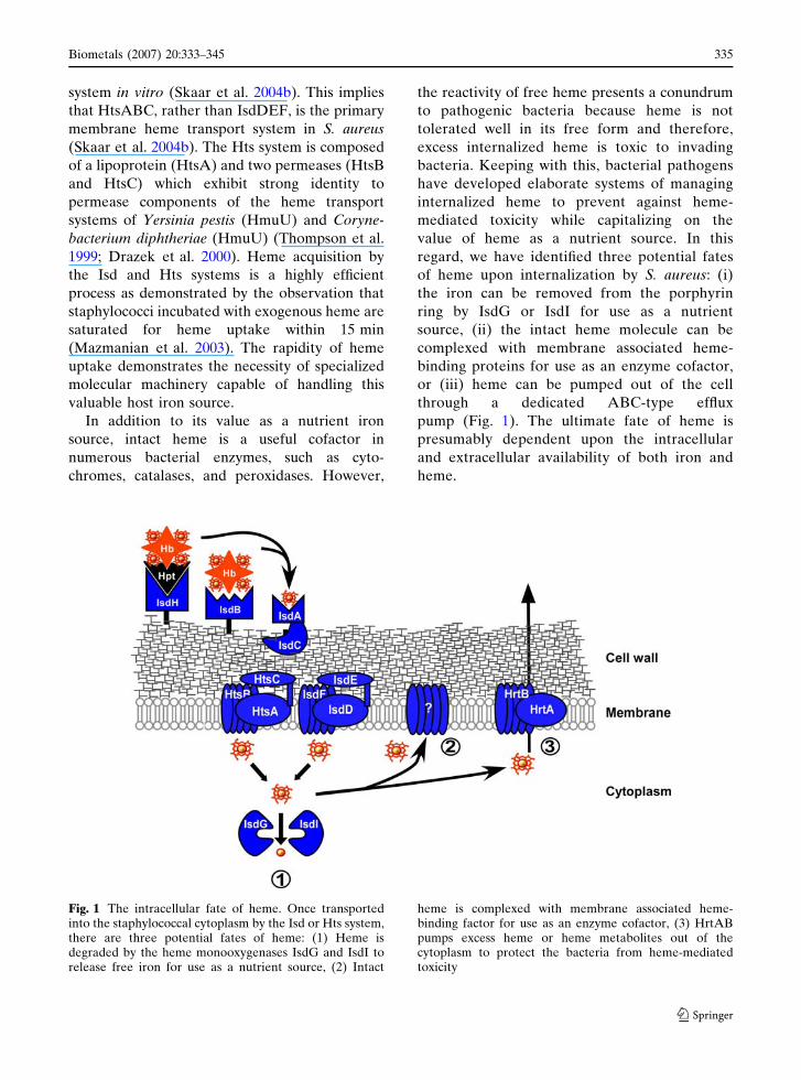
regard, we have identified three potential fates
of heme upon internalization by S. aureus: (i)
the iron can be removed from the porphyrin
ring by IsdG or IsdI for use as a nutrient
source, (ii) the intact heme molecule can be
complexed with membrane associated heme-
binding proteins for use as an enzyme cofactor,
or (iii) heme can be pumped out of the cell
through a dedicated ABC-type efflux
pump (Fig. 1). The ultimate fate of heme is
presumably dependent upon the intracellular
and extracellular availability of both iron and
heme.
Fig. 1 The intracellular fate of heme. Once transportedinto the staphylococcal cytoplasm by the Isd or Hts system,there are three potential fates of heme: (1) Heme isdegraded by the heme monooxygenases IsdG and IsdI torelease free iron for use as a nutrient source, (2) Intact
heme is complexed with membrane associated heme-binding factor for use as an enzyme cofactor, (3) HrtABpumps excess heme or heme metabolites out of thecytoplasm to protect the bacteria from heme-mediatedtoxicity
Biometals (2007) 20:333–345 335
123
Cytoplasmic heme degradation
When the intracellular iron concentration is low,
bacteria can obtain iron for use in various
metabolic pathways through the enzymatic deg-
radation of exogenously acquired heme. Heme
degrading enzymes have been identified in
numerous pathogenic bacteria, including: Cory-
nebacterium sp. (Schmitt 1997b), Yersinia sp.
(Stojiljkovic and Hantke 1994), Neisseria sp.
(Zhu et al. 2000b), Pseudomonas aeruginosa
(Ratliff et al. 2001), Escherichia coli (Suits et al.
2005), Bacillus anthracis (Skaar et al. 2006) and
S. aureus (Skaar et al. 2004a; Wu et al. 2005).
Based on available structural data, bacterial heme
degrading monooxygenases can be classified into
three families, which we refer to as the HO-like
family, the ChuS-family, and the IsdG-family of
monooxygenases.
The first identified bacterial enzyme capable of
degrading heme was HmuO from C. diphtheria
(Schmitt 1997b). HmuO exhibits significant amino
acid identity to vertebrate heme oxygenases, and
consistent with this identity, HmuO cleaves heme
to release a-biliverdin, carbon monoxide, and free
iron (Wilks and Schmitt 1998). Structural analyses
have revealed that HmuO is markedly similar to
the structure of vertebrate HO-1, exhibiting a
monomeric alpha-helical fold containing a con-
served histidine residue at the N-terminus that is
important for heme coordination (Schuller et al.
1999; Frankenberg-Dinkel 2004). Furthermore,
HO-1 and HmuO both contain a single active site
and GXXXG motif required for catalytic activity
(Wilks and Schmitt 1998; Schuller et al. 1999).
Based on these findings, we classify HmuO as a
member of the HO-like family of bacterial heme
monooxygenases. Additional members of this
family have been identified in Pseudomonas aeru-
ginosa (PigA) (Ratliff et al. 2001), N. gononr-
rhoeae (HemO) (Zhu et al. 2000b), and N.
meningitides (HemO) (Zhu et al. 2000b).
A second enzyme family with reported heme
degrading properties is represented by the ChuS
protein from E. coli (Suits et al. 2005). ChuS
shows no sequence or structural similarity to
previously described heme oxygenases (HOs),
however, orthologs of ChuS have been found in
several other bacterial pathogens, including: ShuS
in Shigella (Wilks 2001), HemS in Yersinia
(Stojiljkovic and Hantke 1994), and PhuS in
Pseudomonas (Lansky et al. 2006). In fact, the
original description of HemS proposed that this
enzyme encoded a protein with heme-degrading
activity based on the increased sensitivity of a
hemS mutant to excess heme (Stojiljkovic and
Hantke 1994). Consistent with this, Suits et al.
have reported that ChuS is a monomeric heme
degrading enzyme (Suits et al. 2005). ChuS
degrades heme in the presence of catalase and
superoxide dismutase, demonstrating catalytic
degradation of heme rather than non-enzymatic
coupled oxidation (Suits et al. 2005). Structural
analysis has shown that ChuS folds as a dimeric
beta-barrel type protein comprised of a structural
duplication in which both halves contain a
predicted active site. Each half of the protein is
independently capable of degrading heme, sug-
gesting that the dual active sites are functional in
the mature enzyme. In a conflicting report, Wilks
et al. have proposed that ShuS (with 98% identity
to ChuS) is a cytoplasmic heme shuttling protein
rather than a heme oxygenase, because although
it bound heme, the authors could not demonstrate
enzymatic heme degradation in vitro (Wilks
2001). In addition, the authors reported that ShuS
binds DNA, leading to the hypothesis that DNA
binding by ShuS protects the cell from heme-
dependent oxidative damage (Wilks 2001). How-
ever, a strain inactivated for shuS does not show
increased heme-dependent sensitivity to oxidative
stress as compared to wild type (Wyckoff et al.
2005). It has recently been reported that PhuS of
Pseudomonas (with 41% identity to ChuS) is also
a heme-trafficking protein (Lansky et al. 2006).
PhuS facilitates the degradation of heme in vitro,
however, contrary to the situation with ChuS,
PhuS-dependent heme degradation does not
occur in the presence of catalase. This result
implies that PhuS-dependent heme degradation
occurs through coupled oxidation and not enzy-
matic heme catalysis (Lansky et al. 2006). Based
on these findings, the authors propose that PhuS
acts as a transport protein that delivers heme to
the heme oxygenase pa-HO (Lansky et al. 2006).
More experiments are necessary to clarify the
specific function and activity of this family of
proteins; however, a role for ChuS-family
336 Biometals (2007) 20:333–345
123
members in intracellular heme metabolism has
been firmly established.
IsdG and IsdI, components of the Isd system
discovered in S. aureus, are representative
members of a third family of heme degrading
enzymes which we have termed the IsdG-family
of heme monooxygenases (Skaar et al. 2004a; Wu
et al. 2005). IsdG and IsdI were originally inves-
tigated due to their genetic association with the
Isd heme-transport system. Both IsdG and IsdI
are iron-regulated cytoplasmically localized pro-
teins that are 70% identical at the amino acid
level. Pfam analysis places IsdG and IsdI in a
family of monooxygenases responsible for the
oxygenation of aromatic polyketides (Bateman
et al. 2002; Wu et al. 2005). Based on these
findings, it was predicted that IsdG and IsdI
encode for iron-regulated heme-degrading mono-
oxygenases in the staphylococcal cytoplasm. This
hypothesis was confirmed upon the demonstra-
tion that IsdG and IsdI bind and degrade heme in
the presence of reductant or reductase. This
reaction releases free iron and occurs in the
presence of catalase, ruling out the possibility that
heme-degradation by IsdG or IsdI is occurring
through a non-enzymatic coupled oxidation reac-
tion (Skaar et al. 2004a). Taken together, these
results confirmed that IsdG and IsdI represent the
first identified members of a novel family of heme
degrading enzymes.
IsdG-family members have exclusively been
identified in Gram-positive bacteria, including:
S. aureus, S. epidermidis, B. anthracis, and Listeria
monocytogenes (Skaar and Schneewind 2004).
We have demonstrated functional conservation of
IsdG-family members across genera through
investigations into the B. anthracis IsdG (BaI-
sdG) (Skaar et al. 2006). BaIsdG is 35% identical
to the staphylococcal IsdG-family enzymes, and
BaIsdG is the only identifiable IsdG-family mem-
ber in the B. anthracis genome. BaIsdG enzymat-
ically degrades heme to release free iron, and this
activity is required to protect B. anthracis against
heme-mediated toxicity (Skaar et al. 2006). This
observation implies that IsdG family members
represent an evolutionarily conserved mechanism
utilized by Gram positive pathogens to protect
against heme-mediated toxicity and obtain iron
from host heme during infection.
The IsdG-family members are structurally
very different from the HO-like and ChuS
families of heme oxygenases in that they fold
as a homodimeric beta-barrel type structure
with two separate active sites (Wu et al. 2005).
Moreover, mutational analyses revealed that the
IsdG-family members degrade heme through
the action of a catalytic NWH triad present in
the active site of each monomer of the mature
enzyme (Wu et al. 2005). The functional rami-
fications of two active sites is not known,
however, it is possible that dual sites increases
the efficiency of heme degradation, or alterna-
tively, allows the enzyme to utilize different
electron donors. Interestingly, IsdG and IsdI
exhibit a striking structural similarity to ActVA-
ORF6, a monooxygenase belonging to the same
family of enzymes as IsdG and IsdI as predicted
by Pfam analysis (Bateman et al. 2002; Sciara
et al. 2003). ActVA-ORF6 is a tailoring enzyme
responsible for the oxidation of aromatic poly-
ketides in Streptomyces coelicolor (Sciara et al.
2003). Taken together, these structural findings
suggest that convergent evolution has provided
bacterial pathogens with distinct enzyme fami-
lies capable of releasing iron from the macro-
cyclic conjunction of heme. On the other hand,
divergent evolution appears to have altered the
substrate specificity of IsdG and ActVA-ORF6
enzymes, likely due to the drastically different
environments inhabited by staphylococci and
streptomyces.
Until recently, all heme oxygenases were
reported to degrade heme to one of four isomers
of the blue-green molecule biliverdin (BV IX):
BV IXa, IXb, IXc, or IXd (Frankenberg-Dinkel
2004). However, a novel heme-degradation path-
way was recently described in the blood-sucking
insect Rhodnius prolixus, in which heme is
chemically modified before oxidative cleavage of
the tetrapyrrole and the resulting product is a
dicysteinyl-BV IXc (Paiva-Silva et al. 2006). We
have found that the major heme degradation
product of IsdG and IsdI does not exhibit the
blue-green color consistent with the typical HO
reaction product biliverdin, but rather is a light
yellow (unpublished observation). Mass spectral
analyses of isolated reaction products from both
IsdG and IsdI-catalyzed reactions identified a
Biometals (2007) 20:333–345 337
123
single molecule with a mass of 599 Da, supporting
the colorimetric observation that the IsdG and
IsdI major heme degradation product is not
biliverdin, which has a mass of 582 Da in the
same analysis. Tandem MS has revealed that the
products of IsdG and IsdI-mediated heme
degradation are identical and represent a novel
heme degradation product (unpublished observa-
tion). Based on these observations, members of
the IsdG family of heme monooxygenases evi-
dently cleave the tetrapyrrole in a novel way,
producing a slightly different product than either
BV IX or the newly identified dicysteinyl-BV IXc.
We are currently undertaking further analyses to
definitively identify the structure of this novel
heme degradation product and to elucidate the
exact catalytic mechanism of this class of heme
monooxygenases. Identifying this novel heme
degradation product will have important ramifi-
cations for staphylococcal biology as heme
metabolism plays a vital role in the pathogenesis
of S. aureus infections (Skaar et al. 2004b).
Exogenous heme as an enzyme cofactor
Although a valuable nutrient iron source, intact
heme is potentially an important molecule to
invading bacteria. This presumption is based on
the requirement for heme as a cofactor in
numerous enzymes involved in energy generation
and protection against reactive oxygen species
(Thony-Meyer 1997; Braun 2001). In this regard,
heme is required in the bacterial plasma mem-
brane as a cofactor for cytochromes of the
electron transport chain (Thony-Meyer 1997).
When iron is abundant in the environment, it is
possible that bacteria use exogenously acquired
heme as an enzyme cofactor, as it is thermody-
namically favorable to import host molecules
rather than synthesize a porphyrin ring de novo.
This molecular hijacking hypothesis is supported
by our observation that exogenous heme is
acquired and segregated intact to the staphylo-
coccal membrane when non-heme iron sources
are concurrently available (Skaar et al. 2004b).
Furthermore, using a radiolabeled heme
molecule, it has been shown that heme added
exogenously to growing cultures of B. subtilis
leads to uptake and incorporation of heme into
four distinct c-type cytochromes (Schiott et al.
1997). The molecular mechanisms responsible for
trafficking heme to proteins of the Gram positive
membrane have not been evaluated in detail. The
machinery involved in this process is predicted
to include a number of activities including sys-
tems responsible for heme and apocytochrome
transport across the membrane, chaperones to
assist in apocytochrome folding, and heme lyases
responsible for incorporating heme into mature
cytochromes.
A piece of the staphylococcal cytochrome
synthesis puzzle was solved by the identification
of a mutation in the ctaA gene which was found to
inhibit the ability of S. aureus to survive pro-
longed starvation (Clements et al. 1999). CtaA is
a heme O monooxygenase that converts heme O
to heme A (Svensson and Hederstedt 1994), and
therefore, inactivation of ctaA prevents the for-
mation of heme A-containing cytochromes, which
subsequently leads to an uncoupling of electron
transport. This uncoupling causes a commensu-
rate increase in oxidative stress when the bacteria
are recovering from starvation conditions
(Clements et al. 1999). These results demonstrate
that heme is required in the membrane as a
cofactor in cytochromes and is important for
preventing the formation of toxic oxidizing prod-
ucts. Additional experiments are necessary to
identify the specific factors that bind host-derived
heme in the membrane and to elucidate the
benefit afforded to bacterial pathogens by these
molecular hijacking reactions.
Non-iron metalloporphyrins
Non-iron metalloporphyrins are comprised of the
same tetrapyrrole backbone structure as heme
with a substitution for the coordinated central
metal ion. These molecules became of particular
interest to bacteriologists upon the publication
of a seminal paper by Igor Stojiljkovic and
colleagues that demonstrated a potent antibacte-
rial effect of non-iron metalloporphyrins against
pathogenic bacteria, including several multi-drug
338 Biometals (2007) 20:333–345
123
resistant clinical isolates of S. aureus (Stojiljkovic
et al. 1999). It was hypothesized that these heme-
like molecules act as ‘‘molecular Trojan horses’’
that enter bacteria through heme transport sys-
tems where they then exert their antibacterial
affect in the cytoplasm or membrane (Stojiljkovic
et al. 1999). The potent antibacterial effect of
metalloporphyrins suggests that these molecules
target a vital cellular process inside the bacte-
rium. In turn, this suggests that the intracellular
targets of metalloporphyrins may represent novel
therapeutic options against bacterial infection.
Keeping with this, we have investigated the
intracellular fate of exogenously provided metal-
loporphyrins chelated with gallium, cobalt, man-
ganese and zinc. As an initial measure of the fate
of metalloporphyrin analogues inside S. aureus,
we combined non-iron metalloporphyrins with
purified IsdG and IsdI in vitro and analyzed
degradation spectrophotometrically. IsdG and
IsdI are both capable of binding all metallopor-
phyrins tested, as evident by the presence of a
Soret band, indicative of porphyrin binding.
However, IsdG and IsdI are not able to degrade
non-iron containing metalloporphyrins in the
presence of an electron donor (unpublished data).
These data suggest that the toxic effect of
metalloporphyrins on S. aureus is not due to
release of the metal ion upon tetrapyrrole cleav-
age, but rather, due to some effect of the intact
non-iron metalloporphyrin.
Previous investigations suggest that anaerobi-
cally grown S. aureus are resistant to metal-
loporphyrin toxicity (Stojiljkovic et al. 1999),
implying that the mechanism of metalloporphyrin
toxicity is related to aerobic respiration. In
bacteria, the molecular machinery that carries
out aerobic respiration is typically localized to the
membrane and dependent on the presence of
heme binding cytochromes (von Wachenfeldt and
Hederstedt 1992; Thony-Meyer 1997). Based on
this, we tracked metalloporphyrin metabolism in
S. aureus in an effort to identify the subcellular
fate of these compounds. Bacteria grown in
gallium-protoporphyrin IX (Ga-PPIX) were frac-
tionated and the intracellular localization of
Ga-PPIX was determined using inductively cou-
pled plasma dynamic reaction cell mass spec-
trometry (ICP-DRC-MS). Because we have
found that Ga-PPIX is not degraded by IsdG or
IsdI, it can be assumed that the localization of the
gallium ion reflects the localization of the intact
metalloporphyrin. Furthermore, gallium is not
typically present at detectable levels inside staph-
ylococcal cells so any gallium detected in these
cells represents acquired metalloporphyrin. In
these preliminary experiments greater than fifty
percent of the total cellular gallium was found in
the plasma membrane fraction, supporting the
hypothesis that metalloporphyrins are trans-
ported to and stored in the plasma membrane,
where they likely exert their toxic effect (unpub-
lished data). Unlike iron, which commonly exists
in the +2 or +3 oxidation states, gallium can only
exist as Ga+3. Therefore, Ga-PPIX does not have
the same oxidation potential as heme, which
could render membrane-bound cytochromes un-
able to transfer electrons, resulting in the over-
production of oxidative radicals and observed
toxicity. Interestingly, exposure to exogenous
metalloporphyrin leads to a more potent antimi-
crobial effect than inactivation of the heme
synthesis genes hemB or ctaA (von Eiff et al.
1997; Clements et al. 1999). More specifically, S.
aureus strains inactivated for hemB are predicted
to be unable to form mature cytochromes, yet
they are capable of moderate growth rates on
solid medium (von Eiff et al. 1997). In contrast,
exposure to less than 1 lg ml–1 of gallium proto-
porphyrin IX abrogates staphylococcal growth in
liquid media (unpublished data). This fact sug-
gests that the intracellular toxicity of Ga-PPIX
is more complicated than simply blocking aerobic
respiration through cytochrome inactivation.
Heme efflux
As reviewed above, it is well established that
heme and hemoproteins can serve as iron sources
to a variety of pathogenic bacteria. This is due to
the fact that many bacterial pathogens encode
efficient heme uptake systems that allow the
bacteria to bind and traffic heme-iron into the
cytoplasm. Although heme (Fe-metalloporphy-
rin) is a valuable nutrient source to invading
bacteria, the efficiency of heme-uptake systems
can overpower the heme degradation machinery,
Biometals (2007) 20:333–345 339
123
leading to the accumulation of toxic levels of
heme or heme-metabolites. In fact, high intracel-
lular concentrations of heme can result in oxida-
tive damage due to its ability to catalyze
peroxidase and oxidase reactions. These reactions
lead to the generation of reactive oxygen species
which subsequently damage DNA and proteins
(Everse and Hsia 1997). In the past several years
it has become evident that heme can be toxic
to a variety of bacteria including S. aureus,
Streptococcus faecalis, B. cereus, N. meningitides,
Mycobacterium smegmatis, Y. enterocolitica, S.
dysenteriae, and C. diphtheria (Ladan et al. 1993;
Stojiljkovic et al. 1999; Bibb et al. 2005; Rasmus-
sen et al. 2005; Wyckoff et al. 2005). Thus, for
infecting bacteria, the utility of heme as a nutrient
source must be balanced against its toxicity at
high concentrations.
The avoidance of heme-mediated toxicity is
particularly important during situations where
pathogens are in contact with high levels of
exogenous heme, such as might be encountered
during blood-borne infections. The means by
which bacterial pathogens avoid the toxic effects
of intracellular heme accumulation is not well
understood, however, research into the Gram
negative genus Neisseria has revealed multiple
mechanisms by which this bacterium avoids
heme-mediated toxicity. These include heme
degradation through the heme oxygenase HemO
(see above) (Zhu et al. 2000a; Zhu et al. 2000b),
and heme occlusion through the PilQ pore
(pilQ1) (Chen et al. 2004). In addition, disruption
of the gene of hydrophobic tolerance (ght)
increases the sensitivity of Neisseria to heme,
although the mechanism by which Ght protects
against heme-mediated toxicity has not been
defined (Rasmussen et al. 2005). Finally, disrup-
tion of the neisserial multiple transferable resis-
tance (mtr) efflux system also increases sensitivity
to heme-mediated toxicity (Bozja et al. 2004).
This effect is likely due to the inability of the
mutant bacteria to ‘‘pump out’’ the excess heme
or heme-metabolites, again resulting in an
increased concentration of intracellular heme
and/or heme metabolites.
The mechanisms by which Gram positive
bacteria avoid heme-mediated toxicity are less
clear. Recently, it was shown that disruption of a
C. diphtheriae heme-dependent transduction
system chrAS results in increased sensitivity
to heme-mediated toxicity (Bibb et al. 2005).
The ChrAS system has been shown to sense
heme and subsequently induce expression of
hmuO, the C. diphtheria heme oxygenase (Sch-
mitt 1999; Bibb et al. 2005). In contrast to the
effects described above with mutants of the
neisserial hemO, the increased heme-mediated
toxicity observed in the chrAS mutant is inde-
pendent of hmuO (Bibb et al. 2005). In fact,
hmuO mutants and wild-type strains are equally
sensitive to heme-mediated toxicity (Bibb et al.
2005). These data suggest that the ChrAS system
is also involved in regulating the expression of
other genes that are required for the protection
against heme-mediated toxicity. Another exam-
ple of heme oxygenase-mediated protection
against heme toxicity was demonstrated upon
inactivation of B. anthracis isdG, which led to a
significant increase in heme toxicity (Skaar et al.
2006). Based on these findings, it is clear that
pathogenic bacteria have evolved different mech-
anisms to avoid the toxic effects of heme, allow-
ing them to use heme as a nutrient source while
avoiding the detrimental effects of cytoplasmic
heme accumulation.
A primary focus of our laboratory is investi-
gating how S. aureus avoid heme-mediated tox-
icity. We have shown that S. aureus can grow
using heme and hemoproteins as the sole iron
source (Skaar et al. 2004b), suggesting that S.
aureus is able to capitalize on the nutrient-iron
value of these molecules while avoiding heme-
mediated toxicity. Keeping with this, C. diphthe-
ria (Schmitt 1997a; Schmitt 1999) and Bordetella
sp. (Kirby et al. 2001; Vanderpool and Armstrong
2003) are able to sense heme and subsequently
induce the expression of systems involved in
heme acquisition and metabolism. In light of
these findings, we sought to test the ability of
S. aureus to alter protein expression in response
to changes in intracellular heme status. In this
regard we have performed differential expression
analysis using two-dimensional difference gel
electrophoresis (2D-DIGE), which identified 21
proteins that respond exclusively to the presence
of heme, irrespectively of changes in iron avail-
ability (Friedman et al. 2006). These experiments
340 Biometals (2007) 20:333–345
123
revealed that the protein exhibiting the most
dramatic increase (45-fold) in response to heme is
a hypothetical-uncharacterized ABC-type trans-
porter that we have named the heme regulated
transport system (Hrt) (Friedman et al. 2006).
Importantly, this transporter is conserved across
many pathogenic bacteria, including B. anthracis
and L. monocytogenes and BLAST analyses have
revealed that the Hrt system exhibits low simi-
larity to antibacterial-peptide and lipoprotein
exporters (Altschul et al. 1990). Further analysis
of the localization of this gene in the S. aureus
chromosome indicates that it is in a bicistronic
operon containing an ABC-type transporter
(hrtA) and a transporter permease (hrtB).
We hypothesize that the Hrt system prevents
heme-mediated toxicity through the regulated
expression of an efflux transporter that pumps
excess heme and/or heme-metabolites out of the
cell. This assumption is based on the observa-
tion that HrtA is over-expressed when S. aureus
is grown in heme-containing medium, and
HrtAB has low level identity to small molecule
efflux pumps. To test this hypothesis and to
determine the contribution of the Hrt system
in vivo, we have generated hrtAB mutant
S. aureus strains. Our preliminary studies indi-
cate that the Hrt system is required for
S. aureus growth in high heme concentrations,
suggesting that the Hrt system is involved in
avoidance of heme-mediated toxicity, poten-
tially by exporting excess heme and/or heme-
metabolites out of the cell (unpublished data).
These findings highlight the importance of the
Hrt system to staphylococcal heme metabolism.
Further investigations into the mechanism and
function of heme-mediated induction of Hrt and
Hrt-mediated heme transport are currently
underway in our laboratory.
Summary
Heme acquisition is a process that is vital to
infection by numerous bacterial pathogens
(Genco and Dixon 2001; Stojiljkovic and
Perkins-Balding 2002; Skaar et al. 2004b). Tradi-
tionally, the uptake of heme has been considered
a method by which bacterial pathogens acquire
nutrient iron during infection. This presumption
is supported by the requirement for heme uptake
systems in multiple infection models and the
significant iron requirement of virtually all
bacterial pathogens (Genco and Dixon 2001;
Stojiljkovic and Perkins-Balding 2002; Skaar
et al. 2004b). Keeping with this, heme degrada-
tion machinery has been identified in numerous
bacteria that infect vertebrates (Frankenberg-
Dinkel 2004). The enzymatic degradation of
heme is modeled to be an important process
during pathogenesis; however, this contention has
not yet been supported by animal models of
infection. Although in vitro data support a role
for heme degradation machinery in bacterial
physiology, it is probable that the fate of heme
inside the bacteria is multifaceted. In this regard,
a small body of evidence is emerging that
supports the molecular hijacking hypothesis
which proposes that heme acquired from the host
can be incorporated intact as an enzyme cofactor
in respiratory proteins of the bacterial membrane
(Schiott et al. 1997; Skaar et al. 2004b). Further-
more, we have identified a S. aureus heme-
inducible efflux pump that presumably transports
excess heme or heme metabolites out of the
bacterial cytoplasm to prevent against heme-
mediated toxicity. It is possible that the molecular
decision to degrade, incorporate, or expel heme is
dependent on the level of iron and heme available
to the invading bacterium.
Our model envisions that upon entry into the
host, S. aureus encounters an environment devoid
of free iron. This iron starvation will lead to the
Fur-dependent activation of the Isd and Hts
systems (Mazmanian et al. 2003; Skaar et al.
2004b). Upon hemolysin-mediated erythrocyte
lysis, free hemoglobin and hemoglobin-haptoglo-
bin are bound by the surface proteins of the Isd
system and heme is internalized through the
membrane associated transport systems HtsABC
and IsdDEF (Mazmanian et al. 2003; Skaar et al.
2004b). Initially, cytoplasmic heme may represent
the only abundantly available iron source to
staphylococci. Therefore, IsdG and IsdI degrade
heme to release the iron in order to satisfy the
nutrient iron requirement of the bacteria (Skaar
et al. 2004a). As additional iron sources become
available, or heme-derived iron is accumulated in
Biometals (2007) 20:333–345 341
123
cytoplasmic bacterial ferritins, it is possible that
heme is no longer degraded but is instead utilized
in its intact form as a cofactor for bacterial
proteins, including membrane-associated cyto-
chromes. This would prevent S. aureus from
expending the energy and resources required to
synthesize the complex porphyrin ring of heme.
However, it is conceivable that the efficiency of
heme uptake can overpower this machinery,
leading to the accumulation of high levels of
cytoplasmic heme. Due to the reactivity of heme,
cytoplasmic heme accumulation facilitates the
formation of toxic oxygen radicals leading to
cytotoxicity. This would be particularly detrimen-
tal to staphylococci that have encountered the
oxidative burst of the host neutrophil, the primary
immune cell that responds to staphylococcal
infections (Foster 2005). In this regard, we have
identified the HrtAB system as a novel heme
responsive transporter that we predict is involved
in efflux of heme or heme metabolites. In support
of this model, the HrtAB system increases
expression upon exposure to host neutrophils
(Voyich et al. 2005). Although additional exper-
iments are needed to test the role of HrtAB in
protecting S. aureus against heme-mediated tox-
icity, this system might be a mechanism by which
S. aureus prevents cytoplasmic reactive oxygen
species build-up through heme efflux.
The specialized heme-uptake systems of bac-
teria are expressed during infection in order to
acquire iron from host heme and hemoproteins
(Wandersman and Stojiljkovic 2000; Heinrichs
2004; Skaar and Schneewind 2004). These systems
can be exploited to import toxic heme-like
molecules and in this regard, non-iron metallo-
porphyrins are potential chemotherapeutic agents
against S. aureus infection. In fact, porphyrins and
metalloporphyrins have been used safely in the
clinic for several years as tumor therapy and in
clinical imaging (Bozja et al. 2004). In terms of
antibacterial effects, it was previously demon-
strated that a single dose of deuteroporphyrin IX
mixed with hemin was able to eradicate S. aureus
burn wound infection with a killing efficiency of
99.97% (Orenstein et al. 1997). Additionally,
Stojiljkovic et al. demonstrated that primary
human fibroblasts treated with 100 lg ml–1
gallium-protoporphyrin IX were equally as viable
as untreated cells, and this concentration is 100-
fold greater than what we have found is necessary
to inhibit S. aureus growth in vitro (Stojiljkovic
et al. 1999; unpublished data). Moreover, it
has also been shown that Ga-PPIX is also
highly efficient at inhibiting N. gonorrhoeae and
H. ducreyi growth (Bozja et al. 2004). In light of
the increasing identification of multi-drug resis-
tant strains of S. aureus it is paramount that we
develop new therapeutics against this pathogen.
Metalloporphyrins, specifically Ga-PPIX, have
been shown to be potent antimicrobial agents
against a variety of pathogens, including multi-
drug resistant S. aureus (Stojiljkovic et al. 1999;
Bozja et al. 2004; unpublished data), and pre-
liminary data indicates that Ga-PPIX may repre-
sent a very safe and effective microbicide.
Our model for the fate of exogenously
acquired intracellular heme presented above
leaves many unanswered questions. First, how
does S. aureus sense the intracellular and extra-
cellular availability of both iron and heme in
order to determine the best use for host-derived
heme? Proper metabolism of this reactive nutri-
ent is essential to ensure pathogen survival
inside the host. Second, what is the mechanism
by which heme is degraded by the IsdG-family
of heme monooxygenases? Determining the
molecular structure of the degradation products
of the IsdG and IsdI catalyzed reactions is
paramount to establishing a mechanism for
heme degradation by this newly identified family
of enzymes. Third, what factor(s) in the bacterial
membrane complex exogenously acquired heme?
We have shown that exogenously acquired heme
is preferentially sorted to the staphylococcal
membrane when additional iron sources are
present (Skaar et al. 2004b), and previous data
from B. subtilis demonstrate that exogenously-
provided heme may be used as a cofactor in c-
type cytochromes (Schiott et al. 1997). Based on
a role of heme acquisition in pathogenesis
(Skaar et al. 2004b), determining the fate of
heme in the staphylococcal membrane may lead
to the discovery of novel targets for therapeutic
intervention against S. aureus infection. Fourth,
what is the intracellular target of the toxic non-
iron metalloporphyrin Ga-PPIX? It is possible
that Ga-PPIX targets the same factor(s) in the
342 Biometals (2007) 20:333–345
123
membrane that binds heme, and due to its
limited oxidation potential, leads to an uncou-
pling of electron transport. However, our pre-
liminary studies discussed in this review suggest
that there are additional targets of this toxic
molecule that have yet to be identified. Finally,
what is the role of the HrtAB efflux system
in protection against heme-mediated toxicity?
An initial step toward answering this question
will be the identification of the specific mole-
cules that HrtAB transports. Elucidating the
pathways involved in staphylococcal heme
metabolism is critically important in the devel-
opment of novel therapeutics, as heme metabo-
lism has a vital role in the pathogenesis of S.
aureus infections (Skaar et al. 2004b).
Acknowledgements We would like to thank themembers of the Skaar lab for critical reading of thismanuscript. Work in the Skaar lab is supported by supportfrom the Searle Scholars Program, and United StatesPublic Health Service Grant AI69233 from the NationalInstitute of Allergy and Infectious Diseases. Eric Skaar,Ph.D. holds an Investigators in Pathogenesis of InfectiousDisease Award from the Burroughs Welcome Fund.M.L.R. was funded by NIH Training Grant inMechanisms of Vascular Disease, 5 T32 HL07751. V.J.T.was funded by Ruth L. Kirschstein National ResearchService Award AI071487.
References
Altschul SF, Gish W, Miller W, Myers EW, Lipman DJ(1990) Basic local alignment search tool. J Mol Biol215:403–410
Bateman A, Birney E, Cerruti L et al (2002) The Pfamprotein families database. Nucleic Acids Res 30:276–280
Bibb LA, King ND, Kunkle CA, Schmitt MP (2005)Analysis of a heme-dependent signal transductionsystem in Corynebacterium diphtheriae: deletion ofthe chrAS genes results in heme sensitivity anddiminished heme-dependent activation of the hmuOpromoter. Infect Immun 73:7406–7412
Bozja J, Yi K, Shafer WM, Stojiljkovic I (2004) Porphyrin-based compounds exert antibacterial action againstthe sexually transmitted pathogens Neisseria gonor-rhoeae and Haemophilus ducreyi. Int J AntimicrobAgents 24:578–584
Braun V (2001) Iron uptake mechanisms and theirregulation in pathogenic bacteria. Int J Med Microbiol291:67–79
Bullen JJ, Griffiths E (1999) Iron and infection: molecular,physiological and clinical aspects. John Wiley andSons, New York
Chen CJ, Tobiason DM, Thomas CE, Shafer WM, SeifertHS, Sparling PF (2004) A mutant form of theNeisseria gonorrhoeae pilus secretin protein PilQallows increased entry of heme and antimicrobialcompounds. J Bacteriol 186:730–739
Clements MO, Watson SP, Poole RK, Foster SJ (1999)CtaA of Staphylococcus aureus is required for starva-tion survival, recovery, and cytochrome biosynthesis.J Bacteriol 181:501–507
Crichton R (2001) Inorganic biochemistry of Ironmetabolism: from molecular mechanisms to clinicalconsequences. John Wiley & Sons, Ltd, West Sussex,England
Drazek ES, Hammack CA, Schmitt MP (2000) Coryne-bacterium diphtheriae genes required for acquisitionof iron from haemin and haemoglobin are homolo-gous to ABC haemin transporters. Mol Microbiol36:68–84
Dryla A, Gelbmann D, Von Gabain A, Nagy E (2003)Identification of a novel iron regulated staphylococcalsurface protein with haptoglobin-haemoglobin bind-ing activity. Mol Microbiol 49:37–53
Everse J, Hsia N (1997) The toxicities of native andmodified hemoglobins. Free Radic Biol Med 22:1075–1099
Foster TJ (2005) Immune evasion by staphylococci. NatRev Microbiol 3:948–958
Frankenberg-Dinkel N (2004) Bacterial heme oxygenases.Antioxid Redox Signal 6:825–834
Friedman DB, Whitwell C, Stauff, D, Pishchany G, Skaar,EP (2006) Staphylococcus aureus redirects centralmetabolism to increase iron availability. PLOS Patho-gens Submitted
Genco CA, Dixon DW (2001) Emerging strategies inmicrobial haem capture. Mol Microbiol 39:1–11
Heinrichs DE (2004) Staphylococcus, Streptococcus, andBacillus. In: Jorge H, Crosa ARM, Shelley M, Payne(ed) Iron transport in bacteria. Washington, DC,A.S.M. Press, 387–401
Kirby AE, Metzger DJ, Murphy ER, Connell TD (2001)Heme utilization in Bordetella avium is regulated byRhuI, a heme-responsive extracytoplasmic functionsigma factor. Infect Immun 69:6951–6961
Klapper DG, Cuchens MA, Clem LW (1972) Induction ofhemopexin by streptococcal cells in aged mice. LabInvest 26:731–734
Ladan H, Nitzan Y, Malik Z (1993) The antibacterialactivity of haemin compared with cobalt, zinc andmagnesium protoporphyrin and its effect on potas-sium loss and ultrastructure of Staphylococcus aureus.FEMS Microbiol Lett 112:173–177
Lansky IB, Lukat-Rodgers GS, Block D, Rodgers KR,Ratliff M, Wilks A (2006) The cytoplasmic heme-binding protein (PhuS) from the heme uptake systemof Pseudomonas aeruginosa is an intracellular heme-trafficking protein to the delta-regioselective hemeoxygenase. J Biol Chem 281:13652–13662
Mack J, Vermeiren C, Heinrichs DE, Stillman MJ (2004)In vivo heme scavenging by Staphylococcus aureusIsdC and IsdE proteins. Biochem Biophys ResCommun 320:781–788
Biometals (2007) 20:333–345 343
123
Mazmanian SK, Skaar EP, Gaspar AH et al (2003)Passage of heme-iron across the envelope of Staph-ylococcus aureus. Science 299:906–909
Orenstein A, Klein D, Kopolovic J et al (1997) The useof porphyrins for eradication of Staphylococcusaureus in burn wound infections. FEMS ImmunolMed Microbiol 19:307–314
Paiva-Silva GO, Cruz-Oliveira C, Nakayasu ES et al(2006) A heme-degradation pathway in a blood-sucking insect. Proc Natl Acad Sci USA 103:8030–8035
Rasmussen AW, Alexander HL, Perkins-Balding D,Shafer WM, Stojiljkovic I (2005) Resistance ofNeisseria meningitidis to the toxic effects of hemeiron and other hydrophobic agents requires expres-sion of ght. J Bacteriol 187:5214–5223
Ratliff M, Zhu W, Deshmukh R, Wilks A, Stojiljkovic I(2001) Homologues of neisserial heme oxygenase ingram-negative bacteria: degradation of heme by theproduct of the pigA gene of Pseudomonas aeruginosa.J Bacteriol 183:6394–6403
Schiott T, Throne-Holst M, Hederstedt L (1997) Bacillussubtilis CcdA-defective mutants are blocked in a latestep of cytochrome c biogenesis. J Bacteriol 179:4523–4529
Schmitt MP (1997a) Transcription of the Corynebacteriumdiphtheriae hmuO gene is regulated by iron and heme.Infect Immun 65:4634–4641
Schmitt MP (1997b) Utilization of host iron sources byCorynebacterium diphtheriae: identification of agene whose product is homologous to eukaryoticheme oxygenases and is required for acquisition ofiron from heme and hemoglobin. J Bacteriol 179:838–845
Schmitt MP (1999) Identification of a two-componentsignal transduction system from Corynebacteriumdiphtheriae that activates gene expression in responseto the presence of heme and hemoglobin. J Bacteriol181:5330–5340
Schuller DJ, Wilks A, Ortiz de Montellano PR, Poulos TL(1999) Crystal structure of human heme oxygenase-1.Nat Struct Biol 6:860–867
Sciara G, Kendrew SG, Miele AE et al (2003) Thestructure of ActVA-Orf6, a novel type of monooxy-genase involved in actinorhodin biosynthesis. EMBOJ 22:205–215
Skaar EP, Gaspar AH, Schneewind O (2004a) IsdG andIsdI, heme-degrading enzymes in the cytoplasm ofStaphylococcus aureus. J Biol Chem 279:436–443
Skaar EP, Gaspar AH, Schneewind O (2006) Bacillusanthracis IsdG, a heme-degrading monooxygenase.J Bacteriol 188:1071–1080
Skaar EP, Humayun M, Bae T, DeBord KL, SchneewindO (2004b) Iron-source preference of Staphylococcusaureus infections. Science 305:1626–1628
Skaar EP, Schneewind O (2004) Iron-regulated surfacedeterminants (Isd) of Staphylococcus aureus: stealingiron from heme. Microbes Infect 6:390–397
Stojiljkovic I, Hantke K (1994) Transport of haemin acrossthe cytoplasmic membrane through a haemin-specificperiplasmic binding-protein-dependent transport
system in Yersinia enterocolitica. Mol Microbiol13:719–732
Stojiljkovic I, Kumar V, Srinivasan N (1999) Non-ironmetalloporphyrins: potent antibacterial compoundsthat exploit haem/Hb uptake systems of pathogenicbacteria. Mol Microbiol 31:429–442
Stojiljkovic I, Perkins-Balding D (2002) Processing ofheme and heme-containing proteins by bacteria.DNA Cell Biol 21:281–295
Suits MD, Pal GP, Nakatsu K, Matte A, Cygler M, Jia Z(2005) Identification of an Escherichia coli O157:H7heme oxygenase with tandem functional repeats. ProcNatl Acad Sci USA 102:16955–16960
Svensson B, Hederstedt L (1994) Bacillus subtilis CtaA is aheme-containing membrane protein involved in hemeA biosynthesis. J Bacteriol 176:6663–6671
Thompson JM, Jones HA, Perry RD (1999) Molecularcharacterization of the hemin uptake locus (hmu)from Yersinia pestis and analysis of hmu mutants forhemin and hemoprotein utilization. Infect Immun67:3879–3892
Thony-Meyer L (1997) Biogenesis of respiratory cyto-chromes in bacteria. Microbiol Mol Biol Rev 61:337–376
Todd B (2006) Beyond MRSA: VISA and VRSA: whatwill ward off these pathogens in health care facilities?Am J Nurs 106:28–30
Torres VJ, Pishchany G, Humayun M, Schneewind O,Skaar EP (2006) Staphylococcus aureus IsdB is ahemoglobin receptor required for heme iron utiliza-tion. J Bacteriol 188:8421–8429.
Vanderpool CK, Armstrong SK (2003) Heme-responsivetranscriptional activation of Bordetella bhu genes.J Bacteriol 185:909–917
von Eiff C, Heilmann C, Proctor RA, Woltz C, Peters G,Gotz F (1997) A site-directed Staphylococcus aureushemB mutant is a small-colony variant which persistsintracellularly. J Bacteriol 179:4706–4712
von Wachenfeldt C, Hederstedt L (1992) Molecularbiology of Bacillus subtilis cytochromes. FEMSMicrobiol Lett 79:91–100
Voyich JM, Braughton KR, Sturdevant DE, et al (2005)Insights into mechanisms used by Staphylococcusaureus to avoid destruction by human neutrophils.J Immunol 175:3907–3919
Wandersman C, Stojiljkovic I (2000) Bacterial hemesources: the role of heme, hemoprotein receptorsand hemophores. Curr Opin Microbiol 3:215–220
Weems JJ Jr (2001) The many faces of Staphylococcusaureus infection. Recognizing and managing its life-threatening manifestations. Postgrad Med 110:24–36
Wilks A (2001) The ShuS protein of Shigella dysenteriae isa heme-sequestering protein that also binds DNA.Arch Biochem Biophys 387:137–142
Wilks A, Schmitt MP (1998) Expression and characteriza-tion of a heme oxygenase (Hmu O) from Corynebac-terium diphtheriae. Iron acquisition requires oxidativecleavage of the heme macrocycle. J Biol Chem273:837–841
Wu R, Skaar EP, Zhang R et al (2005) Staphylococcusaureus IsdG and IsdI, heme-degrading enzymes with
344 Biometals (2007) 20:333–345
123
structural similarity to monooxygenases. J Biol Chem280:2840–2846
Wyckoff EE, Lopreato GF, Tipton KA, Payne SM (2005)Shigella dysenteriae ShuS promotes utilization ofheme as an iron source and protects against hemetoxicity. J Bacteriol 187:5658–5664
Zhu W, Hunt DJ, Richardson AR, Stojiljkovic I (2000a)Use of heme compounds as iron sources by patho-
genic Neisseriae requires the product of the hemOgene. J Bacteriol 182:439–447
Zhu W, Wilks A, Stojiljkovic I (2000b) Degradation ofheme in gram-negative bacteria: the product of thehemO gene of Neisseriae is a heme oxygenase.J Bacteriol 182:6783–6790
Biometals (2007) 20:333–345 345
123