ustilago maydis kp6 killer toxin - ncbi
TRANSCRIPT
Vol. 10, No. 4MOLECULAR AND CELLULAR BIOLOGY. Apr. 1990, p. 1373-13810270-7306/90/041373-09$02.00/0Copyright ©3 1990, American Society for Microbiology
Ustilago maydis KP6 Killer Toxin: Structure, Expressionin Saccharomyces cerevisiae, and Relationship
to Other Cellular ToxinsJIANSHI TAO,' IDIT GINSBERG,2 NANDITTA BANERJEE,' W. HELD,3 YIGAL KOLTIN,2
AND JEREMY A. BRUENNl*
Department of Biological Sciences, State University of Neii' Yorlk at Buffalo, Buffalo, News York 142601; Department ofMicrobiology, George Wise Faculty of Life Sciences, Tel Aviv University, Ramat Aviv 69978, Israel2; and Department of
Molecular an1d Cellutlar Biology, Roswell Park- Memorial Inistitutte, Blff(alo, Newt York 142633
Received 18 October 1989/Accepted 27 December 1989
There are a number of yeasts that secrete killer toxins, i.e., proteins lethal to sensitive cells of the same or
related species. Ustilago maydis, a fungal pathogen of maize, also secretes killer toxins. The best characterizedof the U. maydis killer toxins is the KP6 toxin, which consists of two small polypeptides that are not covalentlylinked. In this work, we show that both are encoded by one segment of the genome of a double-stranded RNAvirus. They are synthesized as a preprotoxin that is processed in a manner very similar to that of theSaccharomyces cerevisiae kl killer toxin, also encoded by a double-stranded RNA virus. Active U. maydis KP6toxin was secreted from S. cerevisiae transformants expressing the KP6 preprotoxin. The two secretedpolypeptides were not glycosylated in U. maydis, but one was glycosylated in S. cerevisiae. Comparison ofknown and predicted cleavage sites among the five killer toxins of known sequence established a three-amino-acid specificity for a KEX2-like enzyme and predicted a new, undescribed processing enzyme in thesecretory pathway in the fungi. The mature KP6 toxin polypeptides had hydrophobicity profiles similar to thoseof other known cellular toxins.
Killer toxins are known in eight genera of yeasts (53).Those of Saccharomyces cerevisiae, Picahia kluyveri, andKluyv'eromyces lactis are best characterized (12, 23, 46). InS. cerevisiae, the kl toxin consists of two subunits, alpha(103 amino acids) and beta (83 amino acids), joined by twodisulfide bonds (5, 42, 54). These are encoded on onedouble-stranded RNA (dsRNA) of the S. cerev'isiae virus(ScV) and synthesized as a prepropolypeptide of 317 aminoacids, which is cleaved by signal peptidase, the KEXi andKEX2 proteases, and by at least one more unspecifiedprotease during its maturation (5, 9, 15, 17, 22, 42, 54).Neither subunit is glycosylated, although the precursor isglycosylated (6). The k2 killer toxin of S. cerevisiae is alsoencoded on a single dsRNA, synthesized as a preprotoxin,and processed by KEX2 and other proteases, but its alphapolypeptide is glycosylated (M. Whiteway, D. Dignard, andD. Y. Thomas, personal communication). The K. lactistoxin, characterized by Stark and Boyd (43), consists ofthree polypeptides: alpha (865 amino acids), beta (252 aminoacids), and gamma (229 amino acids). They are synthesizedas two precursors, with alpha and beta synthesized on onetranscript and gamma on another transcript of the lineardouble-stranded DNA kl plasmid. The alpha polypeptide isglycosylated, but the beta and gamma polypeptides are not.Again, processing includes a KEX2-like step, and processingoccurs normally in the heterologous yeast S. c erevisia(e (43).The S. c erevisiae kl and the P. klhvveri toxins are thought
to act by introducing new ion channels into membranes,disrupting ion transport in sensitive cells (8, 12, 23). The K.lactis toxin was reported to inhibit adenylate cyclase activityin S. cerevisiae (46), but recent reports disprove this claimwithout establishing any mode of action of the toxin (48, 51).
* Corresponding author. Electronic mail address: BitnetcambruenC@ ubvms.bitnet.
The Ustilago maydis killer toxins are small proteins en-coded by viral dsRNAs, like those of S. cerevisiae. TheKP1, KP4, and KP6 toxins are encoded on specific medium-size (M) segments of the U. maydis viruses (UmV) P1, P4,and P6, respectively (24, 26-30). Their mode of action isunknown, but like the S. cerevisiae kl toxin, the KP6 toxinhas cell wall receptors (44). Unlike the ScV kl toxin, inwhich the two subunits are joined by disulfide bonds (5), orthe K. lac tis toxin, in which the three subunits are present ina (probably) noncovalently bonded complex (43), the KP6toxin polypeptides interact with the cell independently, asmonomers (37). Genetic and in vitro translation data did notindicate conclusively whether the two KP6 polypeptideswere synthesized as one preprotoxin or as two (28, 37).We have shown that the KP6 toxin polypeptides are
synthesized as one preprotoxin of 219 amino acids with atypical signal sequence. The preprotoxin is cleaved by anenzyme with specificity similar to that of the S. cerevisiaeKEX2 enzyme (22) and by at least one other enzyme withspecificity like that of the enzyme which cleaves off the Nterminus of the S. cerevisiae kl preprotoxin. The result istwo secreted polypeptides alpha (78 amino acids) and beta(81 Amino acids), whose C termini are predicted in analogywith the kl toxin. The KP6 preprotoxin was properly proc-essed in S. cerev-isiae, and active KP6 alpha and beta weresecreted. KP6 alpha has some suggestive similarity to thesnake venom cytotoxins. The cytotoxins are known to havea mode of action similar to that of the Saccharomyces kl andP. klhu,veri killer toxins (25). KP6 beta has possibly signifi-cant sequence similarity to the putative KP1 beta.
MATERIALS AND METHODS
Strains. All of the U. mnavdis strains used have beendescribed elsewhere (37). These were KP6 wild type (75-1),KP6 lacking the alpha subunit (75-1NK3), KP6 lacking the
1373
1374 TAO ET AL.
beta subunit (75-lNK13), KP1 (27-1), and a sensitive strain(strain 18). The S. cerevisiae recipient strain for transfor-mation with cDNA vectors was LL20, with the genotypeMATot leu2-3 leu2-12 his3-11 his3-15 mal KEXI KEX2 GAL'KIL-o.cDNA synthesis and cloning. Synthesis of cDNAs and
cloning into pUC or M13mp vectors were done as describedpreviously (14, 35). The sequence analysis of P6 M2 wascompleted from four cDNA clones, occupying bases 5 to1052, 103 to 1060, 893 to 1060, and 984 to 1233. The S.cerevisiae expression vector used (pMAC105) was a gener-ous gift of M. Innis and M. Holland with the permission ofCetus Corp. and is similar to pAC1 (21), with the addition ofa GAL4 recognition sequence within the promoter.DNA and RNA sequencing. DNA sequencing on double-
stranded pUC template DNAs or single-stranded M13mptemplate DNAs was by the technique of Sanger et al. (39).Direct RNA sequencing was done by the chemical method(36) or the technique of Sanger et al., using 5'-end-labeledsynthetic oligonucleotide primers and reverse transcriptase(3).
Antibodies, Western blotting (immunoblotting), and en-zyme-linked immunosorbent assays. Four synthetic peptidesderived from the predicted P1 Ml prepropolypeptide se-quence were made, and antibodies were raised to the pep-tides conjugated to keyhole limpet hemocyanin at SmithKline & French Laboratories. Only one of these antibodieswas used in this study: Ab22-23, which cross-reacts with theKP6 beta subunit (N. Banerjee et al., manuscript in prepa-ration). Immunological procedures have been describedelsewhere (14).
In vitro translation. Translations of denatured dsRNA orof viral transcript were performed as previously described(11, 20, 41).Cyanogen bromide cleavage. In vitro translation products
labeled with a mixture of 3H-amino acids were cut out of gelsafter sodium dodecyl sulfate (SDS)-polyacrylamide gel elec-trophoresis (PAGE), electroeluted, dialyzed against distilledwater, lyophilized, and cleaved with cyanogen bromide (18);products were detected by SDS-18% PAGE and fluorog-raphy.Assay for killer activity. A 15- to 60-iil sample of a fresh
overnight culture of the sensitive strain 18 at 4 units ofoptical density at 660 nm per ml was suspended in 7.5 ml ofYPD with 1% agar (liquefied) and poured on a plate. Con-centrated toxin preparations were spotted directly on thelawn after the agar solidified. The plates were incubated atroom temperature for 48 h until a clear zone was detected.
Purification of KP6 toxin subunits and amino-terminalsequence analysis. Purification was performed as previouslydescribed (37). Amino-terminal sequence analysis was per-formed on the affinity-purified alpha polypeptide from strain75-lNK13 (I. Ginsberg, N. Smorodinsky, and Y. Koltin,manuscript in preparation) and on the beta polypeptide fromstrain 75-1NK3, since each of these strains secretes only oneof the toxin polypeptides (37). Sequence analysis was per-formed by Sandy Kielland of the University of Victoria,British Columbia, Canada.
Expression of KP6 toxin gene in S. cerevisiae. Yeast trans-formants with the vector (pMAC105) alone (LL20::pMAC105) or the vector with a cDNA insert (YHM59-502;see above) were grown in glucose minimal medium andtransferred to YPGal (1% yeast extract, 2% peptone, 2%galactose) or YPD (2% dextrose instead of galactose). Me-dium was harvested and dialyzed extensively against dis-
200 400 600 800l l l l
1000 1200l l
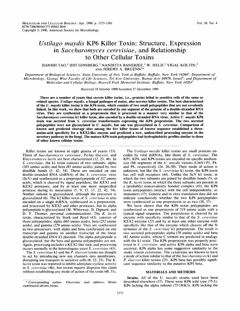
FIG. 1. Sequencing strategy and P6 M2 genome structure. DNAsequencing runs are shown as solid arrows, and RNA sequencingruns are shown as dotted arrows. FI1, Open reading frame; _, P6Li sequence.
tilled water, and the samples were lyophilyzed to dryness.Final samples were concentrated 50-fold for killer tests.
Partial purification of KP6 toxin from yeast transformants.The dialyzed and lyophilized samples were dissolved in 25mM sodium acetate (pH 5.5). Columns of carboxymethyl-Sephadex C-50 were equilibrated with the same buffer, andsamples were applied. When the A280 of the flowthroughreached zero, 0.5 M NaCl in 25 mM sodium acetate (pH 5.5)was applied, and the eluate was collected. Fractions weredialyzed against distilled water and dried by lyophilization.Final samples for application to gels were concentrated2,500-fold from the carboxymethyl-Sephadex fractions.Enzymatic deglycosylation of secreted polypeptides. Sam-
ples were denatured by boiling in 0.5% SDS-1% P-mercap-toethanol for 5 min. The denatured samples were incubatedin 30 mM phosphate buffer (pH 6.0) with or without 0.01 Uof endoglycosidase H (endo H; Boehringer Mannheim Bio-chemicals) in the presence of 1.25% Nonidet P-40. Theproducts were dialyzed against distilled water and lyophi-lized to dryness. Samples were redissolved in distilled waterand loaded on SDS-18% polyacrylamide gels. Silver stainingof gels was done as described previously (52).
RESULTSSequence of P6 M2. The sequence of P6 M2, the toxin-
encoding dsRNA segment of the U. maydis virus P6, wasdetermined on both strands by sequencing several cDNAclones and by direct RNA sequencing primed by syntheticoligonucleotides as shown in Fig. 1. There is only one longopen reading frame. This is on the viral plus strand, identi-fied as for P1 Ml (11). The sequence of P6 Li, a smallnoncoding dsRNA of 355 base pairs, was found to be entirelyderived from the 3' end of the P6 M2 viral plus strand (Fig.1). This is identical to the relationship between P1 Ml and P1Li (11, 16), with which this sequence shows a high degree ofsimilarity (77%) over a region of 231 base pairs encompass-ing the border between Li and M2 (or Ml and Li in P1) (N.Banerjee, J. Tao, J. A. Bruenn, and Y. Koltin, manuscript inpreparation).The sequence of the plus strand is shown in Fig. 2. The
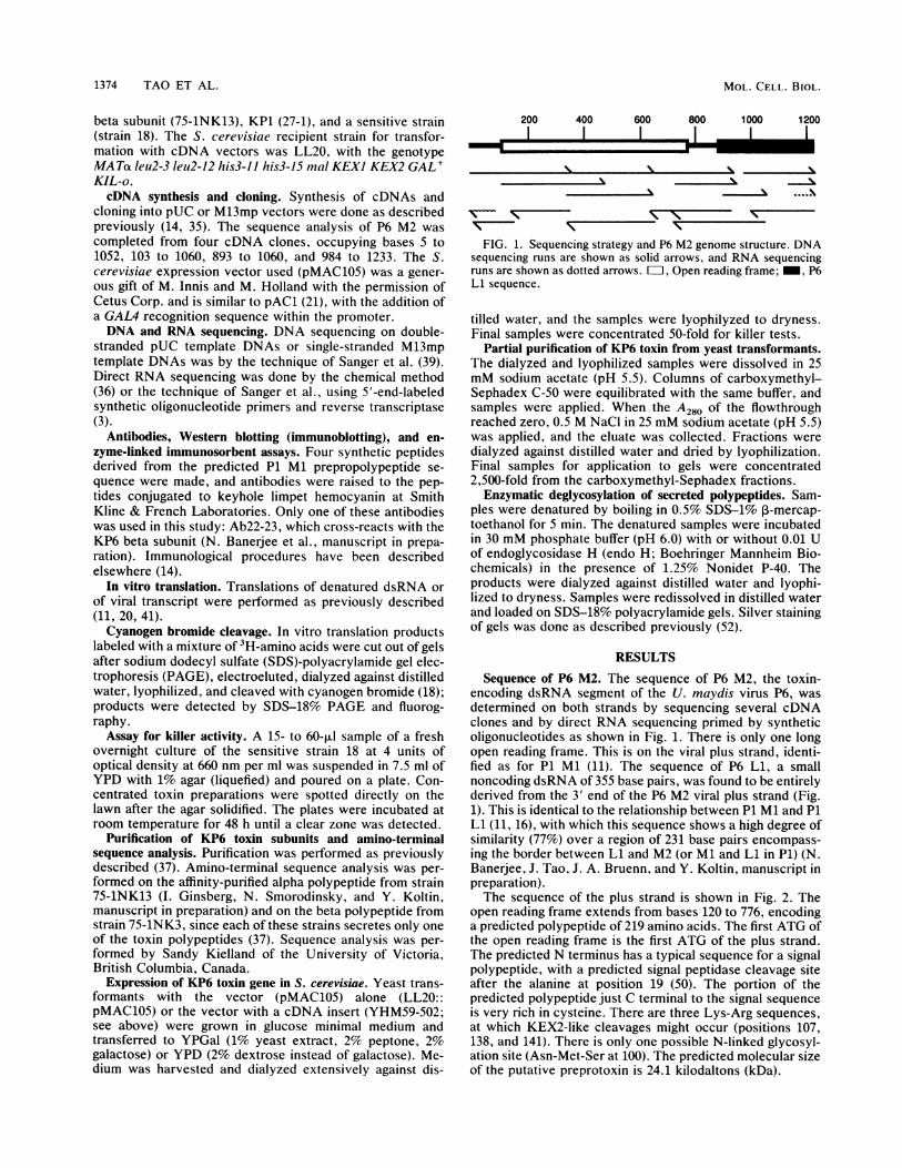
open reading frame extends from bases 120 to 776, encodinga predicted polypeptide of 219 amino acids. The first ATG ofthe open reading frame is the first ATG of the plus strand.The predicted N terminus has a typical sequence for a signalpolypeptide, with a predicted signal peptidase cleavage siteafter the alanine at position 19 (50). The portion of thepredicted polypeptide just C terminal to the signal sequenceis very rich in cysteine. There are three Lys-Arg sequences,at which KEX2-like cleavages might occur (positions 107,138, and 141). There is only one possible N-linked glycosyl-ation site (Asn-Met-Ser at 100). The predicted molecular sizeof the putative preprotoxin is 24.1 kilodaltons (kDa).
M
MOL. CELL. BIOL.
U. MA YDIS KP6 KILLER TOXIN 1375
1 GAAAAACTCAATAAGAACGGATTAAACCAATCACGACTAAGAAGCCTCGGTTAATTATAC61 GGCAGGACTCCCTTGTAAGACTATCCTAGCAGCTCTACCTAAGGTCACTTACAAACGGAA
M
121 TGTTGATATTCAGCGTCCTTATGTACTTAGGGCTGTTGCTCGCAGGAGCTTCCGCACTCC2 etLeuIlePheSerValLeuMetTyrLeuGlyLeuLeuLeuAlaGlyAlaSerAlaLeuP
181 CAAATGGGCTCTCGCCACGCAACAATGCTTTTTGTGCTGGATTTGGTCTCTCTTGCAAGT22 roAsnGlyLeuSerProArgsnAsnAlaPheCysAlaGlyPhe LeuSerCysLysT
ALPHA NH2241 GGGAATGTTGGTGCACAGCACACGGAACGGGCAATGAATTACGGTATGCTACCGCAGCAG42 r_GUC TrDCv_ThrA1aHisGlvThrGlvAsnGluLeuArgTyrAlaThrAIalaG
301 GATGCGGAGATCATCTGTCCAAGTCTTATTACGATGCTCGGGCCGGCCACTGCCTGTTCT62 jyCysGlyAspHisLeuSerLysSerTyrTyrAspAlaArgAlaGlyHisCysLeuPheS
361 CTGACGACCTTCGCAACCAGTTCTACAGCCATTGTTCGTCTCTAAACAACAATATGTCGT82 erAspAspLeuArgAsnGlnPheTyrSerHisCysSerSerLeuAsnAsnAsnMetSerC
601201
18021
24041
30061
36081
420101
421 GCCGGTCGTTGTCTAAACGGACTATCCAAGATAGCGCTACCGACACGGTAGACCTCGGTG 480102 ysArgSerLeuSerLysArgThrIleGlnAspSerAlaThrAspThrValAspLeuGlyA 121
ALPHA COON481 CCGAGCTCCATAGGGATGACCCGCCCCCTACTGCTAGTGACATAGGCAAACGGGGTAAGA 540122 laGluLeuHisArgAspAspProProProThrAlaSerAspIleGlyLysArgIlyLsA 141
BETA NH2541 GGCCTAGACCTGTTATGTGCCAATGTGTAGACACAACGAACGGAGGGGTTCGATTAGACG 600142 rgProArgProValMetCvsGlnCvsValAsPThrThrAsnGlvGlvValArgLeuAsRA 161
601 CGGTGACTAGGGCGGCTTGCAGCATAGACTCGTTTATCGACGGGTACTATACGGAAAAGG162 1aVa1ThrArgA aAlaCysSerIleAspSerPheIleAspGlyTyrTyrThrGluLysA
661 ATGGGTTTTGTAGAGCTAAATATTCCTGGGACTTGTTTACGAGCGGCCAGTTCTACCAGG182 spGlyPheCysArgAlaLysTyrSerTrpAspLeuPheThrSerGlyGlnPheTyrGlnA
721 CATGTTTGAGGTACTCACATGCCGGGACCAACTGCCAACCTGACCCGCAGTATGAATAAG202 laCysLeuArgTyrSerHisAlaGlyThrAsnCysGlnProAspProGlnTyrGlu 219
BETA COON781 TAACCAACGAGGGTTCCTTCAACCCATAACTGGTGTATGTCGAACATGCTAACCAACAGA841 GCTATAAAAGAGCCGCCAGTATAAATAAGAGAGTTTGCATCCGGCCTAAATGTGGAGTTA
Ll901 GGGCGCCGGTTGGGGTATAGTAGGTGAAGAGTACGAAGAGGAGTTGATATGAGAGTGGAA961 AAGTAACCAGATGACCATCCTATGTCGGTTATCCGAGGAAGCGTGGGCTGGAGAAAAATG
1021 CCGTTCGATCCGTGCGATGCTCGATTTGATGCAAGTTGGGATACTATGAACTGAGGTGCT1081 TACGACGGCCCTAACTGCATATTTAGGGTGAGAGGGGACAGCTGCTACGCTGGGCTGCTC1141 CTCGCGGACTCAACAGCGGGAACCGACCTGGCGTACATGAGTGAAATCTGTAGGGATAGC1201 ACAAGATACGCAGGCAGGTGACTAGTAGAATGCA 1234
660181
720201
780
840900
9601020108011401200
FIG. 2. Sequence of the P6 M2 plus-strand cDNA. The sequence of the predicted preprotoxin is indicated under the DNA sequence.Predicted or known proteolytic cleavages follow the italicized dipeptides. Underlined sequences are those of the alpha and beta N terminifound by Edman degradation (Fig. 4). The known N termini of alpha and beta and the predicted C termini are indicated. The LI sequenceis shown in italics. This sequence is available as GenBank accession no. M27418.
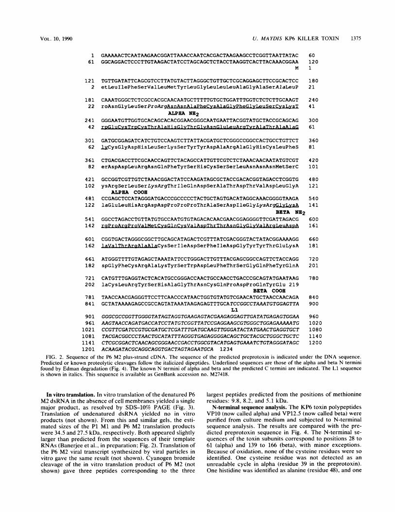
In vitro translation. In vitro translation of the denatured P6M2 dsRNA in the absence of cell membranes yielded a singlemajor product, as resolved by SDS-10% PAGE (Fig. 3).Translation of undenatured dsRNA yielded no in vitroproducts (not shown). From this and similar gels, the esti-mated sizes of the P1 Ml and P6 M2 translation productswere 34.5 and 27.5 kDa, respectively. Both appeared slightlylarger than predicted from the sequences of their templateRNAs (Banerjee et al., in preparation; Fig. 2). Translation ofthe P6 M2 viral transcript synthesized by viral particles invitro gave the same result (not shown). Cyanogen bromidecleavage of the in vitro translation product of P6 M2 (notshown) gave three peptides corresponding to the three
largest peptides predicted from the positions of methionineresidues: 9.8, 8.2, and 5.1 kDa.
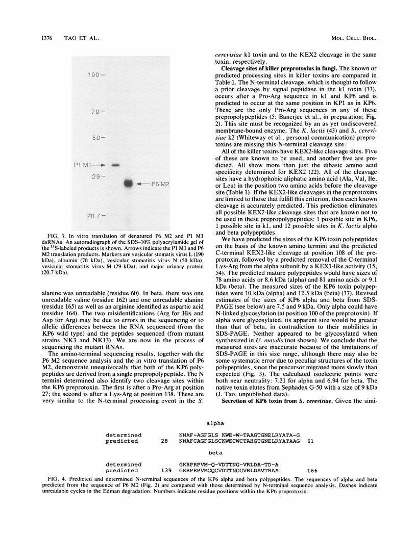
N-terminal sequence analysis. The KP6 toxin polypeptidesVP10 (now called alpha) and VP12.5 (now called beta) werepurified from culture medium and subjected to N-terminalsequence analysis. The results are compared with the pre-dicted preprotoxin sequence in Fig. 4. The N-terminal se-quences of the toxin subunits correspond to positions 28 to61 (alpha) and 139 to 166 (beta), with minor exceptions.Because of oxidation, none of the cysteine residues were soidentified. One cysteine residue was not detected as anunreadable cycle in alpha (residue 39 in the preprotoxin).One histidine was identified as alanine (residue 48), and one
VOL. 10, 1990
1376 TAO ET AL.
1 90 -
70 -
IOI-
.rI N:I 1 _a
29-w - PGtD 2
<01-7
FIG. 3. In vitro translation of denatured P6 M2 and P1 MldsRNAs. An autoradiograph of the SDS-10% polyacrylamide gel ofthe 35S-labeled products is shown. Arrows indicate the P1 Ml and P6M2 translation products. Markers are vesicular stomatis virus L (190kDa), albumin (70 kDa), vesicular stomatitis virus N (50 kDa),vesicular stomatitis virus M (29 kDa), and major urinary protein(20.7 kDa).
alanine was unreadable (residue 60). In beta, there was oneunreadable valine (residue 162) and one unreadable alanine(residue 165) as well as an arginine identified as aspartic acid(residue 164). The two misidentifications (Arg for His andAsp for Arg) may be due to errors in the sequencing or toallelic differences between the RNA sequenced (from theKP6 wild type) and the peptides sequenced (from mutantstrains NK3 and NK13). We are now in the process ofsequencing the mutant RNAs.The amino-terminal sequencing results, together with the
P6 M2 sequence analysis and the in vitro translation of P6M2, demonstrate unequivocally that both of the KP6 poly-peptides are derived from a single prepropolypeptide. The Ntermini determined also identify two cleavage sites withinthe KP6 preprotoxin. The first is after a Pro-Arg at position27; the second is after a Lys-Arg at position 138. These arevery similar to the N-terminal processing event in the S.
cerevisiae kl toxin and to the KEX2 cleavage in the sametoxin, respectively.
Cleavage sites of killer preprotoxins in fungi. The known orpredicted processing sites in killer toxins are compared inTable 1. The N-terminal cleavage, which is thought to followa prior cleavage by signal peptidase in the kl toxin (33),occurs after a Pro-Arg sequence in kl and KP6 and ispredicted to occur at the same position in KP1 as in KP6.These are the only Pro-Arg sequences in any of theseprepropolypeptides (5; Banerjee et al., in preparation; Fig.2). This site must be recognized by an as yet undiscoveredmembrane-bound enzyme. The K. lactis (43) and S. cerevi-siae k2 (Whiteway et al., personal communication) prepro-toxins are missing this N-terminal cleavage site.
All of the killer toxins have KEX2-like cleavage sites. Fiveof these are known to be used, and another five are pre-dicted. All show more than just the dibasic amino acidspecificity determined for KEX2 (22). All of the cleavagesites have a hydrophobic aliphatic amino acid (Ala, Val, Ile,or Leu) in the position two amino acids before the cleavagesite (Table 1). If the KEX2-like cleavages in the preprotoxinsare limited to those that fulfill this criterion, then each knowncleavage is accurately predicted. This prediction eliminatesall possible KEX2-like cleavage sites that are known not tobe used in these prepropolypeptides: 1 possible site in KP6,1 possible site in kl, and 12 possible sites in K. lactis alphaand beta polypeptides.We have predicted the sizes of the KP6 toxin polypeptides
on the basis of the known amino termini and the predictedC-terminal KEX2-like cleavage at position 108 of the pre-protoxin, followed by a predicted removal of the C-terminalLys-Arg from the alpha subunit by a KEXi-like activity (15,54). The predicted mature polypeptides would have sizes of78 amino acids or 8.6 kDa (alpha) and 81 amino acids or 9.1kDa (beta). The measured sizes of the KP6 toxin polypep-tides were 10 kDa (alpha) and 12.5 kDa (beta) (37). Revisedestimates of the sizes of KP6 alpha and beta from SDS-PAGE (see below) are 7.5 and 9 kDa. Only alpha could haveN-linked glycosylation (at position 100 of the preprotoxin). Ifalpha were glycosylated, its apparent size would be greaterthan that of beta, in contradiction to their mobilities inSDS-PAGE. Neither appeared to be glycosylated whensynthesized in U. maydis (not shown). We conclude that themeasured sizes are inaccurate because of the limitations ofSDS-PAGE in this size range, although there may also besome systematic error due to peculiar structures of the toxinpolypeptides, since the precursor migrated more slowly thanexpected (Fig. 3). The calculated isoelectric points wereboth near neutrality: 7.21 for alpha and 6.94 for beta. Thenative toxin elutes from Sephadex G-50 with a size of 9 kDa(J. Tao, unpublished data).
Secretion of KP6 toxin from S. cerevisiae. Given the simi-
alpha
NNAF-AGFGLS KWE-W-TAAGTGNELRYATA-G28 NNAFCAGFGLSCKWECWCTAHGTGNELRYATAAG 61
beta
GKRPRPVM-Q-VDTTNG-VRLDA-TD-A139 GKRPRPVMCQCVDTTNGGVRLDAVTRAA 166
FIG. 4. Predicted and determined N-terminal sequences of the KP6 alpha and beta polypeptides. The sequences of alpha and betapredicted from the sequence of P6 M2 (Fig. 2) are compared with those determined by N-terminal sequence analysis. Dashes indicateunreadable cycles in the Edman degradation. Numbers indicate residue positions within the KP6 preprotoxin.
determinedpredicted
determinedpredicted
MOL. CELL. BIOL.
U. MA YDIS KP6 KILLER TOXIN 1377
TABLE 1. Cleavage sites in preprotoxins in fungi
Sequence" (reference)Preprotoxin Site"
N-terminal site KEX2-like site
kl, S. cerevisiae k 44 Leu-Leu-Pro-Arg*Glu (5)KP6, U. maydis k 26 Leu-Ser-Pro-Arg*AsN (this work)KP1, U. maydis p 26 Pro-Gly-Pro-Arg*Ser (Banerjee et
al., in preparation)kl, S. cerevisiae k 149 Val-Ala-Arg-Arg*Asp (5, 54)kl, S. cerei'isiae k 233 Val-Ala-Lys-Arg*Tyr (5)k2, S. cerevisiae p 221 Ile-Val-Lys-Arg*Asp (D. Y. Thomas, personal
communication)k2, S. cerevisiae p 268 Val-Gly-Lys-Arg*Glu (D. Y. Thomas, personal
communication)alpha, K. lactis k 29 Leu-Val-Lys-Arg*Ala (43)beta, K. lactis k 894 Leu-Tyr-Lys-Arg*Gly (43)KP6, U. inaydis p 108 Leu-Ser-Lys-Arg*Thr (this work)KP6, U. maydis k 137 Ile-Gly-Lys-Arg*Gly (this work)KP1, U. maydis p 108 Ala-Pro-Lys-Arg*Gly (Banerjee et al., in preparation)KP1, U. maydis p 113 Val-Val-Lys-Arg*Asp (Banerjee et al., in preparation)
a k, Known cleavage site, determined by amino-terminal or carboxy-terminal sequence analysis; p. predicted cleavage site.b Asterisks indicate cleavage sites.
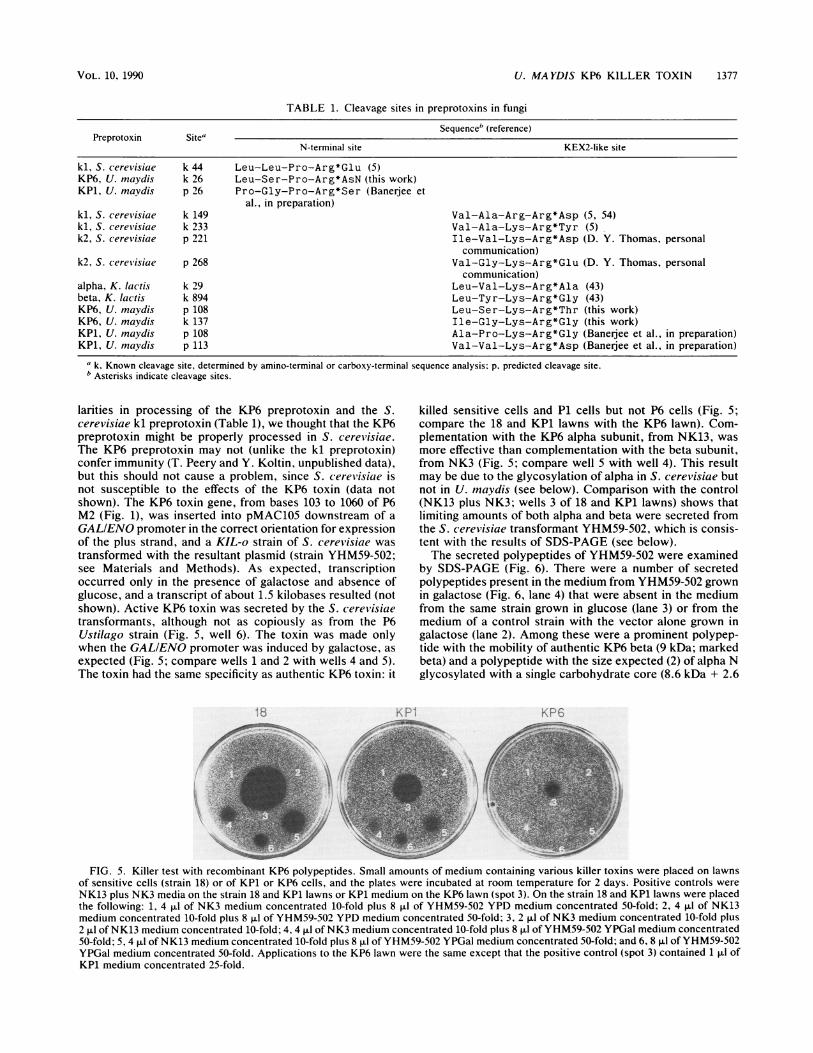
larities in processing of the KP6 preprotoxin and the S.cerevisiae kl preprotoxin (Table 1), we thought that the KP6preprotoxin might be properly processed in S. cerevisiae.The KP6 preprotoxin may not (unlike the kl preprotoxin)confer immunity (T. Peery and Y. Koltin, unpublished data),but this should not cause a problem, since S. cereWisiae isnot susceptible to the effects of the KP6 toxin (data notshown). The KP6 toxin gene, from bases 103 to 1060 of P6M2 (Fig. 1), was inserted into pMAC105 downstream of aGALIENO promoter in the correct orientation for expressionof the plus strand, and a KIL-o strain of S. cerevisiae wastransformed with the resultant plasmid (strain YHM59-502;see Materials and Methods). As expected, transcriptionoccurred only in the presence of galactose and absence ofglucose, and a transcript of about 1.5 kilobases resulted (notshown). Active KP6 toxin was secreted by the S. cerevisiaetransformants, although not as copiously as from the P6Ustilago strain (Fig. 5, well 6). The toxin was made onlywhen the GALIENO promoter was induced by galactose, asexpected (Fig. 5; compare wells 1 and 2 with wells 4 and 5).The toxin had the same specificity as authentic KP6 toxin: it
killed sensitive cells and P1 cells but not P6 cells (Fig. 5;compare the 18 and KP1 lawns with the KP6 lawn). Com-plementation with the KP6 alpha subunit, from NK13, wasmore effective than complementation with the beta subunit,from NK3 (Fig. 5; compare well 5 with well 4). This resultmay be due to the glycosylation of alpha in S. cerevisiae butnot in U. mnaydis (see below). Comparison with the control(NK13 plus NK3; wells 3 of 18 and KP1 lawns) shows thatlimiting amounts of both alpha and beta were secreted fromthe S. cerevisiae transformant YHM59-502, which is consis-tent with the results of SDS-PAGE (see below).The secreted polypeptides of YHM59-502 were examined
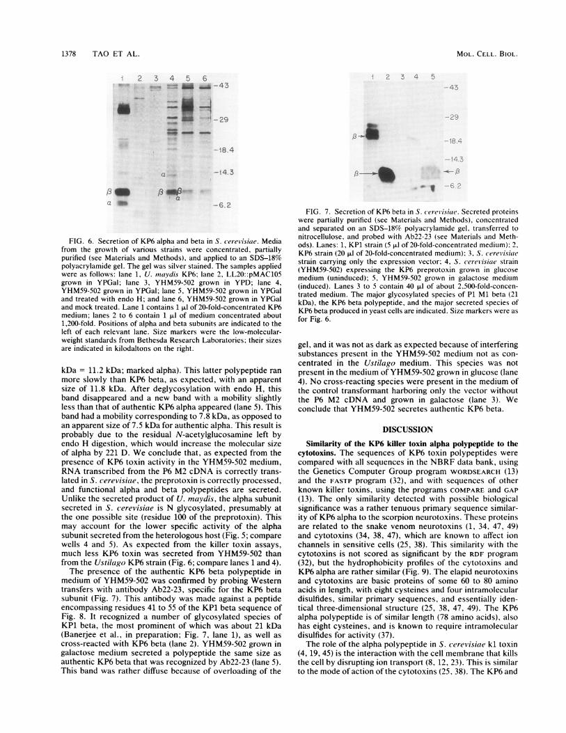
by SDS-PAGE (Fig. 6). There were a number of secretedpolypeptides present in the medium from YHM59-502 grownin galactose (Fig. 6, lane 4) that were absent in the mediumfrom the same strain grown in glucose (lane 3) or from themedium of a control strain with the vector alone grown ingalactose (lane 2). Among these were a prominent polypep-tide with the mobility of authentic KP6 beta (9 kDa; markedbeta) and a polypeptide with the size expected (2) of alpha Nglycosylated with a single carbohydrate core (8.6 kDa + 2.6
FIG. 5. Killer test with recombinant KP6 polypeptides. Small amounts of medium containing various killer toxins were placed on lawnsof sensitive cells (strain 18) or of KP1 or KP6 cells, and the plates were incubated at room temperature for 2 days. Positive controls wereNK13 plus NK3 media on the strain 18 and KP1 lawns or KP1 medium on the KP6 lawn (spot 3). On the strain 18 and KP1 lawns were placedthe following: 1, 4,l of NK3 medium concentrated 10-fold plus 8 ,u1 of YHM59-502 YPD medium concentrated 50-fold; 2, 4,ul of NK13medium concentrated 10-fold plus 8 ,u1 of YHMS9-502 YPD medium concentrated 50-fold; 3, 2 p.1 of NK3 medium concentrated 10-fold plus2 p.1 of NK13 medium concentrated 10-fold; 4,4, l of NK3 medium concentrated 10-fold plus 8 p.1 of YHM59-502 YPGal medium concentrated50-fold; 5, 4 p.1 of NK13 medium concentrated 10-fold plus 8 p.1 of YHM59-502 YPGal medium concentrated 50-fold; and 6, 8 p.1 of YHM59-502YPGal medium concentrated 50-fold. Applications to the KP6 lawn were the same except that the positive control (spot 3) contained 1 p.1 ofKP1 medium concentrated 25-fold.
VOL. 10, 1990
I b.---
;N.c.. V
I
r,F'
1378 TAO ET AL.
2 3 4 5S 6-- I " -43
as --LOA1F..i 29_ _ _
MOL. CELL. BIOL.
2 3 4 5
i4.3
a _
- I13 MAa
-6.2
FIG. 6. Secretion of KP6 alpha and beta in S. cerev isiae. Mediafrom the growth of various strains were concentrated, partiallypurified (see Materials and Methods), and applied to an SDS-18%polyacrylamide gel. The gel was silver stained. The samples appliedwere as follows: lane 1, U. maydis KP6; lane 2, LL20::pMAC105grown in YPGal; lane 3, YHM59-502 grown in YPD; lane 4,YHM59-502 grown in YPGal; lane 5, YHM59-502 grown in YPGaland treated with endo H; and lane 6, YHM59-502 grown in YPGaland mock treated. Lane 1 contains 1 ,ul of 20-fold-concentrated KP6medium; lanes 2 to 6 contain 1 ,ul of medium concentrated about1,200-fold. Positions of alpha and beta subunits are indicated to theleft of each relevant lane. Size markers were the low-molecular-weight standards from Bethesda Research Laboratories; their sizesare indicated in kilodaltons on the right.
kDa = 11.2 kDa; marked alpha). This latter polypeptide ranmore slowly than KP6 beta, as expected, with an apparentsize of 11.8 kDa. After deglycosylation with endo H, thisband disappeared and a new band with a mobility slightlyless than that of authentic KP6 alpha appeared (lane 5). Thisband had a mobility corresponding to 7.8 kDa, as opposed toan apparent size of 7.5 kDa for authentic alpha. This result isprobably due to the residual N-acetylglucosamine left byendo H digestion, which would increase the molecular sizeof alpha by 221 D. We conclude that, as expected from thepresence of KP6 toxin activity in the YHM59-502 medium,RNA transcribed from the P6 M2 cDNA is correctly trans-lated in S. cerevisiae, the preprotoxin is correctly processed,and functional alpha and beta polypeptides are secreted.Unlike the secreted product of U. maydis, the alpha subunitsecreted in S. cerevisiae is N glycosylated, presumably atthe one possible site (residue 100 of the preprotoxin). Thismay account for the lower specific activity of the alphasubunit secreted from the heterologous host (Fig. 5; comparewells 4 and 5). As expected from the killer toxin assays,much less KP6 toxin was secreted from YHM59-502 thanfrom the Ustilago KP6 strain (Fig. 6; compare lanes 1 and 4).The presence of the authentic KP6 beta polypeptide in
medium of YHM59-502 was confirmed by probing Westerntransfers with antibody Ab22-23, specific for the KP6 betasubunit (Fig. 7). This antibody was made against a peptideencompassing residues 41 to 55 of the KP1 beta sequence ofFig. 8. It recognized a number of glycosylated species ofKP1 beta, the most prominent of which was about 21 kDa(Banerjee et al., in preparation; Fig. 7, lane 1), as well ascross-reacted with KP6 beta (lane 2). YHM59-502 grown ingalactose medium secreted a polypeptide the same size as
authentic KP6 beta that was recognized by Ab22-23 (lane 5).This band was rather diffuse because of overloading of the
FIG. 7. Secretion of KP6 beta in S. cerel'isiae. Secreted proteinswere partially purified (see Materials and Methods), concentratedand separated on an SDS-18% polyacrylamide gel, transferred tonitrocellulose, and probed with Ab22-23 (see Materials and Meth-ods). Lanes: 1, KP1 strain (5 ,u1 of 20-fold-concentrated medium); 2,KP6 strain (20 ,ul of 20-fold-concentrated medium); 3. S. cereviisiaestrain carrying only the expression vector; 4, S. cerei'isiae strain(YHM59-502) expressing the KP6 preprotoxin grown in glucosemedium (uninduced); 5, YHM59-502 grown in galactose medium(induced). Lanes 3 to 5 contain 40 ,ul of about 2,500-fold-concen-trated medium. The major glycosylated species of P1 Ml beta (21kDa), the KP6 beta polypeptide, and the major secreted species ofKP6 beta produced in yeast cells are indicated. Size markers were asfor Fig. 6.
gel, and it was not as dark as expected because of interferingsubstances present in the YHM59-502 medium not as con-centrated in the Ustilago medium. This species was notpresent in the medium of YHM59-502 grown in glucose (lane4). No cross-reacting species were present in the medium ofthe control transformant harboring only the vector withoutthe P6 M2 cDNA and grown in galactose (lane 3). Weconclude that YHM59-502 secretes authentic KP6 beta.
DISCUSSION
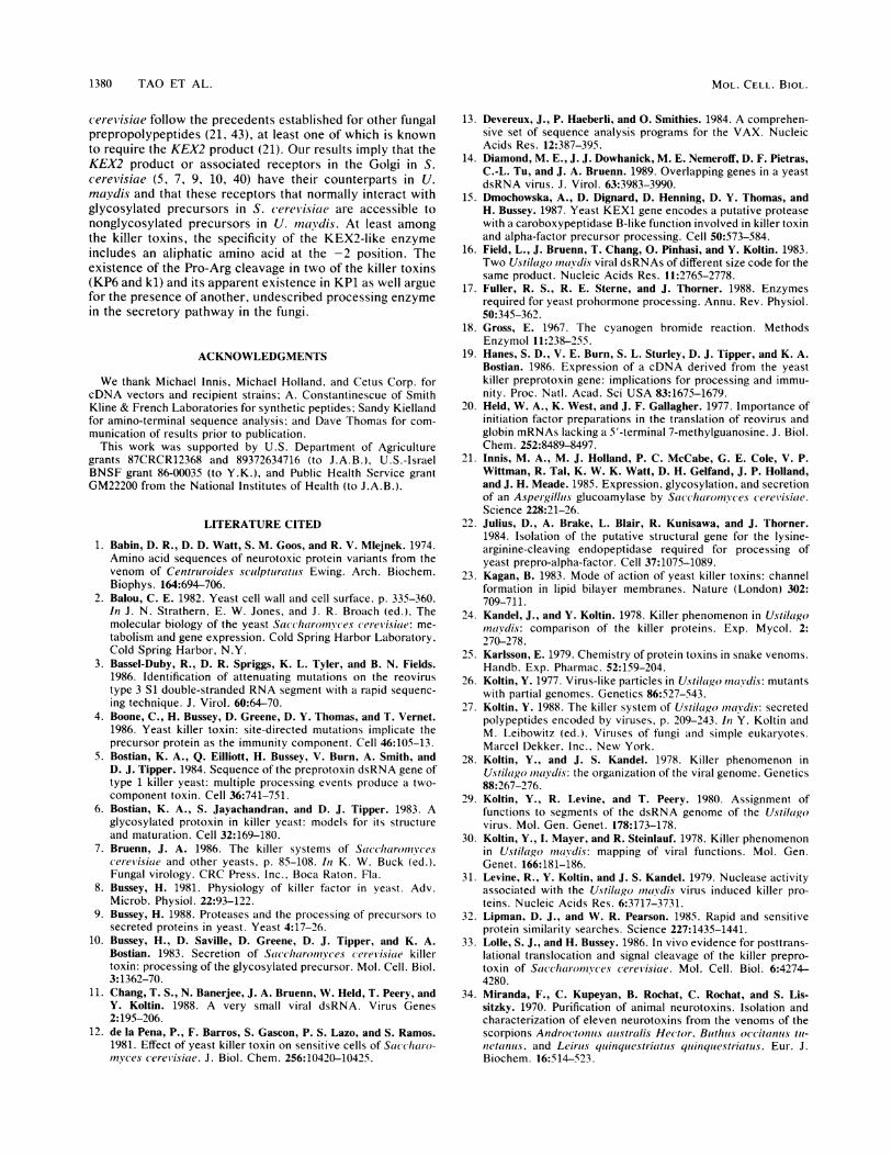
Similarity of the KP6 killer toxin alpha polypeptide to thecytotoxins. The sequences of KP6 toxin polypeptides werecompared with all sequences in the NBRF data bank, usingthe Genetics Computer Group program WORDSEARCH (13)and the FASTP program (32), and with sequences of otherknown killer toxins, using the programs COMPARE and GAP(13). The only similarity detected with possible biologicalsignificance was a rather tenuous primary sequence similar-ity of KP6 alpha to the scorpion neurotoxins. These proteinsare related to the snake venom neurotoxins (1, 34, 47, 49)and cytotoxins (34, 38, 47), which are known to affect ionchannels in sensitive cells (25, 38). This similarity with thecytotoxins is not scored as significant by the RDF program(32), but the hydrophobicity profiles of the cytotoxins andKP6 alpha are rather similar (Fig. 9). The elapid neurotoxinsand cytotoxins are basic proteins of some 60 to 80 aminoacids in length, with eight cysteines and four intramoleculardisulfides, similar primary sequences, and essentially iden-tical three-dimensional structure (25, 38, 47, 49). The KP6alpha polypeptide is of similar length (78 amino acids), alsohas eight cysteines, and is known to require intramoleculardisulfides for activity (37).The role of the alpha polypeptide in S. cerevisiae kl toxin
(4, 19, 45) is the interaction with the cell membrane that killsthe cell by disrupting ion transport (8, 12, 23). This is similarto the mode of action of the cytotoxins (25, 38). The KP6 and
U. MA YDIS KP6 KILLER TOXIN 1379
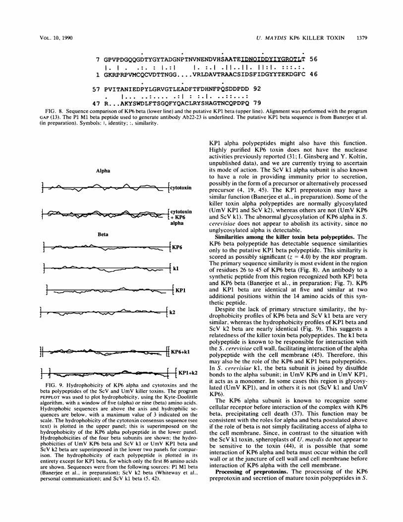
7 GPVPDGQQGDTYGYTADGNPTNVNENDVHSAATE;IDNOIDDYTYGROILT1. I .:. : 1.:1 1. :.I .11-....................11..11:1. :::.:.
1 GKRPRPVMCQCVDTTNGG.... VRLDAVTRAACSIDSFIDGYYTEKDGFC 46
57 PVITANIEDPYLGRVGTLEADFTFDHNFPQSDDPDD 921I.. . ..:.... .:I1 : : .I1. . . :: . .. :
47 R... AKYSWDLFTSGQFYQACLRYSHAGTNCQPDPQ 79FIG. 8. Sequence comparison of KP6 beta (lower line) and the putative KP1 beta (upper line). Alignment was performed with the program
GAP (13). The P1 Ml beta peptide used to generate antibody Ab22-23 is underlined. The putative KP1 beta sequence is from Banerjee et al.(in preparation). Symbols: I, identity; :. similarity.
Alpha
.9ho-~- cytotoxi
cytotoxin~4~ e~ -j + KP6alpha
Beta
[ki
] - [ 1
4k2
sw-F[[|KP6+kl
<H KPl+k2IFIG. 9. Hydrophobicity of KP6 alpha and cytotoxins and the
beta polypeptides of the ScV and UmV killer toxins. The programPEPPLOT was used to plot hydrophobicity, using the Kyte-Doolittlealgorithm, with a window of five (alpha) or nine (beta) amino acids.Hydrophobic sequences are above the axis and hydrophilic se-
quences are below, with a maximum value of 3 indicated on thescale. The hydrophobicity of the cytotoxin consensus sequence (seetext) is plotted in the upper panel; this is superimposed on thehydrophobicity of the KP6 alpha polypeptide in the lower panel.Hydrophobicities of the four beta subunits are shown; the hydro-phobicities of UmV KP6 beta and ScV kl or UmV KP1 beta andScV k2 beta are superimposed in the lower two panels for compar-ison. The hydrophobicity of each polypeptide is plotted in itsentirety except for KP1 beta, for which only the first 86 amino acidsare shown. Sequences were from the following sources: P1 Ml beta(Banerjee et al., in preparation); ScV k2 beta (Whiteway et al..personal communication); and ScV kl beta (5, 42).
KP1 alpha polypeptides might also have this function.Highly purified KP6 toxin does not have the nucleaseactivities previously reported (31; I. Ginsberg and Y. Koltin,unpublished data), and we are currently trying to ascertainits mode of action. The ScV kl alpha subunit is also knownto have a role in providing immunity prior to secretion,possibly in the form of a precursor or alternatively processedprecursor (4, 19, 45). The KP1 preprotoxin may have asimilar function (Banerjee et al., in preparation). Some of thekiller toxin alpha polypeptides are normally glycosylated(UmV KP1 and ScV k2), whereas others are not (UmV KP6and ScV kl). The abnormal glycosylation of KP6 alpha in S.cerevisiae does not appear to abolish its activity, since nounglycosylated alpha is detectable.
Similarities among the killer toxin beta polypeptides. TheKP6 beta polypeptide has detectable sequence similaritiesonly to the putative KP1 beta polypeptide. This similarity isscored as possibly significant (z = 4.0) by the RDF program.The primary sequence similarity is most evident in the regionof residues 26 to 45 of KP6 beta (Fig. 8). An antibody to asynthetic peptide from this region recognized both KP1 betaand KP6 beta (Baneree et al., in preparation; Fig. 7). KP6and KP1 beta are identical at five and similar at twoadditional positions within the 14 amino acids of this syn-thetic peptide.
Despite the lack of primary structure similarity, the hy-drophobicity profiles of KP6 beta and ScV kl beta are verysimilar, whereas the hydrophobicity profiles of KP1 beta andScV k2 beta are nearly identical (Fig. 9). This suggests arelatedness of the killer toxin beta polypeptides. The kl betapolypeptide is known to be responsible for interaction withthe S. cerevisiae cell wall, facilitating interaction of the alphapolypeptide with the cell membrane (45). Therefore, thismay also be the role of the KP6 and KP1 beta polypeptides.In S. cerei'isiae kl, the beta subunit is joined by disulfidebonds to the alpha subunit; in UmV KP6 and in UmV KP1,it acts as a monomer. In some cases this region is glycosy-lated (UmV KP1), and in others it is not (ScV kl and UmVKP6).The KP6 alpha subunit is known to recognize some
cellular receptor before interaction of the complex with KP6beta, precipitating cell death (37). This function may beconsistent with the roles for alpha and beta postulated aboveif the role of beta is not simply facilitating access of alpha tothe cell membrane. Since, in contrast to the situation withthe ScV kl toxin, spheroplasts of U. maydis do not appear tobe sensitive to the toxin (44), it is possible that someinteraction of KP6 alpha and beta must occur within the cellwall or at the juncture of cell wall and cell membrane beforeinteraction of KP6 alpha with the cell membrane.
Processing of preprotoxins. The processing of the KP6preprotoxin and secretion of mature toxin polypeptides in S.
56
1}
L-]
VOL. 10,1990
r- - .f
I
1380 TAO ET AL.
cerevisiae follow the precedents established for other fungalprepropolypeptides (21, 43), at least one of which is knownto require the KEX2 product (21). Our results imply that theKEX2 product or associated receptors in the Golgi in S.(erevisiae (5, 7, 9, 10, 40) have their counterparts in U.maydis and that these receptors that normally interact withglycosylated precursors in S. (erel'isiae are accessible tononglycosylated precursors in U. inavdis. At least amongthe killer toxins, the specificity of the KEX2-like enzymeincludes an aliphatic amino acid at the -2 position. Theexistence of the Pro-Arg cleavage in two of the killer toxins(KP6 and kl) and its apparent existence in KP1 as well arguefor the presence of another, undescribed processing enzymein the secretory pathway in the fungi.
ACKNOWLEDGMENTS
We thank Michael Innis, Michael Holland, and Cetus Corp. forcDNA vectors and recipient strains; A. Constantinescue of SmithKline & French Laboratories for synthetic peptides: Sandy Kiellandfor amino-terminal sequence analysis; and Dave Thomas for com-munication of results prior to publication.
This work was supported by U.S. Department of Agriculturegrants 87CRCR12368 and 89372634716 (to J.A.B.). U.S.-IsraelBNSF grant 86-00035 (to Y.K.), and Public Health Service grantGM22200 from the National Institutes of Health (to J.A.B.).
LITERATURE CITED
1. Babin, D. R., D. D. Watt, S. M. Goos, and R. V. Mlejnek. 1974.Amino acid sequences of neurotoxic protein variants from thevenom of Centriuroides scullptituritis Ewing. Arch. Biochem.Biophys. 164:694-706.
2. Balou, C. E. 1982. Yeast cell wall and cell surface. p. 335-360.In J. N. Strathern, E. W. Jones, and J. R. Broach (ed.). Themolecular biology of the yeast Saccharomvces cerei'isiae: me-tabolism and gene expression. Cold Spring Harbor Laboratory,Cold Spring Harbor, N.Y.
3. Bassel-Duby, R., D. R. Spriggs, K. L. Tyler, and B. N. Fields.1986. Identification of attenuating mutations on the reovirustype 3 S1 double-stranded RNA segment with a rapid sequenc-ing technique. J. Virol. 60:64-70.
4. Boone, C., H. Bussey, D. Greene, D. Y. Thomas, and T. Vernet.1986. Yeast killer toxin: site-directed mutations implicate theprecursor protein as the immunity component. Cell 46:105-13.
5. Bostian, K. A., Q. Eilliott, H. Bussey, V. Burn, A. Smith, andD. J. Tipper. 1984. Sequence of the preprotoxin dsRNA gene oftype 1 killer yeast: multiple processing events produce a two-component toxin. Cell 36:741-751.
6. Bostian, K. A., S. Jayachandran, and D. J. Tipper. 1983. Aglycosylated protoxin in killer yeast: models for its structureand maturation. Cell 32:169-180.
7. Bruenn, J. A. 1986. The killer systems of Saccharomv escerei'isiae and other yeasts, p. 85-108. In K. W. Buck (ed.).Fungal virology. CRC Press, Inc.. Boca Raton. Fla.
8. Bussey, H. 1981. Physiology of killer factor in yeast. Adv.Microb. Physiol. 22:93-122.
9. Bussey, H. 1988. Proteases and the processing of precursors tosecreted proteins in yeast. Yeast 4:17-26.
10. Bussey, H., D. Saville, D. Greene, D. J. Tipper, and K. A.Bostian. 1983. Secretion of Saccharonczyces c(erevisiae killertoxin: processing of the glycosylated precursor. Mol. Cell. Biol.3: 1362-70.
11. Chang, T. S., N. Banerjee, J. A. Bruenn, W. Held, T. Peery, andY. Koltin. 1988. A very small viral dsRNA. Virus Genes2:195-206.
12. de la Pena, P., F. Barros, S. Gascon, P. S. Lazo, and S. Ramos.1981. Effect of yeast killer toxin on sensitive cells of Saccha-ro-minces cerelvisiae. J. Biol. Chem. 256:10420-10425.
13. Devereux, J., P. Haeberli, and 0. Smithies. 1984. A comprehen-sive set of sequence analysis programs for the VAX. NucleicAcids Res. 12:387-395.
14. Diamond, M. E., J. J. Dowhanick, M. E. Nemeroff, D. F. Pietras,C.-L. Tu, and J. A. Bruenn. 1989. Overlapping genes in a yeastdsRNA virus. J. Virol. 63:3983-3990.
15. Dmochowska, A., D. Dignard, D. Henning, D. Y. Thomas, andH. Bussey. 1987. Yeast KEXI gene encodes a putative proteasewith a caroboxypeptidase B-like function involved in killer toxinand alpha-factor precursor processing. Cell 50:573-584.
16. Field, L., J. Bruenn, T. Chang, 0. Pinhasi, and Y. Koltin. 1983.Two Ustilago moaydis viral dsRNAs of different size code for thesame product. Nucleic Acids Res. 11:2765-2778.
17. Fuller, R. S., R. E. Sterne, and J. Thorner. 1988. Enzymesrequired for yeast prohormone processing. Annu. Rev. Physiol.50:345-362.
18. Gross, E. 1967. The cyanogen bromide reaction. MethodsEnzymol 11:238-255.
19. Hanes, S. D., V. E. Burn, S. L. Sturley, D. J. Tipper, and K. A.Bostian. 1986. Expression of a cDNA derived from the yeastkiller preprotoxin gene: implications for processing and immu-nity. Proc. Natl. Acad. Sci USA 83:1675-1679.
20. Held, W. A., K. West, and J. F. Gallagher. 1977. Importance ofinitiation factor preparations in the translation of reovirus andglobin mRNAs lacking a 5'-terminal 7-methylguanosine. J. Biol.Chem. 252:8489-8497.
21. Innis, M. A., M. J. Holland, P. C. McCabe, G. E. Cole, V. P.Wittman, R. Tal, K. W. K. Watt, D. H. Gelfand, J. P. Holland,and J. H. Meade. 1985. Expression, glycosylation, and secretionof an Aspergillis glucoamylase by Saccharomnyces (ereviisiae.Science 228:21-26.
22. Julius, D., A. Brake, L. Blair, R. Kunisawa, and J. Thorner.1984. Isolation of the putative structural gene for the lysine-arginine-cleaving endopeptidase required for processing ofyeast prepro-alpha-factor. Cell 37:1075-1089.
23. Kagan, B. 1983. Mode of action of yeast killer toxins: channelformation in lipid bilayer membranes. Nature (London) 302:709-711.
24. Kandel, J., and Y. Koltin. 1978. Killer phenomenon in Ustilaigomaydis: comparison of the killer proteins. Exp. Mycol. 2:270-278.
25. Karlsson, E. 1979. Chemistry of protein toxins in snake venoms.Handb. Exp. Pharmac. 52:159-204.
26. Koltin, Y. 1977. Virus-like particles in Ustilaigo ,nav!dis: mutantswith partial genomes. Genetics 86:527-543.
27. Koltin, Y. 1988. The killer system of Ustilaigo mnaydis: secretedpolypeptides encoded by viruses, p. 209-243. In Y. Koltin andM. Leibowitz (ed.). Viruses of fungi and simple eukaryotes.Marcel Dekker. Inc.. New York.
28. Koltin, Y., and J. S. Kandel. 1978. Killer phenomenon inUstiligo Inavdis: the organization of the viral genome. Genetics88:267-276.
29. Koltin, Y., R. Levine, and T. Peery. 1980. Assignment offunctions to segments of the dsRNA genome of the Ustila,govirus. Mol. Gen. Genet. 178:173-178.
30. Koltin, Y., I. Mayer, and R. Steinlauf. 1978. Killer phenomenonin Ustilaigo mlaydis: mapping of viral functions. Mol. Gen.Genet. 166:181-186.
31. Levine, R., Y. Koltin, and J. S. Kandel. 1979. Nuclease activityassociated with the Ustilago mnaydis virus induced killer pro-teins. Nucleic Acids Res. 6:3717-3731.
32. Lipman, D. J., and W. R. Pearson. 1985. Rapid and sensitiveprotein similarity searches. Science 227:1435-1441.
33. Lolle, S. J., and H. Bussey. 1986. In vivo evidence for posttrans-lational translocation and signal cleavage of the killer prepro-toxin of Saccharoinvces cerevisiae. Mol. Cell. Biol. 6:4274-4280.
34. Miranda, F., C. Kupeyan, B. Rochat, C. Rochat, and S. Lis-sitzky. 1970. Purification of animal neurotoxins. Isolation andcharacterization of eleven neurotoxins from the venoms of thescorpions Androctonits aiustiralis Hector. Buitliis occitanius tli-n1et(anuils. and Leirui.s quiinquestriitiats qulinqiestr-iatits. Eur. J.Biochem. 16:514-523.
MOL. CELL. BIOL.
U. MAYDIS KP6 KILLER TOXIN 1381
35. Nemeroff, M. E., D. F. Pietras, and J. A. Bruenn. 1988.Construction of full-length cDNA copies of viral double-stranded RNAs. Virus Genes 1:243-253.
36. Peattie, D. A. 1979. Direct chemical method for sequencingRNA. Proc. Natl. Acad. Sci. USA 76:1760-1764.
37. Peery, T., T. Shabat-Brand, R. Steinlauf, Y. Koltin, and J.Bruenn. 1987. The virus encoded toxin of Ustilago mnavdis-twopolypeptides are essential for activity. Mol. Cell. Biol. 7:470-477.
38. Rees, B., J. P. Samma, J. C. Thierry, M. Gilbert, J. Fischer, H.Schweitz, M. Lazdunski, and D. Moras. 1987. Crystal structureof a snake venom cardiotoxin. Proc. Natl. Acad. Sci. USA84:3132-3136.
39. Sanger, F., S. Nicklen, and A. R. Coulson. 1977. Sequencingwith chain-terminating inhibitors. Proc. Natl. Acad. Sci. USA74:5463-5467.
40. Schekman, R. 1985. Protein localization and membrane traffic inyeast. Annu. Rev. Cell Biol. 1:115-143.
41. Schreier, M. H., and T. Staehelin. 1973. Initiation of mammalianprotein synthesis: the importance of ribosome and initiationfactor quality for the efficiency of in vitro systems. J. Mol. Biol.73:329-349.
42. Skipper, N., D. Y. Thomas, and P. C. Lau. 1984. Cloning andsequencing of the preprotoxin-coding region of the yeast Mldouble-stranded RNA. EMBO J. 3:107-111.
43. Stark, M. J. R., and A. Boyd. 1986. The killer toxin ofKluyIieromwces lactis: characterization of the toxin subunits andidentification of the genes which encode them. EMBO J. 5:1995-2002.
44. Steinlauf, R., T. Peery, Y. Koltin, and J. Bruenn. 1988. TheUstilago maydis virus encoded toxin-effect of KP6 on cellsand spheroplasts. Exp. Mycol. 12:264-274.
45. Sturley, S. L., Q. Elliot, J. LeVitre, D. J. Tipper, and K. A.
Bostian. 1986. Mapping of functional domains within the Sac-charoinvces cerevisiae type 1 killer preprotoxin. EMBO J.5:3381-3389.
46. Sugisaki, Y., H. Gunge, K. Sakaguchi, M. Yamasaki, and G.Tamura. 1983. Kluvi'eromvces lactis killer toxin inhibits adenyl-ate cyclase of sensitive yeast cells. Nature (London) 304:464-466.
47. Tamiya, N., and T. Yagi. 1985. Non-divergence theory ofevolution: sequence comparison of some proteins from snakesand bacteria. J. Biochem. 98:289-303.
48. Tokunaga, M., A. Kawamura, and F. Hishinuma. 1989. Expres-sion of pGKL killer 28K subunit in Saccharomvces cereivisiae:identification of 28K subunit as a killer protein. Nucleic AcidsRes. 17:3435-3446.
49. Tsernoglou, S., and G. A. Petsko. 1977. Three-dimensionalstructure of neurotoxin a from venom of Philippines sea snake.Proc. Natl. Acad. Sci. USA 74:971-974.
50. von Heijne, G. 1985. Signal sequences. The limits of variation. J.Mol. Biol. 184:99-105.
51. White, J. H., A. R. Butler, and M. J. R. Stark. 1989. Kluvier-otnvces lactis toxin does not inhibit yeast adenylyl cyclase.Nature (London) 341:666-668.
52. Wray, W., T. Boulikas, V. P. Wray, and R. Hancock. 1981.Silver staining of proteins in polyacrylamide gels. Anal. Bio-chem. 118:197-203.
53. Young, T. 1987. Killer yeasts, p. 131-164. In A. H. Rose andJ. S. Harrison (ed.), The yeasts, vol. 2. Yeasts and the environ-ment. Academic Press, Inc., Orlando, Fla.
54. Zhu, H., H. Bussey, D. Y. Thomas, J. Gagnon, and A. W. Bell.1987. Determination of the carboxyl termini of the alpha andbeta subunits of yeast Kl killer toxin. Requirement of a carbox-ypeptidase B-like activity for maturation. J. Biol. Chem. 262:10728-10732.
VOL. 10, 1990