sho1 and msb2-related proteins regulate appressorium development in the smut fungus ustilago maydis
TRANSCRIPT

Sho1 and Msb2-Related Proteins Regulate AppressoriumDevelopment in the Smut Fungus Ustilago maydis W OA
Daniel Lanver, Artemio Mendoza-Mendoza,1 Andreas Brachmann,2 and Regine Kahmann3
Max Planck Institute for Terrestrial Microbiology, D-35043 Marburg, Germany
The dimorphic fungus Ustilago maydis switches from budding to hyphal growth on the plant surface. In response to
hydrophobicity and hydroxy fatty acids, U. maydis develops infection structures called appressoria. Here, we report that,
unlike in Saccharomyces cerevisiae and other fungi where Sho1 (synthetic high osmolarity sensitive) and Msb2 (multicopy
suppressor of a budding defect) regulate stress responses and pseudohyphal growth, Sho1 and Msb2-like proteins play a
key role during appressorium differentiation in U. maydis. Sho1 was identified through a two-hybrid screen as an interaction
partner of the mitogen-activated protein (MAP) kinase Kpp6. Epistasis analysis revealed that sho1 andmsb2 act upstream of
the MAP kinases kpp2 and kpp6. Furthermore, Sho1 was shown to destabilize Kpp6 through direct interaction with the
unique N-terminal domain in Kpp6, indicating a role of Sho1 in fine-tuning Kpp6 activity. Morphological differentiation in
response to a hydrophobic surface was strongly attenuated in sho1 msb2 mutants, while hydroxy fatty acid–induced
differentiation was unaffected. These data suggest that Sho1 and the transmembrane mucin Msb2 are involved in plant
surface sensing in U. maydis.
INTRODUCTION
A crucial step for phytopathogenic fungi to initiate infection of
their hosts is the penetration of the plant cuticle. The most
common strategy for entry into the plant tissue is the develop-
ment of specialized infection structures known as appressoria
(Tucker and Talbot, 2001). Appressorium formation is induced
upon contact with the plant surface. The cues responsible for this
differentiation range from chemical signals, such as ethylene,
epicuticular waxes, and cutin monomers to the physical nature
of the surface, such as hydrophobicity, hardness, and topogra-
phy (Tucker and Talbot, 2001; Kumamoto, 2008). Processing of
these signals depends on conserved signaling cascades. In
Magnaporthe oryzae and Botrytis cinerea, cAMP signaling
and mitogen-activated protein (MAP) kinase signaling are re-
quired for appressorium differentiation (Lee and Dean, 1993; Xu
and Hamer, 1996; Choi and Dean, 1997; Zhao et al., 2005;
Doehlemann et al., 2006). The involvement of MAP kinases in
appressorium development has also been demonstrated in
Cochliolobus heterostrophus, Pyrenophora teres, and Colleto-
trichum species (Lev et al., 1999; Kim et al., 2000; Takano et al.,
2000; Ruiz-Roldan et al., 2001). While intracellular signaling
cascades have been linked to appressorium development in
many phytopathogenic fungi, there is still poor knowledge about
upstream receptors that regulate appressorial differentiation
(Kumamoto, 2008). The only example so far is Pth11 (gene that
plays a role in pathogenicity), a seven-transmembrane protein
fromM. oryzae. Pth11 has been suggested to be a surface sensor
acting upstream of the cAMP pathway. However, pth11 mutants
are impaired in appressorium formation in response to both cutin
monomers and hydrophobic surface cues (DeZwaan et al., 1999).
Ustilago maydis is the causative agent of maize (Zea mays)
smut disease. A prerequisite for infection of its host plantmaize is
the fusion of two compatible cells, and this is achieved through a
pheromone-receptor system (Bolker et al., 1992). The phero-
mone signal is transmitted through a MAP kinase cascade
consisting of theMEKK (mitogen-activated protein kinase kinase
kinase) Kpp4 (Andrews et al., 2000), the MEK (mitogen-activated
kinase kinase) Fuz7 (Banuett and Herskowitz, 1994), and the
MAP kinase Kpp2 (Mayorga and Gold, 1999; Muller et al., 1999,
2003). After cell fusion, a dikaryotic filament is established, which
represents the infective form of U. maydis (Feldbrugge et al.,
2004). In such filaments, the formation of septa leads to the
accumulation of empty sections in the older parts of the filament.
Only the growing tip cell is filledwith cytoplasm anddifferentiates
into an appressorium at appropriate sites on the plant surface
(Snetselaar and Mims, 1992). In contrast with the dome-shaped,
melanine-pigmented appressoria of M. oryzae and Colletotri-
chum graminicola, where high turgor pressure allows plant
penetration predominantly by mechanical force (Howard et al.,
1991; Bechinger et al., 1999), appressoria in U. maydis are only
slightly swollen tips of the filaments, and penetration is thought
to occur by secretion of lytic enzymes (Schirawski et al.,
2005). Recent work demonstrated that a hydrophobic surface
is essential and sufficient to induce not only septated filaments,
but also appressorium formation in U. maydis, and addition of
1Current address: Bio-Protection Research Centre, Lincoln University,Lincoln 7647, New Zealand.2 Current address: Ludwig-Maximilians-University Munchen, Biozen-trum Martinsried, Großhaderner Straße 2, D-82152 Martinsried,Germany.3 Address correspondence to [email protected] author responsible for distribution of materials integral to thefindings presented in this article in accordance with the policy describedin the Instructions for Authors (www.plantcell.org) is: Regine Kahmann([email protected]).WOnline version contains Web-only data.OAOpen Access articles can be viewed online without a subscription.www.plantcell.org/cgi/doi/10.1105/tpc.109.073734
The Plant Cell, Vol. 22: 2085–2101, June 2010, www.plantcell.org ã 2010 American Society of Plant Biologists

hydroxy fatty acids strongly enhances appressorium formation
efficiency (Mendoza-Mendoza et al., 2009b). The MAP kinase
Kpp2 is involved in appressorium development (Muller et al.,
2003), while another MAP kinase, Kpp6, which also acts down-
stream of Fuz7 (Di Stasio et al., 2009), is required for the
appressorial penetration step (Brachmann et al., 2003; see
Supplemental Figure 1A online). The morphological switch from
budding to filamentous growth in response to the hydrophobic
surface also depends on the MAP kinase Kpp2 (Mendoza-
Mendoza et al., 2009b). However, the upstream receptors for
the MAP kinase cascade that lead to appressorium development
and penetration of the plant cuticle have not been identified so far.
The plasma membrane spanning protein Sho1 and the trans-
membrane mucin Msb2 are conserved proteins that serve as
stress sensors in many fungal systems (Roman et al., 2005, 2009;
Krantz et al., 2006; Norice et al., 2007; Ma et al., 2008; Boisnard
et al., 2008) and function upstream ofMAP kinase cascades (Seet
and Pawson, 2004; Cullen, 2007). In Saccharomyces cerevisiae,
Sho1p and Msb2p were shown to interact and regulate signaling
cascades involved in osmotic stress response and pseudohyphal
growth (Chen and Thorner, 2007). In the high osmolarity glycerol
(HOG) pathway, a SH3 (Src homology 3) domain-mediated inter-
action between Sho1p and the MEK Pbs2p leads to activation of
theMAP kinase Hog1p (Brewster et al., 1993; Maeda et al., 1995).
In the filamentous growth (FG) pathway, the MAP kinase Kss1p is
phosphorylated by the MEK Ste7p (Liu et al., 1993; Roberts and
Fink, 1994). In both pathways, upstream components are shared:
The MEKK Ste11p activates Ste7p in the FG pathway as well as
Pbs2p in the HOG pathway (Liu et al., 1993; Roberts and Fink,
1994; Posas and Saito, 1997). The tetraspan membrane protein
Sho1p and the single transmembrane mucin Msb2p act at the
head of the HOG and the FG pathway. While Sho1p interacts
directly with Ste11p and Pbs2p, Msb2p is thought to function
through the small GTPase Cdc42p and the PAK kinase Ste20p to
activate Ste11p (Maeda et al., 1995; O’Rourke and Herskowitz,
1998; Cullen et al., 2004; Tatebayashi et al., 2007; see Supple-
mental Figure 1B online). S. cerevisiae possesses a second
transmembrane mucin, Hkr1p, that acts in parallel with Msb2p
in the HOG pathway but is dispensable for the FG pathway
(Tatebayashi et al., 2007; Pitoniak et al., 2009).
In a previous study, a homolog of Sho1p was found to interact
with the MAP kinase Kpp6 from U. maydis (Mendoza-Mendoza
et al., 2009a). We also identified a transmembrane mucin with
similarity to Msb2p from S. cerevisiae in the genome of U. maydis
but no ortholog of Hkr1p. In this article, we demonstrate that Sho1
and Msb2 are crucial virulence factors that localize to the plasma
membrane. Intriguingly, Sho1 and Msb2 proteins inU. maydis are
not involved in stress responses but specifically regulate appres-
sorium development in response to a hydrophobic surface by
acting upstream of the MAP kinases Kpp2 and Kpp6.
RESULTS
The PlasmaMembrane Protein Sho1 Interacts Specifically
with the MAP Kinase Kpp6
By yeast two-hybrid screening, using the MAP kinase Kpp6
from U. maydis as bait, we identified the C-terminal domain of
Um03156 as putative interactor (Mendoza-Mendoza et al.,
2009a). Um03156 encodes a 335–amino acid protein (http://
mips.gsf.de/genre/proj/ustilago/) with similarity to the osmosen-
sor Sho1p from S. cerevisiae (25% identity; Figure 1A). Sho1 and
its orthologs in other fungi display an N-terminal secretion signal,
four characteristic transmembrane domains, and a C-terminal
Figure 1. Domain Architecture and Localization of U. maydis Sho1 and
Msb2.
(A) Top rows: Schematic representation of the domain structure of U.
maydis Sho1 and S. cerevisiae Sho1p. Both proteins contain a non-
cleaved secretion signal (SignalP), four transmembrane domains, of
which the first is part of the secretion signal, and a C-terminal SH3
domain. Bottom rows: Schematic representation of the domain structure
of U. maydis Msb2 and S. cerevisiae Msb2p. Msb2 of U. maydis and
Msb2p of S. cerevisiae share common features of signaling mucins. They
have a cleaved signal peptide (SignalP) and one transmembrane domain
close to the N terminus. The large extracellular part is Ser/Thr rich and
includes tandem repeats. The short cytoplasmic tail contains a positively
charged motif (RKHRK in U. maydis Msb2; RRR in S. cerevisiae Msb2p).
aa, amino acids.
(B) Localization of Sho1-GFP and Msb2-mCherry in U. maydis.
SG200sho1GFP/msb2mCherry cells were grown to mid log phase in
YEPSL and analyzed microscopically without addition (top row) or after
addition of latrunculin (bottom row). The fluorescent signals correspond-
ing to Sho1-GFP and Msb2-mCherry (middle columns) were merged
(right column). Bright-field images are shown on the left.
2086 The Plant Cell

SH3 domain. The Um03156 protein fromU. maydis also displays
such a domain structure and shows highest identity (61%) to
Sho1p from yeast in the SH3 domain. Therefore, we designated
um03156 as sho1.
To determine the localization of Sho1 protein in U. maydis, a
green fluorescent protein (gfp) tag was C-terminally fused to
sho1 in the native locus in the solopathogenic strain SG200.
This fusion protein was biologically active (see below and
Supplemental Figure 2A online). Sho1-GFP showed an asym-
metric patchy distribution in the plasma membrane of the
mother cells and only faint staining in the plasma membrane
of daughter cells (Figure 1B). The subcellular localization of
Sho1-GFP was also determined by cell fractionation. Sho1-
GFP was exclusively found in the membrane fraction, confirm-
ing that Sho1 is a plasmamembrane protein (see Supplemental
Figure 3 online).
The interaction between Sho1 and Kpp6was verified by in vivo
coimmunoprecipitation. Since kpp6 is not expressed in budding
cells (Brachmann et al., 2003), amyc-kpp6 fusion construct was
placed under the control of the constitutively active otef pro-
moter. This construct was integrated inmultiple copies into the ip
locus of SG200Dkpp6 and SG200sho1GFP. Antibodies against
theMyc-tag were used to precipitate Myc-Kpp6, and this weakly
coprecipitated Sho1-GFP in SG200sho1GFP/otef:kpp6-mc (see
Supplemental Figure 4A online).
To analyze whether the observed interaction between Sho1
and Kpp6 is specific, the yeast two-hybrid system was used.
The coding region of four MAP kinases from U. maydis were
cloned in the bait vector pGBKT7 and cotransformed with
pGADT7-Sho1131-335 (see Supplemental Figure 4B online) into
the S. cerevisiae strain AH109. The MAP kinases tested were
the previously characterized kinases Kpp2 and Kpp6 (Mayorga
and Gold, 1999; Muller et al., 1999; Brachmann et al., 2003), as
well as Um02357, a MAP kinase related to Hog1p, and
Um10107, a MAP kinase related to Slt2p. All strains showed
similar expression levels of the respective MAP kinase proteins
(see Supplemental Figure 4C online). The resulting strains were
able to grow on selection plates; however, on high stringency
plates, only the strain transformed with pGBKT7-Kpp6 and
pGADT7-Sho1131-335 was able to grow (see Supplemental
Figure 4D online), indicating that Sho1 and Kpp6 interact
specifically.
U. maydis Encodes a Mucin-Like Protein
In S. cerevisiae, Sho1p regulates filamentous growth and os-
motic stress responses together with Msb2p (O’Rourke and
Herskowitz, 2002; Cullen et al., 2004; Tatebayashi et al., 2007).
The U. maydis um00480 gene product showed significant
similarity to yeast Msb2p (28% identity) as well as to proteins
annotated as Msb2 orthologs in other fungi (Krantz et al., 2006)
and was therefore designated Msb2. msb2 encodes a putative
protein of 1140 amino acids and contains all the classical
characteristics described for transmembrane mucin proteins
(Singh and Hollingsworth, 2006), such as an N-terminal signal
sequence (amino acids 1 to 30), a Ser/Thr-rich domain (amino
acids 108 to 692) that includes in the case of U. maydis Msb2
three identical repetitive domains of 69 amino acids each
(amino acids 430 to 636), and one transmembrane domain
(amino acids 895 to 916) followed by a positively charged Lys/
Arg-rich motif (RKHRK between amino acids 916 and 920;
Figure 1A).
A biologically active C-terminal fusion of Msb2 (see below and
Supplemental Figure 2A online) with the cherry fluorescent
protein (mCherry) was generated in SG200sho1GFP to investi-
gate putative colocalization of Msb2 and Sho1. The Msb2-
mCherry fusion protein was concentrated in vacuoles but was
absent from the plasma membrane (Figure 1B). We reasoned
that this might result from a highly efficient actin-dependent
endocytosis pathway and treated cells with latrunculin A, an
inhibitor of actin polymerization. Latrunculin A has been demon-
strated to efficiently inhibit endocytosis of the U. maydis pher-
omone receptor Pra1 (Fuchs et al., 2006). In latrunculin A–treated
cells, the majority of Msb2-mCherry still localized to vacuoles,
but under such conditions, a weak signal could additionally be
detected in the plasma membrane and this signal colocalized
with Sho1-GFP (Figure 1B).
Typically, cell surface–associated mucins are highly glyco-
sylated and proteolytically cleaved, which separates the
large extracellular part from the membrane-bound C terminus
(Hollingsworth and Swanson, 2004). The predicted size of the
Msb2-mCherry fusion protein is 145 kD. Immunoblotting re-
vealed that the Msb2-mCherry protein is larger than 170 kD (see
Supplemental Figure 3 online). Furthermore, a 65-kD fragment
that could represent a specific C-terminal cleavage product of
Msb2-mCherry was detected. The full-length protein as well as
the C-terminal fragment were mainly found in the membrane
fraction and only to a minor extent in the cytoplasm (see Sup-
plemental Figure 3 online). These results indicate that U. maydis
Msb2 is a membrane-associated mucin.
sho1 andmsb2 Are Not Required for Mating and Do Not
Affect Stress Responses
To investigate the function of sho1 and msb2 in U. maydis,
deletion mutants and double deletion mutants were generated in
the compatible haploid strains FB1 and FB2 as well as in the
solopathogenic strain SG200 using a one-step gene replace-
ment procedure (Kamper, 2004). Haploid deletion derivatives
were tested formating by cospotting cultures on potato dextrose
(PD) charcoal plates. After 48 h, all strain combinations had
developed vigorous dikaryotic hyphae that gave the colonies a
fuzzy appearance. However, the mating response of crosses
with Dsho1 Dmsb2 strains was slightly less efficient than in
combinations with sho1 and msb2 single mutants or wild-type
strains (see Supplemental Figure 5A online). When SG200 and
the SG200 mutant derivatives were spotted on PD charcoal
plates, SG200Dsho1 Dmsb2 showed slightly reduced filamenta-
tion after 24 h (Figure 2A). These results likely reflect that sho1
and msb2 are not required for a successful cell fusion reaction
while the development of dikaryotic hyphae is slightly attenuated
in the absence of both genes.
In S. cerevisiae, Candida albicans, and Aspergillus fumigatus,
deletion of putative orthologs of sho1 resulted in strains with
increased sensitivity to oxidative and osmotic stress as well to
cell wall interfering compounds like congo red and calcofluor
Appressorium Development in U. maydis 2087

(Maeda et al., 1995; Roman et al., 2005; Ma et al., 2008). To
investigate whether sho1 and msb2 in U. maydis contribute to
growth under stress conditions, serial dilutions of SG200, the
sho1 and msb2 mutant derivatives, as well as the sho1 msb2
double mutant were plated on agar containing the respective
stressors. Growth on these plates was indistinguishable be-
tween mutant strains and SG200 onmedia containing congo red
(70 mg/mL), calcofluor (50 mM), H2O2 (1.5 mM), NaCl (1 M),
sorbitol (2 M), and water agar containing 1% glucose (see
Supplemental Figure 6A online). Under nitrogen limiting condi-
tions, S. cerevisiae switches from bipolar budding to pseudohy-
phal growth, and this process depends on Sho1p and Msb2p
(Gimeno et al., 1992; Cullen et al., 2004). U. maydis also grows
filamentously on low ammonium medium (Smith et al., 2003). To
test if Sho1 and Msb2 have a function during this process, the
colony morphology of SG200, the sho1 andmsb2 single mutant
derivatives as well as the sho1 msb2 double mutant were
analyzed on nitrogen limiting medium (SLAHD). All strains
showed a filamentous colony morphology after 36 h (see Sup-
plemental Figure 6B online). This indicates that contrary to the
situation in S. cerevisiae and other fungi, sho1 and msb2 in U.
maydis do not participate in stress sensing.
sho1 andmsb2 Are Required for Pathogenic Development
in U. maydis
To analyze the role of sho1 and msb2 during pathogenic devel-
opment, 7-d-old maize plants were infected with SG200,
SG200Dsho1, SG200Dmsb2, and SG200Dsho1Dmsb2. Disease
symptoms were scored after 12 d according to severity (Kamper
et al., 2006), and averages from three independent experi-
ments are shown (Figure 2). Compared with SG200, a decreased
tumor rate was observed in infections with SG200Dsho1
and SG200Dmsb2, and in both cases, the percentage of plants
with severest disease symptoms was most strongly reduced
(Figures 2B and 2C). Interestingly, SG200Dsho1 Dmsb2 was
unable to cause tumors and instead necrotic lesions were
observed (Figures 2B and 2C). To verify the reduced virulence
independently, maize plants were infected with compatible mix-
tures of wild-type and sho1 ormsb2 deletion strains, respectively
(see Supplemental Figure 5B online). While the overall symptom
severity was higher than in SG200 and its derivatives, single sho1
or single msb2 mutants were again attenuated in virulence. In
crosses between FB1Dsho1 Dmsb2 and FB2Dsho1 Dmsb2,
neither tumor formation nor chlorosis was observed and instead
Figure 2. Pathogenicity of sho1 and msb2 Mutant Strains in the SG200 Background.
(A) The solopathogenic strain SG200 and its derivatives indicated below were spotted on PD charcoal plates and incubated for 24 h at 288C. The white
fuzzy colonies reflect the formation of b-dependent filaments.
(B) Representative leaves 12 d after infection with the indicated strains.
(C) Disease symptoms caused by SG200, sho1, and msb2 single mutants and sho1 msb2 double mutants in the SG200 background. The indicated
strains were injected into maize seedlings and symptoms were scored 12 d after infection. Based on the severity of symptoms observed on each plant,
symptoms were grouped into color-coded categories according to Kamper et al. (2006). Colors and patterns for disease scores are indicated to the
right of (D). Three independent experiments were performed and the average values are expressed as a percentage of the total number of infected
plants (n), which is given above each column. Tested strains are listed below each column.
(D) Complementation of SG200-derived sho1 and msb2 single and double mutants. Plants were infected with the indicated strains and evaluated as
described in (C).
2088 The Plant Cell

necrotic lesions developed (see Supplemental Figure 5B online),
which corroborates the findings for SG200Dsho1 Dmsb2.
To complement the sho1 andmsb2 singlemutants, either sho1
or msb2 was reintroduced in single copy in the ip locus of the
respective SG200-derived deletion strains. In the resulting strain
SG200Dmsb2/msb2, virulence could be completely restored,
while in SG200Dsho1/sho1, complementation levels were
slightly lower (Figure 2D). To explain this, we speculate that the
sho1 locus contains regulatory upstream or downstream ele-
ments that are missing in the complementation construct. To
demonstrate that the loss of virulence in SG200Dsho1 Dmsb2
resulted only from disruption of sho1 and msb2, a sho1 msb2
double construct was integrated in single copy in the ip locus of
the sho1msb2doublemutant. In this strain, virulencewas almost
completely restored (Figure 2D). The slight attenuation com-
pared with SG200 is likely due to incomplete complementation
by the sho1 gene (see above).
To investigate what is underlying the induction of necrotic
lesions after infection with SG200Dsho1 Dmsb2, infected leaves
were harvested after 3 d and stained with propidium iodide and
calcofluor. Calcofluor allows detection of the fungal material on
the leaf surface, while the non-membrane-permeable propidium
iodide stains plant cell walls and DNA, if the membrane is
ruptured (i.e., in dead plant cells). In infections with SG200, the
propidium iodide stain was almost exclusively found in the plant
cell walls, while after infection with SG200Dsho1 Dmsb2, plant
cell walls in the infected area were more strongly stained,
plant cell nuclei were brightly stained, and these cells showed
internal blue staining presumed to reflect autoflourescence (see
Supplemental Figure 7 online). This indicates that the Dsho1
Dmsb2 mutant elicits plant defense responses associated with
plant cell death.
To elucidate at which developmental stage the mutants are
affected, the infected plant areaswere stainedwith carbohydrate
binding wheat germ agglutinin–alexa fluor conjugate (WGA-AF
488) and analyzed by confocal microscopy 6 d after inoculation.
Massive intracellular fungal proliferation was observed in leaves
infected with SG200, while leaves infected with SG200Dsho1 or
SG200Dmsb2 showed reduced fungal material (Figure 3A). By
contrast, after infection with SG200Dsho1 Dmsb2, only very few
intracellular hyphae could be detected (Figure 3A). To quantify
fungal biomass, leaves of infected plants were detached 3 d after
infection, fungal cells on the surface were removed by latex
treatment and total DNA was extracted. Relative fungal biomass
was determined by quantitative real-time PCR using the fungal
mfa1 gene and the plant GAPDH gene as internal standards
(Figure 3B). sho1 and msb2 single mutants were about twofold
and threefold reduced in fungal biomass, respectively, com-
pared with SG200, while the double mutant showed a reduction
of about fivefold (Figure 3B). Because of the severely reduced
amount of intracellular fungal material, we consider it likely that
the sho1 msb2 double mutant is affected in a step prior to or
during penetration.
AppressoriumDevelopment Is Regulated by sho1 andmsb2
To address whether filamentation on the leaf surface and ap-
pressorium formation were affected by sho1 and/or msb2 dele-
tion, we introduced the AM1 marker, which is a promoter fused
to a triple gfp that is specifically upregulated in those hyphal
cells that differentiate appressoria (Mendoza-Mendoza et al.,
2009b). Calcofluor staining revealed that SG200AM1, sho1,
and msb2 single as well as double mutant strains showed
comparable filamentation on the leaf surface (Figure 4A). How-
ever, when the percentage of hyphae that had developed
appressoria was determined 18 h after inoculation, both
SG200AM1Dsho1 and SG200AM1Dmsb2 were significantly
reduced in appressorium formation compared with SG200AM1,
while SG200AM1Dsho1 Dmsb2 did not develop any appressoria
(Figures 4A and 4B).
Figure 3. Host Colonization Is Impaired in sho1 and msb2 Mutants.
(A) Confocal projections of representative leaves 6 d after infection with
the indicated strains. Plant tissue is shown at the layer of vascular
bundles. Fungal material was stained by WGA-AF488 (green). Plant
material was stained by propidium iodide (red).
(B) Quantification of fungal biomass by quantitative PCR. U. maydis–
specific and plant-specific primers were used to determine the relative
fungal biomass in plants 3 d after infection with the indicated strains.
Columns give ratios of fungal DNA to plant DNA, and the ratio in SG200-
infected plants was set to 1.0. Means of three independent experiments
with 30 leaves per strain are shown, and error bars indicate standard
deviations.
Appressorium Development in U. maydis 2089
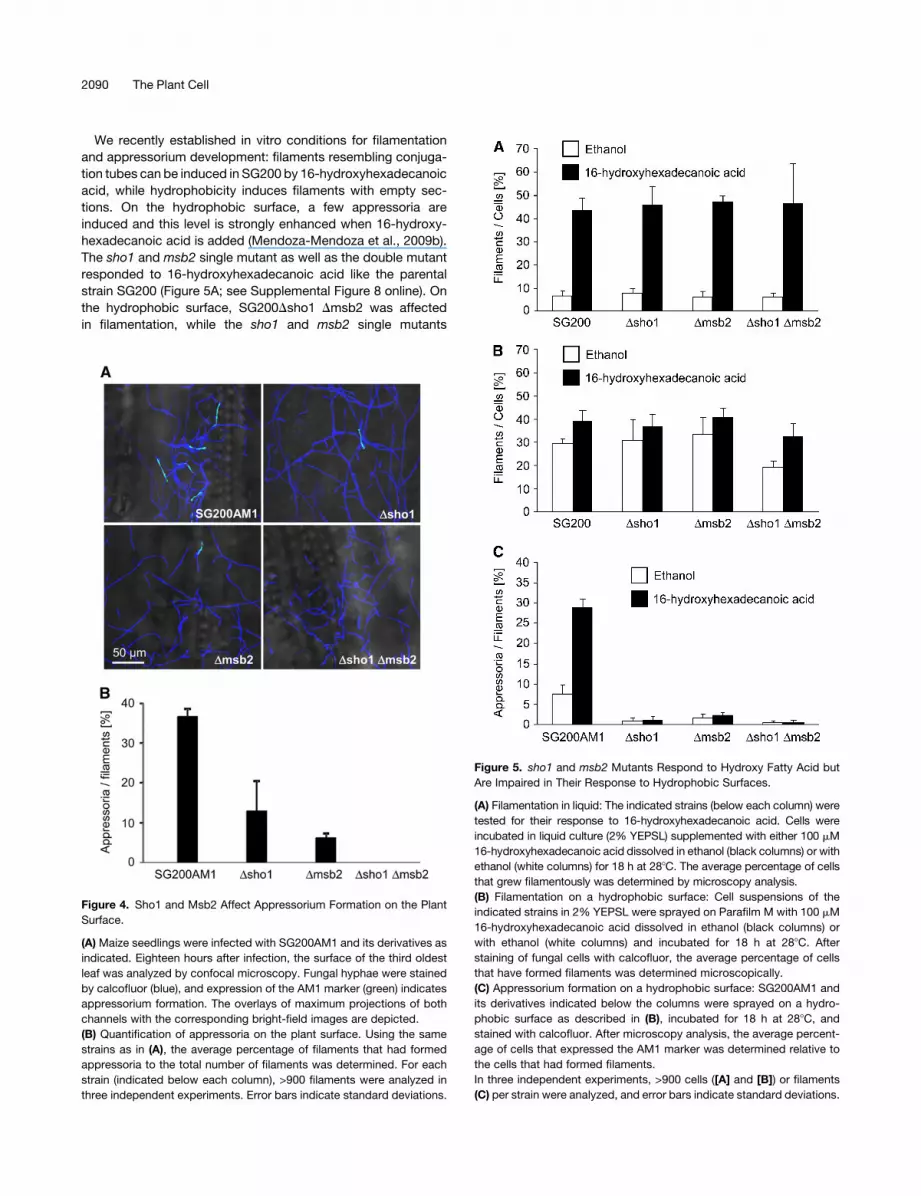
We recently established in vitro conditions for filamentation
and appressorium development: filaments resembling conjuga-
tion tubes can be induced in SG200 by 16-hydroxyhexadecanoic
acid, while hydrophobicity induces filaments with empty sec-
tions. On the hydrophobic surface, a few appressoria are
induced and this level is strongly enhanced when 16-hydroxy-
hexadecanoic acid is added (Mendoza-Mendoza et al., 2009b).
The sho1 and msb2 single mutant as well as the double mutant
responded to 16-hydroxyhexadecanoic acid like the parental
strain SG200 (Figure 5A; see Supplemental Figure 8 online). On
the hydrophobic surface, SG200Dsho1 Dmsb2 was affected
in filamentation, while the sho1 and msb2 single mutants
Figure 4. Sho1 and Msb2 Affect Appressorium Formation on the Plant
Surface.
(A) Maize seedlings were infected with SG200AM1 and its derivatives as
indicated. Eighteen hours after infection, the surface of the third oldest
leaf was analyzed by confocal microscopy. Fungal hyphae were stained
by calcofluor (blue), and expression of the AM1 marker (green) indicates
appressorium formation. The overlays of maximum projections of both
channels with the corresponding bright-field images are depicted.
(B) Quantification of appressoria on the plant surface. Using the same
strains as in (A), the average percentage of filaments that had formed
appressoria to the total number of filaments was determined. For each
strain (indicated below each column), >900 filaments were analyzed in
three independent experiments. Error bars indicate standard deviations.
Figure 5. sho1 and msb2 Mutants Respond to Hydroxy Fatty Acid but
Are Impaired in Their Response to Hydrophobic Surfaces.
(A) Filamentation in liquid: The indicated strains (below each column) were
tested for their response to 16-hydroxyhexadecanoic acid. Cells were
incubated in liquid culture (2% YEPSL) supplemented with either 100 mM
16-hydroxyhexadecanoic acid dissolved in ethanol (black columns) or with
ethanol (white columns) for 18 h at 288C. The average percentage of cells
that grew filamentously was determined by microscopy analysis.
(B) Filamentation on a hydrophobic surface: Cell suspensions of the
indicated strains in 2% YEPSL were sprayed on Parafilm M with 100 mM
16-hydroxyhexadecanoic acid dissolved in ethanol (black columns) or
with ethanol (white columns) and incubated for 18 h at 288C. After
staining of fungal cells with calcofluor, the average percentage of cells
that have formed filaments was determined microscopically.
(C) Appressorium formation on a hydrophobic surface: SG200AM1 and
its derivatives indicated below the columns were sprayed on a hydro-
phobic surface as described in (B), incubated for 18 h at 288C, and
stained with calcofluor. After microscopy analysis, the average percent-
age of cells that expressed the AM1 marker was determined relative to
the cells that had formed filaments.
In three independent experiments, >900 cells ([A] and [B]) or filaments
(C) per strain were analyzed, and error bars indicate standard deviations.
2090 The Plant Cell

reacted like the progenitor strain (Figure 5B; see Supplemental
Figure 9 online). The addition of 16-hydroxyhexadecanoic acid
enhanced filamentation in all mutant strains as described previ-
ously for SG200 (Mendoza-Mendoza et al., 2009b). However,
with respect to appressorium development on the hydrophobic
surface, the sho1 and msb2 single as well as the double mutant
were severely reduced, and the addition of 16-hydroxyhexade-
canoic acid had only a slight enhancing effect in the single
mutants and no effect in the double mutant (Figure 5C; see
Supplemental Figure 9 online). This shows that sho1 and msb2
specifically affect development of appressoria. Therefore, we
determined the localization of Sho1-GFP during appressorium
formation. Sho1-GFP showed a patchy distribution in filaments
but accumulated strongly in appressoria (Figures 6A and 6B).
Attempts to visualizeMsb2-mCherry in filaments failed due to the
background fluorescence of the inducing surface. To enhance
expression of msb2-mCherry, we placed the gene under the
control of the constitutively active otef promoter (Spellig et al.,
1996) and integrated this construct into the ip locus in strains
SG200Dmsb2 and SG200sho1GFP. The construct was able to
complement SG200Dmsb2 (see Supplemental Figure 2B online).
In filaments, Msb2-mCherry accumulated in vacuoles that were
distributed throughout the filament. In appressoria, Msb2-
mCherry strongly accumulated in vacuoles (Figures 6A and
6C), corroborating a function of Msb2 during this stage of
development. Colocalization with Sho1-GFP was not apparent
(Figure 6D).
Sho1 and Msb2 Act Upstream of the MAP Kinases Kpp2
and Kpp6
InU.maydis, theMAP kinase Kpp2 is implicated in appressorium
development induced by the hydrophobic stimulus (Muller et al.,
2003; Mendoza-Mendoza et al., 2009b), while the related MAP
kinase Kpp6 is specifically needed for appressorium function
(Brachmann et al., 2003). The dual specificity phosphatase Rok1
is a negative regulator of the MAP kinases Kpp2 and Kpp6, and
deletion of rok1 leads to hypervirulence (Di Stasio et al., 2009). To
test whether activated Kpp2 and Kpp6 restore tumor develop-
ment in SG200Dsho1Dmsb2,wedeleted rok1 in this strain.While
plants infected with SG200Dsho1 Dmsb2 showed no tumor
development 12 d after infection, SG200Dsho1 Dmsb2 Drok1–
infected plants developed tumors and symptom severity was
comparable to SG200 infections (Figure 7). To substantiate that
Sho1 and Msb2 activate the MAP kinase module containing
Kpp2 and Kpp6 and to rule out the possibility that rok1 sup-
presses the virulence phenotype of the Dsho1 Dmsb2mutant by
a mechanism that is independent of Kpp2 and Kpp6, we inte-
grated the constitutively active version of the MEK fuz7 (fuz7DD;
Muller et al., 2003) under the control of the arabinose-inducible
crg1 promoter in the genome of SG200Dsho1 Dmsb2 as well as
in the genome of SG200. Expression of fuz7DD was induced by
adding arabinose to the inoculum prior to infection of maize
plants. As a control, arabinose was replaced by glucose, which
represses the activity of the crg1 promoter (Bottin et al., 1996).
When SG200fuz7DD was used to infect maize plants, symptom
development was comparable irrespective ofwhether glucose or
arabinose was added to the inoculum (Figure 8A). SG200Dsho1
Dmsb2 fuz7DD induced necrosis when glucose was added
(Figures 8A and 8B). However, when arabinose was added as
an inducer for fuz7DD, we observed chlorotic areas and antho-
cyanin production, which is indicative of successful penetration
(Figures 8A and 8C). Microscopy analysis of leaves 6 d after
infection confirmed that penetration had occurred and the
amount of fungal material inside the leaf was much more abun-
dant when fuz7DD was induced compared with the repressed
situation (Figures 8B and 8C). These data indicate that Sho1 and
Msb2 act upstream of the MAP kinases Kpp2 and Kpp6.
Functional Analysis of Msb2
If Msb2 functions as a cell surface receptor, the transmembrane
domain should be important for function. To test this hypothesis,
the transmembrane domain within the otef:msb2-mCherryHA
construct was deleted and the construct integrated in the ip
locus of SG200Dmsb2. Plant infections revealed thatmsb2DTM-
mCherry-ha could not complement the virulence defect of the
msb2 deletion strain (see Supplemental Figure 10A online).
Furthermore, only the full-length protein could be detected in
immunoblot analysis, whereas the C-terminal Msb2-mCherryHA
fragment was absent (see Supplemental Figure 10B online).
These results indicate that the transmembrane domain of Msb2
is essential for processing and function of Msb2.
Figure 6. Localization of Sho1-GFP and Msb2-mCherry in Appressoria.
(A) to (D) SG200sho1GFP/otef:msb2mCherryHA was sprayed with 100
mM 16-hydroxyhexadecanoic acid on paraffin wax and incubated as
described in Figure 5B. A section displaying two appressoria is analyzed.
The appressorium on the right-hand side is magnified in the inset.
(A) Calcofluor staining.
(B) Visualization of Sho1-GFP (green).
(C) Visualization of Msb2-mCherry (red).
(D) Overlay of (B) and (C).
Appressorium Development in U. maydis 2091

U. maydisMsb2 and S. cerevisiaeMsb2p have similar domain
structures but display only 28% amino acid identity (Figure 1A).
To address whether U. maydis msb2 is able to complement the
S. cerevisiae msb2 mutant, we made use of the yeast strain
PC538 that is defective in the mating pathway and has a FUS1:
HIS3 reporter whose expression is driven only by the FGpathway
(Cullen et al., 2004). The PC538 derivative PC948 carries the
msb2 deletion and is hence unable to grow on medium without
His (Cullen et al., 2004). U. maydis msb2 and S. cerevisiae MSB2
were inserted under the control of the constitutively active ADH1
promoter into the free replicating plasmid pVTU260 and trans-
formed into PC948. Expression of U. maydis Msb2 was verified
by immunoblot analysis (see Supplemental Figure 11A online). S.
cerevisiae strain PC948, expressingU.maydismsb2, was unable
to grow on medium lacking His, while S. cerevisiae MSB2 could
complement the growth phenotype of the msb2 deletion strain
(see Supplemental Figure 11B online). This demonstrates that,
although characteristic features are shared between the Msb2
proteins in U. maydis and S. cerevisiae, they do not represent
functional homologs.
The SH3 Domain of Sho1 Interacts with a Negative
Regulatory Domain of Kpp6
To establish the biological significance of the observed interac-
tion between Sho1 and Kpp6, we delineated the region respon-
sible for the interaction between Kpp6 and Sho1. Different
segments of Sho1 and Kpp6 were tested by two-hybrid interac-
tion for their ability to interact with each other. In all cases, the
amounts of fusion proteins expressed were comparable (see
Supplemental Figure 12A online). We found that Kpp6 interacted
specifically with the SH3 domain of Sho1 (amino acids 279 to
335), while the linker domain (amino acids 131 to 278) that is
situated between the SH3domain and the fourth transmembrane
domain showed no interaction with Kpp6. With respect to Kpp6,
we mapped the interaction domain to amino acids 1 to 170,
which constitutes the unique N-terminal domain of Kpp6 (see
Supplemental Figures 12B and 12C online). Based on the
knowledge that SH3 domains interact with Pro-rich domains
(PRDs; Li, 2005), we searched for such a motif in the N terminus
of Kpp6. This led to the identification of the motif KPLPPSP
(corresponding to amino acids 127 to 133), which conforms to
type I PRDs (R/KxxPxxP; Li, 2005). To test the functional signif-
icance of this motif in Kpp6, we generated a mutant in which two
Figure 7. The Deletion of rok1 Suppresses the Virulence Phenotype of
SG200Dsho1 Dmsb2.
Maize plants were infected with SG200 and the indicated derivatives.
Disease rating was done as described in the legend to Figure 2. Tested
strains are listed below each column, the numbers of infected plants (n)
are given above each column, and the color and pattern code for disease
rating is depicted on the right.
Figure 8. Expression of a Constitutively Active Allele of fuz7 Partially
Bypasses the Need for Sho1 and Msb2.
(A) The indicated SG200 derivatives were inoculated into maize seed-
lings. Prior to infection, either arabinose (to induce fuz7DD expression) or
glucose (to repress fuz7DD expression) was added to the inoculums to a
final concentration of 1%. Twelve days after infection, symptoms were
scored as described in the legend to Figure 2. Inoculated strains and
glucose/arabinose supplements are listed below each column, numbers
of infected plants (n) are given above each column, and the color and
pattern code for disease rating is depicted on the right.
(B) Macroscopy and microscopy symptoms after infection with
SG200Dsho1 Dmsb2 fuz7DD in the presence of glucose. A representa-
tive infected leaf is shown on the left 12 d after infection. Confocal
microscopy performed as described in the legend to Figure 3A reveals
poor colonization of plant tissue 6 d after infection (right panel).
(C) Macroscopy and microscopy symptoms after infection with
SG200Dsho1 Dmsb2 fuz7DD in the presence of arabinose. A represen-
tative infected leaf 12 d after infection is shown on the left. Confocal
microscopy performed as described in legend to Figure 3A reveals
strongly enhanced colonization of plant tissue 6 d after infection (right
panel).
2092 The Plant Cell
Pro residues are substituted by Ala (KPLAASP). Two-hybrid
interactions demonstrated that Kpp6P130A P131A is no longer able
to interact with the SH3 domain of Sho1 despite similar expres-
sion levels of the respective fusion proteins (see Supplemental
Figure 12 online).
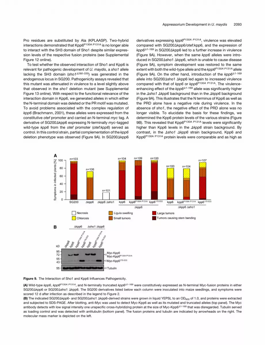
To test whether the observed interaction of Sho1 and Kpp6 is
relevant for pathogenic development of U. maydis, a sho1 allele
lacking the SH3 domain (sho1D280-335) was generated in the
endogenous locus in SG200. Pathogenicity assays revealed that
this mutant was attenuated in virulence to a level slightly above
that observed in the sho1 deletion mutant (see Supplemental
Figure 13 online). With respect to the functional relevance of the
interaction domain in Kpp6, we generated alleles in which either
the N-terminal domain was deleted or the PRmotif wasmutated.
To avoid problems associated with the complex regulation of
kpp6 (Brachmann, 2001), these alleles were expressed from the
constitutive otef promoter and carried an N-terminal myc tag. A
derivative of SG200Dkpp6 expressing N-terminally myc-tagged
wild-type kpp6 from the otef promoter (otef:kpp6) served as
control. In this control strain, partial complementation of the kpp6
deletion phenotype was observed (Figure 9A). In SG200Dkpp6
derivatives expressing kpp6P130A P131A, virulence was elevated
compared with SG200Dkpp6/otef:kpp6, and the expression of
kpp6D1-169 in SG200Dkpp6 led to a further increase in virulence
(Figure 9A). However, when the same kpp6 alleles were intro-
duced in SG200Dsho1 Dkpp6, which is unable to cause disease
(Figure 9A), symptom development was restored to the same
extentwith both thewild-type allele and the kpp6P130A P131A allele
(Figure 9A). On the other hand, introduction of the kpp6D1-169
allele into SG200Dsho1 Dkpp6 led again to increased virulence
compared with that of kpp6 or kpp6P130A P131A. The virulence-
enhancing effect of the kpp6D1-169 allele was significantly higher
in the Dsho1 Dkpp6 background than in the Dkpp6 background
(Figure 9A). This illustrates that the N terminus of Kpp6 as well as
the PRD alone have a negative role during virulence. In the
absence of sho1, the negative effect of the PRD alone was no
longer visible. To elucidate the basis for these findings, we
determined the Kpp6 protein levels of the various strains (Figure
9B). This revealed that Kpp6P130A P131A levels were significantly
higher than Kpp6 levels in the Dkpp6 strain background. By
contrast, in the Dsho1 Dkpp6 strain background, Kpp6 and
Kpp6P130A P131A protein levels were comparable and as high as
Figure 9. The Interaction of Sho1 and Kpp6 Influences Pathogenicity.
(A) Wild-type kpp6, kpp6P130A P131A, and N-terminally truncated kpp6D1-169 were constitutively expressed as N-terminal Myc-fusion proteins in either
SG200Dkpp6 or SG200Dsho1 Dkpp6. The SG200 derivatives listed below each column were inoculated into maize seedlings, and symptoms were
scored 12 d after infection as described in the legend to Figure 2.
(B) The indicated SG200Dkpp6- and SG200Dsho1 Dkpp6-derived strains were grown in liquid YEPSL to an OD600 of 1.0, and proteins were extracted
and subjected to SDS-PAGE. After blotting, anti-Myc was used to detect Myc-Kpp6 as well as its mutated and truncated alleles (top panel). The Myc
antibody detects with low signal intensity one unspecific cross-hybridizing protein at the size of Myc-Kpp6D1-169 that was disregarded. Tubulin served
as loading control and was detected with antitubulin (bottom panel). The fusion proteins and tubulin are indicated by arrowheads on the right. The
molecular mass marker is depicted on the left.
Appressorium Development in U. maydis 2093

the Kpp6P130A P131A levels in the Dkpp6 strain background
(Figure 9B). Compared with the Kpp6P130A P131A level, the
amount of the truncated version Kpp6D1-169 was further elevated
and was indistinguishable in the Dkpp6 and Dsho1 Dkpp6 strain
background (Figure 9B). This suggests that Sho1 negatively
influences the stability of Kpp6 through direct interactionwith the
N terminus of Kpp6.
DISCUSSION
In this article, we showed that Sho1 and Msb2 in U. maydis are
essential virulence factors. sho1 and msb2 mutants are specif-
ically reduced in pathogenic development and are not signifi-
cantly affected in mating and stress responses. The virulence
defects could be traced back to a failure in developing appres-
soria.
Using bioinformatic tools, Msb2 of U. maydis was recently
classified as an ortholog of S. cerevisiae Msb2p (Krantz et al.,
2006). Complementation studies have now shown that these
proteins are not functional homologs, which is not unexpected, in
view of the light that they regulate distinct processes in both
organisms (see below). Nevertheless, with respect to domain
structure, Msb2p from S. cerevisiae and Msb2 from U. maydis
are clearly related (Figure 1A). For Msb2p in S. cerevisiae, it has
been demonstrated that the extracellular domain is highly N- as
well asO-glycosylated (Cullen et al., 2004; Yang et al., 2009), and
using the program NetNGlyc 1.0 (http://cbs.dtu.dk/services/
NetNGlyc/), sevenN-glycosylation sites inU.maydisMsb2 (eight
in S. cerevisiae Msb2p) were predicted. Furthermore, the com-
bination of all six recommended constraints of the program
OGPET 1.0 (http://ogpet.utep.edu/OGPET/) predicts an average
of 12 O-glycosylation sites for Msb2 in U. maydis (nine for S.
cerevisiaeMsb2p). We consider it likely that Msb2 inU. maydis is
a glycosylated transmembrane mucin. This assertion is sup-
ported by the finding that its apparent molecular mass of >170
kD is significantly higher than its expected mass of 147 kD as
well as the finding that the transmembrane domain is crucial for
Msb2 function.
While Sho1- and Msb2-related proteins serve as stress sen-
sors in all fungal systems analyzed to date (Maeda et al., 1995;
O’Rourke and Herskowitz, 2002; Roman et al., 2005, 2009;
Norice et al., 2007; Boisnard et al., 2008; Ma et al., 2008), sho1
and msb2 are not needed for general stress responses in U.
maydis. Responses to osmotic stress, oxidative stress, or cell
wall stress were not altered in sho1 and msb2 single or double
mutants, suggesting that the function of Sho1 and Msb2 in U.
maydis is uncoupled from the HOG pathway and serves in
pathogenicity only.
InU. maydis, pathogenic development is initiated by b-depen-
dent filaments, which are septated and accumulate empty sec-
tions. On the plant surface, sho1 andmsb2mutants were able to
form these filaments as efficiently as the progenitor strain.
However, single deletion mutants in sho1 or msb2 showed
reduced colonization of the plant tissue, and the sho1 msb2
double deletion mutant had an almost complete colonization
defect. The quantification of appressoria on the plant surface
revealed that single deletion strains of sho1 or msb2 form fewer
appressoria than SG200, and the sho1 msb2 double deletion
strain was not able to form appressoria. Thus, Sho1 and Msb2
have cooperative and essential functions during appressorium
development. Interestingly, mutants lacking an ortholog to
MSB2 in Fusarium oxysporum showed delayed invasive growth
and were strongly attenuated in virulence (E. Perez-Nadales and
A. Di Pietro, personal communication), suggesting that mucins
may have a general function during the early infection stages of
phytopathogenic fungi.
We observed strong plant defense responses when the sho1
msb2 double mutant grows on the leaf surface. This most likely
reflects pathogen-associated molecular pattern-induced de-
fense responses that are suppressed after wild-type hyphae
penetrate the epidermal layer (Doehlemann et al., 2008a). Due to
the penetration defect, the sho1 msb2 double mutant would be
unable to suppress this reaction. The failure of the sho1 msb2
mutant to develop appressoria is at variance with results that rely
on fungal biomass determinations. In these experiments, only a
fivefold reduction in colonization was observed relative to the
progenitor strain SG200. In addition, confocal microscopy al-
lowed us to detect rare colonization events by the sho1 msb2
mutant. We consider intracellular fungal biomass determination
during early infection stages to be error prone as it relies on
complete removal of fungal hyphae from the leaf surface by latex
treatment. To explain the rare colonization successes of the sho1
msb2 mutant, we speculate that this reflects the ability of U.
maydis to enter plant tissue as a saprophyte without the forma-
tion of appressoria. This could actually be facilitated by dead
plant cells as they are found underneath SG200Dsho1 Dmsb2
cells growing on the leaf surface.
In yeast, Msb2p has been shown to interact with Sho1p (Cullen
et al., 2004). Attempts to visualize an interaction between Sho1
and Msb2 in U. maydis relying on epitope-tagged proteins and
coimmunoprecipitations were unsuccessful. This could suggest
that interactions are only transient and may be restricted to
appressoria (i.e., a stage not yet accessible to biochemical
analysis). Msb2p of yeast has also been demonstrated to interact
with Cdc42p, and this complex is hypothesized to provide
sensory capacity in the FG pathway that is transmitted via the
PAK kinase Ste20p (Cullen et al., 2004). In U. maydis, Cdc42 is
part of a signaling module consisting of the Rho-GEF Don1 and
the Ste20-like kinase Don3 (Weinzierl et al., 2002). While don3
mutants are not affected in pathogenic development (Weinzierl
et al., 2002), cdc42 mutants are nonpathogenic (Mahlert et al.,
2006). A second PAK in U. maydis, Cla4, a member of the Cla4
subfamily of Ste20-like kinases, is involved in the regulation of
cell polarity during budding and filamentation and cla4 mutants
are nonpathogenic (Leveleki et al., 2004). It will be interesting to
determine whether pathogenicity defects of cdc42 and cla4
mutants occur at the stage of appressorium formation as this
could provide a direct link to Sho1 and Msb2.
Recently, the dual specificity phosphatase Rok1 has been
characterized as a negative regulator of Kpp2 and Kpp6. Dele-
tion of rok1 results in hypervirulence, which can be partially
explained by increased appressorium formation (Di Stasio et al.,
2009). When the rok1 gene was deleted in the Dsho1 Dmsb2
background, the resulting strain became fully pathogenic, indi-
cating that Sho1 andMsb2 feed into the Kpp2/Kpp6MAP kinase
cascade, and a lack of this input can be compensated for by the
2094 The Plant Cell

deletion of rok1. This is corroborated by demonstrating that
expression of the constitutively active allele of the MEK Fuz7
that activates the MAP kinases Kpp2 and Kpp6 (Muller et al.,
2003; Di Stasio et al., 2009) allowed to bypass the colonization
defect of the sho1 msb2 deletion strain. Since tumor formation
was not observed when fuzDD was induced by arabinose prior
to infection with SG200Dsho1 Dmsb2 fuz7DD, this most likely
indicates that fuz7DD expression ceases after penetration (due
to insufficient amounts of arabinose in the plant tissue) and
that Sho1 and Msb2 have additional, Fuz7-dependent func-
tions during the biotrophic phase. Since U. maydis appears to
require the formation of appressoria for cell-to-cell passages
(Doehlemann et al., 2009), it is conceivable that the reinitiation
of this program is affected in the sho1 msb2 double mutant.
Overall, these results place Sho1 and Msb2 upstream of Kpp2
and Kpp6, where they function as sensors to activate MAP
kinase signaling.
An intriguing feature of this signaling pathway is that Sho1
protein was identified as a two-hybrid interactor of Kpp6
(Mendoza-Mendoza et al., 2009a). We demonstrated here that
this interaction occurs via the SH3 domain of Sho1 and the PR-
motif in Kpp6. This motif is located in the N-terminal extension of
Kpp6, which is absent from Kpp2 (Brachmann et al., 2003). kpp6
is strongly upregulated in b-dependent filaments and is specif-
ically needed for appressoria to penetrate (Brachmann et al.,
2003). The finding that a kpp6 allele that lacks the N-terminal
domain is significantly stronger in complementing the pathoge-
nicity defect of kpp6 mutants than wild-type kpp6 suggests that
the N terminus of Kpp6 has an inhibitory function. A Kpp6 variant
that no longer interacts with Sho1 due to mutations in the PR
domain also causes enhanced virulence and this effect requires
Sho1. The demonstration that Kpp6 protein levels are elevated
when the PRD is mutated or sho1 is deleted makes it likely that
the interaction with Sho1 destabilizes Kpp6. We speculate that
the interaction with Sho1 could impose a negative feedback loop
that might fine-tune Kpp6 levels during the differentiation of
appressoria. In S. cerevisiae, it has been demonstrated that a
negative feedback loop exists between Sho1p and Hog1p that is
based on phosphorylation of Sho1p by Hog1p (Hao et al., 2007).
The cytoplasmic domain of U. maydis Sho1 contains three
putative MAP kinase phosphorylation sites. However, simulta-
neous mutation of all three sites did not affect virulence of the
respective strain (D. Lanver and R. Kahmann, unpublished data),
making a phosphorylation-based feedback loop unlikely. De-
spite the negative function of the PRD in the N terminus of Kpp6,
this domain must additionally confer regulatory information. This
is based on the observation that the deletion of the entire N
terminus causes stronger virulence thanmutating only thePRD in
Kpp6. Furthermore, in this case, the enhanced virulence does not
depend on Sho1.
The deletion of the SH3 domain in sho1 reduces virulence to a
similar extent as disruption of the entire sho1 gene. Thus, the
negative effect of the SH3 domain of Sho1 on Kpp6 levels cannot
be the only function of this domain. In yeast, the MEK Pbs2p is
recruited to the plasma membrane via interaction of the Sho1p
SH3 domainwith the Pro-richmotif KPLPPLP in theN terminus of
Pbs2p (Maeda et al., 1995; Raitt et al., 2000; Reiser et al., 2000).
In U. maydis, the MEK Fuz7 acts downstream of Sho1. Since
Fuz7 lacks such a Pro-rich motif and we currently have no
evidence that Sho1 and Fuz7 interact, further experimentation
will be necessary to link the SH3 domain in Sho1 to components
of the MAP kinase module.
In S. cerevisiae, activation of Msb2 is regulated by starvation-
dependent induction of its cognate aspartyl protease Yps1p
(Vadaie et al., 2008). Yps1p processes Msb2p into a secreted
extracellular form and a cell-associated form. This cleavage
releases the inhibitory mucin domain and generates the active
form of Msb2 (Vadaie et al., 2008). Our finding of two forms of
Msb2 in U. maydis, a high molecular mass form and a form
representing the C terminus also suggests a processing event.
However, we currently have no indication that the extracellular
domain in Msb2 assumes a negative regulatory function. In S.
cerevisiae, Msb2-GFP localizes primarily to vacuoles, and this
localization depends on efficient processing of Msb2p (Vadaie
et al., 2008). Given that U. maydis Msb2-mCherry also accumu-
lates in vacuoles in the appressorium, we speculate that Msb2
could be activated when appressoria are formed.
Recently,Msb2p inS. cerevisiae has been identified as a target
for themannosyltransferase Pmt4p and deletion of pmt4 leads to
underglycosylated Msb2p, as well as to enhanced activity of the
FGpathway (Yang et al., 2009). Interestingly, the deletion ofpmt4
in U. maydis causes a specific defect in appressorium develop-
ment, while vegetative growth and filament formation are not
affected (Fernandez-Alvarez et al., 2009). Since Msb2 is an
upstream component of the MAP kinase cascade regulating
appressorium development, Msb2 is likely to be amannosylated
target of Pmt4 in U. maydis.
In U. maydis, appressoria can be efficiently induced on a
hydrophobic surface when hydroxy fatty acids are added
(Mendoza-Mendoza et al., 2009b). In contrast with the situation
on the leaf surface, in vitro appressorium formation was not only
abolished in sho1 msb2 double mutants, but already in sho1
and msb2 single mutants. This could indicate that on the plant
surface additional signals are provided that allow sho1 andmsb2
single mutants to form appressoria. Under the in vitro conditions,
sho1 and msb2 mutants were able to develop filaments in
response to hydroxy fatty acids alone, indicating that this path-
way is unaffected. Formation of septated filaments by the sho1
msb2 double mutant occurred inefficiently on the hydrophobic
surface but was increasedwhen hydroxy fatty acidswere added.
We take this to indicate that the hydrophobic surface is only
poorly sensed. Moreover, with respect to the in vitro differenti-
ation of appressoria, the sho1 and msb2 mutants are defective.
We speculate that the reduced response to hydrophobicity of
sho1 and msb2 mutants in vitro results from inefficient percep-
tion of the hydrophobic stimulus. As this is the crucial signal for
appressorial differentiation (Mendoza-Mendoza et al., 2009b),
the reduced perception could translate into the complete failure
to form these infection structures. So far, it is not entirely clear
what is sensed by Sho1p/Msb2p in S. cerevisiae (Cullen, 2007).
The physical properties of the heavily glycosylated extracellular
domains of mucins change dramatically in response to changes
in extracellular milieu, and in higher eukaryotes, mucins have
been shown to bind to and detect the presence of eukaryotic
cells, proteins, and microorganisms (De Nadal et al., 2007). We
consider these properties to be extendable to abiotic surface
Appressorium Development in U. maydis 2095

recognition, although formal proof for this needs further exper-
imentation.
METHODS
Strain Construction and Growth Conditions
The Escherichia coli strains DH5a (Bethesda Research Laboratories) and
Top10 (Invitrogen) were used for cloning purposes. The Saccharomyces
cerevisiae strain AH109 (Clonetech) was used for two-hybrid interaction
studies. The S. cerevisiae strains PC538 and PC948 (Cullen et al., 2004)
were used formsb2 complementation studies. AllS. cerevisiae strains are
listed in Supplemental Table 1 online.
Ustilagomaydis strains were grown in liquid YEPSL (0.4%yeast extract,
0.4% peptone, and 2% sucrose) or on solid potato dextrose (PD) plates.
For mating assays and filament induction, PD plates containing 1%
activated charcoal were used (Holliday, 1974).
Haploid U. maydis strains FB1 and FB2, and solopathogenic strains
SG200, SG200AM1, SG200Dkpp6, and SG200Drok1 have been de-
scribed previously (Banuett and Herskowitz, 1989; Brachmann et al.,
2003; Kamper et al., 2006; Di Stasio et al., 2009; Mendoza-Mendoza
et al., 2009b).
For gene disruptions, the PCR strategy described by Kamper (2004)
and theSfiI insertion cassette system (Brachmann et al., 2004) were used.
All gene replacement constructs were sequenced after cloning. After
transformation, DNA gel blot analysis was performed to confirm gene
replacements or insertions. All U. maydis strains are listed in Supple-
mental Table 2 online.
For generation of sho1 (um03156) deletion mutants, two 1.0-kb frag-
ments containing the 59 flanking region and the 39 flanking region of the
sho1 gene were amplified by PCR using FB1 genomic DNA as template
with the primer combinations oAM150/oAM151 (59-GAGTCTTG-
GACTGTTCGCGATAC-39/59-CACGGCCTGAGTGGCCCAGCATGCCTT-
GGTTCAGCCAAGCTG-39) for the left border and oAM152/oAM153
(59-GTGGGCCATCTAGGCCGAGATCGAGCTGGTCTATAC-39/59-CGA-
CGACTTGTTTGAGTTTGGCC-39) for the right border. The PCR frag-
ments were digested with SfiI and ligated to the hygromycin resistance
cassette isolated as a 2.7-kb SfiI fragment from plasmid pMF1-h
(Brachmann et al., 2004). The ligation product was cloned into pCRII-
TOPO (Invitrogen) to obtain pDsho1-Hyg. The plasmid was digested with
Eco0109I and HindIII prior to transformation in SG200. The 2.7-kb
hygromycin cassette of pDsho1-Hyg was replaced by the 1.4-kb nour-
seothricin resistance cassette from pMF1-n (Brachmann et al., 2004),
resulting in pDsho1-Nat. This plasmid was digested with PstI and BamHI
before transformation and was used for sho1 disruption in FB1, FB2, and
SG200Dkpp6.
For deletion of only the SH3 domain of sho1, the primer combination
oDL02/oDL03 (59-TAGAAGCTTCTGTTCCACAGCTTGGCTG-39/59-TAT-
GGCGCGCCCTAGTAGCCGTAGTCGGGCAG-39) was used to amplify
the 59 region of sho1. Primer oDL03 introduces a stop codon at amino
acid position 280, just before the SH3 domain starts. The PCR product
was digested with AscI. The 39 flanking region of sho1 was amplified
using the primers oDL04 (59- GATGAGCTCCGAGATCGAGCTGGTC-
TATACC-39) and oDL05 (59-ATCAAGCTTCGACGACTTGTTTGAGTT-
TGG-39), following digestion with SacI. A three-fragment ligation
including a 3.0-kb AscI/SacI fragment containing the nos terminator
and the hygromycin resistance cassette, the PCR fragments corre-
sponding to the 39 flanking region of sho1 and to the C-terminally
truncated sho1 gene was performed. The ligation product was cloned
into pCRII-TOPO to obtain psho1D280-335. The plasmid was di-
gested with HindIII and transformed in SG200, resulting in strain
SG200sho1D280-335.
To generate msb2 (um00480) deletion mutants, two 1.0-kb fragments
comprising the 59 flank and the 39 flank of themsb2 gene were generated
by PCR with the primer pairs oOM1/oOM2 (59-TACACCTCATCATT-
CACGCTAACGC-39/59-CACGGCCTGAGTGGCCAAAGAGACAAGTGG-
GAGGCTGACG-39) and oOM3/oOM4 (59-GTGGGCCATCTAGGCCTGT-
TTGCTTTGGTTGTAACGGAACG-39/59-TGTCTGGCTGCACCACTCTATT-
TACG-39). The PCR products were digested with SfiI and ligated with the
1.9-kb SfiI hygromycin resistance cassette from pBS-hhn (Kamper, 2004).
The resulting product was cloned into pCRII-TOPO to generate pDmsb2-
Hyg, which was digested withHindIII and XbaI and transformed in SG200,
FB1, and FB2. To generate sho1 and msb2 double deletions strains, the
1.9-kb hygromycin resistance cassette of pDmsb2-Hyg was replaced by
the 1.9-kbcarboxin resistancecassette ofpBS-Cbx (Kamper, 2004) to yield
pDmsb2-Cbx. Prior to transformation in FB1Dsho1 and FB2Dsho1, the
plasmidwas digestedwithHindIII andXbaI. For generation of SG200Dsho1
Dmsb2, the PCR-amplified msb2 borders were ligated with the 1.4-kb
nourseothricin resistance cassette from pMF1-n and the ligation product
cloned into pJet1 (Fermentas). Prior to transformation in SG200Dsho1, the
plasmid was digested with BspEI and PvuII.
To complement SG200Dsho1 with sho1, a 1.9-kb fragment corre-
sponding to the promoter and the open reading frame (ORF) of sho1
was amplified using primers oAM154 (59-CGCTCGGTACCCGGTG-
ATTTGTGATTAACACGTC-39) and oDL01 (59-CTAGGCGCGCCCTATA-
GGAGCTGCATGTAGTTGCTG-39), following digestion with AscI. p123
(Aichinger et al., 2003) was linearized with NotI and refilled with Klenow
according to the recommendations of the manufacturer (New England
Biolabs). Then, the linearized plasmid was cut with Acc65I and the
4.7-kb Acc65I/blunt fragment was ligated with the 1.9-kb Acc65I/blunt
sho1 fragment. The resulting plasmid, p123Psho1:sho1, contains the
ORF of sho1 under the control of its own promoter and the nos termi-
nator. p123Psho1:sho1 was linearized with SspI and transformed into
SG200Dsho1. Single-copy integrations in the ip locus were identified as
described previously (Loubradou et al., 2001).
For construction of the msb2 complementation plasmid, p123 was
digested withNdeI andNotI, and the 4.5-kb fragment was ligated with the
6.9-kbNdeI/NotI fragment corresponding to the promoter and the ORF of
msb2, which was amplified using the primers oAM288 (59-GCCCCGCA-
TATGGCTGATGAAGAAAGAGCACT-39) and oAM290 (59- GCCTGCGG-
CCGCATTTAAAGGAGAACCGAGTTG-39). p123Pmsb2:msb2 contains
the ORF of msb2 under the control of its own promoter and the nos
terminator. For complementation, plasmid p123Pmsb2:msb2 was linear-
ized with SspI and integrated in single copy in the ip locus of
SG200Dmsb2. For complementation of the sho1 msb2 double mutant,
p123Psho1:sho1 was digested with PvuII and HpaI, and the 2.3-kb
fragment corresponding to the promoter, the sho1 gene, and the nos
terminator were ligated into HpaI-linearized p123Pmsb2:msb2. The result-
ing plasmid p123Psho1:sho1-Pmsb2:msb2 was integrated in single copy
into the ip locus of SG200Dsho1 Dmsb2.
To generate SG200sho1GFP, a 1.0-kb left border corresponding to
the coding region of sho1 was amplified using the primers oDL08
(59-GGTGGCCGCGTTGGCCCGCTCGATCGCCACCGGTAGGAGCTG-
CATGTAGTTGCTGGG-39) and oDL02. The right border was amplified
using the primers oDL05 and oDL09 (59-ATAGGCCTGAGTGGCCGA-
GATCGAGCTGGTCTATAC-39). Both fragments were digested with SfiI
and ligated with the 3.7-kb SfiI fragment from pBS-eGFP, which contains
the egfp gene, the nos terminator, and the hygromycin resistance
cassette (Brachmann, et al., 2004). The ligation product was cloned in
pCRII-TOPO to yield psho1-eGFP. In this plasmid, the sho1 gene is fused
to egfp via a linker encoding the amino acids PVAIERANAAT. Prior to
transformation and integration in the sho1 locus, the plasmid was
digested with HindIII.
To construct a C-terminal fusion of Msb2 to the red fluorescent protein
mCherry (Shaner et al., 2004), the 0.7-kb NcoI/BsrgI fragment of pMF5-n
(Becht et al., 2006) corresponding to egfp was replaced with a 0.7-kb
NcoI/BsrgI mcherry fragment (kindly provided by M. Bolker) resulting
2096 The Plant Cell

in pMF5-mCherry. A 1.0-kb fragment corresponding to the 39 coding
region of msb2 was amplified with the primer pair oAM331/oAM284
(59-CACGGCCGCGTTGGCCCCGGTGGCGATCGAGCGAAGGAGAAC-
CGAGTTGCTCATC-39/59-GACGGCGCAAATCTTTGCAT-39), and a
second 1.0-kb fragment was generated by PCR with the primers
oAM332 (59-GTTGGCCTGAGTGGCCATCTAGTTTGGTGCTTCTTTT-39)
and oAM333 (59-GCATTCAGTCGGCGTCCCATCCAGC-39). Both frag-
ments were digested with SfiI and ligated with the 2.4-kb SfiI fragment
from pMF5-mCherry. After cloning this construct into pCR4-TOPO
(Invitrogen), the resulting plasmid pmsb2-mCherry was digested with
SnaBI and PmeI and transformed in SG200sho1GFP. In the resulting
strain SG200sho1GFP/msb2mCherry, the nativemsb2 gene is fused to
mcherry via a linker encoding the amino acids RSIATGANAAT, and this
is followed by the nos terminator and the nourseothricin resistance
cassette.
Foroverexpressionofmsb2-ha-mcherry-ha, firstpONGwasconstructed.
Usingprimers oDL114 (59-AATACCATGGTGAGCAAGGGCGAGGAGG-39),
oDL115 (59-TATGCGGCCGCTTTAAGCGTAATCTGGAACATCGTATGG-
GTA-39), and pPotef:vcp1-mCherryHA (A. Djamei and R. Kahmann, unpub-
lished data) as template, a 0.7-kb product containing themcherry-ha gene
was amplified and digestedwithNcoI andNotI. pPotef:vcp1-mCherryHA is a
plasmid in which the secreted effector gene vcp1 is fused to the otef
promoter and aC-terminalmcherry-ha tag. A second PCRwith the primer
pair oD111/oDL112 (59-ATAGGATCCAGGCCTGAGTGGCCATGACA-
GAGGAGGACTCTGTGCTTTATCCG-39/59-TTATCCATGGTGGCCGCG-
TTGGCCCCTAGGAGCTGCATGTAGTTGCTGGG-39) using p123Psho1:
sho1 as template amplified a part of the sho1 gene. The 0.6-kb product
was digestedwithBamHI andNcoI. A three-fragment ligation with the two
fragmentsmentioned above and the 5.5-kbBamHI/NotI fragment of p123
was performed to yield pONG. In pONG, the sho1 part flanked bySfiI sites
serves as stuffer for the integration of genes to be fused tomcherry-ha. To
insert msb2-ha into pONG, the primer combinations oDL79 (59-[P]
GTGCCCGACTATGCCGGCGCCAGTCCCTCCACTGCTCCCTCGT-39)/
oDL125 (59-TATGGCCGCGTTGGCCGCAAGGAGAACCGAGTTGCTC-
ATC-39) and oDL80 (59-[P]GTCGTAGGGGTAGGCTGCGCCGCTGTCAT-
CATCGCTGC-39)/oDL124 (59-ATAGGCCTGAGTGGCCATGGTTCTGT-
TTCGACCCAAC-39) using p123Pmsb2:msb2 as template generated two
PCR products, 2.2 and 1.3 kb in length, respectively. Both fragments
were cut with SfiI and ligated with the 6.3-kb SfiI fragment of pONG,
resulting in pPotef:msb2HA-mCherryHA. In this plasmid, the msb2 gene
with an internal ha tag (corresponding to amino acid 421) is C-terminally
fused to mcherry-ha. Expression of the fusion gene is driven by the otef
promoter. pPotef:msb2HA-mCherryHA was linearized with Ssp1 and
integrated into the ip locus of SG200Dmsb2 and SG200sho1GFP. To
delete the transmembrane domain of msb2, a 9.7-kb fragment was
amplified with the primer pair oDL197 (59-[P]TGGCGCAAGCATCG-
CAAGG-39)/oDL198 (59-[P]ACTGTTGCGCAGCGTCGAGTC-39) using
pPotef:msb2HA-mCherryHA as template. The resulting fragment was
circularized to yield pPotef:msb2HADTM-mCherryHA. This plasmid was
linearized with SspI and integrated in single copy in the ip locus of
SG200Dmsb2.
To express kpp6 in U. maydis, pPotef:kpp6NA (Brachmann, 2001) was
used as progenitor plasmid. This plasmid is a derivative of pkpp6NA
(Brachmann et al., 2003), constructed by exchange of almost the com-
plete kpp6 promoter (region22371 to282) with the 873-bp otef promoter
from pOTEF-SG (Spellig et al., 1996). pPotef:kpp6NA contains the otef
promoter, followed by 81-bp 59 untranslated region, the complete ORF,
and 435-bp 39 untranslated region of kpp6. To integrate myc-tagged
versions of kpp6, three PCR reactions with the primer pair oDL131
(59-ATACCCGGGATGGAGGAGCAGAAGCTGATCTC-39)/oDL132
(5-TATGCGGCCGCTCAACGAAGAAGCGGCTGAAATTC-39) were per-
formed, using pGBKT7-kpp6, pGBKT7-kpp6170-533, and pGBKT7-
kpp6P130A P131A (see Supplemental Methods online) as templates. The
PCR products (1.1, 1.1, and 0.6 kb, respectively) were digested with
SmaI and BspEI and separately ligated with the 6.0 BspEI/SmaI
fragment of pPotef:kpp6NA, resulting in pPotef:myc-kpp6, pPotef:myc-
kpp6D1-169, and pPotef:myc-kpp6P130A P131A. The plasmids were digested
with SspI and integrated in single copy into the ip locus of SG200Dkpp6
(Brachmann et al., 2003) and SG200Dsho1 Dkpp6, respectively. The
plasmid pPotef:myc-kpp6 was also integrated in the ip locus of
SG200Dkpp6 and SG200sho1-GFP in multiple copy.
The triple deletion strain of sho1, msb2, and rok1 was constructed by
linearization of pDrok1-Cbx (Di Stasio et al., 2009) with PvuI and subse-
quent transformation into SG200Dsho1 Dmsb2.
To generate strains carrying the fuz7DD allele under the control of
the crg1 promoter, p123Pcrg1:fuz7DD (Muller et al., 2003) was linearized
with AgeI and integrated in single copy in the ip locus of SG200 and
SG200Dsho1 Dmsb2, respectively.
For construction of sho1 and msb2 mutants containing the AM1
reporter gene, pAM1 (Mendoza-Mendoza et al., 2009b) was linear-
ized with AgeI and transformed in SG200Dsho1, SG200Dmsb2, and
SG200Dsho1 Dmsb2, respectively. Single-copy integrations of the AM1
marker in the ip locus were selected.
Plant Infections
Solopathogenic strains were grown in YEPSL medium to an OD600 of
0.8 and concentrated in water to a final OD600 of 1.0. This suspension
was inoculated into 7-d-old seedlings of Early Golden Bantam (Olds
Seeds). Compatible haploid strains were mixed (1:1) prior to infection.
Disease symptoms were evaluated according to the disease rating
criteria reported by Kamper et al. (2006). Three independent experi-
ments were performed, and the average values (for each symptom
category) were expressed as a percentage of the total number of
infected plants.
Induction of Filaments and Appressoria
The in vitro system for inducing filaments and appressoria in U. maydis
was applied as described previously (Mendoza-Mendoza et al., 2009b)
with minor modifications. Briefly, SG200 and derivatives were grown in
YEPSL at 288C to an OD600 of 0.6 to 0.8. The cells were resuspended in
2% YEPSL to an OD600 of 0.2 and supplemented with either 100 mM (f.c.)
16-hydroxyhexadecanoic acid (Sigma-Aldrich) or an appropriate amount
of ethanol (1%, f.c.), the solvent of 16-hydroxyhexadecanoic acid. For
filament induction in liquid, the samples were incubated at 288C on a
rotating wheel, and after 18 h the number of cells that had developed
filaments relative to the number of total cells was determined using light
microscopy. To quantify filament formation on a hydrophobic surface,
cells were sprayed (EcoSpray Labo Chimie) on ParafilmM and incubated
at 100% humidity at 288C for 18 h. The samples were stained with
calcofluor to visualize fungal cells, and the percentage of cells that had
developed filaments relative to total cells was determined using fluores-
cence microscopy. For appressoria quantification, SG200AM1 deriva-
tives were inoculated into 7-d-old maize seedlings 2 cm above ground or
were sprayed on ParafilmM, as described above. After 18 h, the third leaf
of infected plants was prepared, washed with water, and stained with
calcofluor (Sigma-Aldrich). Likewise, the Parafilm M samples were
washed with water and stained. Using fluorescence microscopy, fila-
ments expressing the AM1 marker that is expressed in cells forming
appressoria were visualized by their GFP fluorescence, and the ratio of
appressoria to filamentous cells was determined. All experiments were
performed in three biological replicates.
To visualize Sho1-GFP and Msb2-mCherryHA in appressoria, a flat
hydrophobic surface lacking background fluorescencewas generated by
melting and casting Granopent P (Carl Roth).
Appressorium Development in U. maydis 2097

Microscopy
For analysis ofU.maydis sporidia, cells were grown at 288C to anOD600 of
0.6. Latrunculin A (Sigma-Aldrich) was applied at a final concentration of
10 mM following a 2-h incubation at room temperature. To stain fungal
material, samples were incubated in calcofluor Fluorescent Brightner 28
(100 mg/mL in 0.2M Tris/HCl, pH 8.0; Sigma-Aldrich) for 30 s. For staining
of plant cells with propidium iodide (Sigma-Aldrich), fresh leaves were
incubated in 10 mg/mL propidium iodide (in PBS, pH 7.4) for 20 min. To
examine fungal colonization inside the leaf tissue 6 d after infection, the
third oldest leaf was destained in ethanol, transferred to 10% KOH,
incubated at 958C overnight, washed once with PBS buffer (140 mM
NaCl, 16 mM Na2HPO4, 2 mM KH2PO4, 3.5 mM KCl, and 1 mM Na2-
EDTA, pH 7.4), and incubated under vacuum in staining solution (10 mg/
mL propidium iodide and 10 mg/mL WGA-AF 488 in PBS, pH 7.4)
according to Doehlemann et al. (2008b). WGA-AF 488 was purchased
from Invitrogen.
For microscopy, an Axioplan II microscope (Zeiss) with differential
interference contrast optics was used. Fluorescence of GFP, mCherry,
and calcofluor was observed using GFP (ET470/40BP, ET495LP, and
ET525/50BP), TexasRed (HC562/40BP, HC593LP, and HC624/40BP),
and 4’,6-diamidino-2-phenylindole (HC375/11BP, HC409BS, and
HC447/60BP) filter sets (Semrock). Pictures were taken with a Cool-
SNAP-HQ charge-coupled device camera (Photometrics). Image pro-
cessing was done with MetaMorph software (Universal Imaging).
Confocal microscopy was performed using a TCS-SP5 confocal mi-
croscope (Leica Microsystems). For GFP fluorescence, an excitation of
488 nm and detection at 495-530 nm was used. Propidium iodide and
mCherry fluorescence was excited with 561 nm and detected at 580-630
nm. To visualize WGA-AF 488, an excitation of 488 nm and subsequent
detection at 500-540 nm was employed. Calcofluor and autofluores-
cence were excited with a 405 nm laser and detected at 415-460 nm.
Images were processed using LAS-AF software (Leica Microsystems).
Quantitative Real-Time PCR
An iCycler-machine (Bio-Rad) in combination with Platinum SYBR Green
qPCR SuperMix-UDG (Invitrogen) was used. For data analysis, iCycler
software (Bio-Rad) was used.
To determine fungal biomass after infection, we followed the procedure
described by Di Stasio et al. (2009), with some modifications. Briefly,
plants were inoculated with the solopathogenic strains SG200,
SG200Dsho1, SG200Dmsb2, and SG200Dsho1 Dmsb2, and 3 d after
inoculation, the third oldest leaf was prepared. Fungal cells on the leaf
surface were peeled off by applying liquid latex (Orion). Subsequently,
4-cm-long sections below the injection holes were excised, and 10 excised
sections were pooled. From this material, total DNA was extracted as
described by Di Stasio et al. (2009). Quantitative real-time PCR analysis
was conducted with primer pairs mfa1-RT-FW (59-GCTTTCGATC-
TTCGCTCAGAC-39)/mfa1-RT-RV (59-CAACAACACAGCTGGAGTAGC-39)
for detecting U. maydis DNA and Gapdh-FW (59-CTTCGGCATTGTT-
GAGGGTTTG-39)/Gapdh-RV (59-TCCTTGGCTGAGGGTCCGTC-39) for
detection of maize DNA. The experiment was done in three biological
replicates, and the ratio of SG200 DNA to plant DNA was set to 1.0.
Molecular Techniques
For molecular analysis, standard procedures were followed (Sambrook
et al., 1989). Transformation of U. maydis was performed as published
previously (Schulz et al., 1990), and DNA was isolated according to the
protocol of Hoffman and Winston (1987). For extraction of proteins from
S. cerevisiae, 10 mL of cells (OD600 of 1.0) were washed with 50 mM Tris/
HCl, pH 7.5, and resuspended in 100 mL ESB buffer (80 mM Tris-HCl, pH
8.0, 2%SDS, 10%glycerol, 100mMDTT, and 0.01%bromophenol blue).
Samples were boiled for 3 min at 958C and supplemented with glass
beads, and cells were disrupted by incubation for 3 min on a Vibrax
shaker (IKA). After additional boiling (1 min at 958C), samples were
subjected to SDS polyacrylamide electrophoresis. For isolation of pro-
teins from U. maydis, 2 mL of cells (OD600 of 1.0) were washed in water
and resuspended in 100 mL Thorner buffer (8 M urea, 5% SDS, 0.1 mM
EDTA, 0.01% bromophenol blue, 100mMDTT, and 100mMTris-HCl, pH
6.8). Samples were boiled for 10 min at 958C, supplemented with glass
beads, and incubated 5 min on a Vibrax shaker (IKA). After a second
boiling step (10 min at 958C), 10 mL was loaded for SDS polyacrylamide
gel electrophoresis. Transfer of proteins to PVDF nitrocellulose mem-
branes (Amersham Pharmacia Biotech) was conducted in a semidry blot
chamber (UniEquip). Blots were probed with monoclonal GAL4-DBD,
GAL4-TA (both Santa Cruz Biotechnology), anti-HA (Sigma-Aldrich), anti-
c-Myc (Sigma-Aldrich), anti-GFP (Roche), and antitubulin (Merck) anti-
bodies. To detect mCherry, a rabbit serum against RFP was used (kindly
provided by M. Thanbichler). Horseradish peroxidase–conjugated anti-
mouse or anti-rabbit IgG (Cell Signaling) was used as secondary anti-
body, and the ECL Plus System (Amersham Pharmacia Biotech) was
used for protein detection.
Accession Numbers
Sequence data from this article can be found at GenBank/EMBL data-
bases under accession numbers XP_759303 (sho1) and XP_756627
(msb2).
Supplemental Data
The following materials are available in the online version of this article.
Supplemental Figure 1. Schematic Illustration of Signaling Cascades
in U. maydis and S. cerevisiae.
Supplemental Figure 2. Sho1-GFP and Msb2-mCherry Fusions Are
Functional.
Supplemental Figure 3. Sho1 and Msb2 Are Membrane Associated.
Supplemental Figure 4. Sho1 Interacts Specifically with Kpp6.
Supplemental Figure 5. Pathogenicity of Haploid sho1 and msb2
Mutant Strains after Mating.
Supplemental Figure 6. Stress Response in sho1 andmsb2Mutants.
Supplemental Figure 7. SG200Dsho1 Dmsb2 Induces Plant Cell
Death.
Supplemental Figure 8. Hydroxy Fatty Acids Induce Filamentation in
sho1 and msb2 Mutants.
Supplemental Figure 9. Hydrophoic Surface-Induced Differentiation
in sho1 and msb2 Mutants.
Supplemental Figure 10. The Transmembrane Domain of Msb2 Is
Essential for Function.
Supplemental Figure 11. U. maydis Msb2 Cannot Functionally
Replace S. cerevisiae Msb2p.
Supplemental Figure 12. The SH3 Domain of Sho1 Interacts with the
N Terminus of Kpp6.
Supplemental Figure 13. The SH3 Domain of Sho1 Is Essential for
Function.
Supplemental Table 1. S. cerevisiae Strains Used in This Study.
Supplemental Table 2. U. maydis Strains Used in This Study.
Supplemental Methods. Two-Hybrid Interaction Studies, Com-
plementation of the S. cerevisiae msb2 Mutant, Stress Assays,
2098 The Plant Cell
Coimmunoprecipitation Analysis, and Subcellular Fractionation of
Proteins.
Supplemental References.
ACKNOWLEDGMENTS
We thank X. Xia for help with plasmid and strain constructions, A.
Djamei and G. Doehlemann for help with confocal microscopy, and P.
Berndt and J. Freitag for stimulating discussions. We thank E. Perez-
Nadales and A. Di Pietro for communicating results prior to publication,
P. J. Cullen for providing yeast strains, M. Bolker for providing the
mcherry gene, and M. Thanbichler for antibodies. D.L. was supported
through the International Max Planck Research School and the
GRK1216. We acknowledge support through the EU network Signal-
path.
Received January 5, 2010; revisedMay 3, 2010; accepted June 11, 2010;
published June 29, 2010.
REFERENCES
Aichinger, C., Hansson, K., Eichhorn, H., Lessing, F., Mannhaupt,
G., Mewes, W., and Kahmann, R. (2003). Identification of plant-
regulated genes in Ustilago maydis by enhancer-trapping mutagen-
esis. Mol. Genet. Genomics 270: 303–314.
Andrews, D.L., Egan, J.D., Mayorga, M.E., and Gold, S.E. (2000). The
Ustilago maydis ubc4 and ubc5 genes encode members of a MAP
kinase cascade required for filamentous growth. Mol. Plant Microbe
Interact. 13: 781–786.
Banuett, F., and Herskowitz, I. (1989). Different a alleles of Ustilago
maydis are necessary for maintenance of filamentous growth but not
for meiosis. Proc. Natl. Acad. Sci. USA 86: 5878–5882.
Banuett, F., and Herskowitz, I. (1994). Identification of fuz7, a Ustilago
maydis MEK/MAPKK homolog required for a-locus-dependent and
-independent steps in the fungal life cycle. Genes Dev. 8: 1367–1378.
Bechinger, C., Giebel, K.F., Schnell, M., Leiderer, P., Deising, H.B.,
and Bastmeyer, M. (1999). Optical measurements of invasive forces
exerted by appressoria of a plant pathogenic fungus. Science 285:
1896–1899.
Becht, P., Konig, J., and Feldbrugge, M. (2006). The RNA-binding
protein Rrm4 is essential for polarity in Ustilago maydis and shuttles
along microtubules. J. Cell Sci. 119: 4964–4973.
Boisnard, S., Ruprich-Robert, G., Florent, M., Da Silva, B.,
Chapeland-Leclerc, F., and Papon, N. (2008). Role of Sho1p adap-
tor in the pseudohyphal development, drugs sensitivity, osmotoler-
ance and oxidant stress adaptation in the opportunistic yeast Candida
lusitaniae. Yeast 25: 849–859.
Bolker, M., Urban, M., and Kahmann, R. (1992). The a mating type
locus of U. maydis specifies cell signaling components. Cell 68:
441–450.
Bottin, A., Kamper, J., and Kahmann, R. (1996). Isolation of a carbon
source-regulated gene from Ustilago maydis. Mol. Gen. Genet. 13:
342–352.
Brachmann, A. (2001). Die fruhe Infektionsphase von Ustilago maydis:
Genregulation durch das bW/bE-Heterodimer. PhD dissertation (Mu-
nich, Germany: Ludwig-Maximilians-University).
Brachmann, A., Konig, J., Julius, C., and Feldbrugge, M. (2004). A
reverse genetic approach for generating gene replacement mutants in
Ustilago maydis. Mol. Genet. Genomics 272: 216–226.
Brachmann, A., Schirawski, J., Muller, P., and Kahmann, R. (2003).
An unusual MAP kinase is required for efficient penetration of the plant
surface by Ustilago maydis. EMBO J. 22: 2199–2210.
Brewster, J.L., de Valoir, T., Dwyer, N.D., Winter, E., and Gustin,
M.C. (1993). An osmosensing signal transduction pathway in yeast.
Science 259: 1760–1763.
Chen, R.E., and Thorner, J. (2007). Function and regulation in MAPK
signaling pathways: Lessons learned from the yeast Saccharomyces
cerevisiae. Biochim. Biophys. Acta 1773: 1311–1340.
Choi, W., and Dean, R.A. (1997). The adenylate cyclase gene MAC1 of
Magnaporthe grisea controls appressorium formation and other as-
pects of growth and development. Plant Cell 9: 1973–1983.
Cullen, P.J. (2007). Signaling mucins: The new kids on the MAPK block.
Crit. Rev. Eukaryot. Gene Expr. 17: 241–257.
Cullen, P.J., Sabbagh, W., Jr., Graham, E., Irick, M.M., van Olden,
E.K., Neal, C., Delrow, J., Bardwell, L., and Sprague, G.F., Jr.
(2004). A signaling mucin at the head of the Cdc42- and MAPK-
dependent filamentous growth pathway in yeast. Genes Dev. 18:
1695–1708.
DeZwaan, T.M., Carroll, A.M., Valent, B., and Sweigard, J.A. (1999).
Magnaporthe grisea pth11p is a novel plasma membrane protein that
mediates appressorium differentiation in response to inductive sub-
strate cues. Plant Cell 11: 2013–2030.
De Nadal, E., Real, F.X., and Posas, F. (2007). Mucins, osmosensors in
eukaryotic cells? Trends Cell Biol. 17: 571–574.
Di Stasio, M., Brefort, T., Mendoza-Mendoza, A., Munch, K., and
Kahmann, R. (2009). The dual specificity phosphatase Rok1 nega-
tively regulates mating and pathogenicity in Ustilago maydis. Mol.
Microbiol. 73: 73–88.
Doehlemann, G., Berndt, P., and Hahn, M. (2006). Different signalling
pathways involving a Galpha protein, cAMP and a MAP kinase control
germination of Botrytis cinerea conidia. Mol. Microbiol. 59: 821–835.
Doehlemann, G., van der Linde, K., Assmann, D., Schwammbach,
D., Hof, A., Mohanty, A., Jackson, D., and Kahmann, R. (2009).
Pep1, a secreted effector protein of Ustilago maydis, is required for
successful invasion of plant cells. PLoS Pathog. 5: e1000290.
Doehlemann, G., Wahl, R., Horst, R.J., Voll, L.M., Usadel, B., Poree,
F., Stitt, M., Pons-Kuhnemann, J., Sonnewald, U., Kahmann, R.,
and Kamper, J. (2008a). Reprogramming a maize plant: Transcrip-
tional and metabolic changes induced by the fungal biotroph Ustilago
maydis. Plant J. 56: 181–195.
Doehlemann, G., Wahl, R., Vranes, M., de Vries, R.P., Kamper, J.,
and Kahmann, R. (2008b). Establishment of compatibility in the
Ustilago maydis/maize pathosystem. J. Plant Physiol. 165: 29–40.
Feldbrugge, M., Kamper, J., Steinberg, G., and Kahmann, R. (2004).
Regulation of mating and pathogenic development in Ustilago maydis.
Curr. Opin. Microbiol. 7: 666–672.
Fernandez-Alvarez, A., Elias-Villalobos, A., and Ibeas, J.I. (2009). The
O-mannosyltransferase PMT4 is essential for normal appressorium
formation and penetration in Ustilago maydis. Plant Cell 21: 3397–
3412.
Fuchs, U., Hause, G., Schuchardt, I., and Steinberg, G. (2006).
Endocytosis is essential for pathogenic development in the corn
smut fungus Ustilago maydis. Plant Cell 18: 2066–2081.
Gimeno, C.J., Ljungdahl, P.O., Styles, C.A., and Fink, G.R. (1992).
Unipolar cell division in the yeast S. cerevisiae lead to filamentous
growth: Regulation by starvation and RAS. Cell 68: 1077–1090.
Hao, N., Behar, M., Parnell, S.C., Torres, M.P., Borchers, C.H.,
Elston, T.C., and Dohlman, H.G. (2007). A systems-biology analysis
of feedback inhibition in the Sho1 osmotic-stress-response pathway.
Curr. Biol. 17: 659–667.
Hoffman, C.S., and Winston, F. (1987). A ten-minute DNA preparation
Appressorium Development in U. maydis 2099

from yeast efficiently releases autonomous plasmids for transforma-
tion of E. coli. Gene 57: 267–272.
Holliday, R. (1974). Ustilago maydis. In Handbook of Genetics, R.C.
King, ed (New York: Plenum Press), pp. 575–595.
Hollingsworth, M.A., and Swanson, B.J. (2004). Mucins in cancer:
Protection and control of the cell surface. Nat. Rev. Cancer 4: 45–60.
Howard, R.J., Ferrari, M.A., Roach, D.H., and Money, N.P. (1991).
Penetration of hard substrates by a fungus employing enormous
turgor pressures. Proc. Natl. Acad. Sci. USA 88: 11281–11284.
Kamper, J. (2004). A PCR-based system for highly efficient generation
of gene replacement mutants in Ustilago maydis. Mol. Genet. Geno-
mics 271: 103–110.
Kamper, J., et al. (2006). Insights from the genome of the biotrophic
fungal plant pathogen Ustilago maydis. Nature 444: 97–101.
Kim, Y.K., Liu, Z.M., Li, D., and Kolattukudy, P.E. (2000). Two novel
genes induced by hard-surface contact of Colletotrichum gloeospor-
ioides conidia. J. Bacteriol. 182: 4688–4695.
Krantz, M., Becit, E., and Hohmann, S. (2006). Comparative genomics
of the HOG-signalling system in fungi. Curr. Genet. 49: 137–151.
Kumamoto, C.A. (2008). Molecular mechanisms of mechanosens-
ing and their roles in fungal contact sensing. Nat. Rev. Microbiol. 6:
667–673.
Lee, Y.H., and Dean, R.A. (1993). cAMP regulates infection structure
formation in the plant pathogenic fungus Magnaporthe grisea. Plant
Cell 5: 693–700.
Lev, S., Sharon, A., Hadar, R., Ma, H., and Horwitz, B.A. (1999). A
mitogen-activated protein kinase of the corn leaf pathogen Cochlio-
bolus heterostrophus is involved in conidiation, appressorium forma-
tion, and pathogenicity: Diverse roles for mitogen-activated protein
kinase homologs in foliar pathogens. Proc. Natl. Acad. Sci. USA 96:
13542–13547.
Leveleki, L., Mahlert, M., Sandrock, B., and Bolker, M. (2004). The
PAK family kinase Cla4 is required for budding and morphogenesis in
Ustilago maydis. Mol. Microbiol. 54: 396–406.
Li, S.S. (2005). Specificity and versatility of SH3 and other proline-
recognition domains: Structural basis and implications for cellular
signal transduction. Biochem. J. 390: 641–653.
Liu, H., Styles, C.A., and Fink, G.R. (1993). Elements of the yeast
pheromone response pathway required for filamentous growth of
diploids. Science 262: 1741–1744.
Loubradou, G., Brachmann, A., Feldbrugge, M., and Kahmann, R.
(2001). A homologue of the transcriptional repressor Ssn6p antago-
nizes cAMP signalling in Ustilago maydis. Mol. Microbiol. 40: 719–730.
Ma, Y., Qiao, J., Liu, W., Wan, Z., Wang, X., Calderone, R., and Li, R.
(2008). The sho1 sensor regulates growth, morphology, and oxidant
adaptation in Aspergillus fumigatus but is not essential for develop-
ment of invasive pulmonary aspergillosis. Infect. Immun. 76: 1695–
1701.
Maeda, T., Takekawa, M., and Saito, H. (1995). Activation of yeast
PBS2 MAPKK by MAPKKKs or by binding of an SH3-containing
osmosensor. Science 269: 554–558.
Mahlert, M., Leveleki, L., Hlubek, A., Sandrock, B., and Bolker, M.
(2006). Rac1 and Cdc42 regulate hyphal growth and cytokinesis in the
dimorphic fungus Ustilago maydis. Mol. Microbiol. 59: 567–578.
Mayorga, M.E., and Gold, S.E. (1999). A MAP kinase encoded by the
ubc3 gene of Ustilago maydis is required for filamentous growth and
full virulence. Mol. Microbiol. 34: 485–497.
Mendoza-Mendoza, A., Berndt, P., Djamei, A., Weise, C., Linne, U.,
Marahiel, M., Vranes, M., Kamper, J., and Kahmann, R. (2009b).
Physical-chemical plant-derived signals induce differentiation in Us-
tilago maydis. Mol. Microbiol. 71: 895–911.
Mendoza-Mendoza, A., Eskova, A., Weise, C., Czajkowski, R., and
Kahmann, R. (2009a). Hap2 regulates the pheromone response
transcription factor prf1 in Ustilago maydis. Mol. Microbiol. 72:
683–698.
Muller, P., Aichinger, C., Feldbrugge, M., and Kahmann, R. (1999).
The MAP kinase kpp2 regulates mating and pathogenic development
in Ustilago maydis. Mol. Microbiol. 34: 1007–1017.
Muller, P., Weinzierl, G., Brachmann, A., Feldbrugge, M., and
Kahmann, R. (2003). Mating and pathogenic development of the
smut fungus Ustilago maydis are regulated by one mitogen-activated
protein kinase cascade. Eukaryot. Cell 2: 1187–1199.
Norice, C.T., Smith, F.J., Jr., Solis, N., Filler, S.G., and Mitchell, A.P.
(2007). Requirement for Candida albicans Sun41 in biofilm formation
and virulence. Eukaryot. Cell 6: 2046–2055.
O’Rourke, S.M., and Herskowitz, I. (1998). The Hog1 MAPK prevents
cross talk between the HOG and pheromone response MAPK path-
ways in Saccharomyces cerevisiae. Genes Dev. 12: 2874–2886.
O’Rourke, S.M., and Herskowitz, I. (2002). A third osmosensing
branch in Saccharomyces cerevisiae requires the Msb2 protein and
functions in parallel with the Sho1 branch. Mol. Cell. Biol. 22: 4739–
4749.
Pitoniak, A., Birkaya, B., Dionne, H.M., Vadaie, N., and Cullen, P.
J. (2009). The signaling mucins Msb2 and Hkr1 differentially
regulate the filamentation mitogen-activated protein kinase path-
way and contribute to a multimodal response. Mol. Biol. Cell 13:
3101–3114.
Posas, F., and Saito, H. (1997). Osmotic activation of the HOG MAPK
pathway via Ste11p MAPKKK: Scaffold role of Pbs2p MAPKK. Sci-
ence 276: 1702–1705.
Raitt, D.C., Posas, F., and Saito, H. (2000). Yeast Cdc42 GTPase and
Ste20 PAK-like kinase regulate Sho1-dependent activation of the
Hog1 MAPK pathway. EMBO J. 19: 4623–4631.
Reiser, V., Salah, S.M., and Ammerer, G. (2000). Polarized localization
of yeast Pbs2 depends on osmostress, the membrane protein Sho1
and Cdc42. Nat. Cell Biol. 2: 620–627.
Roberts, R.L., and Fink, G.R. (1994). Elements of a single MAP kinase
cascade in Saccharomyces cerevisiae mediate two developmental
programs in the same cell type: Mating and invasive growth. Genes
Dev. 8: 2974–2985.
Roman, E., Cottier, F., Ernst, J.F., and Pla, J. (2009). Msb2 signaling
mucin controls activation of Cek1 mitogen-activated protein kinase in
Candida albicans. Eukaryot. Cell 8: 1235–1249.
Roman, E., Nombela, C., and Pla, J. (2005). The Sho1 adaptor protein
links oxidative stress to morphogenesis and cell wall biosynthesis in
the fungal pathogen Candida albicans. Mol. Cell. Biol. 25: 10611–
10627.
Ruiz-Roldan, M.C., Maier, F.J., and Schafer, W. (2001). PTK1, a
mitogen-activated-protein kinase gene, is required for conidiation,
appressorium formation, and pathogenicity of Pyrenophora teres on
barley. Mol. Plant Microbe Interact. 14: 116–125.
Sambrook, J., Frisch, E.F., and Maniatis, T. (1989). Molecular Cloning:
A Laboratory Manual. (Cold Spring Harbor, NY: Cold Spring Harbor
Laboratory Press).
Schirawski, J., Bohnert, H.U., Steinberg, G., Snetselaar, K., Adamikowa,
L., and Kahmann, R. (2005). Endoplasmic reticulum glucosidase
II is required for pathogenicity of Ustilago maydis. Plant Cell 17:
3532–3543.
Schulz, B., Banuett, F., Dahl, M., Schlesinger, R., Schafer, W.,
Martin, T., Herskowitz, I., and Kahmann, R. (1990). The b alleles
of U. maydis, whose combinations program pathogenic development,
code for polypeptides containing a homeodomain-related motif. Cell
60: 295–306.
Seet, B.T., and Pawson, T. (2004). MAPK signaling: Sho business.
Curr. Biol. 14: R708–R710.
Shaner, N.C., Campbell, R.E., Steinbach, P.A., Giepmans, B.N.,
2100 The Plant Cell

Palmer, A.E., and Tsien, R.Y. (2004). Improved monomeric red,
orange and yellow fluorescent proteins derived from Discosoma sp.
red fluorescent protein. Nat. Biotechnol. 22: 1567–1572.
Singh, P.K., and Hollingsworth, M.A. (2006). Cell surface-associated
mucins in signal transduction. Trends Cell Biol. 16: 467–476.
Smith, D.G., Garcia-Pedrajas, M.D., Gold, S.E., and Perlin, M.H.
(2003). Isolation and characterization from pathogenic fungi of genes
encoding ammonium permeases and their roles in dimorhism. Mol.
Microbiol. 50: 259–275.
Snetselaar, K.M., and Mims, C.W. (1992). Sporidial fusion and infec-
tion of maize seedlings by the smut fungus Ustilago maydis. Myco-
logia 84: 193–203.
Spellig, T., Bottin, A., and Kahmann, R. (1996). Green fluorescent
protein (GFP) as a new vital marker in the phytopathogenic fungus
Ustilago maydis. Mol. Gen. Genet. 252: 503–509.
Takano, Y., Kikuchi, T., Kubo, Y., Hamer, J.E., Mise, K., and
Furusawa, I. (2000). The Colletotrichum lagenarium MAP kinase
gene CMK1 regulates diverse aspects of fungal pathogenesis. Mol.
Plant Microbe Interact. 13: 374–383.
Tatebayashi, K., Tanaka, K., Yang, H.Y., Yamamoto, K., Matsushita,
Y., Tomida, T., Imai, M., and Saito, H. (2007). Transmembrane
mucins Hkr1 and Msb2 are putative osmosensors in the SHO1 branch
of yeast HOG pathway. EMBO J. 26: 3521–3533.
Tucker, S.L., and Talbot, N.J. (2001). Surface attachment and pre-
penetration stage development by plant pathogenic fungi. Annu. Rev.
Phytopathol. 39: 385–417.
Vadaie, N., Dionne, H., Akajagbor, D.S., Nickerson, S.R., Krysan,
D.J., and Cullen, P.J. (2008). Cleavage of the signaling mucin Msb2
by the aspartyl protease Yps1 is required for MAPK activation in
yeast. J. Cell Biol. 181: 1073–1081.
Weinzierl, G., Leveleki, L., Hassel, A., Kost, G., Wanner, G., and
Bolker, M. (2002). Regulation of cell separation in the dimorphic
fungus Ustilago maydis. Mol. Microbiol. 45: 219–231.
Xu, J.R., and Hamer, J.E. (1996). MAP kinase and cAMP signaling
regulate infection structure formation and pathogenic growth in the
rice blast fungus Magnaporthe grisea. Genes Dev. 10: 2696–2706.
Yang, H.Y., Tatebayashi, K., Yamamoto, K., and Saito, H. (2009).
Glycosylation defects activate filamentous growth Kss1 MAPK and
inhibit osmoregulatory Hog1 MAPK. EMBO J. 28: 1380–1391.
Zhao, X., Kim, Y., Park, G., and Xu, J.R. (2005). A mitogen-activated
protein kinase cascade regulating infection-related morphogenesis in
Magnaporthe grisea. Plant Cell 17: 1317–1329.
Appressorium Development in U. maydis 2101

This information is current as of August 17, 2010
DOI: 10.1105/tpc.109.073734
2010;22;2085-2101; originally published online Jun 29, 2010; PLANT CELLDaniel Lanver, Artemio Mendoza-Mendoza, Andreas Brachmann and Regine Kahmann
Ustilago maydisSho1 and Msb2-Related Proteins Regulate Appressorium Development in the Smut Fungus
Supplemental Data http://www.plantcell.org/cgi/content/full/tpc.109.073734/DC1
References http://www.plantcell.org/cgi/content/full/22/6/2085#BIBL
This article cites 77 articles, 29 of which you can access for free at:
Permissions https://www.copyright.com/ccc/openurl.do?sid=pd_hw1532298X&issn=1532298X&WT.mc_id=pd_hw1532298X
eTOCs http://www.plantcell.org/subscriptions/etoc.shtml
at: THE PLANT CELLSign up for eTOCs for
CiteTrack Alerts http://www.plantcell.org/cgi/alerts/ctmain at: Plant CellSign up for CiteTrack Alerts for
Subscription Information http://www.aspb.org/publications/subscriptions.cfm
is available at: Plant Physiology and The Plant CellSubscription information for
ADVANCING THE SCIENCE OF PLANT BIOLOGY
© American Society of Plant Biologists