mating and pathogenic development of the smut fungus ustilago maydis are regulated by one...
TRANSCRIPT

EUKARYOTIC CELL, Dec. 2003, p. 1187–1199 Vol. 2, No. 61535-9778/03/$08.00�0 DOI: 10.1128/EC.2.6.1187–1199.2003Copyright © 2003, American Society for Microbiology. All Rights Reserved.
Mating and Pathogenic Development of the Smut Fungus Ustilagomaydis Are Regulated by One Mitogen-Activated Protein
Kinase CascadePhilip Muller, Gerhard Weinzierl,† Andreas Brachmann,‡ Michael Feldbrugge,
and Regine Kahmann*Max Planck Institute for Terrestrial Microbiology, D-35043 Marburg, and Institute of Genetics and Microbiology,
Ludwig-Maxilians-Universitat-Munchen, D-80638 Munich, Germany
Received 11 April 2003/Accepted 16 September 2003
In the phytopathogenic fungus Ustilago maydis, pheromone-mediated cell fusion is a prerequisite for thegeneration of the infectious dikaryon. The pheromone signal elevates transcription of the pheromone genes andelicits formation of conjugation hyphae. Cyclic AMP and mitogen-activated protein kinase (MAPK) signalingare involved in this process. The MAPK cascade is presumed to be composed of Ubc4 (MAPK kinase kinase),Fuz7 (MAPK kinase), and Ubc3/Kpp2 (MAPK). We isolated the kpp4 gene and found it to be allelic to ubc4.Epistasis analyses with constitutively active alleles of kpp4 and fuz7 substantiate that Kpp4, Fuz7, andKpp2/Ubc3 are components of the same module. Moreover, we demonstrate that Fuz7 activates Kpp2 andshows interactions in vitro. Signaling via this cascade regulates expression of pheromone-responsive genes,presumably through acting on the transcription factor Prf1. Interestingly, the same cascade is needed forconjugation tube formation, and this process does not involve Prf1. In addition, fuz7 as well as kpp4 deletionstrains are nonpathogenic, while kpp2 deletion mutants are only attenuated in pathogenesis. Here we show thatstrains expressing the unphosphorylatable allele kpp2T182A/Y184F are severely affected in tumor induction anddisplay defects in early infection-related differentiation.
The phytopathogenic fungus Ustilago maydis, the causalagent of corn smut disease, displays a complex life cycle, whichis linked to distinct morphological changes (27). In its haploidform U. maydis divides by budding and is nonpathogenic. Afterfusion of two compatible haploid cells, the pathogenicdikaryon is formed, which grows filamentously. Compatibilityis genetically regulated by two mating type loci. The biallelic alocus controls recognition and fusion, while the multiallelic blocus regulates filamentous growth and pathogenic develop-ment (5). To exert their regulatory function, the bE and bWhomeodomain proteins encoded by the b locus have to dimer-ize, and a prerequisite for this is that they are derived fromdifferent alleles (20, 28). The a locus encodes pheromone pre-cursor and receptor genes that allow recognition and fusionwith nonself partners (9). Therefore, the generation of aninfectious dikaryon is possible only if cells are compatible, i.e.,if they differ at their a and b loci.
In response to the pheromone signal, conjugation tubes areformed and pheromone-responsive gene expression is ele-vated. Among the induced genes are the pheromone gene(mfa), the pheromone receptor gene (pra), and the b genes(54). Transcriptional activation as well as basal expression ofthese genes requires the high-mobility-group protein Prf1 (22).Prf1 activity is assumed to be controlled by cyclic AMP(cAMP) as well as by mitogen-activated protein kinase
(MAPK) signaling. Adenylyl cyclase (Uac1) is activatedthrough the G� subunit of a heterotrimeric G protein (Gpa3)(29). This in turn leads to the activation of the protein kinaseA (Adr1) by triggering dissociation from its regulatory subunitUbc1 (18). When this signaling route is disturbed, pheromone-induced transcription of the a genes is blocked (29, 41), andsuch strains display filamentous growth that is independent ofthe b heterodimer (18, 21). Conversely, when this signalingroute is activated, e.g., in strains either carrying constitutivealleles of gpa3 or lacking ubc1, strongly elevated expression ofpheromone genes is observed (29, 41). Interestingly, these mu-tations do not lead to the induction of conjugation tubes. It hasbeen hypothesized that the cAMP cascade acts on Prf1 (23,29). Prf1 has also been postulated to act downstream of aMAPK module containing the MAPK Kpp2 (37). This infer-ence stems from the observation that deletion of kpp2/ubc3abolishes pheromone-dependent expression of the mfa genesas well as conjugation tube formation. Furthermore, deletionof fuz7, encoding a MAPK kinase (MAPKK), also results indefects in conjugation tube formation while still allowing pher-omone-dependent gene expression (4, 41). On these grounds ithas been difficult to place Fuz7 in the pheromone-signalingcascade. On the other hand, mutations in fuz7/ubc5 or ubc3/kpp2 were shown to suppress the filamentous phenotype ofuac1 deletion mutants (35). The same screen also led to theisolation of ubc4, presumed to encode a MAPKK kinase(MAPKKK), and ubc2, encoding a protein with similarities toSte50p of Saccharomyces cerevisiae and Ste4 of Schizosaccha-romyces pombe (1, 36). All of these genes were placed in onecascade suppressing filamentous growth caused by low-cAMPconditions (1).
Here we provide genetic as well as biochemical evidence that
* Corresponding author. Mailing address: Max Planck Institute forTerrestrial Microbiology, Karl-von-Frisch-Strasse, D-35043 Marburg,Germany. Phone: 496421178501. Fax: 496421178509. E-mail:[email protected].
† Present address: Vossius & Partner, D-81675 Munich, Germany.‡ Present address: NIH, NIDDK, LBG, Bethesda, MD 20892.
1187
on June 21, 2015 by guesthttp://ec.asm
.org/D
ownloaded from

Kpp2/Ubc3, Fuz7, and Kpp4/Ubc4 act in one cascade that isactivated after pheromone perception. Our experiments showthat the pathways leading to pheromone-dependent gene ex-pression and conjugation tube formation separate downstreamof Kpp2. In addition, the integrity of this MAPK module is alsocrucial for pathogenic development.
MATERIALS AND METHODS
Strains and growth conditions. The Escherichia coli K-12 derivatives DH5�(Bethesda Research Laboratories) and Top10 (Invitrogen) were used for cloningpurposes, and E. coli BL21(DE3)(pLysS) (Novagen) was used for protein ex-pression. The U. maydis strains used in this study are listed in Table 1. Prior totransformation into U. maydis, plasmids were digested with DraI (pKpp4-1,pKpp4WT, pKpp4RA, pGFP-Kpp4WT, pGFP-Kpp4RA, pGE1, pKpp2WT,pKpp2AEF, pKpp2K50R, pKpp2WT-GFP, pKpp2AEF-GFP, and pKpp2K50R-GFP), SspI (p123Pcrg1:kpp4PS, p123Pcrg1:kpp4-2, p123Pcrg1:fuz7, and p123), orBsrGI (pOTEF:pra2). In all cases single homologous integration events into therespective loci were verified by Southern analysis. Single homologous integrationin the ip locus was verified by PCR and Southern analysis as described previously(32).
U. maydis strains were grown at 28°C in liquid CM (25), YEPSL (0.4% yeastextract, 0.4% peptone, 2% sucrose), or potato dextrose (PD) (2.4% PD broth[Difco]) medium on a rotary shaker at 220 rpm or on solid PD agar. Forinduction of crg1 promoter activity, strains were grown in CM medium contain-ing 1% glucose (CM-Glc) to an optical density at 600 nm (OD600) of 0.5, washedtwice with water, and suspended in CM medium with 1% arabinose as a carbonsource (CM-Ara).
Hygromycin B was purchased from Roche, nourseothricin (NAT) was pur-chased from the Hans-Knoll-Institute (Jena, Germany), and carboxin was pur-chased from Riedel de Haen (Seelze, Germany). All other chemicals were ofanalytical grade and were obtained from Sigma or Merck.
Isolation of the kpp4 gene. Degenerate primers MEKK4 (GTITAYYTIGGNATGAAYGC) and MEKK6 (YTTYTTISWDATICCRAARTC) were used foramplification of U. maydis DNA. Reaction mixtures contained 10 mM Tris-HCl(pH 8.3), 3 mM MgCl2, 50 mM KCl, 50 pmol of primers, and 2 U of Taqpolymerase. Amplification was achieved by 35 cycles of 1 min at 95°C, 1 min at48°C, and 1 min at 72°C. For sequencing, PCR products of 420 bp were clonedinto pCR2.1TOPO. The amplified kpp4 fragment was used to screen a genomic�EMBL3 library (45). From a hybridizing clone, kpp4 was subcloned as 5.2-kbHindIII and 7.4-kb BamHI fragments in pTZ19R, and the resulting plasmidswere designated pKpp4H and pKpp4B, respectively. In addition, we cloned a2.5-kb HindIII-BamHI fragment comprising the kpp4 gene into pSP72 to obtainpSP-kpp4H/B.
To isolate cDNA fragments of kpp4, we produced cDNA by using an oli-go(dT)18 primer, Superscript II reverse transcriptase (Life Technologies), andRNA obtained from AB33 (12) as the template. For the subsequent PCR, thefollowing primer combinations were used: kpp4-550 (CGACGCTTCAAGTCGTCC)-OPM45 (GCGTAGCCGGCCGACTG), OPM46 (CGAAGAGGCCAGATGCGAC)-OPM41 (AACCTTGCGTGCATCCTCAC), OMP40 (CAAAGCTCTCTCCGACAACG)-OPM21 (AGGGCCGTGTCGAGGCAG), OPM46 (CGAAGAGGCCAGATGCGAC)-kpp4rev (GCTTGACCGCCATCAGTAG),and kpp4�1490 (CGCGGATCCGCCTCTTCGCTGAACGC)-kpp4�974 (CCGGAATTCCGTCGATCGGTCCATGACC).
Plasmids and plasmid constructions. Plasmids pTZ18R (Pharmacia),pTZ19R (Pharmacia), pSP72 (Promega), pSL1180 (Pharmacia), and pBS-SKII(�) (Stratagene) were used for cloning, subcloning, and sequencing ofgenomic fragments, and pCR2.1TOPO (Invitrogen) was used for cloning andsequencing of fragments generated by PCR. pGEX-2T (Pharmacia) was used forprotein expression in E. coli. Primers were obtained from Sigma ARK. Sequenceanalysis of genomic sequences and fragments generated by PCR was performedwith an automated sequencer (ABI 377) and standard bioinformatic tools.
pRU11 contains the crg1 promoter as a 3.5-kb NotI-NdeI fragment, andpSLHyg(�) contains a hygromycin resistance cassette as a NotI fragment (12).pCU3 is a pSP72 derivative harboring the tef1 promoter as a NotI-NdeI fragment(A. Brachmann and R. Kahmann, unpublished data). pNEBNat(�) and pSL-Nat(�) are pNEB193 (New England Biolabs) and pSL1180 (Pharmacia) deriv-atives, respectively, both containing a NAT resistance cassette as a NotI fragment(37; A. Brachmann and R. Kahmann, unpublished data). p123 is a pSP72 deriv-ative containing the egfp gene (Clontech) fused to the otef promoter and nosterminator and a carboxin resistance cassette (55). pOTEF:pra2 is a p123 deriv-ative. For construction of pOTEF:pra2, we isolated a 1.9-kb HindIII-NotI frag-
ment encompassing the otef promoter and cDNA from pra2 as an ATG fusionfrom pJG10 (M. Feldbrugge, unpublished data) and ligated it into p123 digestedwith HindIII and NotI. The resulting plasmid provides for carboxin resistanceand harbors the pra2 cDNA under the control of the otef promoter and nosterminator.
kpp4 plasmids. In pkpp4-1 the kpp4 open reading frame (ORF) is deleted frombp �24 (XmaI site) to bp 74 after the stop codon (AvrII site). For construction,we ligated a 2.1-kb BamHI-XmaI fragment encompassing the 5� region of kpp4from pKpp2B, a 3-kb AgeI-SpeI fragment from pSLHyg(�) containing the hy-gromycin resistance cassette, and a 0.5-kb AvrII-EcoRI fragment from pKpp4Bcontaining the 3� region into pTZ19R opened with BamHI and EcoRI.
Plasmid pKpp4WT is a pTZ19R derivative containing the 0.8-kb 5� region ofkpp4 as a EcoRI-NotI fragment generated by PCR, the hygromycin resistancecassette as a 2.9-kb NotI-NotI fragment from pSLHyg(�), and the tef1 promoterderived from pCU3 as a NotI-NdeI fragment fused to a 2.7-kb fragment encom-passing kpp4. At position 1 of the kpp4 ORF, an NdeI site was introduced byusing the annealed oligonucleotides kpp4Linker-I (TATGAGTGCTGCAACACCTACCAGC) and kpp4Linker-II (CCGGGCTGGTAGGTGTTGCAGCACTCA).
pKpp4RA is identical to pKpp4WT except for the K481E mutation, which wasgenerated by PCR with primers kpp4RAIII (GCCTCTTCGCAGCGCTATGC),kpp4RAV (GCCGCTAGGCGGCTTGCCGAATTCTTTGAGCACGCGCGCCATGAC), and kpp4RAIV (CCACAGGCATGCGCTCACC).
pGFP-Kpp4WT is a pKpp4WT derivative in which the tef promoter wasreplaced by a 1.6-kb NotI-NdeI fragment encompassing sgfp under the control ofthe otef promoter (47). This results in a translational fusion of the green fluo-rescent protein (GFP) gene to the kpp4 ORF.
pGFP-Kpp4RA is identical to pGFP-Kpp4WT except for the K481E mutation.Plasmid p123Pcrg1:kpp4PS is a p123 derivative in which the otef promoter and
GFP gene were replaced by the crg1 promoter (3.5-kb NotI-NdeI fragment frompRU11) fused to kpp4P681S including the 0.55-kb 3� region. To introduce theP681S mutation, we performed a PCR with primers kpp4PS (AAGATCCGCAACTTCTTCGGCCAGCGATCGCCCTCAGAACTCATC) and kpp4�2239 (GGTGACCATCCATGGAACC).
p123Pcrg1:kpp4-2 is a p123 derivative in which the otef promoter and GFP genewere replaced by the crg1 promoter (3.5-kb NotI-NdeI fragment from pRU11)fused to kpp4-2 including the 0.55-kb 3� region. To generate the kpp4-2 allele, weligated a 0.5-kb NheI-PvuII fragment from pKpp4B and a 4.75-kb PvuI (blunted)-HindIII fragment from pKpp4H into pBS-SKII(�) cut with XbaI-HindIII.
fuz7 plasmids. pHA42 is a pSP72 derivative that contains a 3.3-kb SphIgenomic fragment encompassing the fuz7 gene obtained from pFuz7 (41).
pGE1 is a pHA42 derivative in which a 0.9-kb NaeI-NsiI fragment encompass-ing bp �146 to �1057 of the fuz7 ORF was replaced by a NAT resistancecassette derived as a 1.5-kb StuI-PstI fragment from pSLNat(�).
p123Pcrg1:fuz7DD is a p123 derivative in which the otef promoter and GFPgene were replaced by the crg1 promoter (3.5-kb NotI-NdeI fragment frompRU11) fused to fuz7DD including the 0.2-kb 3� region. To introduce the S259Dand T263D mutations, we performed a PCR with primers fuz7DD (ACATGTAGGTACTTGTACCAACAAAGTCGTCTGCGATATCGTTGATGAGC) andfuz7�1NdeI (CATATGCTTTCGTCCGGTGCG).
pGEX-Fuz7 is derivative of pGEX-2T containing an NcoI-MfeI fragmentencoding a His6-tagged version of fuz7 derived from pET-Fuz7 (P. Muller and R.Kahmann, unpublished data).
kpp2 plasmids. p123kpp2 is a p123 derivative in which the GFP gene wasreplaced by a 1-kb NcoI-NotI fragment that codes for Kpp2 and was generatedby PCR with primers kpp2A (CATGCCATGGCACATGCCCACGGACAGC)and kpp2B (ATTTGCGGCCGCAAGATCAACGCATGATCTC).
pKpp2WT is a pTZ19R derivative that contains a 1.3-kb 5� region of kpp2derived as a HindIII-BglII fragment from pKpp2H, a 0.6-kb BglII-NotI fragmentencoding the 3� part of kpp2 from p123kpp2, the mfa2 terminator as a 0.4-kbNotI-BamHI fragment, a NAT resistance cassette obtained as a 1.5-kb MfeI-BamHI fragment from pNEBNat(�), and a 1.1-kb EcoRI-XhoI fragment frompKpp2H representing the 3� region of kpp2.
pKpp2AEF is identical to pKpp2WT except for the mutations T182A andY184F. These were introduced by PCRs with primers kpp2C (CCATCGTGTGGCAACGAATTCGGCCATGAAACCC), kpp2D (GGAGCTCTCCGATGACCAC), and kpp2B (see above).
pKpp2K50R is identical to pKpp2WT except for the K50R mutation intro-duced by PCRs with primers K50RI (CTCGTGTCGCCATCCGGAAGATCACCCCATTCGATCAC), K50RII (TGACGCGATGCATGTCGG), and K50RIII(CAAAAGACGCGTCGCTGC).
pKpp2WT-GFP is a pKpp2WT derivative containing a kpp2-gfp fusion. TheGFP gene was isolated as 0.7-kb NcoI-NotI fragment from p123. This fragment
1188 MULLER ET AL. EUKARYOT. CELL
on June 21, 2015 by guesthttp://ec.asm
.org/D
ownloaded from

was ligated to a 2.1-kb HindIII-NcoI fragment of kpp2 in which an NcoI site wasintroduced at codon 351 by PCR with primers kpp2�379 (TATCAAACACTGCGTGGCTTG) and kpp2C�NcoI (CCATGGTCTCGTTATAAATCAACCTCTTG).
pKpp2AEF-GFP and pKpp2K50R-GFP were constructed by replacing the2.7-kb BstXI fragments of pKpp2AEF and pKpp2K50R, respectively, with a3.4-kb BstXI fragment encompassing the kpp2-gfp fusion derived frompKpp2WT-GFP.
pGEX-Kpp2 is a pGEX-2T derivative containing a 1.3-kb NcoI-NotI fragmentderived from p123kpp2.
pGEX-Kpp2K50R is a pGEX-Kpp2 derivative in which a 0.5-kb NcoI-BglIIfragment was replaced by a 0.5-kb NcoI-BglII fragment harboring the K50Rmutation.
DNA and RNA procedures. Standard molecular techniques were used (43).Transformation of U. maydis was performed as published previously (45). U.maydis DNA was isolated as described previously (24). RNA from strains grown
TABLE 1. U. maydis strains used in this study
Strain Reference Plasmid transformed Integrationlocus Progenitor strain
FB1 (a1 b1) 3FB2 (a2 b2) 3SG200 (a1::mfa2 bW2bE1) 8HA103 (a1 bW2bE1con) 22FB1�kpp2-1 37FB2�kpp2-1 37FB1�prf1 37FB2�prf1 37FB1�kpp4 This study pKpp4-1 kpp4 FB1FB2�kpp4 This study pKpp4-1 kpp4 FB2SG200�kpp4 This study pKpp4-1 kpp4 SG200HA103�kpp4 This study pKpp4-1 kpp4 HA103FB1kpp4WT This study pKpp4WT kpp4 FB1FB2kpp4WT This study pKpp4WT kpp4 FB2FB1kpp4RA This study pKpp4RA kpp4 FB1FB2kpp4RA This study pKpp4RA kpp4 FB2SG200GFP-kpp4WT This study pGFP-Kpp4WT kpp4 SG200SG200GFP-kpp4RA This study pGFP-Kpp4RA kpp4 SG200FB1�fuz7 This study pGE1 fuz7 FB1FB2�fuz7 This study pGE1 fuz7 FB2SG200�fuz7 This study pGE1 fuz7 SG200HA103�fuz7 This study pGE1 fuz7 HA103FB1kpp2WT-GFP This study pKpp2WT-GFP kpp2 FB1FB2kpp2WT-GFP This study pKpp2WT-GFP kpp2 FB2FB1kpp2AEF-GFP This study pKpp2AEF-GFP kpp2 FB1FB2kpp2AEF-GFP This study pKpp2AEF-GFP kpp2 FB2FB1kpp2K50R-GFP This study pKpp2K50R-GFP kpp2 FB1FB2kpp2K50R-GFP This study pKpp2K50R-GFP kpp2 FB2FB1kpp2WT This study pKpp2WT kpp2 FB1FB2kpp2WT This study pKpp2WT kpp2 FB2SG200kpp2WT This study pKpp2WT kpp2 SG200FB1kpp2AEF This study pKpp2AEF kpp2 FB1FB2kpp2AEF This study pKpp2AEF kpp2 FB2SG200kpp2AEF This study pKpp2AEF kpp2 SG200FB1kpp2K50R This study pKpp2K50R kpp2 FB1FB2kpp2K50R This study pKpp2K50R kpp2 FB2FB1Pcrgl:kpp4PS This study p123Pcrgl:kpp4PS ip FB1FB1Pcrgl:kpp4-2 This study p123Pcrgl:kpp4-2 ip FB1FB1�kpp2-1Pcrgl:kpp4-2 This study p123Pcrgl:kpp4-2 ip FB1�kpp2-1FB1�fuz7Pcrgl:kpp4-2 This study p123Pcrgl:kpp4-2 ip FB1�fuz7FB1�prf1Pcrgl:kpp4-2 This study p123Pcrgl:kpp4-2 ip FB1�prf1FB1�kpp6Pcrgl:kpp4-2 This study p123Pcrgl:kpp4-2 ip FB1�kpp6FB1Pcrgl:fuz7DD This study p123Pcrgl:fuz7DD ip FB1FB1�kpp2-1Pcrgl:fuz7DD This study p123Pcrgl:fuz7DD ip FB1�kpp2-1FB1�kpp4Pcrgl:fuz7DD This study p123Pcrgl:fuz7DD ip FB1�kpp4FB1�prf1Pcrgl:fuz7DD This study p123Pcrgl:fuz7DD ip FB1�prf1FB2pra2con This study pOTEF:pra2 ip FB2FB2�kpp2-1pra2con This study pOTEF:pra2 ip FB2�kpp2-1FB2�fuz7pra2con This study pOTEF:pra2 ip FB2�fuz7FB2�kpp4pra2con This study pOTEF:pra2 ip FB2�kpp4FB2�prf1pra2con This study pOTEF:pra2 ip FB2�prf1FB1kpp2AEF/Pcrgl:fuz7DD This study p123Pcrgl:fuz7DD ip FB1kpp2AEFFB1kpp2K50R/Pcrgl:fuz7DD This study p123Pcrgl:fuz7DD ip FB1kpp2K50RFB1Pcrgl:fuz7DD/kpp2-GFP This study pKpp2WT-GFP kpp2 FB1Pcrgl:fuz7DDFB1Pcrgl:fuz7DD/kpp2AEF-GFP This study pKpp2AEF-GFP kpp2 FB1Pcrgl:fuz7DDFB1Pcrgl:fuz7DD/kpp2K50R-GFP This study pKpp2K50R-GFP kpp2 FB1Pcrgl:fuz7DDSG200Potef:GFP This study p123 ip SG200SG200�kpp4/Potef:GFP This study p123 ip SG200�kpp4SG200kpp2AEF/Potef:GFP This study p123 ip SG200kpp2AEF
VOL. 2, 2003 MAPK CASCADE AND U. MAYDIS MATING AND DEVELOPMENT 1189
on June 21, 2015 by guesthttp://ec.asm
.org/D
ownloaded from

in liquid culture was prepared as described previously (29). The following probeswere used for Northern analyses: a 0.67-kb EcoRV fragment and a 1.3-kbEcoRI-EcoRV fragment from pSP4.2EcoRV (9) for mfa1 and pra1, respectively;a 0.4-kb SpeI-PstI fragment from pTZa2XhoI3.5 (9) for mfa2; a 2.6-kb PvuIIfragment from pbW2-Nde-bE1 (12) for bE and bW; and a 1.2-kb NdeI-MluIfragment from p123Pcrg1:kpp4-2 and a 1.4-kb NdeI-SphI fragment fromp123Pcrg1:fuz7DD for kpp4-2 and fuz7, respectively. Radioactive labeling wasperformed with the NEBlot kit (New England Biolabs). A 5�-end-labeled oligo-nucleotide complementary to the U. maydis 18S rRNA (10) was hybridized as aloading control in Northern analyses. A PhosphorImager (Storm 840; MolecularDynamics) and the program ImageQuant (Molecular Dynamics) were used forvisualization and quantification of radioactive signals.
Mating, pheromone stimulation, and pathogenicity assays. To test for mating,compatible strains were cospotted on charcoal-containing PD plates (25), andthe plates were sealed with Parafilm and incubated at 24°C for 48 h. For pher-omone stimulation, strains were grown in CM-Glc to an OD600 of 0.6. Syntheticpheromone (49) dissolved in dimethyl sulfoxide (DMSO) was added to a finalconcentration of 2.5 �g/ml, and cells were harvested for microscopic observa-tions and RNA preparations after 5 h of incubation in a 15-ml plastic tube on atissue culture roller at 28°C. Quantification was done with photomicrographs bymanual counting.
Plant infections of the corn variety Early Golden Bantam (Olds Seeds, Mad-ison, Wis.) were performed as described previously (37). For coinoculations ofSG200 and derivatives, cells were mixed in equal amounts prior to infection.Fungal structures on the plant surface were visualized by Chlorazole Black E andby Calcofluor staining as described previously (11).
U. maydis cell lysates, glutathione S-transferase (GST) pulldown, and kinaseassay. U. maydis protein extracts were prepared with a French press (gaugepressure of 1,000 lb/in2) and cleared by centrifugation (4°C, 30 min, 33,300 rpm[Sorvall TH-660 rotor]).
GST-Kpp2-, GST-Kpp2K50R-, GST-Fuz7-, or GST-expressing BL21(DE3)(pLysS) cells were grown in dYT containing 1% glucose, ampicillin (100 �g/ml),and chloramphenicol (34 �g/ml) at 37°C. At an OD600 of �0.5, cell suspensionswere cooled, 1 mM IPTG (isopropyl--D-thiogalactopyranoside) was added, andincubation was continued for 16 h at 16°C. Cells were harvested, washed once inbuffer A (50 mM Tris-HCl [pH 7.5], 250 mM NaCl, 2.5 mM EDTA, 2.5 mMEGTA, 1% Triton X-100, 1 mM dithiothreitol [DTT]), and resuspended in 1 mlof buffer A containing complete protease inhibitor cocktail (catalog no. 1873580;Roche). Cells were freeze-thawed, and after DNase treatment, insoluble com-ponents were removed by centrifugation (4°C, 30 min, 28,000 g). The super-natant was incubated with 50 �l of glutathione-Sepharose beads (catalog no.17075601; Amersham Bioscience) for 1 h at 4°C. The beads were washed oncewith buffer A and five times with 1 ml of buffer B (50 mM Tris-HCl [pH 7.5], 125mM NaCl, 2.5 mM EDTA, 2.5 mM EGTA, 0.1% Triton X-100, 1 mM DTT).
To assay the kinase activity of GST-Kpp2 and GST-Kpp2K50R, beads werewashed once with 1 ml of kinase buffer (KB) (20 mM HEPES [pH 7.4], 15 mMMgCl2, 5 mM EGTA, 1 mM DTT) and split into two portions. One was subjectedto kinase assay (see below); the other was resuspended in 50 �l of sodiumdodecyl sulfate-polyacrylamide gel electrophoresis (SDS-PAGE) sample buffer,and after 5 min of boiling, GST-Kpp2 levels were assayed by SDS–10% PAGEand Coomassie blue staining.
For GST pulldown, beads with GST-Fuz7 or GST alone were washed oncewith GST pulldown buffer (GPB) (50 mM Tris-HCl [pH 7.5], 150 mM NaCl,0.5% NP-40, 5 mM EDTA, 1 mM DTT) and incubated for 1 h at 4°C with clearedportions of U. maydis cell extracts (1 mg of protein) prepared in GPB containingcomplete protease inhibitor cocktail (catalog no. 1873580; Roche). After fivewashes with 1 ml of GPB, the beads were resuspended in 50 �l of 2 SDS-PAGEsample buffer and boiled for 5 min at 95°C. After centrifugation, 10 �l wasloaded on two SDS–10% polyacrylamide gels to assay GST or GST-Fuz7 levelsby Coomassie blue staining and to assay GFP-Kpp2 levels by Western analysis.
To obtain extracts from pheromone-treated cells, U. maydis strains were grownin CM-Glc to an OD600 of �0.8 with rotary shaking at 28°C. Cultures weretransferred to 50-ml plastic tubes, pheromone was added to a final concentrationof 2.5 �g/ml, and cultures were incubated on a tissue culture roller at 28°C. Forinduction of fuz7DD, strains were grown in CM-Glc to an OD600 of �0.8, washedwith water twice, and suspended in CM-Ara. After the cultures were harvested,cells were washed once in immunoprecipitation (IP) buffer (25 mM Tris-HCl [pH7.5], 10 mM MgCl2, 15 mM EGTA, 75 mM NaCl, 0.1% Tween 20, 1 mM DTT)and resuspended in ice-cold IP buffer containing cocktails of protease and phos-phatases inhibitors (catalog no. 1873580 [Roche] and catalog no. P-2850 andP-5726 [Sigma]). U. maydis protein extracts were prepared with a French pressand cleared by centrifugation.
GFP-tagged Kpp2 was immunoprecipitated by adding 0.5 �g of rabbit anti-
GFP polyclonal antibody (catalog no. 3999100; Biocat, Heidelberg, Germany),immobilized to Dynabeads protein G (catalog no. 100.03; Dynal) by cross-linking, to portions (1 mg) of cleared cell extracts and mixing for 1 h at 4°C.Precipitated beads were washed once with 1 ml of IP buffer, five times with 1 mlof IP wash buffer (50 mM Tris-HCl [pH 7.5], 5 mM EDTA, 5 mM EGTA, 250mM NaCl, 0.1% Tween 20, 1 mM DTT), and once with 1 ml of KB; all bufferscontained protease and phosphatase inhibitors. The washed beads were split intwo portions to assay kinase activity and precipitated Kpp2-GFP levels.
To determine Kpp2-GFP levels, beads were suspended in 20 �l of SDS-PAGEsample buffer and boiled for 5 min. Ten microliters was separated on an SDS-10% polyacrylamide gel, followed by semidry transfer to a Hybond-P membrane(catalog no. RPN303F; Amersham Bioscience).
GFP-tagged Kpp2 or Kpp4 derivatives were detected by using mouse anti-GFPmonoclonal antibody (clones 7.1 and 13.1) (catalog no. 1814460; Roche) at a1:5,000 dilution, and �-tubulin (�-Tub) was detected with anti-�-Tub monoclo-nal antibody (Oncogene) at a 1:5,000 dilution. To detect phosphorylated Kpp2,the polyclonal phosphoepitope pTEpY-specific antibody 9101 from New En-gland Biolabs was used at a 1:1,000 dilution. As the secondary antibody we usedhorseradish peroxidase-conjugated goat anti-mouse antibody (diluted 1:10,000)(catalog no. W4021; Promega) or goat anti-rabbit antibody (diluted 1:10,000)(catalog no. 1706515; Bio-Rad) followed by detection with ECL� (catalog no.RPN2132; Amersham Bioscience).
To assay kinase activity, residual supernatant was removed and beads wereresuspended in 20 �l of room temperature kinase assay buffer (KB containing 1mg of myelin basic protein (MBP) (catalog no. 13228-010; Gibco) per ml, 50 mMNa--glycerol phosphate, 5 mM NaVO3, 50 �M ATP, 5 mM MgCl2, and 0.2 �M[�-32P]ATP [6,000Ci/mmol]) and incubated for 20 min at 28°C. Reactions werestopped by adding 20 �l of 2 SDS-PAGE sample buffer and boiling for 5 min.Phosphorylation was analyzed by SDS-15% PAGE, and dried gels were exposedto a PhosphorImager (Molecular Dynamics).
Microscopic observation. For microscopic observation, we used a Zeiss Axio-phot microscope with differential interference contrast optics. Calcofluor fluo-rescence was observed with a standard DAPI (4�,6�-diamidino-2-phenylindole)filter set. GFP fluorescence was detected with a specific filter set (BP 470/20, FT493, BP 505-530; Zeiss, Jena, Germany). Pictures were taken with a charge-coupled device camera (catalog no. C4742-95; Hamamatsu, Herrsching, Ger-many). Image processing was done with Image Pro (Media Cybernetics), AdobePhotoshop 6.0, and Canvas 6.0 (Deneba Systems).
Nucleotide sequence accession number. The U. maydis kpp4 gene has beenassigned GenBank accession number AF542505.
RESULTS
Isolation of kpp4, encoding a MAPKKK homologue. In aPCR approach using degenerate primers designed accordingto conserved sequences of two MAPKKK genes, STE11 of S.cerevisiae and byr2 of S. pombe, we isolated the gene kpp4 of U.maydis. After sequencing a corresponding genomic clone, kpp4turned out to be 95% identical to ubc4 on the nucleotide level.kpp4 is predicted to encode a polypeptide of 1,567 amino acids,while an ORF coding for 1,166 amino acids had been assignedto ubc4 (Fig. 1A) (1). The polypeptides are identical after theN-terminal 441 and 39 amino acids, respectively (except for aP794S substitution in Kpp4). Reverse transcription-PCR anal-ysis of kpp4 revealed the absence of introns and placed the 5�end of the mRNA upstream of position �100 (Fig. 1B and datanot shown). This reinforces the assertion that kpp4 codes for aprotein of 1,567 amino acids. Therefore, it is likely that thestart codon of the ubc4 ORF was wrongly assigned due tosequencing errors. Inspection of the N-terminal region ofKpp4 with ISREC (http://hits.isb-sib.ch/cgi-bin/hits_motifscan)identified a sterile-alpha motif (SAM) domain which is notpresent in Ubc4 but can be found in other fungal MAPKKKs,such as Ste11p of Cryptococcus neoformans and Byr2 of S.pombe (Fig. 1A). This domain is thought to mediate bothhomo- and hetero-oligomerization and plays a role in signaling(39, 44). In addition, a Ras association (RA) domain, which is
1190 MULLER ET AL. EUKARYOT. CELL
on June 21, 2015 by guesthttp://ec.asm
.org/D
ownloaded from

known to interact with small G proteins (40), could be identi-fied between positions 446 and 555 (Fig. 1A). Despite thesubstantial differences in the N-terminal domain, we neverthe-less consider kpp4 and ubc4 to represent the same gene.
Deletion of kpp4 attenuates mating, impairs conjugationtube formation, and abolishes pathogenic development. Thesimilarity of Ubc4 to Byr2 of S. pombe and Ste11p of S. cer-evisiae led Andrews et al. to propose a function of ubc4 duringthe mating process besides suppressing filamentous growth ofa uac1 deletion mutant (1). To address this question, we havegenerated deletion mutations of kpp4 in compatible haploidstrains FB1 (a1 b1) and FB2 (a2 b2) as well as in the haploidsolopathogenic strain SG200 (a1::mfa2 bE1bW2).
In plate mating assays, successful fusion of compatiblestrains results in the formation of the filamentous dikaryon,which appears as white fuzziness on charcoal plates (Fig. 1C).In this assay we observed no significant reduction in dikaryonformation of kpp4 deletion strains crossed with compatiblewild-type strains (Fig. 1C). This illustrates that deletion of kpp4does not lead to sterility. However, a mixture of two compat-ible �kpp4 strains failed to develop fuzzy filaments (Fig. 1C).To investigate the mating process in more detail, FB1 andFB1�kpp4 were stimulated with synthetic a2 pheromone.While the wild-type strain formed conjugation tubes, the kpp4deletion strain showed no response (Table 2). This demon-strates that the MAPKKK Kpp4 is essential for this phero-mone-specific change in morphology, as was shown for fuz7and kpp2/ubc3, coding for a MAPKK and a MAPK, respec-tively (Table 2) (4, 35, 37). Moreover, deletion of kpp4 inSG200 (a1::mfa2 bE1bW2), which grows filamentously becauseof active a and b loci, resulted in strongly attenuated filamentformation (Fig. 1D), as described for SG200�kpp2-1 and fordiploid �fuz7/�fuz7 strains (4, 37). This illustrates that kpp4affects postfusion processes such as filament formation.
To assay the function of kpp4 during pathogenic develop-ment, corn plants were infected with mixtures of compatible�kpp4 mutants or with wild-type strains as a control. We ob-served tumors in 90% of plants infected with wild-type strains,while kpp4 deletion strains failed to induce tumors (155 plantswere tested) (Table 3). To exclude the possibility that thisoutcome results from cell fusion defects of �kpp4 strains, wealso performed plant infections with the haploid solopatho-genic strain SG200 (a1::mfa2 bE1bW2), which induces tumorswithout prior fusion (Table 3). Its derivative SG200�kpp4 was
FIG. 1. Deletion of kpp4 affects mating and filamentous growth.(A) Schematic representation of Kpp4 domain structure. Domains wereidentified with ISREC, and domain annotations are PS50105 (SAM do-main), PS50200 (RA domain), and PS500011 (kinase domain). Accessionnumbers are AAN63948 for Kpp4 (1,567 amino acids), AAF86841 forUbc4 (1,166 amino acids), AAG30205 for Ste11p of C. neoformans (1,230amino acids), and P28829 for Bry2 of S. pombe (689 amino acids). Inter-estingly, at bp �4438 of the kpp4 ORF we found hexanucleotide repeats(GCTGCG) which encode an alanine stretch and are present in six copiesin FB1 (a1 b1) and FB2 (a2 b2) but only in five copies in RK32 (a1 b3).(B) Reverse transcription-PCR analysis of kpp4. RNA isolated fromAB33 was reverse transcribed and then subjected to three different PCRs,i.e., PCR 1 with primers kpp4-550 and OPM45, PCR 2 with primersOPM46 and OPM41, and PCR 3 with primers OPM40 and OPM21,amplifying the regions between positions �99 and �1172, �547 and�1695, and �1566 and �2872 of the kpp4 ORF, respectively. As controls,we performed reactions without reverse transcriptase (mock) or withwater or genomic DNA (gDNA) as the template. Lanes M, molecular sizemarkers. (C) The strains indicated on the top were spotted alone and incombinations with the strains indicated on the left on charcoal-containingPD plates. Dikaryotic filaments appear as white fuzziness. (D) The strainsindicated the on top were spotted on charcoal-containing PD plates.SG200 developed filaments characterized by white fuzziness, whileSG200�kpp4 deletion strains are severely affected in filamentation. wt,wild type. (E) The strains indicated on the top were spotted alone (toprow) and in combinations with the wild type (middle row) on charcoal-containing PD plates. In the bottom row compatible combinations ofeither kpp4WT (left) or kpp4RA (right) strains were cospotted.
TABLE 2. Conjugation tube formationa
Strain No. of cells % Conjugationtubes
FB1 506 80FB1�kpp4 503 0FB1kpp4WT 340 96FB1kpp4RA 318 19FB1�fuz7 508 0FB1�kpp2-1 546 0FB1�prf1 550 0FB1kpp2AEF 532 0FB1kpp2K50R 541 0
a Cells were exposed to synthetic a2 pheromone, and conjugation tube forma-tion was determined by microscopic observation.
VOL. 2, 2003 MAPK CASCADE AND U. MAYDIS MATING AND DEVELOPMENT 1191
on June 21, 2015 by guesthttp://ec.asm
.org/D
ownloaded from

unable to induce disease symptoms (Table 3), demonstratingan essential function of kpp4 during pathogenic development.
The RA domain of Kpp4 is important for function. To elu-cidate the role of the RA domain (Fig. 1A) in Kpp4 function,we introduced a K481E mutation into kpp4 and replaced theendogenous allele in FB1 and FB2 with Ptef1:kpp4K481E as wellas with Ptef1:kpp4WT as a control. Both alleles were expressedfrom the constitutive tef1 promoter (47). The correspondingmutation in the RA domain of S. pombe Byr2p was shown toabolish Ras1 binding (52). In a plate mating assay, strainscarrying Ptef1:kpp4WT (FB1kpp4WT and FB2kpp4WT) wereindistinguishable from wild-type strains (Fig. 1E), indicatingthat expression of kpp4 from the tef1 promoter does not inter-fere with function. In addition, Western analysis with proteinextracts from strains expressing either GFP-Kpp4 or GFP-Kpp4RA fusion proteins under the control of the otef pro-moter (47) revealed that the K481E substitution does not in-fluence Kpp4 protein stability (see Fig. 5A, left panel).
In contrast to the kpp4WT strains, mixtures of compatiblePtef1:kpp4K481E strains (FB1kpp4RA and FB2kpp4RA) did notdevelop dikaryotic filaments, while the combination ofFB2kpp4RA with the compatible wild-type strain FB1 dis-played only a slight reduction in the formation of dikaryotichyphae (Fig. 1E). The kpp4K481E strains thus resemble kpp4deletion strains. To investigate whether conjugation tube for-mation is also affected by kpp4K481E, we stimulatedFB1kpp4WT and FB1kpp4RA with synthetic a2 pheromone.
Only 59 out of 318 FB1kpp4RA cells (19%) responded tostimulation, whereas in the control, 96% of the cells developedtubes (Table 2). This result indicates that a functional RAdomain in Kpp4 is necessary for an efficient response to pher-omone.
To assay the role of the RA domain during pathogenicgrowth, we infected corn plants with mixtures of FB1kpp4RAand FB2kpp4RA strains, both carrying the kpp4K481E mutantallele. While in control experiments with mixtures ofFB1kpp4WT and FB2kpp4WT, 86% of infected plants showedtumor formation, compatible kpp4K481E strains induced tumorsin only 40% of the infected plants (Table 3). Thus, kpp4K481E
strains are reduced in pathogenicity but differ from kpp4 de-letion mutants, which are completely impaired in pathogenicdevelopment. This suggests that a functional RA domain inKpp4 is required for full virulence only.
kpp4, fuz7, and kpp2 act in one cascade. To analyze whetherKpp4 acts in one module with the known MAPKK Fuz7 andthe MAPK Kpp2 we carried out genetic epitasis analyses. Tothis end we constructed constitutively active alleles of kpp4 byintroducing mutations that were shown to confer constitutiveactivity to STE11 of S. cerevisiae (15, 48). kpp4PS carries aP681S substitution, and in kpp4-2 the coding region for aminoacids 45 to 1055 of the presumed regulatory domain was de-leted (Fig. 2A). To generate a constitutively active allele offuz7 (fuz7DD), we introduced two point mutations resulting inS259D and T263D substitutions, which likely mimic an acti-vated kinase. These alleles were placed under the control ofthe crg1 promoter, which is repressed by glucose and inducedby arabinose (10), and were introduced in single copy into theip locus of FB1 (32). Under repressing conditions, strains har-boring kpp4PS, kpp4-2, or fuz7DD were morphologically indis-tinguishable from wild-type strains (Fig. 2B and data notshown). However, 4 h after transfer to arabinose-containingmedium, cells expressing either kpp4PS, kpp4-2, or fuz7DDdeveloped irregular filaments at one or both poles of the cell(Fig. 2B, lower panel, and C). These filamentous structuresappeared to be curved and resembled conjugation tubes (Fig.2B, upper panel). Moreover, in cells expressing kpp4PS,kpp4-2, or fuz7DD, we could detect only one nucleus, whichlocalized to the growing filament (not shown). These resultsshow that kpp4PS, kpp4-2, and fuz7DD induce the formation ofconjugation tube-like structures.
As shown in Fig. 2C, the MAPK cascade components fuz7and kpp2 are essential for the morphological transition in-duced by kpp4-2. The ability of fuz7DD to trigger tube forma-tion required kpp2 but was independent of kpp4 (Fig. 2C).These results demonstrate that fuz7 acts downstream of kpp4and upstream of kpp2. Unexpectedly, deletion of prf1 did notabolish the morphological transition induced by either kpp4-2or fuz7DD (Fig. 2C).
Prf1 is dispensable for conjugation tube formation. Wewondered whether a genetically activated MAPK cascade isequivalent to a pheromone stimulus, since deletion of prf1impairs conjugation tube formation (Table 2) but appeared tobe dispensable for the morphological transition triggered by anactivated cascade. Since prf1 is required for the basal transcrip-tion of the a and b genes (22), prf1 deletion mutants lack thepheromone receptor and are blind to pheromone. Therefore,we overexpressed the pheromone receptor pra2 in FB2�prf1
TABLE 3. Plant infection assays
Inoculum
No. of:% TumorformationbInfected
plantsaPlants with
tumors
FB1 FB2 40 36 90FB1�kpp4 FB2�kpp4 155 0 0SG200 40 30 75SG200�kpp4 117 0 0FB1kpp4WT FB2kpp4WT 44 38 86FB1kpp4RA FB2kpp4RA 37 15 40HA103 135 82 60HA103�kpp4 156 0 0
FB1 FB2 18 14 93FB1�fuz7 FB2�fuz7 61 0 0SG200�fuz7 42 0 0HA103 38 32 84HA103�fuz7 75 0 0
FB1 FB2 40 35 87SG200 64 58 91FB1kpp2-GFP FB2kpp2-GFP 77 60 78FB1�kpp2-1 FB2�kpp2-1 77 16 20FB1kpp2WT FB2kpp2WT 40 38 95SG200kpp2WT 40 37 80FB1kpp2AEF FB2 36 18 50FB1kpp2AEF FB2kpp2AEF 76 2c 3SG200kpp2AEF 115 0 0
a All infections were performed twice with two independently generated mu-tants.
b Percentage of plants that developed at least one tumor on a stem or leaf.c Tumors observed were found on leaves only and did not extend 2 mm in
diameter. This is significantly different from infections with wild-type strains,where tumors develop on all green parts of the plant and reach diameters of upto 50 mm.
1192 MULLER ET AL. EUKARYOT. CELL
on June 21, 2015 by guesthttp://ec.asm
.org/D
ownloaded from

by introducing the gene under the control of the constitutivestrong otef promoter in single copy into the ip locus. Uponstimulation with synthetic a1 pheromone, this strain(FB2pra2�prf1) formed conjugation tubes, while the progen-itor strain FB2�prf1 did not react (Fig. 2D and Table 2). Thisshows that the inability of �prf1 strains to develop conjugation
tubes is due to insufficient expression of the receptor gene. Incontrast, overexpression of pra2 in kpp4, fuz7, or kpp2 deletionstrains did not rescue impaired conjugation tube formation,indicating that signaling via this cascade is crucial for thismorphological response (Fig. 2D).
kpp4-2 and fuz7DD induce pheromone gene expression. Inprevious experiments, conjugation tube formation and phero-mone-dependent gene expression always appeared to belinked. The finding that conjugation tubes can be formed in theabsence of Prf1 led us to reinvestigate the prf1 requirement formfa1 gene induction. We performed Northern analysis withRNA isolated from FB2pra2 as well as FB2pra2�prf1 beforeand 5 h after pheromone stimulation and observed that prf1was essential for pheromone-induced mfa2 gene expression(Fig. 3A).
Next we analyzed pheromone gene transcription in strainsharboring either kpp4-2 or fuz7DD before and after induction
FIG. 2. Constitutively active alleles of kpp4 and fuz7 induce conju-gation tube-like structures. (A) Schematic representation of the kpp4and fuz7 alleles used in this study. Kpp4PS harbors a P681S substitu-tion in a conserved region, the so-called catalytic binding domain ofSTE11p-like kinases. In Kpp4-2 the regulatory domain is deleted. InFuz7DD (accession number Q99078, 435 amino acids) two amino acidsubstitutions, S259D and T263D, were introduced in the conservedphosphorylation motif of MAPKK as described for, e.g., MEK1 (26).The D box (amino acids 13 to 21), described to be essential forMAPKK-MAPK interaction, is located in the N terminus of Fuz7 (6).(B) Expression of constitutively active kpp4PS induces structures thatresemble conjugation tubes. FB1 cells were treated with a2 pheromone(upper right panel), and FB1Pcrg1:kpp4PS was incubated with arabi-nose for 4 h (lower right panel). All pictures were taken with the samemagnification. Bar, 10 �m. (C) Kpp4, Fuz7, and Kpp2 act in onecascade. FB1 derivatives harboring either kpp4-2 or fuz7DD are indi-cated on top and with the indicated gene deletions were used. Cellmorphology was scored at 5 h after growth in arabinose-containingmedium. All pictures were taken with the same magnification. Bar, 5�m. (D) Overexpression of the pheromone receptor rescues conjuga-tion tube formation in prf1 deletion strains. FB2 and all FB2-derivedstrains expressed pra2 constitutively, carried the indicated gene dele-tions, and were stimulated with a1 pheromone. All pictures were takenwith the same magnification. Bar, 5 �m.
FIG. 3. Constitutively active kpp4-2 and fuz7DD trigger pheromonegene expression. (A) Overexpression of pra2 does not rescue phero-mone gene expression in prf1 deletion strains. The strains indicated onthe top were treated for 5 h with synthetic a1 pheromone dissolved inDMSO (�) or with the same volume of DMSO (�). RNA was iso-lated, and 10 �g of total RNA was loaded per lane. The blot wasprobed with mfa2 and with rRNA as loading control. (B and C)Activation of the MAPK cascade elevates pheromone gene expression.FB1 and the FB1-derived strains indicated on the top were grown withglucose (�) or arabinose (�) as a carbon source. RNA was isolated, 15�g of total RNA were loaded per lane and the same filters werehybridized in succession with the probes indicated on the right.
VOL. 2, 2003 MAPK CASCADE AND U. MAYDIS MATING AND DEVELOPMENT 1193
on June 21, 2015 by guesthttp://ec.asm
.org/D
ownloaded from

of these alleles. Expression of either kpp4-2 or fuz7DD ele-vated pheromone gene transcription (Fig. 3B and C). Further-more, mfa1 induction by kpp4-2 required the downstream com-ponents fuz7 and kpp2, while the response to fuz7DD wasindependent of kpp4 but dependent on kpp2 (Fig. 3B and C).These results demonstrate that components of the cascadewhich regulate the morphological response to pheromone arealso required to observe pheromone-dependent gene expres-sion. Prf1 was essential for mfa1 gene expression induced byeither kpp4-2 or fuz7DD (Fig. 3B and C), while it was notneeded for the morphological transition. Thus, signalingthrough this MAPK cascade appears to branch downstream ofKpp2.
MAPK cascade mutants are affected in pheromone-respon-sive gene expression. To investigate pheromone-responsivegene expression in �kpp4 strains, we used synthetic a2 phero-mone. As shown in Fig. 4A, pheromone stimulation of a wild-type strain led to induced expression of the a and b genes (pra,mfa and bE, bW), as was shown before with compatible strains
(54). Analysis of the mutant strains revealed that in �kpp4mutants pheromone-induced b gene expression was completelyabolished, while the induction of mfa1 and pra1 was not af-fected (Fig. 4A, lanes 3 and 4). A comparable response wasobserved in �fuz7 strains (Fig. 4A, lanes 5 and 6). However, in�kpp2-1 mutants, pheromone induction of mfa1, pra1, and bwas severely attenuated (37) (Fig. 4A, lanes 7 and 8). Thisindicates that upon pheromone stimulation the entire MAPKmodule is required for b gene expression. In contrast, induc-tion of genes in the a locus requires Kpp2, but the upstreamcomponents, Kpp4 and Fuz7, are not needed.
We wondered whether this regulation of the a locus genesmight rely on Kpp2 kinase activity. To test this, we generatedtwo different kpp2 alleles, kpp2AEF and kpp2K50R. Kpp2AEFcontains two amino acid substitutions in the conserved phos-phate acceptor site (T182A and Y184F) and is therefore anunphosphorylatable MAPK. The kpp2K50R allele should en-code a phosphorylatable but kinase-dead mutant protein dueto a defect in ATP binding capacity. We assayed the kinaseactivity of Kpp2 and Kpp2K50R by mixing the respective pro-teins purified as GST fusions from E. coli with [�-32P]ATP andMBP. These reactions were analyzed by SDS-PAGE for thepresence of radioactively labeled MBP. As shown in Fig. 4B,GST-Kpp2 efficiently phosphorylated MBP, while the mutantGST-Kpp2K50R did not show kinase activity. Thus, kpp2K50Rencodes a kinase-dead protein.
Next we constructed strains carrying kpp2AEF andkpp2K50R as well as strains expressing GFP-tagged Kpp2,Kpp2AEF, or Kpp2K50R by replacing the endogenous kpp2allele in wild-type strains with kpp2AEF, kpp2K50R, kpp2WT-GFP, kpp2AEF-GFP, or kpp2K50R-GFP (see Materials andMethods). Western analysis revealed that kpp2WT-GFP,kpp2AEF-GFP, and kpp2K50R-GFP are expressed to compa-rable levels (Fig. 5A, right panel). Moreover, the resultingkpp2WT-GFP strains showed no significant defect in mating orconjugation tube formation (not shown) or in pathogenic de-velopment (Table 3), demonstrating that the GFP moiety doesnot interfere with the function of Kpp2 in vivo.
Upon pheromone stimulation, FB1kpp2AEF andFB1kpp2K50R failed to form conjugation tubes, indicatingthat phosphorylation as well as kinase activity of Kpp2 is es-sential for the morphological response (Table 2). When as-sayed for pheromone-induced gene expression, neither mutantstrain showed b gene expression (Fig. 4A, lanes 11 to 14).However, the mutants differed with respect to mfa1 induction:while pheromone-responsive mfa1 expression was attenuatedin FB1kpp2AEF (Fig. 4A, lanes 11 and 12), FB1kpp2K50Rdisplayed a reduced basal level of mfa1 transcripts, while pher-omone-induced mfa1 expression was comparable to the levelsseen in FB1 (Fig. 4A, lanes 13 and 14). These findings suggestthat the involvement of Kpp2 in the regulation of a locus geneexpression is independent of its catalytic activity.
To elucidate whether Kpp2 kinase activity is required forfuz7DD-induced pheromone gene expression, we introducedPcrg1:fuz7DD in FB1kpp2AEF and FB1kpp2K50R. After ex-pression of fuz7DD, both strains exhibited budding growth (notshown) and did not show elevated expression of a and b locusgenes (Fig. 4C). These results demonstrate that Fuz7DD cantrigger expression of the a and b locus genes, presumably byincreasing Kpp2 kinase activity.
FIG. 4. Pheromone signaling via the Kpp4/Fuz7/Kpp2 cascade af-fects b gene expression. (A) FB1 and the FB1-derived strains indicatedon the top were treated for 5 h with synthetic a2 pheromone dissolvedin DMSO (�) or with DMSO (�). RNA was isolated, and 10 �g oftotal RNA was loaded per lane. Blots were hybridized with the probesindicated on the right. wt, wild type. (B) kpp2K50R encodes a kinase-dead Kpp2 protein. The GST fusion proteins indicated at the top werepurified from E. coli and subjected to kinase assay with MBP as thesubstrate. The upper panel shows Coomassie blue staining, and thelower panel depicts the incorporated radioactive phosphate in MBP.(C) fuz7DD elevates b gene expression. FB1 and the FB1-derivedstrains indicated on the top were grown with glucose (�) or arabinose(�) as a carbon source. RNA was isolated, and 15 �g of total RNA wasloaded per lane. The filter was hybridized in succession with the probesindicated on the right.
1194 MULLER ET AL. EUKARYOT. CELL
on June 21, 2015 by guesthttp://ec.asm
.org/D
ownloaded from

Taken together, our findings illustrate that genetic activationof the MAPK module elevates transcription of genes in the alocus as well as in the b locus. After pheromone stimulation,this cascade is required only for pheromone-responsive b geneexpression and is dispensable for the induction of the geneslocated in the a locus.
Pheromone stimulation increases Kpp2 kinase activity.MAPK activation results from phosphorylation on the threo-nine and the tyrosine residues in the TXY activation loop.With antibody raised against the phosphoepitope pTEpY ofmammalian ERK1 and ERK2, we examined pheromone-stim-ulated phosphorylation of Kpp2. When protein extracts frompheromone-treated kpp2WT strains were used, the anti-pTEpY antibody reacted specifically with a protein of the size
expected for Kpp2 (41 kDa) (Fig. 5B, left panel), while noreaction was observed with proteins extracted from a strainexpressing the unphosphorylatable derivative Kpp2AEF. Thisresult suggested that Kpp2 is phosphorylated and activatedupon pheromone stimulation. To assay the latter biochemi-cally, the catalytic activity of Kpp2 was monitored in an immu-noprecipitation-kinase assay. Protein extracts fromFB2kpp2WT-GFP were prepared before and after treatmentwith pheromone. Kpp2WT-GFP was immunoprecipitated withanti-GFP antibody, and its ability to phosphorylate MBP wasdetermined. In three independent experiments we observed atwofold induction of Kpp2 kinase activity after 40 min of stim-ulation (Fig. 5B). This indicates that Kpp2 becomes activatedafter pheromone stimulation.
FIG. 5. Pheromone as well as fuz7DD activates Kpp2 kinase activity. (A) Expression levels of kpp4 and kpp2 alleles constructed in this study.Left panel, Western analysis of protein extracts from SG200GFP-kpp4WT and SG200GFP-kpp4RA grown in CM-Glc by using anti-GFP antibody.In the lower panel the Western blot was stained with anti-�-Tub antibody as a loading control. Right panel, protein extracts from FB1kpp2WT-GFP, FB1kpp2AEF-GFP, and FB1kpp2K50R-GFP grown in CM-Glc were analyzed with anti-GFP antibody. In the lower panel the Western blotwas stained with anti-�-Tub antibody as loading control. (B) Left panel, before and after pheromone stimulation, phosphorylation of Kpp2 wasmonitored with anti-pTEpY antibody. Extracts were prepared from FB1kpp2WT and FB1kpp2AEF 90 min after stimulation with synthetic a2pheromone (90�) or after 90 min of DMSO treatment (0�). The upper panel shows a Western blot with anti-pTEpY antibody, and the lower panelshows a Western blot with anti-�-Tub antibody as a loading control. Right panel, Kpp2 kinase activity in FB2 (wild type [wt]) and FB2kpp2WT-GFP was assayed by MBP phosphorylation at 0, 20, and 40 min after pheromone addition. The upper panel shows precipitated Kpp2WT-GFPdetected with anti-GFP antibody. The middle panel shows MBP phosphorylation, and the lower panel summarizes relative kinase activity measuredby quantification of incorporated phosphate in three independent experiments. Error bars indicate standard deviations. (C) FB1 and theFB1-derived strains indicated on the top were shifted to arabinose-containing medium. Extracts were prepared prior to (�) and 90 min after (�)the shift. The upper panel shows precipitated Kpp2-GFP derivatives detected with anti-GFP antibody. The middle panel illustrates MBPphosphorylation, and the lower panel depicts relative kinase activity measured by quantification of incorporated phosphate in three independentexperiments. (D) To demonstrate Kpp2 interaction with Fuz7 in vitro, protein extracts were prepared from strains expressing either kpp2-GFP,kpp2AEF-GFP, or kpp2K50R. These extracts were incubated with either GST-Fuz7 (Fuz7) or GST (�) bound to glutathione-Sepharose. The upperpanel shows precipitated Kpp2-GFP (67 kDa) detected with anti-GFP antibody, and the lower panel illustrates GST fusion proteins (GST-Fuz7[76 kDa] and GST [27 kDa]) bound to glutathione-Sepharose as detected by Coomassie blue staining. Experiments were performed twice withsimilar results.
VOL. 2, 2003 MAPK CASCADE AND U. MAYDIS MATING AND DEVELOPMENT 1195
on June 21, 2015 by guesthttp://ec.asm
.org/D
ownloaded from

Kpp2 is activated after expression of fuz7DD and interactswith Fuz7. To verify the activation of Kpp2 by constitutivelyactive fuz7DD, we replaced the endogenous kpp2 gene inFB1Pcrg1:fuz7DD with either kpp2WT-GFP, kpp2AEF-GFP, orkpp2K50R-GFP and performed immunoprecipitation kinaseassays after 90 min of growth in arabinose-containing medium.As shown in Fig. 5C, the kinase activity of Kpp2WT-GFPincreased fourfold after expression of fuz7DD (compare lanes3 and 4). As expected, expression of fuz7DD did not enhancethe kinase activity of Kpp2AEF-GFP and Kpp2K50R (Fig. 5C,lanes 5 to 8). However, in all assays we detected more phos-phorylated MBP than in control experiments in which proteinextracts from FB1Pcrg1:fuz7DD lacking Kpp2-GFP were used(Fig. 5C, compare lanes 1 and 2 with lanes 5 to 8). This mightindicate that another kinase, whose activity was not altered byFuz7DD, coimmunoprecipitated with Kpp2-GFP.
Interactions of MAPKs and regulatory proteins, e.g., MAPKKsor specific phosphatases, are known to be mediated by a so-called docking site (D box), which is defined by the consensussequence (R/K)2-(x)2-6-L/I-x-L/I (19). Since such a D box islocated in the N terminus of Fuz7 (Fig. 2A) (6), we tested theinteraction of Fuz7 and Kpp2 with a GST pulldown assay. Forthis purpose, bacterially expressed GST and GST-Fuz7 fusionproteins were immobilized on glutathione-Sepharose beadsand incubated with protein extracts prepared from eitherFB2kpp2WT-GFP, FB2kpp2AEF-GFP, or FB2kpp2K50R-GFP. After extensive washing, the beads were subjected toSDS-PAGE to examine for the presence of GST-Fuz7 andKpp2 fusion proteins by Coomassie blue staining (Fig. 5D,lower panel) and by immunoblotting with anti-GFP antibody(Fig. 5D, upper panel), respectively. The results showed thatKpp2WT-GFP, Kpp2AEF-GFP, and Kpp2K50R-GFP inter-acted with GST-Fuz7 but not with GST alone (Fig. 5D).
Integrity of the MAPK module is essential for pathogenicdevelopment. As described above, deletion of kpp4 abolishespathogenic development. In this respect �kpp4 strains behavelike fuz7 deletion strains, which in our hands are nonpatho-genic (Table 3). Furthermore, we did not observe diseasesymptoms in corn plants infected with SG200�fuz7, which isconsistent with the described lack of tumor formation in dip-loid �fuz7/�fuz7 strains (Table 3) (4). These results make itlikely that Fuz7 and Kpp4 act in one cascade also duringpathogenic development. Since �kpp4 and �fuz7 strains areimpaired in b gene expression, we analyzed whether the ob-served loss of tumor formation is simply due to insufficient bgene expression. For this purpose, we introduced the deletionalleles into the haploid solopathogenic strain HA103 (a1 bcon),which expresses a bE1-bW2 heterodimer from constitutivepromoters (22). HA103 as well as the resulting strainsHA103�kpp4 and HA103�fuz7 exhibited filamentous growth,indicating functionality of the bE1-bW2 heterodimer (notshown). However, plants infected with HA103�kpp4 orHA103�fuz7 showed no disease symptoms, while the progen-itor strain HA103 was able to induce plant tumors efficiently(Table 3). This indicates that kpp4 and fuz7 are crucial forpathogenic development and exert their function in parallel ordownstream of the b heterodimer.
If Kpp2 acts downstream of Fuz7 and Kpp4 during patho-genic development as it does during mating, kpp2 deletionsstrains should be unable to cause disease. However, �kpp2-1
deletion strains are only reduced in pathogenicity (35, 37). Thisfinding could reflect genetic redundancy on the level of theMAPK, as was observed in the yeast pheromone pathway. Toanalyze this possibility, we assayed the pathogenicities ofstrains carrying either the inactive mutant allele kpp2AEF orwild-type kpp2 as control. Interestingly, only 3% of plantsinfected with mixtures of FB1kpp2AEF and FB2kpp2AEFshowed small leaf tumors, while combinations of compatiblekpp2WT (FB1kpp2WT FB2kpp2WT) strains were able toinduce tumors in 95% of the infected plants (Table 3). SinceSG200kpp2AEF was also nonpathogenic, the pathogenicitydefect seen in mixtures of compatible kpp2AEF mutant strainscannot simply be a result of inefficient cell fusion (Table 3). Inaddition, mixtures of FB1kpp2AEF and FB2 induced tumordevelopment in 50% of the infected plants, which makes itunlikely that kpp2AEF is a dominant-negative allele. Thus, theunphosphorylatable allele of kpp2 (kpp2AEF) blocks diseasedevelopment much more efficiently than the deletion of kpp2.
To address the question of which stage during pathogenesisis affected in �kpp4, �fuz7, or kpp2AEF mutants, we stainedfungal material on the plant surface with Calcofluor andChlorazole Black E. The latter stain has recently been adaptedfor the visualization of infection structures of U. maydis (11).On leaves prepared from plants infected with either SG200 orHA103, we observed vigorous filaments and formation of ap-pressoria (not shown). In contrast, after infections with eitherSG200�kpp4, SG200kpp2AEF, HA103�kpp4, or HA103�fuz7,only a small proportion of the inoculum developed filaments, andnone of these formed appressoria (not shown). As we could notexclude the possibility that we might have overlooked some ap-pressoria, we directly examined development of appressoria bycoinfecting SG200 with either SG200�kpp4 or SG200kpp2AEF(SG200 and derivatives cannot fuse and therefore allow for thesimultaneous analysis of different strains). In order to distinguishbetween appressoria developed by SG200 and the mutant deriv-atives, we used combinations, in which one of the strains ex-pressed GFP from the strong otef promoter. While in controlinfections with SG200 and SG200Potef:GFP approximately 50%of Calcofluor-stained appressoria showed green fluorescence, incombinations of SG200Potef:GFP and either SG200�kpp4 orSG200kpp2AEF all stained infection structures derived fromSG200Potef:GFP (Table 4). When plants were infected with thereciprocal combinations (SG200 with either SG200�kpp4/Potef:GFP or SG200kpp2AEF/Potef:GFP), none of the appressoriafound showed GFP fluorescence (Table 4). These results showthat integrity of the MAPK module is important for filamentformation as well as for development of appressorial structures onthe plant surface.
TABLE 4. Formation of appressoria
InoculumNo. of
appressoriacounteda
No. ofappressoria
showing greenfluorescence
SG200 � SG200Potef:GFP 32 17SG200�kpp4 � SG200Potef:GFP 37 37SG200 � SG200�kpp4/Potef:GFP 40 0SG200kpp2AEF � SG200Potef:GFP 35 35SG200 � SG200kpp2AEF/Potef:GFP 31 0
a Appressoria were counted on at least 10 different plants.
1196 MULLER ET AL. EUKARYOT. CELL
on June 21, 2015 by guesthttp://ec.asm
.org/D
ownloaded from
DISCUSSION
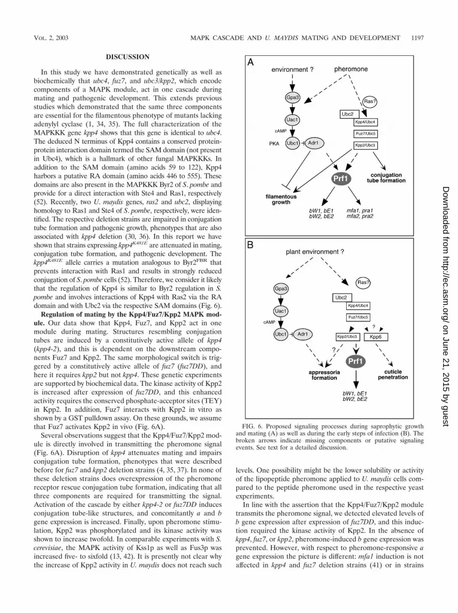
In this study we have demonstrated genetically as well asbiochemically that ubc4, fuz7, and ubc3/kpp2, which encodecomponents of a MAPK module, act in one cascade duringmating and pathogenic development. This extends previousstudies which demonstrated that the same three componentsare essential for the filamentous phenotype of mutants lackingadenylyl cyclase (1, 34, 35). The full characterization of theMAPKKK gene kpp4 shows that this gene is identical to ubc4.The deduced N terminus of Kpp4 contains a conserved protein-protein interaction domain termed the SAM domain (not presentin Ubc4), which is a hallmark of other fungal MAPKKKs. Inaddition to the SAM domain (amino acids 59 to 122), Kpp4harbors a putative RA domain (amino acids 446 to 555). Thesedomains are also present in the MAPKKK Byr2 of S. pombe andprovide for a direct interaction with Ste4 and Ras1, respectively(52). Recently, two U. maydis genes, ras2 and ubc2, displayinghomology to Ras1 and Ste4 of S. pombe, respectively, were iden-tified. The respective deletion strains are impaired in conjugationtube formation and pathogenic growth, phenotypes that are alsoassociated with kpp4 deletion (30, 36). In this report we haveshown that strains expressing kpp4K481E are attenuated in mating,conjugation tube formation, and pathogenic development. Thekpp4K481E allele carries a mutation analogous to Byr2FBR thatprevents interaction with Ras1 and results in strongly reducedconjugation of S. pombe cells (52). Therefore, we consider it likelythat the regulation of Kpp4 is similar to Byr2 regulation in S.pombe and involves interactions of Kpp4 with Ras2 via the RAdomain and with Ubc2 via the respective SAM domains (Fig. 6).
Regulation of mating by the Kpp4/Fuz7/Kpp2 MAPK mod-ule. Our data show that Kpp4, Fuz7, and Kpp2 act in onemodule during mating. Structures resembling conjugationtubes are induced by a constitutively active allele of kpp4(kpp4-2), and this is dependent on the downstream compo-nents Fuz7 and Kpp2. The same morphological switch is trig-gered by a constitutively active allele of fuz7 (fuz7DD), andhere it requires kpp2 but not kpp4. These genetic experimentsare supported by biochemical data. The kinase activity of Kpp2is increased after expression of fuz7DD, and this enhancedactivity requires the conserved phosphate-acceptor sites (TEY)in Kpp2. In addition, Fuz7 interacts with Kpp2 in vitro asshown by a GST pulldown assay. On these grounds, we assumethat Fuz7 activates Kpp2 in vivo (Fig. 6A).
Several observations suggest that the Kpp4/Fuz7/Kpp2 mod-ule is directly involved in transmitting the pheromone signal(Fig. 6A). Disruption of kpp4 attenuates mating and impairsconjugation tube formation, phenotypes that were describedbefore for fuz7 and kpp2 deletion strains (4, 35, 37). In none ofthese deletion strains does overexpression of the pheromonereceptor rescue conjugation tube formation, indicating that allthree components are required for transmitting the signal.Activation of the cascade by either kpp4-2 or fuz7DD inducesconjugation tube-like structures, and concomitantly a and bgene expression is increased. Finally, upon pheromone stimu-lation, Kpp2 was phosphorylated and its kinase activity wasshown to increase twofold. In comparable experiments with S.cerevisiae, the MAPK activity of Kss1p as well as Fus3p wasincreased five- to sixfold (13, 42). It is presently not clear whythe increase of Kpp2 activity in U. maydis does not reach such
levels. One possibility might be the lower solubility or activityof the lipopeptide pheromone applied to U. maydis cells com-pared to the peptide pheromone used in the respective yeastexperiments.
In line with the assertion that the Kpp4/Fuz7/Kpp2 moduletransmits the pheromone signal, we detected elevated levels ofb gene expression after expression of fuz7DD, and this induc-tion required the kinase activity of Kpp2. In the absence ofkpp4, fuz7, or kpp2, pheromone-induced b gene expression wasprevented. However, with respect to pheromone-responsive agene expression the picture is different: mfa1 induction is notaffected in kpp4 and fuz7 deletion strains (41) or in strains
FIG. 6. Proposed signaling processes during saprophytic growthand mating (A) as well as during the early steps of infection (B). Thebroken arrows indicate missing components or putative signalingevents. See text for a detailed discussion.
VOL. 2, 2003 MAPK CASCADE AND U. MAYDIS MATING AND DEVELOPMENT 1197
on June 21, 2015 by guesthttp://ec.asm
.org/D
ownloaded from

expressing a kinase-dead allele of kpp2 (kpp2K50R), while it isstrongly reduced in �kpp2-1 mutants (37). This indicates thatKpp2 influences a gene expression via a kinase-independentmechanism. Precedence for kinase-independent functions ofMAPK proteins is found in S. cerevisiae, where the MAPKKss1p regulates invasive growth in a phosphorylation-depen-dent manner. Unphosphorylatable Kss1p-[AEF] but not ki-nase-dead Kss1p-[K42R] is able to prevent invasive growth ofa FUS3 KSS1 double-deletion strain (7). Phosphorylated Kss1pis a positive regulator of the transcription factor Ste12p, whileunphosphorylated Kss1p inhibits Ste12p function by direct in-teraction (7, 13, 42). In contrast to this situation in S. cerevisiae,unphosphorylatable Kpp2AEF in U. maydis is not able to exertthe kinase-independent function, since strains expressingKpp2AEF are reduced in pheromone-dependent a gene tran-scription. This suggests that the mechanism of kinase-indepen-dent function of Kpp2 in U. maydis differs from that of Kss1p.Preliminary results suggest that this function of Kpp2 mayaffect the transcription factor Prf1, which is absolutely requiredfor pheromone-responsive gene expression. In �kpp2-1 as wellas kpp2AEF strains, prf1 transcription is affected and overex-pression of prf1 rescues the attenuated pheromone gene ex-pression (P. Muller and M. Feldbrugge, unpublished data). Itis conceivable that Kpp2 and Prf1 interact, leading to a stabi-lization of Prf1 protein, and Kpp2AEF might no longer inter-act with Prf1, while stabilization could still be carried out bythe kinase-dead Kpp2 protein.
In contrast to the situations in S. cerevisiae, S. pombe, C.neoformans, and Candida albicans (2, 16, 33), the pheromoneMAPK cascade is not essential for cell fusion in U. maydis. Thisindicates that at least one additional pathway must participate.One likely candidate is the cAMP signaling cascade composedof the G� subunit Gpa3, adenylyl cyclase Uac1, and cAMP-dependent protein kinase A (Fig. 6A). Disruption of the cAMPcascade affects cell fusion and abolishes pheromone-induced agene expression (18, 41). On the other hand, an activatedcAMP cascade results in elevated pheromone gene expressionthat depends on prf1 (23, 29, 41). Thus, we consider it likelythat the pheromone signal is somehow transmitted by thecAMP pathway as well as the MAPK module to Prf1 which inturn promotes pheromone-regulated a and b gene expression(Fig. 6A).
In S. cerevisiae the pheromone signal is transmitted exclu-sively via a MAPK module and the pheromone-responsiveMAPK Fus3p regulates the transcription factor STE12p, pre-sumably by the phosphorylation-dependent inactivation of theDig1/2 proteins, which inhibit the transcription factor STE12p(17, 46, 51). Once activated, Ste12p triggers a transcriptionalprogram that is necessary for the mating response. AnotherFus3p substrate is Far1p, which subsequently inhibits cyclin-Cdc28p kinase activity, resulting in cell cycle arrest, and estab-lishes a site of polarized growth toward the mating partner (14,38, 53). STE12 is absolutely required for projection formation,even when the pheromone MAPK cascade is activated by adominant-active allele of STE11 (15). However, conjugationtube formation in U. maydis does not require the transcriptionfactor prf1. This was evident only when the pheromone recep-tor was constitutively expressed or when the MAPK cascadewas genetically activated by expressing either kpp4-2 orfuz7DD. Thus, pheromone signal transmission bifurcates
downstream of Kpp2 (Fig. 6A). One branch must control theactivation of Prf1 and subsequent induction of the genes lo-cated in the a and b loci. The other branch induces a morpho-logical reprogramming, which might involve a second tran-scription factor and/or proteins regulating the cell cycle andthe cytoskeleton.
Regulation of pathogenic development by the Kpp4/Fuz7/Kpp2 MAPK module. In this study we have demonstrated thatkpp4 and fuz7 are essential for pathogenesis even under con-ditions in which cell fusion and pheromone stimulation are notrequired. Thus, it is likely that Kpp4 and Fuz7 transmit addi-tional signals during pathogenic development (Fig. 6B) (4).The defect in tumor formation caused by deletion of eitherkpp4 or fuz7 cannot be rescued by overexpression of b. Thus,signaling via Kpp4 and Fuz7 appears to regulate pathogenicityin parallel to b (Fig. 6B). Further analysis revealed that kpp4and fuz7 are necessary for filamentous growth on the plantsurface and development of appressoria even under conditionswhere cell fusion was bypassed. While kpp4 and fuz7 are ab-solutely required for pathogenicity, deletion of kpp2 only mod-erately reduced tumor development (35, 37). This is most likelydue to genetic redundancy at the level of the MAPK. U. maydisencodes a Kpp2-related MAPK, Kpp6, which has no obviousrole during mating, and kpp2 kpp6 double-deletion strains arenonpathogenic (11). Since strains expressing an inactive alleleof kpp2 (kpp2AEF) display a stronger phenotype than the�kpp2-1 mutants, we were able to elucidate the function ofkpp2 during the infection process. Such strains behaved like�kpp4 and �fuz7 mutants and were unable to develop appres-soria. Given these results, Kpp2 can be placed in one cascadewith Kpp4 and Fuz7 during appressorial formation.
In a variety of phytopathogenic fungi MAPKs were shown tocontrol appressoria differentiation, e.g., Pmk1 in Magnaporthegrisea, Cmk1 in Colletotrichum lagenarium, and Chk1 in Coch-liobolus heterostrophus (31, 50, 56). Hence, signaling processesthat regulate the infection process appear to be conserved inphytopathogenic fungi. U. maydis is so far unique among phy-topathogenic fungi in having two Pmk1-like MAPKs, Kpp2 andKpp6. These kinases control discrete steps of the infection-related development, with kpp2 being needed for formation ofappressoria and kpp6 being needed for appressorial function(Fig. 6B) (11) Thus, the early infection processes in U. maydisis controlled by a stepwise activation of two highly homologousMAPKs.
ACKNOWLEDGMENTS
We thank J. Gorl for construction of pGEX-Kpp2 and pGEX-Fuz7and Manuel Tonnis and Horst Kessler for providing synthetic phero-mone. We thank Jan Schirawski and Anja Volz-Peters for their criticalcomments on the manuscript.
This work was supported by the DFG through grant SFB369.
REFERENCES
1. Andrews, D. L., J. D. Egan, M. E. Mayorga, and S. E. Gold. 2000. TheUstilago maydis ubc4 and ubc5 genes encode members of a MAP kinasecascade required for filamentous growth. Mol. Plant-Microbe Interact. 13:781–786.
2. Banuett, F. 1998. Signalling in the yeasts: an informational cascade with linksto the filamentous fungi. Microbiol. Mol. Biol. Rev. 62:249–274.
3. Banuett, F., and I. Herskowitz. 1989. Different a alleles are necessary formaintenance of filamentous growth but not for meiosis. Proc. Natl. Acad.Sci. USA 86:5878–5882.
4. Banuett, F., and I. Herskowitz. 1994. Identification of fuz7, a Ustilago maydis
1198 MULLER ET AL. EUKARYOT. CELL
on June 21, 2015 by guesthttp://ec.asm
.org/D
ownloaded from

MEK/MAPKK homolog required for a-locus-dependent and -independentsteps in the fungal life cycle. Genes Dev. 8:1367–1378.
5. Banuett, F., and I. Herskowitz. 1994. Morphological transitions in the lifecycle of Ustilago maydis and their genetic control by the a and b loci. Exp.Mycol. 18:247–266.
6. Bardwell, A. J., L. J. Flatauer, K. Matsukuma, J. Thorner, and L. Bardwell.2001. A conserved docking site in MEKs mediates high-affinity binding toMAP kinases and cooperates with a scaffold protein to enhance signal trans-mission. J. Biol. Chem. 276:10374–10386.
7. Bardwell, L., J. G. Cook, D. Voora, D. M. Baggott, A. R. Martinez, andJ. Thorner. 1998. Repression of yeast Ste12 transcription factor by directbinding of unphosphorylated Kss1 MAPK and its regulation by the Ste7MEK. Genes Dev. 12:2887–2898.
8. Bolker, M., S. Genin, C. Lehmler, and R. Kahmann. 1995. Genetic regula-tion of mating and dimorphism in Ustilago maydis. Can. J. Bot. 73:320–325.
9. Bolker, M., M. Urban, and R. Kahmann. 1992. The a mating type locus of U.maydis specifies cell signaling components. Cell 68:441–450.
10. Bottin, A., J. Kamper, and R. Kahmann. 1996. Isolation of a carbon source-regulated gene from Ustilago maydis. Mol. Gen. Genet. 253:342–352.
11. Brachmann, A., J. Schirawski, P. Muller, and R. Kahmann. 2003. An un-usual MAP kinase is required for efficient penetration of the plant surface byU. maydis. EMBO J. 22:2199–2210.
12. Brachmann, A., G. Weinzierl, J. Kamper, and R. Kahmann. 2001. Identifi-cation of genes in the bW/bE regulatory cascade in Ustilago maydis. Mol.Microbiol. 42:1047–1063.
13. Breitkreutz, A., L. Boucher, and M. Tyers. 2001. MAPK specificity in theyeast pheromone response independent of transcriptional activation. Curr.Biol. 11:1266–1271.
14. Butty, A. C., P. M. Pryciak, L. S. Huang, I. Herskowitz, and M. Peter. 1998.The role of Far1p in linking the heterotrimeric G protein to polarity estab-lishment proteins during yeast mating. Science 282:1511–1516.
15. Cairns, B. R., S. W. Ramer, and R. D. Kornberg. 1992. Order of action ofcomponents in the yeast pheromone response pathway revealed with a dom-inant allele of the STE11 kinase and the multiple phosphorylation of theSTE7 kinase. Genes Dev. 6:1305–1318.
16. Clarke, D. L., G. L. Woodlee, C. M. McClelland, T. S. Seymour, and B. L.Wickes. 2001. The Cryptococcus neoformans STE11alpha gene is similar toother fungal mitogen-activated protein kinase kinase kinase (MAPKKK)genes but is mating type specific. Mol. Microbiol. 40:200–213.
17. Cook, J. G., L. Bardwell, S. J. Kron, and J. Thorner. 1996. Two novel targetsof the MAP kinase Kss1 are negative regulators of invasive growth in theyeast Saccharomyces cerevisiae. Genes Dev. 10:2831–2848.
18. Durrenberger, F., K. Wong, and J. W. Kronstad. 1998. Identification of acAMP-dependent protein kinase catalytic subunit required for virulence andmorphogenesis in Ustilago maydis. Proc. Natl. Acad. Sci. USA 95:5684–5689.
19. Enslen, H., and R. J. Davis. 2001. Regulation of MAP kinases by dockingdomains. Biol. Cell 93:5–14.
20. Gillissen, B., J. Bergemann, C. Sandmann, B. Schroeer, M. Bolker, and R.Kahmann. 1992. A two-component regulatory system for self/non-self rec-ognition in Ustilago maydis. Cell 68:647–657.
21. Gold, S., G. Duncan, K. Barrett, and J. Kronstad. 1994. cAMP regulatesmorphogenesis in the fungal pathogen Ustilago maydis. Genes Dev. 8:2805–2816.
22. Hartmann, H. A., R. Kahmann, and M. Bolker. 1996. The pheromoneresponse factor coordinates filamentous growth and pathogenicity in Ustilagomaydis. EMBO J. 15:1632–1641.
23. Hartmann, H. A., J. Kruger, F. Lottspeich, and R. Kahmann. 1999. Envi-ronmental signals controlling sexual development of the corn Smut fungusUstilago maydis through the transcriptional regulator Prf1. Plant Cell 11:1293–1306.
24. Hoffman, C. S., and F. Winston. 1987. A ten-minute DNA preparation fromyeast efficiently releases autonomous plasmids for transformation of E. coli.Gene 57:267–272.
25. Holliday, R. 1974. Ustilago maydis, p. 575–595. In R. C. King (ed.), Hand-book of genetics, vol. 1. Plenum Press, New York, N.Y.
26. Huang, W., and R. L. Erikson. 1994. Constitutive activation of Mek1 bymutation of serine phosphorylation sites. Proc. Natl. Acad. Sci. USA 91:8960–8963.
27. Kahmann, R., S. G., C. Basse, M. Feldbrugge, and J. Kamper. 2000. Ustilagomaydis, the causative agent of corn smut disease, p. 347–371. In J. W.Kronstad (ed.), Fungal pathology. Kluwer Academic Publishers, Dodrecht,The Netherlands.
28. Kamper, J., M. Reichmann, T. Romeis, M. Bolker, and R. Kahmann. 1995.Multiallelic recognition: nonself-dependent dimerization of the bE and bWhomeodomain proteins in Ustilago maydis. Cell 81:73–83.
29. Kruger, J., G. Loubradou, E. Regenfelder, A. Hartmann, and R. Kahmann.1998. Crosstalk between cAMP and pheromone signalling pathways in Us-tilago maydis. Mol. Gen. Genet. 260:193–198.
30. Lee, N., and J. W. Kronstad. 2002. ras2 controls morphogenesis, pheromoneresponse, and pathogenicity in the fungal pathogen Ustilago maydis. Eu-karyot. Cell 1:954–966.
31. Lev, S., A. Sharon, R. Hadar, H. Ma, and B. A. Horwitz. 1999. A mitogen-activated protein kinase of the corn leaf pathogen Cochliobolus heterostro-phus is involved in conidiation, appressorium formation, and pathogenicity:diverse roles for mitogen-activated protein kinase homologs in foliar patho-gens. Proc. Natl. Acad. Sci. USA 96:13542–13547.
32. Loubradou, G., A. Brachmann, M. Feldbrugge, and R. Kahmann. 2001. Ahomologue of the transcriptional repressor Ssn6p antagonizes cAMP signal-ling in Ustilago maydis. Mol. Microbiol. 40:719–730.
33. Magee, B. B., M. Legrand, A. M. Alarco, M. Raymond, and P. T. Magee.2002. Many of the genes required for mating in Saccharomyces cerevisiae arealso required for mating in Candida albicans. Mol. Microbiol. 46:1345–1351.
34. Mayorga, M. E., and S. E. Gold. 1998. Characterization and moleculargenetic complementation of mutants affecting dimorphism in the fungusUstilago maydis. Fungal Genet. Biol. 24:364–376.
35. Mayorga, M. E., and S. E. Gold. 1999. A MAP kinase encoded by the ubc3gene of Ustilago maydis is required for filamentous growth and full virulence.Mol. Microbiol. 34:485–497.
36. Mayorga, M. E., and S. E. Gold. 2001. The ubc2 gene of Ustilago maydisencodes a putative novel adaptor protein required for filamentous growth,pheromone response and virulence. Mol. Microbiol. 41:1365–1379.
37. Muller, P., C. Aichinger, M. Feldbrugge, and R. Kahmann. 1999. The MAPkinase kpp2 regulates mating and pathogenic development in Ustilago may-dis. Mol. Microbiol. 34:1007–1017.
38. Peter, M., A. Gartner, J. Horecka, G. Ammerer, and I. Herskowitz. 1993.FAR1 links the signal transduction pathway to the cell cycle machinery inyeast. Cell 73:747–760.
39. Peterson, A. J., M. Kyba, D. Bornemann, K. Morgan, H. W. Brock, and J.Simon. 1997. A domain shared by the Polycomb group proteins Scm and phmediates heterotypic and homotypic interactions. Mol. Cell. Biol. 17:6683–6692.
40. Ponting, C. P., and D. R. Benjamin. 1996. A novel family of Ras-bindingdomains. Trends Biochem. Sci. 21:422–425.
41. Regenfelder, E., T. Spellig, A. Hartmann, S. Lauenstein, M. Bolker, and R.Kahmann. 1997. G proteins in Ustilago maydis: transmission of multiplesignals? EMBO J. 16:1934–1942.
42. Sabbagh, W., Jr., L. J. Flatauer, A. J. Bardwell, and L. Bardwell. 2001.Specificity of MAP kinase signaling in yeast differentiation involves transientversus sustained MAPK activation. Mol. Cell 8:683–691.
43. Sambrook, J., E. F. Fritsch, and T. Maniatis. 1989. Molecular cloning: alaboratory manual, 2nd ed. Cold Spring Harbor Laboratory Press, ColdSpring Harbor, N.Y.
44. Schultz, J., C. P. Ponting, K. Hofmann, and P. Bork. 1997. SAM as a proteininteraction domain involved in developmental regulation. Protein Sci. 6:249–253.
45. Schulz, B., F. Banuett, M. Dahl, R. Schlesinger, W. Schafer, T. Martin, I.Herskowitz, and R. Kahmann. 1990. The b alleles of U. maydis, whosecombinations program pathogenic development, code for polypeptides con-taining a homeodomain-related motif. Cell 60:295–306.
46. Song, D., J. W. Dolan, Y. L. Yuan, and S. Fields. 1991. Pheromone-depen-dent phosphorylation of the yeast STE12 protein correlates with transcrip-tional activation. Genes Dev. 5:741–750.
47. Spellig, T., A. Bottin, and R. Kahmann. 1996. Green fluorescent protein(GFP) as a new vital marker in the phytopathogenic fungus Ustilago maydis.Mol. Gen. Genet. 252:503–509.
48. Stevenson, B. J., N. Rhodes, B. Errede, and G. F. Sprague, Jr. 1992. Con-stitutive mutants of the protein kinase STE11 activate the yeast pheromoneresponse pathway in the absence of the G protein. Genes Dev. 6:1293–1304.
49. Szabo, Z., Tonnis, M., Kessler, H., and M. Feldbrugge. 2002. Structure-function analysis of lipopeptide pheromones from the plant pathogen Usti-lago maydis. Mol. Genet. Genomics 268:362–370.
50. Takano, Y., T. Kikuchi, Y. Kubo, J. E. Hamer, K. Mise, and I. Furusawa.2000. The Colletotrichum lagenarium MAP kinase gene CMK1 regulatesdiverse aspects of fungal pathogenesis. Mol. Plant-Microbe Interact. 13:374–383.
51. Tedford, K., S. Kim, D. Sa, K. Stevens, and M. Tyers. 1997. Regulation of themating pheromone and invasive growth responses in yeast by two MAPkinase substrates. Curr. Biol. 7:228–238.
52. Tu, H., M. Barr, D. L. Dong, and M. Wigler. 1997. Multiple regulatorydomains on the Byr2 protein kinase. Mol. Cell. Biol. 17:5876–5887.
53. Tyers, M., and B. Futcher. 1993. Far1 and Fus3 link the mating pheromonesignal transduction pathway to three G1-phase Cdc28 kinase complexes. Mol.Cell. Biol. 13:5659–5669.
54. Urban, M., R. Kahmann, and M. Bolker. 1996. Identification of the phero-mone response element in Ustilago maydis. Mol. Gen. Genet. 251:31–37.
55. Wedlich-Soldner, R., M. Bolker, R. Kahmann, and G. Steinberg. 2000. Aputative endosomal t-SNARE links exo- and endocytosis in the phytopatho-genic fungus Ustilago maydis. EMBO J. 19:1974–1986.
56. Xu, J. R., and J. E. Hamer. 1996. MAP kinase and cAMP signaling regulateinfection structure formation and pathogenic growth in the rice blast fungusMagnaporthe grisea. Genes Dev. 10:2696–2706.
VOL. 2, 2003 MAPK CASCADE AND U. MAYDIS MATING AND DEVELOPMENT 1199
on June 21, 2015 by guesthttp://ec.asm
.org/D
ownloaded from