corynebacterium ulcerans 0102 carries the gene encoding diphtheria toxin on a prophage different...
TRANSCRIPT

Sekizuka et al. BMC Microbiology 2012, 12:72http://www.biomedcentral.com/1471-2180/12/72
RESEARCH ARTICLE Open Access
Corynebacterium ulcerans 0102 carries the geneencoding diphtheria toxin on a prophagedifferent from the C. diphtheriae NCTC 13129prophageTsuyoshi Sekizuka1, Akihiko Yamamoto2, Takako Komiya2, Tsuyoshi Kenri2, Fumihiko Takeuchi1, Keigo Shibayama2,Motohide Takahashi2,3, Makoto Kuroda1 and Masaaki Iwaki2*
Abstract
Background: Corynebacterium ulcerans can cause a diphtheria-like illness, especially when the bacterium islysogenized with a tox gene-carrying bacteriophage that produces diphtheria toxin. Acquisition of toxigenicity uponphage lysogenization is a common feature of C. ulcerans and C. diphtheriae. However, because of a lack of C.ulcerans genome information, a detailed comparison of prophages has not been possible between these twoclinically important and closely related bacterial species.
Results: We determined the whole genome sequence of the toxigenic C. ulcerans 0102 isolated in Japan. Thegenomic sequence showed a striking similarity with that of Corynebacterium pseudotuberculosis and, to a lesserextent, with that of C. diphtheriae. The 0102 genome contained three distinct prophages. One of these,ΦCULC0102-I, was a tox-positive prophage containing genes in the same structural order as for tox-positiveC. diphtheriae prophages. However, the primary structures of the individual genes involved in the phage machineryshowed little homology between the two counterparts.
Conclusion: Taken together, these results suggest that the tox-positive prophage in this strain of C. ulcerans has adistinct origin from that of C. diphtheriae NCTC 13129.
Keywords: Bacteriophage, Toxin gene, Horizontal gene transfer, Diphtheria, Zoonosis
BackgroundA diphtheria-like infectious disease caused by Coryne-bacterium ulcerans is increasing in clinical importancein developed countries and is now regarded as “diph-theria” in Europe [1,2]. Infection with C. ulcerans occursin a wide range of hosts, including cats, dogs, pigs, cows,and whales [3-9]. The first clearly documented case ofzoonotic transmission involved a dog, as reported byLartigue et al. [5]. This is in contrast to the causativeagent of classical diphtheria, C. diphtheriae, whose hostspecies is thought to be limited to humans [10]. Never-theless, the two species share a common feature: upon
* Correspondence: [email protected] of Bacteriology II, National Institute of Infectious Diseases, 4-7-1Gakuen, Musashimurayama-shi, Tokyo 208-0011, JapanFull list of author information is available at the end of the article
© 2012 Sekizuka et al.; licensee BioMed CentraCommons Attribution License (http://creativecreproduction in any medium, provided the or
lysogenization of tox-encoding bacteriophages, they be-come toxigenic and are able to produce the potent diph-theria toxin [1,10]. This toxin is known to contribute todisease progression, occasionally leading to death. It isencoded by a single gene designated tox, situated insideprophages lysogenized in the bacterial genome of C.diphtheriae [11]. The prophages are capable of induc-tion, by ultraviolet light or DNA-damaging agents suchas mitomycin C, and yield β-, δ-, ω- and other functionalbacteriophage particles [12]. Some types of bacterio-phages can infect both C. diphtheriae and C. ulcerans[13-16]. Furthermore, the C. ulcerans tox gene is alsoencoded in a genome region surrounded by phage at-tachment (att) sites conserved between the two species[7,16]. The nucleotide sequences of C. ulcerans tox geneswere published by Sing et al. They showed some
l Ltd. This is an Open Access article distributed under the terms of the Creativeommons.org/licenses/by/2.0), which permits unrestricted use, distribution, andiginal work is properly cited.
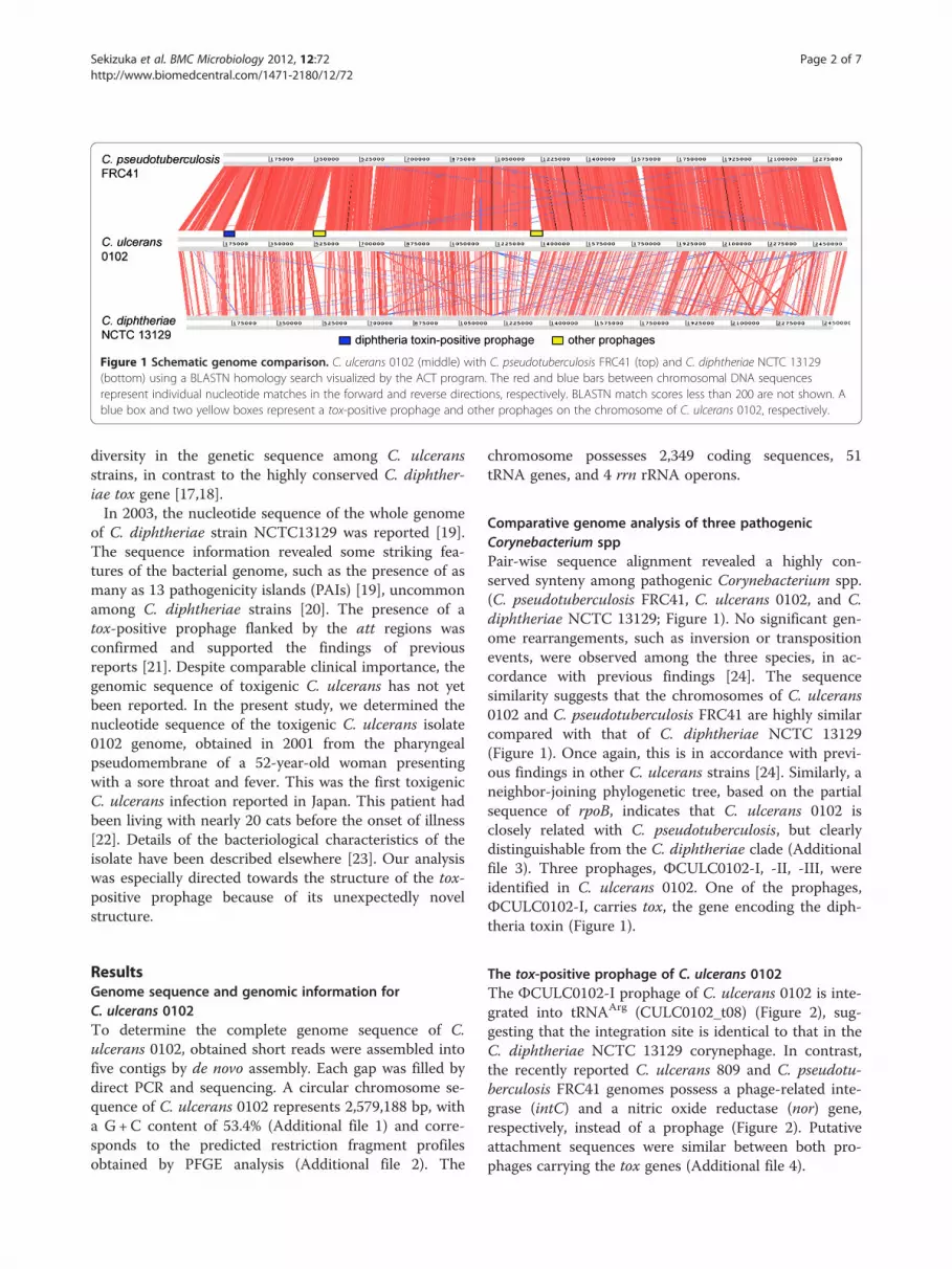
Figure 1 Schematic genome comparison. C. ulcerans 0102 (middle) with C. pseudotuberculosis FRC41 (top) and C. diphtheriae NCTC 13129(bottom) using a BLASTN homology search visualized by the ACT program. The red and blue bars between chromosomal DNA sequencesrepresent individual nucleotide matches in the forward and reverse directions, respectively. BLASTN match scores less than 200 are not shown. Ablue box and two yellow boxes represent a tox-positive prophage and other prophages on the chromosome of C. ulcerans 0102, respectively.
Sekizuka et al. BMC Microbiology 2012, 12:72 Page 2 of 7http://www.biomedcentral.com/1471-2180/12/72
diversity in the genetic sequence among C. ulceransstrains, in contrast to the highly conserved C. diphther-iae tox gene [17,18].In 2003, the nucleotide sequence of the whole genome
of C. diphtheriae strain NCTC13129 was reported [19].The sequence information revealed some striking fea-tures of the bacterial genome, such as the presence of asmany as 13 pathogenicity islands (PAIs) [19], uncommonamong C. diphtheriae strains [20]. The presence of atox-positive prophage flanked by the att regions wasconfirmed and supported the findings of previousreports [21]. Despite comparable clinical importance, thegenomic sequence of toxigenic C. ulcerans has not yetbeen reported. In the present study, we determined thenucleotide sequence of the toxigenic C. ulcerans isolate0102 genome, obtained in 2001 from the pharyngealpseudomembrane of a 52-year-old woman presentingwith a sore throat and fever. This was the first toxigenicC. ulcerans infection reported in Japan. This patient hadbeen living with nearly 20 cats before the onset of illness[22]. Details of the bacteriological characteristics of theisolate have been described elsewhere [23]. Our analysiswas especially directed towards the structure of the tox-positive prophage because of its unexpectedly novelstructure.
ResultsGenome sequence and genomic information forC. ulcerans 0102To determine the complete genome sequence of C.ulcerans 0102, obtained short reads were assembled intofive contigs by de novo assembly. Each gap was filled bydirect PCR and sequencing. A circular chromosome se-quence of C. ulcerans 0102 represents 2,579,188 bp, witha G+C content of 53.4% (Additional file 1) and corre-sponds to the predicted restriction fragment profilesobtained by PFGE analysis (Additional file 2). The
chromosome possesses 2,349 coding sequences, 51tRNA genes, and 4 rrn rRNA operons.
Comparative genome analysis of three pathogenicCorynebacterium sppPair-wise sequence alignment revealed a highly con-served synteny among pathogenic Corynebacterium spp.(C. pseudotuberculosis FRC41, C. ulcerans 0102, and C.diphtheriae NCTC 13129; Figure 1). No significant gen-ome rearrangements, such as inversion or transpositionevents, were observed among the three species, in ac-cordance with previous findings [24]. The sequencesimilarity suggests that the chromosomes of C. ulcerans0102 and C. pseudotuberculosis FRC41 are highly similarcompared with that of C. diphtheriae NCTC 13129(Figure 1). Once again, this is in accordance with previ-ous findings in other C. ulcerans strains [24]. Similarly, aneighbor-joining phylogenetic tree, based on the partialsequence of rpoB, indicates that C. ulcerans 0102 isclosely related with C. pseudotuberculosis, but clearlydistinguishable from the C. diphtheriae clade (Additionalfile 3). Three prophages, ΦCULC0102-I, -II, -III, wereidentified in C. ulcerans 0102. One of the prophages,ΦCULC0102-I, carries tox, the gene encoding the diph-theria toxin (Figure 1).
The tox-positive prophage of C. ulcerans 0102The ΦCULC0102-I prophage of C. ulcerans 0102 is inte-grated into tRNAArg (CULC0102_t08) (Figure 2), sug-gesting that the integration site is identical to that in theC. diphtheriae NCTC 13129 corynephage. In contrast,the recently reported C. ulcerans 809 and C. pseudotu-berculosis FRC41 genomes possess a phage-related inte-grase (intC) and a nitric oxide reductase (nor) gene,respectively, instead of a prophage (Figure 2). Putativeattachment sequences were similar between both pro-phages carrying the tox genes (Additional file 4).

Figure 2 Schematic representation and comparative analysis of tox-positive prophages and flanking regions. The tox-positive prophageand flanking regions of C. ulcerans 0102 and C. diphtheriae NCTC13129 are shown. The corresponding region of C. pseudotuberculosis FRC41 andC. ulcerans 809 is also shown. Boxes indicate individual coding regions with colors assigned to their functions. GenBank accession numbers aregiven in parentheses.
Sekizuka et al. BMC Microbiology 2012, 12:72 Page 3 of 7http://www.biomedcentral.com/1471-2180/12/72
The two tox-positive prophages share the same struc-tural features, with genes aligned in an ‘integrase - pack-aging - head - tail - lysis - toxin’ orientation (Figure 2).Pair-wise alignment of the prophages indicates a highsimilarity in the region encoding the putative integrase,the 3′-ends of CULC0102_0211 and CULC0102_0212,tox, and the attachment sites (Figure 2). The majorphage machineries encoded in the internal phage regionshowed low similarity at the nucleotide and amino acidlevels (less than 18%) between C. ulcerans 0102 and C.diphtheriae NCTC13129.
DiscussionWhole-genome sequencing has revealed that the C.ulcerans 0102 genome is composed of 2,579,188 bp witha G+C content of 53.4%. These values are similar tothose recently reported for C. ulcerans strains 809(2,502,095 bp, 53.3%G+C) and BR-AD22 (2,606,374 bp,53.4%G+C) [24]. C. ulcerans 0102 shares many com-mon features with the two previously reported strains,including 12 virulence factors. Strain 0102 is distinctivewith respect to the features of prophages integrated inits genome. It possesses a unique tox-positive prophage,ΦCULC0102-I, in its chromosome (Figure 1 andAdditional file 1). In the same position of the recentlyreported C. ulcerans 809 genome exists a remnant
phage-related integrase (intC) gene [24] (Figure 2). TheC. ulcerans 0102 prophage differs from the correspond-ing prophage in C. diphtheriae. Although the integraseand tox gene sequences of ΦCULC0102-I showed highsimilarity to those of the corynephage encoding tox in C.diphtheriae NCTC 13129, the major phage machinerygenes in ΦCULC0102-I are distinct from those in othercorynephages in C. diphtheriae (Figure 2). This suggeststhat C. ulcerans 0102 did not immediately acquire the C.diphtheriae tox-positive corynephage.There are many possible explanations for the origins
of these two prophages that are tox-positive but obvi-ously different. One of the simplest explanations wecan postulate is outlined in Figure 3. Generally, bacter-ial prophages are duplicated by excision from chromo-somal DNA and subsequent concatenation at both endsof the att sites (Figure 3A). This duplication step indi-cates that two highly homologous regions, int and tox,could be in close proximity and adjacent to the att siteconcatenation. It could be speculated that homologousrecombination between two prophages may facilitatethe acquisition of the tox gene in C. ulcerans 0102from an unknown tox-positive prophage (Figure 3B)[25]. Horizontal gene transfer is one of the majormechanisms of foreign gene acquisition by bacteria, asreviewed by Ochman et al. [26]. Liu et al. have

Figure 3 Schema of the diphtheria toxin acquisition hypothesis. (A) Pair-wise comparison of regions with high similarity between C. ulceransand C. diphtheriae. These structures of putative phages are constructed by connecting attachment sites. The plots above and below represent theGC content calculated with a window size of 500 bp. (B) Schematic representation of how diphtheria toxin has been acquired in C. ulcerans.
Sekizuka et al. BMC Microbiology 2012, 12:72 Page 4 of 7http://www.biomedcentral.com/1471-2180/12/72
demonstrated that horizontally transferred genes areoften disabled and become pseudogenes. In these casesthe genes are no longer beneficial to the recipients[27]. Non-toxigenic C. diphtheriae (CD450, CD119,CD448, and CD443 strains) carry tox pseudogenes thatare relatively similar to the tox genes of C. ulcerans(Additional file 5), suggesting that horizontal genetransfer among Corynebacterium spp. might occur.Consistent with previous findings [7,17,18,28], tthe toxgene in C. ulcerans 0102 is not identical to that of C.diphtheriae (Additional file 5); phylogenetic analysis oftox showed greater heterogeneity among C. ulceransisolates than that for C. diphtheriae isolates (Additionalfile 5).The C. diphtheriae tox gene is highly conserved
among temporally and geographically diverse strains[29], therefore greater variation in tox genes from C.ulcerans isolates suggests that this strain might haveacquired the tox gene before C. diphtheriae.In a recent report, whole genome sequence analysis of
non-toxigenic C. ulcerans 809 and BR-AD22 [24], the β-corynephage-like truncated integrases (CULC809_00176and CULC22_00173) are located adjacent to thetRNAArg gene, similar to ΦCULC0102-I in C. ulcerans0102 and C. diphtheriae. The tRNAArg gene(CULC0102_t08) appears to be a ‘hotspot’ for the acqui-sition of ΦCULC0102-I-like prophages by homologousintegrase.The whole genome sequences of C. ulcerans 809 and
BR-AD22 contain possible virulence factors, such ascorynebacterial protease (CP40), phospholipase D (Pld),neuraminidase (NanH), venom serine protease (Vsp1),
trypsin-like serine protease (TspA), Rpf interacting pro-tein (RpfI), cell wall-associated hydrolase (CwlH), andfive surface-anchored proteins (SpaB–F) [24]. TheSpaA-type pilin, encoded by the spaABC-srtA genecluster, is considered to play a crucial role in adhesionof C. diphtheriae [30]. The gene encoding the shaftprotein of SpaA-type pilin (spaA) was absent in C.ulcerans 0102, a feature consistent with previous find-ings in C. ulcerans 809 and BR-AD2 [24]. As SpaB andSpaC proteins, which are assumed to be present in allthree C. ulcerans strains, can contribute to host-cell ad-hesion in the absence of SpaA [30], this may imply acommon mechanism of cell adhesion by C. ulcerans[24].The C. ulcerans 809 strain was isolated from a patient
with a rapid fatal pulmonary infection. The 809 strain-unique virulence factor (shiga toxin-like ribosome-bind-ing protein, Rbp) is located adjacent to the truncatedintegrase (CULC809_00176) and corresponds to theintegrase of ΦCULC0102-I. It appears that virulence fac-tors have been acquired as a cassette gene in theΦCULC0102-I-like prophage. It is intriguing to note thatthe 0102 strain does not carry the 809 strain-uniquevirulence factors (Rbp and the additional venom serineprotease, Vsp2), but instead carries the tox gene onΦCULC0102-I, which resulted in a diphtheria-like illnessin a 52-year-old woman.Isolates of C. ulcerans are generally obtained from a
diverse range of animals, including humans. Isolation ofa human pathogen C. diphtheriae from animals has beenreported previously, although it is rare [31]. The toxgene might be frequently transmitted through common

Sekizuka et al. BMC Microbiology 2012, 12:72 Page 5 of 7http://www.biomedcentral.com/1471-2180/12/72
prophages with the aid of the highly homologous regionsamong Corynebacterium spp., including C. diphtheriaeand C. ulcerans isolated from animal sources.
ConclusionsToxigenic C. ulcerans is an emerging pathogen that canbe transmitted from animals to humans [5]. In the hostorganism, as well as in C. diphtheriae, the tox gene [18]is encoded by prophages. Through genome sequencing,we have identified a novel structure in a tox-positive C.ulcerans prophage with no significant sequence hom-ology to those in C. diphtheriae. This suggests distinctorigins of the prophages and thus may also explain thedifference in the primary structures of their tox genes.The tox-positive bacteriophages may increase the dis-semination risk of toxigenic C. ulcerans isolates, there-fore, C. ulcerans isolates from both human and animalsources should be investigated further to determine thelevel of variation.
MethodsThis research was not carried out on humans. No ex-perimental research on animals was carried out.
Bacterial strainThe toxigenic C. ulcerans isolate 0102 was obtained in2001 as a human clinical isolate [22,23].
Preparation of genomic DNAGenomic DNA was isolated by conventional methods,using phenol extraction and ethanol precipitation fromheat-killed bacterial cells propagated in brain-heart infu-sion liquid medium.
Short-read DNA sequencing using an Illumina GenomeAnalyzer IIxDNA libraries of the ~600 bp insert length of C. ulcer-ans 0102 were prepared using a genomic DNA SamplePrep Kit (Illumina, San Diego, CA, USA). DNA clusterswere generated on a slide using a Cluster Generation Kit(ver. 4) on an Illumina Cluster Station (Illumina),according to the manufacturer’s instructions. Sequencingruns for 80-mer short reads were performed using anIllumina Genome Analyzer IIx (GA IIx) and TruSeq SBSkit v5. Fluorescent images were analyzed using the Illu-mina base-calling pipeline RTA2.6/SCS2.8 to obtainFASTQ-formatted sequence data.
De novo assembly of short DNA reads and gap-closingThe 80-mer reads were assembled (parameters k64, n51,c32.1373) using ABySS-pe v1.2.0 [32]. Predicted gapswere amplified with a specific PCR primer pair, followedby Sanger DNA sequencing using a BigDye Terminator
v3.1 Cycle Sequencing Kit (Applied Biosystems, FosterCity, CA, USA).
Validation of the complete genome sequence usingshort-read mapping and pulsed-field gelelectrophoresis (PFGE)To validate the genome sequence, 40–mer short readswere re-aligned with the sequence using Maq software(ver. 0.7.1) and the easyrun Perl-command [33]. Readalignment was inspected using the MapView graphicalalignment viewer [34]. PFGE analysis was performed tovalidate the predicted restriction fragment profiles fromthe complete genome sequence, according to De Zoysaet al. [35]. Bacterial cells were lysed with lysozyme andprotease [36], embedded in plugs, digested with the re-striction endonuclease SfiI (New England Biolabs, Ips-witch, MA, USA) and electrophoresed in a CHEF DRIIapparatus (Bio-Rad, Hercules, CA, USA) at 11°C with apulse time of 5–20 s for the first 20 h and 1–5 s for thefollowing 18 h.
Annotation and pair-wise alignment analysisGene prediction from the complete sequence was per-formed using the NCBI Prokaryotic Genomes Auto-matic Annotation Pipeline (PGAAP; http://www.ncbi.nlm.nih.gov/genomes/static/pipeline.html). Several ofthe suggested errors were revised manually. Pseudogenesthat were identified by PGAAP were checked using theread-mapping correction described above. Genomic in-formation, such as nucleic acid variations and circularrepresentation, was analyzed using IMC-GE software(Insilicobiology, Yokohama, Japan). A BLASTN hom-ology search [37] was performed for the whole chromo-some sequences of C. pseudotuberculosis FRC41(accession no. NC_014329), C. ulcerans 0102, and C.diphtheriae NCTC 13129 (accession no. NC_002935).Aligned images of the homologous regions were visua-lized with the ACT program [38].
Phylogenetic analysisPhylogenetic analyses of all nucleotide sequences wereconducted using the neighbor-joining method with1,000-times bootstrapping in ClustalW2 [39]. FigTreever. 1.3.1 (http://tree.bio.ed.ac.uk/software/figtree/) soft-ware was used to display the generated tree.
Nucleotide sequence accession numbersThe complete chromosome sequence for the C. ulcerans0102 strain has been deposited in the DNA Data Bankof Japan (DDBJ; accession no. AP012284).
Sekizuka et al. BMC Microbiology 2012, 12:72 Page 6 of 7http://www.biomedcentral.com/1471-2180/12/72
Additional files
Additional file: 1 Circular representation of the C. ulcerans 0102genome. From the outside inward, the outer circle 1 indicates the size inbase pairs (Mb). The red bars on Circle 2 show prophage region. Circles 3and 4 show the positions of CDS transcribed in clockwise andanticlockwise directions, respectively. The dark blue bars on circle 5indicate ribosomal DNA loci. Circle 6 shows a plot of G + C content (in a20 kb window). Circle 7 shows a plot of GC skew ([G - C]/[G + C]; in a20 kb window).
Additional file: 2 PFGE analysis of C. ulcerans 0102 with four restrictionenzyme digestions.
Additional file: 3 Jukes-Cantor-derived phylogenetic tree based on thepartial rpoB gene region among Corynebacterium isolates with 1,000-foldbootstrapping. Scale bar indicates number of substitutions per site. Thenumber at each branch node represents the bootstrapping value.GenBank accession nos. given in parentheses.
Additional file: 4 Alignment of the nucleotide sequences of attachmentsite common regions among C. ulcerans 0102 and C. diphtheriae NCTC13129. The red characters show regions annotated as tRNAArg.
Additional file: 5 Phylogenetic tree based on the tox genes amongtoxgenic and nontoxigenic Corynebacterium spp. using the Neighbor-joining method with 1,000-fold bootstrapping. Scale bar indicatesnumber of substitutions per site. The number at each branch noderepresents the bootstrapping value. GenBank accession nos. given inparentheses.
Competing interestsThe authors declare that they have no competing interests.
AcknowledgmentsThe authors are grateful to Akio Hatanaka, Atsuhiro Tsunoda and Kenji Ooefor the 0102 clinical isolate. This work was supported by grants for Researchon Emerging and Re-emerging Infectious Diseases (H23 Shinko-Ippan-007and H22-Shinko-Ippan-010), from the Ministry of Health, Labour and Welfare,Japan.
Author details1Laboratory of Bacterial Genomics, Pathogen Genomics Center, NationalInstitute of Infectious Diseases, 1-23-1 Toyama, Shinjuku-ku, Tokyo 162-8640,Japan. 2Department of Bacteriology II, National Institute of InfectiousDiseases, 4-7-1 Gakuen, Musashimurayama-shi, Tokyo 208-0011, Japan.3Present address: Pharmaceutical and Medical Devices Agency, Tokyo, Japan.
Authors’ contributionsTS and FT carried out the genome sequencing studies, participated in thesequence alignment and drafted the manuscript. TKo carried outmaintenance, quality control and propagation of the bacterial strain forgenome sequencing. AY and TKe participated in the design of the study. MTand KS conceived of and participated in coordination of the study,respectively. MK and MI coordinated the study, and drafted and finalized themanuscript. All authors read and approved the final manuscript.
Received: 10 January 2012 Accepted: 14 May 2012Published: 14 May 2012
References1. Bonnet JM, Begg NT: Control of diphtheria: guidance for consultants in
communicable disease control. Commun Dis Public Health 1999, 2:242–249.2. European Centre for Disease Prevention and Control: Diphtheria. In
Surveillance Report: Annual epidemiological report on communicable diseasesin Europe 2010. 2010:133–135.
3. Dias AASO, Silva FC, Pereira GA, Souza MC, Camello TCF, Damasceno JALD,Pacheco LGC, Miyoshi A, Azevedo VA, Hirata R, et al: Corynebacteriumulcerans isolated from an asymptomatic dog kept in an animal shelter inthe metropolitan area of Rio de Janeiro, Brazil. Vector Borne Zoonotic Dis2010, 10:743–748.
4. Katsukawa C, Kawahara R, Inoue K, Ishii A, Yamagishi H, Kida K, Nishino S,Nagahama S, Komiya T, Iwaki M, Takahashi M: Toxigenic Corynebacteriumulcerans Isolated from the domestic dog for the first time in Japan. Jpn JInfect Dis 2009, 62:171–172.
5. Lartigue M-F, Monnet X, Le Flèche A, Grimont PAD, Benet J-J, Durrbach A,Fabre M, Nordmann P: Corynebacterium ulcerans in animmunocompromised patient with diphtheria and her dog. J ClinMicrobiol 2005, 43:999–1001.
6. Schuhegger R, Schoerner C, Dlugaiczyk J, Lichtenfeld I, Trouillier A,Zeller-Peronnet V, Busch U, Berger A, Kugler R, Hörmansdorfer S, SingA: Pigs as source for toxigenic Corynebacterium ulcerans. Emerg InfectDis 2009, 15:1314–1315.
7. Seto Y, Komiya T, Iwaki M, Kohda T, Mukamoto M, Takahashi M, Kozaki S:Properties of corynephage attachment site and molecular epidemiologyof Corynebcterium ulcerans isolated from humans and animals in Japan.Jpn J Infect Dis 2008, 61:116–122.
8. De Zoysa A, Hawkey PM, Engler K, George R, Mann G, Reilly W, Taylor D,Efstratiou A: Characterization of toxigenic Corynebacterium ulceransstrains isolated from humans and domestic cats in the United Kingdom.J Clin Microbiol 2005, 43:4377.
9. Yoshimura Y, Yamamoto A, Komiya T: A case of axillary lymph nodeabscess caused by percutaneous infection of Corynebacterium ulceransthrough scratch by a pus-discharging cat, June 2010 (in Japanese). InfectAgents Surveillance Rep 2010, 31:331.
10. Murphy JR: Chapter 32 Corynebacterium diphtheriae. In MedicalMicrobiology. 4th edition. Edited by Baron S. Galveston: University of TexasMedical Branch at Galveston; 1996.
11. Pappenheimer AM Jr, Gill DM: Diphtheria. Recent studies have clarifiedthe molecular mechanisms involved in its pathogenesis. Science 1973,182:353–358.
12. Rappuoli R, Michel JL, Murphy JR: Integration of corynebacteriophages:tox+, xtox+ and gtox+ into two attachment sites on the Corynebacteriumdiphtheriae chromosome. J Bacteriol 1983, 153:1202–1210.
13. Ishii-Kanei C, Uchida T, Yoneda M: Isolation of a cured strain fromCorynebacterium diphtheriae PW8. Infect Immun 1979,25:1081–1083.
14. Cianciotto NP, Groman NB: Extended host range of a β-relatedcorynebacteriophage. FEMS Microbiol Lett 1996, 140:221–225.
15. Oram M, Woolston JE, Jacobson AD, Holmes RK, Oram DM:Bacteriophage-based vectors for site-specific insertion of DNA inthe chromosome of Corynebacteria. Gene 2007, 391:53–62.
16. Cianciotto N, Rappuoli R, Groman N: Detection of homology to the betabacteriophage integration site in a wide variety of Corynebacterium spp.J Bacrteriol 1986, 168:103–108.
17. Sing A, Bierschenk S, Heesemann J: Classical diphtheria caused byCorynebacterium ulcerans in Germany: amino acid sequence differencesbetween diphtheria toxins from Corynebacterium diphtheriae and C.ulcerans. Clin Infect Dis 2005, 40:325–326.
18. Sing A, Hogardt M, Bierschenk S, Heesemann J: Detection of differences inthe nucleotide and amino acid sequences of diphtheria toxin fromCorynebacterium diphtheriae and Corynebacterium ulcerans causingextrapharyngeal infections. J Clin Microbiol 2003, 41:4848–4851.
19. Cerdeño-Tárraga A-M, Efstratiou A, Dover LG, Holden MTG, Pallen M, BentleySD, Besra GS, Churcher C, James KD, De Zoysa A, et al: The completegenome sequence and analysis of Corynebacterium diphtheriaeNCTC13129. Nucl Acids Res 2003, 31:6516–6523.
20. Iwaki M, Komiya T, Yamamoto A, Ishiwa A, Nagata N, Arakawa Y, TakahashiM: Genome organization and pathogenicity of Corynebacteriumdiphtheriae C7(−) and PW8 strains. Infect Immun 2010, 78:3791–3800.
21. Cianciotto N, Serwold-Davis T, Groman N, Ratti G, Rappuoli R: DNAsequence homology between attB-related sites of Corynebacteriumdiphtheriae, Corynebacterium ulcerans, Corynebacterium glutamicum, andthe attP site of gamma-corynephage. FEMS Microbiol Lett 1990,66:299–301.
22. Hatanaka A, Tsunoda A, Okamoto M, Ooe K, Nakamura A, Miyakoshi M,Komiya T, Takahashi M: Corynebacterium ulcerans diphtheria in Japan.Emerg Infect Dis 2003, 9:752–753.
23. Komiya T, Seto Y, De Zoysa A, Iwaki M, Hatanaka A, Tsunoda A, Arakawa Y,Kozaki S, Takahashi M: Two Japanese Corynebacterium ulcerans isolatesfrom the same hospital: ribotype, toxigenicity and serum antitoxin titre.J Med Microbiol 2010, 59:1497–1504.

Sekizuka et al. BMC Microbiology 2012, 12:72 Page 7 of 7http://www.biomedcentral.com/1471-2180/12/72
24. Trost E, Al-Dilaimi A, Papavasiliou P, Schneider J, Viehoever P, Burkovski A,Soares SC, Almeida SS, Dorella FA, Miyoshi A, et al: Comparative analysis oftwo complete Corynebacterium ulcerans genomes and detection ofcandidate virulence factors. BMC Genomics 2011, 12:383.
25. Brüssow H, Canchaya C, Hardt W-D: Phages and the evolution of bacterialpathogens: from genomic rearrangements to lysogenic conversion.Microbiol Mol Biol Rev 2004, 68:560–602.
26. Ochman H, Lawrence JG, Groisman EA: Lateral gene transfer and thenature of bacterial innovation. Nature 2000, 405:299–304.
27. Liu Y, Harrison PM, Kunin V, Gerstein M: Comprehensive analysis ofpseudogenes in prokaryotes: widespread gene decay and failure ofputative horizontally transferred genes. Genome Biol 2004, 5:r64.
28. Katsukawa C, Komiya T, Yamagishi H, Ishii A, Nishino S, Nagahama S, IwakiM, Yamamoto A, Takahashi M: Prevalence of Corynebacterium ulcerans indogs in Osaka, Japan. J Med Microbiol 2012, 61:266–273.
29. Nakao H, Mazurova IK, Glushkevich T, Popovic T: Analysis of heterogeneityof Corynebacterium diphtheriae toxin gene, tox, and its regulatoryelement, dtxR, by direct sequencing. Res Microbiol 1997, 148:45–54.
30. Mandlik A, Swierczynski A, Das A, Ton-That H: Corynebacterium diphtheriaeemploys specific minor pilins to target human pharyngeal epithelialcells. Mol Microbiol 2007, 64:111–124.
31. Hall AJ, Cassiday PK, Bernard KA, Bolt F, Steigerwalt AG, Bixler D, PawloskiLC, Whitney AM, Iwaki M, Baldwin A, et al: Novel Corynebacteriumdiphtheriae in domestic cats. Emerg Infect Dis 2010, 16:688–691.
32. Simpson JT, Wong K, Jackman SD, Schein JE, Jones SJM, Birol İ: ABySS: aparallel assembler for short read sequence data. Genome Res 2009,19:1117–1123.
33. Li H, Ruan J, Durbin R: Mapping short DNA sequencing reads and callingvariants using mapping quality scores. Genome Res 2008, 18:1851–1858.
34. Bao H, Guo H, Wang J, Zhou R, Lu X, Shi S: MapView: visualization of shortreads alignment on a desktop computer. Bioinformatics 2009,25:1554–1555.
35. De Zoysa A, Efstratiou A, George RC, Jahkola M, Vuopio-Varkila J, Deshevoi S,Tseneva GY, Rikushin Y: Molecular epidemiology of Corynebacteriumdiphtheriae from northwestern Russia and surrounding countries studiedby using ribotyping and pulsed-field gel electrophoresis. J Clin Microbiol1995, 33:1080–1083.
36. Murrey BE, Singh KV, Heath JD, Sharma BR, Weinstock GM: Comparison ofgenomic DNAs of different enterococcal isolates using restrictionendonucleases with infrequent recognition sites. J Clin Microbiol 1990,28:2059–2063.
37. Altschul SF, Gish W, Miller W, Myers EW, Lipman DJ: Basic local alignmentsearch tool. J Mol Biol 1990, 215:403–410.
38. Carver T, Berriman M, Tivey A, Patel C, Böhme U, Barrell BG, Parkhill J,Rajandream M-A: Artemis and ACT: viewing, annotating and comparingsequences stored in a relational database. Bioinformatics 2008,24:2672–2676.
39. Larkin MA, Blackshields G, Brown NP, Chenna R, McGettigan PA, McWilliamH, Valentin F, Wallace IM, Wilm A, Lopez R, et al: Clustal W and Clustal Xversion 2.0. Bioinformatics 2007, 23:2947–2948.
doi:10.1186/1471-2180-12-72Cite this article as: Sekizuka et al.: Corynebacterium ulcerans 0102 carriesthe gene encoding diphtheria toxin on a prophage different from theC. diphtheriae NCTC 13129 prophage. BMC Microbiology 2012 12:72.
Submit your next manuscript to BioMed Centraland take full advantage of:
• Convenient online submission
• Thorough peer review
• No space constraints or color figure charges
• Immediate publication on acceptance
• Inclusion in PubMed, CAS, Scopus and Google Scholar
• Research which is freely available for redistribution
Submit your manuscript at www.biomedcentral.com/submit