cα–h carries information of a hydrogen bond involving the geminal hydroxyl group: a case study...
TRANSCRIPT
Cα-H Carries Information of Hydrogen Bond Involving
Geminal Hydroxyl Group: A Case Study With Hydrogen
Bonded Complex of HFIP and Tertiary Amines
Uttam Pal, Sudeshna Sen and Nakul Chandra Maiti*
Structural Biology and Bioinformatics Division, CSIR-Indian Institute of Chemical Biology, 4, Raja S.C.
Mullick Road, Kolkata 700032, India
KEYWORDS: deuterium isotope effect, chemical shift, negative hyperconjugation, hydroxyl hydrogen
bond, hexafluoroisopropanol.
This document is the unedited Author’s version of a Submitted Work that was subsequently accepted for
publication in The Journal of Physical Chemistry A, copyright © American Chemical Society after peer
review. To access the final edited and published work see: doi:10.1021/jp411488a
1
ABSTRACT: Experimental measurement of contribution of H-bonding to intermolecular and
intramolecular interactions that provide specificity to biological complex formation is an important
aspect of macromolecular chemistry and structural biology. However, there are a very few viable
methods available to determine the energetic contribution of individual hydrogen bond to binding and
catalysis in biological systems. Therefore, the methods that use secondary deuterium isotope effects
analyzed by NMR or equilibrium or kinetic isotope effect measurements are attractive ways to gain
information on the H-bonding properties of an alcohol system, particularly in biological environment.
Here, we explore the anharmonic contribution to the C-H group when the O-H group of 1,1,1,3,3,3-
hexafluoroisopropanol (HFIP) form intermolecular H-bond with the amines by quantum mechanical
calculations and by experimentally measuring the H/D effect by NMR. Within the framework of density
functional theory, ab initio calculations were carried out for HFIP in its two different conformational
states and their H-bonded complexes with tertiary amines to determine the 13C chemical shielding,
change in their vibrational equilibrium distances and the deuterium isotope effect on 13C2 (secondary
carbon) of HFIP upon formation of complexes with tertiary amines. When C2-OH involved in hydrogen
bond formation (O-H as hydrogen donor), it weakened the geminal C2-H bond; it was reflected in the
NMR chemical shift, coupling constant and the equilibrium distances of the C-H bond. The first
derivative of nuclear shielding at C2 in HFIP was -48.94 and -50.73 ppm Å-1 for anti and gauche
conformations, respectively. In the complex, the values were -50.28 and -50.76 ppm Å-1, respectively.
The C-H stretching frequency was lower than the free monomer indicating enhanced anharmonicity in
the C-H bond in the complex form. In chloroform HFIP formed complex with the amine; δC2 was
69.107 ppm for HFIP-tryethylamine and 68.766 ppm for HFIP-d2-tryethylamine and the difference in
chemical shift, the ΔδC2 was 341 ppb. The enhanced anharmonicity in the hydrogen bonded complex
2
resulted larger vibrational equilibrium distance in C-H/D bonds. An analysis with Morse potential
function indicated that the enhanced anharmonicity encountered in the bond was the origin of larger
isotope effect and the equilibrium distances. Change in vibrational equilibrium distance and the
deuterium isotope effect, as observed in the complex, could be used as parameters in monitoring the
strength of the H-bond in small model system with promising application in bio-macromolecules.
3
1. INTRODUCTION
H-bonding involving hydroxyl (O-H) group has been recognized playing a fundamental role in the
determination of structure, function, stability and dynamics of many chemical and biological systems.1,2
Intra- and inter-molecular H-bonds provide ample structural stability to proteins and nucleic acids. It also
plays an important role in enzyme catalysis and molecular recognition processes.3–5 Although the
existence and importance of these hydrogen bonds are beyond doubt, methods to determine the energetic
contribution of individual hydrogen bond to binding and catalysis are not well established.
The strength and many other physical characteristics of conventional H-bonds involving O-H group are
often measured following the structural and spectroscopic properties, such as chemical shift, coupling
behavior etc. of the O-H group and the adjacent molecular components.6–10 NMR and vibrational (FT-
IR/Raman) spectroscopic methods has been, therefore, used extensively to quantify the parameters in
nonaqueous solvents.11–13 Direct characterization has been done by monitoring the O-H stretching
frequency in the vibrational spectra of the H-bonded complex.14 The presence of H-bond (involving O-H
group as H donor) resulted red-shifts of the IR stretching frequencies of the O-H group. 15 However these
characterizations are useful only for simple systems. Broad intense O-H stretching band from water
hinders the measurement of specific O-H group of the macromolecules. To understand the hydrogen
bonding interactions between enzyme and substrate selective alterations are made in the
substrate/inhibitor or by site directed mutagenesis that eliminates a particular H-bond and the difference
in binding affinity are often interpreted as the H-bond strength.3,4,16,17 However, this process fails to
accommodate changes of solvent properties and the conformational effect.
Previous investigations by Raman spectroscopy and computational analysis have shown that O-H
properties including its H-bonding characteristics and ionization behavior are reflected in the electronic
4
properties of the associated Cα-H bond of the H-Cα-O-H functional group.14,18–21 Quantum calculation by
Gawlita et al. identified a correlation of C-H/D bond vibration frequency, νC-H/D at C2 of ethanol with H-
bond formation.20 Experimentally it was verified and a significant red shift was found in νC-H/D due to H-
bond formation of the H/D-Cα-O-H group of secondary alcohols.14 A close analysis indicated that
increase of H-bond strength correlated with a decrease of the H-C(OH) bond strength.14,18–20 It was
further observed that 1JCH decreases ~0.2 Hz per kJ of H-bond strength.18 Due to hydrogen bond
formation delocalization of sigma OH bond occurred. It caused an increased overlap with the
antibonding orbital of the C-H/D bond resulting in the reduction of the Cα-H/D bond order and the C-
H/D bond became weak.
The average bond length is a manifestation of anharmonicity in the C-H/D stretching vibration. The
weakening of the C-H or C-D bond of H/D-Cα-O-H due to involvement of the O-H group in H-bonding
may be different due to possible differences in anharmonic factor encountered in C-H and C-D bonds.
Deuterium substitution at the C–H hydrogen site leads to this isotope effects. This isotope effect is the
manifestation of a small change in the vibrational state due to the altered reduced mass upon
deuteriation, and the changes in equilibrium geometry due to anharmonicity of the C–H stretching
mode.22–25 One bond deuterium isotope effect on the 13C chemical shift in a C-H system is defined as
1Δ13C(D) = σ13C(D)−σ13C(H) = δ13C(H)−δ15C(D). In the current investigation we aimed to understand the
anharmonic contribution to Cα-H group when O-H group in HFIP is involved in H-bond donation via the
measurement of deuterium isotope effect, changes in equilibrium distances and stretching frequencies.
The calculated deuterium isotope effect on C2 of HFIP was compared with the experimentally observed
values both in the monomeric state and when it involves in complex formation with tertiary amine. Due
to moderate acidic nature of HFIP, it can form stronger H-bonds than many other alcohols. In addition,
5
symmetry nature of the molecule minimizes the number of conformers in solution state. The presence of
a single C-H bond and the previous success at determining a correlation between H-bond strength of
different HFIP-tertiary amine complexes with νC-H/D and 1JCH in HFIP/HFIP-d2 added additional support
to initiate the measurement of the deuterium isotope effect in free HFIP and when it form complex with
the amines.14,18 Besides, the Raman and infrared spectra of HFIP and HFIP-d2 have been previously
characterized.14,26 To calculate the isotope effect, nuclear shielding was calculated on DFT optimized
structures using gauge independent atomic orbital (GIAO) method.27–29 The calculated isotope effect was
compared to the experimental results. Both the calculation and experimental results showed a larger
isotope effect in the amine complex and assigned as due to an increase in the anharmonicity of the
weakened C-H/D bond.
2. EXPERIMENTAL SECTION
We carried out theoretical and experimental investigations on model systems of hydrogen bonded
complexes between HFIP and tertiary amine. Deuterium isotope effect on the chemical shift of
secondary carbon of HFIP was monitored. Nuclear shielding were calculated on DFT optimized
structures using GIAO method.
2.1. Chemicals. 1,1,1,3,3,3-hexafluuroisopropanaol (HFIP), 2-deuterio-2-deuteriooxy-1,1,1,3,3,3-
hexafluoropropane (HFIP-d2) and other solvents were obtained commercially from Sigma Aldrich.
2.2. NMR Spectroscopy. Hydrogen (H) coupled spectra were acquired using a Bruker 600 MHz
spectrometer tuned to 13C and operating at 150.861 MHz. Data were recorded using a 40000 Hz sweep
width, 4.2 μs. acquisition time 1.3 s with a delay of 3 s and referenced to internal DSS 2,2-
dimethylsilapentane-5-sulfonic acid (DSS). The isotope effect was measured by the chemical shift
differences in the 13C NMR resonance for C2 of HFIP and HFIP-d2. HFIP and HFIP-d2 were mixed 1:1
6
ratio (0.5 M) mixed in chloroform and triethylamine and the proton coupled 13C NMR spectra were
recorded at room temperature.
2.3. Theoretical Calculations. Minimum-energy structures of HFIP and its complex with
trimethylamine were obtained using B3LYP density functional method with the triple- basis set 6-ζ
311G+(2d,p).30 All optimizations were performed without symmetry restrictions. Normal coordinate
calculations were carried out at the minimum-energy geometries. The vibrational frequencies and all
DFT frequencies remained unscaled. All calculations were performed using the GAUSSIAN 09
program.
The NMR chemical shifts were calculated with the same basis set using the GIAO method on the
optimized structure. The first derivative of the chemical shift with respect to the C-H bond lengths
(dδ13C/drC-H) was calculated by shortening the C-H bond by 0.01 Å and recalculating the chemical shift.
The amount of the C-H bond shortening due to deuterium isotope substitution to the C-H (ΔrC-H/D) was
calculated by scanning the C-H bond in the bond direction at the B3LYP level in ten increments of 0.01
Å around the equilibrium position. A Morse function was fitted to the points that were below three times
the zero-point energy (ZPE). The C-H bond length perturbation was calculated from the analytical
solution to the Morse oscillator.31,32
3. RESULTS AND DISCUSSIONS
3.1. Geometry Optimization. The molecular geometries of HFIP were optimized by density
functional theory calculations, using the B3LYP hybrid functional and a 6-311G+(2d, p) basis set as
implemented in Gaussian 09.30 The important bond (C-H, O-H) lengths and the characteristic dihedral
angles of optimized geometry of HFIP and their complexes are shown in Table 1. The optimized
structure of HFIP-TMA (trimethylamine) complexes are shown in Figure 1. The energy difference
7

between anti and gauche conformers was ~1.12 kcal mol-1. The difference in energy between the two
conformations decreased when HFIP formed H-bonded complexes with amines. Calculated and
experimental chemical shifts (δ13C) (ppm) of different carbon atoms of HFIP are presented in Table 2.
Vibrational frequency ν (cm-1) and reduced mass μ (amu) for C-H/D bond of HFIP as a monomer and as
its hydrogen bonded complex with TMA (gas phase) are shown in Table 3.
Figure 1. Ball and stick models of the energy minimized structure of HFIP and its H-bonded complexes
with trimethylamine. (A) anti-HFIP, (B) gauche-HFIP, (C) anti-HFIP-TMA (D) gauche-HFIP-TMA.
Atoms are marked and position are indicated in anti-HFIP (A).
Table 1. Calculated energies, important bond lengths and dihedral angles for the DFT optimised
structure of HFIP (anti and gauche conformers) and their complexes with trimethylamine
8
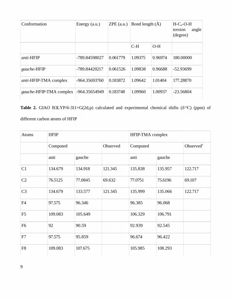
Conformation Energy (a.u.) ZPE (a.u.) Bond length (Å) H-Cα-O-H torsion angle (degree)
C-H O-H
anti-HFIP -789.84598027 0.061779 1.09375 0.96974 180.00000
gauche-HFIP -789.84420217 0.061526 1.09838 0.96688 -52.93699
anti-HFIP-TMA complex -964.35693760 0.183872 1.09642 1.01404 177.28870
gauche-HFIP-TMA complex -964.35654949 0.183748 1.09960 1.00937 -23.56804
Table 2. GIAO B3LYP/6-311+G(2d,p) calculated and experimental chemical shifts (δ13C) (ppm) of
different carbon atoms of HFIP
Atoms HFIP HFIP-TMA complex
Computed Observed Computed Observed†
anti gauche anti gauche
C1 134.679 134.918 121.345 135.838 135.957 122.717
C2 76.5125 77.0845 69.632 77.0751 75.6196 69.107
C3 134.679 133.577 121.345 135.999 135.066 122.717
F4 97.575 96.346 96.385 96.068
F5 109.083 105.649 106.329 106.791
F6 92 90.59 92.939 92.545
F7 97.575 95.859 96.674 96.422
F8 109.083 107.675 105.985 108.293
9
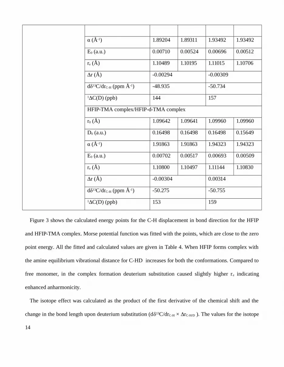
F9 92 95.352 92.978 94.004
O10 28.8499 28.3504 32.0546 33.4956
H11 1.8129 1.634 7.264 9.8827 9.8345 7.359
H12 4.3 4.53 4.397 4.2731 4.2989 4.232
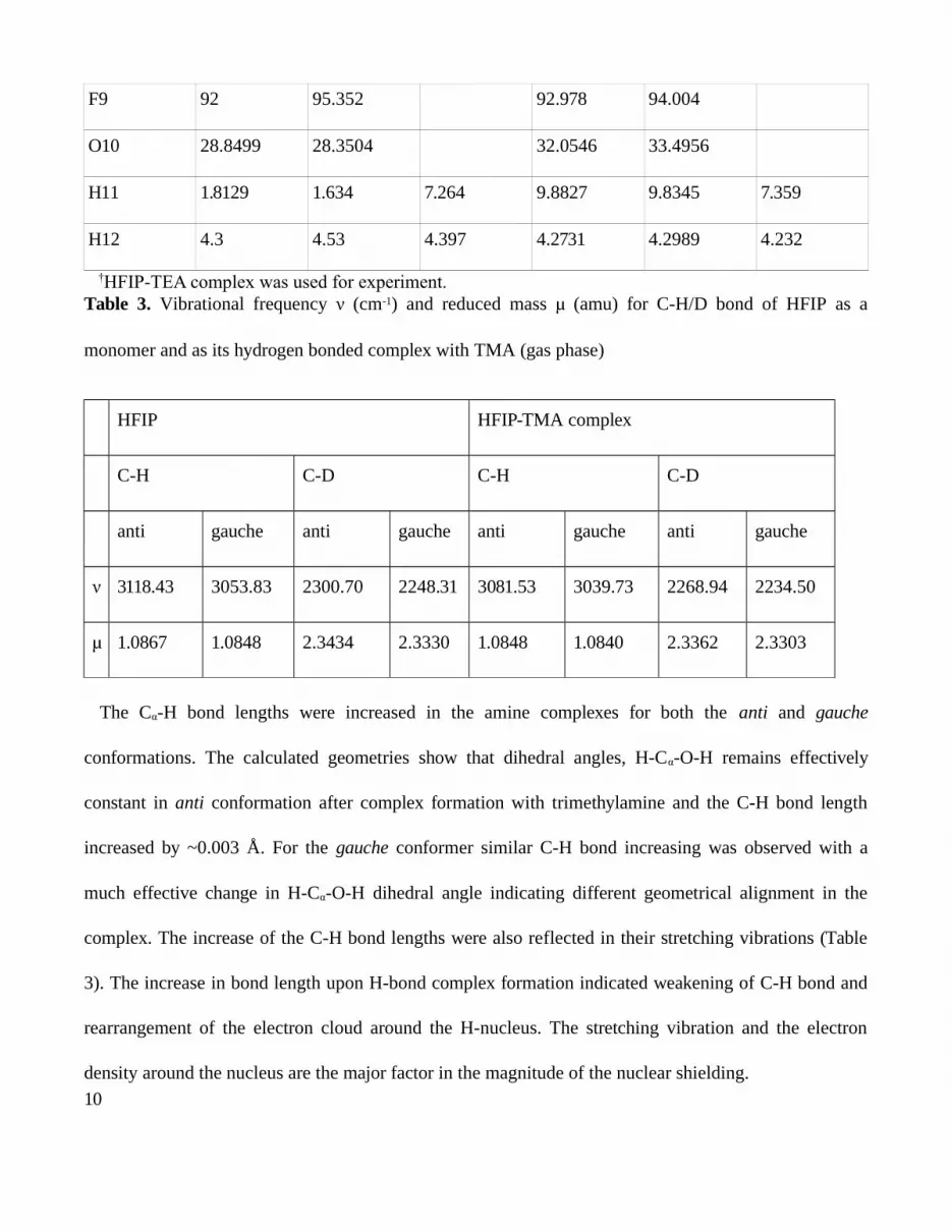
†HFIP-TEA complex was used for experiment.Table 3. Vibrational frequency (cmν -1) and reduced mass (amu) for C-H/D bond of HFIP as aμ
monomer and as its hydrogen bonded complex with TMA (gas phase)
HFIP HFIP-TMA complex
C-H C-D C-H C-D
anti gauche anti gauche anti gauche anti gauche
ν 3118.43 3053.83 2300.70 2248.31 3081.53 3039.73 2268.94 2234.50
μ 1.0867 1.0848 2.3434 2.3330 1.0848 1.0840 2.3362 2.3303
The Cα-H bond lengths were increased in the amine complexes for both the anti and gauche
conformations. The calculated geometries show that dihedral angles, H-Cα-O-H remains effectively
constant in anti conformation after complex formation with trimethylamine and the C-H bond length
increased by ~0.003 Å. For the gauche conformer similar C-H bond increasing was observed with a
much effective change in H-Cα-O-H dihedral angle indicating different geometrical alignment in the
complex. The increase of the C-H bond lengths were also reflected in their stretching vibrations (Table
3). The increase in bond length upon H-bond complex formation indicated weakening of C-H bond and
rearrangement of the electron cloud around the H-nucleus. The stretching vibration and the electron
density around the nucleus are the major factor in the magnitude of the nuclear shielding.
10
3.2. 13C Chemical Shifts and Its Derivatives. The chemical shift is calculated by applying the GIAO
B3LYP method on optimized geometries. The calculated 13C chemical shifts (δ13C) for the three carbon
atoms of HFIP are given in Table 2. Two of the terminal carbon atoms (C1 and C3) of HFIP are of
similar chemical environment and C2 is attached with both C-H and O-H bond and the chemical
environment is quite different for this carbon atom. The calculated chemical shifts for C1 and C3 are
identical for both the conformers. C2 shows slightly higher shielding values for both the anti and gauche
conformations. The chemical shifts for other atoms are also included in the table. However, further
calculation and experiments focused on C2 only.
Calculated chemical shifts for C1 and C3 were very close to each other for both the two conformers.
The values were slightly higher when it formed the complex with the amine. The calculated chemical
shifts are close to the experimental results (Table 2) and provided the confidence in the nuclear shielding
calculation.
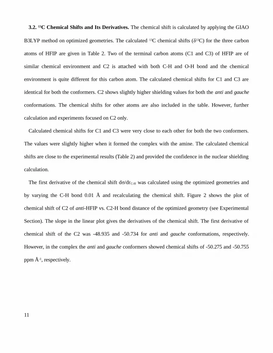
The first derivative of the chemical shift d /drσ C-H was calculated using the optimized geometries and
by varying the C-H bond 0.01 Å and recalculating the chemical shift. Figure 2 shows the plot of
chemical shift of C2 of anti-HFIP vs. C2-H bond distance of the optimized geometry (see Experimental
Section). The slope in the linear plot gives the derivatives of the chemical shift. The first derivative of
chemical shift of the C2 was -48.935 and -50.734 for anti and gauche conformations, respectively.
However, in the complex the anti and gauche conformers showed chemical shifts of -50.275 and -50.755
ppm Å-1, respectively.
11
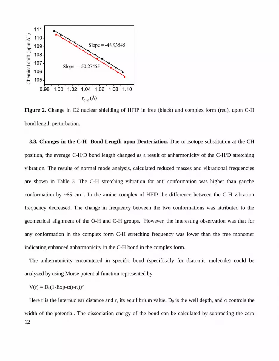
Figure 2. Change in C2 nuclear shielding of HFIP in free (black) and complex form (red), upon C-H
bond length perturbation.
3.3. Changes in the C-H Bond Length upon Deuteriation. Due to isotope substitution at the CH
position, the average C-H/D bond length changed as a result of anharmonicity of the C-H/D stretching
vibration. The results of normal mode analysis, calculated reduced masses and vibrational frequencies
are shown in Table 3. The C-H stretching vibration for anti conformation was higher than gauche
conformation by ~65 cm-1. In the amine complex of HFIP the difference between the C-H vibration
frequency decreased. The change in frequency between the two conformations was attributed to the
geometrical alignment of the O-H and C-H groups. However, the interesting observation was that for
any conformation in the complex form C-H stretching frequency was lower than the free monomer
indicating enhanced anharmonicity in the C-H bond in the complex form.
The anhermonicity encountered in specific bond (specifically for diatomic molecule) could be
analyzed by using Morse potential function represented by
V(r) = D0(1-Exp-α(r-re))2
Here r is the internuclear distance and re its equilibrium value. D0 is the well depth, and α controls the
width of the potential. The dissociation energy of the bond can be calculated by subtracting the zero
12

point energy E0 from the depth of the well. Energies are represented relative to the minimum;
consequently all the energies are positive. A potential energy scan of the C-H bond stretching is
described in the experimental section and shown in Figure 3. By fitting the Morse function the values of
D0, α and re were obtained. The values of the parameters are given in Table 4. E0 values for the anti
HFIP shows 0.00710 a.u. for C-H bond vibration in the ground state and reduced to 0.00524 a.u. for the
deuterium substituted analogue. re was also decreased due to deuteriation. The difference in re due to
deuterium substitution is given by Δr and the value was –0.00294 Å. Similar values were obtained for
the gauche conformation.
Figure 3. Morse potential energy curve for C-H bond length perturbation with zero point energy (ZPE)
correction in HFIP (black) and HFIP-TMA complex.
Table 4. Calculated values of isotope effect on C2 of HFIP in free and when it is associated with
trimethylamine
C-H bond length (Å) Parameters anti gauche
0.90 - 1.40 HFIP/HFIP-d
r0 (Å) 1.09375 1.09348 1.09838 1.09838
D0 (a.u.) 0.17376 0.17357 0.15946 0.15946
13
(Åα -1) 1.89204 1.89311 1.93492 1.93492
E0 (a.u.) 0.00710 0.00524 0.00696 0.00512
re (Å) 1.10489 1.10195 1.11015 1.10706
r (Å)Δ -0.00294 -0.00309
dδ13C/drC-H (ppm Å-1) -48.935 -50.734
1 C(D) (ppb)Δ 144 157
HFIP-TMA complex/HFIP-d-TMA complex
r0 (Å) 1.09642 1.09641 1.09960 1.09960
D0 (a.u.) 0.16498 0.16498 0.16498 0.15649
(Åα -1) 1.91863 1.91863 1.94323 1.94323
E0 (a.u.) 0.00702 0.00517 0.00693 0.00509
re (Å) 1.10800 1.10497 1.11144 1.10830
r (Å)Δ -0.00304 0.00314
dδ13C/drC-H (ppm Å-1) -50.275 -50.755
1 C(D) (ppb)Δ 153 159
Figure 3 shows the calculated energy points for the C-H displacement in bond direction for the HFIP
and HFIP-TMA complex. Morse potential function was fitted with the points, which are close to the zero
point energy. All the fitted and calculated values are given in Table 4. When HFIP forms complex with
the amine equilibrium vibrational distance for C-HD increases for both the conformations. Compared to
free monomer, in the complex formation deuterium substitution caused slightly higher re indicating
enhanced anharmonicity.
The isotope effect was calculated as the product of the first derivative of the chemical shift and the
change in the bond length upon deuterium substitution (dδ13C/drC-H × rΔ C-H/D ). The values for the isotope
14
effects are given in Table 4. As shown in Table 4 that the small changes in the r values causes change in
the isotope effect. The C2 shielding gradient for the free monomer was ~1.3 unit less than when it
formed the complex and the difference in r was ~0.003 Å. However the small perturbations are important
for isotope effect. The deuterium isotope effect increased in the complex, however, the magnitude was
less. For the gauche conformation the calculated deuterium isotope effect on C2 was close to each other
for both the monomer and when it formed the complex, However, there is a trend to higher isotope effect
in complex formation.
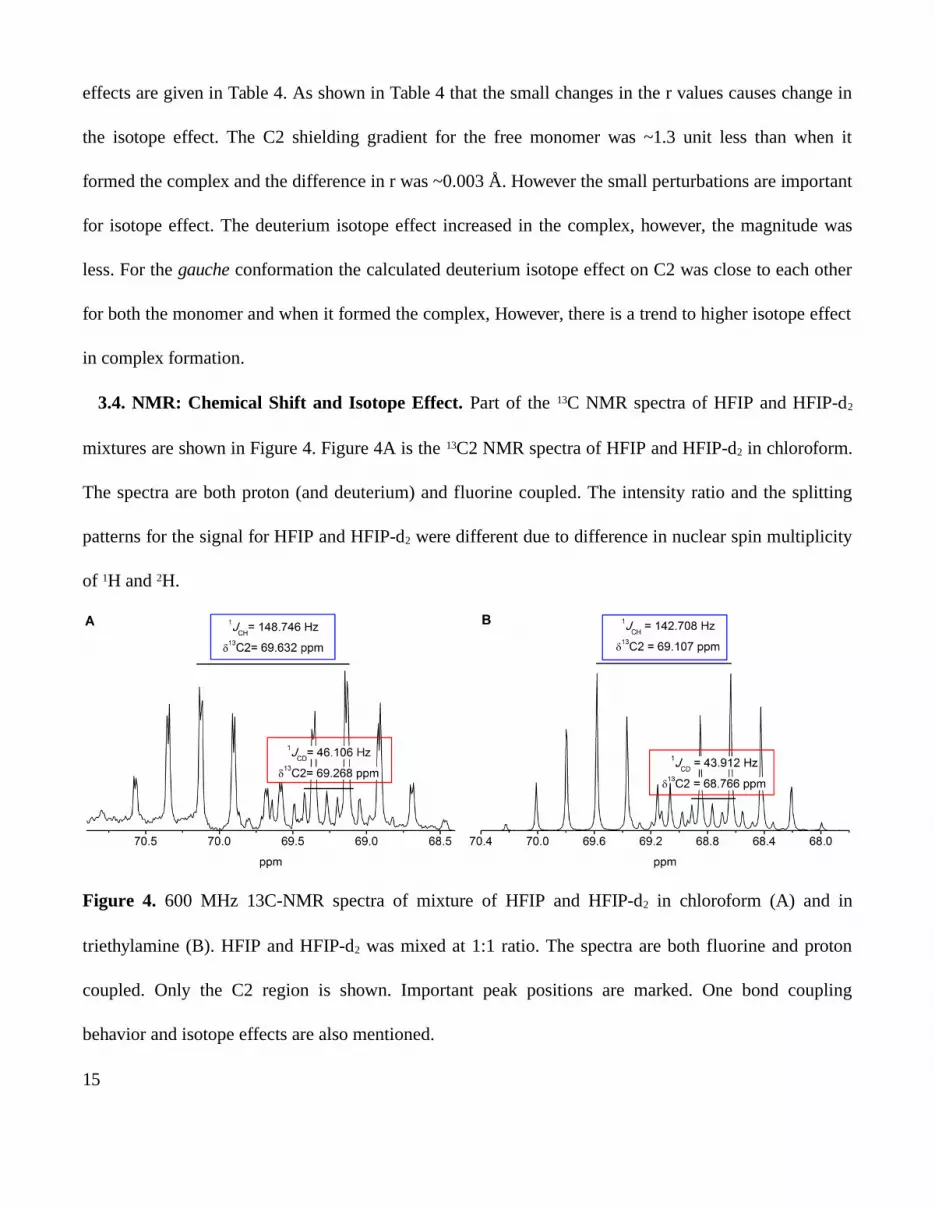
3.4. NMR: Chemical Shift and Isotope Effect. Part of the 13C NMR spectra of HFIP and HFIP-d2
mixtures are shown in Figure 4. Figure 4A is the 13C2 NMR spectra of HFIP and HFIP-d2 in chloroform.
The spectra are both proton (and deuterium) and fluorine coupled. The intensity ratio and the splitting
patterns for the signal for HFIP and HFIP-d2 were different due to difference in nuclear spin multiplicity
of 1H and 2H.
Figure 4. 600 MHz 13C-NMR spectra of mixture of HFIP and HFIP-d2 in chloroform (A) and in
triethylamine (B). HFIP and HFIP-d2 was mixed at 1:1 ratio. The spectra are both fluorine and proton
coupled. Only the C2 region is shown. Important peak positions are marked. One bond coupling
behavior and isotope effects are also mentioned.
15
C2 NMR signal becomes septet (seven splitting) due to coupling with six fluorine atoms attached to
the adjacent carbon atoms (only the dominant five peaks are clearly visible in the Figure 4). Further
coupling with H splits the septet into two groups of signals. Detailed analysis and the coupling pattern
provided the chemical shift for C2 as 69.632 ppm. The coupling constant (1JCH) was 148.746 Hz. In
HFIP-d2, septet of C2 signal was weaker due to substitution of deuterium and each bands in the septet
split into three giving total 21 peak position. However only strong bands could be seen as shown in the
figure. Close analysis of the C2 signal revealed that the chemical shift of C2 of HFIP-d2 was 69.286 and
one bond 1JCD coupling was 46.106 Hz. The difference in the C2 chemical shift values was due to
deuterium isotope effect. This small but certain change was possible to measure confidently by carrying
out the experiment in a single cell and at similar condition.
Figure 4B shows the proton coupled C2 spectra of hydrogen bonded HFIP-amine complex. The
spectrum patterns were similar to the monomers in chloroform. Hydroxyl group (O-H/D) of HFIP/HFIP-
d2 involved in H-bond donation to the tertiary N of triethylamine. In the amine solvent when it formed
complex with the amine, δC2 = 69.107 (HFIP-TEA), δC2=68.766 ppm (HFIP-d2-TEA) and the difference
in chemical shift, ΔδC2 was 341 ppb. The magnitude of the effect in the complex was slightly different
compare to free monomer which was expected as the H-bonding induce more anharmonicity in the
bonding pattern of the attached (either adjacent or further) nucleus.
A few experimental parameters exist that provide direct evidence of individual hydrogen bonds in
complicated biological systems. Usually the existence of hydrogen bonds is inferred from the structure
solved by either X-ray crystallography or NMR. However, mutation technology could help in deuterium
substitution in C-H bond of any specific amino acid participating in enzymatic reaction (e.g., serine in
protein kinases) and could help in measuring the isotope effect, thus, could be useful in determining the
16
strength and nature of the H-bond. The anharmonicity encountered in the potential surface results in
shorter average internuclear separation of the heavier isotopomer. A shorter bonds cause higher
shielding, and hence a shift in the chemical shift.
The chemical shifts for C2 of HFIP and HFIP-d2 were measured in the same experiment and mixing
them in 1:1 ratio (Figure 4). In chloroform solvent the molecules remain as monomer. The isotope effect
was 364 ppb. The gas phase calculation shows isotope effect of 144 ppb for anti conformation and 157
ppb for gauche conformation. The larger isotope effect observed in experimentally because of two
reasons. The calculation done by substituting the single H in the C-H bond by deuterium, however in
experiment C-D and O-D in HFIP-d2 replace both the C-H and O-H. Therefore, the larger isotope effect
was anticipated in the measurement. Triethylamine was used in experiments whereas trimethylamine was
used in calculations. In gas phase calculation, the solvation effect was ignored, which may also have
immense influence on isotope effect.33,34
In HFIP-amine complex hydroxyl group donates hydrogen and the O-H/D bond becomes weaker as it
was reflected in the O-H stretching frequency. Not only the O-H/D bond but also the C-H/D bond
becomes weak and results in decrease of its stretching frequency. It has been recently established both by
quantum mechanical calculation and experiments that the lone pair of O-H/D group may cause overlap
with the C-H σ* (antibonding) orbital (known as negative hyperconjugation) resulting in weakening the
C-H bond strength.14,19
However, the calculation showed higher isotope effect when HFIP form the H-bonded complex.
Marshall pointed out that isotope effect might also occur due to the unequal motion of the centroid of the
electron cloud of the bond and the nucleus.35 For rapid vibration, the electron cloud may move differently
and isotope effect may results. Therefore, the relative movement and the density of the electron cloud
17
becomes important factors in isotope effect. The overlapping of the lone pair with of the C-H σ* orbital
therefore also reflect in the deuterium isotope effect.
Theoretical study by Jameson and Osten established many useful relationships between the isotope
effect and other bonding parameters.36 Nuclear shielding depends on bond length and bond angles. The
bond vibration and rotation lead to different average shift of their equilibrium positions. Therefore, the
bond displacement becomes an important parameter in isotope effect.
The isotope effect may be caused directly by the effect of isotope on nuclear shielding or indirectly by
the fact that substitution cause a change in the chemical equilibrium and consequently the nuclear
shielding. Theoretically we observed that upon complex formation the re increased and the difference in
vibrational equilibrium position also changed due to deuterium substitution. This was reflected in the H-
bonded complex model in which O-H act as the proton donor to the amine bases and could account for
the extra isotope effect on C2.
The change in the shielding for different conformation and upon H-bond complex formation was not
very dramatic with respect to absolute magnitude of the shielding constants. However the change in the
equilibrium position played an important role in the isotope effect.
4. CONCLUSIONS
To realize H-bonding property of a specific O-H group, the study aimed to capture and utilize unique
anomeric effect (negative hyperconjugation) of the O-H group on the Cα-H/D bond ( *). The deuteriumσ
substitution specifies the position. In addition to specificity it also provide an unique observable
(physical parameter), the isotope effect which carries the information of H-bond properties and resultant
anharmonicity. The dominant factor for the deuterium isotope effect was the change in the average
vibrational distance upon deuteriation. The enhanced isotope effect in the H-bonded complex was due to
18
enhanced anharmonicity which caused larger vibrational equilibrium distance. Change in vibrational
equilibrium distance and the deuterium isotope effect could be used as the parameter in monitoring the
strength of the H-bond in small model system with promising application in bio-macromolecules. The
presence of a single hydrogen bond at an enzyme active site can be crucial to catalysis and the discussed
parameters may be unique to determine the energetic contribution of individual hydrogen bonds to
binding and catalysis.
ASSOCIATED CONTENT
Supporting Information. Optimized coordinates for anti- and gauche-HFIP and their complexes with
TMA. This material is available free of charge via the Internet at http://pubs.acs.org.
AUTHOR INFORMATION
Corresponding Author
*Nakul Chandra Maiti: phone, +91-33-2499-5940; fax, +91-33-2473-5197; email, [email protected].
Author Contributions
The manuscript was written through contributions of all authors. All authors have given approval to the
final version of the manuscript.
Funding Sources
This work was supported by grants GENESIS (BSC0121) and Structural Biology (BSC0113) from the
Council of Scientific & Industrial Research (CSIR) and GAP299 from Department of Biotechnology
(DBT). The funders have no role in study design, data collection and analysis, decision to publish, or
preparation of the manuscript.
19
Notes
The authors declare no competing financial interest.
ACKNOWLEDGMENT
We sincerely thank Dr. Vernon E. Anderson whose encouragement helped to initiate this study. Uttam
Pal thanks INSPIRE fellowship programme, Department of Science and Technology, Government of
India, India for financial support. Sudeshna Sen thanks Council of Scientific & Industrial Research,
India for financial support.
ABBREVIATIONS
HFIP, 1,1,1,3,3,3-hexafluoroisopropanol; HFIP-d, 2-deuterio-1,1,1,3,3,3-hexafluoroisopropanol; HFIP-d2,
2-deuterio-2-deuteriooxy-1,1,1,3,3,3-hexafluoropropane; TEA, triethylamine; TMA, trimethylamine;
DFT, density functional theory; B3LYP, Becke, 3-parameter, Lee-Yang-Parr; GIAO, gauge independent
atomic orbital.
REFERENCES
(1) Jeffrey, G. A.; Saenger, W. Hydrogen Bonding in Biological Structures; Springer-Verlag, 1994.(2) Perrin, C. L.; Nielson, J. B. “strong” Hydrogen Bonds in Chemistry and Biology. Annu. Rev. Phys.
Chem. 1997, 48, 511–544.(3) Fersht, A. Enzyme Structure and Mechanism; W. H. Freeman & Company, 1977.(4) Fersht, A. R. The Hydrogen Bond in Molecular Recognition. Trends Biochem. Sci. 1987, 12, 301–
304.(5) Hibbert, F.; Emsley, J. Hydrogen Bonding and Chemical Reactivity. In Advances in Physical
Organic Chemistry; Academic Press Limited: London, 1991; Vol. 26, pp. 255–379.(6) Deshmukh, M. M.; Gadre, S. R. Estimation of N H···O C Intramolecular Hydrogen Bond Energy− ═
in Polypeptides. J. Phys. Chem. A 2009, 113, 7927–7932.(7) Wendler, K.; Thar, J.; Zahn, S.; Kirchner, B. Estimating the Hydrogen Bond Energy. J. Phys. Chem.
A 2010, 114, 9529–9536.(8) Hricovíni, M.; Malkina, O. L.; Bízik, F.; Nagy, L. T.; Malkin, V. G. Calculation of NMR Chemical
Shifts and Spin Spin Coupling Constants in the Monosaccharide Methyl- -D-Xylopyranoside− Β Using a Density Functional Theory Approach†. J. Phys. Chem. A 1997, 101, 9756–9762.
20
(9) Hankache, J.; Hanss, D.; Wenger, O. S. Hydrogen-Bond Strengthening upon Photoinduced Electron Transfer in Ruthenium–Anthraquinone Dyads Interacting with Hexafluoroisopropanol or Water. J. Phys. Chem. A 2012, 116, 3347–3358.
(10) Guo, J.; Tolstoy, P. M.; Koeppe, B.; Golubev, N. S.; Denisov, G. S.; Smirnov, S. N.; Limbach, H.-H. Hydrogen Bond Geometries and Proton Tautomerism of Homoconjugated Anions of Carboxylic Acids Studied via H/D Isotope Effects on 13C NMR Chemical Shifts. J. Phys. Chem. A 2012, 116, 11180–11188.
(11) Rozenberg, M.; Shoham, G.; Reva, I.; Fausto, R. Low-Temperature Fourier Transform Infrared Spectra and Hydrogen Bonding in Polycrystalline L-Alanine. Spectrochim. Acta. A. Mol. Biomol. Spectrosc. 2003, 59, 3253–3266.
(12) Tonge, P. J.; Fausto, R.; Carey, P. R. FTIR Studies of Hydrogen Bonding between , -UnsaturatedΑ β Esters and Alcohols. J. Mol. Struct. 1996, 379, 135–142.
(13) D’Alva Torres, G. S. F.; Pouchan, C.; Teixeira-Dias, J. J. C.; Fausto, R. Hydrogen Bonding Between Substituted Phenols and CH3COOCH3 or CH2CICOOCH3: An FTIR Spectroscopic Study. Spectrosc. Lett. 1993, 26, 913–922.
(14) Maiti, N. C.; Carey, P. R.; Anderson, V. E. Correlation of an Alcohol’s C D Stretch withα − Hydrogen Bond Strength in Complexes with Amines. J. Phys. Chem. A 2003, 107, 9910–9917.
(15) Demmel, F.; Doster, W.; Petry, W.; Schulte, A. Vibrational Frequency Shifts as a Probe of Hydrogen Bonds: Thermal Expansion and Glass Transition of Myoglobin in Mixed Solvents. Eur. Biophys. J. EBJ 1997, 26, 327–335.
(16) Fersht, A. R.; Shi, J.-P.; Knill-Jones, J.; Lowe, D. M.; Wilkinson, A. J.; Blow, D. M.; Brick, P.; Carter, P.; Waye, M. M. Y.; Winter, G. Hydrogen Bonding and Biological Specificity Analysed by Protein Engineering. Nature 1985, 314, 235–238.
(17) Fersht, A. R. Relationships between Apparent Binding Energies Measured in Site-Directed Mutagenesis Experiments and Energetics of Binding and Catalysis. Biochemistry (Mosc.) 1988, 27, 1577–1580.
(18) Maiti, N. C.; Zhu, Y.; Carmichael, I.; Serianni, A. S.; Anderson, V. E. 1JCH Correlates with Alcohol Hydrogen Bond Strength. J. Org. Chem. 2006, 71, 2878–2880.
(19) Jarmelo, S.; Maiti, N.; Anderson, V.; Carey, P. R.; Fausto, R. C H Bond-Stretching Frequency inα− Alcohols as a Probe of Hydrogen-Bonding Strength: A Combined Vibrational Spectroscopic and Theoretical Study of N-[1-D]Propanol. J. Phys. Chem. A 2005, 109, 2069–2077.
(20) Gawlita, E.; Lantz, M.; Paneth, P.; Bell, A. F.; Tonge, P. J.; Anderson, V. E. H-Bonding in Alcohols Is Reflected in the C H Bond Strength: Variation of C D Vibrational Frequency andα− − Fractionation Factor. J. Am. Chem. Soc. 2000, 122, 11660–11669.
(21) Anderson, V. E. Quantifying Energetic Contributions to Ground State Destabilization. Arch. Biochem. Biophys. 2005, 433, 27–33.
(22) Chesnut, D. B.; Foley, C. K. Chemical Shifts and Bond Modification Effects for Some Small First‐row atom Molecules. ‐ J. Chem. Phys. 1986, 84, 852–861.
(23) Barfield, M.; Fagerness, P. Density Functional Theory/GIAO Studies of the 13C, 15N, and 1H NMR Chemical Shifts in Aminopyrimidines and Aminobenzenes: Relationships to Electron Densities and Amine Group Orientations. J. Am. Chem. Soc. 1997, 119, 8699–8711.
(24) De Dios, A. C. Ab Initio Calculations of the NMR Chemical Shift. Prog. Nucl. Magn. Reson. Spectrosc. 1996, 29, 229–278.
21
(25) Munch, M.; Hansen, A. E.; Hansen, P. E.; Bouman, T. D.; Abildgaard, F.; Led, J. J.; Christensen, S. B. Ab Initio Calculations of Deuterium Isotope Effects on Hydrogen and Nitrogen Nuclear Magnetic Shielding in the Hydrated Ammonium Ion. Acta Chem. Scand. 1992, 46, 1065–1071.
(26) Murto, J.; Kivinen, A.; Viitala, R.; Hyömäki, J. Fluoroalcohols—XX: Infrared and Raman Spectra of Hexafluoro-2-Propanol and Its Deuterated Analogues. Spectrochim. Acta Part Mol. Spectrosc. 1973, 29, 1121–1137.
(27) Wolinski, K.; Hinton, J. F.; Pulay, P. Efficient Implementation of the Gauge-Independent Atomic Orbital Method for NMR Chemical Shift Calculations. J. Am. Chem. Soc. 1990, 112, 8251–8260.
(28) Lampert, H.; Mikenda, W.; Karpfen, A.; Kählig, H. NMR Shieldings in Benzoyl and 2-Hydroxybenzoyl Compounds. Experimental versus GIAO Calculated Data. J. Phys. Chem. A 1997, 101, 9610–9617.
(29) Abildgaard, J.; Bolvig, S.; Hansen, P. E. Unraveling the Electronic and Vibrational Contributions to Deuterium Isotope Effects on 13C Chemical Shifts Using Ab Initio Model Calculations. Analysis of the Observed Isotope Effects on Sterically Perturbed Intramolecular Hydrogen-Bonded O-Hydroxy Acyl Aromatics. J. Am. Chem. Soc. 1998, 120, 9063–9069.
(30) Andersson, M. P.; Uvdal, P. New Scale Factors for Harmonic Vibrational Frequencies Using the B3LYP Density Functional Method with the Triple- Basis Set 6-311+G(d,p). Ζ J. Phys. Chem. A 2005, 109, 2937–2941.
(31) Cheeseman, J. R.; Trucks, G. W.; Keith, T. A.; Frisch, M. J. A Comparison of Models for Calculating Nuclear Magnetic Resonance Shielding Tensors. J. Chem. Phys. 1996, 104, 5497–5509.
(32) Nieto, M. M.; Simmons, L. M. Eigenstates, Coherent States, and Uncertainty Products for the Morse Oscillator. Phys. Rev. A 1979, 19, 438–444.
(33) Homer, J. Solvent Effects on Nuclear Magnetic Resonance Chemical Shifts. Appl. Spectrosc. Rev. 1975, 9, 1–132.
(34) Buckingham, A. D.; Schaefer, T.; Schneider, W. G. Solvent Effects in Nuclear Magnetic Resonance Spectra. J. Chem. Phys. 2004, 32, 1227–1233.
(35) Marshall, T. W. Isotope Shifts in the NMR Spectra of H2, HD and D2 due to Zero-Point Vibration. Mol. Phys. 1961, 4, 61–63.
(36) Jameson, C. J.; Osten, H. J. Theoretical Aspects of Isotope Effects on Nuclear Shielding. In Annual Reports on NMR Spectroscopy; Academic Press: London, 1986; Vol. 17, pp. 1–75.
22
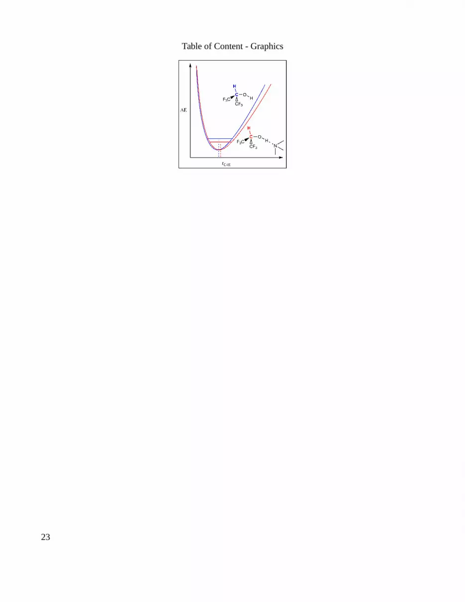
Table of Content - Graphics
23