analysis of anoxybacillus genomes from the aspects of lifestyle adaptations, prophage diversity, and...
TRANSCRIPT

Analysis of Anoxybacillus Genomes from the Aspects ofLifestyle Adaptations, Prophage Diversity, andCarbohydrate MetabolismKian Mau Goh1*, Han Ming Gan2, Kok-Gan Chan3, Giek Far Chan4, Saleha Shahar1, Chun Shiong Chong1,
Ummirul Mukminin Kahar1, Kian Piaw Chai1
1 Faculty of Biosciences and Medical Engineering, Universiti Teknologi Malaysia, Skudai, Johor, Malaysia, 2 Monash School of Science, Monash University Sunway Campus,
Petaling Jaya, Selangor, Malaysia, 3 Division of Genetics and Molecular Biology, Institute of Biological Sciences, Faculty of Science, University of Malaya, Kuala Lumpur,
Malaysia, 4 School of Applied Science, Temasek Polytechnic, Singapore
Abstract
Species of Anoxybacillus are widespread in geothermal springs, manure, and milk-processing plants. The genus is composedof 22 species and two subspecies, but the relationship between its lifestyle and genome is little understood. In this study,two high-quality draft genomes were generated from Anoxybacillus spp. SK3-4 and DT3-1, isolated from Malaysian hotsprings. De novo assembly and annotation were performed, followed by comparative genome analysis with the completegenome of Anoxybacillus flavithermus WK1 and two additional draft genomes, of A. flavithermus TNO-09.006 and A.kamchatkensis G10. The genomes of Anoxybacillus spp. are among the smaller of the family Bacillaceae. Despite havingsmaller genomes, their essential genes related to lifestyle adaptations at elevated temperature, extreme pH, and protectionagainst ultraviolet are complete. Due to the presence of various competence proteins, Anoxybacillus spp. SK3-4 and DT3-1are able to take up foreign DNA fragments, and some of these transferred genes are important for the survival of the cells.The analysis of intact putative prophage genomes shows that they are highly diversified. Based on the genome analysisusing SEED, many of the annotated sequences are involved in carbohydrate metabolism. The presence of glycosylhydrolases among the Anoxybacillus spp. was compared, and the potential applications of these unexplored enzymes aresuggested here. This is the first study that compares Anoxybacillus genomes from the aspect of lifestyle adaptations, thecapacity for horizontal gene transfer, and carbohydrate metabolism.
Citation: Goh KM, Gan HM, Chan K-G, Chan GF, Shahar S, et al. (2014) Analysis of Anoxybacillus Genomes from the Aspects of Lifestyle Adaptations, ProphageDiversity, and Carbohydrate Metabolism. PLoS ONE 9(3): e90549. doi:10.1371/journal.pone.0090549
Editor: Ren Zhang, Wayne State University, United States of America
Received October 14, 2013; Accepted January 31, 2014; Published March 6, 2014
Copyright: � 2014 Goh et al. This is an open-access article distributed under the terms of the Creative Commons Attribution License, which permits unrestricteduse, distribution, and reproduction in any medium, provided the original author and source are credited.
Funding: This study was financially supported by the Universiti Teknologi Malaysia GUP Grant 04H00 and University of Malaya-Ministry of Higher Education HighImpact Research Grant (UMMOHE HIR Grant No. H-50001-A000027). The funders had no role in study design, data collection and analysis, decision to publish, orpreparation of the manuscript.
Competing Interests: The authors have declared that no competing interests exist.
* E-mail: [email protected]
Introduction
The Bacillaceae family remains an important microbial contrib-
utor to industrial biotechnology, mainly due to its abundance of
useful proteins and enzymes [1]. All species within the family are
Gram-positive and in the shape of a rod or coccus. More than
20,000 strains are classified as Bacillaceae, and the genomic
knowledge about its genera is unequally distributed. Based on
the National Center for Bioinformatics Information (NCBI)
database, the accumulated genome sequencing projects registered
for Bacillus and Geobacillus represent more than 99% of the total
sequenced genomes for Bacillaceae. Among the Bacillaceae genomes,
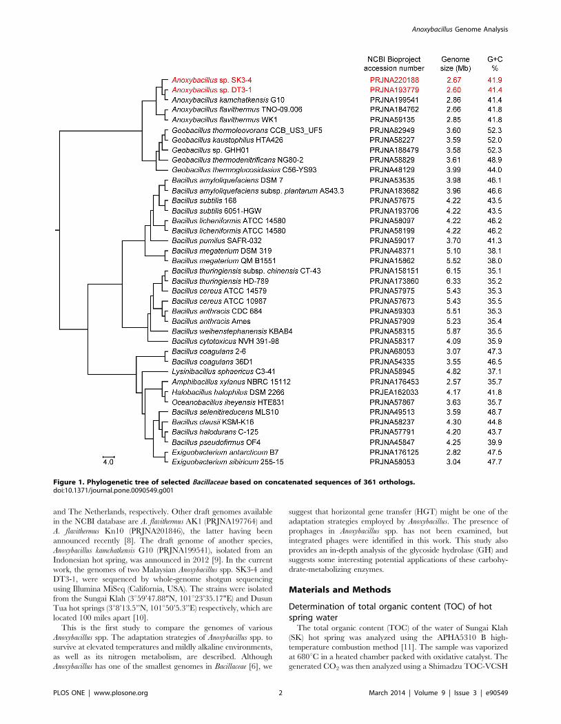
Amphibacillus xylanus NBRC 15112 has the smallest size of 2.57 Mb
(Figure 1) and the lowest total numbers of genes and proteins
(2,489 and 2,411, respectively). On average, Bacillus genomes are
larger in size, with genomes of approximately 5.4 Mb, and with
total open reading frames (ORFs) of more than 5,000 coding
sequences (CDSs). The genome G+C% of Bacillaceae members
does not correlate with their optimum cell growth temperatures
(OGTs). For instance, the average G+C% for thermophilic
Anoxybacillus (OGT 50262uC) is 41.6, and this is lower than that
of the G+C% of mesophilic Halobacillus (OGT 10250uC, G+C%
44.3) and Salimicrobium (OGT 30237uC, G+C% 46.3). Geobacillus
appears to be the closest genus to Anoxybacillus based on the 16S
rRNA phylogeny [1] and concatenated sequence similarity
(Figure 1).
A number of Anoxybacillus spp. have been isolated from around
the world since the first introduction of the genus in 2000 [2]. To
date, a total of 22 species and two subspecies of Anoxybacillus are
described [1,3,4,5]. The cells of Anoxybacillus spp. are generally rod-
shaped and straight or slightly curved, often present in pairs or short
chains, and they form endospores. Interestingly, Anoxybacillus spp.
can be either alkaliphilic or alkalitolerant, and most of them are able
to grow well at neutral pH. Anoxybacillus spp. are moderately
thermophilic (OGT 50262uC), with a slightly lower OGT than
Geobacillus spp. (55265uC). The Anoxybacillus spp. are either aerobes
or facultative anaerobes. Among the Anoxybacillus spp., the genome
of A. flavithermus WK1 (PRJNA59135) remains the only completely
sequenced genome [6]. Recently, the draft genome of Anoxybacillus
flavithermus TNO-09.006 (PRJNA184762) was introduced [7]. Both
strains were isolated from dairy-processing plants in New Zealand
PLOS ONE | www.plosone.org 1 March 2014 | Volume 9 | Issue 3 | e90549
and The Netherlands, respectively. Other draft genomes available
in the NCBI database are A. flavithermus AK1 (PRJNA197764) and
A. flavithermus Kn10 (PRJNA201846), the latter having been
announced recently [8]. The draft genome of another species,
Anoxybacillus kamchatkensis G10 (PRJNA199541), isolated from an
Indonesian hot spring, was announced in 2012 [9]. In the current
work, the genomes of two Malaysian Anoxybacillus spp. SK3-4 and
DT3-1, were sequenced by whole-genome shotgun sequencing
using Illumina MiSeq (California, USA). The strains were isolated
from the Sungai Klah (3u59’47.88"N, 101u23’35.17"E) and Dusun
Tua hot springs (3u8’13.5’’N, 101u50’5.3’’E) respectively, which are
located 100 miles apart [10].
This is the first study to compare the genomes of various
Anoxybacillus spp. The adaptation strategies of Anoxybacillus spp. to
survive at elevated temperatures and mildly alkaline environments,
as well as its nitrogen metabolism, are described. Although
Anoxybacillus has one of the smallest genomes in Bacillaceae [6], we
suggest that horizontal gene transfer (HGT) might be one of the
adaptation strategies employed by Anoxybacillus. The presence of
prophages in Anoxybacillus spp. has not been examined, but
integrated phages were identified in this work. This study also
provides an in-depth analysis of the glycoside hydrolase (GH) and
suggests some interesting potential applications of these carbohy-
drate-metabolizing enzymes.
Materials and Methods
Determination of total organic content (TOC) of hotspring water
The total organic content (TOC) of the water of Sungai Klah
(SK) hot spring was analyzed using the APHA5310 B high-
temperature combustion method [11]. The sample was vaporized
at 680uC in a heated chamber packed with oxidative catalyst. The
generated CO2 was then analyzed using a Shimadzu TOC-VCSH
Figure 1. Phylogenetic tree of selected Bacillaceae based on concatenated sequences of 361 orthologs.doi:10.1371/journal.pone.0090549.g001
Anoxybacillus Genome Analysis
PLOS ONE | www.plosone.org 2 March 2014 | Volume 9 | Issue 3 | e90549
analyzer (Kyoto, Japan). The analyzer measured two paths, one
for total carbon (TC) and one for inorganic carbon, and the TOC
was determined by calculating the difference between the two
values.
Bacterial strains and growth conditionsAnoxybacillus sp. SK3-4 and DT3-1 were grown in modified
Thermus medium (4.0 g/L of peptone, 4.0 g/L of tryptone, 4.0 g/L
of yeast extract, 2.0 g/L of NaCl, and 1.0 g/L of MgSO4N7H2O)
[10], unless specified. The culture was incubated at 55uC with rotary
shaking at 200 rpm. For the experiment on nitrogen utilization in the
medium, the culture was centrifuged at 8000 x g for 15 min at 4uC,
and the cells were washed with 0.5% (w/v) NaCl solution. The cells
were transferred into 100 ml medium (2.0 g/L of NaCl and 1.0 g/L
of MgSO4N7H2O) supplemented with the following nitrogen sources
(4.0 g/L for each medium): tryptone, casamino acids, NaNO3, or
NaNO2, in a 500 ml conical flask. The cell growth was determined
by measuring the optical density at 600 nm (OD600) hourly. All
experiments were performed at least in duplicate.
DNA isolation, sequencing, and annotationThe genomic DNA for Anoxybacillus spp. SK3-4 and DT3-1 was
obtained using the Wizard Genomic DNA Purification kit
(Promega, Wisconsin, USA). Samples were prepared in accor-
dance with the Illumina protocol, and whole-genome shotgun
sequencing was done using Illumina MiSeq platform (California,
USA). De novo assembly and annotations were performed using
CLC Genomics Workbench 4.8 (CLCBio, Aarhus, Denmark) and
Blast2GO [12] programs, respectively. This whole-genome
shotgun sequencing project has been deposited at DDBJ/
EMBL/GenBank under the accession numbers ANOC00000000
and ANMT00000000, respectively, for Anoxybacillus spp. SK3-4
and DT3-1. The versions described in this paper are versions
ANOC01000000 and ANMT01000000. The Bioproject numbers
for Anoxybacillus sp. DT3-1 are PRJNA193779 and PRJNA182115,
and that for Anoxybacillus sp. SK3-4 are PRJNA220188 and
PRJNA174378. To analyze the genomes of other Anoxybacillus
spp., the complete sequences of A. flavithermus WK1 (PRJNA59135)
and A. flavithermus TNO-09.006 (PRJNA184762) and the un-
annotated contigs of A. kamchatkensis G10 (PRJNA199541) were
obtained from NCBI. Other software used in this study included
RNAmmer [13], tRNAscan-SE [14], InterProScan [15], Blast
Ring Image Generator; BRIG [16], PanOCT [17], PHAge Search
Tool; PHAST [18], Rapid Annotations using Subsystems Tech-
nology; RAST [19], SEED [20], and dbCAN carbohydrate-active
enzymes; dbCAN CAZy [21]. For Figure 1, PanOCT was used
to cluster the genes using a 50% identity cutoff and a 70%
minimum aligned length. Using these parameters, a core genome
for Bacillaceae strains across representatives from 361 orthologs was
identified. These orthologs were aligned using ClustalW 2.0 [22]
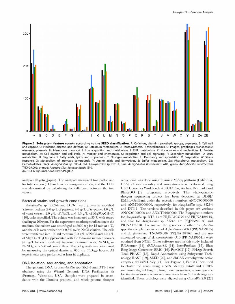
Figure 2. Subsystem feature counts according to the SEED classification. A: Cofactors, vitamins, prosthetic groups, pigments. B: Cell walland capsule. C: Virulence, disease, and defense. D: Potassium metabolism. E: Photosynthesis. F: Miscellaneous. G: Phages, prophages, transposableelements, plasmids. H: Membrane transport. I: Iron acquisition and metabolism. J: RNA metabolism. K: Nucleosides and nucleotides. L: Proteinmetabolism. M: Cell division and cell cycle. N: Motility and chemotaxis. O: Regulation and cell signaling. P: Secondary metabolism. Q: DNAmetabolism. R: Regulons. S: Fatty acids, lipids, and isoprenoids. T: Nitrogen metabolism. U: Dormancy and sporulation. V: Respiration. W: Stressresponse. X: Metabolism of aromatic compounds. Y: Amino acids and derivatives. Z: Sulfur metabolism. ZA: Phosphorus metabolism. ZB:Carbohydrates. Black: Anoxybacillus sp. SK3-4; red: Anoxybacillus sp. DT3-1; blue: Anoxybacillus flavithermus WK1; green: Anoxybacillus flavithermusTNO-09.006; orange: Anoxybacillus kamchatkensis G10.doi:10.1371/journal.pone.0090549.g002
Anoxybacillus Genome Analysis
PLOS ONE | www.plosone.org 3 March 2014 | Volume 9 | Issue 3 | e90549

and eventually concatenated for phylogenomics analysis. The
default settings for PanOCT were used, unless otherwise stated.
The additional information in Figure 1 (i.e., accession numbers,
genome sizes, and G+C%) was obtained from NCBI’s BioProject
database and was added manually to the figure. The SEED
analysis (Figure 2) was done using the RAST database [19,20].
The Venerable package in R was used to construct the five-way
Venn diagram (Figure 3A) using a 50% identity cutoff. PHAST
[18] was used to identify the putative prophages in Anoxybacillus
genomes (Figure 4). To create the phylogenetic tree shown in
Figure 5, unique sequences that were most likely due to
horizontal gene transfer were searched against NCBI’s nucleotide
database, and the most similar sequences were aligned with
ClustalW 2.0. Subsequently, neighbor-joining trees were inferred
using the MEGA5 software [23], and 1000 bootstrap replicates
were performed.
Results and Discussion
General features and comparison of genomes of strainsSK3-4 and DT3-1 with other Anoxybacillus genomes
In comparison to Bacillaceae members such as the Bacillus and
Geobacillus, Anoxybacillus was somehow neglected when it was first
identified as Bacillus flavithermus [1,24]. At present, A. flavithermus
WK1 is the only Anoxybacillus species with a completely sequenced
genome [6], despite the completion of many other genomes from
Bacillus and Geobacillus spp. Recently, several Anoxybacillus spp. were
sequenced using the shotgun approach, and this motivated us to
compare them. So far, no plasmid from these Anoxybacillus spp. has
been examined, though members of Bacillaceae commonly have
one or multiple plasmids. General features of the analyzed
genomes are listed in Table 1. The five genomes fall into the
range of 2.6 to 2.9 Mb, smaller than the closest genus, Geobacillus
(Figure 1). The whole-genome, rRNA and tRNA G+C% across
the Anoxybacillus spp. were more or less similar. The total number
of predicted protein-coding genes in the Anoxybacillus spp. SK3-4
and DT3-1 draft genomes was 2,842 and 2,736, respectively. The
draft genome of A. kamchatkensis G10 had approximately 100 more
protein-coding genes than the completed genome of A. flavithermus
WK1. The average protein length in Anoxybacillus spp. SK3-4 and
DT3-1 was 284 amino acids, which was similar to A. flavithermus
strains WK1 and TNO-09.006, and A. kamchatkensis G10.
Figure 2 shows the SEED analysis using the RAST database
[19,20]. A majority of the protein-encoded sequences functioned
in the catabolic pathways of amino acids and their derivatives and
carbohydrates. The total sequences of A. flavithermus WK1 involved
in the functions of cofactors, vitamins, prosthetic group, pigments,
potassium metabolism, RNA metabolism, cell division and cell
cycle, and amino acids are higher than the other four genomes
(Figure 2). It was initially thought that Anoxybacillus spp. from
natural heated springs would exhibit more stress response
sequences, but unexpectedly, milk processing effluent A. flavithermus
WK1 had the most such genes. In comparison, the total genes for
cell wall and capsule, membrane transport, motility and chemo-
taxis, and DNA metabolism in A. flavithermus WK1 were fewer than
in the other four strains.
All five Anoxybacillus spp. shared the same morphology-related
genes. The Anoxybacillus cells are rod-shaped, and each species
exhibited an operon of three rod-shape-determining proteins
(Anoxybacillus sp. SK3-4: C289_031320315, Anoxybacillus sp. DT3-
1: F510_131021311) and a cell-shape-determining Mbl protein
(C289_1660, F510_1488). The latter protein exhibited similarity
of higher than 80% to Bacillus and Geobacillus rod-shape-
determining protein MreB. Though these proteins have not been
Anoxybacillus Genome Analysis
PLOS ONE | www.plosone.org 4 March 2014 | Volume 9 | Issue 3 | e90549

studied in regard to Anoxybacillus cell morphogenesis, the high level
of similarity suggests a conserved role of the proteins in other
closely related genera within Bacillaceae [25].
The genes that encode the flagellar assembly are complete and
include various ring, motor protein, hook, and filament sequences.
This suggests that the five Anoxybacillus spp. are motile, despite
earlier experimental motility tests that suggested that Anoxybacillus
spp. SK3-4 and DT3-1 were non-motile in certain growth media
[10]. It is unclear at this moment whether Anoxybacillus spp. adopts
polar or peritrichous flagellation because no electron photomicro-
graphs for these five strains or any other Anoxybacillus are available.
A. flavithermus WK1, Anoxybacillus spp. SK3-4 and DT3-1 are spore-
forming bacteria. Sporulation in Bacillus subtilis involves over 500
genes [26,27]. In Anoxybacillus, the total genes involved are fewer
than 100, yet their number still falls within the expected range due
to the size of the genome of Anoxybacillus [27]. The sporulation
feature of Anoxybacillus is one of the proposed reasons why the
bacteria are frequently found in milk powder samples [1].
A Venn diagram (Figure 3A) was used to compare the
orthologous gene complements of the five Anoxybacillus. BRIG data
were generated for strains SK3-4 and DT3-1 as the central
sequence, while other genomes were set as concentric rings
(Figure 3B and 3C). The analysis showed that both strains
exhibited certain regions that were not present in other
Anoxybacillus genomes (Figure 3B and 3C). A part of the unique
regions was contributed by the prophage, while some other
sequences were possibly a result of HGT.
Prophage sequences in Anoxybacillus genomesTo date, there is no detailed report of prophage sequences in the
Anoxybacillus, although the presence of a prophage is acknowl-
edged, at least in A. flavithermus WK1 [6]. Analysis of the five
Anoxybacillus genomes using the PHAST program, [18] identified
at least one complete prophage sequence in all strains except for
A. flavithermus TNO-09.006. Several incomplete prophages or
prophage remnants were also present. For this article, only
putative intact prophages suggested by PHAST will be shown. In
general, all intact prophages in Anoxybacillus spp. SK3-4 and DT3-
Figure 3. Genomes comparison of Anoxybacillus species. (A)Five-way Venn-diagram showing the number of shared and specificCDS among the Anoxybacillus spp. Orthologous groupings were basedon 50% identify cutoff and overlap of at least 70% protein sequencelength. (B) BRIG image with Anoxybacillus sp. SK3-4 genome sequenceset as the central reference. (C) BRIG image with Anoxybacillus sp. DT3-1genome sequence set as the central reference.doi:10.1371/journal.pone.0090549.g003
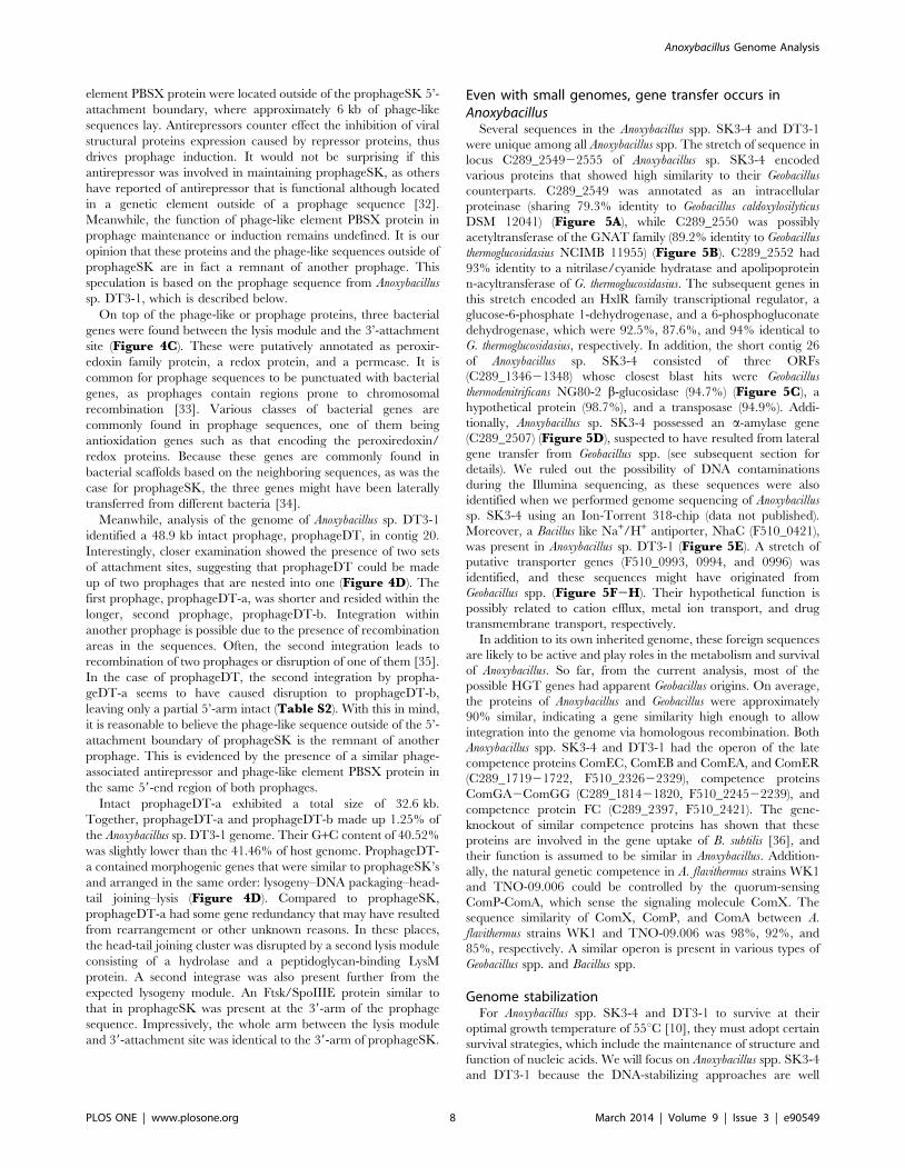
Figure 4. Sequence arrangements of prophages in Anoxybacillus genome as identified by PHAST. (A) ProphageWK of Anoxybacillusflavithermus WK1. (B) ProphageG10 of Anoxybacillus kamchatkensis G10. (C) ProphageSK of Anoxybacillus sp. SK3-4. (D) ProphageDT of Anoxybacillussp. DT3-1. ProphageWK and prophageG10 may not be intact prophages, due to the lack of putative genes encoding morphological proteins. This is incontrast to intact prophageSK and prophageDT, which have more and ordered morphological genes. These morphological genes are arranged inclusters or modules, which is a hallmark of prophage sequences, and in an order typical of temperate tailed-phage genomes. Note that prophageDTis located on the complementary strand of Anoxybacillus sp. DT3-1. The prophage map was reversed for ease of reference. Both prophageSK andprophageDT also share six genes (shown in figure) that appear to be conserved in location and order. The details of ORFs information for prophageSKand prophageDT are provided in Table S1 and Table S2.doi:10.1371/journal.pone.0090549.g004
Anoxybacillus Genome Analysis
PLOS ONE | www.plosone.org 5 March 2014 | Volume 9 | Issue 3 | e90549
1, A. flavithermus WK1, and A. kamchatkensis G10 showed low
conservation (Figure 4). The prophageWK1 and prophageG10
carried a limited number of prophage structural genes that were
interspersed with hypothetical proteins. This could suggest severe
rearrangement that left behind non-functional prophage remnants
despite being identified as intact by PHAST. Therefore, these two
prophages remnants will not be further addressed here.
Determining the relationship between two prophages is not easy
due to the high recombination rate that prophages exhibit.
Attempts to identify a relationship between prophages have been
based on several conserved prophage proteins. Two of these are
the larger subunit of terminase and tail tape measure protein [28].
Based on the low similarities of the two prophage proteins from
Anoxybacillus spp. SK3-4 and DT3-1, we believe that these two
prophages are not related to each other (Tables 2, S1, and S2).
Analysis of phages isolated from both strains would confirm this
hypothesis.
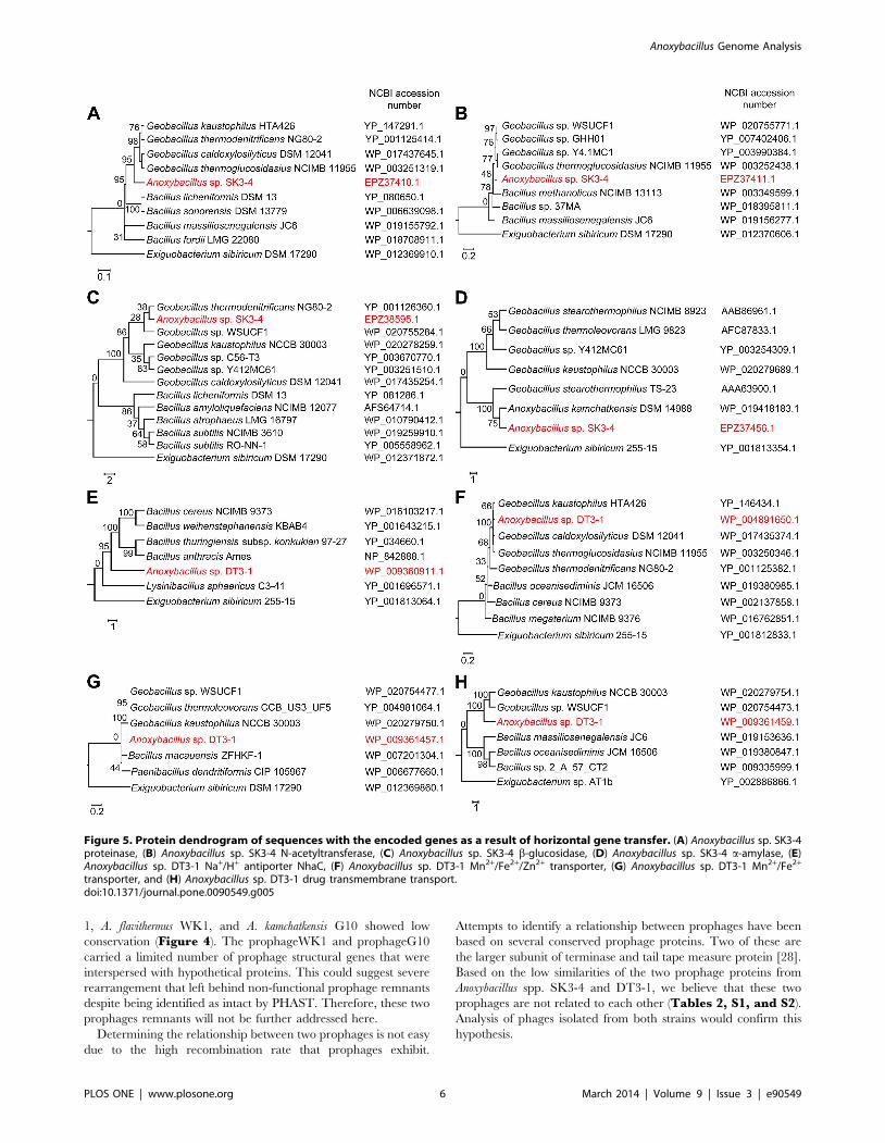
Figure 5. Protein dendrogram of sequences with the encoded genes as a result of horizontal gene transfer. (A) Anoxybacillus sp. SK3-4proteinase, (B) Anoxybacillus sp. SK3-4 N-acetyltransferase, (C) Anoxybacillus sp. SK3-4 b-glucosidase, (D) Anoxybacillus sp. SK3-4 a-amylase, (E)Anoxybacillus sp. DT3-1 Na+/H+ antiporter NhaC, (F) Anoxybacillus sp. DT3-1 Mn2+/Fe2+/Zn2+ transporter, (G) Anoxybacillus sp. DT3-1 Mn2+/Fe2+
transporter, and (H) Anoxybacillus sp. DT3-1 drug transmembrane transport.doi:10.1371/journal.pone.0090549.g005
Anoxybacillus Genome Analysis
PLOS ONE | www.plosone.org 6 March 2014 | Volume 9 | Issue 3 | e90549
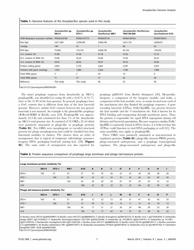
The intact prophage sequence from Anoxybacillus sp. SK3-4,
prophageSK, was identified in contig 46 with a G+C% of 41.77,
close to the 41.89 of the host genome. In general, prophages have
a G+C content that is different from that of the host bacterial
genome. However, similar G+C content between the two genetic
materials is not unusual. An example is the plasmid-like prophage
vB-BcesS-IEBH of Bacillus cereus [29]. ProphageSK was approx-
imately 25.4 kb and constituted less than 1% of the Anoxybacillus
sp. SK3-4 total genome size. It consisted of 52 ORFs, 22 of which
were putatively annotated as phage-like or prophage proteins
(Table S1). Ten ORFs were morphogenic or encoded structural
proteins for phage morphogenesis and could be classified into four
functional modules or clusters. The clusters show an order of
arrangement that is typical of temperate tailed-phage genomes:
lysogeny–DNA packaging–head-tail joining–lysis [28] (Figure4C). The same order of arrangement was also reported for
prophage phIS3501 from Bacillus thuringiensis [30]. Meanwhile,
integrase, a component of the lysogeny module, and holin, a
component of the lysis module, were, as usual, located near each of
the attachment sites that flanked the prophage sequence. A gene
encoding bacterial ATPase, FtsK/SpoIIIE, was present between
the lysis module and the 3’-attachment site. Ftsk is known for its
DNA binding and transporting through membrane pores. Thus,
the protein is responsible for equal DNA segregation during cell
division and bacterial sporulation. Because sequence-similar FtsK/
SpoIIIE is commonly found in DNA viruses, it is believed that this
ATPase could function in viral DNA packaging as well [31]. The
same possibility may apply to prophageSK.
Three ORFs were putatively annotated as non-structural or
regulatory proteins (Table S1): phage-like element PBSX protein,
phage-associated antirepressor, and a prophage transcriptional
regulator. The phage-associated antirepressor and phage-like
Table 1. Genome features of the Anoxybacillus species used in this study.
Anoxybacillus sp.SK3-4
Anoxybacillus sp.DT3-1
Anoxybacillusflavithermus WK1
Anoxybacillus flavithermusTNO-09.006
Anoxybacilluskamchatkensis G10
NCBI Bioproject accession number PRJNA220188 PRJNA193779 PRJNA59135 PRJNA184762 PRJNA199541
Size (bp) 2,671,589 2,596,638 2,846,746 2,651,725 2,858,657
Contigs 148 52 1 68 65
N50 (bp) 75,006 153,724 2,846,746 65,120 130,036
G+C content (%) 41.89 41.46 41.78 41.82 41.35
G+C content of rRNA (%) 55.80 56.24 55.68 57.66 56.33
G+C content of tRNA (%) 59.43 60.03 59.47 59.33 59.40
Protein coding genes 2,842 2,736 2,863 2,595 2,953
Proteins with gene ontology 1,935 1,884 2,526 1,796 1,996
Total rRNA genes 3 5 24 10 6
Total tRNA genes 71 57 77 62 56
Reference This study This study [6] [7] [9]
doi:10.1371/journal.pone.0090549.t001
Table 2. Protein sequence comparison of prophage large terminase and phage tail-measure protein.
Large terminase protein similarity (%)
SK3-4 DT3-1 WK1 G10 A B C D E F G H I
SK3-4 100 27 22 27 55 55 56 22 22 29 28 28 26
DT3-1 100 23 99 22 22 23 25 25 64 62 94 63
WK1 100 24 20 20 20 91 91 23 24 22 22
G10 100 22 22 23 25 25 64 62 94 63
Phage tail-measure protein similarity (%)
SK3-4 DT3-1 WK1 G10 J K L M N F G O P
SK3-4 100 14 31 26 12 32 15 20 31 41 26 43 35
DT3-1 100 15 13 45 12 24 10 11 15 14 18 13
WK1 100 24 17 51 21 34 24 48 39 53 30
G10 100 12 17 43 14 37 35 49 36 22
A: Bacillus cereus VD154 (gi|446509997). B: Bacillus cereus AH1272 (gi|488006653). C: Bacillus thuringiensis (gi|384182753). D: Bacillus virus 1 (gi|155042934). E: Geobacillusphage GBSV1 (gi|115334627). F: Geobacillus thermoglucosidasius C56-YS93 (gi|336234248). G: Geobacillus sp. Y412MC52 (gi|261418101). H: Geobacillus sp. Y4.1MC1(gi|312109743). I: Paenibacillus mucilaginosus K02 (gi|511629193). J: Bacillus cereus AH1271 (gi|488002245). K: Bacillus cereus VD156 (gi|446819371). L: Bacillus sonorensisL12 (gi|493686871). M: Bacillus methanolicus PB1 (gi|490573477). N: Geobacillus kaustophilus HTA426 (gi|56419074). O: Geobacillus virus E2 (gi|148747742). P: Clostridiumljungdahlii DSM 13528 (gi|300853566).doi:10.1371/journal.pone.0090549.t002
Anoxybacillus Genome Analysis
PLOS ONE | www.plosone.org 7 March 2014 | Volume 9 | Issue 3 | e90549
element PBSX protein were located outside of the prophageSK 5’-
attachment boundary, where approximately 6 kb of phage-like
sequences lay. Antirepressors counter effect the inhibition of viral
structural proteins expression caused by repressor proteins, thus
drives prophage induction. It would not be surprising if this
antirepressor was involved in maintaining prophageSK, as others
have reported of antirepressor that is functional although located
in a genetic element outside of a prophage sequence [32].
Meanwhile, the function of phage-like element PBSX protein in
prophage maintenance or induction remains undefined. It is our
opinion that these proteins and the phage-like sequences outside of
prophageSK are in fact a remnant of another prophage. This
speculation is based on the prophage sequence from Anoxybacillus
sp. DT3-1, which is described below.
On top of the phage-like or prophage proteins, three bacterial
genes were found between the lysis module and the 3’-attachment
site (Figure 4C). These were putatively annotated as peroxir-
edoxin family protein, a redox protein, and a permease. It is
common for prophage sequences to be punctuated with bacterial
genes, as prophages contain regions prone to chromosomal
recombination [33]. Various classes of bacterial genes are
commonly found in prophage sequences, one of them being
antioxidation genes such as that encoding the peroxiredoxin/
redox proteins. Because these genes are commonly found in
bacterial scaffolds based on the neighboring sequences, as was the
case for prophageSK, the three genes might have been laterally
transferred from different bacteria [34].
Meanwhile, analysis of the genome of Anoxybacillus sp. DT3-1
identified a 48.9 kb intact prophage, prophageDT, in contig 20.
Interestingly, closer examination showed the presence of two sets
of attachment sites, suggesting that prophageDT could be made
up of two prophages that are nested into one (Figure 4D). The
first prophage, prophageDT-a, was shorter and resided within the
longer, second prophage, prophageDT-b. Integration within
another prophage is possible due to the presence of recombination
areas in the sequences. Often, the second integration leads to
recombination of two prophages or disruption of one of them [35].
In the case of prophageDT, the second integration by propha-
geDT-a seems to have caused disruption to prophageDT-b,
leaving only a partial 5’-arm intact (Table S2). With this in mind,
it is reasonable to believe the phage-like sequence outside of the 5’-
attachment boundary of prophageSK is the remnant of another
prophage. This is evidenced by the presence of a similar phage-
associated antirepressor and phage-like element PBSX protein in
the same 59-end region of both prophages.
Intact prophageDT-a exhibited a total size of 32.6 kb.
Together, prophageDT-a and prophageDT-b made up 1.25% of
the Anoxybacillus sp. DT3-1 genome. Their G+C content of 40.52%
was slightly lower than the 41.46% of host genome. ProphageDT-
a contained morphogenic genes that were similar to prophageSK’s
and arranged in the same order: lysogeny–DNA packaging–head-
tail joining–lysis (Figure 4D). Compared to prophageSK,
prophageDT-a had some gene redundancy that may have resulted
from rearrangement or other unknown reasons. In these places,
the head-tail joining cluster was disrupted by a second lysis module
consisting of a hydrolase and a peptidoglycan-binding LysM
protein. A second integrase was also present further from the
expected lysogeny module. An Ftsk/SpoIIIE protein similar to
that in prophageSK was present at the 39-arm of the prophage
sequence. Impressively, the whole arm between the lysis module
and 39-attachment site was identical to the 39-arm of prophageSK.
Even with small genomes, gene transfer occurs inAnoxybacillus
Several sequences in the Anoxybacillus spp. SK3-4 and DT3-1
were unique among all Anoxybacillus spp. The stretch of sequence in
locus C289_254922555 of Anoxybacillus sp. SK3-4 encoded
various proteins that showed high similarity to their Geobacillus
counterparts. C289_2549 was annotated as an intracellular
proteinase (sharing 79.3% identity to Geobacillus caldoxylosilyticus
DSM 12041) (Figure 5A), while C289_2550 was possibly
acetyltransferase of the GNAT family (89.2% identity to Geobacillus
thermoglucosidasius NCIMB 11955) (Figure 5B). C289_2552 had
93% identity to a nitrilase/cyanide hydratase and apolipoprotein
n-acyltransferase of G. thermoglucosidasius. The subsequent genes in
this stretch encoded an HxlR family transcriptional regulator, a
glucose-6-phosphate 1-dehydrogenase, and a 6-phosphogluconate
dehydrogenase, which were 92.5%, 87.6%, and 94% identical to
G. thermoglucosidasius, respectively. In addition, the short contig 26
of Anoxybacillus sp. SK3-4 consisted of three ORFs
(C289_134621348) whose closest blast hits were Geobacillus
thermodenitrificans NG80-2 b-glucosidase (94.7%) (Figure 5C), a
hypothetical protein (98.7%), and a transposase (94.9%). Addi-
tionally, Anoxybacillus sp. SK3-4 possessed an a-amylase gene
(C289_2507) (Figure 5D), suspected to have resulted from lateral
gene transfer from Geobacillus spp. (see subsequent section for
details). We ruled out the possibility of DNA contaminations
during the Illumina sequencing, as these sequences were also
identified when we performed genome sequencing of Anoxybacillus
sp. SK3-4 using an Ion-Torrent 318-chip (data not published).
Moreover, a Bacillus like Na+/H+ antiporter, NhaC (F510_0421),
was present in Anoxybacillus sp. DT3-1 (Figure 5E). A stretch of
putative transporter genes (F510_0993, 0994, and 0996) was
identified, and these sequences might have originated from
Geobacillus spp. (Figure 5F2H). Their hypothetical function is
possibly related to cation efflux, metal ion transport, and drug
transmembrane transport, respectively.
In addition to its own inherited genome, these foreign sequences
are likely to be active and play roles in the metabolism and survival
of Anoxybacillus. So far, from the current analysis, most of the
possible HGT genes had apparent Geobacillus origins. On average,
the proteins of Anoxybacillus and Geobacillus were approximately
90% similar, indicating a gene similarity high enough to allow
integration into the genome via homologous recombination. Both
Anoxybacillus spp. SK3-4 and DT3-1 had the operon of the late
competence proteins ComEC, ComEB and ComEA, and ComER
(C289_171921722, F510_232622329), competence proteins
ComGA2ComGG (C289_181421820, F510_224522239), and
competence protein FC (C289_2397, F510_2421). The gene-
knockout of similar competence proteins has shown that these
proteins are involved in the gene uptake of B. subtilis [36], and
their function is assumed to be similar in Anoxybacillus. Addition-
ally, the natural genetic competence in A. flavithermus strains WK1
and TNO-09.006 could be controlled by the quorum-sensing
ComP-ComA, which sense the signaling molecule ComX. The
sequence similarity of ComX, ComP, and ComA between A.
flavithermus strains WK1 and TNO-09.006 was 98%, 92%, and
85%, respectively. A similar operon is present in various types of
Geobacillus spp. and Bacillus spp.
Genome stabilizationFor Anoxybacillus spp. SK3-4 and DT3-1 to survive at their
optimal growth temperature of 55uC [10], they must adopt certain
survival strategies, which include the maintenance of structure and
function of nucleic acids. We will focus on Anoxybacillus spp. SK3-4
and DT3-1 because the DNA-stabilizing approaches are well
Anoxybacillus Genome Analysis
PLOS ONE | www.plosone.org 8 March 2014 | Volume 9 | Issue 3 | e90549
conserved in A. flavithermus WK1, A. flavithermus TNO-09.006, and
A. kamchatkensis G10, unless specified. Preserving the secondary
structure of DNA in vivo is usually related to the G+C% and the
torsion constraints of the two DNA strands. The G+C% of
Anoxybacillus spp. SK3-4 and DT3-1 was 41.89% and 41.46%,
respectively, which was lower than expected. However, the G+C%
of rRNA sequences of Anoxybacillus spp. SK3-4 and DT3-1 were
55.80% and 56.24%, respectively, which were higher than
expected. Interestingly, the G+C% of tRNA sequences of
Anoxybacillus spp. SK3-4 and DT3-1 were also higher than
expected, 59.43% and 60.03%, respectively. Similar trends were
observed for A. flavithermus WK1, A. flavithermus TNO-09.006, and
A. kamchatkensis G10 (Table 1). As reviewed by Trivedi et al., the
G+C% in rRNA and tRNA sequences are higher in thermophiles,
though a lack of consistency in this high G+C% in protein-coding
genes of thermophiles was noted [37]. Principle component
analysis (PCA) of Geobacillus kaustophilus and other Bacillus-related
species has revealed that high G+C% of rRNA is one possible
thermophilic signature for Geobacillus [38]. High G+C% could
result in protection of DNA from thermodegradation, though this
alone could not provide thermal stability to DNA [37].
Another reason why Anoxybacillus is able to live in hot springs is
due to the presence of reverse gyrase. Anoxybacillus spp. SK3-4 and
DT3-1 were found to have a gene coding for primosome assembly
protein (PriA; C289_0740, F510_0088). Reverse gyrase, a type 1A
topoisomerase, is a common feature of hyperthermophilic Archaea
and Eubacteria that is not present in mesophiles, and the enzyme
maintains the genome stability at high temperature [37,39].
Furthermore, positive supercoiling is able to stabilize the
secondary structure of DNA. Future biochemical and functional
confirmation via gene-knockout may provide greater insight into
the role of this protein as a possible trait of Anoxybacillus in
thermoadaptation. Whether reverse gyrase is a prerequisite for
thermophiles such as Anoxybacillus or Geobacillus remains unknown.
In fact, there is no accepted explanation why reverse gyrase is
important to thermophiles [40]. We think that no single factor
could be responsible for the thermophily of Anoxybacillus. There-
fore, how Anoxybacillus copes with the hot spring environment
should be addressed.
Polyamines are positively charged compounds that can bind to
DNA and RNA and stabilize the latter [41]. Anoxybacillus spp.
SK3-4 and DT3-1 possessed the biosynthesis pathway of linear
polyamines, such as spermidine and spermine. Genome annota-
tions revealed the presence of genes coding for spermidine
synthases (C289_0840, F510_0514) and spermidine transport
proteins (C289_0889, C289_0890, C289_0891, C289_0892;
F510_563, F510_564, F510_565, F510_566). The enzymes were
mapped to Kyoto Encyclopedia of Genes and Genomes, KEGG
pathways (map 330), which indicated that spermidine and
spermine contributed to the thermoadaptation traits of both
strains. The genome of Anoxybacillus spp. SK3-4 and DT3-1 also
revealed the presence of a gene coding agmatinase (C289_839,
F510_513), which is involved in the production of agmatine. This
corroborates the polyamine profiling by high performance liquid
chromatography (HPLC) and gas chromatography (GC) that has
revealed that spermidine and spermine are the major polyamines
of A. flavithermus, Anoxybacillus gonensis, and Anoxybacillus voinovskiensis
cultured at 50265uC [42]. Agmatine is also detected in A.
flavithermus and A. voinovskiensis. Additionally, putrescine and
thermopentamine are found in A. voinovskiensis [42].
The presence of histone-like proteins in Anoxybacillus spp. SK3-4
and DT3-1 may also provide thermal stability to their genomes.
Both strains have the gene that codes for DNA-binding protein
HU-alpha (C289_1583, F510_2065), which is a 90-amino-acid
histone-like protein. Coded by the hupA gene, the protein in
Anoxybacillus spp. is hypothesized to be involved in wrapping DNA
to stabilize it against denaturation and degradation under extreme
environmental conditions. The DNA-binding protein HU from
hyperthermophilic Thermotoga maritime bends DNA and constrains
negative DNA supercoils in the presence of topoisomerase I. This
unique architectural function may generate stable and compact
aggregates that organize and protect genomic DNA [43].
DNA repairAnoxybacillus spp. SK3-4 and DT3-1 exploits almost all known
DNA repair mechanisms in maintaining the primary structure of
DNA against heat, ultra-violet (UV) irradiation, and other stresses,
such as deamination and depurination. These mechanisms in
A. flavithermus WK1 can be accessed online through the BioCyc
database [44]. To explain the DNA repair strategy used in
Anoxybacillus, the emphasis will be placed on Anoxybacillus spp. SK3-
4 and DT3-1. The enzymes involved in base excision repair (BER)
include DNA glycosylases (formamidopyrimidine-DNA glycosy-
lase C289_0211, F510_1414; uracil-DNA glycosylase C289_0859,
F510_0533; A/G-specific adenine glycosylase C289_2446,
F510_0363; and 3-methyladenine DNA glycosylase C289_0413,
F510_0344), and these enzymes act on the damaged or altered
bases by removing them. The remaining apurinic/apyrimidinic
(AP) sites and deoxyribose phosphate residues are excised,
respectively, by endonuclease (C289_1773, F510_2286) and
enzymes with phosphodiesterase activity that are found in both
strains. DNA polymerase then repairs the gap within the genome.
For nucleotide excision repair (NER), the trimer sequences of
UvrA-UvrB-UvrA (C289_013120132, F510_194121942) were
found in Anoxybacillus spp. SK3-4 and DT3-1. The stretch of
damaged DNA is cut by an UvrC (C289_0254, F510_1371) and is
separated from the intact genome DNA molecule with a helicase
(UvrD, C289_1503, F510_1978).
Both Anoxybacillus spp. SK3-4 and DT3-1 also exhibited genes
related to the photo-reactivation (PR) DNA repair pathway. These
included the proteins photoproduct (thymine dimer) lyase
(C289_1832, F510_2228) and a deoxyribodipyrimidine photolyase
(C289_2706, F510_0958). The PR system removes a DNA lesion
that is caused by the long-term exposure to UV [45]. Genome
analysis of Anoxybacillus sp. DT3-1 revealed the presence of genes
coding for phytoene dehydrogenases (F510_1530,
F510_153521536) and phytoene synthetase (F510_1534), which
are enzymes involved in the biosynthesis of carotenoid. In general,
carotenoids function in photoprotection. The presence of these
genes revealed an interesting aspect of metabolic activities of
Anoxybacillus sp. DT3-1 to adapt to the Malaysian equatorial
climate that receives sunlight throughout the year. Additionally,
the colonies of Anoxybacillus sp. DT3-1 are yellow in color [10],
which could be due to the presence of carotenoid, as described in
A. flavithermus WK1 [6]. Similarly, the genome of A. flavithermus
WK1 also revealed the presence of carotenoid biosynthesis genes.
On the other hand, Anoxybacillus sp. SK3-4 produced creamy-white
colonies, indicating the absence of carotenoid biosynthesis genes.
Genes that encoded mismatch repair (MMR) genes were also
found. Examples of the important MMR enzymes are DNA
mismatch repair proteins MutS and MutL (C289_113221133,
F510_266022661), DNA helicases (C289_1757, C289_1228,
F510_0226, F510_1755), exonuclease VII (C289_1857,
F510_2204), ssDNA-specific exonuclease (C289_0349,
F510_1276), ssDNA DNA-binding protein (C289_0909,
F510_0599), DNA polymerase III holoenzyme (C289_0194,
C289_0649, C289_0926, C289_2575, C289_1717, C289_2686,
F510_1083, F510_0179, F510_0616, F510_0200, F510_2324,
Anoxybacillus Genome Analysis
PLOS ONE | www.plosone.org 9 March 2014 | Volume 9 | Issue 3 | e90549
F510_0296), and NAD-dependent DNA ligase LigA (C289_1758,
F510_0225).
Anoxybacillus spp. also had an alkyl transfer (AT) DNA repair
mechanism, as the gene that encoded O6-methylguanine-DNA
methyltransferase was detected in Anoxybacillus spp. SK3-4 and
DT3-1 (C289_2132, F510_1632). Lastly, Anoxybacillus spp. seemed
to have the homologous recombination repair (HRR) pathway, as
recombination proteins RecA (C289_1120, F510_2671) and RecR
(C289_2577, F510_0198), Holliday junction ATP-dependent
DNA helicases (RuvB, C289_0338, F510_1287), and RuvA
(C289_0337, F510_1288) were present. The HRR system is
important for the maintenance of chromosome integrity in
response to double-strand breaks (DSBs). Due to the lack of
necessary genes, all Anoxybacillus spp. as well as Geobacillus are
unable to repair DSBs by direct ligation via the non-homologous
end-joining (NHEJ) pathway. The Anoxybacillus DNA repair
mechanism appears to be quite similar to that in mesophilic
bacteria. The major machinery that is different between them is
the enzyme involved. Thermophilic enzymes are tolerant to heat
due to their favorable amino acid sequences, structure folding and
inter- and intra-protein interactions. Stable and high-fidelity repair
enzymes will definitely contribute to pushing the temperature
limitation of Anoxybacillus above that of mesophiles.
Adaptation to temperature shiftsAnalysis of a pool of X-ray crystallization-determined protein
structures suggested that proteins from thermophiles are richer in
ionic interactions, hydrogen bonds, and certain amino acids, such
as Arg and Tyr [46]. Because none of the Anoxybacillus proteins are
structurally determined, we are unable to gain insight into the
relationship between protein architecture and Anoxybacillus ther-
mophily.
Although the optimum growth temperature for Anoxybacillus spp.
SK3-4 and DT3-1 is 55uC, they were isolated from a site where
the temperatures of the water fluctuate in the range of 50280uC.
We therefore believe that heat shock proteins play an important
role in the folding, refolding or disaggregation of their proteins.
Most of the Anoxybacillus chaperonin genes were arranged in a
cluster. As an example, the heat shock protein Hsp70 (DnaK)
works in the presence of ATP and Hsp40 (DnaJ, J-protein). Hsp70
and Hsp40 were located near each other (C289_170721709,
F510_231322315). According to the EBI InterProScan analysis
[15], DnaJ was a typical type I J-protein due to the presence of a
J-domain at the N-terminus, flexible linker G/F region, zinc-finger
domain, and chaperone domain at the C-terminus (data not
shown). The dimeric GrpE, a co-chaperone for DnaK, was in the
same gene cluster. A heat-inducible transcription repressor was
located close to the gene cluster (C289_1710, F510_2316). Other
proteins related to temperature adaptations were GroEL (a
60 kDa chaperonin) and its cochaperonin GroES (10 kDa
chaperonin) (C289_130721308, F510_263022631), a few small
Hsp20 molecular chaperones (C289_1527, C289_1738,
F510_0245, F510_2005), Hsp33 (C289_2485, F510_0256), and
ClpC (Hsp100; C289_1992), and its related Clp-protease
(C289_0107, C289_1462, C289_2055, F510_1917, F510_2523,
F510_2734).
Anoxybacillus spp. are able to grow slowly at 30uC [1]. For
Anoxybacillus spp. to counteract the effect of a temperature
decrease, two cold shock proteins (Csps) (C289_0502,
C289_1164, F510_0693, F510_0789) are found in both Anoxyba-
cillus spp. SK3-4 and DT3-1, as well as in other Anoxybacillus spp.
genomes. The Csps are RNA chaperones that are important in
destabilizing the secondary structures of RNAs and presumably
facilitate transcription and translation [47]. Csps from Escherichia
coli and B. subtilis are the most well-studied examples, and some
work on the archaeal Csps has been reported as well [48]. Csps
usually work in synergy with other proteins. As no transcriptome
analysis is available, we cannot identify all the proteins involved in
the cold-adaptation of Anoxybacillus.
Adaptation to alkaline pHSeven ORFs of Na+/H+ antiporter subunits A2G for
Anoxybacillus spp. SK3-4 and DT3-1 were found as an operon
(C289_231122317, F510_062920635) and were also present in
the genomes of A. kamchatkensis G10, A. flavithermus strains WK1
and TNO-09.006. A possible transcriptional regulator and
transcriptional antiterminator were found upstream and down-
stream of the operon, respectively. Unexpectedly, Anoxybacillus sp.
DT3-1 possessed an antiporter NhaC (F510_0421) that was not
present in the genomes of Anoxybacillus sp. SK3-4, A. flavithermus
WK1, A. flavithermus TNO-09.006, and A. kamchatkensis G10. The
protein sequence contained 472 residues and had a high similarity
(90.5%) to the Na+/H+ antiporter NhaC of B. cereus BAG60-2.
The up- and downstream-genes of this Na+/H+ antiporter NhaC
shared an average similarity of 85% with the counterpart
sequences from various Bacillus spp. and were absent in all
analyzed Anoxybacillus spp. and Geobacillus spp.. Anoxybacillus sp.
DT3-1 Na+/H+ antiporter NhaC might have originated from
cohabitation of Bacillus genetic material via HGT. Despite the high
temperature at the original geothermal site, a few naturally
adapted Bacillus spp. were isolated when Anoxybacillus sp. DT3-1
was first obtained (data not published).
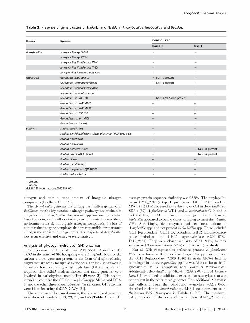
Adaptation of nitrogen metabolismBased on the KEGG analysis, while the gene clusters of
respiratory nitrate reductase (NarGHJI) and assimilatory nitrate
reductase (NasBC) [49,50,51] were missing from the genomes of
Anoxybacillus spp. SK3-4 and DT3-1, and A. flavithermus WK1, a
complete set of NarGHJI was present in A. kamchatkensis G10 and
A. flavithermus TNO-09.006 (Table 3). When compared with
Geobacillus spp., out of ten Geobacillus genomes that were available
in KEGG webpage, nine of them contained at least one complete
NarGHJI or NasBC gene cluster, and five of them showed the
presence of both clusters. At least one gene cluster (NarGHJI or
NasBC) was found in the genomes of Bacillus spp., and four of
these genomes had both NarGHJI and NasBC (Table 3). The
proteobacterial type of nitrate reductase (NapAB) was not detected
in the genome of any Anoxybacillus spp., Geobacillus spp. or Bacillus
spp.
Although a majority of the Anoxybacillus genomes lacked nitrate
reductase gene clusters, all of them encoded a complete set of
amino acid metabolism genes. In addition, proteases were detected
in all five Anoxybacillus genomes. Therefore, it could be postulated
that organic nitrogen compounds (amino acids, small peptides,
and proteins) could be the preferred nitrogen source for the growth
of Anoxybacillus spp. An attempt to grow Anoxybacillus spp. SK3-4
and DT3-1 in minimal medium supplemented with organic
nitrogen compounds (casamino acids/tryptone) or inorganic
nitrogen compounds (nitrate/nitrite) as the sole nitrogen source
demonstrated that casamino acids/tryptone favored the growth of
both strains. When casamino acids were used as the sole nitrogen
source, the specific growth rate (m) of Anoxybacillus spp. SK3-4 and
DT3-1 was 0.48 h21 and 0.83 h21, respectively. In medium
containing tryptone as the sole nitrogen source, the m for
Anoxybacillus spp. SK3-4 and DT3-1 was 0.60 h21 and 1.19 h21,
respectively. Neither Anoxybacillus spp. SK3-4 and DT3-1 was able
to use nitrate or nitrite as the nitrogen source for growth. Our hot
spring water analyses revealed a total of 5.3 mg/L of organic
Anoxybacillus Genome Analysis
PLOS ONE | www.plosone.org 10 March 2014 | Volume 9 | Issue 3 | e90549
nitrogen and only a trace amount of inorganic nitrogen
compounds (less than 0.3 mg/L).
The Anoxybacillus genomes are among the smallest genomes in
Bacillaceae, but the key metabolic nitrogen pathways are retained in
the genomes of Anoxybacillus. Anoxybacillus spp. are mainly isolated
from hot springs and milk-containing environments. Because these
environments are rich in organic nitrogen compounds, the loss of
nitrate reductase gene complexes that are responsible for inorganic
nitrogen metabolism in the genomes of a majority of Anoxybacillus
spp. is an effective and energy-saving strategy.
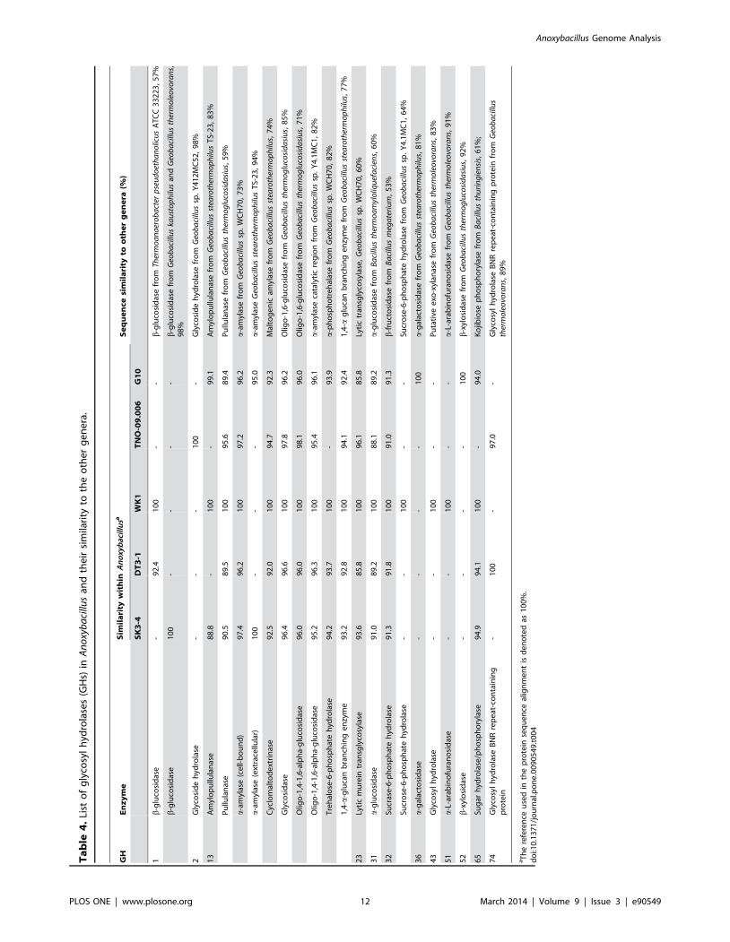
Analysis of glycosyl hydrolase (GH) enzymesAs determined with the standard APHA5310 B method, the
TOC in the water of SK hot spring was 9.0 mg/mL. Most of the
carbon sources were not present in the form of simple reducing
sugars that are ready for uptake by the cells. For the Anoxybacillus to
obtain carbon, various glycosyl hydrolase (GH) enzymes are
required. The SEED analysis showed that many proteins were
involved in carbohydrate metabolism (Figure 2). This section
intends to compare the GHs in Anoxybacillus spp. SK3-4 and DT3-
1, and the other three known Anoxybacillus genomes. GH enzymes
were identified using dbCAN CAZy [21].
The common GHs shared among the five analyzed genomes
were those of families 1, 13, 23, 31, and 65 (Table 4), and the
average protein sequence similarity was 93.5%. The amylopullu-
lanase C289_2785 (a type II pullulanase, GH13, 2033 residues,
MW 221.2 kDa) appeared to be the largest GH in Anoxybacillus sp.
SK3-4 [52], A. flavithermus WK1, and A. kamchatkensis G10, and in
fact the largest ORF in each of those genomes. In general,
Geobacillus appeared to be the closest ortholog to most Anoxybacillus
GHs. Surprisingly, five enzymes had sequences unique to
Anoxybacillus spp. and not present in Geobacillus spp. These included
GH1 b-glucosidase, GH31 a-glucosidase, GH32 sucrose-6-phos-
phate hydrolase, and GH65 sugar-hydrolase (C289_0782,
F510_2484). They were closer (similarity of 53294%) to their
Bacillus and Thermoanaerobacter (57%) counterparts (Table 4).
Not all GHs recognized in reference genome A. flavithermus
WK1 were found in the other four Anoxybacillus spp. For instance,
the GH1 b-glucosidase (C289_1346) in strain SK3-4 had no
homologue in other Anoxybacillus spp. but was 98% similar to the b-
glucosidases in G. kaustophilus and Geobacillus thermoleovorans.
Additionally, Anoxybacillus sp. SK3-4 (C289_2507) and A. kamchat-
kensis G10 exhibited an additional extracellular a-amylase that was
not present in the other three genomes. This additional a-amylase
was different from the cell-bound a-amylase (C289_0468)
described earlier in Anoxybacillus sp. SK3-4 (or equivalent to A.
flavithermus WK1 a-amylase in Table 4) [53]. The biochemi-
cal properties of the extracellular amylase (C289_2507) are
Table 3. Presence of gene clusters of NarGHJI and NasBC in Anoxybacillus, Geobacillus, and Bacillus.
Genus Species Gene cluster
NarGHJI NasBC
Anoxybacillus Anoxybacillus sp. SK3-4 2 2
Anoxybacillus sp. DT3-1 2 2
Anoxybacillus flavithermus WK-1 2 2
Anoxybacillus flavithermus TNO + 2
Anoxybacillus kamchatkensis G10 + 2
Geobacillus Geobacillus kaustophilus 2, NarI is present +
Geobacillus thermodenitrificans 2, NarI is present +
Geobacillus thermoglucosidasius + 2
Geobacillus thermoleovorans + +
Geobacillus sp. WCH70 2, NarG and NarI is present 2
Geobacillus sp. Y412MC61 + +
Geobacillus sp. Y412MC52 + +
Geobacillus sp. C56 T-3 + +
Geobacillus sp. Y4.1MC1 + 2
Geobacillus sp. GHH01 + +
Bacillus Bacillus subtilis 168 + +
Bacillus amyloliquefaciens subsp. plantarum YAU B9601-Y2 + +
Bacillus atrophaeus + +
Bacillus halodurans 2 +
Bacillus anthracis Ames + 2, NasB is present
Bacillus cereus ATCC 14579 + 2, NasB is present
Bacillus clausii + +
Bacillus pseudofirmus 2 +
Bacillus megaterium QM B1551 2 +
Bacillus cellulosilyticus 2 +
+ present;2 absent.doi:10.1371/journal.pone.0090549.t003
Anoxybacillus Genome Analysis
PLOS ONE | www.plosone.org 11 March 2014 | Volume 9 | Issue 3 | e90549
Ta
ble
4.
List
of
gly
cosy
lh
ydro
lase
s(G
Hs)
inA
no
xyb
aci
llus
and
the
irsi
mila
rity
toth
eo
the
rg
en
era
.
GH
En
zy
me
Sim
ila
rity
wit
hin
An
oxy
bac
illu
saS
eq
ue
nce
sim
ila
rity
too
the
rg
en
era
(%)
SK
3-4
DT
3-1
WK
1T
NO
-09
.00
6G
10
1b
-glu
cosi
das
e-
92
.41
00
--
b-g
luco
sid
ase
fro
mTh
erm
oa
na
ero
ba
cter
pse
ud
oet
ha
no
licu
sA
TC
C3
32
23
,5
7%
b-g
luco
sid
ase
10
0-
--
-b
-glu
cosi
das
efr
om
Geo
ba
cillu
ska
ust
op
hilu
san
dG
eob
aci
llus
ther
mo
leo
vora
ns,
98
%
2G
lyco
sid
eh
ydro
lase
--
-1
00
-G
lyco
sid
eh
ydro
lase
fro
mG
eob
aci
llus
sp.
Y4
12
MC
52
,9
8%
13
Am
ylo
pu
llula
nas
e8
8.8
-1
00
-9
9.1
Am
ylo
pu
llula
nas
efr
om
Geo
ba
cillu
sst
earo
ther
mo
ph
ilus
TS-
23
,8
3%
Pu
llula
nas
e9
0.5
89
.51
00
95
.68
9.4
Pu
llula
nas
efr
om
Geo
ba
cillu
sth
erm
og
luco
sid
asi
us,
59
%
a-a
myl
ase
(ce
ll-b
ou
nd
)9
7.4
96
.21
00
97
.29
6.2
a-a
myl
ase
fro
mG
eob
aci
llus
sp.
WC
H7
0,
73
%
a-a
myl
ase
(ext
race
llula
r)1
00
--
-9
5.0
a-a
myl
ase
Geo
ba
cillu
sst
earo
ther
mo
ph
ilus
TS-
23
,9
4%
Cyc
lom
alto
de
xtri
nas
e9
2.5
92
.01
00
94
.79
2.3
Mal
tog
en
icam
ylas
efr
om
Geo
ba
cillu
sst
earo
ther
mo
ph
ilus,
74
%
Gly
cosi
das
e9
6.4
96
.61
00
97
.89
6.2
Olig
o-1
,6-g
luco
sid
ase
fro
mG
eob
aci
llus
ther
mo
glu
cosi
da
siu
s,8
5%
Olig
o-1
,4-1
,6-a
lph
a-g
luco
sid
ase
96
.09
6.0
10
09
8.1
96
.0O
ligo
-1,6
-glu
cosi
das
efr
om
Geo
ba
cillu
sth
erm
og
luco
sid
asi
us,
71
%
Olig
o-1
,4-1
,6-a
lph
a-g
luco
sid
ase
95
.29
6.3
10
09
5.4
96
.1a
-am
ylas
eca
taly
tic
reg
ion
fro
mG
eob
aci
llus
sp.
Y4
.1M
C1
,8
2%
Tre
hal
ose
-6-p
ho
sph
ate
hyd
rola
se9
4.2
93
.71
00
-9
3.9
a-p
ho
sph
otr
eh
alas
efr
om
Geo
ba
cillu
ssp
.W
CH
70
,8
2%
1,4
-a-g
luca
nb
ran
chin
ge
nzy
me
93
.29
2.8
10
09
4.1
92
.41
,4-a
glu
can
bra
nch
ing
en
zym
efr
om
Geo
ba
cillu
sst
earo
ther
mo
ph
ilus,
77
%
23
Lyti
cm
ure
intr
ansg
lyco
syla
se9
3.6
85
.81
00
96
.18
5.8
Lyti
ctr
ansg
lyco
syla
se,
Geo
ba
cillu
ssp
.W
CH
70
,6
0%
31
a-g
luco
sid
ase
91
.08
9.2
10
08
8.1
89
.2a
-glu
cosi
das
efr
om
Ba
cillu
sth
erm
oa
myl
oliq
uef
aci
ens,
60
%
32
Sucr
ase
-6-p
ho
sph
ate
hyd
rola
se9
1.3
91
.81
00
91
.09
1.3
b-f
ruct
osi
das
efr
om
Ba
cillu
sm
ega
teri
um
,5
3%
Sucr
ose
-6-p
ho
sph
ate
hyd
rola
se-
-1
00
--
Sucr
ose
-6-p
ho
sph
ate
hyd
rola
sefr
om
Geo
ba
cillu
ssp
.Y
4.1
MC
1,
64
%
36
a-g
alac
tosi
das
e-
--
-1
00
a-g
alac
tosi
das
efr
om
Geo
ba
cillu
sst
earo
ther
mo
ph
ilus,
81
%
43
Gly
cosy
lh
ydro
lase
--
10
0-
-P
uta
tive
exo
-xyl
anas
efr
om
Geo
ba
cillu
sth
erm
ole
ovo
ran
s,8
3%
51
a-L
-ara
bin
ofu
ran
osi
das
e-
-1
00
--
a-L
-ara
bin
ofu
ran
osi
das
efr
om
Geo
ba
cillu
sth
erm
ole
ovo
ran
s,9
1%
52
b-x
ylo
sid
ase
--
--
10
0b
-xyl
osi
das
efr
om
Geo
ba
cillu
sth
erm
og
luco
sid
asi
us,
92
%
65
Sug
arh
ydro
lase
/ph
osp
ho
ryla
se9
4.9
94
.11
00
-9
4.0
Ko
jibio
sep
ho
sph
ory
lase
fro
mB
aci
llus
thu
rin
gie
nsi
s,6
1%
;
74
Gly
cosy
lh
ydro
lase
BN
Rre
pe
at-c
on
tain
ing
pro
tein
-1
00
-9
7.0
-G
lyco
syl
hyd
rola
seB
NR
rep
eat
-co
nta
inin
gp
rote
infr
om
Geo
ba
cillu
sth
erm
ole
ovo
ran
s,8
9%
aT
he
refe
ren
ceu
sed
inth
ep
rote
inse
qu
en
ceal
ign
me
nt
isd
en
ote
das
10
0%
.d
oi:1
0.1
37
1/j
ou
rnal
.po
ne
.00
90
54
9.t
00
4
Anoxybacillus Genome Analysis
PLOS ONE | www.plosone.org 12 March 2014 | Volume 9 | Issue 3 | e90549

unexplored at the time of writing; yet based on its amino acid
sequence, the closest match is an amylase of Geobacillus
stearothermophilus TS-23, with 94% similarity. On the other hand,
Anoxybacillus sp. DT3-1 had an extra GH74 glycosyl hydrolase with
a bacterial neuraminidase repeat (BNR) domain at the center of
the sequence (F510_1955). The sequence was highly similar to the
sequence in A. flavithermus TNO-09.006. The function of
F510_1955 is unknown, but based on the InterProScan analysis
[15], the N-terminus of the sequence had a signal peptide and a
membrane lipoprotein lipid attachment site. F510_1955 also had a
domain found to be an oligoxyloglucan-reducing end-specific
cellobiohydrolase (OXG-RCBH).
All of the GHs in the Anoxybacillus spp., in particular Anoxybacillus
spp. SK3-4 and DT3-1, were not part of any gene cluster, except
for the one shown in Figure 6. The gene cluster consisted of an
intracellular a-glucosidase, a cell-anchoring a-amylase, three
maltose/maltodextrin ABC transporter periplasmic proteins
(MalE, MalF, and MalG; also known as permease), and an
intracellular cyclomaltodextrinase (CDase). This gene cluster was
sandwiched between a transcriptional regulator (AraC family) and
the maltose operon transcriptional repressor MalR (LacI family)
(C289_046320470, F510_152121528). The starch degradation
process is most likely initiated by the action of the cell-anchoring
a-amylase (C289_0468) and amylopullulanase (C289_2785),
which hook to the cells by their transmembrane region and S-
layer homology (SLH) domain, respectively, at their C-termini
[52,53]. The resulting oligosaccharides formed from the hydrolysis
of starch are then transported into the cells by the maltose/
maltodextrin ABC transporter periplasmic proteins and further
degraded by the intracellular a-glucosidase and CDase. Although
A. flavithermus CDase (AfCda13) (77% identity to CDase from
Anoxybacillus spp. SK3-4 and DT3-1) most actively acts on
cyclodextrins, the enzyme can degrade starch into sugar [54,55].
Without doubt, other, lone GHs (Table 4) help in the whole
process of oligosaccharide degradation inside the cell.
We hope that knowing the types of GHs present in Anoxybacillus
spp. will drive the discovery of new applications for thermostable
enzymes in the starch industry. Many of the GHs listed in Table 4have not been biochemically characterized. For instance, the b-
glucosidase in Anoxybacillus sp. DT3-1 (92.4% similar to A. flavithermus
WK1) and a Geobacillus-similar b-glucosidase in Anoxybacillus sp. SK3-
4 are yet to be explored. From the perspective of biofuel production,
b-glucosidase (EC 3.2.1.21) is involved in the final step of converting
cellobiose to glucose. It is an important enzyme, frequently known as
a bottleneck enzyme in the hydrolysis of ligno-biomass [56]. In the
food industry, b-glucosidases have various applications, such as to
reduce the viscosity of gellan food, remove the bitterness of certain
juices and unripe olives, and many other applications [57].
Another uncharacterized Anoxybacillus glucosidase is the a-gluco-
sidase (synonym maltase, EC3.2.1.20, acts upon a-1, 4-glycosidic
bonds). Although Anoxybacillus salavatliensis has shown an a-glucosidase
activity [58], no a-glucosidase gene or its properties had been
reported. In industry, a-glucosidase is mainly used to convert starchy
substrates to glucose. In addition to a-glucosidase, oligo-1,4-1,6-
alpha-glucosidase (synonym O16G, oligo-1,6-glucosidase, isomaltase,
sucrase, EC 3.2.1.10) has not been examined for its biological
function. According to BRENDA [59], O16G enzymes mainly attack
the a-1,6-glucosidic linkages in isomaltose (two glucose units) or
isomalto-oligosaccharides (226 glucose units), and certain reported
O16Gs can hydrolyze the a-1, 6 bonds in starch, a-limit dextrins, and
glycogen, resulting in the formation of maltose and linear or
branched dextrins. This reactivity suggests that O16G from
Anoxybacillus can serve as an alternative to pullulanase in assisting
starch hydrolysis in industrial applications. We do not know why
Anoxybacillus, for instance Anoxybacillus sp. SK3-4, requires numerous
types of oligo-1,6-glucosidases, pullulanase type I, and amylopullu-
lanase, in which all these enzymes can break identical a-1,6-
glucosidic linkages. However, we think their presence is somehow
related to the lifestyle of the cell. Apart from glucosidases, Anoxybacillus
sp. DT3-1, A. flavithermus strains WK1 and TNO-09.006 each have a
unique type of GHs (GH74, GH2, GH43, respectively) (Table 4).
The efficiency of these glycosidases in degradation of biomass, such as
cellulose and hemicellulose, has not been reported. Our group is
currently analyzing some of these enzymes and hopes to determine
the roles of each protein in the near future.
Conclusion
Anoxybacillus is a mild thermophile. The closest genus to
Anoxybacillus is Geobacillus, yet the genomes of the latter genus are
larger and the cells grow at higher temperatures. Based on the
genome annotation, the thermophily of Anoxybacillus is attributable
to many features that stabilize proteins, DNA, and RNA. The
presence of adaptive genes is sufficient for the cells to live in an
alkaline environment with the presence of organic nitrogen and
carbohydrates and to overcome the threats from UV radiation. In
addition, for Anoxybacillus to survive under extreme conditions, we
think that genetic exchange, especially uptake of genetic material via
HGT, is important. Based on our genomic information, this process
can take place via transduction or transformation, though at this
moment, the possibility of HGT by conjugation remains undecided.
Supporting Information
Table S1 Database matches for prophageSK fromAnoxybacillus sp. SK3-4. 22 ORFs putatively annotated as
phage-like or prophage proteins. Ten ORFs designated 23–26, 28,
29, 31, 34, 38, and 39 are involved in phage morphogenesis, while
three ORFs designated 10, 14, and 41 are annotated as non-
structural or regulatory proteins.
(DOC).
Figure 6. The gene cluster of various GHs, transporters, transcriptional regulators, and transcriptional repressors in Anoxybacillussp. SK3-4. Identical clusters of genes are present in other Anoxybacillus species.doi:10.1371/journal.pone.0090549.g006
Anoxybacillus Genome Analysis
PLOS ONE | www.plosone.org 13 March 2014 | Volume 9 | Issue 3 | e90549
Table S2 Database matches for prophageDT fromAnoxybacillus sp. DT3-1. ProphageDT is annotated on the
complementary strand of contig 20 of Anoxybacillus sp. DT3-1. 32
ORFs putatively annotated as phage-like or prophage proteins.
ORFs designated 1 to 53 form prophageDT-a, while ORFs 54 to
82 form the remnant of prophageDT-b.
(DOC).
Author Contributions
Conceived and designed the experiments: KMG GHM K-GC GFC SS
CSC UMK KPC. Performed the experiments: KMG GHM K-GC GFC
SS CSC UMK KPC. Analyzed the data: KMG GHM K-GC GFC SS
CSC UMK KPC. Contributed reagents/materials/analysis tools: KMG
GHM K-GC GFC. Wrote the paper: KMG GHM K-GC GFC SS CSC
UMK KPC.
References
1. Goh KM, Kahar UM, Chai YY, Chong CS, Chai KP, et al. (2013) Recent
discoveries and applications of Anoxybacillus. Appl Microbiol Biotechnol 97:147521488.
2. Pikuta E, Lysenko A, Chuvilskaya N, Mendrock U, Hippe H, et al. (2000)
Anoxybacillus pushchinensis gen. nov., sp. nov., a novel anaerobic, alkaliphilic,
moderately thermophilic bacterium from manure, and description of Anoxyba-
cillus flavithermus comb. nov. Int J Syst Evol Microbiol 50: 210922117.
3. Zhang XQ, Zhang ZL, Wu N, Zhu XF, Wu M (2013) Anoxybacillus vitaminiphilus
sp. nov., a strictly aerobic and moderately thermophilic bacterium from the Pugehot spring in southwest China. Int J Syst Evol Microbiol: in press.
4. Cihan AC, Cokmus C, Koc M, Ozcan B (2013) Anoxybacillus calidus sp. nov., a
novel thermophilic bacterium isolated from a soil near a thermal power plant in
Denizli, Turkey. Int J Syst Evol Microbiol: in press.
5. Deep K, Poddar A, Das SK (2013) Anoxybacillus suryakundensis sp. nov, amoderately thermophilic, alkalitolerant bacterium isolated from hot spring at
Jharkhand, India. PLoS ONE 8: e85493.
6. Saw JH, Mountain BW, Feng L, Omelchenko MV, Hou S, et al. (2008)Encapsulated in silica: genome, proteome and physiology of the thermophilic
bacterium Anoxybacillus flavithermus WK1. Genome Biol 9: R161.
7. Caspers MP, Boekhorst J, Abee T, Siezen RJ, Kort R (2013) Complete genome
sequence of Anoxybacillus flavithermus TNO-09.006, a thermophilic sporeformerassociated with a dairy-processing environment. Genome Announc 1:
e00010200013.
8. Matsutani M, Shirakihara Y, Imada K, Yakushi T, Matsushita K (2013) Draftgenome sequence of a thermophilic member of the Bacillaceae, Anoxybacillus
flavithermus Strain Kn10, isolated from the Kan-nawa hot spring in Japan.Genome Announc 1: e00346200313.
9. Lee SJ, Lee YJ, Ryu N, Park S, Jeong H, et al. (2012) Draft genome sequence ofthe thermophilic bacterium Anoxybacillus kamchatkensis G10. J Bacteriol 194:
668426685.
10. Chai YY, Kahar UM, Salleh MM, Illias RM, Goh KM (2012) Isolation andcharacterization of pullulan-degrading Anoxybacillus species isolated from
Malaysian hot springs. Environ Technol 33: 123121238.
11. Van Hall CE, Safranko J, Stenger VA (1963) Rapid combustion method for the
determination of organic substances in aqueous solutions. Anal Chem 35:3152319.
12. Conesa A, Gotz S, Garcıa-Gomez JM, Terol J, Talon M, et al. (2005) Blast2GO:
a universal tool for annotation, visualization and analysis in functional genomicsresearch. Bioinformatics 21: 367423676.
13. Lagesen K, Hallin P, Rødland EA, Stærfeldt HH, Rognes T, et al. (2007)
RNAmmer: consistent and rapid annotation of ribosomal RNA genes. Nucleic
Acids Res 35: 310023108.
14. Schattner P, Brooks AN, Lowe TM (2005) The tRNAscan-SE, snoscan andsnoGPS web servers for the detection of tRNAs and snoRNAs. Nucleic Acids
Res 33: W6862W689.
15. Zdobnov EM, Apweiler R (2001) InterProScan – an integration platform for thesignature-recognition methods in InterPro. Bioinformatics 17: 8472848.
16. Alikhan NF, Petty NK, Ben Zakour NL, Beatson SA (2011) BLAST Ring Image
Generator (BRIG): simple prokaryote genome comparisons. BMC Genomics 12:
402.
17. Fouts DE, Brinkac L, Beck E, Inman J, Sutton G (2012) PanOCT: automatedclustering of orthologs using conserved gene neighborhood for pan-genomic
analysis of bacterial strains and closely related species. Nucleic Acids Res 40:e172.
18. Zhou Y, Liang Y, Lynch KH, Dennis JJ, Wishart DS (2011) PHAST: a fast
phage search tool. Nucleic Acids Res 39: W3472W352.
19. Aziz RK, Bartels D, Best AA, DeJongh M, Disz T, et al. (2008) The RAST
Server: rapid annotations using subsystems technology. BMC Genomics 9: 75.
20. Overbeek R, Begley T, Butler RM, Choudhuri JV, Chuang HY, et al. (2005)The subsystems approach to genome annotation and its use in the project to
annotate 1000 genomes. Nucleic Acids Res 33: 569125702.
21. Yin Y, Mao X, Yang J, Chen X, Mao F, et al. (2012) dbCAN: a web resource forautomated carbohydrate-active enzyme annotation. Nucleic Acids Res 40:
W4452W451.
22. Larkin MA, Blackshields G, Brown NP, Chenna R, McGettigan PA, et al. (2007)
Clustal W and Clustal X version 2.0. Bioinformatics 23: 294722948.
23. Tamura K, Peterson D, Peterson N, Stecher G, Nei M, et al. (2011) MEGA5:molecular evolutionary genetics analysis using maximum likelihood, evolution-
ary distance, and maximum parsimony methods. Mol Biol Evol 28: 273122739.
24. Heinen W, Lauwers AM, Mulders JWM (1982) Bacillus flavothermus, a newlyisolated facultative thermophile. Antonie van Leeuwenhoek 48: 2652272.
25. Schirner K, Errington J (2009) Influence of heterologous MreB proteins on cell
morphology of Bacillus subtilis. Microbiology 155: 361123621.
26. Piggot PJ, Hilbert DW (2004) Sporulation of Bacillus subtilis. Curr Opin
Microbiol 7: 5792586.
27. Galperin MY, Mekhedov SL, Puigbo P, Smirnov S, Wolf YI, et al. (2012)
Genomic determinants of sporulation in Bacilli and Clostridia: towards the
minimal set of sporulation-specific genes. Environ Microbiol 14: 287022890.
28. Casjens S (2003) Prophages and bacterial genomics: what have we learned so
far? Mol Microbiol 49: 2772300.
29. Smeesters PR, Dreze PA, Bousbata S, Parikka KJ, Timmery S, et al. (2011)
Characterization of a novel temperate phage originating from a cereulide-
producing Bacillus cereus strain. Res Microbiol 162: 4462459.
30. Moumen B, Nguen-The C, Sorokin A (2012) Sequence analysis of inducible
prophage phIS3501 integrated into the haemolysin II gene of Bacillus thuringiensis
var israelensis ATCC35646. Genet Res Int 2012: 543286.
31. Iyer LM, Makarova KS, Koonin EV, Aravind L (2004) Comparative genomics
of the FtsK-HerA superfamily of pumping ATPases: implications for the origins
of chromosome segregation, cell division and viral capsid packaging. Nucleic
Acids Res 32: 526025279.
32. Davis BM, Kimsey HH, Kane AV, Waldor MK (2002) A satellite phage-
encoded antirepressor induces repressor aggregation and cholera toxin gene
transfer. EMBO J 21: 424024249.
33. Iguchi A, Iyoda S, Terajima J, Watanabe H, Osawa R (2006) Spontaneous
recombination between homologous prophage regions causes large-scale
inversions within the Escherichia coli O157:H7 chromosome. Gene 372:
1992207.
34. Sharon I, Battchikova N, Aro EM, Giglione C, Meinnel T, et al. (2011)
Comparative metagenomics of microbial traits within oceanic viral communities.
ISME J 5: 117821190.
35. Birge EA (2006) Genetics of temperate bacteriophages. Bacterial and
Bacteriophage Genetics: Springer New York. pp. 2612303.
36. Kramer N, Hahn J, Dubnau D (2007) Multiple interactions among the
competence proteins of Bacillus subtilis. Mol Microbiol 65: 4542464.
37. Trivedi S, Rao SR, Gehlot HS (2005) Nucleic acid stability in thermophilic
prokaryotes: a review. J Cell Mol Biol 4: 61269.
38. Takami H, Takaki Y, Chee GJ, Nishi S, Shimamura S, et al. (2004)
Thermoadaptation trait revealed by the genome sequence of thermophilic
Geobacillus kaustophilus. Nucleic Acids Res 32: 629226303.
39. Perugino G, Valenti A, D’Amaro A, Rossi M, Ciaramella M (2009) Reverse
gyrase and genome stability in hyperthermophilic organisms. Biochem Soc
Trans 37: 69273.
40. Heine M, Chandra SB (2009) The linkage between reverse gyrase and
hyperthermophiles: a review of their invariable association. J Microbiol 47:
2292234.
41. Hou MH, Lin SB, Yuann JMP, Lin WC, Wang AHJ, et al. (2001) Effects of
polyamines on the thermal stability and formation kinetics of DNA duplexes
with abnormal structure. Nucleic Acids Res 29: 512125128.
42. Hosoya R, Hamana K, Niitsu M, Itoh T (2004) Polyamine analysis for
chemotaxonomy of thermophilic eubacteria: polyamine distribution profiles
within the orders Aquificales, Thermotogales, Thermodesulfobacteriales, Thermales,
Thermoanaerobacteriales, Clostridiales and Bacillales. J Gen Appl Microbiol 50:
2712287.
43. Mukherjee A, Sokunbi AO, Grove A (2008) DNA protection by histone-like
protein HU from the hyperthermophilic eubacterium Thermotoga maritima.
Nucleic Acids Res 36: 395623968.
44. Karp PD, Ouzounis CA, Moore-Kochlacs C, Goldovsky L, Kaipa P, et al.
(2005) Expansion of the BioCyc collection of pathway/genome databases to 160
genomes. Nucleic Acids Res 33: 608326089.
45. Goosen N, Moolenaar GF (2008) Repair of UV damage in bacteria. DNA
Repair 7: 3532379.
46. Kumar S, Tsai CJ, Nussinov R (2000) Factors enhancing protein thermosta-
bility. Protein Eng 13: 1792191.
47. Phadtare S (2004) Recent developments in bacterial cold-shock response. Curr
Issues Mol Biol 6: 1252136.
48. Giaquinto L, Curmi PM, Siddiqui KS, Poljak A, DeLong E, et al. (2007)
Structure and function of cold shock proteins in archaea. J Bacteriol 189:
573825748.
49. Hoffmann T, Troup B, Szabo A, Hungerer C, Jahn D (1995) The anaerobic life
of Bacillus subtilis: cloning of the genes encoding the respiratory nitrate reductase
system. FEMS Microbiol Lett 131: 2192225.
Anoxybacillus Genome Analysis
PLOS ONE | www.plosone.org 14 March 2014 | Volume 9 | Issue 3 | e90549
50. Ogawa KI, Akagawa E, Yamane K, Sun ZW, LaCelle M, et al. (1995) The nasB
operon and nasA gene are required for nitrate/nitrite assimilation in Bacillus
subtilis. J Bacteriol 177: 140921413.
51. Richardson DJ, Berks BC, Russell DA, Spiro S, Taylor CJ (2001) Functional,
biochemical and genetic diversity of prokaryotic nitrate reductases. Cell Mol LifeSci 58: 1652178.
52. Kahar UM, Chan KG, Salleh MM, Hii SM, Goh KM (2013) A high molecular-mass Anoxybacillus sp. SK3-4 amylopullulanase: characterization and its
relationship in carbohydrate utilization. Int J Mol Sci 14: 11302211318.
53. Chai YY, Rahman RN, Illias RM, Goh KM (2012) Cloning and characteriza-tion of two new thermostable and alkalitolerant a-amylases from the Anoxybacillus
species that produce high levels of maltose. J Ind Microbiol Biotechnol 39:7312741.
54. Turner P, Labes A, Fridjonsson OH, Hreggvidson GO, Schonheit P, et al.(2005) Two novel cyclodextrin-degrading enzymes isolated from thermophilic
bacteria have similar domain structures but differ in oligomeric state and activity
profile. J Biosci Bioeng 100: 3802390.
55. Turner P, Holst O, Karlsson EN (2005) Optimized expression of soluble
cyclomaltodextrinase of thermophilic origin in Escherichia coli by using a soluble
fusion-tag and by tuning of inducer concentration. Protein Expr Purif 39:
54260.
56. Singhania RR, Patel AK, Sukumaran RK, Larroche C, Pandey A (2013) Role
and significance of beta-glucosidases in the hydrolysis of cellulose for bioethanol
production. Bioresour Technol 127: 5002507.
57. Bhatia Y, Mishra S, Bisaria VS (2002) Microbial b-glucosidases: cloning,
properties, and applications. Crit Rev Biotechnol 22: 3752407.
58. Cihan AC, Ozcan B, Cokmus C (2011) Anoxybacillus salavatliensis sp. nov., an a-
glucosidase producing, thermophilic bacterium isolated from Salavatli, Turkey. J
Basic Microbiol 51: 1362146.
59. Schomburg I, Chang A, Placzek S, Sohngen C, Rother M, et al. (2013)
BRENDA in 2013: integrated reactions, kinetic data, enzyme function data,
improved disease classification: new options and contents in BRENDA. Nucleic
Acids Res 41: D7642D772.
Anoxybacillus Genome Analysis
PLOS ONE | www.plosone.org 15 March 2014 | Volume 9 | Issue 3 | e90549