bacteriophage p4 vis protein is needed for prophage excision
TRANSCRIPT
www.elsevier.com/locate/yviro
Virology 322 (2004) 82–92
Bacteriophage P4 Vis protein is needed for prophage excision
Simona Calı,a,1,2 Elena Spoldi,a,1,2 Daniela Piazzolla,a,3 Ian B. Dodd,b Francesca Forti,a
Gianni Deho,a and Daniela Ghisottia,*
aDipartimento di Scienze Biomolecolari e Biotecnologie, Universita degli Studi di Milano, 20133 Milan, ItalybDepartment of Molecular Biosciences (Biochemistry), University of Adelaide, South Australia, Australia
Received 15 December 2003; returned to author for revision 12 January 2004; accepted 16 January 2004
Abstract
Upon infection of its host Escherichia coli, satellite bacteriophage P4 can integrate its genome into the bacterial chromosome by Int-
mediated site-specific recombination between the attP and the attB sites. The opposite event, excision, may either occur spontaneously or be
induced by a superinfecting P2 helper phage. In this work, we demonstrate that the product of the P4 vis gene, a regulator of the P4 late
promoters PLL and Psid, is needed for prophage excision. This conclusion is supported by the following evidence: (i) P4 mutants carrying
either a frameshift mutation or a deletion of the vis gene were unable to excise both spontaneously or upon P2 phage superinfection; (ii)
expression of the Vis protein from a plasmid induced P4 prophage excision; (iii) excision depended on a functional integrase (Int) protein,
thus suggesting that Vis is involved in the formation of the excision complex, rather than in the excision recombination event per se; (iv) Vis
protein bound P4 DNA in the attP region at two distinct boxes (Box I and Box II), located between the int gene and the attP core region, and
caused bending of the bound DNA. Furthermore, we mapped by primer extension the 5V end of the int transcript and found that ectopic
expression of Vis reduced its signal intensity, suggesting that Vis is also involved in negative regulation of the int promoter.
D 2004 Elsevier Inc. All rights reserved.
Keywords: Lysogenization; DNA binding; DNA bending; Vis regulator; Bacteriophage P4
Introduction
The temperate bacteriophage E represents the model
system for integration and excision of phage genome into
the bacterial chromosome. The site-specific recombination
between the attP site on the phage DNA and the attB site on
the bacterial chromosome is promoted by an integrase, the
product of the E int gene, that cuts and religates the two
DNA molecules (Friedman, 1992; Gingery and Echols,
1967; Nash, 1981, 1990; Swalla et al., 2003). A protein–
DNA bond, which involves an integrase active tyrosine,
conserves the energy of the phosphodiester bond and an
0042-6822/$ - see front matter D 2004 Elsevier Inc. All rights reserved.
doi:10.1016/j.virol.2004.01.016
* Corresponding author. Dipartimento di Scienze Biomolecolari e
Biotecnologie, Universita degli Studi di Milano, Via Celoria 26, 20133
Milan, Italy. Fax: +39-02-50315044.
E-mail address: [email protected] (D. Ghisotti).1 Present address: Keryos SpA, Biotecnology Division, 20097 San
Donato Milanese, Italy.2 The two authors equally contributed to the work.3 Present address: Max F. Perutz Laboratories at the Vienna Biocenter,
Department of Microbiology and Genetics, University of Vienna, 1030
Vienna, Austria.
external high-energy source is thus not required. In addition
to the integrase, host-encoded proteins such as IHF and FIS
assemble into a macromolecular machine known as inta-
some and participate in the site-specific recombination event
(Finkel and Johnson, 1992; Freundlich et al., 1992). The EattP and the bacterial site attB share an identical 15-bp core
region within which the recombination event occurs. The
attP site is flanked by regions recognized and bound by the
proteins forming the intasome. The recombination event that
leads to prophage excision requires, beside the integrase,
another phage-coded protein named excisionase. Excisio-
nase binds DNA at attL and assists integrase binding and the
formation of the intasome (Cho et al., 2002; Numrynch et
al., 1992).
The temperate bacteriophage P4 is the prototype of
satellite phages (for reviews, see Briani et al., 2001;
Lindqvist et al., 1993). P4 lacks all the structural genes
necessary for the construction of its viral particle and
exploits the genetic information carried by a helper phage
such as P2 for the construction of its capsid and for cell
lysis. Thus, P4 can enter the lytic cycle only when the helper
phage genome is present in the infected Escherichia coli
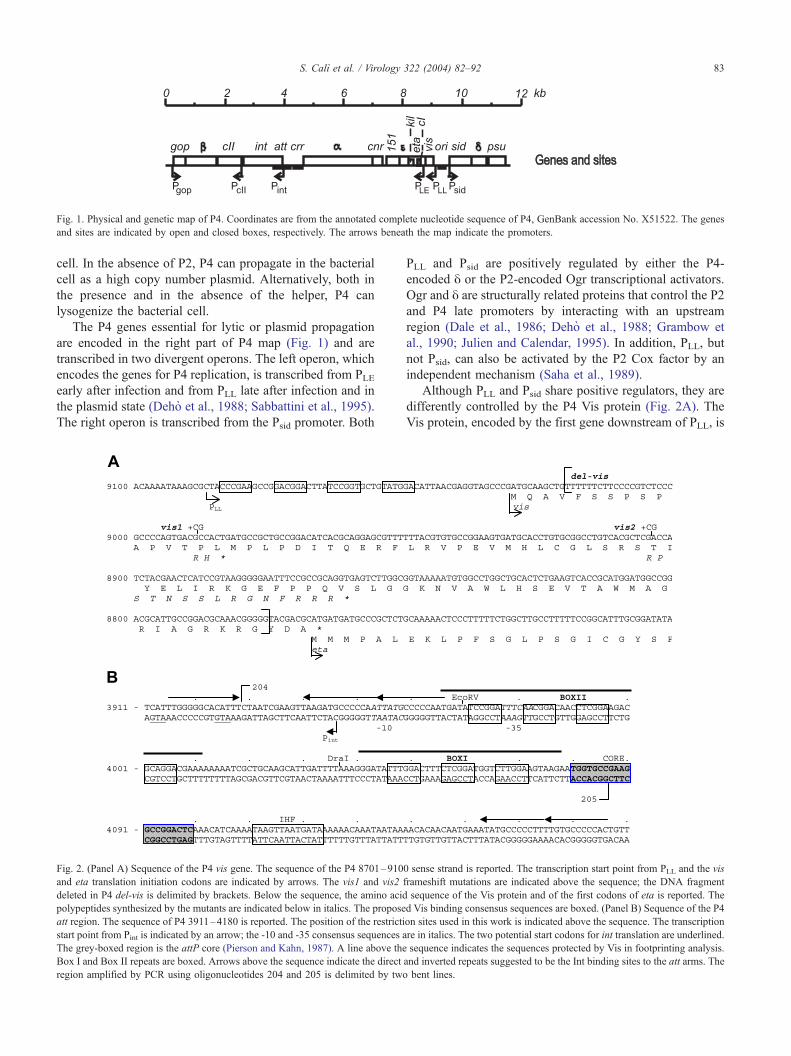
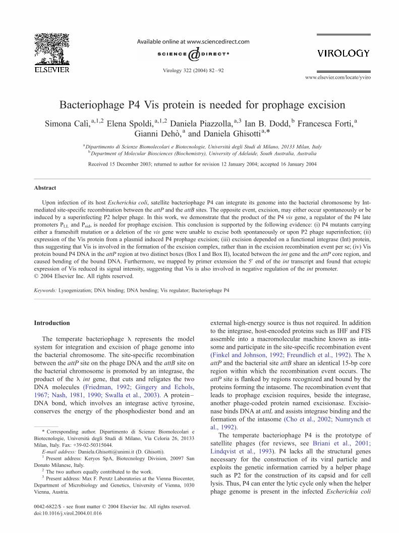
Fig. 1. Physical and genetic map of P4. Coordinates are from the annotated complete nucleotide sequence of P4, GenBank accession No. X51522. The genes
and sites are indicated by open and closed boxes, respectively. The arrows beneath the map indicate the promoters.
S. Calı et al. / Virology 322 (2004) 82–92 83
cell. In the absence of P2, P4 can propagate in the bacterial
cell as a high copy number plasmid. Alternatively, both in
the presence and in the absence of the helper, P4 can
lysogenize the bacterial cell.
The P4 genes essential for lytic or plasmid propagation
are encoded in the right part of P4 map (Fig. 1) and are
transcribed in two divergent operons. The left operon, which
encodes the genes for P4 replication, is transcribed from PLEearly after infection and from PLL late after infection and in
the plasmid state (Deho et al., 1988; Sabbattini et al., 1995).
The right operon is transcribed from the Psid promoter. Both
Fig. 2. (Panel A) Sequence of the P4 vis gene. The sequence of the P4 8701–910
and eta translation initiation codons are indicated by arrows. The vis1 and vis2 f
deleted in P4 del-vis is delimited by brackets. Below the sequence, the amino acid
polypeptides synthesized by the mutants are indicated below in italics. The propose
att region. The sequence of P4 3911–4180 is reported. The position of the restricti
start point from Pint is indicated by an arrow; the -10 and -35 consensus sequences a
The grey-boxed region is the attP core (Pierson and Kahn, 1987). A line above the
Box I and Box II repeats are boxed. Arrows above the sequence indicate the direct
region amplified by PCR using oligonucleotides 204 and 205 is delimited by two
PLL and Psid are positively regulated by either the P4-
encoded y or the P2-encoded Ogr transcriptional activators.
Ogr and y are structurally related proteins that control the P2
and P4 late promoters by interacting with an upstream
region (Dale et al., 1986; Deho et al., 1988; Grambow et
al., 1990; Julien and Calendar, 1995). In addition, PLL, but
not Psid, can also be activated by the P2 Cox factor by an
independent mechanism (Saha et al., 1989).
Although PLL and Psid share positive regulators, they are
differently controlled by the P4 Vis protein (Fig. 2A). The
Vis protein, encoded by the first gene downstream of PLL, is
0 sense strand is reported. The transcription start point from PLL and the vis
rameshift mutations are indicated above the sequence; the DNA fragment
sequence of the Vis protein and of the first codons of eta is reported. The
d Vis binding consensus sequences are boxed. (Panel B) Sequence of the P4
on sites used in this work is indicated above the sequence. The transcription
re in italics. The two potential start codons for int translation are underlined.
sequence indicates the sequences protected by Vis in footprinting analysis.
and inverted repeats suggested to be the Int binding sites to the att arms. The
bent lines.

Table 1
Growth of P4 vis mutants in infection of E. coli (P2)a
Infecting Cells yielding Phage produced– Survivorsc Lysogenic clonesd
phageb phagec (%) infected cellc (%)Immune to P4
cI405 infection (%)
Releasing P4
phage (%)
P4 38 78 86 76 76
P4 vis1 30 20 78 83 0
P4 vis2 32 15 71 83 0
P4 del-vis nte nte 70 92 0
a The P2 lysogenic bacterial host was C-339.b A culture of C-339 was grown to 1 � 108 cells/ml in LD broth, 5 mM CaCl2, at 37 jC and infected with the phage at a multiplicity of infection of 5–10. After
5 min, the culture was diluted 100 times in fresh broth and incubated with aeration.c The infected cells yielding phage and the survivors were measured 20 min after infection, and the fraction was calculated on the cell titer measured at the
infection time point. The phage titer was assayed 70 min after the infection.d The presence of lysogens among 100 surviving colonies was determined by testing both immunity to P4 cI405 infection and the presence in the culture of free
P4 phage by a replica plating assay (Alano et al., 1986).e Not tested.
S. Calı et al. / Virology 322 (2004) 82–9284
an 88 amino acid long polypeptide that presents a helix-
turn-helix motif and is able to bind P4 DNA in several
specific sites: (i) downstream of PLL; (ii) upstream of Psid;
and (iii) upstream of the P4 int gene (Polo et al., 1996). The
Vis protein was found to repress transcription from PLL,
whereas it appears to enhance y-dependent transcription
from Psid (Polo et al., 1996).
P4 lysogenization requires integration of the phage
genome into the bacterial chromosome and depends on P4
integrase, encoded by the P4 int gene (Fig. 1; Calendar et
al., 1981; Pierson and Kahn, 1984, 1987). The P4 attP site is
located immediately upstream of the int gene. The P4 attB
site is located at 97.2 min of the E. coli map, within the leuX
gene, which encodes a tRNALeu isoacceptor (Calendar et al.,
1981; Pierson and Kahn, 1987). Site-specific recombination
occurs between identical 20 nt long G + C-rich core regions
present in attP and attB, thus preserving the integrity of the
leuX gene sequence upon prophage integration (Pierson and
Kahn, 1987; Fig. 2B).
P4 integrase belongs to the tyrosine recombinases, which
include the well-characterized E integrase. The C-terminal
half of the protein, which is directly involved in the
recombination event, is particularly well conserved, whereas
the N-terminal region that specifically binds the arm sequen-
ces of the att sites is more divergent (Esposito and Scocca,
1997; Grainge and Jayaram, 1999). In the P4 att site, a pair
of 16-bp direct repeats, present on either side of the core
sequence, and an inverted repeat in the left arm are supposed
to be bound by Int (Pierson and Kahn, 1987; Fig. 2B). A
consensus sequence for binding of IHF is present in the right
arm of attP, suggesting that this bacterial protein is part of
the P4 integration complex.
A potential int promoter was suggested based on canon-
ical j70 consensus and it has been proposed that Int
autoregulates its own expression by binding to the attP left
arm sequences that overlap the Pint region (Fig. 2B; Pierson
and Kahn, 1987).
Excision of P4 prophage occurs spontaneously at low
frequency, but can be induced upon P2 infection of a P4
lysogenic strain at much higher frequency (Six and Lindqvist,
1978). One might suppose that a P4 protein is required as
excisionase, although such a protein has not been identified.
In this work, we show that Vis, in addition to its regulatory
role on PLL and Psid, is essential for P4 excision and
negatively regulates Pint.
Results
Mutations in the P4 vis gene prevent prophage induction
To better characterize the role of vis gene in P4 regula-
tion, we constructed by in vitro mutagenesis two different
mutations: vis1, an insertion of 2 bp at 8988, and vis2, an
insertion of 2 bp at 8904 (see Materials and methods). Both
mutations generated a premature stop codon (Fig. 2A).
The P4 vis mutants formed very turbid plaques on P2
lysogenic E. coli indicators. One step growth analysis (Table
1) showed that P4 vis mutant phage yields were slightly
reduced as compared to P4+. In both wild-type and mutant
infections, the largest fraction of cells surviving P4 infection
(z76%) was immune to P4 cI405 infection, a clear plaque,
immunity sensitive mutant, indicating that lysogenization
was not impaired. However, none of the P4 vis survivors
spontaneously released P4, in contrast to the 100% of P4
immune clones obtained upon P4+ infection.
The titer of free P4 phage present in overnight cultures of
strains doubly lysogenic for either P2 and P4 vis1 or P2 and
P4 vis2 was less than 10 pfu/ml as compared to 104 pfu/ml
in the corresponding strain doubly lysogenic for P2 and
wild-type P4.
In a singly lysogenic strain, the P4 prophage may be
induced by infection with P2 A� (Six and Lindqvist, 1978).
We therefore measured P4 production after infection of C-1a
(P4 vis1) and C-1a (P4 vis2) lysogens with P2 Aam129. No
P4 phage was released by the lysogenic strains (<10 pfu/ml
compared to about 108 pfu/ml released by the P4 wild type
lysogenic strain). Therefore, the mutations in the P4 vis gene
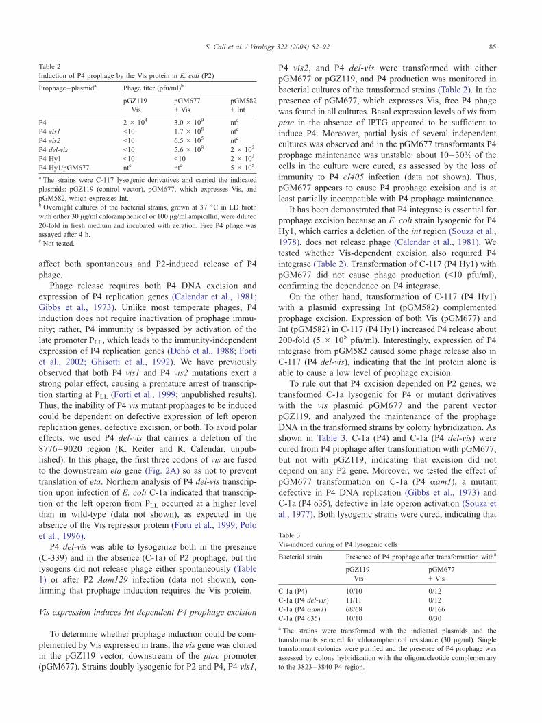
Table 3
Vis-induced curing of P4 lysogenic cells
Bacterial strain Presence of P4 prophage after transformation witha
pGZ119
� Vis
pGM677
+ Vis
C-1a (P4) 10/10 0/12
C-1a (P4 del-vis) 11/11 0/12
C-1a (P4 aam1) 68/68 0/166
C-1a (P4 y35) 10/10 0/30
a The strains were transformed with the indicated plasmids and the
transformants selected for chloramphenicol resistance (30 Ag/ml). Single
transformant colonies were purified and the presence of P4 prophage was
assessed by colony hybridization with the oligonucleotide complementary
to the 3823–3840 P4 region.
Table 2
Induction of P4 prophage by the Vis protein in E. coli (P2)
Prophage–plasmida Phage titer (pfu/ml)b
pGZ119
� Vis
pGM677
+ Vis
pGM582
+ Int
P4 2 � 104 3.0 � 109 ntc
P4 vis1 <10 1.7 � 108 ntc
P4 vis2 <10 6.5 � 105 ntc
P4 del-vis <10 5.6 � 108 2 � 102
P4 Hy1 <10 <10 2 � 103
P4 Hy1/pGM677 ntc ntc 5 � 105
a The strains were C-117 lysogenic derivatives and carried the indicated
plasmids: pGZ119 (control vector), pGM677, which expresses Vis, and
pGM582, which expresses Int.b Overnight cultures of the bacterial strains, grown at 37 jC in LD broth
with either 30 Ag/ml chloramphenicol or 100 Ag/ml ampicillin, were diluted
20-fold in fresh medium and incubated with aeration. Free P4 phage was
assayed after 4 h.c Not tested.
S. Calı et al. / Virology 322 (2004) 82–92 85
affect both spontaneous and P2-induced release of P4
phage.
Phage release requires both P4 DNA excision and
expression of P4 replication genes (Calendar et al., 1981;
Gibbs et al., 1973). Unlike most temperate phages, P4
induction does not require inactivation of prophage immu-
nity; rather, P4 immunity is bypassed by activation of the
late promoter PLL, which leads to the immunity-independent
expression of P4 replication genes (Deho et al., 1988; Forti
et al., 2002; Ghisotti et al., 1992). We have previously
observed that both P4 vis1 and P4 vis2 mutations exert a
strong polar effect, causing a premature arrest of transcrip-
tion starting at PLL (Forti et al., 1999; unpublished results).
Thus, the inability of P4 vis mutant prophages to be induced
could be dependent on defective expression of left operon
replication genes, defective excision, or both. To avoid polar
effects, we used P4 del-vis that carries a deletion of the
8776–9020 region (K. Reiter and R. Calendar, unpub-
lished). In this phage, the first three codons of vis are fused
to the downstream eta gene (Fig. 2A) so as not to prevent
translation of eta. Northern analysis of P4 del-vis transcrip-
tion upon infection of E. coli C-1a indicated that transcrip-
tion of the left operon from PLL occurred at a higher level
than in wild-type (data not shown), as expected in the
absence of the Vis repressor protein (Forti et al., 1999; Polo
et al., 1996).
P4 del-vis was able to lysogenize both in the presence
(C-339) and in the absence (C-1a) of P2 prophage, but the
lysogens did not release phage either spontaneously (Table
1) or after P2 Aam129 infection (data not shown), con-
firming that prophage induction requires the Vis protein.
Vis expression induces Int-dependent P4 prophage excision
To determine whether prophage induction could be com-
plemented by Vis expressed in trans, the vis gene was cloned
in the pGZ119 vector, downstream of the ptac promoter
(pGM677). Strains doubly lysogenic for P2 and P4, P4 vis1,
P4 vis2, and P4 del-vis were transformed with either
pGM677 or pGZ119, and P4 production was monitored in
bacterial cultures of the transformed strains (Table 2). In the
presence of pGM677, which expresses Vis, free P4 phage
was found in all cultures. Basal expression levels of vis from
ptac in the absence of IPTG appeared to be sufficient to
induce P4. Moreover, partial lysis of several independent
cultures was observed and in the pGM677 transformants P4
prophage maintenance was unstable: about 10–30% of the
cells in the culture were cured, as assessed by the loss of
immunity to P4 cI405 infection (data not shown). Thus,
pGM677 appears to cause P4 prophage excision and is at
least partially incompatible with P4 prophage maintenance.
It has been demonstrated that P4 integrase is essential for
prophage excision because an E. coli strain lysogenic for P4
Hy1, which carries a deletion of the int region (Souza et al.,
1978), does not release phage (Calendar et al., 1981). We
tested whether Vis-dependent excision also required P4
integrase (Table 2). Transformation of C-117 (P4 Hy1) with
pGM677 did not cause phage production (<10 pfu/ml),
confirming the dependence on P4 integrase.
On the other hand, transformation of C-117 (P4 Hy1)
with a plasmid expressing Int (pGM582) complemented
prophage excision. Expression of both Vis (pGM677) and
Int (pGM582) in C-117 (P4 Hy1) increased P4 release about
200-fold (5 � 105 pfu/ml). Interestingly, expression of P4
integrase from pGM582 caused some phage release also in
C-117 (P4 del-vis), indicating that the Int protein alone is
able to cause a low level of prophage excision.
To rule out that P4 excision depended on P2 genes, we
transformed C-1a lysogenic for P4 or mutant derivatives
with the vis plasmid pGM677 and the parent vector
pGZ119, and analyzed the maintenance of the prophage
DNA in the transformed strains by colony hybridization. As
shown in Table 3, C-1a (P4) and C-1a (P4 del-vis) were
cured from P4 prophage after transformation with pGM677,
but not with pGZ119, indicating that excision did not
depend on any P2 gene. Moreover, we tested the effect of
pGM677 transformation on C-1a (P4 aam1), a mutant
defective in P4 DNA replication (Gibbs et al., 1973) and
C-1a (P4 y35), defective in late operon activation (Souza et
al., 1977). Both lysogenic strains were cured, indicating that
S. Calı et al. / Virology 322 (2004) 82–9286
P4 replication and P4 late gene expression were not required
for P4 excision.
The Vis protein binds P4 DNA in the att region
The Vis protein acts as a transcriptional regulator and was
shown to bind P4 DNA in the PLL and Psid regions (Polo et
al., 1996). If Vis were an excisionase it would also be
expected to bind the P4 att region. To test this, we performed
electrophoretic mobility shift assays using a GST-Vis fusion
protein and DNA fragments of the P4 att region. The P4
3927–4084 region (Fig. 2B), which includes the region
upstream of the int gene and the att site, was amplified by
PCR in the presence of (a32P)-ATP (Fig. 3A). This fragment
was efficiently bound by the GST-Vis protein (Fig. 3B).
Several retarded bands were observed and slower bands
appeared by increasing the amount of GST-Vis protein
suggesting multiple binding of the protein to the DNA. No
retarded bands could be observed with the control GST
protein. Moreover, the addition of nonspecific competitor
DNA up to 1:100 ratio did not alter GST-Vis specific
binding.
To define more accurately the Vis binding site(s), we
performed mobility shift assays with subfragments of the
above region. The DNA fragment was digested either at
3969 with EcoRV or at 4034 with DraI. The 115-bp
fragment (3969–4084) obtained by EcoRV digestion was
Fig. 3. Binding of Vis to the attP region. (Panel A) The 3927–4084 P4 DNA
(a32P)dATP, as indicated in Materials and methods. (Panel B) Binding reaction mix
of the GST or the GST-Vis proteins indicated (in ng) at the top of the lanes. A 7
reaction, as indicated. Gel electrophoresis was in 6% polyacrylamide gel. (Panel C)
bp fragment of panel A. The dimensions of the resulting fragments are indicated. T
amount of GST-Vis protein (in ng) is indicated at the top of the lanes. (Panel D) Th
digestion were purified and bound with the amount of the GST-Vis protein (in n
bound by GST-Vis, whereas the remaining 43-bp fragment
(3927–3969) was not (Fig. 3C). On the contrary, both the
108-bp (3927–4034) and the 50-bp (4035–4084) frag-
ments derived by DraI digestion were bound. The two
fragments of the DraI digestion were purified and used
separately in mobility shift experiments (Fig. 3D). Both
were bound and multiple retarded bands were visible in
each case. This demonstrates that two different Vis-bind-
ing sites are present in the att region between the putative
int promoter and the att core, namely Box II, within the
3927–4034 region, and Box I, in the 4035–4084 frag-
ment (Fig. 2B). The efficiency of Vis binding to the two
sites was estimated by competition experiments using a
1:1 mixture of the two fragments. Vis bound about 2-fold
more efficiently to Box II than to Box I (data not shown).
Moreover, Vis affinity to Box II, estimated on the con-
centration of protein necessary to bind 50% of the input
DNA fragment, was about five times higher than its
affinity to the PLL region (data not shown; Polo et al.,
1996).
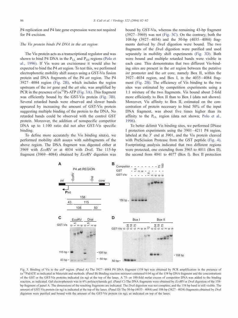
To better delimit Vis binding sites, we performed DNase
I protection experiments using the 3901–4211 P4 region,
labeled at the 3V end at 3901, and the Vis protein cleaved
with PreScission Protease from the GST peptide (Fig. 4).
Footprinting analysis indicated that two different regions
were protected, one extending from 3965 to 4011 (Box II),
the second from 4041 to 4077 (Box I). Box II protection
fragment (158 bp) was obtained by PCR amplification in the presence of
tures contained 0.04 ng of the 158 bp DNA fragment and the concentrations
5- or 100-fold molar excess of competitor DNA was added to the binding
The DNA fragments were obtained by EcoRVor DraI digestion of the 158-
he DraI digestion was not complete, and the 158 bp band is still visible. The
e 50-bp (4035–4084) and 108-bp (3827–4034) fragments obtained by DraI
g), as indicated on top of the lanes.
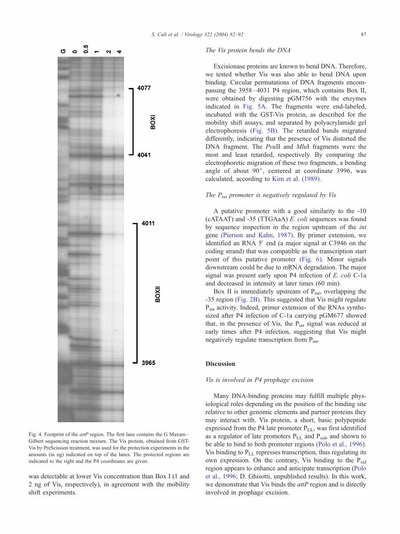
Fig. 4. Footprint of the attP region. The first lane contains the G Maxam–
Gilbert sequencing reaction mixture. The Vis protein, obtained from GST-
Vis by PreScission treatment, was used for the protection experiments in the
amounts (in ng) indicated on top of the lanes. The protected regions are
indicated to the right and the P4 coordinates are given.
S. Calı et al. / Virology
was detectable at lower Vis concentration than Box I (1 and
2 ng of Vis, respectively), in agreement with the mobility
shift experiments.
The Vis protein bends the DNA
Excisionase proteins are known to bend DNA. Therefore,
we tested whether Vis was also able to bend DNA upon
binding. Circular permutations of DNA fragments encom-
passing the 3958–4031 P4 region, which contains Box II,
were obtained by digesting pGM756 with the enzymes
indicated in Fig. 5A. The fragments were end-labeled,
incubated with the GST-Vis protein, as described for the
mobility shift assays, and separated by polyacrylamide gel
electrophoresis (Fig. 5B). The retarded bands migrated
differently, indicating that the presence of Vis distorted the
DNA fragment. The PvuII and MluI fragments were the
most and least retarded, respectively. By comparing the
electrophoretic migration of these two fragments, a bending
angle of about 90j, centered at coordinate 3996, was
calculated, according to Kim et al. (1989).
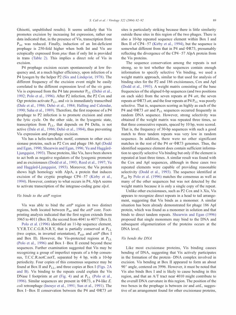
The Pint promoter is negatively regulated by Vis
A putative promoter with a good similarity to the -10
(cATAAT) and -35 (TTGAaA) E. coli sequences was found
by sequence inspection in the region upstream of the int
gene (Pierson and Kahn, 1987). By primer extension, we
identified an RNA 5V end (a major signal at C3946 on the
coding strand) that was compatible as the transcription start
point of this putative promoter (Fig. 6). Minor signals
downstream could be due to mRNA degradation. The major
signal was present early upon P4 infection of E. coli C-1a
and decreased in intensity at later times (60 min).
Box II is immediately upstream of Pint, overlapping the
-35 region (Fig. 2B). This suggested that Vis might regulate
Pint activity. Indeed, primer extension of the RNAs synthe-
sized after P4 infection of C-1a carrying pGM677 showed
that, in the presence of Vis, the Pint signal was reduced at
early times after P4 infection, suggesting that Vis might
negatively regulate transcription from Pint.
322 (2004) 82–92 87
Discussion
Vis is involved in P4 prophage excision
Many DNA-binding proteins may fulfill multiple phys-
iological roles depending on the position of the binding site
relative to other genomic elements and partner proteins they
may interact with. Vis protein, a short, basic polypeptide
expressed from the P4 late promoter PLL, was first identified
as a regulator of late promoters PLL and Psid, and shown to
be able to bind to both promoter regions (Polo et al., 1996).
Vis binding to PLL represses transcription, thus regulating its
own expression. On the contrary, Vis binding to the Psidregion appears to enhance and anticipate transcription (Polo
et al., 1996; D. Ghisotti, unpublished results). In this work,
we demonstrate that Vis binds the attP region and is directly
involved in prophage excision.
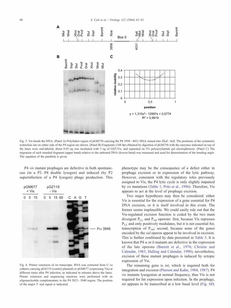
Fig. 5. Vis bends the DNA. (Panel A) Polylinker region of pGM756 carrying the P4 3958–4031 DNA cloned into XbaI –SalI. The positions of the symmetric
restriction site on either side of the P4 region are shown. (Panel B) Fragments (188 bp) obtained by digestion of pGM756 with the enzymes indicated on top of
the lanes were end-labeled, about 0.05 ng was incubated with 3 ng of GST-Vis, and separated on 5% polyacrylamide gel electrophoresis. (Panel C) The
migration of each retarded fragment (upper band) relative to the unbound DNA (lowest band) was measured and used for determination of the bending angle.
The equation of the parabola is given.
S. Calı et al. / Virology 322 (2004) 82–9288
P4 vis mutant prophages are defective in both spontane-
ous (in a P2–P4 double lysogen) and induced (by P2
superinfection of a P4 lysogen) phage production. This
Fig. 6. Primer extension of int transcripts. RNA was extracted from C-1a
cultures carrying pGZ119 (control plasmid) or pGM677 (expressing Vis) at
different times after P4 infection, as indicated in minutes above the lanes.
Primer extension and sequencing reactions were performed with an
oligonucleotide complementary to the P4 3823–3840 region. The position
of the major 5V end signal is indicated.
phenotype may be the consequence of a defect either in
prophage excision or in expression of the lytic pathway.
However, consistent with the regulatory roles previously
assigned to Vis, the P4 lytic cycle is only slightly impaired
by vis mutations (Table 1; Polo et al., 1996). Therefore, Vis
appears to act at the level of prophage excision.
Two major hypotheses may then be considered: either
Vis is essential for the expression of a gene essential for P4
DNA excision, or it is itself involved in this event. The
former seems implausible. We could easily rule out that the
Vis-regulated excision function is coded by the two main
divergent PLL and Psid operons: first, because Vis represses
PLL and only positively modulates, but it is not essential for,
transcription of Psid; second, because none of the genes
encoded by the sid operon appear to be involved in excision.
This is further confirmed by data presented in Table 3. It is
known that P4 a or y mutants are defective in the expression
of the late operons (Barrett et al., 1976; Christie and
Calendar, 1983; Halling and Calendar, 1990); nevertheless,
excision of these mutant prophages is induced by ectopic
expression of Vis.
The remaining gene is int, which is required both for
integration and excision (Pierson and Kahn, 1984, 1987). P4
vis mutants lysogenize at normal frequency, thus Vis is not
required for Int expression upon infection. In the prophage,
int appears to be transcribed at a low basal level (Fig. 6D;
S. Calı et al. / Virology 322 (2004) 82–92 89
Ghisotti, unpublished results). It seems unlikely that Vis
promotes excision by increasing Int expression, rather our
data indicated that, in the presence of Vis, transcription from
Pint was reduced. Finally, induction of an Int-deficient
prophage is 250-fold higher when both Int and Vis are
ectopically expressed from ptac than if only Int is provided
in trans (Table 2). This implies a direct role of Vis in
excision.
P4 prophage excision occurs spontaneously at low fre-
quency and, at a much higher efficiency, upon infection of a
P4 lysogen by the helper P2 (Six and Lindqvist, 1978). The
different frequency of the excision event might be easily
correlated to the different expression level of the vis gene.
Vis is expressed from the P4 late promoter PLL (Deho et al.,
1992; Polo et al., 1996). After P2 infection, the P2 Cox and
Ogr proteins activate PLL, and vis is immediately transcribed
(Dale et al., 1986; Deho et al., 1984; Halling and Calendar,
1990; Saha et al., 1989). Therefore, the first response of a P4
prophage to P2 infection is to promote excision and enter
the lytic cycle. On the other side, in the lysogenic state,
transcription from PLL, that depends on P4 Delta, is not
active (Dale et al., 1986; Deho et al., 1984), thus preventing
Vis expression and prophage excision.
Vis has a helix-turn-helix motif, common to other exci-
sionase proteins, such as P2 Cox and phage 186 Apl (Dodd
and Egan, 1990; Shearwin and Egan, 1996; Yu and Haggard-
Ljungquist, 1993). These proteins, like Vis, have been shown
to act both as negative regulators of the lysogenic promoter
and as excisionases (Dodd et al., 1993; Reed et al., 1997; Yu
and Haggard-Ljungquist, 1993). Moreover, the Vis protein
shows high homology with AlpA, a protein that induces
excision of the cryptic prophage CP4–57 (Kirby et al.,
1994). However, contrary to what occurs in P4, AlpA seems
to activate transcription of the integrase-coding gene slpA.
Vis binds to the attP region
Vis was able to bind the attP region in two distinct
regions, both located between Pint and the attP core. Foot-
printing analysis indicated that the first region extends from
3965 to 4011 (Box II), the second from 4041 to 4077 (Box I).
Polo et al. (1996) identified an 11-bp sequence element,
Y.Y.R.T.C.C.G.R.N.R.Y, that is partially conserved at PLL(two copies, in inverted orientation), Psid, and attP (Box I
and Box II). However, the Vis-protected regions at PLL(Polo et al., 1996) and Box I–Box II extend beyond these
sequences. Further examination suggested that Vis may be
recognizing a group of imperfect repeats of a 6-bp consen-
sus, T.C.C.R.notC.notT, separated by 4 bp, with a 10-bp
periodicity. Four copies of this consensus sequence may be
found at Box II and PLL, and three copies at Box I (Figs. 2A
and B). Vis binding to the repeats could explain the Vis
DNase I footprints at att (Fig. 4) and at PLL (Polo et al.,
1996). Similar sequences are present in AR73, a P4-like E.
coli retronphage (Inouye et al., 1991; Sun et al., 1991). The
Box I–Box II conservation between the P4 and AR73 att
sites is particularly striking because there is little similarity
outside these sites in this region of the two phages. There is
also a 10-bp repeated sequence element within Box I and
Box II of CP4–57 (Kirby et al., 1994), but the sequence is
somewhat different from that in P4 and AR73, presumably
reflecting the divergence of the CP4–57 AlpA protein from
the Vis proteins.
The sequence conservation among the repeats is not
strong, so to test whether the sequences contain enough
information to specify selective Vis binding, we used a
weight matrix approach, similar to that used for analysis of
binding sites for the P2 and 186 excisionases, Cox and Apl
(Dodd et al., 1993). A weight matrix consisting of the base
frequencies of the aligned 6-bp sequences (and two positions
on each side) from the seven repeats at P4 att, the seven
repeats atAR73 att, and the four repeats at P4 PLL was poorly
selective. That is, sequences scoring as highly as each of the
P4 and AR73 att and PLL sequences occurred frequently in
random DNA sequence. However, strong selectivity was
obtained if the weight matrix was repeated three times, so
that three consecutive repeats, spaced by 10 bp, were scored.
That is, the frequency of 30-bp sequences with such a good
match to three tandem repeats was very low in random
sequence. In addition, there were no other significant
matches in the rest of the P4 or AR73 genomes. Thus, the
identified sequence element does contain sufficient informa-
tion to specify selective Vis binding but only if the element is
repeated at least three times. A similar result was found with
the Cox and Apl sequences, although in these cases two
repeated elements were capable of providing reasonable
selectivity (Dodd et al., 1993). The sequence identified at
Psid by Polo et al. (1996) matches the consensus as well as
many of the other sequences but was not detected by the
weight matrix because it is only a single copy of the repeat.
Unlike other excisionases, such as P2 Cox and E Xis, Vis
appears to recognize direct repeats in a head to tail arrange-
ment, suggesting that Vis binds as a monomer. A similar
situation has been already demonstrated for phage 186 Apl
protein, which was found as a monomer in solution and that
binds to direct tandem repeats. Shearwin and Egan (1996)
proposed that single monomers may bind to the DNA and
subsequent oligomerization of the proteins occurs at the
DNA level.
Vis bends the DNA
Like most excisionase proteins, Vis binding causes
bending of DNA, suggesting that Vis actively participates
in the formation of the protein–DNA complex involved in
excision. Vis bending at Box II appeared to form an about
90j angle, centered on 3996. However, it must be noted that
Vis also binds Box I and is likely to cause bending in this
region, and that an A/T tract near 4010 might contribute to
the overall DNA curvature in this region. The position of the
two boxes in the prophage is between int and attL, sugges-
tive of an arrangement found for other excisionase proteins,
S. Calı et al. / Virology 322 (2004) 82–9290
such as Cox and Apl (Dodd et al., 1993; Yu and Haggard-
Ljungquist, 1993).
Materials and methods
Bacteria, phages, plasmids, and cultures media
The bacterial and phage strains and the plasmids used
are listed in Table 4. P4 coordinates throughout refer to
the complete P4 sequence, GenBank accession number
X51522. Bacterial strains were grown in LD broth (Sabbat-
Table 4
Bacterial and phage strains
Strain Relevant genotype Reference or source
E. coli C and E. coli K12
C-1a F�, prototrophic Sasaki and Bertani,
1965
C-117 C-1a (P2) Bertani, 1968
C-117 (P4 Hy1) P4 Hy1 prophage
inserted in attB
Calendar et al., 1981
C-339 C-1a (P2 lg cc) Barrett et al., 1976
C-5205 polyauxotrophic Deho, 1983
JM-101 polyauxotrophic Messing et al., 1981
Bacteriophages
P2 Wild type Bertani, 1968
P2 Aam129 Replication defective Lindahl, 1970
P2 lg cc Large plaques Bertani et al., 1969
P4 Wild type Six and Klug, 1973
P4 cI405 C8446T mutation in cI;
immunity defective,
recessive mutant
Calendar et al., 1981;
Lin, 1984
P4 del-vis deletion of the P4 8776–
9020 region, internal to vis
K. Reiter and
R. Calendar,
unpublished
P4 Hy1 substitution of the P4 220–
3631 region with E DNA;
lacks the gop, h, cII andint genes
Souza et al., 1978
P4 vis1 frameshift mutation in vis This work
P4 vis2 frameshift mutation in vis This work
P4 aam1 Replication defective Gibbs et al., 1973
P4 h35 Transactivation deficient Souza et al., 1977
Plasmids
pBend2 DNA bending vector Kim et al., 1989
pGEX-6P-1 expression vector Pharmacia
pGM582 P4 4249–2471 cloned into
pUC18; int gene expression
This work
pGM677 P4 9030–8762 cloned into
pGZ119EH; expression of
the Vis protein
This work
pGM756 P4 3958–4031 cloned into
pBend2
This work
pGM825 P4 8762–9027 cloned into
pGEX-6P-1; expression of the
GST-Vis fusion peptide
Polo et al., 1996
pGZ119EH cloning vector;
chloramphenicol resistance
Lessl et al., 1992
pUC18 cloning vector; ampicillin
resistance
Yanisch-Perron
et al., 1985
tini et al., 1995) and specific antibiotics were added when
required.
Construction of the P4 vis1 and P4 vis2 mutants
The 6447–10657 P4 region was cloned in pUC18 and
the resulting plasmid DNA was partially digested with
either AhaII or TaqI, end filled, and religated to create the
vis1 and vis2 mutations, respectively, and strain JM101
was transformed. The DNA extracted from the trans-
formants was sequenced to confirm the presence of the
vis1 mutation (a CG insertion at 8988 (sense strand); Fig.
2A) or the vis2 mutation (a CG insertion at 8904; Fig.
2A). The MluI–ApaLI fragments (P4 8626–10653) de-
rived from the above plasmids, carrying the vis mutations,
were ligated to the P4 wild-type 10653-cos-8626 fragment
and transfected into the C-5205 strain. The presence of
the vis mutations was confirmed by sequencing the phage
DNA.
Colony hybridization
The procedure was performed as described by Sambrook
et al. (1990) using as a probe a 17 nt long oligonucleotide
complementary to the 3823–3840 P4 sequence.
RNA extraction and primer extension
RNA was extracted from E. coli cultures at different
times after infection with P4, as described by Deho et al.
(1992). The concentration of the RNA was determined
spectrophotometrically and the quality of the RNA was
controlled by agarose gel electrophoresis. An oligonucleo-
tide complementary to the P4 3823–3840 region was end-
labeled and used both for sequencing the P4 int region and
in primer extension experiments.
Labeled DNA fragment amplification
The P4 3927–4984 region was amplified by PCR using
two oligonucleotides corresponding to the P4 3927–3947
and 4084–4056 sequence. Radioactive amplification with
AmpliTaq polymerase (Perkin-Elmer) was performed with 1
ng P4 DNA template in the presence of 30 ACi of (a32-P)
dATP (0.1 AM) and 1 AM of each dCTP, dGTP, and dTTP.
The labeled DNA fragment was separated by polyacryl-
amide gel electrophoresis and the band, visualized by
autoradiography, was cut out of the gel and eluted over-
night in H2O. Subfragment purification of this DNA
region, obtained by EcoRV or DraI digestion, was per-
formed in the same way.
Electrophoretic mobility shift assay
Purification of the GST-Vis fusion protein, binding to the
labeled DNA fragments, and gel electrophoresis in non-
S. Calı et al. / Virology 322 (2004) 82–92 91
denaturing 6% polyacrylamide gels were performed as in
Polo et al. (1996). Competitor DNA was obtained by PCR
amplification of a 200-bp-long portion of E. coli pnp region.
DNase I footprinting
GST tag was removed from GST-Vis fusion protein
purified from pGM825 by treatment with PreScission Pro-
tease (Amersham) as indicated by the manufacturer. DNaseI
footprinting was performed as described by Polo et al.
(1996). The P4 DNA region used covered 3901–4211 and
was end-labeled at 3901.
DNA bending
DNA fragments containing circular permutations of the
P4 3958–4031 region were obtained by digestion of
pGM756 with different enzymes, purification, and end-
labeling either by Klenow filling or with T4 polynucleotide
kinase. The electrophoretic migration was tested after bind-
ing with the GST-Vis protein, as described above. The
relative positions of the retarded bands were compared
and the bending angle was calculated according to Kim et
al. (1989).
Acknowledgments
We thank Richard Calendar and Kaye Reiter for kindly
providing P4 del-vis and the strain lysogenic for P4 Hy1.
This work was supported by grant no. 2002053757_001
from the Ministero dell’Istruzione, dell’Universita e della
Ricerca, Rome, Italy, by grant no. 01-0786 of INTAS, and
by grants from the Ministero dell’Universita e della Ricerca
Scientifica e Tecnologica, Rome, Italy.
References
Alano, P., Deho, G., Sironi, G., Zangrossi, S., 1986. Regulation of the plas-
mid state of the genetic element P4. Mol. Gen. Genet. 203, 445–450.
Barrett, K.J., Marsh, M.L., Calendar, R., 1976. Interactions between a
satellite bacteriophage and its helper. J. Mol. Biol. 106, 683–707.
Bertani, L.E., 1968. Abortive induction of bacteriophage P2. Virology 36,
87–103.
Bertani, G., Choe, B.K., Lindahl, G., 1969. Calcium sensitive and other
mutants of bacteriophage P2. J. Gen. Virol. 5, 97–104.
Briani, F., Deho, G., Forti, F., Ghisotti, D., 2001. The plasmid status of
satellite bacteriophage P4. Plasmid 45, 1–17.
Calendar, R., Ljungquist, E., Deho, G., Usher, D.C., Goldstein, R., You-
derian, P., Sironi, G., Six, E.W., 1981. Lysogenization by satellite phage
P4. Virology 113, 20–38.
Cho, E.H., Gumport, R.I., Gardner, J.F., 2002. Interactions between inte-
grase and excisionase in the phage lambda excisive nucleoprotein com-
plex. J. Bacteriol. 184, 5200–5203.
Christie, G.E., Calendar, R., 1983. Bacteriophage P2 late promoters.
Transcription initiation sites for two late mRNAs. J. Mol. Biol. 167,
773–790.
Dale, E., Christie, G.E., Calendar, R., 1986. Organization and expression of
the satellite bacteriophage P4 late gene cluster and the sequence of the
polarity suppression gene. J. Mol. Biol. 192, 793–803.
Deho, G., 1983. Circular genetic map of satellite bacteriophage P4. Virol-
ogy 126, 267–278.
Deho, G., Ghisotti, D., Alano, P., Zangrossi, S., Borrello, M.G., Sironi, G.,
1984. Plasmid mode of propagation of the genetic element P4. J. Mol.
Biol. 178, 191–207.
Deho, G., Zangrossi, S., Ghisotti, D., Sironi, G., 1988. Alternative pro-
moters in the development of bacteriophage plasmid P4. J. Virol. 62,
1697–1704.
Deho, G., Zangrossi, S., Sabbattini, P., Sironi, G., Ghisotti, D., 1992.
Bacteriophage P4 immunity controlled by small RNAs via transcription
termination. Mol. Microbiol. 6, 3415–3425.
Dodd, I.B., Egan, J.B., 1990. Improved detection of helix-turn-helix
DNA-binding motifs in protein sequences. Nucleic Acids Res. 18,
5019–5026.
Dodd, I.B., Reed, M., Egan, J.B., 1993. The Cro-like Apl repressor of
coliphage 186 is required for prophage excision and binds near the
phage attachment site. Mol. Microbiol. 10, 1139–1150.
Esposito, D., Scocca, J., 1997. The integrase family of tyrosine recombi-
nases: evolution of a conserved active site domain. Nucleic Acids Res.
25, 3605–3614.
Finkel, S.E., Johnson, R.C., 1992. The Fis protein: it’s not just for DNA
inversion anymore. Mol. Microbiol. 6, 3257–3265.
Forti, F., Polo, S., Lane, K.B., Six, E.W., Deho, G., Ghisotti, D., 1999.
Translation of two nested genes in bacteriophage P4 controls immunity-
specific transcription termination. J. Bacteriol. 181, 5225–5233.
Forti, F., Dragoni, I., Briani, F., Deho, G., Ghisotti, D., 2002. Character-
ization of the small antisense CI RNA that regulates bacteriophage P4
immunity. J. Mol. Biol. 315, 541–549.
Freundlich, M., Ramani, N., Mathew, E., Sirko, A., Tsui, P., 1992. The role
of integration host factor in gene expression in Escherichia coli. Mol.
Microbiol. 6, 2557–2563.
Friedman, D.I., 1992. Interaction between bacteriophage lambda and its
Escherichia coli host. Curr. Opin. Genet. Dev. 2, 727–738.
Ghisotti, D., Chiaramonte, R., Forti, F., Zangrossi, S., Sironi, G., Deho, G.,
1992. Genetic analysis of the immunity region of phage-plasmid P4.
Mol. Microbiol. 6, 3405–3413.
Gibbs, W., Goldstein, R.N., Wiener, R., Lindqvist, B., Calendar, R., 1973.
Satellite bacteriophage P4: characterization of mutants in two essential
genes. Virology 53, 24–39.
Gingery, R., Echols, H., 1967. Mutants of bacteriophage lambda unable to
integrate into the host chromosome. Proc. Natl. Acad. Sci. U.S.A. 58,
1507–1515.
Grainge, I., Jayaram, M., 1999. The integrase family of recombinases:
organization and function of the active site. Mol. Microbiol. 33,
449–456.
Grambow, N.J., Birkeland, N.K., Anders, D.L., Christie, G.E., 1990.
Deletion analysis of a bacteriophage P2 late promoter. Gene 95,
9–15.
Halling, C., Calendar, R., 1990. Bacteriophage P2 ogr and P4 y genes act
independently and are essential for P4 multiplication. J. Bacteriol. 172,
3549–3558.
Inouye, S., Sunshine, M.G., Six, E.W., Inouye, M., 1991. Retronphage
AR73: an E. coli phage that contains a retroelement and integrates into
a tRNA gene. Science 252, 969–971.
Julien, B., Calendar, R., 1995. Purification and characterization of the
bacteriophage P4 delta protein. J. Bacteriol. 177, 3743–3751.
Kim, J., Zwieb, C., Wu, C., Adhya, S., 1989. Bending of DNA by gene-
regulatory proteins: construction and use a DNA bending vector. Gene
85, 15–23.
Kirby, E.J., Trempy, E.J., Gottesman, S., 1994. Excision of a P4-like
cryptic prophage leads to Alp protease expression in Escherichia coli.
J. Bacteriol. 176, 2068–2081.
Lessl, M., Balzer, D., Lurz, R., Waters, V.L., Guiney, D.G., Lanka, E.,
1992. Dissection of IncP conjugative plasmid transfer: definition of
S. Calı et al. / Virology 322 (2004) 82–9292
the transfer region Tra2 by mobilization of the Tra1 region in trans.
J. Bacteriol. 174, 2493–2500.
Lin, C.S., 1984. Nucleotide sequence of the essential region of bacterio-
phage P4. Nucleic Acids Res. 12, 8667–8684.
Lindahl, G., 1970. Bacteriophage P2: replication of the chromosome
requires a protein which acts only on the genome that coded for it.
Virology 42, 522–533.
Lindqvist, B.H., Deho, G., Calendar, R., 1993. Mechanisms of genome
propagation and helper exploitation by satellite phage P4. Microbiol.
Rev. 57, 683–702.
Messing, J., Crea, R., Seeburg, P.H., 1981. A system for shotgun DNA
sequencing. Nucleic Acids Res. 9, 309–321.
Nash, H.A., 1981. Integration and excision of bacteriophage lambda: the
mechanism of conservation site specific recombination. Annu. Rev.
Genet. 15, 143–167.
Nash, H.A., 1990. Bending and supercoiling of DNA at the attachment site
of bacteriophage lambda. Trends Biochem. Sci. 15, 222–227.
Numrynch, E.T., Gumport, I.R., Gardner, F.J., 1992. Characterization of the
bacteriophage lambda excisionase (Xis) protein: the C-terminus is re-
quired for Xis-integrase cooperativity but not for DNA binding. EMBO
J. 11, 3797–3806.
Pierson III, L.S., Kahn, M.L., 1984. Cloning of the integration and attach-
ment regions of bacteriophage P4. Mol. Gen. Genet. 195, 44–51.
Pierson III, L.S., Kahn, M.L., 1987. Integration of satellite bacteriophage
P4 in E. coli: DNA sequences of the phage and host regions involved in
site-specific recombination. J. Mol. Biol. 196, 487–496.
Polo, S., Sturniolo, T., Deho, G., Ghisotti, D., 1996. Identification of a
phage coded DNA-binding protein that regulates transcription from late
promoters in bacteriophage P4. J. Mol. Biol. 257, 745–755.
Reed, M.R., Shearwin, K., Peel, L.M., Egan, B., 1997. The dual role of Apl
in prophage induction of coliphage 186. Mol. Microbiol. 23, 669–681.
Sabbattini, P., Forti, F., Ghisotti, D., Deho, G., 1995. Control of transcrip-
tion termination by an RNA factor in bacteriophage P4 immunity:
identification of the target sites. J. Bacteriol. 177, 1425–1434.
Saha, S., Haggard-Ljungquist, E., Nordstrom, K., 1989. Activation of pro-
phage P4 by the P2 Cox protein and the sites of action of the Cox
protein on the two phage genomes. Proc. Natl. Acad. Sci. U.S.A. 66,
3973–3977.
Sambrook, J., Fritsch, E.F., Maniatis, T., 1990. Molecular Cloning: A
Laboratory Manual. Cold Spring Harbor Laboratory Press, Cold spring
Harbor, NY.
Sasaki, I., Bertani, G., 1965. Growth abnormalities in Hfr derivatives of
Escherichia coli strain C. J. Gen. Microbiol. 40, 365–376.
Shearwin, K., Egan, B., 1996. Purification and self-association equilibria of
the lysis-lysogeny switch proteins of coliphage 186. J. Biol. Chem. 27,
11525–11531.
Six, E.W., Klug, C.A.C., 1973. Bacteriophage P4: a satellite virus depend-
ing on a helper such as prophage P2. Virology 51, 327–344.
Six, E.W., Lindqvist, B.H., 1978. Mutual derepression in the P2–P4 bac-
teriophage system. Virology 87, 217–230.
Souza, L., Calendar, R., Six, E.W., Lindqvist, B.H., 1977. A transactivation
mutant of satellite phage P4. Virology 81, 81–90.
Souza, L., Geisselsoder, J., Hopkins, A., Calendar, R., 1978. Physical
mapping of the satellite phage P4 genome. Virology 85, 335–342.
Sun, J., Inouye, M., Inouye, S., 1991. Association of a retroelement with
a P4-like cryptic prophage (retronphage phi R73) integrated into the
selenocystyl tRNA gene of Escherichia coli. J. Bacteriol. 173,
4171–4181.
Swalla, B.M., Gumport, R.I., Gardner, J.F., 2003. Conservation of structure
and function among tyrosine recombinases: homology-based modeling
of the lambda integrase core-binding domain. Nucleic Acids Res. 31,
805–818.
Yanisch-Perron, C., Vieira, J., Messing, J., 1985. Improved M13 phage
cloning vectors and host strains: nucleotide sequence of the M13mp18
and pUC19 vectors. Gene 33, 103–119.
Yu, A., Haggard-Ljungquist, E., 1993. The Cox protein is a modulator of
directionality in bacteriophage P2 site-specific recombination. J. Bac-
teriol. 175, 7848–7855.