comparison of four molecular typing methods for characterization of corynebacterium diphtheriae and...
TRANSCRIPT

JOURNAL OF CLINICAL MICROBIOLOGY, Nov. 2008, p. 3626–3635 Vol. 46, No. 110095-1137/08/$08.00�0 doi:10.1128/JCM.00300-08Copyright © 2008, American Society for Microbiology. All Rights Reserved.
Comparison of Four Molecular Typing Methods for Characterizationof Corynebacterium diphtheriae and Determination of TranscontinentalSpread of C. diphtheriae Based on BstEII rRNA Gene Profiles�
Aruni De Zoysa,1* Peter Hawkey,2 Andre Charlett,3 and Androulla Efstratiou1
Respiratory and Systemic Infection Laboratory, Health Protection Agency, Centre for Infections, 61 Colindale Avenue,London NW9 5EQ, United Kingdom1; Health Protection Agency, Heartlands Hospital, Birmingham, United Kingdom,
and Division of Immunity and Infection, Medical School, University of Birmingham, Birmingham,United Kingdom2; and Statistics, Modelling & Bioinformatics, Health Protection Agency,
Centre for Infections, 61 Colindale Avenue, London NW9 5EQ, United Kingdom3
Received 13 February 2008/Returned for modification 8 May 2008/Accepted 2 September 2008
The diphtheria epidemic in the Russian Federation in the 1990s made diphtheria a focus of global concernonce again. The development of rapid and reproducible typing methods for the molecular characterization ofCorynebacterium diphtheriae has become a priority in order to be able to monitor the spread of this importantpathogen on a global scale. We report on a comparison of four molecular typing methods (ribotyping,pulsed-field gel electrophoresis [PFGE], random amplification of polymorphic DNA [RAPD], and amplifiedfragment length polymorphism [AFLP]) for the characterization of C. diphtheriae strains. Initially, 755 isolatesoriginating from 26 countries were analyzed by ribotyping. One strain of each ribotype was then randomlychosen and characterized by PFGE, RAPD, and AFLP. In order to ascertain whether the Eastern Europeanepidemic ribotype could be further discriminated, 10 strains of ribotype D1 (the epidemic ribotype) fromdifferent geographical regions were randomly chosen and subjected to analysis by PFGE, RAPD, and AFLP.The results revealed that ribotyping is highly discriminatory and reproducible and is currently the method ofchoice for typing C. diphtheriae. PFGE and AFLP were less discriminatory than ribotyping and RAPD. Anassessment of the transcontinental spread of the organism showed that several genotypes of C. diphtheriaecirculated on different continents of the world and that each outbreak was caused by a distinct clone. Theribotypes seen in Europe appeared to be distinct from those seen elsewhere, and certain ribotypes appeared tobe unique to particular countries.
Due to the highly effective diphtheria vaccine that becameavailable in the 1940s and 1950s, the incidence of diphtheriadeclined dramatically in many parts of the world. However,epidemic diphtheria reemerged in Eastern Europe in the1990s, and the disease spread to all 15 Newly IndependentStates (NIS) of the former USSR. The disease is endemic incountries such as Turkey, Bangladesh, India, Pakistan, andVietnam and in Africa and parts of South America (7). Theonset of these epidemics and the occurrence of endemicity insome countries highlighted the importance of monitoring thespread of C. diphtheriae from index cases to the community,country, and beyond. It is also necessary to distinguish domes-tic from imported cases to allow the adequate implementationof local preventive measures. Therefore, the availability ofrapid and reproducible typing tools for the molecular charac-terization of C. diphtheriae became a high priority.
Here we report on a comparison of four molecular typingmethods (ribotyping, pulsed-field gel electrophoresis [PFGE],random amplification of polymorphic DNA [RAPD], and am-plified fragment length polymorphism [(AFLP]) for character-ization of C. diphtheriae. Initially, 755 isolates originating from
26 countries were analyzed by ribotyping. One strain of eachribotype was randomly chosen and characterized by PFGE,RAPD, and AFLP. In order to ascertain whether the EasternEuropean epidemic ribotype could be further discriminated byother typing methods, 10 strains of ribotype D1 (the epidemicribotype) from different geographical regions were randomlychosen and were subjected to analysis by PFGE, RAPD, andAFLP. The transcontinental spread of C. diphtheriae was de-termined by using the extensive set of ribotyping results ob-tained from this study.
MATERIALS AND METHODS
Bacterial isolates. Seven hundred fifty-five C. diphtheriae isolates from 26countries (Armenia, Australia, Belarus, Denmark, Dominican Republic, Estonia,Finland, France, Germany, Italy, Kazakhstan, Kenya, Kyrgyzstan, Latvia, Poland,Romania, Rwanda, Russia, Sweden, Thailand, Turkmenistan, the United King-dom, Ukraine, Uzbekistan, the United States, and Vietnam) referred to theStreptococcus and Diphtheria Reference Unit at the Centre for Infections,London, United Kingdom, for identification and typing from 1985 to 2000 werechosen for use in the ribotyping studies. The isolates were from patients withdiphtheria, pharyngitis, or tonsillitis; asymptomatic carriers; and contacts. Thedisease status was unknown for the sources of 68% of the isolates. The isolateswere from patients aged between 12 months and 83 years (the age was not knownfor 52% of the patients). Table 1 summarizes the 755 C. diphtheriae strainsanalyzed by ribotyping. One isolate of each ribotype was randomly chosen foranalysis by PFGE, RAPD, and AFLP (Table 2).
A majority of the isolates analyzed were from the NIS of the former USSR.Isolates from Thailand, Sweden, and the United States were outbreak strains;and the isolates from the United Kingdom were mainly from imported cases ofdiphtheria. All isolates were first identified by standard microbiology techniques
* Corresponding author. Mailing address: Systemic Infection Labo-ratory, Health Protection Agency Centre for Infections, 61 ColindaleAvenue, London NW9 5EQ, United Kingdom. Phone: 44 208-327-7536. Fax: 44 208-200-6528. E-mail: [email protected].
� Published ahead of print on 10 September 2008.
3626

at the local microbiology laboratory and sent to the diphtheria reference centerswithin each country for confirmation of species identity and toxigenicity. Theisolates were then transported to the Respiratory and Systemic Infection Labo-ratory at the Centre for Infections, London, United Kingdom, for additionaltyping.
The isolates described above were obtained through an extensive collaborativenetwork that was formed through the European Laboratory Working Group onDiphtheria (6).
Biotyping and toxigenicity testing. Biotyping of all 755 isolates was performedwith the API Coryne system, as described previously (8). All isolates were testedfor toxin production by the conventional Elek immunoprecipitation test (8).Isolates submitted after 1996 were also tested by the modified Elek test (9).
Ribotyping. All 755 isolates were ribotyped by using BstEII, as previouslydescribed by De Zoysa et al. (4). A cDNA probe derived from the 16S and 23SrRNA of C. diphtheriae type strain NCTC 11397 was used.
PFGE, RAPD, and AFLP. One isolate of each ribotype (a total of 74 isolates)was randomly chosen and subjected to analysis by PFGE, RAPD, and AFLP.Ten isolates of the most predominant ribotype in Russia (ribotype D1) alsorandomly chosen from different geographical regions were analyzed by the threemethods. All methods were performed as described previously by De Zoysa et al.(2–4). RAPD and AFLP were performed with purified DNA samples.
Data capture for computer analysis of ribotype, PFGE, RAPD, and AFLPprofiles. The ribotype, PFGE, RAPD, and AFLP profiles were scanned (Scan-maker E6; Microtek Lab) and analyzed with the Bionumerics program (version
TABLE 1. Summary of the 755 C. diphtheriae isolates analyzed by ribotyping
Country No. ofisolates Yr of isolation Biotype(s) (toxigenicitya) Ribotype(s) (no. of isolates)b
Armeniac 19 1999 Gravis (18 tox�), mitis (1 tox�) D1 (11), D4 (7), D7 (1)Australia 32 1992–1995 Gravis (17 tox�), mitis (1 tox�, 2 tox�) D9 (18), D15 (1), D46 (1), D47 (1), D48 (2),
D49 (1), D50 (1), D51 (3), D52 (1), D56(2), D57 (1)
Belarusc 79 1996–2000 Gravis (48 tox�, 12 tox�), mitis(16 tox�, 3 tox�)
D1 (13), D4 (26), D5 (1), D6 (2), D7 (7),D10 (25), D15 (1), D19 (2), D30 (1),D40 (1)
Denmark 1 Mitis (tox�) D74 (1)Dominican Republic 3 1995 Mitis (3 tox�) D71 (2), D72 (1)Estoniad 26 1993–1995 Gravis (13 tox�, 11 tox �), mitis
(1 tox�, 1 tox �)D1 (12), D4 (10), D5 (1), D7 (1), D10 (2)
Finlandd 6 1993–1995 Gravis (4 tox�), mitis (1 tox�, 1 tox�) D1 (4), D10 (2)France 1 1993 Mitis (tox�) D46 (1)Germany 24 1993–1994 Gravis (4 tox�, 9 tox�), mitis (3 tox�,
9 tox�), belfanti (3 tox�)D1 (3), D4 (1), D11 (8), D13 (1), D14 (3),
D15 (3), D16 (1), D17 (1), D18 (1),D26 (2)
Italy 6 1994–1996 Gravis (1 tox�, 4 tox�), mitis (1tox�) D11 (4), D45 (1), D73 (1)Kazakhstanc 43 1995–1997 Gravis (39 tox�), mitis (5 tox�) D1 (7), D4 (32), D7 (4)Kenya 1 1998 Mitis (1 tox�) D62 (1)Kyrghyzstanc 4 1993–1995 Gravis (1 tox�), mitis (1 tox�) D1 (1), D7 (3)Latviac 115 1999–2000 Gravis (105 tox�, 1 tox�), mitis
(7 tox�, 2 tox�)D1 (50), D4 (61), D10 (4)
Poland 1 1994 Intermedius (tox�) D54 (1)Romania 17 1994 Gravis (2 tox�, 6 tox�), mitis (5 tox�,
1 tox�), belfanti (1 tox�),intermedius (1 tox�)
D11 (6), D20 (1), D21 (6), D22 (1), D23 (1),D24 (1), D25 (1)
Ruanda 2 1994 Mitis (tox�) D54 (1), D55 (1)Russiac 218 1966, 1993–95 Gravis (148 tox�, 7 tox�), mitis
(63 tox�, 8 tox�)D1 (81), D2 (1), D3 (2), D4 (56), D5 (1),
D6 (3), D7 (56), D8 (1), D9 (3), D10 (11),D11 (2), D12 (1)
Swedene 13 1994 Gravis (2 tox�), mitis (6 tox�, 3tox�),belfanti (2 tox�)
D4 (1), D11 (1), D17 (1), D22 (1), D26 (6),D27 (1), D28 (1), D29 (1)
Thailandf 31 1994–1996 Gravis (1 tox�), mitis (27 tox�, 3 tox�) D19 (1), D34 (13), D63 (2), D64 (6), D65(2), D66 (3), D67 (1), D68 (1), D69 (1),D70 (1)
Turkmenistanc 25 1995 Gravis (17 tox�, 4 tox�), mitis (3 tox�,1 tox�)
D1 (2), D4 (18), D7 (3), D10 (2)
United Kingdomg 22 1985–1998 Gravis (3 tox�), mitis (17 tox�, 2 tox�) D1 (1), D7 (1) D30 (1), D31 (1), D32 (2),D33 (1), D34 (1), D35 (1), D36 (1), D37(1), D38 (1), D39 (1), D40 (4), D41 (1),D42 (2), D43 (1), D44 (1)
Ukrainec 19 1999 Gravis (19 tox�) D1 (19)Uzbekistanc 11 1995 Gravis (3 tox�, 2 tox�), mitis (6 tox�) D1 (3), D7 (1), D10 (2), D15 (1), D19 (4)United Statesh 23 1972–82, 1995 Gravis (8 tox�, 3 tox�), mitis (6 tox�),
intermedius (2 tox�, 3 tox�)D4 (1), D13 (3), D50 (3), D57 (5), D58 (8),
D59 (3)Vietnam 13 1995 Mitis (13 tox�) D9 (1), D60 (3), D61 (9)
a The numbers of isolate that are toxin positive (tox�) and toxin negative (tox�) are given in parentheses.b International designations for some of the predominant ribotypes are as follows: D1, Sankt Petersburg; D7, Otchakov; D11, Vladimir; D4, Rossija; D10, Cluj.c The majority of the isolates are from the epidemic which began in the Russian Federation in 1990.d Imported cases of diphtheria from the NIS.e Isolates from the Scandinavian outbreak, which occurred in the mid-1980s.f Isolates from the outbreak in Thailand in 1994.g The majority of the isolates are from imported cases of diphtheria from Asia, Africa, and the Far East.h Isolates from the outbreak in Seattle, WA (1971 to 1982).
VOL. 46, 2008 EPIDEMIOLOGY OF CORYNEBACTERIUM DIPHTHERIAE 3627

3.0, Applied Maths, Kortrijk, Belgium). Normalization within each gel wasachieved with a bacteriophage � HindIII marker (Invitrogen) for ribotyping, abacteriophage � concatemer (Bio-Rad) for PFGE, and the Gene ruler DNAladder mix (MBI Fermentas) for RAPD and AFLP. Molecular size markers were
placed in every third lane of the gel, and one gel marker lane (selected atrandom) was used as the normalization standard for the between-gel normaliza-tion of all gels in the study. The bands for each profile (ribotype, PFGE, RAPD,and AFLP) were first identified by use of the auto search facility (settings at 10
TABLE 2. Summary of the 74 strains analyzed by PFGE, RAPD, and AFLP
Lab no. Region, country, yr of isolation Age (yr) Sexa Disease Biotype Toxb Ribotype
CD93/46 St. Petersburg, Russia, 1993 46 M Tonsillitis Gravis � D1CD95/66 Omsk region, Russia Gravis � D2CD93/69 Murmansk, Russia, 1993 30 Diphtheria Gravis � D3CD93/78 Murmansk, Russia, 1993 21 M Diphtheria Gravis � D4CD93/266 St. Petersburg, Russia, 1993 4 F Diphtheria Gravis � D5CD93/181 Moscow, Russia, 1966 Carrier Gravis � D6CD93/45 St. Petersburg, Russia, 1993 34 M Carrier Mitis � D7CD93/132 Kaliningrad Oblast, Russia 1993 28 M Carrier Mitis � D8CD93/183 Dagestan, Russia, 1983 Mitis � D9CD93/186 St. Petersburg, Russia, 1989 Carrier Mitis � D10CD93/274 Vladimir, Russia Carrier Gravis � D11CD93/277 Moscow, Russia 19 Carrier Gravis � D12CD94/68 Erlabrunn, Germany 5 Mitis � D13CD94/66 Berlin, Germany 42 M Mitis � D14CD94/69 Erlabrunn, Germany 5 Gravis � D15CD94/72 Greifswald, Germany Belfanti � D16CD94/76 Dresden, Germany Belfanti � D17CD94/260 Germany, 1993 Bronchus (source) Belfanti � D18CD94/263 Thailand 28 F Skin lesion (German patient) Mitis � D19CD94/238 Romania Gravis � D20CD94/252 Romania Gravis � D21CD94/249 Romania Mitis � D22CD94/240 Romania Intermedius � D24CD94/241 Romania Intermedius � D25CD94/91 Sweden (outbreak) Mitis � D26CD94/94 Sweden Mitis � D27CD94/232 Sweden Mitis � D28CD94/231 Sweden Belfanti � D29CD93/32 United Kingdom 39 Immigrant Gravis � D30CD94/149 United Kingdom (Birmingham) 14 M Pharyngitis Gravis � D31CD85/2 Swansea, United Kingdom Tonsillitis Mitis � D32CD85/29 Swansea, United Kingdom (imported case from
Tunisia)M Mitis � D33
CD90/39 London, United Kingdom Tonsillitis Mitis � D34CD93/4 United Kingdom (imported from Australia) M Mitis � D35CD93/19 United Kingdom (imported from Bangladesh) 15 Diphtheria Mitis � D36CD93/117 United Kingdom 6 Mitis � D37CD93/121 United Kingdom 43 F Mitis � D38CD93/154 United Kingdom (contact of a Somali) 14 F Mitis � D39CD98/135 United Kingdom (imported case from Tanzania) 19 Cutaneous Mitis � D40CD92/48 Bristol, United Kingdom Mitis � D41CD94/214 United Kingdom (imported case from India) 32 F Pharyngitis Mitis � D42CD94/8 United Kingdom 64 Mitis � D43CD94/9 United Kingdom 31 F Mitis � D44CD94/16 Italy (imported case from Peru) Gravis � D45CD93/242 France Mitis � D46CD93/28 Perth, Australia 45 Mitis � D47CD94/38 Western Australia 21 M Gravis � D48CD94/39 Australia 20 M Gravis � D49CD94/34 Cairns, Queensland, Australia 78 M Gravis � D50CD94/35 New South Wales, Australia 47 F Gravis � D51CD93/29 Western Australia 24 Gravis � D52CD94/62 Przemysl, Poland 38 F Intermedius � D53CD94/281 Rwanda (imported case into Italy) Wound (source) Mitis � D54CD94/282 Rwanda (imported case into Italy) Pharyngeal Mitis � D55CD95/184 CGH, Western Australia Gravis � D56CD94/126 Seattle, WA Intermedius � D57CD94/132 Seattle, WA Gravis � D58CD94/145 Seattle, WA Mitis � D59CD95/437 Vietnam Mitis � D60CD95/439 Vietnam Mitis � D61CD98/138 Kenya Cutaneous Mitis � D62CD96/241 Thailand (patient from Nan Province) Mitis � D63CD96/244 Thailand 4 Mitis � D64CD96/264 Thailand 11 Mitis � D65CD96/246 Thailand 3 Mitis � D66CD96/248 Thailand 14 Mitis � D67CD96/250 Thailand Mitis � D68CD96/260 Thailand 8 Mitis � D69CD96/261 Thailand 2 Gravis � D70CD95/385 Dominican Republic Mitis � D71CD95/387 Dominican Republic Mitis � D72CD95/404 Italy Mitis � D73CD98/10 Denmark Mitis � D74
a M, male; F, female.b Tox, toxigenicity.
3628 DE ZOYSA ET AL. J. CLIN. MICROBIOL.

to 14% minimum profiling and 0.5% minimal area), followed by review andmodification after a careful visual comparison of the image provided by theBionumerics program. The methods were compared by generating a dendrogramby using the Dice similarity coefficient, together with the unweighted pair groupmethod with arithmetic averages clustering method, with the position toleranceset at 1.2% and the optimization set at 0.5%.
Statistical methods. Two indices were used to provide summaries of thediversity of the different types observed by the typing methods. These wereSimpson’s index of diversity (D) (�S) and the Hunter and Gaston modified index
(�HG) (11, 17). The formulas for these two indices are �S � 1 � �i � 1
n �xi
N�2
and
�HG � 1 � �i � 1
n �xi
N� ��xi � 1N � 1�, respectively, where N is the total number of
isolates and xi is the number of isolates of the ith type.These indices can be interpreted as the probability that two isolates selected at
random are of different types. The isolates used in this study are unlikely to berepresentative of all clinical C. diphtheriae isolates throughout the world duringthis time period; thus, these diversity estimates are provided to distinguishwhether one typing method provides a greater discrimination between the iso-lates than another method rather than providing a gauge of the biologicaldiversity in a population. Bootstrap estimates of the 95% confidence intervals(CIs) around the diversity estimates were calculated by using 1,000 resamplestaken with replacement.
RESULTS
Biotyping and toxigenicity testing. A total of 755 isolateswere biotyped and tested for toxin production. The number ofisolates examined from each country, together with their bio-types and toxigenicity status, are summarized in Table 1.Among the 755 isolates examined, there were 433 (57.3%)toxigenic biotype gravis isolates, 91 (12.05%) nontoxigenic bio-type gravis, 176 (23.3%) toxigenic biotype mitis isolates, 42(5.5%) nontoxigenic biotype mitis isolates, and 6 (0.79%) non-toxigenic biotype belfanti isolates. Seven strains belonged tobiotype intermedius (three were toxigenic isolates and fourwere nontoxigenic isolates). The majority of isolates from theNIS examined were toxigenic and belonged to biotype gravis,and isolates from Australia were mainly nontoxigenic biotypegravis. The isolates from the Far East (Thailand and Vietnam)were predominantly toxigenic biotype mitis.
Ribotyping. All 755 isolates were typed by ribotyping withBstEII. The technique showed 100% typeability and was highly
reproducible. The characteristic patterns of the bands ob-served for individual strains were found to be independent ofsuch variables as the batch of DNA or the batch of probe used.Each ribotype profile obtained with BstEII comprised 9 to 11bands, and the ribotype profiles were analyzed by using theBionumerics computer software program (Applied Maths).
Analysis of the 755 C. diphtheriae ribotype profiles with theBionumerics program revealed 74 distinct ribotype patterns.The patterns were designated D1 to D74 (Table 2), and Fig. 1illustrates ribotype profiles D1 to D29. The international des-ignations for some of the predominant ribotypes are listed inTable 1. Figure 2 represents the relationships between the 74BstEII restriction digestion patterns determined by using theBionumerics program to generate the dendrogram by use ofthe criteria described in Materials and Methods. The predom-inant ribotypes in Russia (ribotypes D1 and D4) appeared tobe very closely related (similarity, 95.24%). The profiles ofthese two ribotypes were also very closely related to the profileof ribotype D12. The profiles of ribotypes D1, D4, and D12were 82.24% similar.
It must be noted that in a previous study (4) the ribotypeprofiles of biotype gravis isolates were designated with theprefix G and the profiles of biotype mitis isolates were desig-nated with the prefix M. However, the ribotype nomenclaturedescribed in 1995 (4) was later revised and the prefixes G andM were replaced by the prefix D, which is the nomenclatureused in this study.
PFGE, RAPD, and AFLP. Seventy-four isolates (one strainof each ribotype) were analyzed by PFGE, RAPD, and AFLP.In addition, 10 strains of the most predominant ribotype inRussia (ribotype D1) randomly chosen from different geo-graphical regions were also analyzed by the three methods. Allprofiles were analyzed by using the Bionumerics program (Ap-plied Maths). PFGE with the restriction endonuclease SfiIproduced 72 distinct PFGE profiles, which were designated P1to P72. The profiles consisted of 15 to 25 DNA fragmentsranging from 24 kb to 339.5 kb. Figure 3 illustrates the PFGEprofiles of ribotypes D1 to D12.
FIG. 1. BstEII ribotype profiles for the C. diphtheriae isolates tested. Lanes 1, 3, 5, 8, 11, 14, 17, 20, 23, 26, 29, 32, 35, 38, 41, and 45,bacteriophage lambda HindIII digests used as size standards (sizes are indicated on the left); lane 2, ribotype D1; lane 4, ribotype D2; lane 6,ribotype D3; lane 7, ribotype D4; lane 9, ribotype D5; lane 10, ribotype D6; lane 12, ribotype D7; lane 13, ribotype D8; lane 15, ribotype D9; lane16, ribotype D10; lane 18, ribotype D11; lane 19, ribotype D12; lane 21, ribotype D13; lane 22, ribotype D14; lane 24, ribotype D15; lane 25,ribotype D16; lane 27, ribotype D17; lane 28, ribotype D18; lane 30, ribotype D19; lane 31, ribotype D20; lane 33, ribotype D21; lane 34, ribotypeD22; lane 36, ribotype D23; lane 37, ribotype D24; lane 39, ribotype D25; lane 40, ribotype D26; lane 42, ribotype D27; lane 43, ribotype D28; lane44, ribotype D29. When the results from different gels are combined, markers from each gel are included.
VOL. 46, 2008 EPIDEMIOLOGY OF CORYNEBACTERIUM DIPHTHERIAE 3629
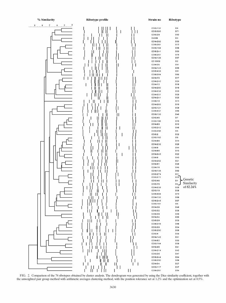
FIG. 2. Comparison of the 74 ribotypes obtained by cluster analysis. The dendrogram was generated by using the Dice similarity coefficient, together withthe unweighted pair group method with arithmetic averages clustering method, with the position tolerance set at 1.2% and the optimization set at 0.5%.
3630

Analysis of the 74 isolates by RAPD revealed 74 distinctprofiles, which were designated Rp1 to Rp74. The profilesconsisted of 13 to 27 fragments that ranged from 200 bp to2,072 bp in size. Profiles Rp1 to Rp13 are illustrated in Fig. 4.AFLP revealed 72 profiles among the 74 isolates, and theprofiles were designated AP1 to AP72. The AFLP profileswere comprised of 24 to 33 fragments that ranged from 200 bpto 300 bp in size. All three methods showed 100% typeability.PFGE and AFLP did not distinguish between strains ofribotypes D1, D4, and D12 (Fig. 3 and 5), and therefore, thePFGE and AFLP profiles of these three ribotypes clusteredtogether at a similarity level of 100%. The 10 isolates ofribotype D1 from different geographical origins were not distin-guished further by these methods. The RAPD profiles ofribotypes D1, D4, and D12 showed a similarity of 91.77%.
Statistical analysis. The diversity indices and their 95% con-fidence intervals calculated for the ribotyping results are shown
in Table 3. As expected, the diversity indices were much lowerwhen they were calculated by using the data for all 755 isolates.This is due to the abundance of certain ribotypes (i.e., ri-botypes D1 and D4) that have occurred in spatiotemporalclusters. Table 4 shows the diversity of ribotypes and their 95%CIs within disease types (diphtheria, tonsillitis, and carriers).The results show that the ribotypes were less diverse amongisolates from cases of diphtheria and were more diverse amongisolates from cases of tonsillitis. The relative frequency of par-ticular ribotypes between different countries was also exploredby using the diversity indices. The ribotypes observed in veryfew countries have low diversity indices, while those observedin many countries are likely to have larger diversities. Theseresults are shown in Table 5.
Ribotype D4 appeared to be far more diverse (�HG � 0.812)than ribotype D34 (�HG � 0.143). Ribotype D4 was seenamong isolates from 11 countries, and ribotype D34 was seenonly among isolates from Thailand and the United Kingdom.
Calculation of the diversity indices and their 95% CIs forPFGE, AFLP, and RAPD showed that the results of RAPDwere identical to those of ribotyping. The diversity indices forPFGE and AFLP were much lower than those of ribotypingand very similar to each other, indicating that PFGE andAFLP are less discriminatory than ribotyping.
Transcontinental spread of C. diphtheriae. The results ob-tained by the four typing methods (ribotyping, PFGE, RAPD,and AFLP) were used to assess the transcontinental spreadand epidemiology of C. diphtheriae. Among the isolates fromthe Russian epidemic analyzed, 40% were of ribotype D1,which was the predominant ribotype seen in Russia. Otherpredominant ribotypes in Russia were D4 and D7 (26% wereD4 and 25.2% were D7).
The analysis of 218 C. diphtheriae strains from 15 differentregions in Russia showed that certain ribotypes prevailed incertain regions. Figure 6 shows the prevalence of the ribotypesin each region. A total of 568 strains isolated from EasternEurope between 1993 and 2000 were analyzed. Among theseisolates, 195 isolates were of ribotype D1 (189 toxigenic bio-
FIG. 3. SfiI PFGE profiles for the C. diphtheriae isolates tested. Lanes 1, 4, 7, 10, 13, 16, and 19, bacteriophage lambda concatemer used as asize standard (sizes are indicated on the left); lanes, 2, 3, 5, 6, 8, 9, 11, 12, 14, 15, 17, and 18, PFGE profiles P1 to P10. The ribotype designationfor each isolate is indicated above each lane, and the PFGE type is indicated below each lane. When the results from different gels are combined,markers from each gel are included.
FIG. 4. RAPD profiles for the C. diphtheriae isolates tested. Lanes1, 4, 7, 10, 13, 16, and 19, 100-bp molecular weight standard (sizes areindicated on the left); lanes, 2, 3, 5, 6, 8, 9, 11, 12, 14, 15, 17, 18, and20, RAPD profiles Rp1 to Rp13. The RAPD type and ribotype des-ignations for each isolate are indicated below and above each lane,respectively.
VOL. 46, 2008 EPIDEMIOLOGY OF CORYNEBACTERIUM DIPHTHERIAE 3631

type gravis isolates and 6 toxigenic biotype mitis isolates [all 6biotype mitis isolates were from Latvia]), 215 isolates were ofribotype D4 (196 toxigenic biotype gravis biotype isolates, 17nontoxigenic biotype gravis isolates, and 2 toxigenic biotypemitis isolates), and 75 isolates were of ribotype D7 (all weretoxigenic, and 71 isolates were biotype mitis and 4 were biotypegravis). Figure 7 shows the distribution of the predominantribotypes in Eastern Europe and cases imported to otherneighboring countries. From the results obtained (Fig. 6 andFig. 7), it is clear that ribotypes D1, D4, and D7 were theepidemic ribotypes in Russia and they had disseminated to allstates of the former USSR and to a few neighboring countries.Ribotypes D4 and D7 were documented prior to the Russianepidemic: ribotype D4 was seen for a Swedish isolate, a non-toxigenic biotype mitis isolate that was isolated in 1984, andribotype D7 was seen for a United Kingdom isolate, a toxigenicbiotype mitis isolate that was isolated in 1989.
Some ribotypes (ribotypes D5, D6, D10, D11, D17, D26,D30, and D40) were seen only among isolates from Easternand Western Europe. Ribotype D10 was found in Estonia,Finland, Russia, Uzbekistan, Belarus, Latvia, and Turkmeni-stan. Ribotype D10 appeared to be the fourth most commonribotype in Eastern Europe, and it was seen only among theepidemic isolates from Eastern Europe.
A number of ribotypes were rare and were identified only inparticular countries. Quite a few uncommon ribotypes wereseen among isolates from the United Kingdom and Russia.Fourteen ribotypes were documented in the United Kingdom;and the majority of these isolates were from patients who hadreturned from Asia, Australia, Africa, the Middle East, and theFar East. These unusual ribotypes could be endemic strains of
C. diphtheriae circulating in individual countries. Ribotypespreviously seen among strains responsible for large outbreaks(i.e., Seattle, WA [1971 to 1982], Sweden [1984], Thailand[1994], and Vietnam [1995]) were different from those seen inthe Eastern Europe epidemic.
DISCUSSION
Comparison of four molecular typing methods. Optimaltypeability, a high degree of reproducibility, adequate stability,and resolving power characterize a “gold standard” typingtechnique. We found ribotyping to be highly discriminatoryand reproducible. The statistical analysis data calculated forthe different typing methods indicate that ribotyping is themost suitable technique and the method of choice for thetyping of C. diphtheriae.
In 1997, the method used for the ribotyping if C. diphtheriaewas standardized by Regnault et al. (16). In the present study,the majority of the isolates analyzed were collected before1997, and therefore, the method described by Regnault et al.(16) was not used. The only difference between the two meth-ods is the format of the probe used. Both probes are based on23S and 16S rRNA, and the probe described by Regnault et al.(16) is commercially available. In 2004, the C. diphtheriaeribotype nomenclature was revised again (ribotypes were
TABLE 3. Diversity indices and their 95% CIs for theribotyping methoda
Sample �HGBootstrap 95%
CI for �HG�S
Bootstrap 95%CI for �S
All data 0.766 0.719–0.801 0.762 0.716–0.797Unrelated samples 0.948 0.929–0.957 0.944 0.924–0.952
a The indices and 95% CIs were calculated by using the results for all 755strains and the 198 unrelated strains.
FIG. 5. AFLP profiles for the C. diphtheriae isolates tested. Lanes 1, 4, 7, 10, 13, 16, and 19, 100-bp molecular weight standard (sizes areindicated on the left); the remaining lanes show AFLP profiles Ap1 to Ap13. The AFLP types are given below each lane, and the ribotypedesignations are given at the top of each lane.
TABLE 4. Diversity of ribotypes within disease types and their 95%CIs for all 755 strains and the 198 unrelated strains
Disease orstatus Data �HG
Bootstrap 95%CI for �HG
�SBootstrap 95%
CI for �S
Diphtheria All isolates 0.676 0.625–0.708 0.671 0.620–0.703198 unrelated
isolates0.791 0.664–0.835 0.763 0.641–0.806
Tonsillitis All isolates 0.816 0.738–0.853 0.800 0.723–0.835198 unrelated
isolates0.958 0.817–0.950 0.898 0.766–0.891
Carrier All isolates 0.790 0.756–0.812 0.784 0.751–0.806198 unrelated
isolates0.818 0.686–0.859 0.794 0.666–0.834
3632 DE ZOYSA ET AL. J. CLIN. MICROBIOL.

named after the geographical origin of the strain) and a ri-botype database with international designations was con-structed by Grimont et al. (10). The international designationsfor the predominant ribotypes are presented in Table 1.
The two PCR-based techniques, RAPD and AFLP, provedto be rapid and easier to perform than ribotyping and PFGE.The diversity indices indicate that AFLP and PFGE are lessdiscriminatory than ribotyping and RAPD. However, in a sep-arate study carried out in 2000 (3), we reported that AFLPsubdivided certain ribotypes further, but statistical analysis wasnot performed on the results. RAPD and AFLP are rapid
methods which can be used as screening techniques, prior toribotyping, during outbreak investigations. Use of these meth-ods avoids the need for the undertaking of complex ribotypinganalyses with strains which are unrelated.
In conclusion, all four molecular techniques that were usedin this study showed 100% typeability. PFGE and AFLP ap-peared to be less discriminatory than ribotyping, as neithertechnique could distinguish between strains of ribotypes D1,D4, and D12. These results suggest that isolates of ribotypeD1, D4, and D12 may have arisen from a single clonal group.RAPD also supported a clonal relationship between thesestrains, as they were 91.77% similar by RAPD.
Transcontinental epidemiology. The results show that theEastern European epidemic was caused by strains of ribotypesD1, D4, and D7. The cases of diphtheria imported into Fin-land, Germany, and the United Kingdom may have resultedfrom the marked increase in travel between Russia and itsneighboring countries (5). Our results show that strains ofribotypes D4 and D7 were documented in Russia before theEastern European epidemic began. A study carried out byPopovic et al. in 1996 (13) with preepidemic and epidemicstrains of C. diphtheriae from Russia found that during thepreepidemic period, a diverse group of ribotypes was circulat-ing in Russia and ribotype D7 (previously referred to asribotype M1) was the predominant ribotype among the preepi-demic isolates. Popovic et al. (13) also reported that strains ofribotypes D1 and D4 (previously referred to as ribotypes G1and G4, respectively) were rarely seen in Russia before theepidemic, but since the epidemic, ribotypes D1 and D4 ac-counted for more than 80% of all ribotypes identified (with
TABLE 5. Between-country diversity indices and their 95% CIs forcertain predominant and rare ribotypes
Ribotype �HGBootstrap 95%
CI for �HG�S
Bootstrap 95%CI for �S
D1 0.766 0.719–0.801 0.762 0.716–0.797D4 0.812 0.782–0.830 0.808 0.779–0.827D6 0.600 0.000–0.600 0.480 0.000–0.480D9 0.325 0.091–0.515 0.310 0.086–0.492D10 0.676 0.535–0.762 0.662 0.524–0.746D11 0.762 0.610–0.810 0.726 0.580–0.771D13 0.500 0.000–0.667 0.375 0.000–0.500D15 0.800 0.333–0.867 0.667 0.278–0.722D19 0.667 0.289–0.762 0.571 0.245–0.653D26 0.429 0.000–0.571 0.375 0.000–0.500D34 0.143 0.000–0.363 0.133 0.000–0.337D46 0.667 0.000–0.667 0.444 0.000–0.444D50 0.500 0.000–0.667 0.375 0.000–0.500D56 0.500 0.000–0.667 0.444 0.000–0.444D57 0.333 0.000–0.600 0.278 0.000–0.500
FIG. 6. Regions in Russia and the prevalence of ribotypes in each region. *, the number of isolates analyzed from the region was not enoughfor use for determination of ribotype prevalence.
VOL. 46, 2008 EPIDEMIOLOGY OF CORYNEBACTERIUM DIPHTHERIAE 3633

ribotype D4 being the predominant one). Those workers foundthat ribotype D4 was the predominant epidemic ribotype,probably because they focused particularly on one region inRussia (the Vladimir region). In our study, analysis of 218isolates from 15 regions in Russia revealed that ribotype D1predominated in some regions and that ribotype D4 or D7predominated in other regions (Fig. 6). Several other geno-types are also circulating within Russia, and these unique ge-notypes could be endemic in Russia. In 2002, Skogen et al. (18)analyzed 47 preepidemic C. diphtheriae strains (isolated from1957 to 1987) from Russia. The authors reported that theearliest reported toxigenic strain of ribotype D4 was identifiedin Smolensk, Russia, in 1985 and that strains of this ribotypewere simultaneously present in several different geographicalregions in Russia from 1985 through 1987. Therefore, theirfindings suggest that the Eastern European epidemic clone wasprobably an integral part of the endemic reservoir that existedin the former Soviet Union at least 5 years before the epidemicbegan.
The analysis of sporadic strains from France, Italy, Roma-nia, and Poland and outbreak strains from Thailand, Vietnam,Sweden, and the United States enabled a comparison of iso-lates from the Eastern European epidemic with those circulat-ing in other parts of the world. It seems that different patternsare seen in different parts of the world and that distinct cloneshad caused each outbreak. The majority of the cases importedinto the United Kingdom from Africa, Asia, and the MiddleEast were caused by toxigenic biotype mitis isolates, and the
ribotypes of these isolates were distinct from those circulatingin Europe and the United States and appeared to be charac-teristic of the ribotypes found in those countries. These strainsprobably represent endemic strains circulating within thosecountries.
Endemic strains of toxigenic C. diphtheriae still circulatewithin certain communities. In 2001, Marston et al. (12) re-ported on the circulation of toxigenic endemic strains of C.diphtheriae within two communities in the United States andCanada for at least 25 years. The reason why certain strains ofC. diphtheriae cause outbreaks and others remain endemic is asubject for speculation. Outbreak strains could be better col-onizers and may produce higher levels of toxin than endemicstrains. Adherence factors bring the microbe and host cell intoclose contact to ensure efficient colonization and to deliver thetoxin to the specific host cellular target. In 1988, Rappuoli et al.(15) reported that the introduction of a single epidemic strainof C. diphtheriae, which then spread from person to person,resulted in the 1984 to 1985 diphtheria outbreak in Sweden.Those workers also reported that the epidemic strain had anunidentified selective advantage. The genome sequence datafor C. diphtheriae strain NCTC 13129 (a representative isolateof ribotype D1 of the Russian epidemic clone) revealed that itpossesses a number of putative virulence factors, such as ad-hesins and fimbria-related proteins (1). Whether toxigenic en-demic strains and nontoxigenic C. diphtheriae strains possessthese virulence factors or not is still unknown.
The use of molecular methods for studying the epidemiology
FIG. 7. Distribution of predominant ribotypes in Eastern Europe.
3634 DE ZOYSA ET AL. J. CLIN. MICROBIOL.

of C. diphtheriae has identified several new findings that couldnot have been obtained by conventional epidemiological ap-proaches. The application of molecular typing methods andcontinuous monitoring of the Eastern European epidemicclone has had a significant public health impact. It was possibleto distinguish rapidly between epidemic, endemic, and im-ported cases, which allowed the implementation of timely andadequate preventive measures when they were needed, and nosecondary spread was reported following any of the importa-tions (14). Diphtheria appears to be endemic in some of thecountries neighboring Russia, and travel between Russia andits neighboring countries markedly increased in the 1990s,which may have introduced the Russian epidemic clone. Diph-theria may also have been introduced into Russia in the late1980s with the demobilization of Soviet military forces andtheir return from the countries neighboring Russia. Few his-torical and preepidemic isolates were available for evaluationin this study; therefore, knowledge of the extent to which theoutbreak strain was introduced or whether the transfer of toxingenes among indigenous strains was more important is limited.
This study has underlined the need for a deeper understand-ing of the biological properties of C. diphtheriae and their rolein diversity and the appearance of epidemic strains. The C.diphtheriae ribotype database, which is curated at the InstitutPasteur, Paris, France, should facilitate the surveillance ofclones causing infection and colonizing clones which couldacquire tox genes by horizontal gene transfer and cause spo-radic cases and outbreaks.
ACKNOWLEDGMENT
We do not have commercial or other associations that might poseconflicts of interest.
REFERENCES
1. Cerdeno-Tarraga, A. M., A. Efstratiou, L. G. Dover, et al. 2003. The com-plete genome sequence and analysis of Corynebacterium diphtheriae NCTC13129. Nucleic Acids Res. 31:6516–6523.
2. De Zoysa, A., and A. Efstratiou. 1999. PCR typing of Corynebacterium diph-theriae by random amplification of polymorphic DNA. J. Med. Microbiol.48:335–340.
3. De Zoysa, A., and A. Efstratiou. 2000. Use of amplified fragment lengthpolymorphisms for typing Corynebacterium diphtheriae. J. Clin. Microbiol.38:3843–3845.
4. De Zoysa, A., A. Efstratiou, R. C. George, et al. 1995. Molecular epidemi-ology of Corynebacterium diphtheriae from northwestern Russia and sur-rounding countries studied by using ribotyping and pulsed-field gel electro-phoresis. J. Clin. Microbiol. 33:1080–1083.
5. De Zoysa, A., A. Efstratiou, R. C. George, et al. 1993. Diphtheria and travel.Lancet 342:446.
6. Efstratiou, A. 1995. Corynebacterium diphtheriae: molecular epidemiologyand characterisation studies on epidemic and sporadic isolates. Microecol.Ther. 25:63–71.
7. Efstratiou, A., and N. T. Begg. 1993. The changing epidemiology of diph-theria. J. Public Health Med. 15:203–204.
8. Efstratiou, A., and P. A. C. Maple. 1994 Manual for the laboratory diagnosisof diphtheria. Report ICP/EPI038 (C). The Expanded Programme on Im-munization in the European Region of WHO, Copenhagen, Denmark.
9. Engler, K. H., T. Glushkevich, I. K. Mazurova, et al. 1997. A modified Elektest for detection of toxigenic corynebacteria in the diagnostic laboratory.J. Clin. Microbiol. 35:495–498.
10. Grimont, P. A., F. Grimont, A. Efstratiou, et al. 2004. International nomen-clature for Corynebacterium diphtheriae ribotypes. Res. Microbiol. 155:162–166.
11. Hunter, P. R., and M. A. Gaston. 1988. Numerical index of the discrimina-tory ability of typing systems: an application of Simpson’s index of diversity.J. Clin. Microbiol. 26:2465–2466.
12. Marston, C. K., F. Jamieson, F. Cahoon, et al. 2001. Persistence of a distinctCorynebacterium diphtheriae clonal group within two communities in theUnited States and Canada where diphtheria is endemic. J. Clin. Microbiol.39:1586–1590.
13. Popovic, T., S. Y. Kombarova, M. W. Reeves, et al. 1996. Molecular epide-miology of diphtheria in Russia, 1985–1994. J. Infect. Dis. 174:1064–1072.
14. Popovic, T., I. K. Mazurova, A. Efstratiou, et al. 2000. Molecular epidemi-ology of diphtheria. J. Infect. Dis. 181S:168–177.
15. Rappuoli, R., M. Perugini, and E. Falsen. 1988. Molecular epidemiology ofthe 1984–1986 outbreak of diphtheria in Sweden. N. Engl. J. Med. 318:12–14.
16. Regnault, B., F. Grimont, and P. A. D. Grimont. 1997. Universal ribotypingmethod using a chemically labelled oligonucleotide probe mixture. Res.Microbiol. 148:649–659.
17. Simpson, E. H. 1949. Measurement of diversity. Nature 163:688.18. Skogen, V., V. V. Cherkasova, N. Maksimova, et al. 2002. Molecular char-
acterisation of Corynebacterium diphtheriae isolates, Russia, 1957–1987.Emerg Infect. Dis. 8:516–518.
VOL. 46, 2008 EPIDEMIOLOGY OF CORYNEBACTERIUM DIPHTHERIAE 3635