discovery of inhibitors of bacterial histidine kinases - wur
TRANSCRIPT
Discovery of Inhibitors of Bacterial
Histidine Kinases
Nadya Velikova
2014
2
Thesis committee
Promotor
Prof. dr. Jerry M. Wells
Professor of Host-Microbe Interactomics
Wageningen University
Co-promotor
Dr. Alberto Marina
Director of the Department of Genomics and Proteomics
Institute of Biomedicine of Valencia (IBV)
Spanish National Research Council (CSIC), Valencia, Spain
Other members
Dr. Maria Teresa Pellicer, Ferrer, Barcelona, Spain
Dr. Bruno Gonzales Zorn, Computense University of Madrid, Madrid, Spain
Prof. dr. Pedro Alzari, Institute Pasteur, Paris, France
Prof. dr. ir. Geert Wiegertjes, Wageningen University
This research was conducted under the auspices of the Graduate School of
Wageningen Institute of Animal Sciences (WIAS)
Discovery of Inhibitors of Bacterial Histidine Kinases
3
Discovery of Inhibitors of Bacterial
Histidine Kinases
Nadya Velikova
Thesis
submitted in fulfillment of the requirements for the degree of doctor
at Wageningen University
by the authority of the Rector Magnificus
Prof. dr. M.J. Kropff,
in the presence of the
Thesis Committee appointed by the Academic Board
to be defended in public
on Friday 7 November 2014
at 11 a.m. in the Aula.
2014
4
Nadya Velikova
Discovery of Inhibitors of Bacterial Histidine Kinases , 219 pages.
Thesis, Wageningen University, Wageningen, NL (2014)
With references, with summaries in English and Dutch
ISBN: 978-94-6257-111-2

Discovery of Inhibitors of Bacterial Histidine Kinases
5
To my family and all the wholeheartedly devoted teachers
На моето семейство и на всички всеотдайни учители

2014
6
Discovery of Inhibitors of Bacterial Histidine Kinases
7
TABLE OF CONTENTS
Chapter 1 General Introduction 9
Chapter 2 Discovery of bacterial histidine kinase inhibitors
with antibacterial activity against clinical isolates
of MRSA and Staphylococcus epidermidis
31
Chapter 3 Histidine-kinase inhibitors with broad-spectrum
antibacterial effect identified by fragment-based
screens
67
Chapter 4 Broadening the antibacterial spectrum of
histidine kinase autophosphorylation inhibitors
via the use of ε-poly-L-lysine capped
mesoporous silica-based nanoparticles
91
Chapter 5 Computer-aided approaches in hit optimisation 123
Chapter 6 Preliminary biochemical studies on the
interaction of LactoferricinB-derived peptides
with two-component systems
141
Chapter 7 Expression, purification, (co-)crystallization and
preliminary X-ray diffraction analysis of Two-
component Systems and Two-component
System-inhibitor co-crystals
157
Chapter 8 WalK, the Path towards New Antibacterials with
Low Potential for Resistance Development 173
Chapter 9 General Discussion 183
Summary 201
Samenvatting 205
Acknowledgements 209
Personalia
Curriculum Vitae
List of Publications
215

2014
8
Discovery of Inhibitors of Bacterial Histidine Kinases
9
CHAPTER 1
GENERAL INTRODUCTION
Nadya Velikova

2014
10
Discovery of Inhibitors of Bacterial Histidine Kinases
11
THE GROWING PROBLEM OF BACTERIAL RESISTANCE TO ANTIBIOTICS
Bacterial multi-drug resistance (MDR) is defined as acquisition of non-
susceptibility to at least one agent in three categories of antibacterials by pathogenic
bacteria 1. MDR is a growing problem worldwide
2. The World Health Organisation
(WHO) has identified antibacterial resistance and the antibiotics crisis as ´´bigger than
AIDS´´ The so-called “ESKAPE” pathogens (Enterococcus faecium, Staphylococcus
aureus, Klebsiella pneumonia, Acinetobacter baumannii, Pseudomonas aeruginosa,
Enterobacter spp.) are the main cause of hospital infections and are resistant to
virtually all the currently marketed antibiotics3, 4
..Infections due to resistant bacteria
have 1.3 – 2 fold higher associated healthcare costs than susceptible bacteria due to
increased mortality, morbidity and treatment costs 5. The aging population and
growing number of immunocompromised patients due to HIV, cancer therapy or
transplantation are making the need for novel antibacterials even more acute.
CLASSICAL AND POST-GENOMIC ANTIBACTERIAL DRUG DISCOVERY
All currently used antibiotic classes have been discovered by screening natural
products from various sources or synthetic compounds for antibacterial activities
against a spectrum of different bacteria 6, 2, 7
. Over the past decade the large number of
available sequenced genomes from multiple strains of pathogenic bacterial stimulated
a change in the screening approaches for antibacterials 8. Rather than screening for
whole-cell antibacterial activity, efforts shifted to screening for inhibitors of rationally
preselected targets, otherwise known as target-based drug discovery 6.
CLASSICAL PHENOTYPIC SCREENING
Phenotypic screening for inhibition of bacterial growth with libraries of chemical
compounds, including natural products from soil or marine ecosystems, fungi or
plants, and bacterial secondary metabolites, has to date been the most successful way
of discovering novel antibacterials 9. Typically, inhibition of bacterial growth in the
pre-clinical screening stage is followed up by isolation and purification of the active
compound and evaluation of its toxicity. Promising hits are then optimized by
synthesizing a series of similar compounds to investigate their structure-activity
relationship and identify a lead compound. Ultimately, the mode of action of the lead
and its antibacterial spectrum are determined. Nearly all antibacterials on the market
are based on antibiotics produced by Streptomyces and their chemically modified
derivatives 10, 11
. However, since the 1950s the number of novel classes of antibiotics
discovered by phenotypic screenings has dwindled and novel strategies have been
investigated to overcome the raising problem of bacterial multi-drug resistance and the
lack of novel antibacterials12
.
Chapter 1
2014
12
TARGET-BASED ANTIBACTERIALS DRUG DISCOVERY
In the genomic era, more potential antibacterial targets were identified and
rational target-based antibacterial drug design gained popularity as an approach 13
.
This approach is based on the assumption that some targets, proteins or whole
biological pathways, are essential or conditionally essential for bacterial survival.
Therefore, blocking them will lead to bacterial death 14
. In target-based drug
discovery, selected targets are purified and used in in vitro screenings of compound
libraries for inhibitors of the target activity. Typically, screenings are performed in a
high-throughput manner (i.e. high-throughput screening, HTS) and libraries of
hundreds of thousands of compounds are screened.
Recent advances in computational biology and chemistry made it became possible
to screen compound libraries for putative target ligands in silico commonly referred to
as virtual screening 15, 16, 17
. This reduced the number of hits that have to be evaluated
in vitro for the desired biochemical activity. Target-based screening in silico (or
structure-based virtual screening) utilizes a three-dimensional structure of the target
protein obtained experimentally by X-ray crystallography or NMR or a homology
model based on the structure of homologous proteins. Then, large virtual libraries of
chemical compounds are screened in silico to identify ligands predicted to bind to the
target site on the protein. The most promising hits with biochemical activity against
the target are optimized by iterative rounds of structure-based drug design 18, 19
.
Another approach combining the principles behind high-throughput in vitro
screening and structure-based drug design is fragment-based drug discovery (FBDD).
In FBDD a library of few thousands fragment-like compounds 20, 21, 22
is screened for
ligands of a selected drug target in vitro. Ideally, hits are confirmed by elucidating the
target-fragment structure by X-ray crystallography or NMR. The obtained structural
information is used to guide structure-based design of more complex inhibitors with
enhanced selectivity and potency by chemically linking fragment hits binding to
different regions of the target site.
TWO-COMPONENT SYSTEMS AS ANTIBACTERIAL DRUG TARGETS
Signaling by two-component systems (TCS) is one of the promising antibacterial targets to be proposed since the genomics revolution (Figure 1) 23, 24,
25.
TCS are the primary regulators used by bacteria to respond and adapt to a large
variety of environmental and intracellular signals 26
. Prototypical TCS consist of two
proteins, a sensor histidine kinase (HK) and an effector response regulator (RR)
(Figure 1, 2, and 3).
Discovery of Inhibitors of Bacterial Histidine Kinases
13
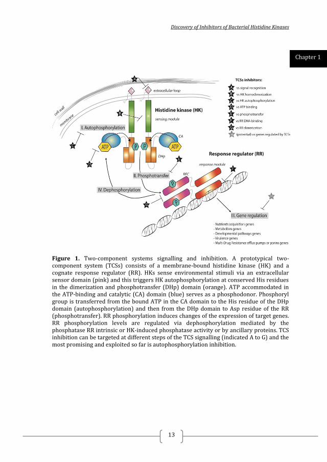
Figure 1. Two-component systems signalling and inhibition. A prototypical two-component system (TCSs) consists of a membrane-bound histidine kinase (HK) and a cognate response regulator (RR). HKs sense environmental stimuli via an extracellular sensor domain (pink) and this triggers HK autophosphorylation at conserved His residues in the dimerization and phosphotransfer (DHp) domain (orange). ATP accommodated in the ATP-binding and catalytic (CA) domain (blue) serves as a phosphodonor. Phosphoryl group is transferred from the bound ATP in the CA domain to the His residue of the DHp domain (autophosphorylation) and then from the DHp domain to Asp residue of the RR (phosphotransfer). RR phosphorylation induces changes of the expression of target genes. RR phosphorylation levels are regulated via dephosphorylation mediated by the phosphatase RR intrinsic or HK-induced phosphatase activity or by ancillary proteins. TCS inhibition can be targeted at different steps of the TCS signalling (indicated A to G) and the most promising and exploited so far is autophosphorylation inhibition.
Chapter 1
2014
14
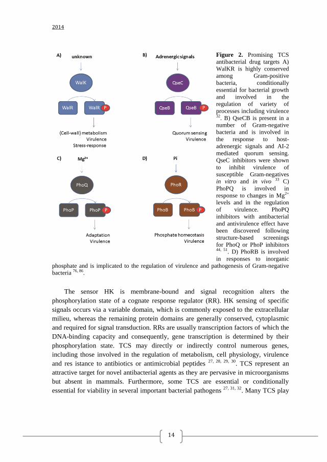
Figure 2. Promising TCS
antibacterial drug targets A)
WalKR is highly conserved
among Gram-positive
bacteria, conditionally
essential for bacterial growth
and involved in the
regulation of variety of
processes including virulence 32. B) QseCB is present in a
number of Gram-negative
bacteria and is involved in
the response to host-
adrenergic signals and AI-2
mediated quorum sensing.
QseC inhibitors were shown
to inhibit virulence of
susceptible Gram-negatives
in vitro and in vivo 33 C)
PhoPQ is involved in
response to changes in Mg2+
levels and in the regulation
of virulence. PhoPQ
inhibitors with antibacterial
and antivirulence effect have
been discovered following
structure-based screenings
for PhoQ or PhoP inhibitors 44, 51. D) PhoRB is involved
in responses to inorganic
phosphate and is implicated to the regulation of virulence and pathogenesis of Gram-negative
bacteria 76, 86.
The sensor HK is membrane-bound and signal recognition alters the
phosphorylation state of a cognate response regulator (RR). HK sensing of specific
signals occurs via a variable domain, which is commonly exposed to the extracellular
milieu, whereas the remaining protein domains are generally conserved, cytoplasmic
and required for signal transduction. RRs are usually transcription factors of which the
DNA-binding capacity and consequently, gene transcription is determined by their
phosphorylation state. TCS may directly or indirectly control numerous genes,
including those involved in the regulation of metabolism, cell physiology, virulence
and res istance to antibiotics or antimicrobial peptides 27, 28, 29, 30
. TCS represent an
attractive target for novel antibacterial agents as they are pervasive in microorganisms
but absent in mammals. Furthermore, some TCS are essential or conditionally
essential for viability in several important bacterial pathogens 27, 31, 32
. Many TCS play
Discovery of Inhibitors of Bacterial Histidine Kinases
15
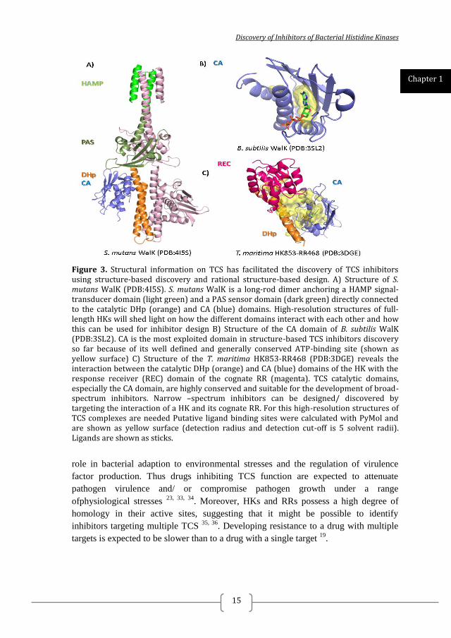
Figure 3. Structural information on TCS has facilitated the discovery of TCS inhibitors using structure-based discovery and rational structure-based design. A) Structure of S. mutans WalK (PDB:4I5S). S. mutans WalK is a long-rod dimer anchoring a HAMP signal-transducer domain (light green) and a PAS sensor domain (dark green) directly connected to the catalytic DHp (orange) and CA (blue) domains. High-resolution structures of full-length HKs will shed light on how the different domains interact with each other and how this can be used for inhibitor design B) Structure of the CA domain of B. subtilis WalK (PDB:3SL2). CA is the most exploited domain in structure-based TCS inhibitors discovery so far because of its well defined and generally conserved ATP-binding site (shown as yellow surface) C) Structure of the T. maritima HK853-RR468 (PDB:3DGE) reveals the interaction between the catalytic DHp (orange) and CA (blue) domains of the HK with the response receiver (REC) domain of the cognate RR (magenta). TCS catalytic domains, especially the CA domain, are highly conserved and suitable for the development of broad-spectrum inhibitors. Narrow –spectrum inhibitors can be designed/ discovered by targeting the interaction of a HK and its cognate RR. For this high-resolution structures of TCS complexes are needed Putative ligand binding sites were calculated with PyMol and are shown as yellow surface (detection radius and detection cut-off is 5 solvent radii). Ligands are shown as sticks.
role in bacterial adaption to environmental stresses and the regulation of virulence
factor production. Thus drugs inhibiting TCS function are expected to attenuate
pathogen virulence and/ or compromise pathogen growth under a range
ofphysiological stresses 23, 33, 34
. Moreover, HKs and RRs possess a high degree of
homology in their active sites, suggesting that it might be possible to identify
inhibitors targeting multiple TCS 35, 36
. Developing resistance to a drug with multiple
targets is expected to be slower than to a drug with a single target 19
.
Chapter 1
2014
16
TWO-COMPONENT SYSTEM SIGNALLING
Upon sensing of a ligand or physiological stimulus TCS signal transduction is
initiated by autophosphorylation of a conserved histidine (His) residue in the
dimerization and phosphotransfer (DHp) domain 37
. Autophosphorylation involves the
helical DHp domain hosting the conserved His and the C-terminal catalytic and ATP-
binding (CA) domain that binds ATP and phosphorylates the His (Figure 1 and 3). The
phosphoryl group from the phosphorylated His in the DHp domain is then transferred
to an exposed Asp that belongs to the response receiver (REC) domain of the cognate
RR. Phosphotransfer, i.e. phosphorylation of the RR, triggers changes in the
conformation of the RR, modulating the affinity of the effector domain for its targets,
typically DNA binding motifs in specific gene promoters 38, 26
. In some cases the RR
lacks the effector domain and the REC domain takes over the effector role. The
phosphorylation levels of the RR are tightly regulated by the phosphatase activity of
the HK; the RR itself or a partner protein 26, 39, 40, 41
. The DHp, CA, and REC domains
are always present in all TCS and relatively well conserved in amino-acid sequence
(Figure 4), compared to the sensor domains of HKs (the periplasmic domain, HAMP,
PAS and GAF domains) or the effector domains of RRs. The presence or absence and
the variability of the sensor and effector domains is determined by the wide range of
specific signals or targets to be recognized by the different TCS 38, 26
. Therefore,
inhibitors of the catalytic activity of the highly conserved DHp, CA and REC domain
are expected to possess broad-spectrum antibacterial activity.
DISCOVERY OF TWO-COMPONENT SYSTEM INHIBITORS
There are several publications describing the discovery of TCS inhibitors which
have been reviewed elsewhere 34, 36, 42
and some aspects are discussed in more detail in
the following sections. The target TCS include among others, S. pneumoniae and S.
epidermidis WalKR 43, 43
, E. coli QseCB 43
, S. flexnerii and S. eneterica PhoQP 44, 45, 45
.
The identified TCS inhibitors were from libraries of natural products 46
, small
molecules 34
and antibacterial peptides 47
(Figure 5). Different approaches have been
employed to discover TCS inhibitors, including high-throughput target-based
screening 48, 33, 49
and virtual structure-based screening 43, 50, 51, 44, 35
.
Discovery of Inhibitors of Bacterial Histidine Kinases
17
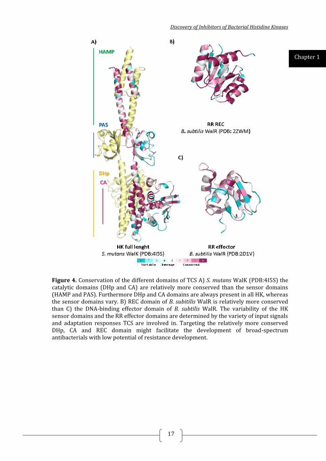
Figure 4. Conservation of the different domains of TCS A) S. mutans WalK (PDB:4I5S) the catalytic domains (DHp and CA) are relatively more conserved than the sensor domains (HAMP and PAS). Furthermore DHp and CA domains are always present in all HK, whereas the sensor domains vary. B) REC domain of B. subitilis WalR is relatively more conserved than C) the DNA-binding effector domain of B. subtilis WalR. The variability of the HK sensor domains and the RR effector domains are determined by the variety of input signals and adaptation responses TCS are involved in. Targeting the relatively more conserved DHp, CA and REC domain might facilitate the development of broad-spectrum antibacterials with low potential of resistance development.
Chapter 1
2014
18
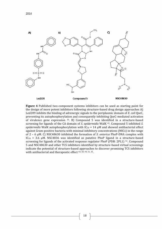
Figure 4 Published two-component systems inhibitors can be used as starting point for the design of more potent inhibitors following structure-based drug design approaches A) Led209 inhibits the binding of adrenergic signals to the periplasmic domain of E. coli QseC, preventing its autophosphorylation and consequently inhibiting QseC-mediated activation of virulence gene expression 33. B) Compound 5 was identified in a structure-based screening for ligands of the CA domain of S. epidermidis WalK 43. Compound 5 inhibited S. epidermidis WalK autophosphorylation with IC50 = 14 µM and showed antibacterial effect against Gram-positive bacteria with minimal inhibitory concentrations (MICs) in the range of 2 – 6 µM. C) NSC48630 inhibited the formation of S. enterica PhoP-DNA complex with IC50 = 3.6 µM. NSC4836 was identified as putative PhoP ligand in a structure-based screening for ligands of the activated response regulator PhoP (PDB: 2PL1) 51. Compound 5 and NSC48630 and other TCS inhibitors identified by structure-based virtual screenings indicate the potential of structure-based approaches to discover promising TCS inhibitors with antibacterial and therapeutic effect 43, 50, 44, 51, 35.
Discovery of Inhibitors of Bacterial Histidine Kinases
19
PROMISING TWO-COMPONENT SYSTEM TARGETS
WalKR
WalKR (aka YycF/G, MicA/B, VicK/R) is highly conserved and has been
identified in the genomes of the major group of Gram-positive bacteria with a low
genomic GC-content (Figure 2A) 52, 53, 54, 55, 32, 30
. This includes pathogenic species
belonging to the genus Staphylococcus, Streptococcus, Enterococcus, Clostridium, and
Listeria. In the important pathogens S. aureus, S. pneumoniae, and Enterococcus
faecalis WalKR has been shown to be essential for viability in rich laboratory growth
medium 52, 30, 56
. WalKR regulates genes responsible for cell wall metabolism and cell
wall homeostasis 55, 57, 53
but the specific genes and pathways regulated by WalKR may
differ from species to species. Additionally, walKR regulates genes involved in
metabolism, stress response, virulence, host-microbe interactions, transport and
regulatory pathways depending on the species 27, 58
.
Of all the TCS WalKR is considered one of the most attractive targets for
antibacterial drugs due to its essentiality, links to virulence and antibiotic resistance.
Imidazole and zerumbone derivatives were the first inhibitors of WalK to be identified 59, 46
. Following this pioneering discovery, Utsumi´s group developed biochemical and
genetic high-throughput screening methods to search for WalK and WalR inhibitors.
This led to the discovery of WalK inhibitors such as aranorosinol, walkmycin B,
waldiomycin, signermycin B and the WalR inhibitors, walrycin A and B 48, 49
. Later,
some of these inhibitors were not proven to be selective to WalKR and other
mechanisms such as membrane damage and adverse effects on macromolecules
biosynthesis most likely account for the observed antibacterial effects.
Elucidation of the structure of HK catalytic domains opened up the possibility for
structure-based virtual screening (SBVS) for WalK inhibitors 60, 61, 43, 43
. The catalytic
ATP-binding domain (CA domain) appeared to be the most promising target for this
approach as this domain contains a well-defined and partially conserved pocket where
ATP is accommodated (Figure 3B). Before the elucidation of WalK structure (PDB:
3SL2 and PDB:4I5S), homology models of the CA domains of S. pneumoniae and S.
epidermidis WalK based on the three-dimensional structures of Thermotoga maritima
HK853 (PDB: 2C2A) and E. coli EnvZ (PDB: 1BXD) HKs were used for SBVS of
drug-lead compound libraries. These screenings yielded WalK autophosphorylation
inhibitors belonging to different classes of chemical structures, such as imidazole
analogues and derivatives of furan, thiophene, thiazolidinone, benzamide and
pyrimidinone (Figure 4). The SBVS hits inhibited the growth of S. pneumoniae and S.
epidermidis and showed bactericidal effects towards both planktonic and biofilm cells 62, 63
. Furthermore, some of these inhibitors decreased the mortality of mice infected
with S. pneumoniae in an in vivo sepsis model, demonstrating that SBVS is a valuable
tool for the identification of WalK inhibitors with therapeutic effect 43
. Nevertheless,
Chapter 1
2014
20
the design of potent ATP-competitive inhibitors specific for WalK is challenging, even
more so when structural homology models are used instead of experimental (e.g. X-
ray) data. This issue is crucial when attempting to generate a WalK inhibitor with
higher specificity and affinity in the hit-to-lead optimization phase. Although the
overall fold of the CA domain as well as the catalytic residues is generally conserved,
there are large variations in size and sequence in the ATP-lid, a HK distinctive flexible
loop that covers the ATP-binding pocket and is crucially involved in ATP binding and
in HK catalytic reactions 26, 64, 65
. The recently published WalK structures of the entire
intracellular portion from Streptococcus mutans (PDB: 4I5S) (Figure 3A) and the CA
domain from Bacillus subtilis (PDB: 3SL2, Figure 3B) might provide the key to
design of improved WalK inhibitors 66, 67
. These structures have not only revealed
specific characteristics of the WalK CA domain but also insights into the molecular
mechanism of WalK autokinase activation and clues how it can be effectively
inhibited by exploring HK domains for structure-based drug design 25
.
QseCB
Targeting microbial virulence without inhibiting growth has been proposed as
promising strategy in antibacterial drug discovery as it was proposed that antivirulence
drugs present less selective pressure for resistance development 33, 24, 51
. QseCB is a
TCS involved in recognizing the host-derived adrenergic signals and the bacterial
aromatic signal autoinducer-3 AI-3 (epinephrine, norepinephrine) to trigger expression
of virulence genes (Figure 2B) 68
. Homologues of QseC are present in at least 25
important human and plant pathogens, therefore, a QseC inhibitor is expected to be a
promising drug for antivirulence therapy against a wide range of pathogenic bacteria 33
.
Adrenergic signals, as well as, AI-3 are recognized by QseC periplasmic domain 68
. Drugs targeting QseC periplasmic domain have an advantage compared to drugs
with intracellular targets of Gram-negative bacteria as they only need to pass the outer
membrane to reach the target.
High-throughput screening to search for a compound that inhibits the activation of
QseC by AI-3 has been reported 33
. LED209 (Figure 4) was identified as inhibitor of
the binding of the signaling molecules to QseC from different Gram-negative
pathogenic bacteria. LED209 inhibited QseC autophosphorylation, bacterial
pathogenicity in vitro and in vivo, but did not inhibit cell growth. The discovery of
LED209 demonstrates that targeting microbial virulence without inhibiting growth is a
promising strategy to identify antibacterials with therapeutic effect 33
.
Discovery of Inhibitors of Bacterial Histidine Kinases
21
PhoPQ
The PhoQ/PhoP two-component regulatory system is a major regulator of
virulence in the enteric pathogen Salmonella enterica serovar Typhimurium 69, 70
and is
present in a range of Gram-negative bacteria. It also controls the adaptation to low
Mg2+
environments by governing the expression and/or activity of Mg2+
transporters
and of enzymes modifying the Mg2+
-binding sites on the bacterial cell surface (Figure
2C). The RR PhoP modifies expression of about 3% of the Salmonella genes in
response to the periplasmic Mg2+
concentration detected by the HK PhoQ 71
. Genes
that are directly controlled by PhoP often differ in their promoter structures, resulting
in distinct expression levels and kinetics in response to the low Mg2+
inducing signal.
PhoP regulates a large number of genes indirectly via other transcription factors and
TCS that form a panoply of regulatory networks. These regulatory networks include
transcriptional cascades, feed-forward loops and the use of connector proteins to
modify the activity of RRs 72
. These networks confer distinct expression properties
that may be important contributors to the lifestyle of Salmonella.
The structure of the catalytic domain of PhoQ has been well studied 73
. PhoQ has
also been co-crystallized with the inhibitor radicicol 45
. The available structural
information has been exploited in structure-based screening for PhoQ inhibitors that
attenuate the virulence of S. flexneri in vitro and in vivo 44
. PhoP inhibitors have also
been discovered via structure-based screenings and shown to inhibit PhoP-DNA
complex formation (Figure 4) 51
. Taken together these findings highlight good
potential to develop novel antibacterials against PhoPQ using structure-based
screening and design.
PhoRB
PhoRB is a two-component system sensing the extracellular concentration of
phosphate (Figure 2D) 74
. It consists of the HK PhoR and its cognate RR PhoB.
PhoRB responds to phosphate limitation, when the extracellular concentration of
phosphate falls below 4 µM. In phosphate-limiting conditions, PhoR phosphorylates
PhoB, which then binds to specific DNA sequences, known as Pho boxes. PhoB
binding either induces or represses the genes belonging to the Pho (phosphate)
regulon, which includes genes involved in acquisition and metabolism of different
phosphates.
The Pho regulon in E. coli K12 comprises of 31 genes, and it is not only involved
in phosphate homeostasis, but it is also related to bacterial virulence as the induction
of Pho regulon results in attenuated pathogenesis 75, 76
. Examples of virulence
attributes altered by induction of the Pho regulon are a significant reduction in the
amount of capsular antigen at the cell surface, resistance to the bactericidal effect of
serum, to cationic antimicrobial peptides, and to acid and oxidative stress, as well as
the production of type 1 fimbriae 75, 76
. Therefore, PhoRB presents a promising target
for antibacterials that inhibit bacterial virulence.
Chapter 1
2014
22
ANTIMICROBIAL PEPTIDES INHIBITING TCS SIGNALING
Antimicrobial peptides (AMPs) have been proposed as promising strategy to
address the growing problem of MDR 77, 78
. Eukaryotic cationic AMPs are produced at
sites of infection or inflammation in many different organisms 79, 80
. Typically they are
peptides of 12 to 45 amino acids with a net positive charge and a high proportion of
hydrophobic amino acids 81
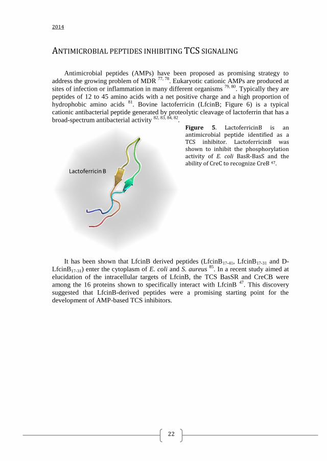
. Bovine lactoferricin (LfcinB; Figure 6) is a typical
cationic antibacterial peptide generated by proteolytic cleavage of lactoferrin that has a
broad-spectrum antibacterial activity 82, 83, 84, 82
. Figure 5. LactoferricinB is an antimicrobial peptide identified as a TCS inhibitor. LactoferricinB was shown to inhibit the phosphorylation activity of E. coli BasR-BasS and the ability of CreC to recognize CreB 47.
It has been shown that LfcinB derived peptides (LfcinB17-41, LfcinB17-31 and D-
LfcinB17-31) enter the cytoplasm of E. coli and S. aureus 85
. In a recent study aimed at
elucidation of the intracellular targets of LfcinB, the TCS BasSR and CreCB were
among the 16 proteins shown to specifically interact with LfcinB 47
. This discovery
suggested that LfcinB-derived peptides were a promising starting point for the
development of AMP-based TCS inhibitors.
Discovery of Inhibitors of Bacterial Histidine Kinases
23
THESIS OUTLINE AND AIMS The main aims of the current thesis were:
1. To discover hits that can be further developed to broad-spectrum TCS inhibitors
following a multidisciplinary approach.
2. To provide structural-knowledge on TCS inhibition to facilitate the
optimization of the identified TCS inhibitors using structure-based drug design.
3. To evaluate the identified hits for their biochemical, antibacterial and cytotoxic
effects in vitro
4. To explore ways to improve hit compound permeability of the Gram-negative
bacteria outer membrane using nanoparticles and AMPs
In Chapter 2, we describe the discovery of histidine kinase (HK)
autophosphorylation inhibitors targeted at the ATP-binding and catalytic (CA) domain
of multiple HKs using combined structure-based and ligand-based virtual screening
approaches.
In Chapter 3, we describe the application of fragment-based screenings by
differential scanning fluorimetry and ligand-based similarity searches to discover
fragment-like HK autophosphorylation inhibitors targeted at the CA domain of
multiple HKs.
Due to the weak or absent antibacterial effect of identified HK inhibitors (Chapter
2 and 3) against Gram-negative bacteria, nanoparticles were investigated as potential
drug delivery vehicles. Silica-based mesoporous nanoparticles capped with ε-poly-L-
lysine were loaded with HK autophosphorylation inhibitors and their antibacterial
effect, cytotoxicity and immunotoxicity were studied and described in Chapter 4.
In Chapter 5 we used computer-aided approaches in an attempt to discover
improved HK inhibitors.
As lactoferricinB-derived peptides seemed to present a promising starting point
for the development of antimicrobial-peptides-based TCS inhibitors, in Chapter 6 we
investigated the possibility to use T. maritima HK853-RR468 and E. coli PhoRB for
structural studies of TCS inhibition by LactoferricinB – derived peptides.
In Chapter 7 the (co-)crystallization and preliminary X-ray diffraction analysis of
the T. maritima CheA-inhibitors co-crystals and CopM periplasmic domain are
described.
In Chapter 8 the potential of targeting WalK for developing antibacterial drugs
with low potential of resistance development is discussed in the light of current
literature and results.
Chapter 9 is a concluding discussion on the results described in the thesis and
their implications for antibacterial drug discovery and development.
2014
24
REFERENCES 1. Magiorakos, A., Multidrug-resistant, extensively drug-resistant and pandrug-resistant bacteria: an international expert proposal for interim standard definitions for acquired resistance. Clin Microbiol Infect 2012, 18, 268-281. 2. Bassetti, M.; Merelli, M.; Temperoni, C.; Astilean, A., New antibiotics for bad bugs: where are we? Annals of clinical microbiology and antimicrobials 2013, 12, 22. 3. Boucher, H. W., Bad Bugs, No Drugs: No ESKAPE! An Update from the Infectious Diseases Society of America. Clin Infect Dis 2009, 48, 1-12. 4. Peterson, L. R., Bad bugs, no drugs: no ESCAPE revisited. Clinical infectious diseases : an official publication of the Infectious Diseases Society of America 2009, 49 (6), 992-3. 5. Cosgrove, S. E.; Carmeli, Y., The impact of antimicrobial resistance on health and economic outcomes. Clinical infectious diseases : an official publication of the Infectious Diseases Society of America 2003, 36 (11), 1433-7. 6. Brotz-Oesterhelt, H.; Sass, P., Postgenomic strategies in antibacterial drug discovery. Future microbiology 2010, 5 (10), 1553-79. 7. Butler, M. S.; Blaskovich, M. A.; Cooper, M. A., Antibiotics in the clinical pipeline in 2013. The Journal of antibiotics 2013, 66 (10), 571-91. 8. Dougherty, T. J.; Miller, P. F., Microbial genomics and drug discovery: exploring innovative routes of drug discovery in the postgenomic era. IDrugs : the investigational drugs journal 2006, 9 (6), 420-2. 9. Aminov, R. I., A brief history of the antibiotic era: lessons learned and challenges for the future. Frontiers in microbiology 2010, 1, 134. 10. Fernandes, P., Antibacterial discovery and development--the failure of success? Nature biotechnology 2006, 24 (12), 1497-503. 11. Singh, S. B.; Barrett, J. F., Empirical antibacterial drug discovery--foundation in natural products. Biochemical pharmacology 2006, 71 (7), 1006-15. 12. Talbot, G. H.; Bradley, J.; Edwards, J. E., Jr.; Gilbert, D.; Scheld, M.; Bartlett, J. G.; Antimicrobial Availability Task Force of the Infectious Diseases Society of, A., Bad bugs need drugs: an update on the development pipeline from the Antimicrobial Availability Task Force of the Infectious Diseases Society of America. Clinical infectious diseases : an official publication of the Infectious Diseases Society of America 2006, 42 (5), 657-68. 13. Alksne, L. E.; Dunman, P. M., Target-based antimicrobial drug discovery. Methods in molecular biology 2008, 431, 271-83. 14. Pucci, M. J., Overview of antibacterial target selection. Current protocols in pharmacology / editorial board, S.J. Enna 2006, Chapter 13, Unit13A 2. 15. Waszkowycz, B., Structure-based approaches to drug design and virtual screening. Current opinion in drug discovery & development 2002, 5 (3), 407-13. 16. Muegge, I., Synergies of virtual screening approaches. Mini reviews in medicinal chemistry 2008, 8 (9), 927-33. 17. Wildman, S. A., Approaches to virtual screening and screening library selection. Current pharmaceutical design 2013, 19 (26), 4787-96. 18. Simmons, K. J.; Chopra, I.; Fishwick, C. W., Structure-based discovery of antibacterial drugs. Nature reviews. Microbiology 2010, 8 (7), 501-10. 19. Ferreira, R. S.; Andricopulo, A. D., Structure-based drug design to overcome drug resistance: Challenges and opportunities. Current pharmaceutical design 2014, 20 (5), 687-693. 20. Rule of three. Nature biotechnology 2000, 18 (10), 1025.
Discovery of Inhibitors of Bacterial Histidine Kinases
25
21. Jhoti, H.; Williams, G.; Rees, D. C.; Murray, C. W., The 'rule of three' for fragment-based drug discovery: where are we now? Nature reviews. Drug discovery 2013, 12 (8), 644-5. 22. Baker, M., Fragment-based lead discovery grows up. Nature reviews. Drug discovery 2013, 12 (1), 5-7. 23. Frosco, M.; Barrett, J. F., Bacterial two-component systems as antimicrobial drug discovery targets. Drug News and Perspectives 1999, 12 (5), 293-299. 24. Gotoh, Y.; Eguchi, Y.; Watanabe, T.; Okamoto, S.; Doi, A.; Utsumi, R., Two-component signal transduction as potential drug targets in pathogenic bacteria. Current opinion in microbiology 2010, 13 (2), 232-9. 25. Velikova, N.; Bem, A. E.; van Baarlen, P.; Wells, J. M.; Marina, A., WalK, the Path towards New Antibacterials with Low Potential for Resistance Development. ACS Medicinal Chemistry Letters 2013, 4 (10), 891-894. 26. Casino, P.; Rubio, V.; Marina, A., The mechanism of signal transduction by two-component systems. Current opinion in structural biology 2010, 20 (6), 763-71. 27. Dubrac, S.; Msadek, T., Identification of Genes Controlled by the Essential YycG/YycF Two-Component System of Staphylococcus aureus. Journal of bacteriology 2004, 186 (4), 1175-1181. 28. Dubrac, S.; Msadek, T., Tearing down the wall: peptidoglycan metabolism and the WalK/WalR (YycG/YycF) essential two-component system. Advances in experimental medicine and biology 2008, 631, 214-28. 29. Paterson, G. K.; Blue, C. E.; Mitchell, T. J., Role of two-component systems in the virulence of Streptococcus pneumoniae. Journal of medical microbiology 2006, 55 (Pt 4), 355-63. 30. Mohedano, M. L.; Overweg, K.; de la Fuente, A.; Reuter, M.; Altabe, S.; Mulholland, F.; de Mendoza, D.; Lopez, P.; Wells, J. M., Evidence that the essential response regulator YycF in Streptococcus pneumoniae modulates expression of fatty acid biosynthesis genes and alters membrane composition. Journal of bacteriology 2005, 187 (7), 2357-67. 31. Fukuchi, K.; Kasahara, Y.; Asai, K.; Kobayashi, K.; Moriya, S.; Ogasawara, N., The essential two-component regulatory system encoded by yycF and yycG modulates expression of the ftsAZ operon in Bacillus subtilis. Microbiology 2000, 146 ( Pt 7), 1573-83. 32. Winkler, M. E.; Hoch, J. A., Essentiality, bypass, and targeting of the YycFG (VicRK) two-component regulatory system in gram-positive bacteria. Journal of bacteriology 2008, 190 (8), 2645-8. 33. Rasko, D. A.; Moreira, C. G.; Li de, R.; Reading, N. C.; Ritchie, J. M.; Waldor, M. K.; Williams, N.; Taussig, R.; Wei, S.; Roth, M.; Hughes, D. T.; Huntley, J. F.; Fina, M. W.; Falck, J. R.; Sperandio, V., Targeting QseC signaling and virulence for antibiotic development. Science 2008, 321 (5892), 1078-80. 34. Worthington, R. J.; Blackledge, M. S.; Melander, C., Small-molecule inhibition of bacterial two-component systems to combat antibiotic resistance and virulence. Future medicinal chemistry 2013, 5 (11), 1265-84. 35. Francis, S.; Wilke, K. E.; Brown, D. E.; Carlson, E. E., Mechanistic insight into inhibition of two-component system signaling. MedChemComm 2013, 4 (1), 269-277. 36. Hilliard, J. J.; Goldschmidt, R. M.; Licata, L.; Baum, E. Z.; Bush, K., Multiple mechanisms of action for inhibitors of histidine protein kinases from bacterial two-component systems. Antimicrobial agents and chemotherapy 1999, 43 (7), 1693-9. 37. Mascher, T.; Helmann, J. D.; Unden, G., Stimulus perception in bacterial signal-transducing histidine kinases. Microbiology and molecular biology reviews : MMBR 2006, 70 (4), 910-38.
2014
26
38. Gao, R.; Stock, A. M., Biological insights from structures of two-component proteins. Annual review of microbiology 2009, 63, 133-54. 39. Kenney, L. J., How important is the phosphatase activity of sensor kinases? Current opinion in microbiology 2010, 13 (2), 168-76. 40. Zhu, Y.; Qin, L.; Yoshida, T.; Inouye, M., Phosphatase activity of histidine kinase EnvZ without kinase catalytic domain. Proceedings of the National Academy of Sciences of the United States of America 2000, 97 (14), 7808-13. 41. Gutu, A. D.; Wayne, K. J.; Sham, L. T.; Winkler, M. E., Kinetic characterization of the WalRKSpn (VicRK) two-component system of Streptococcus pneumoniae: dependence of WalKSpn (VicK) phosphatase activity on its PAS domain. Journal of bacteriology 2010, 192 (9), 2346-58. 42. Kurosu, M.; Begari, E., Bacterial protein kinase inhibitors. Drug Development Research 2010, 71 (3), 168-187. 43. Qin, Z.; Zhang, J.; Xu, B.; Chen, L.; Wu, Y.; Yang, X.; Shen, X.; Molin, S.; Danchin, A.; Jiang, H.; Qu, D., Structure-based discovery of inhibitors of the YycG histidine kinase: new chemical leads to combat Staphylococcus epidermidis infections. BMC microbiology 2006, 6, 96. 44. Cai, X.; Zhang, J.; Chen, M.; Wu, Y.; Wang, X.; Chen, J.; Zhang, J.; Shen, X.; Qu, D.; Jiang, H., The effect of the potential PhoQ histidine kinase inhibitors on Shigella flexneri virulence. PloS one 2011, 6 (8), e23100. 45. Guarnieri, M. T.; Zhang, L.; Shen, J.; Zhao, R., The Hsp90 inhibitor radicicol interacts with the ATP-binding pocket of bacterial sensor kinase PhoQ. Journal of molecular biology 2008, 379 (1), 82-93. 46. Watanabe, T.; Okada, A.; Gotoh, Y.; Utsumi, R., Inhibitors targeting two-component signal transduction. Advances in experimental medicine and biology 2008, 631, 229-36. 47. Ho, Y. H.; Sung, T. C.; Chen, C. S., Lactoferricin B inhibits the phosphorylation of the two-component system response regulators BasR and CreB. Molecular & cellular proteomics : MCP 2012, 11 (4), M111 014720. 48. Okada, A.; Igarashi, M.; Okajima, T.; Kinoshita, N.; Umekita, M.; Sawa, R.; Inoue, K.; Watanabe, T.; Doi, A.; Martin, A.; Quinn, J.; Nishimura, Y.; Utsumi, R., Walkmycin B targets WalK (YycG), a histidine kinase essential for bacterial cell growth. The Journal of antibiotics 2010, 63 (2), 89-94. 49. Gotoh, Y.; Doi, A.; Furuta, E.; Dubrac, S.; Ishizaki, Y.; Okada, M.; Igarashi, M.; Misawa, N.; Yoshikawa, H.; Okajima, T.; Msadek, T.; Utsumi, R., Novel antibacterial compounds specifically targeting the essential WalR response regulator. The Journal of antibiotics 2010, 63 (3), 127-34. 50. Li, N.; Wang, F.; Niu, S.; Cao, J.; Wu, K.; Li, Y.; Yin, N.; Zhang, X.; Zhu, W.; Yin, Y., Discovery of novel inhibitors of Streptococcus pneumoniae based on the virtual screening with the homology-modeled structure of histidine kinase (VicK). BMC microbiology 2009, 9, 129. 51. Tang, Y. T.; Gao, R.; Havranek, J. J.; Groisman, E. A.; Stock, A. M.; Marshall, G. R., Inhibition of bacterial virulence: drug-like molecules targeting the Salmonella enterica PhoP response regulator. Chemical biology & drug design 2012, 79 (6), 1007-17. 52. Dubrac, S.; Boneca, I. G.; Poupel, O.; Msadek, T., New insights into the WalK/WalR (YycG/YycF) essential signal transduction pathway reveal a major role in controlling cell wall metabolism and biofilm formation in Staphylococcus aureus. Journal of bacteriology 2007, 189 (22), 8257-69. 53. Delaune, A.; Dubrac, S.; Blanchet, C.; Poupel, O.; Mader, U.; Hiron, A.; Leduc, A.; Fitting, C.; Nicolas, P.; Cavaillon, J. M.; Adib-Conquy, M.; Msadek, T., The WalKR system
Discovery of Inhibitors of Bacterial Histidine Kinases
27
controls major staphylococcal virulence genes and is involved in triggering the host inflammatory response. Infection and immunity 2012, 80 (10), 3438-53. 54. Delaune, A., Poupel O., Mallet A., Coic YM., Msadek T., Dubrac S., Peptidoglycan Crosslinking Relaxation Plays an Important Role in Staphylococcus aureus WalKR Dependent Cell Viability. PloS one 2010, 6 (2). 55. Howell, A.; Dubrac, S.; Andersen, K. K.; Noone, D.; Fert, J.; Msadek, T.; Devine, K., Genes controlled by the essential YycG/YycF two-component system of Bacillus subtilis revealed through a novel hybrid regulator approach. Molecular microbiology 2003, 49 (6), 1639-1655. 56. JA, F. C. a. H., Two-Component Signal Transduction System Essential for Growth of Bacillus subtilis: Implications for Anti-Infective Therapy. Journal of bacteriology 1998, 180 (23), 6375–6383. 57. Howden, B. P.; McEvoy, C. R.; Allen, D. L.; Chua, K.; Gao, W.; Harrison, P. F.; Bell, J.; Coombs, G.; Bennett-Wood, V.; Porter, J. L.; Robins-Browne, R.; Davies, J. K.; Seemann, T.; Stinear, T. P., Evolution of multidrug resistance during Staphylococcus aureus infection involves mutation of the essential two component regulator WalKR. PLoS pathogens 2011, 7 (11), e1002359. 58. Luz Mohedano, M., Evidence that the Essential Response Regulator YycF in Streptococcus pneumoniae Modulates Expression of Fatty Acid Biosynthesis Genes and Alters Membrane Composition. Journal of Bacteriology 2005, 187 (7), 2357-2367. 59. Okada, A.; Gotoh, Y.; Watanabe, T.; Furuta, E.; Yamamoto, K.; Utsumi, R., Targeting two-component signal transduction: a novel drug discovery system. Methods in enzymology 2007, 422, 386-95. 60. Marina, A.; Waldburger, C. D.; Hendrickson, W. A., Structure of the entire cytoplasmic portion of a sensor histidine-kinase protein. The EMBO journal 2005, 24 (24), 4247-59. 61. Tanaka, T.; Saha, S. K.; Tomomori, C.; Ishima, R.; Liu, D.; Tong, K. I.; Park, H.; Dutta, R.; Qin, L.; Swindells, M. B.; Yamazaki, T.; Ono, A. M.; Kainosho, M.; Inouye, M.; Ikura, M., NMR structure of the histidine kinase domain of the E. coli osmosensor EnvZ. Nature 1998, 396 (6706), 88-92. 62. Huang, R. Z.; Zheng, L. K.; Liu, H. Y.; Pan, B.; Hu, J.; Zhu, T.; Wang, W.; Jiang, D. B.; Wu, Y.; Wu, Y. C.; Han, S. Q.; Qu, D., Thiazolidione derivatives targeting the histidine kinase YycG are effective against both planktonic and biofilm-associated Staphylococcus epidermidis. Acta pharmacologica Sinica 2012, 33 (3), 418-25. 63. Liu, H.; Zhao, D.; Chang, J.; Yan, L.; Zhao, F.; Wu, Y.; Xu, T.; Gong, T.; Chen, L.; He, N.; Wu, Y.; Han, S.; Qu, D., Efficacy of novel antibacterial compounds targeting histidine kinase YycG protein. Applied microbiology and biotechnology 2014, 98 (13), 6003-13. 64. Podgornaia, A. I.; Casino, P.; Marina, A.; Laub, M. T., Structural basis of a rationally rewired protein-protein interface critical to bacterial signaling. Structure 2013, 21 (9), 1636-47. 65. Casino, P., Miquel-Romero, L., Marina, A., Visualizing autophosphorylation in histidine kinases. Nat Commun 2014. 66. Wang, C.; Sang, J.; Wang, J.; Su, M.; Downey, J. S.; Wu, Q.; Wang, S.; Cai, Y.; Xu, X.; Wu, J.; Senadheera, D. B.; Cvitkovitch, D. G.; Chen, L.; Goodman, S. D.; Han, A., Mechanistic insights revealed by the crystal structure of a histidine kinase with signal transducer and sensor domains. PLoS biology 2013, 11 (2), e1001493. 67. Celikel, R.; Veldore, V. H.; Mathews, I.; Devine, K. M.; Varughese, K. I., ATP forms a stable complex with the essential histidine kinase WalK (YycG) domain. Acta crystallographica. Section D, Biological crystallography 2012, 68 (Pt 7), 839-45.
2014
28
68. Clarke, M. B.; Hughes, D. T.; Zhu, C.; Boedeker, E. C.; Sperandio, V., The QseC sensor kinase: a bacterial adrenergic receptor. Proceedings of the National Academy of Sciences of the United States of America 2006, 103 (27), 10420-5. 69. Groisman, E. A., The pleiotropic two-component regulatory system PhoP-PhoQ. Journal of bacteriology 2001, 183 (6), 1835-42. 70. Miller, S. I.; Kukral, A. M.; Mekalanos, J. J., A two-component regulatory system (phoP phoQ) controls Salmonella typhimurium virulence. Proceedings of the National Academy of Sciences of the United States of America 1989, 86 (13), 5054-8. 71. Lejona, S.; Aguirre, A.; Cabeza, M. L.; Garcia Vescovi, E.; Soncini, F. C., Molecular characterization of the Mg2+-responsive PhoP-PhoQ regulon in Salmonella enterica. Journal of bacteriology 2003, 185 (21), 6287-94. 72. Park, S. Y.; Groisman, E. A., Signal-specific temporal response by the Salmonella PhoP/PhoQ regulatory system. Molecular microbiology 2014, 91 (1), 135-44. 73. Marina, A.; Mott, C.; Auyzenberg, A.; Hendrickson, W. A.; Waldburger, C. D., Structural and mutational analysis of the PhoQ histidine kinase catalytic domain. Insight into the reaction mechanism. The Journal of biological chemistry 2001, 276 (44), 41182-90. 74. Yamada, M.; Makino, K.; Amemura, M.; Shinagawa, H.; Nakata, A., Regulation of the phosphate regulon of Escherichia coli: analysis of mutant phoB and phoR genes causing different phenotypes. Journal of bacteriology 1989, 171 (10), 5601-6. 75. Chekabab, S. M.; Jubelin, G.; Dozois, C. M.; Harel, J., PhoB activates Escherichia coli O157:H7 virulence factors in response to inorganic phosphate limitation. PloS one 2014, 9 (4), e94285. 76. Crepin, S.; Chekabab, S. M.; Le Bihan, G.; Bertrand, N.; Dozois, C. M.; Harel, J., The Pho regulon and the pathogenesis of Escherichia coli. Veterinary microbiology 2011, 153 (1-2), 82-8. 77. Steckbeck, J. D.; Deslouches, B.; Montelaro, R. C., Antimicrobial peptides: new drugs for bad bugs? Expert opinion on biological therapy 2014, 14 (1), 11-4. 78. Hassan, M.; Kjos, M.; Nes, I. F.; Diep, D. B.; Lotfipour, F., Natural antimicrobial peptides from bacteria: characteristics and potential applications to fight against antibiotic resistance. Journal of applied microbiology 2012, 113 (4), 723-36. 79. Palffy, R.; Gardlik, R.; Behuliak, M.; Kadasi, L.; Turna, J.; Celec, P., On the physiology and pathophysiology of antimicrobial peptides. Molecular medicine 2009, 15 (1-2), 51-9. 80. Pasupuleti, M.; Schmidtchen, A.; Malmsten, M., Antimicrobial peptides: key components of the innate immune system. Critical Reviews in Biotechnology 2012, 32 (2), 143-171. 81. Hancock, R. E. W., Peptide antibiotics. Lancet 1997, 349 (9049), 418-422. 82. Bellamy, W.; Takase, M.; Wakabayashi, H.; Kawase, K.; Tomita, M., Antibacterial spectrum of lactoferricin B, a potent bactericidal peptide derived from the N-terminal region of bovine lactoferrin. The Journal of applied bacteriology 1992, 73 (6), 472-9. 83. Strom, M. B.; Haug, B. E.; Rekdal, O.; Skar, M. L.; Stensen, W.; Svendsen, J. S., Important structural features of 15-residue lactoferricin derivatives and methods for improvement of antimicrobial activity. Biochemistry and cell biology = Biochimie et biologie cellulaire 2002, 80 (1), 65-74. 84. (a) Del Olmo, A.; Morales, P.; Nuñez, M., Bactericidal effect of lactoferrin and its amidated and pepsin-digested derivatives on Pseudomonas fluorescens: Influence of environmental and physiological factors. Journal of food protection 2008, 71 (12), 2468-2474; (b) Roseanu, A.; Florian, P.; Condei, M.; Cristea, D.; Damian, M., Antibacterial activity of lactoferrin and lactoferricin against oral streptococci. Romanian Biotechnological Letters 2010, 15 (6), 5788-5792.
Discovery of Inhibitors of Bacterial Histidine Kinases
29
85. Haukland, H. H.; Ulvatne, H.; Sandvik, K.; Vorland, L. H., The antimicrobial peptides lactoferricin B and magainin 2 cross over the bacterial cytoplasmic membrane and reside in the cytoplasm. FEBS letters 2001, 508 (3), 389-93. 86. Scholten, M.; Janssen, R.; Bogaarts, C.; van Strien, J.; Tommassen, J., The pho regulon of Shigella flexneri. Molecular microbiology 1995, 15 (2), 247-54.

2014
30
Discovery of Inhibitors of Bacterial Histidine Kinases
31
CHAPTER 2
DISCOVERY OF BACTERIAL HISTIDINE KINASE
INHIBITORS WITH ANTIBACTERIAL ACTIVITY
AGAINST CLINICAL ISOLATES OF MRSA AND
STAPHYLOCOCCUS EPIDERMIDIS
Nadya Velikova1, Simone Fulle
2, Ana Sousa Manso
3, 4, Nico Taverne
6, Milena
Mechkarska5, J. Michael Conlon
5, Jerry M. Wells
6, Marco Rinaldo Oggioni
3, 4, Paul
Finn2, Alberto Marina
1, 7
1Instituto de Biomedicina de Valencia, Consejo Superior de Investigaciones
Científicas (CSIC), Jaume Roig 11, 46010 Valencia, Spain; 2InhibOx, Oxford, United Kingdom;
3Dipartimento di Biotecnologie Mediche, Universita di Siena, 53100 Siena, Italy;
4Department of Genetics, University of Leicester, Leicester, Le1 7RH, United
Kingdom;
5Department of Biochemistry, College of Medicine and Health Science, United Arab
Emirates University, P.O. Box 17666 Al Ain, United Arab Emirates; 6Host-Microbe Interactomics Chair Group, Animal Sciences, University of
Wageningen, P.O. Box 338, 6700 AH Wageningen, The Netherlands; 7CIBER de Enfermedades Raras (CIBERER), ISCIII, Valencia, Spain
TO BE SUBMITTED TO ASM ANTIMICROBIAL AGENTS AND CHEMOTHERAPY

2014
32

Discovery of Inhibitors of Bacterial Histidine Kinases
33
ABSTRACT
The emergence of multi-drug resistant bacteria is an overwhelming world-wide
public health problem. Two-component systems, consisting of a sensor histidine
kinase (HK) and an effector response regulator, are the main signaling devices in
bacteria and have been proposed as promising targets for the development of novel
broad-spectrum antibacterials. Rational design of competitive inhibitors for the
nucleotide-binding site of HKs that inhibit the autophosphorylation activity is
considered a promising strategy to generate a novel class of antibacterials. To discover
HK inhibitors we employed a virtual screening approach, including structure-based
virtual screening (SBVS) and a ligand-based similarity search (LBSS). Two of the hits
identified using SBVS inhibited the autophosphorylation activity of multiple HKs in
vitro and had a weak antibacterial activity against several Gram-positive bacteria
species. Using the two hits as query molecules for LBSS, three compounds were
identified with greater autophosphorylation inhibitory capacity (IC50 ≥16 µg/m for the
best hit, B13) and stronger antibacterial activity against different Gram-positive
laboratory strains and clinical isolates of Staphyloccus epidermidis and methicillin-
resistant Staphylococcus aureus (MRSA). One of these compounds, B14, inhibits the
growth of multi-drug resistant clinical isolates of both Gram-positive and Gram-
negative bacteria. Analysis of the predicted binding modes of the reported inhibitors
suggests different modes of interaction with the ATP-binding site of HKs and thus can
be used as a starting point for designing inhibitors with higher affinity and selectivity
for HKs.
Chapter 2
2014
34
INTRODUCTION
There is an urgent need to discover new antibacterials to combat the growing
problem of antibiotic resistance world-wide 1, 2
. Two-component systems (TCS) have
been proposed for almost two decades as promising antibacterial drug-targets 3, 4, 5
.
TCS are highly pervasive in bacteria and used for signal transduction but are not
present in mammals, making them attractive antibacterial drug targets 6. Moreover,
TCS are involved in the regulation of a variety of processes related to bacterial
pathogenicity 7, including virulence
8, biofilm formation
9, 10, antibiotic resistance
11,
and bacterial persistence 12
. Some TCS are essential or conditionally essential for
bacterial growth 13
. Furthermore, the high degree of conservation among TCS active
sites and the existence of multiple TCS in each bacterium suggest that it should be
possible to identify an inhibitor of multiple TCS regulatory networks with a broad-
spectrum activity. Overall, targeting TCS is expected to effectively incapacitate the
ability of bacteria to adapt to environmental and physiological changes.
A prototypical TCS consists of a membrane-bound histidine kinase (HK) and a
cognate response regulator (RR) with transcription activity 14
. Upon sensing
environmental stimuli, the HK is autophosphorylated at conserved histidine residues.
Subsequently, the phosphoryl groups attached to His are transferred to conserved
aspartic acid residues in the receiver domains (RECs) of the cognate RRs. For some
RRs, phosphorylation has been shown to alter affinity of binding to the operator sites
and alter gene transcription due to conformational changes and protein dimerization 14
.
HK autophosphorylation involves two well-conserved domains, a dimerization and
histidine phosphotransfer domain (DHp) containing the phospho-accepting His and a
C-terminal catalytic ATP-binding (CA) domain that phosphorylates His through ATP
hydrolysis. The structure and amino acid sequence of the ATP-binding domain is well
conserved so inhibitors targeted at ATP -binding would be expected to inhibit multiple
TCS. Here we describe the identification of HK autophosphorylation inhibitors
(HKAIs) by combined virtual screening approach including structure-based virtual
screening (SBVS), followed by ligand-based similarity search (LBSS) for homologues
of the initial hits. The compounds were demonstrated to inhibit Escherichia coli and
Staphylococcus aureus PhoR autophosphorylation and are expected to inhibit the
autophosphorylation of other HKs. The HKAIs were also tested for their antibacterial
activity against reference and multi-drug resistant bacterial strains, including MRSA.
Discovery of Inhibitors of Bacterial Histidine Kinases
35
MATERIALS AND METHODS
STRUCTURE-BASED VIRTUAL SCREENING
Target preparation
The chosen molecular targets for molecular docking were the CA domains of
Thermotoga maritima HK853 (PDB: 3DGE) 15
, Geobacillus stearothermophillus
KinB (PDB: 3D36) 16
and T. maritima CheA (PDB: I58B) 17
. Residues corresponding
to the CA domain of each A chain (320-480 for 3DGE, 270-415 for 3D36 and 354-540
for I58B) were selected for each structure and additional atoms corresponding to water
molecules, ions or ligands were removed. Hydrogen atoms were added in the absence
of the cognate ligand using the GOLD program 18
.
Docking parameters
All docking calculations were performed with the GOLD docking software
(version 5.2) using ChemPLP as a scoring function 19
. Binding sites were defined as
being 10 Å around the geometric center of the cognate ligand.
Library
For the initial screening, a diversity set (600 000) of the Scopius – CSpace
database (over 6 million commercially available drug-like compounds) 20, 21
was
docked into each of the three HK structures. The search efficiency parameter was set
to 30 % and 10 solutions were generated for each compound of which only the
highest-scoring poses were saved.
Post-processing of docking results
Compounds with unwanted functional groups (in-house rules used by InhibOx)
were removed and the resulting set of compounds was ranked in two lists: i) by the
ChemPLP GOLD docking score (ChemPLP) and ii) by a ligand efficiency score (i.e.)
which is ChemPLP divided by the number of non-hydrogen atoms in the compound 22
.
The top 3500 compounds in each list were used to extract the top 100 compounds
docking to all three HK CA domain structures. This resulted in two final lists of
compounds: one with respect to ChemPLP and one with respect to ligand efficiency.
The top 100 compounds of each list were finally visually inspected and ten compounds
were purchased for experimental testing.
LIGAND-BASED SIMILARITY SEARCH
The database from the Developmental Therapeutics program of the National
Cancer Institute and the National Institute of Health (DTP) was searched for analogue
structures of the initial hits A5 and A6. The similarity search with A5 or A6 as query
molecules was performed using the Morgan fingerprint as implemented in RDKit 23
,
which is a variation of the “extended connectivity fingerprints” (ECFP) 24
. The top 100
hits of each similarity search were visually inspected of which in total 25 compounds
were ordered and experimentally tested.
Chapter 2
2014
36
BINDING AND INTERACTION MODE PREDICTION
Docking calculations to predict the binding mode of selected inhibitors were
performed with the initial hits, A5 and A6, and the LBSS hits showing antibacterial
effect, B7, B13 and B14, using the CA domain of T. maritima HK853 (PDB: 3DGE,
chain A) and GOLD docking software. For each ligand 100 solutions were generated,
of which the top 20 were visually inspected.
CHEMICAL REAGENTS
Compounds A1 to A10 from the initial SBVS screening were purchased from
Ukrainian Organic Synthesis (Kiev, Ukraine). Compounds B1 to B25 from the ligand-
based similarity search (LBSS) were obtained from DTP. Compounds were dissolved
in 100% DMSO and stored at 4ºC protected from direct light. [γ-32
] ATP was
purchased from Perkin Elmer.
CLONING
Streptococcus pneumoniae walK encoding the catalytic portion (DHp and CA
domain) of WalK (amino acids from 208 to 449) was amplified by PCR from S.
pneumoniae CDC3059-06 genomic DNA using the following primers: forward 5´-
aagttctgtttcagggcccgatggagcaggagaaggaagaacgc-3´ and reverse 5´-
atggtctagaaagctctagtcttctacttcatccac-3´. The PCR product was purified by PCR product
purification kit (Macherey-Nagel) and cloned into a gel-purified pOpinF vector
(kindly provided by Nick Berow, IRB, Spain) linearized with KpnI and HindIII
(Fischer Scientific). The insert was cloned into the pOpinF vector with In-Fusion HD
cloning system (Clontech). Positive clones were confirmed by colony PCR and DNA
sequencing.
PROTEIN EXPRESSION AND PURIFICATION
The catalytic portions (DHp and CA domain) of T. maritima HK853 (HK853), E.
coli PhoR (PhoRE) and S. aureus PhoR (PhoR
S) were expressed and purified as
previously described 15, 25
. In brief, the proteins were expressed in E. coli RIL and
purified by Ni-affinity (PhoRE and PhoR
S) or anion-exchange (HK853) and size-
exclusion chromatography. S. pneumoniae WalK (WalK) was expressed in E. coli
RIL. Luria Broth (LB) media supplemented with 100 µg/ml ampicillin and 33 µg/ml
chloramphenicol was inoculated with an overnight pre-culture (1/50 of the culture
volume). At exponential phase (OD600 0.2 – 0.4) protein expression was induced by
addition of 1 mM IPTG for 3 to 5 h at 37°C. The cells were harvested by
centrifugation at 4000 g, 4 ° C for 25 min and the pellets were stored at -80°C until
use. The cell pellets were resuspended in lysis buffer (100 mM Tris pH 8.0, 150 mM
NaCl, 0.1 mM PMSF) and sonicated at 4°C for 5 min with pulses of 15 sec at intervals
Discovery of Inhibitors of Bacterial Histidine Kinases
37
of 1 minute. The cell debris and the supernatant were separated by centrifugation at 11
000 g, 4°C for 60 min. The cell debris were resuspended in equilibration buffer (100
mM Tris pH 8.0, 150 mM NaCl) containing 2M urea and incubated overnight at 4°C
with rotation. After centrifugation at 11 000 g, the supernatant was injected into a Ni-
affinity chromatography column (GE Healthcare) equilibrated with equilibration
buffer, washed with 5 volumes of equilibration buffer and eluted with equilibration
buffer containing 0.5 M imidazole. WalK was concentrated with AmiconUltra
(Millipore) centrifugal filters, aliquoted and stored at –80°C until use. The yield was ≤
0.5 mg/L culture.
KINASE ASSAY
Kinase assay was performed as previously described 26
. When comparing the
inhibitory capacity of ligands and measuring IC50, the final DMSO concentration in
the assay was 10% (v/v). Controls lacking ligands and containing an equal
concentration of DMSO were carried out in parallel. Inhibition of HK
autophosphorylation was determined by incubating 0.12 mg/ml (≈4 µM HK) and up to
20 mM compound in kinase buffer (50 mM Tris HCl, pH 8.5, 50 mM KCl, 5 mM
MgCl2, 0.5 mM EDTA and 0.1 mM DTT). Autophosporylation reactions were
initiated by addition of 0.1 µCi/µl [γ-32P
] ATP containing from 0.03 to 0.06 µM ATP
(final concentrations). Autophosphorylation was quenched with 2x SDS-PAGE sample
buffer supplemented with 50 mM EDTA. Samples were applied without heating to
15% (w/v) Tris-glycine SDS-polyacrylamide gels. After electrophoresis, the bottoms
of the gels were removed to lower the background signal from the unincorporated
radiolabeled ATP. Gels were dried without staining on a Bio-Rad Gel Air drying
system and the phosphorylated protein was quantified by phosphor-imaging using a
Fluoro Image Analyzer FLA-5000 (Fujifilm) and evaluated with the MultiGauge
software (Fujifilm). IC50 is the concentration at which 50% residual enzyme activity
was observed compared to the negative control, DMSO. Prism GraphPad v.4 was used
for curve fitting and statistical analysis 27
.
AGGREGATION ANALYSIS BY NATIVE POLYACRYLAMIDE GEL
ELECTROPHORESIS
PhoRE
and PhoRS (0.12 µg/ml, final concentration) were prepared in kinase
buffer. Compounds were added to a final concentration of 5 mM or 2 mM for the
initial SBVS or the secondary LBSS hits, respectively. DMSO in the assays was
maintained to a final concentration 10% (v/v). After 30 min of incubation at room
temperature Native polyacrylamide gel electrophoresis (Native-PAGE) loading buffer
was added and samples loaded. Coomassie blue staining was used for protein
visualization.
Chapter 2
2014
38
BACTERIAL STRAINS
Bacterial strains used in this study for antibacterial susceptibility testing are listed
in Table S3. The strains (Table S3) were propagated using standard microbiological
procedures.
Uropathogenic Escherichia coli CFT 073, S. aureus DSM 20231 and S.
epidermidis DSM 20044 were obtained from the German Collection of
Microorganisms and Cell culture (DSMZ). S. aureus ATCC 25293, S. epidermidis
RP62A and RP62A/1, E. coli ATCC 25276, Klebsiella pneumoniae ATCC 700603,
Pseudomonas aeruoginosa ATCC 27853, and S. pneumoniae ATCC 49619 were
obtained from the American Type Culture Collection (ATCC). Streptococcus suis
3881/S10 was provided by the Central Veterinary Institute of Wageningen University
and Research Centre (CVI, Lelystad, The Netherlands).
Six clinical sporadic isolates of MRSA (127/08, 145/08, 274/08, V4180, S908,
and T4/6) were obtained from wounds of patients admitted to Tawam hospital (Al Ain,
UAE). Characterization of the MRSA strains by multilocus sequence typing (MLST),
staphylococcal cassette chromosome (SCCmec) typing, accessory gene regulator (agr)
typing, Staphylococcus protein A (spa) typing, and toxin gene carriage has been
described previously [36]. The MRSA strains were resistant to all β-lactam antibiotics
tested and to a range of non-β-lactam antibiotics 28
.
Three isolates of S. epidermidis (T7/3, T6/19, and T37/8) were obtained from
wounds of patients admitted to Tawam hospital (Al Ain, UAE). The biofilm-producing
Staphylococcus epidermidis RP62A strain produces polysaccharide intercellular
adhesin that protects the bacteria against the components of the human innate immune
system. Its full genome sequence is in the GenBank: NC002976. S. epidermidis
RP62A/1 is a stable biofilm non-producer phase variant of RP62A 29
.
Five independent well-characterized multidrug-resistant Acinetobacter baumanii
strains (NM8, NM35, NM75, NM109, and NM124) and three Stenotrophomonas
maltophilia strains (B32/1, B5/5, and B6/2) were included in the study. These strains
were isolated at four different hospitals in Abu Dhabi Emirate, UAE and their clonal
lineages and antibiotic susceptibilities have been previously described 30, 31
.
ANTIBACTERIAL SUSCEPTIBILITY TESTING
For all microorganisms tested except S. pneumoniae, the minimal inhibitory
concentrations (MICs) were determined as previously described following a standard
double-dilution method 32, 33, 34
. MICs were recorded as the lowest concentration of the
compound where no visible growth was observed. After plating the dilutions around
the MIC, MBC was recorded as the lowest concentration of the compound at which no
colonies were formed after 24h of incubation at 37°C. For S. pneumoniae MICs were
determined by adapting the standard double-dilution method of this microorganism
(use of Todd Hewitt Yeast extract (THY) with 200U/mL of catalase and continuous
monitoring of growth) to anaerobic conditions 34
. MBCs for S. pneumoniae were
determined by inoculation of 10 µl from each well that did not show visible bacterial
growth on THY 0,5% 3% blood agar plates. After 24 h of incubation at 37°C 5% CO2,
Discovery of Inhibitors of Bacterial Histidine Kinases
39
the first dilution yielding three colonies or fewer was scored as the MBC, as described
by the CLSI for starting inoculate of 1 x 10⁵ CFU/ml
35.
HEMOLYSIS ASSAY
Hemolytic activity against human erythrocytes taken from a healthy donor was
measured as previously described 36
. Erythrocytes were incubated with up to 500
µg/ml compounds and the LC50 value was recorded as the mean concentration of
compound producing 50% hemolysis in three independent incubations.
CELL LINES AND MEDIUM
Caco-2 BBE cells (CRL 2102) were purchased from the American Type Culture
Center (Manassas, VA) and grown in DMEM (Invitrogen, Paisley, UK) containing
Glutamax and supplemented with 10% fetal bovine serum (FBS; PAA Laboratories,
Colbe, Germany) and 100 U/ml penicillin/100 μg streptomycin (Sigma, St. Louis,
MO) in an atmosphere of 5% CO2-95% O2 at 37°C. Cells were trypsinized weekly.
NEUTRAL RED UPTAKE ASSAY OF CELL VIABILITY
Neutral red uptake assay of cell viability with Caco-2 cells was performed as
previously described 37
. Briefly, after overnight incubation (16 to 24 h) with
concentration ranges of B7, B13 or B14, 10 µl of neutral red solution (33 µg/ml) was
added to the wells. After 3 h of incubation at 37°C the medium was removed and cells
were washed rapidly with PBS. Neutral red was extracted from the cells with 150 μl
1% acetic acid-50% ethanol, shaken for 10 min at RT. The neutral red content was
measured on a SpectraMax M5 microplate reader (Molecular Devices) at 540 nm. The
readings were expressed as neutral-red uptake relative to the neutral-red uptake of the
cells exposed to DMSO. IC50 is the concentration causing 50% reduction in neutral-red
uptake. Prism GraphPad v.4 was used for curve fitting and statistical analysis 27
.
HUMAN PBMCS ISOLATION AND FLOW CYTOMETRY
This study was approved by Wageningen University Ethical Committee and was
performed according to the principles of the Declaration of Helsinki. Buffy coats from
healthy blood donors were obtained from the Sanquin Blood bank in Nijmegen (The
Netherlands). A written informed consent was obtained from each volunteer before
sample collection.
PBMCs were isolated from buffy coats of healthy donors using Ficoll Paque Plus
density gradient (GE Healthcare, Diegem Belgium) according to the manufacturer’s
protocol. After centrifugation, the mononuclear cells were collected, washed in IMDM
+ glutamax (Invitrogen, Breda, The Netherlands) and resuspended in IMDM +
glutamax supplemented with 10% foetal calf serum (FCS), 100 U/ml penicillin and
Chapter 2
2014
40
100 µg/ml streptomycin (Sigma, St. Louis, MO). PBMCs were seeded at 1 x 10⁶ cells/
well in 48-well plates and, treated with B7 and B13. After 24 hours, the cells were
stained with annexin V and PI (BD Biosciences, Breda, The Netherlands) and
analysed on a flow cytometer (FACS Canto II, BD).
Discovery of Inhibitors of Bacterial Histidine Kinases
41
RESULTS
VIRTUAL SCREENING STRATEGY FOR POTENTIAL HISTIDINE-KINASE
INHIBITORS
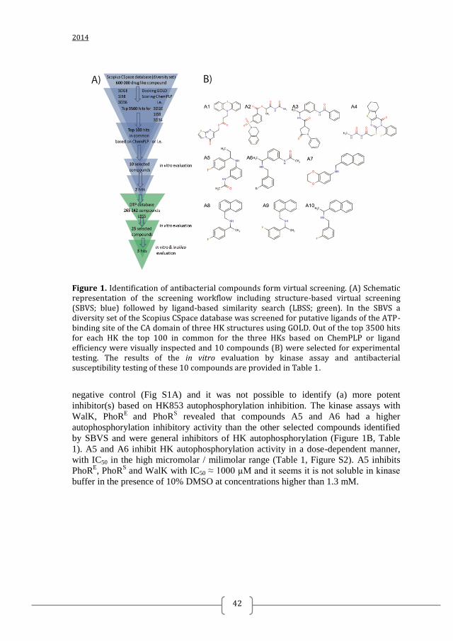
The ATP-binding pocket of the CA domains of HKs is considered to be the most
promising target for inhibition of TCS HKs and has been used previously in structure-
based virtual screenings 38, 39, 40, 41
. To identify drug-like ligands of the ATP-binding
sites of the HK CA domains with putative broad spectrum of inhibition, a diverse set
of 600 000 compounds was screened via in silico docking calculation using as target
receptors the ATP-binding sites of the CA domains of three different HKs: T.
maritima HK853 (PDB: 3DGE) 42
, T. maritima CheA (PDB: 1I58) 17
and G.
stearothermophilus KinB (PDB: 3D36) 16
. These three CA domains were selected
because the structures were solved in the presence of a nucleotide and re-docking of
the cognate ligand was successful (RMSD 1Å for 3DGE and 3D36, and 3 Å for 1I58).
Virtual screening with the three structures that have some sequence variability at the
ATP-binding site was expected to facilitate the identification of broad-spectrum
histidine kinase autophosphorylation inhibitors (HKAI). The screened compounds
were ranked based on the raw docking score, ChemPLP, as well as ligand efficiency.
The top 100 docked compounds in common for the three HKs were ranked on both
scoring schemes (i.e. ChemPLP and ligand efficiency) and then visually inspected to
select 10 compounds, A1-A10 (Figure 1, Tables 1, S1 and S2), for experimental
testing.
INHIBITION OF HK AUTOPHOSPHORYLATION IN VITRO AND
ANTIBACTERIAL SUSCEPTIBILITY TESTING REVEALED TWO HIT
COMPOUNDS
The inhibitory activity of the 10 selected compounds from the SBVS on HKs
autophosphorylation was tested in vitro using four different HKs: T. maritima HK853
(HK853), as a representative of the structures used in the docking assays, the highly
extended HK PhoR from a Gram-negative (E. coli; PhoRE), and Gram-positive (S.
aureus; PhoRS) representative and S. pneumoniae WalK (WalK) as a representative of
the essential WalKR TCS ubiquitous among Gram-positive bacteria 43
. As a fast way
to compare the autophosphorylation inhibitory capacity of the 10 selected compounds
the kinase assays were performed at a single and high (5 mM) compound
concentration and at one time point (30 sec). Autophosphorylation activity of HK853
was not or weakly (up to 30%) inhibited by the 10 compounds compared to the
Chapter 2
2014
42
Figure 1. Identification of antibacterial compounds form virtual screening. (A) Schematic representation of the screening workflow including structure-based virtual screening (SBVS; blue) followed by ligand-based similarity search (LBSS; green). In the SBVS a diversity set of the Scopius CSpace database was screened for putative ligands of the ATP-binding site of the CA domain of three HK structures using GOLD. Out of the top 3500 hits for each HK the top 100 in common for the three HKs based on ChemPLP or ligand efficiency were visually inspected and 10 compounds (B) were selected for experimental testing. The results of the in vitro evaluation by kinase assay and antibacterial susceptibility testing of these 10 compounds are provided in Table 1.
negative control (Fig S1A) and it was not possible to identify (a) more potent
inhibitor(s) based on HK853 autophosphorylation inhibition. The kinase assays with
WalK, PhoRE
and PhoRS revealed that compounds A5 and A6 had a higher
autophosphorylation inhibitory activity than the other selected compounds identified
by SBVS and were general inhibitors of HK autophosphorylation (Figure 1B, Table
1). A5 and A6 inhibit HK autophosphorylation activity in a dose-dependent manner,
with IC50 in the high micromolar / milimolar range (Table 1, Figure S2). A5 inhibits
PhoRE, PhoR
S and WalK with IC50 ≈ 1000 µM and it seems it is not soluble in kinase
buffer in the presence of 10% DMSO at concentrations higher than 1.3 mM.
Discovery of Inhibitors of Bacterial Histidine Kinases
43
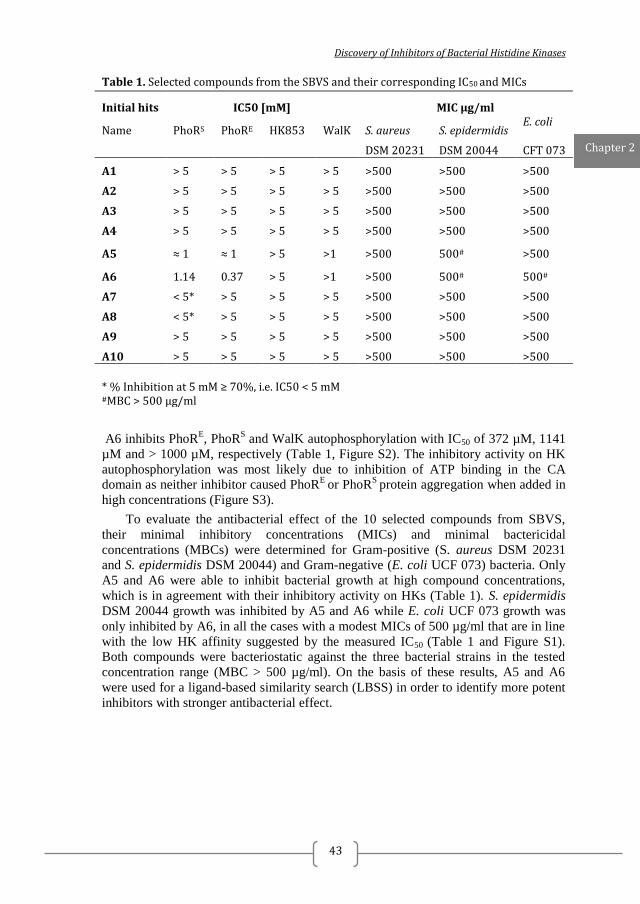
Table 1. Selected compounds from the SBVS and their corresponding IC50 and MICs
Initial hits IC50 [mM] MIC µg/ml
Name PhoRS PhoRE HK853 WalK S. aureus S. epidermidis E. coli
DSM 20231 DSM 20044 CFT 073
A1 > 5 > 5 > 5 > 5 >500 >500 >500
A2 > 5 > 5 > 5 > 5 >500 >500 >500
A3 > 5 > 5 > 5 > 5 >500 >500 >500
A4 > 5 > 5 > 5 > 5 >500 >500 >500
A5 ≈ 1 ≈ 1 > 5 >1 >500 500# >500
A6 1.14 0.37 > 5 >1 >500 500# 500#
A7 < 5* > 5 > 5 > 5 >500 >500 >500
A8 < 5* > 5 > 5 > 5 >500 >500 >500
A9 > 5 > 5 > 5 > 5 >500 >500 >500
A10 > 5 > 5 > 5 > 5 >500 >500 >500
* % Inhibition at 5 mM ≥ 70%, i.e. IC50 < 5 mM #MBC > 500 µg/ml
A6 inhibits PhoRE, PhoR
S and WalK autophosphorylation with IC50 of 372 µM, 1141
µM and > 1000 µM, respectively (Table 1, Figure S2). The inhibitory activity on HK
autophosphorylation was most likely due to inhibition of ATP binding in the CA
domain as neither inhibitor caused PhoRE
or PhoRS
protein aggregation when added in
high concentrations (Figure S3).
To evaluate the antibacterial effect of the 10 selected compounds from SBVS,
their minimal inhibitory concentrations (MICs) and minimal bactericidal
concentrations (MBCs) were determined for Gram-positive (S. aureus DSM 20231
and S. epidermidis DSM 20044) and Gram-negative (E. coli UCF 073) bacteria. Only
A5 and A6 were able to inhibit bacterial growth at high compound concentrations,
which is in agreement with their inhibitory activity on HKs (Table 1). S. epidermidis
DSM 20044 growth was inhibited by A5 and A6 while E. coli UCF 073 growth was
only inhibited by A6, in all the cases with a modest MICs of 500 µg/ml that are in line
with the low HK affinity suggested by the measured IC50 (Table 1 and Figure S1).
Both compounds were bacteriostatic against the three bacterial strains in the tested
concentration range (MBC > 500 µg/ml). On the basis of these results, A5 and A6
were used for a ligand-based similarity search (LBSS) in order to identify more potent
inhibitors with stronger antibacterial effect.
Chapter 2
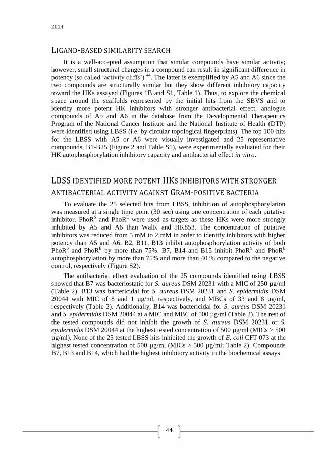
2014
44
LIGAND-BASED SIMILARITY SEARCH
It is a well-accepted assumption that similar compounds have similar activity;
however, small structural changes in a compound can result in significant difference in
potency (so called ‘activity cliffs’) 44
. The latter is exemplified by A5 and A6 since the
two compounds are structurally similar but they show different inhibitory capacity
toward the HKs assayed (Figures 1B and S1, Table 1). Thus, to explore the chemical
space around the scaffolds represented by the initial hits from the SBVS and to
identify more potent HK inhibitors with stronger antibacterial effect, analogue
compounds of A5 and A6 in the database from the Developmental Therapeutics
Program of the National Cancer Institute and the National Institute of Health (DTP)
were identified using LBSS (i.e. by circular topological fingerprints). The top 100 hits
for the LBSS with A5 or A6 were visually investigated and 25 representative
compounds, B1-B25 (Figure 2 and Table S1), were experimentally evaluated for their
HK autophosphorylation inhibitory capacity and antibacterial effect in vitro.
LBSS IDENTIFIED MORE POTENT HKS INHIBITORS WITH STRONGER
ANTIBACTERIAL ACTIVITY AGAINST GRAM-POSITIVE BACTERIA
To evaluate the 25 selected hits from LBSS, inhibition of autophosphorylation
was measured at a single time point (30 sec) using one concentration of each putative
inhibitor. PhoRS and PhoR
E were used as targets as these HKs were more strongly
inhibited by A5 and A6 than WalK and HK853. The concentration of putative
inhibitors was reduced from 5 mM to 2 mM in order to identify inhibitors with higher
potency than A5 and A6. B2, B11, B13 inhibit autophosphorylation activity of both
PhoRS and PhoR
E by more than 75%. B7, B14 and B15 inhibit PhoR
S and PhoR
E
autophosphorylation by more than 75% and more than 40 % compared to the negative
control, respectively (Figure S2).
The antibacterial effect evaluation of the 25 compounds identified using LBSS
showed that B7 was bacteriostatic for S. aureus DSM 20231 with a MIC of 250 µg/ml
(Table 2). B13 was bactericidal for S. aureus DSM 20231 and S. epidermidis DSM
20044 with MIC of 8 and 1 µg/ml, respectively, and MBCs of 33 and 8 µg/ml,
respectively (Table 2). Additionally, B14 was bactericidal for S. aureus DSM 20231
and S. epidermidis DSM 20044 at a MIC and MBC of 500 µg/ml (Table 2). The rest of
the tested compounds did not inhibit the growth of S. aureus DSM 20231 or S.
epidermidis DSM 20044 at the highest tested concentration of 500 µg/ml (MICs > 500
µg/ml). None of the 25 tested LBSS hits inhibited the growth of E. coli CFT 073 at the
highest tested concentration of 500 µg/ml (MICs > 500 µg/ml; Table 2). Compounds
B7, B13 and B14, which had the highest inhibitory activity in the biochemical assays
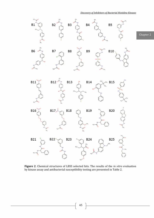
Discovery of Inhibitors of Bacterial Histidine Kinases
45
Figure 2. Chemical structures of LBSS selected hits. The results of the in vitro evaluation by kinase assay and antibacterial susceptibility testing are presented in Table 2.
Chapter 2
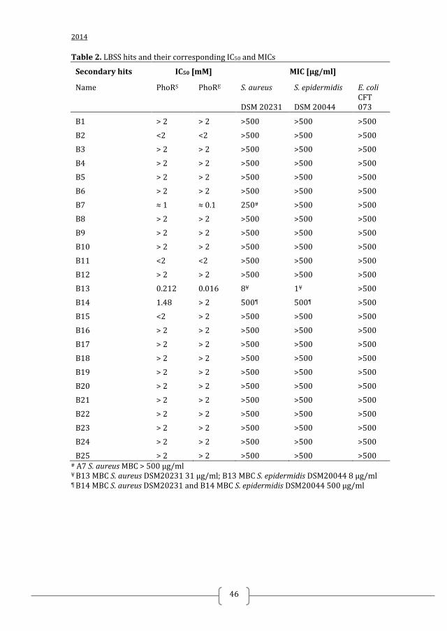
2014
46
Table 2. LBSS hits and their corresponding IC50 and MICs
Secondary hits IC50 [mM] MIC [µg/ml]
Name PhoRS PhoRE S. aureus S. epidermidis E. coli
DSM 20231 DSM 20044 CFT 073
B1 > 2 > 2 >500 >500 >500
B2 <2 <2 >500 >500 >500
B3 > 2 > 2 >500 >500 >500
B4 > 2 > 2 >500 >500 >500
B5 > 2 > 2 >500 >500 >500
B6 > 2 > 2 >500 >500 >500
B7 ≈ 1 ≈ 0.1 250# >500 >500
B8 > 2 > 2 >500 >500 >500
B9 > 2 > 2 >500 >500 >500
B10 > 2 > 2 >500 >500 >500
B11 <2 <2 >500 >500 >500
B12 > 2 > 2 >500 >500 >500
B13 0.212 0.016 8¥ 1¥ >500
B14 1.48 > 2 500¶ 500¶ >500
B15 <2 > 2 >500 >500 >500
B16 > 2 > 2 >500 >500 >500
B17 > 2 > 2 >500 >500 >500
B18 > 2 > 2 >500 >500 >500
B19 > 2 > 2 >500 >500 >500
B20 > 2 > 2 >500 >500 >500
B21 > 2 > 2 >500 >500 >500
B22 > 2 > 2 >500 >500 >500
B23 > 2 > 2 >500 >500 >500
B24 > 2 > 2 >500 >500 >500
B25 > 2 > 2 >500 >500 >500 # A7 S. aureus MBC > 500 µg/ml ¥ B13 MBC S. aureus DSM20231 31 µg/ml; B13 MBC S. epidermidis DSM20044 8 µg/ml ¶ B14 MBC S. aureus DSM20231 and B14 MBC S. epidermidis DSM20044 500 µg/ml

Discovery of Inhibitors of Bacterial Histidine Kinases
47
and antibacterial activity were selected for further evaluation in vitro. First, their IC50
against PhoRE and PhoR
S was measured (Figure S2, Table 2). B7 inhibits PhoR
E
autophosphorylation with IC50 in the lower micromolar range (≥ 100 µM) and PhoRS
in the higher micromolar/ milimolar range (IC50 PhoRS ≥ 1000 µM). The IC50 curves
indicated that B7 is not soluble in concentrations higher than 1000 µM in kinase buffer
in the presence of 10% DMSO. B13 inhibits PhoRE and PhoR
S with IC50 PhoR
E < 100
µM and IC50 PhoRS = 212 µM and possess good solubility in kinase buffer in the
presence of 10% DMSO. B14 IC50 against PhoRE and PhoR
S is higher than 2000 µM
and higher than 1000 µM, respectively. The possibility of non-specific inhibition by
aggregation was ruled out since none of the compounds caused protein aggregation of
PhoRS or PhoR
E at 2 mM in native PAGE (Figure S3). Another reported side effect of
previously published TCS inhibitors is membrane damage. To exclude that the
antibacterial activity of these compounds could be mediated by this mechanism, the
hemolytic activity of B7, B13 and B14 was tested. B7 and B14 did not cause
hemolysis at 500 µg/ml (i.e. LC50> 500 µg/ml). Hemolysis was observed with
compound B13 at concentrations higher than the observed MICs (LC 50 277 µg/ml).
CLINICAL ISOLATES OF MULTIDRUG RESISTANT BACTERIA ARE
SUSCEPTIBLE TO SELECTED HKAIS IN VITRO
The most promising LBSS hits B7, B13 and B14 (Figure 2) were tested for their
ability to inhibit the growth of well-characterized methicillin resistant clinical isolates
of S. aureus (MRSA) 28
and clinical isolates of S. epidermidis, as well as reference
strains of S. pneumoniae and S. suis and additional reference strains of S. aureus and S.
epidermidis. Since B7, B13 and B14 are expected to inhibit the autophosphorylation of
multiple HKs, including HKs from Gram-negative bacteria, their antibacterial effect
against multi-drug resistant clinical isolates of the important pathogens A. baumannii 45
, 46
and S. maltophilia 31
and against reference strains from Klebsiella pneumoniae
and Pseudomonas aeruoginosa were also tested (Table 3, Table S3). B7 inhibited the
growth of two of the 6 MRSA strains in vitro with MICs ≥250 µg/ml, which was
comparable to the MICs for the S. aureus reference strains 25293 and DSM 202231.
The other four MRSA strains were not susceptible to B7. As expected from the results
of the antibacterial susceptibility testing with S. epidermidis DSM 20044, compound
B7 had no antibacterial effect on the clinical isolates of S. epidermidis. B7 inhibits the
growth of all tested S. pneumoniae strains with MIC of 128 µg/ml, which was similar
to the MICs for the reference strains of S. aureus. B7 did not inhibit the growth of the
S. suis 3881/ S10 strain in the tested concentration range (≤ 500 µg/ml).
B13 inhibited the growth of all tested MRSA stains with MICs between 8 and 16
µg/ml, which was comparable to the MICs for the reference strains of S. aureus. B13
also inhibited the growth of clinical isolates of S. epidermidis with MICs between 8
and 16 µg/ml, which is similar to the MICs for, the biofilm forming S. epidermidis
RP62A and non-biofilm forming S. epidermidis RP62A/1. Like B7, B13 inhibited the
growth of the S. pneumoniae strains with MIC of 16 µg/ml.
B14 inhibits growth of MRSA with MICs of 250 µg/ml, which was comparable to
the MICs for the reference strains of S. aureus. B14 inhibited growth of all S.
Chapter 2

2014
48
epidermidis strains except S. epidermidis strain RP62A, with MICs in the range of 250
to 500 µg/ml. B14 was not active against S. pneumoniae strains but inhibited growth
of the S. suis S10 with MIC of 125 µg/ml.
Even though the three selected HKAIs, B7, B13 and B14, inhibit PhoRE,
homologues of which are present in many Gram-negative bacteria, only B14 inhibits
the growth of the Gram-negative A. baumanii and S. maltohilia strains with MICs
from 250 μg/ml to 500 μg/ml. None of the 3 selected inhibitors inhibit the growth of
K. pneumoniae ATCC 700603 and P. aeruginosa ATCC 27853 at the highest tested
concentration (MIC > 500 µg/ml).
INCREASED ANTIBACTERIAL ACTIVITY IS NOT RELATED TO INCREASED
CYTOTOXICITY TO MAMMALIAN CELLS
To check the effect of the most promising inhibitors (B7, B13 and B14) on
mammalian cell viability, cytotoxicity was evaluated i) with neutral-red uptake assays
with Caco-2 cells (Figure 3) and ii) measurement of dead (propidium iodide positive)
and apoptotic (annexin V positive) freshly isolated human peripheral blood
mononuclear cells (PBMCs) by flow cytometry (Figure 4). After overnight incubation
(16 to 20 h), B7, B13 and B14 inhibited neutral-red uptake with IC50 values of 105, 29
and 3 µg/ml, respectively. B7 and B14 IC50 are lower than the observed MICs.
However, B13 IC50 is from 1.8 to 29 times higher than the observed MICs for the
panel of Gram-positive strains tested. PBMCs were incubated with 0.2, 1, 3, 13 and 50
µg/ml B7 and B13 for 24 h. There was no significant difference in the number of dead
cells in the control (DMSO treated) and B13 treated samples. However, the number of
cells binding annexin V, a marker of early apoptosis, was significantly higher in cells
incubated with 50 µg/ml of B13 than the control cells (DMSO). This is reflected in a
reduction of viable (PI and annexin V negative) cells (Figure 4). Similar results were
obtained for compound B7 (Figure 4C) suggesting that a concentration between 13
and 50 µg/ ml induces apoptosis in a proportion of the PBMCs after 24 h incubation.
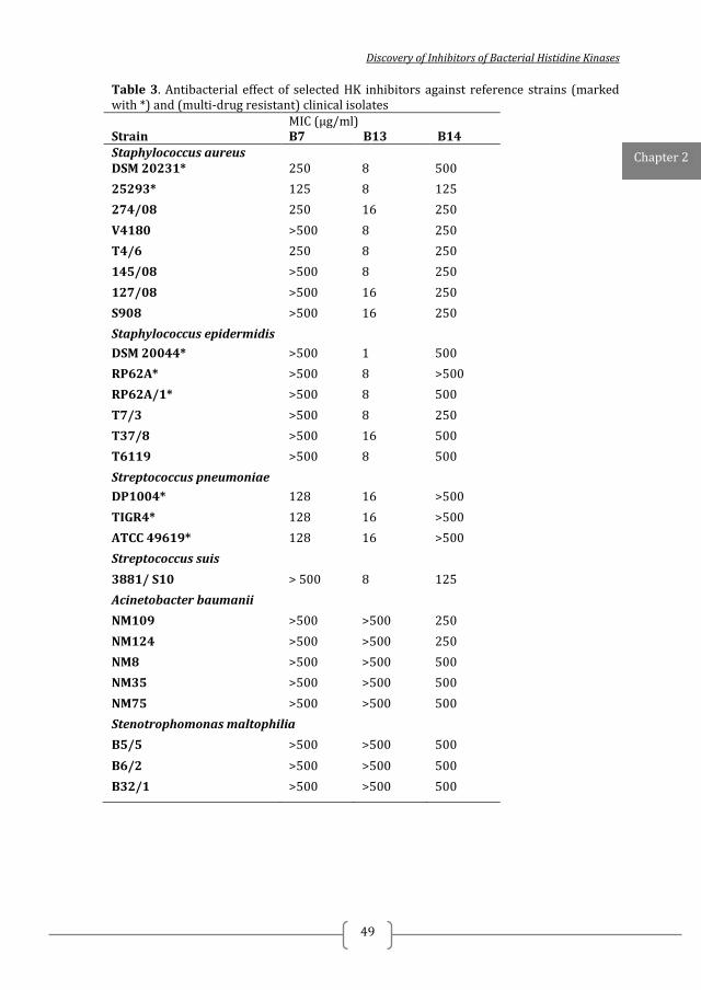
Discovery of Inhibitors of Bacterial Histidine Kinases
49
Table 3. Antibacterial effect of selected HK inhibitors against reference strains (marked with *) and (multi-drug resistant) clinical isolates MIC (µg/ml) Strain B7 B13 B14 Staphylococcus aureus DSM 20231* 250 8 500
25293* 125 8 125
274/08 250 16 250
V4180 >500 8 250
T4/6 250 8 250
145/08 >500 8 250
127/08 >500 16 250
S908 >500 16 250 Staphylococcus epidermidis
DSM 20044* >500 1 500
RP62A* >500 8 >500
RP62A/1* >500 8 500
T7/3 >500 8 250
T37/8 >500 16 500
T6119 >500 8 500
Streptococcus pneumoniae
DP1004* 128 16 >500
TIGR4* 128 16 >500
ATCC 49619* 128 16 >500
Streptococcus suis
3881/ S10 > 500 8 125
Acinetobacter baumanii
NM109 >500 >500 250
NM124 >500 >500 250
NM8 >500 >500 500
NM35 >500 >500 500
NM75 >500 >500 500
Stenotrophomonas maltophilia
B5/5 >500 >500 500
B6/2 >500 >500 500
B32/1 >500 >500 500
Chapter 2
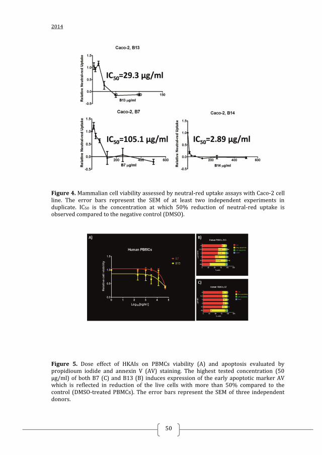
2014
50
Figure 4. Mammalian cell viability assessed by neutral-red uptake assays with Caco-2 cell line. The error bars represent the SEM of at least two independent experiments in duplicate. IC50 is the concentration at which 50% reduction of neutral-red uptake is observed compared to the negative control (DMSO).
Figure 5. Dose effect of HKAIs on PBMCs viability (A) and apoptosis evaluated by propidioum iodide and annexin V (AV) staining. The highest tested concentration (50 µg/ml) of both B7 (C) and B13 (B) induces expression of the early apoptotic marker AV which is reflected in reduction of the live cells with more than 50% compared to the control (DMSO-treated PBMCs). The error bars represent the SEM of three independent donors.
Discovery of Inhibitors of Bacterial Histidine Kinases
51
THE HKAIS ARE PREDICTED TO INTERACT WITH CONSERVED
RESIDUES IN THE ATP-BINDING SITE OF THE HK CA DOMAIN
To gain insights into the mechanism of inhibition at the atomic level, the binding
modes of A5, A6, B7, B13 and B14 to T. maritima HK853 (PDB: 3DGE) were
predicted with GOLD and visually investigated. All selected inhibitors were predicted
to occupy the ATP-binding pocket and interact with key residues involved in binding
of the natural substrate. A common feature for all the compounds was the presence of
an aromatic ring that accommodates into the hydrophobic cavity occupied by the
pyrimidine ring of adenine. (Figure 6) The aromatic ring forms π – π stacking
interactions with Y384 on one side and van der Waals contacts with I416 on the other
side of the ring. Similar hydrophobic interactions have been observed for the adenine
in the structures of T. maritima HK853 and other HKs in complex with nucleotides 26
, 42
. Located at the bottom of the hydrophobic pocket is the conserved Asp (D411) in the
G1 box that gives specificity for recognition of the N6 amino group in the pyrimidine
ring of adenine (Figure 3). For B13, the most potent inhibitor, and B14, the hydroxyl
group of the phenolic ring stacked in the adenine pocket is predicted to be hydrogen
bonded to the conserved D411 residue (Figure 5). Similar mode of interaction was
observed for the binding of the Hsp90 inhibitor radicicol to the ATP-binding domain
of the HK PhoQ 47
. In contrast, A5, A6 and B7 are predicted to exploit their common
amide moieties to mediate polar interaction on the other part of the active site, which
in HK structures with the native ligand is occupied by the nucleotide phosphates and
the Mg2+
cation (Figure 5). In all the three compounds, A5, A6, and B7, the nitrogen
of the amide moiety is predicted to be hydrogen bonded to the conserved N-box Mg2+
chelating residue N380 whereas the oxygen is predicted to bind to the main-chain of
the G2-Box residues G443, L444 and G445, mimicking in this way the interactions of
the ATP γ-phosphate 48
.
Chapter 2
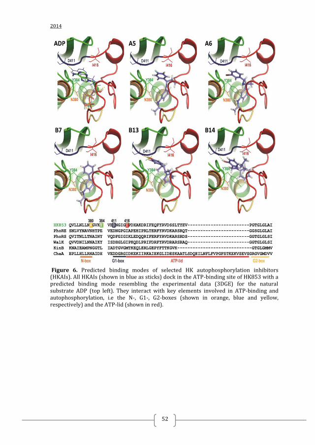
2014
52
Figure 6. Predicted binding modes of selected HK autophosphorylation inhibitors (HKAIs). All HKAIs (shown in blue as sticks) dock in the ATP-binding site of HK853 with a predicted binding mode resembling the experimental data (3DGE) for the natural substrate ADP (top left). They interact with key elements involved in ATP-binding and autophosphorylation, i.e the N-, G1-, G2-boxes (shown in orange, blue and yellow, respectively) and the ATP-lid (shown in red).
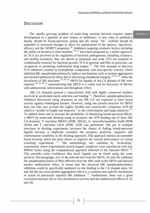
Discovery of Inhibitors of Bacterial Histidine Kinases
53
DISCUSSION
The rapidly growing problem of multi-drug resistant bacteria requires urgent
development of a pipeline of new classes of antibiotics. A new class of antibiotics
ideally should be broad-spectrum, potent and the initial ´´hit´´ scaffold should be
amenable to structural changes to allow for optimization of the potency, specificity,
efficacy and the ADMET properties 49
. Inhibitors targeting virulence factors including
the ability of bacteria to form biofilms 50, 51
have been proposed as a useful approach.
As TCS are involved in the regulation of bacterial pathogenesis, including virulence,
and biofilm formation, they are absent in mammals and some TCS are essential or
conditionally essential for bacterial growth, TCS in general, and HKs in particular, are
recognized as promising antibacterial drug targets 51
. The first attempts to discover
TCS inhibitors resulted in hydrophobic compounds with non-specific activity, which
inhibited HK autophosphorylation by indirect mechanisms such as protein aggregation
and showed antibacterial effect due to decreasing membrane integrity 52, 41, 53
. After the
elucidation of HK structures 54, 26, 42
SBVS for ligands of the CA domain of HK were
reported 38, 39, 40, 41
demonstrating that SBVS is a viable tool for discovery of HKAIs
with antibacterial, antivirulence and therapeutic effect.
HK CA domains present a characteristic fold with highly conserved residues
involved in nucleotide-cation selection and binding 55
. Therefore, autophosphorylation
inhibitors discovered using structures of one HK CA are expected to have broad
activity against orthologous kinases. However, using one protein structure for SBVS
does not take into account the highly flexible and catalytically competent ATP-lid
which is variable in length and sequence 6 or the conformation and target plasticity
56.
To address these and to increase the probability of identifying broad-spectrum HKAI,
a SBVS by molecular docking using as receptors the ATP-binding site of three HK
CA-domains: T. maritima HK853 (PDB: 3DGE), G. stearothermophilus KinB (PDB
3D36) and T. maritima CheA (PDB: 1I58) was performed. The use of multiple
structures in docking experiments increases the chance of finding broad-spectrum
ligands because it implicitly considers the receptors plasticity, sequence and
conformation variability in the docking approach. This approach mimics an ensemble
based docking which has been shown to improve the overall performance of virtual
screening experiments 57
. The methodology was validated by “re-docking”
experiments where experimental protein-ligand complexes were reproduced with low
RMSD values using the computational approach followed in the reported SBVS 58
.
This provides some confidence that novel ligands can be found using the same
protocol. Encouragingly, two of the selected hits from the SBVS, A5 and A6, inhibited
the autophosphorylation of HKs different from the HKs used in the SBVS and showed
modest antibacterial effect. It seems that the observed HK autophosphorylation
inhibition could be specifically mediated via the binding to the CA domain since A5
and A6 did not cause protein aggregation which is a common non-specific mechanism
of action of previously reported HK inhibitors 41
. Furthermore, there was a good
correlation between the in vitro biochemical activity and the antibacterial effects of A5
and A6.
Chapter 2

2014
54
To identify other possible inhibitors with stronger antibacterial effect, A5 and A6
were successfully used as query molecules in a ligand-based similarity search (LBSS).
Among the LBSS hits, the most potent inhibitor was B13 (IC50 PhoRE = 16 µM, IC50
PhoRS
= 212 µM), which inhibited the growth of clinical isolates of MRSA and S.
epidermidis with MICs ≤ 16 µg/ml. The MICs against multi-drug resistant clinical
isolates are comparable with the MICs against reference strains. This suggests that the
putative mechanism of action of B13 differs from the known antibiotics and/or the
mechanisms of resistance of the tested strains to known antibiotics are not functional
against B13. Furthermore, B13 is amenable for chemical modification and further
optimization. Encouragingly, the stronger antibacterial effect of B13 was not related to
higher cytotoxicity. B13 inhibited neutral-red uptake by Caco-2 cells with IC50 of 29
µg/ml, which is higher than the observed MICs for Gram-positive strains.
Furthermore, even 24 h incubation of PBMCs with up to 50 µg/ml of B13 and B7 did
not increase proportion of dead cells. However, the highest B13 concentration tested
(50 µg/ml) increased the proportion cells expressing an early marker of apoptosis.
Together these results suggest that B13 is a good lead candidate for the development
of antibacterials for Gram-positive bacteria. Even though B13 is the most potent
PhoRE inhibitor among the once here reported it did not inhibit the growth of any of
the tested Gram-negative strains at the highest tested concentration (500 µg/ml). The
cell envelope of Gram-negative bacteria is well known to be more effective barrier to
penetration of drugs than that of Gram-positive bacteria but ultimately the compound
may be modified to promote uptake or penetrate prokaryotic membranes according to
experience with other antibacterials entering into Gram-negatives or by applying
nanotechnology approaches 59, 60, 49
.
The predicted binding mode of B14 exploits similar features to B13 as well as
polar interaction with the conserved G1-box Asp residue, inhibits Gram-positive (S.
aureus reference strains and MRSA, S. epidermidis strains and S. suis 3881/S10) and
Gram-negative (A. baumannii and S. maltophilia) strains with MICs in the range of
125 to 500 µg/ml. The MICs range is higher that the observed for B13 in close
agreement with B14 higher than B13 IC50 for PhoRE and PhoR
S autophosphorylation
inhibition. Therefore, B13 and B14 could represent a promising scaffold to re-design
new compounds that could be expected to inhibit a broad spectrum of HKs.
Furthermore, B14 scaffold seems to possess good safety profile since the screening of
B14 with 60-cell lines 61
showed that except for HL-60(TB) and UO-31, the compound
was not cytotoxic (cell growth > 90%).
On the other hand, the relatively weaker than B13 HKAI B7 (IC50 PhoRE ≈
100µM, IC50 PhoRS
≥ 1000 µM) inhibits the growth of the two reference S. aureus
strains and 2 of 6 tested MRSA strains. B7 inhibited the growth of S. pneumoniae with
MICs in the same range as the MICs for the S. aureus strains. However, B7 was not
active against S. epidermidis or any of the Gram-negative bacteria tested. This strain
specificity of B7 correlated with the quite different IC50 observed for PhoRE and
PhoRS HKs. A tentative explanation for this fact can arise from the predicted binding
mode of B7 that suggests a main interaction mediated by its amide group with the
variable part of the ATP-binding site, the ATP lid. All together these indicate that B7
would inhibit autophosphorylation of different HKs with affinity affected by sequence
variability. B7 did not cause protein aggregation or hemolysis and seems to possess
good toxicity properties as intraperitoneal injection on mice leukemia model L1210 62
Discovery of Inhibitors of Bacterial Histidine Kinases
55
with up to 400 mg/kg of B7 led to from 93 to 98% survival of the treated animals,
showing B7 was nontoxic in vivo. All together these data 62
, indicate B7 as a good hit
candidate for development of an antibacterial drug. Furthermore, the effect of B7 on
PBMCs was similar to that of B13 and B7 (Fig 5). As B7 has been shown to be
nontoxic in vivo it could be expected that B13 would also be nontoxic in vivo.
Except of the number of rotatable bonds, the reported inhibitors B7, B13 and B14
are fragment-like, i.e. they follow the so called ¨rule of 3¨ (MW < 300 DA, hydrogen
bond donors (HBD) < 3, hydrogen bond acceptors (HBA) < 3). Therefore, they present
a promising starting point for the rational re-design of more potent and/ or specific
HKAIs. The predicted binding modes of these candidates point to different ways of
compound binding to the HK active site that could be exploited in the re-design
process. In this way, the interaction with the conserved Asp in the G1 box mediated by
the phenolic ring predicted for B13, the most potent inhibitor, and B14 could be
combined with the amide group of B7 that could interact with the ATP-lid at the other
side of the active center. This new generation of structure-based re-designed
compounds could increases potency following the features of B13 and/or specificity
exploiting the sequence and structural differences between HKs, mainly located at the
ATP-lid, to generate general or strain-specific HK inhibitors.
Chapter 2
2014
56
REFERENCES 1. Gootz, T. D., The global problem of antibiotic resistance. Critical reviews in immunology 2010, 30 (1), 79-93. 2. Worthington, R. J.; Melander, C., Overcoming resistance to beta-lactam antibiotics. The Journal of organic chemistry 2013, 78 (9), 4207-13. 3. Desnottes, J. F., New targets and strategies for the development of antibacterial agents. Trends in biotechnology 1996, 14 (4), 134-40. 4. Frosco, M.; Barrett, J. F., Bacterial two-component systems as antimicrobial drug discovery targets. Drug News and Perspectives 1999, 12 (5), 293-299. 5. Matsushita, M.; Janda, K. D., Histidine kinases as targets for new antimicrobial agents. Bioorganic & medicinal chemistry 2002, 10 (4), 855-67. 6. Casino, P.; Rubio, V.; Marina, A., The mechanism of signal transduction by two-component systems. Current opinion in structural biology 2010, 20 (6), 763-71. 7. Crepin, S.; Chekabab, S. M.; Le Bihan, G.; Bertrand, N.; Dozois, C. M.; Harel, J., The Pho regulon and the pathogenesis of Escherichia coli. Veterinary microbiology 2011, 153 (1-2), 82-8. 8. Delaune, A.; Dubrac, S.; Blanchet, C.; Poupel, O.; Mader, U.; Hiron, A.; Leduc, A.; Fitting, C.; Nicolas, P.; Cavaillon, J. M.; Adib-Conquy, M.; Msadek, T., The WalKR system controls major staphylococcal virulence genes and is involved in triggering the host inflammatory response. Infection and immunity 2012, 80 (10), 3438-53. 9. Dubrac, S.; Boneca, I. G.; Poupel, O.; Msadek, T., New insights into the WalK/WalR (YycG/YycF) essential signal transduction pathway reveal a major role in controlling cell wall metabolism and biofilm formation in Staphylococcus aureus. Journal of bacteriology 2007, 189 (22), 8257-69. 10. Unal, C. M.; Singh, B.; Fleury, C.; Singh, K.; Chavez de Paz, L.; Svensater, G.; Riesbeck, K., QseC controls biofilm formation of non-typeable Haemophilus influenzae in addition to an AI-2-dependent mechanism. International journal of medical microbiology : IJMM 2012, 302 (6), 261-9. 11. Howden, B. P.; McEvoy, C. R.; Allen, D. L.; Chua, K.; Gao, W.; Harrison, P. F.; Bell, J.; Coombs, G.; Bennett-Wood, V.; Porter, J. L.; Robins-Browne, R.; Davies, J. K.; Seemann, T.; Stinear, T. P., Evolution of multidrug resistance during Staphylococcus aureus infection involves mutation of the essential two component regulator WalKR. PLoS pathogens 2011, 7 (11), e1002359. 12. Mirabella, A.; Yanez Villanueva, R. M.; Delrue, R. M.; Uzureau, S.; Zygmunt, M. S.; Cloeckaert, A.; De Bolle, X.; Letesson, J. J., The two-component system PrlS/PrlR of Brucella melitensis is required for persistence in mice and appears to respond to ionic strength. Microbiology 2012, 158 (Pt 10), 2642-51. 13. Dubrac, S.; Bisicchia, P.; Devine, K. M.; Msadek, T., A matter of life and death: cell wall homeostasis and the WalKR (YycGF) essential signal transduction pathway. Molecular microbiology 2008, 70 (6), 1307-22. 14. Casino, P.; Rubio, V.; Marina, A. 15. Casino, P.; Rubio, V.; Marina, A., Structural insight into partner specificity and phosphoryl transfer in two-component signal transduction. Cell 2009, 139 (2), 325-36. 16. Bick, M. J.; Lamour, V.; Rajashankar, K. R.; Gordiyenko, Y.; Robinson, C. V.; Darst, S. A., How to switch off a histidine kinase: crystal structure of Geobacillus stearothermophilus KinB with the inhibitor Sda. Journal of molecular biology 2009, 386 (1), 163-77. 17. Bilwes, A. M.; Quezada, C. M.; Croal, L. R.; Crane, B. R.; Simon, M. I., Nucleotide binding by the histidine kinase CheA. Nat Struct Biol 2001, 8 (4), 353-60.
Discovery of Inhibitors of Bacterial Histidine Kinases
57
18. Verdonk, M. L.; Cole, J. C.; Hartshorn, M. J.; Murray, C. W.; Taylor, R. D., Improved protein-ligand docking using GOLD. Proteins 2003, 52 (4), 609-23. 19. Korb, O.; Stutzle, T.; Exner, T. E., Empirical scoring functions for advanced protein-ligand docking with PLANTS. Journal of chemical information and modeling 2009, 49 (1), 84-96. 20. Ebejer, J. P.; Fulle, S.; Morris, G. M.; Finn, P. W., The emerging role of cloud computing in molecular modelling. Journal of molecular graphics & modelling 2013, 44, 177-87. 21. www.inhibox.com. 22. Hopkins, A. L.; Groom, C. R.; Alex, A., Ligand efficiency: a useful metric for lead selection. Drug discovery today 2004, 9 (10), 430-1. 23. RDKit: Open-source cheminformatics. 24. Rogers, D.; Hahn, M., Extended-connectivity fingerprints. Journal of chemical information and modeling 2010, 50 (5), 742-54. 25. Podgornaia, A. I.; Casino, P.; Marina, A.; Laub, M. T., Structural basis of a rationally rewired protein-protein interface critical to bacterial signaling. Structure 2013, 21 (9), 1636-47. 26. Marina, A.; Mott, C.; Auyzenberg, A.; Hendrickson, W. A.; Waldburger, C. D., Structural and mutational analysis of the PhoQ histidine kinase catalytic domain. Insight into the reaction mechanism. The Journal of biological chemistry 2001, 276 (44), 41182-90. 27. Christopoulos, H. M. a. A., Fitting Models to Biological Data using Linear and Nonlinear Regression. A Practical Guide to Curve Fitting. Oxford University Press: New York, 2004. 28. Sonnevend, A.; Blair, I.; Alkaabi, M.; Jumaa, P.; Al Haj, M.; Ghazawi, A.; Akawi, N.; Jouhar, F. S.; Hamadeh, M. B.; Pal, T., Change in meticillin-resistant Staphylococcus aureus clones at a tertiary care hospital in the United Arab Emirates over a 5-year period. Journal of clinical pathology 2012, 65 (2), 178-82. 29. Ziebuhr, W.; Krimmer, V.; Rachid, S.; Lossner, I.; Gotz, F.; Hacker, J., A novel mechanism of phase variation of virulence in Staphylococcus epidermidis: evidence for control of the polysaccharide intercellular adhesin synthesis by alternating insertion and excision of the insertion sequence element IS256. Molecular microbiology 1999, 32 (2), 345-56. 30. Sonnevend, A.; Ghazawi, A.; Al Munthari, N.; Pitout, M.; Hamadeh, M. B.; Hashmey, R.; Girgis, S. K.; Sheikh, F. A.; Al Haj, M.; Nagelkerke, N.; Pal, T., Characteristics of epidemic and sporadic strains of Acinetobacter baumannii isolated in Abu Dhabi hospitals. Journal of medical microbiology 2013, 62 (Pt 4), 582-90. 31. Jumaa, P. A.; Sonnevend, A.; Pal, T.; El Hag, M.; Amith, R.; Trad, O., The molecular epidemiology of Stenotrophomonas maltophilia bacteraemia in a tertiary referral hospital in the United Arab Emirates 2000-2004. Annals of clinical microbiology and antimicrobials 2006, 5, 32. 32. Conlon, J. M.; Mechkarska, M.; Prajeep, M.; Sonnevend, A.; Coquet, L.; Leprince, J.; Jouenne, T.; Vaudry, H.; King, J. D., Host-defense peptides in skin secretions of the tetraploid frog Silurana epitropicalis with potent activity against methicillin-resistant Staphylococcus aureus (MRSA). Peptides 2012, 37 (1), 113-9. 33. CLSI, Methods for dilution antimicrobial susceptibility tests for bacteria that grow aerobically. CLSI: Wayne, PA, 2009; Vol. 8th ed. M7-A8. 34. Clinical Laboratory and Standards Institute Methods for Antimicrobial Susceptibility Testing of Anaerobic Bacteria; Approved Standard. M11-A7. 2007; Vol. 7th ed.
2014
58
35. NCCLS, Methods for determining bactericidal activity of antimi-crobial agents. NCCLS: Wayne, PA, 1999; Vol. M26-A. 36. Mechkarska, M.; Ahmed, E.; Coquet, L.; Leprince, J.; Jouenne, T.; Vaudry, H.; King, J. D.; Conlon, J. M., Antimicrobial peptides with therapeutic potential from skin secretions of the Marsabit clawed frog Xenopus borealis (Pipidae). Comparative biochemistry and physiology. Toxicology & pharmacology : CBP 2010, 152 (4), 467-72. 37. Repetto, G.; del Peso, A.; Zurita, J. L., Neutral red uptake assay for the estimation of cell viability/cytotoxicity. Nature protocols 2008, 3 (7), 1125-31. 38. Qin, Z.; Zhang, J.; Xu, B.; Chen, L.; Wu, Y.; Yang, X.; Shen, X.; Molin, S.; Danchin, A.; Jiang, H.; Qu, D., Structure-based discovery of inhibitors of the YycG histidine kinase: new chemical leads to combat Staphylococcus epidermidis infections. BMC microbiology 2006, 6, 96. 39. Li, N.; Wang, F.; Xu, W.; Wang, H.; Luo, X.; Zhu, W.; Yin, Y.; Zhang, X., [Histidine kinase (YycG) protein of Streptococcus pneumoniae: homology modeling and analysis]. Sheng wu gong cheng xue bao = Chinese journal of biotechnology 2009, 25 (2), 207-14. 40. Cai, X.; Zhang, J.; Chen, M.; Wu, Y.; Wang, X.; Chen, J.; Zhang, J.; Shen, X.; Qu, D.; Jiang, H., The effect of the potential PhoQ histidine kinase inhibitors on Shigella flexneri virulence. PloS one 2011, 6 (8), e23100. 41. Francis, S.; Wilke, K. E.; Brown, D. E.; Carlson, E. E., Mechanistic insight into inhibition of two-component system signaling. MedChemComm 2013, 4 (1), 269-277. 42. Marina, A.; Waldburger, C. D.; Hendrickson, W. A., Structure of the entire cytoplasmic portion of a sensor histidine-kinase protein. The EMBO journal 2005, 24 (24), 4247-59. 43. Mohedano, M. L.; Overweg, K.; de la Fuente, A.; Reuter, M.; Altabe, S.; Mulholland, F.; de Mendoza, D.; Lopez, P.; Wells, J. M., Evidence that the essential response regulator YycF in Streptococcus pneumoniae modulates expression of fatty acid biosynthesis genes and alters membrane composition. Journal of bacteriology 2005, 187 (7), 2357-67. 44. Stumpfe, D.; Bajorath, J., Exploring activity cliffs in medicinal chemistry. Journal of medicinal chemistry 2012, 55 (7), 2932-42. 45. Turton, J. F.; Woodford, N.; Glover, J.; Yarde, S.; Kaufmann, M. E.; Pitt, T. L., Identification of Acinetobacter baumannii by detection of the blaOXA-51-like carbapenemase gene intrinsic to this species. Journal of clinical microbiology 2006, 44 (8), 2974-6. 46. Turton, J. F.; Gabriel, S. N.; Valderrey, C.; Kaufmann, M. E.; Pitt, T. L., Use of sequence-based typing and multiplex PCR to identify clonal lineages of outbreak strains of Acinetobacter baumannii. Clinical microbiology and infection : the official publication of the European Society of Clinical Microbiology and Infectious Diseases 2007, 13 (8), 807-15. 47. Guarnieri, M. T.; Zhang, L.; Shen, J.; Zhao, R., The Hsp90 inhibitor radicicol interacts with the ATP-binding pocket of bacterial sensor kinase PhoQ. Journal of molecular biology 2008, 379 (1), 82-93. 48. Casino, P., Miquel-Romero, L., Marina, A., Visualizing autophosphorylation in histidine kinases. Nat Commun 2014. 49. O'Shea, R.; Moser, H. E., Physicochemical properties of antibacterial compounds: implications for drug discovery. Journal of medicinal chemistry 2008, 51 (10), 2871-8. 50. Rasko, D. A.; Moreira, C. G.; Li de, R.; Reading, N. C.; Ritchie, J. M.; Waldor, M. K.; Williams, N.; Taussig, R.; Wei, S.; Roth, M.; Hughes, D. T.; Huntley, J. F.; Fina, M. W.; Falck, J. R.; Sperandio, V., Targeting QseC signaling and virulence for antibiotic development. Science 2008, 321 (5892), 1078-80.
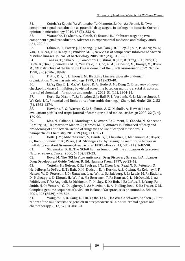
Discovery of Inhibitors of Bacterial Histidine Kinases
59
51. Gotoh, Y.; Eguchi, Y.; Watanabe, T.; Okamoto, S.; Doi, A.; Utsumi, R., Two-component signal transduction as potential drug targets in pathogenic bacteria. Current opinion in microbiology 2010, 13 (2), 232-9. 52. Watanabe, T.; Okada, A.; Gotoh, Y.; Utsumi, R., Inhibitors targeting two-component signal transduction. Advances in experimental medicine and biology 2008, 631, 229-36. 53. Gilmour, R.; Foster, J. E.; Sheng, Q.; McClain, J. R.; Riley, A.; Sun, P. M.; Ng, W. L.; Yan, D.; Nicas, T. I.; Henry, K.; Winkler, M. E., New class of competitive inhibitor of bacterial histidine kinases. Journal of bacteriology 2005, 187 (23), 8196-200. 54. Tanaka, T.; Saha, S. K.; Tomomori, C.; Ishima, R.; Liu, D.; Tong, K. I.; Park, H.; Dutta, R.; Qin, L.; Swindells, M. B.; Yamazaki, T.; Ono, A. M.; Kainosho, M.; Inouye, M.; Ikura, M., NMR structure of the histidine kinase domain of the E. coli osmosensor EnvZ. Nature 1998, 396 (6706), 88-92. 55. Dutta, R.; Qin, L.; Inouye, M., Histidine kinases: diversity of domain organization. Molecular microbiology 1999, 34 (4), 633-40. 56. Li, Y.; Kim, D. J.; Ma, W.; Lubet, R. A.; Bode, A. M.; Dong, Z., Discovery of novel checkpoint kinase 1 inhibitors by virtual screening based on multiple crystal structures. Journal of chemical information and modeling 2011, 51 (11), 2904-14. 57. Korb, O.; Olsson, T. S.; Bowden, S. J.; Hall, R. J.; Verdonk, M. L.; Liebeschuetz, J. W.; Cole, J. C., Potential and limitations of ensemble docking. J. Chem. Inf. Model. 2012, 52 (5), 1262-1274. 58. Hawkins, P. C.; Warren, G. L.; Skillman, A. G.; Nicholls, A., How to do an evaluation: pitfalls and traps. Journal of computer-aided molecular design 2008, 22 (3-4), 179-90. 59. Mas, N.; Galiana, I.; Mondragon, L.; Aznar, E.; Climent, E.; Cabedo, N.; Sancenon, F.; Murguia, J. R.; Martinez-Manez, R.; Marcos, M. D.; Amoros, P., Enhanced efficacy and broadening of antibacterial action of drugs via the use of capped mesoporous nanoparticles. Chemistry 2013, 19 (34), 11167-71. 60. Bolla, J. M.; Alibert-Franco, S.; Handzlik, J.; Chevalier, J.; Mahamoud, A.; Boyer, G.; Kiec-Kononowicz, K.; Pages, J. M., Strategies for bypassing the membrane barrier in multidrug resistant Gram-negative bacteria. FEBS letters 2011, 585 (11), 1682-90. 61. Shoemaker, R. H., The NCI60 human tumour cell line anticancer drug screen. Nature reviews. Cancer 2006, 6 (10), 813-23. 62. Boyd, M., The NCI In Vitro Anticancer Drug Discovery Screen. In Anticancer Drug Development Guide, Teicher, B., Ed. Humana Press: 1997; pp 23-42. 63. Tettelin, H.; Nelson, K. E.; Paulsen, I. T.; Eisen, J. A.; Read, T. D.; Peterson, S.; Heidelberg, J.; DeBoy, R. T.; Haft, D. H.; Dodson, R. J.; Durkin, A. S.; Gwinn, M.; Kolonay, J. F.; Nelson, W. C.; Peterson, J. D.; Umayam, L. A.; White, O.; Salzberg, S. L.; Lewis, M. R.; Radune, D.; Holtzapple, E.; Khouri, H.; Wolf, A. M.; Utterback, T. R.; Hansen, C. L.; McDonald, L. A.; Feldblyum, T. V.; Angiuoli, S.; Dickinson, T.; Hickey, E. K.; Holt, I. E.; Loftus, B. J.; Yang, F.; Smith, H. O.; Venter, J. C.; Dougherty, B. A.; Morrison, D. A.; Hollingshead, S. K.; Fraser, C. M., Complete genome sequence of a virulent isolate of Streptococcus pneumoniae. Science 2001, 293 (5529), 498-506. 64. Wang, Y.; Li, D.; Song, L.; Liu, Y.; He, T.; Liu, H.; Wu, C.; Schwarz, S.; Shen, J., First report of the multiresistance gene cfr in Streptococcus suis. Antimicrobial agents and chemotherapy 2013, 57 (8), 4061-3.
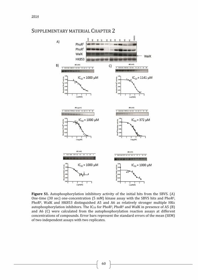
2014
60
SUPPLEMENTARY MATERIAL CHAPTER 2
Figure S1. Autophosphorylation inhibitory activity of the initial hits from the SBVS. (A) One-time (30 sec) one-concentration (5 mM) kinase assay with the SBVS hits and PhoRS, PhoRE, WalK and HK853 distinguished A5 and A6 as relatively stronger multiple HKs autophosphorylation inhibitors. The IC50 for PhoRS, PhoRE and WalK in presence of A5 (B) and A6 (C) were calculated from the autophosphorylation reaction assays at different concentrations of compounds. Error bars represent the standard errors of the mean (SEM) of two independent assays with two replicates.
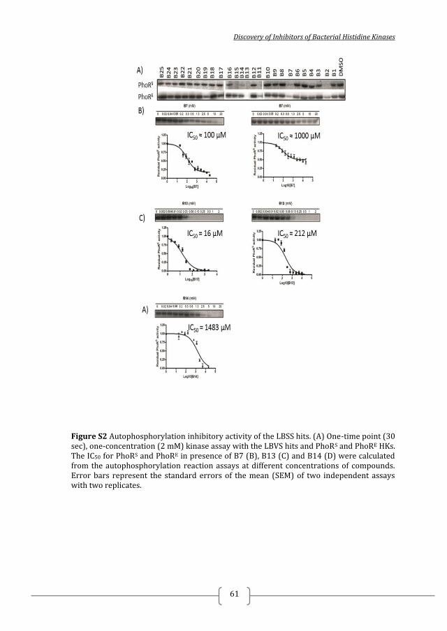
Discovery of Inhibitors of Bacterial Histidine Kinases
61
Figure S2 Autophosphorylation inhibitory activity of the LBSS hits. (A) One-time point (30 sec), one-concentration (2 mM) kinase assay with the LBVS hits and PhoRS and PhoRE HKs. The IC50 for PhoRS and PhoRE in presence of B7 (B), B13 (C) and B14 (D) were calculated from the autophosphorylation reaction assays at different concentrations of compounds. Error bars represent the standard errors of the mean (SEM) of two independent assays with two replicates.
2014
62
Figure S3. Native PAGE-based aggregation analysis of five inhibitors. A5 and A6 (5 mM), and B7, B13 and B14 (2 mM) do not cause HK aggregation as demonstrated by native-PAGE with PhoRS and PhoRE HKs.
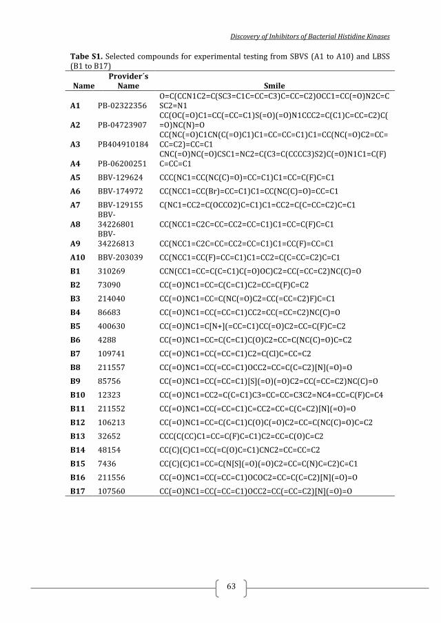
Discovery of Inhibitors of Bacterial Histidine Kinases
63
Tabe S1. Selected compounds for experimental testing from SBVS (A1 to A10) and LBSS (B1 to B17)
Name Provider´s
Name Smile
A1 PB-02322356 O=C(CCN1C2=C(SC3=C1C=CC=C3)C=CC=C2)OCC1=CC(=O)N2C=CSC2=N1
A2 PB-04723907 CC(OC(=O)C1=CC(=CC=C1)S(=O)(=O)N1CCC2=C(C1)C=CC=C2)C(=O)NC(N)=O
A3 PB404910184 CC(NC(=O)C1CN(C(=O)C1)C1=CC=CC=C1)C1=CC(NC(=O)C2=CC=CC=C2)=CC=C1
A4 PB-06200251 CNC(=O)NC(=O)CSC1=NC2=C(C3=C(CCCC3)S2)C(=O)N1C1=C(F)C=CC=C1
A5 BBV-129624 CCC(NC1=CC(NC(C)=O)=CC=C1)C1=CC=C(F)C=C1
A6 BBV-174972 CC(NCC1=CC(Br)=CC=C1)C1=CC(NC(C)=O)=CC=C1
A7 BBV-129155 C(NC1=CC2=C(OCCO2)C=C1)C1=CC2=C(C=CC=C2)C=C1
A8 BBV-34226801 CC(NCC1=C2C=CC=CC2=CC=C1)C1=CC=C(F)C=C1
A9 BBV-34226813 CC(NCC1=C2C=CC=CC2=CC=C1)C1=CC(F)=CC=C1
A10 BBV-203039 CC(NCC1=CC(F)=CC=C1)C1=CC2=C(C=CC=C2)C=C1
B1 310269 CCN(CC1=CC=C(C=C1)C(=O)OC)C2=CC(=CC=C2)NC(C)=O
B2 73090 CC(=O)NC1=CC=C(C=C1)C2=CC=C(F)C=C2
B3 214040 CC(=O)NC1=CC=C(NC(=O)C2=CC(=CC=C2)F)C=C1
B4 86683 CC(=O)NC1=CC(=CC=C1)CC2=CC(=CC=C2)NC(C)=O
B5 400630 CC(=O)NC1=C[N+](=CC=C1)CC(=O)C2=CC=C(F)C=C2
B6 4288 CC(=O)NC1=CC=C(C=C1)C(O)C2=CC=C(NC(C)=O)C=C2
B7 109741 CC(=O)NC1=CC(=CC=C1)C2=C(Cl)C=CC=C2
B8 211557 CC(=O)NC1=CC(=CC=C1)OCC2=CC=C(C=C2)[N](=O)=O
B9 85756 CC(=O)NC1=CC(=CC=C1)[S](=O)(=O)C2=CC(=CC=C2)NC(C)=O
B10 12323 CC(=O)NC1=CC2=C(C=C1)C3=CC=CC=C3C2=NC4=CC=C(F)C=C4
B11 211552 CC(=O)NC1=CC(=CC=C1)C=CC2=CC=C(C=C2)[N](=O)=O
B12 106213 CC(=O)NC1=CC=C(C=C1)C(O)C(=O)C2=CC=C(NC(C)=O)C=C2
B13 32652 CCC(C(CC)C1=CC=C(F)C=C1)C2=CC=C(O)C=C2
B14 48154 CC(C)(C)C1=CC(=C(O)C=C1)CNC2=CC=CC=C2
B15 7436 CC(C)(C)C1=CC=C(N[S](=O)(=O)C2=CC=C(N)C=C2)C=C1
B16 211556 CC(=O)NC1=CC(=CC=C1)OCOC2=CC=C(C=C2)[N](=O)=O
B17 107560 CC(=O)NC1=CC(=CC=C1)OCC2=CC(=CC=C2)[N](=O)=O
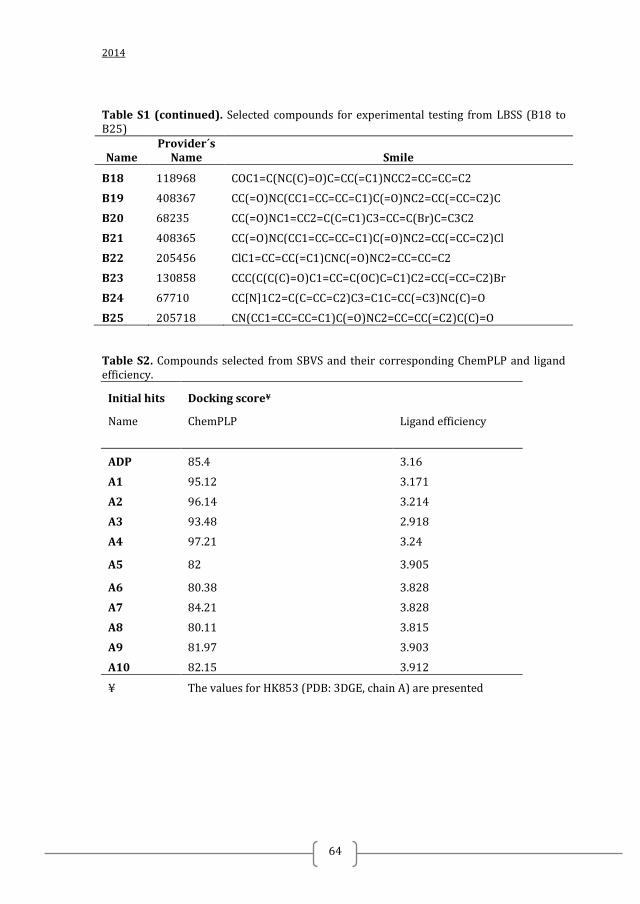
2014
64
Table S1 (continued). Selected compounds for experimental testing from LBSS (B18 to B25)
Name Provider´s
Name Smile
B18 118968 COC1=C(NC(C)=O)C=CC(=C1)NCC2=CC=CC=C2
B19 408367 CC(=O)NC(CC1=CC=CC=C1)C(=O)NC2=CC(=CC=C2)C
B20 68235 CC(=O)NC1=CC2=C(C=C1)C3=CC=C(Br)C=C3C2
B21 408365 CC(=O)NC(CC1=CC=CC=C1)C(=O)NC2=CC(=CC=C2)Cl
B22 205456 ClC1=CC=CC(=C1)CNC(=O)NC2=CC=CC=C2
B23 130858 CCC(C(C(C)=O)C1=CC=C(OC)C=C1)C2=CC(=CC=C2)Br
B24 67710 CC[N]1C2=C(C=CC=C2)C3=C1C=CC(=C3)NC(C)=O
B25 205718 CN(CC1=CC=CC=C1)C(=O)NC2=CC=CC(=C2)C(C)=O Table S2. Compounds selected from SBVS and their corresponding ChemPLP and ligand efficiency.
Initial hits Docking score¥
Name ChemPLP Ligand efficiency
ADP 85.4 3.16
A1 95.12 3.171
A2 96.14 3.214
A3 93.48 2.918
A4 97.21 3.24
A5 82 3.905
A6 80.38 3.828
A7 84.21 3.828
A8 80.11 3.815
A9 81.97 3.903
A10 82.15 3.912
¥ The values for HK853 (PDB: 3DGE, chain A) are presented
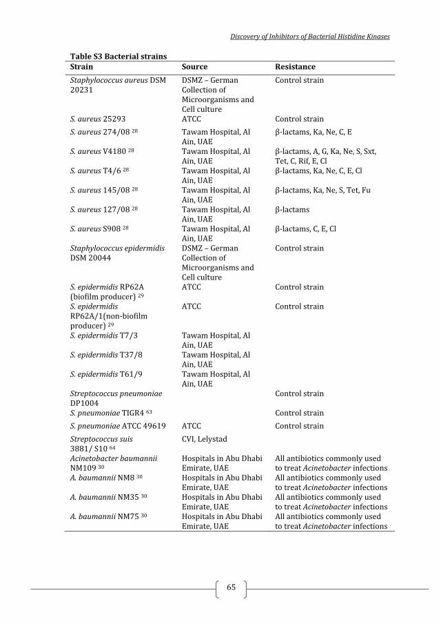
Discovery of Inhibitors of Bacterial Histidine Kinases
65
Table S3 Bacterial strains Strain Source Resistance
Staphylococcus aureus DSM 20231
DSMZ – German Collection of Microorganisms and Cell culture
Control strain
S. aureus 25293 ATCC Control strain
S. aureus 274/08 28 Tawam Hospital, Al Ain, UAE
β-lactams, Ka, Ne, C, E
S. aureus V4180 28 Tawam Hospital, Al Ain, UAE
β-lactams, A, G, Ka, Ne, S, Sxt, Tet, C, Rif, E, Cl
S. aureus T4/6 28 Tawam Hospital, Al Ain, UAE
β-lactams, Ka, Ne, C, E, Cl
S. aureus 145/08 28 Tawam Hospital, Al Ain, UAE
β-lactams, Ka, Ne, S, Tet, Fu
S. aureus 127/08 28 Tawam Hospital, Al Ain, UAE
β-lactams
S. aureus S908 28 Tawam Hospital, Al Ain, UAE
β-lactams, C, E, Cl
Staphylococcus epidermidis DSM 20044
DSMZ – German Collection of Microorganisms and Cell culture
Control strain
S. epidermidis RP62A (biofilm producer) 29
ATCC Control strain
S. epidermidis RP62A/1(non-biofilm producer) 29
ATCC Control strain
S. epidermidis T7/3 Tawam Hospital, Al Ain, UAE
S. epidermidis T37/8 Tawam Hospital, Al Ain, UAE
S. epidermidis T61/9 Tawam Hospital, Al Ain, UAE
Streptococcus pneumoniae DP1004
Control strain
S. pneumoniae TIGR4 63 Control strain
S. pneumoniae ATCC 49619 ATCC Control strain
Streptococcus suis 3881/ S10 64
CVI, Lelystad
Acinetobacter baumannii NM109 30
Hospitals in Abu Dhabi Emirate, UAE
All antibiotics commonly used to treat Acinetobacter infections
A. baumannii NM8 30 Hospitals in Abu Dhabi Emirate, UAE
All antibiotics commonly used to treat Acinetobacter infections
A. baumannii NM35 30 Hospitals in Abu Dhabi Emirate, UAE
All antibiotics commonly used to treat Acinetobacter infections
A. baumannii NM75 30 Hospitals in Abu Dhabi Emirate, UAE
All antibiotics commonly used to treat Acinetobacter infections
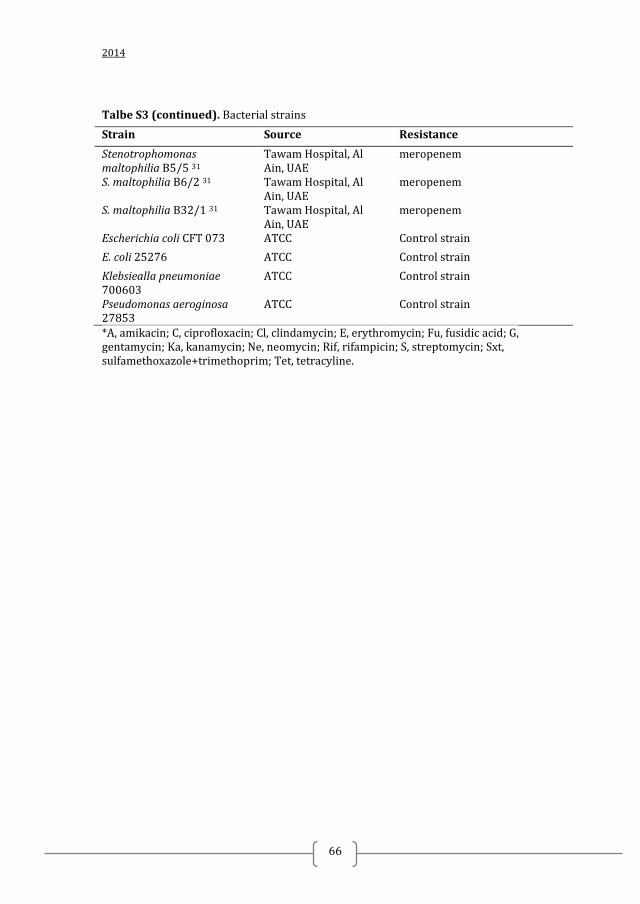
2014
66
Talbe S3 (continued). Bacterial strains
Strain Source Resistance
Stenotrophomonas maltophilia B5/5 31
Tawam Hospital, Al Ain, UAE
meropenem
S. maltophilia B6/2 31 Tawam Hospital, Al Ain, UAE
meropenem
S. maltophilia B32/1 31 Tawam Hospital, Al Ain, UAE
meropenem
Escherichia coli CFT 073 ATCC Control strain
E. coli 25276 ATCC Control strain
Klebsiealla pneumoniae 700603
ATCC Control strain
Pseudomonas aeroginosa 27853
ATCC Control strain
*A, amikacin; C, ciprofloxacin; Cl, clindamycin; E, erythromycin; Fu, fusidic acid; G, gentamycin; Ka, kanamycin; Ne, neomycin; Rif, rifampicin; S, streptomycin; Sxt, sulfamethoxazole+trimethoprim; Tet, tetracyline.
Discovery of Inhibitors of Bacterial Histidine Kinases
67
Chapter 3
HISTIDINE-KINASE INHIBITORS WITH BROAD-
SPECTRUM ANTIBACTERIAL EFFECT IDENTIFIED BY
FRAGMENT-BASED SCREENS Nadya Velikova
1, Simone Fulle
2, Ana Sousa Manso
3, 4, Milena Mechkarska
5, Nico
Taverne6, Paul Finn
2, J. Michael Conlon
5, Marco Rinaldo Oggioni
3, 4, Jerry M. Wells
6,
Alberto Marina1, 7,
1Instituto de Biomedicina de Valencia, Consejo Superior de Investigaciones
Científicas (CSIC), Jaume Roig 11, 46010 Valencia, Spain; 2InhibOx Limited, Oxford, OX1 1BY, United Kingdom;
3Dipartimento di Biotecnologie Mediche, Universita di Siena, 53100 Siena, Italy;
4Department of Genetics, University of Leicester, Leicester, Le1 7RH, United
Kingdom; 5Department of Biochemistry, College of Medicine and Health Science, United Arab
Emirates University, P.O. Box 17666 Al Ain, United Arab Emirates; 6Host-Microbe Interactomics Chair Group, Animal Sciences, University of
Wageningen, P.O. Box 338, 6700 AH Wageningen, The Netherlands; 7CIBER de Enfermedades Raras (CIBERER), ISCIII, Valencia, Spain
SUBMITTED TO EUROPEAN JOURNAL OF MEDICINAL CHEMISTRY

2014
68
Discovery of Inhibitors of Bacterial Histidine Kinases
69
ABSTRACT
Novel antibacterials are urgently needed to address the growing problem of
bacterial resistance to conventional antibiotics. Two-component systems (TCS) have
been previously proposed as promising antibacterial targets. TCS are widely used by
bacteria to regulate gene expression in response to various environmental stimuli and
physiological stress. They consist of a response regulator and a histidine kinase (HK)
containing a highly conserved ATP-binding site that could be a good target for broad
spectrum antibacterial drugs. Here we describe fragment-based screens using
differential scanning fluorimetry and virtual ligand-based similarity searches that
yielded HK autophosphorylation inhibitors with antibacterial effect against multi-drug
resistant clinical isolates.
Keywords: antibacterials, histidine kinase autophosphorylation inhibitors,
fragment-based screening, two-component systems
Abbreviations: TCS – two-component system/s; HK- histidine kinase; RR –
response regulator; FBS – fragment-based screening; LBSS – ligand-based similarity
search; DSF – differential scanning fluorimetry
Chapter 3
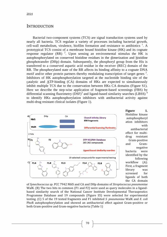
2014
70
INTRODUCTION
Bacterial two-component systems (TCS) are signal transduction systems used by
nearly all bacteria. TCS regulate a variety of processes including bacterial growth,
cell-wall metabolism, virulence, biofilm formation and resistance to antibiotics 1. A
prototypical TCS consist of a membrane bound histidine kinase (HK) and its cognate
response regulator (RR) 2. Upon sensing an environmental stimulus the HK is
autophosphorylated on conserved histidine residues in the dimerization and histidine
phosphotransfer (DHp) domain. Subsequently, the phosphoryl group from the His is
transferred to a conserved aspartic acid residue in the receiver (REC) domain of the
RR. The phosphorylated state of the RR affects its binding affinity to a cognate DNA
motif and/or other protein partners thereby modulating transcription of target genes 2.
Inhibitors of HK autophosphorylation targeted at the nucleotide binding site of the
catalytic and ATP-binding (CA) domains of HKs are expected to simultaneously
inhibit multiple TCS due to the conservation between HKs CA domains (Figure S1).
Here we describe the step-wise application of fragment-based screenings (FBS) by
differential scanning fluorimetry (DSF)3 and ligand-based similarity searches (LBSS)
4
to identify HKs autophosphorylation inhibitors with antibacterial activity against
multi-drug resistant clinical isolates (Figure 1).
Figure 1. Histidine kinase autophosphoryl
ation inhibitors with
antibacterial effect for multi-drug resistant
Gram-positive and Gram-
negative bacteria were identified by the
following workflow (A): First, a fragment library was screened for ligands of both the CA domain
of Synechococcus sp. PCC 7942 NblS and CA and DHp domains of Streptococcus pneumoniae WalK. (B) The two hits in common (F1 and F2) were used as query molecules in a ligand-based similarity search of the National Cancer Institute Developmental Theraupeutics Programme Database and 19 compounds (Figure S5) were selected for experimental testing. (C) 5 of the 19 tested fragments and F1 inhibited S. pneumoniae WalK and E. coli PhoR autophosphorylation and showed an antibacterial effect against Gram-positive or both Gram-positive and Gram-negative bacteria (Table 1)

Discovery of Inhibitors of Bacterial Histidine Kinases
71
Table 1. Antibacterial activities of selected fragments for a panel of clinical isolates and reference strains. Strain MIC (µg/mL)
F1 F1.6 F1.8 F2.3 F2.4 F2.8 Staphylococcus aureus
20231 25 125 125 125 250 31
25293 125 250 250 125 125 250
274/08 125 500 250 >500 500 250
V4180 125 >500 250 250 500 250
T4/6 125 250 250 250 >500 250
145/08 125 250 250 250 250 250
127/08 n.d 250 250 250 500 250
S908 n.d 500 250 250 500 n.d.
Staphylococcus epidermidis
20044 4 500 63 31 500 31
RP62A >500 >500 500 125 > 500 250
RP62A/1 250 >500 250 125 n. d. 250
T7/3 63 125 31 31 n. d. 63
T37/8 >500 >500 500 125 n. d. 250
T6119 >500 >500 250 125 n. d. 250
Streptococcus suis
3881/ S10 >500º >500 250 >500 250 125
Streptococcus pneumoniae 49619 n. d. 256 128 4 64 n. d.
Acinetobacter baumanii
NM109 n. d. n. d. 125 n. d. 500 n. d.
NM124 n. d. n. d. 125 n. d. 500 n. d.
NM8 >500 >500 125 >500 500 >500
NM35 n. d. n. d. 125 n. d. >500 n. d.
NM75 n. d. n. d. 250 n. d. >500 n. d.
Stenotrophomonas maltophilia
B5/5 n. d. n. d. n. d. n. d. >500 n. d.
B6/2 n. d. n. d. n. d. n. d. 500 n. d.
B32/1 >500 >500 >500 >500 500 >500
Chapter 3
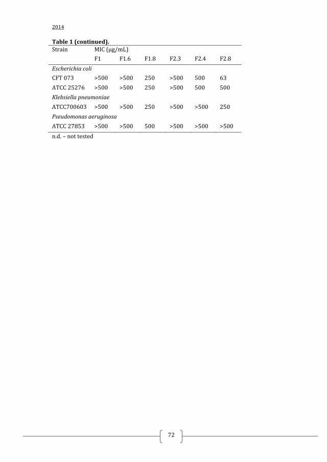
2014
72
Table 1 (continued). Strain MIC (µg/mL)
F1 F1.6 F1.8 F2.3 F2.4 F2.8
Escherichia coli
CFT 073 >500 >500 250 >500 500 63
ATCC 25276 >500 >500 250 >500 500 500
Klebsiella pneumoniae
ATCC700603 >500 >500 250 >500 >500 250
Pseudomonas aeruginosa
ATCC 27853 >500 >500 500 >500 >500 >500
n.d. – not tested
Discovery of Inhibitors of Bacterial Histidine Kinases
73
MATERIALS AND METHODS
REAGENTS
A fragment library of 898 compounds (>95% purity) was purchased from Chem-
X-Infinity (Romanville, France) and the individual compounds were stored at -80°C at
a concentration of 20 mM. For screening purposes cocktails of 10 compounds at final
concentrations of 2 mM were prepared in a 96-well-plate. Re-supply of compounds F2
and F2.5 to F2.9 (Table S2) were purchased from Sigma-Aldrich (Spain). Re-supply of
F1 was purchased from Apolo Scientific (United Kingdom). Compounds F1.1 to F1.10
and F2.1 to F2.3 (Table S2) were obtained from Developmental Therapeutics program
of the National Cancer Institute and the National Institute of Health (DTP NCI/NIH).
γ-32
P-ATP was purchased from Perkin Elmer.
CLONING, EXPRESSION AND PURIFICATION
S. pneumoniae walK encoding the catalytic portion (DHp and CA domain) of
WalK (amino acids from 208 to 449) was amplified by PCR from S. pneumoniae
CDC3059-06 genomic DNA using the following primers: forward 5´-
aagttctgtttcagggcccgatggagcaggagaaggaagaacgc-3´ and reverse 5´-
atggtctagaaagctctagtcttctacttcatccac-3´. The PCR product was purified by PCR product
purification kit (Macherey-Nagel) and cloned into a gel-purified pOpinF vector
(kindly provided by Nick Berow, IRB, Spain) linearized with KpnI and HindIII
(Fermentas). The insert was cloned into the pOpinF vector with InFusion HD cloning
system (Clontech). Positive clones were confirmed by colony PCR and DNA
sequencing.
S. pneumoniae WalK (WalK) was expressed in E. coli RIL. Luria Broth (LB)
media supplemented with 100 µg/mL ampicillin and 33 µg/mL chloramphenicol was
inoculated with an overnight pre-culture (1/50 of the culture volume). At exponential
phase (OD600 0.2 – 0.4) protein expression was induced by addition of 1 mM IPTG for
3 to 5 h at 37°C. The cells were harvested by centrifugation at 4000 g, 4°C for 25 min
and the pellets were stored at -80°C until use. The cell pellets were resuspended in
lysis buffer (100 mM Tris pH 8.0, 150 mM NaCl, 0.1 mM PMSF) and sonicated at
4°C for 5 min at pulses of 15 sec every 1 minute The cell debris and the supernatant
were separated by centrifugation at 11 000 g, 4°C for 60 min. The cell debris were
resuspended in equilibration buffer (100 mM Tris pH 8.0, 150 mM NaCl) containing
2M urea and incubated overnight at 4°C with rotation. After centrifugation at 11 000
g, the supernatant was injected into a Ni-affinity chromatography column (GE
Healthcare) equilibrated with equilibration buffer, washed with 5 volumes of
equilibration buffer and eluted with equilibration buffer containing 0.5 M imidazole.
WalK was concentrated with AmiconUltra (Millipore, USA) centrifugal filters,
aliquoted and stored at –80°C until use. The yield was ≤ 0.5 mg/L culture.
Chapter 3
2014
74
The catalytic portion (DHp and CA domain) of E. coli PhoR (PhoRE), E. coli
EnvZ, and S. aureus PhoR, and the CA domain of Synechococcus sp. PCC 7942 NblS
were expressed and purified as previously described 5, 6, 7, 8
. Shortly, proteins were
expressed in E. coli RIL and purified by Ni-affinity and size-exclusion
chromatography.
Purified proteins were stored in 20 – 50 μl aliquots at -80°C.
DIFFERENTIAL SCANNING FLUORIMETRY
To monitor protein unfolding, the fluorescent dye Sypro orange was used 9.
Differential scanning fluorimetry experiments (DSF) were conducted in the iCycleriQ
Real Time Detection System (Bio-Rad, Hercules, CA). Solutions of 20 µl of 0.1
mg/mL protein (final concentration), 200 µM fragment cocktails or individual
fragments (final concentration), 10X sypro orange (final concentration) and buffer
(100 mM TrisCl pH 8, 150 mM NaCl) were added to the wells of the 96-well iCycler
iQ PCR plate. The plate was heated from 20 to 85 or 99 ºC at a heating rate of 1
ºC/min. The fluorescent intensity was measured with Ex/Em: 490/530 nm. Prism
GraphPad was used for curve fitting and statistical analysis 10
.
KINASE ASSAY
To evaluate the inhibitory capacity of selected hits from DSF and LBSS in vitro
autophosphorylation kinase assays with γ-32
P-ATP was performed as previously
described 11
. Ligands were dissolved in 100% DMSO. When comparing the inhibitory
capacity of ligands in one concentration-one time point experiments or when
measuring IC50 (the concentration at which 50% residual HK autophosphorylation
activity is observed), the final DMSO concentration in the assay was 10% (v/v).
Controls lacking ligands contained an equal concentration of DMSO. Inhibition of
autophosphorylation was determined by incubating 0,12 mg/mL ( ≈ 4 µM) HK and up
to 20 mM fragment in kinase buffer (50 mM Tris HCl, pH 8.5, 50 mM KCl, 5 mM
MgCl2, 0.5 mM EDTA and 0.1 mM DTT). Autophosporylation reactions were
initiated by addition of 0.1 µCi/µl γ-32
P-ATP containing from 0.03 to 0.06 µM ATP
(final concentrations). Autophosphorylation was quenched with 2xSDS-PAGE sample
buffer supplemented with 50 mM EDTA. Samples were applied without heating to
15% (w/v) Tris-glycine SDS-polyacrylamide gels. After electrophoresis, the bottoms
of the gels were removed to lower the background from the unincorporated
radiolabeled ATP. Gels were dried without staining on a Bio-Rad Gel Air drying
system and the phosphorylated protein was quantified by phosphor-imaging. Prism
GraphPad was used for curve fitting and statistical analysis 10
.

Discovery of Inhibitors of Bacterial Histidine Kinases
75
AGGREGATION ANALYSIS BY NATIVE POLYACRYLAMIDE GEL
ELECTROPHORESIS
E. coli PhoR and EnvZ, and S. aureus PhoR (0.12 µg/mL, final concentration)
were prepared in kinase buffer. F1 and F2 were added to a final concentration of 2
mM. DMSO in the assays was maintained to a final concentration 10% (v/v). After 30
min of incubation at room temperature native polyacrylamide gel electrophoresis
(native-PAGE) loading buffer was added and samples loaded. Coomassie blue staining
was used for protein visualization.
LIGAND-BASED SIMILARITY SEARCH
The database from the National Cancer Institute (NCI) was searched for
analogous structures of the F1 and F2 as query molecules; the similarity search was
performed using the Morgan fingerprint as implemented in RDKit 12
, which is a
variation of the ECFP “extended connectivity fingerprints” 4, 13
. The top 100
compounds of each similarity search were visually inspected of which in total 14
compounds were selected and experimentally tested.
ANTIBACTERIAL SUSCEPTIBILITY TESTING
Bacterial strains used in this study for antibacterial susceptibility testing (Table
S3, Chapter 1) were propagated using standard microbiological procedures. Minimal
inhibitory concentrations (MICs) were determined following a standard double-
dilution method 14, 15
. MICs were recorded as the lowest concentration of the
compound where no visible growth was observed. After plating the dilutions around
the MIC or growing them in fresh MH media, minimal bactericidal concentration
(MBC) was recorded as the lowest concentration of the compound at which no
colonies were formed or no growth was observed, respectively. For S. pneumoniae
MICs were determined by adapting the standard double-dilution method to anaerobic
conditions and of this microorganism (use of Todd Hewitt Yeast extract with
200U/mL of catalase and continuous monitoring of growth) 15
. MBCs for S.
pneumoniae were determined by inoculation of 10 µl from each well that did not
shown visible bacterial growth on THY 0,5% 3% blood agar plates. After 24 h of
incubation at 37°C 5% CO2, the first dilution yielding three colonies or fewer was
scored as the MBC, as described by the CLSI for starting inoculate of 1 x 10⁵
CFU/mL 16
.
HEMOLYSIS ASSAY
Hemolytic activity against human erythrocytes taken from a healthy donor was
measured as previously described 17
. Erythrocytes were incubated with (up to) 500
µg/mL compounds and the LC50 value was recorded as the mean concentration of
compound producing 50% hemolysis in three independent incubations.
Chapter 3
2014
76
BINDING MODE PREDICTION
Docking calculations to predict the binding modes of the reported fragments were
performed using the ATP-binding domain of HK853CD (PDB: 3DGE, chain A,
residues from 270 to 415) and the GOLD docking software 18
. For each ligand 100
solutions were generated of which the top 20 were visually inspected. In Figure 2 the
dominant binding mode within the top 20 solutions is shown for the respective ligand.
Discovery of Inhibitors of Bacterial Histidine Kinases
77
RESULTS AND DISCUSSION
To identify fragment-like ligands (MW < 300, ClogP < 3, number of hydrogen
bond donors and hydrogen bond acceptors < 3, number of rotatable bonds < 3 19
) of
the CA domains of HKs we used the Chem-X-Infinity library (Chem-X-Infinity,
Romanville, France) comprising 898 fragments and differential scanning fluorimetry
(DSF) as screening method 3. As targets, we selected the HKs of two essential TCS,
WalK-WalR of Streptococcus pneumoniae and NblS-RapB of Synechococcus sp. PCC
7942 The presence of 4-(4-bromophenyl)-1,3-thiazol-2-amine (F1) and 2-hydroxy-
carbazole (F2) increased the temperature at which HK NblS (CA domain) unfolds
(Tm) by 2.1 and 2.2 °C, respectively, suggesting both F1 and F2 are ligands for the
CA domain of NblS. Encouragingly, the screening for ligands of HK WalK (DHp and
CA domain) showed that F1 and F2 were also among the hits increasing WalK Tm. F1
and F2 increased WalK Tm by 4.5 and 3.9 °C, respectively (Figure S2). Furthermore,
in vitro kinase assays with γ-32
P-ATP showed that F1 and F2 inhibited the
autophosphorylation of the screened catalytic portion of WalK. In addition, these
compounds also inhibited the autophosphorylation of PhoR from the Gram-negative
Escherichia coli (PhoRE, Figure S3), suggesting a general HK inhibitory activity for
F1 and F2.
Compound library screens have previously identified non-drug like inhibitors of
HK autophosphorylation with an unspecific mechanism of action due to protein
aggregation20
. However, even at high concentrations (up to 2 mM) F1 and F2 did not
cause protein aggregation of the catalytic portions (DHp and CA domains) of E. coli
PhoR, E. coli EnvZ or S. aureus PhoR as determined by native polyacrylamide gel
electrophoresis (Figure S4).
In an attempt to identify more potent HK autophosphorylation inhibitors than F1
and F2, LBSS were performed using the Morgan fingerprint as implemented in
RDKit4,7
, the National Cancer Institute Developmental Therapeutics Programme
database (NCI/DTP) and F1 and F2 as query molecules. From the LBSS 14
compounds were selected plus 5 additional substituted carbazoles similar to F2, to a
final pool of 19 compounds (Figure S5) that was evaluated for WalK and PhoRE
autophosphorylation inhibitory activity and antibacterial effect in vitro.
WalK and PhoRE kinase assays with γ-
32P-ATP and the 19 compounds at one
concentration (2 mM) and one time point (30 sec) showed that the fragments inhibited
WalK and PhoRE autophosphorylation activity compared to the negative control from
11 to 62% and 17 to 80%, respectively (Figure S3A). Only F1.6 inhibited WalK
autophosphorylation by more than 50% at a concentration of 2 mM meaning that the
remaining 18 of the tested compounds are weak (Ki >> 2 mM) WalK
autophosporylation inhibitors. Inhibition of PhoRE autophosphorylation was greater
than 50% for F1, F1.8, F2, F2.1, F2.2, F2.8 and F2.9 meaning that the remaining 12 of
the tested compounds are weak PhoRE autophosphorylation inhibitors. The IC50 of the
active compounds (F2.2. and F2.9 were excluded since they did not show antibacterial
effect, see below) were measured in a multiple concentrations one time point (30 sec)
PhoRE kinase assays (Table 1, Figure S3B). F1 and F1.8 inhibited PhoR
E
Chapter 3
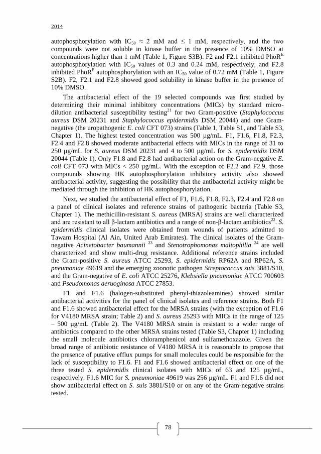
2014
78
autophosphorylation with IC50 ≈ 2 mM and ≤ 1 mM, respectively, and the two
compounds were not soluble in kinase buffer in the presence of 10% DMSO at
concentrations higher than 1 mM (Table 1, Figure S3B). F2 and F2.1 inhibited PhoRE
autophosphorylation with IC50 values of 0.3 and 0.24 mM, respectively, and F2.8
inhibited PhoRE autophosphorylation with an IC50 value of 0.72 mM (Table 1, Figure
S2B). F2, F2.1 and F2.8 showed good solubility in kinase buffer in the presence of
10% DMSO.
The antibacterial effect of the 19 selected compounds was first studied by
determining their minimal inhibitory concentrations (MICs) by standard micro-
dilution antibacterial susceptibility testing21
for two Gram-positive (Staphylococcus
aureus DSM 20231 and Staphylococcus epidermidis DSM 20044) and one Gram-
negative (the uropathogenic E. coli CFT 073) strains (Table 1, Table S1, and Table S3,
Chapter 1). The highest tested concentration was 500 µg/mL. F1, F1.6, F1.8, F2.3,
F2.4 and F2.8 showed moderate antibacterial effects with MICs in the range of 31 to
250 µg/mL for S. aureus DSM 20231 and 4 to 500 µg/mL for S. epidermidis DSM
20044 (Table 1). Only F1.8 and F2.8 had antibacterial action on the Gram-negative E.
coli CFT 073 with MICs < 250 µg/mL. With the exception of F2.2 and F2.9, those
compounds showing HK autophosphorylation inhibitory activity also showed
antibacterial activity, suggesting the possibility that the antibacterial activity might be
mediated through the inhibition of HK autophosphorylation.
Next, we studied the antibacterial effect of F1, F1.6, F1.8, F2.3, F2.4 and F2.8 on
a panel of clinical isolates and reference strains of pathogenic bacteria (Table S3,
Chapter 1). The methicillin-resistant S. aureus (MRSA) strains are well characterized
and are resistant to all β-lactam antibiotics and a range of non-β-lactam antibiotics22
. S.
epidermidis clinical isolates were obtained from wounds of patients admitted to
Tawam Hospital (Al Ain, United Arab Emirates). The clinical isolates of the Gram-
negative Acinetobacter baumannii 23
and Stenotrophomonas maltophilia 24
are well
characterized and show multi-drug resistance. Additional reference strains included
the Gram-positive S. aureus ATCC 25293, S. epidermidis RP62A and RP62A, S.
pneumoniae 49619 and the emerging zoonotic pathogen Streptococcus suis 3881/S10,
and the Gram-negative of E. coli ATCC 25276, Klebsiella pneumoniae ATCC 700603
and Pseudomonas aeruoginosa ATCC 27853.
F1 and F1.6 (halogen-substituted phenyl-thiazoleamines) showed similar
antibacterial activities for the panel of clinical isolates and reference strains. Both F1
and F1.6 showed antibacterial effect for the MRSA strains (with the exception of F1.6
for V4180 MRSA strain; Table 2) and S. aureus 25293 with MICs in the range of 125
– 500 µg/mL (Table 2). The V4180 MRSA strain is resistant to a wider range of
antibiotics compared to the other MRSA strains tested (Table S3, Chapter 1) including
the small molecule antibiotics chloramphenicol and sulfamethoxazole. Given the
broad range of antibiotic resistance of V4180 MRSA it is reasonable to propose that
the presence of putative efflux pumps for small molecules could be responsible for the
lack of susceptibility to F1.6. F1 and F1.6 showed antibacterial effect on one of the
three tested S. epidermidis clinical isolates with MICs of 63 and 125 µg/mL,
respectively. F1.6 MIC for S. pneumoniae 49619 was 256 µg/mL. F1 and F1.6 did not
show antibacterial effect on S. suis 3881/S10 or on any of the Gram-negative strains
tested.
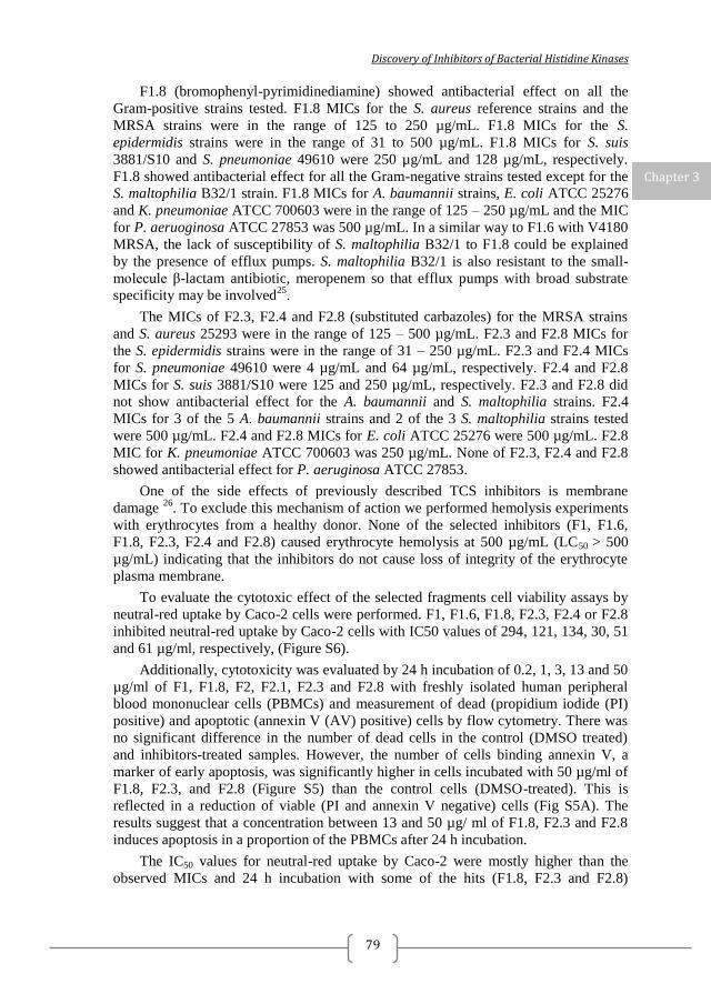
Discovery of Inhibitors of Bacterial Histidine Kinases
79
F1.8 (bromophenyl-pyrimidinediamine) showed antibacterial effect on all the
Gram-positive strains tested. F1.8 MICs for the S. aureus reference strains and the
MRSA strains were in the range of 125 to 250 µg/mL. F1.8 MICs for the S.
epidermidis strains were in the range of 31 to 500 µg/mL. F1.8 MICs for S. suis
3881/S10 and S. pneumoniae 49610 were 250 µg/mL and 128 µg/mL, respectively.
F1.8 showed antibacterial effect for all the Gram-negative strains tested except for the
S. maltophilia B32/1 strain. F1.8 MICs for A. baumannii strains, E. coli ATCC 25276
and K. pneumoniae ATCC 700603 were in the range of 125 – 250 µg/mL and the MIC
for P. aeruoginosa ATCC 27853 was 500 µg/mL. In a similar way to F1.6 with V4180
MRSA, the lack of susceptibility of S. maltophilia B32/1 to F1.8 could be explained
by the presence of efflux pumps. S. maltophilia B32/1 is also resistant to the small-
molecule β-lactam antibiotic, meropenem so that efflux pumps with broad substrate
specificity may be involved25
.
The MICs of F2.3, F2.4 and F2.8 (substituted carbazoles) for the MRSA strains
and S. aureus 25293 were in the range of 125 – 500 µg/mL. F2.3 and F2.8 MICs for
the S. epidermidis strains were in the range of 31 – 250 µg/mL. F2.3 and F2.4 MICs
for S. pneumoniae 49610 were 4 µg/mL and 64 µg/mL, respectively. F2.4 and F2.8
MICs for S. suis 3881/S10 were 125 and 250 µg/mL, respectively. F2.3 and F2.8 did
not show antibacterial effect for the A. baumannii and S. maltophilia strains. F2.4
MICs for 3 of the 5 A. baumannii strains and 2 of the 3 S. maltophilia strains tested
were 500 µg/mL. F2.4 and F2.8 MICs for E. coli ATCC 25276 were 500 µg/mL. F2.8
MIC for K. pneumoniae ATCC 700603 was 250 µg/mL. None of F2.3, F2.4 and F2.8
showed antibacterial effect for P. aeruginosa ATCC 27853.
One of the side effects of previously described TCS inhibitors is membrane
damage 26
. To exclude this mechanism of action we performed hemolysis experiments
with erythrocytes from a healthy donor. None of the selected inhibitors (F1, F1.6,
F1.8, F2.3, F2.4 and F2.8) caused erythrocyte hemolysis at 500 µg/mL (LC50 > 500
µg/mL) indicating that the inhibitors do not cause loss of integrity of the erythrocyte
plasma membrane.
To evaluate the cytotoxic effect of the selected fragments cell viability assays by
neutral-red uptake by Caco-2 cells were performed. F1, F1.6, F1.8, F2.3, F2.4 or F2.8
inhibited neutral-red uptake by Caco-2 cells with IC50 values of 294, 121, 134, 30, 51
and 61 µg/ml, respectively, (Figure S6).
Additionally, cytotoxicity was evaluated by 24 h incubation of 0.2, 1, 3, 13 and 50
µg/ml of F1, F1.8, F2, F2.1, F2.3 and F2.8 with freshly isolated human peripheral
blood mononuclear cells (PBMCs) and measurement of dead (propidium iodide (PI)
positive) and apoptotic (annexin V (AV) positive) cells by flow cytometry. There was
no significant difference in the number of dead cells in the control (DMSO treated)
and inhibitors-treated samples. However, the number of cells binding annexin V, a
marker of early apoptosis, was significantly higher in cells incubated with 50 µg/ml of
F1.8, F2.3, and F2.8 (Figure S5) than the control cells (DMSO-treated). This is
reflected in a reduction of viable (PI and annexin V negative) cells (Fig S5A). The
results suggest that a concentration between 13 and 50 µg/ ml of F1.8, F2.3 and F2.8
induces apoptosis in a proportion of the PBMCs after 24 h incubation.
The IC50 values for neutral-red uptake by Caco-2 were mostly higher than the
observed MICs and 24 h incubation with some of the hits (F1.8, F2.3 and F2.8)
Chapter 3
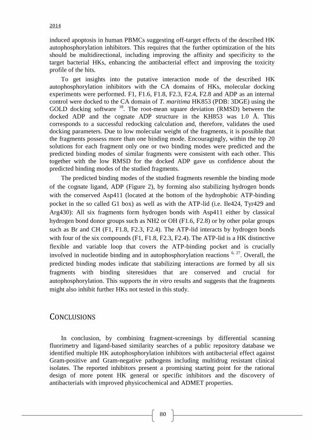
2014
80
induced apoptosis in human PBMCs suggesting off-target effects of the described HK
autophosphorylation inhibitors. This requires that the further optimization of the hits
should be multidirectional, including improving the affinity and specificity to the
target bacterial HKs, enhancing the antibacterial effect and improving the toxicity
profile of the hits.
To get insights into the putative interaction mode of the described HK
autophosphorylation inhibitors with the CA domains of HKs, molecular docking
experiments were performed. F1, F1.6, F1.8, F2.3, F2.4, F2.8 and ADP as an internal
control were docked to the CA domain of T. maritima HK853 (PDB: 3DGE) using the
GOLD docking software 18
. The root-mean square deviation (RMSD) between the
docked ADP and the cognate ADP structure in the KH853 was 1.0 Å. This
corresponds to a successful redocking calculation and, therefore, validates the used
docking parameters. Due to low molecular weight of the fragments, it is possible that
the fragments possess more than one binding mode. Encouragingly, within the top 20
solutions for each fragment only one or two binding modes were predicted and the
predicted binding modes of similar fragments were consistent with each other. This
together with the low RMSD for the docked ADP gave us confidence about the
predicted binding modes of the studied fragments.
The predicted binding modes of the studied fragments resemble the binding mode
of the cognate ligand, ADP (Figure 2), by forming also stabilizing hydrogen bonds
with the conserved Asp411 (located at the bottom of the hydrophobic ATP-binding
pocket in the so called G1 box) as well as with the ATP-lid (i.e. Ile424, Tyr429 and
Arg430): All six fragments form hydrogen bonds with Asp411 either by classical
hydrogen bond donor groups such as NH2 or OH (F1.6, F2.8) or by other polar groups
such as Br and CH (F1, F1.8, F2.3, F2.4). The ATP-lid interacts by hydrogen bonds
with four of the six compounds (F1, F1.8, F2.3, F2.4). The ATP-lid is a HK distinctive
flexible and variable loop that covers the ATP-binding pocket and is crucially
involved in nucleotide binding and in autophosphorylation reactions 6, 27
. Overall, the
predicted binding modes indicate that stabilizing interactions are formed by all six
fragments with binding siteresidues that are conserved and crucial for
autophosphorylation. This supports the in vitro results and suggests that the fragments
might also inhibit further HKs not tested in this study.
CONCLUSIONS
In conclusion, by combining fragment-screenings by differential scanning
fluorimetry and ligand-based similarity searches of a public repository database we
identified multiple HK autophosphorylation inhibitors with antibacterial effect against
Gram-positive and Gram-negative pathogens including multidrug resistant clinical
isolates. The reported inhibitors present a promising starting point for the rational
design of more potent HK general or specific inhibitors and the discovery of
antibacterials with improved physicochemical and ADMET properties.
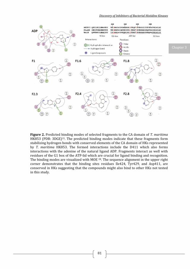
Discovery of Inhibitors of Bacterial Histidine Kinases
81
Figure 2. Predicted binding modes of selected fragments to the CA domain of T. maritima HK853 (PDB: 3DGE)15. The predicted binding modes indicate that these fragments form stabilizing hydrogen bonds with conserved elements of the CA domain of HKs represented by T. maritima HK853. The formed interactions include the D411 which also forms interactions with the adenine of the natural ligand ADP. Fragments interact as well with residues of the G1 box of the ATP-lid which are crucial for ligand binding and recognition. The binding modes are visualized with MOE 28. The sequence alignment in the upper right corner demonstrates that the binding sites residues Ile424, Tyr429, and Asp411, are conserved in HKs suggesting that the compounds might also bind to other HKs not tested in this study.
Chapter 3
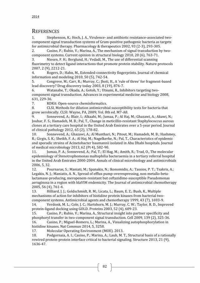
2014
82
REFERENCES 1. Stephenson, K.; Hoch, J. A., Virulence- and antibiotic resistance-associated two-component signal transduction systems of Gram-positive pathogenic bacteria as targets for antimicrobial therapy. Pharmacology & therapeutics 2002, 93 (2-3), 293-305. 2. Casino, P.; Rubio, V.; Marina, A., The mechanism of signal transduction by two-component systems. Current opinion in structural biology 2010, 20 (6), 763-71. 3. Niesen, F. H.; Berglund, H.; Vedadi, M., The use of differential scanning fluorimetry to detect ligand interactions that promote protein stability. Nature protocols 2007, 2 (9), 2212-21. 4. Rogers, D.; Hahn, M., Extended-connectivity fingerprints. Journal of chemical information and modeling 2010, 50 (5), 742-54. 5. Congreve, M.; Carr, R.; Murray, C.; Jhoti, H., A 'rule of three' for fragment-based lead discovery? Drug discovery today 2003, 8 (19), 876-7. 6. Watanabe, T.; Okada, A.; Gotoh, Y.; Utsumi, R., Inhibitors targeting two-component signal transduction. Advances in experimental medicine and biology 2008, 631, 229-36. 7. RDKit: Open-source cheminformatics. 8. CLSI, Methods for dilution antimicrobial susceptibility tests for bacteria that grow aerobically. CLSI: Wayne, PA, 2009; Vol. 8th ed. M7-A8. 9. Sonnevend, A.; Blair, I.; Alkaabi, M.; Jumaa, P.; Al Haj, M.; Ghazawi, A.; Akawi, N.; Jouhar, F. S.; Hamadeh, M. B.; Pal, T., Change in meticillin-resistant Staphylococcus aureus clones at a tertiary care hospital in the United Arab Emirates over a 5-year period. Journal of clinical pathology 2012, 65 (2), 178-82. 10. Sonnevend, A.; Ghazawi, A.; Al Munthari, N.; Pitout, M.; Hamadeh, M. B.; Hashmey, R.; Girgis, S. K.; Sheikh, F. A.; Al Haj, M.; Nagelkerke, N.; Pal, T., Characteristics of epidemic and sporadic strains of Acinetobacter baumannii isolated in Abu Dhabi hospitals. Journal of medical microbiology 2013, 62 (Pt 4), 582-90. 11. Jumaa, P. A.; Sonnevend, A.; Pal, T.; El Hag, M.; Amith, R.; Trad, O., The molecular epidemiology of Stenotrophomonas maltophilia bacteraemia in a tertiary referral hospital in the United Arab Emirates 2000-2004. Annals of clinical microbiology and antimicrobials 2006, 5, 32. 12. Pournaras, S.; Maniati, M.; Spanakis, N.; Ikonomidis, A.; Tassios, P. T.; Tsakris, A.; Legakis, N. J.; Maniatis, A. N., Spread of efflux pump-overexpressing, non-metallo-beta-lactamase-producing, meropenem-resistant but ceftazidime-susceptible Pseudomonas aeruginosa in a region with blaVIM endemicity. The Journal of antimicrobial chemotherapy 2005, 56 (4), 761-4. 13. Hilliard, J. J.; Goldschmidt, R. M.; Licata, L.; Baum, E. Z.; Bush, K., Multiple mechanisms of action for inhibitors of histidine protein kinases from bacterial two-component systems. Antimicrobial agents and chemotherapy 1999, 43 (7), 1693-9. 14. Verdonk, M. L.; Cole, J. C.; Hartshorn, M. J.; Murray, C. W.; Taylor, R. D., Improved protein-ligand docking using GOLD. Proteins 2003, 52 (4), 609-23. 15. Casino, P.; Rubio, V.; Marina, A., Structural insight into partner specificity and phosphoryl transfer in two-component signal transduction. Cell 2009, 139 (2), 325-36. 16. Casino, P.; Miguel-Romero, L.; Marina, A., Visualizing autophosphorylation in histidine kinases. Nat Commun 2014, 5, 3258. 17. Molecular Operating Environment (MOE). 2013. 18. Podgornaia, A. I.; Casino, P.; Marina, A.; Laub, M. T., Structural basis of a rationally rewired protein-protein interface critical to bacterial signaling. Structure 2013, 21 (9), 1636-47.
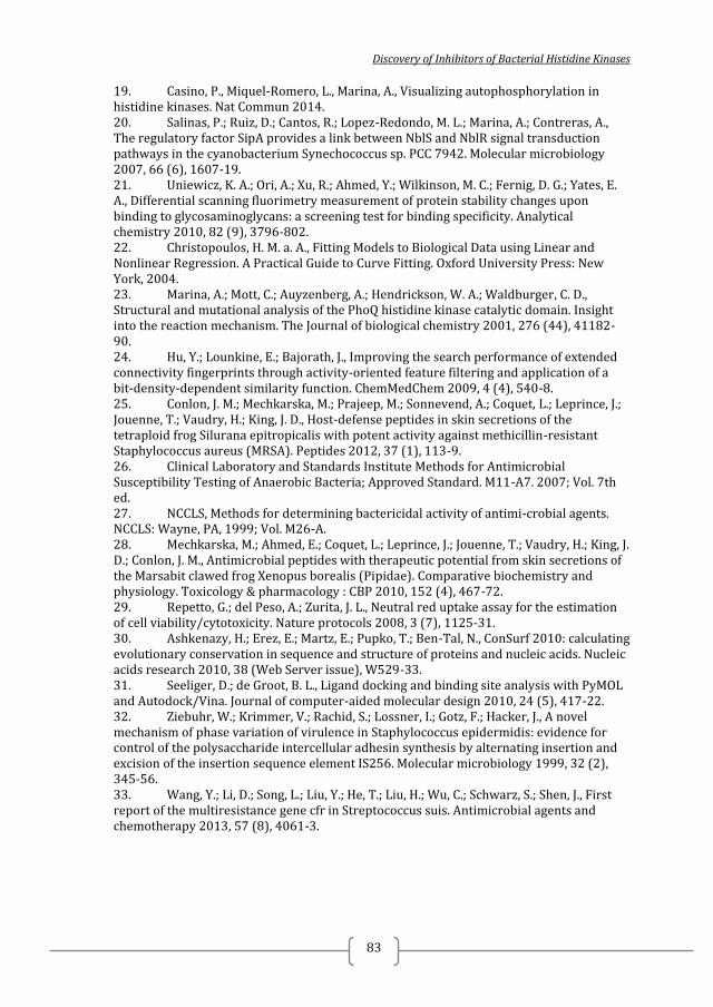
Discovery of Inhibitors of Bacterial Histidine Kinases
83
19. Casino, P., Miquel-Romero, L., Marina, A., Visualizing autophosphorylation in histidine kinases. Nat Commun 2014. 20. Salinas, P.; Ruiz, D.; Cantos, R.; Lopez-Redondo, M. L.; Marina, A.; Contreras, A., The regulatory factor SipA provides a link between NblS and NblR signal transduction pathways in the cyanobacterium Synechococcus sp. PCC 7942. Molecular microbiology 2007, 66 (6), 1607-19. 21. Uniewicz, K. A.; Ori, A.; Xu, R.; Ahmed, Y.; Wilkinson, M. C.; Fernig, D. G.; Yates, E. A., Differential scanning fluorimetry measurement of protein stability changes upon binding to glycosaminoglycans: a screening test for binding specificity. Analytical chemistry 2010, 82 (9), 3796-802. 22. Christopoulos, H. M. a. A., Fitting Models to Biological Data using Linear and Nonlinear Regression. A Practical Guide to Curve Fitting. Oxford University Press: New York, 2004. 23. Marina, A.; Mott, C.; Auyzenberg, A.; Hendrickson, W. A.; Waldburger, C. D., Structural and mutational analysis of the PhoQ histidine kinase catalytic domain. Insight into the reaction mechanism. The Journal of biological chemistry 2001, 276 (44), 41182-90. 24. Hu, Y.; Lounkine, E.; Bajorath, J., Improving the search performance of extended connectivity fingerprints through activity-oriented feature filtering and application of a bit-density-dependent similarity function. ChemMedChem 2009, 4 (4), 540-8. 25. Conlon, J. M.; Mechkarska, M.; Prajeep, M.; Sonnevend, A.; Coquet, L.; Leprince, J.; Jouenne, T.; Vaudry, H.; King, J. D., Host-defense peptides in skin secretions of the tetraploid frog Silurana epitropicalis with potent activity against methicillin-resistant Staphylococcus aureus (MRSA). Peptides 2012, 37 (1), 113-9. 26. Clinical Laboratory and Standards Institute Methods for Antimicrobial Susceptibility Testing of Anaerobic Bacteria; Approved Standard. M11-A7. 2007; Vol. 7th ed. 27. NCCLS, Methods for determining bactericidal activity of antimi-crobial agents. NCCLS: Wayne, PA, 1999; Vol. M26-A. 28. Mechkarska, M.; Ahmed, E.; Coquet, L.; Leprince, J.; Jouenne, T.; Vaudry, H.; King, J. D.; Conlon, J. M., Antimicrobial peptides with therapeutic potential from skin secretions of the Marsabit clawed frog Xenopus borealis (Pipidae). Comparative biochemistry and physiology. Toxicology & pharmacology : CBP 2010, 152 (4), 467-72. 29. Repetto, G.; del Peso, A.; Zurita, J. L., Neutral red uptake assay for the estimation of cell viability/cytotoxicity. Nature protocols 2008, 3 (7), 1125-31. 30. Ashkenazy, H.; Erez, E.; Martz, E.; Pupko, T.; Ben-Tal, N., ConSurf 2010: calculating evolutionary conservation in sequence and structure of proteins and nucleic acids. Nucleic acids research 2010, 38 (Web Server issue), W529-33. 31. Seeliger, D.; de Groot, B. L., Ligand docking and binding site analysis with PyMOL and Autodock/Vina. Journal of computer-aided molecular design 2010, 24 (5), 417-22. 32. Ziebuhr, W.; Krimmer, V.; Rachid, S.; Lossner, I.; Gotz, F.; Hacker, J., A novel mechanism of phase variation of virulence in Staphylococcus epidermidis: evidence for control of the polysaccharide intercellular adhesin synthesis by alternating insertion and excision of the insertion sequence element IS256. Molecular microbiology 1999, 32 (2), 345-56. 33. Wang, Y.; Li, D.; Song, L.; Liu, Y.; He, T.; Liu, H.; Wu, C.; Schwarz, S.; Shen, J., First report of the multiresistance gene cfr in Streptococcus suis. Antimicrobial agents and chemotherapy 2013, 57 (8), 4061-3.
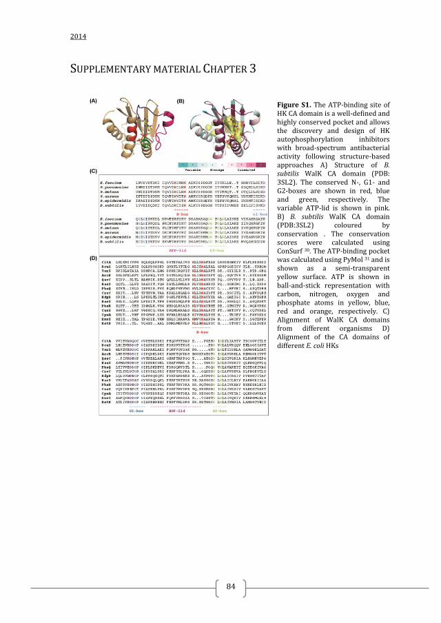
2014
84
SUPPLEMENTARY MATERIAL CHAPTER 3
Figure S1. The ATP-binding site of HK CA domain is a well-defined and highly conserved pocket and allows the discovery and design of HK autophosphorylation inhibitors with broad-spectrum antibacterial activity following structure-based approaches A) Structure of B. subtilis WalK CA domain (PDB: 3SL2). The conserved N-, G1- and G2-boxes are shown in red, blue and green, respectively. The variable ATP-lid is shown in pink. B) B. subtilis WalK CA domain (PDB:3SL2) coloured by conservation . The conservation scores were calculated using ConSurf 30. The ATP-binding pocket was calculated using PyMol 31 and is shown as a semi-transparent yellow surface. ATP is shown in ball-and-stick representation with carbon, nitrogen, oxygen and phosphate atoms in yellow, blue, red and orange, respectively. C) Alignment of WalK CA domains from different organisms D) Alignment of the CA domains of different E. coli HKs
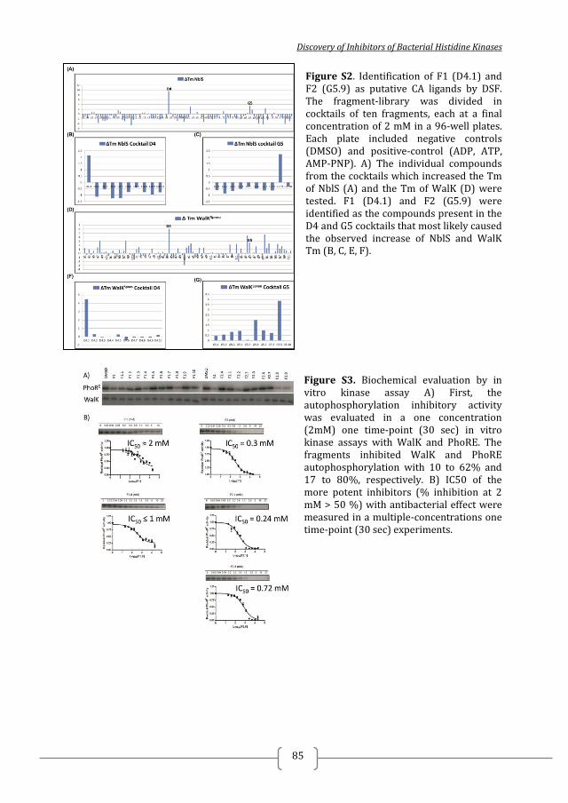
Discovery of Inhibitors of Bacterial Histidine Kinases
85
Figure S2. Identification of F1 (D4.1) and F2 (G5.9) as putative CA ligands by DSF. The fragment-library was divided in cocktails of ten fragments, each at a final concentration of 2 mM in a 96-well plates. Each plate included negative controls (DMSO) and positive-control (ADP, ATP, AMP-PNP). A) The individual compounds from the cocktails which increased the Tm of NblS (A) and the Tm of WalK (D) were tested. F1 (D4.1) and F2 (G5.9) were identified as the compounds present in the D4 and G5 cocktails that most likely caused the observed increase of NblS and WalK Tm (B, C, E, F).
Figure S3. Biochemical evaluation by in vitro kinase assay A) First, the autophosphorylation inhibitory activity was evaluated in a one concentration (2mM) one time-point (30 sec) in vitro kinase assays with WalK and PhoRE. The fragments inhibited WalK and PhoRE autophosphorylation with 10 to 62% and 17 to 80%, respectively. B) IC50 of the more potent inhibitors (% inhibition at 2 mM > 50 %) with antibacterial effect were measured in a multiple-concentrations one time-point (30 sec) experiments.
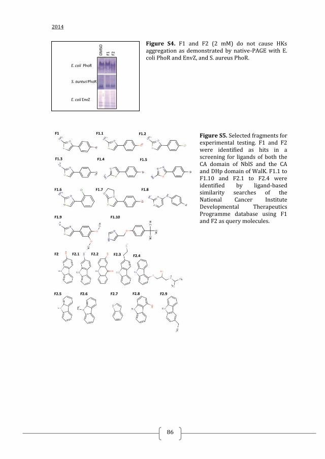
2014
86
Figure S4. F1 and F2 (2 mM) do not cause HKs aggregation as demonstrated by native-PAGE with E. coli PhoR and EnvZ, and S. aureus PhoR.
Figure S5. Selected fragments for experimental testing. F1 and F2 were identified as hits in a screening for ligands of both the CA domain of NblS and the CA and DHp domain of WalK. F1.1 to F1.10 and F2.1 to F2.4 were identified by ligand-based similarity searches of the National Cancer Institute Developmental Therapeutics Programme database using F1 and F2 as query molecules.
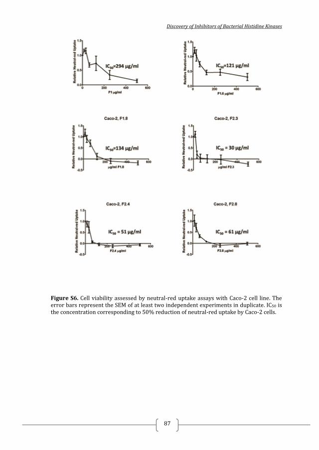
Discovery of Inhibitors of Bacterial Histidine Kinases
87
Figure S6. Cell viability assessed by neutral-red uptake assays with Caco-2 cell line. The error bars represent the SEM of at least two independent experiments in duplicate. IC50 is the concentration corresponding to 50% reduction of neutral-red uptake by Caco-2 cells.

2014
88
Figure S7. Dose effect of identified inhibitors on PBMCs viability (A) and apoptosis evaluated by propidioum iodide and annexin V (AV) staining. 50 µg/ml of F1.8 (C) and F2.3 (E), F2.8 (F) induce stronger expression of the early apoptotic marker AV compared to the control (0 µg/ml, DMSO-treated PBMCs) which is reflected in reduction of the live cells (A) with around or more than 50% compared to the control (DMSO-treated PBMCs). Error bars represent SEM for three independent donors.
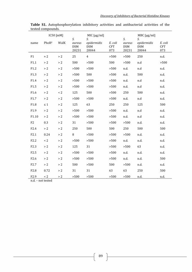
Discovery of Inhibitors of Bacterial Histidine Kinases
89
Table S1. Autophosphorylation inhibitory activities and antibacterial activities of the tested compounds.
IC50 [mM] MIC [µg/ml] MBC [µg/ml]
name PhoRE WalK S. aureus
S. epidermidis E. coli
S. aureus
S. epidermidis E. coli
DSM 20231
DSM 20044
CFT 073
DSM 20231
DSM 20044
CFT 073
F1 ≈ 2 > 2 25 4 >500 >500 250 n.d.
F1.1 > 2 > 2 500 >500 500 >500 n.d >500
F1.2 > 2 > 2 >500 >500 >500 n.d. n.d n.d.
F1.3 > 2 > 2 >500 500 >500 n.d. 500 n.d.
F1.4 > 2 > 2 >500 >500 >500 n.d. n.d n.d.
F1.5 > 2 > 2 >500 >500 >500 n.d. n.d n.d.
F1.6 > 2 < 2 125 500 >500 250 500 n.d.
F1.7 > 2 > 2 >500 >500 >500 n.d. n.d n.d.
F1.8 ≤ 1 > 2 125 63 250 250 125 500
F1.9 > 2 > 2 >500 >500 >500 n.d. n.d n.d.
F1.10 > 2 > 2 >500 >500 >500 n.d. n.d n.d.
F2 0.3 > 2 31 >500 >500 >500 n.d. n.d.
F2.4 > 2 > 2 250 500 500 250 500 500
F2.1 0.24 > 2 8 >500 >500 >500 n.d. n.d.
F2.2 < 2 > 2 >500 >500 >500 n.d. n.d. n.d.
F2.3 > 2 > 2 125 31 >500 >500 63 n.d.
F2.5 > 2 > 2 >500 >500 >500 n.d. n.d. n.d.
F2.6 > 2 > 2 >500 >500 >500 n.d. n.d. 500
F2.7 > 2 > 2 500 >500 500 >500 n.d. n.d.
F2.8 0.72 > 2 31 31 63 63 250 500
F2.9 < 2 > 2 >500 >500 >500 >500 n.d. n.d. n.d. – not tested
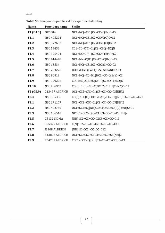
2014
90
Table S2. Compounds purchased for experimental testing.
Name Providers name Smile
F1 (D4.1) OR5604 NC1=NC(=CS1)C2=CC=C(Br)C=C2
F1.1 NSC 405294 NC1=NC(=CS1)C2=CC=C(O)C=C2
F1.2 NSC 372682 NC1=NC(=CS1)C2=CC=C(Cl)C=C2
F1.3 NSC 54436 CC1=CC=C(C=C1)C2=CSC(=N2)N
F1.4 NSC 176404 NC1=NC=C(S1)C2=CC=C(Br)C=C2
F1.5 NSC 614448 NC1=NN=C(O1)C2=CC=C(Br)C=C2
F1.6 NSC 13534 NC1=NC(=CS1)C2=C(Cl)C=CC=C2
F1.7 NSC 223276 BrC1=CC=C(C=C1)C2=CSC3=NCCN23
F1.8 NSC 80819 NC1=NC(=CC=N1)NC2=CC=C(Br)C=C2
F1.9 NSC 329206 COC1=C(OC)C=C(C=C1)C2=CSC(=N2)N
F1.10 NSC 206952 CC(C)(C)C1=CC=C(OCC2=C[NH]C=N2)C=C1
F2 (G5.9) 213497 ALDRICH OC1=CC2=C(C=C1)C3=CC=CC=C3[NH]2
F2.4 NSC 305336 CC(C)NCC(O)COC1=C2C(=CC=C1)[NH]C3=CC=CC=C23
F2.1 NSC 171107 NC1=CC2=C(C=C1)C3=CC=CC=C3[NH]2
F2.2 NSC 402750 OC1=CC2=C([NH]C3=C(C=CC=C3)[C]2=O)C=C1
F2.3 NSC 106510 NCCC1=CC2=C(C=C1)C3=CC=CC=C3[NH]2
F2.5 C5132 SIGMA [NH]1C2=CC=CC=C2C3=CC=CC=C13
F2.6 325325 ALDRICH C[N]1C2=CC=CC=C2C3=CC=CC=C13
F2.7 I3408 ALDRICH [NH]1C=CC2=CC=CC=C12
F2.8 543896 ALDRICH OC1=CC=CC2=C1C3=CC=CC=C3[NH]2
F2.9 754781 ALDRICH CCC1=CC2=C([NH]C3=CC=CC=C23)C=C1
Discovery of Inhibitors of Bacterial Histidine Kinases
91
CHAPTER 4
BROADENING THE ANTIBACTERIAL SPECTRUM OF
HISTIDINE KINASE AUTOPHOSPHORYLATION
INHIBITORS VIA THE USE OF Ε-POLY-L-LYSINE
CAPPED MESOPOROUS SILICA-BASED
NANOPARTICLES
Nadya Velikova1, 2
, Nuria Mas3, 4
, Ellen Stolte2, Edoardo Zaccaria
2, Rui Cao
2, Nico
Taverne2, José Ramón Murguía
3, 4, Ramon Martinez-Manez
3, 4, Alberto Marina
2, 5,
Jerry Wells2
1 Instituto de Biomedicina de Valencia, Consejo Superior de Investigaciones
Científicas (CSIC), Jaume Roig 11, 46010 Valencia, Spain; 2 Host-Microbe Interactomics Chair Group, Animal Sciences, University of
Wageningen, P.O. Box 338, 6700 AH Wageningen, The Netherlands; 3 Centro de Reconocimiento Molecular y Desarrollo Tecnologico, Departamento de
Quimica, Univesidad Politecnica de Valencia, Camino de Vera s/n, 46022 Valencia,
Spain; 4 CIBER de Bioingeneria, Biomateriales y Nanomedicina, CIBER – BBN;
5 CIBER de Enfermedades Raras (CIBERER), ISCIII, Valencia, Spain
TO BE SUBMITTED TO NANOMEDICINE: NANOTECHNOLOGY, BIOLOGY AND
MEDICINE AFTER PUBLISHING OF CHAPTER 2 AND 3

2014
92
Discovery of Inhibitors of Bacterial Histidine Kinases
93
ABBREVIATIONS
Gr- Gram negative
Gr+ Gram positive
TCS Two-component systems
HK Histidine kinase
RR Response regulator
HKAI Histidine-kinase autophosphorylation inhibitor
f n-[(3-trimethoxysilyl)propyl]ethylendiamine triacetic acid trisodium salt
ePLL ε-poly-L-lysine
N.X MCM-41 funcionalized with n-[(3-trimethoxysilyl)propyl]ethylendiamine
triacetic acid trisodium salt and capped ε-poly-L-lysine (ePLL) cationic polymer and
loaded with:
N.Van – vancomycin
N.HKAIs – histidine kinase autophosphorylation inhibitors
N.Rho – rhodamine
F.HKAIs – free histidine kinase autophosphorylation inhibitors
Chapter 4
2014
94
INTRODUCTION
Infections caused by Gram negative (Gr-) multidrug-resistant (MDR) bacteria
have become a growing challenge worldwide 1. The most important resistance
problems are encountered in Enterobacteriaceae, Pseudomonas aeruginosa and
Acinetobacter spp. which have developed resistance to last-line antibiotics such as
expanded-spectrum cephalosporins and/ or carbapenems 2. Urinary tract infections
(UTIs) caused by Gr- uropathogenic Escherichia coli (UPEC) are among the most
common bacterial infections in humans. Recently multi-drug resistant E. coli have
been reported 3, 4, 5
.
Most of the bacterial infections in fish are caused by Gr- bacteria, including
Aeromonas hydrophilia, Aeromonas salmonicida, Flavobacterium columnare, Vibrio,
and Pseudomonas spp. 6. Currently antimicrobials are routinely used to directly control
bacterial infections in pet (ornamental) fish and are added to the water these fish are
shipped in to suppress the growth of potential pathogens during transport 7. However,
this activity has led to high prevalence of multi-drug tolerant or resistant bacteria and
associated antimicrobial resistance genes both in ornamental fish and their carriage
water 7. This might present a potential health risk as studies have suggested that there
is bi-directional transfer of resistance genes between aquatic and other Animalia as
evidenced by the fact that antimicrobial resistance (AMR) genes of bacteria recovered
from the aquatic environment can share very high sequence homology to clinically
important AMR genes on plasmids and integrons found in clinical isolates of human
pathogens 8 , 9, 10, 11 , 12
. The transmission of zoonotic multidrug resistant Gr- bacteria
between food-producing animals and humans and between companion animals and
humans has also been reported 13 , 14, 15
.
Therefore, novel strategies to target infections caused by Gr- bacteria are urgently
needed. Two-component systems (TCS) are signal transduction systems found in
nearly all bacteria and have been proposed as promising antibacterial drugs targets for
both Gram positive (Gr+) and Gr- infections 16, 17, 18
. A prototypical TCS consist of
membrane bound histidine kinase (HK) and its cognate response regulator (RR) 19
.
Upon environmental stimuli the catalytic and ATP-binding (CA) domain of the HK
autophosphorylates a conserved histidine found within the dimerization and histidine
phosphotransfer (DHp) domain, which subsequently serves as the phosphodonor for a
cognate RR 19
. Many HKs are bi-functional and also dephosphorylate their cognate
response regulator 20, 21
. The changes in the phosphorylation levels of the RR are
usually related to changes of the expression of target genes involved in the regulation
of variety of processes including growth, virulence, antibacterial resistance and
adaptation to environmental changes 22, 23, 16
. Following structure-based and fragment-
based drug discovery approaches (Chapter 2 and 3), bacterial histidine kinase
autophosphorylation inhibitors (HKAIs) were identified (Figure 1). The HKAIs
inhibited the autophosphorylation of HKs from both Gr+ and Gr- bacteria, including
E. coli PhoR. PhoR belongs to the PhoR-PhoB TCS, which is involved in the
regulation of the pho regulon. The pho regulon is not only a regulatory circuit of
phosphate homeostasis but also plays an important adaptive role in stress response and
bacterial virulence. 24, 25, 26
. Despite the fact that the HKAIs inhibited
Discovery of Inhibitors of Bacterial Histidine Kinases
95
autophosphorylation of HKs from Gr- bacteria, they showed only moderate to strong
antibacterial activity against Gr+ bacteria and not against Gr- bacteria. This was
anticipated to be a consequence of the permeability barrier provided by the outer
membrane (OM) of Gr- negative bacteria, which are inherently resistant to many
hydrophobic antibiotics 27, 28
. This resistance limits the arsenal of antibiotics that are
effective in treating Gr-negative bacterial infections. To overcome this problem
several compounds have been used to facilitate permeability of the OM and sensitize
Gr- bacteria to hydrophobic antibiotics 29, 30
. These compounds are typically cationic,
amphiphilic molecules that can be prepared from peptides or steroids. Nanotechnology
has also been proposed and exploited in addressing the problem related to Gr-
membrane permeability 31
.
A nanodevice loaded with vancomycin was shown to possess antibacterial activity
against a range of Gr- bacteria including E. coli (DH5α, 100, 405), Salmonella
typhimurium and Erwinia carotovora 32
. The device consisted of a nanometric
mesoporous MCM-41 phase (ca. 100 nM; MCM-41) loaded with vancomycin,
functionalized with n-[(3-trimethoxysilyl)propyl]ethylendiamine triacetic acid
trisodium salt (f) and capped with a cationic polymer of ε-poly-L-lysine (ePLL),
referred to here as N.Van. Vancomycin is glycopeptide antibiotic that selectively
targets Gr+ bacteria 33
. Most Gr- bacteria are intrinsically resistant to vancomycin due
to its relatively high molecular weight and size and inability to pass through porins in
the OM of Gr-.bacteria. Capping the NPs with polycationic ePLL is anticipated to
promote ionic bonding with the negatively charged OM and facilitate permeability or
antibacterial effects of the inhibitor through its disruptive effects on the bacterial
membrane 34
. Indeed, N.Van broadened the spectrum of vancomycin and enhanced the
efficacy of ePLL 32
.
Here we employed a similar approach to N.Van 32
to investigate the potential to
deliver novel HKAIs to Gr- bacteria. Silica-based nanoparticles were synthesized,
loaded with HKAIs and capped with ePLL, referred here as N.HKAIs. The
antibacterial effect of N.HKAIs against Gr- bacteria in vitro was studied against a
range of Gr- bacteria. To facilitate the potential clinical translation of these
nanomedicines we tested their potential cytotoxicity using epithelial and immune cells,
as well as their possible immunotoxicity to macrophages, which are crucial for
immune defence against pathogens. Furthermore, we evaluated the uptake of the
nanoparticles in zebrafish models.
Chapter 4
2014
96
MATERIALS AND METHODS
CHEMICALS
The chemicals tetraethylorthosilicate (TEOS) (98 %), n-cetyltrimethylammonium
bromide (CTAB) (≥99 %), sodium hydroxide (≥98 %), and rhodamide B were
provided by Aldrich. ε-poly-L-lysine (ePLL) was purchased from Chengdu Jinkai
Biology Engineering Co. Ltd. N-[(3-Trimethoxysilyl)propyl]ethylendiamine triacetic
acid trisodium salt was purchased from FluoroChem. Na2HPO4·7H2O, KH2PO4, NaCl,
NH4Cl, MgSO4, glucose and CaCl2 where purchased from Scharlab (used for the
preparation of M9 minimal medium). All reagents were used as received.
Histidine kinase autophosphorylation inhibitors (HKAIs; Table S1, Figure 1) were
obtained from the National Cancer Institute Developmental Therapeutics Programme
(NCI/DTP).
GENERAL TECHNIQUES
Powder XRD, TG analysis, elemental analysis, TEM and N2 adsorption-
desorption techniques were employed to characterize the prepared materials as
previously described 32
. Powder X-ray diffraction measurements were performed on a
Philips D8 Advance diffractometer using Cu K radiation. Thermo-gravimetric analysis
were carried out on a TGA/SDTA 851e Mettler Toledo balance, using an oxidant
atmosphere (air, 80 mL/min) with a heating program consisting on a heating ramp of
10⁰C per minute from 393 to 1273 K and an isothermal heating step at this
temperature for 30 minutes. Elemental analysis was performed in a CE Instrument EA-
1110 CHN Elemental Analyzer. TEM images were obtained with a 100 kV Jeol JEM-
1010 microscope. N2 adsorption-desorption isotherms were recorded on a
Micromeritics ASAP2010 automated sorption analyser. The samples were degassed at
120⁰C in vacuum overnight. The specific surface areas were calculated from the
adsorption data in the low pressures range using the BET model.
SYNTHESIS OF THE MESOPOROUS SILICA SUPPORT
The starting nanoparticulated MCM-41 mesoporous solid was prepared following
well-known procedures using n-cetyltrimethylammonium bromide (CTAB) as
template and tetraethylorthosilicate (TEOS) as hydrolytic inorganic precursor 32
. N-
cetyltrimethylammonium bromide (CTAB, 1.00 g, 2.74 mmol) was first dissolved in
480 mL of deionised water. Then 3.5 mL of NaOH 2.00 M in deionised water were
added to the CTAB solution. The solution temperature was adjusted to 80°C. TEOS
(5.00 mL, 2.57 x 10⁻²
mol) was then added dropwise to the surfactant solution. The
mixture was stirred for 2 h to give a white precipitate. Finally, the solid product was
centrifuged, washed with deionised water and ethanol, and was dried at 60°C (MCM-
41 as-synthesised). To prepare the final porous material (MCM-41), the as-synthesised
Discovery of Inhibitors of Bacterial Histidine Kinases
97
solid was calcined at 550°C using an oxidant atmosphere for 5 h in order to remove
the template phase.
SYNTHESIS OF MESOPOROUS SILICA-BASED NANOPARTICLES LOADED
WITH RHODAMINE AND CAPPED WITH Ε-POLY-L-LYSINE (N.RHO)
The material designed and synthesized in our previous work 32
, (named in the
current paper as N.Rho) was used as control solid in some of the experiments. It
consisted on MCM-41 silica nanoparticles loaded with the Rhodamine B dye and
externally functionalized with N-[(3-Trimethoxysilyl)propyl]ethylendiamine triacetic
acid trisodium salt (f) and ε-poly-L-lysine (ePLL).
SYNTHESIS OF Ε-POLY-L-LYSINE CAPPED MESOPOROUS
NANOPARTICLES LOADED WITH HISTIDINE-KINASE
AUTOPHOSPHORYLATION INHIBITORS (N.HKAIS)
ε-poly-L-lysine capped mesoporous nanoparticles loaded with histidine-kinase
autophosphorylation inhibitors (N.HKAIs; N.B2, N.B11, N.B13, N.B14, N.B15,
N.F1.8, N.F2.3) were obtained essentially as previously reported 32
. The solids
N.HKAIs consist of mesoporous nanoparticles loaded with the free HKAIs (F.HKAIs;
F.B2, F.B11, F.B13, F.B14, F.B15, F.F1.8 and F.F2.3; Table S1), externally
functionalized with N-[(3-Trimethoxysilyl)propyl]ethylendiamine triacetic acid
trisodium salt (f) and capped with ε-poly-L-lysine (ePLL). In a typical synthesis, 100
mg (except for NB.11 where 30 mg was used) of the template-free MCM-41 was
suspended in solution containing the corresponding antibacterial derivative. F.B13,
F.B14 and F.F1.8 (corresponding to N.B13, N.B14, N.F1.8 final solids loading) were
dis-solved in DMSO. The rest of the F.HKAIs (F.B2, F.B7, F.B11, F.B15, F.F2.3)
were dis-solved in H2O:2DMSO. After stirring at room temperature for 24 h, an excess
(15 mmol/g MCM-41) of N-[(3-Trimethoxysilyl)propyl]ethylendiamine triacetic acid
trisodium salt (f) was added and the mixture stirred for 12 h at room temperature.
After that, the solid was centrifuged and washed with 1mL of water. Then, the solids
were dried under vacuum. In a second step, the pre-functionalized solid was suspended
in a solution containing ε-poly-L-lysine in M9 minimal medium (1.8 mL/100mg of
prefunctionalized solid of a 0.033M ε-poly-L-lysine solution). This suspension was
stirred for 1 h at room temperature. Finally, each solid was filtered and washed once
by centrifugation with 1 mL of M9 medium. The final solids were labelled as N.B2,
N.B11, N.B13, N.B14, N.B15, N.F1.8 and N.F2.3 and were dried under vacuum for 12
h.
BACTERIAL STRAINS
Escherichia coli CFT 073, E. coli DH5α and S. marcescens 21639 were obtained
from Leibniz Institute DSMZ – German Collections of Microorganisms and Cell
Chapter 4
2014
98
Cultures (Braunschweig, Germany) and were propagated using standard
microbiological procedures.
ANTIMICROBIAL SUSCEPTIBILITY TESTING
Minimal inhibitory concentrations (MICs) of MCM-41, N.Rho, ε-poly-L-lysine
(ePLL), N.HKAIs (N.B2, N.B7, N.B11, N.B13, N.B14, N.B15, N.F1.8 and N.F2.3),
and F.HKAIs (F.B2, F.B7, F.B11, F.B13, F.B14, F.B15, F.F1.8 and F.F2.3) were
determined using a standard double dilution method 35, 36, 37
. Antimicrobial
susceptibility testing was performed with inoculum of 1.105 – 1.10
6 CFU/ml. N.HKAIs
were suspended in water and Mueller Hinton (MH) broth (1:1) to concentration
corresponding to 1.105 µg/ml (N.B2, N.B7, N.B11, N.B13, N.B14, N.B15) or 2.5x10
4
µg/ml (N.F1.8 and N.F2.3) F.HKAIs and sonicated until obtaining homogenous
suspension. The suspensions were serially diluted in MH broth in 96-well plates. The
MIC was recorded as the lowest concentration where no visible growth was observed.
Minimal bactericidal concentrations (MBCs) were determined by plating 10 µl of the
wells where no growth was observed on MH agar plates and incubating overnight at
37 °C or by inoculating 90 µl MH broth with 10 µl of the wells where no growth was
observed. MBC was recorded as the lowest concentration where no colonies were
formed or no visible growth was observed after overnight incubation at 37°C.
SYNERGY ASSESSMENT
Synergy between ε-poly-L-lysine (ePLL) and F.HKAIs (F.B2, F.B7, F.B11,
F.B13, F.B14, F.B15, F.F1.8 and F.F2.3) was assessed by the check-board method 38
in 96-well-plates. Fractional inhibitory concentrations (FICs) were calculated based on
the content in the wells in the growth-no growth area. FIC > 0.5 indicates no synergy 38
.
CELL LINES AND MEDIUM
Cells were grown in medium supplemented with 10% fetal bovine serum (FBS;
PAA Laboratories, Colbe, Germany),100 U/ml penicillin and 100 μg/ml streptomycin
(Sigma, St. Louis, MO) in an atmosphere of 5% CO2-95% O2 at 37°C and were
passaged weekly.
Caco-2 BBE cells (CRL 2102), and Raw 264.7 (Mouse leukemic monocyte
macrophage cell line) were purchased from the American Type Culture Center
(Manassas, VA) and grown in DMEM (Invitrogen, Paisley, UK) containing Glutamax,
in RPMI 1640 (Invitrogen, Paisley, UK) supplemented with 2mM Glutamine, in
DMEM, respectively.
Discovery of Inhibitors of Bacterial Histidine Kinases
99
NEUTRAL RED UPTAKE ASSAY OF CELL VIABILITY
Neutral red uptake assay of cell viability with Caco-2 cells was performed as
previously described 39
. Briefly, after overnight incubation with the NPs (MCM-41,
N.Rho and N.HKAIs), the F.HKAIs or ePLL, 10 µl of neutral red solution (33 µg/ml)
was added to the wells. After 3 h of incubation at 37°C, the medium was removed and
cells were washed rapidly with PBS. Neutral red was extracted from the cells with 150
μl 1% acetic acid-50% ethanol, shaken for 10 min at RT. The neutral red content was
measured on a SpectraMax M5 microplate reader (Molecular Devices) at 540 nm. The
readings were expressed as neutral-red uptake relative to the uptake of the cells
exposed to the negative control (medium or DMSO).
ACTIVATION OF MACROPHAGES AND TREATMENT WITH NPS AND
FREE HISTIDINE KINASE AUTOPHOSPHORYLATION INHIBITORS
Raw cells were seeded in 96-well-plates (1.105cells/well) and allowed to attach to
the bottom of the wells at 37°C and 5% CO2 for 1 h. Cells were treated with NPs
(MCM-41, N.Rho, N.HKAIs) or F.HKAIs, in the presence or absence of 1 μg/ml LPS.
Controls containing solvent (water or DMSO) were run in parallel. A NO assay and a
viability assay (XTT) were run consecutively for every treatment group. The
macrophages cultures were incubated at 37°C and 5% CO2 for 24 h before performing
the appropriate assays.
NITRIC OXIDE ASSAY
The NO production of the macrophages was indirectly determined using the
Griess reaction, a colorimetric assay to measure the accumulation of the stable end
product of NO degradation, nitrite, within the culture supernatant 40
. Briefly, after
overnight incubation with NPs or F.HKAIs, plates were centrifuged at 2000 rpm, at
RT for 7 min. 75-μl volumes of the supernatants were transferred to a new 96-well
plate. 100 μl of 1% sulfanilamide (Sigma) in 2.5% phosphoric acid was added to each
well. This was followed by addition of 100 μl of 0.1% N-naphthyl-ethylenediamine
(Sigma) in 2.5% phosphoric acid. The plate was gently tapped to mix the contents,
incubated at RT for 5 min, and the optical density (OD) at 540 nm with 690 nm as a
reference was determined using an automated spectrophotometer SpectraMax M5
microplate reader (Molecular Devices).
CELL RESPIRATION (XTT VIABILITY ASSAY)
Changes in macrophage viability were assessed using the XTT viability assay.
This assay measures the respiratory activity of mitochondria by determining the
accumulation of a coloured formazan by-product within the supernatants of treated
wells 41
. Briefly, a 20% volume of 1 mg/ml sodium 3,3′-[1[(phenylamino)carbonyl]-
Chapter 4
2014
100
3,4-tetrazolium]-bis (4-methoxy-6-nitro)benzene sulphonic acid hydrate (XTT) in 1×
RPMI was added to each well of the treatment plate. The plates were gently tapped to
mix the contents, incubated for up to 2 h at 37°C and 5% CO2, and the OD at 450
using 690 nm as a reference was determined using an automated spectrophotometer
SpectraMax M5 microplate reader (Molecular Devices).
TUMOR NECROSIS FACTOR ALPHA ELISA
Tumor-necrosis factor alpha (TNFα) protein levels were measured by quantitative
enzyme linked immunosorbet assays (ELISA) using the Mouse TNF alpha ELISA
Ready-SET-Go!® reagent set (Affymetrix, eBioscience, Netherlands). Briefly, after
overnight incubation of Raw cells with NPs or F.HKAIs before or after stimulation
with 1 µg/ml LPS, plates were centrifuged at 2000 rpm, at RT for 7 min. 75-μl
volumes of the supernatants were transferred to a new 96-well plate and ELISA was
performed according to the manufactures protocol.
ZEBRAFISH MAINTENANCE
Zebrafish (Danio rerio) were bred at the animal facility of Wageningen University
and Research Centrum as previously described 42
, 43
. Experiments with zebrafish
embryos were conducted in accordance with the guidelines of the Wageningen
University Animal Experiments Committee. The breeding tank was cleaned twice a
day until hatching by removing debris and unfertilized eggs. Embryos were collected
as eggs on different days and maintained in oxygenated water at 28˚C. For
experiments zebrafish embryos between 1 and 4 days post-hatching were used. After 4
days post-hatching, embryos were killed by incubation on ice for at least 30 minutes.
INCUBATION OF ZEBRAFISH WITH N.RHO
Zebrafish were cultured to 96-well plates (1 embryo/ well) in a total volume of
100 µl oxygenated water at 28˚ C. Then 1, 2, 5, 10 µl of the nanoparticle stock
solution (1 mg/ml) were added to the wells. Each treatment group contained 5
embryos 3 days post-hatching and 5 replicates were tested per group. To visualize the
NPs two embryos per group were anesthetized using tricaine (MS-222) at standard
concentration. Images were taken after 1.5, 6 and 24 hours post incubation using Leica
M205 FA Fluorescence Stereo Microscope (Leica Microsystems).
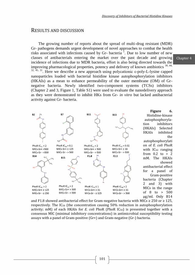
Discovery of Inhibitors of Bacterial Histidine Kinases
101
RESULTS AND DISCUSSION
The growing number of reports about the spread of multi-drug resistant (MDR)
Gr- pathogens demands urgent development of novel approaches to combat the health
risks associated with infections caused by Gr- bacteria 1. Due to low number of new
classes of antibacterials entering the market over the past decade and growing
incidence of infections due to MDR bacteria, effort is also being directed towards the
improving pharmacological properties, potency and delivery of known antibiotics 30, 44,
32, 45, 31. Here we describe a new approach using polycationic ε-poly-L-lysine capped
nanoparticles loaded with bacterial histidine kinase autophosphorylation inhibitors
(HKAIs) as a mean to enhance permeability of the outer membrane (OM) of Gr-
negative bacteria. Newly identified two-component systems (TCSs) inhibitors
(Chapter 2 and 3, Figure 1, Table S1) were used to evaluate the nanodelivery approach
as they were demonstrated to inhibit HKs from Gr- in vitro but lacked antibacterial
activity against Gr- bacteria.
Figure 6. Histidine-kinase
autophosphoryla-tion inhibitors (HKAIs) Selected HKAIs inhibited
the autophosphorylation of E. coli PhoR with IC50 ranging from 0.2 to > 2 mM. The HKAIs
showed antibacterial effect for a panel of
Gram-positive bacteria (Chapter 2 and 3) with MICs in the range of 8 to > 500 µg/ml. Only B14
and F1.8 showed antibacterial effect for Gram-negative bacteria with MICs ≥ 250 or ≥ 125, respectively. The IC50 (the concentration causing 50% reduction in autophosphorylation activity; mM) of each HKAIs for E. coli PhoR (PhoR IC50) is presented together with a consensus MIC (minimal inhibitory concentrations) in antimicrobial susceptibility testing assays with a panel of Gram-positive (Gr+) and Gram-negative (Gr-) bacteria.
Chapter 4
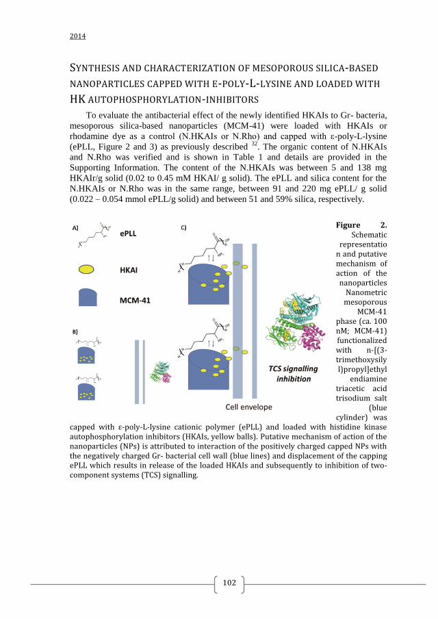
2014
102
SYNTHESIS AND CHARACTERIZATION OF MESOPOROUS SILICA-BASED
NANOPARTICLES CAPPED WITH Ε-POLY-L-LYSINE AND LOADED WITH
HK AUTOPHOSPHORYLATION-INHIBITORS
To evaluate the antibacterial effect of the newly identified HKAIs to Gr- bacteria,
mesoporous silica-based nanoparticles (MCM-41) were loaded with HKAIs or
rhodamine dye as a control (N.HKAIs or N.Rho) and capped with ε-poly-L-lysine
(ePLL, Figure 2 and 3) as previously described 32
. The organic content of N.HKAIs
and N.Rho was verified and is shown in Table 1 and details are provided in the
Supporting Information. The content of the N.HKAIs was between 5 and 138 mg
HKAIr/g solid (0.02 to 0.45 mM HKAI/ g solid). The ePLL and silica content for the
N.HKAIs or N.Rho was in the same range, between 91 and 220 mg ePLL/ g solid
(0.022 – 0.054 mmol ePLL/g solid) and between 51 and 59% silica, respectively.
Figure 2. Schematic
representation and putative mechanism of action of the nanoparticles
Nanometric mesoporous
MCM-41 phase (ca. 100 nM; MCM-41) functionalized with n-[(3-trimethoxysilyl)propyl]ethyl
endiamine triacetic acid trisodium salt
(blue cylinder) was
capped with ε-poly-L-lysine cationic polymer (ePLL) and loaded with histidine kinase autophosphorylation inhibitors (HKAIs, yellow balls). Putative mechanism of action of the nanoparticles (NPs) is attributed to interaction of the positively charged capped NPs with the negatively charged Gr- bacterial cell wall (blue lines) and displacement of the capping ePLL which results in release of the loaded HKAIs and subsequently to inhibition of two-component systems (TCS) signalling.
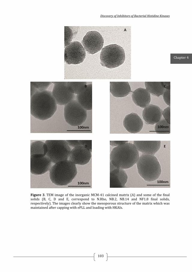
Discovery of Inhibitors of Bacterial Histidine Kinases
103
Figure 3. TEM image of the inorganic MCM-41 calcined matrix (A) and some of the final solids (B, C, D and E, correspond to N.Rho, NB.2, NB.14 and NF1.8 final solids, respectively). The images clearly show the mesoporous structure of the matrix which was maintained after capping with ePLL and loading with HKAIs.
Chapter 4
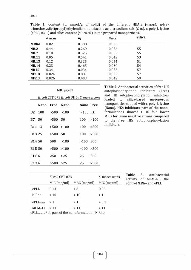
2014
104
Table 1. Content (α, mmol/g of solid) of the different HKAIs (αHKAIs), n-[(3-trimethoxysilyl)propyl]ethylendiamine triacetic acid trisodium salt (f, αf), ε-poly-L-lysine (ePLL, αePLL) and silica content (silica, %) in the prepared nanoparticles.
α HKAIs αf αePLL silica
N.Rho 0.021 0.388 0.025
NB.2 0.44 0.269 0.036 55 NB.7 0.18 0.325 0.052 55 NB.11 0.05 0.541 0.042 53 NB.13 0.12 0.325 0.054 51 NB.14 0.23 0.465 0.030 54 NB15 0.34 0.034 0.033 57 NF1.8 0.024 0.88 0.022 57 NF2.3 0.026 0.403 0.042 59
Table 2. Antibacterial activities of free HK autophosphorylation inhibitors (Free) and HK autophosphorylation inhibitors loaded to silica-based mesoporous nanoparticles capped with ε-poly-L-lysine (Nano). HKs inhibitors part of the nano-formulations showed > 10 fold lower MICs for Gram negative strains compared to the free HKs autophosphorylation inhibitors.
Table 3. Antibacterial activity of MCM-41, the control N.Rho and ePLL
ePLLnano ePLL part of the nanoformulation N.Rho
MIC µg/ml
E. coli CFT 073 E. coli DH5α S. marcescens
Nano Free Nano Nano Free
B2 100 >500 >100 > 100 n.t.
B7 50 >500 50 100 >100
B11 13 >500 >100 100 >500
B13 25 >500 50 100 >500
B14 50 500 >100 >100 500
B15 50 >500 >100 >100 >500
F1.8 6 250 >25 25 250
F2.3 6 >500 >25 25 >500
E. coli CFT 073 S. marcescens
MIC [mg/ml] MBC [mg/ml] MIC [mg/ml]
ePLL 0.13 1.6 0.25
N.Rho > 10 > 10 > 1
ePLLnano > 1 > 1 > 0.1
MCM-41 > 11 > 11 > 11
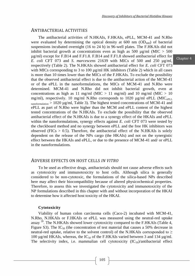
Discovery of Inhibitors of Bacterial Histidine Kinases
105
ANTIBACTERIAL ACTIVITIES
The antibacterial activities of N.HKAIs, F.HKAIs, ePLL, MCM-41 and N.Rho
were evaluated by determining the optical density at 600 nm (OD600) of bacterial
suspensions incubated overnight (16 to 24 h) in 96-well plates. The F.HKAIs did not
inhibit bacterial growth at concentrations even as high as 500 µg/ml (MIC > 500
µg/ml) except for F.B14 and F.F1.8. F.B14 and F.F1.8 showed antibacterial effect for
E. coli CFT 073 and S. marcescens 21639 with MICs of 500 and 250 µg/ml,
respectively (Table 2). The N.HKAIs showed antibacterial effect for E. coli CFT 073
with MICs corresponding to 3 to 100 µg/ml HK inhibitors (Table 2) which in all cases
is more than 10 times lower than the MICs of the F.HKAIs. To exclude the possibility
that the observed antibacterial effect is due to the antibacterial action of the MCM-41
or of the ePLL in the nanoformulations, the MICs of MCM-41 and N.Rho were
determined. MCM-41 and N.Rho did not inhibit bacterial growth, even at
concentrations as high as 11 mg/ml (MIC > 11 mg/ml) and 10 mg/ml (MIC > 10
mg/ml), respectively. 10 mg/ml N.Rho corresponds to 1020 µg/ml ePLL (MICePLL
nanoformulation > 1020 µg/ml, Table 3). The highest tested concentrations of MCM-41 and
ePLL as part of N.Rho were higher than the MCM and ePLL content of the highest
tested concentrations of the N.HKAIs. To exclude the possibility that the observed
antibacterial effect of the N.HKAIs is due to a synergy effect of the HKAIs and ePLL
within the nanoformulations, synergy effects against E. coli CFT 073 were tested by
the checkboard method and no synergy between ePLL and the free HK inhibitors was
observed (FICs > 0.5). Therefore, the antibacterial effect of the N.HKAIs is solely
dependent on the release of the NPs cargo (the HKAIs) and not on the synergistic
effect between the HKAIs and ePLL, or due to the presence of MCM-41 and/ or ePLL
in the nanoformulations.
ADVERSE EFFECTS ON HOST CELLS IN VITRO
To be used as effective drugs, antibacterials should not cause adverse effects such
as cytotoxicity and immunotoxicity to host cells. Although silica is generally
considered to be non-cytotoxic, the formulations of the silica-based NPs described
here may affect their biocompatibility because of altered physicochemical properties.
Therefore, to assess this we investigated the cytotoxicity and immunotoxicity of the
NP formulations described in this chapter with and without incorporation of the HKAI
to determine how it affected host toxicity of the HKAI.
Cytotoxicity
Viability of human colon carcinoma cells (Caco-2) incubated with MCM-41,
N.Rho, N.HKAIs or F.HKAIs or ePLL was measured using the neutral-red uptake
assay 39
. The N.HKAIs showed lower cytotoxicity compared to the F.HKAIs (Table 4,
Figure S3). The IC50 (the concentration of test material that causes a 50% decrease in
neutral-red uptake, relative to the solvent control) of the N.HKAIs corresponded to ≥
100 µg/ml HKAIs, whereas, the IC50 of the F.HKAIs varied between 3 and 134 µg/ml.
The selectivity index, i.e. mammalian cell cytotoxicity (IC50)/antibacterial effect
Chapter 4
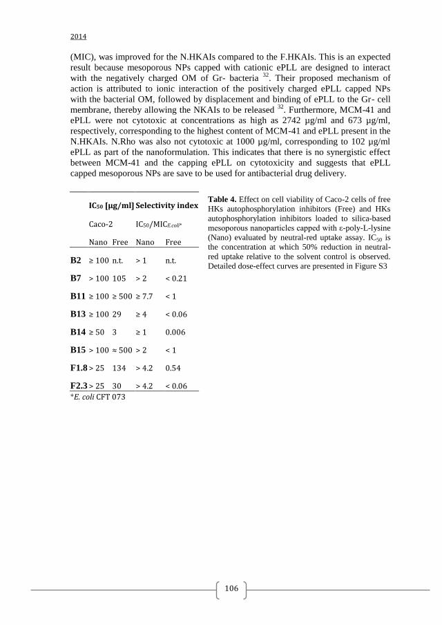
2014
106
(MIC), was improved for the N.HKAIs compared to the F.HKAIs. This is an expected
result because mesoporous NPs capped with cationic ePLL are designed to interact
with the negatively charged OM of Gr- bacteria 32
. Their proposed mechanism of
action is attributed to ionic interaction of the positively charged ePLL capped NPs
with the bacterial OM, followed by displacement and binding of ePLL to the Gr- cell
membrane, thereby allowing the NKAIs to be released 32
. Furthermore, MCM-41 and
ePLL were not cytotoxic at concentrations as high as 2742 µg/ml and 673 µg/ml,
respectively, corresponding to the highest content of MCM-41 and ePLL present in the
N.HKAIs. N.Rho was also not cytotoxic at 1000 µg/ml, corresponding to 102 µg/ml
ePLL as part of the nanoformulation. This indicates that there is no synergistic effect
between MCM-41 and the capping ePLL on cytotoxicity and suggests that ePLL
capped mesoporous NPs are save to be used for antibacterial drug delivery.
Table 4. Effect on cell viability of Caco-2 cells of free
HKs autophosphorylation inhibitors (Free) and HKs
autophosphorylation inhibitors loaded to silica-based
mesoporous nanoparticles capped with ε-poly-L-lysine
(Nano) evaluated by neutral-red uptake assay. IC50 is
the concentration at which 50% reduction in neutral-
red uptake relative to the solvent control is observed.
Detailed dose-effect curves are presented in Figure S3
IC50 [µg/ml] Selectivity index
Caco-2 IC50/MICE.coli*
Nano Free Nano Free
B2 ≥ 100 n.t. > 1 n.t.
B7 > 100 105 > 2 < 0.21
B11 ≥ 100 ≥ 500 ≥ 7.7 < 1
B13 ≥ 100 29 ≥ 4 < 0.06
B14 ≥ 50 3 ≥ 1 0.006
B15 > 100 ≈ 500 > 2 < 1
F1.8 > 25 134 > 4.2 0.54
F2.3 > 25 30 > 4.2 < 0.06
*E. coli CFT 073
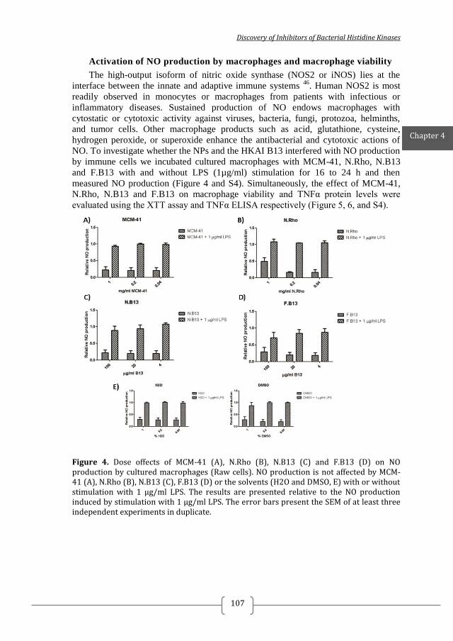
Discovery of Inhibitors of Bacterial Histidine Kinases
107
Activation of NO production by macrophages and macrophage viability
The high-output isoform of nitric oxide synthase (NOS2 or iNOS) lies at the
interface between the innate and adaptive immune systems 46
. Human NOS2 is most
readily observed in monocytes or macrophages from patients with infectious or
inflammatory diseases. Sustained production of NO endows macrophages with
cytostatic or cytotoxic activity against viruses, bacteria, fungi, protozoa, helminths,
and tumor cells. Other macrophage products such as acid, glutathione, cysteine,
hydrogen peroxide, or superoxide enhance the antibacterial and cytotoxic actions of
NO. To investigate whether the NPs and the HKAI B13 interfered with NO production
by immune cells we incubated cultured macrophages with MCM-41, N.Rho, N.B13
and F.B13 with and without LPS (1µg/ml) stimulation for 16 to 24 h and then
measured NO production (Figure 4 and S4). Simultaneously, the effect of MCM-41,
N.Rho, N.B13 and F.B13 on macrophage viability and TNFα protein levels were
evaluated using the XTT assay and TNFα ELISA respectively (Figure 5, 6, and S4).
Figure 4. Dose offects of MCM-41 (A), N.Rho (B), N.B13 (C) and F.B13 (D) on NO production by cultured macrophages (Raw cells). NO production is not affected by MCM-41 (A), N.Rho (B), N.B13 (C), F.B13 (D) or the solvents (H2O and DMSO, E) with or without stimulation with 1 µg/ml LPS. The results are presented relative to the NO production induced by stimulation with 1 µg/ml LPS. The error bars present the SEM of at least three independent experiments in duplicate.
Chapter 4
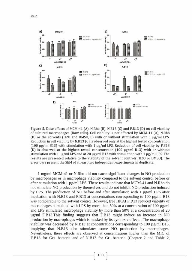
2014
108
Figure 5. Dose effects of MCM-41 (A), N.Rho (B), N.B13 (C) and F.B13 (D) on cell viability of cultured macrophages (Raw cells). Cell viability is not affected by MCM-41 (A), N.Rho (B) or the solvents (H2O and DMSO, E) with or without stimulation with 1 µg/ml LPS. Reduction in cell viability by N.B13 (C) is observed only at the highest tested concentration (100 µg/ml B13) with stimulation with 1 µg/ml LPS. Reduction of cell viability by F.B13 (D) is observed at the highest tested concentration (100 µg/ml B13) with or without stimulation with 1 µg/ml LPS and at 20 µg/ml B13 with stimulation with 1 µg/ml LPS. The results are presented relative to the viability of the solvent controls (H2O or DMSO). The error bars present the SEM of at least two independent experiments in duplicate.
1 mg/ml MCM-41 or N.Rho did not cause significant changes in NO production
by macrophages or in macrophage viability compared to the solvent control before or
after stimulation with 1 µg/ml LPS. These results indicate that MCM-41 and N.Rho do
not stimulate NO production by themselves and do not inhibit NO production induced
by LPS. The production of NO before and after stimulation with 1 µg/ml LPS after
incubation with N.B13 and F.B13 at concentrations corresponding to 100 µg/ml B13
was comparable to the solvent control However, free HKAI F.B13 reduced viability of
macrophages stimulated with LPS by more than 50% at a concentration of 100 µg/ml
and LPS stimulated macrophage viability by more than 50% at a concentration of 20
µg/ml F.B13.This finding suggests that F.B13 might induce an increase in NO
production by macrophages which is masked by its cytotoxic effect. . The macrophage
viability was decreased by N.B13 at concentrations corresponding to 100 µg/ml B13,
implying that N.B13 also stimulates some NO production by macrophages.
Nevertheless, these effects are observed at concentrations higher than the MIC of
F.B13 for Gr+ bacteria and of N.B13 for Gr- bacteria (Chapter 2 and Table 2,
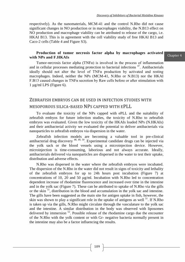
Discovery of Inhibitors of Bacterial Histidine Kinases
109
respectively). As the nanomaterials, MCM-41 and the control N.Rho did not cause
significant changes in NO production or in macrophages viability, the N.B13 effect on
NO production and macrophage viability can be attributed to release of the cargo, i.e.
HKAI B13. This is in agreement with the cell viability study of free HKAI B13 and
Caco-2 cells (Table 4 and Figure S3).
Production of tumor necrosis factor alpha by macrophages activated
with NPs and F.HKAIs
Tumor-necrosis factor alpha (TNFα) is involved in the process of inflammation
and in cellular processes mediating protection to bacterial infections 47
. Antibacterials
ideally should not alter the level of TNFα production by activated and resting
macrophages. Indeed, neither the NPs (MCM-41, N.Rho or N.B13) nor the HKAI
F.B13 caused changes in TNFα secretion by Raw cells before or after stimulation with
1 µg/ml LPS (Figure 6).
ZEBRAFISH EMBRYOS CAN BE USED IN INFECTION STUDIES WITH
MESOPOROUS SILICA-BASED NPS CAPPED WITH EPLL
To evaluate the toxicity of the NPs capped with ePLL and the suitability of
zebrafish embyos for future infection studies, the toxicity of N.Rho to zebrafish
embryos was evaluated. Given the low toxicity of the HKAIs loaded NPs (N.HKAIs)
and their antibacterial activity we evaluated the potential to deliver antibacterials via
nanoparticles to zebrafish embryos via dispersion in the water.
Zebrafish infection models are becoming a valuable tool in pre-clinical
antibacterial drug discovery 48, 49, 50
. Experimental candidate drugs can be injected via
the yolk sack or the blood vessels using a microinjection device. However,
microinjection is time-consuming, laborious and not always accurate. Ideally,
antibacterials delivered via nanoparticles are dispersed in the water to test their uptake,
distribution and adverse effects.
N.Rho was dispersed in the water where the zebrafish embryos were incubated.
The dispersion of the N.Rho in the water did not result in signs of toxicity and lethality
of the zebrafish embryos for up to 24h hours post incubation (Figure 7) at
concentrations of 10, 20 and 50 µg/ml. Incubation with N.Rho led to concentration
dependent increase of rhodamine fluorescence and increased over time in the intestine
and in the yolk sac (Figure 7). These can be attributed to uptake of N.Rho via the gills
or the skin 51
, distribution in the blood and accumulation in the yolk sac and intestine.
The gills have been suggested as the main site for antigen uptake in fish, however, the
skin was shown to play a significant role in the uptake of antigens as well 51
. If N.Rho
is taken up via the gills, N.Rho might circulate through the vasculature to the yolk sac
and the intestine. A similar distribution in the body was observed with liposomes
delivered by immersion 52
. Possible release of the rhodamine cargo due the encounter
of the N.Rho with the yolk content or with Gr- negative bacteria normally present in
the intestine may also be a factor influencing the results.
Chapter 4
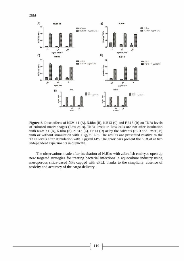
2014
110
Figure 6. Dose effects of MCM-41 (A), N.Rho (B), N.B13 (C) and F.B13 (D) on TNFα levels of cultured macrophages (Raw cells). TNFα levels in Raw cells are not after incubation with MCM-41 (A), N.Rho (B), N.B13 (C), F.B13 (D) or by the solvents (H2O and DMSO, E) with or without stimulation with 1 µg/ml LPS. The results are presented relative to the TNFα levels after stimulation with 1 µg/ml LPS. The error bars present the SEM of at two independent experiments in duplicate.
The observations made after incubation of N.Rho with zebrafish embryos open up
new targeted strategies for treating bacterial infections in aquaculture industry using
mesoporous silica-based NPs capped with ePLL thanks to the simplicity, absence of
toxicity and accuracy of the cargo delivery.
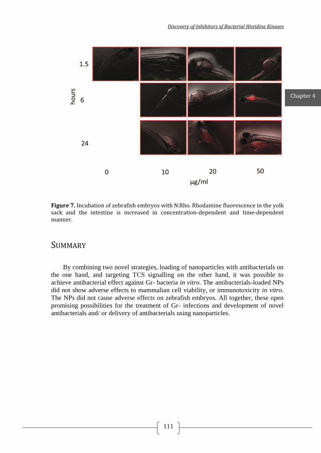
Discovery of Inhibitors of Bacterial Histidine Kinases
111
Figure 7. Incubation of zebrafish embryos with N.Rho. Rhodamine fluorescence in the yolk sack and the intestine is increased in concentration-dependent and time-dependent manner.
SUMMARY
By combining two novel strategies, loading of nanoparticles with antibacterials on
the one hand, and targeting TCS signalling on the other hand, it was possible to
achieve antibacterial effect against Gr- bacteria in vitro. The antibacterials-loaded NPs
did not show adverse effects to mammalian cell viability, or immunotoxicity in vitro.
The NPs did not cause adverse effects on zebrafish embryos. All together, these open
promising possibilities for the treatment of Gr- infections and development of novel
antibacterials and/ or delivery of antibacterials using nanoparticles.
Chapter 4
2014
112
REFERENCES 1. Curcio, D., Multidrug-Resistant Gram-Negative Bacterial Infections: Are you Ready for the Challenge? Current clinical pharmacology 2014, 9 (1), 27-38. 2. Dancer, S. J., The problem with cephalosporins. The Journal of antimicrobial chemotherapy 2001, 48 (4), 463-78. 3. Ibrahim, M. E.; Bilal, N. E.; Hamid, M. E., Increased multi-drug resistant Escherichia coli from hospitals in Khartoum state, Sudan. African health sciences 2012, 12 (3), 368-75. 4. Alhashash, F.; Weston, V.; Diggle, M.; McNally, A., Multidrug-resistant Escherichia coli bacteremia. Emerging infectious diseases 2013, 19 (10), 1699-701. 5. Vigil, K. J.; Adachi, J. A.; Aboufaycal, H.; Hachem, R. Y.; Reitzel, R. A.; Jiang, Y.; Tarrand, J. J.; Chemaly, R. F.; Bodey, G. P.; Rolston, K. V.; Raad, I., Multidrug-resistant Escherichia coli bacteremia in cancer patients. American journal of infection control 2009, 37 (9), 741-5. 6. UF/IFAS Fact Sheets FA-14 Aeromonas Infections, FA-31 Vibrio Infections of Fish and FA-11 Columnaris disease. 7. Verner-Jeffreys, D. W.; Welch, T. J.; Schwarz, T.; Pond, M. J.; Woodward, M. J.; Haig, S. J.; Rimmer, G. S.; Roberts, E.; Morrison, V.; Baker-Austin, C., High prevalence of multidrug-tolerant bacteria and associated antimicrobial resistance genes isolated from ornamental fish and their carriage water. PloS one 2009, 4 (12), e8388. 8. Hall, R. M.; Collis, C. M.; Kim, M. J.; Partridge, S. R.; Recchia, G. D.; Stokes, H. W., Mobile gene cassettes and integrons in evolution. Annals of the New York Academy of Sciences 1999, 870, 68-80. 9. Schmidt, A. S.; Bruun, M. S.; Dalsgaard, I.; Larsen, J. L., Incidence, distribution, and spread of tetracycline resistance determinants and integron-associated antibiotic resistance genes among motile aeromonads from a fish farming environment. Applied and environmental microbiology 2001, 67 (12), 5675-82. 10. Rhodes, G.; Huys, G.; Swings, J.; McGann, P.; Hiney, M.; Smith, P.; Pickup, R. W., Distribution of oxytetracycline resistance plasmids between aeromonads in hospital and aquaculture environments: implication of Tn1721 in dissemination of the tetracycline resistance determinant tet A. Applied and environmental microbiology 2000, 66 (9), 3883-90. 11. Furushita, M.; Shiba, T.; Maeda, T.; Yahata, M.; Kaneoka, A.; Takahashi, Y.; Torii, K.; Hasegawa, T.; Ohta, M., Similarity of tetracycline resistance genes isolated from fish farm bacteria to those from clinical isolates. Applied and environmental microbiology 2003, 69 (9), 5336-42. 12. Welch, T. J.; Fricke, W. F.; McDermott, P. F.; White, D. G.; Rosso, M. L.; Rasko, D. A.; Mammel, M. K.; Eppinger, M.; Rosovitz, M. J.; Wagner, D.; Rahalison, L.; Leclerc, J. E.; Hinshaw, J. M.; Lindler, L. E.; Cebula, T. A.; Carniel, E.; Ravel, J., Multiple antimicrobial resistance in plague: an emerging public health risk. PloS one 2007, 2 (3), e309. 13. Da Costa, P. M.; Loureiro, L.; Matos, A. J. F., Transfer of multidrug-resistant bacteria between intermingled ecological niches: The interface between humans, animals and the environment. International journal of environmental research and public health 2013, 10 (1), 278-294. 14. Ewers, C.; Grobbel, M.; Bethe, A.; Wieler, L. H.; Guenther, S., Extended-spectrum beta-lactamases-producing gram-negative bacteria in companion animals: action is clearly warranted! Berliner und Munchener tierarztliche Wochenschrift 2011, 124 (3-4), 94-101. 15. Ahmed, A. M.; Motoi, Y.; Sato, M.; Maruyama, A.; Watanabe, H.; Fukumoto, Y.; Shimamoto, T., Zoo animals as reservoirs of gram-negative bacteria harboring integrons
Discovery of Inhibitors of Bacterial Histidine Kinases
113
and antimicrobial resistance genes. Applied and environmental microbiology 2007, 73 (20), 6686-6690. 16. Velikova, N.; Bem, A. E.; van Baarlen, P.; Wells, J. M.; Marina, A., WalK, the Path towards New Antibacterials with Low Potential for Resistance Development. ACS Medicinal Chemistry Letters 2013, 4 (10), 891-894. 17. Rutherford, S. T.; Bassler, B. L., Bacterial quorum sensing: its role in virulence and possibilities for its control. Cold Spring Harbor perspectives in medicine 2012, 2 (11). 18. Rasko, D. A.; Moreira, C. G.; Li de, R.; Reading, N. C.; Ritchie, J. M.; Waldor, M. K.; Williams, N.; Taussig, R.; Wei, S.; Roth, M.; Hughes, D. T.; Huntley, J. F.; Fina, M. W.; Falck, J. R.; Sperandio, V., Targeting QseC signaling and virulence for antibiotic development. Science 2008, 321 (5892), 1078-80. 19. Casino, P.; Rubio, V.; Marina, A., The mechanism of signal transduction by two-component systems. Current opinion in structural biology 2010, 20 (6), 763-71. 20. Kenney, L. J., How important is the phosphatase activity of sensor kinases? Current opinion in microbiology 2010, 13 (2), 168-76. 21. Zhu, Y.; Qin, L.; Yoshida, T.; Inouye, M., Phosphatase activity of histidine kinase EnvZ without kinase catalytic domain. Proceedings of the National Academy of Sciences of the United States of America 2000, 97 (14), 7808-13. 22. Skerker, J. M.; Prasol, M. S.; Perchuk, B. S.; Biondi, E. G.; Laub, M. T., Two-component signal transduction pathways regulating growth and cell cycle progression in a bacterium: a system-level analysis. PLoS biology 2005, 3 (10), e334. 23. Wick, L. M.; Egli, T., Molecular components of physiological stress responses in Escherichia coli. Advances in biochemical engineering/biotechnology 2004, 89, 1-45. 24. Crepin, S.; Chekabab, S. M.; Le Bihan, G.; Bertrand, N.; Dozois, C. M.; Harel, J., The Pho regulon and the pathogenesis of Escherichia coli. Veterinary microbiology 2011, 153 (1-2), 82-8. 25. Chekabab, S. M.; Harel, J.; Dozois, C. M., Interplay between genetic regulation of phosphate homeostasis and bacterial virulence. Virulence 2014, 5 (5). 26. Chekabab, S. M.; Jubelin, G.; Dozois, C. M.; Harel, J., PhoB activates Escherichia coli O157:H7 virulence factors in response to inorganic phosphate limitation. PloS one 2014, 9 (4), e94285. 27. Nikaido, H., Molecular basis of bacterial outer membrane permeability revisited. Microbiology and molecular biology reviews : MMBR 2003, 67 (4), 593-656. 28. Page, M. G., The role of the outer membrane of Gram-negative bacteria in antibiotic resistance: Ajax' shield or Achilles' heel? Handbook of experimental pharmacology 2012, (211), 67-86. 29. Brehm-Stecher, B. F.; Johnson, E. A., Sensitization of Staphylococcus aureus and Escherichia coli to antibiotics by the sesquiterpenoids nerolidol, farnesol, bisabolol, and apritone. Antimicrobial agents and chemotherapy 2003, 47 (10), 3357-60. 30. Bolla, J. M.; Alibert-Franco, S.; Handzlik, J.; Chevalier, J.; Mahamoud, A.; Boyer, G.; Kiec-Kononowicz, K.; Pages, J. M., Strategies for bypassing the membrane barrier in multidrug resistant Gram-negative bacteria. FEBS letters 2011, 585 (11), 1682-90. 31. Huh, A. J.; Kwon, Y. J., "Nanoantibiotics": a new paradigm for treating infectious diseases using nanomaterials in the antibiotics resistant era. Journal of controlled release : official journal of the Controlled Release Society 2011, 156 (2), 128-45. 32. Mas, N.; Galiana, I.; Mondragon, L.; Aznar, E.; Climent, E.; Cabedo, N.; Sancenon, F.; Murguia, J. R.; Martinez-Manez, R.; Marcos, M. D.; Amoros, P., Enhanced efficacy and broadening of antibacterial action of drugs via the use of capped mesoporous nanoparticles. Chemistry 2013, 19 (34), 11167-71.

2014
114
33. Waltho, J. P.; Williams, D. H., The natural design of vancomycin family antibiotics to bind their target peptides. Ciba Foundation symposium 1991, 158, 73-86; discussion 87-91, 92-7. 34. Ye, R.; Xu, H.; Wan, C.; Peng, S.; Wang, L.; Xu, H.; Aguilar, Z. P.; Xiong, Y.; Zeng, Z.; Wei, H., Antibacterial activity and mechanism of action of epsilon-poly-L-lysine. Biochemical and biophysical research communications 2013, 439 (1), 148-53. 35. Conlon, J. M.; Mechkarska, M.; Prajeep, M.; Sonnevend, A.; Coquet, L.; Leprince, J.; Jouenne, T.; Vaudry, H.; King, J. D., Host-defense peptides in skin secretions of the tetraploid frog Silurana epitropicalis with potent activity against methicillin-resistant Staphylococcus aureus (MRSA). Peptides 2012, 37 (1), 113-9. 36. Clinical Laboratory and Standards Institute Methods for Antimicrobial Susceptibility Testing of Anaerobic Bacteria; Approved Standard. M11-A7. 2007; Vol. 7th ed. 37. CLSI, Methods for dilution antimicrobial susceptibility tests for bacteria that grow aerobically. CLSI: Wayne, PA, 2009; Vol. 8th ed. M7-A8. 38. Gialdroni Grassi, G.; Ferrara, A.; Peona, V.; Colombo, M. L., [Evaluation of the bactericidal activity of the combination of fosfomycin with other antibiotics by 2 different methods (checkboard and bactericidal curves)]. Giornale italiano di chemioterapia 1983, 30 (2-3), 77-84. 39. Repetto, G.; del Peso, A.; Zurita, J. L., Neutral red uptake assay for the estimation of cell viability/cytotoxicity. Nature protocols 2008, 3 (7), 1125-31. 40. Green, L. C.; Wagner, D. A.; Glogowski, J.; Skipper, P. L.; Wishnok, J. S.; Tannenbaum, S. R., Analysis of nitrate, nitrite, and [15N]nitrate in biological fluids. Analytical biochemistry 1982, 126 (1), 131-8. 41. Roehm, N. W.; Rodgers, G. H.; Hatfield, S. M.; Glasebrook, A. L., An improved colorimetric assay for cell proliferation and viability utilizing the tetrazolium salt XTT. Journal of immunological methods 1991, 142 (2), 257-65. 42. Kessels, M. Y.; Huitema, L. F.; Boeren, S.; Kranenbarg, S.; Schulte-Merker, S.; van Leeuwen, J. L.; de Vries, S. C., Proteomics analysis of the zebrafish skeletal extracellular matrix. PloS one 2014, 9 (3), e90568. 43. Fiaz, A. W.; Leon-Kloosterziel, K. M.; Gort, G.; Schulte-Merker, S.; van Leeuwen, J. L.; Kranenbarg, S., Swim-training changes the spatio-temporal dynamics of skeletogenesis in zebrafish larvae (Danio rerio). PloS one 2012, 7 (4), e34072. 44. O'Shea, R.; Moser, H. E., Physicochemical properties of antibacterial compounds: implications for drug discovery. Journal of medicinal chemistry 2008, 51 (10), 2871-8. 45. Abed, N.; Couvreur, P., Nanocarriers for antibiotics: A promising solution to treat intracellular bacterial infections. International journal of antimicrobial agents 2014, 43 (6), 485-96. 46. MacMicking, J.; Xie, Q. W.; Nathan, C., Nitric oxide and macrophage function. Annual review of immunology 1997, 15, 323-50. 47. Cooper, A. M.; Appelberg, R.; Orme, I. M., Immunopathogenesis of Mycobacterium avium infection. Frontiers in bioscience : a journal and virtual library 1998, 3, e141-8. 48. van der Sar, A. M.; Musters, R. J.; van Eeden, F. J.; Appelmelk, B. J.; Vandenbroucke-Grauls, C. M.; Bitter, W., Zebrafish embryos as a model host for the real time analysis of Salmonella typhimurium infections. Cellular microbiology 2003, 5 (9), 601-11. 49. Medina, C.; Royo, J. L., Zebrafish as a model organism to study host-pathogen interactions. Methods 2013, 62 (3), 241-5. 50. Meijer, A. H.; Spaink, H. P., Host-pathogen interactions made transparent with the zebrafish model. Current drug targets 2011, 12 (7), 1000-17. 51. Nakanishi, T.; Ototake, M., Antigen uptake and immune responses after immersion vaccination. Developments in biological standardization 1997, 90, 59-68.
Discovery of Inhibitors of Bacterial Histidine Kinases
115
52. Ruyra, A.; Cano-Sarabia, M.; Garcia-Valtanen, P.; Yero, D.; Gibert, I.; Mackenzie, S. A.; Estepa, A.; Maspoch, D.; Roher, N., Targeting and stimulation of the zebrafish (Danio rerio) innate immune system with LPS/dsRNA-loaded nanoliposomes. Vaccine 2014, 32 (31), 3955-62.

2014
116
SUPPLEMENTARY MATERIAL CHAPTER 4
MATERIALS CHARACTERIZATION
MCM-41 as-synthesized, MCM-41 and the final N.Rho and N.HKAIs (N.B2,
N.B7, N.B11, N.B13, N.B14, N.B15, N.F1.8 and N.F2.3) synthesized materials were
characterized through standard techniques. Figure S1 shows the X-ray diffraction
(XRD) patterns of the nanoparticulated MCM-41 matrix as-synthesised, the MCM-41
calcined and the final N.Rho solid. (The rest of X-ray diffraction patterns of the
antibacterial loaded solids are not shown due to the low amount of final solids
obtained). The MCM-41 as-synthesised (curve a) displayed the four typical low-angle
reflections of a hexagonal-ordered matrix indexed at (100), (110), (200) and (210)
Bragg peaks. In curve b (MCM-41 calcined), a significant shift of the (100) peak in the
XRD and a broadening of the (100) and (200) peaks are observed. These changes are
due to the condensation of silanols in the calcination step, which caused an
approximate cell contraction of 4 Å. Finally, curve c shows the N.Rho solid XRD
pattern.
For this material, reflections (110) and (200) were mostly lost due to a reduction
in contrast related to the functionalisation process and to the filling of mesopores with
rhodamine B. Although these reductions, the intensity of the (100) peak in this pattern
strongly indicates that the loading process with the dye and the additional
functionalisation with N-[(3-Trimethoxysilyl)propyl]ethylendiamine triacetic acid
trisodium salt and the covering with ε-poly-L-lysine did not modify the mesoporous
MCM-41 scaffold nature.
Likewise, TEM analysis of the different prepared solids was performed. TEM
images showed the typical channels of the MCM-41 scaffolding and can be visualised
as alternate black and white stripes in which the typical hexagonal porosity of the
MCM-41 calcined material can also be observed (see Figure 2). In the same way,
TEM images of some of the final solids loaded with the antibacterial products are also
shown in Figure 2. The remaining final solids also showed the same morphology
under the TEM analysis. TEM images also show that the materials were obtained as
spherical nanoparticles of ca 80 - 100 nm. Then, it can be concluded that the spherical
shape and mesoporous nature of the inorganic matrix remains after the loading and
functionalization processes.
Figure S2 (curve a) displays the N2 adsorption-desorption isotherms of the MCM-
41 calcined nanoparticles. This curve shows an adsorption step with a P/P0 value
between 0.2 and 0.35, due to a type IV isotherm, which is typical of mesoporous
materials. This first step corresponds to nitrogen condensation in the mesopore inlets.
With the BJH[3]
model on the adsorption curve of the isotherm, a narrow pore size
distribution with an average pore diameter of 2.56 nm (see Figure S2 inlet) and a pore
volume of 0.64 cm3g
-1 were calculated. The absence of a hysteresis loop in this
pressure range and the low BJH pore distribution is due to the cylindrical uniformity
of mesopores. The total specific area was 916 m2g
-1, calculated with the BET model.
[4]
The a0 cell parameter 42.9 Å (d100= 37.17 Å), the pore diameter (2.56 nm) and the wall
Discovery of Inhibitors of Bacterial Histidine Kinases
117
thickness value (17.31 Å) were calculatedfrom the XRD, porosimetry and TEM
measurements. Other important feature of the curve is the characteristic H1 hysteresis
loop that appears in the isotherm at a high relative pressure (P/P0> 0.8) which can be
closely associated with a wide pore size distribution. This hysteresis loop is due to the
filling of the large pores among the nanoparticles (0.22 cm3g
-1 calculated by the BJH
model) because of textural porosity. In relation to the control syntehsized N.Rho
material, the N2 adsorption-desorption isotherm of this solid was associated to the
typical mesoporous systems with filled mesopores (see Figure S2, curve b). Thus, as it
was expected, a lower N2 adsorbed volume (BJH mesopore volume = 0.15 cm3g
-1) and
surface area (204.9 m2g
-1) were found, compared with the initial MCM-41 material. As
it can be observed, this solid presents a curve with no gaps at low relative pressure
values if compared to the mother MCM-41 matrix (curve a). Another important feature
of N.Rho is that no maximum was found in the pore size distribution curve, which can
be associated with the closed pores due to the functionalization process. Table S2
shows a summary of the BET-specific surface values, pore volumes and pore sizes
calculated from the N2 adsorption-desorption isotherms for MCM-41 calcined and
N.Rho.
Thermogravimetric studies and elemental analyses of all final prepared solids
were carried out, in order to quantify the organic contents of each material. In
particular, the amount of the different loading products and functionalization with
product f and ePLL were calculated. All the resuts are summarized in Table 1 in the
body text.
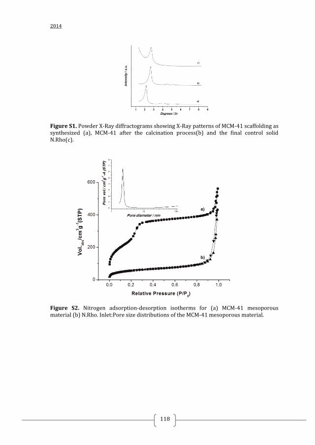
2014
118
Figure S1. Powder X-Ray diffractograms showing X-Ray patterns of MCM-41 scaffolding as synthesized (a), MCM-41 after the calcination process(b) and the final control solid N.Rho(c).
Figure S2. Nitrogen adsorption-desorption isotherms for (a) MCM-41 mesoporous material (b) N.Rho. Inlet:Pore size distributions of the MCM-41 mesoporous material.
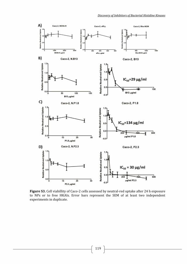
Discovery of Inhibitors of Bacterial Histidine Kinases
119
Figure S3. Cell viabillity of Caco-2 cells assessed by neutral-red uptake after 24 h exposure to NPs or to free HKAIs. Error bars represent the SEM of at least two independent experiments in duplicate.
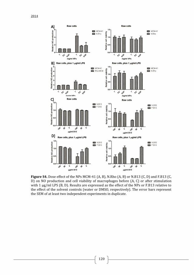
2014
120
Figure S4. Dose effect of the NPs MCM-41 (A, B), N.Rho (A, B) or N.B13 (C, D) and F.B13 (C, D) on NO production and cell viability of macrophages before (A, C) or after stimulation with 1 µg/ml LPS (B, D). Results are expressed as the effect of the NPs or F.B13 relative to the effect of the solvent controls (water or DMSO, respectively). The error bars represent the SEM of at least two independent experiments in duplicate.
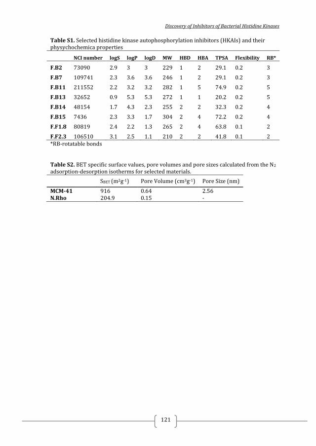
Discovery of Inhibitors of Bacterial Histidine Kinases
121
Table S1. Selected histidine kinase autophosphorylation inhibitors (HKAIs) and their physychochemica properties
NCI number logS logP logD MW HBD HBA TPSA Flexibility RB*
F.B2 73090 2.9 3 3 229 1 2 29.1 0.2 3
F.B7 109741 2.3 3.6 3.6 246 1 2 29.1 0.2 3
F.B11 211552 2.2 3.2 3.2 282 1 5 74.9 0.2 5
F.B13 32652 0.9 5.3 5.3 272 1 1 20.2 0.2 5
F.B14 48154 1.7 4.3 2.3 255 2 2 32.3 0.2 4
F.B15 7436 2.3 3.3 1.7 304 2 4 72.2 0.2 4
F.F1.8 80819 2.4 2.2 1.3 265 2 4 63.8 0.1 2
F.F2.3 106510 3.1 2.5 1.1 210 2 2 41.8 0.1 2 *RB-rotatable bonds Table S2. BET specific surface values, pore volumes and pore sizes calculated from the N2 adsorption-desorption isotherms for selected materials.
SBET (m2g-1) Pore Volume (cm3g-1) Pore Size (nm)
MCM-41 916 0.64 2.56 N.Rho 204.9 0.15 -

2014
122
Discovery of Inhibitors of Bacterial Histidine Kinases
123
CHAPTER 5
COMPUTER-AIDED APPROACHES IN HIT
OPTIMISATION
Nadya Velikova

2014
124
Discovery of Inhibitors of Bacterial Histidine Kinases
125
ABBREVIATIONS
QSAR – quantitative structure-activity relationship
LEI – ligand-efficiency index
SEI – surface-efficiency index
BEI – binding-efficiency index
TCS – two-component systems
HK – histidine kinase
RR – response regulator
Chapter 5
2014
126
INTRODUCTION
Computational chemistry has been applied widely in the pharmaceutical industry
for drug discovery, lead optimization, risk assessment, toxicity prediction and
regulatory decisions 1. Over the past four decades, quantitative structure–activity
relationship (QSAR) modelling has completely changed the way the interaction of
organic compounds with the various forms of life is studied 2. It attempts to formulate
the relationship between structure and activity as a mathematical model. Therefore, the
binding affinity of a new molecule can be predicted using a QSAR model derived
from the known inhibitors and their experimental bioassay values 3. Using such an
approach, a large library of possible drug candidates can be tested for their predicted
selectivity and potency 4, saving time and money in drug discovery research.
Successful drug discovery requires not only identifying hits with desired biochemical
properties (e.g. target affinity) but also hits with optimal physicochemical and
ADMET (adsorption, distribution, metabolism, excretion and toxicity) properties 5.
Once a hit is identified its optimisation should be bidirectional. Ligand-efficiency
indices (LEIs) connect the physicochemical properties of hits (e.g. MW and polar
surface area [PSA]) with the biological target via the affinity parameter (IC50, enzyme
activity or other related measures of affinity) 6, 7
.
Herein we describe the application of computer-aided drug discovery approaches
(including QSAR modelling, ligand-based similarity search and LEIs-based
evaluation) for the identification of putative HK autophosphorylation inhibitors with
antibacterial activity and low toxicity.

Discovery of Inhibitors of Bacterial Histidine Kinases
127
MATERIALS AND METHODS
QSAR PREDICTIVE MODEL BUILDING
The experimental E. coli PhoR autophosphorylation inhibition data (% inhibition
at 2 mM) of 39 compounds (Chapter 3 and 4) were used as a dataset for building a
quantitative structure–activity relationship (QSAR) model for inhibition of E. coli
PhoR autophosphorylation (Table S1). The percentage inhibition range was from 6 to
85%. Predictive QSAR model was built automatically using the Auto-Modeller
module of StarDrop (Optibrium, Cambridge) 8.
LIGAND-BASED SIMILARITY SEARCHES The National Cancer Institute Developmental Theraupeutics Programme database
(DTP NCI/NIH) was searched for analogue structures of the query molecules B7, B11,
B13, B15, F1.8, F2.4, F2 (Figure 1) using the Enhanced NCI database browser v 2.2 9.
Molecules with more than 80% similarity to the query molecules (Tanimoto
coefficient > 80%) and with predicted drug-like properties were saved for further
scoring.
SCORING AND SELECTION OF COMPOUNDS FOR EXPERIMENTAL
TESTING
Ligand-based similarity searches (LBSS) hits were evaluated using the StarDrop
Oral non central nervous system (CNS) scoring profile ( logS>1, importance 90%;
positive HIA category, importance 85%; 0<logP<0.35, importance 60%; -0.2<BBB
log<1, importance 55%; positive BBB category, importance 55%; P-gp category
´´no´´, importance 50%; hERG IC50≤5, importance 50%; 2C9 pKi≤6, importance
30%; 2D6 affinity category ´´low to medium´´, importance 30%; PPB90 category
´´low´´, importance 20%).
Predicted E. coli PhoR autophosphorylation inhibition score was calculated using
the QSAR Random Forest Regression model built with StarDrop.
After visual inspection, 18 compounds with oral non-CNS score > 0.4 and
predicted E. coli PhoR autophosphorylation inhibition higher than 0.45, were
purchased for experimental testing.
The predicted E. coli PhoR autophosphorylation activity was used as input to
calculate binding efficiency indices (nBEIs) and surface efficiency indices (NSEIs)
using AtlasCBS 6 and the following equations:
NSEI = -log10 pREA/ NPOL, where pREA stands for predicted residual enzyme
activity in one concentration (2 mM) one time-point (30 sec) kinase assay experiments
with PhoRE, and NPOL is number of polar N and O atoms in the compound.
Chapter 5
2014
128
nBEI = - log10[pREA/NHEA], where NHEA is the number of non-hydrogen
atoms in the compound.
Out of the 1012 LBSS hits, 36 were purchased for experimental testing following
visual inspection.
CHEMICALS
All chemicals (Figure S1 and Table S2) were obtained from DTP NCI/NIH.
ANTIMICROBIAL SUSCEPTIBILITY TESTING
Strains used for antimicrobial susceptibility testing (Table 1) were propagated
using standard microbiological procedures and Müller-Hinton (MH) agar or broth. For
all microorganisms minimal inhibitory concentrations (MICs) were determined as
previously described following a standard double-dilution method 10
, 11
.
PROTEIN EXPRESSION AND PURIFICATION
The catalytic portions (DHp and CA domain) of E. coli PhoR (PhoRE) were
expressed and purified as previously described 12
. In brief the protein was expressed in
E. coli RIL and purified by Ni-affinity and size-exclusion chromatography.
KINASE ASSAY
Kinase assay was performed as previously described 13
. When comparing the
inhibitory capacity of ligands and measuring IC50, the final DMSO concentration in
the assay was 10% (v/v). Controls lacking ligands and containing an equal
concentration of DMSO were carried out in parallel. Inhibition of HKs
autophosphorylation was determined by incubating 0.12 mg/ml (≈ 4 µM HK) and up
to 20 mM of test compound in kinase buffer (50 mM Tris HCl, pH 8.5, 50 mM KCl, 5
mM MgCl2, 0.5 mM EDTA and 0.1 mM DTT). Autophosporylation reactions were
initiated by addition of 0.1 µCi/µl [γ-32P
] ATP containing from 0.03 to 0.06 µM ATP
(final concentrations). Autophosphorylation was stopped by addition of 2x SDS-
PAGE sample buffer supplemented with 50 mM EDTA. Samples were applied
without heating to 15% (w/v) Tris-glycine SDS-polyacrylamide gels. After
electrophoresis, the bottom part of the gels were removed to lower the background
signal from the unincorporated radiolabeled ATP. Gels were dried without staining on
a Bio-Rad Gel Air drying system and the phosphorylated protein was quantified by
phosphor-imaging using a Fluoro Image Analyzer FLA-5000 (Fujifilm, Japan) and
evaluated with the MultiGauge software (Fujifilm, Japan). IC50 is the concentration at
which 50% residual enzyme activity was observed compared to the negative control,
DMSO. Prism GraphPad v.4 was used for curve fitting and statistical analysis 14
.
Discovery of Inhibitors of Bacterial Histidine Kinases
129
BINDING MODE PREDICTION
The binding mode of selected ligands to the structure of the CA domain of B.
subtilis WalK (PDB: 3SL2) was predicted by molecular docking using SwissDock 15
.
In Figure 2 the lowest energy binding pose is shown.
Chapter 5
2014
130
RESULTS AND DISCUSSION
SELECTION OF COMPOUNDS FOR EXPERIMENTAL TESTING
To facilitate the selection of putative histidine kinase inhibitors for experimental
testing in the process of hit-to-lead optimization quantitative structure–activity
relationship (QSAR) models were built using a trial version of StarDrop8. The best
among the generated models was the random forest regression QSAR model however,
it was not of high merit with r2 of 0.71 (Figure 1, Figure S2). In many QSAR studies
the criterion r2 greater than 0.9 is employed to decide whether a model is internally
self-consistent. However, r2 makes no assessment of the intrinsic precision or
accuracy of the data itself. Nevertheless, we used this model to predict the E. coli
PhoR autophosphorylation inhibitory capacity of 1012 hits from ligand-based
similarity searches (LBSS) with previously identified histidine kinase
autophosphorylation inhibitors as query molecules. The predicted autophosphorylation
activities were used to evaluate the physicochemical properties of the selected LBSS
hits using AtlasCBS 6. The hits fell into one of seven groups based on NSEI (surface
efficiency index) with the group around B13 showing highest predicted surface
efficiency (Figure S3). The following group in terms of NSEI grouped around the F1.8
but with nearly two-fold lower predicted NSEIs. In total 54 compounds were ordered
for experimental testing based on the StarDrop and AtlasCBS evaluation and visual
inspection of the hits for promising functional groups.
H30 AND H31 SHOW ANTIBACTERIAL EFFECT FOR BOTH GRAM-
POSITIVE AND GRAM-NEGATIVE BACTERIA
To evaluate the antibacterial effect of the selected hits, the MICs (minimal
inhibitory concentrations) and MBCs (minimal bactericidal concentrations) for S.
aureus DSM 20231, S. aureus CH3657, S. epidermidis DSM 20044 and E. coli CFT
073 were determined. H30 and H31 showed the strongest antibacterial effect. H30
inhibited S. aureus CH3657, S. epidermidis DSM 20044 and uropathogenic E. coli
CFT 073 with MICs of 62.5, 500 µg/ml and 500 µg/ml, respectively (Table S4). H31
inhibits S. aureus CH3657, S. epidermidis DSM 20044 and E. coli CFT 073 with
MICs of 62.5, 125 and 250 µg/ml, respectively. H12 and H27 also showed some
antibacterial effect with MICs higher than 250 µg/ml. Therefore, only H30 and H31
were tested against a panel of Gram-positive and Gram-negative bacteria and the
results are summarized in Table 1. H30 did not show antibacterial effect for any of the
additional bacterial strains tested. H31 inhibited the growth of all tested bacterial
strains, including the vancomycin resistant E. faecium strains with MICs of 125 µg/ml
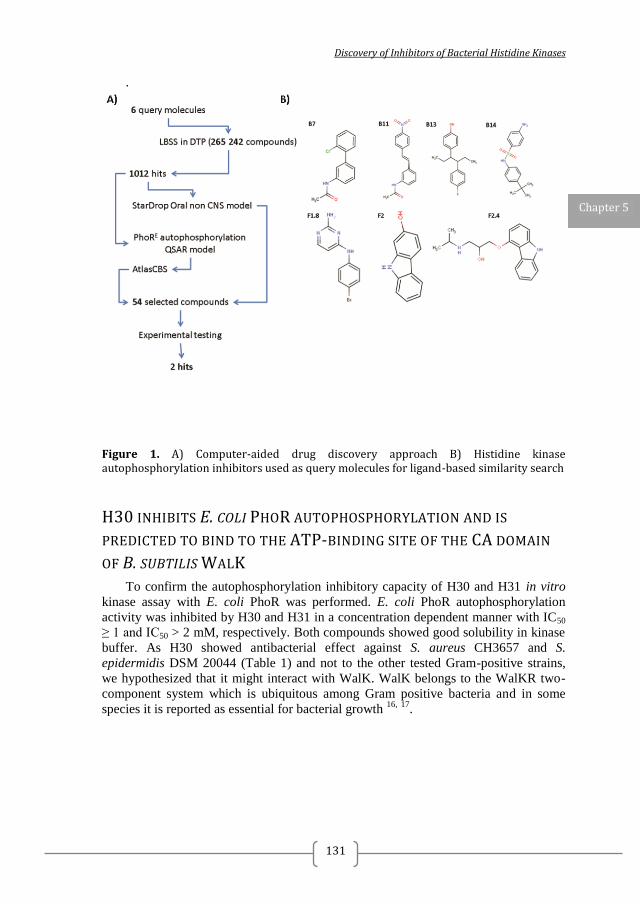
Discovery of Inhibitors of Bacterial Histidine Kinases
131
.
Figure 1. A) Computer-aided drug discovery approach B) Histidine kinase autophosphorylation inhibitors used as query molecules for ligand-based similarity search
H30 INHIBITS E. COLI PHOR AUTOPHOSPHORYLATION AND IS
PREDICTED TO BIND TO THE ATP-BINDING SITE OF THE CA DOMAIN
OF B. SUBTILIS WALK
To confirm the autophosphorylation inhibitory capacity of H30 and H31 in vitro
kinase assay with E. coli PhoR was performed. E. coli PhoR autophosphorylation
activity was inhibited by H30 and H31 in a concentration dependent manner with IC50
≥ 1 and IC50 > 2 mM, respectively. Both compounds showed good solubility in kinase
buffer. As H30 showed antibacterial effect against S. aureus CH3657 and S.
epidermidis DSM 20044 (Table 1) and not to the other tested Gram-positive strains,
we hypothesized that it might interact with WalK. WalK belongs to the WalKR two-
component system which is ubiquitous among Gram positive bacteria and in some
species it is reported as essential for bacterial growth 16,
17
.
Chapter 5
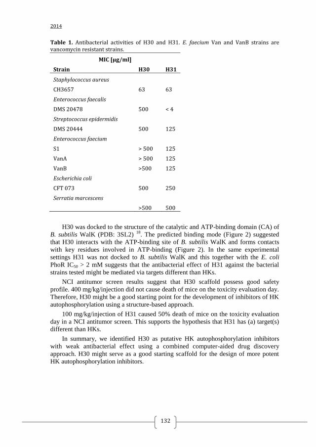
2014
132
Table 1. Antibacterial activities of H30 and H31. E. faecium Van and VanB strains are vancomycin resistant strains.
MIC [µg/ml]
Strain H30 H31
Staphylococcus aureus CH3657 63 63
Enterococcus faecalis DMS 20478 500 < 4
Streptococcus epidermidis DMS 20444 500 125
Enterococcus faecium S1 > 500 125
VanA > 500 125
VanB >500 125
Escherichia coli CFT 073 500 250
Serratia marcescens >500 500
H30 was docked to the structure of the catalytic and ATP-binding domain (CA) of
B. subtilis WalK (PDB: 3SL2) 18
. The predicted binding mode (Figure 2) suggested
that H30 interacts with the ATP-binding site of B. subtilis WalK and forms contacts
with key residues involved in ATP-binding (Figure 2). In the same experimental
settings H31 was not docked to B. subtilis WalK and this together with the E. coli
PhoR IC50 > 2 mM suggests that the antibacterial effect of H31 against the bacterial
strains tested might be mediated via targets different than HKs.
NCI antitumor screen results suggest that H30 scaffold possess good safety
profile. 400 mg/kg/injection did not cause death of mice on the toxicity evaluation day.
Therefore, H30 might be a good starting point for the development of inhibitors of HK
autophosphorylation using a structure-based approach.
100 mg/kg/injection of H31 caused 50% death of mice on the toxicity evaluation
day in a NCI antitumor screen. This supports the hypothesis that H31 has (a) target(s)
different than HKs.
In summary, we identified H30 as putative HK autophosphorylation inhibitors
with weak antibacterial effect using a combined computer-aided drug discovery
approach. H30 might serve as a good starting scaffold for the design of more potent
HK autophosphorylation inhibitors.
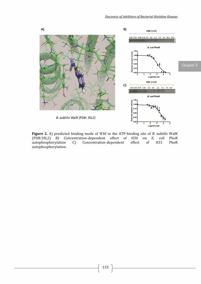
Discovery of Inhibitors of Bacterial Histidine Kinases
133
Figure 2. A) predicted binding mode of H30 to the ATP-binding site of B. subtilis WalK (PDB:3SL2) B) Concentration-dependent effect of H30 on E. coli PhoR autophosphorylation C) Concentration-dependent effect of H31 PhoR autophosphorylation.
Chapter 5
2014
134
REFERENCES 1. Sliwoski, G.; Kothiwale, S.; Meiler, J.; Lowe Jr, E. W., Computational methods in drug discovery. Pharmacological reviews 2014, 66 (1), 334-395. 2. Hansch, C.; Hoekman, D.; Leo, A.; Zhang, L.; Li, P., The expanding role of quantitative structure-activity relationships (QSAR) in toxicology. Toxicology letters 1995, 79 (1-3), 45-53. 3. Yan, A.; Chong, Y.; Wang, L.; Hu, X.; Wang, K., Prediction of biological activity of Aurora-A kinase inhibitors by multilinear regression analysis and support vector machine. Bioorganic & medicinal chemistry letters 2011, 21 (8), 2238-43. 4. Khadikar, P. V.; Sharma, V.; Karmarkar, S.; Supuran, C. T., QSAR studies on benzene sulfonamide carbonic anhydrase inhibitors: need of hydrophobic parameter for topological modeling of binding constants of sulfonamides to human CA-II. Bioorganic & medicinal chemistry letters 2005, 15 (4), 923-30. 5. Wang, J., Comprehensive assessment of ADMET risks in drug discovery. Current pharmaceutical design 2009, 15 (19), 2195-2219. 6. Abad-Zapatero, C.; Perisic, O.; Wass, J.; Bento, A. P.; Overington, J.; Al-Lazikani, B.; Johnson, M. E., Ligand efficiency indices for an effective mapping of chemico-biological space: the concept of an atlas-like representation. Drug discovery today 2010, 15 (19-20), 804-11. 7. Cortes-Cabrera, A.; Morreale, A.; Gago, F.; Abad-Zapatero, C., AtlasCBS: a web server to map and explore chemico-biological space. Journal of computer-aided molecular design 2012, 26 (9), 995-1003. 8. Auto-Modeller StarDrop. 9. Enhanced NCI Database Browser 2.2. 10. Conlon, J. M.; Mechkarska, M.; Prajeep, M.; Sonnevend, A.; Coquet, L.; Leprince, J.; Jouenne, T.; Vaudry, H.; King, J. D., Host-defense peptides in skin secretions of the tetraploid frog Silurana epitropicalis with potent activity against methicillin-resistant Staphylococcus aureus (MRSA). Peptides 2012, 37 (1), 113-9. 11. Clinical Laboratory and Standards Institute Methods for Antimicrobial Susceptibility Testing of Anaerobic Bacteria; Approved Standard. M11-A7. 2007; Vol. 7th ed. 12. Casino, P.; Rubio, V.; Marina, A., Structural insight into partner specificity and phosphoryl transfer in two-component signal transduction. Cell 2009, 139 (2), 325-36. 13. Marina, A.; Mott, C.; Auyzenberg, A.; Hendrickson, W. A.; Waldburger, C. D., Structural and mutational analysis of the PhoQ histidine kinase catalytic domain. Insight into the reaction mechanism. The Journal of biological chemistry 2001, 276 (44), 41182-90. 14. Christopoulos, H. M. a. A., Fitting Models to Biological Data using Linear and Nonlinear Regression. A Practical Guide to Curve Fitting. Oxford University Press: New York, 2004. 15. SwissDock. http://www.swissdock.ch/. 16. Mohedano, M. L.; Overweg, K.; de la Fuente, A.; Reuter, M.; Altabe, S.; Mulholland, F.; de Mendoza, D.; Lopez, P.; Wells, J. M., Evidence that the essential response regulator YycF in Streptococcus pneumoniae modulates expression of fatty acid biosynthesis genes and alters membrane composition. Journal of bacteriology 2005, 187 (7), 2357-67. 17. Velikova, N.; Bem, A. E.; van Baarlen, P.; Wells, J. M.; Marina, A., WalK, the Path towards New Antibacterials with Low Potential for Resistance Development. ACS Medicinal Chemistry Letters 2013, 4 (10), 891-894.
Discovery of Inhibitors of Bacterial Histidine Kinases
135
18. Celikel, R.; Veldore, V. H.; Mathews, I.; Devine, K. M.; Varughese, K. I., ATP forms a stable complex with the essential histidine kinase WalK (YycG) domain. Acta crystallographica. Section D, Biological crystallography 2012, 68 (Pt 7), 839-45.
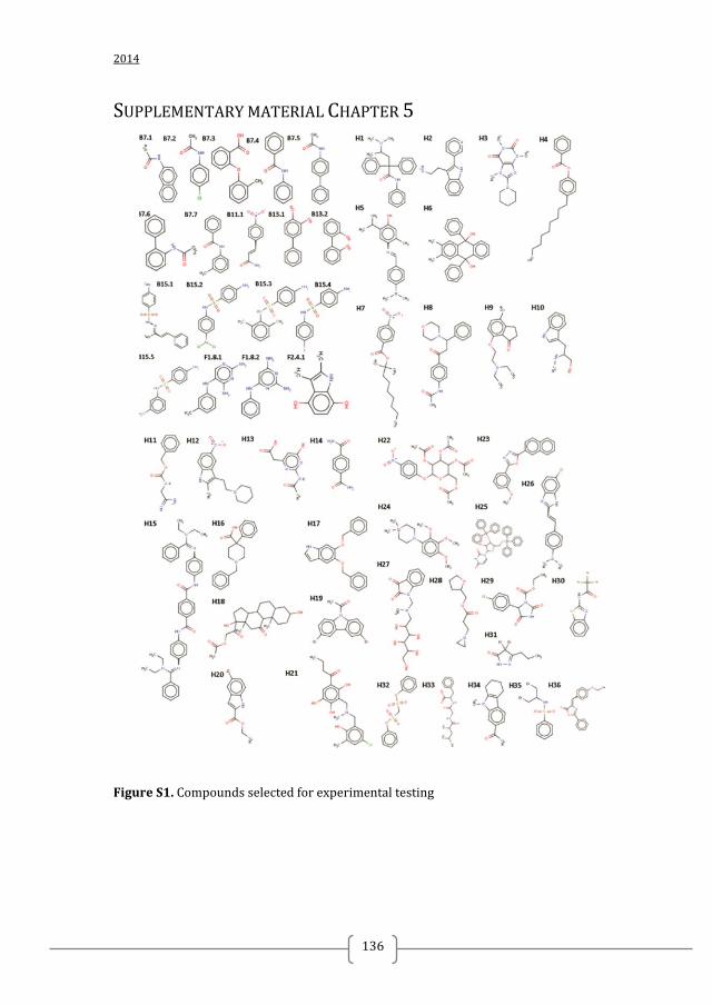
2014
136
SUPPLEMENTARY MATERIAL CHAPTER 5
Figure S1. Compounds selected for experimental testing
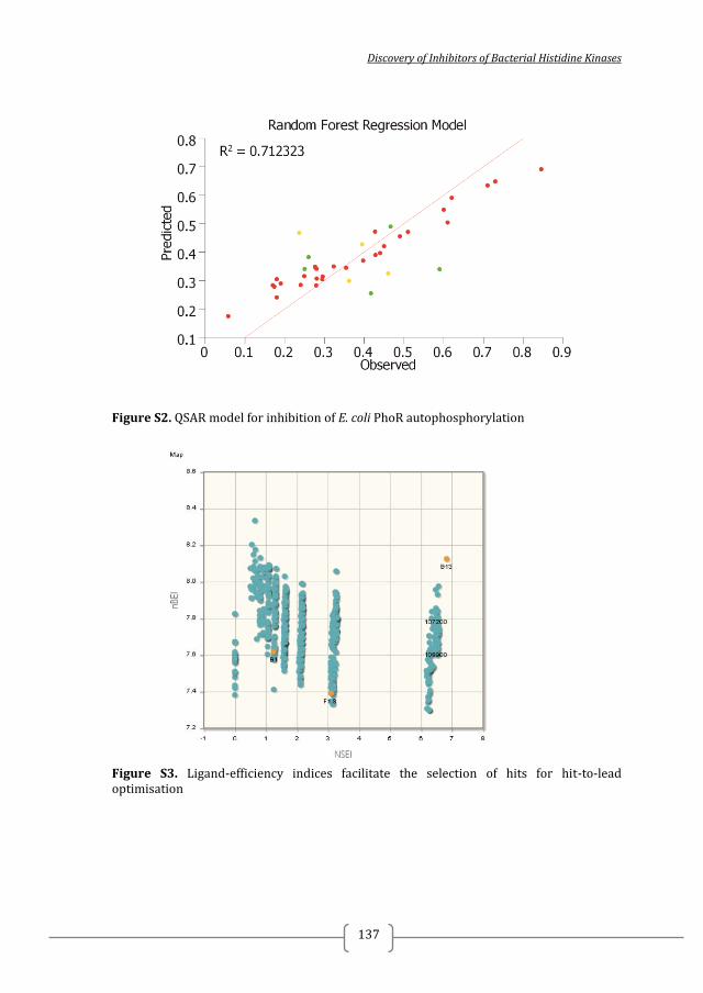
Discovery of Inhibitors of Bacterial Histidine Kinases
137
Figure S2. QSAR model for inhibition of E. coli PhoR autophosphorylation
Figure S3. Ligand-efficiency indices facilitate the selection of hits for hit-to-lead optimisation
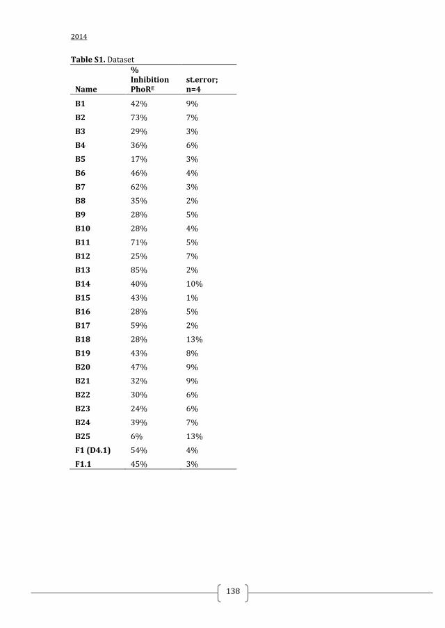
2014
138
Table S1. Dataset
Name
% Inhibition PhoRE
st.error; n=4
B1 42% 9%
B2 73% 7%
B3 29% 3%
B4 36% 6%
B5 17% 3%
B6 46% 4%
B7 62% 3%
B8 35% 2%
B9 28% 5%
B10 28% 4%
B11 71% 5%
B12 25% 7%
B13 85% 2%
B14 40% 10%
B15 43% 1%
B16 28% 5%
B17 59% 2%
B18 28% 13%
B19 43% 8%
B20 47% 9%
B21 32% 9%
B22 30% 6%
B23 24% 6%
B24 39% 7%
B25 6% 13%
F1 (D4.1) 54% 4%
F1.1 45% 3%
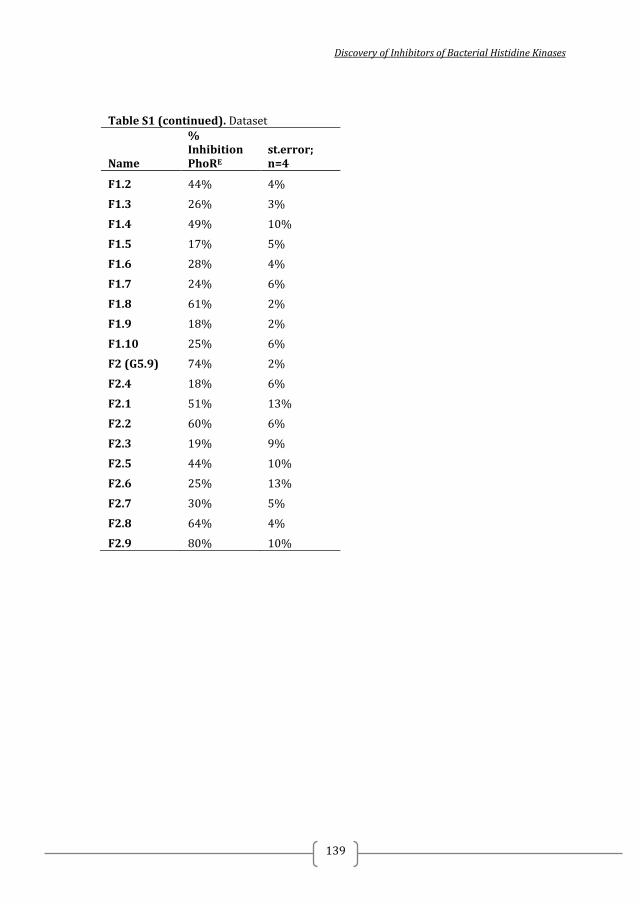
Discovery of Inhibitors of Bacterial Histidine Kinases
139
Table S1 (continued). Dataset
Name
% Inhibition PhoRE
st.error; n=4
F1.2 44% 4%
F1.3 26% 3%
F1.4 49% 10%
F1.5 17% 5%
F1.6 28% 4%
F1.7 24% 6%
F1.8 61% 2%
F1.9 18% 2%
F1.10 25% 6%
F2 (G5.9) 74% 2%
F2.4 18% 6%
F2.1 51% 13%
F2.2 60% 6%
F2.3 19% 9%
F2.5 44% 10%
F2.6 25% 13%
F2.7 30% 5%
F2.8 64% 4%
F2.9 80% 10%

2014
140
Discovery of Inhibitors of Bacterial Histidine Kinases
141
CHAPTER 6
PRELIMINARY BIOCHEMICAL STUDIES ON THE
INTERACTION OF LACTOFERRICINB-DERIVED
PEPTIDES WITH TWO-COMPONENT SYSTEMS
Nadya Velikova, Jerry Wells, Alberto Marina

2014
142
Discovery of Inhibitors of Bacterial Histidine Kinases
143
BACKGROUND
LactoferricinB (LfcinB) is an antimicrobial peptide with broad-spectrum of
antibacterial activity. It has been shown by in vitro and in vivo experiments that
LfcinB inhibits the phosphorylation of the response regulators (RRs), BasR and CreB,
and their cognate histidine kinases (HKs), BasS and CreC. Co-crystal structures of
(peptide) inhibitors bound to their two-component system (TCS) targets would open
up possibilities for the structure based drug design of more potent inhibitors. Here we
investigated interaction of LfcinB-derived peptides with the T. maritima TCS HK853-
RR468, for which X-ray structures of both components have been solved in different
conformations.
FINDINGS
We show that LfcinB-derived peptides inhibit the phosphatase activity of T.
maritima HK853-RR468 and seem to interact with T. maritima HK853 and E. coli
PhoB. However, a possible mechanism of interaction is non-specific protein
aggregation.
CONCLUSIONS
LfcinB has been proposed as a promising broad-spectrum inhibitor of bacterial
TCS. LfcinB-derived peptides seem to be non-specific TCS inhibitors acting via
protein aggregation. The latter would be in agreement with the broad-spectrum of
antibacterial activity of LfcinB as protein aggregation would affect multiple targets.
List of abbreviations: TCS – two component systems; HK – histidine kinase; RR –
response regulator; LfcinB – lactoferricin B
Key words: Lactoferricin B, antimicrobial peptides, two-component system inhibitors,
antibacterials
Chapter 6
2014
144
INTRODUCTION
Antimicrobial peptides have been proposed as a promising alternative to fight
resistant bacterial infections and to address the growing problem of multi-drug
resistance 1, 2, 3.
Eukaryotic cationic antimicrobial peptides (AMPs) are produced at
sites of infection or inflammation in many different organisms 4, 5
. Typically they are
peptides of 12 to 45 amino acids with a net positive charge and a high proportion of
hydrophobic amino acids 6. There are four structural classes of AMPs, the most
common being the β-sheet peptides stabilized by 2–4 disulphide bridges, and the
unstructured peptides that fold into amphipathic α-helices upon contact with
membranes. Interaction with the membrane may form pores, or act by thinning the
membrane or by destabilizing the membrane bilayer. As net result interaction of AMPs
and membranes increases permeability of the membrane, leads to loss of pH gradient
and cell death. Some cationic peptides have been shown to interact with intracellular
targets 7,
8 but in most cases the exact mechanism of intracellular AMPs action
remains unclear.
One of the AMPs proposed to possess intracellular targets is bovine lactoferricin
(LfcinB). LfcinB is a typical cationic antibacterial peptide produced by gastric pepsin
digestion of the N-terminal region of bovine lactoferrin and has be identified in vivo 9,
10. The antibacterial core LfcinB includes six residues (positions 20 – 25 in the
parental sequence) with the C-terminus amidated (peptide RRWQWR-NH2) 11, 12
. A
physiologically diverse range of Gram-positive and Gram-negative bacteria have been
found to be susceptible to inhibition and inactivation by LfcinB-derived peptides
including LfcinB20-25 and LfcinB17-31 13, 14, 9
. Concentrations of LfcinB required to cause
complete inhibition of bacterial growth vary within the range of 0.3 to 150 µg/ml,
depending on the strain and the culture medium used 9. The broad spectrum of
antibacterial action of LfcinB suggests a common mechanism of action and/ or a wide-
spread intracellular target present in all susceptible strains.
It has been shown that LfcinB-derived peptides (Lfcin17-41, LfcinB17-31 and D-
Lfcin17-31) enter the cytoplasm of E. coli and S. aureus 15
. In a recent study the
intracellular LfcinB protein targets were identified by hybridisation of biotin-labelled
LfcinB to an Escherichia coli K12 proteome chip 16
. In total 16 proteins were shown to
interact with LfcinB, including two response regulators (RRs), BasR and CreB,
belonging to the two-component systems (TCS) BasSR and CreCB. TCS are the
predominant signalling systems in bacteria. TCS connect input stimuli and output
responses with a core phosphotransfer between a membrane bound histidine kinase
(HK) and a cognate response regulator (RR) 17, 18
. Simply described, the signalling
pathway is a series of steps including autophosphorylation of the HK, phosphotransfer
to the cognate RR, and output modulation, usually via transcription regulation,
mediated by the phosphorylated RR 19, 18
. TCS are highly conserved between bacterial
species and TCS signalling inhibition by LfcinB seemed a logical common mechanism
explaining the broad-spectrum antibacterial effect of LfcinB. It has been shown by in
vitro and in vivo experiments that LfcinB inhibits the phosphorylation of the RRs
(BasR and CreB) by their cognate histidine kinases (HKs), BasS and CreC 16
but does
not inhibit the DNA binding capacity of both RRs suggesting that LfcinB-derived
Discovery of Inhibitors of Bacterial Histidine Kinases
145
peptides most likely do not interact with the effector domain of RRs, but with the REC
domain of RRs. Additionally, antibacterial assays have shown that LfcinB reduced the
tolerance of E. coli to environmental challenges, such as excessive ferric ions and
minimal medium conditions. TCS are known to be involved in the regulation of
adaptive response to stress and logically TCS signalling inhibition could be correlated
with reduced stress tolerance. Taken together, these results were the first indication
that an AMP inhibits the growth of bacteria by influencing the catalytic activities of a
TCS directly 16
and seemed to provide a promising starting point for the development
of broad-spectrum antimicrobial-peptide drugs targeted at TCS signalling. TCS have
been proposed as attractive targets for novel antibacterial as they are well-conserved
among bacteria and not present in higher eukaryotes 20, 21
.
The aim of this study was to investigate the interaction between LfcinB with TCS
at atomic level by using X-ray crystallography. The structure of a LfcinB-RR complex
would i) elucidate the molecular mechanism of RRs inhibition by LfcinB, ii) identify
putative target sites in the RR which can further be exploited in structure-based drug
design approaches, and iii) point to RR-peptide interactions and possible peptide
modifications which would lead to stronger inhibitory effect. Altogether, these would
facilitate the rational design of modified peptides with higher affinity of binding to
TCS and may lead to the development of more potent peptide TCS inhibitors. For this
purpose we have used the T. maritima HK853-RR468 which is a well-studied TCS 22,
18 and the X-ray structures of both components, HK853 and RR468, alone and in
complex are known 23, 24, 25
. E. coli PhoR-PhoB is a prototypical two-component
system involved in the regulation of the pho regulon and structures of the RR PhoB are
known 26, 27, 26
. As a first step towards a structure of a RR in complex with LfcinB, we
verified whether LfcinB-derived peptides inhibit the catalytic activities of T. maritima
HK853-RR468 and E. coli PhoR-PhoB TCS.
Chapter 6
2014
146
RESULTS AND DISCUSSION
INHIBITION OF PHOSPHOTRANSFER AND PHOSPHATASE ACTIVITIES OF
PURIFIED BACTERIAL TWO- COMPONENT SYSTEMS.
To check whether LfcinB-derived peptides inhibit the phosphorylation of RR468
and/or PhoB by their cognate HKs, HK853 and PhoR, respectively, phosphotransfer
assays were performed in the presence of two LfcinB-derived peptides (LfcinB17–25
and LfcinB17–32). The two peptides were chosen as both contain the antibacterial core
sequence (positions 20 – 25 in the parental sequence) 11, 12
. In the phosphotransfer
assays the HKs were first auto-phosphorylated using [γ-32
P]ATP 24
. This leads to
accumulation of [γ-32
P]ATP-autophosphorylated HK which in a SDS-PAGE gel
appears as a well-defined band (Figure 1). The autophosphorylated HK was then
diluted in ATP-free phosphorylation solution containing an equimolar amount (in
terms of subunits) of RR and 0.5 mM of analysed peptide. Upon mixture of the HK
and the RR phosphotransfer occurs, and in a SDS-PAGE gel the intensity of the band
corresponding to the HK becomes lower and a band corresponding to the
phosphorylated RR appears. As several HKs have phosphatase activity (i.e. HK853) 28,
19, 29, 18, the band corresponding to the phosphorylated RR in the SDS-PAGE gel lower
its intensity over time. The time course of the reaction (10 min) showed that in the
absence of LfcinB-derived peptides the radioactive band corresponding to
autophosphorylated HK853 almost completely disappeared by 1 min and coincided
with appearance of a labelled band corresponding to phosphorylated RR468 indicating
phosphotransfer from HK853 to RR468 (Figure 1A). However, the intensity of the
bands corresponding to the phosphorylated RR468 at 1 min was lower than the
intensity of the band corresponding to HK853, confirming dephosphorylation of the
RR468 due to HK853 phosphatase activity. In the presence of LfcinB-derived peptides
phosphotransfer from HK853 to RR468 did occur but to a lesser extend compared to
the negative control evidenced by the slightly higher intensity of the bands
corresponding to the autophosphorylated HK853. In the presence of Lfcin B17–25 or
LfcinB17–32 a proportion of the autophosphorylated HK853 remained visible, indicating
either inhibition of phosphotransfer to RR468, or inhibition of the phosphatase activity
leading to putative shift in the phosphotransfer/ phosphatase reaction balance.
Furthermore, in the presence of LfcinB17–25 and LfcinB17–32 the bands corresponding to
the phosphorylated RR468 were with higher intensity compared to the negative control
or Peptide 3 (a pentapeptide of different sequence) and with similar intensity over
time, indicating that the phosphatase activity of HK853 was inhibited. In the case of
PhoR-PhoB, the band corresponding to the autophosphorylated PhoR almost
completely disappeared by 1 min and a band corresponding to the phosphorylated
PhoB appeared. In the presence of LfcinB-derived peptides, the intensity of the bands
corresponding to the autophosphorylated PhoR was similar to the assay in the absence
of peptide indicating that phosphotransfer was not affected. In the absence of peptide,
the intensity of the bands corresponding to the phosphorylated PhoB remained similar
over time indicating no or very slow phosphatase activity. Intriguingly, in the presence
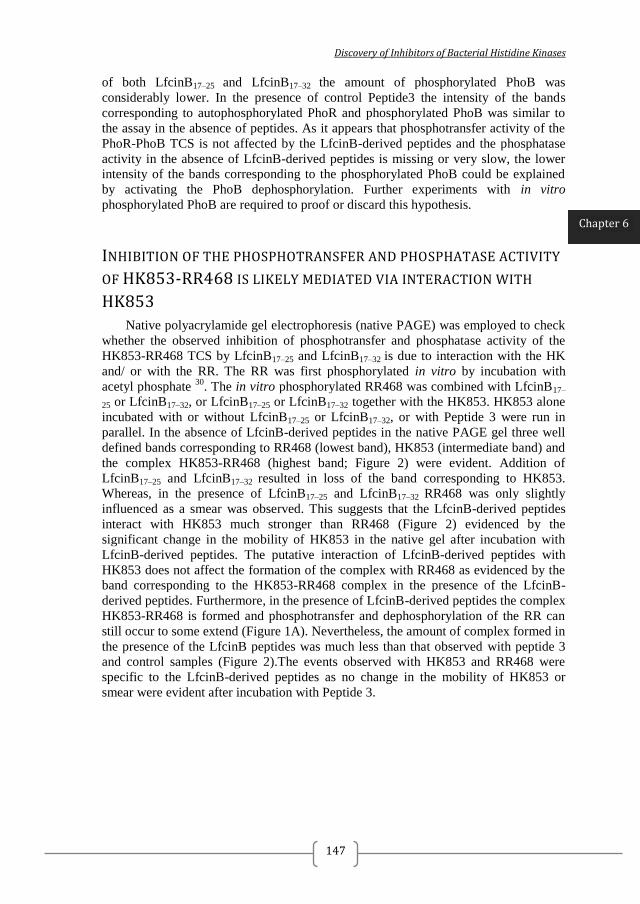
Discovery of Inhibitors of Bacterial Histidine Kinases
147
of both LfcinB17–25 and LfcinB17–32 the amount of phosphorylated PhoB was
considerably lower. In the presence of control Peptide3 the intensity of the bands
corresponding to autophosphorylated PhoR and phosphorylated PhoB was similar to
the assay in the absence of peptides. As it appears that phosphotransfer activity of the
PhoR-PhoB TCS is not affected by the LfcinB-derived peptides and the phosphatase
activity in the absence of LfcinB-derived peptides is missing or very slow, the lower
intensity of the bands corresponding to the phosphorylated PhoB could be explained
by activating the PhoB dephosphorylation. Further experiments with in vitro
phosphorylated PhoB are required to proof or discard this hypothesis.
INHIBITION OF THE PHOSPHOTRANSFER AND PHOSPHATASE ACTIVITY
OF HK853-RR468 IS LIKELY MEDIATED VIA INTERACTION WITH
HK853
Native polyacrylamide gel electrophoresis (native PAGE) was employed to check
whether the observed inhibition of phosphotransfer and phosphatase activity of the
HK853-RR468 TCS by LfcinB17–25 and LfcinB17–32 is due to interaction with the HK
and/ or with the RR. The RR was first phosphorylated in vitro by incubation with
acetyl phosphate 30
. The in vitro phosphorylated RR468 was combined with LfcinB17–
25 or LfcinB17–32, or LfcinB17–25 or LfcinB17–32 together with the HK853. HK853 alone
incubated with or without LfcinB17–25 or LfcinB17–32, or with Peptide 3 were run in
parallel. In the absence of LfcinB-derived peptides in the native PAGE gel three well
defined bands corresponding to RR468 (lowest band), HK853 (intermediate band) and
the complex HK853-RR468 (highest band; Figure 2) were evident. Addition of
LfcinB17–25 and LfcinB17–32 resulted in loss of the band corresponding to HK853.
Whereas, in the presence of LfcinB17–25 and LfcinB17–32 RR468 was only slightly
influenced as a smear was observed. This suggests that the LfcinB-derived peptides
interact with HK853 much stronger than RR468 (Figure 2) evidenced by the
significant change in the mobility of HK853 in the native gel after incubation with
LfcinB-derived peptides. The putative interaction of LfcinB-derived peptides with
HK853 does not affect the formation of the complex with RR468 as evidenced by the
band corresponding to the HK853-RR468 complex in the presence of the LfcinB-
derived peptides. Furthermore, in the presence of LfcinB-derived peptides the complex
HK853-RR468 is formed and phosphotransfer and dephosphorylation of the RR can
still occur to some extend (Figure 1A). Nevertheless, the amount of complex formed in
the presence of the LfcinB peptides was much less than that observed with peptide 3
and control samples (Figure 2).The events observed with HK853 and RR468 were
specific to the LfcinB-derived peptides as no change in the mobility of HK853 or
smear were evident after incubation with Peptide 3.
Chapter 6
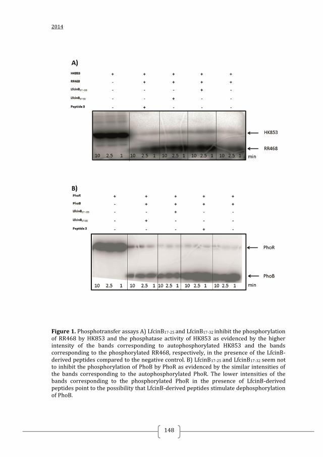
2014
148
Figure 1. Phosphotransfer assays A) LfcinB17-25 and LfcinB17-32 inhibit the phosphorylation of RR468 by HK853 and the phosphatase activity of HK853 as evidenced by the higher intensity of the bands corresponding to autophosphorylated HK853 and the bands corresponding to the phosphorylated RR468, respectively, in the presence of the LfcinB-derived peptides compared to the negative control. B) LfcinB17-25 and LfcinB17-32 seem not to inhibit the phosphorylation of PhoB by PhoR as evidenced by the similar intensities of the bands corresponding to the autophosphorylated PhoR. The lower intensities of the bands corresponding to the phosphorylated PhoR in the presence of LfcinB-derived peptides point to the possibility that LfcinB-derived peptides stimulate dephosphorylation of PhoB.
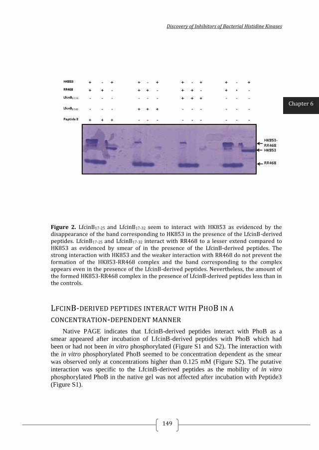
Discovery of Inhibitors of Bacterial Histidine Kinases
149
Figure 2. LfcinB17-25 and LfcinB17-32 seem to interact with HK853 as evidenced by the disappearance of the band corresponding to HK853 in the presence of the LfcinB-derived peptides. LfcinB17-25 and LfcinB17-32 interact with RR468 to a lesser extend compared to HK853 as evidenced by smear of in the presence of the LfcinB-derived peptides. The strong interaction with HK853 and the weaker interaction with RR468 do not prevent the formation of the HK853-RR468 complex and the band corresponding to the complex appears even in the presence of the LfcinB-derived peptides. Nevertheless, the amount of the formed HK853-RR468 complex in the presence of LfcinB-derived peptides less than in the controls.
LFCINB-DERIVED PEPTIDES INTERACT WITH PHOB IN A
CONCENTRATION-DEPENDENT MANNER
Native PAGE indicates that LfcinB-derived peptides interact with PhoB as a
smear appeared after incubation of LfcinB-derived peptides with PhoB which had
been or had not been in vitro phosphorylated (Figure S1 and S2). The interaction with
the in vitro phosphorylated PhoB seemed to be concentration dependent as the smear
was observed only at concentrations higher than 0.125 mM (Figure S2). The putative
interaction was specific to the LfcinB-derived peptides as the mobility of in vitro
phosphorylated PhoB in the native gel was not affected after incubation with Peptide3
(Figure S1).
Chapter 6
2014
150
SUMMARY
In summary, it seems the inhibition of phosphotransfer and phosphatase activities
of the T. maritima TCS HK853-RR468 caused by LfcinB17–25 and LfcinB17–32 can be
attributed to interaction with HK853 although some interaction with RR468 might also
have contributed to this effect. The LfcinB-derived peptides seem to interact in a
concentration dependent manner with in vitro phosphorylated E. coli response
regulator PhoB. Changes in the mobility in native gels of PhoB after incubation with
LfcinB-derived peptides indicate possible interaction with this protein. Further
experiments are needed to check the hypothesis that incubation with LfcinB-derived
peptides might activate the dephosphorylation of E. coli PhoB. The observed changes
in the mobility of T. maritima HK853 and PhoB in native gels could be due to protein
aggregation. However, further experiments such as size-exclusion chromatography
and/ or protein-aggregation evaluation by cross-linking and SDS-PAGE are required
to confirm this hypothesis. Taken together, the results point to the possibility that the
antibacterial effect caused by LfcinB-derived peptides might be attributed to
interaction with multiple cellular targets after entering into the cell. This is in
agreement with the fact that multiple E. coli proteins were shown to interact with
LfcinB using proteomics 16
. Furthermore, non-specific mechanism of action such as
protein aggregation could not be excluded at this point. Attempts to solve the structure
of T. maritima RR468 co-crystallized with LfcinB-derived peptides resulted in high-
resolution twinned crystals of RR468 and the LfcinB-derived peptides were not
encountered in the asymmetric unit (data not shown). It was not possible to obtain
diffracting crystals of PhoB co-crystallized with LfcinB-derived peptides. No attempts
have been made to co-crystallize LfcinB-derived peptides with any HK. However,
before any future crystallization trials additional biochemical experiments are required
to confirm that the mechanism of interaction of LfcinB-derived peptides with TCS is
not mediated via non-specific protein aggregation.
Discovery of Inhibitors of Bacterial Histidine Kinases
151
MATERIALS AND METHODS
PEPTIDES
Lactoferricin B17-25 and Lactoferricin B 17-32 were kindly provided by Paloma
Manzanares, IATA, CSIC (Valencia, Spain).
PROTEIN EXPRESSION AND PURIFICATION
T. maritima HK853 and RR468, E. coli PhoR and PhoB were expressed and
purified as previously described 23, 22, 24, 25, 31, 31
. Shortly proteins were expressed in E.
coli and purified by anion-exchange (HK853 and RR468) or Ni-affinity
chromatography (PhoR and PhoB), and size-exclusion chromatography.
PHOSPHOTRANSFER ASSAY
Phosphotransfer assays were performed as previously described 22
. HK853 and
PhoR were autophosphorylated in kinase buffer (50 mM Tris pH 8.0, 100 mM KCl, 10
mM MgCl2) with 0.1 μCi μl−1
[γ-32
P] ATP (3000 Ci/mmol Perkin Elmer) for 30min
(exceeding equilibrium time) at room temperature. Then, each sample was aliquoted
into three tubes and phosphotransfer reactions were initiated by the addition of
equimolecular amounts of each RR (the final concentration of RRs and HKs in
reaction was 2 μM) pre-incubated with 0.5 mM Lactoferricin B17-25, Lactoferricin B 17-
32, Peptide 3 or buffer. Aliquots were removed from each tube at different times (1, 2.5
and 10 min) and the reactions were stopped by adding 1/4 SDS-PAGE loading buffer
(62.5 mM Tris pH 6.8, 30% glycerol, 0.01% bromophenol blue) supplemented with 50
mM EDTA. Afterwards, the samples were subjected to SDS-PAGE on a 15% gel to
separate the protein from free nucleotide. After drying, the phosphorylated proteins
were visualized by phosphorimaging using a Fluoro Image Analyzer FLA-5000 (Fuji).
NATIVE POLYACRYLAMIDE GEL ELECTROPHORESIS (NATIVE PAGE)
Purified PhoB and RR468 were autophosphorylated in kinase buffer (50 mM Tris
pH 8.0, 100 mM KCl, 10 mM MgCl2) containing 12.5 mM acetyl phosphate for 1 h at
room temperature. Phosphorylated PhoB and RR468 were mixed with 0.5 mM
Lactoferricin B17-25, Lactoferricin B 17-32, Peptide 3 or buffer. RR468 mixed with 0.5
mM Lactoferricin B17-25, Lactoferricin B 17-32, Peptide 3 or buffer was incubated with
or without equimolar amounts of HK853. After 30 min of incubation at room
temperature Native polyacrylamide gel electrophoresis (Native-PAGE) loading buffer
was added and samples loaded. Coomassie blue staining was used for protein
visualization.
Chapter 6
2014
152
REFERENCES 1. HASSAN, M.; KJOS, M.; NES, I. F.; DIEP, D. B.; LOTFIPOUR, F., NATURAL ANTIMICROBIAL PEPTIDES FROM BACTERIA: CHARACTERISTICS AND POTENTIAL APPLICATIONS TO FIGHT AGAINST ANTIBIOTIC RESISTANCE. JOURNAL OF APPLIED MICROBIOLOGY 2012, 113 (4), 723-36. 2. MOK, W. W.; LI, Y., THERAPEUTIC PEPTIDES: NEW ARSENAL AGAINST DRUG RESISTANT PATHOGENS. CURRENT PHARMACEUTICAL DESIGN 2014, 20 (5), 771-92. 3. STECKBECK, J. D.; DESLOUCHES, B.; MONTELARO, R. C., ANTIMICROBIAL PEPTIDES: NEW DRUGS FOR BAD BUGS? EXPERT OPINION ON BIOLOGICAL THERAPY 2014, 14 (1), 11-4. 4. PALFFY, R.; GARDLIK, R.; BEHULIAK, M.; KADASI, L.; TURNA, J.; CELEC, P., ON THE PHYSIOLOGY AND PATHOPHYSIOLOGY OF ANTIMICROBIAL PEPTIDES. MOLECULAR MEDICINE 2009, 15 (1-2), 51-9. 5. PASUPULETI, M.; SCHMIDTCHEN, A.; MALMSTEN, M., ANTIMICROBIAL PEPTIDES: KEY COMPONENTS OF THE INNATE IMMUNE SYSTEM. CRITICAL REVIEWS IN BIOTECHNOLOGY 2012, 32 (2), 143-171. 6. HANCOCK, R. E. W., PEPTIDE ANTIBIOTICS. LANCET 1997, 349 (9049), 418-422. 7. FRIEDRICH, C. L.; MOYLES, D.; BEVERIDGE, T. J.; HANCOCK, R. E., ANTIBACTERIAL ACTION OF STRUCTURALLY DIVERSE CATIONIC PEPTIDES ON GRAM-POSITIVE BACTERIA. ANTIMICROBIAL AGENTS AND CHEMOTHERAPY 2000, 44 (8), 2086-92. 8. CUDIC, M.; OTVOS, L., JR., INTRACELLULAR TARGETS OF ANTIBACTERIAL PEPTIDES. CURRENT DRUG TARGETS 2002, 3 (2), 101-6. 9. BELLAMY, W.; TAKASE, M.; WAKABAYASHI, H.; KAWASE, K.; TOMITA, M., ANTIBACTERIAL SPECTRUM OF LACTOFERRICIN B, A POTENT BACTERICIDAL PEPTIDE DERIVED FROM THE N-TERMINAL REGION OF BOVINE LACTOFERRIN. THE JOURNAL OF APPLIED BACTERIOLOGY 1992, 73 (6), 472-9. 10. ARIAS, M.; MCDONALD, L. J.; HANEY, E. F.; NAZMI, K.; BOLSCHER, J. G.; VOGEL, H. J., BOVINE AND HUMAN LACTOFERRICIN PEPTIDES: CHIMERAS AND NEW CYCLIC ANALOGS. BIOMETALS : AN INTERNATIONAL JOURNAL ON THE ROLE OF METAL IONS IN BIOLOGY, BIOCHEMISTRY, AND MEDICINE 2014. 11. TOMITA, M.; TAKASE, M.; BELLAMY, W.; SHIMAMURA, S., A REVIEW: THE ACTIVE PEPTIDE OF LACTOFERRIN. ACTA PAEDIATRICA JAPONICA; OVERSEAS EDITION 1994, 36 (5), 585-91. 12. (A) SCHIBLI, D. J.; HWANG, P. M.; VOGEL, H. J., STRUCTURE OF THE ANTIMICROBIAL PEPTIDE TRITRPTICIN BOUND TO MICELLES: A DISTINCT MEMBRANE-BOUND PEPTIDE FOLD. BIOCHEMISTRY 1999, 38 (51), 16749-55; (B) SCHIBLI, D. J.; HWANG, P. M.; VOGEL, H. J., THE STRUCTURE OF THE ANTIMICROBIAL ACTIVE CENTER OF LACTOFERRICIN B BOUND TO SODIUM DODECYL SULFATE MICELLES. FEBS LETTERS 1999, 446 (2-3), 213-7. 13. STROM, M. B.; HAUG, B. E.; REKDAL, O.; SKAR, M. L.; STENSEN, W.; SVENDSEN, J. S., IMPORTANT STRUCTURAL FEATURES OF 15-RESIDUE LACTOFERRICIN DERIVATIVES AND METHODS FOR IMPROVEMENT OF ANTIMICROBIAL ACTIVITY. BIOCHEMISTRY AND CELL BIOLOGY = BIOCHIMIE ET BIOLOGIE CELLULAIRE 2002, 80 (1), 65-74. 14. (A) DEL OLMO, A.; MORALES, P.; NUÑEZ, M., BACTERICIDAL EFFECT OF LACTOFERRIN AND ITS AMIDATED AND PEPSIN-DIGESTED DERIVATIVES ON PSEUDOMONAS FLUORESCENS: INFLUENCE OF ENVIRONMENTAL AND PHYSIOLOGICAL FACTORS. JOURNAL OF FOOD PROTECTION 2008, 71 (12), 2468-2474; (B) ROSEANU, A.; FLORIAN, P.; CONDEI, M.; CRISTEA, D.; DAMIAN, M., ANTIBACTERIAL ACTIVITY OF
Discovery of Inhibitors of Bacterial Histidine Kinases
153
LACTOFERRIN AND LACTOFERRICIN AGAINST ORAL STREPTOCOCCI. ROMANIAN BIOTECHNOLOGICAL LETTERS 2010, 15 (6), 5788-5792. 15. HAUKLAND, H. H.; ULVATNE, H.; SANDVIK, K.; VORLAND, L. H., THE ANTIMICROBIAL PEPTIDES LACTOFERRICIN B AND MAGAININ 2 CROSS OVER THE BACTERIAL CYTOPLASMIC MEMBRANE AND RESIDE IN THE CYTOPLASM. FEBS LETTERS 2001, 508 (3), 389-93. 16. HO, Y. H.; SUNG, T. C.; CHEN, C. S., LACTOFERRICIN B INHIBITS THE PHOSPHORYLATION OF THE TWO-COMPONENT SYSTEM RESPONSE REGULATORS BASR AND CREB. MOLECULAR & CELLULAR PROTEOMICS : MCP 2012, 11 (4), M111 014720. 17. STOCK, A. M.; ROBINSON, V. L.; GOUDREAU, P. N., TWO-COMPONENT SIGNAL TRANSDUCTION. ANNUAL REVIEW OF BIOCHEMISTRY 2000, 69, 183-215. 18. CASINO, P.; RUBIO, V.; MARINA, A., THE MECHANISM OF SIGNAL TRANSDUCTION BY TWO-COMPONENT SYSTEMS. CURRENT OPINION IN STRUCTURAL BIOLOGY 2010, 20 (6), 763-71. 19. KENNEY, L. J., HOW IMPORTANT IS THE PHOSPHATASE ACTIVITY OF SENSOR KINASES? CURRENT OPINION IN MICROBIOLOGY 2010, 13 (2), 168-76. 20. GOTOH, Y.; DOI, A.; FURUTA, E.; DUBRAC, S.; ISHIZAKI, Y.; OKADA, M.; IGARASHI, M.; MISAWA, N.; YOSHIKAWA, H.; OKAJIMA, T.; MSADEK, T.; UTSUMI, R., NOVEL ANTIBACTERIAL COMPOUNDS SPECIFICALLY TARGETING THE ESSENTIAL WALR RESPONSE REGULATOR. THE JOURNAL OF ANTIBIOTICS 2010, 63 (3), 127-34. 21. VELIKOVA, N.; BEM, A. E.; VAN BAARLEN, P.; WELLS, J. M.; MARINA, A., WALK, THE PATH TOWARDS NEW ANTIBACTERIALS WITH LOW POTENTIAL FOR RESISTANCE DEVELOPMENT. ACS MEDICINAL CHEMISTRY LETTERS 2013, 4 (10), 891-894. 22. CASINO, P.; FERNANDEZ-ALVAREZ, A.; ALFONSO, C.; RIVAS, G.; MARINA, A., IDENTIFICATION OF A NOVEL TWO COMPONENT SYSTEM IN THERMOTOGA MARITIMA. COMPLEX STOICHIOMETRY AND CRYSTALLIZATION. BIOCHIMICA ET BIOPHYSICA ACTA 2007, 1774 (5), 603-9. 23. MARINA, A.; WALDBURGER, C. D.; HENDRICKSON, W. A., STRUCTURE OF THE ENTIRE CYTOPLASMIC PORTION OF A SENSOR HISTIDINE-KINASE PROTEIN. THE EMBO JOURNAL 2005, 24 (24), 4247-59. 24. CASINO, P.; RUBIO, V.; MARINA, A., STRUCTURAL INSIGHT INTO PARTNER SPECIFICITY AND PHOSPHORYL TRANSFER IN TWO-COMPONENT SIGNAL TRANSDUCTION. CELL 2009, 139 (2), 325-36. 25. PODGORNAIA, A. I.; CASINO, P.; MARINA, A.; LAUB, M. T., STRUCTURAL BASIS OF A RATIONALLY REWIRED PROTEIN-PROTEIN INTERFACE CRITICAL TO BACTERIAL SIGNALING. STRUCTURE 2013, 21 (9), 1636-47. 26. CREPIN, S.; CHEKABAB, S. M.; LE BIHAN, G.; BERTRAND, N.; DOZOIS, C. M.; HAREL, J., THE PHO REGULON AND THE PATHOGENESIS OF ESCHERICHIA COLI. VETERINARY MICROBIOLOGY 2011, 153 (1-2), 82-8. 27. BACHHAWAT, P.; SWAPNA, G. V.; MONTELIONE, G. T.; STOCK, A. M., MECHANISM OF ACTIVATION FOR TRANSCRIPTION FACTOR PHOB SUGGESTED BY DIFFERENT MODES OF DIMERIZATION IN THE INACTIVE AND ACTIVE STATES. STRUCTURE 2005, 13 (9), 1353-63. 28. GUTU, A. D.; WAYNE, K. J.; SHAM, L. T.; WINKLER, M. E., KINETIC CHARACTERIZATION OF THE WALRKSPN (VICRK) TWO-COMPONENT SYSTEM OF STREPTOCOCCUS PNEUMONIAE: DEPENDENCE OF WALKSPN (VICK) PHOSPHATASE ACTIVITY ON ITS PAS DOMAIN. JOURNAL OF BACTERIOLOGY 2010, 192 (9), 2346-58. 29. ZHU, Y.; QIN, L.; YOSHIDA, T.; INOUYE, M., PHOSPHATASE ACTIVITY OF HISTIDINE KINASE ENVZ WITHOUT KINASE CATALYTIC DOMAIN. PROCEEDINGS OF THE NATIONAL ACADEMY OF SCIENCES OF THE UNITED STATES OF AMERICA 2000, 97 (14), 7808-13.
2014
154
30. RUIZ, D.; SALINAS, P.; LOPEZ-REDONDO, M. L.; CAYUELA, M. L.; MARINA, A.; CONTRERAS, A., PHOSPHORYLATION-INDEPENDENT ACTIVATION OF THE ATYPICAL RESPONSE REGULATOR NBLR. MICROBIOLOGY 2008, 154 (PT 10), 3002-15. 31. BILWES, A. M.; ALEX, L. A.; CRANE, B. R.; SIMON, M. I., STRUCTURE OF CHEA, A SIGNAL-TRANSDUCING HISTIDINE KINASE. CELL 1999, 96 (1), 131-41.
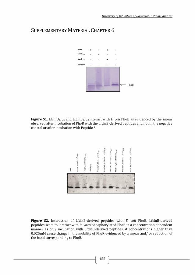
Discovery of Inhibitors of Bacterial Histidine Kinases
155
SUPPLEMENTARY MATERIAL CHAPTER 6
Figure S1. LfcinB17-25 and LfcinB17-32 interact with E. coli PhoB as evidenced by the smear observed after incubation of PhoB with the LfcinB-derived peptides and not in the negative control or after incubation with Peptide 3.
Figure S2. Interaction of LfcinB-derived peptides with E. coli PhoB. LfcinB-derived peptides seem to interact with in vitro phosphorylated PhoB in a concentration dependent manner as only incubation with LfcinB-derived peptides at concentrations higher than 0.025mM cause change in the mobility of PhoR evidenced by a smear and/ or reduction of the band corresponding to PhoB.

2014
156
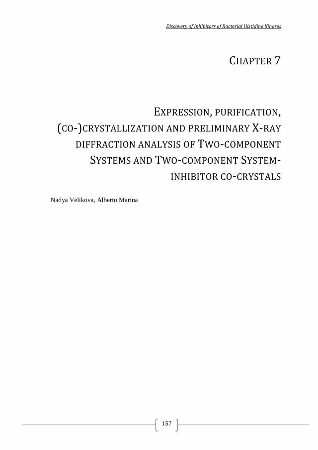
Discovery of Inhibitors of Bacterial Histidine Kinases
157
CHAPTER 7
EXPRESSION, PURIFICATION,
(CO-)CRYSTALLIZATION AND PRELIMINARY X-RAY
DIFFRACTION ANALYSIS OF TWO-COMPONENT
SYSTEMS AND TWO-COMPONENT SYSTEM-
INHIBITOR CO-CRYSTALS
Nadya Velikova, Alberto Marina

2014
158
Discovery of Inhibitors of Bacterial Histidine Kinases
159
7.1. CO-CRYSTALLIZATION OF HISTIDINE-KINASE
AUTOPHOSPHORYLATION INHIBITORS WITH THERMOTOGA MARITIMA
CHEA
INTRODUCTION
Obtaining the structure of a putative target histidine kinase (HK) and a histidine-
kinase autophosphorylation inhibitor (HKAI) would allow the rational structure-based
design of more potent and specific inhibitors. The structure of Thermotoga maritima
CheA CA domain has been previously crystallized in complex with the native ligand
ATP/ADP (PDB:1I58, 1I59) to atomic resolution (1.6 Å). and the conditions to obtain
high-resolution well-diffracting crystals are well-known1,2
. Thus the aim of this study
was to obtain X-ray structures of T. maritima CheA in complex with the HKAIs
discovered in this thesis (Chapter 2, 3, and 5).
The structure of the CA domain of T. maritima CheA contains the characteristic
ATP-binding Bergerat fold including the ATP-lid (Figure 1). The ATP-lid is a loop
variable in sequence and in length crucial for nucleotide binding and catalytic activity 3 , 4
. The ATP-lid is proposed as promising feature for the design of specific HK
autophosphorylation inhibitors with reduced adverse effects to host cells 5, 6.
Figure 1. Catalytic and ATP-binding (CA) domain of T. maritima CheA (PDB: 1I58, chain A). Residues are coloured based on their conservation score calculated with ConSurf. ATP is shown in sticks. The ATP-lid is a variable loop that can be used to design more specific HK inhibitors.
Chapter 7
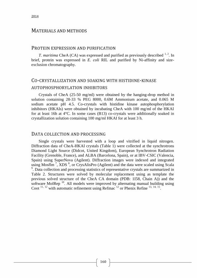
2014
160
MATERIALS AND METHODS
PROTEIN EXPRESSION AND PURIFICATION
T. maritima CheA (CA) was expressed and purified as previously described 1, 2
. In
brief, protein was expressed in E. coli RIL and purified by Ni-affinity and size-
exclusion chromatography.
CO-CRYSTALLIZATION AND SOAKING WITH HISTIDINE-KINASE
AUTOPHOSPHORYLATION INHIBITORS
Crystals of CheA (25-50 mg/ml) were obtained by the hanging-drop method in
solution containing 28-33 % PEG 8000, 0.6M Ammonium acetate, and 0.065 M
sodium acetate pH 4.5. Co-crystals with histidine kinase autophosphorylation
inhibitors (HKAIs) were obtained by incubating CheA with 100 mg/ml of the HKAI
for at least 16h at 4°C. In some cases (B13) co-crystals were additionally soaked in
crystallization solution containing 100 mg/ml HKAI for at least 3 h.
DATA COLLECTION AND PROCESSING
Single crystals were harvested with a loop and vitrified in liquid nitrogen.
Diffraction data of CheA-HKAI crystals (Table 1) were collected at the synchrotrons
Diamond Light Source (Didcot, United Kingdom), European Synchrotron Radiation
Facility (Grenoble, France), and ALBA (Barcelona, Spain), or at IBV-CSIC (Valencia,
Spain) using SuperNova (Agilent). Diffraction images were indexed and integrated
using Mosflm 7, XDS
8, or CrysAlisPro (Agilent) and the data were scaled using Scala
9. Data collection and processing statistics of representative crystals are summarized in
Table 2. Structures were solved by molecular replacement using as template the
previous solved structure of the CheA CA domain (PDB: 1I58, Chain A)) and the
software MolRep 10
. All models were improved by alternating manual building using
Coot 11,
12
with automatic refinement using Refmac 12
or Phenix Refine 13,
14,
15
.
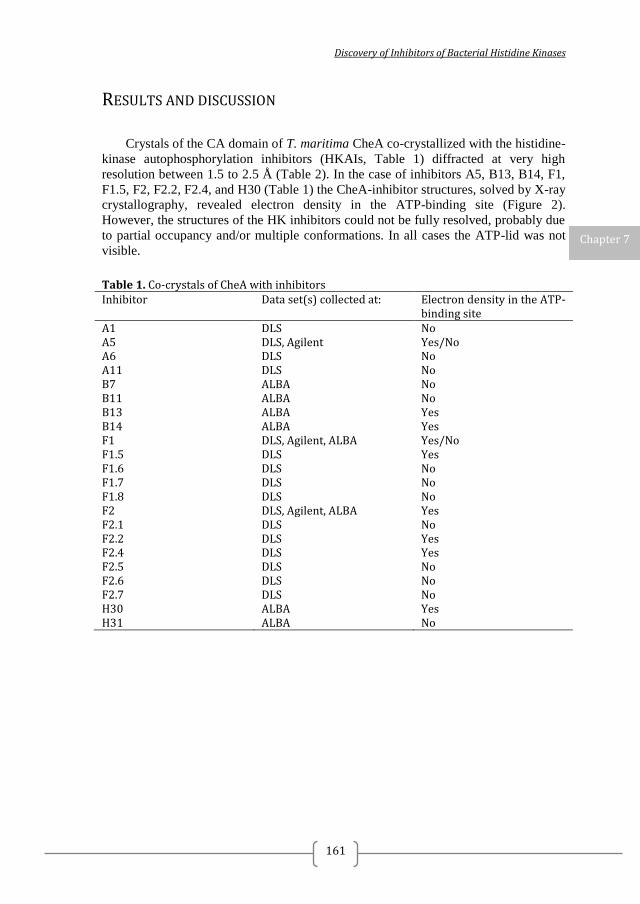
Discovery of Inhibitors of Bacterial Histidine Kinases
161
RESULTS AND DISCUSSION
Crystals of the CA domain of T. maritima CheA co-crystallized with the histidine-
kinase autophosphorylation inhibitors (HKAIs, Table 1) diffracted at very high
resolution between 1.5 to 2.5 Å (Table 2). In the case of inhibitors A5, B13, B14, F1,
F1.5, F2, F2.2, F2.4, and H30 (Table 1) the CheA-inhibitor structures, solved by X-ray
crystallography, revealed electron density in the ATP-binding site (Figure 2).
However, the structures of the HK inhibitors could not be fully resolved, probably due
to partial occupancy and/or multiple conformations. In all cases the ATP-lid was not
visible.
Table 1. Co-crystals of CheA with inhibitors Inhibitor Data set(s) collected at: Electron density in the ATP-
binding site A1 DLS No A5 DLS, Agilent Yes/No A6 DLS No A11 DLS No B7 ALBA No B11 ALBA No B13 ALBA Yes B14 ALBA Yes F1 DLS, Agilent, ALBA Yes/No F1.5 DLS Yes F1.6 DLS No F1.7 DLS No F1.8 DLS No F2 DLS, Agilent, ALBA Yes F2.1 DLS No F2.2 DLS Yes F2.4 DLS Yes F2.5 DLS No F2.6 DLS No F2.7 DLS No H30 ALBA Yes H31 ALBA No
Chapter 7
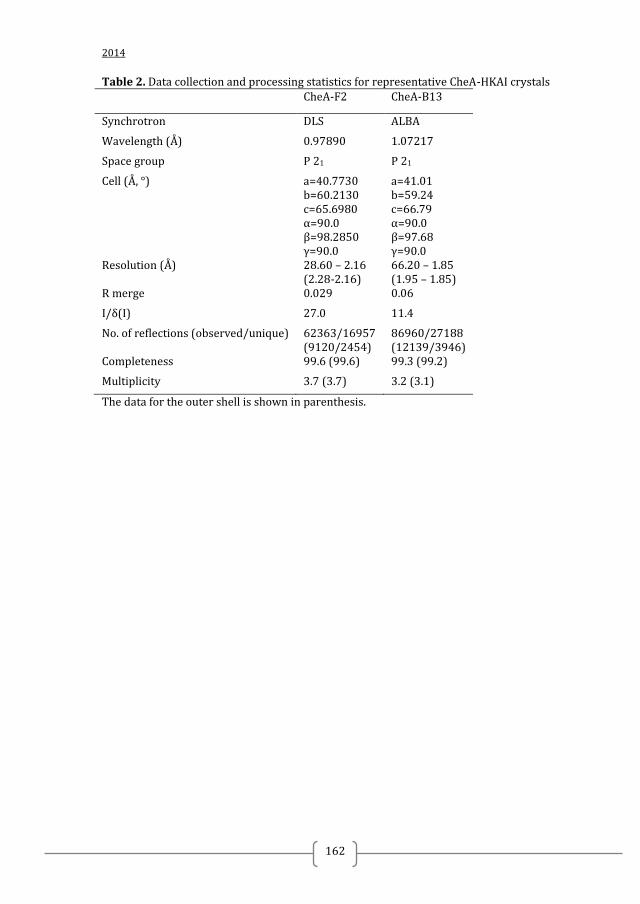
2014
162
Table 2. Data collection and processing statistics for representative CheA-HKAI crystals CheA-F2 CheA-B13
Synchrotron DLS ALBA
Wavelength (Å) 0.97890 1.07217
Space group P 21 P 21
Cell (Å, °) a=40.7730 b=60.2130 c=65.6980 α=90.0 β=98.2850 γ=90.0
a=41.01 b=59.24 c=66.79 α=90.0 β=97.68 γ=90.0
Resolution (Å) 28.60 – 2.16 (2.28-2.16)
66.20 – 1.85 (1.95 – 1.85)
R merge 0.029 0.06
I/δ(I) 27.0 11.4
No. of reflections (observed/unique) 62363/16957 (9120/2454)
86960/27188 (12139/3946)
Completeness 99.6 (99.6) 99.3 (99.2)
Multiplicity 3.7 (3.7) 3.2 (3.1)
The data for the outer shell is shown in parenthesis.

Discovery of Inhibitors of Bacterial Histidine Kinases
163
Figure 2. T. maritima CheA-HKAI structures solved by X-ray crystallography. A) Co-crystals of T. maritima CheA (CA domain) and the initial hits from the structure-based virtual screening (A5, Chapter 2) and the fragment-based screening by differential scanning fluorimetry (F1 and F2) were obtained by co-crystallization with 100 mM HKAI. B) Co-crystal of T. maritima CheA and the most promising HKAI described in the thesis B13 (Chapter 2) was obtained by co-crystallization followed by soaking with 100 mM B13. The 2Fo-Fc (blue) and Fo-Fc (green) electron density maps contoured to 1 and 2.5 σ, respectively, showed extra density in the active site that was not possible to explain by the protein model and were ascribed to the HKAI. However, the HKAIs could not be fully resolved, probably due to partial occupancy and/or multiple conformations.
Chapter 7
2014
164
7.2. PURIFICATION, CRYSTALLIZATION AND PRELIMINARY X-RAY
DIFFRACTION ANALYSIS OF SYNECCOCYSTIS SPP. COPM
INTRODUCTION
Copper is an essential trace element for organisms living under aerobic condition 16
. It is required in fundamental cellular processes such as oxidative phosphorylation,
photosynthesis, and free radical control. However copper is highly toxic when it is
present in excess, thus, many organisms have developed homeostatic mechanism to
tightly regulate its cellular concentration 17
. Cyanobacteria are an attractive model to
investigate the systems involved in copper homeostasis since they are unique bacteria
with internal copper requirement for two proteins: the blue-copper protein
plastocyanin, and the caa3-type cytochrome oxidase 18
. These two proteins are
localized in the thylakoids, a special internal structure where photosynthesis and
respiration take place in cyanobacteria. Cyanobacteria are model microorganisms for
the study of photosynthesis, carbon and nitrogen assimilation, evolution of plant
plastids, and adaptability to environmental stresses. Synechocystis sp. PCC 6803 is one
of the most highly studied types of cyanobacteria as it can grow both autotrophically
or heterotrophically in the absence of light. Copper resistance in cyanobacteria has
been mainly investigated in Synechocystis sp. PCC 6803 (hereafter Synechocystis),
which involves a two-component system, CopRS, a protein of unknown function,
CopM, and an HME-RND exports system, CopBAC 19,
17
. These proteins are encoded
by two operons: copMRS, and copBAC. Both copM and copBAC are regulated by the
CopRS two-component system, and are specifically induced by the presence of Cu2+
19
. CopM is an unknown protein that contains a predicted periplasmic signal domain
and a high number of histidine (8) and methionine (23) residues, which are usually
implicated in direct metal binding in proteins. Furthermore, CopM has two domain of
unknown function DUF305, which is present in other secretion proteins in bacteria and
belongs to the ferritin superfamily 20
. Herein, we report the purification, crystallization
and preliminary X-ray diffraction analysis of Synechocystis CopM.
Discovery of Inhibitors of Bacterial Histidine Kinases
165
MATERIALS AND METHODS
CLONING, EXPRESSION AND PURIFICATION
A 519-bp band (from ORF sll0788) coding for the CopM25-196 periplasmic domain
was PCR amplified from genomic DNA with oligonucleotides ACOPMSTF-
COPMSTR (Table 1), digested with KpnI and SacI, and cloned into pET51 digested
with the same enzymes. CopM25-196 was expressed in E. coli BL21. An overnight pre-
culture was used to inoculate Luria broth medium and was grown to an optical density
at 600 nm of 0.6. The culture was cooled down on ice for 20 min, protein expression
induced by addition of 0.2 mM isopropyl-b-D-thiogalactopyranoside, and incubation
continued for 4 h at 25°C. Cells were harvested by centrifugation and frozen at -80°C.
Frozen pellets were re-suspended in buffer S (100 mM Tris HCl pH 8, 150 mM NaCl,
1 mM BCSA, 1 mM EDTA, and 2 mM Tris (2-carboxyethyl)-phosphine) and broken
by sonication. The suspension was centrifuged 30 min at 30, 000 g at 4°C and the
supernatant was loaded into a 5-ml streptavidin beads (IBA GmbH) column
equilibrated in buffer S. Beads were washed with 50 mL of buffer S and CopM25-196
was eluted with 1x Strep-Tag elution buffer (IBA GmbH). CopM25-196 was further
purified by gel filtration in a Hi-Load 75 (GE-Healthcare) column equilibrated with 20
mM Tris HCl pH 8, 150 mM NaCl. The purified protein was concentrated using a 3K
Vivaspin concentrator and store at -20 °C until use.
CRYSTALLIZATION
Initial sitting-drop vapour-diffusion crystallization trials to identify promising
crystallization conditions for CopM were performed using the commercial
crystallization screens JCSG+ (Qiagen, Germany), JBC I and JBC II (Jena Bioscience,
Germany). In an attempt to obtain crystals of both the apo and Cu2+
-bound CopM
initial crystallization conditions screenings were performed in the presence of 1 mM
EDTA and 1 mM CuSO4, respectively. Crystallization drops of 0.6 µl
(protein:precipitant ratio of 1:1) were set up in 96-well plates containing 75 µl
reservoir solution. Initial hits were obtained after 4-6 days in multiple conditions
(Table S1). Optimization of the crystallization conditions was attempted by varying
the concentration of PEGs and salts under the conditions that initially generated
diffracting crystals.
Chapter 7
2014
166
DATA COLLECTION AND PROCESSING
For data collection, the crystals were soaked in cryoprotectant (reservoir solution
supplemented with 40% PEG 3350) for 5-10 sec. A single crystal was harvested with a
loop and vitrified in liquid nitrogen. Diffraction data were collected the synchrotronsat
Diamond Light Source (Didcot, United Kingdom) or European Synchrotron Radiation
Facility (Grenoble, France). Diffraction images were indexed and integrated using
Mosflm 7 or XDS
8 and the data were scaled using Scala
9. Data collection and
processing statistics are summarized in Table 2.
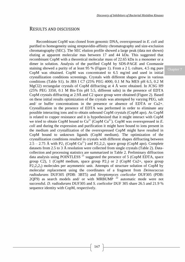
Discovery of Inhibitors of Bacterial Histidine Kinases
167
RESULTS AND DISCUSSION
Recombinant CopM was cloned from genomic DNA, overexpressed in E. coli and
purified to homogeneity using streptavidin-affinity chromatography and size-exclusion
chromatography (SEC). The SEC elution profile showed a large peak (data not shown)
eluting at apparent molecular mas between 17 and 44 kDa. This suggested that
recombinant CopM with a theoretical molecular mass of 22.65 kDa is a monomer or a
dimer in solution. Analysis of the purified CopM by SDS-PAGE and Coomassie
staining showed a purity of at least 95 % (Figure 1). From a 2 L culture, 4.5 mg pure
CopM was obtained. CopM was concentrated to 6.5 mg/ml and used in initial
crystallization conditions screenings. Crystals with different shapes grew in various
conditions (Table S1). In JBS I C7 (25% PEG 4000, 0.1 M Na MES pH 6.5, 0.2 M
MgCl2) rectangular crystals of CopM diffracting at 4 Å were obtained. In JCSG H9
(25% PEG 3350, 0.1 M Bis-Tris pH 5.5, different salts) in the presence of EDTA
CopM crystals diffracting at 2.9Å and C2 space group were obtained (Figure 2). Based
on these initial results optimization of the crystals was attempted by varying PEG, salt
and/ or buffer concentrations in the presence or absence of EDTA or Cu2+.
Crystallization in the presence of EDTA was performed in order to eliminate any
possible interacting ions and to obtain unbound CopM crystals (CopM apo). As CopM
is related to copper resistance and it is hypothesized that it might interact with CopM
we tried to obtain CopM bound to Cu2+
(CopM Cu2+
). CopM was overexpressed in E.
coli and during the expression and purification it might have bound to ions present in
the medium and crystallization of the overexpressed CopM might have resulted in
CopM bound to unknown ligands (CopM medium). The optimization of the
crystallization conditions resulted in crystals with different shapes diffracting between
2.5 – 2.75 Å with P21 (CopM Cu2+
) and P212121 space group (CopM apo). Complete
datasets from 2.5 to 3 Å resolution were collected from single crystals (Table 2). Data-
collection and processing statistics are summarized in Table 2. Preliminary diffraction
data analysis using POINTLESS 21
suggested the presence of 5 (CopM EDTA, space
group C2), 1 (CopM medium, space group P21) or 2 (CopM Cu2+, space group
P212121) molecules per asymmetric unit. Attempts of structure solution of CopM by
molecular replacement using the coordinates of a fragment from Deinococcus
radiodurans DUF305 (PDB: 3BT5) and Streptomyces coelicolor DUF305 (PDB:
2QF9) as search models and/ or with MRBUMP 22
automatic mode were not
successful. D. radiodurans DUF305 and S. coelicolor DUF 305 share 26.5 and 21.9 %
sequence identity with CopM, respectively.
Chapter 7
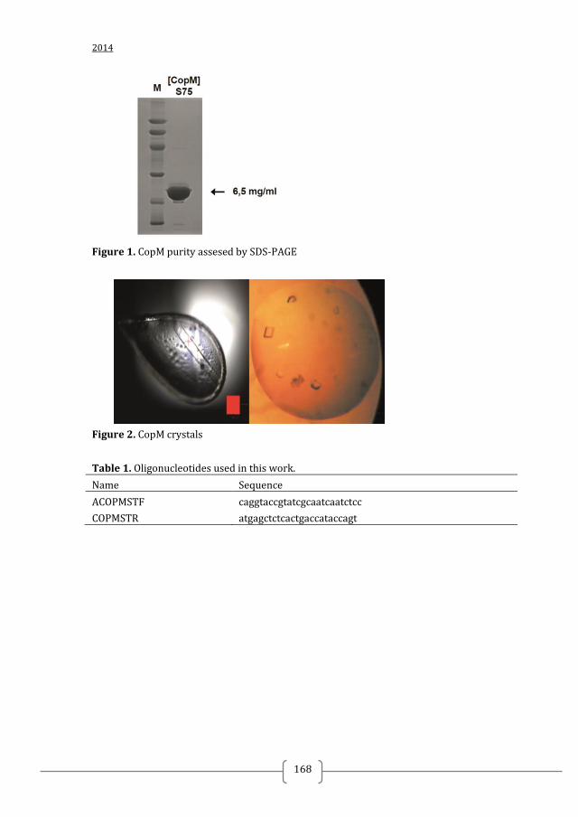
2014
168
Figure 1. CopM purity assesed by SDS-PAGE
Figure 2. CopM crystals
Table 1. Oligonucleotides used in this work.
Name Sequence
ACOPMSTF caggtaccgtatcgcaatcaatctcc
COPMSTR atgagctctcactgaccataccagt
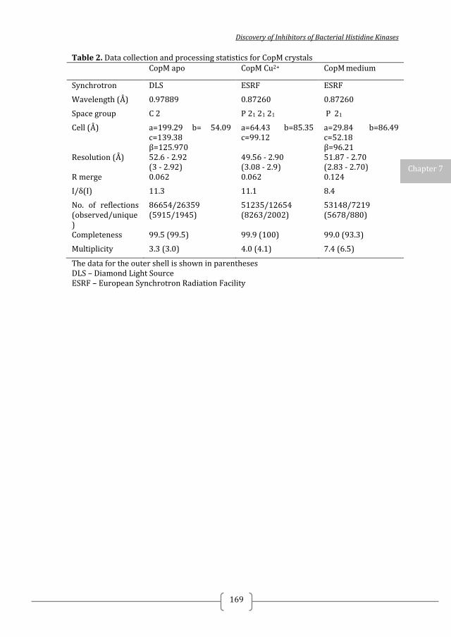
Discovery of Inhibitors of Bacterial Histidine Kinases
169
Table 2. Data collection and processing statistics for CopM crystals CopM apo CopM Cu2+ CopM medium
Synchrotron DLS ESRF ESRF
Wavelength (Å) 0.97889 0.87260 0.87260
Space group C 2 P 21 21 21 P 21
Cell (Å) a=199.29 b= 54.09 c=139.38 β=125.970
a=64.43 b=85.35 c=99.12
a=29.84 b=86.49 c=52.18 β=96.21
Resolution (Å) 52.6 - 2.92 (3 - 2.92)
49.56 - 2.90 (3.08 - 2.9)
51.87 - 2.70 (2.83 - 2.70)
R merge 0.062 0.062 0.124
I/δ(I) 11.3 11.1 8.4
No. of reflections (observed/unique)
86654/26359 (5915/1945)
51235/12654 (8263/2002)
53148/7219 (5678/880)
Completeness 99.5 (99.5) 99.9 (100) 99.0 (93.3)
Multiplicity 3.3 (3.0) 4.0 (4.1) 7.4 (6.5)
The data for the outer shell is shown in parentheses DLS – Diamond Light Source ESRF – European Synchrotron Radiation Facility
Chapter 7
2014
170
REFERENCES 1. Bilwes, A. M.; Alex, L. A.; Crane, B. R.; Simon, M. I., Structure of CheA, a signal-transducing histidine kinase. Cell 1999, 96 (1), 131-41. 2. Bilwes, A. M.; Quezada, C. M.; Croal, L. R.; Crane, B. R.; Simon, M. I., Nucleotide binding by the histidine kinase CheA. Nature structural biology 2001, 8 (4), 353-60. 3. Casino, P.; Rubio, V.; Marina, A., The mechanism of signal transduction by two-component systems. Current opinion in structural biology 2010, 20 (6), 763-71. 4. Casino, P.; Miguel-Romero, L.; Marina, A., Visualizing autophosphorylation in histidine kinases. Nat Commun 2014, 5, 3258. 5. Guarnieri, M. T.; Zhang, L.; Shen, J.; Zhao, R., The Hsp90 inhibitor radicicol interacts with the ATP-binding pocket of bacterial sensor kinase PhoQ. Journal of molecular biology 2008, 379 (1), 82-93. 6. Velikova, N.; Bem, A. E.; van Baarlen, P.; Wells, J. M.; Marina, A., WalK, the Path towards New Antibacterials with Low Potential for Resistance Development. ACS Medicinal Chemistry Letters 2013, 4 (10), 891-894. 7. Powell, H. R., The Rossmann Fourier autoindexing algorithm in MOSFLM. Acta Crystallographica Section D: Biological Crystallography 1999, 55 (10), 1690-1695. 8. Kabsch, W., XDS. Acta Crystallographica Section D: Biological Crystallography 2010, 66 (2), 125-132. 9. Evans, P., Scaling and assessment of data quality. Acta crystallographica. Section D, Biological crystallography 2006, 62 (Pt 1), 72-82. 10. Vagin, A.; Teplyakov, A., Molecular replacement with MOLREP. Acta crystallographica. Section D, Biological crystallography 2010, 66 (Pt 1), 22-5. 11. Emsley, P.; Cowtan, K., Coot: model-building tools for molecular graphics. Acta crystallographica. Section D, Biological crystallography 2004, 60 (Pt 12 Pt 1), 2126-32. 12. Emsley, P.; Lohkamp, B.; Scott, W. G.; Cowtan, K., Features and development of Coot. Acta crystallographica. Section D, Biological crystallography 2010, 66 (Pt 4), 486-501. 13. Afonine, P. V.; Grosse-Kunstleve, R. W.; Echols, N.; Headd, J. J.; Moriarty, N. W.; Mustyakimov, M.; Terwilliger, T. C.; Urzhumtsev, A.; Zwart, P. H.; Adams, P. D., Towards automated crystallographic structure refinement with phenix.refine. Acta crystallographica. Section D, Biological crystallography 2012, 68 (Pt 4), 352-67. 14. Afonine, P. V.; Mustyakimov, M.; Grosse-Kunstleve, R. W.; Moriarty, N. W.; Langan, P.; Adams, P. D., Joint X-ray and neutron refinement with phenix.refine. Acta crystallographica. Section D, Biological crystallography 2010, 66 (Pt 11), 1153-63. 15. Headd, J. J.; Echols, N.; Afonine, P. V.; Grosse-Kunstleve, R. W.; Chen, V. B.; Moriarty, N. W.; Richardson, D. C.; Richardson, J. S.; Adams, P. D., Use of knowledge-based restraints in phenix.refine to improve macromolecular refinement at low resolution. Acta crystallographica. Section D, Biological crystallography 2012, 68 (Pt 4), 381-90. 16. Bondarczuk, K.; Piotrowska-Seget, Z., Molecular basis of active copper resistance mechanisms in Gram-negative bacteria. Cell biology and toxicology 2013, 29 (6), 397-405. 17. Lopez-Maury, L.; Giner-Lamia, J.; Florencio, F. J., Redox control of copper homeostasis in cyanobacteria. Plant signaling & behavior 2012, 7 (12), 1712-4. 18. Manna, P.; Vermaas, W., Lumenal proteins involved in respiratory electron transport in the cyanobacterium Synechocystis sp. PCC6803. Plant molecular biology 1997, 35 (4), 407-16. 19. Giner-Lamia, J.; Lopez-Maury, L.; Reyes, J. C.; Florencio, F. J., The CopRS two-component system is responsible for resistance to copper in the cyanobacterium Synechocystis sp. PCC 6803. Plant physiology 2012, 159 (4), 1806-18.
Discovery of Inhibitors of Bacterial Histidine Kinases
171
20. Uchida, M.; Kang, S.; Reichhardt, C.; Harlen, K.; Douglas, T., The ferritin superfamily: Supramolecular templates for materials synthesis. Biochimica et biophysica acta 2010, 1800 (8), 834-45. 21. Evans, P. R., An introduction to data reduction: Space-group determination, scaling and intensity statistics. Acta Crystallographica Section D: Biological Crystallography 2011, 67 (4), 282-292. 22. Keegan, R. M.; Winn, M. D., MrBUMP: an automated pipeline for molecular replacement. Acta crystallographica. Section D, Biological crystallography 2008, 64 (Pt 1), 119-24.

2014
172
SUPPLEMENTARY MATERIAL CHAPTER 7.2
Table S1. Crystallization screenings results Sample Screening,
position, morphology
Condition
CopM glycerol
JBS I, A11 25% PEG 1000, 0.1 M Na Hepes pH 7.5
CopM JBS I, C7 cubes 25% PEG 4000, 0.1 M Na MES pH 6.5, 0.2 M MgCl2
CopM JBS I, C11 very small
30% PEG 4000, 0.1 M Na Hepes pH 7.5, 0.2 M CaCl2
CopM MIDAS, H3 20% glycerol ethoxylate, 3% polyethylene imine
CopM MIDAS, G7, needles 30% glycerol ethoxylate, 0.2 M Amm Acetate pH 6.5, 0.1 M MES-NaOH
CopM EDTA
JBS I, G2 10% PEG 8000, 0.1 M Na Hepes pH 7.5, 0.2 mM Na Acetate
CopM, EDTA
JBS I, C8 25% PEG 4000, 0.1 M Tris HCl 8.5, 0.2 M CaCl2
CopM EDTA
JCSG+, from H6 to H11
25% PEG 3350, 0.1 M Bis-Tris pH 5.5, different salts
CopM EDTA
JCSG+, G1, needles, thin plates
30% Jeaffamine ED 2001, 0.1 M Hepes pH 7
CopM EDTA
JCSG+, E1 1M Tri-Na citrate, 0.1 M Na cacodylate pH 6.5
CopM EDTA
JCSG+, E2 0.2 N NaCl, 0.1 M Na cacocylate pH 6.5, 2M AmSO4
CopM EDTA
JCSG+, G10 0.15 M KBr, 30% PEG MME 2000
CopM EDTA
JCSG+, G11 2M AmSO4, 0.1 M Bis-Tris pH 5.5
CopM JBS I, F4 15% PEG 6000, 50 mM KCl, 10 mM MgCl
CopM JBS I, B12 12% PEG 4000, 0.1 M Na Hepes pH 7.5, 0.1 M Na Acetate

Discovery of Inhibitors of Bacterial Histidine Kinases
173
CHAPTER 8
WALK, THE PATH TOWARDS NEW
ANTIBACTERIALS WITH LOW POTENTIAL FOR
RESISTANCE DEVELOPMENT
Nadya Velikova1, Agnieszka E. Bem
2, Peter van Baarlen
2, Jerry M. Wells
3, Alberto
Marina1, 3
1Instituto de Biomedicina de Valencia, Consejo Superior de Investigaciones
Científicas (CSIC), Jaume Roig 11, 46010 Valencia, Spain; 2Host−Microbe Interactomics Chair Group, Animal Sciences, University of
Wageningen, 6700 AH Wageningen, The Netherlands; 3CIBER de Enfermedades Raras (CIBERER), ISCIII, 46010 Valencia, Spain
PUBLISHED IN ACS MEDICINAL CHEMISTRY LETTERS, DOI:
10.1021/ML400320S

2014
174
Discovery of Inhibitors of Bacterial Histidine Kinases
175
ABSTRACT
Resistance to antibiotics used in the treatment of bacterial infectious diseases is a
global health problem. More than a decade ago, two-component systems such as
WalKR were proposed as ideal targets for the development of new antibiotics.
Biochemical screens for WalKR inhibitors using compound libraries have identified
many hits, some of which were shown to have non-specific effects. The recently
published structures of the S. mutans and B. subtilis WalK provide the opportunity to
study inhibitors of WalK autophosphorylation at the atomic level and means to design
compounds with improved specificity and affinity using a structure-based approach. Chapter 8
2014
176
Recently scientists have been watching in awe as bacteria develop resistance to
the latest antibiotics. Multidrug resistant tuberculosis (MDR-TB), penicillin-resistant
Streptococcus pneumoniae, and hospital-acquired infections with vancomycin-
resistant enterococci (VRE) or methicillin-resistant Staphylococcus aureus (MRSA)
are regularly making headline news. The problem is not only one of resistance to front
line drugs and the fear that one day, infections will be incurable, but also the increased
cost of patient care and the prospect of future inability to perform trivial medical
procedures without significant risk.
The scarcity of new antibiotics has been put down to dwindling returns on
screening natural sources but also poor incentives for companies to invest in this sector
as opposed to treatment for cancer and chronic diseases. However, the challenge of
developing anti-infective drugs has been taken up by small enterprises and academia
with the support of governments and other funding organizations.
The genomics revolution has accelerated finding new targets for anti-infective
drugs, but designing drug-like inhibitors against specific targets remains highly
challenging, costly, and not without significant risk of failure.
Against this somewhat gloomy perspective, there are some promising
developments unfolding, one of which concerns bacterial two-component systems
(TCSs) as drug targets. Bacteria respond and adapt to a large variety of environmental
and intracellular signals via TCS signal transduction. The minimum components of a
TCS are a sensor histidine kinase (HK) and an effector response regulator
(RR)(8)(Figure 1). Typically, the sensor histidine kinase (HK) is membrane-bound,
and signal recognition alters the phosphorylation state of a cognate response regulator
(RR). HK sensing of specific signals occurs via a variable domain that is commonly
exposed to the extracellular milieu, whereas the remaining protein domains are
generally conserved, cytoplasmic, and required for signal transduction (8) (Figure1).
RRs are usually transcription factors of which the DNA-binding capacity and
consequently, gene transcription is determined by their phosphorylation state. TCSs
may directly or indirectly control numerous genes, including those involved in the
regulation of metabolism, cell physiology, virulence, persistence, and resistance to
antibiotics or antimicrobial peptides. Targeting virulence is a promising antibacterial-
drug discovery strategy as it exerts less selective pressure on the pathogen and
resistance development will be slower.
TCSs were proposed as attractive targets more than 20 years ago because they are
absent in mammals and essential or conditionally essential for viability in several
important bacterial pathogens. WalKR system (a.k.a., YycGF, VicKR, MicAB) is an
obligate essential regulatory system in Firmicutes including MRSA, VRE, and some
other notorious pathogens. Other TCSs might not be essential for growth in vitro, but
in vivo they can be necessary for survival or persistence (e.g., the DosRS system in
Mycobacterium). Besides, some bacteria possess as many as 200 TCSs depending on
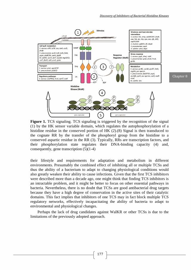
Discovery of Inhibitors of Bacterial Histidine Kinases
177
Figure 1. TCS signaling. TCS signaling is triggered by the recognition of the signal
(1) by the HK sensor variable domain, which regulates the autophosphorylation of a
histidine residue in the conserved portion of HK (2).(8) Signal is then transduced to
the cognate RR by the transfer of the phosphoryl group from the histidine to a
conserved aspartic residue in the RR (3). Typically, RRs are transcription factors, and
their phosphorylation state regulates their DNA-binding capacity (4) and,
consequently, gene transcription (5)(1-4)
their lifestyle and requirements for adaptation and metabolism in different
environments. Presumably the combined effect of inhibiting all or multiple TCSs and
thus the ability of a bacterium to adapt to changing physiological conditions would
also greatly weaken their ability to cause infections. Given that the first TCS inhibitors
were described more than a decade ago, one might think that finding TCS inhibitors is
an intractable problem, and it might be better to focus on other essential pathways in
bacteria. Nevertheless, there is no doubt that TCSs are good antibacterial drug targets
because they have a high degree of conservation in the active sites of their catalytic
domains. This fact implies that inhibitors of one TCS may in fact block multiple TCS
regulatory networks, effectively incapacitating the ability of bacteria to adapt to
environmental and physiological changes.
Perhaps the lack of drug candidates against WalKR or other TCSs is due to the
limitations of the previously adopted approach.
Chapter 8
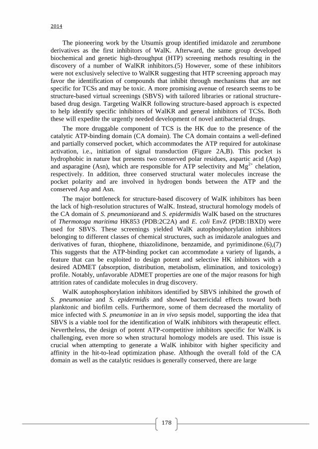
2014
178
The pioneering work by the Utsumís group identified imidazole and zerumbone
derivatives as the first inhibitors of WalK. Afterward, the same group developed
biochemical and genetic high-throughput (HTP) screening methods resulting in the
discovery of a number of WalKR inhibitors.(5) However, some of these inhibitors
were not exclusively selective to WalKR suggesting that HTP screening approach may
favor the identification of compounds that inhibit through mechanisms that are not
specific for TCSs and may be toxic. A more promising avenue of research seems to be
structure-based virtual screenings (SBVS) with tailored libraries or rational structure-
based drug design. Targeting WalKR following structure-based approach is expected
to help identify specific inhibitors of WalKR and general inhibitors of TCSs. Both
these will expedite the urgently needed development of novel antibacterial drugs.
The more druggable component of TCS is the HK due to the presence of the
catalytic ATP-binding domain (CA domain). The CA domain contains a well-defined
and partially conserved pocket, which accommodates the ATP required for autokinase
activation, i.e., initiation of signal transduction (Figure 2A,B). This pocket is
hydrophobic in nature but presents two conserved polar residues, aspartic acid (Asp)
and asparagine (Asn), which are responsible for ATP selectivity and Mg2+
chelation,
respectively. In addition, three conserved structural water molecules increase the
pocket polarity and are involved in hydrogen bonds between the ATP and the
conserved Asp and Asn.
The major bottleneck for structure-based discovery of WalK inhibitors has been
the lack of high-resolution structures of WalK. Instead, structural homology models of
the CA domain of S. pneumoniaeand and S. epidermidis WalK based on the structures
of Thermotoga maritima HK853 (PDB:2C2A) and E. coli EnvZ (PDB:1BXD) were
used for SBVS. These screenings yielded WalK autophosphorylation inhibitors
belonging to different classes of chemical structures, such as imidazole analogues and
derivatives of furan, thiophene, thiazolidinone, benzamide, and pyrimidinone.(6),(7)
This suggests that the ATP-binding pocket can accommodate a variety of ligands, a
feature that can be exploited to design potent and selective HK inhibitors with a
desired ADMET (absorption, distribution, metabolism, elimination, and toxicology)
profile. Notably, unfavorable ADMET properties are one of the major reasons for high
attrition rates of candidate molecules in drug discovery.
WalK autophosphorylation inhibitors identified by SBVS inhibited the growth of
S. pneumoniae and S. epidermidis and showed bactericidal effects toward both
planktonic and biofilm cells. Furthermore, some of them decreased the mortality of
mice infected with S. pneumoniae in an in vivo sepsis model, supporting the idea that
SBVS is a viable tool for the identification of WalK inhibitors with therapeutic effect.
Nevertheless, the design of potent ATP-competitive inhibitors specific for WalK is
challenging, even more so when structural homology models are used. This issue is
crucial when attempting to generate a WalK inhibitor with higher specificity and
affinity in the hit-to-lead optimization phase. Although the overall fold of the CA
domain as well as the catalytic residues is generally conserved, there are large
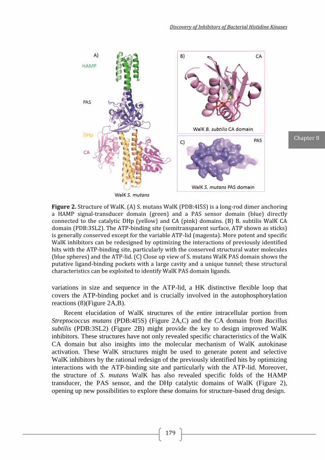
Discovery of Inhibitors of Bacterial Histidine Kinases
179
Figure 2. Structure of WalK. (A) S. mutans WalK (PDB:4I5S) is a long-rod dimer anchoring a HAMP signal-transducer domain (green) and a PAS sensor domain (blue) directly connected to the catalytic DHp (yellow) and CA (pink) domains. (B) B. subtilis WalK CA domain (PDB:3SL2). The ATP-binding site (semitransparent surface, ATP shown as sticks) is generally conserved except for the variable ATP-lid (magenta). More potent and specific WalK inhibitors can be redesigned by optimizing the interactions of previously identified hits with the ATP-binding site, particularly with the conserved structural water molecules (blue spheres) and the ATP-lid. (C) Close up view of S. mutans WalK PAS domain shows the putative ligand-binding pockets with a large cavity and a unique tunnel; these structural characteristics can be exploited to identify WalK PAS domain ligands.
variations in size and sequence in the ATP-lid, a HK distinctive flexible loop that
covers the ATP-binding pocket and is crucially involved in the autophosphorylation
reactions (8)(Figure 2A,B).
Recent elucidation of WalK structures of the entire intracellular portion from
Streptococcus mutans (PDB:4I5S) (Figure 2A,C) and the CA domain from Bacillus
subtilis (PDB:3SL2) (Figure 2B) might provide the key to design improved WalK
inhibitors. These structures have not only revealed specific characteristics of the WalK
CA domain but also insights into the molecular mechanism of WalK autokinase
activation. These WalK structures might be used to generate potent and selective
WalK inhibitors by the rational redesign of the previously identified hits by optimizing
interactions with the ATP-binding site and particularly with the ATP-lid. Moreover,
the structure of S. mutans WalK has also revealed specific folds of the HAMP
transducer, the PAS sensor, and the DHp catalytic domains of WalK (Figure 2),
opening up new possibilities to explore these domains for structure-based drug design.
Chapter 8
2014
180
PAS (Per-Arnt-Sim) domains are small sensor modules that bind a chemically
diverse range of small molecules and present a conserved three-dimensional
architecture but a divergent primary sequence. The B. subtilis PAS domain is required
for WalK activity and localization to the divisome during cell division. The structure
of S. mutans WalK shows that the putative ligand binding site forms unique pockets
for a variety of ligands to bind (Figure 2C) suggesting that WalK PAS domain might
bind ligands differently from other HK PAS domains. Therefore, the WalK PAS
domain appears to be a promising target for novel antimicrobials, and its structure
might also facilitate the rational design of specific and potent WalK inhibitors.
Furthermore, WalK PAS ligands remain unknown, and their identification might
generate chemical reagents to facilitate mechanistic studies.
In summary, we argue that discovery of new WalKR, and in general TCS,
inhibitors, which will be further developed into antibacterial drugs, is still a realistic
scenario. However, the focus should be on structure-based approaches. Certainly,
ongoing efforts to solve the structures of HK from other important pathogens will help
to design inhibitors with optimized activity. The high homology between TCSs and
their wide distribution among bacteria imply that such inhibitors can be developed into
broad-spectrum antibiotics. The road ahead is still challenging and arduous, but given
the importance of the problem of drug resistance, we are convinced it is a road we
should take.
Discovery of Inhibitors of Bacterial Histidine Kinases
181
REFERENCES 1. Dubrac,S.; Boneca,I. G.; Poupel,O.; Msadek,T.New insights into the WalK/WalR (YycG/YycF) essential signal-transduction pathway reveal a major role in controlling cell wall metabolism and biofilm formation inStaphylococcus aureus J. Bacteriol.2007,189(22)8257–8269 2. Delauné,A.; Dubrac,S.; Blanchet,C.; Poupel,O.; Mäder,U.; Hiron,A.; Leduc,A.; Fitting,C.; Nicolas,P.; Cavaillon,J.-M.; Adib-Conquy,M.; Msadek,T.The WalKR system controls major staphylococcal virulence genes and is involved in triggering host inflammatory response Infect. Immun.2012,80(10)3438–3453 3. Mohedano,M. L.; Overweg,K.; De La Fuente,A.; Reuter,M.; Altabe,S.; Mulholland,F.; De Mendoza,D.; López,P.; Wells,J. M.Evidence that the essential response regulator YycF inStreptococcus pneumoniae modulates expression of fatty acid biosynthesis genes and alters membrane-compositionJ. Bacteriol 2005,187(7)2357–2367 4. Howden,B. P.; McEvoy,C. R. E.; Allen,D. L.; Bell,J.; Coombs,G.; Bennett-Wood,V.; Porter,J. L.; Robins-Browne,R.; Davies,J. K.; Seemann,T.; Stinear,T. P.Evolution of multidrug resistance during Staphylococcus aureus infection involves mutation of the essential two component regulator WalKRPLoS Pathog 2011,7(11)e1002359 5. Okada,A.; Gotoh,Y.; Watanabe,T.; Furuta,E.; Yamamoto,K.; Usumi,R. Targeting two-component signal transduction: a novel drug discovery system Methods Enzymol. 2007,422,386–395 6. Qin,Z.; Zhang,J.; Xu,B.; Chen,L.; Wu,Y.; Yang,X.; Shen,X.; Molin,S.; Danchin,A.; Jiang,H.; Qu,D. Structure-based discovery of inhibitors of the YycG histidine kinase: new chemical leads to combat Staphyloccocus epidermidis infections BMC Microbiol. 2006 6,96 7. Li,N.; Wang,F.; Niu,S.; Cao,J.; Wu,K.; Li,Y.; Yin,N.; Zhang,X.; Zhu,W.; Yin,Y.Discovery of novel inhibitors of Streptococcus pneumoniae based on the virtual screening with the homology-modelled structure of histidine kinase (VicK)BMC Microbiol 2009,9,129 8. Casino,P.; Rubio,V.; Marina, A.The mechanism of signal transduction by two-component systems Curr. Opin. Struct. Biol. 2010,20(6)763–771

2014
182
Discovery of Inhibitors of Bacterial Histidine Kinases
183
CHAPTER 9
GENERAL DISCUSSION
Nadya Velikova

2014
184
Discovery of Inhibitors of Bacterial Histidine Kinases
185
THE NEED OF NEW ANTIBACTERIALS AND THE IDEAL ANTIBACTERIAL
TARGETS
The growing problem of bacterial multi-drug resistance (MDR) requires the
urgent development of novel antibacterials with different mechanisms of action from
known antibiotics 1, 2
. At the outset of this thesis bacterial two-component systems
(TCS) were selected as targets for the discovery of novel lead candidates for the
development of antibacterial drugs. At this point it seems prudent to revaluate TCS
suitability as drug targets based on the results presented in the thesis and developments
published in the literature. Recent reviews 3, 4, 5, 6, 7, 8
from opinion leaders in the field
suggest that from pharmaceutical point of view novel antibacterial targets should fulfil
the following criteria:
1. Have no human homologues or structurally similar proteins.
2. Be highly conserved among various bacterial species to assure broad-
spectrum of antibacterial action.
3. Be strictly essential for bacterial viability such that their inhibition would lead
to bacterial death but as targeting essentiality might increase the rise of resistance,
targeting virulence targets has been proposed as an alternative.
4. Be ´´assayable´´, i.e. should have easy measurable activity, assays amenable
to high-throughput screening (HTS), target structural data should be available or easy
to disclose and genetic tools should be available to validate the target in a key species.
5. Be patentable.
The suitability of TCS as targets for novel antibacterials is discussed below in
relation to the first four criteria listed above.
1. HOMOLOGUES OF TWO-COMPONENT SIGNALLING SYSTEMS ARE
ABSENT IN HIGHER EUKARYOTES
In all living organisms cellular processes are regulated via signal transduction.
Both prokaryotes and eukaryotes depend on the ability to rapidly sense and respond to
changes in intracellular and extracellular signals to adapt and survive. Two-component
systems (TCS) are the major mode of signal transduction in bacteria 9. They are
attractive antibacterial (drug) targets because multiple TCS are found in nearly all
bacteria and homologues have not been identified in mammals, including humans 10
11
, 12
. TCS signalling involves autophosphorylation of a membrane-bound histidine kinase
(HK), phosphotransfer of the phosphoryl group to a cognate response regulator (RR),
and ultimately modulation of the expression of target genes (Figure 1) 13
. The
Chapter 9
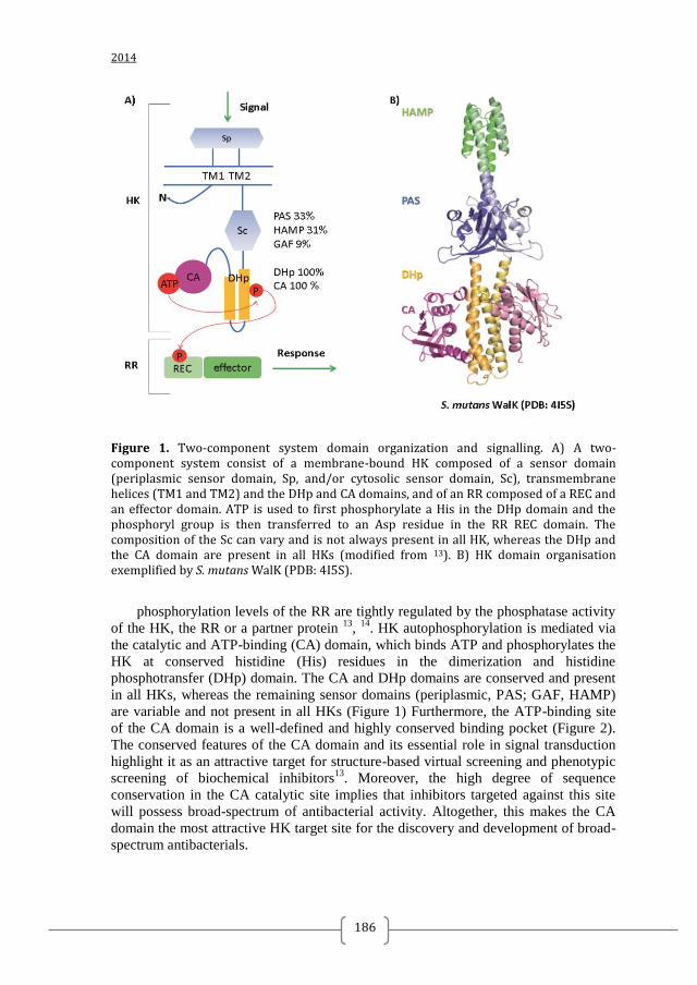
2014
186
Figure 1. Two-component system domain organization and signalling. A) A two-component system consist of a membrane-bound HK composed of a sensor domain (periplasmic sensor domain, Sp, and/or cytosolic sensor domain, Sc), transmembrane helices (TM1 and TM2) and the DHp and CA domains, and of an RR composed of a REC and an effector domain. ATP is used to first phosphorylate a His in the DHp domain and the phosphoryl group is then transferred to an Asp residue in the RR REC domain. The composition of the Sc can vary and is not always present in all HK, whereas the DHp and the CA domain are present in all HKs (modified from 13). B) HK domain organisation exemplified by S. mutans WalK (PDB: 4I5S).
phosphorylation levels of the RR are tightly regulated by the phosphatase activity
of the HK, the RR or a partner protein 13
, 14
. HK autophosphorylation is mediated via
the catalytic and ATP-binding (CA) domain, which binds ATP and phosphorylates the
HK at conserved histidine (His) residues in the dimerization and histidine
phosphotransfer (DHp) domain. The CA and DHp domains are conserved and present
in all HKs, whereas the remaining sensor domains (periplasmic, PAS; GAF, HAMP)
are variable and not present in all HKs (Figure 1) Furthermore, the ATP-binding site
of the CA domain is a well-defined and highly conserved binding pocket (Figure 2).
The conserved features of the CA domain and its essential role in signal transduction
highlight it as an attractive target for structure-based virtual screening and phenotypic
screening of biochemical inhibitors13
. Moreover, the high degree of sequence
conservation in the CA catalytic site implies that inhibitors targeted against this site
will possess broad-spectrum of antibacterial activity. Altogether, this makes the CA
domain the most attractive HK target site for the discovery and development of broad-
spectrum antibacterials.
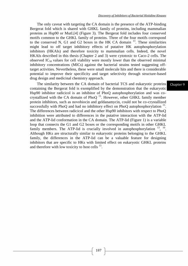
Discovery of Inhibitors of Bacterial Histidine Kinases
187
The only caveat with targeting the CA domain is the presence of the ATP-binding
Bergerat fold which is shared with GHKL family of proteins, including mammalian
proteins as Hsp90 or MutL[4] (Figure 3). The Bergerat fold includes four conserved
motifs common to the GHKL family of proteins. Three of the four motifs correspond
to the conserved N, G1 and G2 boxes in the HK CA domain 13
. These similarities
might lead to off target inhibitory effects of putative HK autophosphorylation
inhibitors (HKAIs) and therefore toxicity to mammalian cells. Indeed, the novel
HKAIs described in this thesis (Chapter 2 and 3) were cytotoxic to Caco-2 cells. The
observed IC50 values for cell viability were mostly lower than the observed minimal
inhibitory concentrations (MICs) against the bacterial strains tested suggesting off-
target activities. Nevertheless, these were small molecule hits and there is considerable
potential to improve their specificity and target selectivity through structure-based
drug design and medicinal chemistry approach.
The similarity between the CA domain of bacterial TCS and eukaryotic proteins
containing the Bergerat fold is exemplified by the demonstration that the eukaryotic
Hsp90 inhibitor radicicol is an inhibitor of PhoQ autophosphorylation and was co-
crystallized with the CA domain of PhoQ 15
. However, other GHKL family member
protein inhibitors, such as novobiocin and geldanamycin, could not be co-crystallized
successfully with PhoQ and had no inhibitory effect on PhoQ autophosphorylation 15
.
The differences between radicicol and the other Hsp90 inhibitors with respect to PhoQ
inhibition were attributed to differences in the putative interaction with the ATP-lid
and the ATP-lid conformation in the CA domain. The ATP-lid (Figure 1) is a variable
loop that connects the G1 and G2 boxes or the corresponding motifs in other GHKL
family members. The ATP-lid is crucially involved in autophosphorylation 13
, 16
.
Although HKs are structurally similar to eukaryotic proteins belonging to the GHKL
family, the differences in the ATP-lid can be a valuable feature for designing
inhibitors that are specific to HKs with limited effect on eukaryotic GHKL proteins
and therefore with low toxicity to host cells 15
.
Chapter 9
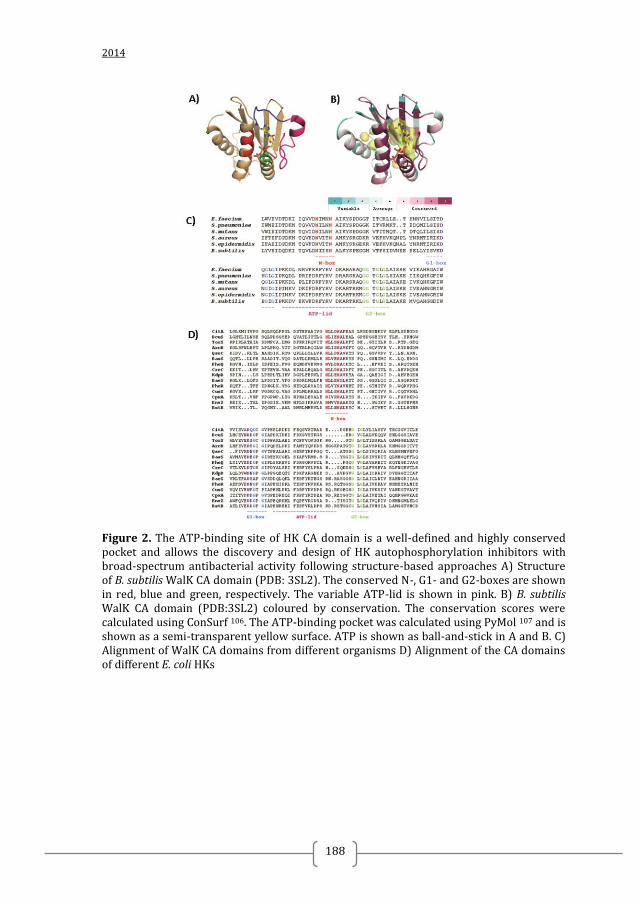
2014
188
Figure 2. The ATP-binding site of HK CA domain is a well-defined and highly conserved pocket and allows the discovery and design of HK autophosphorylation inhibitors with broad-spectrum antibacterial activity following structure-based approaches A) Structure of B. subtilis WalK CA domain (PDB: 3SL2). The conserved N-, G1- and G2-boxes are shown in red, blue and green, respectively. The variable ATP-lid is shown in pink. B) B. subtilis WalK CA domain (PDB:3SL2) coloured by conservation. The conservation scores were calculated using ConSurf 106. The ATP-binding pocket was calculated using PyMol 107 and is shown as a semi-transparent yellow surface. ATP is shown as ball-and-stick in A and B. C) Alignment of WalK CA domains from different organisms D) Alignment of the CA domains of different E. coli HKs
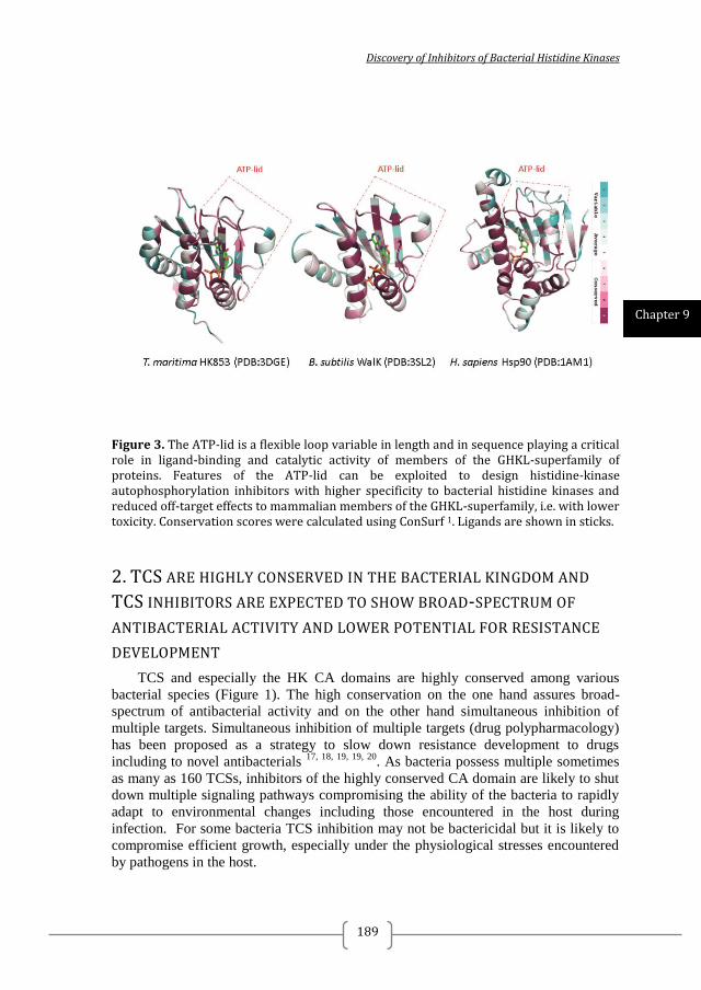
Discovery of Inhibitors of Bacterial Histidine Kinases
189
Figure 3. The ATP-lid is a flexible loop variable in length and in sequence playing a critical role in ligand-binding and catalytic activity of members of the GHKL-superfamily of proteins. Features of the ATP-lid can be exploited to design histidine-kinase autophosphorylation inhibitors with higher specificity to bacterial histidine kinases and reduced off-target effects to mammalian members of the GHKL-superfamily, i.e. with lower toxicity. Conservation scores were calculated using ConSurf 1. Ligands are shown in sticks.
2. TCS ARE HIGHLY CONSERVED IN THE BACTERIAL KINGDOM AND
TCS INHIBITORS ARE EXPECTED TO SHOW BROAD-SPECTRUM OF
ANTIBACTERIAL ACTIVITY AND LOWER POTENTIAL FOR RESISTANCE
DEVELOPMENT
TCS and especially the HK CA domains are highly conserved among various
bacterial species (Figure 1). The high conservation on the one hand assures broad-
spectrum of antibacterial activity and on the other hand simultaneous inhibition of
multiple targets. Simultaneous inhibition of multiple targets (drug polypharmacology)
has been proposed as a strategy to slow down resistance development to drugs
including to novel antibacterials 17, 18, 19, 19, 20
. As bacteria possess multiple sometimes
as many as 160 TCSs, inhibitors of the highly conserved CA domain are likely to shut
down multiple signaling pathways compromising the ability of the bacteria to rapidly
adapt to environmental changes including those encountered in the host during
infection. For some bacteria TCS inhibition may not be bactericidal but it is likely to
compromise efficient growth, especially under the physiological stresses encountered
by pathogens in the host.
Chapter 9
2014
190
The notion that inhibitors of TCS could inhibit multiple TCS targets is supported
by the activity studies performed on the novel histidine kinase autophosphorylation
inhibitors (HKAIs) described in this thesis (Chapter 2 and 3). Promising HKAIs were
discovered by structure-based virtual screening (SBVS) and fragment-based screening
(FBS) for putative ligands of three or two different HKs, respectively. Inhibition of
autophosphorylation was then shown in biochemical assays with HKs not used for
screening, indicating the potential to identify inhibitors with a broad-spectrum of HK
autophosporylation inhibitory activity using such approaches. The broad spectrum of
activity against HKs was also reflected in the capacity of these inhibitors to prevent
growth of several species of Gram-positive bacteria (Chapter 2 and 3). Furthermore,
the most promising inhibitors were effective against multi-drug resistant (MDR)
clinical isolates, including MRSA strains resistant to a wide range of β-lactam and
non-β-lactam antibiotics 21
. These results suggest that the HKAIs possess different
mechanism of action than the known antibiotics and/or the mechanisms of resistance
present in the MDR strains are not active against the HKAIs. The activity of the novel
HKAIs against Gram-negative bacteria was initially found to be low (minimal
inhibitory concentrations (MICs) ≥ 63 µg/ml for the active compounds, and > 500
µg/ml for the majority of the tested inhibitors) but when combined with ε-poly-L-
lysine capped nanoparticles (Chapter 4) the activity increased more than 10-fold. The
antibacterial activity of the nanoparticles was only observed when combined with the
HKAIs, suggesting that the cell envelope of Gram-negative bacteria is most likely
limiting intracellular uptake of the HKAIs. Despite the evidence stated above, it
remains possible that the antibacterial activity is due to inhibition of targets other than
TCS HKs. Until this is proven we cannot unequivocally conclude that inhibition of
multiple HKs is the reason for their antibacterial activity of the identified HKAIs.
3. SOME TCS ARE ESSENTIAL FOR BACTERIAL GROWTH AND/OR
INVOLVED IN THE REGULATION OF VIRULENCE
Ideally novel antibacterials should target essential proteins or processes to assure
bactericidal effect but most TCS are not essential for viability of bacteria in their
normal habitat. This has been demonstrated by the systematic knock out of TCS in
different bacteria 22, 23, 24
. However, this approach does not rule out the possibility that
simultaneous inhibition of multiple TCS would cause bacterial death. In only a few
organisms, more than one TCS has been genetically deleted or inactivated and this is
restricted to typically just two or three TCS 24
. One TCS that is known to be essential
for bacterial viability is WalKR, which is highly conserved among Gram-positives
with a low G+C content in their genomic DNA (Figure 1) 25
.
In this thesis (Chapter 3) inhibitors of S. pneumoniae WalK autophosphorylation
with moderate antibacterial effect for Gram-positive strains, including S. pneumoniae
and MRSA 21
, and Gram-negative strains, including multidrug resistant Acinetobacter
baumanii 26
were discovered by FBS for putative ligands of S. pneumoniae WalK and
then ligand-based similarity searches (LBS) with the two best hits. The SBVS hits, A5
and A6 (Chapter 2), also inhibited S. pneumoniae WalK autophosphorylation in vitro
and showed antibacterial effect for Gram-positive bacteria. Nevertheless it is difficult
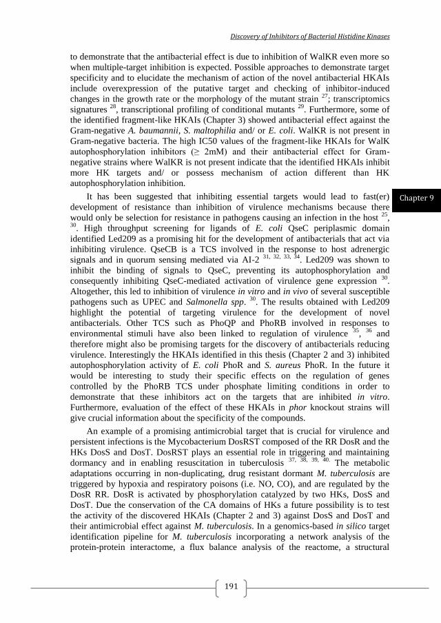
Discovery of Inhibitors of Bacterial Histidine Kinases
191
to demonstrate that the antibacterial effect is due to inhibition of WalKR even more so
when multiple-target inhibition is expected. Possible approaches to demonstrate target
specificity and to elucidate the mechanism of action of the novel antibacterial HKAIs
include overexpression of the putative target and checking of inhibitor-induced
changes in the growth rate or the morphology of the mutant strain 27
; transcriptomics
signatures 28
, transcriptional profiling of conditional mutants 29
. Furthermore, some of
the identified fragment-like HKAIs (Chapter 3) showed antibacterial effect against the
Gram-negative A. baumannii, S. maltophilia and/ or E. coli. WalKR is not present in
Gram-negative bacteria. The high IC50 values of the fragment-like HKAIs for WalK
autophosphorylation inhibitors (≥ 2mM) and their antibacterial effect for Gram-
negative strains where WalKR is not present indicate that the identified HKAIs inhibit
more HK targets and/ or possess mechanism of action different than HK
autophosphorylation inhibition.
It has been suggested that inhibiting essential targets would lead to fast(er)
development of resistance than inhibition of virulence mechanisms because there
would only be selection for resistance in pathogens causing an infection in the host 25
, 30
. High throughput screening for ligands of E. coli QseC periplasmic domain
identified Led209 as a promising hit for the development of antibacterials that act via
inhibiting virulence. QseCB is a TCS involved in the response to host adrenergic
signals and in quorum sensing mediated via AI-2 31, 32, 33, 34
. Led209 was shown to
inhibit the binding of signals to QseC, preventing its autophosphorylation and
consequently inhibiting QseC-mediated activation of virulence gene expression 30
.
Altogether, this led to inhibition of virulence in vitro and in vivo of several susceptible
pathogens such as UPEC and Salmonella spp. 30
. The results obtained with Led209
highlight the potential of targeting virulence for the development of novel
antibacterials. Other TCS such as PhoQP and PhoRB involved in responses to
environmental stimuli have also been linked to regulation of virulence 35
, 36
and
therefore might also be promising targets for the discovery of antibacterials reducing
virulence. Interestingly the HKAIs identified in this thesis (Chapter 2 and 3) inhibited
autophosphorylation activity of E. coli PhoR and S. aureus PhoR. In the future it
would be interesting to study their specific effects on the regulation of genes
controlled by the PhoRB TCS under phosphate limiting conditions in order to
demonstrate that these inhibitors act on the targets that are inhibited in vitro.
Furthermore, evaluation of the effect of these HKAIs in phor knockout strains will
give crucial information about the specificity of the compounds.
An example of a promising antimicrobial target that is crucial for virulence and
persistent infections is the Mycobacterium DosRST composed of the RR DosR and the
HKs DosS and DosT. DosRST plays an essential role in triggering and maintaining
dormancy and in enabling resuscitation in tuberculosis 37, 38, 39, 40.
The metabolic
adaptations occurring in non-duplicating, drug resistant dormant M. tuberculosis are
triggered by hypoxia and respiratory poisons (i.e. NO, CO), and are regulated by the
DosR RR. DosR is activated by phosphorylation catalyzed by two HKs, DosS and
DosT. Due the conservation of the CA domains of HKs a future possibility is to test
the activity of the discovered HKAIs (Chapter 2 and 3) against DosS and DosT and
their antimicrobial effect against M. tuberculosis. In a genomics-based in silico target
identification pipeline for M. tuberculosis incorporating a network analysis of the
protein-protein interactome, a flux balance analysis of the reactome, a structural
Chapter 9
2014
192
assessment of targetability as well as experimentally derived phenotype essentiality
data, DosS was selected as a high-confidence target for dormant mycobacteria 41
.
4. A VARIETY OF DRUG-DISCOVERY APPROACHES CAN BE APPLIED TO
TCS INHIBITORS DISCOVERY
The first published TCS inhibitors were discovered by high-throughput screenings
of natural products or chemical libraries for biochemical inhibition of the target 42
.
With the elucidation of TCS structures, structure-based virtual screenings were
employed to identify TCS inhibitors hits 43, 43, 43, 44, 45
and some of the hits have been
further improved by rational design 46, 47, 45
. Drug design strategies for identifying TCS
inhibitors with broad spectrum of inhibition as well as specific TCS inhibitors is
facilitated by the available structural and genomic information for TCS as well as the
large body of research on their role in bacterial physiology and virulence. Virtual
structure-based or in vitro fragment-based screenings with multiple targets (Chapter 2
and 3, respectively) and selecting for common hits facilitates the discovery of broad-
spectrum inhibitors, whereas selecting hits specific to particular target will facilitate
the discovery of narrow-spectrum inhibitors. In this case high-resolution structures of
TCS targets in complex with their inhibitors are still needed to facilitate the rational
design of improved inhibitors against specific TCS targets that are either essential or
controlling key virulence factors. With the advance of high-throughput crystallography 48
, 49
, 50
it seems feasible to crystallize the kinome of a bacterial pathogen or promising
HK targets from different pathogens and use the structural information to design more
potent and/or specific inhibitors following structure-based design approach.
Complementary approaches should include the structural determination of co-crystals
of putative target HK CA domains that has been previously crystallized (e.g. WalK
from B. subtilis, PDB:3SL2) with fragment libraries or known kinase inhibitors and
co-crystals with mammalian members of the GHKL protein family (e.g. Hsp90) to
facilitate structure-based design of inhibitors with improved affinity and specificity.
APPROACHES FOR TCS INHIBITORS DISCOVERY
STRUCTURE-BASED VIRTUAL SCREENINGS USING MULTIPLE HK
STRUCTURES INCREASES THE CHANCES OF IDENTIFYING HK
INHIBITORS WITH BROAD SPECTRUM OF INHIBITION
Molecular docking and receptor-based virtual screening have been an
indispensable component within structure-based drug discovery, including TCS
inhibitors discovery, for hits identification and hit-to-lead optimization 43, 51, 44, 35, 45, 52,
53, 54, 55. Although, ligand flexibility can be handled by variety of algorithms in current
docking implementations 56, 57
, receptor flexibility remains a major outstanding
challenge in the practice of molecular docking-based virtual screening 58, 59
. The
Discovery of Inhibitors of Bacterial Histidine Kinases
193
challenge is mainly because of the high dimensionality of the conformational space
and the complexity of scoring energy function. It is generally accepted protein
flexibility to be often coupled to ligand binding in numerous experimental and
theoretical studies 60, 61
. Two kinds of ligand-binding mechanisms have been well
discussed 62
. The first is conformational selection, which assumes that the ligand binds
to a pre-existing receptor conformation in an equilibrated ensemble; and the second is
induced fit, which presumes that the binding of the ligand induces conformational
changes in the receptor. In both cases, structural conformations of the receptors need
to be taken into account in molecular docking studies and, ideally, (multiple) ligand-
bound structures should be used instead of apo-structures.
Various modeling methods have been developed to account for receptor flexibility
in molecular docking and structure-based virtual screening. 58, 63
. Soft docking, which
docks ligands to a rigid receptor with a soft scoring function tolerating some steric
clashes, has been reported to be worse for identifying known ligands than the hard
scoring function when multiple receptor conformations were used. 64
. Several docking
programs limit protein flexibility to side chains by exploration of rotamer libraries and
make the problem of protein flexibility less computationally demanding 65
. However,
this approach cannot deal with backbone movement or other major structural re-
arrangements. Another implementation is docking of ligands to multiple receptor
conformations 66
, which may either be obtained experimentally by X-ray
crystallography and NMR spectroscopy or computationally by molecular dynamics,
normal-mode analysis, and other techniques.
In Chapter 2 we applied an approach combining structure-based virtual screenings
for putative ligands of three different HK X-ray structures followed by ligand-based
similarity searches with the most promising hits as query molecules. The problem of
receptor flexibility in the docking approach was addressed by using three different
receptor structures. The use of multiple structures in the docking experiments
increases the chance of finding broad-spectrum ligands because it implicitly considers
the receptors plasticity, sequence and conformation variability in the docking
approach. This approach mimics an ensemble based docking which has been shown to
improve the overall performance of virtual screening experiments 67
.
Screening by molecular docking for putative ligands of three different HKs
successfully identified two hits, A5 and A6, which inhibited the autophosphorylation
activity of multiple HKs and showed weak antibacterial effects. A5 and A6 inhibited
the autophosphorylation of three HKs not employed in the virtual screenings (i.e. E.
coli and S. aureus PhoR, and S. pneumoniae WalK). Therefore, A5 and A6 were used
as query molecules in ligand-based similarity searches (LBSS). The LBSS
successfully identified B13 as a promising lead candidate. B13 (R,S)-4-[1-Ethyl-2-(4-
fluorophenyl)butyl]phenol) inhibited the autophosphorylation of E. coli PhoR and S.
aureus PhoR with IC50 values of 16 and 212 µM, respectively (Chapter 2). The fact
that B13 inhibited the autophosphorylation of multiple HKs including targets not used
in the structure-based virtual screening supports the concept that use of multiple
structures in the virtual screening by molecular docking, facilitates the identification of
broad-spectrum inhibitors. Furthermore, even the next generation hits identified by
other methods, in this case ligand-based similarity search (LBSS), possessed broad-
spectrum inhibitory capacity. The putative broad spectrum HK autophosphorylation
Chapter 9
2014
194
inhibitory capacity of B13 was reflected in its broad-spectrum antibacterial effect
(Chapter 2). B13 showed antibacterial effect for a range of Gram-positive bacteria,
including MRSA clinical isolates. The MICs for the different Gram-positive strains
ranged between 1 and 16 µg/ml.
Antibacterials should not cause adverse effects to host cells. Furthermore, one of
the main causes of high attrition rates in drug discovery is so called adverse "off-
target" effects. Therefore, as early as possible in the developmental program the
toxicity of the hits has to be evaluated. B13 inhibited cell viability of Caco-2 cells and
macrophages with IC50 values of 29 µg/ml and > 20 µg/ml, respectively (Chapter 2
and 4). The selectivity index (IC50/MICs) > 1 indicates that B13 is a promising
candidate for hit-to-lead optimisation in the preclinical development of antibacterials
for Gram-positive bacteria. B13 showed no antibacterial effect for Gram-negative
bacteria. We hypothesized that this was most probably due to inability of B13 to cross
the outer membrane of Gram-negative bacteria and explored the possibility to use
nanoparticles for enhanced delivery to Gram-negative bacteria (Chapter 4). Indeed, as
part of a nano-formulation for delivery of antibacterials to Gram-negative bacteria 68
B13 showed antibacterial effect for the uropathogenic E. coli CFT 073 with MIC of 25
µg/ml (Chapter 4). The latter is in agreement with B13 autophosphorylation inhibitory
capacity for HKs from Gram-negative bacteria (Chapter 2). This is encouraging
because it might be possible to enhance the permeability of next generation hits based
on B13 using medicinal chemistry approaches 69
.
The other two promising hits coming from the LBSS following SBVS (Chapter
2), B7 and B14, showed lower HK autophosphorylation inhibitory capacity, weaker
antibacterial effect and stronger cytotoxicity to Caco-2 cells than B13. Nevertheless,
B7 and B14 are worth considering as starting points for the design of improved HK
inhibitors because they are predicted to interact with the ATP-lid. The ATP-lid is a
highly flexible and variable in sequence component of the ATP-binding site of HKs
and is critically involved in autophosphorylation 13
, 16
. The variability of the ATP-lid
has been proposed as a valuable feature for the design of inhibitors that are specific to
bacterial HKs with limited off-target effects on eukaryotic proteins sharing the ATP-
binding Bergerat fold (the so called GHKL proteins, including Hsp90 and MutL) 15
or
for the design of strain-specific HK inhibitors 20
.
FRAGMENT-BASED SCREENING BY DIFFERENTIAL SCANNING
FLUORIMETRY IS A POTENT TOOL FOR THE DISCOVERY OF HK
AUTOPHOSPHORYLATION INHIBITORS WITH DESIRED SPECTRUM OF
INHIBITION
So far, TCS inhibitors have been discovered mainly by high-throughput
screenings (HTS) 70, 30, 71
or by structure-based virtual screenings 43, 51, 44, 35, 45
.
Fragment-based screening (FBS) is a more recent approach and the methodology
behind it is complementary to HTS 72
, 73
. Fragments are small molecules with low
affinity (usually Kd > 0.1 mM) following the rule of three (MW < 300, ClogP < 3,
number of hydrogen bond donors and hydrogen bond acceptors < 3, number of
Discovery of Inhibitors of Bacterial Histidine Kinases
195
rotatable bonds < 3) 74, 75
. Fragments serve as starting point for the elaboration of more
potent inhibitors by rational design 76
. The main advantage of FBS over HTS is that
fragment libraries are much smaller than conventional compound libraries 77, 78, 79
. It
has been estimated that there are only ~ 107 molecules of up to 11 heavy atoms which
are also fragment-like (MW ≤ 300 Da), compared to ~ 1063
molecules of molecular
weight less than 500 Da, i.e. drug-like. This means that a library of 103 fragments
covers 0.01 % of the fragment-like chemical space compared to 10-58
% of the
chemical drug-like space.
The low molecular weight of fragments (typically ≤300Da) and their
corresponding reduced functionality implies that they will generally bind a target
protein with weak affinity (Kd > 0.1 mM), if at all. Despite their weak affinities,
fragments possess high intrinsic binding energy to overcome a large entropic barrier
upon binding 80
and often exhibit high ligand efficiency (∆G of binding per non-
hydrogen atoms) 81
. Therefore, fragments are suitable starting points for elaboration
into larger, more potent inhibitors.
Considering the advantages of FBS over HTS mentioned above, we performed
fragment-based screenings (FBS) by differential scanning fluorimetry (DSF) for
ligands of multiple HKs (Chapter 3). By choosing the hits identified with different
targets we were able to identify HKAIs that inhibited the autophosphorylation of HKs
that were not used in the screens, showing that the use of multiple targets selects for
inhibitors with broad-spectrum inhibitory capacity. The hits identified by FBS by DSF
were used as query molecules in ligand-based similarity searches (LBSS) and the
LBSS hits showed broad-spectrum antibacterial effect against multi-drug resistant
Gram-positive and Gram-negative strains (Chapter 3). Probably due to their small size,
the fragment-like HKAIs showed lower affinity to HKs (indicated by their IC50s for E.
coli PhoR autophosphorylation) and higher cytotoxicity to Caco-2 cells compared to
the hits identified by SBVS and LBSS in Chapter 2. The antibacterial activity of the
FBS hits was comparable to the less potent hits identified by SBVS and LBSS, B7 and
B14 (Chapter 2). The comparable antibacterial effect despite of the lower affinity to
HKs and the higher cytotoxicity might be related to off-target effects as these hits are
fragment-like and most likely bind also to multiple prokaryotic and eukaryotic targets.
The structural similarity with previously published TCS inhibitors (F1 and F1.6, 43, 46,
47) and the limited options for further improvement of the carbazoles from drug
discovery perspective (F2.1, F2.3 and F2.8) makes these hits unattractive for further
investigation. Nevertheless, F1.8 (4-N-(4-bromophenyl)pyrimidine-2,4-diamine)
seems to present a promising starting point for structure-based drug design of HK
inhibitors with broad-spectrum antibacterial effect. F1.8 possess weak HK
autophosphorylation inhibitory capacity, however, it shows broad-spectrum
antibacterial activity including against multi-drug Gram-negative strains. The latter
suggests that F1.8 scaffold is attractive for the development of antibacterials with
improved permeability for Gram-negative bacteria.
Chapter 9
2014
196
COMPUTER-AIDED APPROACHES CAN FACILITATE HIT IDENTIFICATION
AND OPTIMISATION PROVIDED SCREENINGS ARE PERFORMED BY
STANDARDISED METHODS
Nowadays early-stage antibacterial drug discovery is largely done by academic
organisations or SMEs. It is unlikely that such organisations would have the resources
for extensive screening 82
. However, if screening and in vitro evaluation of screening
hits are performed under standardized conditions in different laboratories and the
information is made (publicly) available different computational methods can be
exploited to speed up the discovery and optimisation of hits. These include predictive
quantitative structure-activity relationship (QSAR) 83, 84
models and ligand-efficiency
indices (LEIs) approaches 85, 86
(Chapter 5).
MOLECULAR MODELING CAN GUIDE RATIONAL OPTIMISATION OF HITS
IN THE ABSENCE OF EXPERIMENTAL TARGET-INHIBITOR STRUCTURES
The hits identified by SBVS, FBS, LBSS, QSAR and LEIs approaches (Chapter 2,
3 and 6) were co-crystallized with the CA domain of T. maritima CheA HK as a model
system (data not shown) 87
. In some cases, such as A5, B13, B14, F1, F2, F2.3, H30
the CheA-inhibitor structures, solved by X-ray crystallography, revealed electron
density in the binding site, although the structures of the HK inhibitors could not be
fully resolved, probably due to partial occupancy and/or multiple conformations.
Unfortunately, these structures could not be used to guide rational design of more
potent inhibitors following structure-based drug design approach. Therefore, the
putative binding mode of the HK inhibitors was predicted by molecular docking to the
structure of the CA domain of T. maritima HK853 88
or B. subtilis WalK 89
. The
predicted binding modes of the HKAIs (Chapter 2 and 3) resembled the binding mode
of the natural substrate ATP/ADP. The most potent inhibitors, e.g. B13, were
predicted to form contacts with conserved residues in the N-, G1- and G2-boxes in the
ATP-binding site. Some inhibitors, such as B7, were predicted to interact with the
variable ATP-lid and showed significant differences in the autophosphorylation
inhibitory capacity for different HKs. Other inhibitors predicted to interact with the
ATP-lid, e.g. F1.8 (Chapter 3), showed broad spectrum HK inhibitory activity and
antibacterial action. Although the ATP-lid is more variable compared to the remaining
parts of the ATP-binding site of the CA domains (Figure 2), it contains conserved
residues, such as Arg, Ile or Leu, and Tyr (Figure 2B). These residues are conserved
among HKs from different species or different HKs from the same bacteria (Figure 2C
and 2D). The binding modes predicted by molecular modeling can serve as starting
point for the structure-based design of more potent and specific or broad-spectrum
inhibitors with lower off-target effects by exploring the features of the ATP-lid.
Discovery of Inhibitors of Bacterial Histidine Kinases
197
ANTIMICROBIAL PEPTIDES AND TWO-COMPONENT SYSTEMS
INHIBITION
Eukaryotic cationic antimicrobial peptides (AMPs) are produced at sites of
infection or inflammation in many different organisms 90, 91, 92
. Usually, AMPs
increase the permeability of the bacterial membrane, leading to loss of pH gradient and
cell death. Some AMPs have been shown to interact with intracellular targets 93, 94
but
in most cases the exact mechanism of action of intracellular AMPs remains unclear.
INHIBITION OF T. MARITIMA HK853-RR468 BY LACTOFERRICIN B-
DERIVED PEPTIDES IS NOT SUITABLE FOR STRUCTURAL STUDIES ON
TCS INHIBITION BY AMPS
LactoferricinB (LfcinB) is an AMP with broad-spectrum antibacterial activity 95
, 96
. It has been shown by in vitro and in vivo experiments and bioinformatics methods
that LfcinB inhibits the phosphorylation of the RRs (BasR and CreB) by their cognate
histidine kinases (HKs), BasS and CreC 97
. We showed that LfcinB-derived peptides
inhibit the phosphotransfer and phosphatase activity of T. maritima HK853-RR468
(Chapter 6). However, the LfcinB-derived peptides seemed to induce aggregation of T.
maritima HK853 and E. coli PhoB. Therefore, T. maritima HK853-RR468 and E. coli
PhoRB might not be suitable for structural studies of TCS inhibition by LfcinB-
derived peptides. Furthermore, the described results taken together with the broad-
spectrum of antibacterial activity of LfcinB point to the possibility that the
antibacterial effect caused by LfcinB and LfcinB-derived peptides might be attributed
to non-specific protein aggregation and might act on multiple cellular targets after
entering into the cell. On this basis further work on LfcinB-derived peptide inhibitors
was abandoned.
SYNERGY EFFECT BETWEEN ANTIMICROBIAL PEPTIDES AND TCS
INHIBITORS
Antimicrobial peptides have been proposed as a way to overcome the lower
permeability of Gram-negatives cell envelope 98, 99
. Therefore, we tested the
synergistic effect of three frog AMPs with antibacterial effect against Gram-negative
bacteria and B13, as the most potent HK inhibitor described in this thesis. The AMPs
tested were [E4k]alyteserin-1c, [E6K]hymenochirin-1B and [K20K27]esculentin-2Cha 100, 101
. Unfortunately, no synergistic antibacterial effect against E. coli CFT 073 was
observed (data not shown). ε-Poly-L-lysine (ePLL) is a homo-polypeptide with
demonstrated antibacterial effect 102
. Similar to the frog AMPs, ePLL showed no
synergistic effect with HK inhibitors on antibacterial activity against E. coli CFT073
(Chapter 4). The MICs against E. coli CFT073 of the tested frog AMPs and ePLL
Chapter 9
2014
198
were ≥ 63 µg/ml. Combinations of more potent AMPs and/or more potent HKAIs
might be required to achieve synergistic effect and ultimately to observe antibacterial
effect against Gram-negative pathogens.
HK INHIBITOR SPECTRUM CAN BE BROADENED AND THE
CYTOTOXICITY REDUCED VIA THE USE OF NANOTECHNOLOGY
Although the best hits described in Chapter 2 and 3 have weak or no activity
(MICs in the range of 125-500 µg/ml) against Gram-negative pathogens, they do
inhibit autophosphorylation of HKs from Gram-negative bacteria suggesting
permeability of the Gram-negative cell envelope is limiting their anti-bacterial effects.
This assumption was shown to be correct using histidine kinase autophosphorylation
inhibitors (HKAIs) loaded mesoporous silica-based nanoparticles capped with ε-poly-
L-lysine as a strategy for effective drug delivery (Chapter 4) 68
. Whereas the
nanoparticles themselves had no antibacterial activity, the MICs of HKAIs combined
with nanoparticles was decreased more than 10-fold (Chapter 4). The free HKAIs
showed relatively low toxicity to human cells and even lower toxicity when combined
with nanoparticles. Taken together these data indicate high potential for developing a
new structural class of antibacterial lead compounds with activity against multiple
targets in several MDR Gram-positive and Gram-negative pathogens.
COMPARISON OF THE NOVEL HK INHIBITORS WITH OTHER
PUBLISHED TCS INHIBITORS
Structure-based virtual screenings for S. epidermidis WalK ligands yielded
inhibitors which have been subsequently further optimized by rational design 43, 46, 47
.
The last generation of S. epidirimids WalK inhibitors had MICs for S. epidermidis and
S. aureus lower than 3.1 µM, corresponding to lower than 1.66 µg/ml 47
. B13, the most
potent hit described in this thesis (Chapter 2), possess MICs for S. epidermidis
laboratory strains and clinical isolates in the range of 1 to 8 µg/ml. B13 MICs for
reference strains of S. aureus and clinical isolates of MRSA are in the range of from 8
to 16 µg/ml (Chapter 2). Therefore, in terms of antibacterial activity B13 is
comparable to the last generation of published S. epidermidis WalK inhibitors. S.
epidermidis WalK inhibitors inhibited S. epidermidis WalK autophosphorylation with
IC50 values in the range of 24.2 to 71.2 µM 47
. B13 has not been tested for its
autophosphorylation inhibitory capacity for any WalK, however, it inhibits E. coli
PhoR and S. aureus PhoR with IC50 of 16 and 212 µM, respectively (Chapter 2).
Considering the differences in the experiments used to determine the IC50s of the S.
epidermidis WalK inhibitors and B13 and the fact that different HKs Km differ in the
range 2 and 200 µM 103
, it is difficult to conclude how B13 compares to the last
generation of published S. epidermidis WalK inhibitors. The cytotoxicity of the S.
Discovery of Inhibitors of Bacterial Histidine Kinases
199
epidermidis WalK inhibitors was evaluated with Vero Cells using the MTT assay and
the IC50 were higher than 100 µg/ml 47
corresponding to > 60 selectivity index
(IC50viability/MIC). B13 cytotoxicty was evaluated with Caco-2 cells using the
neutral-red uptake assay (Chapter 2) and with macrophages in cell culture (Raw cells)
using the XTT assay (Chapter 4) and the IC50 were 29 µg/ml and ≤ 20 µg/ml,
respectively, corresponding to selectivity index ≥ 1.25 for the different S. epidermidis
and S. aureus strains tested (Chapter 2). Even though the cell viability was evaluated
using different cell lines and methods, it is clear that B13 selectivity is worse than the
selectivity of the last generation published S. epidermidis WalK inhibitors and needs to
be improved. However, B13 (MW 272.4) is a relatively smaller compound compared
to the last generation S. epidermidis WalK inhibitors (MW between 498.02 and
534.02) implying that it possesses higher ligand efficiency and more possibilities for
further improvements using an iterative structure-based drug design approach.
FUTURE CHALLENGES AND PERSPECTIVES OF THE DISCOVERY OF TCS
INHIBITORS
Both SBVS and FBS seem to be promising tools for the discovery of TCS
inhibitors. The application of multidisciplinary screening approach (including SBVS,
FBD, and LBSS) allowed us to identify HK inhibitors with activity against multiple
HKs and a broad-spectrum antibacterial effect. Furthermore, the identified inhibitors
are predicted to interact with different parts of the ATP-binding sites. The latter
together with the relatively small size of the identified HK inhibitors (MW < 300)
provides the possibility to design improved inhibitors by combining chemical moieties
from the different hits to generate inhibitors with stronger affinity and higher
specificity to bacterial HKs.
Although a very promising starting point for development of novel antibacterials,
the identified HKAIs have a number of shortcomings that need to be addressed in the
hit-to-lead optimisation process:
- The affinity to model HKs such as T. maritima CheA is most likely not high
enough and/ or multiple conformations of the inhibitors are possible and therefore, the
CheA-inhibitors structures cannot be resolved and used for structure-based drug
design. This liability can be addressed by discovering more potent HK
autophosphorylation inhibitors and/ or by solving the structures of the discovered
inhibitors with other putative target HKs, such as the recently published B. subtilis and
S. mutans WalK (PDB: 3SL2 and 4I5S) or E. coli/T. maritima EnvZ (PDB: 4KP4) 89,
104.
- The identified HKs inhibitors are not sufficiently permeable to Gram-
negative bacteria. This can be overcome by using nanoparticles (Chapter 4). The latter
however, raises additional issues related to the following steps of the drug discovery
process related to the safety of such complex therapeutic agents. Therefore, it is
desirable to apply medicinal chemistry approaches to design inhibitors with improved
permeability for Gram-negatives 69
. Addition of functional groups to the identified
Chapter 9

2014
200
HKAIs, given their small size, could increases the permeability in Gram-negatives.
Nevertheless, increasing the specificity of the identified inhibitors to Gram-positive
targets such as the obligatory essential WalKR can be a promising strategy to develop
narrow-spectrum antibacterials against Gram-positive bacterial infections.
- The identified HKAIs showed some cytotoxicity to Caco-2 cells evaluated by
neutral-red uptake assays (Chapter 2 and 3) and B13 showed cytotoxic effect to
macrophages (Raw cells) evaluated by the XTT assay (Chapter 4). The most
promising and most potent HK inhibitor B13 possesses a selectivity index for Gram
positives (IC50 viability/ MICs) ≥ 1.25. Increasing the potency and the selectivity of
the HK inhibitors will most likely lead to decrease in their cytotoxicity. To facilitate
the discovery of more selective HK inhibitors, the next generation of HK inhibitors
should be tested also for their inhibitory activity on the structurally closely related
mammalian members of the GHKL superfamily 15, 105
.
In summary, following structure-based and ligand-based virtual screenings and
fragment-based in vitro screenings, we have successfully identified HK
autophosphorylation inhibitors (HKAIs) that can serve as a starting point for the
design of HKAIs with improved target affinity and selectivity, which would lead to
stronger antibacterial effect and lower toxicity.

Discovery of Inhibitors of Bacterial Histidine Kinases
201
SUMMARY

2014
202

Discovery of Inhibitors of Bacterial Histidine Kinases
203
The growing problem of bacterial multi-drug resistance requires the urgent
establishment of pipelines for discovery of novel antibacterials. Two-component
systems (TCS) have been proposed for years as promising novel antibacterial targets.
Inhibitors of TCS, especially autophosphorylation inhibitors targeted at the CA
domains of multiple histidine kinases (HKs) are expected to show broad-spectrum of
antibacterial activity and low toxicity to host cells.
Using structure-based virtual screenings (SBVS) for putative ligands of three
different HKs followed by ligand–based similarity search with the most promising
SBVS hits (Chapter 2) we identified HK autophosphorylation inhibitors of multiple
HKs from both Gram-positive and Gram-negative bacteria. The inhibitors showed
broad-spectrum antibacterial activity for Gram-positive bacteria, including clinical
isolates of MRSA strains resistant to a wide range of β-lactam and non-β-lactam
antibiotics. The most promising hit, B13, showed relatively low cytotoxicity and
hemolytic activity and therefore presents a promising starting point for hit-to-lead
optimisation using a structure-based drug design approach. The predicted binding
modes of the other less potent hits, B7 and B14, point to possibilities to design more
potent and selective inhibitors by combining chemical moieties belonging to the
different hits.
In Chapter 3 fragment-based in vitro screenings (FBS) by differential scanning
fluorimetry (DSF) were performed for ligands of two different HKs followed by
ligand-based similarity searches with the two identified hits. Weak HK
autophosphorylation inhibitors with broad spectrum of antibacterial activity were
identified. Compounds F1.8 and F2.3 showed antibacterial effect for both Gram-
positive and Gram-negative strains, including clinical isolates of MRSA and multi-
drug resistant Acinetobacter baumannii and Stenotrophomonas maltophilia. The
identified hits present a promising starting point for the design of HK
autophosphorylation inhibitors with improved permeability.
In Chapter 5 a computer-aided approach was used in an attempt to identify more
promising hits for HK autophosphorylation inhibitors using
Nanoparticles delivery was employed to show that the most potent HK inhibitors,
e.g. B13, possess antibacterial effect against Gram-negative bacteria (Chapter 4).
Mesoporous silica-based nanoparticles loaded with HK autophosphorylation inhibitors
showed 10-fold lower MICs compared to the free HK autophosphorylation inhibitors
for UPEC E. coli CFT 073 and Serratia marcescens. Despite the lower MICs of HK
autophosphorylation inhibitors loaded in nanoparticles for bacteria they were less
cytotoxic than the free inhibitors at equivalent concentrations
Lactoferricin (LfcinB) has been shown to inhibit TCS from E. coli. We
demonstrated that LfcinB-derived peptides interact with T. maritima HK853-RR468
and E. coli PhoRB however, the mechanism of interaction was most likely non-
specific and due to protein aggregation. (Chapter 6). Therefore efforts to co-crystalize
these peptides with TCS for the rational structure-based drug design of improved
inhibitors was not pursued further.
Co-crystallization of the HK autophosphorylation inhibitors with T. maritima
CheA is described in Chapter 7.1. Furthermore, the purification, crystallization and

2014
204
preliminary X-ray analysis of the periplasmic domain of CopM, a protein suggested to
be involved in copper resistance, is presented in Chapter 7.2.
Chapter 8 is a discussion on the possibility to develop novel antibacterials with
low potential for resistance development by targeting TCS signalling in general and
the essential WalKR in particular.
Chapter 9 is a discussion of the main results of this thesis in the context of
current literature on antibacterial drug discovery and published research on discovery
of TCS inhibitors.

Discovery of Inhibitors of Bacterial Histidine Kinases
205
SAMENVATTING

2014
206
Discovery of Inhibitors of Bacterial Histidine Kinases
207
Het toenemende probleem van bacteriële multidrug resistentie vereist het vlotte
opzetten van onderzoeks-structuren om nieuwe antibiotica te ontdekken. Enkele jaren
geleden werd voorgesteld dat twee-component systemen (TCSen) veelbelovende
nieuwe antibacteriële doelwitten zouden kunnen vormen. Het is de verwachting dat
remmers van TCSen, vooral autofosforylatie remmers gericht op de zogenoemde "CA"
domeinen van histidine kinases (HKs), een breed spectrum van antibacteriële activiteit
en lage toxiciteit voor gastheercellen zullen vertonen.
Met behulp van structuur-gebaseerde virtuele screenings (SBVS) voor vermeende
liganden van drie verschillende HKs, gevolgd door zoeken naar liganden die het
sterkst lijken op de meest veelbelovende SBVS treffers (Hoofdstuk 2) identificeerden
we HK autofosforylatie remmers van HKs van zowel Gram-positieve als Gram-
negatieve bacteriën. De remmers toonden breed-spectrum antibacteriële activiteit ten
aanzien van Gram-positieve bacteriën, inclusief klinische isolaten van MRSA-
stammen met resistentie tegen diverse β-lactam en niet-β-lactam antibiotica. De meest
veelbelovende treffer, B13, liet een relatief lage cytotoxiciteit en hemolytische
activiteit zien en lijkt daarom een veelbelovend uitgangspunt voor de zogenoemde
"hit-to-lead" optimalisatie waarbij een structuur-gebaseerde drug ontwerp aanpak
wordt gevolgd. De voorspelde bindingscapaciteit van twee minder krachtige treffers,
B7 en B14, biedt mogelijkheden om krachtiger en selectievere remmers te ontwerpen,
door het combineren van chemische groepen die behoren tot de verschillende treffers.
In hoofdstuk 3 is beschreven hoe fragment-gebaseerde in vitro screenings (FBS)
door differentiële scanning fluorimetrie (DSF) werden uitgevoerd om liganden van
twee verschillende HKs te zoeken, gevolgd door het zoeken naar chemische
verbindingen die een ligand-gebaseerde gelijkenis vertonen, met als uitgangspunt de
twee in hoofdstuk 2 geïdentificeerde treffers. Zwakke HK autofosforylatie remmers
met een breed spectrum van antibacteriële activiteit werden gevonden. De
verbindingen F1.8 en F2.3 toonden antibacteriële werking tegen zowel Gram-positieve
als Gram-negatieve stammen, met inbegrip van klinische isolaten van MRSA en
multiresistente Acinetobacter baumannii en Stenotrophomonas maltophilia. De
geïdentificeerde treffers bieden een veelbelovend uitgangspunt voor het ontwerp van
HK autofosforylatie remmers met verbeterde doorlaatbaarheid voor bacterie
membranen.
In hoofdstuk 5 werd een computer-gebaseerde benadering gebruikt in een poging
om meer veelbelovende treffers van HK autofosforylatie remmers te vinden.
Nanodeeltjes werden gebruikt om te laten zien dat de meest potente HK-remmers,
bijvoorbeeld B13, een antibacteriële werking hebben tegen Gram-negatieve bacteriën
(Hoofdstuk 4). Mesoporeuze silica-gebaseerde nanodeeltje gevuld met HK
autofosforylatie remmers toonden 10-voudig lagere MICs in vergelijking met de MIC
van de pure HK autofosforylatie remmers; deze remmers hadden aantoonbare activiteit
tegen UPEC E. coli CFT 073 en Serratia marcescens. Ondanks het feit dat de in
nanodeeltjes verpakte HK autofosforylatie remmers een lagere MIC hadden wanneer
toegepast om bacteriën te bestrijden, waren deze verpakte remmers minder cytotoxisch
dan de pure remmers bij dezelfde concentraties.
Het is aangetoond dat Lactoferricine (LfcinB) TCSen uit E. coli kan remmen. We
toonden aan dat van LfcinB afgeleide peptides activiteit bezitten tegen T. maritima
HK853-RR468 en E. coli PhoRB, en dat het mechanisme van interactie
2014
208
hoogstwaarschijnlijk niet specifiek was maar werd bepaald door eiwit aggregatie
(Hoofdstuk 6). Pogingen om deze peptiden samen te kristalliseren met TCSen om
betere remmers te vinden via een rationele structuur-gebaseerde drug ontwerp
benadering werden daarom niet doorgezet.
Het tegelijk kristalliseren van HK autofosforylatie remmers met het T. maritima
CheA TCS wordt beschreven in hoofdstuk 7.1. In hoofdstuk 7.2 worden eveneens de
zuivering, kristallisatie en voorlopige röntgenanalyse van het periplasmatische domein
van CopM, een eiwit dat gedacht wordt betrokken te zijn bij koper resistentie,
beschreven.
Hoofdstuk 8 presenteert een discussie over de mogelijkheid om nieuwe
antibacteriële middelen met een lage kans op ontwikkeling van resistentie te
ontwikkelen, door middelen te ontwikkelen die het doorgeven van cellulaire signalen
via TCSen in het algemeen, en in het bijzonder de essentiële WalKR, blokkeren.
In hoofdstuk 9 worden de belangrijkste resultaten van dit proefschrift en hun
inbedding in de huidige literatuur over antibacteriële drug ontdekking in het algemeen,
en de specifiekere literatuur over de ontdekking van TCS-remmers, bediscussiëerd.
Translated from English to Dutch by Peter van Baarlen
Discovery of Inhibitors of Bacterial Histidine Kinases
209
ACKNOWLEDGEMENTS

2014
210
Discovery of Inhibitors of Bacterial Histidine Kinases
211
Those who know me well/ long enough are aware of my difficulties in expressing positive thoughts especially gratitude, but I will do my best to thank the people who have helped me reach the final stage of my formal education (at least in Biology), and who have been an inspiration and example to follow.
Obtaining a PhD degree takes around four years, ´´a moment´´, and the ´´journey´´ to starting a graduate programme takes at least six times longer. First , I would like to thank the people involved in the ´´journey´´ in retrospective order:
- My family for letting me grow independent, taking my own decisions and for their support in times when it was needed.
- The dentist who took the initiative, responsibility and the risk in probably my first fight with multi-drug resistant bacteria when I was 2-3-years-old.
- My primary school teacher Neli Koshnicharova for holding my hand to my first competition in Mathematics in my future high school.
- All the amazing people who still work in this high school for their extraordinary professionalism. Despite minor distractions such as electricity and water regimens, no fuel for public transport and heating in the winter, wars in neighbouring countries etc. they managed to keep us excited and curious about life and science, and motivated to make a diffirence . Special gratitudes to Elena Stancheva, Mima Ilieva, Penka Nikolova, Miroslava Georgieva, Elena Yordanova-Kostadinova, and Antoaneta Velikova.
- To my classmates whose success in their personal and professional life makes me proud and keeps me optimistic. Especially to Peter, Ayshe and Marina.
- My teacher and friend Nikolina Stoyanova. Unfortunately, I still sound bored during presentations.
- Lyubo, whenever I feel down I know I can count on you for a ridiculous remark full of wisdom.
- Iliana Ivanova for being an example of a dedicated, positive, optimistic and down-to-earth researcher and teacher.
- My classmates and friends in the Faculty of Biology for the parties, wisdom and talks and walks, hiking in Vitosha, the tripe soup , the ´´conference calls´´, trainings in the gym etc. crazy memories. Especially to Nadezhda, Velmira, Sylvia, Apo, and Niki.
- Eugene Semenov (RIP) for the great opportunity to work on an international project already as a BSc student and for sharing his wisdom. I try to always remember that ´´All people are nice, but sometimes it is difficult to work with them.´´
- My colleagues at the Molecular Neurobiology, and especially Shazie, for the friendly and relaxed atmosphere. Working in the lab and chilling can be the same action.
- My classmates and friends at WUR, Christina, Agata, Christos, Man, and George for making the bad weather in the Netherlands seem ´´not so bad´´.
- All the Bulgarians in Wageningen, especially Nikolay P., Daniela and Slav, Radostina and her mother, and Denica B.
- All the people I have met at IC, especially Michiel, Mensur, Jessin, Hida, Remko, and the wild dancer Alexander.
2014
212
- The people at the Toxicology Department and at RIKILT, especially Si, Alexandros, Ana, and Jaime. Laura, Jac, Ivonne, Ad, Toine, and Tinca you gave me an opportunity but apparently I was not ready for it and now I am extremely grateful for helping me take the right decision.
- Prof. Iliana Ionkova for confirming what I already knew. - Nikolay R. for the countless hours of skype and phone calls, the helpful
distraction, moral support, the nice presents and the life lessons. And now the people involved in the ´´moment´´ called PhD studies: - Alberto for the coolest job interview I have ever had/ will have. For being
a great example of a dedicated, passionate, focused structural biologist (… however, sometimes too focused), and relatively patient and fair group leader, flexible about rules and deadlines. I have learned (some) Structural Biology in your group and hopefully one day I will publish a protein structure, or even better protein-inhibitor structure. And furthermore, everytime I feel upset and dissatisfied I try to remember that ´´There are plenty of people in the world who dream to have my problems.´´ and sometimes it helps.
- Paqui for being an inspiring example of a structured, organized, efficient, calm, open-minded, confident and friendly female scientist from a country where women had no rights up until 40 years ago.
- My colleagues at UCM-IBV, for forcing me to learn Spanish and for training my patience, which in long-term is probably more useful than any lab technique I could have learnt from them.
- El president de la CdF and the others responsible for the party atmosphere at IBV. I regret I haven´t enjoyed more.
- InhibOx for hosting my first secondment and for being involved in the project. As a frustrated, unlucky crystallographer, without your contribution I would have had a much shorter thesis.
- Especially to Simone who did all the scripting and computational experiments. She also taught me how to do some experiments myself, and to me she is an example of a professional, organized, reliable, and very helpful person. Although this project was not exactly what she was meant to work on, she was sincerely interested in the progress, provided me with moral support and advice in times of disappointment and frustration, and I hope we´ll stay in touch and remain friends.
- Prof. Conlon, Milena, Manju, Akela, Agnes Sonnevend and Tibor Pal, and everyone else at UAEU, Al Ain, for the productive, exotic and exciting secondment!
- Ana and Marco for the MICs and MBCs with S. pneumoniae. - Nuria, Ramon and anyone else from UPV for synthesizing the
nanoparticles. Your collaboration made the coolest and the fastest chapter of my thesis! Thank you!
- My Bulgarian friends in Valencia, Associacio L´orneta blanca, Daniel, Pavel, for helping me feel at home and stimulating me to keep in shape.
- Everyone in the STARS ITN for the nice times during our ´´paid holidays´´.
Discovery of Inhibitors of Bacterial Histidine Kinases
213
- Everyone at the Host-microbe Interactomics group at WUR for the friendly and happy atmosphere. Nico, Ellen, Edo and his students for their contribution to my coolest chapter, I promise to publish it asap. Loes and Mayo for their help on administrative, printing, publishing etc issues, and for their positivism. Aga for writing together a viewpoint and a review before having solid results. Rogier for his help, I can´t remember a particular example right now, but I´ve bothered him few times. Peter for his thorough proof-reading of the viewpoint, the review and translation of the summary.
- Jerry for letting me and helping me finish my PhD studies on time at HMI, for his ´´contagious´´ multidisciplinary scientific curiosity and enthusiasm to make it possible to continue working on TCS inhibitors discovery and development, and for creating an atmosphere where you see everything as easy and feasible. Unfortunately, in the 6-7 months at HMI I couldn´t accomplish all ideas you´ve given me during our meetings but hopefully in future I can do more.
3 hours to thank people I have met for 30 years … I thank also everyone else who is not mentioned due to dehydration, my short memory or ´´blindness´´ to their impact, and last but not least laziness to read the text again …

2014
214
Discovery of Inhibitors of Bacterial Histidine Kinases
215
PERSONALIA
CURRICULUM VITAE
LIST OF PUBLICATIONS

2014
216
Discovery of Inhibitors of Bacterial Histidine Kinases
217
Nadya Velikova (Надя Великова) was born on 17th
of January 1984 in Burgas, Bulgaria. She finised her
secondary school education in the High School for
Mathematics and Natural Sciences´´Akad. Nikola
Obreshkov´´, Burgas, Bulgaria, in 2002. In the same
year she was among the winners of the National
Olimpiad in Biology, the winner of the National Contest
for High School Students Projects in Ecology and
Environmental protection and participiant in the finals of
the National Competition in Chemistry. The first granted
her admission to the Bachelor of Science programme in
Molecular Biology at the Faculty of Biology, Sofia
University ¨St. Kliment Ohridski¨, Sofia, Bulgaria. In
parallel to her BSc studies she worked in Eugene
Semeonov´s group at the Department of Molecular Neurobiology, Institute of
Molecular Biology, Bulgarian Academy of Sciences, on ivermectin resistance. After
finishing her BSc studies in 2007, Nadya continued her education in the Master of
Science programme in Medical Biotechnology at Wageningen University, The
Netherlands. She did a MSs thesis at the Department of Toxicology under the
supervision of Dr. ir. Jac Aarts, and an internship at the Toxicology and Bioassays
Business Unit at RIKILT-Instite of Food Safety, under the supervision of Dr. ir. Toine
Bovee. Both the the thesis and the internship were related to design, development and
implementation of reporter-gene assays for nuclear-hormone receptors ligands. In
2010 In 2010, Nadya continued her education and research experience in Dr. Alberto
Marina´s group in the Institute of Biomedicine of Valencia, Spanish National Research
Council, Valencia, Spain, working on the structure-based discovery of two-component
system inhibitors as part of the EU FP7-funded Marie Curie ITN STARS – Scientific
Training in Antimicrobial Research Strategies. The project included secondments at
InhibOx, Oxford, United Kingdom, and at the Departmentment of Biochemistry,
United Arab Emirates University, Al Ain, United Arab Emirates. In 2014 Nadya
continued her PhD studies as an external PhD candidate at the Host-Microbe
Interactomics Group at Wageningen University under the supervision of Prof. dr. Jerry
Wells.
2014
218
PUBLICATIONS
PUBLICATIONS RELATED TO THE CURRENT THESIS Velikova, N.; Bem, A. E.; van Baarlen, P.; Wells, J. M.; Marina, A., WalK, the Path
towards New Antibacterials with Low Potential for Resistance Development. ACS
Medicinal Chemistry Letters 2013, 4 (10), 891-894.(viewpoint)
Velikova, N., Fulle, S., Manso, A.S., Mechkarska, M., Finn, P., Conlon, M.J.,
Oggioni, M.R., Wells, J.M., Marina, A., Identification of putative fragment-like
histidine-kinase inhibitors active against multi-drug resistant bacterial strains,
submitted to the European Journal of Medicinal Chemistry
Bem, A., Velikova, N., Pellicer, T., van Baarlen, P., Marina, A., Wells, J.M., Bacterial
Histidine Kinases as Novel Antibacterial Drug Targets, submitted to ACS Chemical
Biology (review)
Velikova, N., Fulle, S., Manso, A.S., Mechkarska, M., Conlon, M.J., Wells, J.M.,
Oggioni, M.R., Finn, P., Marina, A., Discovery of bacterial histidine kinase inhibitors
with antibacterial activity against clinical isolates of MRSA and and Staphylococcus
epidermidis, in preparation
Velikova, N., Mas, N., Stolte, E., Zaccaria, E., Cao, R., Taverne, N., Murgina, J.R.,
Martinez-Manez, R., Marina, A., Wells, J.M., Broadening the Antibacterial Spectrum
of Histidine-kinase Autophosphorylation Inhibitors via the use of ε-poly-L-lysine
Capped Mesoporous Silica-based Nanoparticles, in preparation
Velikova, N., Marina, A., Purification, Crystallization and Preliminary X-ray
Diffraction Analysis of Syneccocystis spp. CopM, in preparation
OTHER PUBLICATIONS Loonen, L.M.P., Taverne, N., Velikova, N., Stolte, E.H., Marina, A., Dekker, J.,
Wells, J.M., Purification and characterization of recombinant mouse RegIII proteins,
thesis chapter, Wageningen University, ISBN 987-94-6173-672-7, 2013
Sotoca, A.M., Bovee, T.F.H., Brand, W., Velikova, N., Boeren, S.,Vervoort, J.,
Rietjens, I.M.C.M., Superinduction of estrogen receptor mediated gene expression in
luciferase based reporter gene assays is mediated by a post-transcriptional mechanism,
The Journal of Steroid Biochemistry and Molecular Biology, Volume 122, Issue 4,
October 2010, Pages 204-211, ISSN 0960-0760, 10.1016/j.jsbmb.2010.06.010.
Yusein, S., Velikova, N., Kupenova, R., Hardie, R., Wolstenholme, A., Semenov, E.,
Altered ivermectin pharmacology and defective visual system in Drosophila mutants
for histamine receptor HCLB, Invertebrate Neurosciences 2008 Dec; 8(4):211-22. doi:
10.1007/s10158-008-0078-2

Discovery of Inhibitors of Bacterial Histidine Kinases
219
The research described in this thesis was financially supported by the European Union
Framework Programme 7-funded Marie Curie Initial Training Nework STARS
(Contract No. PITN-GA-2009-238490)
Cover design: Nadya Velikova
Layout: Nadya Velikova
Printed by: GVO drukkers & vormgevers B.V. Ponsen & Looijen