megasporogenesis - wur edepot
TRANSCRIPT

581.331.1
MEDEDELINGEN LANDBOUWHOGESCHOOL WAGENINGEN • NEDERLAND • 78-16 (1978)
MEGASPOROGENESIS
A comparative study of the ultrastructural aspects of megasporogenesis
in Lilium, Allium and Impatiens
(with a summary in Dutch)
M.J. DE BOER-DE JEU
Department of Botany, Agricultural University, Wageningen, The Netherlands
(received 12-V-78)
H. VEENMAN & ZONEN B.V. - WAGENINGEN - 1978
Mededelingen Landbouwhogeschool Wageningen 78-16 (1978)
(Communications Agricultural University) is also published as a thesis
CONTENTS
INTRODUCTION 1 1.1. General introduction 1 1.2. Research object 2
APPLIED METHODS 3 2.1. Light-microscopic (LM) methods 3 2.1.1. HERR's clearing-squash technics 3 2.1.2. Preparation of 3 pm thick sections 3 2.1.3. Callose-checking method 3 2.2. Electron-microscopic (EM) methods 4 2.2.1. Glutaraldehyde-osmiumtetroxyde fixation 4 2.2.2. Potassiumpermanganate fixation 4 2.2.3. Re-embedding method 5 2.2.4. THIÉRY-test 6 2.2.5. Quantitative methods 6
MEGASPOROGENESIS AND EARLY MEGAGAMETOGENESIS IN THE LILIUM HYBRID 'ENCHANTMENT" 8 3.1. Introduction 8 3.2. Material and methods 10 3.3. Results and discussion 10 3.3.1. Light-microscopy 10 3.3.1.1. Ovules 10 3.3.1.2. From megasporocyte to four-nucleate embryosac 12 3.3.1.3. The position of the nucleus 13 3.3.2. Electron-microscopy 15 3.3.2.1. Nucleus 17 3.3.2.2. Microtubules 25 3.3.2.3. Cytoplasmic ribosomes 25 3.3.2.4. Endoplasmic reticulum (ER) 28 3.3.2.5. Dictyosomes 32 3.3.2.6. Vesicle- and vacuolar system 32 3.3.2.7. Plasma membrane 34 3.3.2.8. Mitochondria 36 3.3.2.9. Plastids 37 3.3.2.10. Lipid bodies 39 3.3.2.11. Plasmodesmata . . . 40 3.3.2.12. Cell-wall 40 3.3.2.13. Nucelluscell 41 3.4. Final discussion 42
MEGASPOROGENESIS AND EARLY MEGAGAMETOGENESIS IN ALLIUM CEP A L 44 4.1. Introduction 44 4.2. Material and methods 44 4.3. Results and discussion 45 4.3.1. Light-microscopy 45 4.3.2. Electron-microscopy 45 4.3.2.1. Nucleus 45 4.3.2.2. Microtubules 52
4.3.2.3. Cytoplasmic ribosomes 52 4.3.2.4. Endoplasmic reticulum 53 4.3.2.5. Dictyosomes 53 4.3.2.6. Vesicle- and vacuolar system 55 4.3.2.7. Plasma membrane 56 4.3.2.8. Mitochondria 56 4.3.2.9. Plastids 58 4.3.2.10. Lipid bodies 59 4.3.2.11. Plasmodesmata 59 4.3.2.12. Cell-wall 59 4.3.2.13. Degeneration of the micropylar dyad cell 62 4.3.2.14. Nucelluscell 65 4.4. Final discussion 66
MEGASPOROGENESIS AND EARLY MEGAGAMETOGENESIS IN IMPATIENS WALLER1ANA HOOK. F. 69 5.1. Introduction 69 5.2. Material and methods 69 5.3. Results and discussion 69 5.3.1. Light-microscopy 69 5.3.2. Electron-microscopy 71 5.3.2.1. Nucleus 73 5.3.2.2. Microtubules 79 5.3.2.3. Cytoplasmic ribosomes 80 5.3.2.4. Endoplasmic reticulum 80 5.3.2.5. Dictyosomes 83 5.3.2.6. Vesicle-and vacuolar system 84 5.3.2.7. Plasma membrane 85 5.3.2.8. Mitochondria 85 5.3.2.9. Plastids 86 5.3.2.10. Lipid bodies 86 5.3.2.11. Plasmodesmata 87 5.3.2.12. Cell-wall 87 5.3.2.13. Degeneration of the micropylar dyad cell 88 5.3.2.14. Nucelluscell 89 5.4. Final discussion 90
SIMILARITIES AND DIFFERENCES BETWEEN THE THREE SPECIES DURING MEGASPOROGENESIS ON FIXATION-LEVEL 92 6.1. Fixation methods 92 6.2. Cell organelle level 92 6.2.1. Results 92 6.2.2. Discussion 95 6.2.2.1. Nuclear undulations and nuclear pores 95 6.2.2.2. The sacculation of the inner nuclear membrane 96 6.2.2.3. Chromatin structure 98 6.2.2.4. Membrane-like structures 98 6.2.2.5. Nucleolus 99 6.2.2.6. Ribosomes 100 6.2.2.7. Dictyosomes 101 6.2.2.8. Electron-dense vacuoles, clear vacuoles amd paramural bodies . . . . 102 6.2.2.9. Mitochondria 103 6.2.2.10. Plastids 103 6.2.2.11. Lipid bodies 104 6.2.2.12. Plasmodesmata and cell-wall 104

6.3. The organization of the cell 105 6.3.1. Results 105 6.3.2. Discussion 107 6.3.2.1. Classification 107 6.3.2.2. The polar distribution of cell organelles 107 6.3.2.3. The polar distribution of plastids, dictyosomes, endoplasmic reticulum
(ER), plasmodesmata and nuclei 108 6.3.2.4. The partial presence of callosic components in the cell-wall of the megaspo-
re mother-cell (MMC) 109 6.3.2.5. The relation between nucellus and megaspore mother-cell 109
7. MEGASPOROGENESIS AS A DEVELOPMENTAL PROCESS I l l 7.1. Introduction I l l 7.2. Meiosis I l l 7.3 Megasporogenesis I l l 7.3.1. Characteristics of the process of megasporogenesis 112 7.3.2. Species-specific characteristics 113 7.3.3. Developmental type-specific characteristics 113 7.4 Microsporogenesis and megasporogenesis 114
8. SUMMARY 115
9. ACKNOWLEDGEMENTS 118
10. SAMENVATTING 119
11. REFERENCES 122
ABBREVIATIONS
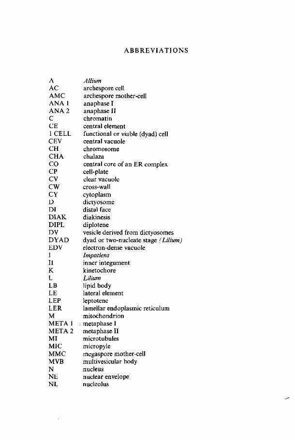
A AC AMC ANA 1 ANA 2 C CE 1 CELL CEV CH CHA CO CP CV CW CY D DI DIAK DIPL DV DYAD EDV I II K L LB LE LEP LER M METAl META 2 MI MIC MMC MVB N NE NL
Allium archespore cell archespore mother-cell anaphase I anaphase II chromatin central element functional or viable (dyad) cell central vacuole chromosome chalaza central core of an ER complex cell-plate clear vacuole cross-wall cytoplasm dictyosome distal face diakinesis diplotene vesicle derived from dictyosomes dyad or two-nucleate stage (Lilium) electron-dense vacuole Impatiens inner integument kinetochore Lilium lipid body lateral element leptotene lamellar endoplasmic reticulum mitochondrion metaphase I metaphase II microtubules micropyle megaspore mother-cell multivesicular body nucleus nuclear envelope nucleolus
NLB nucleolus-like body NP nuclear pore NS nucleolema structure NUC nucellus 4 NUC e four-nucleate embryosac 2 NUC m two-nucleate megaspore 4 NUC st four-nucleate stage OI outer integument P plastid PACH pachytene PB paramural body PF protein-fibrils PR proximal face RER rough endoplasmic reticulum 5 starch SS sacculation structure TELO 1 telophase I TELO 2 telophase II TSER tubular smooth endoplasmic reticulum ZYG zygotene
1. I N T R O D U C T I O N
1.1. GENERAL INTRODUCTION
Generative reproduction in Angiosperms starts with two subsequent processes, sporogenesis and gametogenesis. Sporogenesis leads by meiotic divisions to the formation of haploid spore nuclei. At the end of gametogenesis the haploid gametes are formed as a result of the development from spore to gametophyte. The processes of male development are generally called micro-sporogenesis and microgametogenesis. The processes of female development are called megasporogenesis and megagametogenesis or macrosporogenesis and macrogametogenesis. In this thesis the terms megasporogenesis and megagametogenesis are used.
The formation of male gametes starts in the anther with the differentiation of vegetative cells to microspore mother-cells. Each microspore mother-cell undergoes meiosis leading to the formation of four haploid microspores. Each microspore develops into a male gametophyte - the pollengrain - in which one vegetative and one generative cell are formed. Eventually the generative cell divides into two male gametes.
The formation of female gametes starts in the nucellus of young ovules with the differentiation of one nucellus cell into the megaspore mother-cell. During the meiotic divisions the megaspore mother-cell's nucleus forms four haploid nuclei. The nuclear divisions are not always followed by cell divisions.
Based on cell-plate formation during the meiotic divisions three different main types of megasporogenesis are distinguished according to MAHESWARI
(1950): monosporic type: all nuclear divisions are followed by cell divisions, resulting
in the formation of four haploid spores. This occurs in 70-80% of the Angiosperms.
bisporic type: the first nuclear meiotic division is followed by cell-plate formation leading to cell division. The second nuclear meiotic division is not followed by cell division ; megasporogenesis results in two cells each having two haploid nuclei.
tetrasporic type: the nuclear divisions are not followed by cell divisions; megasporogenesis therefore results in one cell with four haploid nuclei.
In all three types only one cell - having respectively one, two or four nuclei -becomes functional megaspore and starts megagametogenesis. If present, the other spores degenerate. The megagametogenesis can be classified in different groups based on the number of mitotic nuclear - and cell divisions resulting in a varying number of cells and a varying number of haploid nuclei in the mature embryosac. In the mature embryosac - the female gametophyte - usually only one female gamete, involved in the formation of an embryo, is formed - the
Meded. Landbouwhogeschool Wageningen 78-16 (1978) 1
egg-cell - . Megasporogenesis always results in the formation of one viable megaspore in contrast to the microsporogenesis which always gives rise to four viable microspores.
1.2. RESEARCH OBJECT
Numerous botanists have studied the megasporogenesis in Angiosperms with light-microscopic technics. A survey about this work was given by P. MAHESWARI (1950 and 1963), RUTISHAUSER (1969) and PODDUBNAYA-ARNOLDI
(1976). On the ultrastructural level there were electron-microscopic studies about megasporogenesis in Angiosperms: ISRAEL (1963), ISRAEL and SAGAWA
(1964, 1965), JALOUZOT (1971, 1973), RODKIEWICZ and BEDNARA (1974, 1976), RODKIEWICZ and MIKULSKA (1963, 1964, 1965), DICKINSON and HESLOP-
HARRISON (1977) and DICKINSON and POTTER (1978). None of them described the ultrastructural development of the cell organelles during the whole process of megasporogenesis, they all dealt with a specific part or aspect of the process.
Much morR was published about the ultrastructural development of the cell organelles during microsporogenesis, as by MARUYAMA (1968), DICKINSON
and HESLOP-HARRISON (1970a, 1970b, 1977), VAZART (1973), WILLEMSE
(1971a, 1971b, 1971c, 1972) and others.
This thesis presents the results of a comparative study of the megasporogenesis in Angiosperms, mainly based on electron-microscopic technics and includes a discussion about the ultrastructure of the cell organelles in the various developmental stages. Since light-microscopic studies have indicated a polarity of the cell special attention has been drawn to the distribution of the cell organelles in the cytoplasm of the cell. Furthermore the organelle composition of the megaspores is estimated, in order to obtain information about their viability and abortivity.
Three species have been examined : Impatiens walleriana, Hook, ƒ., supposedly having the monosporic type of megasporogenesis as reported by STEFFEN (1951), Allium cepa L., having the bisporic type of megasporogenesis, Lilium hybrid 'Enchantment', having the tetrasporic type of megasporogenesis. Chapter 2 comprises a survey of the technics applied. In the chapters 3, 4 and 5 the megasporogenesis of respectively Lilium, Allium and Impatiens is described and discussed. In chapter 6 the developments of the three species are compared. In chapter 7 the major conclusions are presented. Chapter 8 and 10 comprise the summary respectively samenvatting, chapter 9 the acknowledgements and chapter 11 the references.
Meded. Landbouwhogeschool Wageningen 78-16(1978)
2. APPLIED METHODS
2.1 . L IGHT-MICROSCOPIC (LM) METHODS
2.1.1. HERR's clearing-squash technics Starting the studies of the ovule- and megasporophyte development the fol
lowing clearing-squash technics was used (HERR, 1971) : Ovules dissected from ovaries were fixed for 24 hours in FAA (formalin,
acetic acid, 50% ethanol, 5:5:90) and stored in 70% ethanol. Ovules treated in this way were transferred to a freshly prepared fluid composed of lactic acid (85%), chloralhydrate, phenol, clove oil and xylene (2:2:2:2:1 by weight). After treatment for 24 hours at room temperature the ovules were transferred within a drop of clearing fluid to a special prepared slide for microscopic examination. This kind of slide has two coverglasses affixed 1 cm apart. Within the space formed in this way a drop of clearing fluid, containing the ovules, was placed and a third coverglass was placed on top of the preparation, resting on the two mounted coverglasses, which eliminate the pressure of the top cover on the ovules. The preparations were examined with phase-contrast optics of Leitz Dialux and a Wild M-20 microscope. The cells of the ovules will gradually get apart through lightly and repeatedly pressing the coverglass with a needle midway between the support. The following small variation of HERR'S
technics was also applied: in stead of a fixation with FAA, a fixation with buffered glutaraldehyde as for electron-microscopy was used. The compositions of the specific buffered glutaraldehyde fixatives, used for the three species will be mentioned in the concerning chapters. After glutaraldehyde fixation the ovules were dehydrated by graded ethanol series up to 70% ethanol (2.2.).
2.1.2. Preparation of 3 fim sections The technics mentioned above were applied in order to study the develop
ment of the inner- and outer integuments of the ovule. These technics were also used to examine the place of the nuclei in the megaspore mother-cell (especially in Liliurri). The nuclear structure, however, was not very clear and the methods were not applicable for the determination of the various prophase stages. For this determination the ovules embedded in epon (see 2.2.1.) were longitudinally cut into 3 um sections. The sections were placed on slides in a drop of water. When the slides are heated to 80 C the sections stretch and flatten. The preparations were examined with a phasecontrast microscope with immersionoil under the coverslip.
2.1.3. Callose-checking method To check the presence of callosic compounds in the cell-wall of the megas-
porocyte the fluorescence method was described by RODKIEWICZ and GÓRSKA-
BRYLASS (1968) was used. Ovules were removed from the ovaries and fixed in
Meded. Landbouwhogeschool Wageningen 78-16 (1978) 3
ethanol-acetic acid (3:1). After fixation, the ovules were hydrolysed in 1 N HCL for 5-10 minutes and then washed in water. Squash preparations were made in a aqueous solution of 0.05% aniline-blue in 0.06 M K2H PO4. By using an UV-fluorescence microscope callose gives a yellow fluorescence.
2.2. ELECTRON-MICROSCOPIC (EM) METHODS
2.2.1. Glutaraldehyde-osmiumtetroxydefixation A glutaraldehyde-osmiumtetroxyde fixation was applied principally. For
the three species different concentrations of buffered glutaraldehyde and osmiumtetroxyde solutions at different fixation times were used. These concentrations and times are mentioned in the chapter specific for each of the species.
During all developmental stages of one species always the same fixation method was used. Ovules were dissected from the ovaries and fixed in a buffered solution of glutaraldehyde at room temperature. For buffering always a phosphate buffer (primary potassium phosphate and secondary sodium phosphate) at pH 7.2 was used. For Lilium and Allium saccharose was added to the phosphate buffer. After fixation the material was rinsed three times during 15 minutes in the buffer. The material was postfixed in a buffered osmiumtetroxyde solution; for Allium and Lilium also containing saccharose. After this fixation the material was rinsed in buffer again and dehydrated by graded ethanol/propylene oxyde-series. The different gradation steps are - in their proper sequence - 10%, 25%, 50%, 70%, 90% and 100% (2 x ) ethanol, mixtures of ethanol/propylene oxyde, respectively in the proportion of 3:1, 1:1 and 1:3, and then a mixture of propylene oxyde/epon in the proportion of 3:1. Every step took 10 minutes except the last one, which took an overnight during which the propylene oxyde evaporated and the epon concentration increased. The material was embedded in epon. The ovules were sectioned longitudinally with a LKB ultramicrotome. Thin sections, made with glass knives or diamant knives, were picked up on coppergrids (75 mesh) coated with formvar-film. The sections were poststained with leadcitrate (REYNOLDS 1963). For the selection of the sections a Siemens electron-microscope Elmiskop 51 was used. Selected sections were studied with a Philips EM 300. When the use of a goniometer was necessary we availed ourselves of a Philips EM 301 with goniometer stage.
2.2.2. Potassiumpermanganatefixation Potassiumpermanganate fixation was only used in the initial period of each
species study, when the proper glutaraldehyde-osmiumtetroxyde fixation was not yet developed. Usually a series of combinations of KMn04 concentrations (1-5%) and fixation times (0,5-3 hours) was used. After fixation the material was rinsed in water, dehydrated by graded ethanol/propylene oxyde series and embedded in epon. It was thick-sectioned (3/xm) for light-microscopic
4 Meded. Landbouwhogeschool Wageningen 78-16 (1978)
Observation and thin-sectioned (60-90 nm) for EM observation. This method primarily was used to check the development stages of the megaspore mother-cell in relation to the size of the flowerbuds. In the 3 fim sections the chromosomal structure of the various prophase stages, easily observed with the phase-contrast LM, could be determined.
However, thin sections of KMnCU material were not usable for the study of the nuclear ultrastructure. By potassiumpermanganate fixation apparently nucleic acids were partly destroyed. For this reason only glutaraldehyde-osmiumtetroxyde fixation was used for ultrastructural study of the organelles.
2.2.3. Re-embedding method As already mentioned, for the determination of the developmental stages
of the nucleus, 3 /im sections of the epon-embedded material were used, because in the ultrathin sections only small pieces of the chromosomes were present and therefore determination was practically impossible.
In order to obtain sections for both determination studies and ultrastructural studies, both 3 ̂ m sections and the ultrathin sections had to be cut from the same ovule. The ovule was initially cut semithin, until enough information could be gained for proper determination and then the remaining material was cut ultrathin. This method is very time-consuming because, when during cutting the megaspore mother-cell is reached, each 3 pm section has to be studied until the nucleus is reached, otherwise one risks that the whole ovule is thick-sectioned and no material is left for thin-sectioning. To solve this problem, a modified method for re-embedding as described by MOGENSEN (1971) and Cox and SEELY (1974) was used. The ovule was sectioned completely in 3-5 urn sections. The sections were prepared for observation and the best of them were selected. The slides with the sections still affixed were thoroughly rinsed in absolute ethanol to wash out the immersionoil. The selected sections were removed from the slides by carefully cutting with a razor blade. In the meantime small precast epon blocks were prepared in a Blazers flat-embedding mould, by filling the holes to the half with epon. The selected sections were placed on the flat side of these small blocks in a drop of water. By heating the blocks upto 80 C the sections would stretch and flatten. After being dried the sections were fixed upon the small epon blocks. The sections were enclosed in epon fluid by filling the remaining holes in the flat-embedding mould to the top. The blocks with the re-embedded material were so small that they could easily be observed with a light-microscope. For the re-orientation of the material before thin-sectioning, a light-microscope without an object-table, condensor and microscopic light was used. The LKB block with the objectholder was placed under the microscope at the correct distance from the lens for the material to be in focus. With a table-spotlight the magnifications of 40 x and 100 x could easily be used. In this way the orientation of the flat section could be brought perpendicularly to the field of sectioning.
Meded. Landbouwhogeschool Wageningen 78-16 (1978)
2.2.4. THIERY-test For the detection of polysaccharides on electron-microscopic level the
method described by THIÉRY (1967) was used : Thin sections of glutaraldehyde-osmiumtetroxyde fixated material were picked up on golden grids of 300 mesh without formvar film. The sections on the grids were treated in the following way: 30 minutes with 1% periodic acid; rinsing thoroughly in water; for 20 hours in 0.2% solution of thiocarbohydrazid in 20% acetic acid ; rinsing 3 times in 15% acetic acid, 1-2 times in 5% acetic acid, 1-2 times in 2% acetic acid and in water ; followed by treatment with 1 % silverproteinate during 30 minutes in the darkroom and rinsing in water frequently. By way of control a treatment with 20% acetic acid without thiocarbohydrazid was used. By the TmÉRY-test polysaccharides could be detected by the deposition of small silvergranules which were visible as electron-dense granules in the electron-microscope.
2.2.5. Quantitative methods See enclosures I, II, III, figures 4, 16 and 27 and enclosure V. For measuring and counting the various cell organelles electron-microscopic
(EM) photos of sections of complete megaspore mother-cells and mega spore cells were used. These photos were selected on the distinctness of the various cell organelles and on the presence of the nuclei in the various developmental stages. For each species photos of distinctly differing developmental stages were chosen. The magnification of the photos did not vary within the species. For Allium and Impatiens a magnification of 8,000 x was applicable, whereas for Lilium a lower magnification of 2,500 x was necessary, since this species has extremely large megaspore cells which causes troubles when taking EM photos of the whole cell, using a higher magnification. For each species only one photo of every developmental stage was used for measuring and counting.
The area of the total cell, of the nucleus and of the nucleolus were determined by measuring the length and the width of the cell, the nucleus and the nucleolus. For each species the sizes of the cell and the nucleus are presented in //m in the enclosures I, II and III, which please find in the map on the inside of the back cover of this thesis. The area of the nucleolus, as far as it was possible to be measured, is presented in the figures 4,16 and 27 and in enclosure V, expressed in square/zm.
The thickness of the cell-wall was determined by measuring the whole cell-wall between the megaspore mother-cell and the surrounding nucellus cells, data of which are presented in the enclosures I, II and III. After measuring the thickness of the cell-wall of the nucellus cells, the exact thickness of the megaspore mother-cells and megaspore cells could be determined and this size is represented in the figures 4, 16 and 27 and in enclosure V.
The area of cell cytoplasm was determined by counting the number of points found to be present in the cytoplasm by the use of a mould. This mould consists of a tracing-paper on which a lattice of points with a mutual distance of 4.45 mm is printed (WILLEMSE 1971c). By covering the EM photos with this mould the points present on the inside of the cell cytoplasm can be counted.
6 Meded. Landbouwhogeschool Wageningen 78-16(1978)
The amount of electron-transparent vacuolar substances and of lipidic compounds within the cytoplasm was determined by counting the number of points found in the clear vacuoles and in the lipid bodies. These amounts are expressed in points per cytoplasmic area and are represented in the figures 4, 16 and 27 and in enclosure V.
The amount of lamellar endoplasmic reticulum (LER) membranes in Lilium was determined by the use of a mould consisting of a tracing-paper presenting parallel lines with a mutual distance of 1 cm. The total length of the lines crossing the cytoplasm of the cell was measured, when also the total number of cross-points between the LER membranes and the lines was counted. A measure for the number of LER membranes was reflected by the quotient between the total number of the cross-points and the total length of the lines (WEIBEL 1973). The mould with the lines was always applied in the cell in such a way, that the lines were perpendicular to the axis micropyle-chalaza.
The total number of dictyosomes, mitochondria and plastids was counted per cell at every developmental stage of the nucleus. The number per cytoplasmic area of dictyosomes, mitochondria and plastids was calculated. This cytoplasmic area contains 150 points for the species Impatiens and Allium (magnification 8,000 x ) and 50 points for Lilium (magnification 2,500 x ). These differences in points of cytoplasm were chosen to enable us to compare the the numbers of the cell organelles mentioned above per cytoplasmic area of the three species, found in the comparable developmental stage.
The number of ribosomes and polysomes was counted in photos with a higher magnification. For each species the 25,000 fold magnification was applied. Also for ribosomes and polysomes a number per cytoplasmic area was calculated, but this cytoplasmic area was not expressed in points of cytoplasm but in square /mi, because for counting the ribosomes and polysomes a square mould was used (figures 4, 16 and 27 and enclosure V.
As only one cell of every developmental stage of each of the species was counted, a statistical analysis of the quantitative data could not be made.
The quantities of the other cell organelles represented in the figures 4, 16 and 27 and enclosure V were estimated, since no available measuring or counting method could be applied. These estimated quantitative results were reproduced in black histograms, whereas the quantitative data counted were reproduced in dotted histograms.
Meded. Landbouwhogeschool Wageningen 78-16(1978)

3. MEGASPOROGENESIS AND EARLY MEGAGAMETOGENESIS IN THE LILIUM HYBRID
'ENCHANTMENT'
3.1. INTRODUCTION
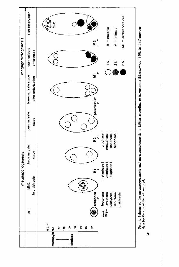
Lilium has the tetrasporic type of embryosac development. Figure 1 shows schematically the megasporogenesis and megagametogenesis in Lilium according to BAMBACIONI (MAHESWARI 1950). In this figure our data for the size of the cell are used. MAHESWARI stated that after meiosis the four nuclei of the megaspore are distributed at random in the cytoplasm (four-nucleate stage I). The cell becomes functional megaspore when three nuclei move to the chalazal pole and one nucleus to the micropylar pole (four-nucleate stage I after 'polarization'). The cell polarized in this way enters the megagametogenesis. During the first mitotic division the spindle of the three dividing nuclei at the chalazal pole fuse together to one spindle, thus after division forming two triploid nuclei. The nucleus at the micropylar pole divides normally to form two haploid nuclei. This stage is called the second four-nucleate stage - in our thesis called four-nucleate embryosac - . After the second mitotic division the eight-celled mature embryosac is formed.
The large proportions of the Lilium megasporocyte enables light-microscopic examination of cytoplasmic changes during megasporogenesis. GUILLERMOND (1924) examined the mitochondria and plastids during meiosis. He suggested that these organelles are self-propagating and that their continuity is preserved during meiosis. He also observed so-called ergastoplasmic bodies which he considered as fixation artefacts. FLINT and JOHANSEN (1958) revealed an 'extrusion' of nucleolar substances from the nucleus into the cytoplasm. They suggested a relation between these nucleolar substances and spindle formation as well as nuclear movement. EYMÉ ( 1965) described the development and distribution of extensive so-called ergastoplasmic structures. These structures, examined by electron-microscopy, in co-operation with RODKIEWICZ, appeared to consist of endoplasmic reticulum cisternae. EYMÉ also observed a polar distribution of cell organelles in the young megaspore mother-cell. RODKIEWICZ and MIKULSKA (1963, 1965) started with electron-microscopic examination of Lilium megasporogenesis and megagametogenesis. They revealed the ultra-structural development of the endoplasmic reticulum during megasporogenesis. They also decribed the ultrastructure of the cell organelles at the second four-nucleate stage and in the mature embryosac (MIKULSKA and ROD
KIEWICZ (1965, 1967a, 1967b). A comparative study of membrane-bound cytoplasmic inclusions during meiotic prophase of Lilium micro- and megasporogenesis was published by DICKINSON and ANDREWS (1977). They suggested that these inclusions preserve reserves necessary for postmeiotic develop-
8 Meded. Landbouwhogeschool Wageningen 78-16 (1978)
CO
o
co <ä c <D O) O 0) E co O) co
<0
E
CD O)
O Q.
CO d> c CD O) O i_ O O . CO CD O ) CD
CU a> co
•o
<
CM
2
ca co O fl) E II er
co 'co o
. t i
E II S
o Q . CO CU
.c o ca
II O <
z z T - CM
ït
CM
CC
CD Q> CU CD CO CO CO CO ca ca ca ca c c c x : a a a a o ca co o Q- | « S
= t Q. ca CD
E
a ca c ca
O. o £
CO l CD CD CD CD
•5 t O O 'S, O o I S. 5>-S Œ
co CD
S •- * cp -a —
]
a 2 o
I
co
N
CO
3 M
«3
3
en W S <
o 0 <
< CQ O
S O
e
<\ c
fl> G (IJ W) O
<!> Fi « on nt 011 <]>
S -o a CO
r/1 4> e « bil O
O
a r/i nt BI) a> 6 O
J3
< * -O o ia &> J3 O
C/l
•
o U-
• Ü
3
o
"5 di
J3
o
N
<u X l-l
<+-«Î
•o

ment and permit the continuity of protein synthesis during meiosis. DICKINSON
and HESLOP-HARRISON (1977) and DICKINSON and POTTER (1978) revealed changes in the ribosome population during microsporogenesis and megaga-metogenesis in Lilium and other species. They established a correlation between these changes in ribosome population and the membrane-bound cytoplasmic inclusions. They also stated so-called cycles of dedifferentiation and redifferentiation of the plastids and mitochondria during Lilium micro- and megasporogenesis.
3.2. MATERIAL AND METHODS
For light-microscopy 3 ^m sections from glutaraldehyde-osmiumtetroxyde fixed material as described previously in 2.1.2. and clearing-technics preparations (2.1.1.) were used.
For electron-microscopy the glutaraldehyde-osmiumtetroxyde fixation (2.2.1.) and a potassiumpermanganate fixation (2.2.2.) were applied in combination with the re-embedding method (2.2.3.). For staining of carbohydrates at EM level the THiÉRY-test (2.2.4.) was used.
The glutaraldehyde-osmiumtetroxyde fixation was applied as follows: ovaries of - in greenhouses grown - Lilium hybrid 'Enchantment' plants were cut transversely in sections of 0,5 mm thick. The sections were fixed for 1,5 hour in 5% glutaraldehyde in 0,1 M phosphate buffer at pH 7,2 containing 0,025 M saccharose. They were rinsed in 0,1 M phosphate buffer containing 0,25 M saccharose and postfixed during 20 hours in buffered 2°/0 OsCU containing 0,025 M saccharose. The epon-embedded ovules were sectioned longitudinally. The sections were poststained with leadcitrate (REYNOLDS 1963).
3.3. RESULTS AND DISCUSSION
3.3.1. Light-microscopy
3.3.1.1. Ovules The Lilium ovaries are syncarp consisting of three carpels. At the edges of
the carpels the young ovules are of subepidermal origin. The very young ovules consist of mitotic dividing nucellus cells, showing meristematic characters with dark cytoplasm without central vacuoles (figure 2a). The epidermis cells of the nucellus remain intact during the whole process of megasporogenesis and megagametogenesis. One of the inner nucellus cells, laying just under the epidermis becomes archespore mother-cell (AMC). The archespore cell (AC) originates directly from the AMC without division. The AC can be disting-huished from the surrounding nucellus cells by its larger size and pronounced nucleus. The AC becomes megaspore mother-cell (MMC) when the nucleus enters meiotic prophase (according to the definition of RUTISHAUSER, 1969).
10 Meded. Landbouwhogeschool Wageningen 78-16 (1978)
5» i* - . " w
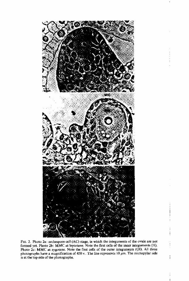
FIG. 2. Photo 2a: archespore cell (AC) stage, in which the integuments of the ovule are not formed yet. Photo 2b: MMC at leptotene. Note the first cells of the inner integuments (II). Photo 2c: MMC at zygotene. Note the first cells of the outer integuments (OI). All three photographs have a magnification of 450 x. The line represents 10 /un. The micropylar side is at the top side of the photographs.
During the archespore cell stage the integuments of the ovule are not formed yet (figure 2a). When the nucleus of the young M MC is at leptotene the first dividing nucellus cells, which give rise to the formation of the inner integuments, appear (figure. 2b). At about zygotene the first cells of the outer integuments appear (figure 2c) and at about pachytene-diplotene the integuments are fully grown. The mature ovule is anatropous, bitegmic and tenuinucellate.
In the ovaries the ovules are arranged in long rows on the placenta. Mainly by light-microscopic examination of the chromatin structure the different stages are to be distinghuished. The developmental stage of the inner- and outer integuments only gives an indication. The ovules of one ovary have developmental variations depending on their position in the ovary. In upper position - near the style - they are less developed than in lower position. Growing from archespore mother-cell till metaphase I takes about 10 days in summertime. In one ovary the ovules then mostly vary within one prophase stage, in consequence of which a clear identification of the exact prophase stage is difficult to establish. From metaphase I till four-nucleate stage it takes less than two days. Within this range of stages there are three longer stages, namely metaphase I, two-nucleate stage and four-nucleate stage. These three stages can be present in one ovary often without any ana- and telophases. We regret that during our research the ana- and telophase I stages were not found. Even after various diurnal and nocturnal fixation-times, in which ana- and telophase 11 were found, we did not come across ana- and telophase I. These latter stages are probably of a short duration.
3.3.1.2. F r om megaspo rocy t e to f ou r -nuc l ea t e embryosac The young archespore cell has a length of about 25 /mi (along the axis
micropyle-chalaza) and a width of about 35 /im. During the premeiotic interphase the AC enlarges to more than twice its original size, both in length and in width. The shape of the AC changes from a common elliptic cell shape with the long axis parallel to the epidermis to a conical shape with the top pushed deep in the nucellar tissue (figure 2a, b and c and encl. IV A).
During the prophase stages the MMC enlarges to about twice its original length again whereas the width remains about constant. The shape of the MMC is getting oval. From metaphase I up to four-nucleate embryosac the cell enlarges 1.5 times again. The length of the cell in the four-nucleate embryosac is about 180 /mi, the width about 65 /mi (figure 1).
The nucleus enlarges gradually during the premeiotic interphase and the meiotic prophase from a diameter of 17 /mi to a diameter of 35 /mi. In the AC the nucleus contains 2 -3 nucleoli having small vacuoles. At leptotene only one large spherical nucleolus is found, having one large vacuole and a few small vacuoles (figure 2b). At zygotene the nucleolus appear to be closely pressed to the nuclear envelope (figure 2c). During pachytene and diplotene the nucleolus is spherical, also usually located near the nuclear envelope. The nucleolus enlarges during the prophase stage till its maximum at diplotene (figure 4) and still contains small vacuoles. At diakinesis the nucleolus has disappeared. At
12 Meded. Landbouwhogeschool Wageningen 78-16 (1978)
two-nucleate and four-nucleate stage two spherical compact nucleoli per nucleus are perceptible.
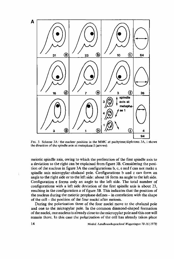
3.3.1.3. The po s i t i on of the nuc leus During leptotene and zygotene, when the MMC has a conical shape the
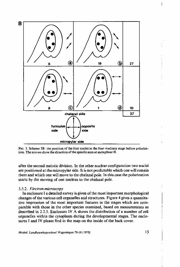
nucleus is situated in the centre, a little bit more on the micropylar side. During pachytene and diplotene, when the MMC has a more oval shape, the position of the nucleus varies strongly. Using the clearing technics with glutaraldehyde fixation 94 ovules at pachytene-diplotene were scored on their nuclear position. Figure 3A schematically shows the results. Also the position of the four nuclei in the four-nucleate stage before polarization was examined and 37 ovules were scored. Figure 3B shows the results.
Discussion From figure 3A the conclusion is justifiable that the nucleus is preferably
positioned in the centre of the cell, when the cell has an oval-like shape. When the cell has a pear-like shape the nucleus prefers to be located at that side of the cell where there is the greatest width. Consequently there seems to be a relation between the shape of the cell and the position of the nucleus. Because of the occurrence of the nucleus in the centre twice as frequent as at the funicular and the opposite side, the conclusion interpreted that the nucleus has a rotation movement during this stage seems obvious. The nucleus has a tendency to be positioned as far as possible from the funiculus side, as is shown in figure 3A. From figure 3B we may conclude that the nuclei are preferably positioned in diamond-shaped formation. Also here there is a relation between the shape of the cell and the position of the nuclei.
Presuming that the rotation movement of the prophase nucleus stops when the cell enters metaphase and that after the first meiotic division no movement of the nuclei takes place, we can presume a certain relation between the two figures described above. There is a relation between the central position in figure 3A (64) and the diamond-shaped formation in figure 3B (27). Also the pear-like shaped cells in figure 3A and 3B show a relation. When we presume that spindles are formed by a microtubule organizing centre (PICKETT-HEAPS,
1970), which duplicates and separates during prophase along an axis, the direction of which defines the final position of the nuclei, the next considerations are applicable.
From the available results it appears that the spindle axis during the first meiotic division preferably has the direction micropylar-chalazal pole, sometimes with a deviation to the right side (figure 3A, i). When the cell has a round shape there is enough room for the second meiotic spindle axis to run perpendicular to the first meiotic spindle axis, resulting in the square-formation of the nuclei. When the cell has a pear-like or an oval-like shape this formation is not possible, so that a deviation of the second meiotic spindle axis is necessary. This deviation is directed in 27 cases (b and c) to the left and in 8 (a) cases to the right (figure 3B). The second meiotic spindle axis forms an angle with the first
Meded. Landbouwhogeschool Wageningen 78-16(1978) 13
spindle axis at metapha se 1
©
64
26
94
FIG. 3. Scheme 3A : the nuclear position in the MMC at pachytene/diplotene. 3A, i shows the direction of the spindle axis at metaphase I (arrows).
meiotic spindle axis, owing to which the preferation of the first spindle axis to a deviation to the right can be explained from figure 3B. Considering the position of the nucleus in figure 3 A the configurations b, c, e and f can not make a spindle axis micropylar-chalazal pole. Configurations b and c can form an angle to the right side or to the left side : about 16 form an angle to the left side. Configuration e forms only an angle to the left side. The total number of configurations with a left side deviation of the first spindle axis is about 23, resulting in the configuration a of figure 3B. This indicates that the position of the nucleus during the meiotic prophase defines - in correlation with the shape of the cell - the position of the four nuclei after meiosis.
During the polarization three of the four nuclei move to the chalazal pole and one to the micropylar pole. In the common diamond-shaped formation of the nuclei, one nucleus is already close to the micropylar pole and this one will remain there. In this case the polarization of the cell has already taken place
14 Meded. Landbouwhogeschool Wageningen 78-16(1978)
B
chalazal side
funiculus 1+. side
27
10
37
*J opposite side
micropylar side
FIG. 3. Scheme 3B : the position of the four nuclei in the four-nucleate stage before polarization. The arrows show the direction of the spindle axes at metaphase II.
after the second meiotic division. In the other nuclear configuration two nuclei are positioned at the micropylar side. It is not predictable which one will remain there and which one will move to the chalazal pole. In this case the polarization starts by the moving of one nucleus to the chalazal pole.
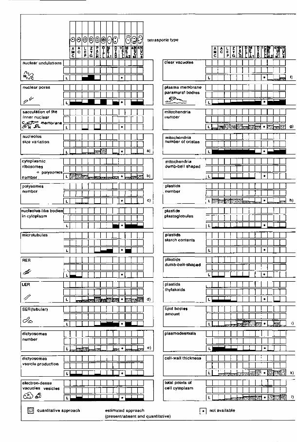
3.3.2. Electron-microscopy In enclosure I a detailed survey is given of the most important morphological
changes of the various cell organelles and structures. Figure 4 gives a quantitative impression of the most important features in the stages which are comparable with those in the other species examined, based on measurements as described in 2.2.5. Enclosure IV A shows the distribution of a number of cell organelles within the cytoplasm during the developmental stages. The enclosures I and IV please find in the map on the inside of the back cover.
Meded. Landbouwhogeschool Wageningen 78-16(1978) 15
nuclear undulations |
/ > ? • > '
<Pc T
© A M C
0 A C
nuclear pores |
*>* rr sacculation of the 1
inner nuclear i—
« Â |T nucleolus 1
size variation i
| L
cytoplasmic |
r ibosomes 1—
number I L
poiysomes | number i —
| L
nucleolus-like bodies| in cytoplasm | —
| L
microtubules 1
| L
RER |
^ k LER |
s rr SER(tubular) |
, /? 1 c ^ IY dictyosomes [ number i
| L
dictyosomes | vesicle production ]
| L
electron-dense | vacuoles vesicles [—
<SS><$ | T
| | | quantitative app
@ L E P
@©®0© z Y G
P A C H
0 i p L
M E T A1
D Y A p
1C E L L
•
S©0 M E
h 4 N
ï 4 N
c ç
tetrasporic type
M I N I 1 1 1 1 1 1
1 1 1 1 1 1 1 1 1 M 1 1 LLU 1 M I M I M M M
M M M M M M
r^ t r fe iXU™
1 M M 1 1
l | 1 M 1
1 M M M M M M
M M M 1 M M 1
J - B M M M M M M
— M M M M M M M M L^Wsüslikäte;
M M M M M M M I L
M M 1 M M 1
™isteUiJ™,
M M 1
M M 1
-,
?nw
•
•
•
•
•
•
•
•
•
•
•
•
| I
I |
I I |
I |
I I |
I I
I |
• | I I
I wa^w.
| I
I I
I I
I I
7F
w
^ ^ ^ ^ ^ ^ ^ ^ ^ ^ ^ ^ ^ ^ ^ ^ ^ 1 * ^ ^ ^ ^ ^ ^ ^
oa ch
( >sti
pre
ma
se
ed
i t/a
appro
bsent
act
an
a)
b)
c)
d)
e)
ï
d qua ntitat
clear vacuoles I
| L
A M C
A C
L E P
Z Y G
P A
8
D 1 P L
M E T A1
D Y A 0
1C
•
M E T A2
4N U
S
4N
8 e
o plasma membrane |
paramural bodies |—
_ 5 ' - " ' " ~ - — 1 .
mitochondria |
number i—
rr mitochondria L number of cristae 1—
|T
mitochondria | dumb-bell shaped i—
rr plastids |
number i
u~ plastids | plastoglobules i
| L
plastids |
starch contents i—'
| L
plastids |
dumb-bell-shaped i —
^ plastids 1
thylakoids | —
| L
l ipid bodies |
aniounl 1
| L
plasmodesmata |
| L
cell-wall thickness |
| L
total points of |
cell cytoplasm |
| L
M J M I I 1 1 1 1 1 1 1
M M M 1 1 1 1 1 M 1
„J?4. 'M-; ' fc^*i
M M M 1 1 1 1 1 1 1 1
M I M M _. 1 1 M 1 M
L±^_ 1 1 I I
M I I 1 M 1 1 1 1 1 1 !
1 1 M M 1 M M M 1
1 M M ! M M M 1 M M M 1
1 1 1 1 1 M M 1 M M
1 1 1 1 i 1 I
N I M M M I I I I 1
l M M M M 1 M M 1, .M-rH^H- '¥? I l 1 1 1 M I l I I M 1
I I M M M ! W-4-J - f -H ' s
M M M 1 M M M 1
U J - W H r l n * :
•
«
•
•
•
•
•
•
•
•
•
•
•
"^ht~ g)
I u. h)
_
'>
•"ri"' k)
" " % — * s " 1 » FT! not available
ive)
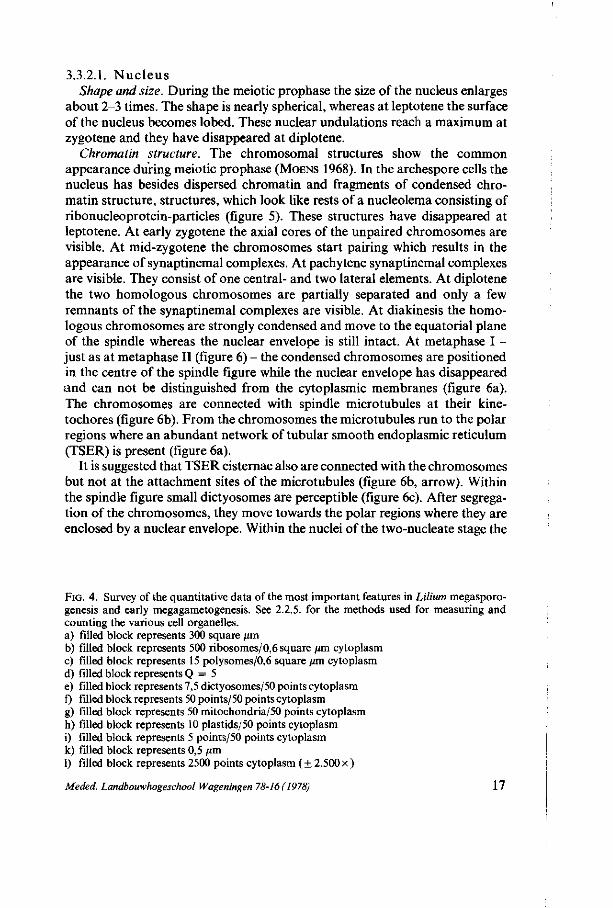
3.3.2.1. Nuc l eus Shape and size. During the meiotic prophase the size of the nucleus enlarges
about 2-3 times. The shape is nearly spherical, whereas at leptotene the surface of the nucleus becomes lobed. These nuclear undulations reach a maximum at zygotene and they have disappeared at diplotene.
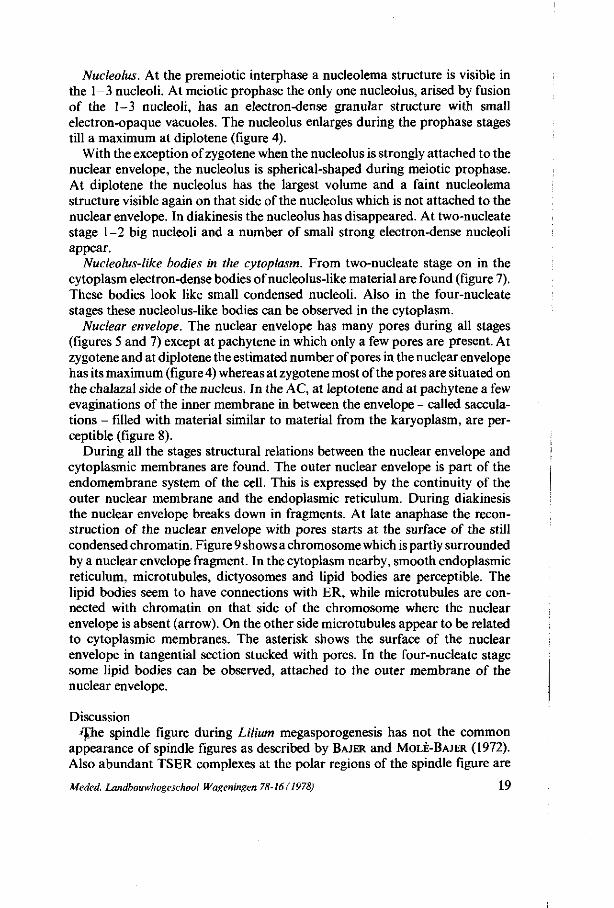
Chromatin structure. The chromosomal structures show the common appearance during meiotic prophase (MOENS 1968). In the archespore cells the nucleus has besides dispersed chromatin and fragments of condensed chromatin structure, structures, which look like rests of a nucleolema consisting of ribonucleoprotein-particles (figure 5). These structures have disappeared at leptotene. At early zygotene the axial cores of the unpaired chromosomes are visible. At mid-zygotene the chromosomes start pairing which results in the appearance of synaptinemal complexes. At pachytene synaptinemal complexes are visible. They consist of one central- and two lateral elements. At diplotene the two homologous chromosomes are partially separated and only a few remnants of the synaptinemal complexes are visible. At diakinesis the homologous chromosomes are strongly condensed and move to the equatorial plane of the spindle whereas the nuclear envelope is still intact. At metaphase I -just as at metaphase II (figure 6) - the condensed chromosomes are positioned in the centre of the spindle figure while the nuclear envelope has disappeared and can not be distinguished from the cytoplasmic membranes (figure 6a). The chromosomes are connected with spindle microtubules at their kine-tochores (figure 6b). From the chromosomes the microtubules run to the polar regions where an abundant network of tubular smooth endoplasmic reticulum (TSER) is present (figure 6a).
It is suggested that TSER cisternae also are connected with the chromosomes but not at the attachment sites of the microtubules (figure 6b, arrow). Within the spindle figure small dictyosomes are perceptible (figure 6c). After segregation of the chromosomes, they move towards the polar regions where they are enclosed by a nuclear envelope. Within the nuclei of the two-nucleate stage the
FIG. 4. Survey of the quantitative data of the most important features in Lilium megasporo-genesis and early megagametogenesis. See 2.2.5. for the methods used for measuring and counting the various cell organelles. a) filled block represents 300 square um b) filled block represents 500 ribosomes/0,6 square /mi cytoplasm c) filled block represents 15 polysomes/0,6 square /xm cytoplasm d) filled block represents Q = 5 e) filled block represents 7,5 dictyosomes/50 points cytoplasm f) filled block represents 50 points/50 points cytoplasm g) filled block represents 50 mitochondria/50 points cytoplasm h) filled block represents 10 plastids/50 points cytoplasm i) filled block represents 5 points/50 points cytoplasm k) filled block represents 0,5 pm 1) filled block represents 2500 points cytoplasm ( ± 2.500 x )
Meded. Landbouwhogeschool Wageningen 78-16 (1978) 17
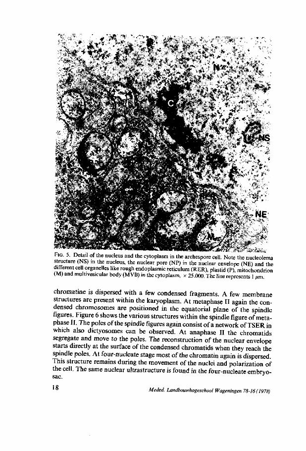
''''•#* V"**"̂
FIG. 5. Detaü of the nucleus and the cytoplasm in the archespore cell. Note the nucleolema structure (NS) in the nucleus, the nuclear pore (NP) in the nuclear envelope (NE) and the " ' c d l organelles like rough endoplasmic reticulum (RER), plastid (P), mitochondrion (M) and multivesicular body (MVB) in the cytoplasm, x 25.000. The line represents 1 ßm.
chromatine is dispersed with a few condensed fragments. A few membrane structures are present within the karyoplasm. At metaphase II again the condensed chromosomes are positioned in the equatorial plane of the spindle figures. Figure 6 shows the various structures within the spindle figure of metaphase II. The poles of the spindle figures again consist of a network of TSER in which also dictyosomes can be observed. At anaphase II the chromatids segregate and move to the poles. The reconstruction of the nuclear envelope starts directly at the surface of the condensed chromatids when they reach the spindle poles. At four-nucleate stage most of the chromatin again is dispersed. This structure remains during the movement of the nuclei and polarization of the cell. The same nuclear ultrastructure is found in the four-nucleate embryo-sac.
U Meded. Landbouwhogeschool Wageningen 78-16(1978)
Nucleolus. At the premeiotic interphase a nucleolema structure is visible in the 1-3 nucleoli. At meiotic prophase the only one nucleolus, arised by fusion of the 1-3 nucleoli, has an electron-dense granular structure with small electron-opaque vacuoles. The nucleolus enlarges during the prophase stages till a maximum at diplotene (figure 4).
With the exception of zygotene when the nucleolus is strongly attached to the nuclear envelope, the nucleolus is spherical-shaped during meiotic prophase. At diplotene the nucleolus has the largest volume and a faint nucleolema structure visible again on that side of the nucleolus which is not attached to the nuclear envelope. In diakinesis the nucleolus has disappeared. At two-nucleate stage 1-2 big nucleoli and a number of small strong electron-dense nucleoli appear.
Nucleolus-like bodies in the cytoplasm. From two-nucleate stage on in the cytoplasm electron-dense bodies of nucleolus-like material are found (figure 7). These bodies look like small condensed nucleoli. Also in the four-nucleate stages these nucleolus-like bodies can be observed in the cytoplasm.
Nuclear envelope. The nuclear envelope has many pores during all stages (figures 5 and 7) except at pachytene in which only a few pores are present. At zygotene and at diplotene the estimated number of pores in the nuclear envelope has its maximum (figure 4) whereas at zygotene most of the pores are situated on the chalazal side of the nucleus. In the AC, at leptotene and at pachytene a few evaginations of the inner membrane in between the envelope - called sacculations - filled with material similar to material from the karyoplasm, are perceptible (figure 8).
During all the stages structural relations between the nuclear envelope and cytoplasmic membranes are found. The outer nuclear envelope is part of the endomembrane system of the cell. This is expressed by the continuity of the outer nuclear membrane and the endoplasmic reticulum. During diakinesis the nuclear envelope breaks down in fragments. At late anaphase the reconstruction of the nuclear envelope with pores starts at the surface of the still condensed chromatin. Figure 9 shows a chromosome which is partly surrounded by a nuclear envelope fragment. In the cytoplasm nearby, smooth endoplasmic reticulum, microtubules, dictyosomes and lipid bodies are perceptible. The lipid bodies seem to have connections with ER, while microtubules are connected with chromatin on that side of the chromosome where the nuclear envelope is absent (arrow). On the other side microtubules appear to be related to cytoplasmic membranes. The asterisk shows the surface of the nuclear envelope in tangential section stucked with pores. In the four-nucleate stage some lipid bodies can be observed, attached to the outer membrane of the nuclear envelope.
Discussion »TTie spindle figure during Lilium megasporogenesis has not the common
appearance of spindle figures as described by BAJER and MOLÈ-BAJER (1972). Also abundant TSER complexes at the polar regions of the spindle figure are
Meded. Landbouwhogeschool Wageningen 78-16(1978) 19
* • _ - *
/ *
T;*» ^ ^ « ^ • . ' . • J p t - ^ S
.•4
ratest
20 Meded. Landbouwhogeschool Wageningen 78-16(1978)
'•**< '
. • • * $ . * % - . •
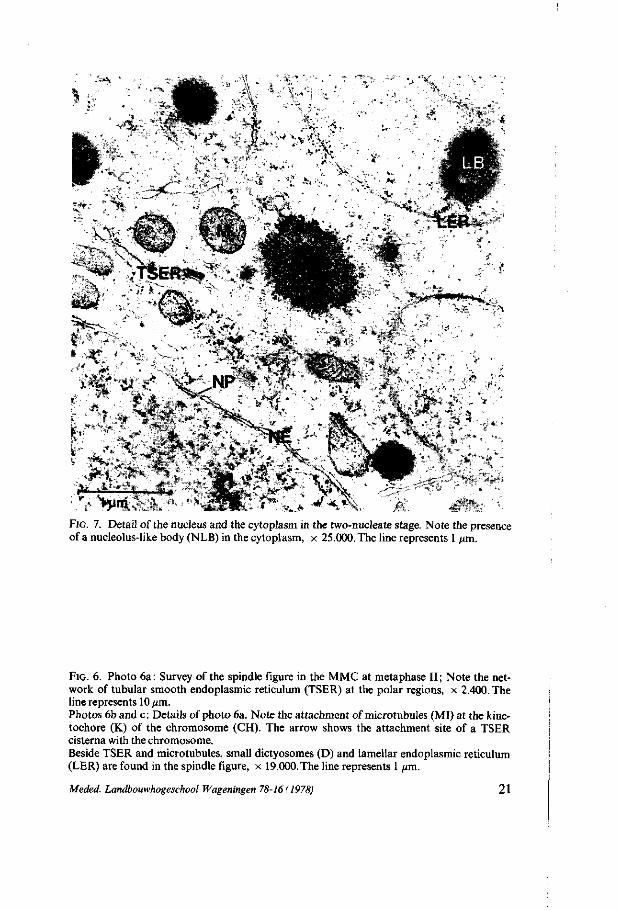
FIG. 7. Detail of the nucleus and the cytoplasm in the two-nucleate stage. Note the presence of a nucleolus-like body (NLB) in the cytoplasm, x 25.000. The line represents 1 jan.
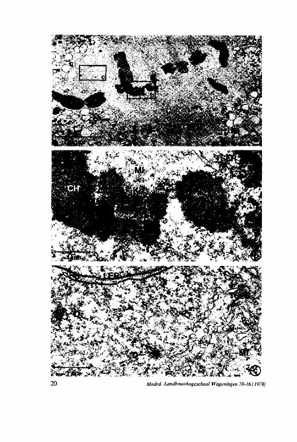
FIG. 6. Photo 6a: Survey of the spindle figure in the MMC at metaphase II; Note the network of tubular smooth endoplasmic reticulum (TSER) at the polar regions, x 2.400. The line represents 10 pm. Photos 6b and c: Details of photo 6a. Note the attachment of microtubules (MI) at the kine-tochore (K) of the chromosome (CH). The arrow shows the attachment site of a TSER cisterna with the chromosome. Beside TSER and microtubules, small dictyosomes (D) and lamellar endoplasmic reticulum (LER) are found in the spindle figure, x 19.000. The line represents 1 pm.
Meded. Landbouwhogeschool Wageningen 78-16 (1978) 21
i:. JfeÉv v-v;-.. •• • v
•• "... sé i
"%
I
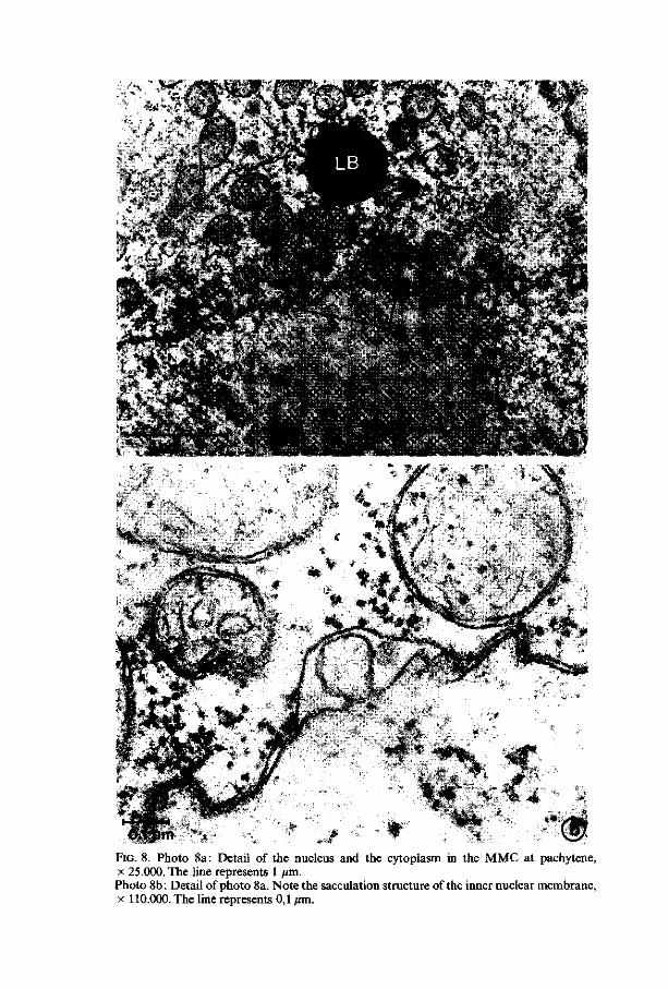
FIG. 8. Photo 8a: Detail of the nucleus and the cytoplasm in the MMC at pachytene, x 25.000. The line represents 1 /an. Photo 8b: Detail of photo 8a. Note the sacculation structure of the inner nuclear membrane, x 110.000. The line represents 0,1 ̂ m.
;-- v «•:
.Ai
ft-* • | i '<*0 ' , • « •
VS
VHÛÈIV
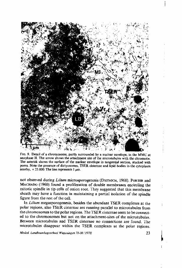
FIG. 9. Detail of a chromosome, partly surrounded by a nuclear envelope, in the MMC at anaphase II. The arrow shows the attachment site of the microtubules with the chromatin. The asterisk shows the surface of the nuclear envelope in tangential section, stucked with pores. Note the presence of dictyosomes, TSER cisternae and lipid bodies in the cytoplasm nearby, x 25.000. The line represents 1 ftm.
not observed during Lilium microsporogenesis (DIETRICH, 1968). PORTER and MACHADO (1960) found a proliferation of double membranes encircling the mitotic spindle in tip cells of onion root. They suggested that this membrane sheath may have a function in maintaining a partial isolation of the spindle figure from the rest of the cell.
In Lilium megasporogenesis, besides the abundant TSER complexes at the polar regions, also TSER cisternae are running parallel to microtubules from the chromosomes to the polar regions. The TSER cisternae seem to be connected to the chromosomes but not on the attachment-sites of the microtubules. Between microtubules and TSER cisternae no connections are found. The microtubules disappear within the TSER complexes at the polar regions.
Meded. Landbouwhogeschool Wageningen 78-16(1978) 23

These results suggest that the TSER cisternae may have a similar function as the microtubules have in the spindle apparatus. The occur: ence of TSER complexes at the polar regions does not seem to be occasionally, they also seem to have a function in the spindle apparatus. As it is generally assumed that fragments of the nuclear envelope after this break-down loose their specificity and become part of the endomembrane system (PORTER and MACHADO, 1960), not only the TSER complexes at the polar regions in particular can at least partially consist of fragments of the nuclear envelope, but that is also the case with the other endoplasmic reticulum structures round the spindle figure.
The nucleolus-like bodies, present in the two and four-nucleate stages of Lilium megasporogenesis, are also found in Lilium microsporogenesis (DICKINSON and HESLOP-HARRISON, 1970b) and in the microsporogenesis of Pinus (WILLEMSE, 1971c). The nucleolus-like bodies consist of RNA (possibly r-RNA) originated during diplotene from the nucleolus organizing region (WILLIAMS et al., 1973). The latter authors suggest that these nucleolus-like bodies are precursors for ribosomes and polysomes in the cytoplasm.
Nuclear envelope break-down and reformation during mitosis is studied by several authors (PORTER and MACHADO, 1960; BAJER and MOLÈ-BAJER, 1969; ZATSEPINA et al., 1977). According to BAJER and MOLÈ-BAJER the break-down of the nuclear envelope is caused by the disruption of this envelope by spindle microtubules. ZATSEPINA et al. suggest that NE fragmentation is caused by processes as undulation of the NE and destruction and disappearance of the pore complexes. By the disappearance of the granular layer of peripheral chromatin, the nuclear envelope looses its rigidity causing folds and invaginations. Fragmentation occurs between the nuclear pores and the fragments loose their specificity and become part of the cytoplasmic endomembrane system. Reformation starts at the chromosomal surface by contact of this surface with membrane fragments. These fragments increase in length during the reformation and they are assumed to be initiators for the formation of the new synthesized nuclear envelope. The last place in which the NE is reforming is in the region where the remaining spindle microtubules are still attached to the chromatin, thus being a handicap for the reformation of the NE around the nucleus to be completed (ZATSEPINA et al., 1977).
According to these theories, the undulations of the nuclear envelope seem to be a starting point of the fragmentation of the nuclear envelope. But in Lilium megasporogenesis at zygotene maximal undulation of the nuclear envelope is found while at the same time numerous nuclear pores are present. Maybe here the undulations also serve for an increase of the contact surface between the nucleus and the cytoplasm. In chapter 6.2.2.1 we shall revert to this subject.
It is possible that at zygotene an increase of the nucleo-cytoplasmic exchange takes place. The few small evaginations of the inner nuclear membrane called sacculations - , present also at pachytene, are possibly caused by the loss of rigidity of the nuclear envelope. Similar but more extended structures are found during the prophase in Allium and Impatiens (Chapters 4 and 5) megasporogenesis. JALOUZOT (1973) describes the same structure in the meiotic
24 Meded. Landbouwhogeschool Wageningen 78-16 (1978)
prophase of Oenothera lamarckiana megasporogenesis, while LA COUR and WELLS (1972) have found similar structures in the prophase of megasporogenesis of Triticum durum. The latter authors also describe a disappearance of nuclear pores during the meiotic prophase and they presume a decrease of nucleo-cytoplasmic exchange of ribonucleoproteins.
All these structures can be found at the meiotic prophase and may indicate a possible function during this stage. The presence of karyoplasm-like material between the two nuclear membranes may point to an increased nucleo-cytoplasmic exchange of probably more soluble material, especially because of the loss of nuclear pores at this phase. In chapter 6.2.2.2 we shall revert to this subject.
The presence of lipid bodies and dictyosomes close to the nuclear envelope reformation site can indicate a relation of these organelles to this process. The lipid bodies can provide in a need of phospholipidic compounds, necessary for the formation of the nuclear envelope. This suggests a de novo synthesis of the new membrane. However, there are also ER membranes present in the proximity, which may be rests of the previous nuclear envelope and can act as synthesis starting sites. The microtubules present in the proximity are left there after the segregation of the chromosomes. No clear connections between microtubules and ER cisternae are found so that it would serve no useful purpose to make here a suggestion for a possible transport function of microtubules during this process.
3.3.2.2. M i c ro t ubu l e s At pachytene microtubules appear in the perinuclear zone. At diplotene more
microtubules are found distributed at random in the cytoplasm. At diakinesis, metaphase I, II and anaphase II numerous microtubules are perceptible within the spindle apparatus. After these stages no microtubules are found.
3.3.2.3. Cy t op l a sm ic r i bo somes At the premeiotic interphase ribosomes are found as monosomes, as poly
somes and attached to ER membranes. In the early meiotic prophase the number of ribosomes attached has diminished and after zygotene very few polysomes are found. At zygotene, when the nuclear envelope shows undulations, the cytoplasmic invaginations in the nucleus have a more electron-dense appearance in consequence of the presence of more ribosomes. At pachytene, when concentric ER complexes are formed, the cores with cytoplasm enclosed contain less ribosomes than the surrounding cytoplasm. In the later stages some of the cores contain more ribosomes than the surrounding cytoplasm. In the four-nucleate stages the number of polysomes increases.
The archespore mother-cell shows a maximum number of ribosomes per cytoplasmic area (figure 4). In the next stages this number decreases till a minimum level at zygotene. After zygotene the number of ribosomes per cytoplasmic area remains about constant till the four-nucleate embryosac. From the latter stage on, the number of ribosomes per cytoplasmic area in-
Meded. Landbouwhogeschool Wageningen 78-16 ( 1978) 2 5
26 Meded. Landbouwhogeschool Wageningen 78-16 ( 1978)
creases. During the decrease of the number of ribosomes per cytoplasmic area from AMC till zygotene, the cytoplasmic volume of the cell increases more than twice. This indicates that the total number of ribosomes during this period remains about constant. From zygotene on, the cytoplasmic volume still increases, while the number of ribosomes per cytoplasmic area remains about constant, indicating that the number of ribosomes increases nearly equal to the cytoplasmic growth. While the cell growth continues, the number of ribosomes increases too from the four-nucleate embryosac on.
Discussion DICKINSON and HESLOP-HARRISON (1977) and DICKINSON and POTTER (1978)
described an apparent decline of cytoplasmic ribosomes during micro- and megasporogenesis in Lilium. In microsporogenesis the main fall of cytoplasmic ribosomes takes place between leptotene and pachytene, while a restoration of the population is found after diakinesis. After metaphase I they find nu-cleolus-like bodies in the cytoplasm. The above authors relate the increase of the ribosome population after metaphase I with the disintegration of the cytoplasmic nucleolus-like bodies, which are interpreted to consist of precursors for ribosomes and polysomes. They also find single membrane-bound bodies containing more cytoplasmic ribosomes than the surrounding cytoplasm. These bodies desintegrate during the later stages and therefore being responsible for the increase of the ribosome population. In megasporogenesis the authors remark a fall of the ribosome population between leptotene and zygotene and a restoration until leptotene level at the dyad stage. They do not find nucleolus-like bodies in the female cytoplasm and they attribute the restoration of the ribosome population in this case to the multi-membrane-bound cytoplasmic bodies, which are present during all the stages after zygotene.
According to our results a break-down of the ribosome population is not found, so that there is no reason for a restoration of this population. However, the total amount of ribosomes apparently increases during the meiotic divisions. In the same time nucleolus-like bodies are found in the cytoplasm and
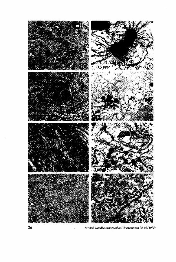
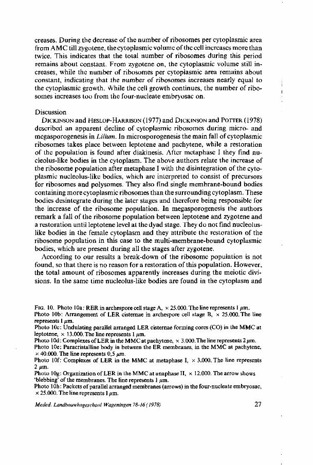
FIG. 10. Photo 10a: RER in archespore cell stage A, x 25.000. The line represents 1 urn. Photo 10b: Arrangement of LER cisternae in archespore cell stage B, x 25.000. The line represents 1 /xm. Photo 10c: Undulating parallel arranged LER cisternae forming cores (CO) in the MMC at leptotene, x 13.000. The line represents 1 /an. Photo lOd: Complexes ofLER in the MMC at pachytene, x 3.000. The line represents 2/xm. Photo 10e: Paracristalline body in between the ER membranes, in the MMC at pachytene, x 40.000. The line represents 0,5 ion. Photo lOf: Complexes of LER in the MMC at metaphase I, x 3.000. The line represents 2 urn. Photo 10g: Organization of LER in the MMC at anaphase II, x 12.000. The arrow shows 'Webbing' of the membranes. The line represents 1 trni. Photo 10h : Packets of parallel arranged membranes (arrows) in the four-nucleate embryosac, x 25.000. The line represents 1 /un.
Meded. Landbouwhogeschool Wageningen 78-16(1978) 27
these may be responsible for the increase of the ribosome population. A ribosome 'flow' during metaphase is described by WILLEMSE and LINSKENS
(1968). Because of the disintegration of the nuclear envelope, ribosomes from the karyoplasm can mix with cytoplasmic ribosomes, thus increasing the amount of cytoplasmic ribosomes.
In our material the multi-membrane-bound cytoplasmic bodies - the cores of the concentric ER complexes - contain at the same time in one cell both more and less ribosomes than the cytoplasm. It seems that these cores have no relation with the increase of the ribosome population.
3.3.2.4. Endop l a sm i c r e t i cu lum (ER) Figure 10 shows the development of ER during megasporogenesis. In the
archespore mother-cell rough endoplasmic reticulum (RER) is found. In the early archespore cell (figure 10a) the RER lamellae tend to be situated parallel to each other and to the nuclear envelope. The number of ribosomes attached is decreasing. In this stage also a few tubular smooth endoplasmic reticulum (TSER) cisternae are found. In the later archespore cell (figure 10b) the ER cisternae show only a few ribosomes attached. This type of endoplasmic reticulum is called lamellar endoplasmic reticulum (LER). The lamellae are arranged in parallel arrays. A number of ER cisternae is concentrated in spherical complexes, which look like unreeling balls (figure 10b). At leptotene the parallel arranged ER cisternae show undulations. At zygotene the ER cisternae tend to enclose cytoplasm by the formation of complexes of concentric arranged cisternae (figure 10c). The central core of these complexes sometimes contains mitochondria or lipid bodies. At pachytene the number of cisternae in the complexes varies from 5 to 20 (figure lOd). Incidentally electron-dense structures are observed in between the ER membranes, which are connected with both LER and TSER cisternae, sometimes the same time (figure 11). The size of these structures is of 0,3-0,6 /xm. The structures show units with different shapes, depending on the point of observation. The units have electron-dense contents and are surrounded by a membrane. Sometimes a regular lattice of rectangles is found (figure 10e). By using a goniometer rectangles, hexagons, round units and squares can be seen (figure 12). These structures are found at late zygotene, pachytene and diplotene and at the four-nucleate stages. During diplotene and diakinesis the number of concentrically arranged cisternae per complex decreases. Meanwhile TSER complexes appear in the cytoplasm. At metaphase I abundant complexes of TSER membranes are part of the polar regions of the spindle figure (mentioned before). Numerous complexes with 1-3 concentric cisternae are found (figure lOf). At two-nucleate stage small complexes of TSER-cisternae and concentric LER lamellae are perceptible. At metaphase II TSER complexes are found again in the polar regions of the spindle figure. At anaphase II these complexes disintegrate into groups of smaller ones. From anaphase II on, the LER cisternae show drastic changes. The concentric complexes are getting irregular; the membranes are 'blebbing' to form vesicles (figure 10g). From four-nucleate stage on, the complexes are
28 Meded. Landbouwhogeschool Wageningen 78-16(1978)
•.v.«.-:'
V v v . . v<-'f?'
• v * * - ": ,À : *
* .-**
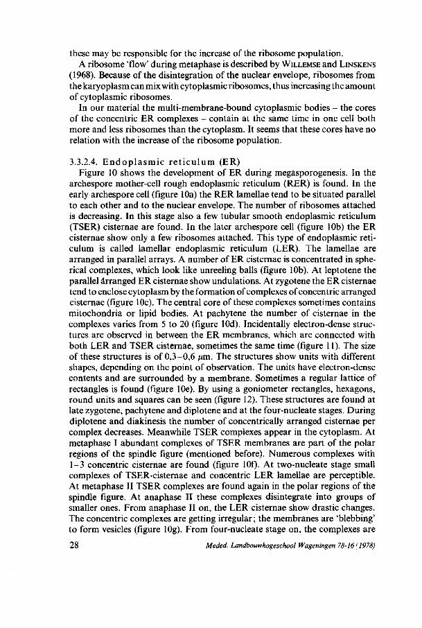
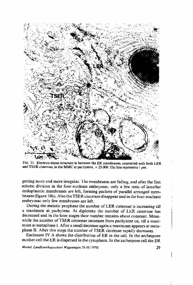
FIG. 11. Electron-dense structure in between the ER membranes, connected with both LER and TSER cisternae, in the MMC at pachytene, x 25.000. The line represents 1 jrni.
getting more and more irregular. The membranes are fading, and after the first mitotic division in the four-nucleate embryosac, only a few rests of lamellar endoplasmic membranes are left, forming packets of parallel arranged membranes (figure 10h). Also the TSER cisternae disappear and in the four-nucleate embryosac only few membranes are left.
During the meiotic prophase the number of LER cisternae is increasing till a maximum at pachytene. At diplotene the number of LER cisternae has decreased and in the later stages their number remains about constant. Meanwhile the number of TSER cisternae increases from pachytene on, till a maximum at metaphase I. After a small decrease again a maximum appears at meta-phase II. After this stage the number of TSER cisternae rapidly decreases.
Enclosure IV A shows the distribution of ER in the cell. In the archespore mother-cell the ER is dispersed in the cytoplasm. In the archespore cell the ER
Meded. Landbouwhogeschool Wageningen 78-16(1978) 29
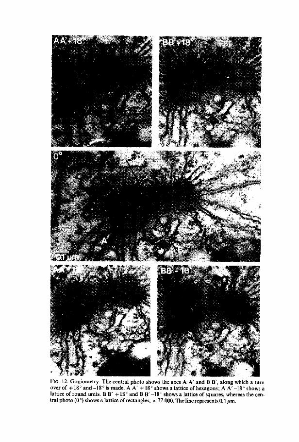
FIG. 12. Goniometry. The central photo shows the axes A A' and B B', along which a turn over of +18° and -18° is made. A A' +18° shows a lattice of hexagons; A A' -18° shows a lattice of round units. B B' +18° and BB'-18° shows a lattice of squares, whereas the central photo (0°) shows a lattice of rectangles, x 77.000. The line represents 0,1 /an.
cisternae are being arranged in the perinuclear zone. On the micropylar side more LER is found. At zygotene the ER complexes are distributed at random in the cytoplasm. At pachytene and diplotene a preference of the ER complexes for the micropylar side of the cell is found. At two-nucleate stage the complexes are particularly found in the zone between the two nuclei. This distribution remains till four-nucleate stage I, in which the complexes are distributed at random. After the polarization of the cell the complexes are found again in the zone between the polar distributed nuclei. TSER cisternae are distributed at random in the cytoplasm or found in small groups, except at metaphase, in which abundant complexes of TSER are found in the polar regions.
Discussion The concentric complexes of endoplasmic reticulum lamellae are described
by EYMÉ (1963), RODKIEWICZ and MIKULSKA (1965), DICKINSON and POTTER
(1978) during meiotic prophase of Lilium megasporogenesis. RODKIEWICZ and MIKULSKA suggest that these structures may have arisen under 'asphyxiai' conditions, caused by the megaspore mother-cell being coated with integuments during the growth of the ovule. In our opinion there seems to be a relation between these complexes of ER lamellae and the non-formation of cell-plates during meiosis (DE BOER-DE JEU, 1978: see discussion 3.4.).
Comparable electron-dense structures are also found in the endoplasmic reticulum of the oosphère of Mnium (BAJON-BARBIER, RIMSKY and WILLAIME,
1973). These authors find the same networks of hexagons and squares in the tilting range of the specimens, but at different tilting angles. They do not find a regular round unit structure. The ultrastructure and organization of prola-mellar bodies within etioplasts (GUNNING and STEER, 1975) show similarities with the electron-dense structures, with this difference, that the prolamellar bodies have a much larger size. In all these cases membrane components like lipoproteins are stacked lattice-like within structures which can be considered as paracristalline. In the etioplasts the prolamellar bodies are storage sites of lipoproteins, from which the synthesis of thylakoid membranes takes place.
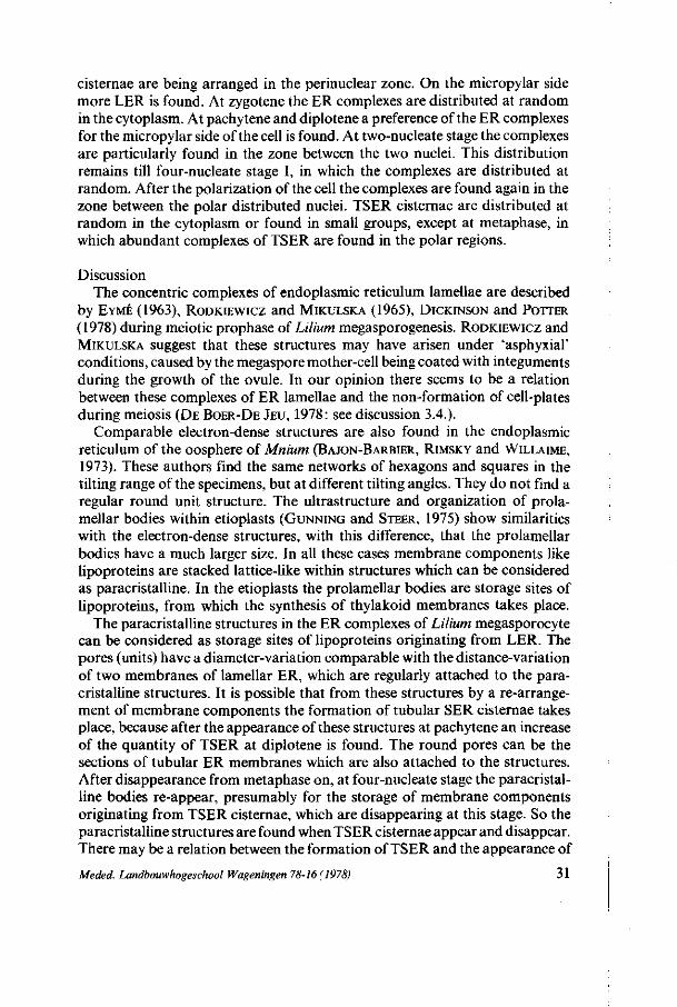
The paracristalline structures in the ER complexes of Lilium megasporocyte can be considered as storage sites of lipoproteins originating from LER. The pores (units) have a diameter-variation comparable with the distance-variation of two membranes of lamellar ER, which are regularly attached to the paracristalline structures. It is possible that from these structures by a re-arrangement of membrane components the formation of tubular SER cisternae takes place, because after the appearance of these structures at pachytene an increase of the quantity of TSER at diplotene is found. The round pores can be the sections of tubular ER membranes which are also attached to the structures. After disappearance from metaphase on, at four-nucleate stage the paracristalline bodies re-appear, presumably for the storage of membrane components originating from TSER cisternae, which are disappearing at this stage. So the paracristalline structures are found when TSER cisternae appear and disappear. There may be a relation between the formation of TSER and the appearance of
Meded. Landbouwhogeschool Wageningen 78-16(1978) 31
microtubules which are first found at pachytene. These microtubules are found near the TSER cisternae. BURGESS and NORTHCOTE (1968) suggest that SER is concerned with the transportation of microtubular proteins to the sites in which the aggregation of microtubules takes place.
3.3.2.5. D i c t yo somes In the premeiotic interphase the dictyosomes show a polar organization
with a proximal and a distal face. During the meiotic prophase the size of the dictyosome cisternae decreases whereas the number of cisternae (7-8) per dictyosome remains constant. At the end of prophase two types of dictyosomes are found. One type has 7-8 small, stacked cisternae and is found in the periferal zone. The other type has a small network-shaped appearance and is found in the perinuclear zone and in the spindle figures at meta- and anaphase (figure 6c). Also in the four-nucleate stage and the four-nucleate embryosac both types of dictyosomes are found.
During all the stages the dictyosomes produce small electron-dense vesicles, which show polysaccharidic contents after treatment with thiocarbohydrazide and silverproteinate (THIÉRY, 1967,2.2.4, figure 13b).
The number of dictyosomes per cytoplasmic area increases from premeiotic interphase till leptotene. At zygotene the number has diminished and remains about constant during the later phases, except at diplotene and metaphase I, in which a minimal number of dictyosomes per cytoplasmic area is found. Considering the growth of the cell volume the number of dictyosomes rapidly increases during premeiotic prophase, and in the stages after leptotene the increase of the dictyosomes is about equal to the growth of the cell, except at diplotene and metaphase I, where presumably no increase of dictyosomes takes place. In the premeiotic interphase and at leptotene the dictyosomes are distributed at random in the cytoplasm. From zygotene on, two zones of dictyosomes are found, separated by the network of ER, a periferal and a perinuclear zone. At metaphase I and II dictyosomes are found within the spindle apparatus, except at the polar regions. In the later stages the early mentioned zonal distribution of the dictyosomes remains.
3.3.2.6. Vesicle- and v acuo l a r sys tem During all stages various vesicles and vacuole-like structures are perceptible :
multivesicular bodies, which are structures, surrounded by a membrane, in which small electron-dense and clear vesicles are found. The matrix in which these vesicle structures are embedded has a light electron-dense appearance. These structures are found during the premeiotic interphase stages (figure 5), at pachytene, at metaphase II and in the four-nucleate stage. The electron-dense vesicles show a positive reaction when tested on polysaccharidic contents (THIÉRY, 1967,2.2.4.); electron-dense vacuoles, which are irregularly shaped structures, surrounded by a membrane and containing an electron-dense granular matrix with small clear vacuoles (figure 13). These structures are found in all the stages in which multi-
32 Meded. Landbouwhogeschool Wageningen 78-16(1978)
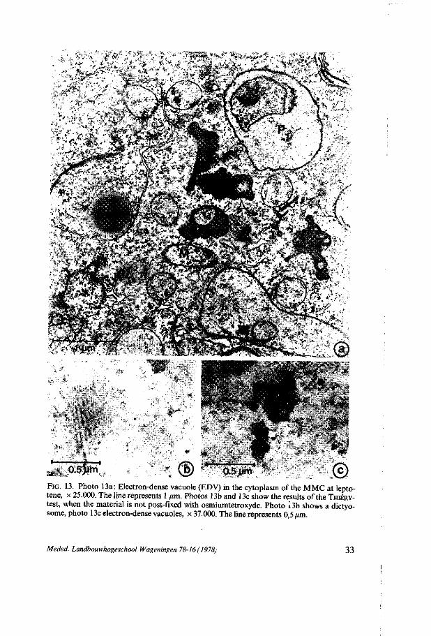
^ Q.sjim '. ® : ' 0.5>im .© FIG. 13. Photo 13a: Electron-dense vacuole (EDV) in the cytoplasm of the MMC at lepto-tene, x 25.000. The line represents 1 /im. Photos 13b and 13c show the results of the THIÉRY-test, when the material is not post-fixed with osmiumtetroxyde. Photo 13b shows a dictyo-some, photo 13c electron-dense vacuoles, x 37.000. The line represents 0,5 um.
Meded. Landbouwhogeschool Wageningen 78-16(1978) 33
vesicular bodies are absent. Perhaps these structures are modified multivesicular bodies with an electron-dense granular matrix. The electron-dense granular matrix shows a positive reaction for polysaccharides (figure 13c) ; electron-gray vesicles, which are smooth oval-like vesicles filled with light electron-dense material. Their structure is very similar to tubular smooth endoplasmic reticulum and derivation from TSER is supposed. These structures are found in all stages ; 'clear' vacuoles, which are smooth globular-shaped vacuoles filled with electron-transparent contents, sometimes with some condensed electron-dense material. These vacuoles appear at first in the two-nucleate stage. Their number increases during the next stages and in the four-nucleate stage after polarization the first large clear vacuoles appear, which are supposed to fuse together to form the central vacuole which is present in the four-nucleate embryosac and in the eight-celled embryosac.
3.3.2.7. P l asma membrane During all stages small vesicles and tubules filled with electron-dense material
are found between plasma membrane and cell-wall (figure 14a). In the later developmental stages also non-membrane-surrounded electron-dense granular material is found at the same sites. The number of these so-called paramural bodies increases during the development of the MMC. Even in the eight-celled embryosac paramural bodies are found and also within the cross-walls between the cells. It seems that the cross-walls at least partly consist of electron-dense vesicles and tubules (figure 14b).
After a treatment with carbohydrazide and silverproteinate (2.2.4.) polysaccharides has been proved to be present in these structures (figure 14c).
Discussion MARCHANT and ROBARDS (1968) give a classification of paramural bodies.
These bodies are called lomasomes, when the surrounded membranes have been derived from cytoplasmic membranes, and are called plasmalemmasomes when the surrounded membranes have been derived from the plasmalemma. ROBARDS and KIDWAI (1969) describe processes which participate in the formation of paramural bodies. They suppose that vesicles derived from dictyosomes and ER are transported to the cell-wall and fuse with the plasma membrane to form lomasomes. Also multivesicular bodies formed from dictyosome-derived vesicles move to the plasma membrane and fuse with it. All these processes are supposed to be associated with the formation and the transportation of various cell-wall precursors and of enzymes involved in cell-wall synthesis.
In Lilium megasporogenesis multivesicular bodies and electron-dense vacuoles, both containing polysaccharidic material, are found (3.3.2.6.). This polysaccharidic material is derived from dictyosomes (3.3.2.5.). The paramural bodies show polysaccharidic contents as well, so it is likely that the process described by ROBARDS and KIDWAI can also be observed in Lilium mega-
34 Meded. Landbouwhogeschool Wageningen 78-16(1978)

FIG. 14. Photo 14a: Paramural bodies (PB) between the plasma membrane and the cell-wall in the MMC at anaphase II, x 25.000. Photo 14b: Cross-wall (CW) in the eight-celled embryosac, x 25.000. Photo 14c: TmÉRY-test of paramural bodies in the archespore cell stage. The thickness of the cell-wall is much smaller than at anaphase II, x 25.000. The lines represent 1 fim.
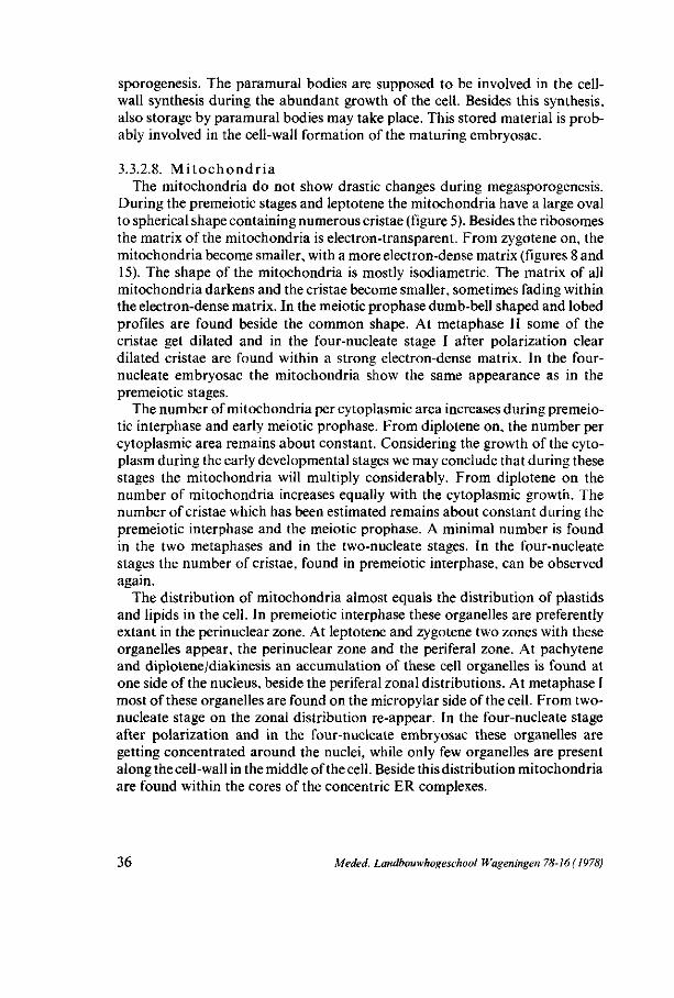
sporogenesis. The paramural bodies are supposed to be involved in the cell-wall synthesis during the abundant growth of the cell. Besides this synthesis, also storage by paramural bodies may take place. This stored material is probably involved in the cell-wall formation of the maturing embryosac.
3.3.2.8. M i t o chond r i a The mitochondria do not show drastic changes during megasporogenesis.
During the premeiotic stages and leptotene the mitochondria have a large oval to spherical shape containing numerous cristae (figure 5). Besides the ribosomes the matrix of the mitochondria is electron-transparent. From zygotene on, the mitochondria become smaller, with a more electron-dense matrix (figures 8 and 15). The shape of the mitochondria is mostly isodiametric. The matrix of all mitochondria darkens and the cristae become smaller, sometimes fading within the electron-dense matrix. In the meiotic prophase dumb-bell shaped and lobed profiles are found beside the common shape. At metaphase II some of the cristae get dilated and in the four-nucleate stage I after polarization clear dilated cristae are found within a strong electron-dense matrix. In the four-nucleate embryosac the mitochondria show the same appearance as in the premeiotic stages.
The number of mitochondria per cytoplasmic area increases during premeiotic interphase and early meiotic prophase. From diplotene on, the number per cytoplasmic area remains about constant. Considering the growth of the cytoplasm during the early developmental stages we may conclude that during these stages the mitochondria will multiply considerably. From diplotene on the number of mitochondria increases equally with the cytoplasmic growth. The number of cristae which has been estimated remains about constant during the premeiotic interphase and the meiotic prophase. A minimal number is found in the two metaphases and in the two-nucleate stages. In the four-nucleate stages the number of cristae, found in premeiotic interphase, can be observed again.
The distribution of mitochondria almost equals the distribution of plastids and lipids in the cell. In premeiotic interphase these organelles are preferently extant in the perinuclear zone. At leptotene and zygotene two zones with these organelles appear, the perinuclear zone and the periferal zone. At pachytene and diplotene/diakinesis an accumulation of these cell organelles is found at one side of the nucleus, beside the periferal zonal distributions. At metaphase I most of these organelles are found on the micropylar side of the cell. From two-nucleate stage on the zonal distribution re-appear. In the four-nucleate stage after polarization and in the four-nucleate embryosac these organelles are getting concentrated around the nuclei, while only few organelles are present along the cell-wall in the middle of the cell. Beside this distribution mitochondria are found within the cores of the concentric ER complexes.
36 Meded. Landbouwhogeschool Wageningen 78-16 ( 1978)

Discussion DICKINSON and HESLOP-HARRISON (1977) and DICKINSON and POTTER (1978)
describe a so-called cyclic development of the mitochondria in both Lilium micro- and megasporogenesis. The authors call the more condensed phase of the mitochondria 'promitochondria' whereas the common appearance is considered to be of the 'somatic' type.
In our material a transition of the 'somatic' type to the 'promitochondria' type - as it is called by DICKINSON et al. - is found at zygotene. A transition of the 'promitochondria' type back to the 'somatic' type is found in the four-nucleate embryosac. So in the early megagametogenesis changes of the ultra-structure of the mitochondria lead to the original ultrastructure. In 6.2.2.9 we shall revert to this subject.
Several authors suppose a replication of mitochondria by way of fission (BAGSHAW e.a., 1969) as a result of which dumb-bell shaped mitochondria appear, and by way of bud formation (TANDLER e.a., 1969) as a result of which lobed profiles are found. These mitochondrial configurations are found at premeiotic interphase and meiotic prophase of Lilium megasporogenesis. At the same stage the number of mitochondria increases. This indicates that previously described configurations can be related to the mitochondrial replication.
A similar aggregation of mitochondria, plastids and lipid bodies as found in our material was observed at the second meiotic anaphase of Onoclea sensibilis by MARENGO (1977) and at the first second telophase of Ribes rubrum micro-sporogenesis described by GENCVES (1967). Both authors find an accumulation of the cell organelles between the two daughter nuclei but they have no explanation for this phenomenon.
The accumulation of mitochondria, plastids and lipids in our material on one side of the nucleus at pachytene, diplotene and diakinesis may be related to a special function of these organelles at that site or to an occasional aggregation of these organelles. In the first case the presence of these organelles can be related to the beginning of the spindle apparatus formation. We may presume that an energy provision is needed at or nearby the site of synthesis of spindle figure components. In the second case we may presume that the rotation movement of the nucleus during pachytene/diplotene as described previously (3.3.1.3.) causes a similar movement to the cell organelles. When the nuclear movement stops the smaller cell organelles as mitochondria, plastids and lipids will be stopped by the nucleus and become accumulated at one site nearby the nucleus.
3.3.2.9. P l a s t i d s In the archespore mother-cell common plastids are found containing
thylakoids and plastoglobules. Also ribosomes and fibrils are perceptible in the matrix. The plastids generally have long-extended shapes while the so-called dumb-bell shapes are also found. Neither in this stage nor during all the other stages starch contents are found.
Meded. Landbouwhogeschool Wageningen 78-16(1978) 37
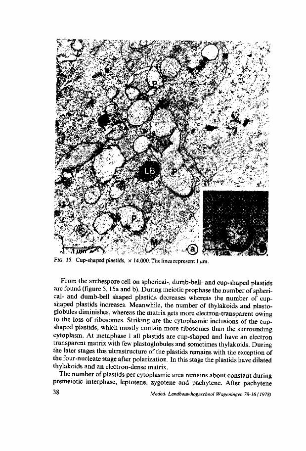
» : > • • -
y ,-*
• UP* *#* * '
iï&'M
FIG. 15. Cup-shaped plastids, x 14.000. The lines represent 1/mi.
From the archespore cell on spherical-, dumb-bell- and cup-shaped plastids are found (figure 5, 15a and b). During meiotic prophase the number of spherical- and dumb-bell shaped plastids decreases whereas the number of cup-shaped plastids increases. Meanwhile, the number of thylakoids and plasto-globules diminishes, whereas the matrix gets more electron-transparent owing to the loss of ribosomes. Striking are the cytoplasmic inclusions of the cup-shaped plastids, which mostly contain more ribosomes than the surrounding cytoplasm. At metaphase I all plastids are cup-shaped and have an electron transparent matrix with few plastoglobules and sometimes thylakoids. During the later stages this ultrastructure of the plastids remains with the exception of the four-nucleate stage after polarization. In this stage the plastids have dilated thylakoids and an electron-dense matrix.
The number of plastids per cytoplasmic area remains about constant during premeiotic interphase, leptotene, zygotene and pachytene. After pachytene
38 Meded. Landbouwhogeschool Wageningen 78-16(1978)
their number diminishes till the four-nucleate embryosac. Considering the cytoplasmic growth during premeiotic interphase and early prophase an intensive replication of plastids takes place at these stages. At diplotene the replication of the plastids cannot keep up with the growth of the cytoplasm causing a decrease of the number of plastids per cytoplasmic area.
The distribution of the plastids equals the distribution of mitochondria (3.3.2.8.) and lipids. No plastids are ever found to be present within the cores of the concentric complexes.
Discussion DICKINSON and HESLOP-HARRISON (1977) and DICKINSON and POTTER (1978)
describe a so-called cycle of 'dedifferentiation' and 'redifferentiation' of the plastids during Lilium micro- and megasporogenesis. In our material a transition from a 'differentiated' type to a 'dedifferentiated' type according to DICKINSON et al. occur in the archespore cell. The manifestation of the 'dedifferentiated' plastids however remains during all the developmental stages, also in the eight-celled embryosac. So we cannot speak about a 'cycle' in our material. In 6.2.2.10 we shall revert to this subject.
It is generally assumed that plastids multiply by binary fission (GREEN, 1964). Fission configurations like dumb-bell shapes are commonly found in cells. In our material dumb-bell-shaped plastids are found during premeiotic interphase and meiotic prophase. In these stages a replication of plastids occurs, which makes us conclude that the dumb-bell-shaped plastids are related to the replication of plastids.
3.3.2.10. L ip id bodies At the premeiotic interphase and the meiotic prophase the lipid bodies are
small with a diameter of 0,2-0,8 /an. At late zygotene small clear vesicles are attached to the lipid bodies. The vesicles sometimes seem to be continue with TSER cisternae. At diakinesis and metaphase I the diameter of the lipid droplets has increased till a size varying from 0,6-4,0 (im. The lipid bodies at these stages are accumulated in complexes, mostly consisting of large lipid bodies in the centre, surrounded by small lipid bodies closely attached to the central body. After metaphase I these complexes have disappeared. From metaphase II on, sometimes small clear vacuoles are found within the lipid bodies. In these later developmental stages the diameter of the lipid bodies varies from 0,4 to 7,0 /tm.
The amount of lipid per cytoplasmic area apparently increases during meiotic prophase. At metaphase I the maximal amount of lipid per cytoplasmic area is found. After metaphase I the amount of lipid diminishes. Considering the cytoplasmic growth of the cell, the amount of lipid increases enormously during the meiotic prophase.
The distribution of the lipids equals the distribution of the mitochondria and plastids (3.3.2.8.). Lipids are also found within the cores of the concentric LER complexes. At metaphase I the complexes of lipid bodies preferently exist on
Meded. Landbouwhogeschool Wageningen 78-16(1978) 39
both sides of the spindle figure.
Discussion The attachment of vesicles to the lipid bodies is also described by POWELL
(1976). This author suggests enzymic contents of these vesicles for the digestion of lipidic compounds. In this way the lipid bodies are consumed. In our material the vesicles are continuous with tubular ER membranes. By this passway lipid compounds are probably transported to the sites of synthesis. It is also possible that the TSER membranes are responsible for the transportation of the digestive enzymes to the lipid bodies. The appearance of the clear vacuoles within the lipid bodies at later stages can also be related to the digestion of these lipid bodies.
A 'cycle' of lipidic aggregation and disaggregation is described by HESLOP-
HARRISON and DICKINSON (1967) in Lilium microsporogenesis. The authors assume a relation between the break-down of the nuclear envelope and an aggregation of lipid bodies. They suggest a changement of the degree of hydratation of the cytoplasm and an increase of the pH by the break-down of the nuclear envelope causing an aggregation of lipid bodies. In our material also an aggregation of lipid bodies appears probably under similar conditions but only at metaphase I.
3.3.2.11. P l a smode sma t a In the premeiotic interphase numerous plasmodesmata in all parts of the
cell-wall are perceptible. During meiotic prophase only in the chalazal part of the wall plasmodesmata occur, although their number decreases. Generally only in the section parts of the plasmodesmata are visible. Sometimes pieces attached to the plasma membranes without the connecting middle are observed, or only the middle part of the tubule is present. After metaphase I no plasmodesmata are found intact, only remainders.
It is very likely that the plasmodesmata only partly transverse the cell-wall, implicating that there is no plasmodesmata connection between the megasporo-cyte after metaphase I/megagametophyte and the surrounding nucellus cells.
3.3.2.12. Cel l -wal l During megasporogenesis the cell-wall is constantly thickening. In the two-
nucleate stage a maximum thickness of 0,4-0,8 /im is reached and this thickness remains. In Lilium the plasmodesmata seem to become closed off by cell-wall growth during megasporogenesis. In this way the cell gets isolated from the surrounding nucellus cells, as far as its contact with plasmodesmata is concerned. After application of the fluorescence-aniline-blue method used by ROD-KIEWICZ (1970) no callosic substances are found in the cell-wall of Lilium. This result is in accordance with the results of RODKIEWICZ for Lilium species.
40 Meded. Landbouwhogeschool Wageningen 78-16 (1978)
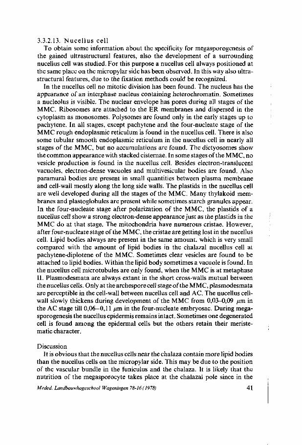
3.3.2.13. Nucellus cell To obtain some information about the specificity for megasporogenesis of
the gained ultrastructural features, also the development of a surrounding nucellus cell was studied. For this purpose a nucellus cell always positioned at the same place on the micropylar side has been observed. In this way also ultra-structural features, due to the fixation methods could be recognized.
In the nucellus cell no mitotic division has been found. The nucleus has the appearance of an interphase nucleus containing heterochromatin. Sometimes a nucleolus is visible. The nuclear envelope has pores during all stages of the MMC. Ribosomes are attached to the ER membranes and dispersed in the cytoplasm as monosomes. Polysomes are found only in the early stages up to pachytene. In all stages, except pachytene and the four-nucleate stage of the MMC rough endoplasmic reticulum is found in the nucellus cell. There is also some tubular smooth endoplasmic reticulum in the nucellus cell in nearly all stages of the MMC, but no accumulations are found. The dictyosomes show the common appearance with stacked cisternae. In some stages of the MMC, no vesicle production is found in the nucellus cell. Besides electron-translucent vacuoles, electron-dense vacuoles and multivesicular bodies are found. Also paramural bodies are present in small quantities between plasma membrane and cell-wall mostly along the long side walls. The plastids in the nucellus cell are well developed during all the stages of the MMC. Many thylakoid membranes and plastoglobules are present while sometimes starch granules appear. In the four-nucleate stage after polarization of the MMC, the plastids of a nucellus cell show a strong electron-dense appearance just as the plastids in the MMC do at that stage. The mitochondria have numerous cristae. However, after four-nucleate stage of the MMC, the cristae are getting lost in the nucellus cell. Lipid bodies always are present in the same amount, which is very small compared with the amount of lipid bodies in the chalazal nucellus cell at pachytene-diplotene of the MMC. Sometimes clear vesicles are found to be attached to lipid bodies. Within the lipid body sometimes a vacuole is found. In the nucellus cell microtubules are only found, when the MMC is at metaphase II. Plasmodesmata are always extant in the short cross-walls mutual between the nucellus cells. Only at the archespore cell stage of the MMC, plasmodesmata are perceptible in the cell-wall between nucellus cell and AC. The nucellus cell-wall slowly thickens during development of the MMC from 0,03-0,09 pm in the AC stage till 0,06-0,11 pm in the four-nucleate embryosac. During megasporogenesis the nucellus epidermis remains intact. Sometimes one degenerated cell is found among the epidermal cells but the others retain their meriste-matic character.
Discussion It is obvious that the nucellus cells near the chalaza contain more lipid bodies
than the nucellus cells on the micropylar side. This may be due to the position of the vascular bundle in the funiculus and the chalaza. It is likely that the nutrition of the megasporocyte takes place at the chalazal pole since in the
Meded. Landbouwhogeschool Wageningen 78-16 ( 1978) 4 1
developing MMC a high amount of lipid bodies is found, also near the chalazal pole.
Apart from microtubule formation in the nucellus cell, when the MMC is at metaphase II, and the appearance of the plastids when the MMC is at four-nucleate stage, no details of the ultrastructural development of the nucellus cell can be correlated with established details of the ultrastructure development of the megasporocyte/megagametophyte. Each of them has its own specific development.
3.4. FINAL DISCUSSION
In addition to the short discussions concerning the various cell organelles presented in the individual chapters, this chapter contains a more conclusive discussion and a summary of the results.
Because the Lilium MMC does not form walls after nuclear division, the cell organelles which normally are related to cell-plate formation : dictyosomes, ER, microtubules (HEPLER and NEWCOMB, 1967) are discussed in view of this fact. The few dictyosomes present within and near the spindle figure show a low activity since they produce few visicles. The ultrastructure of these dictyosomes in the MMC differs from the ultrastructure of the dictyosomes in nucellus cells. In the nucellus cells normal cell-plate formation takes place after nuclear division. In the MMC however, it seems that the dictyosomes have not the possibility to produce vesicles which are supposed to fuse with the phragmo-plast, thus forming a cell-plate (HEPLER and NEWCOMB, 1967, HEPLER and JACKSON, 1968). Instead of vesicle producing dictyosomes, numerous concentric complexes of endoplasmic reticulum cisternae are extant.
According to the endomembrane concept as described by MORRÉ and MOLLENHAUER (1974) both dictysomes and ER form a continuous system of endomembranes in various stages of differentiation. We suppose that in normally dividing cells a formation of a large endomembrane system and a differentiation of this system into many dictyosomes takes place. In the Lilium MMC this endomembrane formation may take place without subsequent differentiation into functional organelles i.e. dictyosomes necessary for cell-plate formation (DE BOER-DE JEU, 1978). Endomembrane formation then should lead to an increase of the ER system. The numerous ER cisternae are concentrically stacked because in this arrangement they occupy minimal room.
However, commonly shaped dictyosomes are found in the MMC, but only in the zone along the cell-wall, where they produce vesicles filled with polysaccharides necessary for cell-wall growth and thickening. In the paramural bodies also vesicles with polysaccharidic contents are found. Their number is increasing during the development of the MMC. In the eight-celled embryosac the cross-walls seem to consist of similar structures as the paramural bodies. Supposing that even in the eight-nucleate embryosac not enough dictyosomes and vesicles derived from dictyosomes are extant to form common cell-plates,
42 Meded. Landbouwhogeschool Wageningen 78-16(1978)
the cross-walls may arise by a kind of a cleavage of the cell. By this cleavage cross-walls with an uncommon appearance, consisting of similar structures as paramural bodies, may occur.
Based on the distribution of the plasmodesmata it is acceptable that the MMC is fed on the chalazal side of the cell, the side where the vascular bundle ends in the nucellus and stored reserve substances as lipids are present in the nucellus cells. Beside through plasmodesmata it is also possible that nutrients are diffusing through the cell-wall, especially in the later developmental stages when no plasmodesmata are found anymore. However, it is also possible that in these stages the MMC has enough stored nutrients so that a transportation of nutrition substances through the chalazal cell-wall is not necessary. This is in agreement with the observation that after the disappearance of the plasmodesmata the amount of stored lipids gradually decreases.
In the four-nucleate stage a polarization takes place by the movement of two nuclei to the chalazal pole, thus creating a cell with one nucleus at the micro-pylar pole and three nuclei at the chalazal pole. There is no information to determine clearly, which type(s) of organelle(s) is (are) responsible for the movement of the nuclei to the chalazal pole. Neither microtubules nor microfilaments are found to be attached to the nuclei. No structural differences are found between the four nuclei either. During this polarization the MMC is getting longer, and its width is getting narrower. It is possible that this change of size is related to the movement of the nuclei. After polarization the distribution of the ER lamellae has changed as well. Maybe the endoplasmic reticulum is involved in the movement of the nuclei.
After the polarization in the four-nucleate cell, the first large clear vacuoles appear. These vacuoles seem to fuse together to form a central vacuole. This central vacuole maintains the polar distribution of the nuclei. During the further development of the cell a volume growth takes place during which the cytoplasm and the central vacuole grow synchronously.
Already in the early developmental stages the polar distribution of the cell organelles indicates a polarity of the cell. The distribution of the parallel arranged LER cisternae show a difference between micropylar and chalazal side of the cell. The perinuclear - and the periferal zone in which most of the plastids, mitochondria and lipid bodies are found, are separated from each other by endoplasmic reticulum. After diplotene no difference in the ER distribution between the micropylar and chalazal side is found. The ER cisternae then are mostly situated in the centre of the cell. The clear zonal distribution of plastids, mitochondria and lipid bodies in the periferal- and perinuclear zones remains until the movement of the nuclei has taken place, when periferal- and perinuclear zones are fused together and all organelles are found near the nuclei. It seems that in Lilium the endoplasmic reticulum plays an important role in the polar distribution of the cell organelles indicating the presence of an early polarity of the cell.
Meded. Landbouwhogeschool Wageningen 78-16(1978) 4 3
4. M E G A S P O R O G E N E S I S A N D EARLY M E G A G A M E T O G E N E S I S IN ALLIUM CLP A L.
4.1. INTRODUCTION
The members of the genus Allium have the bisporic type of embryosac development. MAHESWARI (1950) described an inequal division of the mega-spore mother-cell. The micropylar cell is much smaller and soon degenerates before the second meiotic division. After the second nuclear meiotic division the chalazal cell forms a two-nucleate cell, the functional megaspore. After two mitotic nuclear divisions the embryosac is formed. PORTER (1936) describes a similar development of megasporogenesis in Allium mutabile. According to RUTISHAUSER (1969) the MMC undergoes an equal division, forming a dyad with one nucleus per cell, followed by the second meiotic division of the nuclei, leading to a dyad with two nuclei per cell. Eventually the micropylar cell of the dyad degenerates.
The megasporogenesis in Allium has not been studied before on electron-microscopic level. From the microsporogenesis, the nucleolar ultrastructure during meiotic prophase of Allium cepa L. was studied by GIMÉNEZ-MARTIN
and STOCKERT (1970) and by ESPONDA and GIMÉNEZ-MARTIN (1975). Nucleolar ultrastructure of Allium cepa during mitosis was described by CHOUINARD
(1975) and by MORENO DIAZ DE LA ESPINA and RISUENO (1976). Also ultra-structural changes of cell organelles concerned by cell-plate formation during mitosis of Allium sativum were described by HANZELY and VIGIL (1975).
4.2. MATERIAL AND METHODS
For light-microscopy the preparation of 3 fim thick sections was used (2.1.2.). For electron-microscopy the glutaraldehyde-osmiumtetroxyde fixation
(2.2.1.) was applied, often in combination with the re-embedding technics. The glutaraldehyde-osmiumtetroxyde fixation was applied as follows:
Ovules were dissected from the ovaries of the, in glasshouse or in the garden grown, Allium cepa L. plants and fixed for 1,5 hour in 5% glutaraldehyde in 0,1 M phosphate buffer at pH 7,2. At the very young stages, whole ovaries or parts of ovaries were fixed. The fixed material was rinsed thoroughly in 0,1 M phosphate buffer containing 0,2 M saccharose. Postfixation occurred in 2% Os0 4 in 0,1 M phosphate buffer containing 0,2 M saccharose during 20 hours. Longitudinal sections of the epon-embedded ovules were poststained with leadcitrate (REYNOLDS, 1963). The orientation of the ovules for longitudinally sectioning was very difficult because of their rounded shapes. Moreover, the orientation of the ovules within the ovary strongly varies so that within one section of an ovary there is at most one longitudinally sectioned ovule.
44 Meded. Landbouwhogeschool Wageningen 78-16 (1978)
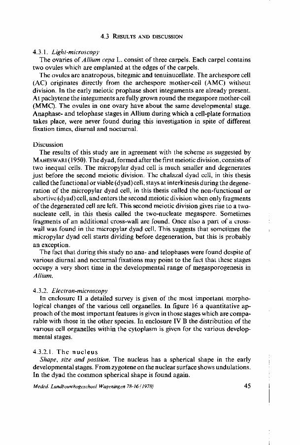
4.3 RESULTS AND DISCUSSION
4.3.1. Light-microscopy The ovaries of Allium cepa L. consist of three carpels. Each carpel contains
two ovules which are emplanted at the edges of the carpels. The ovules are anatropous, bitegmic and tenuinucellate. The archespore cell
(AC) originates directly from the archespore mother-cell (AMC) without division. In the early meiotic prophase short integuments are already present. At pachytene the integuments are fully grown round the megaspore mother-cell (MMC). The ovules in one ovary have about the same developmental stage. Anaphase- and telophase stages in Allium during which a cell-plate formation takes place, were never found during this investigation in spite of different fixation times, diurnal and nocturnal.
Discussion The results of this study are in agreement with the scheme as suggested by
MAHESWARI ( 1950). The dyad, formed after the first meiotic division, consists of two inequal cells. The micropylar dyad cell is much smaller and degenerates just before the second meiotic division. The chalazal dyad cell, in this thesis called the functional or viable (dyad) cell, stays at interkinesis during the degeneration of the micropylar dyad cell, in this thesis called the non-functional or abortive (dyad) cell, and enters the second meiotic division when only fragments of the degenerated cell are left. This second meiotic division gives rise to a two-nucleate cell, in this thesis called the two-nucleate megaspore. Sometimes fragments of an additional cross-wall are found. Once also a part of a cross-wall was found in the micropylar dyad cell. This suggests that sometimes the micropylar dyad cell starts dividing before degeneration, but this is probably an exception.
The fact that during this study no ana- and telophases were found despite of various diurnal and nocturnal fixations may point to the fact that these stages occupy a very short time in the developmental range of megasporogenesis in Allium.
4.3.2. Electron-microscopy In enclosure II a detailed survey is given of the most important morpho
logical changes of the various cell organelles. In figure 16 a quantitative approach of the most important features is given in those stages which are comparable with those in the other species. In enclosure IV B the distribution of the various cell organelles within the cytoplasm is given for the various developmental stages.
4.3.2.1. The nuc leus Shape, size and position. The nucleus has a spherical shape in the early
developmental stages. From zygotene on the nuclear surface shows undulations. In the dyad the common spherical shape is found again.
Meded. Landbouwhogeschool Wageningen 78-16 (1978) 45
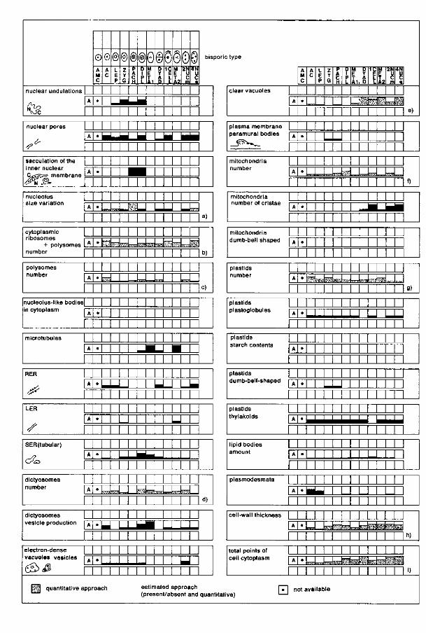
nuclear undulations |
^ ^ 3& r
0 A M C
•
0 A C
@ L E P
® Z Y
®(§)0©0§0© p A C H
u
P L
M E T A1
D Y A D
1C E L L
M E T A2
2N U C m
4 N u c
bisporictype
nuclear pores |
1 A
^ r sacculation of the |
inner nuclear r p
&%B. r nucleolus | size variation i —
A
cytoplasmic [ nbosomes i —
+ polysomes VÎL number 1
polysomes | number i .
A
nucleolus-like bodies|
in cytoplasm r ~
microtubules |
jT
RER 1 _
^ ^ £< [~ LER | _
[A"
s r SER(tubular) |
s? XL C/CD 1—
dictyosomes number i A
dictyosomes | vesicle production i A
electron-dense | vacuoles vesicles rr
A
eS>̂ s r
1 M M M M M • • ^ ^ ^ ^ • _ _ M M IL____J I ^ ^ L ^ i
1 1 1 1 ! 1 1 1 ! 1 1
•
•
•
•
•
•
•
•
•
•
•
•
M M M M M 1 1 ^ B 1 1 1 1 1 M 1 1 1 1 1 1 1 1
1 1 1 1 1 1 1 1 1 1
1 1 1 1 1 1 ! 1 1 1
1 1 1 1 1 1 1 1 1 1 SfcsiksiL* | l _
sSïèss te î llfeiraSI M M 1 ! M ! 1
M 1 ! 1 M 1 1 1 sd—1—LI 1 l.J 1 L L
1 1 1 ! 1 M 1 1 1
1 1 1 M 1 1 1 1 1 J J 1 M 1 1 1 1 1 M 1 M 1 1 1 M
1 1 1 1 1 1 1 1 M 1 1 1 L *_J • 1 1 1 ! 1 1 1 ! 1 1 1
1 ! 1 1 1 1 ! 1 M -LU
1 1 1 1 1 1 1 1 1
1 U U L U 1 1 M 1 1 1 M 1 1 1 M 1 U 1 M I N I
1 1 1 M 1 1 M 1 1 I I — 1 1 1 1 1 1 1 1 1 1 1 1 1 1
1 1 1 1 1 1 1 M 1 raisLJ K»»J™Jsrîckd—
1 1 1 1 1 1 1 1 M
1 1 1 1 1 1 1 1 1 1 _ l 1 1 L ^ 1 1 1 1
1 1 1 1 1 1 1 1 1 1
a)
b)
c)
à)
1 1 M M 1 1 1 J 1 1 I I 1 1 1 1 U 1 M 1 1 M M 1
H quantitative approach estimated approach (present/absent and quar titati
clear vacuoles j
| A
A M C
•
A C
L E P
Z Y G
P A C H
D 1 P 1-
M E T A1
D Y A D
n'*£
1C E L L
M E
!*f
2f4
m
TT1
4N
n'A e)
plasma membrane j paramural bodies i A
_ i ï r v ^ _ - | mitochondria | number rr
A
mitochondria | number ot cristae i —
mitochondria | dumD-beii shaped 1 A
plastids | number i .
A
plastids | plastoglobules r~r
plastids ! starch contents r—
A
plastids | dumb-bell-shaped rr
plastids | i h y i a K O i a s 1 A
l ipid bodies I amount I A
plasmodesmata |
1 A
cell-wall thickness j
!A
total points of | ceil cytoplasm i A
•
•
•
•
'
•
•
•
•
•
'
•
M 1 1 1 M 1 ! L U M ! 1 1 1 1 M M 1 M 1 1 1 1 1 1
J |-M J^-^Tp-^.;] | ,,, ,, |...i k -4,
1 M 1 1 1 1 1
1 M 1 M 1 1 1 1 1 1 1 1 M 1 M M 1 1 1
1 1 1 1 1 1 M _L 1 1 1 J 1 1 1 1 M 1 1 M 1
1 J 1 1 1 1 1 1 wimMm^JJ^kém
1 1 1 1 1 M 1
M 1 1 M ! 1 1 1 1 1 1 1 U 1 1 1 1 1 M 1
1 1 1 1 1 1 1 1 1 1 1 1 1 1 1 1 1 1 M 1 1 1 1 1 1 1 1 1 1 1 1 1 L U 1 M 1 1 1 1 1 M M 1 M 1 1 M 1
M 1 1 1 1 1 1 1 1 1 1 1 1 LI 1 1 M 1 1 1 1
M 1 M 1 M • U 1 U U 1
M 1 1 1 1 1 1 i-J M^W^istete
M 1 1 M 1 1 1 1 1 1 1 1 1 1
^
-
.
"
g)
-
_
E h)
•U04pMdifcép«i 1 1 1 1 1 1 M 1 M ')
[ T l not available ve) '—'
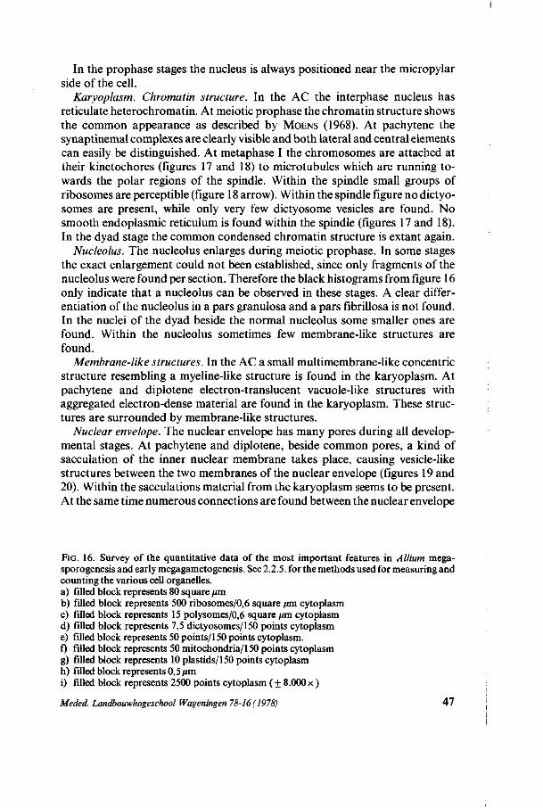
In the prophase stages the nucleus is always positioned near the micropylar side of the cell.
Karyoplasm. Chromatin structure. In the AC the interphase nucleus has reticulate heterochromatin. At meiotic prophase the chromatin structure shows the common appearance as described by MOENS (1968). At pachytene the synaptinemal complexes are clearly visible and both lateral and central elements can easily be distinguished. At metaphase I the chromosomes are attached at their kinetochores (figures 17 and 18) to microtubules which are running towards the polar regions of the spindle. Within the spindle small groups of ribosomes are perceptible (figure 18 arrow). Within the spindle figure no dictyo-somes are present, while only very few dictyosome vesicles are found. No smooth endoplasmic reticulum is found within the spindle (figures 17 and 18). In the dyad stage the common condensed chromatin structure is extant again.
Nucleolus. The nucleolus enlarges during meiotic prophase. In some stages the exact enlargement could not been established, since only fragments of the nucleolus were found per section. Therefore the black histograms from figure 16 only indicate that a nucleolus can be observed in these stages. A clear differentiation of the nucleolus in a pars granulosa and a pars fibrillosa is not found. In the nuclei of the dyad beside the normal nucleolus some smaller ones are found. Within the nucleolus sometimes few membrane-like structures are found.
Membrane-like structures. In the AC a small multimembrane-like concentric structure resembling a myeline-like structure is found in the karyoplasm. At pachytene and diplotene electron-translucent vacuole-like structures with aggregated electron-dense material are found in the karyoplasm. These structures are surrounded by membrane-like structures.
Nuclear envelope. The nuclear envelope has many pores during all developmental stages. At pachytene and diplotene, beside common pores, a kind of sacculation of the inner nuclear membrane takes place, causing vesicle-like structures between the two membranes of the nuclear envelope (figures 19 and 20). Within the sacculations material from the karyoplasm seems to be present. At the same time numerous connections are found between the nuclear envelope
FIG. 16. Survey of the quantitative data of the most important features in Allium mega-sporogenesis and early megagametogenesis. See 2.2.5. for the methods used for measuring and counting the various cell organelles. a) filled block represents 80 square fim b) filled block represents 500 ribosomes/0,6 square /an cytoplasm c) filled block represents 15 polysomes/0,6 square /an cytoplasm d) filled block represents 7,5 dictyosomes/150 points cytoplasm e) filled block represents 50 points/150 points cytoplasm. f) filled block represents 50 mitochondria/150 points cytoplasm g) filled block represents 10 plastids/150 points cytoplasm h) filled block represents 0,5 fim i) filled block represents 2500 points cytoplasm (+ 8.000x )
Meded. Landbouwhogeschool Wageningen 78-16(1978) 47
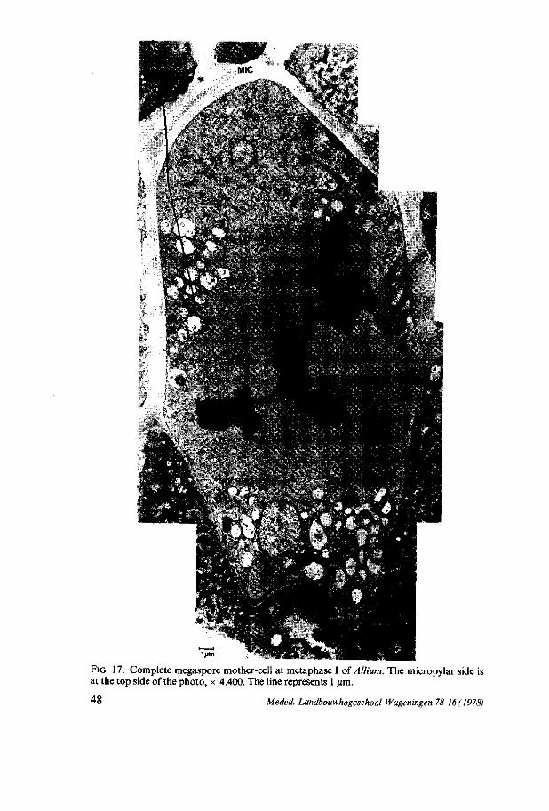
FIG. 17. Complete megaspore mother-cell at metaphase I of Allium. The micropylar side is at the top side of the photo, x 4.400. The line represents 1 /jm.
48 Meded. Landbouwhogeschool Wageningen 78-16(1978)
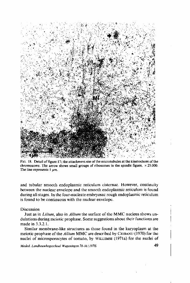
. i
'h •• . '•', • - .
. / •
^ . • -, • ' • > J ï> . *• f' , , 'i * ' - " ; - - • '- ; . . £
•• ••;•• -•-• > ':. >•!•' -' :• A , . ;>fe;:r- • -M^¥ : •-.'• • • •::••• \A;: . . - • ' .V.-^V .-.. » -V" .%>* ' .••• O'-^--%,.;*•:•
• -*A:.- \ v * 0 . - :;ï-»\.r *,:*5 % W :&<* x:-,«
vil ^ ; . "v^;; :• : • .*^"
- .^ • •v - - ' •
FIG. 18. Detail of figure 17 ; the attachment site of the microtubules at the kinetochore of the chromosome. The arrow shows small groups of ribosomes in the spindle figure, x 25.000. The line represents 1 fim.
and tubular smooth endoplasmic reticulum cisternae. However, continuity between the nuclear envelope and the smooth endoplasmic reticulum is found during all stages. In the four-nucleate embryosac rough endoplasmic reticulum is found to be continuous with the nuclear envelope.
Discussion Just as in Lilium, also in Allium the surface of the MMC nucleus shows un
dulations during meiotic prophase. Some suggestions about their functions are made in 3.3.2.1.
Similar membrane-like structures as those found in the karyoplasm at the meiotic prophase of the Allium MMC are described by CIOBANU (1970) for the nuclei of microsporocytes of tomato, by WILLEMSE (1971a) for the nuclei of
Meded. Landbouwhogeschool Wageningen 78-16 (1978) 49
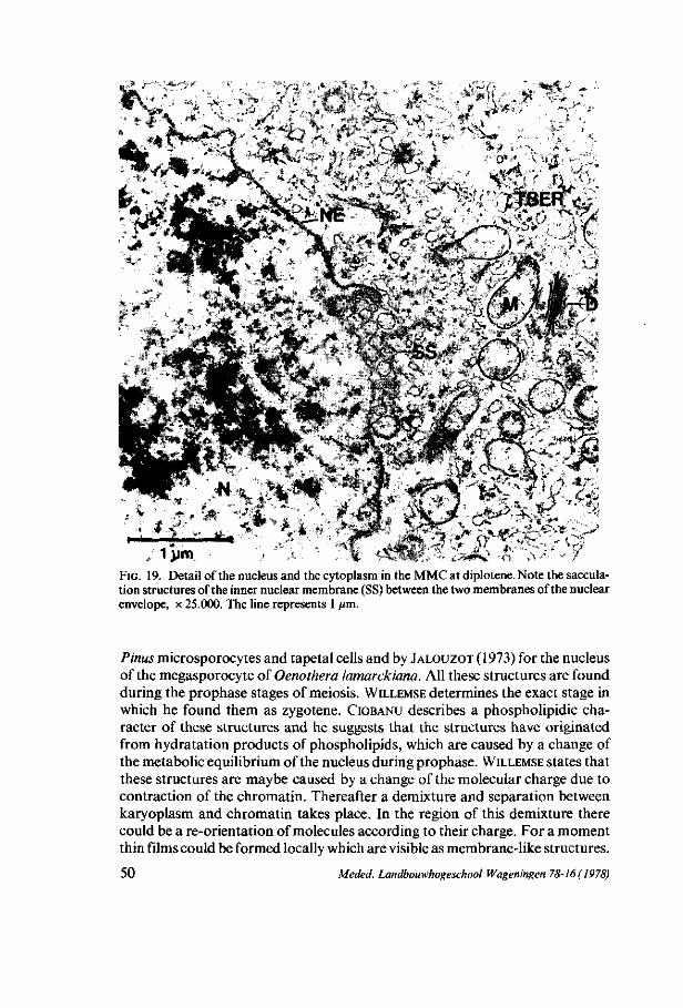
•u
, / # " > *
. * • * *s«
* » -.J*
--' >s
FIG. 19. Detail of the nucleus and the cytoplasm in the MMC at diplotene. Note the sacculation structures of the inner nuclear membrane (SS) between the two membranes of the nuclear envelope, x 25.000. The line represents 1 /an.
J t «
Pinus microsporocytes and tapetal cells and by JALOUZOT (1973) for the nucleus of the megasporocyte of Oenothera lamarckiana. All these structures are found during the prophase stages of meiosis. WILLEMSE determines the exact stage in which he found them as zygotene. CIOBANU describes a phospholipidic character of these structures and he suggests that the structures have originated from hydratation products of phospholipids, which are caused by a change of the metabolic equilibrium of the nucleus during prophase. WILLEMSE states that these structures are maybe caused by a change of the molecular charge due to contraction of the chromatin. Thereafter a demixture and separation between karyoplasm and chromatin takes place. In the region of this demixture there could be a re-orientation of molecules according to their charge. For a moment thin films could be formed locally which are visible as membrane-like structures.
50 Meded. Landbouwhogeschool Wageningen 78-16(1978)
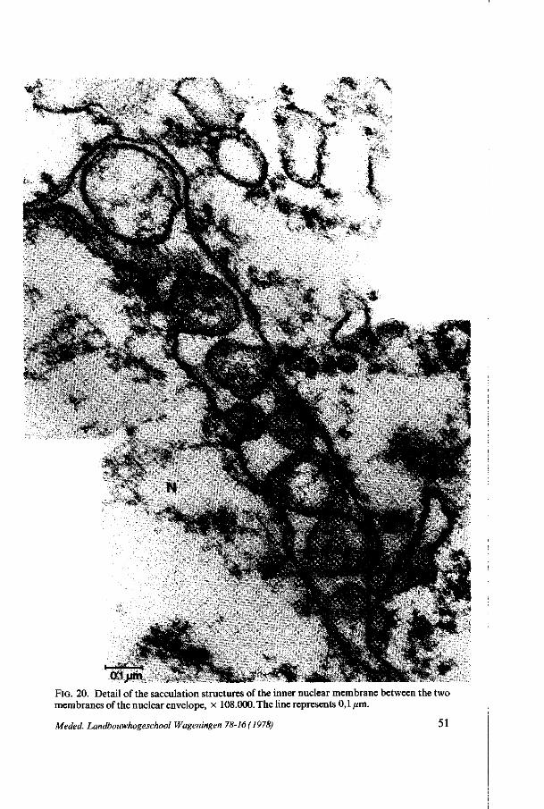
FIG. 20. Detail of the sacculation structures of the inner nuclear membrane between the two membranes of the nuclear envelope, x 108.000. The line represents 0,1 /mi.
Meded. Landbouwhogeschool Wageningen 78-16 ( 1978) 51
The whole process may be analogous to the formation of coacervate droplets'. In Allium the membrane-like structures are found in the AC and in the MMC
at pachytene and diplotene. In the latter stages a contraction of the chromatin takes place in preparation of the condensed phase of the chromosomes at meta-phase I. So the hypothesis of WILLEMSE may be applicable for Allium too. In Allium an abundant sacculation of the inner nuclear membrane is found at many sites of the nuclear envelope. As we already discussed for Lilium (3.3.2.1.), a specific function for these structures is not evident. There is always found a continuity of the karyoplasm and the sacculation, whereas a continuity of the sacculation contents and the cytoplasm was not observed. In 6.2.2.2. we shall revert to this subject.
4.3.2.2. M i c r o t ubu l e s The first microtubules appear at diplotene in the perinuclear zone. They are
found near the nuclear envelope and near the tubular smooth endoplasmic reticulum (TSER) cisternae which are continuous with the outer nuclear envelope. Within the spindle figure many spindle microtubules with a rough surface are perceptible, attached to the chromosomes at their kinetochores. In the viable dyad cell sometimes a cell-plate is partly formed during ana-telophase. Small electron-dense vesicles string together within the phragmoplast, in which a lot of microtubules are running perpendicularly to the equatorial plane.
4.3.2.3. Cy top l a smic r i bo somes At premeiotic interphase, ribosomes are found as monosomes, as polysomes
free and attached to ER cisternae. During meiotic prophase the number of ribosomes attached diminishes. At metaphase I groups of monosomes are found in the spindle figure. After this stage the groups of monosomes have disappeared.
In all stages polysomes are extant, except in the viable (dyad) cell and this cell at telophase II. The number of polysomes per cytoplasmic area decreases after the AC stages. At diplotene a small increase of the number is found, and in the two-nucleate megaspore a higher amount is reached than that of the polysomes per cytoplasmic area, found in the AC. From two-nucleate megaspore till four-nucleate embryosac the increase of the number of polysomes does not keep up with the cytoplasmic growth of the cell.
The number of ribosomes per cytoplasmic area inclusive of those attached to membranes and those organized as polysomes, decreases during meiotic prophase. The minimal number of ribosomes per cytoplasmic area is reached at pachytene. At diplotene and metaphase I a small increase of the ribosome number is found. In contrast to the number of polysomes, the increase of the total number of ribosomes per cytoplasmic area does keep up with the cytoplasmic growth of the cell during early megagametogenesis. In the degenerating micropylar cell of the dyad the number of ribosomes per cytoplasmic area is much higher than their number in the chalazal viable cell. Ribosomes, mostly attached to ER membranes, are perceptible around lipid granules and vacuoles
° *' Meded. Landbouwhogeschool Wageningen 78-16(1978)
52

Discussion The small increase of the ribosome population at metaphase I is due to the
groups of ribosomes found in the spindle figure at this stage. These groups of ribosomes, which are presumably originated from the disintegrated nucleolus, will fall apart, thus giving rise to a ribosome population flow, as described by WILLEMSE and LINSKENS (1968) during meiotic metaphase I of Pinus silvestris microsporogenesis.
The large number of ribosomes in the degenerating dyad cell present as monosomes, polysomes free and attached to ER cisternae, presumably has a task in the degeneration of the cytoplasm. They possibly synthesize proteins, which are involved in the enzymic digestion of cytoplasmic material or in the re-utilization of digested cytoplasmic material.
4.3.2.4. Endop l a sm ic r e t i cu lum (ER) In the AC and in the MMC at leptotene and zygotene lamellar rough endo
plasmic reticulum (RER) is perceptible. The number of ribosomes attached diminishes during meiotic prophase. A transition from RER to SER takes place. From the dyad stage on, the RER cisternae are found again except at telophase II. The number of ribosomes attached is increasing in these stages and in the four-nucleate embryosac a network of rough endoplasmic reticulum is perceptible, which is also continuous with the outer membrane of the nuclear envelope.
In the AC and in the MMC at zygotene a small amount of tubular, smooth endoplasmic reticulum (TSER) can be observed. During the prophase stages after zygotene the number of TSER increases very rapidly till an estimated maximum at diplotene when the cisternae are found in large quantities in the perinuclear zone (figure 19).
The distribution of the ER cisternae within the cell during megasporogenesis does not conspicuously vary in the developmental stages. The ER cisternae are mostly present in the zone round the nucleus. At metaphase I TSER cisternae are extant at the poles of the spindle figure. When the two-nucleate megaspore is formed the degenerating micropylar cell still has rough endoplasmic reticulum cisternae mostly round lipid bodies and vacuoles.
Discussion In 6.2.2.2. a possible relation is discussed between the occurrence of numerous
TSER cisternae in the perinuclear zone at diplotene, their continuity with the outer nuclear membrane, the occurrence of sacculation of the inner nuclear membrane and the appearance of microtubules. In 4.3.2.13 a possible function of the rough endoplasmic reticulum cisternae in the degeneration process of the micropylar dyad cell will be discussed.
4.3.2.5. D i c t yosomes In the dictyosomes about 5-7 cisternae are stacked during all the develop
mental stages except at zygotene and at diplotene. At zygotene the dictyosomes
Meded. Landbouwhogeschool Wageningen 78-16(1978) 53
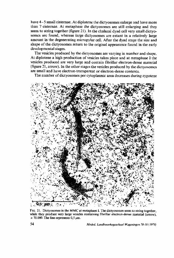
have 4-5 small cisternae. At diplotene the dictyosomes enlarge and have more than 7 cisternae. At metaphase the dictyosomes are still enlarging and they seem to string together (figure 21). In the chalazal dyad cell very small dictyosomes are found, whereas large dictyosomes are extant in a relatively large amount in the degenerating micropylar cell. After the dyad stage the size and shape of the dictyosomes return to the original appearance found in the early developmental stages.
The vesicles produced by the dictyosomes are varying in number and shape. At diplotene a high production of vesicles takes place and at metaphase I the vesicles produced are very large and contain fibrillar electron-dense material (figure 21, arrow). In the other stages the vesicles produced by the dictyosomes are small and have electron-transparent or electron-dense contents.
The number of dictyosomes per cytoplasmic area decreases during zygotene
FIG. 21. Dictyosomes in the MMC at metaphase I. The dictyosomes seem to string together, while they produce very large vesicles containing fibrillar electron-dense material (arrow), x 70.000. The line represents 0,5 urn.
54 Meded. Landbouwhogeschool Wageningen 78-16(1978)
and pachytene and increases after this stage. At diplotene the number of dictyo-somes per cytoplasmic area reaches its maximum and after this stage the quantity diminishes again. The number of vesicles produced by the dictyosomes reaches its maximum at diplotene and metaphase I.
The localization of the dictyosomes changes during the developmental stages. In the AC and in the early prophase stages the dictyosomes are distributed at random in the cytoplasm. At diplotene an accumulation of dictyosomes appears on both polar sides of the nucleus. More dictyosomes are found on the chalazal side of the cell than on the micropylar side. At metaphase I the dictyosomes are notably found near the polar regions of the spindle. In the dyad the dictyosomes are extant in the perinuclear zone of the micropylar cell and on both polar sides of the chalazal nucleus. After the degeneration of the micropylar cell no changes in this localization of the dictyosomes in the chalazal cell occur. At telophase II the dictyosomes appear on both sides of the nuclei, situated in the direction of the future spindle axis. In the two-nucleate megaspore the dictyosomes are situated between the two nuclei and on the micropylar and the chalazal sides of the cell. In the four-nucleate embryosac the dictyosomes are distributed at random in the cytoplasm.
Discussion As it was suggested by WHALEY et al. (1966) and by FRASER (1975) vesicles
derived from dictyosomes are fusing in the phragmoplast during cell-plate formation. This indicates that a large number of vesicles, produced by the dictyosomes must be available at the stage in which the cell-plate formation takes place.
The large number of dictyosomes per cytoplasmic area found at diplotene, the large size and the typical ultrastructure of the dictyosomes at diplotene and metaphase I and the accumulation of the dictyosomes in the polar regions of the spindle figure, all apparently are related to the production of vesicles necessary for the future cell-plate.
The localization of the dictyosomes on both polar sides of the nucleus before spindle formation takes place at diplotene and in the dyad may indicate a possible relation between the dictyosomes and the future spindle poles. Maybe dictyosomes have a function in the formation or are involved in the activity of the microtubular organization centres, presuming that these centres are present in all species (PICKETT-HEAPS, 1970). Probably the accumulated dictyosomes may be part of this organization centre.
The presence of a relatively large number of dictyosomes in the micropylar dyad cell is probably related to the degeneration process of this cell. These dictyosomes may have a function in the production of digestive enzymes, necessary for the demolition of the cell organelles.
4.3.2.6. Vesicle- and v acuo l a r system In all developmental stages electron-dense and electron-transparent vacuoles
are found. In the AC and in the early meiotic prophase stages few electron-
Meded. Landbouwhogeschool Wageningen 78-16(1978) 55
dense vacuoles are perceptible, sometimes showing an erratic appearance. Similar electron-dense structures are perceptible in the two-nucleate megaspore (figure 24, 4.3.2.12.).
Electron-translucent vacuoles are found in small amounts in the early developmental stages. At metaphase I, however, more vacuoles appear. Their number and size increase during further development. In the four-nucleate embryosac a central vacuole is found. Close to this central vacuole some small electron-transparent vacuoles are perceptible. Evidence for a probable fusion of these small vacuoles with the central vacuole is not found.
The electron-dense vesicles and the electron-translucent vacuoles are distributed at random in the cytoplasm.
4.3.2.7. P l asma membrane At zygotene and pachytene a small amount of electron-dense granular
material is found between the plasma membrane and the cell-wall. This electron-dense granular material seems to be similar to the material found in the paramural bodies, present in the early developmental stages of Lilium. In Allium, on the other hand, the paramural bodies have disappeared after pachytene.
4.3.2.8. M i t o chond r i a In the AC, in meiotic prophase, in metaphase I and in the chalazal dyad cell
the mitochondria have a spherical shape, in which very few cristae are observed. In the chalazal dyad cell also transformations of mitochondria showing a largely elongated shape, are found (figure 22, arrow). In the viable (dyad) cell at telophase II the same type of mitochondria are found. Cristae appear in the mitochondria from the functional cell on.
The number of mitochondria per cytoplasmic area increases during the meiotic prophase stages till a maximum is reached at diplotene. At metaphase I the number has strongly diminished and in the further stages the quantity of the mitochondria per cytoplasmic area fluctuates.
The distribution of the mitochondria in the cytoplasm during the development of the MMC does not vary. In all stages the mitochondria are distributed at random. The mitochondrial transformations are found on both polar sides of the nucleus in the functional megaspore and on one side of the nucleus in the viable (dyad) cell at telophase II.
Discussion The low number of cristae in the mitochondria during premeiotic interphase
and meiotic prophase may be related to a low metabolic state of the cell at these stages, presuming that we may relate the number of cristae within the mitochondria to the metabolic activity of these mitochondria as HACKENBROCK
(1966) and DAMSKY (1976) suggest. An increase of the number of mitochondria per cytoplasmic area may then be related to an increase of the metabolic state of the cell. In chapter 6.2.2.9 we shall revert to this subject.
56 Meded. Landbouwhogeschool Wageningen 78-16 (1978)
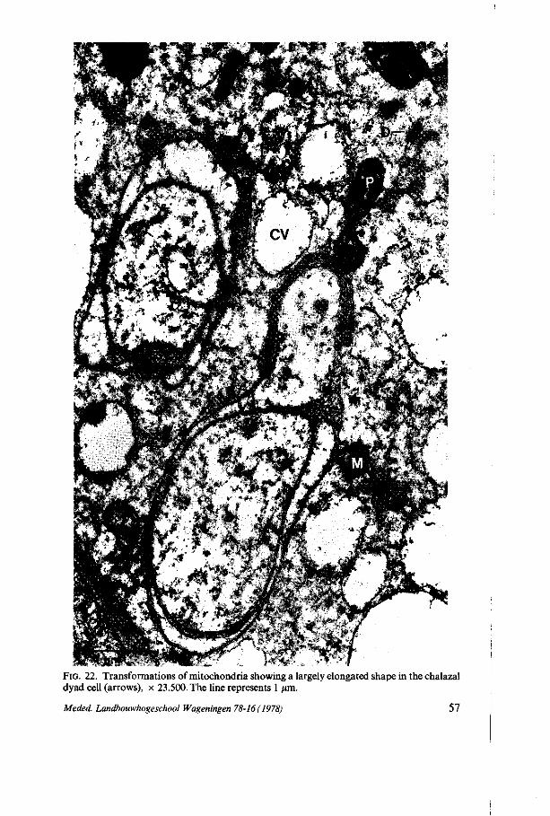
FIG. 22. Transformations of mitochondria showing a largely elongated shape in the chalazal dyad cell (arrows), x 23.500. The line represents 1 /mi.
Meded. Landbouwhogeschool Wageningen 78-16(1978) 57
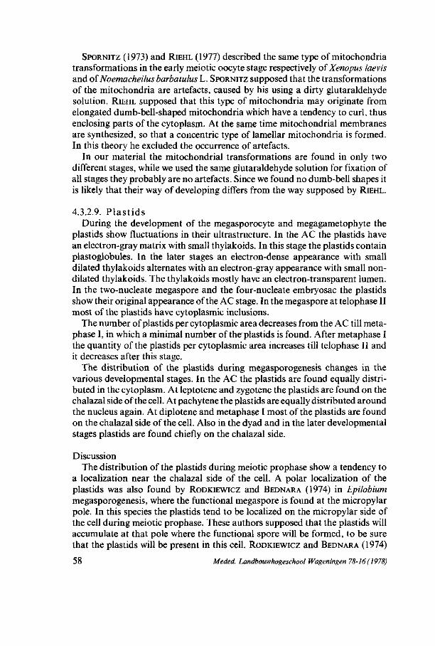
SPORNITZ (1973) and RIEHL (1977) described the same type of mitochondria transformations in the early meiotic oocyte stage respectively of Xenopus laevis and of Noemacheilus barbatulus L. SPORNITZ supposed that the transformations of the mitochondria are artefacts, caused by his using a dirty glutaraldehyde solution. RIEHL supposed that this type of mitochondria may originate from elongated dumb-bell-shaped mitochondria which have a tendency to curl, thus enclosing parts of the cytoplasm. At the same time mitochondrial membranes are synthesized, so that a concentric type of lamellar mitochondria is formed. In this theory he excluded the occurrence of artefacts.
In our material the mitochondrial transformations are found in only two different stages, while we used the same glutaraldehyde solution for fixation of all stages they probably are no artefacts. Since we found no dumb-bell shapes it is likely that their way of developing differs from the way supposed by RIEHL.
4.3.2.9. P l a s t i d s During the development of the megasporocyte and megagametophyte the
plastids show fluctuations in their ultrastructure. In the AC the plastids have an electron-gray matrix with small thylakoids. In this stage the plastids contain plastoglobules. In the later stages an electron-dense appearance with small dilated thylakoids alternates with an electron-gray appearance with small non-dilated thylakoids. The thylakoids mostly have an electron-transparent lumen. In the two-nucleate megaspore and the four-nucleate embryosac the plastids show their original appearance of the AC stage. In the megaspore at telophase II most of the plastids have cytoplasmic inclusions.
The number of plastids per cytoplasmic area decreases from the AC till meta-phase I, in which a minimal number of the plastids is found. After metaphase I the quantity of the plastids per cytoplasmic area increases till telophase II and it decreases after this stage.
The distribution of the plastids during megasporogenesis changes in the various developmental stages. In the AC the plastids are found equally distributed in the cytoplasm. At leptotene and zygotene the plastids are found on the chalazal side of the cell. At pachytene the plastids are equally distributed around the nucleus again. At diplotene and metaphase I most of the plastids are found on the chalazal side of the cell. Also in the dyad and in the later developmental stages plastids are found chiefly on the chalazal side.
Discussion The distribution of the plastids during meiotic prophase show a tendency to
a localization near the chalazal side of the cell. A polar localization of the plastids was also found by RODKIEWICZ and BEDNARA (1974) in Lpilobium megasporogenesis, where the functional megaspore is found at the micropylar pole. In this species the plastids tend to be localized on the micropylar side of the cell during meiotic prophase. These authors supposed that the plastids will accumulate at that pole where the functional spore will be formed, to be sure that the plastids will be present in this cell. RODKIEWICZ and BEDNARA (1974)
58 Meded. Landbouwhogeschool Wageningen 78-16(1978)
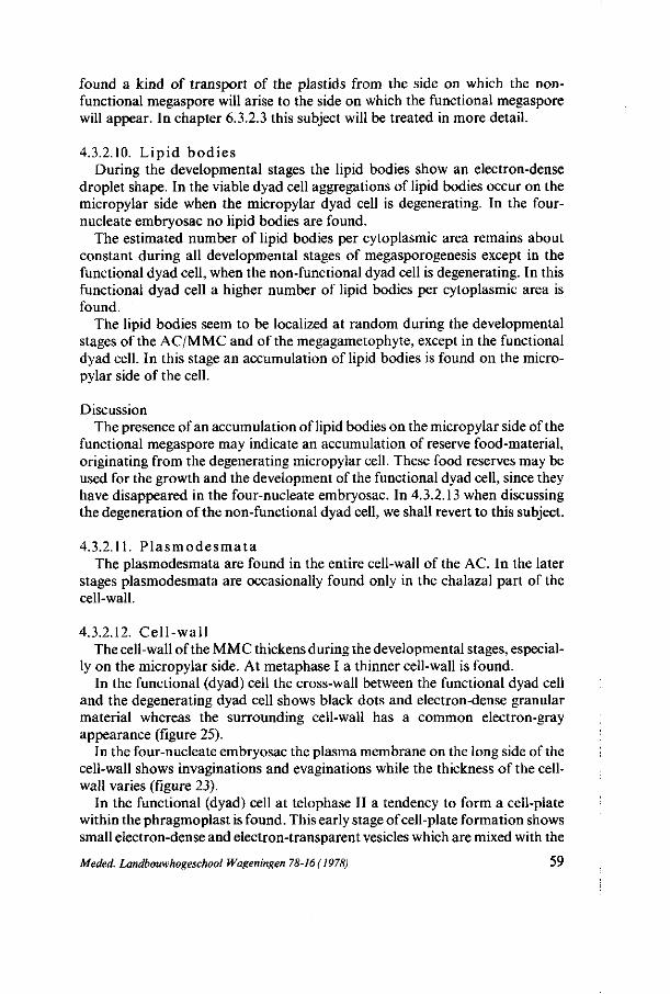
found a kind of transport of the plastids from the side on which the nonfunctional megaspore will arise to the side on which the functional megaspore will appear. In chapter 6.3.2.3 this subject will be treated in more detail.
4.3.2.10. L ipid bod ies During the developmental stages the lipid bodies show an electron-dense
droplet shape. In the viable dyad cell aggregations of lipid bodies occur on the micropylar side when the micropylar dyad cell is degenerating. In the four-nucleate embryosac no lipid bodies are found.
The estimated number of lipid bodies per cytoplasmic area remains about constant during all developmental stages of megasporogenesis except in the functional dyad cell, when the non-functional dyad cell is degenerating. In this functional dyad cell a higher number of lipid bodies per cytoplasmic area is found.
The lipid bodies seem to be localized at random during the developmental stages of the AC/MMC and of the megagametophyte, except in the functional dyad cell. In this stage an accumulation of lipid bodies is found on the micropylar side of the cell.
Discussion The presence of an accumulation of lipid bodies on the micropylar side of the
functional megaspore may indicate an accumulation of reserve food-material, originating from the degenerating micropylar cell. These food reserves may be used for the growth and the development of the functional dyad cell, since they have disappeared in the four-nucleate embryosac. In 4.3.2.13 when discussing the degeneration of the non-functional dyad cell, we shall revert to this subject.
4.3.2.11. P l a smode sma t a The plasmodesmata are found in the entire cell-wall of the AC. In the later
stages plasmodesmata are occasionally found only in the chalazal part of the cell-wall.
4.3.2.12. Cel l -wal l The cell-wall of the MMC thickens during the developmental stages, especial
ly on the micropylar side. At metaphase I a thinner cell-wall is found. In the functional (dyad) cell the cross-wall between the functional dyad cell
and the degenerating dyad cell shows black dots and electron-dense granular material whereas the surrounding cell-wall has a common electron-gray appearance (figure 25).
In the four-nucleate embryosac the plasma membrane on the long side of the cell-wall shows invaginations and evaginations while the thickness of the cell-wall varies (figure 23).
In the functional (dyad) cell at telophase II a tendency to form a cell-plate within the phragmoplast is found. This early stage of cell-plate formation shows small electron-dense and electron-transparent vesicles which are mixed with the
Meded. Landbouwhogeschool Wageningen 78-16(1978) 59
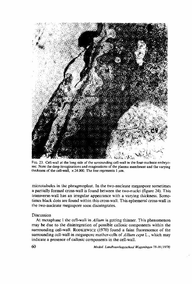
FIG. 23. Cell-wall at the long side of the surrounding cell-wall in the four-nucleate embryo-sac. Note the deep invaginations and evaginations of the plasma membrane and the varying thickness of the cell-wall, x 24.000. The line represents 1 /mi.
microtubules in the phragmoplast. In the two-nucleate megaspore sometimes a partially formed cross-wall is found between the two-nuclei (figure 24). This transverse-wall has an irregular appearance with a varying thickness. Sometimes black dots are found within this cross-wall. This ephemeral cross-wall in the two-nucleate megaspore soon disintegrates.
Discussion At metaphase I the cell-wall in Allium is getting thinner. This phenomenon
may be due to the disintegration of possible callosic components within the surrounding cell-wall. RODKIEWICZ (1970) found a faint fluorescence of the surrounding cell-wall in megaspore mother-cells of Allium cepa L., which may indicate a presence of callosic components in the cell-wall.
60 Meded. Landbouwhogeschool Wageningen 78-16 (1978)
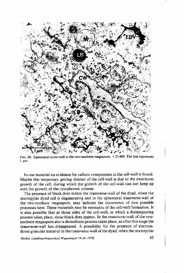
•^V x%f, rtz
*m\ -"Ȉ 1*1 fl
».* C . -ut - ^ ^ ?'•• . . f .:•;•% A4;1?:. ̂ ;^-^-'-'X ^ > a ' .
a» '«r : / ^i.C.
f - * 0
* * ,
FIG. 24. Ephemeral cross-wall in the two-nucleate megaspore, x 25.000. The line represents 1 /im.
In our material no evidence for callosic components in the cell-wall is found. Maybe this temporary getting thinner of the cell-wall is due to the enormous growth of the cell, during which the growth of the cell-wall can not keep up with the growth of the cytoplasmic volume.
The presence of black dots within the transverse-wall of the dyad, when the micropylar dyad cell is degenerating and in the ephemeral transverse-wall of the two-nucleate megaspore, may indicate the occurrence of two possible processes here. These materials may be remnants of the cell-wall formation. It is also possible that at those sides of the cell-wall, in which a disintegrating process takes place, these black dots appear. In the transverse-wall of the two-nucleate megaspore also a demolition process takes place, as after this stage the transverse-wall has disappeared. A possibility for the presence of electron-dense granular material in the transverse-wall of the dyad, when the micropylar
Meded. Landbouwhogeschool Wageningen 78-I6( 1978) 61

cell is degenerating, is that a transport of material takes place through the transverse-wall from the degenerating micropylar cell to the functional chalazal cell or vice versa. As no plasmodesmata are found in the transverse-wall, a transport of material has to take place through the cell-wall. The surrounding cell-wall does not show such peculiar features as the electron-dense granular material, so that a transport of material only seems to take place through the cell-wall between the micropylar and the chalazal cell of the dyad. It is possible that this is a transport of demolition material from the degenerating micropylar cell to benefit the chalazal cell. It is also possible that a transport of digestive enzymes takes place from the chalazal cell to the micropylar cell, necessary for the demolition of the cell organelles in the latter cell. In 4.3.2.13 we shall revert to this subject.
The irregular plasma membrane evaginations and invaginations within the cell-wall of the four-nucleate embryosac, while the thickness of the cell-wall strongly varies, are similar to those found in transfer-cells (SCHNEPF and PROSS,
1976). In the four-nucleate embryosac the irregular plasma membrane evaginations and invaginations may have the same function as those found in transfer-cells for the transport of material through the cell-wall. In 4.4 we shall revert to this subject.
The formation of an ephemeral cell-wall in the two-nucleate megaspore of a bisporic type of embryosac development is also found by GOVINDAPPA (1955) in Xyris pauciflora Willd. This cell-wall formation may indicate that the bisporic type is related to the monosporic type. Maybe the bisporic type is derived from the monosporic type or vice versa.
4.3.2.13. Degene r a t i on of the m ic ropy la r dyad cell Figure 25 shows the degenerating micropylar dyad cell, when the functional
chalazal cell has become two-nucleate megaspore. In this photo the nucleus has two nucleoli, localized opposite to each other. Within the cytoplasm numerous ribosomes, both as monosomes and as polysomes free and attached to the endoplasmic reticulum membranes, are perceptible. This rough endoplasmic reticulum is mostly found round the lipid bodies and sometimes round electron-transparent vacuoles (arrows). The presence of large dictyosomes and a large number of lipid bodies is obvious. The electron-transparent vacuoles sometimes contain electron-dense material. The cell-wall between the micropylar cell and the chalazal cell contains black dots and electron-dense granular material.
Discussion In several paragraphs of chapter 4 some features of the cell organelles men
tioned above were already discussed in connection with the degeneration of the micropylar cell. The suggestion made in those paragraphs will be recapitulated below.
The presence of two nucleoli within the karyoplasm of the nucleus may indicate the diploid nature of the nucleus and its tendency to divide. This division has to be the second meiotic division, which, however, is disturbed by
62 Meded. Landbouwhogeschool Wageningen 78-16(1978)
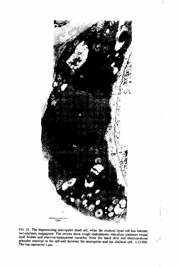
Ijim
FIG. 25. The degenerating micropylar dyad cell, when the chalazal dyad cell has become two-nucleate megaspore. The arrows show rough endoplasmic reticulum cisternae round lipid bodies and electron-transparent vacuoles. Note the black dots and electron-dense granular material in the cell-wall between the micropylar and the chalazal cell, x 15.000. The line represents 1 /mi.
the degeneration of the cytoplasm. In one micropylar cell a partially formed cross-wall is found, indicating in this case that the second meiotic division has been substantially progressed to a later developmental stage before the degeneration of the cell started. The temporary formation of a partial cross-wall after the second meiotic division is also found in the two-nucleate megaspore.
The presence of a number of ribosomes, especially as polysomes and as ribosomes attached to ER membranes may indicate a high production of proteins, probably for the synthesis of necessary enzymes. The RER membranes round the lipid bodies and round other cell organelles may have a function in the enzyme-synthesis as well. Probably this enzyme-synthesis is necessary for the conversion of lipoproteins into lipidic components. The amount of lipid bodies increases during the degeneration process of the cytoplasmic organelles, serving as a storage of useful reserves.
The presence of large dictyosomes in a relatively large number per cytoplasmic area may indicate a function for these organelles in the demolition process. These organelles may have a function in the synthesis and/or the transport of digestive enzymes. This transport takes place to the sites of digestion.
The electron-transparent vacuoles are similar to the autophagic vacuoles found at the end of the lytic process in endosperm cells by CRESTI et al. (1972). These vacuoles seem to have a function in the degeneration process as they contain digestive enzymes.
The presence of electron-dense material in the cell-wall between the micropylar- and the chalazal cell may indicate a transport of material through this cell-wall (4.3.2.12.). This transport can take place from the micropylar to the chalazal cell or vice versa. A transport of material from the chalazal cell to the micropylar cell may signify a transport of digestive enzymes necessary for the demolition of the micropylar cell. However, since in the micropylar cell enough cell organelles seem to be present for the synthesis of digestive enzymes, a transport of material from the micropylar cell to the chalazal cell seems to be more likely. This may signify a transport of useful material to the chalazal cell in order to benefit the growth and development of this cell.
SHELDRAKE (1974) supposed that the functional cell must get rid of its deleterious products and thus induces the degeneration process of the micropylar cell, in which these deleterious products would be accumulated. In our material no evidence for this theory is found, as before the start of the degeneration process the number of lipid bodies (the presumed deleterious demolition products) in the micropylar cell was not excessively high. In the two-nucleate megaspore, on the other hand, lipid bodies are localized near the micropylar side of the cell, thus supporting the theory previously mentioned, that lipidic amounts are transported from the degenerating micropylar cell to the chalazal cell in order to benefit this latter cell.
64 Meded. Landbouwhogeschool Wageningen 78-16 (1978)
4.3.2.14. Nucellus cell Just as with Lilium, also with Allium the development of one cell of the sur
rounding nucellus was studied. For this purpose always the nucellus cell situated at the same place on the micropylar side of the megasporocyte or of the mega-gametophyte was observed.
During megasporogenesis the nucellus cells round the megasporocyte remain intact. At the micropylar pole the cell-walls of these cells are thickening as much that in the four-nucleate embryosac very little cytoplasm of these nucellus cells is visible, whereas a thick cell-wall layer is extant. The cytoplasm of the cells remains intact; no degeneration process is found. The nucellus cells do not have central vacuoles. Except in the very young developmental stages of megasporogenesis, no mitotic divisions are found in the nucellus epidermis cells. The cells are elongating to keep up with the growth of the megasporocyte.
The nucleus of the nucellus cell observed has an elongated shape. The condensed parts of chromatin show a reticulate character. The nuclear envelope has pores during all the developmental stages of the megasporocyte and the megagametophyte. Microtubules are found in the nucellus cell, when the MMC is at zygotene. In the dyad stage, they are always present in the periferal zone. Cytoplasmic ribosomes are found as monosomes and as polysomes, both free or attached to the ER membranes. Polysomes are only found in the nucellus cell, when the MMC is at diplotene and in the dyad stage and when the two-nucleate megaspore is formed. Rough endoplasmic reticulum is found in the nucellus cell in all stages of the megasporocyte. At diplotene and metaphase I tubular smooth endoplasmic reticulum is found in the nucellus cell. Dictyo-somes are found in the nucellus cell in nearly all the developmental stages of the megasporocyte and of the megagametophyte. At the diplotene stage of the MMC the number of dictyosomes in the nucellus cell is apparently larger than in the other stages of the MMC. In some stages of the MMC no vesicle production of the dictyosomes is found in the nucellus cell. Electron-transparent vacuoles are occasionally found in the nucellus cell, whereas sometimes the vacuoles have electron-dense contents. Only when the archespore cell is present, some electron-dense material is found between the plasma membrane and the cell-wall of the nucellus cell. The plastids in the nucellus cell show thylakoids and in some stages after pachytene of the MMC few starch grains are found in the plastids. The mitochondria are spherical-shaped and contain a large number of cristae in the nucellus cell in all the developmental stages of the MMC, except in the AC and at leptotene. In the latter stages very few cristae are found in the mitochondria of the nucellus cell. Lipid bodies are found in the nucellus cell after the pachytene stage of the MMC. In the AC and in the meiotic prophase stages of the MMC a number of plasmodesmata are found especially in the cross-walls between the nucellus cells. Only in the AC plasmodesmata are found in the cell-wall between the AC and the nucellus cells. The cell-wall of the nucellus cell thickens enormously so that a thick cell-wall layer exists on the micropylar side of the MMC. On the chalazal side a thinner cell-wall is perceptible.
Meded. Landbouwhogeschool Wageningen 78-16(1978) 65
Discussion From these results we may conclude that only a small correlation exists
between the ultrastructural development of the nucellus cell and the characteristics of the ultrastructural development of the megasporocyte mentioned previously. The presence of tubular smooth ER in the nucellus cell when the MMC is at diplotene and at metaphase I, and the higher number of dictyo-somes in the nucellus cell when the MMC is at diplotene, may point to the small correlation mentioned above. Apart from these exceptions the nucellus cell and the megasporocyte show their own specific development.
The presence of plasmodesmata only on the chalazal side between MMC and nucellus may indicate a transport of nutrients from this side of the nucellus to the developing MMC. This nutrition-supply, coming from the chalazal side of the nucellus cell, originates from the vascular bundle which ends at that part of the ovule.
The continuous thickening of the cell-wall of the surrounding nucellus cells on the micropylar side induces us to suppose that this tissue serves as a solidity tissue to protect the developing MMC and the developing embryosac.
4.4. DISCUSSION
In this thesis we call the chalazal dyad cell the functional or viable cell, when the micropylar cell starts degenerating. This micropylar dyad cell is then called the non-functional or abortive cell. After degeneration of the micropylar dyad cell the chalazal dyad cell starts the second nuclear meiotic division, giving rise to a two-nucleate cell. This cell is called in our study the two-nucleate mega-spore. After the first and the second mitotic nuclear division respectively the four-nucleate and the eight-celled embryosac will be formed. MAHESWARI
(1950) and RUTISHAUSER (1969) both called this two-nucleate megaspore the 'functional' megaspore. In our opinion they ment with their term 'functional' either the cell which enters the megagametogenesis, which is also expressed in the term 'megaspore', or the fact that the other non-functional megaspore degenerates. In the last case we prefer to use the term 'functional' for the chalazal dyad cell, when the micropylar dyad cell degenerates, as this degeneration takes place before the second meiotic division. In our opinion the second meiotic division will be regarded to belong to the process of megasporogenesis as well, leading to the formation of the two-nucleate megaspore. We prefer to use the term 'two-nucleate megaspore' instead of 'functional megaspore', to show that the actual degeneration of the non-functional cell, as a result of which the other cell gets functional, already takes place, before the second meiotic division in the bisporic types of development we studied.
During megasporogenesis in Allium cepa L. there seems to be a relation between some cell organelle changes. The presence of the nuclear pores, the undulations of the nuclear envelope and the sacculations of the inner nuclear membrane may indicate an information flow between the nucleus and the cyto-
66 Meded. Landbouwhogeschool Wageningen 78-16(1978)
plasm necessary for the specific development of some organelles in the corresponding stages of the MMC as it is suggested in 3.3.2.1. In paragraph 6.2.2.2 these structures of the nuclear envelope are discussed extensively in relation to a possible function in the nucleo-cytoplasmic exchange of material.
The ultrastructural changes of the dictyosomes at diplotene and at metaphase I of the MMC are considered to be typical for their function in the cell-plate formation. During these typical changes the dictyosomes are getting longer while they seem to string together producing numerous electron-dense and electron-transparent vesicles. The appearance of these changes may be influenced by a flow of information during the intensive nucleo-cytoplasmic exchange of material at pachytene and diplotene. At the same time tubular smooth ER occurs when microtubules appear in the perinuclear zone. WHALEY et al. (1966) suggested that smooth ER may produce tubulin proteins necessary for the synthesis of microtubules. In our material this may occur in the perinuclear zone. Summarizing : the extensive nucleo-cytoplasmic exchange of material, indicated by the structural features at the nuclear envelope, is followed by typical changes of the ultrastructure of dictyosomes and of their vesicle production, by a sudden increase of the amount of smooth endoplasmic reticulum and by the appearance of numerous microtubules in the perinuclear zone.
Within the developing megasporocyte and megagametophyte very few lipid bodies are found that could serve as storage sites for reserves. No storage of starch is found within the plastids. Plasmodesmata are only found in the chalazal cell-wall of the developing cell, whereas they are not perceptible in each developmental stage of the cell. The mitochondria contain very few cristae during meiotic prophase, which may indicate that their metabolic activity seems to be low. All these results suggest that the development of the megasporocyte and of the megagametophyte is dependent on a nutrition-supply coming from the chalazal nucellus cells through the plasmodesmata. This nutrition-supply is sufficient for the growth and development of the cell, as no storage of reserves takes place.
An unequal cell division results from the position of the nucleus during the cell division and the position of the newly formed cell-wall. In Allium the nucleus show a tendency to be localized near the micropylar side of the MMC during meiotic prophase. By this position of the nucleus a possible condition for an unequal division is created.
A polar distribution of cell organelles during the division causes, that the by unequal division formed cells contain a specific cell organelle population. Some aspects of these organelle populations seem to be related to the subsequent development and function of the two cells. In Allium a polar distribution of the plastids is found during meiotic prophase by means of their tendency to be localized near the chalazal side of the MMC. The presence of nearly all plastids on the chalazal side may be related to their necessary presence in the future functional chalazal cell, which develops into the embryosac. By this distribution the plastids are preserved for the future embryosac and protected for demolition.
Meded. Landbouwhogeschool Wageningen 78-16(1978) 67

The presence of a considerable number of dictyosomes and ER cisternae in the micropylar dyad cell may be related to the degeneration process in this cell. \Ve may even speculate that the composition of the cell organelle population determines the future development of the cell e.g. the degeneration of the micropylar dyad cell.
In the two-nucleate megaspore the growth in volume of the cell takes place in relation to the growth of the cytoplasm and the increasing vacuolisation of the cell. In the four-nucleate embryosac a central vacuole appears probably by the fusion of the small electron-transparent vacuoles. Owing to the presence of this central vacuole the four nuclei are pressed in twos into both polar sides of the cell.
68 Meded. Landbouwhogeschool Wageningen 78-16 ( 1978)

5. M E G A S P O R O G E N E S I S AND EARLY M E G A G A M E T O G E N E S I S IN IMPATIENS WALLERIANA
HOOK F.
5.1. INTRODUCTION
DAHLGREN (1934) and STEFFEN (1951) concluded from cytological studies that the embryosac development of Impatiens species was of the Polygonum type. By light-microscopic studies the authors had proved the presence of a dyad and of a tetrad in which one functional megaspore was visible, together with three degenerated cells laying in a cap at the micropylar pole of the ovule. In this paper the results of an electron-microscopic study are offered in order to gain more detailed information of the process of megasporogenesis in Impatiens.
5.2. MATERIAL AND METHODS
For this study hothouse-grown plants of Impatiens walleriana Hook./, were used.
For light-microscopy the previously described technics for the preparation of 3 (im sections (2.1.2.) and the clearing preparation (2.1.1.) were applied.
For electron-microscopy the glutaraldehyde-osmiumtetroxyde fixation as described in 2.2.1 was applied together with the re-embedding method (2.2.3.). The glutaraldehyde-osmiumtetroxyde fixation was used as follows : complete ovaries were dissected and when necessary sectioned transversely in smaller parts. The ovaries (or parts of them) were fixed for 2 hours in 2,5% glutaral-dehyde in 0,1 M phosphatebuffer at pH 7,2. They were rinsed thoroughly in buffer and postfixed in buffered 1% osmiumtetroxyde during 20 hours. The ovules were embedded in epon. After longitudinally sectioning of the ovules the sections were post-stained with leadcitrate (REYNOLDS, 1963).
5.3. RESULTS AND DISCUSSION
5.3.1. Light-microscopy The ovaries of Impatiens walleriana Hook. ƒ. consist of one central placenta
upon which the ovules are emplanted. At early stages the ovules are anatropous and they become apotropous when the embryosac is maturing. The ovule is bitegmic and tenuinucellate. The archespore cell (AC) originates directly from the archespore mother-cell (AMC) without division.
At the early prophase the inner- and outer integuments are not formed yet.
Meded. Landbouwhogeschool Wageningen 78-16 (1978) 69

At V"*
^ ^ / ;- •-. .. <• • v ^"^L
FIG. 26. Photo 26a shows the ovule of Impatiens after degeneration of the micropylar dyad cell, x 750. The micropylar side is at the top side of the photograph. The line represents 10 urn. Photo 26b shows an EM detail of the thick cell-wall layer between the degenerated micropylar dyad cell and the functional chalazal dyad cell, x 8.000. The line represents 1 /an.
At pachytene the inner integuments are still not completely surrounding the megaspore mother-cell (MMC) but in the dyad the formation of the integuments is completed. Near the funiculus the outer integument remains rudimentary (figure 26).
The ovules of one ovary show various developmental stages. For instance : from top till bottom of the ovary one can find ovules with MMC's in pro- and metaphase, ovules with just formed dyads and ovules with dyads of which the topcell at the micropylar pole is already degenerated.
The archespore mother-cell has a spherical shape. During the developmental stages the megasporocyte changes to ellipsoidal. The size of the cell enlarges about three times from AMC till mature embryosac.
In spite of careful examination no tetrads have ever been found. It is always dyads that can be noticed and at the oldest meiotic stages always one degenerated cell at micropylar side is found. Between the functional cell and the degenerating cell a thick layer is present (figure 26). After glutaraldehyde-osmiumtetroxyde fixation this layer is moderately electron-dense indicating definitely the presence of cell-wall material (figure 26b).
Discussion According to the fact that never tetrads have been found in spite of numerous
samples taken at different diurnal and nocturnal times, it seems reasonable to presume that tetrads are not present during megasporogenesis of Impatiens walleriana. The presence of dyads with always one degenerating or degenerated micropylar cell supports the assumption that the bisporic type of development is present here. STEFFEN (1951) probably found the same kind of dyads with a thick cell-wall layer between the degenerating cell and the functional cell. He concluded that this layer consists of a degenerated cell too, so that he found a functional cell with two degenerated cells. He concluded probably erroneously that the third degenerated cell of the tetrad expected by him is not present in the field of his section.
Analogous to the bisporic type in Allium the chalazal dyad cell in Impatiens is called the functional cell, when the micropylar dyad cell starts degenerating. After the second meiotic division in the functional cell the two-nucleate megaspore appears.
5.3.2. Electron-microscopy In enclosure III a detailed survey is given of the most important morpho
logical changes of various cell organelles and structures. Figure 27 gives a quantitative impression of the most important features in the stages which are comparable with those in the other species examined, based on measurements as described in 2.2.5.
Enclosure IV C shows the distribution of a number of cell organelles within the cytoplasm during the developmental stages.
Meded. Landbouwhogeschool Wageningen 78-16(1978) 71
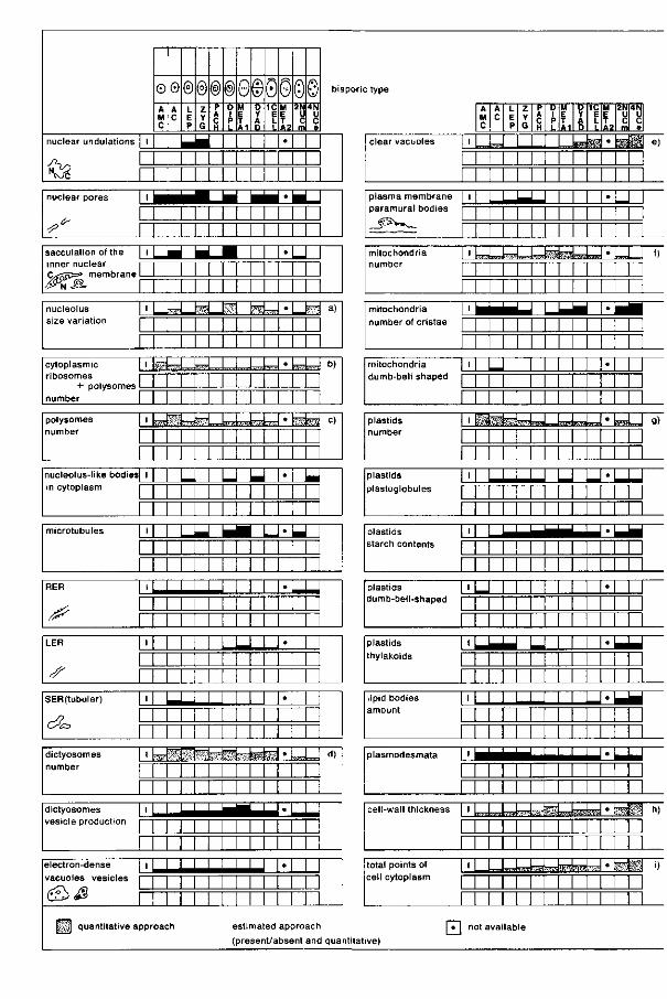
nuclear undulations j 1
Jh^ L_ •v& r nuclear pores | I
^ r sacculation of the | I
inner nuclear i— C l ^ s ö ^ membrane 1—
Ä r nucleolus [ 1 size variation I
r cytoplasmic [ 1 r ibosomes i
number [
polysomes [ I number i
nucleolus-like bodiesl 1
in cytoplasm [
r microtubules | 1
RER | _ l _
^ E ^ r
LER | _ l _
L
s r SER(tubular) | _ l _
r^? L LS£D I
dictyosomes j 1 number 1
r dictyosomes I I
vesicle production i —
electron-dense j i
vacuoles vesicles |—
&<& p
H I ] quantitative appr
o A M C
0 A C
© L E P
© Z Y G
® P A C H
®0 D 1 P 1-
M E T A 1
e D Y A D
000© 1C
E L L
M E T A2
•
2N U C m
4N U C e
bisporic type
_ _ I H ta^HH * ^ H _ _
1 1 1 1 1 1 1 1
1 1 1 1 1 1 1 1
M M 1 1 1 1
W f e ^ i r ^ J i ^ L - ^
1 1 1 1 1 1 1 1
T^Kss feag l i™
1 1 1 1 1 1 1 1
1 U 1 1 1 1 1 1 1 1 1
1 ' ' ' 1 M 1 M 1 1
1 1 M 1 1 1 1 1 M 1
1 1 1 1 1 1 1 1 1 1 1 1
i i i i 1 1 1 1 1 1 1 1
J M I i t a
1 1 1 1
1 1 1 1 1 1 1 1 i i i i
1 1 1 1 1 1 M
1 1 1 1 1 1
I I 1 1 1 I I I
•
IS \mU> 1 1 1 1 I N I
JLSJU-
M M 1 M 1
J J J J . 1 1 1 1 1 1 1 1
J U 1-1 1 1 1 1 1 1 1
^̂ ^̂ 1 1 1 *
1 1 1 1 1 1
1 1 1 1 1 1 1 1 1
U 1 1 1 1 I I I
1 1 1 l l l l J J
«
•
•
i faésW-M M 1 1 M
M^^^^^^^^J »
1 1 1 1 1 1 1 1 1 1 1 1 1 1 1
•
J _
I*??'
rsdroïs
sufcs
L
_
a)
b)
C
d)
oach estimated approach
(present/absent and qua ntitat
clear vacuoles
plasma membrane paramural bodies
mitochondria number
mitochondria
number of cristae
mitochondria dumb-bell shaped
plastids number
plastids
plastoglobutes
plastids starch contents
plastids dumb-bell-shaped
plastids
thylakoids
lipid bodies amount
plasmodesmata
cell-wall thickness
total points of cell cytoplasm
I I
A M C
A C
L E P
Z Y G
P A C H
D 1 P L
M E T A1
D Y A D
•p>
1C E L L
S
M E T A2
•
2N U C m
m
4N
e
i e)
| I
| I
| I
! I
h
11
11
h
!'
I'
I'
I'
11
I U I I I I I M I I I I I I I I I I I I I
rrrA™Ài7vi-~*\r^f:t蜣A:.'.:,.lMV*i
I I I I I M I M I I I I I I
I M I I I I I I I I I I I I I
U I I I I I I I I I I I I I I I I I I I I I I
» l é d ^ U ^ U . J ^ M I I I M I I I I I I I I I
I I I I I I I J I I I I I I I I
I I I I I I I I I I I I I I I I
LJ M I I I I I I M I I I I I I I I
J - U U N I I I I M I I I I I M I I I I
I I Li..] I I l_ I M I I I I I I I I I I I I I
M I I I I I I M M I I I I
irJ»-iL>44-i-œL»-JiSBraK!®fes
I I I I I I I I | | | | | | | | L^ii^JiBl!*»^!^»*.1:.-!.':
I I 1 1 1 1 1 1
•
'
•
•
'
•
'
•
'
J .
'
•
•
I f)
ssm™
I I
g)
I - b
I I I I I
_
- •
_ I I
mm I I
n)
mm I I
i)
| • | not available
ve)
5.3.2.1. The nuc l eus Shape, size and position. During the meiotic prophase stages the nucleus
enlarges 1,5 times. The shape of the nucleus is spherical. At leptotene the surface of the nucleus becomes irregular and at zygotene it shows a lobed shape with connections with membrane-like structures present in the karyoplasm (figure 28, arrows). These membrane-like structures may look like 'blebs' of the nuclear envelope containing the membrane-like structures. At pachytene the original shape of the nucleus is restored.
The position of the nucleus within the cell during the meiotic prophase stages varies. At leptotene and zygotene the nucleus is found at the micropylar pole. At pachytene and diplotene the nucleus is localized in the centre. At early telophase the spindle figure is a-centrally positioned in the cell, which results in the formation of an unequally divided dyad.
Karyoplasm. Chromatin structure. The chromatin-structure shows the common appearance during meiosis (MOENS, 1968). The premeiotic interphase nuclei have dense chromatin structures which are referred to by LAFONTAINE
(1968) as heterochromatic masses. At leptotene small parts of chromatin string together and at zygotene axial cores are visible. Synaptinemal complexes are not clearly found. At diplotene the condensed chromosomes move to the equatorial plane of the spindle. At metaphase the chromosomes in the equatorial plane are attached with their kinetochores to numerous spindle microtubules running towards the poles of the spindle figure. The spindle figure also contains some smooth lamellar endoplasmic reticulum (LER). At the polar regions of the spindle figure concentrations of LER cisternae are found (figure 29). At early telophase a phragmoplast is formed between the two groups of sister chromosomes. Within the spindle figure no dictyosomes are present close to the cell-plate. In the meantime the nuclear envelope is rebuilt round the groups of sister chromosomes. Near the new nuclear envelope ER cisternae -apparently in close relationship with microtubules - are extant. In the young dyad the chromatin has a heterochromatic structure.
Membrane-like structures. At leptotene and zygotene some membrane-like structures are found within the karyoplasm, sometimes being continuous with the nuclear envelope (figure 28).
FIG. 27. Survey of the quantitative data of the most important features in Impatiens mega-sporogenesis and megagametogenesis. See 2.2.5. for the methods used for measuring and counting the various cell organelles. a) filled block represents 12,5 square ^m b) filled block represents 500 ribosomes/0,6 square /xm cytoplasm c) filled block represents 15 polysomes/0,6 square /an cytoplasm d) filled block represents 7,5 dictyosomes/150 points cytoplasm e) filled block represents 50 points/150 points cytoplasm f) filled block represents 50 mitochondria/150 points cytoplasm g) filled block represents 10 plastids/150 points cytoplasm h) filled block represents 0,5 /mi i) filled block represents 2500 points cytoplasm ( ± 8.000 x )
Meded. Landbouwhogeschool Wageningen 78-16(1978) 73
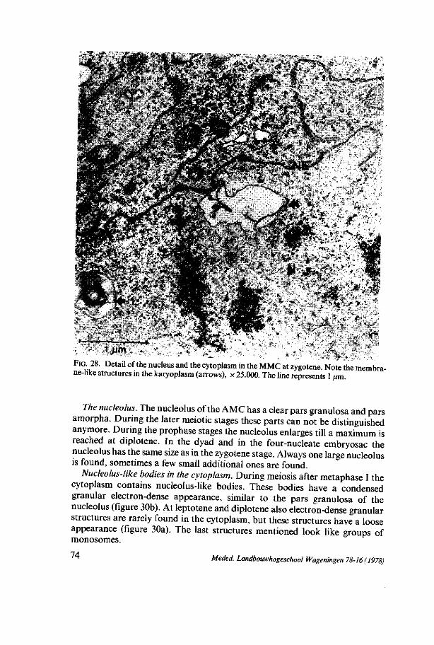
- ! & ' ' ' - ' t ' " / ?* ' ' ' . • * . ' ' » ' ' , • • -' - ' .*•«*•%.• * ' <•
**s' • • '* J^ü^i*••%?:} : '•-*,;..'•*• . ' X
FIG. 28. Detail of the nucleus and the cytoplasm in the MMC at zygotene. Note the membrane-like structures in the karyoplasm (arrows), x 25.000. The line represents 1 urn.
The nucleolus. The nucleolus of the AMC has a clear pars granulosa and pars amorpha. During the later meiotic stages these parts can not be distinguished anymore. During the prophase stages the nucleolus enlarges till a maximum is reached at diplotene. In the dyad and in the four-nucleate embryosac the nucleolus has the same size as in the zygotene stage. Always one large nucleolus is found, sometimes a few small additional ones are found.
Nucleolus-like bodies in the cytoplasm. During meiosis after metaphase I the cytoplasm contains nucleolus-like bodies. These bodies have a condensed granular electron-dense appearance, similar to the pars granulosa of the nucleolus (figure 30b). At leptotene and diplotene also electron-dense granular structures are rarely found in the cytoplasm, but these structures have a loose appearance (figure 30a). The last structures mentioned look like groups of monosomes.
74 Meded. Landbouwhogeschool Wageningen 78-16(1978)
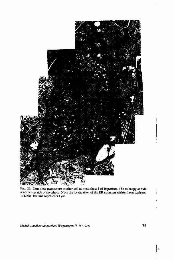
FIG. 29. Complete megaspore mother-cell at metaphase I of Impatiens. The micropylar side is at the top side of the photo. Note the localization of the ER cisternae within the cytoplasm, x 4.000. The line represents 1 pm.
Meded. Landbouwhogeschool Wageningen 78-16 (1978) 75
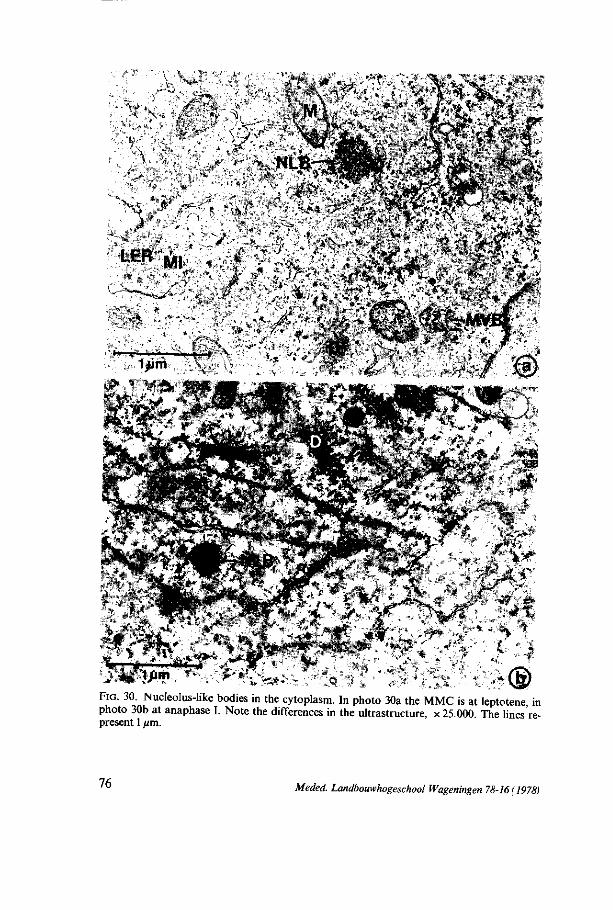
• \
' ^ > V < " ^ , ' ^
&i' y».«*
> * > , . : ® FIG. 30. Nucleolus-like bodies in the cytoplasm. In photo 30a the MMC is at leptotene in photo 30b at anaphase I. Note the differences in the ultrastructure, x 25.000. The lines'represent 1 /xm.
76 Meded. Landbouwhogeschool Wageningen 78-16(1978)
The nuclear envelope. In those stages where it is perceptible the nuclear envelope has many pores except at pachytene and in the four-nucleate embryo-sac, in which only few pores are found. At zygotene a maximum number of pores is found. The ultrastructure of the pores has not been studied in detail. In the AMC, AC and MMC at zygotene, pachytene and diplotene small electron-dense vesicles of irregular size between the inner- and outer nuclear membrane are visible (figure 31). At diplotene one also finds - together with these small electron-dense vesicles - large ones in between the nuclear membranes. The diplotene stage shows a maximal amount of these structures whereas similar structures also are perceptible in the cytoplasm, mostly within cisternae of the endoplasmic reticulum laying dispersed within the cytoplasm (figures 31 a and c, arrows). During all stages the outer nuclear envelope is found to be continuous with the endoplasmic reticulum.
Discussion Just as in Lilium and Allium the nuclear surface of Impatiens shows un
dulations during meiotic prophase. Some suggestions about their function are made in 3.3.2.1. In 6.2.2.1 a general discussion about the appearance and function of the nuclear undulations together with the presence of nuclear pores will be given.
As in Allium membrane-like structures are found in the karyoplasm at meiotic prophase. In 4.3.2.1 a short discussion about these structures is already given, whereas in 6.2.2.4 a general discussion about the presence of these structures during meiosis will be presented. In Impatiens the membrane-like structures also are attached to the nuclear envelope. Probably the inner nuclear membrane forms 'blebs' at the attachment site with the membrane-like structures. Clear evidence for this suggestion is not found. If the inner nuclear envelope is really continuous with the membrane-like structures, the membranes of these structures have to consist of the same components as the inner nuclear membrane does, at least at the attachment site. However, the ultrastructure of the membranes of the membrane-like structure is not to be similar to the ultrastructure of one of the nuclear membranes because their thickness is different. This is the reason why we speak about membrane-//A:e structures.
A possible function of the nucleolus-like bodies is discussed in relation to fluctuations in the ribosome population in 3.3.2.3. In 6.2.2.5 we shall revert to this subject.
As in Allium and in Lilium, a type of sacculation structure is found between the two nuclear membranes in Impatiens. A possible function of these structures has already been discussed in 3.3.2.1 and a general discussion will be given in 6.2.2.2. In Impatiens the same structures are also found within the cisternae of the endoplasmic reticulum. Since the outer nuclear membrane is found to be continuous with the endoplasmic reticulum cisternae, the presence of the electron-dense vesicles within the ER cisternae can be accounted for. The possibility that the electron-dense inclusions in the nuclear envelope and in the ER cisternae are caused by a virus infection is excluded, since the inclusions
Meded. Landbouwhogeschool Wageningen 78-16(1978) 11
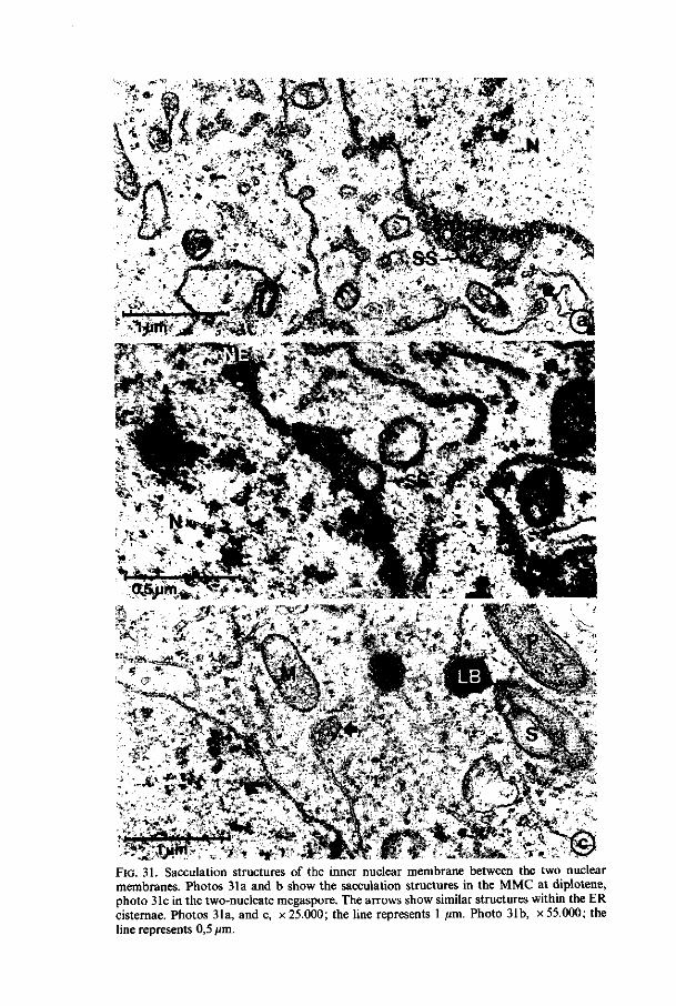
• « * * . • -5-,- "•*lP,• " ,"••• <\ *»•
V - * " r**..* P i *
9 JljnHi f^M 3L»" ̂ yaFTvfl
V-3 :#Ç r, *;
M'
?% t*&>. *
FIG. 31. Sacculation structures of the inner nuclear membrane between the two nuclear membranes. Photos 31a and b show the sacculation structures in the MMC at diplotene, photo 31c in the two-nucleate megaspore. The arrows show similar structures within the ER cisternae. Photos 31a, and c, x 25.000; the line represents 1 /mi. Photo 31b, x 55.000; the line represents 0,5 /mi.
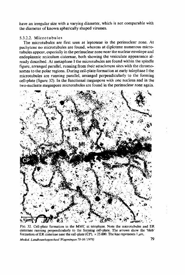
have an irregular size with a varying diameter, which is not comparable with the diameter of known spherically shaped virusses.
5.3.2.2. Microtubules The microtubules are first seen at leptotene in the perinuclear zone. At
pachytene no microtubules are found, whereas at diplotene numerous microtubules appear, especially in the perinuclear zone near the nuclear envelope and endoplasmic reticulum cisternae, both showing the vesiculate appearance already described. At metaphase I the microtubules are found within the spindle figure, arranged parallel, running from their attachment sites with the chromosomes to the polar regions. During cell-plate formation at early telophase I the microtubules are running parallel, arranged perpendicularly to the forming cell-plate (figure 32). In the functional megaspore with one nucleus and in the two-nucleate megaspore microtubules are found in the perinuclear zone again.
FIG. 32. Cell-plate formation in the MMC at telophase. Note the microtubules and ER cisternae running perpendicularly to the forming cell-plate. The arrows show the 'bleb' formation of ER cisternae near the cell-plate (CP), x 25.000. The line represents 1 urn.
Meded. Landbouwhogeschool Wageningen 78-16(1978) 79
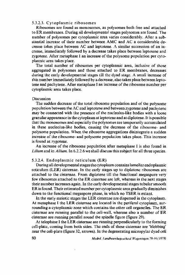
5.3.2.3. Cytoplasmic ribosomes Ribosomes are found as monosomes, as polysomes both free and attached
to ER membranes. During all developmental stages polysomes are found. The number of polysomes per cytoplasmic area varies considerably. After a substantial increase of their number between AMC and AC a considerable decrease takes place between AC and leptotene. A similar succession of an increase, immediately followed by a decrease takes place between leptotene and zygotene. After metaphase I an increase of the polysome population per cytoplasmic area takes place.
The total number of ribosomes per cytoplasmic area, inclusive of those aggregated in polysomes and those attached to ER membranes, decreases during the early developmental stages till the dyad stage. A small increase of this number immediately followed by a decrease, also takes place between leptotene and pachytene. After metaphase I an increase of the ribosome number per cytoplasmic area takes place.
Discussion The sudden decrease of the total ribosome population and of the polysome
population between the AC and leptotene and between zygotene and pachytene may be connected with the presence of the nucleolus-like bodies with a loose granular appearance in the cytoplasm at leptotene and at diplotene. It is possible that the monosomes and especially the polysomes are temporarily accumulated in these nucleolus-like bodies, causing the decrease of the ribosome- and polysome population. When the ribosome aggregations disintegrate a sudden increase of the ribosome- and polysome population takes place. This increase is found at zygotene.
An increase of the ribosome population after metaphase I is also found in Lilium and in Allium. In 6.2.2.6 we shall discuss this subject for all three species.
5.3.2.4. Endoplasmic reticulum (ER) During all developmental stages the cytoplasm contains lamellar endoplasmic
reticulum (LER) cisternae. In the early stages up to diplotene ribosomes are attached to the cisternae. From diplotene till the functional megaspore very few ribosomes attached to the ER cisternae are left, whereas in the next stages their number increases again. In the early developmental stages tubular smooth ER is found. Their estimated number per cytoplasmic area gradually diminishes down to the functional megaspore phase, in which no TSER is extant.
In the early meiotic stages the LER cisternae are dispersed in the cytoplasm. At metaphase I the LER cisternae are located in the periferal cytoplasm, surrounding a cytoplasmic zone which contains the other cell organelles. The ER cisternae are running parallel to the cell-wall, whereas also a number of ER cisternae are running parallel round the spindle figure (figure 29).
At telophase I the LER cisternae are running perpendicularly to the forming cell-plate, coming from both sides. The ends of these cisternae are 'blebbing' near the cell-plate (figure 32, arrows). In the degenerating micropylar dyad cell
80 Meded. Landbouwhogeschool Wageningen 78-16 (1978)

parallel arranged profiles of the endoplasmic reticulum are found. The vesiculate structures within the ends of the ER cisternae, as already
described in 5.3.2.1, are found at leptotene, zygotene and diplotene and in the functional cell and the two-nucleate megaspore (figure 31a and c).
Discussion The parallel arrangements of the ER cisternae and their characteristic
distribution in the cytoplasm at metaphase I probably arose during the transition of the nucleus from diplotene to metaphase I. At diplotene the ER cisternae are attached to the nuclear envelope and to the plasmodesmata, owing to which their localization was fixed as it were. When the nuclear envelope disintegrates the ER cisternae are getting dispersed freely in the cytoplasm. When the nuclear contents mix with the cytoplasm the ER cisternae may get arranged parallel to each other near the cell-wall and round the spindle, in the zones where they were previously bound. The ER cisternae round the spindle figure may be remnants of the nuclear envelope, but no pores are found in these cisternae.
The arrangement of the ER cisternae perpendicular to the cell-plate may indicate a possible function of these cisternae in cell-plate formation. No connections are found between the ER cisternae and microtubules or electron-dense vesicles.
One theory could be that the ER cisternae (and microtubules) are present on those sites of the cell-plate where the future plasmodesmata in the cell-wall will be formed. In our material no plasmodesmata are found within the cross-wall of the dyad, so that this theory must be rejected here.
Another possibility is that the ER cisternae have a function in the transport of the electron-dense vesicles derived from the dictyosomes to the phragmo-plast, where these vesicles string together to form the cell-plate. It is also possible that the ER cisternae have a function in the synthesis and the transport of other components necessary for cell-plate formation, e.g. enzymes.
However, in the phragmoplast a considerable number of microtubules is found, which are more likely to be responsible for this transport (HEPLER and JACKSON, 1968).
HEPLER and JACKSON (1968) found that ER membranes actively participate in cell-plate formation by providing membrane for the plasma membrane and material for the plate matrix. CRESTI and VAN WENT (1976) found an activity of ER cisternae in relation to production and secretion of callosic cell-wall material during the growth of the pollen tube.
In our material callosic components are found in the cross-wall of the dyad by the use of the fluorescence-aniline-blue method (BEDNARA, 1977). Probably these callosic components are synthesized by the ER cisternae near the cell-plate. The 'bleb' formation on the ends of these ER cisternae may indicate the vesicle production of the ER cisternae.
P ro t e in - f i b r i l s At diplotene and in the later stages after the dyad, regularly structured bund-
Meded. Landbouwhogeschool Wageningen 78-16 (1978) 81
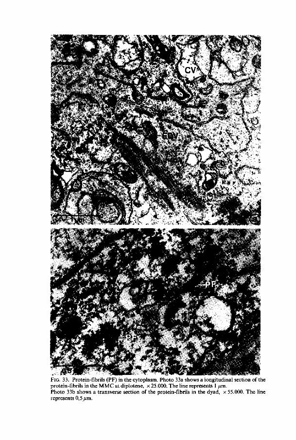
FIG. 33. Protein-fibrils (PF) in the cytoplasm. Photo 33a shows a longitudinal section of the protein-fibrils in the MMC at diplotene, x 25.000. The line represents 1 /im. Photo 33b shows a transverse section of the protein-fibrils in the dyad, x 55.000. The line represents 0,5ßm.
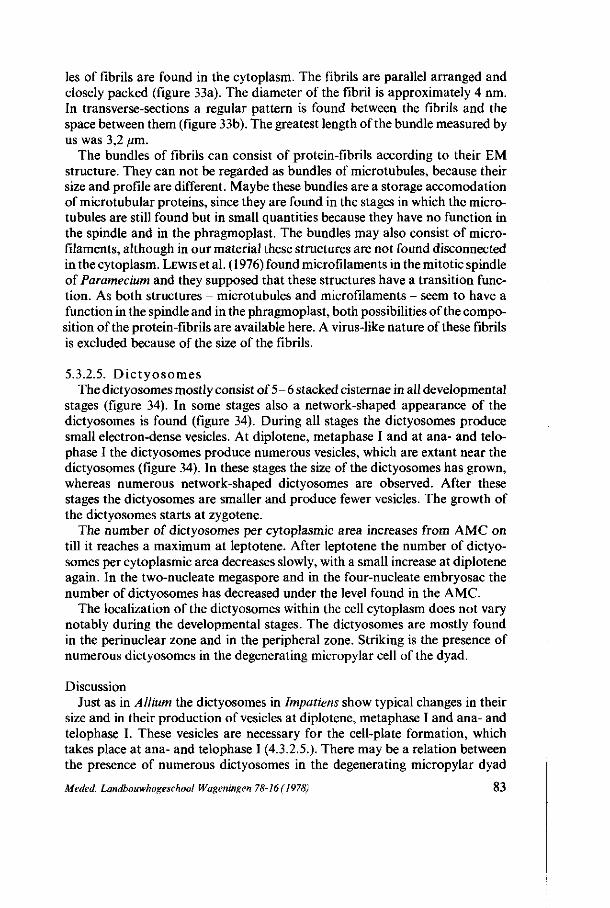
les of fibrils are found in the cytoplasm. The fibrils are parallel arranged and closely packed (figure 33a). The diameter of the fibril is approximately 4 nm. In transverse-sections a regular pattern is found between the fibrils and the space between them (figure 33b). The greatest length of the bundle measured by us was 3,2 /an.
The bundles of fibrils can consist of protein-fibrils according to their EM structure. They can not be regarded as bundles of microtubules, because their size and profile are different. Maybe these bundles are a storage accomodation of microtubular proteins, since they are found in the stages in which the microtubules are still found but in small quantities because they have no function in the spindle and in the phragmoplast. The bundles may also consist of microfilaments, although in our material these structures are not found disconnected in the cytoplasm. LEWIS et al. (1976) found microfilaments in the mitotic spindle of Paramecium and they supposed that these structures have a transition function. As both structures - microtubules and microfilaments - seem to have a function in the spindle and in the phragmoplast, both possibilities of the composition of the protein-fibrils are available here. A virus-like nature of these fibrils is excluded because of the size of the fibrils.
5.3.2.5. Dictyosomes The dictyosomes mostly consist of 5- 6 stacked cisternae in all developmental
stages (figure 34). In some stages also a network-shaped appearance of the dictyosomes is found (figure 34). During all stages the dictyosomes produce small electron-dense vesicles. At diplotene, metaphase I and at ana- and telophase I the dictyosomes produce numerous vesicles, which are extant near the dictyosomes (figure 34). In these stages the size of the dictyosomes has grown, whereas numerous network-shaped dictyosomes are observed. After these stages the dictyosomes are smaller and produce fewer vesicles. The growth of the dictyosomes starts at zygotene.
The number of dictyosomes per cytoplasmic area increases from AMC on till it reaches a maximum at leptotene. After leptotene the number of dictyosomes per cytoplasmic area decreases slowly, with a small increase at diplotene again. In the two-nucleate megaspore and in the four-nucleate embryosac the number of dictyosomes has decreased under the level found in the AMC.
The localization of the dictyosomes within the cell cytoplasm does not vary notably during the developmental stages. The dictyosomes are mostly found in the perinuclear zone and in the peripheral zone. Striking is the presence of numerous dictyosomes in the degenerating micropylar cell of the dyad.
Discussion Just as in Allium the dictyosomes in Impatiens show typical changes in their
size and in their production of vesicles at diplotene, metaphase I and ana- and telophase I. These vesicles are necessary for the cell-plate formation, which takes place at ana- and telophase I (4.3.2.5.). There may be a relation between the presence of numerous dictyosomes in the degenerating micropylar dyad
Meded. Landbouwhogeschool Wageningen 78-16(1978) 83
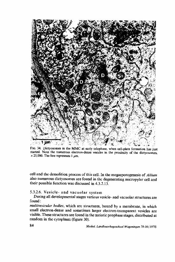
3*jf\ la'
* -.
\— ,. . .v\
" o, f"" C - /*•
A ;.̂ >, • #.
."!'N"
FIG. 34. Dictyosomes in the MMC at early telophase, when cell-plate formation has just started. Note the numerous electron-dense vesicles in the proximity of the dictyosomes, x 25.000. The line represents 1 /im.
cell and the demolition process of this cell. In the megasporogenesis of Allium also numerous dictyosomes are found in the degenerating micropylar cell and their possible function was discussed in 4.3.2.13.
5.3.2.6. Vesicle- and vacuolar system During all developmental stages various vesicle- and vacuolar structures are
found : multivesicular bodies, which are structures, bound by a membrane, in which small electron-dense and sometimes larger electron-transparent vesicles are visible. These structures are found in the meiotic prophase stages, distributed at random in the cytoplasm (figure 30).
°4 Meded. Landbouwhogeschool Wageningen 78-16(1978)
electron-dense vacuoles, which are bound by a membrane. They often have an erratically shaped appearance. Their electron-dense contents are mostly granular. These structures are found in the AMC, in the AC and in the MMC at diplotene (figure 33), telophase I and in the chalazal dyad cell, when the micropylar dyad cell is degenerating. ''clear' vacuoles, which are more globular-shaped vacuoles filled with electron-transparent material, sometimes with some condensed electron-dense material (figure 33). Small clear vacuoles are found in all developmental stages. Up to the dyad stage the number of small clear vacuoles remains approximately constant. In the dyad the number of vacuoles begins to enlarge, which will eventually lead to the central vacuole in the eight-nucleate embryosac. During all developmental stages the clear vacuoles are distributed at random in the cytoplasm.
5.3.2.7. Plasma membrane In some developmental stages vesiculate and membranous structures are
found between the plasma membrane and the cell-wall, especially in the early meiotic prophase and at a considerably lower degree in the two-nucleate megaspore. These structures are comparable with those found in Lilium mega-sporogenesis (3.3.2.7.).
5.3.2.8. Mitochondria In the premeiotic interphase stages the mitochondria have cristae. The
matrix of the mitochondria is relatively electron-transparent. At leptotene and zygotene the cristae are getting dilated and simultaneously the matrix becomes electron-dense (figures 28 and 30). The number of cristae diminishes. At pachytene and diplotene very few cristae are left while the matrix has a granular electron-dense appearance. From metaphase I on the mitochondria contain an increasing amount of cristae except in the degenerating and in the functional dyad cell, in which very few cristae are perceptible in the mitochondria. In the four- and eight-nucleate embryosac the mitochondria show an appearance, similar to those in the premeiotic interphase.
The number of mitochondria per cytoplasmic area remains approximately constant during the early developmental stages. At diplot ;ne and at metaphase I a maximal number of the mitochondria is found. After metaphase I their number decreases and in the four-nucleate embryosac o ily half of the original number per cytoplasmic area is left.
In the early developmental stages the mitochondria are distributed at random in the cytoplasm. In metaphase I, ana- and telophase I all cell organelles, including the mitochondria, are located at the polar sid< :s of the cell. Remarkable is the relatively large number of mitochondria in the micropylar dyad cell which will soon degenerate.
Discussion In Impatiens the mitochondria show a return to their original ultrastructure
Meded. Landbouwhogeschool Wageningen 78-16 ( 1978) 85

during megasporogenesis. The same occurs in Lilium megasporogenesis. In 6.2.2.9 we shall revert to this subject.
The relative large number of mitochondria in the micropylar dyad cell may suppose that these mitochondria are needed for the degeneration process, which will soon take place in this cell. In 5.3.2.13 we shall discuss their possible function in the demolition process.
5.3.2.9. P l a s t id s In the archespore cell few plastids contain starch. In the later developmental
stages the number of plastids containing starch and the amount of starch within each of the plastids increase. The presence of starch in the plastids clouds the developmental ultrastructural changes. Besides starch the plastids regularly contain plastoglobules. In the early developmental stages up to diplotene the plastids bear thylakoids. After pachytene the thylakoids have disappeared till the two-nucleate megaspore, in which phase they are present again, just as they are in the four-nucleate embryosac.
The number of plastids per cytoplasmic volume decreases during the early developmental stages till this number reaches a minimum at diplotene. In metaphase I their number increases slowly and then remains constant during the later stages.
In the AC, at leptotene, pachytene and in ana- and telophase I the plastids show a slight tendency to be localized on the chalazal side of the cell. In the micropylar dyad the relative number of plastids is much smaller than in the chalazal dyad cell, this in contrast with the number of mitochondria in these cells.
Discussion In Impatiens megasporogenesis a storage of reserves takes place in the forma
tion of starch in the plastids. The amount of starch is increasing constantly during the developmental process. Of the three species examined only Impatiens shows a storage of starch in the plastids. In 5.4 we shall revert to this subject.
Just as we noted in Allium, the plastids in Impatiens show a tendency to be localized on the chalazal side of the cell during meiotic prophase. This localization of the plastids probably leads to the unequal distribution of the plastids in the two dyad cells. The future functional cell gets more plastids in the cytoplasm than the future non-functional cell. In paragraph 6.3.2.3 we shall go into this subject in detail.
5.3.2.10. L ipid bod ies The lipid bodies show different appearances during megasporogenesis in
Impatiens walleriana. In the AMC and in the AC the common droplet feature is found. In meiotic prophase and in metaphase I electron-transparent vacuoles are perceptible in the lipid droplets. In ana- and telophase electron-transparent vesicles are attached to the lipid droplet. From two-nucleate stage on the common lipid droplets appear again, with a larger diameter.
86 Meded. Landbouwhogeschool Wageningen 78-16 ( 1978)
The amount of lipids per cytoplasmic area increases during the two-nucleate megaspore and the four-nucleate embryosac. In the eight-celled embryosac we find groups of lipid bodies in the central cell.
Discussion As in Lilium the lipid bodies of Impatiens show electron-transparent vacuoles
and an attachment to electron-transparent vesicles. These vesicles may contain enzymes for the digestion of the lipidic compounds (3.3.2.10). In Impatiens a storage of lipidic compounds takes place, which increases during megagame-togenesis. In the eight-celled embryosac most of the lipid bodies are found in the central cell, which will form the endosperm after fertilization.
5.3.2.11. P l a smode sma t a In the AMC plasmodesmata are found in the whole surrounding cell-
wall. During all other developmental stages the plasmodesmata are found in the chalazal part of the cell-wall.
Discussion The continuous presence of plasmodesmata between the MMC and the
chalazal nucellus cells may indicate the presence of a transport-canal between these cells. In the chalazal nucellus cells the vascular bundle, transporting nutrients, ends. This causes a nutrition-gradient in the nucellus cells from the chalazal side to the micropylar side. The MMC will take up nutritions necessary for its development. This assimilation of nutrients probably takes place through the plasmodesmata in the chalazal cell-wall of the megasporocyte and mega-gametophyte. In 5.4 we shall discuss this nutrient-transport in relation to the storage of starch and lipids in the cell.
5.3.2.12. Ce l l -wal l The cell-wall thickens during meiotic prophase. A maximum thickness of the
cell-wall is found in diplotene. At metaphase I the thickness of the cell-wall has abruptly decreased and in the later stages the cell-wall thickens again.
After applying the fluorescence-aniline-blue method for checking the presence of callosic compounds in the cell-wall (RODKIEWICZ et al., 1968), callosic compounds are found to be present in the surrounding cell-wall at diplotene, except on the chalazal side of the cell-wall and in the cross-wall of the dyad (BEDNARA,
1977). However, the EM ultrastructure of the cell-wall containing callose does not show the common - more electron-transparent - appearance of callosic cell-walls as found by JALOUZOT (1971) in Oenothera lamarckiana.
Discussion The maximum thickness of the cell-wall at diplotene may be due to the
presence of callosic compounds in this cell-wall. After disintegration of these callosic compounds between diplotene and metaphase I a thinner cell-wall is found at metaphase I. The cross-wall of the dyad is extremely thick and during
Meded. Landbouwhogeschool Wageningen 78-16(1978) 87
the formation of this cell-wall an extensive network of ER cisternae, running perpendicularly to the cell-plate, is found. According to our discussion in 5.3.2.4 it might be possible that the ER cisternae are in this case involved in the deposition of callose.
The difference in EM ultrastructure between the cell-wall containing callose in our material and this cell-wall in Oenothera lamarckiana may be due to different methods of fixation or to a difference in the quantity of callosic components in the cell-wall.
5.3.2.13. Degene r a t i on of the m i c ropy l a r dyad cell In the degenerating of the micropylar dyad cell the nucleus has an irregular
shape. Nuclear pores are not visible. The cytoplasm is strong electron-dense, and contains numerous ribosomes as monosomes and polysomes, whereas very few ribosomes are attached to ER membranes. Structures of parallel arranged ER cisternae are found, as well as dictyosomes in a relatively large number. Also mitochondria are found in the degenerating micropylar cell in a relatively large number per cytoplasmic area, whereas the number of plastids is very low. Electron-transparent vacuoles are perceptible, mostly containing condensed electron-dense material. Lipid bodies are not found in the degenerating micropylar cell. Within the cross-wall black dots and granular electron-dense material is found.
Discussion The irregular shape of the nucleus may be due to the degeneration process.
The strong electron-dense appearance of the cytoplasm is probably caused by the presence of numerous ribosomes and by the presence of proteins, which are precipitated during the degeneration process. A possible function of the ribosomes was already suggested in 4.3.2.13 of Allium. They may have a function in the synthesis of lytic enzymes. The dictyosomes may have a function in the synthesis and/or transport of digestive enzymes. The relatively high number of mitochondria in the degenerating cell also suggests a function in the demolition process. During this process energy comes free, which may be stored in the mitochondria. Also in the perimitochondrial space enzymes are synthesized, necessary for the synthesis of fats. The electron-transparent vacuoles may serve as autophagic vacuoles (4.3.2.13).
The presence of granular electron-dense material in the cell-wall between the micropylar and the chalazal cell may indicate the possibility that material is transported through this cell-wall. Maybe the callosic compounds in the cell-wall are locally disintegrated to make this transport possible. On the other hand, an accumulation of lipid bodies in the chalazal dyad cell is not found. Because an accumulation of lipid bodies starts in the two-nucleate megaspore, a possible transport of lipidic compounds through the cross-wall of the dyad -as is found in Allium - may not take place in Impatiens presuming that the two-nucleate megaspore receives the nutrients for the storage of fats from the chalazal side of the cell. Maybe other useful material is transported through the
88 Meded. Landbouwhogeschool Wageningen 78-16 (1978)
cell-wall to promote the growth and development of the functional chalazal cell.
5.3.2.14. Nucellus cell During its development the MMC is surrounded by a layer of nucellus cells.
This layer consists of young appearing cells without central vacuoles. In the early stages of megasporogenesis these cells are small and they will elongate during the later stages to surround the growing MMC. No division of the nucellus epidermis cells is found. In the dyad stage of the MMC the nucellus cells which are in contact with the MMC start degenerating, with the exception of some cells located on the chalazal side of the MMC. In the four-nucleate embryosac all nucellus cells which are in contact with the MMC have collapsed except on the chalazal side, where the MMC remains in contact with some viable nucellus cells. In the cell-wall between these cells and the young embryosac numerous plasmodesmata remain present.
One particular nucellus cell, positioned near the micropyle has been studied in all the developmental stages of the MMC observed. The nucleus has an elongated shape with a smooth surface. It contains a reticulate chromatin structure. The nuclear envelope shows open pores in the AMC and AC stage of the mega-sporocyte and plugged pores during the meiotic prophase of the MMC. Microtubules are found in the periferal zone in half the stages observed. In the early developmental stages of the MMC ribosomes are found as monosomes, as polysomes both free and attached to the nuclear envelope and to ER cisternae. From pachytene to telophase of the MMC ribosomes are only found as monosomes free and attached to ER cisternae. From the telophase of the MMC on, polysomes are found in the nucellus cell again. In the early developmental stages of the MMC some smooth ER is found in the nucellus cell, whereas in all stages of the MMC rough ER is found in the nucellus cell. Dictyosomes are found in the stacked shape, producing small electron-dense vesicles in all developmental stages of the MMC, except in zygotene and diplotene. Electron-transparent vacuoles are found in the nucellus cell from zygotene of the MMC on. Within the vacuoles some condensed electron-dense material is often found. Except in leptotene of the MMC the plasma membrane of the nucellus cell has no invaginations nor evaginations. The mitochondria in the nucellus cell have a more condensed shape, with very few cristae in pachytene of the MMC and in the two-nucleate megaspore. In all the other stages of the MMC the nucellus cell has mitochondria with the common appearance, containing small cristae. The plastids have plastoglobules in nearly all the stages «of the MMC. Starch granules are only found in the plastids of the nucellus cell when the dyad is formed. Plastids thylakoids are not always found. Sometimes lipid bodies are found. They have the common appearance of a lipid droplet, with sometimes a clear vacuole within the droplet. Plasmodesmata are found in all cases in the side walls, not in the cell-wall between the nucellus cell and the MMC. The thickness of the cell-wall remains approximately constant until the degenerating of the cell starts. Then the cell-wall gets thinner.
Meded. Landbouwhogeschool Wageningen 78-16(1978) 89
Discussion From these data we may conclude that there is only a slight correlation
between the ultrastructural development of the nucellus cell and the features previously mentioned of the ultrastructural development of the megasporocyte. There may be a correlation between the increase of the polysome population in the developing MMC from pachytene on and the disappearance of polysomes in the nucellus cell at that time. Apart from this possible correlation the nucellus cell and the megasporocyte show their own specific development. A conspicious fact is that in the nucellus cell no storage of starch is found in the plastids, such in contrast to the plastids in the MMC.
5.4. DISCUSSION
From the results of our investigations we already concluded that Impatiens shows the bisporic type of embryosac development. Just as in Allium the degeneration of the non-functional cell already starts at the dyad stage, before the second meiotic division takes place. Therefore we called the chalazal dyad cell the functional cell, when the micropylar dyad cell starts degenerating. After the second meiotic division in the functional cell, the two-nucleate megaspore appears. After one and two mitotic divisions respectively the four-nucleate and the eight-celled embryosac appear.
Just as it is seen in Allium a number of developmental phenomena during megasporogenesis seems to be related. Also the presence of nuclear pores, the undulations of the nuclear envelope and the sacculations of the inner nuclear membrane seem to be related to the presence of microtubules and the appearance of typical changes in the size and ultrastructure and in the vesicle-production of dictyosomes. In 6.2.2.2 this possible relation will be discussed.
In the developing megasporocyte and megagametophyte an increasing amount of starch is found in the plastids. In the last stages of megagameto-phytic development also an increasing amount of lipidic compounds is found in tho cytoplasm. By means of plasmodesmata a close connection exists between the developing megasporocyte/megagametophyte and the chalazal nucellus cells. In the stages after the dyad these chalazal cells remain viable, in contrast with the other surrounding nucellus cells. All these facts may indicate that the developing megasporocyte and megagametophyte receive nutritions from the chalazal side of the cell through the plasmodesmata. This nutrition-supply seems to be more than ample for the maintenance, the growth and the development of the cell, which appears from the increasing amount of food material stored in the cell. In Impatiens the megasporocyte/megagametophyte shows a high degree of heterotrophy as the cell not only obtains nutrients for its own development but also as a storage for future needs.
The occurrence of callose in the surrounding cell-wall at diplotene except in the chalazal cell-wall - the side on which the future functional megaspore will appear - has been described in many other species having the monosporic- or
90 Meded. Landbouwhogeschool Wageningen 78-16(1978)
bisporic type of embryosac development (RODKIEWICZ, 1970). The permeability of a cell-wall containing callose is lower than the permeability of a cellulosic cell-wall according to KNOX and HESLOP-HARRISON (1970). It is suggested that this callosic wall protects the differentiating haploid cell from a damaging interaction with the diploid nucellus cells or vice versa. On the chalazal side, on which the plasmodesmata remain open, no callosic compounds are found. The nutrition of the developing cell remains always possible. Also in the cross-wall between the micropylar and the chalazal dyad cell callosic compounds are found. Together with the remnants of degenerating nucellus-cells surrounding the micropylar cell-wall, the micropylar cell gets isolated from the living nucellus cells, especially in behalf of its nutrient-supply. This may be one of the causes that the micropylar cell dies. During the degeneration of the micropylar cell the cross-wall is getting irregular, with small electron-dense parts. Maybe the callosic components are partly degenerating to enable a diffusion of material through the cell-wall. There may be a relation between the localization of the plastids during meiotic prophase and the future unequal division of the cell. The plastids show a tendency to be located near the chalazal side of the cell. Owing to this localization an unequal distribution of the plastids in the two dyad cells takes place. The relatively large number of mitochondria and dictyo-somes in the micropylar dyad cell may be caused by a more equal spread of these cell organelles over the two dyad cells. Because of the unequal division of the cytoplasm a larger number of these cell organelles per cytoplasmic area is found in the micropylar dyad cell. This specific cell organelle composition in the micropylar dyad cell may be related to the future degeneration process in this cell.
In the two-nucleate megaspore the volumic growth of the cell takes place proportionally to the growth of the cytoplasm and the increasing vacuolisation of the cell. Big vacuoles appear in the four-nucleate embryosac, whereas the central vacuole is found in the eight-celled embryosac. In Impatiens no connection is found between the appearance of the big vacuoles and the distribution of the nuclei in the cell. The four nuclei in the four-nucleate embryosac are regularly distributed in the cytoplasm. In the eight-celled embryosac the common localization of the eight cells in the embryosac is found, maybe as a result of the position of the central vacuole in the eight-nucleate embryosac.
Meded. Landbouwhogeschool Wageningen 78-16(1978) 9 1
6. S I M I L A R I T I E S A N D D I F F E R E N C E S BETWEEN T H E T H R E E SPECIES D U R I N G M E G A S P O R O G E N E S I S
6.1. FIXATION METHODS
During glutaraldehyde-osmiumtetroxyde fixation saccharose had to be added to the fixation fluid of two of the three species, but in different concentrations, to prevent swelling and shrinking of the cells and organelles. This suggests a difference in osmolarities between the three species. Lilium has the greatest osmotic value and Impatiens the smallest. Since all preparation procedures in the various species - apart from the osmolarity differences - are basically similar we suppose that the individual results can be compared in relation with each other. Especially so because we took special care that in all cases fragile structures as ribosomes and microtubules were well preserved.
6.2. CELL ORGANELLE LEVEL
6.2.1. Results In enclosure V most of the morphological and quantitative results of the
three species have been assembled. This figure is composed of the three figures presented in the chapters 3, 4 and 5. The dyad stage of the bisporic type is supposed to be comparable with the two-nucleate stage of the tetrasporic type. The two-nucleate megaspore of the bisporic type is supposed to be comparable with the four-nucleate stage of the tetrasporic type.
Nuc leus Nuclear undulations. All three species show nuclear undulations starting at
leptotene with an estimated maximum at zygotene. Nuclear pores. The nuclear envelope has pores but their number changes
during the development of all three species. At zygotene and diplotene a maximal number of pores is found, whereas at pachytene a minimal number is found for all three species.
Sacculation of the inner nuclear membrane. In all three species a sacculation of the inner membrane is extant but in different quantities. For Allium and Impatiens a maximum is found at diplotene.
Chromatin structure. All the species show the common chromatin structures as described by MOENS (1968) except in Impatiens, where never a clearly visible synaptinemal complex is found.
Membrane-like structures. During meiotic prophase of Impatiens and Allium membrane-like structures are found in the nuclei, for the two species respectively at zygotene and at diplotene.
Nucleolus. During meiotic prophase the nucleolus enlarges for all three
92 Meded. Landbouwhogeschool Wageningen 78-16(1978)

species. The ultrastructure of the nucleolus is not described in detail in this investigation.
Cy top l a sm Ribosomes. The number of ribosomes per cytoplasmic area decreases for all
three species during premeiotic interphase and early meiotic prophase. After metaphase I the number of ribosomes per cytoplasmic area increases for all three species. Polysomes are perceptible in all three species at various stages and in different quantities. Impatiens continuously has a polysome population which decreases and increases at similar stages as with monosomes. In Allium and Lilium polysomes are absent in some developmental stages and when present their number is relatively low, except for Allium in the archespore cell, where a large number is found.
Nucleolus-like bodies. In Impatiens and Lilium, but not in Allium, nucleolus-like bodies are perceptible. They are not found in each stage of Impatiens mega-sporogenesis, whereas their ultrastructure changes in those stages in which they are found during the development of the MMC. In Lilium the nucleolus-like bodies are continuously extant after metaphase I and their ultrastructure does not change.
Microtubules. During meiotic prophase microtubules appear in all three species. In Impatiens they appear at leptotene whereas for Lilium and Allium they appear respectively at pachytene and diplotene.
Endoplasmic reticulum. RER is found in all three species in premeiotic interphase. In Impatiens and Allium RER can also be observed in later stages but not continuously. LER is perceptible in Impatiens and Lilium in those stages in which RER is not found. The same can be observed in Allium in the stages after the dyad stage. TSER is not continuously perceptible in all stages of the species. Allium and Lilium have a large amount of TSER at diplotene and metaphase I.
Dictyosomes. In all three species dictyosomes are observed in all developmental stages. In all species fluctuations of the number of dictyosomes per cytoplasmic area occur. Impatiens and Allium have a large number of dictyosomes at diplotene/metaphase I, whereas Lilium has a small number at these stages. The dictyosomes produce vesicles during the various developmental stages in all three species, although in Impatiens and Allium the dictyosomes show a higher production of vesicles at diplotene and metaphase I in contrast with Lilium. The dictyosomes in Allium show a changement in size as well at these stages.
Llectron-dense vacuoles. In all three species electron-dense vacuoles containing polysaccharidic material are perceptible. Only in Lilium they are continuously extant while their number is increasing.
Clear vacuoles. Clear vacuoles appear in large quantities in Allium and Impatiens after metaphase I and in Lilium after metaphase II.
Paramural bodies. In all three species paramural bodies are observed in various developmental stages. Only in Lilium they are perceptible in all stages.
Meded. Landbouwhogeschool Wageningen 78-16(1978) 93
Mitochondria. In all three species mitochondria are continously perceptible during all stages. Their ultrastructure changes during megasporogenesis of the three species. The number of mitochondria per cytoplasmic area shows fluctuations for each species in different stages. The mitochondria contain cristae in all stages of Lilium and in various stages of Impatiens and Allium. However, Allium shows very few cristae in the mitochondria during premeiotic interphase and meiotic prophase.
Plastids. In all three species plastids are found in all stages. The plastid ultrastructure show developmental changes in Lilium, whereas in Allium these changes lead to the original ultrastructure at the end of megasporogenesis. It is difficult to study the ultrastructural development of the plastids in Impatiens because of their large starch-contents, but a small variation can be observed. The number of plastids per cytoplasmic area shows different fluctuations in all three species. In all stages of Lilium the plastids contain plastoglobules, whereas they are not found in all stages of Allium and Impatiens. Only in Impatiens the plastids contain starch, the amount of which increases during megasporogenesis. In all three species dumb-bell-shaped plastids are extant in various meiotic prophase stages. In Allium thylakoids are always found in the plastids during megasporogenesis. Impatiens and Lilium have only thylakoids in their plastids at premeiotic interphase, meiotic prophase and in the two- or four-nucleate megaspore.
Lipid bodies. In almost all stages lipid bodies are perceptible for all three species. In Lilium the amount of lipid bodies per cytoplasmic area increases during the early developmental stages till metaphase I and then decreases. In Impatiens the amount of lipid bodies per cytoplasmic area increases after degeneration of the non-viable megaspore.
Plasmodesmata. At early developmental stages plasmodesmata are found in all three species. In Impatiens the plasmodesmata are perceptible during all stages of megasporogenesis and megagametogenesis. In Lilium the plasmodesmata disappear after metaphase II whereas in Allium they are not continuously found in the stages after leptotene.
Cell-wall. In all three species the cell-wall gradually thickens during megasporogenesis. Impatiens and Allium show a sudden thinning of the cell-wall at metaphase I. Impatiens has callosic components in the cell-wall during meiotic prophase as indicated by a fluorescence-aniline-blue method. According to RODKIEWICZ (1970) Allium presumably has callosic components in the cell-wall during meiotic prophase, whereas Lilium has not. Allium has cell-wall thickenings in the four-nucleate embryosac, similar to those found in transfer cells. In all three species the megaspore mother-cell enlarges during its development. A small decrease of the cell volume is found at diplotene in Impatiens and at metaphase I in Lilium.
94 Meded. Landbouwhogeschool Wageningen 78-16(1978)
6.2.2. Discussion
6.2.2.1. Nuc l ea r u ndu l a t i on s and nuc lea r po res The nuclear undulations, present in all three species seem to be characteristic
for nuclear divisions. They are described by JALOUZOT ( 1973) in meiotic prophase of megasporogenesis in Oenothera lamarckiana and by ZATSEPINA et al. (1977) in mitotic prophase. ZATSEPINA et al. suggest a possible role of these undulations in the break-down of the nuclear envelope (3.3.2.1). These authors suppose that the undulations are caused by the loss of the rigidity of the nucleus.
In our material the nucleus enlarges in the meiotic prophase stages where the undulations of the nuclear envelope are found. The shape of the nucleus is dependent of the elasticity of the nuclear envelope, both the osmolarity of the karyoplasm and the osmolarity of the cytoplasm and of the synthesis of new nuclear membrane.
In our material a maximal quantity of undulations is found at zygotene at which stage pairing of the chromosomes takes place and a maximal contraction of the chromosomes is found. During this contraction one of the possibilities may be that water is transferred from the nucleus to the cytoplasm, leading to a temporary decrease of the nuclear volume. Because the perinuclear space (the space between the two nuclear membranes) does not change at zygotene, we may presume that the nuclear envelope has not enough elasticity to compensate this temporary decrease of the nuclear volume without any reaction. So the temporary decrease of the nuclear volume causes a loss of rigidity of the nucleus which is followed by undulations of the nuclear envelope. Simultaneously a synthesis of nuclear envelope material may take place, which is probably defined by future necessaries. This synthesis may occur locally at the nuclear surface, causing folds of the nuclear envelope. In view of this the undulations are merely the passive results of previous processes.
The undulation of the nuclear envelope and the increasing nuclear volume implies that the contact-surface between nucleus and cytoplasm is getting larger. Nuclear pores are abundantly extant at the same time, especially in the undulating parts of the nuclear envelope. The general assumption is that nuclear pore-complexes have a function in the nucleo-cytoplasmic exchange of material (GALL, 1964;SCHEER, 1973;GALÄN-CANoetal., 1975). The undulating parts of the nuclear envelope seem to have a function in this nucleo-cytoplasmic exchange process by the location of pores in the undulations.
In all three species a decrease of the number of pores is observed after zygotene. It is known that the number of pores is varying during development of the nucleus (LA COUR and WELLS, 1972; SCHEL, 1977). SCHEL correlates an increase of the number of nuclear pores to an increase of the surface area in Physarum polycephalum. In our material a decrease of the number of nuclear pores occurs while the nucleus enlarges thus increasing the surface-area of the nucleus. It is possible that the low number of nuclear pores at pachytene is caused by the organization of the nucleus at that stage. By various authors it is assumed that pores are not extant in the nuclear envelope at those sites, where condensed
Meded. Landbouwhogeschool Wageningen 78-16 (1978) 9 5
chromatin is closely appressed to the nuclear envelope (LA COUR and WELLS, 1972; JALOUZOT, 1973; GALÂN-CANO et al., 1975). At pachytene the present synaptinemal complexes are attached to the nuclear envelope (MOENS , 1969; M C Q U A D E and WELLS , 1975). Also the large nucleolus is generally with one side attached to the nuclear envelope (CH. 3, 4 and 5, WILLIAMS et al., 1973; G A L Ä N - C A N O et al., 1975; G IMÉNEZ-MARTIN et al., 1977). Because of the
appearance of all synaptinemal complexes with one side closely to the nuclear envelope and the presence of a nucleolus closely to the nuclear envelope, the number of nuclear pores can be diminished by a decrease of the surface-area of the nucleus, which is suitable for nuclear pore complexes. In this case the undulation of the nuclear envelope and the location of pores in the undulations could be a mechanism to assure the presence of an essential surface-area, where nuclear pores can be extant and active, presuming that the a t tachment of chromatin to the nuclear envelope does not occur in the undulating parts. After pachytene the synaptinemal complexes break down and the condensed chromatin masses get loose of the nuclear envelope. The number of pores increases at this t ime, probably because of the increasing nuclear surface-area suitable for pore complexes. At the same time the undulations are disappearing.
6.2.2.2. T h e s a c c u l a t i o n o f t h e i n n e r n u c l e a r m e m b r a n e At meiotic prophase sacculation of the inner nuclear membrane can be
observed. As already mentioned in 3.3.2.1 these sacculations are also found in meiotic prophase of Oenothera lamarckiana megasporogenesis ( JALOUZOT, 1973) and of Triticum durum microsporogenesis ( L A COUR and WELLS , 1972). The function of these structures is unknown yet. Some suggestions are made here.
In our material sacculation structures are perceptible at pachytene in all three species. In Allium and Impatiens the maximal amount of sacculation structures is found at diplotene. In Lilium, however, only very few structures are found at leptotene and pachytene. At the same time the number of nuclear pores is small in those stages where sacculation structures are perceptible. In Lilium the number of nuclear pores at diplotene is larger than their number in Allium and Impatiens a t that stage, while for the latter two species a maximal amount of sacculation structures occurs. There seems to be a relation between the number of nuclear pores and the existence of sacculations. This may indicate that the sacculation structures are ment to take over a task of the nuclear pores. As already mentioned one task of the nuclear pores is a t ransport function in the nucleo-cytoplasmic exchange of material. The 'blebs' of the inner nuclear membrane contain karyoplasmic material and this material is also supposed to be present within the vesicles between the two nuclear membranes. Since no fusing of these vesicles with the outer nuclear membrane is found a process comparable with the exocytotic process as described by MORRÉ and MOLLEN-HAUER (1974) is not likely to take place here. Probably the inner nuclear membrane, surrounding the vesicles, gets incorporated within the outer nuclear membrane and the vesicle-contents get released in the cytoplasm and in the
96 Meded. Landbouwhogeschool Wageningen 78-16(1978)
perinuclear space in an as yet unknown way. It is also possible that the contents of the vesicles, which are extant in the
perinuclear space, are transferred from this space through the lumen of the endoplasmic reticulum, which is found to be continuous with the perinuclear space. Anyway, a transport mechanism seems to function, in some unknown way. By this process a nucleo-cytoplasmic exchange of material may take place, thus replacing the nuclear pores.
We also consider the possibility that the sacculations of the inner membrane are just local folds of the inner nuclear membrane as a result of a variation in the surface-area of the inner and outer nuclear membrane. Such variation may be due to an increase of the surface-area of the inner membrane, without a corresponding increase of the surface-area of the outer membrane or as a result from a shortening of the outer membrane without a corresponding shortening of the inner membrane. Such shortenings of the outer nuclear membrane may take place by its reverse into endoplasmic reticulum, since they are continuous. This process may bring about the presence of numerous endoplasmic reticulum cisternae in the perinuclear zone, as is found in Allium. An increase of the surface-area of the inner nuclear membrane may be related to a synthesis of new membrane material for future need.
What kind of material is transported in case of a transport function of the nuclear membrane sacculations is as yet unknown. When ribonucleoproteinic in nature, it has to be a soluble constitution, because concrete particles are not found in the 'blebs' and vesicles between both nuclear membranes.
A nucleo-cytoplasmic transport of ribonucleoproteinic material is found in microspore tetrads after the meiotic divisions as described by DICKINSON and BELL (1970), DICKINSON (1971), DICKINSON and POTTER (1975) and BELL (1975). These authors relate the nucleo-cytoplasmic exchange with pollen wall formation. As is generally assumed the microspore wall contains - beside cellulose -a callosic component (WILLEMSE, 1972; SOUTHWORTH, 1973). During meiotic prophase of megasporogenesis no extensive cell-wall synthesis takes place except the formation of a callosic component in the cell-wall of Impatiens (BEDNARA, 1977) and possibly of Allium (RODKIEWICZ, 1970). Presuming the presence of a callose-containing cell-wall in Allium these results may suggest a relation between the nuclear envelope sacculations and the callosic cell-wall formation.
According to RODKIEWICZ (1970) a callosic cell-wall is not formed in Lilium. The absence of a callosic cell-wall in Lilium, however, is somewhat doubtful, since by the aniline-blue method of RODKIEWICZ and BEDNARA callose seems not always to be stained (REYNOLDS and DASHEK, 1976). Presuming that Lilium does not form callose, and since in all three species sacculation is found, the process of sacculation therefore can not be related to cell-wall formation and callose deposition.
The sacculation structures at the nuclear envelope can also be correlated with the appearance of microtubules in the perinuclear zone. This implies that a relation between both phenomena is possible. In Impatiens the microtubules
Meded. Landbouwhogeschool Wageningen 78-16 (1978) 97
appear at leptotene whereas the sacculation structures at the nuclear envelope are already perceptible in the archespore mother-cell. For all of the three species an aggregation of the microtubules starts at diplotene. The nucleo-cytoplasmic exchange can be related to the synthesis and/or transport of precursors, necessary for the formation and aggregation of microtubules. For Allium and Lilium the number of tubular smooth endoplasmic reticulum increases at the same time, especially around the nuclear envelope. One possibility was already mentioned earlier in this chapter, especially for Allium : the SER cisternae could be a result of the disintegration of the outer nuclear membrane. Another possibility is, that the SER cisternae play a role in microtubular aggregation as suggested by BURGESS and NORTHCOTE (1968). In Impatiens and Allium the dictyosomes increase their vesicular production at diplotene. At that stage a maximal number of lamellar endoplasmic reticulum is found in Lilium. As already mentioned in 3.3.3 this LER in Lilium is perhaps comparable with the specific dictyosome population in Allium and Lilium (see also 6.2.2.7). This increase of vesicle production of the dictyosomes may also be related to the nucleo-cytoplasmic exchange which is found at pachytene and diplotene.
Summarizing : it is suggested that through the nuclear envelope, its nuclear pore-complexes and sacculation structures, precursors and signals are exchanged for the formation of tubular smooth ER, which may be related with the synthesis of microtubules and with the activation of the dictyosomes producing vesicles ; the latter two processes must be common in all dividing cells having nuclear and cell divisions.
6.2.2.3. C h r o m a t i n s t r u c t u r e The chromatin structure during meiotic prophase was described by several
authors (MOENS, 1968; JALOUZOT, 1973; MCQUADE and WELLS, 1975). These authors especially described the ultrastructure of the synaptinemal complex. These complexes are clearly found in our material during pachytene of Lilium and Allium megasporogenesis. In Impatiens no clearly visible synaptinemal complexes are found but they undoubtedly are extant. The absence of clearly visible complexes may be due to the fact that the exactly appropriate developmental stage was not available.
6.2.2.4. Membrane - l i k e s t r u c tu r e s In Allium and Impatiens megasporogenesis membrane-like structures in the
nucleus are perceptible. Some suggestions of the origin of these structures were made by WILLEMSE (1971a) (see 4.3.2.1). As these structures are found in both microsporogenesis (WILLEMSE, 1971a) and megasporogenesis (this thesis) they may be considered as prophasic characteristics for meiosis in spermatophyta, because they only occur during the contraction of chromosomes. A possible explanation for the absence of membrane-like structures in Lilium megasporogenesis may be the fact that not the exactly appropriate developmental stage was available.
98 Meded. Landbouwhogeschool Wageningen 78-16 (1978)
6.2.2.5. N u c l e o l u s The ultrastructure of the nucleolus and the nucleolus organizing region
during meiosis was studied by several authors (BRASELTON and BOWEN, 1971 ; GIMÉNEZ-MARTIN and STOCKERT, 1970; WILLIAMS et al., 1973; LUCK and
JORDAN, 1977; G IMÉNEZ-MARTIN et al., 1977). In this thesis changes of the ultrastructure of the nucleolus organizing region and of the nucleolus have not been described in detail. An enlargement of the nucleolus during meiotic prophase as found in our species has not been described by one of the authors previously mentioned. BRASELTON and BOWEN find an absence of nucleolar granules in Lilium meiotic prophase of microsporogenesis. They suggest a reduction or absence of r ibosomal RNA synthesis. In our species the nucleolus has a granular ultrastructure, indicating the presence of RNP particles. The enlargement of the nucleolus may point to an accumulation of synthesized RNP . It is generally assumed, that R N P particles of the nucleolus are transported to the cytoplasm through nuclear pores and are used as precursors of cytoplasmic R N A (JONES, 1965 and ROBARDS, 1973).
The number of nuclear pores decreases during meiotic prophase (this thesis) and this may cause a nucleolar accumulation of the R N P particles. At the same time the number of cytoplasmic ribosomes per cytoplasmic area decreases for all three species, probably as the result of a low supply, caused by a lower number of nuclear pores.
The number of ribosomes increases at metaphase I. During this stage the karyoplasm mixes with the cytoplasm and the nucleolus disappears. It is likely that the increase of the cytoplasmic RNA is caused by the mixing of karyoplasm and cytoplasm and the disintegration of the nucleolus. WILLEMSE and LINSKENS (1968) also described an increase of the r ibosome number per cytoplasmic area during metaphase, caused by the mixing of the karyoplasm with the cytoplasm. The nucleolus probably has dissolved in diakinesis (GIMÉNEZ-MARTIN et al., 1977), causing an increase of polysomes and monosomes in the cytoplasm during metaphase. Also some additional nucleoli may be produced during meiotic prophase, which will remain in the cytoplasm after nuclear division as so-called nucleolus-like bodies (WILLIAMS et al., 1973). According to the latter authors these nucleolus-like bodies are presumably also related to the restoration of the r ibosome population. Nucleolus-like bodies are found during Lilium and Impatiens megasporogenesis but not in Allium. In Lilium the nucleolus-like bodies are extant after metaphase I. In Impatiens the nucleolus-like bodies are not only found after metaphase I but also in meiotic prophase at leptotene and diplotene, in which stages their appearance has changed. At meiotic prophase they have a loosely granular ultrastructure, probably consisting of groups of monosomes, whereas the common appearance of nucleolus-like bodies is a compact electron-dense granular structure. These ' common' nucleolus-like bodies are probably precursors of r ibosomes after their release into the cytoplasm.
Meded. Landbouwhogeschool Wageningen 78-16(1978) 9 9
6.2.2.6. R i bo somes As it was mentioned earlier in this chapter, a possible explanation for the
decrease of the ribosome population in the cytoplasm is given by assuming the presence of a barrier, resulting from the decrease of the number of nuclear pores. DICKINSON and ANDREWS (1977) suggest that during meiotic prophase lytic enzymes are active to reduce the ribosome population. They suggest that the so-called 'membrane-bound' cytoplasmic inclusions isolate parts of the cytoplasm from the activity of the lytic enzymes, thus preserving cytoplasmic components for the stages after meiotic division and serving the continuity of protein synthesis during meiosis. The authors speak about an eradication of the ribosome population during microsporogenesis, as it was also described by MACKENZIE et al. (1967), DICKINSON and HESLOP-HARRISON (1970b) and WILLIAMS et al. ( 1973). In our material the number of ribosomes per cytoplasmic area decreases during meiotic prophase. However, during this phase the cytoplasmic volume increases abundantly, causing a dilution of the ribosome population. Therefore the total amount of cytoplasmic ribosomes increases, so that no eradication of the ribosome population takes place. Therefore there is no reason for us to assume an activity of lytic enzymes. DICKINSON and POTTER
(1978) describe an elimination and restoration of the ribosome population in Lilium megasporogenesis, similar to those in Lilium microsporogenesis. They have measured the reduction in the number of ribosomes by the use of a micro-densitometer, but a correction factor for the cytoplasmic growth is not included in the discussion.
In our material the increase of the ribosome number per cytoplasmic area after metaphase I seems to be caused by a ribosome-flow during nuclear envelope disintegration (WILLEMSE and LINSKENS, 1968) and by a disintegration of the nucleolus-like bodies in the cytoplasm (WILLIAMS et al., 1973). The so-called 'membrane-bound' inclusions in our material of Lilium sometimes contain more and sometimes fewer ribosomes than the surrounding cytoplasm. Therefore these inclusions can not be responsible for the increase of the cytoplasmic ribosome population.
About the function of the dilution of the ribosome population at meiotic prophase and the restoration of the population after meiosis, DICKINSON and POTTER (1978) propose 'an effectively removing of the "diplophase" information carrying molecules from the cytoplasm preparing it for the arrival of "haplophase" information produced by the new nucleus'. Certainly a reverse of information will take place when the sporophytic generation turns over into a gametophytic generation. However, a mechanism such as the eradication of the ribosome population and the rapid restoration of the ribosome population after meiosis does not seem to be essential for such a reverse.
In protein synthesis both free polysomes and polysomes attached to ER membranes are involved, according to PALADE (1975). A continuous polysome population as found in Impatiens megasporogenesis suggests a continuous protein synthesis during its development. Also Allium shows a more or less continuous protein synthesis, whereas Lilium sometimes has only a small
100 Meded. Landbouwhogeschool Wageningen 78-16 ( 1978)
number of polysomes. Based on the presence of polysomes in Lilium the bulk of protein synthesis probably takes place at early meiotic prophase. This bulk of protein synthesis may be related to the abundant membrane synthesis which occurs at Lilium meiotic prophase.
6.2.2.7. D i c t y o s o m e s Cell-plate formation is a process which takes place during anaphase/telo
phase of the nuclear division within the phragmoplast. WHALEY et al. (1966) and FRASER ( 1975) suppose that the vesicles which are fusing in the phragmoplast are derived from the Golgi apparatus. HEPLER and NEWCOMB (1967) and HEPLER and JACKSON (1968) do not exclude the derivation of the vesicles from dictyosomes but they also suggest a derivation of the vesicles from the endoplasmic reticulum cisternae. Microtubules are running perpendicularly to the cell-plate and they are presumed to have a function in the t ransport of the vesicles from the sites of synthesis to the cell-plate (HEPLER and NEWCOMB, 1967; HEPLER and JACKSON, 1968). MOLLENHAUER and MORRÉ (1976) suggest
a t ransport function of microfilaments for the movement of secretory vesicles from the site of synthesis to the cell-plate.
In Allium and Impatiens typical changes manifest themselves in the dictyo-some population during diplotene/metaphase I. Besides an increase of the number of dictyosomes per cytoplasmic area, an increase of the vesicle production takes place. Also the position of the dictyosomes in the cell changes. In Allium the dictyosomes show an enlargement in size and a change in ultra-structure too. In Lilium these typical changes of the dictyosome population do not take place at diplotene/metaphase I. The number of dictyosomes per cytoplasmic area remains low, while their number of produced vesicles remains low. Though dictyosomes are found near the spindle figure and within the spindle figure, these dictyosomes consist of very few cisternae producing very few vesicles.
The typical changes of the dictyosome population in Allium and Impatiens likely are related to the cell-plate formation during meiotic division. In Lilium no cell-plate formation takes place after nuclear division, so the absence of the typical changes of the dictyosomes at diplotene/metaphase I in this species may therefore be related to the absence of cell-plate formation. Lilium on the other hand has an abundant endoplasmic reticulum, arranged in concentric running cisternae complexes with central cores at diplotene/metaphase I. MORRÉ, MOLLENHAUER and BRACKER (1971) suppose a continuity between ER cisternae and dictyosome cisternae at the forming face of the dictyosomes. F rom the ER cisternae new dictyosome cisternae are formed. It is supposed that in Lilium M M C there is a lack in this system owing to which no active dictyosomes are formed by the ER cisternae ( D E BOER-DE JEU, 1978). It is also supposed that this lack causes a bulk production of ER cisternae, which are stacked in concentric complexes, the most economic way to stack this amount of cisternae. Probably these ER cisternae also have a task in the organization of the cell.
Meded. Landbouwhogeschool Wageningen 78-16(1978) 101
6.2.2.8. E l e c t ron -dense v acuo le s , c lear vacuo les and p a r a m u r a l bod ies
It can be proved that electron-dense vacuoles can contain contents as polysaccharides and enzymes. Electron-dense vacuoles with polysaccharidic contents are found in all three species. In Allium and Impatiens megasporogenesis they are probably incorporated in the cell-plates. In Lilium megasporogenesis -in which no cell-plate is formed - also vesicles containing polysaccharides are formed, but only in the periferal part of the cell. An accumulation of this material takes place between the plasma membrane and the cell-wall. MAR-CHANT and ROBARDS (1968) and ROBARDS and KIDWAI (1969) suppose that the material present in the paramural bodies is concerned with cell-wall synthesis. According to these authors multivesicular bodies are supposed to be derived from dictyosomes and to contain cell-wall precursors. FOWKE and SETTERFIELD
(1969), however, suppose an artificial reorganization of the plasma membrane without any involvement in cell-wall deposition for the structures mentioned above. With a view to the polysaccharidic contents in the paramural bodies and in the multivesicular bodies, we prefer the hypothesis of ROBARDS and KIDWAI.
Probably the polysaccharidic material in Lilium is necessary for cross-wall formation, when the eight-celled embryosac is formed. Presumably not a normal cell-plate formation takes place then, but a kind of cleavage as in nucellar endosperm when it becomes cellular as described by MORRISON and O'BRIEN
(1976).
The electron-dense vacuoles with enzymic contents are called microbodies in which, according to FREDERICK et al. ( 1975) different types can be distinguished. In our material the nature of the microbodies is unknown, except in Lilium megasporogenesis after meiotic prophase, they likely are glyoxisomes. In these stages, when a decrease of the lipidic amount takes place, glyoxisomes are found to be attached to the lipid bodies. In the biochemical conversion of reserve fats into carbohydrates glyoxisomes are known to be involved (RICHARDSON, 1974; POWELL, 1976). It is probable that glyoxisomes are also found during megasporogenesis in Allium and Impatiens.
Electron-translucent vacuoles (clear vacuoles) are derived from dictyosomes, endoplasmic reticulum and the plasmalemma (ROBARDS, 1973). They may also be derived from electron-dense vesicles, because they appear when the electron-dense vesicles are disappearing during megasporogenesis in Allium and Impatiens. The small electron-translucent vacuoles are supposed to fuse together to form a central vacuole (MOUSSEL, 1971). This central vacuole appears with each of the three species in the stage after the completion of the meiotic nuclear divisions, in the two-nucleate megaspore with Allium and Impatiens and in the four-nucleate embryosac with Lilium. For megasporogenesis the presence of the central vacuole in the first stages of the megagametophyte seems to be common because it is found in a considerable number of species by RUTISHAUSER (1969) and MOUSSEL (1971). These authors suppose a correlation between the central position of the vacuole and the distribution of the nuclei after their first mitotic division. The two (or four in Lilium) nuclei are
102 Meded. Landbouwhogeschool Wageningen 78-16 (1978)
then closely pressed into the micropylar- and the chalazal pole. After two mitotic divisions the synergids and the egg-cell are located at the micropylar pole, whereas the antipodes are located at the chalazal pole. The presence of a central vacuole immediately after the megasporogenesis seems to be essential for female gametophytic development in higher plants.
6.2.2.9. M i t o chond r i a DICKINSON and HESLOP-HARRISON (1977) and DICKINSON and POTTER (1978)
describe so-called 'cycles' of 'dedifferentiation' and 'redifferentiation' of mitochondria and plastids during meiotic divisions both in Lilium micro- and megasporogenesis. In our thesis we prefer not to avail ourselves of the terms 'cycle', 'dedifferentiation' and 'redifferentiation', but to speak about morphological changes which repeatedly may recur during the short time of the cell cycle we have studied: megasporogenesis and early megagametogenesis. In our material the 'somatic' type of mitochondria as it is called by DICKINSON is found at premeiotic interphase, except with Allium at meiotic prophase and in the four-nucleate embryosac. In all other stages a condensed type of mitochondria, which is called the 'dedifferentiated type' by DICKINSON, is perceptible from zygotene on, except for Allium in which the condensed type is already extant at premeiotic prophase. It is clear that the mitochondria show their own development during meiotic nuclear divisions, which may be related to the physiological state of the cell.
A maximal number of mitochondria per cytoplasmic area is found in Impatiens and Allium at diplotene and in Lilium at leptotene and pachytene. At these stages the number of cristae remains constant for Allium and Lilium, whereas this number has decreased in Impatiens. HACKENBROCK (1966) and DAMSKY (1976) relate the crista ultrastructure to the metabolic state of the cell. Usually, a low number of cristae found within the mitochondria is thought to indicate a low activity of the mitochondria corresponding to a low metabolic activity of the cell. Presuming that the number of mitochondria is also related to the metabolic state of the cell, we may suppose that in Allium and Lilium at those stages where a maximal number of mitochondria is found a higher need is necessary. This energy need is probably related to the preparation of the nucleus and the cytoplasm a. for the nuclear divisions, b. for the formation of microtubules, c. for the vesicle production of the dictyosomes in Allium and d. for the ER membrane synthesis in Lilium. In Impatiens the increase of mitochondria and the decrease of cristae within the mitochondria may compensate each other so that the metabolic state of the cell probably does not change. The very few cristae in the mitochondria of Allium during premeiotic interphase and meiotic prophase is then related to a low metabolic activity of the cell during these stages.
6.2.2.10. P l a s t i d s Besides the authors mentioned in 6.2.2.9 also PETTITT (1976) has described
structural changes of the plastids during megasporogenesis but in Isoetes. In
Meded. Landbouwhogeschool Wageningen 78-16 (1978) 103
our material morphological changes of the plastids during megasporogenesis in Lilium and Allium take place at the same developmental stages as the morphological changes of the mitochondria do in these species. Apparently the development of the mitochondria and the plastids seem to be synchronized within a species. Only Impatiens differs, because it has starch contents within the plastids, which starch contents continuously increase during megasporogenesis. The presence of starch within the plastids is clouding the developmental ultra-structural changes in the plastids of Impatiens.
6.2.2.11. L ipid bod ies Beside the presence of starch in the plastids, which indicates a storage of
reserve nutrients, also the presence of lipid bodies in the cytoplasm signifies a type of storage of reserve food material. Especially in Lilium megasporogenesis an accumulation of lipid bodies during meiotic prophase indicates that the food supply in this cell is higher than the food consumption. Allium has very few lipid bodies and the total amount of lipid remains constant in all developmental stages. So Allium has no storage of reserve food material, since apart from lipid it does not accumulate starch either during megasporogenesis or megagametogenesis. Impatiens shows an accumulation of both starch and lipid bodies.
The absence of chloroplasts with distinct thylakoid membranes indicates a heterotrophic character of the MMC. Nutrients are transported to the developing MMC, where they are consumed and storaged when the food supply is higher than the consumption.
6.2.2.12. P l a smode sma t a and cel l -wall The plasmodesmata between the MMC and the nucellus cells, especially
those situated at the chalazal pole, probably serve as pathways for the transport of nutrients during the early stages of megasporogenesis in all three species. These nutrient pathways keep functioning in Impatiens during megasporogenesis and megagametogenesis, which explains the storage of reserve foods during all stages in this species. In Lilium the plasmodesmata are disappearing when megagametogenesis starts. At these stages also the amount of lipid bodies decreases, which indicates that the lipid is used and that the import of food material from the surrounding nucellus cells is blocked. In some stages after early meiotic prophase plasmodesmata are found in Allium which probably serve for nutrient pathways to the cell. Maybe the cell-wall thickenings, present in the four-nucleate embryosac, serve as nutrient pathways analogous to the function of the thickened cell-walls of transfer-cells, as described by SCHNEPF
and PROSS (1976).
In 6.2.2.2 the presence of callose in the cell-wall was already discussed. The sudden thinning of the cell-wall of the MMC at metaphase I of Impatiens and Allium is maybe related to the demolition of a callosic component previously present in the surrounding cell-wall of these species. This demolition of the callosic cell-wall usually takes place before the cross-wall of the dyad is formed
104 Meded. Landbouwhogeschool Wageningen 78-16(1978)
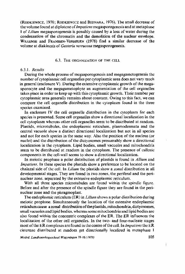
(RoDKiEWicz, 1970; RODKIEWICZ and BEDNARA, 1976). The small decrease of the volume found at diplotene of Impatiens megasporogenesis and at metaphase I of Lilium megasporogenesis is possibly caused by a loss of water during the condensation of the chromatin and the demolition of the nuclear envelope. WILLEMSE and FRANSSEN-VERHEIJEN (1978) find a similar decrease of the volume at diakinesis of Gasteria verrucosa megasporogenesis.
6.3. THE ORGANIZATION OF THE CELL
6.3.1. Results During the whole process of megasporogenesis and megagametogenesis the
number of cytoplasmic cell organelles per cytoplasmic area does not vary much in general (enclosure V). During the extensive cytoplasmic growth of the mega-sporocyte and the megagametophyte an augmentation of the cell organelles takes place in order to keep up with this cytoplasmic growth. Their number per cytoplasmic area generally remains about constant. Owing to this fact, we can compare the cell organelle distribution in the cytoplasm found in the three species examined.
In enclosure IV the cell organelle distribution in the cytoplasm for each species is presented. Some cell organelles show a directional localization in the cell cytoplasm whereas other cell organelles seem to be distributed at random. Plastids, microtubules, the endoplasmic reticulum, plasmodesmata and the central vacuole show a distinct directional localization but not in all species and not for each species in the same way. Also the position of the nucleus (or nuclei) and the distribution of the dictyosomes presumably show a directional localization in the cytoplasm. Lipid bodies, small vacuoles and mitochondria seem to be distributed at random in the cytoplasm. The presence of callosic components in the cell-wall seems to show a directional localization.
In meiotic prophase a polar distribution of plastids is found in Allium and Impatiens. In these species the plastids show a preference to be located on the chalazal side of the cell. In Lilium the plastids show a zonal distribution in all developmental stages. They are found in two zones, the periferal and the perinuclear zone, separated by the extensive endoplasmic reticulum.
With all three species microtubules are found within the spindle figure. Before and after the presence of the spindle figure they are found in the perinuclear zone and the phragmoplast.
The endoplasmic reticulum (ER) in Lilium shows a polar distribution during meiotic prophase. Simultaneously the location of the extensive endoplasmic reticulum cause a zonal distribution of the plastids, mitochondria, dictyosomes, small vacuoles and lipid bodies, whereas some mitochondria and lipid bodies are also found within the concentric complexes of the ER. The ER influences the localization of the other cell organelles. In the two- and four-nucleate stages most of the ER complexes are found in the centre of the cell. In Impatiens the ER cisternae distributed at random get directionally localized in metaphase I
Meded. Landbouwhogeschool Wageningen 78-16 (1978) 105
around the spindle figure and in the periferal zone of the cytoplasm along the cell-wall. In this localization the ER cisternae seem to separate the spindle figure from the cell organelles and the cell organelles from the cell-wall. In Allium smooth endoplasmic reticulum (SER) is found in the perinuclear zone at pachytene and diplotene. In the other stages the SER is distributed at random.
Plasmodesmata, present at premeiotic interphase on all sides of the cell, are localized on the chalazal side of the cell with all three species after premeiotic interphase.
The central vacuole found in the initial stages of megagametogenesis is localized in the centre of the cell, whereas the nuclei show a polar position in these stages.
The nucleus at meiotic prophase in Allium is situated in the micropylar part of the cell. In the initial stages of megagametogenesis the nuclei show a polar localization with all three species. With Allium and Impatiens always an equal number of nuclei is found in the polar regions of the megagametophyte, whereas with Li Hum initially an unequal distribution of the nuclei in the polar regions is found. This stage is called the 'polarization' stage of the cell by MAHESWARI
(1950). Before this stage the four nuclei mostly have a diamond-shaped configuration in the cell, with one nucleus near the micropylar pole and one nucleus near the chalazal pole. During the so-called 'polarization' the two nuclei located more centrally move to the chalazal pole.
Dictyosomes show a polar distribution during meiotic prophase in Allium and Impatiens. At metaphase I in Allium they are found near the polar regions of the spindle figure, whereas in this stage in Impatiens they are found in the zone between the ER cisternae. In the stages after metaphase I the polar distribution is extant again in Impatiens and Allium. Besides, in the latter species dictyosomes are found on the polar sides of the nuclei in these stages. In Lilium the dictyosomes show the zonal distribution mentioned previously, whereas at metaphase I very few and small dictyosomes are found within the spindle figure.
In our material callosic components are found in the surrounding cell-wall in Impatiens at pachytene and diplotene with the exception of the chalazal pole of the cell-wall. In the dyad callosic components are only found in the cross-wall and in the surrounding cell-wall of the micropylar dyad cell according to BEDNARA (1977). During degeneration of the micropylar dyad cell the callosic components in the cell-wall are disintegrating as well. Callosic components were also found during megasporogenesis in Allium by RODKIEWICZ
(1970) but only in the cross-wall. In our material callosic components were not found in Allium when we used the fluorescence-aniline-blue method.
106 Meded. Landbouwhogeschool Wageningen 78-16(1978)
6.3.2. Discussion
6.3.2.1. C lass i f i ca t ion Three different types can be distinguished in the directional localization of
the cell organelles : a directly functional type, an indirectly probably functional type and a non-functional type. The directly functional type is found by the presence of microtubules within the spindle figure, where they have a distinct function. The indirectly probably functional type and the non-functional type are found by the zonal and polar distribution of some cell organelles. Maybe the cell organelles which show a zonal distribution have an indirect function in these zones, for instance : the dictyosomes near the cell-wall have a function for cell-wall synthesis. It is more likely, however, that as a result of the distribution of other cell organelles, such as the endoplasmic reticulum in Lilium, the localization of the zonal distributed cell organelles is influenced. In this case a non-functional type is found.
The polar distribution of some cell-organelles and their possible function will be discussed next.
6.3.2.2. The po l a r d i s t r i b u t i on of cell o rgane l l e s The polar distribution of some organelles indicates a polar organization of
the cytoplasm, since there are differences between the micropylar and the chalazal half of the cell. In our material these differences are caused by the localization of the plastids, the endoplasmic reticulum (ER), the plasmodes-mata, the nucleus (nuclei) and dictyosomes and by the partial presence of callosic components in the surrounding cell-wall.
Factors which may be involved in the polar distribution of some cell organelles can be distinguished in external and internal factors. As one of these external factors the position of the cell within the nucellus cells may be considered. The ovule in which the nucellus cells and the megaspore mother-cell (MMC) are situated already shows a difference between micropylar and chalazal side. Also the position of the ovule in the ovary and the position of the ovary in the flower-bud may influence the distribution of cell organelles as for example the distribution of the plastids can be influenced by a geotropic reaction of these organelles, as demonstrated by VOLKMAN and SIEVERS (1975). On the chalazal side of the nucellus the vascular bundle ends. This causes a nutrition-gradient in the nucellus cells going from chalaza to micropyle. This nutrition-gradient may also be present in the MMC, because at the chalazal pole plasmodesmata are found, probably serving as pathways for nutrients. The nutrition-gradient in the MMC may cause a polar distribution of the cell organelles. On the other hand, a specific localization of one type of cell organelles may influence the localization of the other cell organelles. By the presence of a central vacuole in the initial stages of megagametogenesis the polar distribution of the nuclei is more or less fixed. The oval-like shape of the megagametophyte plays an important role here. Also in meiotic prophase the shape of the MMC may play a role in the polar distribution of the cell organelles.
Meded. Landbouwhogeschool Wageningen 78-16 (1978) 107
In the above contemplation a possible streaming of the cytoplasm is not included. However, it is found that a cytoplasmic streaming takes place during meiotic prophase in meiotic dividing cells. This cytoplasmic streaming will stop at metaphase I. About a possible cytoplasmic streaming in the functional megaspore and in the megagametophyte after the meiotic divisions no data are known. Maybe the cytoplasmic streaming in meiotic prophase does not change the position of some organelles such as the endoplasmic reticulum, which is present as extensive complexes in Lilium and as plastids which are heavier than dictyosomes, mitochondria, small vesicles and lipid bodies. A possible cytoplasmic streaming after the meiotic division is excluded in this contemplation.
6.3.2.3. The po l a r d i s t r i bu t i on of p l a s t i d s , d i c t yosomes , ER , p l a smode sma t a and nucle i
A polar distribution of plastids in the MMC was also described by STEWART
and GIFFORD (1967), JALOUZOT (1971), GODINEAU (1973) and RODKIEWICZ and BEDNARA (1974). All these authors found an unequal distribution of the plastids in the MMC, in such a way, that most of the plastids were extant on that polar side, where the future functional megaspore will arise. In our material the plastids are also localized on that polar side, to be sure that most of them will be present in the future megaspore. This polarity expressed by the plastids in the MMC seems to be functional in relation to the future megaspore and the future embryosac.
The unequal polar distribution of cell organelles in the MMC has its consequence for the, by unequal division formed, dyad. In the micropylar dyad cell hardly any plastids are found, whereas dictyosomes are perceptible in a relatively larger amount in this cell. Here the presence of a larger amount of dictyosomes per cytoplasmic area seems to be in relation with their presumed function of producing digestive enzymes for the destruction of the micropylar cell.
In Lilium the distribution of the ER expresses a polarity of the MMC in early meiotic prophase. This was also found by EYMÉ (1965) in the Lilium MMC, by STEWART and GIFFORD (1967) in the MMC of Ginkgo biloba and by GODI
NEAU (1973) in the MMC of Zea mays and Crépis tectorum. In all these cases an extensive endoplasmic reticulum is developing at the micropylar pole of the MMC. Because the latter three species have a monosporic type of embryosac development, with a development of the chalazal megaspore to functional megaspore and Lilium has the tetrasporic type of embryosac development, the presence of an extensive ER at the micropylar pole in Lilium may not be comparable with this feature in the other species. In the monosporic type this ER may be related to the position of the future functional megaspore. In Lilium, however, there may be another reason for the presence of an abundant endoplasmic reticulum at the micropylar pole during meiotic prophase. Maybe the cause can be found in the presence of a nutrition-gradient in the MMC. It is possible that the ER membranes react on this difference in nutrition concentrations within the cell.
The presence of plasmodesmata on the chalazal side of the cell seems to serve
108 Meded. Landbouwhogeschool Wageningen 78-16(1978)
the nutrition-supply coming from the chalazal nucellus cells. The occurrence of plasmodesmata only on the chalazal side may be influenced by the chalazal neighbour nucellus cells or by the MMC itself.
There seems to be a relation between the polar distribution of the nuclei at the initial stages of megagametogenesis and the appearance of the central vacuole at these stages. By way of this central vacuole the nuclei seem to be pressed to the polar sides of the cell as it was suggested by RUTISHAUSER (1969) and MOUSSEL (1971). It is not evident whether the nuclear position is either caused or maintained as a result of the presence of the central vacuole. This polar distribution of the nuclei is necessary for the organization of the future eight-celled embryosac.
6.3.2.4. The p a r t i a l p resence of ca l los ic c omponen t s in the ce l l -wal l of the M M C
A polarity of the MMC is also determined by the partial presence of callosic components in the surrounding cell-wall of the MMC, as it was also found by RODKIEWICZ (1970), JALOUZOT (1971), RODKIEWICZ and BEDNARA (1974, 1976) and NOHER DE HALAC and HARTE (1975). When the functional megaspore will appear on the chalazal side of the MMC the chalazal pole shows the same characteristics as the micropylar pole does when the functional megaspore appears on the micropylar side. A callosic wall appears at that pole of the megaspore mother-cell where the abortive megaspore(s) will be formed. At the opposite pole the cell-wall initially contains some callosic components but the callosic substances will soon disintegrate thus offering the future functional megaspore the opportunity to ingest nutritions through the cell-wall from the surrounding cells as it was suggested by NOHER DE HALAC and HARTE (1975) and by RODKIEWICZ and BEDNARA (1976). The callosic cell-wall layer seems to be a mechanical barrier against the penetration of larger molecules in the cell (KNOX and HESLOP-HARRISON, 1970). This type of polarity is found in all those species with the monosporic type arid the bisporic type of embryosac development (Allium cepa) examined thus far by RODKIEWICZ (1970). Species with a tetrasporic embryosac development, examined by the latter author, have no callosic compounds in their cell-walls.
In our material only Impatiens walleriana has a definite callosic components containing cell-wall at diplotene, which disintegrates at metaphase I. A polarity as described above is found in the Impatiens MMC, but, besides that, in all stages plasmodesmata are found on the chalazal side of the cell. The callosic components remain in the cell-wall of the micropylar dyad cell, thus isolating this cell from the surrounding cells, by which an ingestion of nutritions can not take place. Probably this isolation causes the destruction of the cell.
6.3.2.5. The r e l a t i on be tween nuce l lus and M M C The polar distribution of plastids, plasmodesmata and callosic cell-wall
components seems to be related to the persistence of the functional megaspore. As it is mentioned before, the relation between the MMC and the surrounding
Meded. Landbouwhogeschool Wageningen 78-16(1978) 109
nucellus cells is very close, because of the position of the M MC within the nucellus and the ingestion of nutritions of the MMC from the nucellus. The M MC initially originates from one cell between the nucellus cells. The induction of this MMC formation has to be an external factor probably originating from the nucellus, together with the nutrition streaming. It is suggested that the MMC produces the callosic components in the cell-wall during meiotic prophase, analogous to the cell-wall thickening found in species without callosic components in the cell-wall, although no indications for the production of callose is found in our material. The partial disintegration of the callosic cell-wall and the persistence of plasmodesmata on the chalazal side may be caused by the nucellus cells on that side, thus providing the MMC with nutritions. Through this pathway a nutrition-gradient is caused in the cytoplasm of the MMC, leading to a polar distribution of some cell organelles. By this polar distribution unequal dyad cells appear. In consequence of the presence of callosic components in the cell-wall of the micropylar dyad cell, this cell can not ingest nutritions and dies. In the functional megaspore the central vacuole appears in consequence of which a polar distribution of the nuclei fixed, necessary for the organization of the embryosac in which - beside other cell organelles - the plastids are present.
In the above contemplation an interaction between the nucellus and the MMC is suggested. This interaction starts with the induction of one nucellus cell to become MMC. This interaction is possible because of the heterotrophic character of the MMC, by which its development is depending of a nutrition-supply originating from the nucellus. It seems that as a result of this interaction the nucellus cells play a dominant role in the development of MMC.
110 Meded. Landbouwhogeschool Wageningen 78-16 (1978)

M E G A S P O R O G E N E S I S AS A D E V E L O P M E N T A L PROCESS
7.1. INTRODUCTION
Not all the ultrastructural changes mentioned and discussed earlier can be related to the process of megasporogenesis. Some of these changes are characteristic for meiosis. By distinguishing the ultrastructural changes characteristic for meiosis we can give a more exact description of the process of megasporogenesis, in which also species-specific and developmental type-specific characteristics are considered.
7.2. MEIOSIS
The developmental processes occurring within or close to the nucleus can be related to the meiotic process. All the meiotic developmental stages as used in this study are confined by the chromatin ultrastructure during meiosis. The nuclear envelope undulations and the sacculation of the inner nuclear membrane are supposed to have a function in the nucleo-cytoplasmic exchange of information during meiosis. The microtubules with their function in the spindle can also be related to the process of meiosis. The decrease of the number of cytoplasmic ribosomes per cytoplasmic area, followed by the increase of this number in relation with the break-down of the nuclear envelope and the presence of nucleolus-like bodies in the cytoplasm, is supposed to be specific for meiosis as well, since it is observed in both micro- and megasporogenesis.
7.3. MEGASPOROGENESIS
The process of megasporogenesis concerns the process during which one nucellus cell develops into megaspore mother-cell, and next into functional megaspore. This process seems to be an interaction between the surrounding nucellus cells and this cell. Within the nucellar tissue a nutrition-gradient is extant, caused by a one-sided nourishing streaming viz. the chalazal side. The nucellus cell, positioned just under the nucellar epidermis in the middle of the very young ovule, probably receives information by this nourishing streaming for differentiation into a sporogenic cell.
This cell which now becomes the developing megaspore mother-cell reacts on this induction by increasing its cytoplasmic volume, which involves a growth of the nucleus, that starts dividing meiotically. During the cytoplasmic growth no big vacuoles appear. The shape of the developing megaspore mother-cell changes and the cell-wall thickens, while in several species callosic compo-
Meded. Landbouwhogeschool Wageningen 78-16 (1978) 111
nents are partially deposited in the cell-wall. On the chalazal side this cell-wall shows places in which no callosic components are perceptible, while an intensive contact between the nucellus and the developing megaspore mother-cell is also preserved by the presence of plasmodesmata. The thickening of the cell-wall and in some cases the formation of callosic components in the cell-wall during megasporogenesis by the megaspore mother-cell is a mechanism to isolate itself partially from external influences. However, during megasporogenesis a heterotrophic condition of the megaspore mother-cell makes contact necessary with its environment, in this case the chalazal nucellus cells. From this side nutritions are provided to the megaspore mother-cell while, through this intensive contact with the nucellus cells, the development of the megaspore mother-cell is getting influenced. It seems that as a result of this interaction between the nucellus cells and the megaspore mother-cell the whole developmental process of megasporogenesis - including the formation of only one functional megaspore - is defined.
During megasporogenesis the ultrastructures of the mitochondria and plastids change, whereas they return to their original ultrastructure during megagametogenesis. This reoccurrence of the original ultrastructure takes place under different environmental conditions. The original ultrastructure was found in the diploid megaspore mother-cell which was just starting meiotic divisions, whereas the return to the original ultrastructure takes place in the haploid megagametophyte. Therefore it is not correct to speak about a cycle of the ultrastructure of plastids and mitochondria during megasporogenesis as DICKINSON and POTTER (1978) do.
During megagametogenesis, when a central vacuole is formed, the extension of the volume involves a growth of the vacuole, through which a polar distribution of the nuclei is fixed for their future position in the embryosac.
7.3.1. Characteristics of the process of megasporogenesis All the situations and changes mentioned in 7.3 are more or less found in the
species investigated and seem to be related to the process of megasporogenesis. Characteristics of the process of megasporogenesis are to be found in the developmental ultrastructural features. These features show a large variety since they are observed in three different species (species-specific characteristics). Characteristics of the process of megasporogenesis also depend on the classification of the process in different types which was described by MAHESWARI (1950) and RUTISHAUSER (1969) as monosporic, bisporic and tetrasporic type according to the number of nuclei in the functional megaspore (type specific characteristics).
Some species-specific characteristics are the storage of reserves in the megaspore mother-cell and in the developing megaspore, the differences in contact with the chalazal neighbour nucellus cells and the reaction of the surrounding nucellus cells on the presence of the megasporocyte and the megagametophyte.
Some type-specific characteristics, concerned with the classification of the process are the development of the endoplasmic reticulum and the dictyosomes, the polar localization of some organelles and the degeneration of the nonfunctional megaspore(s). , , , , r „ , , , „,
Medea. Landbouwhogeschool Wagenmgen 78-16 (1978)
112
7.3.2. Species-specific characteristics During the whole process of megasporogenesis and megagametogenesis the
number of cell organelles per cytoplasmic area does not vary much in general. Small fluctuations in the number of some cell organelles per cytoplasmic area are species-specific.
The heterotrophic character of the megaspore mother-cell is expressed by the storage of reserves in the cell. The degree of this heterotrophic character seems to be species-specific. A species with a high degree of heterotrophy shows a continuous storage of reserves in the formation of starch and lipids, as is shown in Impatiens. Allium on the other hand shows a low degree of heterotrophy.
The degree of contact between the chalazal nucellus cells and the megaspore mother-cell is found in the presence of plasmodesmata, which can be found in all stages with Impatiens and not in all stages with Allium and Lilium. Lilium seems to close the plasmodesmata after a storage of lipids, thus changing its heterotrophic character into a temporary individual self-supporting character, which is expressed by a decrease of the lipidic amount. In some stages Allium has plasmodesmata, in other stages it has not, whereas a storage of reserves is not effectuated.
The nucellus cells directly surrounding the megaspore mother-cell either remain unchanged during the whole of the volumic growth of the megasporo-cyte and megagametophyte (Lilium) or degenerate with the exception of two chalazal neighbour cells (Impatiens) or transform into a solidity tissue by thickening of the cell-wall with the exception of two chalazal neighbour cells (Allium). The reaction of the nucellus cells is probably defined by characteristics of the nucellar tissue in relation to the presence of the megaspore mother-cell and not by an influence only proceeding from developing megasporocyte itself.
7.3.3. Type-specific characteristics There is a relation between the characteristics, specific for the different
types of development and the formation of a cell-plate, the occurrence of a dyad and the destruction of the non-functional megaspore.
The megaspore mother-cells having a bisporic type of development show a typical development of the dictyosomes in relation with the formation of the cell plate. The tetrasporic type shows a differing development of the dictyosomes, which is accompanied by and related to a typical development of the endoplasmic reticulum.
Owing to the polar distribution of some cell organelles an unequal division gives rise to a dyad, having relatively more plastids in the chalazal (functional) cell and relatively more dictyosomes in the smaller (non-functional) micropylar cell. The dyad is surrounded by a cell-wall containing callosic components. The micropylar dyad cell is isolated from the surrounding cells by this callosic cell-wall material and contains more dictyosomes necessary for the production of digestive enzymes. The destruction of the non-functional megaspore is a consequence of the isolation of the cell as a result of which no ingestion of
Meded. Landbouwhogeschool Wageningen 78-16 ( 1978) 113
nutritions can take place in combination with a specific cell organelle population. The degeneration has already been inducted before the process has started, as it is indicated by the polar distribution of some cell organelles and the partial presence of callosic components in the cell-wall at diplotene. The realization of the degeneration can be influenced by the functional megaspore. The demolition products of the degenerating cell benefit the development of the functional cell by diffusion through the cross-wall, when the callosic components in this wall are disintegrated. This means that the theory of SHELDRAKE (1974) which supposes that the functional cell must get rid off its deleterious products, which are then accumulated in the non-functional megaspore cells, is rejected here.
The characteristics of the process of megasporogenesis in relation to the developmental type indicate a more complicated classification of the process than was given by MAHESWARI (1950) and RUTISHAUSER (1969) who only counted the number of nuclei in the functional megaspore. In our opinion the classification according to the number of nuclei may only serve as a basic information, whereas an intensive investigation of the cytoplasmic changes will procure information of the process of megasporogenesis and the species-specific characteristics and type-specific characteristics of the process.
7.4. MiCROSPOROGENESIS AND MEGASPOROGENESIS
In this thesis the work of DICKINSON and HESLOP-HARRISON has often been referred to. These scientific investigators compare the process of megasporogenesis with the process of microsporogenesis without any restriction. In our opinion these two processes are not totally comparable because they occur un-very different circumstances and show their own specific ultrastructural development.
Microsporogenesis takes place in the anther tapetum, where at the same time a lot of microspore mother-cells are developing. The microspore mother-cells show a polarity within the cell by means of the orientation of the spindle figure. In the anther the microspore mother-cells are entirely surrounded by the tapetum, which is serving as a nourishing tissue. The nutritions can come from all sides of the microspore mother-cell, so that the presence of a polar nutrition-gradient within the cell can be excluded. The resulting microspores show some characteristics specific for spores as those found in lower plants e.g. a solid cell-wall protecting the spore for drying-up, a condensed cytoplasm with small vacuoles and characteristics for a self-supporting character.
These developmental differences between microsporogenesis and megasporogenesis make them not totally comparable. The processes characteristic for meiosis as the ultrastructural changes within and close to the nucleus previously mentioned are similar. The ultrastructural changes, related to the process of megasporogenesis, however, very strongly depend on the solitary position of the megaspore mother-cell in the nucellus cells, the nutrition-gradient within the megaspore mother-cell and the nucellus cells, the heterotrophic character of the megaspore mother-cell and the functional megaspore.
114 Meded. Landbouwhogeschool Wageningen 78-16(1978)
8. S U M M A R Y
In higher plants the development of the female gametophyte - the embryo-sac - involves two subsequent processes : megasporogenesis and megagameto-genesis. Megasporogenesis is the process during which one functional mega-spore is formed by meiosis of one particular nucellus cell. This functional mega-spore can contain one, two or four haploid nuclei (respectively following the mono-, bi- or tetrasporic developmental type) depending on the circumstance whether a cell-wall is formed after the first and/or the second meiotic division or not. Additional megaspores, when formed, degenerate during megasporogenesis. The functional megaspore develops into the embryosac after a number of mitotic divisions. This process is called megagametogenesis. The cell involved is called megasporocyte or megaspore mother-cell during megasporogenesis and forms a megagametophyte during megagametogenesis.
In this thesis the ultrastructural aspects of megasporogenesis have been studied with the employ of electron-microscopic technics. Initially we intended to study the megasporogenesis in three species, each representing one of the three types mentioned above. For this purpose the species Impatiens walleriana Hook, ƒ., Allium cepa L. and the Lilium hybrid 'Enchantment', were chosen which, according to other investigators respectively would show the mono-, bi- and tetrasporic types of development. However, our observations revealed a bisporic type of development for Impatiens as well, so that eventually two species having the bisporic type and one species having the tetrasporic type were examined.
During our investigation the ultrastructural changes and the localization of the various cell organelles in the different developmental stages were observed. At the same time quantitative data concerning the number of the various cell organelles were collected during the whole of the developmental process. See enclosures I, II, III, IV and V.
In the chapters 3, 4 and 5 the ultrastructural aspects of megasporogenesis respectively of Lilium, Allium and Impatiens are described and discussed separately. From the results we can conclude that each species shows its specific ultrastructural aspects during megasporogenesis. Lilium shows a very specific formation of an extensive endoplasmic reticulum, whereas in Allium the dictyo-somes show a typical ultrastructure and localization. Impatiens shows an increasing amount of starch granules in the plastids, which is not found in the other two species.
The differences and similarities in the ultrastructural changes of the cell organelles found in the three species are discussed more in detail per cell organelle in chapter 6. In chapter 7 a classification is given of these similarities and differences in different groups. Some of the similarities in the ultrastructural aspects of megasporogenesis between the three species examined seem to be specific for the process of meiosis, because of a known or presumed functional
Meded. Landbouwhogeschool Wageningen 78-16 ( 1978) 115
relation to this process. The other similarities and all the differences are specific for the process of megasporogenesis. The differences can be distinguished in species-specific and (developmental) type-specific characteristics.
The similarities in the ultrastructural aspects of the three species, which are related to the process of meiosis, are in the first place found in the development of the nuclear chromatin and of the microtubules. The chromatin structure in the nucleus of each species shows similar ultrastructural changes, owing to the different developmental stages. The microtubules have a function in the spindle figure. Secondly, the ultrastructural changes of the nuclear envelope, the nucleolus, the nucleolus-like bodies in the cytoplasm and the cytoplasmic ribosomes show similarities between the three species examined. The undulations of the nuclear envelope, probably caused by a synthesis of new nuclear membrane before nuclear division and the sacculation of the inner nuclear membrane are probably related to a nucleo-cytoplasmic exchange of information, which takes place from nucleus to cytoplasm or vice versa during meiosis. The decrease of the number of ribosomes per cytoplasmic area during meiotic prophase is related to a low number of nuclear pores and an increase in volume of the nucleolus at this stage ; the increase of the number of ribosomes per cytoplasmic area after metaphase I is related to the disintegration of the nuclear envelope before metaphase I and the presence of nucleolus-like bodies in the cytoplasm after metaphase I.
Some similarities related to the process of megasporogenesis are the position of the developing megasporocyte in the nucellus, the localization of the plas-modesmata in the cell-wall and the storage of reserve-food in the cell. The developmental changes of the mitochondria, the plastids and the appearance of the central vacuole are related to both megasporogenesis and megagameto-genesis. During megasporogenesis the developing megaspore mother-cell is surrounded by nucellus cells and is dependent on these cells for its nutrient-supply. There seems to be an interaction between the nucellus cells and the developing megaspore mother-cell, as a result of which the development of the megaspore mother-cell is influenced by the nucellus. A direct contact between the megaspore mother-cell and the nucellus cells is found only on the chalazal side of the megaspore mother-cell, where plasmodesmata are found in all three species examined. On the chalazal side of the nucellar tissue the vascular bundle ends and from this side a nutrient transport to the megaspore mother-cell takes place, causing a nutrient-gradient in the cell. This nutrient-gradient may define the polar distribution of some cell organelles in the cell. In the megasporocyte a storage of reserve-food takes place. The degree of heterotrophy of the megasporocyte can be determined by this storage of reserve-food and seems to be species-specific. During megasporogenesis the mitochondria and the plastids show ultrastructural changes, whereas they return to their original ultrastructure during megagametogenesis. The formation of a central vacuole during megagametogenesis is also related to the presence of small vacuoles during megasporogenesis.
Apart from these general characteristics of the process of megasporogenesis
116 Meded. Landbouwhogeschool Wageningen 78-16(1978)
we can distinguish species-specific and type-specific characteristics of the process. The species-specific characteristics are specific differences in the contact with the chalazal neighbour nucellus cells and in the reaction of the nucellus cells on the presence of the megasporocyte and the developing megagameto-phyte. The mitochondria and the plastids show specific ultrastructures, whereas the storage of reserve-food in the formation of starch and lipid bodies seem to be species-specific. The type-specific characteristics are concerned with the classification of the process in the bi- and tetrasporic types of development. The ultrastructural changes of the dictyosomes and of the endoplasmic reticulum, the polar localization of some cell organelles and the degeneration of the non-functional cell are specific characteristics related to the type of mega-sporogenesis.
Meded. Landbouwhogeschool Wageningen 78-16(1978) 117

9. A C K N O W L E D G E M E N T S
This dissertation could be effected thanks to the cooperation of many people. I realize that I shall not be able to mention them all by name. Nevertheless, I wish - besides a general word of thanks - to express my particular gratitude to the following persons :
Adriaan van Aelst and Tiny Franssen-Verheijen, who initiated me into the laboratory technics required, mr. T. Zaal, who developed and printed hundreds of photographs, mr. G. van Geerenstein and mr. J. Verbürg, who saw to it that there was always a supply of plants in flower and mr. E. Rothuis who provided me with his delicious, indispensable coffee.
Wouter van Ooijen looked after the photographic items of this dissertation, whereas Hans Keeris carried out the extensive drawing and the design of the cover.
Prof.dr.M.Th.M. Willemse and dr.J.L.vanWentregularlyputquestions and made critical remarks during the entire period of the research, which positively influenced my investigations. Owing to their lively concern with the research they could, in our long discussions, comment upon the manuscript in a constructive sense.
In Utrecht dr. J. J. Geuze of the Centre for Medical Electronmicroscopy procured the possibility for me to avail myself of an electron microscope with goniometer stage, in the application of which I was assisted by Maurits Niekerk.
On the T.F.D.L., where practically all the electron-microscopical observations were performed, especially mr. H. G. Elerie did his utmost to keep the electron-microscope in the best possible condition.
Special thanks are due to dr. J. Bednara, who on our request applied the fluorescence-aniline-blue method for checking callosic components in the cell-wall of Impatiens walleriana Hook . / , when he was attached to our laboratory as a guest.
Rosa Groot-Scholte undertook the - by no means slight - task to prepare the manuscript for printing.
A special word of gratitude I wish to address to my father-in-law mr. H. de Boer, who, with great dedication, corrected the English version of this dissertation.
118 Meded. Landbouwhogeschool Wageningen 78-16(1978)
10. S A M E N V A T T I N G
In de hogere planten wordt de ontwikkeling van de vrouwelijke gameto-phyt - de embryozak - ingeleid door twee op elkaar volgende processen : de megasporogenese en de megagametogenese. De megasporogenese is het proces waarbij één functionele megaspore wordt gevormd door méiose in één bepaalde nucellus cel. Deze functionele megaspore kan één, twee of vier haploïde nuclei (respectievelijk volgens het mono-, bi- of tetraspore ontwikkelingstype) bevatten, afhankelijk van het feit of er al dan niet een celwand wordt gevormd na de eerste en/of de tweede meiotische deling. Als er meerdere megasporen zijn gevormd tijdens méiose, degenereren deze tijdens de megasporogenese. De functionele megaspore ontwikkelt zich na een aantal mitotische delingen tot de embryozak en dit proces wordt megagametogenese genoemd. De zich ontwikkelende cel wordt tijdens de megasporogenese, mega-sporocyt of megaspore-moedercel genoemd en vormt de megagametophyt tijdens de megagametogenese.
Tijdens dit onderzoek zijn de ultrastructurele aspecten van de megasporogenese bestudeerd met behulp van electronenmicroscopische technieken. In eerste instantie lag het in de bedoeling de megasporogenese te bestuderen in drie species die elk één van de drie hiervoor genoemde ontwikkelingstypen representeren. Voor dit doel werden de species Impatiens walleriana Hook./!, Allium cepa L. en de Lilium hybrida 'Enchantment' bestudeerd welke volgens aangehaalde referenties, respectievelijk het mono-, bi- en tetraspore ontwikkelingstype te zien zouden geven. Onze waarnemingen brachten echter voor Impatiens een ontwikkeling volgens het bispore type aan het licht, met als gevolg dat er twee species met een ontwikkeling volgens het bispore- en een species met het tetraspore type zijn onderzocht.
Gedurende ons onderzoek zijn de ultrastructurele veranderingen en de ligging van de celorganellen in de verschillende ontwikkelingsstadia bestudeerd. Tegelijkertijd zijn gedurende het gehele ontwikkelingsproces kwantitatieve gegevens verzameld over de verschillende celorganellen. Zie bijlagen I, II, III, IV en V.
In de hoofdstukken 3, 4 en 5 zijn afzonderlijk de ultrastructurele aspecten van de megasporogenese van respectievelijk Lilium, Allium en Impatiens beschreven en besproken. Uit de resultaten hiervan kunnen we concluderen dat elke species zijn eigen specifieke ultrastructurele aspecten toont gedurende de megasporogenese. Lilium laat een specifieke vorming van een uitgebreid endoplasmatisch reticulum zien, terwijl bij Allium juist de dictyosomen een voor de soort kenmerkende ultrastructuur en locatie vertonen. Impatiens, toont een toenemende hoeveelheid zetmeelkorrels in de plastiden ; een proces dat bij de andere twee soorten niet voorkomt.
De verschillen en overeenkomsten in de ultrastructurele veranderingen van de celorganellen die bij de drie species zijn gevonden zijn zeer gedetailleerd be-
Meded. Landbouwhogeschool Wageningen 78-16 (1978) 119
sproken per celorganel in hoofdstuk 6. In hoofdstuk 7 wordt een classificatie in verschillende groepen gegeven van deze overeenkomsten en verschillen. Enkele van de overeenkomsten tussen de drie bestudeerde soorten in de ultrastructurele aspecten van de megasporogenese schijnen specifiek te zijn voor het proces van de méiose, door de bekende of veronderstelde functionele relatie met dit proces. De andere overeenkomsten en alle verschillen zijn specifiek voor het proces van de megasporogenese. De verschillen kunnen worden onderscheiden naar species-specifieke en (ontwikkelings) type-specifieke karakteristieken.
De overeenkomsten in de ultrastructurele aspecten van de drie species, welke gerelateerd zijn aan het proces van de méiose, zijn in eerste instantie gevonden bij de ontwikkeling van de kern-chromatine en van de microtubuli. De chro-matine structuur in de kern vertoont overeenkomstige ultrastructurele veranderingen aan de hand waarvan de verschillende ontwikkelingsstadia van elke species zijn vastgelegd. De microtubuli hebben een functie in de spoelfiguur. Ten tweede vertonen de ultrastructurele veranderingen van het kernmembraan, de nucleolus, de nucleolus-achtige lichaampjes in het cytoplasma en de cytoplasmatische ribosomen overeenkomsten bij de drie bestudeerde species. De welvingen van het kernmembraan-oppervlak en de blaasvorming van het binnenste kernmembraan zijn gerelateerd aan een nucleo-cytoplasmatische informatie uitwisseling, welke, van kern naar cytoplasma of omgekeerd, plaatsvindt tijdens de méiose. De afname van het aantal ribosomen per cytoplasma-tisch gebied gedurende de profase van de méiose staat in relatie met een laag aantal kernporen en een volumetoename van de nucellus in dit stadium; de toename van het aantal ribosomen per cytoplasmatisch gebied na de metafase I is gerelateerd aan de desintegratie van de kernmembraan vóór metafase I en de aanwezigheid van nucleolus-achtige lichamen in het cytoplasma na metafase I.
Enkele van de overeenkomsten die gerelateerd zijn aan het proces van de megasporogenese zijn de positie van de zich ontwikkelende megasporocyt in de nucellus, de lokalisatie van de plasmodesmata in de celwand en de opslag van reservevoedsel in de cel. De ontwikkeling van de veranderingen in de mitochondria, de plastiden en het voorkomen van de centrale vacuole zijn zowel aan de megasporogenese als aan de megagametogenese gerelateerd. Gedurende de megasporogenese wordt de megaspore-moedercel omgeven door nucellus-cellen en zij is voor haar voedselvoorziening van deze cellen afhankelijk. Er schijnt een interactie te bestaan tussen de nucellus cellen en de megaspore-moedercel, met als resultaat, dat de ontwikkeling van de megaspore-moedercel door de nucellus wordt beïnvloed. Aan de kant van de chalaza van de megaspore-moedercel wordt een direct contact aangetroffen tussen de megaspore-moedercel en de nucellus cellen ; hier worden bij alle drie de onderzochte species plasmodesmata gevonden. Aan de chalaza kant van het nucellusweefsel eindigt de vaatbundel en vanaf deze kant vindt een voedseltransport naar de megaspore-moedercel plaats dat een voedingsgradiënt veroorzaakt in de cel. Deze gradiënt kan de polaire verdeling van enkele celorganel typen in de cel mede bepalen. De mate van heterotrofie van de megasporocyt kan worden bepaald aan de hand van deze opslag van reservevoedsel en schijnt voor elke species specifiek
120 Meded. Landbouwhogeschool Wageningen 78-16(1978)
te zijn. Gedurende de megasporogenese vertonen de mitochondriën en de plas-tiden ultrastructurele veranderingen, terwijl ze gedurende de megagametogene-se weer terugkeren naar hun oorspronkelijke ultrastructuur. De vorming van een centrale vacuole gedurende de megagametogenese is ook gerelateerd aan de aanwezigheid van kleine vacuolen tijdens de megasporogenese.
Naast deze voornaamste kenmerken van het proces van de megasporogenese kunnen we species-specifieke en type-specifieke kenmerken van het proces onderscheiden. De species-specifieke kenmerken vertonen specifieke verschillen in het contact met de aanliggende nucellus cellen aan de kant van de chalaza en de reactie van de nucellus cellen op de aanwezigheid van de megasporocyt en de zich ontwikkelende megagametophyt. De mitochondriën en de plastiden hebben specifieke ultrastructuren, terwijl de opslag van reservevoedsel in de vorming van zetmeel en vetten species-specifiek schijnt te zijn. De type-specifieke kenmerken zijn beschouwd aan de hand van de onderverdeling van het proces in een bi- en een tetraspore ontwikkelings type.
De ultrastructurele veranderingen van de dictyosomen en van het endoplasmatisch reticulum, de polaire lokalisatie van sommige celorganel typen en de degeneratie van de niet-functionele cel zijn specifieke kenmerken, gerelateerd aan het ontwikkelingstype van de megasporogenese.
Meded. Landbouwhogeschool Wageningen 78-16(1978) 121
11. REFERENCES
BAGSHAW, V., BROWN, R. and YEOMAN, M. M. (1969): Changes in the mitochondrial complex accompanying callus growth. Ann. Bot. (N.S.) 33: 35-44.
BAJER, A. S. and MOLÈ-BAJER, J. (1969): Formation of spindle fibers, kinetochore orientation, and behaviour of the nuclear envelope during mitosis in endosperm. Chromosoma 27 :448^84. .
BAJER, A. S. and MOLÈ-BAJER, J. (1972): Spindle dynamics and chromosome movements. Academic Press, London-New York.
BAJON-BARBIER, C , RIMSKY, A. and WILLAIME, CH . (1973): Observation et interprétation de structures périodiques en relation avec le reticulum endoplasmique, dans l'oosphère du Mnium undulatum (Bryophytes). Caryologia 25, suppl.: 121-134.
BEDNARA, J. (1977): Personal communication. BELL, P. R. (1975): Physical interactions of nucleus and cytoplasm in plant cells. Endeavour
XXXIV: 19-22. BOER-DE JEU, M. J. DE (1978): Ultrastructural aspects of megasporogenesis and initiation of
megagametogenesis in Lilium. Soc. bot. Fr., Actualités botaniques 1-2:175-181. BRASELTON, J. P. and BOWEN, C. C. (1971): The fine structure of the nucleolus organizers of
Lilium longiflorum during meiosis. Cytologia 36: 529-533. BURGESS, J. and NORTHCOTE, D. H. (1968) : The relationship between the endoplasmic reticu
lum and micro tubular aggregation and disaggregation. Planta (berl.) 80:1-14. CHOUINARD, L. A. (1975): An electron-microscope study of the intranucleolar chromatin
during nucleologenesis in root meristematic cells of Allium cepa. J. Cell Sei. 19: 85-102. CIOBANU, L. R. (1970): Corps lamellaires dans le noyau et le cytoplasme des cellules mères
des microspores des tomates (Lycopersicum esculentum Mill.) Ann. Sc. Nat. Bot. Paris 11: 115-128.
COUR, L. F. LA and WELLS, B. (1972): The nuclear pores of early meiotic prophase nuclei of plants. Z. Zellforsch. 123:178-194.
Cox, V. S. Jr. and SEELY, J. C. (1974): A simplified resectioning technique. Stain Technology 49: 329-333.
CRESTI, M, PACINI, E. and SARFATTI, G. (1972): Ultrastructural studies on the autophagic vacuoles in Eranthis hiemalis endosperm. J. Submicr. Cytol. 4: 33-44.
CRESTI, M. and WENT, J. L. VAN (1976): Callose deposition and plug formation in Petunia pollen tubes in situ. Planta 133: 35-40.
DAHLGREN, K. V. O. (1934): Die Embryologie von Impatiens roylei. Svensk Botanisk Tids-krift28: 103-125.
DAMSKY, C. H. (1976): Environmentally induced changes in mitochondria and endoplasmic reticulum of Saccharomyces carlsbergensis Yeast. J. Cell Biol. 71 : 123-135.
DICKINSON, H. G. (1971): Nucleocytoplasmic interaction following meiosis in the young microspores of Lilium longiflorum: Events at the nuclear envelope. Grana 11 : 117-127.
— and ANDREWS, L. (1977): The rôle of membrane-bound cytoplasmic inclusions during gametogenesis in Lilium longiflorum Thunb., Planta 134: 229-240.
— and BELL, P. R. (1970): Nucleocytoplasmic interaction at the nuclear envelope in post meiotic microspores of Pinus banksiana. J. Ultr. Res. 33: 356-359.
— and HESLOP-HARRISON, J. (1970a) : The behaviour of plastids during meiosis in the micro-sporocyte of Lilium longiflorum Thunb. Cytobios. 6:103-118.
— and HESLÓP-HARRISON, J. (1976b) : The ribosome cycle, nucleoli and cytoplasmic nucleo-loids in the meiocytes of Lilium. Protoplasma 69: 187-200.
— and HESLOP-HARRISON, J. (1977) : Ribosomes, membranes and organelles during meiosis in angiosperms. Phil. Trans. R. Soc. Lond. B. 277:327-342.
— and POTTER, U. (1975): Post-meiotic nucleo-cytoplasmic interactions in Pinus banksiana. The secretion of RNA by the nucleus. Planta 122: 99-104.
122 Meded. Landbouwhogeschool Wageningen 78-16(1978)
— and POTTER, U. (1978): Cytoplasmic changes accompanying the female meiosis in Lilium longiflorum Thunb., J. Cell Sc. 29: 147-169.
DIETRICH, M. J. (1968): Organisation ultrastructurale du fuseau de caryocinèse en prometaphase dans les cellules-mères de microspores du Lis. C. R. Acad. Se. Paris t. 266:579-581.
ESPONDA, P. and GIMÉNEZ-MARTIN, G. (1975): Nucleolar organizer ultrastructure in Allium cepa. Chromosoma 52: 73-87.
EYMÉ, J. (1963): Observations cytologiques sur les nectaires de trois renonculacées. (Helle-borus foetidus L., Helleborus niger L. et Nigella damascena L.). Le Botaniste XLI : 137-179.
— (1965): Recherches sur la constitution cytoplasmique de l'archéspore et du sac embryonnaire de Lilium candidum L., Le Botaniste XLVIII : 99-155.
FLINT, F. F. and JOHANSEN, D. A. (1958): Nucleocytoplasmic relationships in the Fritillaria type of megagametogenesis. Amer. J. Bot. 45: 464^473.
FOWKE, L. C. and SETTERFIELD, G. (1969): Multivesicular structures and cell wall growth. Can. J. Bot. 47: 1873-1877.
FRASER, T. W. (1975): Involvement of the Golgi apparatus and microtubules in the formation and final positioning of the phycoplast of Bulbochaete hiloensis (Nordst.) Tiffany. Protoplasma 83: 103-110.
FREDERICK, S. E., GRUBER, P. J. and NEWCOMB, E. H. (1975): Plant microbodies. Protoplasma 84: 1-29.
GALÂN-CANO, J. RISUENO, M. C. and GIMÉNEZ-MARTIN, G. (1975): Nucleolar ultrastructure in megaspore mother cells oïPisum sativum L., Cytobiologie 10:479-484.
GALL, J.G. (1964) : Electron microscopy of the nuclear envelope. Protoplasmatologia 5: 4-25. GENEVES, L. (1967): Sur la répartition et les mouvements des organites cytoplasmiques au
cours de la méiose staminale, et principalement pendant les telophases héterotypique et homéotypique, dans le Ribes rubrum (Grossulariaceés). C. R. Acad. Se. Paris, t. 265: 1913-1916.
GIMÉNEZ-MARTIN, G., TORRE, C. DE LA, LOPEZ-SAEZ, J. F. and ESPONDA, P. (1977): Plant-
nucleolus- structure and physiology. Cytobiologie 14: 421-462. — and STOCKERT, J. C. (1970): Nucleolar structure during the meiotic prophase in Allium
cepa anthers. Z. Zellforsch. 107: 551-563. GODINEAU, J. C. (1973): Le sac embryonnaire des Angiospermes. Morphogenèse et infra
structure. Bull. Soc. Bot. Fr. Mémoires 1973: 25-54. GOVINDAPPA, D. A. (1955): Embryological studies in Xyris pauciflora Willd. Proc. Indian
Acad. Sei. B42: 47-57. GREEN, P. B. (1964): Cinematic observations on the growth and division of chloroplasts in
Nitella. Amer. J. Botan. 51: 334-342. GUILLERMOND, A. (1924): Recherches sur l'évolution du chondriome pendant le develop
ment du sac embryonnaire et des cellules-mères des grains de pollen dans les Liliacées et sur la significance des formations ergastoplasmiques. Ann. Sei. Nat. Bot. 6: 1-52.
GUNNING, B. E. S. and STEER, M. W. (1975): Plant cell biology, an ultrastructural approach. Edwards Arnolds (Publishers) Ltd., London.
HACKENBROCK, C. (1966) in: Basic biology course unit 2, Book 8: Metabolism and mitochondria. 1976. Cambridge University Press/London-New York-Melbourne : 101-102.
HANZELY, L. and VIGIL, E. L. (1975): Fine structural and cytochemical analysis of phrag-mosomes (microbodies) during cytokinesis in Allium root tip cells. Protoplasma 86: 269-277.
HEPLER, P. K. and JACKSON, W. T. (1968): Microtubules and early stages of cell-plate formation in the endosperm of Haemanthus katherinae. J. Cell Biol. 38: 437-446.
— and NEWCOMB, E. H. (1967): Fine structure of cell plate formation in the apical meristem oiPhaseolus roots. J. Ultrastr. Res. 19: 498-513.
HERR, J. M. JR. (1971): A new clearing squash technique for the study of ovule development in Angiosperms. Am. J. Bot. 58: 785-790.
HESLOP-HARRISON, J. and DICKINSON, H. G. (1967): A cycle of sphaerosome aggregation and disaggregation correlated with the meiotic divisions in Lilium. Phytomorphology 17 : 195-199.
Meded. Landbouwhogeschool Wageningen 78-16(1978) 123
ISRAEL, H. W. (1963): The ultrastructural modifications of early megasporogenesis in Den-drobium (Orchidaceae). Amer. J. Bot. 50: 615 (Abstr.).
— and SAGAWA, Y. (1964): Post pollination ovule development in Dendrobium orchids: II. Fine structure of the nucellar and archesporial phases. Caryologia 17: 301-316.
— and SAGAWA, Y. (1965): Post pollination ovule development in Dendrobium orchids: III. Fine structure of meiotic prophase I. Caryologia 18: 15-34.
JALOUZOT, M. F. (1971): Aspects ultrastructuraux de la mégasporogenèse dOenothera la-marckiana en rapport avec les dépôts callosiques observés. Ann. Univ. et de l 'ARERS 9: 36-45.
— (1973): Quelques aspects ultrastructuraux du noyau du megasporocyte ^Oenothera lamarckiana en prophase héterotypique. Caryologia 25(suppl.): 67-84.
JONES, K. W. (1965): The role of the nucleus in the formation of ribosomes. J. Ultrastr. Res. 13:257-262.
KNOX, R. B. and HESLOP-HARRISON, J. (1970): Direct demonstration of the low permeability of the angiosperm meiotic tetrad using a fluorogenic ester. Z. für Pflanzenphysiologie 62 : 451^59 .
LAFONTAINE, J. G. (1968): Structural components of the nucleus in mitotic plant cells. In: Ultrastructure in biological systems: The Nucleus. Eds. DALTON, A. J. and HAGUENAU, F., volume 3 Academic Press Inc., New York: 151-196.
LEWIS, L. M., WITKUS, E. R. and VERNON, G. M. (1976) : The role of microtubules and microfilaments in the micronucleus of Paramecium bursaria during mitosis. Protoplasma 89 : 203-219.
LUCK, B. T. and JORDAN, E. G. (1977): The nucleolus and meiosis during microsporogenesis in Endymion non-scriptus (L.). J. Cell. Sei. 25: 111-123.
MACKENZIE, A., HESLOP-HARRISON, J. and DICKINSON, H. G. (1967): Elimination of ribosomes during meiotic prophase. Nature, Lond: 215: 997-999.
MAHESWARI, P. (1950): An introduction to the embryology of angiosperms. McGraw-Hill Book Comp. Inc. New York.
— ( 1963) : Recent advances in the embryology of angiosperms. Int. Soc. of Plant Morpholo-gists. University of Delhi.
MARCHANT, R. and ROBARDS, A. W. (1968): Membrane systems associated with the plas-molemma of plant cells. Ann. Bot. 32: 457-471.
MARENGO, N. P. (1977) : Ultrastructural features of the dividing meiocyte ofOnoclea sensibilis Amer. J. Bot. 64: 600-601.
MARUYAMA, K. (1968): Electron microscopic observation of plastids and mitochondria during pollen development in Tradescantia paludosa. Cytologia 33: 482-497.
MIKULSKA, E. and RODKIEWICZ, B. (1965): Fine structure of a developing embryosac of Lilium candidum Flora 155: 586-595.
— and RODKIEWICZ, B. (1967a): Fine structure of four-nucleate stages and the central cell of Lilium regale embryosac. Flora Abt. A 157: 365-372.
— and RODKIEWICZ, B. (1967b): Ultrastructure of the maturing embryosac of Lilium regale Acta Soc. Bot. Polon. XXXVI: 555-572.
MOENS, P. B. (1968): The structure and function of the synaptinemal complex in Lilium longiflorum sporocytes. Chromosoma 23:418-451.
— (1969): The fine structure of meiotic chromosome polarization and pairing in Locusta migratoria Spermatocytes. Chromosoma 28: 1-25.
MOGENSEN, L. H. (1971): A modified method for re-embedding thick epoxysections for ultratomy. Journal of the Arizona Academy of Science 6: 249-250.
MOLLENHAUER, H. H. and MORRÉ, D. J. (1976): Cytochalasin B, but not colchicine, inhibits migration of secretory vesicles in root tips of maize. Protoplasma 87: 39-48.
MORENO DIAZ DE LA ESPINA, S. and RISUENO, M. C. (1976): Effect of a-amanitine on the nucleolus of meristematic cells of Allium cepa in interphase and mitosis: An ultrastructural analysis. Cytobiologie 12: 175-188.
MORRÉ, D. J. and MOLLENHAUER, H. H. (1974): The endomembrane concept: a functional integration of endoplasmic reticulum and Golgi apparatus. In : Dynamic aspects of plant
124 Meded. Landbouwhogeschool Wageningen 78-16(1978)

ultrastructure. Ed. ROBARDS, A. W. McGraw-Hill Book Company (U.K.) Ltd.: 84-131. — MOLLENHAUER, H. H. and BRACKER, C. E. (1971): In: REINERT, J. and URSPRUNG, H.,
Origin and continuity of cell organelles. Springer Verlag, Berlin: 82-126. MORRISON, J. N. and O'BRIEN, T. P. (1976): Cytokinesis in the developing wheat grain;
division with and without a phragmoplast. Planta 130: 57-67. MOUSSEL, B. (1971): Sur la mise en place des deux premiers noyaux et du système vacuolaire
du gamétophyte à'Ephedradistachya L. C. R. Acad. Se. Paris, t. 272: 1222-1225. NOHER DE HALAC, I. and HARTE, C. (1975): Female gamétophyte competence in relation to
polarization phenomena during the megagametogenesis and development of the embryo-sac in the genus Oenothera. In : Gamete competition in plants and animals. Ed. MULCAHY, D. L., North-Holland Publishing Company-Amsterdam, the Netherlands: 43-56.
PALADE, G. (1975): Intracellular aspects of the process of protein synthesis. Science 189: 347-358.
PETTITT, J. M. (1976): Developmental mechanisms in heterospory. Ill The plastid cycle during megasporogenesis in Isoetes. J. Cell Sei. 20: 671-685.
PICKETT-HEAPS, J. D. (1970): Mitosis and autospore formation in the green alga Kirch-neriella lunaris. Protoplasma 70: 325-348.
PODDUBNAYA-ARNOLDI, B. A. (1976): Cyto-embryology of angiosperms. Basement and perpectives. Nauka, Moscow.
PORTER, T. R. (1936): Development of the megagametophyte and embryo of Allium mutabile. Bot. Gaz. 98:317-327.
PORTER, K. and MACHADO, R. (1960) : Studies on the endoplasmic reticulum. IV. Its form and distribution during mitosis in cells of onion root tip. J. Biophys. Bioch. Cytol. 7:167-180.
POWELL, M. J. (1976): Ultrastructure and isolation of glyoxysomes in zoospores. Protoplasma 89:1-27.
QUADE, H. A. M C . and WELLS, B. (1975): The synaptinemal complex in Rhoeo spathaceae. J. Cell Sei. 17:349-369.
REYNOLDS, E. S. (1963): The use of lead citrate at high pH as an electron opaque stain in electron microscopy. J. Cell Biol. 17: 208-212.
— and DASHEK, W. V. (1976): Cytochemical analysis of callose localization in Liliutn longiflorum pollen tubes. Ann. Bot. 40: 409-416.
RICHARDSON, M. (1974): Microbodies (glyoxysomes and peroxisomes) in plants. Sei. Prog. Oxf. 61:41-61.
RIEHL, R. (1977): Konzentrische 'Lamellen' in jungen Oocyten von Noemacheilus barbatulus (L.) (Teleostei, Cobitidae). Biol. Zbl. 96: 523-528.
ROBARDS, A. W. (1973): Electron microscopy and plant ultrastructure. McGraw-Hill Book Company (U.K.) Ltd. London-New York.
ROBARDS, A. W. (1974): Dynamic aspects of plant ultrastructure. McGraw-Hill Book Company (U.K.) Ltd. London-New York.
— and KIDWAI, P. (1969) : Vesicular involvement in differentiating plant vascular cells. New Phytol. 68: 343-349.
RODKIEWICZ, B. (1970): Callose in cell walls during megasporogenesis in angiosperms. Planta 93: 39-47.
— and BEDNAR A, J. (1974): Distribution of organelles and starch grains during megasporogenesis in Epilobium. In: Fertilization in higher plants. Ed. LINSKENS, H. F., North-Holland Publishing Company/American Elsevier Amsterdam/New York: 89-95.
— and BEDNARA, J. (1976) : Cell wall ingrowths and callose distribution in megasporogenesis in some orchidaceae. Phytomorphology 26:276-281.
— and GÓRSKA-BRYLASS (1968): Callose in the walls of the developing megasporocyte and megaspores in the orchid ovule. Acta Soc. Bot. Polon. XXXVII: 19-28.
— and MIKULSKA, E. (1963): Electron microscope observations of cytoplasmic changes in developing megasporocyte of Lilium candidum. Flora 154: 383-387.
— and MIKULSKA, E. (1964) : The endoplasmic reticulum of Lilium candidum megasporocyte. Naturwissenschaften 21 : 517-518.
— and MIKULSKA, E. (1965): Electron microscope observations of endosplasmic reticulum
Meded. Landbouwhogeschool Wageningen 78-16 ( 1978) 125

in Lilium megasporocyte. Flora 155: 341-346. RUTISHAUSER, A. (1969): Embryologie und Fortpflanzungsbiologie der Angiospermen.
Springer Verlag. Wien-New York. SCHEER, U. (1973): Nuclear pore flow rate of ribosomal RNA and chain growth rate of its
precursor during oogenesis oiXenopus laevis. Dev. Biol. 30: 13-28. SCHEL, J. H. N. (1977): Observations on interphase nuclei in relation to DNA replication.
Thesis, University of Nijmegen. SCHNEPF, E. and PROSS, E. (1976): Differentiation and redifferentiation of a transfer cell:
development of septal nectaries of Aloe and Gasteria. Protoplasma 89: 105-115. SHELDRAKE, A.R.(1974): The ageing, growth and death of cells. Nature 250:381-385. STEFFEN, K. (1951): Zur Kenntnis des Befruchtungsvorganges bei Impatiens glanduligera
Lindl. Cytologische Studien am Embryosack der Balsamineen. Planta 39: 175-244. SOUTHWORTH, D. (1973): Cytochemical reactivity of pollen walls. J. Histochem. Cytochem.
2 1 : 73-80. SPORNITZ, U. M. (1973): Lamellar bodies in oocytes of Xenopus laevis and their relation to
the mode of fixation. Experentia 29: 589-591. STEWART, K. D. and GIFFORD, E. M. (1967): Ultrastructure of the developing megaspore
mother cell of Ginkgo biloba. Amer. J. Bot. 54: 375-383. TANDLER, B., ERLANDSON, R. A., SMITH, A. L. and WYNDER, E. L. (1969): Riboflavin and
mouse hepatic cell structure and function. 2. Division of mitochondria during recovery from simple deficiency. J. Cell Biol. 41 : 477^493.
THIÉRY, J. P. (1967): Mise en évidence des polysaccharides sur coupes fines en microscopie électronique. J. Microscopie 6: 987-1018.
VAZART, B. (1973): Ultrastructure des microspores de tabac dans les anthères embryogènes. Caryologia 25 (Suppl.): 303-314.
VOLKMAN, D. and SIEVERS, A. (1975): Wirkung des Inversion auf die Anordnung des Endoplasmatischen Reticulums and die Polarität von Statocyten in Wurzeln von Lepidium sativum. Planta (Berl.) 127: 11-19.
WEIBEL, E. R. (1973): Stereological techniques for electron microscopic morphometry. In: Principles and techniques of electron microscopy. Biological applications. Ed. HAYAT, M. A. vol. 3 : 239-296.
WHALEY, W. G., DAUWALDER, M. and KEPHART, J. E. (1966): The Golgi apparatus and an early stage in cell plate formation. J. Ultrastr. Res. 15: 169-180.
WILLEMSE, M. T H . M. (1971a): Morphological and quantitative changes in the population of cell organelles during microsporogenesis of Pinus sylvestris. L. I. Morphological changes from zygotene until prometaphase I. Acta Bot. Neerl. 20: 261-274.
— (1971b): Morphological and quantitative changes in the population of cell organelles during microsporogenesis of Pinus sylvestris L. II. Morphological changes from prometaphase I until the tetrad stage. Acta Bot. Neerl. 20: 411-427.
— (1971c): Morphological and quantitative changes in the population of cell organelles during microsporogenesis of Pinus sylvestris L. III. Morphological changes during the tetrad stage and in the young microspore. A quantitative approach to the changes in the population of cell organelles. Acta Bot. Neerl. 20:498-523.
— (1972): Changes in the autofluorescence of the pollen wall during microsporogenesis and chemical treatments. Acta Bot. Neerl. 20: 1-16.
— and FRANSSEN-VERHEIJEN, M. A. W. (1978) : Cell organelle changes during megasporoge-nesis and megagametogenesis in Gasteria verrucosa (Mill.) Haw. Soc. bot. Fr., Actualités botaniques 1-2: 187-191.
— and LINSKENS, H. F. (1968): Transfer of ribosome-rich caryoplasm during meiosis. Naturwissenschaften 55: 396.
WILLIAMS, E., HESLOP-HARRISON, J. and DICKINSON, H. G. (1973): The activity of the nu
cleolus organising region and the origin of cytoplasmic nucleoloids in meiocytes ofLilium. Protoplasma 77: 79-93.
ZATSEPINA, O. V., POLYAKOV, V. Yu. and CHENTSOV, YU. S. (1977). Some structural aspects of the fate of the nuclear envelope during mitosis. Cytobiologie 16: 130-144.
126 Meded. Landbouwhogeschool Wageningen 78-16( 1978)
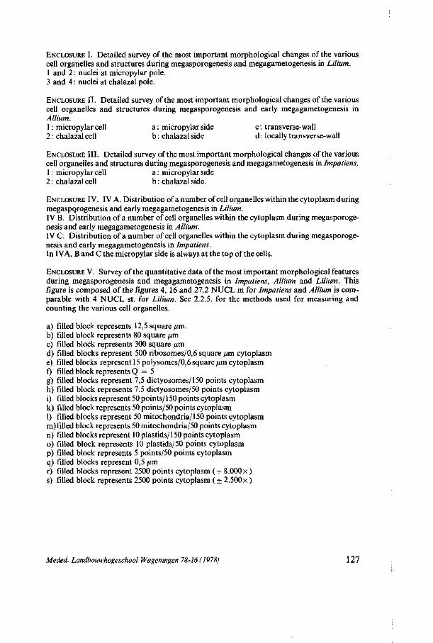
ENCLOSURE I. Detailed survey of the most important morphological changes of the various cell organelles and structures during megasporogenesis and megagametogenesis in Lilium. 1 and 2: nuclei at micropylar pole. 3 and 4 : nuclei at chalazal pole.
ENCLOSURE II. Detailed survey of the most important morphological changes of the various cell organelles and structures during megasporogenesis and early megagametogenesis in Allium. 1 : micropylar cell a : micropylar side c : transverse-wall 2 : chalazal cell b : chalazal side d : locally transverse-wall
ENCLOSURE III. Detailed survey of the most important morphological changes of the various cell organelles and structures during megasporogenesis and megagametogenesis in Impatiens. 1 : micropylar cell a : micropylar side 2 : chalazal cell b : chalazal side.
ENCLOSURE IV. IV A. Distribution of a number of cell organelles within the cytoplasm during megasporogenesis and early megagametogenesis in Lilium. IV B. Distribution of a number of cell organelles within the cytoplasm during megasporogenesis and early megagametogenesis in Allium. IV C. Distribution of a number of cell organelles within the cytoplasm during megasporogenesis and early megagametogenesis in Impatiens. In IVA, B and C the micropylar side is always at the top of the cells.
ENCLOSURE V. Survey of the quantitative data of the most important morphological features during megasporogenesis and megagametogenesis in Impatiens, Allium and Lilium. This figure is composed of the figures 4, 16 and 27.2 NUCL m for Impatiens and Allium is comparable with 4 NUCL st. for Lilium. See 2.2.5. for the methods used for measuring and counting the various cell organelles.
a) filled block represents 12,5 square /«n. b) filled block represents 80 square pm c) filled block represents 300 square /xm d) filled blocks represent 500 ribosomes/0,6 square /an cytoplasm e) filled blocks represent 15 polysomes/0,6 square pm cytoplasm f) filled block represents Q = 5 g) filled blocks represent 7,5 dictyosomes/150 points cytoplasm h) filled block represents 7,5 dictyosomes/50 points cytoplasm i) filled blocks represent 50 points/150 points cytoplasm k) filled block represents 50 points/50 points cytoplasm 1) filled blocks represent 50 mitochondria/150 points cytoplasm m) filled block represents 50 mitochondria/50 points cytoplasm n) filled blocks represent 10 plastids/150 points cytoplasm o) filled block represents 10 plastids/50 points cytoplasm p) filled block represents 5 points/50 points cytoplasm q) filled blocks represent 0,5 pm r) filled blocks represent 2500 points cytoplasm ( + 8.000 x ) s) filled block represents 2500 points cytoplasm (+ 2.500 x )
Meded. Landbouwhogeschool Wageningen 78-16 (1978) 127
128 Meded. Landbouwhogeschool Wageningen 78-16 (1978)
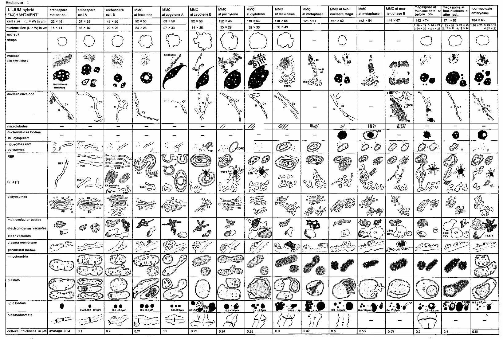
Enclosure I
LILIUM hybrid ENCHANTMENT'
cell size (L x W) in urn
nucleus size (L x W) in um
archespore
mother-cell
22 X16
15 x 14
archespore cell A
37 * 25
18 x 16
archespore
ce l lB
45 x50
22 x22
MMC
at leptotene
52 x56
24 x26
MMC
at zygotene A
63 x59
27 x33
MMC at zygotene B
92 X58
34 x35
MMC
at pachytene
122 x 46
35 x29
MMC at diplotene
119 *53
35 x36
MMC
at diakinesis
110 x56
30 x43
MMC at metaphase I
128 x 61
MMC at two-
nucleate stage
127 x 52
MMC at metaphase I
162 x54
MMC at ana-
telophase II
144 x67
megaspore at four-nucleate st before pol.
142 x 74 1:16 » 19 3:24 * 21 2:24 x 20 4:21 x 22
megaspore at four-nucleate st after pol.
171 x 52 1:23 »26 3:28 x 18 2:17 x 11 4:16 x 14
four-nucleate embryosac
184 x68 1:20 x25 3:20 * 18
4:27 X20
nucleus
shape O 0 o o nuclear
ultrastructure . : • /•' *><##;
A« nucleolema structure
A
* 0
*̂:/>y/t
axial core J
ß W
j p t S ü4LEï
w \\\
TSER
* » D
TSEI £>i°b
o
0 * n
% • • • • •
Vi 4Ov2rJ
0 Q
*©
• ;
A*
nuclear envelope
" 0 N 0.
CY N / ( C Y
^
V, L « P C -
CY
* *
»
.CY
microtubules // //// 40/ IW/// / / / Will Willi nucleolus-like bodies
in cytoplasm
JËk%. m «
ribosomes and
polysomes <~%Z-
^ . •• *e • CY. •. VNE • . .
RER
SER (T) TSER
TSER " ^
1**1
cP
cs POO Ä0
<*%% cßsl
7$ p,
tfô ? V
a V k
dictyosomes Dl
Cy= , c = ^ > <=>oo
PF
<g>°°S^°° 'So'*
O0
&<WlJon
0 ~o~0 ' " 'o1-
'£>Q0
o&qv.
?So,
% multivesicular bodies
electron-dense vacuoles
clear vacuoles S2. (2> I o > cv 2 big / c v
CV» m cvN 5 big / CV CV« »CE V
plasma membrane
paramural bodies (•• 4'. Vi« »
mitochondria
^ 0'
plastids
lipid bodies
diam.02-O.4um 0.5 - 0 8>jm • • • 0.5-0.8um
• • • 0.3 - 0,5um
&=,-0.6-a8^Pß3u1
• * 0.6-1.4um 0,4 • 1.2jjm #•<£ ae - i.6ji ft.» 0.6 - 4.4>im <-1.6umVaj 0.4- '"'••»f 04 -*1j6/im 0.6-4.8 Mm
, 0 4 - 1 ^ » ^ J ^ M l as - 3.0 um -3.0 um 1,
plasmodesmata
- < ^ -I
cell-wall thickness in um avarage 0.04 0.1 0.2 0.21 0.2 0.22 0.24 0.25 0.3 0.32 0.5 0.53 0.55 0.5 0.4 0.51
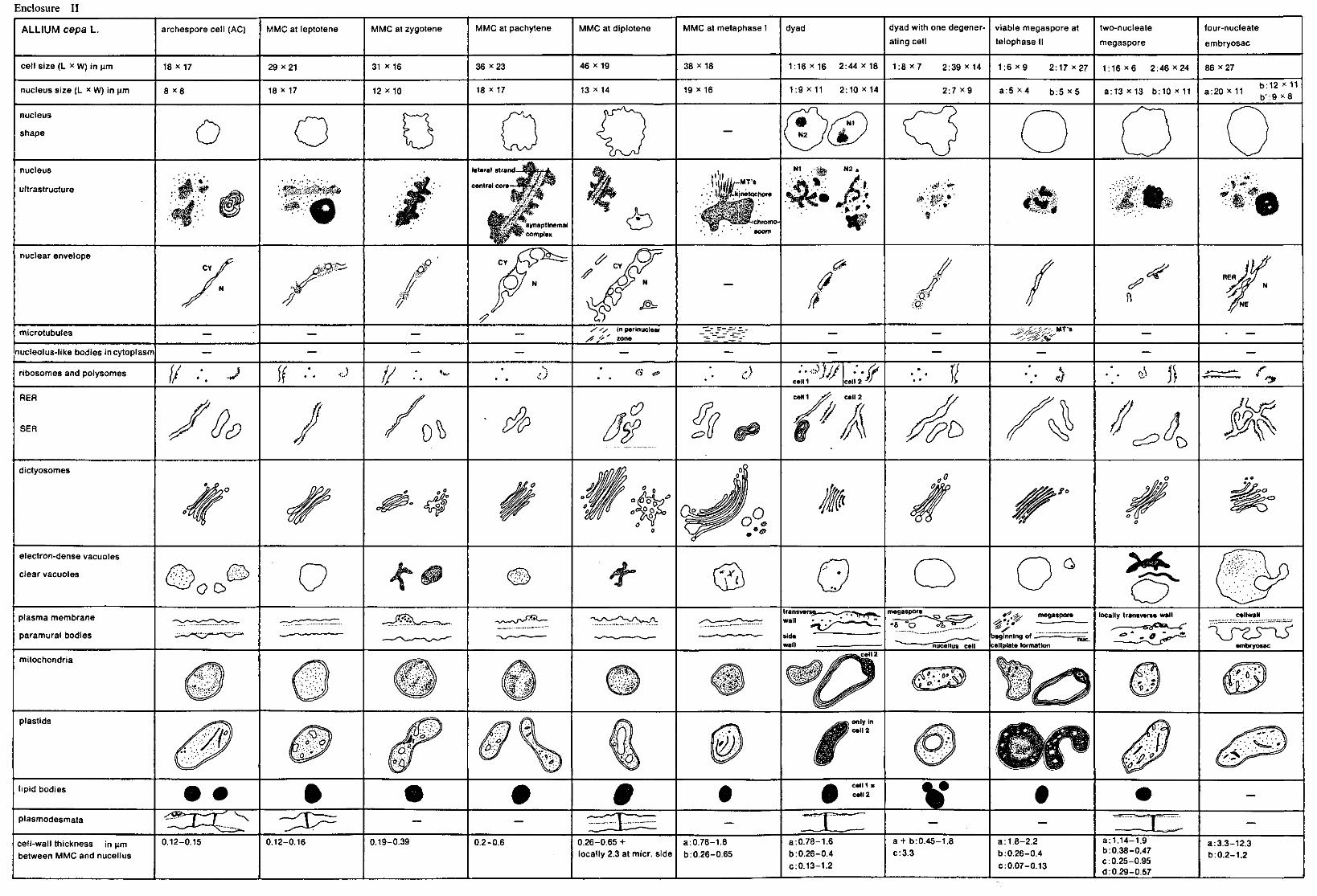
Enclosure II
ALLIUM cepa L. archespore cell (AC) MMC at leptotene MMC at zygotene MMC at pachytene MMC at diplotene MMC at metaphase I dyad dyad with one degener
ating cell
viable megaspore at
telophase II
two-nucleate
megaspore
four-nucleate
embryosac
cell size (L * W) in um 18 X17 29 x21 31 X16 36 x 2 3 46 x 19 38 X18 1 :16x16 2 : 44X18 1 : 8 x 7 2 :39x14 1 : 6 x 9 2:17 x 27 1 :16x6 2 :46x24 86 x27
nucleus size (L x w) in urn 8 x 8 18 x 17 12 x 10 18 X 1 7 13 x 14 19 x 16 1:9X11 2 :10x14 2:7 x 9 a:5 x 4 b:5 x 5 a : 1 3 x i 3 b : 1 Q x l l a:20 X11 b:12 x 11 b':9 x 8
nucleus
shape O ©( N1
m nucleus
ultrastructure
•Ä'.'Jf% m
F^ïsfet. »vaptinemal * complex
ö
,N1 . - ^ j N 2 .
•;'JE;' . . - . * •
-'-HS
nuclear envelope CY s®£ J&
// 'M$ f
ff /
< s * , / <^
microtubules y 'S in perinuclear /f 6 ' zone
M T s
~(&'j*? nucleolus-like bodies in cytoplasm
ribosomes and polysomes / / -of If ••• / / • • • ' •
G <& eelM cell % y' % o»
RER
SER
O & <Z>
celM cell 2
/A o^u£> M dictyosomes
% &£.
electron-dense vacuoles
clear vacuoles
oo a
plasma membrane
paramural bodies <2 ' « •%
.'& megaspore locally transverse wall cellwall
'X" beginning of -cellplate formation
X_rzs~xï embryosac
mitochondria cell 2
plastids
lipid bodies cell 1 : cell 2
plasmodesmata
V X
cell-wall thickness in urn between MMC and nucellus
0.12-0.15 0.12-0.16 0.19-0.39 0.2-0.6 0.26-0.65 + locally 2.3 at micr. side
a:0.78-1.8 b:0.26-0.65
a:0.78-1.6 b:0.26-0.4 c:0.13-1.2
a + b:0.45-1.8 c:3.3
a: 1.8-2.2 b:0.26-0.4 c:0.07-0.13
a:1.14-1.9 b: 0.38-0.47 c: 0.25-0.95 d: 0.29-0.57
a:3.3-12.3 b:0.2-1.2
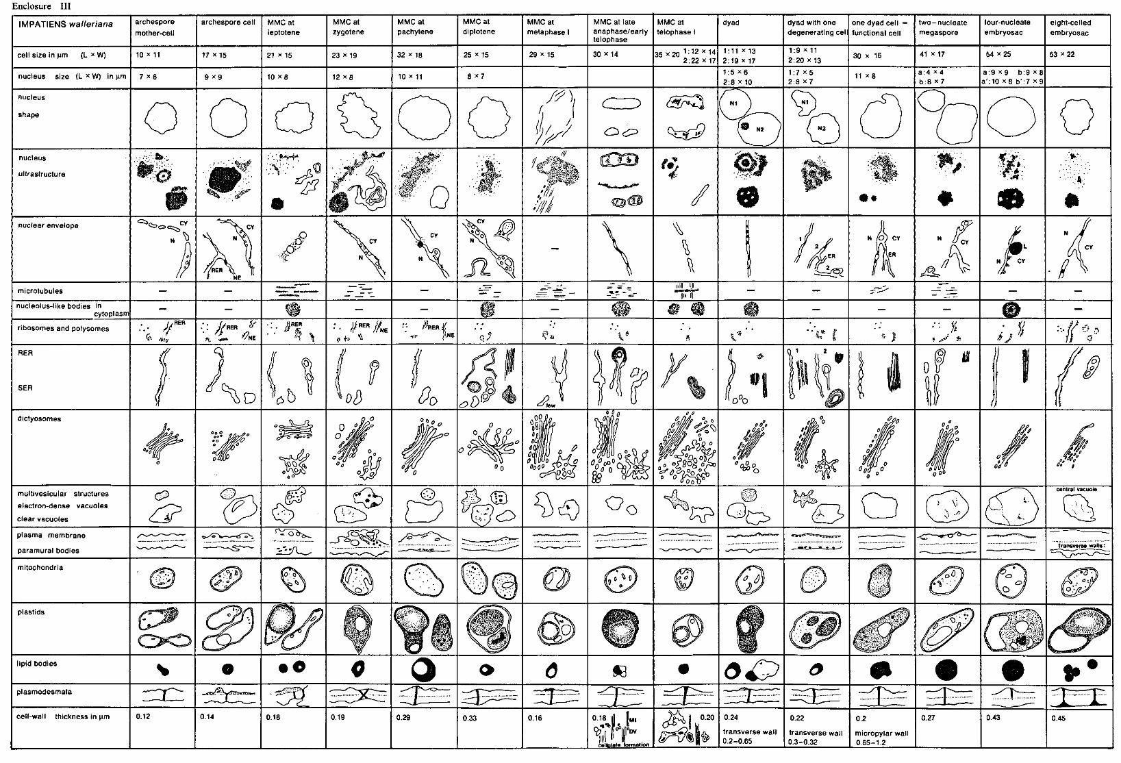
Enclosure III
IMPATIENS walleriana archespore
mother-cell
archespore cell MMCat leptotene
MMCat zygotene
MMCat pachytene
MMCat diplotene
MMCat
metaphase I
MMC at late anaphase/early telophase
MMCat telophase I
dyad dyad with one degenerating cell
one dyad cell = functional cell
two-nucleate megaspore
four-nucleate
embryosac eight-celled
embryosac
cell size in urn (L * W) 10 x 11 17 x i 5 21 x 15 23 x 19 32 x 18 25 x 15 29 x 15 30 x 14 35X20 1 : 1 2 * 1 4 2:22 x 17
1:11 x 13 2:19 x 17
1:9 x 11 2:20 x 13
30 x 16 41 x 17 54 x25 53 x22
nucleus size (L x w) in um 7 X 6 9 x 9 10 x 8 12 x 8 10 x 11 8 x 7 1:5 x6 2:8 x 10
1 :7x5 2:8 x 7
11 x 8 a:4 M b:8 x7
a:9 x 9 b:9 x8 a':10 x8 b':7 x 9
nucleus
shape
/?)
(/
CT3 N2
nucleus
ultrastructure %i0.
f
\ jt-»*"!»*'
\-rjp
* & &&
' « • '
"/ Il - * ' * ^ f e ,
•;V--">i<. V,-^3..;!Y
ik m
w M
® • • Ä . .
1 # «
^ ; -.'?»».. •# • * • *..<*•.
80 nuclear envelope > c ^ > .
jy
\
^
^
CY
jenj1 VS
microtubules IUI II
nucleolus-like bodies in cytoplasm
WM m m &$
ribosomes and polysomes V ,\i,„
RER
«. „ . . VNE n • / / R E R / / N E h
<• •*"•.> NE .. -S ' t * f #
? , v ' * J
RER
SER
Q
^ D d b 00 0t 0 A few
«5-
D°0
0
/i dictyosomes
4r 2°Ö 00 0
0A°
» nO <#°o Ä '
.*«?
multivesicular structures
electron-dense vacuoles
clear vacuoles ^ « & >
i-/cD O o ^? ^ >
central vacuole
plasma membrane
paramural bodies
mitochondria
transverse walls:
plastids
lipid bodies • 0 O Sö ö£? plasmodesmata
cell-wall thickness in urn 0.12 0.14 0.18 0.19 0.29 0.33 0.16 0°|
0.18
cellplate formation
Ml
JDV 3) I" 0.24
transverse wall 0.2-0.65
0.22
transverse wall 0.3-0.32
0.2
micropylar wall 0.65-1.2
0.27
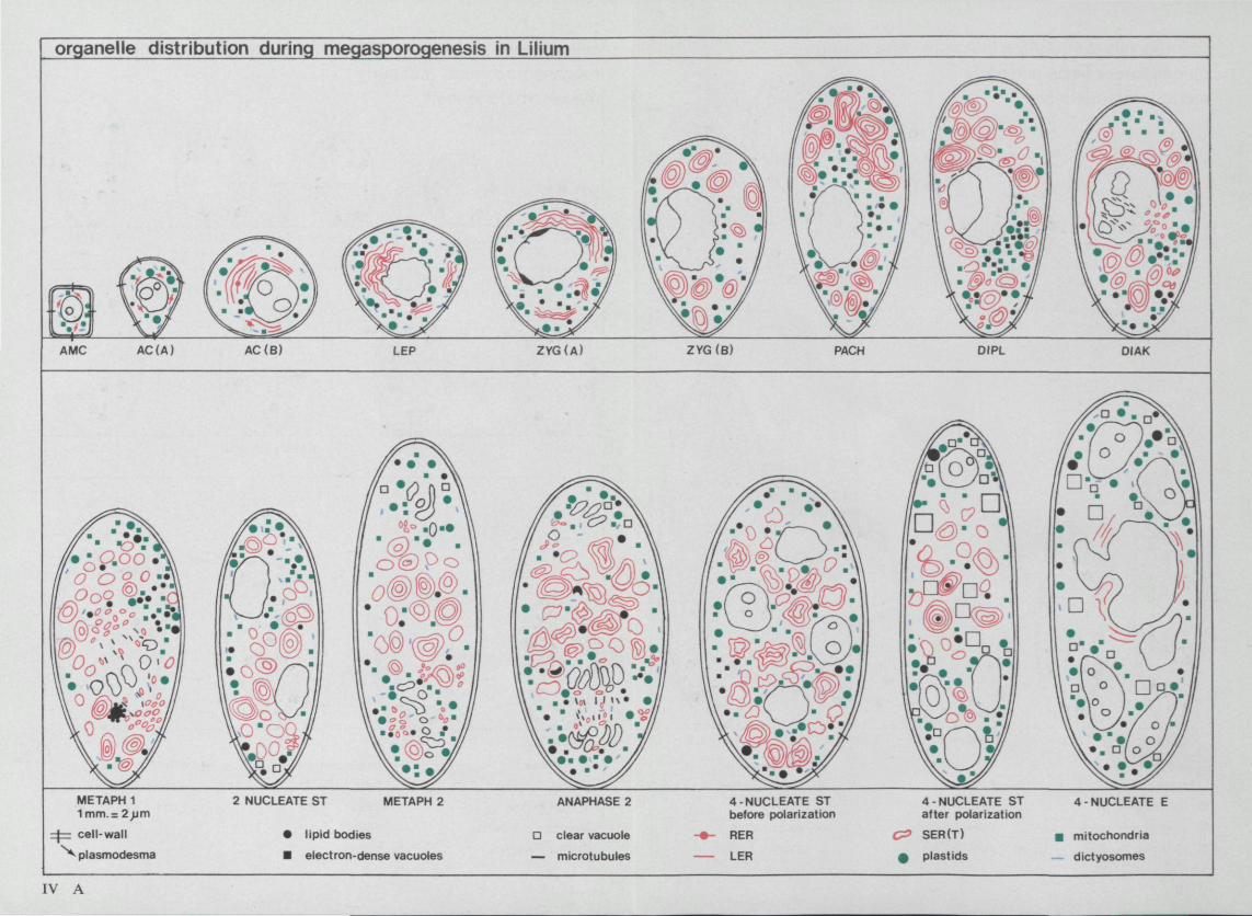
organelle distribution during megasporogenesis in Lilium
AMC AC (A) AC(B) LEP ZYG(A) ZYG(B) PACH DIPL DIAK
'0§) (§.©.
METAPH 1 1mm.= 2>jm
=j=z cell-wall
^ piasmodesma
2 NUCLEATE ST METAPH 2
• lipid bodies
• electron-dense vacuoles
ANAPHASE 2
Q clear vacuole
— microtubules
4-NUCLEATE ST before polarization
RER
LER
C?
•
4-NUCLEATE ST after polarization
SER(T)
plastids
4-NUCLEATE E
• mitochondria
— dictyosomes
IV A

Enclosure IV
cellorganelle distribution in Allium cepa during megasporogenesis and early megagametogenesis
LEP ZYG PACH DIPL METAPH 1
i f cce l l -wa
^plasmodesma
ipid bodies
• el-dense vacuoles
O el-transi vacuoles
IV B
4 NUCLE
• mitochondria
® transformation of mitochondrion
— dictyosomes
1 m m . : 1^m
cellorganelle distribution in Impatiens walleriana during megasporogenesis and early megagametogenesis
AMC
DIPL
LEP ZYG
METAPH 1 EARLY TELOPH
DYAD
z f c cell-wall
^ plasmodesma
• lipid bodies
• el-dense vacuoles
O clear vacuoles
IV C
1 CELL 2 NUCL M
— microtubules
>*- RER
— LER
< = SER(T)
• plastids
PACH
TELOPH
4 NUCL E
g mitochondria
— dictyosomes
1 mm. = 1 urn
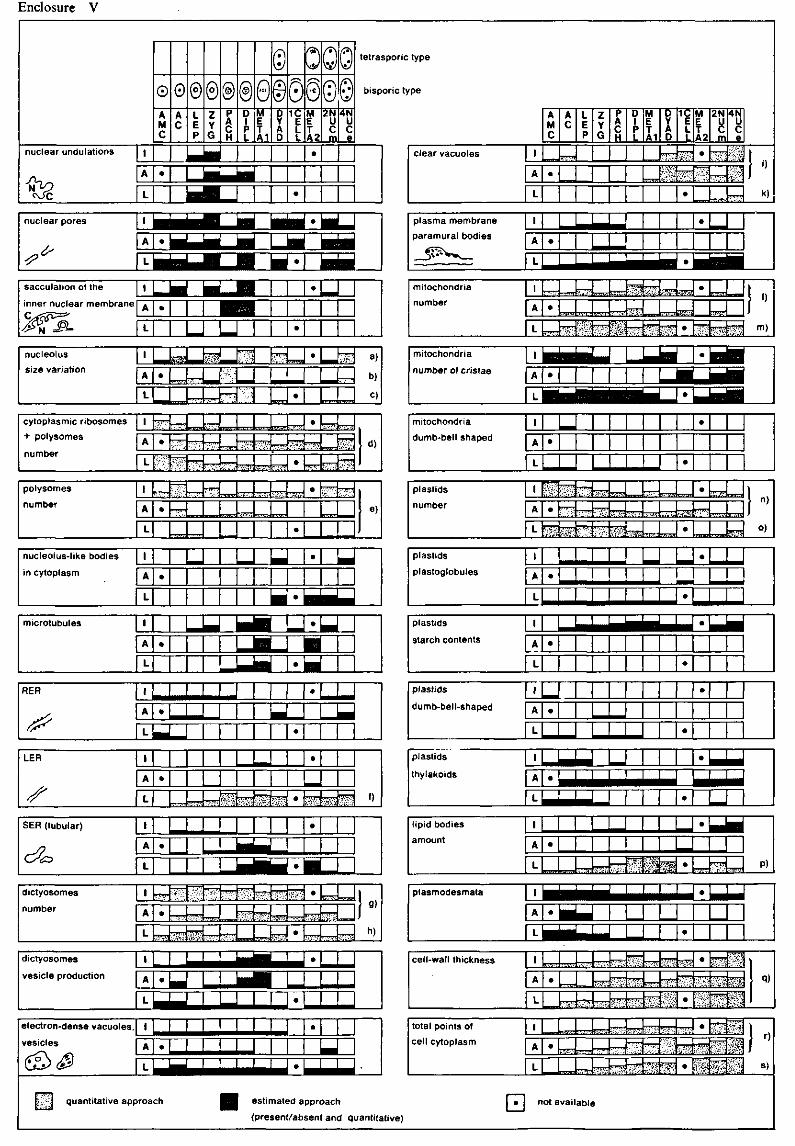
Enclosure V
nuclear undulations |
^ A
•02
0 A M C
•
0 A C
© L E P
®@ Z Y G
P A C H
©0 D 1 P L
M E T A1
© © Ù Y A D
OOQ 0@(D© 1C
E L L
•
M E T A2
•
2N U C m
4N U C e
tetrasporic type
bisporic type
clear vacuoles I
A
L
A M C
•
A C
TTj-TH1
L E P
Z Y G
P A C H
D 1 P L
M E T A1
5 5 W
D Y A 0
îraa
$ ! f
1C E L l
"T??-
W:
•
M E T A2
• :V-S
2N
8 m
U7~-
4N u c el
rti S l̂ ^
0
k)
sacculation of the
inner nuclear membrane
^SL
\
A • L
•
•
nucleolus
size variation
I
A
L
• . , * . ; • •
';.*™?
•77T-
' • ' - ' ? . " r^rn
•
• ™s a)
b)
c)
cytoplasmic ribosomes
+ polysomes
number
I
A
L
•m\^
•
;?W
^ e m
*~r
:^u:
rr^
.• - fl'. •
~ r -
.•:-Vwr '
'••rffi-
— T T -
- • . . , B !
~ r ^
r^-r
•
•
• • • ' ; * '
T V
R m
> • : • : ; : . ;
'tfft ' ; ?-VW -.
polysomes
number I
A
L
• - m
^ 7 T m
nnrr V Ï ' i - . • / . • • • . ! ' •
•
•
_
; ; ' • ' : / . ' r*7.
e)
nucleolus-like bodies
in cytoplasm
I
A
L
• •
•
microtubules I
A
L
•
• ^_
• D RER I •
, A . — — — —
f L J_J LER I •
A •
sf L ^Jjzs^m^ • ^^m\ •)
SER (tubular) 1
A •
L bel
•
1 M [J * g | |
dictyosomes
number
1
A
1
•
i'̂ V
*v'^
™
£'-*'•'
".«'•
W T T T
!-'.W;''
!p5 ^ r
•
•
r ^ r
rr^TJ ^ 1"
h)
dictyosomes
vesicle production
1
A
1
•
•mum»
•
plasma membrane
paramural bodies
i
A
L
• •
^ ^ ^ ^ ^ ^ M M M H A d • • ^ f l ^ ^ H
mitochondria
number 1
A
1
• - m - i '•. j V'* MS£
*** :v":i.'V:.
?Î5ÏÏ1 =7777 •
^ T T T T = ^ • $&'&
1 » m)
mitochondria
number of cristae i b 1 1 i—
L
• 1 1 • tiBii
• ^ ^ ^ H B H
plastids
number
mitochondria
dumb-bell shaped
1
A
L
•
| •
•
%TT~-f.K>:r. \r- VJ.J1T
L FZ? 7m Z&r. 7 ^ SK3 _™
n)
o)
plastids
plastoglobules
1
A
L
• ! ! ! !
•
U •
plastids
starch contents
1
A
L
• •
•
plastids
dumb-bell-shaped
1
A
L
•
•
•
plastids
thylakoids
_ _ • ^ M ^ M
• ~JZ lipid bodies
amount
1
A
L
•
iV.'.H —̂ yu nrr i •
•
P)
plasmodesmata
A
L
• I M ^ M
•
•
cell-wall thickness » A
L
•
Zp?T;
*.i.;V-
^
^••-•TV
»•--.••••; ~ r r
£$
^
•
•
^iiiil
.ivV'^
vS£]
q)
electron-dense vacuoles.
vesicles
i «
%~. quantitative app
1 •
A •
roach ^ H estimated approach
(present/absent and quantiu itive)
total points of
cell cytoplasm
H
1 a^T.rrrr
A •
L
not aval
S T
abU
Ififi
JUii
T ^ n -
FTR •:-?'.
<-•.•
m fe&»
-^rrJ • .v;;s; f~>;i
•
iiiiai
t'.^jy •mm
\ ,) I °
S )