disc1 ser704cys impacts thalamic-prefrontal connectivity
TRANSCRIPT
ORIGINAL ARTICLE
DISC1 Ser704Cys impacts thalamic-prefrontal connectivity
Bing Liu • Lingzhong Fan • Yue Cui • Xiaolong Zhang •
Bing Hou • Yonghui Li • Wen Qin • Dawei Wang •
Chunshui Yu • Tianzi Jiang
Received: 15 August 2013 / Accepted: 12 September 2013
� The Author(s) 2013. This article is published with open access at Springerlink.com
Abstract The Disrupted-in-Schizophrenia 1 (DISC1)
gene has been thought as a putative susceptibility gene for
various psychiatric disorders, and DISC1 Ser704Cys is
associated with variations of brain morphology and func-
tion. Moreover, our recent diffusion magnetic resonance
imaging (dMRI) study reported that DISC1 Ser704Cys was
associated with information transfer efficiency in the brain
anatomical network. However, the effects of the DISC1
gene on functional brain connectivity and networks, espe-
cially for thalamic-prefrontal circuit, which are disrupted in
various psychiatric disorders, are largely unknown. Using a
functional connectivity density (FCD) mapping method
based on functional magnetic resonance imaging data in a
large sample of healthy Han Chinese subjects, we first
investigated the association between DISC1 Ser704Cys and
short- and long-range FCD hubs. Compared with Ser
homozygotes, Cys-allele individuals had increased long-
range FCD hubs in the bilateral thalami. The functional and
anatomical connectivity of the thalamus to the prefrontal
cortex was further analyzed. Significantly increased tha-
lamic-prefrontal functional connectivity and decreased
thalamic-prefrontal anatomical connectivity were found in
DISC1 Cys-allele carriers. Our findings provide consistent
evidence that the DISC1 Ser704Cys polymorphism influ-
ences the thalamic-prefrontal circuits in humans and may
provide new insights into the neural mechanisms that link
DISC1 and the risk for psychiatric disorders.
Keywords DISC1 � Functional connectivity
density � Anatomical connectivity � Thalamus �Thalamic-prefrontal circuit
Introduction
The Disrupted-in-Schizophrenia 1 (DISC1) gene (Millar
et al. 2000) first identified in a large Scotland pedigree
study (Blackwood et al. 2001) is well established as a
genetic risk factor for multiple psychiatric disorders
(Chubb et al. 2008; Bradshaw and Porteous 2012). Studies
in cell and animal models suggest that DISC1 is func-
tionally involved in several key processes that regulate
neural development and brain maturation (Schurov et al.
B. Liu and L. Fan contributed equally to this work and should be
considered co-first authors.
Electronic supplementary material The online version of thisarticle (doi:10.1007/s00429-013-0640-5) contains supplementarymaterial, which is available to authorized users.
B. Liu � L. Fan � Y. Cui � X. Zhang � B. Hou � T. Jiang
Brainnetome Center, Institute of Automation, Chinese Academy
of Sciences, Beijing 100190, China
B. Liu � Y. Cui � B. Hou � T. Jiang (&)
National Laboratory of Pattern Recognition, Institute of
Automation, Chinese Academy of Sciences, Beijing 100190,
China
e-mail: [email protected]
Y. Li � T. Jiang
Queensland Brain Institute, The University of Queensland,
Brisbane, QLD 4072, Australia
W. Qin � D. Wang � C. Yu (&)
Department of Radiology, Tianjin Medical University General
Hospital, No. 154, Anshan Road, Heping District,
Tianjin 300052, China
e-mail: [email protected]
T. Jiang
Key Laboratory for NeuroInformation of Ministry of Education,
School of Life Science and Technology, University of Electronic
Science and Technology of China, Chengdu 610054, China
123
Brain Struct Funct
DOI 10.1007/s00429-013-0640-5

2004; Clapcote et al. 2007; Duan et al. 2007; Kim et al.
2009; Ming and Song 2009; Brandon and Sawa 2011;
Johnstone et al. 2011; Tomita et al. 2011). One common
missense variant in the DISC1 gene, Ser704Cys
(rs821616), has been independently reported by several
groups as associated with an increased risk for various
psychiatric disorders (Hashimoto et al. 2006; DeRosse
et al. 2007; Palo et al. 2007; Qu et al. 2007; Kim et al.
2008), although a meta-analysis has recently questioned
this claim (Mathieson et al. 2012). The associations
between this genetic variant and changes in brain mor-
phology and function have been reported (Callicott et al.
2005; Hashimoto et al. 2006; Di Giorgio et al. 2008; Prata
et al. 2008; Takahashi et al. 2009; Johnstone et al. 2011;
Raznahan et al. 2011; Sprooten et al. 2011; Trost et al.
2013). Moreover, our recent diffusion magnetic resonance
imaging (dMRI) study reported that DISC1 Ser704Cys is
associated with information transfer efficiency in the brain
anatomical network of healthy subjects (Li et al. 2013). A
relevant next question was whether the functional con-
nectivity of the brain, especially in specific circuits, was
measurably influenced by the DISC1 Ser704Cys
polymorphism.
The effects of the DISC1 gene on functional brain
connectivity and networks, which have been shown to be
disrupted in various psychiatric disorders (Hulvershorn
et al. 2011; Rosazza and Minati 2011), are largely
unknown. Resting-state functional connectivity, which is
measured with functional magnetic resonance imaging
(fMRI), can be used to explore the overall organization of
functional brain network (van den Heuvel and Hulshoff Pol
2010). Recently, Tomasi and Volkow (2010a, b) proposed
a functional connectivity density mapping (FCDM) method
to characterize the voxel-wise short- and long-range func-
tional connectivity density (FCD) hubs in the human brain.
This data-driven method can effectively calculate individ-
ual functional connectivity maps with voxel-wise spatial
resolution and overcome the limitations of seed-based
methods for identifying and locating functional connec-
tivity hubs in the brain. Here we aimed to use this data-
driven method based on resting state fMRI data to inves-
tigate the effects of the psychiatric risk variant (DISC1
Ser704Cys) on the functional connectivity hubs of brain
networks and to reveal the functional connectivity changes
of the underlying neural circuits in a large sample of
healthy Han Chinese subjects. Our hypothesis was that this
analytical approach to brain imaging for functional con-
nectivity, especially in the thalamic-prefrontal circuit,
might provide new insights into the neural mechanisms that
link DISC1 and the risk for psychiatric disorders.
The goals of the present study were as follows: (1)
delineate the effects of DISC1 Ser704Cys on short- and
long-range functional connectivity hubs throughout the
whole brain; and (2) determine specific functional con-
nectivity circuits that are affected by DISC1 genetic vari-
ants. We first used FCDM based on resting-state fMRI to
identify the effects of DISC1 Ser704Cys on short- and
long-range functional connectivity hubs. We hypothesized
that the FCD hubs of the thalamus were influenced by
DISC1 Ser704Cys. The FCD hubs that showed significant
differences between groups of individuals bearing different
DISC1 genotypes served as the region of interest (ROI) in
the following analyses. We next analyzed the correspond-
ing changes of functional and anatomical connectivity of
the ROI. Specifically, functional connectivity mapped the
correlations between the ROI and all other voxels in the
brain, and anatomical connectivity projected the structural
pathways of the ROI throughout the whole brain.
Accordingly, we established the impact of DISC1
Ser704Cys on functional and anatomical connectivity of
the ROI.
Materials and methods
Subjects
We recruited 323 young healthy Han Chinese subjects (157
males and 166 females, mean age = 22.7 years, ran-
ge 18–31 years) as described in our previous studies (Li
et al. 2013). After excluding subjects with missing DISC1
genotype information and subjects without sufficient neu-
roimaging data, finally 284 subjects were included in the
fMRI analyses and 278 subjects were included in the dMRI
analyses (Supplementary Figure 1).
DISC1 genotyping
We extracted genomic DNA from whole blood using the
EZgeneTM Blood gDNA Miniprep Kit (BIOMIGA Inc,
San Diego, CA, USA). The DISC1 Ser704Cys (rs821616)
was then genotyped using the PCR and Ligation Detection
Reaction (LDR) method as previously described (Li et al.
2013). Nine subjects with missing genotype data were
excluded for further analysis. The distribution of DISC1
genotypes in our samples (SerSer: N = 218, SerCys:
N = 86, CysCys: N = 10) did not significantly deviate
from the Hardy–Weinberg equilibrium (v2 = 0.18, df = 1,
P = 0.67).
Image data acquisition
All subjects were scanned on the same Signa HDx 3.0 T
magnetic resonance scanner (GE Healthcare; Milwaukee,
WI, USA). During scanning, we used foam padding and ear
plugs to reduce head motion and scanning noise,
Brain Struct Funct
123
respectively. All the participants underwent a high-reso-
lution T1-weighted brain volume (BRAVO) 3D MRI
sequence [repetition time (TR) = 8.1 ms; echo time
(TE) = 3.1 ms; flip angle = 13�; voxel size = 1 9 1 9
1 mm3; 176 sagittal slices]. Subsequently, resting-state
functional imaging was collected with a single-shot gra-
dient-echo echo-planar-imaging (SS-GRE-EPI) sequence
sensitive to blood oxygenation level-dependent (BOLD)
contrast [TR = 2,000 ms; TE = 30 ms; field of view
(FOV) = 240 9 240 mm2; matrix = 64 9 64; flip angle
= 90�; voxel size = 3.75 9 3.75 9 4.0 mm3; 40 slices
and 180 volumes]. During resting-state fMRI, all subjects
were instructed to be eye-closed and to move as little as
possible without falling asleep. Finally, the diffusion-
weighted images were collected using echo-planar imaging
with 45 contiguous 3 mm axial slices and 55 non-collinear
diffusion gradients together with three non-diffusion-
weighted acquisitions (TR = 10,000 ms; TE = 64.2 ms;
b-value = 1,000 s/mm2; matrix = 128 9 128; FOV =
256 9 256 mm2; flip angle = 90�; voxel size = 2 9 2 9
3 mm3).
fMRI preprocessing
Fourteen subjects were excluded for apparent inter-slice
motion artifacts of their raw fMRI data as evaluated by the
visual inspection of two radiologists who were blinded to
genotype (Yu and Qin). Further data preprocessing was
carried out in the 300 remaining subjects using FSL tools
(www.fmrib.ox.ac.uk/fsl) and in-house programs. A series
of preprocessing steps were performed: (1) discarding the
first ten volumes for the magnetization equilibrium; (2)
slice timing correction; (3) head motion correction; (4)
non-brain removal; (5) temporal filtering (0.01–0.1 Hz);
and (6) regressing nuisance signals (six motion parame-
ters). The registration of the resting-state data to high-
resolution T1-weighted images and the T1 data to the
3-mm isotropic MNI-152 standard space template (Mon-
treal Neurological Institute) were carried out. The resulting
transformation matrices were combined to obtain a native
MNI space transformation matrix and its inverse. Sixteen
subjects who had a maximum displacement in any of the
cardinal directions (x, y, z) larger than 2 mm or a maximum
spin (x, y, z) larger than 2� were excluded from subsequent
analysis. In total, 284 subjects were included in the fMRI
data analysis (Supplementary Figure 1). The distribution of
the DISC1 genotype in the 284 remaining subjects (SerSer:
N = 197, SerCys: N = 79, CysCys: N = 8) still did not
significantly deviate from the Hardy–Weinberg equilibrium
(v2 = 0.001, df = 1, P = 0.98). Moreover, there was no
significant bias in the genotype distrubition of the 30
exclusionary (SerSer: N = 21, SerCys: N = 7, CysCys:
N = 2) and the 284 remaining samples (v2 = 1.20, df = 2,
P = 0.55).
Short- and long-range FCD
Here, we used FCDM to evaluate the individual FCD maps
with voxel-vise spatial resolution (Tomasi and Volkow
2010a, 2011). First, we computed the global FCD based on
prior knowledge that two voxels are considered function-
ally connected if their Pearson correlation coefficient is
greater than 0.6 (Tomasi and Volkow 2011). The global
FCD of a given voxel was defined as the number of
functional connections between this voxel and all other
voxels in the brain. Second, we obtained the short-range
FCD using a ‘‘growing’’ algorithm. In this algorithm, given
a voxel x0, an additional voxel xj, was added to the list of
neighbors of x0 if it was adjacent to a voxel that was linked
to x0 by a continuous path of functionally connected voxels
and the correlation coefficient between x0 and xj was[0.6.
This calculation was repeated for all voxels that were
adjacent to the neighbors of x0 in an iterative manner until
no new neighbors could be added to the list. The short-
range FCD of x0 was defined as the number of elements in
the list of neighbors. The strength of the long-range FCD
was equated to the difference between the global FCD and
the short-range FCD. These calculations were performed
on all voxels in the brain. Therefore, the strength of the
short-range FCD of a given voxel reflects the functional
correlation between that voxel and other voxels within a
local cluster, and the strength of the long-range FCD
reflects the functional correlation between a given voxel
and other voxels located at a distance. Finally, the short and
long-range FCD maps were spatially smoothed using a
Gaussian kernel of full-width at half-maximum (FWHM)
of 8 mm. The short and long-range FCD of each voxel was
divided by the mean value of each individual to normalize
the distribution of the FCD strength.
Functional connectivity analyses
We calculated the voxel-wise functional connectivity
throughout the entire brain by taking the brain regions that
revealed different FCD hubs between DISC1 Ser704Cys
genotypes as the ROI. The functional connectivity map of
each individual was computed by averaging the BOLD
time series in the ROI and then computing the Pearson’s
correlation coefficient between the seed average time series
and those from each voxel in the brain. The resulting
correlations were transformed to approximate a Gaussian
distribution using Fisher’s z transformation. In this way, a
functional connectivity map of the ROI for each participant
was produced.
Brain Struct Funct
123
dMRI preprocessing and anatomical connectivity
analyses
Two radiologists (Yu and Qin) visually inspected the raw
dMRI data for apparent artifacts arising from subject
motion and instrument malfunction. After excluding 36
subjects (SerSer: N = 20; SerCys: N = 14; CysCys:
N = 2), the 278 remaining subjects, including 198 Ser
homozygotes and 80 Cys-allele carriers (SerCys: N = 72;
CysCys: N = 8), underwent further dMRI data analysis
(Supplementary Figure 1). There was no significant bias in
the genotype distribution of the 36 exclusionary and 278
remaining samples (v2 = 3.57, df = 2, P = 0.17). The
distribution of the DISC1 genotype in the 278 remaining
subjects still did not significantly deviate from the Hardy–
Weinberg equilibrium (v2 = 0.22, df = 1, P = 0.63).
We further described the anatomical connectivity map
of the ROI with different FCD hubs and aimed to find
whether specific anatomical connectivity contributed to the
DISC1 genetic effects. First, the significant ROIs obtained
in the previous FCD analyses were transformed into the
individual diffusion space. For this purpose, the skull-
stripped T1-weighted images of each subject were co-
registered with the non-diffusion-weighted images
(b = 0 s/mm2) using FLIRT. Then, the T1 images in dif-
fusion space were wrapped linearly and nonlinearly into
the MNI (Montreal Neurological Institute) space using
FLIRT and FNIRT. After that, we applied the inverse
linear and non-linear transformations to transform the ROIs
into the individual diffusion space. Second, the diffusion
MRI data were processed using the FMRIB Software
Library (FSL). Eddy-current distortions and simple head
motion were corrected by applying affine registration of all
volumes to a target volume with no diffusion weighting,
and three-dimensional maps of the diffusion tensor and the
fractional anisotropy (FA) values were calculated using
FSL. Moreover, the local probability distributions of fiber
directions were estimated at each voxel (Behrens et al.
2003). We chose the computation model and allowed for
the automatic estimation of two fiber directions within each
voxel (Behrens et al. 2007). Then, we drew 5,000 samples
from the connectivity distribution for each voxel in the ROI
and calculated the connection probability of each voxel. To
reduce the false-positive rates, the fiber tracking results
were thresholded to exclude all connection probability
values of P \ 0.002 (10 out of 5,000 samples) (Wang et al.
2012; Zhang et al. 2012). Next, the probability connectivity
map for each subject was binarized with P and transformed
into standard MNI space. A population-based anatomical
connectivity map of the ROI with different FCD hubs was
obtained by averaging the transformed individual binary
maps. Finally, we binarized the population-based anatom-
ical connectivity map with a threshold of 80 % (the
threshold was tested from 50 to 100 % in all subjects with
no differences on the following analysis) as a mask for the
following voxel-based FA analysis between DISC1 Cys-
allele carriers and Ser homozygotes.
Statistical analyses
We used a Pearson Chi square test to test for differences in
gender and a two-sample t test to test for differences in age,
educational year, and intelligence quotient (IQ) score
between the two genotype groups. Voxel-wise two-sample
t-tests with four covariates (age, gender, educational year,
and IQ score) were implemented in SPM8 to map group
differences in the strength of short- and long-range FCDs,
functional and anatomical connectivity from the ROI
between DISC1 Cys-allele carriers and Ser homozygotes.
For all imaging analyses, the Monto Carlo simulation
method was used throughout the entire brain to correct for
multiple comparisons by AlphaSim program (corrected
P \ 0.01). For the thalamic functional connectivity anal-
yses, corrected for multiple comparisons was used within
prefrontal cortex as defined in previous studies (Wei et al.
2012).
Results
Demographic information
In total, 323 young subjects were recruited. Of these, 284
subjects were included in the fMRI data analyses and 278
were included in the previous and current dMRI data
analyses (Table 1; Supplementary Figure 1). All of the
individuals in the study were categorized into two groups
based on DISC1 Ser704Cys genotypes: Ser homozygotes
and Cys-allele carriers. No significant differences were
found in age, gender, educational year, and IQ score
between two groups (Table 1).
Functional connectivity density analysis
We calculated the short- and long-range FCD maps for
each individual, and group comparisons were performed.
Using the averaged short- and long-range FCD maps (Fig.
1a, b), we detected similar patterns as observed in previous
studies (Tomasi and Volkow 2010a, 2011). The short- and
long-range FCD patterns were observed bilaterally, with
maximal magnitude in posterior cingulate, occipital and
prefrontal cortices. Compared with Ser homozygotes, Cys-
allele carriers showed significantly increased long-range
FCD in the bilateral thalami (peak voxel MNI coordinate:
x = 12, y = -9, z = 15; T = 4.29; cluster size = 67),
Brain Struct Funct
123

mainly located at the mediodorsal thalamic nucleus
(Fig. 1c, d). No significant differences in short-range FCD
were found between the two DISC1 genotypes.
Thalamic functional connectivity analysis
Since the long-range FCD in the bilateral thalami has been
found to be significantly increased Cys-allele carriers
compared with Ser homozygotes as we hypothesized, we
further investigated the specific functional connectivity
circuits that are affected by DISC1 genetic variants by ROI-
based functional connectivity analyses. To determine the
effects of DISC1 Ser704Cys on functional connectivity to
the thalamus, we performed voxel-vise functional connec-
tivity analyses using the thalamic region that showed sig-
nificantly different long-range FCD as the ROI. We found
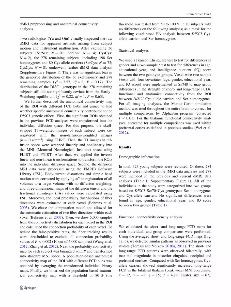
that Cys-allele carriers showed increased thalamic func-
tional connectivity in the bilateral dorsolateral prefrontal
Table 1 Demographic information
Subjects included in fMRI data analyses Subjects included in dMRI data analyses
SerSer (n = 197) Cys-allele (n = 87) P-value SerSer (n = 198) Cys-allele (n = 80) P-value
Gender 93 M/104 F 36 M/51 F 0.37 94 M/104 F 32 M/48 F 0.26
Age in years* 22.75 (2.53) 22.83 (2.24) 0.81 22.77 (2.50) 23.09 (2.19) 0.33
Education in years* 15.56 (2.51) 15.49 (2.72) 0.84 15.61 (2.51) 15.54 (3.03) 0.85
FSIQ* 117.02 (9.59) 117.38 (7.71) 0.75 116.84 (9.82) 117.41 (8.14) 0.65
F female, M male, FSIQ full scale intelligence quotient
* Values are mean (SD)
Fig. 1 Results of FCD analyses. a, b Shows the spatial distribution of
the average short-range FCD and long-range FCD, respectively,
superimposed on the cerebral cortex for all subjects. c Significantly
increased long-range FCD of Cys-allele carriers compared with Ser
homozygotes (corrected P \ 0.01). d For illustrative purposes, the
average value of the long-range FCD of bilateral thalami (within
significant clusters) was displayed in the bar graph for two different
genotype groups (mean ± SEM)
Brain Struct Funct
123
cortex (DLPFC) than did the Ser homozygotes (Fig. 2;
Supplementary Table 1).
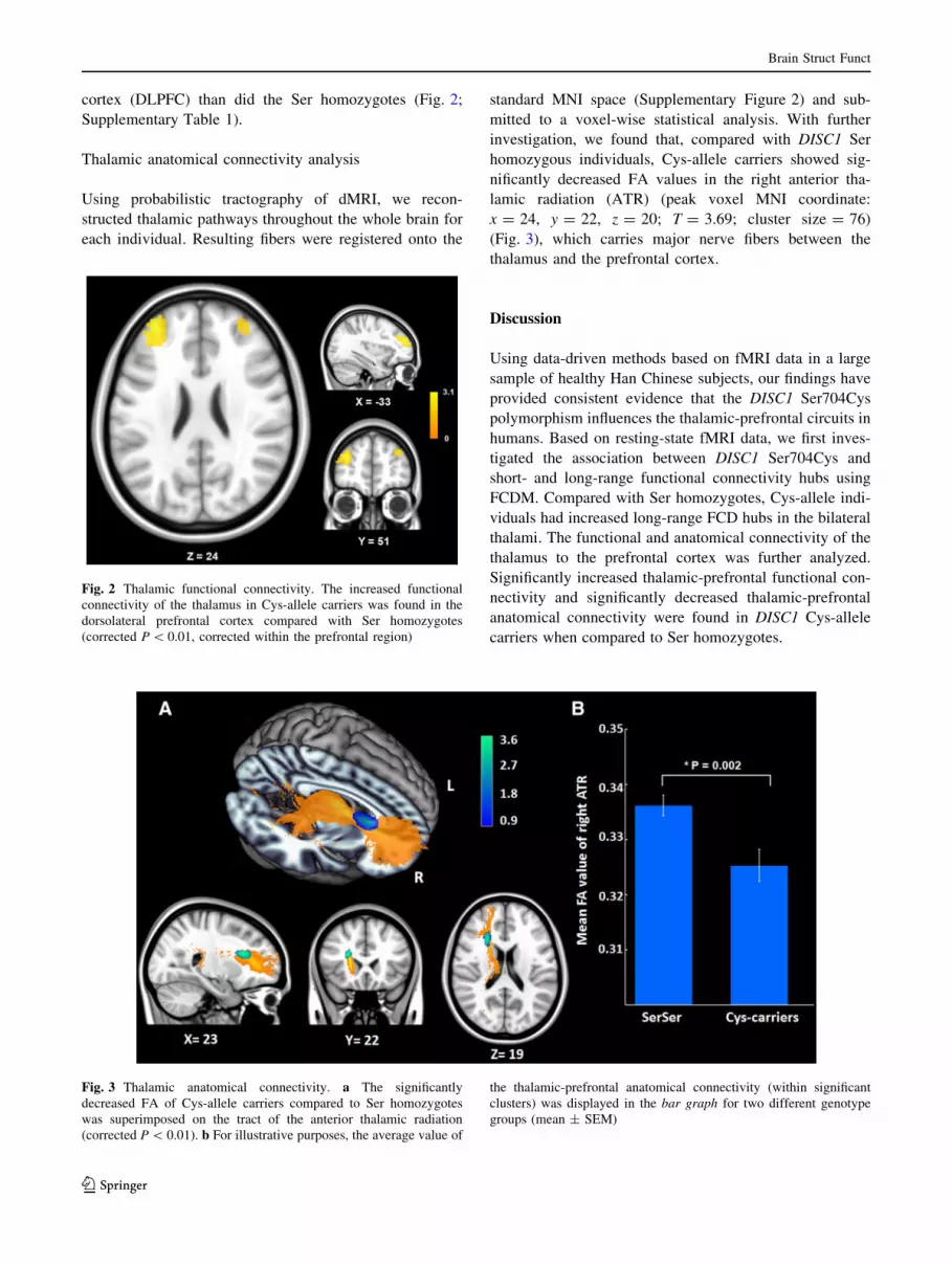
Thalamic anatomical connectivity analysis
Using probabilistic tractography of dMRI, we recon-
structed thalamic pathways throughout the whole brain for
each individual. Resulting fibers were registered onto the
standard MNI space (Supplementary Figure 2) and sub-
mitted to a voxel-wise statistical analysis. With further
investigation, we found that, compared with DISC1 Ser
homozygous individuals, Cys-allele carriers showed sig-
nificantly decreased FA values in the right anterior tha-
lamic radiation (ATR) (peak voxel MNI coordinate:
x = 24, y = 22, z = 20; T = 3.69; cluster size = 76)
(Fig. 3), which carries major nerve fibers between the
thalamus and the prefrontal cortex.
Discussion
Using data-driven methods based on fMRI data in a large
sample of healthy Han Chinese subjects, our findings have
provided consistent evidence that the DISC1 Ser704Cys
polymorphism influences the thalamic-prefrontal circuits in
humans. Based on resting-state fMRI data, we first inves-
tigated the association between DISC1 Ser704Cys and
short- and long-range functional connectivity hubs using
FCDM. Compared with Ser homozygotes, Cys-allele indi-
viduals had increased long-range FCD hubs in the bilateral
thalami. The functional and anatomical connectivity of the
thalamus to the prefrontal cortex was further analyzed.
Significantly increased thalamic-prefrontal functional con-
nectivity and significantly decreased thalamic-prefrontal
anatomical connectivity were found in DISC1 Cys-allele
carriers when compared to Ser homozygotes.
Fig. 2 Thalamic functional connectivity. The increased functional
connectivity of the thalamus in Cys-allele carriers was found in the
dorsolateral prefrontal cortex compared with Ser homozygotes
(corrected P \ 0.01, corrected within the prefrontal region)
Fig. 3 Thalamic anatomical connectivity. a The significantly
decreased FA of Cys-allele carriers compared to Ser homozygotes
was superimposed on the tract of the anterior thalamic radiation
(corrected P \ 0.01). b For illustrative purposes, the average value of
the thalamic-prefrontal anatomical connectivity (within significant
clusters) was displayed in the bar graph for two different genotype
groups (mean ± SEM)
Brain Struct Funct
123

One of the major findings of the present study was the
effect of DISC1 Ser704Cys on the long-range FCD in the
bilateral thalami, especially on the mediodorsal nucleus of
the thalamus. Ever since the DISC1 gene was originally
discovered in a Scottish pedigree, the accumulating bio-
logical evidence has indicated that DISC1 plays diverse
and important roles in brain development and maturation,
including early neurodevelopment, synaptic regulation, and
oligodendrocyte development (Brandon et al. 2009; Bran-
don and Sawa 2011; Johnstone et al. 2011). These roles are
consistent with the current hypotheses for the etiopathol-
ogy of major psychiatric disorders. DISC1 Ser704Cys is
one of the vital non-synonymous mutations of the DISC1
gene, and it has been widely reported to be associated with
different clinical and structural brain phenotypes both in
patients and healthy individuals, though the results are
inconsistent (Callicott et al. 2005; Hashimoto et al. 2006;
DeRosse et al. 2007; Qu et al. 2007; Di Giorgio et al. 2008;
Sprooten et al. 2011). In particular, the DISC1 Cys allele
was positively associated with schizophrenia in a study of
Han Chinese subjects (Qu et al. 2007). Individuals carrying
the Cys allele for Ser704Cys have reduced gray matter
volume in the cingulate cortex and decreased FA in pre-
frontal white matter (Hashimoto et al. 2006). Moreover,
evidence indicates that the Ser704Cys genetic variant may
inhibit the migration of neurons in the developing neo-
cortex (Singh et al. 2011) and alter oligomeric assembly
(Narayanan et al. 2011). On the other hand, the thalamus
has been universally believed to serve as a relay of infor-
mation processing with its nerve fibers projecting to the
cerebral cortex in all directions with extensively intercon-
nected major cortical circuits (Andreasen 1997; Briggs and
Usrey 2008). Abnormal structure and function of the
thalamus as observed in postmortem and neuroimaging
studies, respectively, has been widely reported in various
psychiatric disorders (Andreasen 1997; Byne et al. 2009;
Adriano et al. 2010; Cronenwett and Csernansky 2010). As
a psychiatric risk gene, DISC1 would, therefore, be
expected to affect thalamic function. We speculate that the
increased long-range FCD observed in bilateral thalami in
this study may be a manifestation of the compensatory
effect of thalamic connectivity because of the weaker
information processing function of the thalamus in DISC1
Cys-allele carriers compared with Ser homozygotes, with
more thalamic functional connectivity required to achieve
the same behavioral output.
The most significant differences of long-range FCD are
observed in the mediodorsal nucleus of the thalamus,
which mainly provides projections to and from various
regions of the prefrontal cortex (Andreasen 1997). Previous
anatomical and functional neuroimaging findings have
shown disrupted prefrontal-thalamic circuitry in schizo-
phrenia (Lewis 2000; Lehrer et al. 2005; Rose et al. 2006;
Pakkenberg et al. 2009; Marenco et al. 2012; Woodward
et al. 2012) and other psychiatric disorders (Strakowski
et al. 2005). Alterations in thalamic-prefrontal circuitry
might be a pivotal endophenotypic trait that should be
studied to understand the neural mechanisms of genes
associated with psychiatric disorders. Using functional
connectivity analyses of resting-state fMRI, we calculated
the thalamic functional connectivity map for each indi-
vidual and performed voxel-wise comparisons between the
two genotypes. DISC1 Cys-allele individuals showed
increased thalamic-prefrontal functional connectivity
compared with Ser homozygotes. Here we further used two
complementary approaches based on diffusion MRI
(dMRI), probabilistic fiber tractography and voxel-wise
morphometry of FA, to ‘‘dissect’’ the affected white matter
pathways from the thalamus and quantify the altered white-
matter integrity on an individual basis (Mori and Zhang
2006; Behrens et al. 2007). We reconstructed thalamic
pathways throughout the whole brain and further per-
formed a voxel-wise statistical analysis comparing differ-
ent DISC1 genotypes. Individuals with the DISC1 Cys-
allele exhibited less thalamic-prefrontal anatomical con-
nectivity, particularly involving the right ATR, which is
one of the most important fibers connecting the medio-
dorsal thalamic nucleus and the prefrontal cortex. The
finding of current voxel-wise analyses of the thalamic
white matter pathways was consistent with our tract-based
analyses of the previous study, which has reported that of
the 20 major fiber tracts only the white matter integrity of
the ATR has the trend to differ between the individuals
with different DISC1 Ser704Cys genotypes (Li et al. 2013).
The white matter integrity of the ATR has been widely
reported to be decreased in psychiatric disorders (McIntosh
et al. 2008; Mamah et al. 2010; Perez-Iglesias et al. 2010)
and in psychosis-risk-associated neuregulin-1 variants
(Sprooten et al. 2009; Barnes et al. 2012). These findings
combined with our previous study consistently support that
the DISC1 Ser704Cys polymorphism, as a key psychosis-
risk-related genetic variation, impacts the thalamic-pre-
frontal functional and anatomical connectivity. These
effects provide a possible explanation for the abnormal
thalamic function and disrupted thalamic-prefrontal circuit
that is often reported in psychiatric disorders. These effects
also suggest that there is a potential mechanistic link
between DISC1 Ser704Cys and the risk for psychiatric
disorders.
Using resting-state fMRI and dMRI data, our present
study combining with the previous finding reported that
DISC1 Cys-allele carriers had significantly increased long-
range FCD in the thalamus, increased thalamic-prefrontal
functional connectivity, and decreased thalamic-prefrontal
anatomical connectivity compared with Ser homozygotes
in a large sample of healthy subjects. Although some of the
Brain Struct Funct
123
functional connectivity may have an anatomical basis, the
relationship between functional and anatomical connec-
tivity is complex. For example, anatomical connectivity
measured by dMRI is nearly uniformly decreased in
schizophrenia (Ellison-Wright and Bullmore 2009; Bora
et al. 2011). On the other hand, the founding of functional
connectivity in schizophrenia is inconsistent regardless of
the specific connections or the directions (Skudlarski et al.
2010; Fornito et al. 2012). Our findings of increased
functional connectivity may represent increased neural
effort due to decreased anatomical connectivity in the
thalamic-prefrontal circuit. This type of compensatory
mechanism may limit the effects of genes related to psy-
chiatric disorders and help maintain a normal range of
behavioral performance in healthy subjects. The compen-
satory mechanism of increased thalamic-prefrontal func-
tional connectivity in healthy subjects may conversely
explain the finding of reduced prefrontal-thalamic func-
tional connectivity in schizophrenia (Woodward et al.
2012). However, more biological evidences are needed to
elucidate the relationship between anatomical and func-
tional connectivity.
We performed comprehensive functional connectivity
analyses based on brain imaging data of healthy individuals
with different DISC1 genotypes, therefore providing new
insights into the neural mechanisms that link DISC1
Ser704Cys and risk for psychiatric disorders by the imaging
endophenotype of thalamic-prefrontal circuit. Here it should
be noted that another important functional polymorphism of
DISC1, Leu607Phe, was not considered in the present study
because the sample size carrying the minor allele in all the
subjects (3/323) is too few to make group comparisons.
Moreover, there are still large gaps between the genetic
variants and neural connectivity. Systematic studies using
animal models or other techniques may provide more solid
biological evidence to explain this relationship.
Acknowledgments This work was supported by the National Key
Basic Research and Development Program (973) (Grant No.
2011CB707800), the Strategic Priority Research Program of the
Chinese Academy of Sciences (Grant No. XDB02030300), the Natural
Science Foundation of China (Grant Nos. 81000582, 91132301 and
91232718), and the Beijing Nova Program (Grant No. 2010B061).
Open Access This article is distributed under the terms of the
Creative Commons Attribution License which permits any use, dis-
tribution, and reproduction in any medium, provided the original
author(s) and the source are credited.
References
Adriano F, Spoletini I, Caltagirone C, Spalletta G (2010) Updated
meta-analyses reveal thalamus volume reduction in patients with
first-episode and chronic schizophrenia. Schizophr Res
123(1):1–14. doi:10.1016/j.schres.2010.07.007
Andreasen NC (1997) The role of the thalamus in schizophrenia. Can
J Psychiatry 42(1):27–33
Barnes A, Isohanni M, Barnett JH, Pietilainen O, Veijola J, Miettunen
J, Paunio T, Tanskanen P, Ridler K, Suckling J, Bullmore ET,
Jones PB, Murray GK (2012) Neuregulin-1 genotype is associ-
ated with structural differences in the normal human brain.
Neuroimage 59(3):2057–2061. doi:10.1016/j.neuroimage.2011.
10.007
Behrens TE, Woolrich MW, Jenkinson M, Johansen-Berg H, Nunes
RG, Clare S, Matthews PM, Brady JM, Smith SM (2003)
Characterization and propagation of uncertainty in diffusion-
weighted MR imaging. Magn Reson Med 50(5):1077–1088.
doi:10.1002/mrm.10609
Behrens TE, Berg HJ, Jbabdi S, Rushworth MF, Woolrich MW
(2007) Probabilistic diffusion tractography with multiple fibre
orientations: what can we gain? Neuroimage 34(1):144–155.
doi:10.1016/j.neuroimage.2006.09.018
Blackwood DH, Fordyce A, Walker MT, St Clair DM, Porteous DJ,
Muir WJ (2001) Schizophrenia and affective disorders–cosegre-
gation with a translocation at chromosome 1q42 that directly
disrupts brain-expressed genes: clinical and P300 findings in a
family. Am J Hum Genet 69(2):428–433 S0002-9297(07)
61088-X
Bora E, Fornito A, Radua J, Walterfang M, Seal M, Wood SJ, Yucel
M, Velakoulis D, Pantelis C (2011) Neuroanatomical abnormal-
ities in schizophrenia: a multimodal voxelwise meta-analysis and
meta-regression analysis. Schizophr Res 127(1–3):46–57.
doi:10.1016/j.schres.2010.12.020
Bradshaw NJ, Porteous DJ (2012) DISC1-binding proteins in neural
development, signalling and schizophrenia. Neuropharmacology
62(3):1230–1241. doi:10.1016/j.neuropharm.2010.12.027
Brandon NJ, Sawa A (2011) Linking neurodevelopmental and
synaptic theories of mental illness through DISC1. Nat Rev
Neurosci 12(12):707–722. doi:10.1038/nrn3120
Brandon NJ, Millar JK, Korth C, Sive H, Singh KK, Sawa A (2009)
Understanding the role of DISC1 in psychiatric disease and
during normal development. J Neurosci 29(41):12768–12775.
doi:10.1523/JNEUROSCI.3355-09.2009
Briggs F, Usrey WM (2008) Emerging views of corticothalamic
function. Curr Opin Neurobiol 18(4):403–407. doi:10.1016/j.
conb.2008.09.002
Byne W, Hazlett EA, Buchsbaum MS, Kemether E (2009) The
thalamus and schizophrenia: current status of research. Acta
Neuropathol 117(4):347–368. doi:10.1007/s00401-008-0404-0
Callicott JH, Straub RE, Pezawas L, Egan MF, Mattay VS, Hariri AR,
Verchinski BA, Meyer-Lindenberg A, Balkissoon R, Kolachana
B, Goldberg TE, Weinberger DR (2005) Variation in DISC1
affects hippocampal structure and function and increases risk for
schizophrenia. Proc Natl Acad Sci USA 102(24):8627–8632.
doi:10.1073/pnas.0500515102
Chubb JE, Bradshaw NJ, Soares DC, Porteous DJ, Millar JK (2008)
The DISC locus in psychiatric illness. Mol Psychiatry
13(1):36–64. doi:10.1038/sj.mp.4002106
Clapcote SJ, Lipina TV, Millar JK, Mackie S, Christie S, Ogawa F,
Lerch JP, Trimble K, Uchiyama M, Sakuraba Y, Kaneda H,
Shiroishi T, Houslay MD, Henkelman RM, Sled JG, Gondo Y,
Porteous DJ, Roder JC (2007) Behavioral phenotypes of Disc1
missense mutations in mice. Neuron 54(3):387–402. doi:10.
1016/j.neuron.2007.04.015
Cronenwett WJ, Csernansky J (2010) Thalamic pathology in schizo-
phrenia. Curr Top Behav Neurosci 4:509–528
DeRosse P, Hodgkinson CA, Lencz T, Burdick KE, Kane JM,
Goldman D, Malhotra AK (2007) Disrupted in schizophrenia 1
Brain Struct Funct
123
genotype and positive symptoms in schizophrenia. Biol Psychi-
atry 61(10):1208–1210. doi:10.1016/j.biopsych.2006.07.023
Di Giorgio A, Blasi G, Sambataro F, Rampino A, Papazacharias A,
Gambi F, Romano R, Caforio G, Rizzo M, Latorre V, Popolizio
T, Kolachana B, Callicott JH, Nardini M, Weinberger DR,
Bertolino A (2008) Association of the SerCys DISC1 polymor-
phism with human hippocampal formation gray matter and
function during memory encoding. Eur J Neurosci
28(10):2129–2136. doi:10.1111/j.1460-9568.2008.06482.x
Duan X, Chang JH, Ge S, Faulkner RL, Kim JY, Kitabatake Y, Liu
XB, Yang CH, Jordan JD, Ma DK, Liu CY, Ganesan S, Cheng
HJ, Ming GL, Lu B, Song H (2007) Disrupted-In-Schizophrenia
1 regulates integration of newly generated neurons in the adult
brain. Cell 130(6):1146–1158. doi:10.1016/j.cell.2007.07.010
Ellison-Wright I, Bullmore E (2009) Meta-analysis of diffusion tensor
imaging studies in schizophrenia. Schizophr Res 108(1–3):3–10.
doi:10.1016/j.schres.2008.11.021
Fornito A, Zalesky A, Pantelis C, Bullmore ET (2012) Schizophrenia,
neuroimaging and connectomics. Neuroimage 62(4):2296–2314.
doi:10.1016/j.neuroimage.2011.12.090
Hashimoto R, Numakawa T, Ohnishi T, Kumamaru E, Yagasaki Y,
Ishimoto T, Mori T, Nemoto K, Adachi N, Izumi A, Chiba S,
Noguchi H, Suzuki T, Iwata N, Ozaki N, Taguchi T, Kamiya A,
Kosuga A, Tatsumi M, Kamijima K, Weinberger DR, Sawa A,
Kunugi H (2006) Impact of the DISC1 Ser704Cys polymor-
phism on risk for major depression, brain morphology and ERK
signaling. Hum Mol Genet 15(20):3024–3033. doi:10.1093/hmg/
ddl244
Hulvershorn LA, Cullen K, Anand A (2011) Toward dysfunctional
connectivity: a review of neuroimaging findings in pediatric
major depressive disorder. Brain Imaging Behav 5(4):307–328.
doi:10.1007/s11682-011-9134-3
Johnstone M, Thomson PA, Hall J, McIntosh AM, Lawrie SM,
Porteous DJ (2011) DISC1 in schizophrenia: genetic mouse
models and human genomic imaging. Schizophr Bull
37(1):14–20. doi:10.1093/schbul/sbq135
Kim HJ, Park HJ, Jung KH, Ban JY, Ra J, Kim JW, Park JK, Choe
BK, Yim SV, Kwon YK, Chung JH (2008) Association study of
polymorphisms between DISC1 and schizophrenia in a Korean
population. Neurosci Lett 430(1):60–63. doi:10.1016/j.neulet.
2007.10.010
Kim JY, Duan X, Liu CY, Jang MH, Guo JU, Pow-anpongkul N,
Kang E, Song H, Ming GL (2009) DISC1 regulates new neuron
development in the adult brain via modulation of AKT-mTOR
signaling through KIAA1212. Neuron 63(6):761–773. doi:10.
1016/j.neuron.2009.08.008
Lehrer DS, Christian BT, Mantil J, Murray AC, Buchsbaum BR,
Oakes TR, Byne W, Kemether EM, Buchsbaum MS (2005)
Thalamic and prefrontal FDG uptake in never medicated patients
with schizophrenia. Am J Psychiatry 162(5):931–938. doi:10.
1176/appi.ajp.162.5.931
Lewis DA (2000) Is there a neuropathology of schizophrenia? Recent
findings converge on altered thalamic-prefrontal cortical con-
nectivity. Neuroscientist 6(3):208–218. doi:10.1177/1073858400
00600311
Li Y, Liu B, Hou B, Qin W, Wang D, Yu C, Jiang T (2013) Less
efficient information transfer in Cys-allele carriers of DISC1: a
brain network study based on diffusion MRI. Cereb Cortex
23(7):1715–1723. doi:10.1093/cercor/bhs167
Mamah D, Conturo TE, Harms MP, Akbudak E, Wang L, McMichael
AR, Gado MH, Barch DM, Csernansky JG (2010) Anterior
thalamic radiation integrity in schizophrenia: a diffusion-tensor
imaging study. Psychiatry Res 183(2):144–150. doi:10.1016/j.
pscychresns.2010.04.013
Marenco S, Stein JL, Savostyanova AA, Sambataro F, Tan HY,
Goldman AL, Verchinski BA, Barnett AS, Dickinson D, Apud
JA, Callicott JH, Meyer-Lindenberg A, Weinberger DR (2012)
Investigation of anatomical thalamo-cortical connectivity and
FMRI activation in schizophrenia. Neuropsychopharmacology
37(2):499–507. doi:10.1038/npp.2011.215
Mathieson I, Munafo MR, Flint J (2012) Meta-analysis indicates that
common variants at the DISC1 locus are not associated with
schizophrenia. Mol Psychiatry 17(6):634–641. doi:10.1038/mp.
2011.41
McIntosh AM, Munoz Maniega S, Lymer GK, McKirdy J, Hall J,
Sussmann JE, Bastin ME, Clayden JD, Johnstone EC, Lawrie
SM (2008) White matter tractography in bipolar disorder and
schizophrenia. Biol Psychiatry 64(12):1088–1092. doi:10.1016/j.
biopsych.2008.07.026
Millar JK, Wilson-Annan JC, Anderson S, Christie S, Taylor MS,
Semple CA, Devon RS, St Clair DM, Muir WJ, Blackwood DH,
Porteous DJ (2000) Disruption of two novel genes by a
translocation co-segregating with schizophrenia. Hum Mol
Genet 9(9):1415–1423
Ming GL, Song H (2009) DISC1 partners with GSK3beta in
neurogenesis. Cell 136(6):990–992. doi:10.1016/j.cell.2009.03.
005
Mori S, Zhang J (2006) Principles of diffusion tensor imaging and its
applications to basic neuroscience research. Neuron
51(5):527–539. doi:10.1016/j.neuron.2006.08.012
Narayanan S, Arthanari H, Wolfe MS, Wagner G (2011) Molecular
characterization of disrupted in schizophrenia-1 risk variant
S704C reveals the formation of altered oligomeric assembly.
J Biol Chem 286(51):44266–44276. doi:10.1074/jbc.M111.
271593
Pakkenberg B, Scheel-Kruger J, Kristiansen LV (2009) Schizophre-
nia; from structure to function with special focus on the
mediodorsal thalamic prefrontal loop. Acta Psychiatr Scand
120(5):345–354. doi:10.1111/j.1600-0447.2009.01447.x
Palo OM, Antila M, Silander K, Hennah W, Kilpinen H, Soronen P,
Tuulio-Henriksson A, Kieseppa T, Partonen T, Lonnqvist J,
Peltonen L, Paunio T (2007) Association of distinct allelic
haplotypes of DISC1 with psychotic and bipolar spectrum
disorders and with underlying cognitive impairments. Hum Mol
Genet 16(20):2517–2528. doi:10.1093/hmg/ddm207
Perez-Iglesias R, Tordesillas-Gutierrez D, Barker GJ, McGuire PK,
Roiz-Santianez R, Mata I, de Lucas EM, Quintana F, Vazquez-
Barquero JL, Crespo-Facorro B (2010) White matter defects in
first episode psychosis patients: a voxelwise analysis of diffusion
tensor imaging. Neuroimage 49(1):199–204. doi:10.1016/j.
neuroimage.2009.07.016
Prata DP, Mechelli A, Fu CH, Picchioni M, Kane F, Kalidindi S,
McDonald C, Kravariti E, Toulopoulou T, Miorelli A, Murray R,
Collier DA, McGuire PK (2008) Effect of disrupted-in-schizo-
phrenia-1 on pre-frontal cortical function. Mol Psychiatry 13
(10):915–917, 909. doi:10.1038/mp.2008.76
Qu M, Tang F, Yue W, Ruan Y, Lu T, Liu Z, Zhang H, Han Y, Zhang
D, Wang F (2007) Positive association of the Disrupted-in-
Schizophrenia-1 gene (DISC1) with schizophrenia in the
Chinese Han population. Am J Med Genet B Neuropsychiatr
Genet 144B(3):266–270. doi:10.1002/ajmg.b.30322
Raznahan A, Lee Y, Long R, Greenstein D, Clasen L, Addington A,
Rapoport JL, Giedd JN (2011) Common functional polymor-
phisms of DISC1 and cortical maturation in typically developing
children and adolescents. Mol Psychiatry 16(9):917–926. doi:10.
1038/mp.2010.72
Rosazza C, Minati L (2011) Resting-state brain networks: literature
review and clinical applications. Neurol Sci 32(5):773–785.
doi:10.1007/s10072-011-0636-y
Rose SE, Chalk JB, Janke AL, Strudwick MW, Windus LC, Hannah
DE, McGrath JJ, Pantelis C, Wood SJ, Mowry BJ (2006)
Evidence of altered prefrontal-thalamic circuitry in
Brain Struct Funct
123

schizophrenia: an optimized diffusion MRI study. Neuroimage
32(1):16–22. doi:10.1016/j.neuroimage.2006.03.003
Schurov IL, Handford EJ, Brandon NJ, Whiting PJ (2004) Expression
of disrupted in schizophrenia 1 (DISC1) protein in the adult and
developing mouse brain indicates its role in neurodevelopment.
Mol Psychiatry 9(12):1100–1110. doi:10.1038/sj.mp.4001574
Singh KK, De Rienzo G, Drane L, Mao Y, Flood Z, Madison J,
Ferreira M, Bergen S, King C, Sklar P, Sive H, Tsai LH (2011)
Common DISC1 polymorphisms disrupt Wnt/GSK3beta signal-
ing and brain development. Neuron 72(4):545–558. doi:10.1016/
j.neuron.2011.09.030
Skudlarski P, Jagannathan K, Anderson K, Stevens MC, Calhoun VD,
Skudlarska BA, Pearlson G (2010) Brain connectivity is not only
lower but different in schizophrenia: a combined anatomical and
functional approach. Biol Psychiatry 68(1):61–69. doi:10.1016/j.
biopsych.2010.03.035
Sprooten E, Lymer GK, Munoz Maniega S, McKirdy J, Clayden JD,
Bastin ME, Porteous D, Johnstone EC, Lawrie SM, Hall J,
McIntosh AM (2009) The relationship of anterior thalamic
radiation integrity to psychosis risk associated neuregulin-1
variants. Mol Psychiatry 14(3):237–238, 233. doi:10.1038/mp.
2008.136
Sprooten E, Sussmann JE, Moorhead TW, Whalley HC, Ffrench-
Constant C, Blumberg HP, Bastin ME, Hall J, Lawrie SM,
McIntosh AM (2011) Association of white matter integrity with
genetic variation in an exonic DISC1 SNP. Mol Psychiatry
16(7):685, 688–689. doi:10.1038/mp.2011.15
Strakowski SM, Delbello MP, Adler CM (2005) The functional
neuroanatomy of bipolar disorder: a review of neuroimaging
findings. Mol Psychiatry 10(1):105–116. doi:10.1038/sj.mp.
4001585
Takahashi T, Suzuki M, Tsunoda M, Maeno N, Kawasaki Y, Zhou
SY, Hagino H, Niu L, Tsuneki H, Kobayashi S, Sasaoka T, Seto
H, Kurachi M, Ozaki N (2009) The Disrupted-in-Schizophrenia-
1 Ser704Cys polymorphism and brain morphology in schizo-
phrenia. Psychiatry Res 172(2):128–135. doi:10.1016/j.
pscychresns.2009.01.005
Tomasi D, Volkow ND (2010a) Functional connectivity density
mapping. Proc Natl Acad Sci USA 107(21):9885–9890. doi:10.
1073/pnas.1001414107
Tomasi D, Volkow ND (2010b) Ultrafast method for mapping local
functional connectivity hubs in the human brain. Conf Proc IEEE
Eng Med Biol Soc 2010:4274–4277. doi:10.1109/IEMBS.2010.
5626180
Tomasi D, Volkow ND (2011) Aging and functional brain networks.
Mol Psychiatry 17(5):549–558. doi:10.1038/mp.2011.81
Tomita K, Kubo K, Ishii K, Nakajima K (2011) Disrupted-in-
Schizophrenia-1 (Disc1) is necessary for migration of the
pyramidal neurons during mouse hippocampal development.
Hum Mol Genet 20(14):2834–2845. doi:10.1093/hmg/ddr194
Trost S, Platz B, Usher J, Scherk H, Wobrock T, Ekawardhani S,
Meyer J, Reith W, Falkai P, Gruber O (2013) DISC1 (disrupted-
in-schizophrenia 1) is associated with cortical grey matter
volumes in the human brain: a voxel-based morphometry (VBM)
study. J Psychiatr Res 47(2):188–196. doi:10.1016/j.jpsychires.
2012.10.006
van den Heuvel MP, Hulshoff Pol HE (2010) Exploring the brain
network: a review on resting-state fMRI functional connectivity.
Eur Neuropsychopharmacol 20(8):519–534. doi:10.1016/j.
euroneuro.2010.03.008
Wang J, Fan L, Zhang Y, Liu Y, Jiang D, Yu C, Jiang T (2012)
Tractography-based parcellation of the human left inferior
parietal lobule. Neuroimage 63(2):641–652. doi:10.1016/j.
neuroimage.2012.07.045
Wei SM, Eisenberg DP, Kohn PD, Kippenhan JS, Kolachana BS,
Weinberger DR, Berman KF (2012) Brain-derived neurotrophic
factor Val(6)(6)Met polymorphism affects resting regional
cerebral blood flow and functional connectivity differentially
in women versus men. J Neurosci 32(20):7074–7081. doi:10.
1523/JNEUROSCI.5375-11.2012
Woodward ND, Karbasforoushan H, Heckers S (2012) Thalamocor-
tical dysconnectivity in schizophrenia. Am J Psychiatry
169(10):1092–1099. doi:10.1176/appi.ajp.2012.12010056
Zhang Y, Fan L, Wang J, Zhu M, Yu C, Jiang T (2012) Connectivity-
based parcellation of the human posteromedial cortex. Cereb
Cortex. doi:10.1093/cercor/bhs353
Brain Struct Funct
123