optogenetic dissection of medial prefrontal cortex circuitry
TRANSCRIPT

Seediscussions,stats,andauthorprofilesforthispublicationat:https://www.researchgate.net/publication/270004106
Optogeneticdissectionofmedialprefrontalcortexcircuitry
ARTICLEinFRONTIERSINSYSTEMSNEUROSCIENCE·DECEMBER2014
DOI:10.3389/fnsys.2014.00230·Source:PubMed
CITATIONS
7
READS
88
6AUTHORS,INCLUDING:
DanaiRiga
VUUniversityAmsterdam
11PUBLICATIONS119CITATIONS
SEEPROFILE
SabineSpijker
VUUniversityAmsterdam
87PUBLICATIONS2,261CITATIONS
SEEPROFILE
Allin-textreferencesunderlinedinbluearelinkedtopublicationsonResearchGate,
lettingyouaccessandreadthemimmediately.
Availablefrom:DanaiRiga
Retrievedon:03February2016
SYSTEMS NEUROSCIENCEREVIEW ARTICLE
published: 09 December 2014doi: 10.3389/fnsys.2014.00230
Optogenetic dissection of medial prefrontal cortex circuitryDanai Riga†, Mariana R. Matos†, Annet Glas, August B. Smit, Sabine Spijker and Michel C. Van den Oever *
Department of Molecular and Cellular Neurobiology, Center for Neurogenomics and Cognitive Research, Neuroscience Campus Amsterdam, Vrije UniversityAmsterdam, Amsterdam, Netherlands
Edited by:Chris John Tinsley, NottinghamTrent University, UK
Reviewed by:Helen J. Cassaday, University ofNottingham, UKMichael Thomas Stefanik, RosalindFranklin University, USA
*Correspondence:Michel C. Van den Oever,Department of Molecular andCellular Neurobiology, Center forNeurogenomics and CognitiveResearch, Neuroscience CampusAmsterdam, Vrije UniversityAmsterdam, De Boelelaan 1085,1081 HV Amsterdam, Netherlandse-mail: [email protected]†These authors have contributedequally to this work.
The medial prefrontal cortex (mPFC) is critically involved in numerous cognitive functions,including attention, inhibitory control, habit formation, working memory and long-termmemory. Moreover, through its dense interconnectivity with subcortical regions (e.g.,thalamus, striatum, amygdala and hippocampus), the mPFC is thought to exert top-down executive control over the processing of aversive and appetitive stimuli. Becausethe mPFC has been implicated in the processing of a wide range of cognitive andemotional stimuli, it is thought to function as a central hub in the brain circuitry mediatingsymptoms of psychiatric disorders. New optogenetics technology enables anatomicaland functional dissection of mPFC circuitry with unprecedented spatial and temporalresolution. This provides important novel insights in the contribution of specific neuronalsubpopulations and their connectivity to mPFC function in health and disease states.In this review, we present the current knowledge obtained with optogenetic methodsconcerning mPFC function and dysfunction and integrate this with findings from traditionalintervention approaches used to investigate the mPFC circuitry in animal models ofcognitive processing and psychiatric disorders.
Keywords: optogenetics, prefrontal cortex, cognition, depression, addiction, fear, memory
INTRODUCTIONDetailed insight into the connectivity and functionality of thenervous system is of pivotal importance for understanding howthe brain functions in health and disease states. The medialprefrontal cortex (mPFC) is a brain region that has been impli-cated in a plethora of neurological and psychiatric disorders.However for a long time, its anatomical complexity has hin-dered a thorough investigation of the contribution of differentmPFC cell-types and their afferent and efferent projections, to thedevelopment and expression of behavior associated with neuraldysfunction. Through its many connections with other corticaland subcortical areas (Groenewegen et al., 1997), the mPFC mayact as a control board, integrating information it receives fromnumerous input structures and converging updated informationto output structures (Miller and Cohen, 2001). Several humanpsychiatric conditions, including depression, schizophrenia andsubstance abuse, have been linked to altered mPFC function(Tzschentke, 2001; Heidbreder and Groenewegen, 2003; Van denOever et al., 2010). This is supported by a substantial num-ber of experimental animal studies in which lesions, pharma-cological intervention and electrophysiological techniques wereemployed to determine whether the mPFC is involved in cognitiveprocesses and symptoms of psychiatric disorders (as detailedbelow). However, accurate dissection of the complex organiza-tion of the mPFC requires intervention with high cell-specificityand temporal resolution at a subsecond timescale. In recentyears, a rapidly growing number of studies have used opto-genetic approaches to address this issue, which substantiallyenhanced our understanding of mPFC circuitry. We will first
briefly introduce the technological background and possibilitiesof optogenetic tools and will then review currently availableliterature that used optogenetics to dissect the contribution ofdifferent mPFC cell-types, and their connections within themPFC and with other brain regions, to cognition and psychiatricdisorders.
OPTOGENETICS TECHNOLOGYOptogenetics technology takes advantage of genetically encodedlight-sensitive proteins, such as microbial opsins, that are intro-duced in intact living mammalian neurons, allowing manipu-lation of neuronal activity in vitro and in vivo (Boyden et al.,2005; Deisseroth, 2010). The technique is characterized by theability to modulate neuronal firing on a millisecond timescalewith great cell-type specificity in awake, freely moving animals(Gradinaru et al., 2007). A widely used depolarizing opsin isChannelrhodopsin-2 (ChR2; and genetically modified variants),a cation channel that induces action potential firing upon illumi-nation with pulses of blue light (Mattis et al., 2012). In contrast,the chloride pump Halorhodopsin (NpHR) or the proton pumpArchaerhodopsin (Arch or ArchT) are often used to hyperpolarizeneuronal membranes (Mattis et al., 2012). An elaborate discus-sion on the use and relevance of different opsin variants andoptogenetic tools would be beyond the scope of this review, buthas been excellently reviewed by others (Zhang et al., 2010; Yizharet al., 2011a). In brief, cell-type specific expression of opsinscan be achieved using gene-based targeting strategies (Zhanget al., 2010). Transgenic animals and viral constructs carryingopsin genes under direct control of tissue specific promoter
Frontiers in Systems Neuroscience www.frontiersin.org December 2014 | Volume 8 | Article 230 | 1
Riga et al. Optogenetic dissection of prefrontal cortex
sequences enable the expression of opsins in genetically definedcell-types (see supplementary Table S1 for an overview of optoge-netic manipulations discussed in this review). Alternatively, cellselective expression can be achieved using mouse or rat Cre-recombinase (Cre) driver lines combined with Cre-dependentviral opsin vectors. With respect to excitatory pyramidal neuronsthat are present in the mPFC, the CaMKIIα or Thy1 promotercan be used to express opsins in these cells (Gradinaru et al.,2007; Van den Oever et al., 2013). As these are relatively strongpromoters, they are suitable to drive the expression of an opsingene placed downstream of the promoter. Promoter regions thatare used to target GABAergic interneurons are generally relativelyweak promoters, and therefore modulation of mPFC interneuronactivity is typically achieved using transgenic mice in which aGABAergic cell-specific promoter drives the expression of Cre(Zhang et al., 2010). For example, to manipulate fast-spikingGABAergic interneurons, parvalbumin (PV)::Cre mice are widelyused (Sohal et al., 2009; Sparta et al., 2014). When these trans-genic animals receive a viral vector in which the opsin gene isinserted in a double floxed inversed open reading frame, Creexpressing cells will irreversibly invert the open reading frame toenable opsin expression driven by a strong ubiquitously activepromoter (e.g., elongation factor 1α; EF1α promoter) (Zhanget al., 2010).
For in vivo experiments, light can be delivered in the brainby a laser or LED device coupled to a thin optical fiber(∼100–300 µm) that is implanted in the brain and aimed atopsin expressing cells (Sparta et al., 2012). The type of opsinused and the depth of the tissue illuminated determine thewavelength and appropriate light source required. In addition tooptic modulation of opsin expressing somata, projection-specificmanipulation is feasible by illuminating opsin expressing efferentprojections in an innervated target region (Zhang et al., 2010).Other advantages include rapid reversibility and repeatability ofphotostimulation, integration with electrophysiological record-ings and anatomical tracing using fluorescent reporter proteinsfused to opsins (Gradinaru et al., 2007). Important limitationsto consider are the toxicity of viral vectors and the potentiallyharmful heating of neurons during photostimulation. Albeit withfew limitations, optogenetic approaches have an unprecedentedcapacity to selectively and robustly modulate mPFC neuronalactivity in behavioral paradigms and acute slice preparations(Yizhar et al., 2011a). As the vast majority of currently publishedoptogenetic experiments were performed in mice and rats, we willprimarily focus on the anatomy and functionality of the rodentmPFC circuitry.
ANATOMYWithin the mPFC, four distinct areas have been defined along adorsal to ventral axis, i.e., the medial precentral area (PrCm; alsoknown as the second frontal area (Fr2)), the anterior cingulatecortex (ACC), the prelimbic cortex (PLC) and the infralimbic cor-tex (ILC; Heidbreder and Groenewegen, 2003). In addition to thisdivision, which is mainly based on cytoarchitectural differences,the mPFC is often divided into a dorsal component (dmPFC),encompassing the ACC and dorsal region of the PLC, and aventral component (vmPFC), encompassing the ventral PLC, ILC
and dorsal peduncular cortex (DPC), according to functionalcriteria and connectivity with other brain areas (Heidbreder andGroenewegen, 2003). For the purpose of this review, in thefollowing sections we will focus mainly on anatomical evidencederived with optogenetic tools, and mention the precise subregionof the mPFC when this information is available, or otherwise referto dmPFC and vmPFC.
CYTOARCHITECTURE OF THE mPFCThe local mPFC network consists mainly of excitatory pyramidalcells (80–90% of the total population) and inhibitory GABAergicinterneurons (10–20% of the total population), both of whichcan be further subdivided into different cell types based on mor-phological, physiological and molecular properties (Ascoli et al.,2008; DeFelipe et al., 2013). Well-studied GABAergic interneuronsubtypes include the perisomatic targeting fast spiking parval-bumin (PV) interneurons, and the dendritic targeting somato-statin (SOM) interneurons. PV interneurons are of particularclinical interest, as their numbers are known to be decreased inschizophrenia patients (discussed below) (Beasley and Reynolds,1997; Lewis et al., 2005). Both interneuron types exert strongcontrol over local circuitry, as they are able to synchronize thespiking activity of pyramidal cells generating neuronal oscilla-tions (Kvitsiani et al., 2013). Selective photostimulation of ChR2-expressing PV and SOM interneurons in the mPFC of mice hasbeen shown to generate distinct circuit responses (Kvitsiani et al.,2013). Parvalbumin neurons were found to control the outputsof principal pyramidal neurons, as they exerted fast, powerfuland uniform inhibition on principal cell firing (Kvitsiani et al.,2013; Sparta et al., 2014). Somatostatin neurons on the other handmodulated the input that principal pyramidal neurons receivedand the inhibitory effect of synchronous photostimulation ofthese neurons was weak, more variable and stretched over alonger time (Kvitsiani et al., 2013). Optogenetic approaches val-idated the critical contribution of GABAergic interneuron firingto gamma oscillations and emotional behavior (Vertes, 2006;Cruikshank et al., 2012; Yizhar, 2012; Little and Carter, 2013).Pyramidal neurons in layer V (see below) of the mPFC can becharacterized as thick tufted, subcortically projecting cells and asthin-tufted, colossally projecting cells (Dembrow and Johnston,2014). Optogenetic modulation revealed that colossally projectingcells differentially innervate both subtypes and showed that PVinterneurons preferentially inhibit subcortically projecting pyra-midal neurons (Lee et al., 2014a). Pyramidal cell subtypes canalso be distinguished based on expression of the dopamine D1 ordopamine D2 receptor (D1-R and D2-R), of which D1-R neuronshave been implicated in control over food intake by selectiveoptogenetic activation of this population (Land et al., 2014).
LAYERS AND CONNECTIVITY OF THE mPFCThe laminar organization of the rodent mPFC is slightly differentfrom that of other cortical regions, which have a distinct inputlayer IV (Uylings et al., 2003). The efferent projections of granularcortices to subcortical areas arise from the deep layers V andVI, and granular cortico-cortico connections are mainly madeby neurons in the superficial layers II and III (Douglas andMartin, 2004). The rodent mPFC however lacks the classical
Frontiers in Systems Neuroscience www.frontiersin.org December 2014 | Volume 8 | Article 230 | 2

Riga et al. Optogenetic dissection of prefrontal cortex
input layer IV (Uylings et al., 2003). Furthermore, both deep andsuperficial mPFC layers receive long-range inputs from corticaland subcortical regions and project to other (limbic) structures(Sesack et al., 1989; Gabbott et al., 2005; Hoover and Vertes,2007).
The laminar pattern has important implications for signalprocessing in the mPFC. Afferent projections originating fromlimbic and cortical regions mainly target the superficial layers Iand II/III (Romanski et al., 1999). For long, technical constraintshave hampered the mapping of functional connections, as mereoverlap of a spine and axonal varicosity does not necessarilyindicate a functional connection and paired recordings are unsuit-able for exploring long-range connections (Petreanu et al., 2007).Furthermore, most long-range excitatory inputs are severed inacute slices, hindering measurements with electrical stimulation.Optogenetic activation of ChR2-expressing presynaptic terminalsshowed that layer II PLC pyramidal neurons received functionalinputs from the contralateral mPFC, midline thalamic nucleus(MTN), basolateral amygdala (BLA), and ventral hippocampus(HPC; Little and Carter, 2012). These input fibers synapsed atdifferent dendritic locations, which were often poorly predictedby anatomy alone, and the connections showed bias for popula-tions of spines of distinct volume (Little and Carter, 2012). Asspine volume has been suggested to correlate with the strengthof excitatory postsynaptic current (EPSC; Humeau et al., 2005),this finely tuned anatomical and functional connectivity ideallypositions the mPFC to integrate and relay information frompreferential afferent sources. Both dmPFC and vmPFC are heavilyinterconnected with the thalamus (Gabbott et al., 2005; Vertes,2006). Thalamocortical connections are vital for mediating pro-cesses of sensation, perception, and consciousness (John, 2002;Alitto and Usrey, 2003). In addition to the thalamic input receivedby layer II neurons (Little and Carter, 2012), thalamic neuronsthat synapse onto mPFC layer I neurons have also been iden-tified with optogenetics (Cruikshank et al., 2012). Photostimu-lation of thalamocortical projections originating from midlineand paralaminar thalamic nuclei drove fast and robust synapticresponses in layer I late-spiking interneurons, which were moreheavily excited than pyramidal cells (Cruikshank et al., 2012).These interneurons were able to drive feed forward inhibition oflayer II/III pyramidal cells (Cruikshank et al., 2012). In contrast,pharmacological activation of layer I neocortical interneuronsusing cholinergic agonists did not induce feed-forward inhibition(Christophe et al., 2002). Furthermore, synaptic responses ofmPFC interneurons were sustained upon repetitive photostim-ulation of thalamocortical projections (Cruikshank et al., 2012).These optogenetic findings suggest that thalamocortical projec-tion neurons are able to drive transmission over relatively longperiods of time (minutes), required for working memory function(discussed below).
The mPFC subregions are also reciprocally interconnected(Heidbreder and Groenewegen, 2003). Connectivity between ILCand PLC has been assessed by tracing methods and recentlyalso by optogenetic tools (Vertes, 2004; Ji and Neugebauer,2012). Ji and Neugebauer demonstrated that photostimulationof ILC pyramidal cells reduced spontaneous and evoked activ-ity in PLC pyramidal cells, probably mediated by feed forward
inhibition (Ji and Neugebauer, 2012). In contrast, both spon-taneous and evoked activity in ChR2 expressing deep-layer ILCpyramidal neurons was increased upon optical activation of thisneuronal population, without affecting ILC inhibitory neuronspiking behavior (Ji and Neugebauer, 2012). As the ILC andPLC project differently over the brain and have differentialroles in several processes, including habitual behavior, expres-sion of conditioned-fear and addictive behavior (Killcross andCoutureau, 2003; Vertes, 2004; Van den Oever et al., 2010; Sierra-Mercado et al., 2011), this mechanism may allow the ILC toinhibit PLC output, while simultaneously activating its subcorti-cal target regions.
The mPFC heavily projects to other cortical and subcorticalbrain regions, which enables it to exert control over visceral,automatic, limbic and cognitive functions (Miller and Cohen,2001; Hoover and Vertes, 2007). Tracing studies have shown adorsoventral shift along the mPFC from predominantly senso-rimotor target regions of the dmPFC to limbic target regionsof the vmPFC (Sesack et al., 1989; Hoover and Vertes, 2007).Glutamatergic projections of the mPFC to the nucleus accumbens(NAc) core and shell have been well described and validated byoptogenetic approaches (Britt et al., 2012; Suska et al., 2013).Interestingly, by microinjection of a Cre-dependent ChR2 AAVvector in Dlxi12b::Cre mice, Lee et al. (2014c) provided evi-dence for the existence of mPFC GABAergic neurons that havelong-range projections to the NAc. This indicates that not allGABAergic neurons residing in the mPFC are local interneurons.In addition, glutamatergic PLC projections to the BLA have beenstudied using optogenetics technology. This pathway is thoughtto be important for integrating higher cognitive processing withinnate emotional responses (Yizhar, 2012), a process dysregulatedin mood disorders (covered in greater detail below). Little andCarter (2013) optogenetically targeted PLC layer II and identifiedtwo distinct pyramidal cell populations within this layer thateither project to the contralateral mPFC or to the BLA. These PLCprojection neurons were similar in anatomy and physiologicalproperties, complicating examination of their circuit function.Photostimulation of contralateral mPFC or BLA ChR2-expressingpresynaptic terminals paired with whole-cell recordings of mPFCor BLA projecting pyramidal neurons demonstrated that BLAto BLA-projecting PLC neurons exhibited the strongest synapticconnection. Enhanced synaptic transmission in this pathway wasassociated with increased spine density, larger spine volume andsynaptic targeting. Moreover, BLA inputs targeted spines near thesoma of PLC-BLA neurons, which were able to elicit strongerEPSCs than projections targeting the dendrite (Little and Carter,2013). PLC-BLA projections also target a fraction of GABAergicinterneurons in the BLA, which in some cases evoked feed-forward inhibition of GABAergic transmission (Hübner et al.,2014). This unique interconnectivity between the PLC and BLAmay enable highly efficient bi-directional communication, whichcould be important for top-down control over responding toemotional stimuli.
These initial investigations demonstrate the unique possi-bilities of optogenetics to probe the mPFC circuitry at thelevel of individual cells, intra-mPFC connectivity and long-range afferent and efferent projections. Photostimulation in
Frontiers in Systems Neuroscience www.frontiersin.org December 2014 | Volume 8 | Article 230 | 3
Riga et al. Optogenetic dissection of prefrontal cortex
acute slice preparations is a highly relevant method to anatom-ically dissect functional connections and to measure synapticproperties between different neuronal populations. However, todetermine whether a specific connection is causally involvedin a defined cognitive process, in vivo modulation of neuralactivity is required. In the following sections, we will discussfindings derived from optogenetic interventions in freely movinganimals.
COGNITIONTraditional manipulation techniques have implicated the mPFCin a diverse range of cognitive functions, of which working andlong-term memory performance, alertness and habitual behaviorthus far have been addressed by optogenetics technology.
WORKING MEMORY PERFORMANCE, ALERTNESS AND TEMPORALCONTROLWorking memory is a complex brain process that refers totemporary storage of information (time scale of seconds tominutes) necessary for cognitive performance (Baddeley, 1992).The mPFC has been implicated in this process as it was foundthat reversible pharmacological inactivation of the PLC impairedworking memory performance (Gilmartin and Helmstetter,2010). Working memory function can be assessed using thetrace fear-conditioning task, in which a conditioned stimulus isfollowed by an aversive unconditioned stimulus after a delay ofseveral seconds. Prefrontal neurons are known to exhibit persis-tent firing during the delay (Gilmartin and McEchron, 2005),suggesting a role for the mPFC in maintaining a representationof the conditioned stimulus during the delay. However, causalevidence for necessity of mPFC neuronal activity bridging thedelay has only recently been provided using optogenetic inter-vention. Gilmartin et al. (2013) expressed ArchT in PLC neu-rons (using a non-selective CAG promoter) to allow inhibitionspecifically during the delay phase of the trace fear-conditioningtask. Indeed, photoinhibition during the delay impaired learningof an association between the conditioned and unconditionedstimulus, confirming that spiking of PLC neurons is required forworking memory performance during trace fear-conditioning.A different task to measure working memory performance isthe operant delayed alternation task, in which animals alternatelever presses with a predetermined delay to obtain a reward(Dunnett et al., 1999). Excitotoxic lesions and pharmacologi-cal inactivation of the mPFC specifically impaired acquisitionand expression of the delayed alternation task with long delays,indicating that mPFC activity is crucial when working memorydemands are high (Rossi et al., 2012). Lesions of the ventralstriatum or dorsal hippocampus, areas that are heavily connectedwith mPFC, did not lead to reduced delayed alternation perfor-mance. Importantly, ChR2-mediated activation of PV interneu-rons in the PLC selectively during the delay also significantlyimpaired performance in this task (Rossi et al., 2012). Together,these studies show that PLC activity is necessary for workingmemory performance and demonstrate that photoactivation ofPV interneurons can mimic the effects of chronic lesion andpharmacological inactivation in a spatially and temporally precisemanner.
Working memory function of the mPFC is modulated byseveral monoamine systems, including the noradrenaline anddopamine (DA) system (Rossetti and Carboni, 2005; Robbinsand Roberts, 2007). During spatial working memory, extracel-lular noradrenaline levels increase in the mPFC and pharma-cological stimulation of alpha-2A adrenoreceptors in the PLCenhanced working memory performance (Rossetti and Carboni,2005; Ramos et al., 2006). Using optogenetics, it was found thatphotoactivation of ChR2-expressing noradrenergic projectionsfrom the locus coeruleus evoked persistent firing, a cellular cor-relate of working memory, in PLC and ACC pyramidal neurons,which was mediated through activation of presynaptic alpha1 andpostsynaptic alpha2 adrenoreceptors (Zhang et al., 2013). Corticalnoradrenaline has not only been implicated in working memoryfunction, but is believed to correlate more generally with statesof attention, wakefulness and arousal (Berridge, 2008). Carteret al. (2010) used optogenetic intervention to precisely evokenoradrenaline transmission and to study its influence on alert-ness in mice. Illumination of NpHR-expressing locus coeruleusnoradrenergic neurons reduced wakefulness during the animal’sactive period and caused a decrease of extracellular noradrenalinelevels in the mPFC. In line with this, tonic and phasic pho-tostimulation of ChR2-expressing locus coeruleus neurons pro-duced immediate sleep-to-wake transitions. Interestingly, tonicactivation increased general locomotor activity, whereas phasicactivation had the opposite effect. Moreover, sustained highfrequency (>5 Hz) photoactivation of locus coeruleus neuronsevoked a state of behavioral arrest. Carter et al. (2010) showthat this latter effect may be induced by a depletion of mPFCnoradrenaline stores, as prolonged photostimulation reducedextracellular noradrenaline levels in the mPFC, and behavioralarrests were attenuated by noradrenaline reuptake inhibitors.This elegant study shows that prefrontal noradrenaline releaseis finely tuned to influence wakefulness, with even subtle differ-ences having significant effects on sleep-to-wake transitions andarousal.
Working memory is generally considered to represent memoryof two sensory stimuli that are separated by a delay. Time-trackingor memory of a defined time-interval at a timescale of secondsis thought to involve an internal clock system, in which themPFC circuitry has also been implicated (Kim et al., 2013). Inparticular, DA transmission in the mPFC has been implicated inthe timing of a defined interval using the fixed interval-timingtask (Drew et al., 2003). In a recent study, D1-R transmissionin the mPFC was shown to have a critical role in the temporalcontrol of movement towards a goal (reward) during a definedtime-interval (Narayanan et al., 2012). Pharmacological blockadeof D1-R, but not the D2-R in the ILC and PLC impaired temporalcontrol over responding in the fixed interval-timing task. Insupport of the specific role of D1-Rs, NpHR-mediated opticalinhibition of mPFC D1-R expressing neurons impaired fixedinterval timing performance (Narayanan et al., 2012). Strikingly,ChR2-mediated stimulation of D1-R neurons during the last10 s of a 20-s interval enhanced responding only at 20 s. Basedon this evidence, the authors argue that the mPFC D1 systemregulates temporal control of goal-directed behavior, rather thanthe encoding of passage of time.
Frontiers in Systems Neuroscience www.frontiersin.org December 2014 | Volume 8 | Article 230 | 4

Riga et al. Optogenetic dissection of prefrontal cortex
Despite considerable advances in recent years, much remainsto be learned about the neurobiological substrate of workingmemory and related functions by comparing mPFC optogeneticinterventions in different tasks within the same animal. Thisis of relevance to, for example, assess the commonalities anddifferences in mPFC circuitry mechanisms that regulate intervaltiming and working memory performance. Finely tuned firingof mPFC D1 neurons mediates precise temporal control overgoal directed responding, but whether (sustained) activity ofthis neuronal population is also required for optimal work-ing memory performance remains to be studied (Narayananet al., 2012; Gilmartin et al., 2013). Furthermore, althoughtraditional manipulation approaches indicate that the mPFCcholinergic system has a pivotal role in working memory(Chudasama et al., 2004), within the mPFC, this neurotrans-mitter system has not been directly targeted yet by optogeneticstechnology.
LEARNING, MEMORY AND EXTINCTIONThe mPFC is thought to exert cognitive control over conditionedresponding to aversive and rewarding stimuli by integrating infor-mation about experienced contexts and events (Euston et al.,2012). The fear-conditioning paradigm is a widely used animalmodel to study learning and memory function, as well as extinc-tion of acquired fear memories (LeDoux, 2000; Milad and Quirk,2012; Maren et al., 2013). Specific roles for mPFC subareas havebeen established in the expression of conditioned fear memory,with dorsal regions mediating the encoding and expression offear memory and ventral regions contributing to consolidationand expression of extinction memory (Peters et al., 2009; Courtinet al., 2013). These findings are supported by lesions, pharma-cological inactivations and in vivo spike recordings (Morgan andLeDoux, 1995; Milad and Quirk, 2002; Courtin et al., 2013). How-ever, research into the temporal contribution of specific mPFCcircuitry elements has only been initiated recently. Using opto-genetics, Courtin et al. (2014) established that phasic inhibitionof dmPFC PV interneurons underlies the expression of fear, asassessed by freezing behavior in the fear-conditioning paradigm.They first showed that activity of a specific subpopulation ofGABAergic interneurons is inhibited during the presentation of aconditioned stimulus associated with a foot-shock. Next, this sub-population was identified as PV interneurons, since ChR2- andArchT-mediated optical modulation of PV neurons, respectively,attenuated or evoked expression of conditioned-fear. Remarkably,optical inhibition of these neurons also evoked freezing behaviorbefore fear-conditioning and reinstated expression of fear afterextinction training (Courtin et al., 2014). They found that thePV neuron controlled fear response was mediated by resettingof theta phase oscillations in the mPFC and disinhibition ofpyramidal cells projecting to the BLA, further supporting the roleof the mPFC-BLA projection in emotional control. This studyalso identified a second population of inhibitory interneuronsthat showed increased activity during fear states. The authorsspeculate that this subpopulation may inhibit PV interneuronsand receives input from brain regions (e.g., hippocampus, BLA)that drive the expression of fear (Courtin et al., 2014), an inter-esting hypothesis that remains to be addressed by future research.
Extinction of conditioned-fear is associated with decreased exci-tatory synaptic efficacy transmission of mPFC to BLA pyramidalcells, but did not affect output to GABAergic BLA interneu-rons and intercalated cells, as demonstrated using optogenetics(Cho et al., 2013). As a result, the excitation/inhibition (E/I)balance in this pathway is likely changed, favoring inhibition andresulting in suppression of the conditioned-fear response (Choet al., 2013). These optogenetic studies confirm the role of thedmPFC in driving of fear responses and refine the temporalcontribution of subpopulations of GABAergic interneurons inthis behavior. An interesting study by Lee et al. (2014c) showedthat photoactivation of long-range GABAergic mPFC projec-tions to the NAc evoked real-time place avoidance, suggestingthis novel pathway may also regulate responding to aversivestimuli.
HABITUAL BEHAVIORHabits are defined as behavioral patterns that are insensitiveto changes in outcome value. Habitual behavior is differentiallyregulated by mPFC subareas; whereas the PLC promotes flexi-bility, ILC activation inhibits flexibility and promotes behavioralrigidity (Killcross and Coutureau, 2003). Previous studies demon-strated that lesion and pharmacological inactivation of the ILCinduce a switch from fixed to flexible responding (Coutureauand Killcross, 2003). The temporal control of ILC neurons tohabitual behavior has been confirmed and refined by repetitiveoptogenetic modulation. Brief photoinhibition of ILC pyramidalcells blocked the formation and expression of habitual behavior,but the subsequent behavioral response depended on the timingof inhibition (Smith et al., 2012; Smith and Graybiel, 2013). Inthese studies, habitual behavior was assessed by training rats toobtain a reward in a cued T-maze task. Following overtraining,rats became insensitive to devaluation of the reward. Animalscontinued goal-directed behavior when ILC pyramidal cells wereoptogenetically silenced during habit formation, but once thehabit was fully expressed, photoinhibition evoked a new habitualpattern. Moreover, when photoinhibition was repeated duringexecution of the new habit, animals re-expressed the original habit(Smith et al., 2012). This immediate switching between habitualbehaviors demonstrates that even semiautomatic behaviors areunder cortical control while they are being performed. The ILCtarget region that mediates switching between habits has notbeen identified yet, but projections to the dorsolateral striatumare of particular interest, as a similar spike activity pattern wasobserved in both regions after a habit was established (Smith andGraybiel, 2013). Based on this evidence, the authors suggestedthat the development of habitual performance is determined bythe balance of sensorimotor striatal activity and value-sensitiveILC activity. Interestingly, only the superficial ILC layers mim-icked spiking activity in the dorsolateral striatum (Smith andGraybiel, 2013), stressing the need to apply layer- and pathway-specific optogenetic manipulations to study the habit circuitry inmore detail.
PSYCHIATRIC DISORDERSOptogenetics provided important new insights in mPFC func-tion in the healthy brain, but has also been used to elucidate
Frontiers in Systems Neuroscience www.frontiersin.org December 2014 | Volume 8 | Article 230 | 5

Riga et al. Optogenetic dissection of prefrontal cortex
neural circuitry elements involved in disease-related phenotypes(Steinberg et al., 2014). In the following sections, we will discusshow optogenetic manipulations have validated, and in some casesupdated current theories that aim to explain the contributionof the mPFC circuitry to various psychiatric disorders, includingdepression, schizophrenia and drug addiction.
DEPRESSIONMajor Depressive Disorder (MDD) is one of the most preva-lent psychiatric disorders, estimated to affect about 5% of theglobal population and therefore considered as a leading cause ofdisability worldwide (World Health Organization, 2012). MajorDepressive Disorder diagnosis criteria include depressed moodand anhedonia (reduced ability to experience pleasure) that per-sist over time and affect every day-life experience (American Psy-chiatric Association, 2013). In addition, MDD diagnosis includessomatic effects, such as disturbances in food intake (weight lossor gain), in sleep (insomnia or hypersomnia), as well as in lev-els of psychomotor activity (agitation or retardation). Cognitivedecline characterized by impairments in working memory anddecision making, loss of concentration and attentional biases isalso considered a key factor in perpetuation of the depressive state(Murrough et al., 2011). The multifaceted phenotypic expressionsthat accompany depression are attributed to dysfunctional pro-cesses in multiple brain areas and circuitries, including the brain’sreward, affective and executive control centers.
As the mPFC is considered a circuit hub that promotes higher-order cognitive functions and provides top-down control overautomatic limbic system-associated processes (Clark et al., 2009;Murrough et al., 2011; Treadway and Zald, 2011), it is suggested tohave critical role in affective and cognitive deficits associated withdepression. In humans, depressive states are linked to disruptedfrontal activity (hyper- or hypo-activation) and morphology,which are thought to contribute to working memory deficits,maladaptive regulation of emotions (anhedonia, negative affect),attentional biases and impaired decision making (Southwick et al.,2005; Fales et al., 2008; Beevers et al., 2010; Disner et al., 2011).Stress exposure, tightly associated with the onset and develop-ment of the depressive state, is considered detrimental for mPFCfunctioning. Proper mPFC performance is necessary for modulat-ing stress-induced behavioral adaptations and for exerting controlover stress-activated subcortical regions (Amat et al., 2005; Czéhet al., 2007; Arnsten, 2009; Dias-Ferreira et al., 2009; Treadwayet al., 2013). In recent years, the clinical toolbox for treatingdepression has been expanded with deep brain stimulation (DBS)of the PFC. These recent studies showed that chronic stimulationof the subgenual cingulate cortex (Cg25), the human equiva-lent of the rodent vmPFC (Hamani et al., 2010b; Chang et al.,2014), reverses depression-induced cortical functional deficitsand alleviates symptoms in treatment-resistant depressed patients(Mayberg et al., 2005). Subsequent reverse translational studiesconfirmed the involvement of the mPFC in antidepressant-likeresponses, as high-frequency electrical stimulation of the rat PLCalleviated behavioral despair modeled in the forced swim test(FST; Hamani et al., 2010a), which correlates with motivational,active adaptation to challenging environments. Similarly, fol-lowing chronic unpredictable mild stress, chronic vmPFC DBS
reduced depression-associated anhedonia, as assessed by a sucrosepreference test in rats and relieved from social avoidance in micesusceptible to chronic social defeat stress (Hamani et al., 2012;Veerakumar et al., 2014). Taken together, over the years bothclinical and preclinical research implicated the mPFC as a crucialmediator of depressive symptomatology (Koenigs and Grafman,2009), which triggered a quest for causality and a clarificationof the exact contributions of mPFC subregions and their distinctafferent and efferent projections in the development of the disor-der and antidepressant response.
The first optogenetic experiments that directly assessed therole of mPFC activity in depression-like behavior confirmed thatactivation of vmPFC neurons reverses depressive-like symptoma-tology in a depression-vulnerable population of mice (Covingtonet al., 2010; Figure 1). In this study, the authors used the chronicsocial defeat paradigm, a depression model with high face, pre-dictive and construct validity (Nestler and Hyman, 2010) todistinguish mice on their resilience/vulnerability to social stress.Photostimulation of the vmPFC was achieved using a herpessimplex virus (HSV) viral vector coding for ChR2 driven bythe IE4/5 promoter, which targeted ChR2 to mPFC neurons ina non-selective manner (Covington et al., 2010). Specifically,the ILC and PLC of stress-susceptible mice were stimulated ina pattern similar to DBS parameters that previously alleviateddepressive symptoms, mimicking cortical burst firing (Hamaniet al., 2010a). Photostimulation fully restored social interactionscores and diminished anhedonia, as expressed in preference fordrinking a sucrose solution over water, without altering anxietylevels or social memory performance (Covington et al., 2010).Notably, traditional mPFC manipulations have led to contradic-tory observations. For example, generic mPFC lesions led to theexpression of depressive-like behavior, including learned help-lessness (Klein et al., 2010), whereas transient pharmacologicalinactivation of the ILC resulted in an antidepressant response,as assessed by the FST (Slattery et al., 2011). These opposingfindings might originate from the different temporal resolutionof the methodologies and/or the different (sub) regions examined,e.g., whole mPFC (Klein et al., 2010) vs. vmPFC (Covington et al.,2010) or ILC (Slattery et al., 2011). As optogenetic activationof the vmPFC by Covington et al. (2010) was not specific fora particular neuronal subtype, the direction of the net effect ofstimulation at the circuit level remains unresolved. These datamay reflect the variability of mPFC involvement seen in humanstudies, which support either reduced or increased activity ofdistinct frontal areas in the expression of the depressive state.
In a subsequent study, Kumar et al. (2013) employed layer Vpyramidal cell-specific photostimulation of the PLC to examinethe contribution of this mPFC sub-region in depressive-likesymptomatology. To this end, Thy1::Chr2 mice expressing ChR2in pyramidal cells projecting to limbic structures, including theventral tegmental area (VTA), BLA and NAc were used. Acute PLCstimulation in naïve animals induced a robust antidepressant-likeresponse, as expressed in reduced immobility in the FST. Accord-ingly, in animals subjected to the chronic social defeat model,chronic optical stimulation of PLC pyramidal cells induced along-lasting anxiolytic effect in the elevated plus maze (EPM)test, a classical test to assess anxiety. In addition to the behavioral
Frontiers in Systems Neuroscience www.frontiersin.org December 2014 | Volume 8 | Article 230 | 6

Riga et al. Optogenetic dissection of prefrontal cortex
FIGURE 1 | Optogenetic evidence for the involvement of the mPFC indepressive-like behavior and anxiety. Yellow flash: photoinhibition; blueflash: photoactivation; ↑ = pro-depressive/anxiogenic effects;↓ = antidepressant/anxiolytic effects. 1Covington et al. (2010): photoactivationincreased sucrose preference and restored social interaction indefeat-susceptible mice. 2Kumar et al. (2013): photoactivation layer Vpyramidal cells decreased immobility FST in naïve animals. 3Kumar et al.(2013): photoactivation layer V pyramidal cells increased time in open armsEPM test in defeated animals. 4Warden et al. (2012): photoactivation ofmPFC-LHb projection promoted immobility FST in naïve animals. 5Wardenet al. (2012): photoactivation of mPFC-DRN projection decreased immobilityFST in naïve animals. 6Challis et al. (2014): photoactivation of vmPFC-DRNprojection reduced social interaction in naïve animals. 7Challis et al. (2014):
photoinhibition of vmPFC-DRN projection prevented social withdrawal indefeated animals. 8Vialou et al. (2014): photoactivation of dmPFC-Nacprojection prevented social withdrawal. 9Vialou et al. (2014): photoactivationof dmPFC-BLA projection increased time in open arms EPM test.10Chaudhury et al. (2013): photoinhibition of VTA-mPFC DA projection reducedsocial interaction in sub-threshold defeat animals. 11Friedman et al. (2014):photoactivation of VTA-mPFC DA projection restored social interaction indefeat-susceptible mice. 12Gunaydin et al. (2014): photoactivation ofVTA-mPFC DA projection evoked anxiety-like behavior and place avoidance innaïve mice. dmPFC: dorsal medial prefrontal cortex; vmPFC: ventral medialprefrontal cortex; NAcc: nucleus accumbens core; NAcsh: nucleusaccumbens shell; LHb: lateral habenula; DRN: dorsal raphe nucleus; BLA:basolateral amygdala; VTA: ventral tegmental area.
effects of PLC stimulation, the authors reported synchronizedoscillatory activity across PLC target limbic structures (VTA,BLA and NAc), providing evidence for downstream effects ofPLC pyramidal cell modulation on subcortical regions respon-sible for affective and reward-related processing. Importantly,similar alterations in neuronal activity in this circuit has beenobserved in depressed patients (Sheline et al., 2010) and mightunderlie the antidepressant-like effects of mPFC DBS in humans(Mayberg et al., 2005). Interestingly, in contrast to vmPFCactivation, PLC pyramidal cell stimulation did not reverse thewell characterized defeat-induced social avoidance phenotype(Kumar et al., 2013). These discrepancies may be attributedto the different frequency stimulation parameters used or thedifferent cell-types and mPFC layers targeted. Importantly, asthe optic fiber in these experiments was targeted to ChR2+somata in the mPFC, the exact projections that exerted theantidepressant-like effects remain to be determined by projection-specific targeting.
Warden et al. examined the role of mPFC efferents indepressive behavior, with a focus on projections to the dor-sal raphe nucleus (DRN) and the lateral habenula (LHb;Warden et al., 2012), regions that are heavily implicated in MDD(Sartorius et al., 2010; Willner et al., 2013; Albert et al., 2014;Mahar et al., 2014). The mPFC-DRN projection is of particular
interest, as the antidepressant effect of vmPFC DBS in rats isaccompanied by structural and functional alterations in sero-toninergic DRN neurons (Veerakumar et al., 2014) and it is com-pletely abolished following serotoninergic depletion in the DRN(Hamani et al., 2012). In naïve animals, optogenetic activationof the mPFC-DRN excitatory projection through illuminationof mPFC terminals in the DRN promoted behavioral activationin the FST (Warden et al., 2012). In contrast, photoactivationof mPFC terminals in the LHb induced immobility in the FST,whereas illumination of vmPFC pyramidal cell bodies was with-out effect. More recently, the vmPFC-DRN pathway contribu-tion to a depressive-like state was examined using the chronicsocial defeat paradigm (Challis et al., 2014). In naïve animals,repeated ChR2-mediated activation of vmPFC-DRN projectionsincreased avoidance of a social target, pointing to a depressive-like phenotype. In line with this, Arch-mediated photoinhibi-tion of the same pathway prevented the development of socialwithdrawal in animals subjected to social defeat (Challis et al.,2014). The authors provide evidence that vmPFC neurons mainlytarget GABAergic neurons in the DRN, which likely inhibitserotonergic neurons, explaining the pro-depressant effects theyobserved. However, their data is inconsistent with the anti-depressive, proactive effects that were found in the FST followingstimulation of the vmPFC-DRN pathway (Warden et al., 2012).
Frontiers in Systems Neuroscience www.frontiersin.org December 2014 | Volume 8 | Article 230 | 7
Riga et al. Optogenetic dissection of prefrontal cortex
This suggests that the mPFC-DRN pathway may be differentiallyinvolved in regulating social interaction and behavioral despair,the two behavioral constructs these tests assess. Alternatively,the contrasting observations may be explained by a differentialeffect of acute (Warden et al., 2012) vs. repeated post-defeatphotoactivation of the vmPFC-DRN pathway (Challis et al.,2014) on expression of depressive-like behavior. Nonetheless,these experiments demonstrate the contributions of the mPFCto the adaptive capacity under physically (proactive vs. passivereactivity) or emotionally (affective decision-making) challengingconditions, which is severely disrupted in depression (Gotlib et al.,2008; Derntl et al., 2011; Volman et al., 2011; Cruwys et al.,2014). Vialou et al. (2014) showed that PLC-NAc and PLC-BLAprojections are differentially involved in depression susceptibilityand anxiety-related behavior. They found that chronic socialdefeat stress up-regulated ∆FosB in the PLC, which was linkedto increased cholecystokinin B (CCKB) receptor expression andthe induction of a depression-susceptible phenotype in animalsexposed to sub-threshold defeat stress (Vialou et al., 2014). Insupport of this, local application of a CCK agonist (CCK-8) inthe PLC promoted a susceptible phenotype and ChR2-mediatedoptical stimulation of PLC glutamatergic terminals in the NAcprevented CCK-8 administration-induced social deficits (Vialouet al., 2014). CCK-8 infusion in the PLC also produced ananxiogenic effect in the EPM and this effect was reversed byphotostimulation of the PLC-BLA, but not PLC-NAc, pathway.Taken together, these data highlight the importance of selectivelymanipulating specific mPFC projections to determine their rolein top-down control of subcortical structures in depressive-likebehavior and (mal) adaptive responsiveness to stressors (Loboet al., 2012; Yizhar, 2012; Shenhav and Botvinick, 2013).
In addition to the modulation of efferent projections,optogenetics has also been used to intervene with mPFC affer-ent DA projections (Chaudhury et al., 2013; Friedman et al.,2014; Gunaydin et al., 2014). To selectively manipulate the VTA-mPFC DA projection, Chaudhury et al. (2013) microinjecteda retrograde traveling pseudorabies virus coding for Cre inthe mPFC and Cre-dependent ChR2 or NpHR vectors in theVTA. Photoinhibition of the VTA-mPFC pathway reduced socialinteraction in mice that underwent sub-threshold social defeat(Chaudhury et al., 2013). Interestingly, they also found that thefiring rate of VTA DA neurons that project to the mPFC wassubstantially reduced in susceptible mice that received socialdefeat stress. Together, this indicates that DA release in themPFC may prevent the development of a depression suscepti-ble phenotype. Channelrhodopsin-2-mediated activation of theVTA-mPFC pathway did not affect the development of a suscepti-ble phenotype following sub-threshold social defeat (Chaudhuryet al., 2013). However, repeated stimulation of ChR2-expressingVTA-mPFC neurons reversed social avoidance in a depression-susceptible population following chronic social defeat (Friedmanet al., 2014). Opposite effects have been observed of ChR2-mediated stimulation of the VTA-mPFC DA pathway in naïvemice, which showed no change in social interaction, but insteadshowed an increase in anxiety-like behavior and conditionedplace aversion (Gunaydin et al., 2014). Together, these studiesdemonstrate that the direction of behavioral effects depends on
the behavioral state of an animal. In depression-prone animals,alterations in the activity of mPFC afferent DA projections are suf-ficient to enhance vulnerability to develop a depressed phenotypeor to reverse depressive-like behavior.
Optogenetic control of the mPFC and connected brain regionshas greatly advanced our understanding of the neurobiologicalunderpinnings of depression (Lammel et al., 2014). In partic-ular, important steps have been made in the dissection of thecontribution of specific mPFC efferent projections to specificbehavioral components of the depressive symptomatology, suchas social, anxiety and reward-related behaviors. Interestingly,these studies have also revealed resilience mechanisms, includinganatomical (VTA-mPFC DA projection) and molecular (CCK)pathways, which could prove of great use in the battle againstthis debilitating disorder. In the future, profiling of gene andprotein expression changes in the mPFC upon optogenetic stim-ulation could provide insight in molecular mechanisms under-lying susceptibility and resilience to depressive behavior andmay open new avenues for medical intervention (Lobo et al.,2012).
Despite these advances that have been made possible byoptogenetic tools, several clinically relevant issues have not beenaddressed yet. As depression is characterized by individual-basedphenotypic expression, with versatile symptomatology, single-construct assessment of depressive-like behavior and anxietyusing relatively simplistic behavioral assays (FST, EPM, sucrosepreference) may restrict the translational value of these findings(Belzung et al., 2014), arguing for the development and useof models with enhanced validity to study a depressed state.Importantly, cortical manipulations that affect social interactionin animals do not necessarily reflect a depressive-like phenotype,but may be indicative of mechanisms supporting social behaviorin general. As such, identified mPFC circuits might also havea role in other psychiatric conditions that are characterized bysocial impairments, e.g., autism-spectrum disorders, anxiety dis-orders and schizophrenia (see below; Yizhar, 2012; Allsop et al.,2014). In addition, depending on the behavioral read-out (e.g.,sociability or anhedonia), optogenetic intervention can have adifferential effect (Albert, 2014), further complicating interpre-tation of the role of specific circuitry elements in a complexbehavioral state. Finally, perturbation of the circuitries medi-ating depression-induced cognitive decline, which is a criticalvulnerability factor for the perseverance of the disorder, hasremained an unexplored area regarding optogenetic manipula-tions, but holds high promise for elucidation of novel targetsthat can be used for treatment of this prevalent psychiatricdisorder.
SCHIZOPHRENIASchizophrenia is characterized by highly heterogeneous cognitive(working memory, attention), positive (delusions, hallucinations)and negative (flat affect, anhedonia) symptoms, as well as dis-organized speech and abnormal motor behavior (American Psy-chiatric Association, 2013). Current pharmacotherapy addressesonly a small fraction of symptoms, with the majority of treat-ments being limited in controlling psychosis-related deficits andunable to attend to the primary cause of disability, i.e., cognitive
Frontiers in Systems Neuroscience www.frontiersin.org December 2014 | Volume 8 | Article 230 | 8

Riga et al. Optogenetic dissection of prefrontal cortex
decline (Ross et al., 2006; Cho and Sohal, 2014). As the patho-genesis of schizophrenia remains unclear and likely involves acomplex neural circuitry, optogenetic dissection of the underlyingneural substrates and neuroadaptations will be instrumental forunderstanding this severe and currently incurable mental disorder(Peled, 2011; Cho and Sohal, 2014).
Many of the cognitive deficits accompanying schizophrenia,such as impaired working and episodic memory and impairedaffective control and reward evaluation, have been traced backto dysregulated PFC function, resulting in altered connectivitywith subcortical areas, such as the amygdala, striatum and thehippocampus (Ross et al., 2006; Meyer-Lindenberg, 2010; Arnstenet al., 2012). Several theories exist concerning mPFC alterationsthat cause schizophrenia symptoms, including altered dopamin-ergic modulation, a change in E/I balance and abnormal oscilla-tory activity in the gamma frequency range (Meyer-Lindenberg,2010; Lisman, 2012). Optogenetic approaches have begun toaddress the merits of these theories by providing causal insightin the underlying mechanisms of the heterogeneous symptomsof schizophrenia, in particular the cognitive dysfunction andaberrant information processing associated with this disorder(Wang and Carlén, 2012; Touriño et al., 2013).
A dual role of dopamine has been hypothesized to contributeto the development of schizophrenia. In particular, it is thoughtthat increased DA transmission in the mesolimbic system andparallel DA hypoactivity in the mPFC account for the expres-sion of schizophrenic symptoms (Brisch et al., 2014; Cho andSohal, 2014). Additionally, imbalanced activation of cortical D1-Rs and D2-Rs, which have opposing effects on neuronal excitabil-ity (Beaulieu and Gainetdinov, 2011), is considered crucial forimpaired information processing and the manifestation of bothpositive and negative symptoms in schizophrenia (Seamans andYang, 2004; Durstewitz and Seamans, 2008; Brisch et al., 2014).The involvement of D2-Rs is supported by the fact that allantipsychotics that are being used to treat positive symptomsof schizophrenia, block D2-R function (Cho and Sohal, 2014).Furthermore, prefrontal D2-Rs have a critical role in cognitiveprocesses that are disrupted in schizophrenia, including workingmemory and sensorimotor gating, as determined with mutantmice and pharmacological interventions (Ralph et al., 1999;Seamans and Yang, 2004; Durstewitz and Seamans, 2008). Opto-genetic modulation of D2-R expressing neurons in the mPFCprovided new insight in the functionality of D2-Rs and theirpotential contribution to schizophrenia symptoms. Intra-mPFCinfusion of a Cre-dependent ChR2 vector in D2-R::Cre miceenabled robust expression of ChR2 in a subpopulation of layerV pyramidal cells projecting to the thalamus (Gee et al., 2012).Acute slice recordings demonstrated that, at baseline, the D2-R agonist quinpirole had minimal effect on current injectionsin D2-R neurons, however, a significant after-depolarizationoccurred when quinpirole application was closely preceded byoptogenetic activation of contralateral D2-R-expressing mPFCprojection neurons, generating voltage fluctuations and spik-ing for hundreds of milliseconds (Gee et al., 2012). Giventhe specificity of D2-R expression in cortico-thalamic project-ing layer V neurons, D2-R-mediated after-depolarization mightenhance outputs to subcortical structures. Under pathological
conditions, such as D2-R overrepresentation seen in schizophre-nia (Seeman and Kapur, 2000), this sustained signal amplifi-cation might enhance the level of noise in the mPFC, therebydistorting relay of information to subcortical areas and poten-tially enhancing susceptibility to psychosis. As the level of noisewithin the mPFC is thought to be increased in schizophrenicpatients (discussed below), diminishing the D2-R-mediated after-depolarization might be a neurophysiological basis for the benefi-cial effect of antipsychotics on schizophrenia symptoms. Furtherresearch using in vivo models will have to verify whether D2-Rinduced after-depolarization is involved in the cognitive dysfunc-tion observed in schizophrenia.
The E/I balance theory poses that an elevation in the ratio ofcortical E/I, mediated either via hyperexcitability of pyramidalcells or hypoactivity of inhibitory interneurons, underlies thebehavioral and cognitive symptoms of schizophrenia, includingsocial dysfunction (Lisman, 2012; Wang and Carlén, 2012). Net-work and behavioral effects of an altered E/I balance in the mPFChas been addressed using the stable step function opsin (SSFO),a ChR2 mutant with significantly reduced deactivation time(∼30 min) (Yizhar et al., 2011b; Yizhar, 2012) upon excitationwith a single pulse of blue light, thereby reducing the thresholdfor action potential firing in SSFO-expressing neurons. Briefphotoactivation of SSFO-expressing mPFC pyramidal neuronsincreased the E/I balance, impaired information processing atthe cellular level and increased rhythmic high-frequency activity,resembling clinical indications of schizophrenia (Yizhar et al.,2011b) (see section below). At a behavioral level, these manip-ulations were sufficient to completely abolish social interactionand reversibly impaired acquisition of conditioned-fear memory.Enhanced E/I balance in the primary visual cortex did not altersocial behavior, which alludes to specificity of the mPFC inmediating these behavioral deficits. Interestingly, depolarizationof SSFO-expressing mPFC GABAergic PV neurons did not affectsocial interaction and conditioned-fear (Yizhar et al., 2011b),despite the fact that it robustly reduced spiking and synapticactivity. However, social deficits observed after photoactivationof SSFO-expressing pyramidal cells were partially rescued by co-activation of ChR2-expressing PV neurons (Yizhar et al., 2011b).As discussed earlier, inhibition of mPFC PV neurons can resultin severe working memory deficits (Rossi et al., 2012), furtherstressing the importance of a properly balanced cortical exci-tatory tone. Notably, an elevated E/I balance within the mPFCis also thought to contribute to social dysfunction associatedwith autism spectrum disorders (Yizhar et al., 2011b), hence,these findings may point to a pathophysiological mechanism thatmediates general impairments in social behavior. Although theuse of SSFOs aids in explaining the consequence of a distortedmPFC E/I balance at a cellular level and on social interaction,altered E/I balance in schizophrenia and autism is likely theresult of an aberrant neurodevelopmental mechanism. Hence,in patients, E/I balance is elevated for a time-period that isfar beyond the deactivation time-scale of currently availableSSFOs. The relatively “acute” effects of a change in E/I balancein developmentally normal animals should therefore be inter-preted with caution. That being said, optogenetic manipula-tions using SSFOs have for the first time demonstrated robust
Frontiers in Systems Neuroscience www.frontiersin.org December 2014 | Volume 8 | Article 230 | 9

Riga et al. Optogenetic dissection of prefrontal cortex
differential effects of an alteration in mPFC E/I balance on net-work activity and behavior. Furthermore, SSFOs can be usedto assess whether E/I balance is perturbed in other psychiatricdiseases, including autism, depression and addiction, potentiallyunifying the etiology of these disorders (Tye and Deisseroth,2012).
A third avenue that aims to explain the cognitive deficitsof schizophrenia patients involves gamma rhythms, 30–80 Hzneuronal oscillations that play a pivotal function in synchro-nizing neuronal activity within and between areas, which isknown to be required for working memory, perception andattention (Lewis et al., 2005; Wang and Carlén, 2012), and islikely important for many other brain functions. In schizophreniapatients, abnormal gamma oscillations have been consistentlyobserved, and they correlate with changes in working mem-ory and cognitive control (Uhlhaas et al., 2008; Uhlhaas andSinger, 2010). When PV neuron function is impaired, subop-timal inhibitory drive leads to desynchronization, contributingto altered gamma rhythm and presumably to working memoryimpairments associated with schizophrenia (Lewis et al., 2005). Inaccordance with this notion, local GABA synthesis and reuptakeare consistently reduced in the PFC of schizophrenia patientsand this change is specifically mediated by PV neurons, implyingaberrant functionality of this particular interneuron population(Lewis et al., 2005). Similarly, reduced PV immunoreactivity inthe PFC of schizophrenic patients has been reported (Beasleyand Reynolds, 1997). Optogenetic studies validated the criti-cal importance of cortical PV interneurons in driving gammaoscillations (Cardin et al., 2009; Sohal et al., 2009). Sohal et al.(2009) showed that photostimulation of ChR2-expressing PFCpyramidal cells elicited gamma oscillations in vivo, however,simultaneous NpHR-mediated inhibition of PV+ interneuronsspecifically suppressed gamma power, suggesting that pyramidalcells stimulation activated downstream PV neurons. Importantly,when subjecting pyramidal neurons to gamma-frequency input,microcircuit signal transmission was improved by reducing cir-cuit noise and amplifying circuit signals, including signals tolocal interneurons (Sohal et al., 2009). Parvalbumin interneuron-driven gamma-mediated neuronal synchrony dependents onNMDA receptor activation, as targeted NMDA receptor dele-tion in PV neurons impaired optogenetic induction of gammaoscillations and resulted in selective cognitive decline, resemblingschizophrenic deficits (Carlén et al., 2012). Together, selectiveoptogenetic modulation of PV interneuron activity confirmedthat this neuronal subtype drives gamma oscillations, whichsequentially promotes fast and targeted information processing;a “sharpening” of cortical response to sensory inputs (Wangand Carlén, 2012). Changes in oscillation synchrony are alsothought to underlie other psychiatric conditions, including bipo-lar disorder and autism, as well as epilepsy (Uhlhaas and Singer,2006; Sheline et al., 2010). Thus, efforts aimed at further elu-cidation of circuit and molecular adaptations that contributeto aberrant generation of neuronal oscillations are of utmostimportance.
Taken together, the first optogenetic manipulations of themPFC circuitry have at least partially validated existing theoriesthat aim to explain neuropathological mechanisms underlying
schizophrenia. Enhanced excitatory drive, potentially as a resultof D2-R overexpression, resulting in desynchronized neuronaltransmission and impaired cortical information processing con-tributes to symptoms associated with this disorder. Given themultifaceted and complex nature of schizophrenia, it will likelybe impossible to mimic the full phenotypic spectrum in ananimal model. Although optogenetic manipulations in the rodentbrain are invaluable for providing new directions into this fieldof research, the translational value of the observed mecha-nisms remains a challenge that needs to be addressed in thefuture.
ADDICTIONAddicted individuals display a behavioral repertoire restricted torepeated cycles of drug seeking, consumption and recovery fromdrug use despite often severe negative consequences (Hyman,2005). Drug addiction is the endpoint of a series of transitionsfrom initial, hedonic drug use to habitual and ultimately com-pulsive drug use, which coincides with long-lasting adaptations inneural circuits (Robinson and Berridge, 1993; Kalivas and Volkow,2005). High relapse rates are a major problem in treatmentof addiction, as addicted individuals remain highly susceptibleto relapse even after long periods (months to years) of absti-nence (Kalivas and O’Brien, 2008). This persistent vulnerabilityis thought to be maintained by strong and persistent associativememories of drug effects and environmental cues (Hyman et al.,2006). The brain circuitry that supports addiction is complex,but ample evidence indicates that the mPFC has a significantrole in the development and persistence of addictive behavior(Kalivas, 2009). More specifically, the mPFC has been implicatedin the attribution of salience to rewarding stimuli, compulsivedrug taking, the expression of drug-associated memories andrelapse to drug seeking (Van den Oever et al., 2010; Hogarth et al.,2013; Peters et al., 2013). Optogenetic approaches confirmed theimportant function of the mPFC in animal models of addictivebehavior and provided interesting new insights in the temporalcontribution of mPFC subregions and projections to the NAc tocompulsive drug taking and drug seeking behavior.
Evidence from neuroimaging studies suggests that hypofunc-tion of the mPFC contributes to a loss of control over lim-iting intake in human addicts (Goldstein and Volkow, 2011).This hypothesis was recently addressed using optogenetics inrats that continued to self-administer cocaine despite the pairingof cocaine reward with delivery of a noxious stimulus (foot-shock). Chen et al. (2013) showed that long-term cocaine self-administration reduced PLC neuron excitability, with the mostrobust effect in aversion-resistant rats. Restoring PLC pyramidalfunction by optogenetic stimulation alleviated cocaine intakein aversion-resistant rats (Figure 2A). In contrast, when PLCneurons were optogenetically silenced, non-resistant rats engagedin cocaine self-administration paired with a foot-shock. Thisstudy indicates that when cocaine use is paired with an adverseconsequence, hypoactivity of PLC pyramidal cells contributes toa loss of inhibitory control over compulsive cocaine intake.
Pharmacological interventions in animal models of con-ditioned drug seeking indicate that the dmPFC and vmPFCdifferentially contribute to the expression of this specific behavior
Frontiers in Systems Neuroscience www.frontiersin.org December 2014 | Volume 8 | Article 230 | 10
Riga et al. Optogenetic dissection of prefrontal cortex
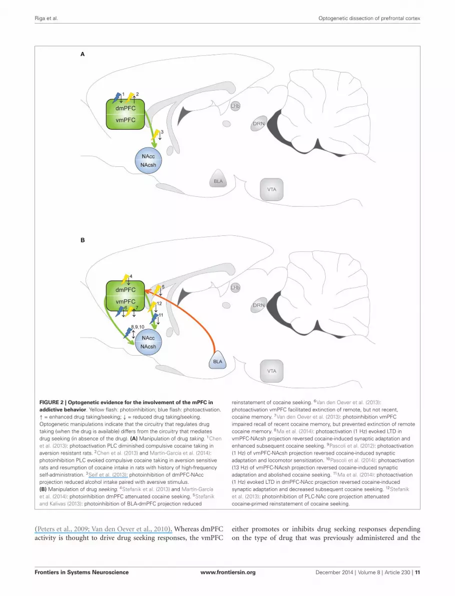
FIGURE 2 | Optogenetic evidence for the involvement of the mPFC inaddictive behavior. Yellow flash: photoinhibition; blue flash: photoactivation.↑ = enhanced drug taking/seeking; ↓ = reduced drug taking/seeking.Optogenetic manipulations indicate that the circuitry that regulates drugtaking (when the drug is available) differs from the circuitry that mediatesdrug seeking (in absence of the drug). (A) Manipulation of drug taking. 1Chenet al. (2013): photoactivation PLC diminished compulsive cocaine taking inaversion resistant rats. 2Chen et al. (2013) and Martín-García et al. (2014):photoinhibition PLC evoked compulsive cocaine taking in aversion sensitiverats and resumption of cocaine intake in rats with history of high-frequencyself-administration. 3Seif et al. (2013): photoinhibition of dmPFC-NAccprojection reduced alcohol intake paired with aversive stimulus.(B) Manipulation of drug seeking. 4Stefanik et al. (2013) and Martín-Garcíaet al. (2014): photoinhibition dmPFC attenuated cocaine seeking. 5Stefanikand Kalivas (2013): photoinhibition of BLA-dmPFC projection reduced
reinstatement of cocaine seeking. 6Van den Oever et al. (2013):photoactivation vmPFC facilitated extinction of remote, but not recent,cocaine memory. 7Van den Oever et al. (2013): photoinhibition vmPFCimpaired recall of recent cocaine memory, but prevented extinction of remotecocaine memory. 8Ma et al. (2014): photoactivation (1 Hz) evoked LTD invmPFC-NAcsh projection reversed cocaine-induced synaptic adaptation andenhanced subsequent cocaine seeking. 9Pascoli et al. (2012): photoactivation(1 Hz) of vmPFC-NAcsh projection reversed cocaine-induced synapticadaptation and locomotor sensitization. 10Pascoli et al. (2014): photoactivation(13 Hz) of vmPFC-NAcsh projection reversed cocaine-induced synapticadaptation and abolished cocaine seeking. 11Ma et al. (2014): photoactivation(1 Hz) evoked LTD in dmPFC-NAcc projection reversed cocaine-inducedsynaptic adaptation and decreased subsequent cocaine seeking. 12Stefaniket al. (2013): photoinhibition of PLC-NAc core projection attenuatedcocaine-primed reinstatement of cocaine seeking.
(Peters et al., 2009; Van den Oever et al., 2010). Whereas dmPFCactivity is thought to drive drug seeking responses, the vmPFC
either promotes or inhibits drug seeking responses dependingon the type of drug that was previously administered and the
Frontiers in Systems Neuroscience www.frontiersin.org December 2014 | Volume 8 | Article 230 | 11

Riga et al. Optogenetic dissection of prefrontal cortex
implementation of extinction sessions before a drug-seeking test(McLaughlin and See, 2003; Peters et al., 2008; Rogers et al.,2008; Koya et al., 2009; Willcocks and McNally, 2013; Lubberset al., 2014). In fact, several lines of evidence suggest that the ILCmediates the consolidation and expression of extinction memory(Peters et al., 2008; LaLumiere et al., 2010), and as such, inhibitionof this region after extinction learning evokes expression of theoriginal cocaine seeking response. Optogenetic manipulation ofthe vmPFC extended these findings by showing that vmPFCpyramidal cells indeed contribute to expression and extinctionof conditioned cocaine seeking, but in a time-dependent manner(Van den Oever et al., 2013; Figure 2B). Channelrhodopsin-2-mediated activation of vmPFC pyramidal cells facilitated extinc-tion of a cocaine conditioned place preference (CPP) memoryonly when photostimulation was applied 3 weeks after, but not1 day after conditioning. In line with this observation, NpHR-mediated inhibition of these neurons blocked extinction of CPPmemory 3 weeks after conditioning. Surprisingly, photoinhibitionselectively attenuated expression of a 1 day-old cocaine memory.Together, optogenetic manipulation of pyramidal cells pointed toa temporal reorganization of the circuitry that controls expres-sion of cocaine-associated memories and a differential role ofthe vmPFC in regulation of conditioned cocaine seeking overtime.
Optogenetic studies confirmed that PLC activity is required forreinstatement of cocaine seeking in extinguished animals. Similarto pharmacological inactivation, photoinhibition of PLC neurons(using a non-selective promoter) reduced cocaine-primed rein-statement of cocaine seeking (Stefanik et al., 2013). In addition,the same group demonstrated that the BLA-PLC pathway iscritically involved in reinstatement of cocaine seeking by opticalinhibition of BLA presynaptic terminals in the PLC (Stefanikand Kalivas, 2013). Optogenetic inhibition of dmPFC pyramidalneurons also attenuated stress-induced reinstatement of palatablefood seeking in rats (Calu et al., 2013), suggesting that differentmodalities activate dmPFC circuitry to evoke reinstatement ofreward seeking. In addition, this shows that PLC activity drivesreinstatement of cocaine and natural reward seeking, whereasincreased activity of the same neurons suppresses compulsivecocaine taking (Chen et al., 2013). The opposing function the PLCmay depend on the presence or absence of cocaine in operanttests. This is supported by the observation that photoinhibitionof PLC pyramidal cells enhanced cocaine self-administration andattenuated reinstatement of cocaine seeking in rats that were sub-jected to a high-frequency cocaine intake schedule (Martín-Garcíaet al., 2014). GABAergic interneurons have not been manipulatedyet in addiction models, but the role of PV interneurons innatural reward (sucrose) learning and extinction was recentlyexamined. Channelrhodopsin-2-mediated activation of PLC PVinterneurons did not affect acquisition of sucrose reward self-administration, but accelerated extinction of reward seeking byinhibiting PL network activity (Sparta et al., 2014). Whether PLCPV activity also affects extinction of drug seeking remains a topicfor future research.
By integrating input from sources such as the BLA, VTA andHPC and conveying excitatory output to the NAc, the mPFCis thought to exert control over the motor circuitry to regulate
responding to drugs and drug-associated stimuli (Kalivas et al.,2005). Dorsal regions of the mPFC mainly project to the dorso-lateral striatum and NAc core, whereas ventral regions predom-inantly target the dorsomedial striatum and NAc shell (Voornet al., 2004). Pharmacological disconnection experiments haveindeed implicated the dmPFC-NAc core and vmPFC-NAc shellpathway in drug- and cue-induced cocaine and heroin seeking(McFarland et al., 2003; LaLumiere and Kalivas, 2008; Peterset al., 2008; Bossert et al., 2012), but with this method the effectson indirect pathways cannot be ruled out. Photoinhibiton ofPLC presynaptic terminals in the NAc core attenuated cocaine-primed reinstatement of cocaine seeking (Stefanik et al., 2013),confirming that a monosynaptic glutamatergic projection fromPLC to NAc core has a critically role in this behavioral response.Optogenetic evidence for the involvement of the mPFC-NAc shellpathway was provided by a optic modulation of ILC terminalsin NAc brain slices obtained from animals that were exposedto cocaine (Suska et al., 2013). This revealed that presynapticinput of mPFC terminals in the NAc shell was strengthenedafter both short- (1 day) and long-term (45 days) abstinencefrom non-contingent and contingent exposure to cocaine, butonly after contingent exposure this strengthening significantlyincreased over time. The presynaptic enhancement was caused byan increase in glutamate release probability, rather than increasedquantal size of glutamatergic release or the number of activerelease sites (Suska et al., 2013). Interestingly, cocaine exposuredid not affect presynaptic transmission in the BLA-NAc shellprojection (Suska et al., 2013), suggesting that input from themPFC is favored over BLA input after cocaine administration. Inan elegant study by Ma et al. (2014) it was shown that cocaineself-administration induced silent synapses in the mPFC-NAcpathway. Interestingly, silent synapses in the ILC-NAc shell path-way matured by recruiting GluA2-lacking AMPA-Rs (observedat day 45 of abstinence), whereas silent synapses in the PLC-NAc core pathway recruited GluA2-containing AMPA-Rs. α-amino-3-hydroxy-5-methyl-4-isoxazolepropionic acid receptorslacking the GluA2 subunit are calcium permeable, have greaterchannel conductance, exhibit faster channel deactivation kineticsand thereby contribute to rapid synaptic signaling, homeostaticsynaptic scaling and specialized forms of short- and long-termplasticity (for excellent review see Isaac et al., 2007). Optogeneti-cally evoked long-term depression (1 Hz for 10 min) reintroducedsilent synapses in both pathways, but this either enhanced (ILC-NAc shell) or reduced (PLC-NAc core) subsequent cocaine seek-ing (Ma et al., 2014), further supporting differential roles of thedmPFC and vmPFC in this behavior.
The principal cell population in the NAc consists of GABAer-gic medium spiny neurons (MSNs) that can be subdivided ina D1-R and D2-R expressing population, together comprising∼90–95% of all NAc neurons (Lobo et al., 2006). Selectiveexpression of ChR2 in each NAc MSN population showed thatactivation of D1-R neurons enhanced cocaine reward learningin the CPP paradigm, whereas activation of D2-R neurons hadthe opposite effect (Lobo et al., 2010). Photostimulation ofmPFC terminals in the NAc core specifically induced ∆FosBexpression in D1-R neurons, whereas in the NAc shell, ∆FosBexpression was induced in both D1-R and D2-R subtypes (Lobo
Frontiers in Systems Neuroscience www.frontiersin.org December 2014 | Volume 8 | Article 230 | 12

Riga et al. Optogenetic dissection of prefrontal cortex
et al., 2013). This suggests that the distribution of mPFC ter-minals onto NAc neurons differs for the shell and core (Loboet al., 2013). However, this will require validation by whole-cell recordings. The functional relevance of mPFC to NAc D1-RMSNs projections was demonstrated by Pascoli et al. (2012)who showed that low frequency (1 Hz) photostimulation ofthe ILC-NAc shell pathway reversed non-contingent cocaine-induced synaptic potentiation in D1-R neurons and locomotorsensitization. More recently, the same group used optogenet-ics to reveal the presence of GluA2-lacking AMPA-Rs in theILC-NAc D1-R MSN projection 1 month after cocaine self-administration (Pascoli et al., 2014). Photostimulation of thispathway at 13 Hz, but not 1 Hz, reversed synaptic adapta-tions after cocaine self-administration and abolished cue-inducedcocaine seeking. The authors speculated that a 13-Hz stimulationwas required for this effect because this evokes mGluR-mediatedlong-term depression, an efficient mechanism to remove synapticGluA2-lacking AMPA-Rs (Lüscher and Huber, 2010). However,this finding contradicts with the observation by Ma et al. (2014);(discussed above). Differences in circuit specificity (optogeneticmodulation of projections to D1-R neurons vs. projections to allNAc shell MSN neurons) and in the cocaine self-administrationregimen may explain the opposing effects observed in thesestudies.
In addition to being involved in relapse to drug seeking, themPFC-NAc pathway has been implicated in compulsive aversion-resistant alcohol consumption. Photoinhibition of the dmPFC-NAc core projection reduced alcohol intake paired with aversivestimuli of different sensory modalities and different methods ofintake (Seif et al., 2013). Alcohol intake was unaffected by pho-toinhibition when it was not paired with an adverse consequence,suggesting that this pathway predominates in orchestrating theaversion-resistant, compulsive aspects of alcoholism, in whichintake is often accompanied by conflict or challenge (Tiffanyand Conklin, 2000). However, these results contradict with thefinding that photoinhibition of the PLC enhances aversion-resistant cocaine intake (Chen et al., 2013), suggesting that thePLC might differentially regulate compulsive alcohol and cocaineintake.
The involvement of the mPFC-NAc pathway in acquisitionof reward and drug self-administration has also been exploredwith optogenetic approaches. Stuber et al. (2011) found thatoptical activation of the mPFC-NAc shell projection (20 Hz) didnot support the acquisition of operant self-stimulation behavior(active responses triggered light pulses delivered to presynap-tic mPFC terminals in the NAc), despite the fact that opticalactivation of the mPFC projection elicited EPSCs in the NAc.A subsequent study demonstrated that animals acquire opti-cal self-stimulation of the mPFC-NAc shell pathway when thefrequency of stimulation is increased to 30 Hz (Britt et al.,2012). Hence, the glutamatergic projection from the mPFC toNAc may only evoke spiking of MSNs and reinforce behaviorwith strong activation of the mPFC or when DA levels in theNAc are elevated in parallel. The exact stimulation site withinthe mPFC may be of critical importance to achieve this effect,considering that the ILC is thought to have a stronger projectionto the NAc shell than the PLC (Voorn et al., 2004). As in the
above mentioned studies ChR2 expression was not specificallytargeted to the PLC or ILC, it remains to be determined whethera difference exists in the potency of both pathways to evokespiking in NAc shell MSNs and to reinforce reward seekingbehavior.
In line with traditional intervention techniques, optogeneticmanipulations of the mPFC circuitry in rodent addiction modelshave validated the critical involvement of this region in regulatingdrug taking and drug seeking behavior and further support afunctional segregation along the dorsal-ventral axis of the mPFC.Moreover, pathway specific modulation has provided new insightsin the role of BLA-PLC and mPFC-NAc projections. In particular,optic stimulation of PLC and ILC axonal terminals in acutebrain slices preparations of the NAc core and shell demonstratedcocaine-induced pathway-specific neuroadaptations that could bereversed using defined photoactivation frequencies (Pascoli et al.,2012, 2014; Ma et al., 2014). This may provide opportunitiesfor DBS-mediated reversal of drug-induced neuroadaptationsin addicts. However, as electrical stimulation affects neuronalactivity in a non-selective manner, translational efficacy to DBSremains to be approached with caution and requires furtherstudies.
CONCLUDING REMARKSThe relatively recent application of optogenetic technology toneuroscience research has deepened insight into function ofvarious types of circuitry in the brain, and already contributedsubstantially to our understanding of the mPFC circuitry in healthand disease conditions. Optogenetic manipulations enable causalsystem-level research on diverse cognitive and neuropathologicalbehaviors in freely moving animals and allow integration ofin vivo and ex vivo electrophysiological recordings, which wasnot feasible with traditional intervention methods. However, overdecades, the extensive body of research involving lesion, pharma-cological and electrophysiological methods has provided crucialknowledge on the involvement of the mPFC in diverse cognitiveprocesses. Integration of data obtained with these traditionalintervention methods and optogenetic modulations will continueto be invaluable for our understanding of mPFC circuitry and forcreating computational models of mPFC function.
A major breakthrough in dissection of neuronal circuitriesthat has been enabled by optogenetics technology is the directmanipulation of neuronal projections within and between brainregions. With respect to the mPFC circuitry, this has led to a betterunderstanding of intra-mPFC connectivity, the role of afferentand efferent mPFC projections in cognitive processes and mentaldisorders, and even to the discovery of a new GABAergic cellpopulation with long-range projections to the NAc (Lee et al.,2014c). Moreover, due to the excellent compatibility of opto-genetics and ex vivo brain slice physiology, differential cocaine-induced neuroadaptations in PLC and ILC projections to theNAc have been elucidated (Ma et al., 2014), demonstrating thefeasibility of dissection of mPFC subregion-specific mechanismsusing optogenetics.
Although great progress has been made, several factors havereceived little attention and in some cases require technicalimprovements to be properly addressed in future experiments.
Frontiers in Systems Neuroscience www.frontiersin.org December 2014 | Volume 8 | Article 230 | 13

Riga et al. Optogenetic dissection of prefrontal cortex
With respect to the GABAergic interneuron population in themPFC, opsin expression has thus far been primarily targetedto PV interneurons, leaving the role of many other GABAergiccell-types (e.g., SOM+, calretinin+ cells, etc.) unaddressed. Astransgenic mouse and rat Cre-driver lines become increasinglyavailable, this opens new avenues to investigate the role of othermPFC subpopulations in cognitive performance and psychiatricdisorders. Importantly, previous optogenetic studies have pointedto the existence of subpopulations within the GABAergic andpyramidal cell population that may only be distinguished basedon their differential activity during defined behavioral states(Little and Carter, 2013; Courtin et al., 2014). For instance, PVinterneurons have been linked to working memory performance(Rossi et al., 2012), expression of fear responses (Courtin et al.,2014), maintaining a proper E/I balance (Yizhar et al., 2011b;Kvitsiani et al., 2013), and synchronization of gamma oscillations(Sohal et al., 2009; Sohal, 2012). Optogenetic tagging of neuronsthat show increased activity during a particular behavioral taskwill be a crucial next step to dissect the causal involvement ofthese specific neuronal ensembles in expression of behavioralperformance (Cruz et al., 2013). Opsin expression driven by thepromoter of the immediate early gene c-fos, a widely used markerof neuronal activity, in hippocampal neurons that were activeduring fear-conditioning demonstrates that this is an attainablegoal (Liu et al., 2012). Interpretation of optogenetic data is oftenhindered by non-specific targeting of opsins to mPFC subregions.As it becomes increasingly clear that dorsal and ventral regions ofthe mPFC have different and sometimes even opposing functions(Heidbreder and Groenewegen, 2003; Van den Oever et al., 2010),stereotactic delivery of opsin vectors to these defined subregionsis of high relevance. Furthermore, technical advances that enabletargeting of opsins to specific layers within the mPFC would beof great value given the complex layer- and subregion-definedneuronal connectivity of mPFC neurons (Groenewegen et al.,1997; Voorn et al., 2004; Hoover and Vertes, 2007).
Currently, many FDA approved pharmaceutical agents targetG-protein coupled receptors in the brain (Lee et al., 2014b).Thus, improving insight in the temporal role of these receptorsto specific behavioral states will be instrumental for treatmentof psychiatric disorders with novel, more-selective pharma-cotherapy. Design of opsins that consist of a chimera of anopsin fused to the intracellular domain of a G-protein cou-pled receptor (optoXR), enables interrogation of the causalinvolvement of G-protein coupled signaling cascades withhigh spatiotemporal resolution (Airan et al., 2009). Thus far,optoXRs have not been used to study the contribution ofspecific signaling cascades to mPFC circuitry function, butwould be extremely useful for explaining the role of alteredG-protein signaling observed in psychiatric diseases (Hearinget al., 2012; Luján et al., 2014). In addition, new develop-ments in the field of chemogenetic technology (e.g., DREADD:Designer Receptors Exclusively Activated by Designer Drugs)will further contribute to the dissection of mPFC circuitryand the identification of drugable targets (Sternson and Roth,2014).
The use of optogenetics in humans for treatment of neu-rological disorders has been extensively discussed (Peled, 2011;
Kumar et al., 2013; Touriño et al., 2013), however, clinical appli-cation of optogenetics technology is, to our knowledge, currentlynot feasible. Extending optogenetic methods to species beyondrodents has only been stably, safely and efficiently applied in therhesus macaque, a non-human primate (Han et al., 2009; Diesteret al., 2011; Han et al., 2011; Cavanaugh et al., 2012; Gerits et al.,2012; Jazayeri et al., 2012). Further studies and clinical trialswill be required to safely express and photostimulate opsins inthe human brain. Hence, in spite of high promise for clinicaltreatment, at present, optogenetics should primarily be regardedas a powerful toolbox to functionally dissect neural circuits inanimal models of disease-related symptoms and to discover andrefine targets for pharmaceutical and DBS treatment.
ACKNOWLEDGMENTSMariana R. Matos is funded by EU MSCA-ITN CognitionNet(FP7-PEOPLE-2013-ITN 607508). August B. Smit, Sabine Spijkerare funded partly by a grant awarded to NeuroBasic Pharmaphe-nomics consortium. Danai Riga is partly funded by a NCA proof-of-concept fund (Sabine Spijker). Michel C. Van den Oever isfunded by a ZonMw VENI grant (916.12.034) and Hersensticht-ing Nederland grant (KS 2012(1)-162).
SUPPLEMENTARY MATERIALThe Supplementary Material for this article can be foundonline at: http://www.frontiersin.org/journal/10.3389/fnsys.2014.00230/abstract
REFERENCESAiran, R. D., Thompson, K. R., Fenno, L. E., Bernstein, H., and Deisseroth, K.
(2009). Temporally precise in vivo control of intracellular signalling. Nature 458,1025–1029. doi: 10.1038/nature07926
Albert, P. R. (2014). Light up your life: optogenetics for depression? J. PsychiatryNeurosci. 39, 3–5. doi: 10.1503/jpn.130267
Albert, P. R., Vahid-Ansari, F., and Luckhart, C. (2014). Serotonin-prefrontalcortical circuitry in anxiety and depression phenotypes: pivotal role of pre-and post-synaptic 5-HT1A receptor expression. Front. Behav. Neurosci. 8:199.doi: 10.3389/fnbeh.2014.00199
Alitto, H. J., and Usrey, W. M. (2003). Corticothalamic feedback and sensoryprocessing. Curr. Opin. Neurobiol. 13, 440–445. doi: 10.1016/s0959-4388(03)00096-5
Allsop, S. A., Vander Weele, C. M., Wichmann, R., and Tye, K. M. (2014).Optogenetic insights on the relationship between anxiety-related behaviors andsocial deficits. Front. Behav. Neurosci. 8:241. doi: 10.3389/fnbeh.2014.00241
Amat, J., Baratta, M. V., Paul, E., Bland, S. T., Watkins, L. R., and Maier, S. F. (2005).Medial prefrontal cortex determines how stressor controllability affects behaviorand dorsal raphe nucleus. Nat. Neurosci. 8, 365–371. doi: 10.1038/nn1399
American Psychiatric Association. (2013). Diagnostic and Statistical Manual ofMental Disorders. Arlington, VA: American Psychiatric Publishing.
Arnsten, A. F. (2009). Stress signalling pathways that impair prefrontal cortexstructure and function. Nat. Rev. Neurosci. 10, 410–422. doi: 10.1038/nrn2648
Arnsten, A. F., Wang, M. J., and Paspalas, C. D. (2012). Neuromodulation ofthought: flexibilities and vulnerabilities in prefrontal cortical network synapses.Neuron 76, 223–239. doi: 10.1016/j.neuron.2012.08.038
Ascoli, G. A., Alonso-Nanclares, L., Anderson, S. A., Barrionuevo, G., Benavides-Piccione, R., Burkhalter, A., et al. (2008). Petilla terminology: nomenclature offeatures of GABAergic interneurons of the cerebral cortex. Nat. Rev. Neurosci. 9,557–568. doi: 10.1038/nrn2402
Baddeley, A. (1992). Working memory. Science 255, 556–559. doi: 10.1126/science.1736359
Beasley, C. L., and Reynolds, G. P. (1997). Parvalbumin-immunoreactive neuronsare reduced in the prefrontal cortex of schizophrenics. Schizophr. Res. 24, 349–355. doi: 10.1016/s0920-9964(96)00122-3
Frontiers in Systems Neuroscience www.frontiersin.org December 2014 | Volume 8 | Article 230 | 14

Riga et al. Optogenetic dissection of prefrontal cortex
Beaulieu, J. M., and Gainetdinov, R. R. (2011). The physiology, signaling andpharmacology of dopamine receptors. Pharmacol. Rev. 63, 182–217. doi: 10.1124/pr.110.002642
Beevers, C. G., Clasen, P., Stice, E., and Schnyer, D. (2010). Depression symptomsand cognitive control of emotion cues: a functional magnetic resonance imagingstudy. Neuroscience 167, 97–103. doi: 10.1016/j.neuroscience.2010.01.047
Belzung, C., Turiault, M., and Griebel, G. (2014). Optogenetics to study the circuitsof fear- and depression-like behaviors: a critical analysis. Pharmacol. Biochem.Behav. 122, 144–157. doi: 10.1016/j.pbb.2014.04.002
Berridge, C. W. (2008). Noradrenergic modulation of arousal. Brain Res. Rev. 58,1–17. doi: 10.1016/j.brainresrev.2007.10.013
Bossert, J. M., Stern, A. L., Theberge, F. R., Marchant, N. J., Wang, H. L., Morales,M., et al. (2012). Role of projections from ventral medial prefrontal cortex tonucleus accumbens shell in context-induced reinstatement of heroin seeking. J.Neurosci. 32, 4982–4991. doi: 10.1523/JNEUROSCI.0005-12.2012
Boyden, E. S., Zhang, F., Bamberg, E., Nagel, G., and Deisseroth, K. (2005).Millisecond-timescale, genetically targeted optical control of neural activity.Nat. Neurosci. 8, 1263–1268. doi: 10.1038/nn1525
Brisch, R., Saniotis, A., Wolf, R., Bielau, H., Bernstein, H. G., Steiner, J., et al. (2014).The role of dopamine in schizophrenia from a neurobiological and evolutionaryperspective: old fashioned, but still in vogue. Front. Psychiatry 5:47. doi: 10.3389/fpsyt.2014.00047
Britt, J. P., Benaliouad, F., McDevitt, R. A., Stuber, G. D., Wise, R. A., and Bonci, A.(2012). Synaptic and behavioral profile of multiple glutamatergic inputs to thenucleus accumbens. Neuron 76, 790–803. doi: 10.1016/j.neuron.2012.09.040
Calu, D. J., Kawa, A. B., Marchant, N. J., Navarre, B. M., Henderson, M. J., Chen,B., et al. (2013). Optogenetic inhibition of dorsal medial prefrontal cortexattenuates stress-induced reinstatement of palatable food seeking in female rats.J. Neurosci. 33, 214–226. doi: 10.1523/JNEUROSCI.2016-12.2013
Cardin, J. A., Carlén, M., Meletis, K., Knoblich, U., Zhang, F., Deisseroth, K., et al.(2009). Driving fast-spiking cells induces gamma rhythm and controls sensoryresponses. Nature 459, 663–667. doi: 10.1038/nature08002
Carlén, M., Meletis, K., Siegle, J. H., Cardin, J. A., Futai, K., Vierling-Claassen, D.,et al. (2012). A critical role for NMDA receptors in parvalbumin interneuronsfor gamma rhythm induction and behavior. Mol. Psychiatry 17, 537–548. doi: 10.1038/mp.2011.31
Carter, M. E., Yizhar, O., Chikahisa, S., Nguyen, H., Adamantidis, A., Nishino, S.,et al. (2010). Tuning arousal with optogenetic modulation of locus coeruleusneurons. Nat. Neurosci. 13, 1526–1533. doi: 10.1038/nn.2682
Cavanaugh, J., Monosov, I. E., McAlonan, K., Berman, R., Smith, M. K., Cao, V.,et al. (2012). Optogenetic inactivation modifies monkey visuomotor behavior.Neuron 76, 901–907. doi: 10.1016/j.neuron.2012.10.016
Challis, C., Beck, S. G., and Berton, O. (2014). Optogenetic modulation ofdescending prefrontocortical inputs to the dorsal raphe bidirectionally biassocioaffective choices after social defeat. Front. Behav. Neurosci. 8:43. doi: 10.3389/fnbeh.2014.00043
Chang, C. H., Chen, M. C., Qiu, M. H., and Lu, J. (2014). Ventromedial prefrontalcortex regulates depressive-like behavior and rapid eye movement sleep in therat. Neuropharmacology 86C, 125–132. doi: 10.1016/j.neuropharm.2014.07.005
Chaudhury, D., Walsh, J. J., Friedman, A. K., Juarez, B., Ku, S. M., Koo, J. W.,et al. (2013). Rapid regulation of depression-related behaviours by control ofmidbrain dopamine neurons. Nature 493, 532–536. doi: 10.1038/nature11713
Chen, B. T., Yau, H. J., Hatch, C., Kusumoto-Yoshida, I., Cho, S. L., Hopf, F. W.,et al. (2013). Rescuing cocaine-induced prefrontal cortex hypoactivity preventscompulsive cocaine seeking. Nature 496, 359–362. doi: 10.1038/nature12024
Cho, J. H., Deisseroth, K., and Bolshakov, V. Y. (2013). Synaptic encoding of fearextinction in mPFC-amygdala circuits. Neuron 80, 1491–1507. doi: 10.1016/j.neuron.2013.09.025
Cho, K. K., and Sohal, V. S. (2014). Optogenetic approaches for investigating neuralpathways implicated in schizophrenia and related disorders. Hum. Mol. Genet.23, R64–R68. doi: 10.1093/hmg/ddu225
Christophe, E., Roebuck, A., Staiger, J. F., Lavery, D. J., Charpak, S., and Audinat, E.(2002). Two types of nicotinic receptors mediate an excitation of neocorticallayer I interneurons. J. Neurophysiol. 88, 1318–1327. doi: 10.1152/jn.00199.2002
Chudasama, Y., Dalley, J. W., Nathwani, F., Bouger, P., and Robbins, T. W. (2004).Cholinergic modulation of visual attention and working memory: dissociableeffects of basal forebrain 192-IgG-saporin lesions and intraprefrontal infusionsof scopolamine. Learn. Mem. 11, 78–86. doi: 10.1101/lm.70904
Clark, L., Chamberlain, S. R., and Sahakian, B. J. (2009). Neurocognitive mecha-nisms in depression: implications for treatment. Annu. Rev. Neurosci. 32, 57–74.doi: 10.1146/annurev.neuro.31.060407.125618
Courtin, J., Bienvenu, T. C., Einarsson, E. O., and Herry, C. (2013). Medialprefrontal cortex neuronal circuits in fear behavior. Neuroscience 240, 219–242.doi: 10.1016/j.neuroscience.2013.03.001
Courtin, J., Chaudun, F., Rozeske, R. R., Karalis, N., Gonzalez-Campo, C., Wurtz,H., et al. (2014). Prefrontal parvalbumin interneurons shape neuronal activityto drive fear expression. Nature 505, 92–96. doi: 10.1038/nature12755
Coutureau, E., and Killcross, S. (2003). Inactivation of the infralimbic prefrontalcortex reinstates goal-directed responding in overtrained rats. Behav. Brain Res.146, 167–174. doi: 10.1016/j.bbr.2003.09.025
Covington, H. E. 3rd, Lobo, M. K., Maze, I., Vialou, V., Hyman, J. M., Zaman,S., et al. (2010). Antidepressant effect of optogenetic stimulation of the medialprefrontal cortex. J. Neurosci. 30, 16082–16090. doi: 10.1523/JNEUROSCI.1731-10.2010
Cruikshank, S. J., Ahmed, O. J., Stevens, T. R., Patrick, S. L., Gonzalez, A. N.,Elmaleh, M., et al. (2012). Thalamic control of layer 1 circuits in prefrontalcortex. J. Neurosci. 32, 17813–17823. doi: 10.1523/JNEUROSCI.3231-12.2012
Cruwys, T., Haslam, S. A., Dingle, G. A., Haslam, C., and Jetten, J. (2014).Depression and social identity: an integrative review. Pers. Soc. Psychol. Rev. 18,215–238. doi: 10.1177/1088868314523839. [Epub ahead of print].
Cruz, F. C., Koya, E., Guez-Barber, D. H., Bossert, J. M., Lupica, C. R., Shaham, Y.,et al. (2013). New technologies for examining the role of neuronal ensembles indrug addiction and fear. Nat. Rev. Neurosci. 14, 743–754. doi: 10.1038/nrn3597
Czéh, B., Müller-Keuker, J. I., Rygula, R., Abumaria, N., Hiemke, C., Domenici, E.,et al. (2007). Chronic social stress inhibits cell proliferation in the adult medialprefrontal cortex: hemispheric asymmetry and reversal by fluoxetine treatment.Neuropsychopharmacology 32, 1490–1503. doi: 10.1038/sj.npp.1301275
DeFelipe, J., López-Cruz, P. L., Benavides-Piccione, R., Bielza, C., Larrañaga, P.,Anderson, S., et al. (2013). New insights into the classification and nomenclatureof cortical GABAergic interneurons. Nat. Rev. Neurosci. 14, 202–216. doi: 10.1038/nrn3444
Deisseroth, K. (2010). Controlling the brain with light. Sci. Am. 303, 48–55. doi: 10.1038/scientificamerican1110-48
Dembrow, N., and Johnston, D. (2014). Subcircuit-specific neuromodulation in theprefrontal cortex. Front. Neural Circuits 8:54. doi: 10.3389/fncir.2014.00054
Derntl, B., Seidel, E. M., Eickhoff, S. B., Kellermann, T., Gur, R. C., Schneider, F.,et al. (2011). Neural correlates of social approach and withdrawal in patientswith major depression. Soc. Neurosci. 6, 482–501. doi: 10.1080/17470919.2011.579800
Dias-Ferreira, E., Sousa, J. C., Melo, I., Morgado, P., Mesquita, A. R., Cerqueira,J. J., et al. (2009). Chronic stress causes frontostriatal reorganization and affectsdecision-making. Science 325, 621–625. doi: 10.1126/science.1171203
Diester, I., Kaufman, M. T., Mogri, M., Pashaie, R., Goo, W., Yizhar, O., et al.(2011). An optogenetic toolbox designed for primates. Nat. Neurosci. 14, 387–397. doi: 10.1038/nn.2749
Disner, S. G., Beevers, C. G., Haigh, E. A., and Beck, A. T. (2011). Neuralmechanisms of the cognitive model of depression. Nat. Rev. Neurosci. 12, 467–477. doi: 10.1038/nrn3027
Douglas, R. J., and Martin, K. A. (2004). Neuronal circuits of the neocortex. Annu.Rev. Neurosci. 27, 419–451. doi: 10.1146/annurev.neuro.27.070203.144152
Drew, M. R., Fairhurst, S., Malapani, C., Horvitz, J. C., and Balsam, P. D. (2003).Effects of dopamine antagonists on the timing of two intervals. Pharmacol.Biochem. Behav. 75, 9–15. doi: 10.1016/s0091-3057(03)00036-4
Dunnett, S. B., Nathwani, F., and Brasted, P. J. (1999). Medial prefrontal andneostriatal lesions disrupt performance in an operant delayed alternation taskin rats. Behav. Brain Res. 106, 13–28. doi: 10.1016/s0166-4328(99)00076-5
Durstewitz, D., and Seamans, J. K. (2008). The dual-state theory of prefrontal cortexdopamine function with relevance to catechol-o-methyltransferase genotypesand schizophrenia. Biol. Psychiatry 64, 739–749. doi: 10.1016/j.biopsych.2008.05.015
Euston, D. R., Gruber, A. J., and McNaughton, B. L. (2012). The role of medialprefrontal cortex in memory and decision making. Neuron 76, 1057–1070.doi: 10.1016/j.neuron.2012.12.002
Fales, C. L., Barch, D. M., Rundle, M. M., Mintun, M. A., Snyder, A. Z., Cohen,J. D., et al. (2008). Altered emotional interference processing in affective andcognitive-control brain circuitry in major depression. Biol. Psychiatry 63, 377–384. doi: 10.1016/j.biopsych.2007.06.012
Frontiers in Systems Neuroscience www.frontiersin.org December 2014 | Volume 8 | Article 230 | 15

Riga et al. Optogenetic dissection of prefrontal cortex
Friedman, A. K., Walsh, J. J., Juarez, B., Ku, S. M., Chaudhury, D., Wang, J.,et al. (2014). Enhancing depression mechanisms in midbrain dopamine neuronsachieves homeostatic resilience. Science 344, 313–319. doi: 10.1126/science.1249240
Gabbott, P. L., Warner, T. A., Jays, P. R., Salway, P., and Busby, S. J. (2005).Prefrontal cortex in the rat: projections to subcortical autonomic, motor andlimbic centers. J. Comp. Neurol. 492, 145–177. doi: 10.1002/cne.20738
Gee, S., Ellwood, I., Patel, T., Luongo, F., Deisseroth, K., and Sohal, V. S. (2012).Synaptic activity unmasks dopamine D2 receptor modulation of a specific classof layer V pyramidal neurons in prefrontal cortex. J. Neurosci. 32, 4959–4971.doi: 10.1523/JNEUROSCI.5835-11.2012
Gerits, A., Farivar, R., Rosen, B. R., Wald, L. L., Boyden, E. S., and Vanduffel, W.(2012). Optogenetically induced behavioral and functional network changes inprimates. Curr. Biol. 22, 1722–1726. doi: 10.1016/j.cub.2012.07.023
Gilmartin, M. R., and Helmstetter, F. J. (2010). Trace and contextual fear condition-ing require neural activity and NMDA receptor-dependent transmission in themedial prefrontal cortex. Learn. Mem. 17, 289–296. doi: 10.1101/lm.1597410
Gilmartin, M. R., and McEchron, M. D. (2005). Single neurons in the medialprefrontal cortex of the rat exhibit tonic and phasic coding during trace fearconditioning. Behav. Neurosci. 119, 1496–1510. doi: 10.1037/0735-7044.119.6.1496
Gilmartin, M. R., Miyawaki, H., Helmstetter, F. J., and Diba, K. (2013). Prefrontalactivity links nonoverlapping events in memory. J. Neurosci. 33, 10910–10914.doi: 10.1523/JNEUROSCI.0144-13.2013
Goldstein, R. Z., and Volkow, N. D. (2011). Dysfunction of the prefrontal cortex inaddiction: neuroimaging findings and clinical implications. Nat. Rev. Neurosci.12, 652–669. doi: 10.1038/nrn3119
Gotlib, I. H., Joormann, J., Minor, K. L., and Hallmayer, J. (2008). HPA axisreactivity: a mechanism underlying the associations among 5-HTTLPR, stressand depression. Biol. Psychiatry 63, 847–851. doi: 10.1016/j.biopsych.2007.10.008
Gradinaru, V., Thompson, K. R., Zhang, F., Mogri, M., Kay, K., Schneider, M. B.,et al. (2007). Targeting and readout strategies for fast optical neural control invitro and in vivo. J. Neurosci. 27, 14231–14238. doi: 10.1523/jneurosci.3578-07.2007
Groenewegen, H. J., Wright, C. I., and Uylings, H. B. (1997). The anatomicalrelationships of the prefrontal cortex with limbic structures and the basalganglia. J. Psychopharmacol. 11, 99–106. doi: 10.1177/026988119701100202
Gunaydin, L. A., Grosenick, L., Finkelstein, J. C., Kauvar, I. V., Fenno, L. E.,Adhikari, A., et al. (2014). Natural neural projection dynamics underlying socialbehavior. Cell 157, 1535–1551. doi: 10.1016/j.cell.2014.05.017
Hamani, C., Diwan, M., Isabella, S., Lozano, A. M., and Nobrega, J. N. (2010a).Effects of different stimulation parameters on the antidepressant-like responseof medial prefrontal cortex deep brain stimulation in rats. J. Psychiatr. Res. 44,683–687. doi: 10.1016/j.jpsychires.2009.12.010
Hamani, C., Diwan, M., Macedo, C. E., Brandão, M. L., Shumake, J., Gonzalez-Lima, F., et al. (2010b). Antidepressant-like effects of medial prefrontal cortexdeep brain stimulation in rats. Biol. Psychiatry 67, 117–124. doi: 10.1016/j.biopsych.2009.08.025
Hamani, C., Machado, D. C., Hipólide, D. C., Dubiela, F. P., Suchecki, D., Macedo,C. E., et al. (2012). Deep brain stimulation reverses anhedonic-like behav-ior in a chronic model of depression: role of serotonin and brain derivedneurotrophic factor. Biol. Psychiatry 71, 30–35. doi: 10.1016/j.biopsych.2011.08.025
Han, X., Chow, B. Y., Zhou, H., Klapoetke, N. C., Chuong, A., Rajimehr, R.,et al. (2011). A high-light sensitivity optical neural silencer: development andapplication to optogenetic control of non-human primate cortex. Front. Syst.Neurosci. 5:18. doi: 10.3389/fnsys.2011.00018
Han, X., Qian, X., Bernstein, J. G., Zhou, H. H., Franzesi, G. T., Stern, P.,et al. (2009). Millisecond-timescale optical control of neural dynamics in thenonhuman primate brain. Neuron 62, 191–198. doi: 10.1016/j.neuron.2009.03.011
Hearing, M. C., Zink, A. N., and Wickman, K. (2012). Cocaine-induced adaptationsin metabotropic inhibitory signaling in the mesocorticolimbic system. Rev.Neurosci. 23, 325–351. doi: 10.1515/revneuro-2012-0045
Heidbreder, C. A., and Groenewegen, H. J. (2003). The medial prefrontal cortexin the rat: evidence for a dorso-ventral distinction based upon functional andanatomical characteristics. Neurosci. Biobehav. Rev. 27, 555–579. doi: 10.1016/j.neubiorev.2003.09.003
Hogarth, L., Balleine, B. W., Corbit, L. H., and Killcross, S. (2013). Associativelearning mechanisms underpinning the transition from recreational drug useto addiction. Ann. N Y Acad. Sci. 1282, 12–24. doi: 10.1111/j.1749-6632.2012.06768.x
Hoover, W. B., and Vertes, R. P. (2007). Anatomical analysis of afferent projectionsto the medial prefrontal cortex in the rat. Brain Struct. Funct. 212, 149–179.doi: 10.1007/s00429-007-0150-4
Hübner, C., Bosch, D., Gall, A., Lüthi, A., and Ehrlich, I. (2014). Ex vivo dissectionof optogenetically activated mPFC and hippocampal inputs to neurons inthe basolateral amygdala: implications for fear and emotional memory. Front.Behav. Neurosci. 8:64. doi: 10.3389/fnbeh.2014.00064
Humeau, Y., Herry, C., Kemp, N., Shaban, H., Fourcaudot, E., Bissiere, S., et al.(2005). Dendritic spine heterogeneity determines afferent-specific Hebbianplasticity in the amygdala. Neuron 45, 119–131. doi: 10.1016/j.neuron.2004.12.019
Hyman, S. E. (2005). Addiction: a disease of learning and memory. Am. J. Psychiatry162, 1414–1422. doi: 10.1176/appi.ajp.162.8.1414
Hyman, S. E., Malenka, R. C., and Nestler, E. J. (2006). Neural mechanisms ofaddiction: the role of reward-related learning and memory. Annu. Rev. Neurosci.29, 565–598. doi: 10.1146/annurev.neuro.29.051605.113009
Isaac, J. T., Ashby, M., and McBain, C. J. (2007). The role of the GluR2 subunit inAMPA receptor function and synaptic plasticity. Neuron 54, 859–871. doi: 10.1016/j.neuron.2007.06.001
Jazayeri, M., Lindbloom-Brown, Z., and Horwitz, G. D. (2012). Saccadic eyemovements evoked by optogenetic activation of primate V1. Nat. Neurosci. 15,1368–1370. doi: 10.1038/nn.3210
Ji, G., and Neugebauer, V. (2012). Modulation of medial prefrontal cortical activityusing in vivo recordings and optogenetics. Mol. Brain 5:36. doi: 10.1186/1756-6606-5-36
John, E. R. (2002). The neurophysics of consciousness. Brain Res. Brain Res. Rev.39, 1–28. doi: 10.1016/s0165-0173(02)00142-x
Kalivas, P. W. (2009). The glutamate homeostasis hypothesis of addiction. Nat. Rev.Neurosci. 10, 561–572. doi: 10.1038/nrn2515
Kalivas, P. W., and O’Brien, C. (2008). Drug addiction as a pathology of stagedneuroplasticity. Neuropsychopharmacology 33, 166–180. doi: 10.1038/sj.npp.1301564
Kalivas, P. W., and Volkow, N. D. (2005). The neural basis of addiction: a pathologyof motivation and choice. Am. J. Psychiatry 162, 1403–1413. doi: 10.1176/appi.ajp.162.8.1403
Kalivas, P. W., Volkow, N., and Seamans, J. (2005). Unmanageable motivationin addiction: a pathology in prefrontal-accumbens glutamate transmission.Neuron 45, 647–650. doi: 10.1016/j.neuron.2005.02.005
Killcross, S., and Coutureau, E. (2003). Coordination of actions and habits inthe medial prefrontal cortex of rats. Cereb. Cortex 13, 400–408. doi: 10.1093/cercor/13.4.400
Kim, J., Ghim, J. W., Lee, J. H., and Jung, M. W. (2013). Neural correlates ofinterval timing in rodent prefrontal cortex. J. Neurosci. 33, 13834–13847. doi: 10.1523/JNEUROSCI.1443-13.2013
Klein, J., Winter, C., Coquery, N., Heinz, A., Morgenstern, R., Kupsch, A., et al.(2010). Lesion of the medial prefrontal cortex and the subthalamic nucleusselectively affect depression-like behavior in rats. Behav. Brain Res. 213, 73–81.doi: 10.1016/j.bbr.2010.04.036
Koenigs, M., and Grafman, J. (2009). The functional neuroanatomy of depression:distinct roles for ventromedial and dorsolateral prefrontal cortex. Behav. BrainRes. 201, 239–243. doi: 10.1016/j.bbr.2009.03.004
Koya, E., Uejima, J. L., Wihbey, K. A., Bossert, J. M., Hope, B. T., and Shaham,Y. (2009). Role of ventral medial prefrontal cortex in incubation of cocainecraving. Neuropharmacology 56(Suppl. 1), 177–185. doi: 10.1016/j.neuropharm.2008.04.022
Kumar, S., Black, S. J., Hultman, R., Szabo, S. T., Demaio, K. D., Du, J., et al. (2013).Cortical control of affective networks. J. Neurosci. 33, 1116–1129. doi: 10.1523/JNEUROSCI.0092-12.2013
Kvitsiani, D., Ranade, S., Hangya, B., Taniguchi, H., Huang, J. Z., and Kepecs,A. (2013). Distinct behavioural and network correlates of two interneu-ron types in prefrontal cortex. Nature 498, 363–366. doi: 10.1038/nature12176
LaLumiere, R. T., and Kalivas, P. W. (2008). Glutamate release in the nucleusaccumbens core is necessary for heroin seeking. J. Neurosci. 28, 3170–3177.doi: 10.1523/JNEUROSCI.5129-07.2008
Frontiers in Systems Neuroscience www.frontiersin.org December 2014 | Volume 8 | Article 230 | 16

Riga et al. Optogenetic dissection of prefrontal cortex
LaLumiere, R. T., Niehoff, K. E., and Kalivas, P. W. (2010). The infralimbic cor-tex regulates the consolidation of extinction after cocaine self-administration.Learn. Mem. 17, 168–175. doi: 10.1101/lm.1576810
Lammel, S., Tye, K. M., and Warden, M. R. (2014). Progress in understanding mooddisorders: optogenetic dissection of neural circuits. Genes Brain Behav. 13, 38–51. doi: 10.1111/gbb.12049
Land, B. B., Narayanan, N. S., Liu, R. J., Gianessi, C. A., Brayton, C. E., Grimaldi,D. M., et al. (2014). Medial prefrontal D1 dopamine neurons control foodintake. Nat. Neurosci. 17, 248–253. doi: 10.1038/nn.3625
LeDoux, J. E. (2000). Emotion circuits in the brain. Annu. Rev. Neurosci. 23, 155–184. doi: 10.1146/annurev.neuro.23.1.155
Lee, A. T., Gee, S. M., Vogt, D., Patel, T., Rubenstein, J. L., and Sohal, V. S.(2014a). Pyramidal neurons in prefrontal cortex receive subtype-specific formsof excitation and inhibition. Neuron 81, 61–68. doi: 10.1016/j.neuron.2013.10.031
Lee, H. M., Giguere, P. M., and Roth, B. L. (2014b). DREADDs: novel tools for drugdiscovery and development. Drug Discov. Today 19, 469–473. doi: 10.1016/j.drudis.2013.10.018
Lee, A. T., Vogt, D., Rubenstein, J. L., and Sohal, V. S. (2014c). A class of GABAergicneurons in the prefrontal cortex sends long-range projections to the nucleusaccumbens and elicits acute avoidance behavior. J. Neurosci. 34, 11519–11525.doi: 10.1523/JNEUROSCI.1157-14.2014
Lewis, D. A., Hashimoto, T., and Volk, D. W. (2005). Cortical inhibitory neuronsand schizophrenia. Nat. Rev. Neurosci. 6, 312–324. doi: 10.1038/nrn1648
Lisman, J. (2012). Excitation, inhibition, local oscillations, or large-scale loops:what causes the symptoms of schizophrenia? Curr. Opin. Neurobiol. 22, 537–544.doi: 10.1016/j.conb.2011.10.018
Little, J. P., and Carter, A. G. (2012). Subcellular synaptic connectivity of layer 2pyramidal neurons in the medial prefrontal cortex. J. Neurosci. 32, 12808–12819.doi: 10.1523/JNEUROSCI.1616-12.2012
Little, J. P., and Carter, A. G. (2013). Synaptic mechanisms underlying strongreciprocal connectivity between the medial prefrontal cortex and basolateralamygdala. J. Neurosci. 33, 15333–15342. doi: 10.1523/JNEUROSCI.2385-13.2013
Liu, X., Ramirez, S., Pang, P. T., Puryear, C. B., Govindarajan, A., Deisseroth, K.,et al. (2012). Optogenetic stimulation of a hippocampal engram activates fearmemory recall. Nature 484, 381–385. doi: 10.1038/nature11028
Lobo, M. K., Covington, H. E. 3rd, Chaudhury, D., Friedman, A. K., Sun, H.,Damez-Werno, D., et al. (2010). Cell type-specific loss of BDNF signalingmimics optogenetic control of cocaine reward. Science 330, 385–390. doi: 10.1126/science.1188472
Lobo, M. K., Karsten, S. L., Gray, M., Geschwind, D. H., and Yang, X. W. (2006).FACS-array profiling of striatal projection neuron subtypes in juvenile and adultmouse brains. Nat. Neurosci. 9, 443–452. doi: 10.1038/nn1654
Lobo, M. K., Nestler, E. J., and Covington, H. E. 3rd (2012). Potential utility ofoptogenetics in the study of depression. Biol. Psychiatry 71, 1068–1074. doi: 10.1016/j.biopsych.2011.12.026
Lobo, M. K., Zaman, S., Damez-Werno, D. M., Koo, J. W., Bagot, R. C.,DiNieri, J. A., et al. (2013). 1FosB induction in striatal medium spiny neu-ron subtypes in response to chronic pharmacological, emotional and optoge-netic stimuli. J. Neurosci. 33, 18381–18395. doi: 10.1523/JNEUROSCI.1875-13.2013
Lubbers, B. R., van Mourik, Y., Schetters, D., Smit, A. B., De Vries, T. J., andSpijker, S. (2014). Prefrontal gamma-aminobutyric acid type a receptor inser-tion controls cue-induced relapse to nicotine seeking. Biol. Psychiatry 76, 750–758. doi: 10.1016/j.biopsych.2014.02.001
Luján, R., Marron Fernandez de Velasco, E., Aguado, C., and Wickman, K. (2014).New insights into the therapeutic potential of Girk channels. Trends Neurosci.37, 20–29. doi: 10.1016/j.tins.2013.10.006
Lüscher, C., and Huber, K. M. (2010). Group 1 mGluR-dependent synaptic long-term depression: mechanisms and implications for circuitry and disease. Neuron65, 445–459. doi: 10.1016/j.neuron.2010.01.016
Ma, Y. Y., Lee, B. R., Wang, X., Guo, C., Liu, L., Cui, R., et al. (2014). Bidirectionalmodulation of incubation of cocaine craving by silent synapse-based remodelingof prefrontal cortex to accumbens projections. Neuron 83, 1453–1467. doi: 10.1016/j.neuron.2014.08.023
Mahar, I., Bambico, F. R., Mechawar, N., and Nobrega, J. N. (2014). Stress,serotonin and hippocampal neurogenesis in relation to depression and
antidepressant effects. Neurosci. Biobehav. Rev. 38, 173–192. doi: 10.1016/j.neubiorev.2013.11.009
Maren, S., Phan, K. L., and Liberzon, I. (2013). The contextual brain: implicationsfor fear conditioning, extinction and psychopathology. Nat. Rev. Neurosci. 14,417–428. doi: 10.1038/nrn3492
Martín-García, E., Courtin, J., Renault, P., Fiancette, J. F., Wurtz, H., Simonnet,A., et al. (2014). Frequency of cocaine self-administration influences drugseeking in the rat: optogenetic evidence for a role of the prelimbic cortex.Neuropsychopharmacology 39, 2317–2330. doi: 10.1038/npp.2014.66
Mattis, J., Tye, K. M., Ferenczi, E. A., Ramakrishnan, C., O’Shea, D. J., Prakash,R., et al. (2012). Principles for applying optogenetic tools derived from directcomparative analysis of microbial opsins. Nat. Methods 9, 159–172. doi: 10.1038/nmeth.1808
Mayberg, H. S., Lozano, A. M., Voon, V., McNeely, H. E., Seminowicz, D., Hamani,C., et al. (2005). Deep brain stimulation for treatment-resistant depression.Neuron 45, 651–660. doi: 10.1016/j.neuron.2005.02.014
McFarland, K., Lapish, C. C., and Kalivas, P. W. (2003). Prefrontal glutamate releaseinto the core of the nucleus accumbens mediates cocaine-induced reinstatementof drug-seeking behavior. J. Neurosci. 23, 3531–3537.
McLaughlin, J., and See, R. E. (2003). Selective inactivation of the dorsomedialprefrontal cortex and the basolateral amygdala attenuates conditioned-cuedreinstatement of extinguished cocaine-seeking behavior in rats. Psychopharma-cology (Berl) 168, 57–65. doi: 10.1007/s00213-002-1196-x
Meyer-Lindenberg, A. (2010). From maps to mechanisms through neuroimagingof schizophrenia. Nature 468, 194–202. doi: 10.1038/nature09569
Milad, M. R., and Quirk, G. J. (2002). Neurons in medial prefrontal cortexsignal memory for fear extinction. Nature 420, 70–74. doi: 10.1038/nature01138
Milad, M. R., and Quirk, G. J. (2012). Fear extinction as a model for translationalneuroscience: ten years of progress. Annu. Rev. Psychol. 63, 129–151. doi: 10.1146/annurev.psych.121208.131631
Miller, E. K., and Cohen, J. D. (2001). An integrative theory of prefrontal cor-tex function. Annu. Rev. Neurosci. 24, 167–202. doi: 10.1146/annurev.neuro.24.1.167
Morgan, M. A., and LeDoux, J. E. (1995). Differential contribution of dorsal andventral medial prefrontal cortex to the acquisition and extinction of conditionedfear in rats. Behav. Neurosci. 109, 681–688. doi: 10.1037//0735-7044.109.4.681
Murrough, J. W., Iacoviello, B., Neumeister, A., Charney, D. S., and Iosifescu,D. V. (2011). Cognitive dysfunction in depression: neurocircuitry and newtherapeutic strategies. Neurobiol. Learn. Mem. 96, 553–563. doi: 10.1016/j.nlm.2011.06.006
Narayanan, N. S., Land, B. B., Solder, J. E., Deisseroth, K., and DiLeone, R. J. (2012).Prefrontal D1 dopamine signaling is required for temporal control. Proc. Natl.Acad. Sci. U S A 109, 20726–20731. doi: 10.1073/pnas.1211258109
Nestler, E. J., and Hyman, S. E. (2010). Animal models of neuropsychiatricdisorders. Nat. Neurosci. 13, 1161–1169. doi: 10.1038/nn.2647
Pascoli, V., Terrier, J., Espallergues, J., Valjent, E., O’Connor, E. C., and Lüscher, C.(2014). Contrasting forms of cocaine-evoked plasticity control components ofrelapse. Nature 509, 459–464. doi: 10.1038/nature13257
Pascoli, V., Turiault, M., and Lüscher, C. (2012). Reversal of cocaine-evokedsynaptic potentiation resets drug-induced adaptive behaviour. Nature 481, 71–75. doi: 10.1038/nature10709
Peled, A. (2011). Optogenetic neuronal control in schizophrenia. Med. Hypotheses76, 914–921. doi: 10.1016/j.mehy.2011.03.009
Peters, J., Kalivas, P. W., and Quirk, G. J. (2009). Extinction circuits for fearand addiction overlap in prefrontal cortex. Learn. Mem. 16, 279–288. doi: 10.1101/lm.1041309
Peters, J., LaLumiere, R. T., and Kalivas, P. W. (2008). Infralimbic prefrontal cortexis responsible for inhibiting cocaine seeking in extinguished rats. J. Neurosci. 28,6046–6053. doi: 10.1523/JNEUROSCI.1045-08.2008
Peters, J., Pattij, T., and De Vries, T. J. (2013). Targeting cocaine versus heroin mem-ories: divergent roles within ventromedial prefrontal cortex. Trends Pharmacol.Sci. 34, 689–695. doi: 10.1016/j.tips.2013.10.004
Petreanu, L., Huber, D., Sobczyk, A., and Svoboda, K. (2007). Channelrhodopsin-2-assisted circuit mapping of long-range callosal projections. Nat. Neurosci. 10,663–668. doi: 10.1038/nn1891
Ralph, R. J., Varty, G. B., Kelly, M. A., Wang, Y. M., Caron, M. G., Rubinstein, M.,et al. (1999). The dopamine D2, but not D3 or D4, receptor subtype is essential
Frontiers in Systems Neuroscience www.frontiersin.org December 2014 | Volume 8 | Article 230 | 17

Riga et al. Optogenetic dissection of prefrontal cortex
for the disruption of prepulse inhibition produced by amphetamine in mice. J.Neurosci. 19, 4627–4633.
Ramos, B. P., Stark, D., Verduzco, L., Van Dyck, C. H., and Arnsten, A. F. (2006).Alpha2A-adrenoceptor stimulation improves prefrontal cortical regulation ofbehavior through inhibition of cAMP signaling in aging animals. Learn. Mem.13, 770–776. doi: 10.1101/lm.298006
Robbins, T. W., and Roberts, A. C. (2007). Differential regulation of fronto-executive function by the monoamines and acetylcholine. Cereb. Cortex17(Suppl. 1), i151–i160. doi: 10.1093/cercor/bhm066
Robinson, T. E., and Berridge, K. C. (1993). The neural basis of drug craving: anincentive-sensitization theory of addiction. Brain Res. Brain Res. Rev. 18, 247–291. doi: 10.1016/0165-0173(93)90013-p
Rogers, J. L., Ghee, S., and See, R. E. (2008). The neural circuitry underlyingreinstatement of heroin-seeking behavior in an animal model of relapse. Neu-roscience 151, 579–588. doi: 10.1016/j.neuroscience.2007.10.012
Romanski, L. M., Tian, B., Fritz, J., Mishkin, M., Goldman-Rakic, P. S., andRauschecker, J. P. (1999). Dual streams of auditory afferents target multipledomains in the primate prefrontal cortex. Nat. Neurosci. 2, 1131–1136. doi: 10.1038/16056
Ross, C. A., Margolis, R. L., Reading, S. A., Pletnikov, M., and Coyle, J. T. (2006).Neurobiology of schizophrenia. Neuron 52, 139–153. doi: 10.1016/j.neuron.2006.09.015
Rossetti, Z. L., and Carboni, S. (2005). Noradrenaline and dopamine elevations inthe rat prefrontal cortex in spatial working memory. J. Neurosci. 25, 2322–2329.doi: 10.1523/jneurosci.3038-04.2005
Rossi, M. A., Hayrapetyan, V. Y., Maimon, B., Mak, K., Je, H. S., and Yin, H. H.(2012). Prefrontal cortical mechanisms underlying delayed alternation in mice.J. Neurophysiol. 108, 1211–1222. doi: 10.1152/jn.01060.2011
Sartorius, A., Kiening, K. L., Kirsch, P., Von Gall, C. C., Haberkorn, U., Unterberg,A. W., et al. (2010). Remission of major depression under deep brain stimulationof the lateral habenula in a therapy-refractory patient. Biol. Psychiatry 67, e9–e11. doi: 10.1016/j.biopsych.2009.08.027
Seamans, J. K., and Yang, C. R. (2004). The principal features and mechanismsof dopamine modulation in the prefrontal cortex. Prog. Neurobiol. 74, 1–58.doi: 10.1016/j.pneurobio.2004.05.006
Seeman, P., and Kapur, S. (2000). Schizophrenia: more dopamine, more D2receptors. Proc. Natl. Acad. Sci. U S A 97, 7673–7675. doi: 10.1073/pnas.97.14.7673
Seif, T., Chang, S. J., Simms, J. A., Gibb, S. L., Dadgar, J., Chen, B. T., et al. (2013).Cortical activation of accumbens hyperpolarization-active NMDARs mediatesaversion-resistant alcohol intake. Nat. Neurosci. 16, 1094–1100. doi: 10.1038/nn.3445
Sesack, S. R., Deutch, A. Y., Roth, R. H., and Bunney, B. S. (1989). Topographicalorganization of the efferent projections of the medial prefrontal cortex in therat: an anterograde tract-tracing study with Phaseolus vulgaris leucoagglutinin.J. Comp. Neurol. 290, 213–242. doi: 10.1002/cne.902900205
Sheline, Y. I., Price, J. L., Yan, Z., and Mintun, M. A. (2010). Resting-state functionalMRI in depression unmasks increased connectivity between networks via thedorsal nexus. Proc. Natl. Acad. Sci. U S A 107, 11020–11025. doi: 10.1073/pnas.1000446107
Shenhav, A., and Botvinick, M. M. (2013). Motivated action: new light onprefrontal-neuromodulatory circuits. Curr. Biol. 23, R161–R163. doi: 10.1016/j.cub.2012.12.028
Sierra-Mercado, D., Padilla-Coreano, N., and Quirk, G. J. (2011). Dissociable rolesof prelimbic and infralimbic cortices, ventral hippocampus and basolateralamygdala in the expression and extinction of conditioned fear. Neuropsy-chopharmacology 36, 529–538. doi: 10.1038/npp.2010.184
Slattery, D. A., Neumann, I. D., and Cryan, J. F. (2011). Transient inactivation of theinfralimbic cortex induces antidepressant-like effects in the rat. J. Psychophar-macol. 25, 1295–1303. doi: 10.1177/0269881110368873
Smith, K. S., and Graybiel, A. M. (2013). A dual operator view of habitual behaviorreflecting cortical and striatal dynamics. Neuron 79, 361–374. doi: 10.1016/j.neuron.2013.07.032
Smith, K. S., Virkud, A., Deisseroth, K., and Graybiel, A. M. (2012). Reversibleonline control of habitual behavior by optogenetic perturbation of medialprefrontal cortex. Proc. Natl. Acad. Sci. U S A 109, 18932–18937. doi: 10.1073/pnas.1216264109
Sohal, V. S. (2012). Insights into cortical oscillations arising from optogeneticstudies. Biol. Psychiatry 71, 1039–1045. doi: 10.1016/j.biopsych.2012.01.024
Sohal, V. S., Zhang, F., Yizhar, O., and Deisseroth, K. (2009). Parvalbumin neuronsand gamma rhythms enhance cortical circuit performance. Nature 459, 698–702. doi: 10.1038/nature07991
Southwick, S. M., Vythilingam, M., and Charney, D. S. (2005). The psychobiologyof depression and resilience to stress: implications for prevention and treatment.Annu. Rev. Clin. Psychol. 1, 255–291. doi: 10.1146/annurev.clinpsy.1.102803.143948
Sparta, D. R., Hovelsø, N., Mason, A. O., Kantak, P. A., Ung, R. L., Decot, H. K., et al.(2014). Activation of prefrontal cortical parvalbumin interneurons facilitatesextinction of reward-seeking behavior. J. Neurosci. 34, 3699–3705. doi: 10.1523/jneurosci.0235-13.2014
Sparta, D. R., Stamatakis, A. M., Phillips, J. L., Hovelso, N., Van Zessen, R., andStuber, G. D. (2012). Construction of implantable optical fibers for long-termoptogenetic manipulation of neural circuits. Nat. Protoc. 7, 12–23. doi: 10.1038/nprot.2011.413
Stefanik, M. T., and Kalivas, P. W. (2013). Optogenetic dissection of basolateralamygdala projections during cue-induced reinstatement of cocaine seeking.Front. Behav. Neurosci. 7:213. doi: 10.3389/fnbeh.2013.00213
Stefanik, M. T., Moussawi, K., Kupchik, Y. M., Smith, K. C., Miller, R. L., Huff,M. L., et al. (2013). Optogenetic inhibition of cocaine seeking in rats. Addict.Biol. 18, 50–53. doi: 10.1111/j.1369-1600.2012.00479.x
Steinberg, E. E., Christoffel, D. J., Deisseroth, K., and Malenka, R. C. (2014).Illuminating circuitry relevant to psychiatric disorders with optogenetics. Curr.Opin. Neurobiol. 30C, 9–16. doi: 10.1016/j.conb.2014.08.004
Sternson, S. M., and Roth, B. L. (2014). Chemogenetic tools to interrogatebrain functions. Annu. Rev. Neurosci. 37, 387–407. doi: 10.1146/annurev-neuro-071013-014048
Stuber, G. D., Sparta, D. R., Stamatakis, A. M., Van Leeuwen, W. A., Hardjoprajitno,J. E., Cho, S., et al. (2011). Excitatory transmission from the amygdala tonucleus accumbens facilitates reward seeking. Nature 475, 377–380. doi: 10.1038/nature10194
Suska, A., Lee, B. R., Huang, Y. H., Dong, Y., and Schlüter, O. M. (2013). Selectivepresynaptic enhancement of the prefrontal cortex to nucleus accumbens path-way by cocaine. Proc. Natl. Acad. Sci. U S A 110, 713–718. doi: 10.1073/pnas.1206287110
Tiffany, S. T., and Conklin, C. A. (2000). A cognitive processing model of alcoholcraving and compulsive alcohol use. Addiction 95(Suppl. 2), S145–S153. doi: 10.1046/j.1360-0443.95.8s2.3.x
Touriño, C., Eban-Rothschild, A., and de Lecea, L. (2013). Optogenetics in psychi-atric diseases. Curr. Opin. Neurobiol. 23, 430–435. doi: 10.1016/j.conb.2013.03.007
Treadway, M. T., Buckholtz, J. W., and Zald, D. H. (2013). Perceived stress predictsaltered reward and loss feedback processing in medial prefrontal cortex. Front.Hum. Neurosci. 7:180. doi: 10.3389/fnhum.2013.00180
Treadway, M. T., and Zald, D. H. (2011). Reconsidering anhedonia in depression:lessons from translational neuroscience. Neurosci. Biobehav. Rev. 35, 537–555.doi: 10.1016/j.neubiorev.2010.06.006
Tye, K. M., and Deisseroth, K. (2012). Optogenetic investigation of neural circuitsunderlying brain disease in animal models. Nat. Rev. Neurosci. 13, 251–266.doi: 10.1038/nrn3171
Tzschentke, T. M. (2001). Pharmacology and behavioral pharmacology ofthe mesocortical dopamine system. Prog. Neurobiol. 63, 241–320. doi: 10.1016/s0301-0082(00)00033-2
Uhlhaas, P. J., and Singer, W. (2006). Neural synchrony in brain disorders: relevancefor cognitive dysfunctions and pathophysiology. Neuron 52, 155–168. doi: 10.1016/j.neuron.2006.09.020
Uhlhaas, P. J., and Singer, W. (2010). Abnormal neural oscillations and synchronyin schizophrenia. Nat. Rev. Neurosci. 11, 100–113. doi: 10.1038/nrn2774
Uhlhaas, P. J., Haenschel, C., Nikolic, D., and Singer, W. (2008). The role ofoscillations and synchrony in cortical networks and their putative relevancefor the pathophysiology of schizophrenia. Schizophr. Bull. 34, 927–943. doi: 10.1093/schbul/sbn062
Uylings, H. B., Groenewegen, H. J., and Kolb, B. (2003). Do rats have a prefrontalcortex? Behav. Brain Res. 146, 3–17. doi: 10.1016/j.bbr.2003.09.028
Van den Oever, M. C., Rotaru, D. C., Heinsbroek, J. A., Gouwenberg, Y., Deisseroth,K., Stuber, G. D., et al. (2013). Ventromedial prefrontal cortex pyramidal cellshave a temporal dynamic role in recall and extinction of cocaine-associatedmemory. J. Neurosci. 33, 18225–18233. doi: 10.1523/jneurosci.2412-13.2013
Frontiers in Systems Neuroscience www.frontiersin.org December 2014 | Volume 8 | Article 230 | 18

Riga et al. Optogenetic dissection of prefrontal cortex
Van den Oever, M. C., Spijker, S., Smit, A. B., and De Vries, T. J. (2010). Prefrontalcortex plasticity mechanisms in drug seeking and relapse. Neurosci. Biobehav.Rev. 35, 276–284. doi: 10.1016/j.neubiorev.2009.11.016
Veerakumar, A., Challis, C., Gupta, P., Da, J., Upadhyay, A., Beck, S. G., et al.(2014). Antidepressant-like effects of cortical deep brain stimulation coincidewith pro-neuroplastic adaptations of serotonin systems. Biol. Psychiatry 76, 203–212. doi: 10.1016/j.biopsych.2013.12.009
Vertes, R. P. (2004). Differential projections of the infralimbic and prelimbic cortexin the rat. Synapse 51, 32–58. doi: 10.1002/syn.10279
Vertes, R. P. (2006). Interactions among the medial prefrontal cortex, hip-pocampus and midline thalamus in emotional and cognitive process-ing in the rat. Neuroscience 142, 1–20. doi: 10.1016/j.neuroscience.2006.06.027
Vialou, V., Bagot, R. C., Cahill, M. E., Ferguson, D., Robison, A. J., Dietz, D. M., et al.(2014). Prefrontal cortical circuit for depression- and anxiety-related behaviorsmediated by cholecystokinin: role of1FosB. J. Neurosci. 34, 3878–3887. doi: 10.1523/jneurosci.1787-13.2014
Volman, I., Roelofs, K., Koch, S., Verhagen, L., and Toni, I. (2011). Anteriorprefrontal cortex inhibition impairs control over social emotional actions. Curr.Biol. 21, 1766–1770. doi: 10.1016/j.cub.2011.08.050
Voorn, P., Vanderschuren, L. J., Groenewegen, H. J., Robbins, T. W., and Pennartz,C. M. (2004). Putting a spin on the dorsal-ventral divide of the striatum. TrendsNeurosci. 27, 468–474. doi: 10.1016/j.tins.2004.06.006
Wang, X., and Carlén, M. (2012). Optogenetic dissection of cortical informationprocessing-shining light on schizophrenia. Brain Res. 1476, 31–37. doi: 10.1016/j.brainres.2012.04.015
Warden, M. R., Selimbeyoglu, A., Mirzabekov, J. J., Lo, M., Thompson, K. R.,Kim, S. Y., et al. (2012). A prefrontal cortex-brainstem neuronal projectionthat controls response to behavioural challenge. Nature 492, 428–432. doi: 10.1038/nature11617
Willcocks, A. L., and McNally, G. P. (2013). The role of medial prefrontal cortexin extinction and reinstatement of alcohol-seeking in rats. Eur. J. Neurosci. 37,259–268. doi: 10.1111/ejn.12031
Willner, P., Scheel-Kruger, J., and Belzung, C. (2013). The neurobiology of depres-sion and antidepressant action. Neurosci. Biobehav. Rev. 37, 2331–2371. doi: 10.1016/j.neubiorev.2012.12.007
World Health Organization. (2012). Depression Fact Sheet No. 290. Available onlineat: http://www.who.int/mediacentre/factsheets/fs369/en/. Accessed on August21, 2014.
Yizhar, O. (2012). Optogenetic insights into social behavior function. Biol. Psychi-atry 71, 1075–1080. doi: 10.1016/j.biopsych.2011.12.029
Yizhar, O., Fenno, L. E., Davidson, T. J., Mogri, M., and Deisseroth, K. (2011a).Optogenetics in neural systems. Neuron 71, 9–34. doi: 10.1016/j.neuron.2011.06.004
Yizhar, O., Fenno, L. E., Prigge, M., Schneider, F., Davidson, T. J., O’shea, D. J., et al.(2011b). Neocortical excitation/inhibition balance in information processingand social dysfunction. Nature 477, 171–178. doi: 10.1038/nature10360
Zhang, Z., Cordeiro Matos, S., Jego, S., Adamantidis, A., and Seguela, P. (2013).Norepinephrine drives persistent activity in prefrontal cortex via synergisticalpha1 and alpha2 adrenoceptors. PLoS One 8:e66122. doi: 10.1371/journal.pone.0066122
Zhang, F., Gradinaru, V., Adamantidis, A. R., Durand, R., Airan, R. D., de Lecea, L.,et al. (2010). Optogenetic interrogation of neural circuits: technology for prob-ing mammalian brain structures. Nat. Protoc. 5, 439–456. doi: 10.1038/nprot.2009.226
Conflict of Interest Statement: The authors declare that the research was conductedin the absence of any commercial or financial relationships that could be construedas a potential conflict of interest.
Received: 17 September 2014; accepted: 18 November 2014; published online: 09December 2014.Citation: Riga D, Matos MR, Glas A, Smit AB, Spijker S and Van den Oever MC (2014)Optogenetic dissection of medial prefrontal cortex circuitry. Front. Syst. Neurosci.8:230. doi: 10.3389/fnsys.2014.00230This article was submitted to the journal Frontiers in Systems Neuroscience.Copyright © 2014 Riga, Matos, Glas, Smit, Spijker and Van den Oever. This is an open-access article distributed under the terms of the Creative Commons Attribution License(CC BY). The use, distribution and reproduction in other forums is permitted, providedthe original author(s) or licensor are credited and that the original publication in thisjournal is cited, in accordance with accepted academic practice. No use, distribution orreproduction is permitted which does not comply with these terms.
Frontiers in Systems Neuroscience www.frontiersin.org December 2014 | Volume 8 | Article 230 | 19